PDF (2.56 MB) - Egyptian Dental Journal
11
www.eda-egypt.org • Codex : 227/1801 I.S.S.N 0070-9484 Fixed Prosthodontics, Dental materials, Conservative Dentistry and Endodontics EGYPTIAN DENTAL JOURNAL Vol. 64, 835: 845, January, 2018 * Associate professor of Operative Dentistry, Faculty of Dentistry, Cairo University, Egypt ** Lecturer of Operative Dentistry, Faculty of Oral and Dental Medicine, Nahda University, Egypt RESIN COMPOSITE BOND INTEGRITY WITH DESENSITIZED DENTIN Asmaa A. Yassen * , Mohamed F. Haridy * and Mohamed A. Abdelaal ** ABSTRACT Objectives: To study the influence of using nano-hydroxyapatite or self-assembling peptides containing desensitizing agent on the dentin-resin composite microtensile bond strength and interfacial micromorphology. Materials and methods: Standardized mid coronal flat dentin was obtained from a collected thirty molars. Exposed dentin surfaces were demineralized and randomly divided into three equal groups (10 each) according to the desensitizing agent used; without (control), nano-hydroxyapatite containing (Remin Pro, VOCO, Germany) or self-assembling peptide (Curodont TM Protect, Credentis, Switzerland). Desensitizing agents were uniformly distributed over the demineralized surfaces and left for 5 minutes/ once daily for 7 days. Specimens were stored in artificial saliva during the seven days. Resin composite (Aelite Aesthetic Enamel, Bisco, Inc, Schaumburg, USA) core was built over the demineralized substrate after being bonded with All-Bond Universal adhesive (Bisco, Inc, Schaumburg, USA). Slabs with a cross-sectional area of approximately 1mm 2 were obtained from all bonded specimens and subjected to microtensile bond strength testing at a cross head speed of 1.0mm/min. Fractured specimens were examined under a digital microscope at 50x to examine the failure mode. Micromorphological analysis for the resin dentin interface and fractured specimens were done using SEM. Statistical analysis was done using One way-ANOVA followed by Tuckey’s post hoc test for comparison (P≤ 0.05). Results: The mean bond strength values recorded in (MPa) were (38.0(6.7)) Remin Pro, (30.17(6.3)) Curodont and (30.65(6.4)) control with a statistical significant difference between them. Adhesive failure was prominent in Curodont, however, cohesive and mixed failures were common in the other two groups. Micromorphological analysis revealed crystal deposits along the hybrid layer and resin tags in Remin Pro group and defective hybridization with Curodont. Conclusions: Dentin desensitization has not any adverse effect on the resin composite bond integrity. The use of Nano-hydroxyapatite containing agent plays a role in the reinforcement of hybrid layer. KEYWORDS: Bond strength; Curodont; Desensitization; Nanohydroxyapatite; Protect; Remin Pro; Self assembling peptides;
-
Upload
khangminh22 -
Category
Documents
-
view
1 -
download
0
Transcript of PDF (2.56 MB) - Egyptian Dental Journal
www.eda-egypt.org • Codex : 227/1801
I . S . S . N 0 0 7 0 - 9 4 8 4
Fixed Prosthodontics, Dental materials, Conservative Dentistry and Endodontics
EGYPTIANDENTAL JOURNAL
Vol. 64, 835:845, January, 2018
* Associate professor of Operative Dentistry, Faculty of Dentistry, Cairo University, Egypt ** Lecturer of Operative Dentistry, Faculty of Oral and Dental Medicine, Nahda University, Egypt
مالحظاتعدد الربوفاتطباعة ديجيتالعدد رقم املقالة
227-p4April1287-297-227-p4
RESIN COMPOSITE BOND INTEGRITY WITH DESENSITIZED DENTIN
Asmaa A. Yassen*, Mohamed F. Haridy* and Mohamed A. Abdelaal **
ABSTRACTObjectives: To study the influence of using nano-hydroxyapatite or self-assembling peptides
containing desensitizing agent on the dentin-resin composite microtensile bond strength and interfacial micromorphology.
Materials and methods: Standardized mid coronal flat dentin was obtained from a collected thirty molars. Exposed dentin surfaces were demineralized and randomly divided into three equal groups (10 each) according to the desensitizing agent used; without (control), nano-hydroxyapatite containing (Remin Pro, VOCO, Germany) or self-assembling peptide (CurodontTM Protect, Credentis, Switzerland). Desensitizing agents were uniformly distributed over the demineralized surfaces and left for 5 minutes/ once daily for 7 days. Specimens were stored in artificial saliva during the seven days. Resin composite (Aelite Aesthetic Enamel, Bisco, Inc, Schaumburg, USA) core was built over the demineralized substrate after being bonded with All-Bond Universal adhesive (Bisco, Inc, Schaumburg, USA). Slabs with a cross-sectional area of approximately 1mm2
were obtained from all bonded specimens and subjected to microtensile bond strength testing at a cross head speed of 1.0mm/min. Fractured specimens were examined under a digital microscope at 50x to examine the failure mode. Micromorphological analysis for the resin dentin interface and fractured specimens were done using SEM. Statistical analysis was done using One way-ANOVA followed by Tuckey’s post hoc test for comparison (P≤ 0.05).
Results: The mean bond strength values recorded in (MPa) were (38.0(6.7)) Remin Pro, (30.17(6.3)) Curodont and (30.65(6.4)) control with a statistical significant difference between them. Adhesive failure was prominent in Curodont, however, cohesive and mixed failures were common in the other two groups. Micromorphological analysis revealed crystal deposits along the hybrid layer and resin tags in Remin Pro group and defective hybridization with Curodont.
Conclusions: Dentin desensitization has not any adverse effect on the resin composite bond integrity. The use of Nano-hydroxyapatite containing agent plays a role in the reinforcement of hybrid layer.
KEYWORDS: Bond strength; Curodont; Desensitization; Nanohydroxyapatite; Protect; Remin Pro; Self assembling peptides;

(836) Asmaa A. Yassen, et al.E.D.J. Vol. 64, No. 1
INTRODUCTION
Naturally, the dentin surface is covered with a highly mineralized enamel layer which hinders any external stimulus from reaching the dentin surface. Enamel loss or gingival recession with subsequent cementum loss will lead to exposure of dentin surface and hypersensitivity occurs. (1,2) It is characterized by short, sharp, shooting pain provoked on exposure to any external stimuli due to movement of dentinal fluids inside the dentinal tubules. In this situation, it is mandatory to treat the exposed dentin with a variety of agents which seal the dentinal tubules and make it less sensitive. These agents include desensitizing agents, bonding agents, restorative materials. (3)
There is a wide range of the desensitizing agents that are available in the markets including Fluoride, Oxalate, Calcium Phosphate, Potassium Nitrate, and Gluma products with different modes of action. Curodont repair is a self-assembling peptide which is commonly used as a remineralizing agent for enamel. This peptide penetrates inside the demineralized structure and acts as a frame for deposition of Calcium and Phosphate and formation of hydroxyapatite crystals. (4) However, the manufacturer introduced Curodont protect to be used as a desensitizing agent which is capable of dentinal tubules sealing. It has the P11-4-based Curolox TM technology, fluoride, and calcium phosphate. It is mentioned that this gel containing peptides can diffuse into subsurface micro pores and form a 3D scaffold made of small fibers, enhancing hydroxyapatite formation. (5,6) Remin Pro is another agent which has Calcium and Phosphate in the hydroxyapatite form and it is indicated to be used in the treatment of dentin hypersensitivity. In addition, it has fluoride and xylitol which play an important role in the formation of fluoroapatite crystals and prevention of bacterial activity respectively. (7)
However, after the dentin desensitization patients might still have unsatisfactory feelings
and they need another relieving treatment. The option here will be the use of restorative material as resin composite to restore these desensitized surfaces. (8) Reviewing the literature revealed limited information about the influence of dentin desensitization with nano-hydroxyapatite or self-assembling peptides containing agents on the resin composite bond integrity. The null hypothesis stated that there was not any significant effect of the dentin desensitization using Curodont or Remin Pro on the resin composite bond quality.
MATERIALS AND METHODS
This study was conducted on thirty freshly extracted sound human molars which were extracted for periodontal reasons (20-40 years). They were collected, cleaned from any soft tissue or any deposits using hand scalers (Primadent, Germany) and examined using magnifying lens 6x (CarsonR Magnilook) to exclude any teeth having any developmental or pathological defects. Teeth were stored in distilled water at 4˚C until their use.(9) Each tooth was vertically mounted inside an acrylic block 2mm apical to the cervical line to facilitate the procedural steps. Flat dentin surfaces were obtained by grinding of occlusal enamel using tapered stone mounted in high-speed hand piece with water coolant. A magnifying lens was used to ensure total removal of enamel. Finally, dentin surfaces were hand polished with 600 grit silicon carbide abrasive papers under running water to create uniform surfaces. All enamel surfaces were covered with double coats of nail varnish leaving windows (4X4 mm) of exposed dentin surfaces. Dentin demineralization was done to simulate the histological picture of the hypersensitive dentin with patent dentinal tubules. It was induced by inverting the acrylic blocks and immersing the coronal part of teeth in a demineralizing solution (50mM acetic acid derivation, 2.25 mM CaCl2 2H2O, 1.35mM KH2PO4; 130 mm KCl for pH=5.0), for 72 hours, at room temperature.(10) Specimens

RESIN COMPOSITE BOND INTEGRITY WITH DESENSITIZED DENTIN (837)
were randomly distributed among three groups (10 each); either left demineralized (Control), desensitized with nanohydroxyapatite containing (Remin Pro, VOCO, Germany) or self-assembling peptide (Curodont TM Protect, Credentis) paste.
Desensitization of the demineralized dentin
A standardized amount of the desensitizing agent (1ml) was uniformly distributed over the demineralized surface using a disposable brush and left for 5 minutes/ once daily for 7 days. A graduated plastic syringe was used to carry the predetermined amount of the desensitizing agent. The applied agent was thoroughly rinsed with an air–water jet for 15 s. Specimens were stored in a daily changed artificial saliva at 37˚C in an incubator. Saliva was composed of [Na3PO4 (3.90mM), NaCl (4.29mM), KCl (17.98mM), CaCl2 (1.10mM), MgCl2 (0.08mM), H2SO4 (0.50mM), NaHCO3 (3.27mM) and distilled water with pH adjusted to 7.2] .(11) This was done to mimic the clinical situation with a lag time between
the prescription of the desensitization treatment and the restorative time.
Application of resin composite
Two separate coats of the All bond universal adhesive (Bisco, Inc, Schaumburg, USA) were actively applied for 10-15 s for each coat to the dentin surfaces. Excess solvent was evaporated thoroughly by air drying with an air syringe for at least 10 s till glossy dentin surfaces were obtained and light cured for 10 s using Elipar light curing unit with an intensity of 1200 MW/cm2. Resin composite (Aelite Aesthetic Enamel, Bisco, Inc, Schaumburg, USA) core was built up using a specially constructed Teflon mold in two increments (2mm each) and each increment was light-cured for 20s. The intensity of the curing unit was checked regularly using LED radiometer. Specimens were kept in distilled water at room temperature (22±3) for 24 h. Materials, compositions, and manufacturers are summarized in Table 1.
TABLE (1) Specifications of the materials used in this study
Material Description Composition Manufacturer
Remin ProNano-hydroxyapatite
containing desensitizing agent
20nm Hydroxyapatite, Sodium Fluoride
(1,450 ppm fluoride) , Xylitol, waterVOCO, Germany
Curodont Protect Self-assembling peptides
900 ppm fluoride as sodium
monofluorophosphate, 0.1% di-calcium-phoshate,
0.028% calcium-glycerophosphate,
self-assembling peptide P11-4
Credentis,
Switzerland
All bond
universalUniversal adhesive
HEMA, ethanol, MDP, Bis-GMA, water, initiator,
Ultra mild acidity (pH =3.2) Bisco, Inc,
Schaumburg, USAAelite Aesthetic
EnamelNano-hybrid resin composite
EBPADMA, TEGDMA, Bis-GMA, Glass filler,
amorphous silica (0.04 – 5.0μm) 73wt %
Bis-GMA: bisphenol-A diglycidyl ether dimethacrylate; UDMA: Urethane dimethacrylate; HEMA: hydroxyethyl methacrylate; MDP: 10-methacryloyloxydecyl dihydrogen phosphate; TEGDMA: triethylene glycol dimethacrylate; EBPADMA: ethoxylated bisphenol A dimethacrylate
(838) Asmaa A. Yassen, et al.E.D.J. Vol. 64, No. 1
Microtensile bond strength testing
Composite-dentin slabs with a cross-sectional area of approximately 1mm2 were prepared by sectioning the specimens along the buccolingual and mesiodistal planes using a diamond disk (MTI Corporation, Richmond, CA, USA) in a low-speed micro-slicing machine (Isomet, Buehler, Lake Bluff, IL, USA) under water-cooling. Centralized 2 slabs were taken from each cavity and mean value was calculated for each tooth. Slabs were attached to the Instron testing machine with a cyanoacrylate adhesive (Zapit; Dental Ventures of America, Corona, CA, USA) and stressed by tensile forces at a cross head speed of 1mm/minute until failure occurs. Calculation of the bond strength was done by dividing the magnitude of the applied force by the cross-sectional area of the specimens and expressed in MPa.
Examination of the failure mode:
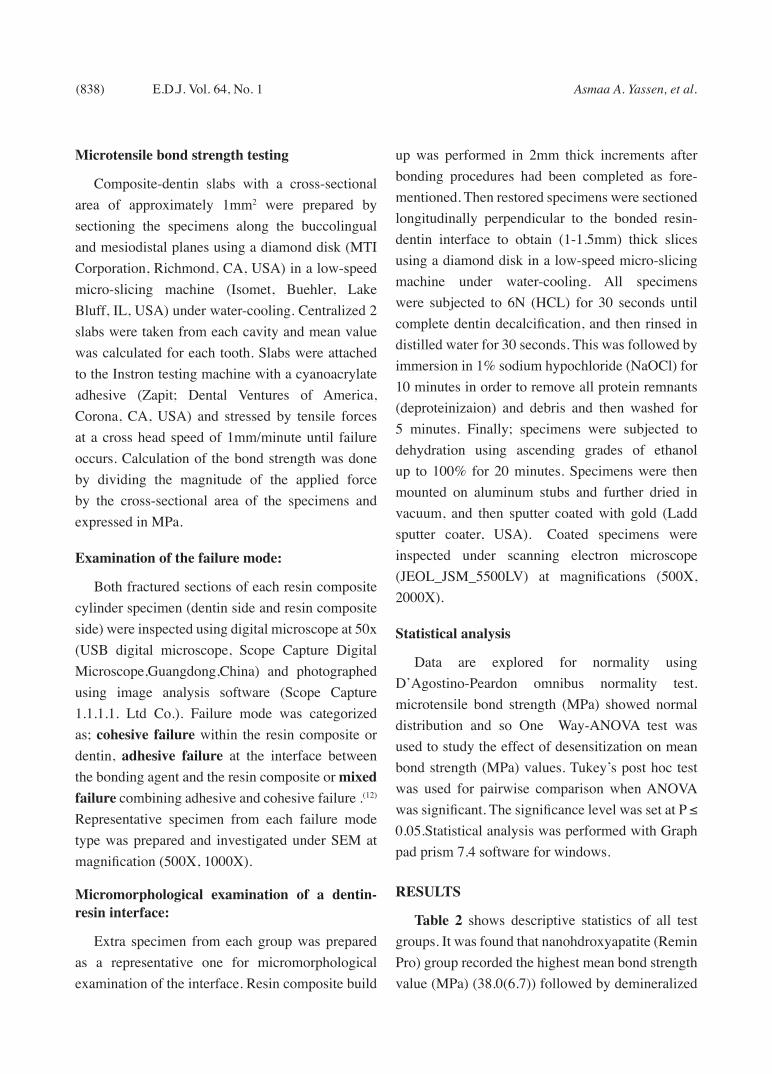
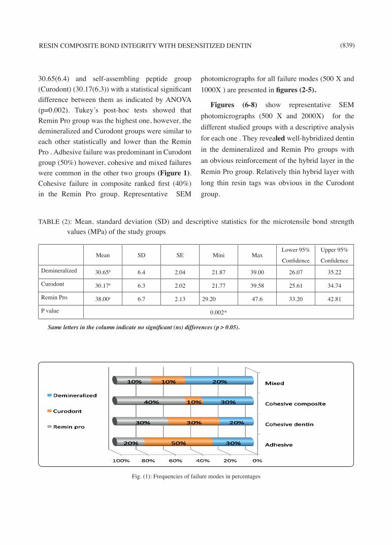
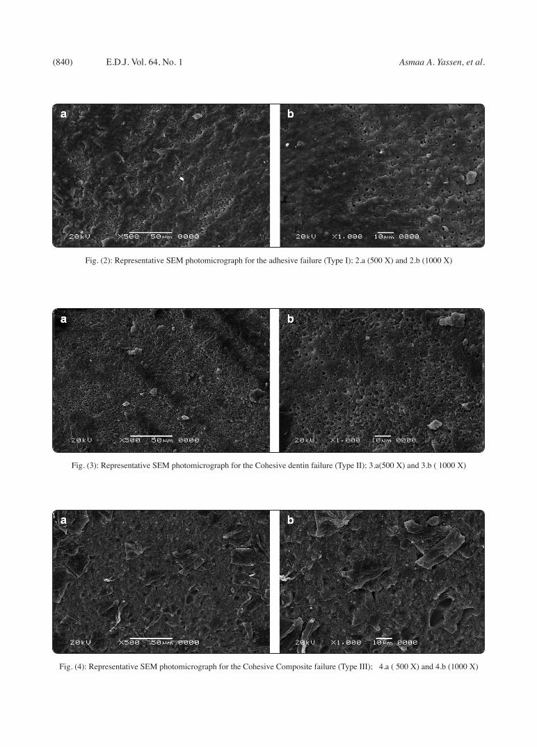
Both fractured sections of each resin composite cylinder specimen (dentin side and resin composite side) were inspected using digital microscope at 50x (USB digital microscope, Scope Capture Digital Microscope,Guangdong,China) and photographed using image analysis software (Scope Capture 1.1.1.1. Ltd Co.). Failure mode was categorized as; cohesive failure within the resin composite or dentin, adhesive failure at the interface between the bonding agent and the resin composite or mixed failure combining adhesive and cohesive failure .(12) Representative specimen from each failure mode type was prepared and investigated under SEM at magnification (500X, 1000X).
Micromorphological examination of a dentin-resin interface:
Extra specimen from each group was prepared as a representative one for micromorphological examination of the interface. Resin composite build
up was performed in 2mm thick increments after bonding procedures had been completed as fore-mentioned. Then restored specimens were sectioned longitudinally perpendicular to the bonded resin-dentin interface to obtain (1-1.5mm) thick slices using a diamond disk in a low-speed micro-slicing machine under water-cooling. All specimens were subjected to 6N (HCL) for 30 seconds until complete dentin decalcification, and then rinsed in distilled water for 30 seconds. This was followed by immersion in 1% sodium hypochloride (NaOCl) for 10 minutes in order to remove all protein remnants (deproteinizaion) and debris and then washed for 5 minutes. Finally; specimens were subjected to dehydration using ascending grades of ethanol up to 100% for 20 minutes. Specimens were then mounted on aluminum stubs and further dried in vacuum, and then sputter coated with gold (Ladd sputter coater, USA). Coated specimens were inspected under scanning electron microscope
(JEOL_JSM_5500LV) at magnifications (500X, 2000X).
Statistical analysis
Data are explored for normality using D’Agostino-Peardon omnibus normality test. microtensile bond strength (MPa) showed normal distribution and so One Way-ANOVA test was used to study the effect of desensitization on mean bond strength (MPa) values. Tukey’s post hoc test was used for pairwise comparison when ANOVA was significant. The significance level was set at P ≤ 0.05.Statistical analysis was performed with Graph pad prism 7.4 software for windows.
RESULTS
Table 2 shows descriptive statistics of all test groups. It was found that nanohdroxyapatite (Remin Pro) group recorded the highest mean bond strength value (MPa) (38.0(6.7)) followed by demineralized
RESIN COMPOSITE BOND INTEGRITY WITH DESENSITIZED DENTIN (839)
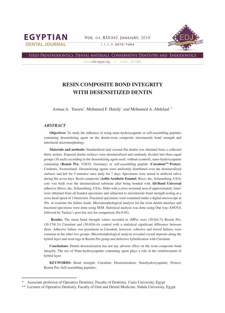
30.65(6.4) and self-assembling peptide group (Curodont) (30.17(6.3)) with a statistical significant difference between them as indicated by ANOVA (p=0.002). Tukey’s post-hoc tests showed that Remin Pro group was the highest one, however, the demineralized and Curodont groups were similar to each other statistically and lower than the Remin Pro . Adhesive failure was predominant in Curodont group (50%) however, cohesive and mixed failures were common in the other two groups (Figure 1). Cohesive failure in composite ranked first (40%) in the Remin Pro group. Representative SEM
photomicrographs for all failure modes (500 X and 1000X ) are presented in figures (2-5).
Figures (6-8) show representative SEM photomicrographs (500 X and 2000X) for the different studied groups with a descriptive analysis for each one . They revealed well-hybridized dentin in the demineralized and Remin Pro groups with an obvious reinforcement of the hybrid layer in the Remin Pro group. Relatively thin hybrid layer with long thin resin tags was obvious in the Curodont group.
TABLE (2): Mean, standard deviation (SD) and descriptive statistics for the microtensile bond strength values (MPa) of the study groups
Mean SD SE Mini MaxLower 95%
Confidence
Upper 95%
ConfidenceDemineralized 30.65b 6.4 2.04 21.87 39.00 26.07 35.22
Curodont 30.17b 6.3 2.02 21.77 39.58 25.61 34.74
Remin Pro 38.00a 6.7 2.13 29.20 47.6 33.20 42.81
P value 0.002*
Same letters in the column indicate no significant (ns) differences (p > 0.05).
Fig. (1): Frequencies of failure modes in percentages
(840) Asmaa A. Yassen, et al.E.D.J. Vol. 64, No. 1
Fig. (2): Representative SEM photomicrograph for the adhesive failure (Type I); 2.a (500 X) and 2.b (1000 X)
Fig. (3): Representative SEM photomicrograph for the Cohesive dentin failure (Type II); 3.a(500 X) and 3.b ( 1000 X)
Fig. (4): Representative SEM photomicrograph for the Cohesive Composite failure (Type III); 4.a ( 500 X) and 4.b (1000 X)

RESIN COMPOSITE BOND INTEGRITY WITH DESENSITIZED DENTIN (841)
Fig. (5): Representative SEM photomicrograph for the mixed failure (Type IV) ;5.a ( 500 X) and 5.b (1000 X)
Fig. (6): Representative SEM photomicrograph for the demineralized dentin interface with the resin composite showing thick hybrid layer and a well-hybridized dentin with a numerous anastomosing very long resin tags [6.a (500 X)]. Multiple lateral branching with irregular resin tags boundaries denoting their intimate adaptation with the dentinal tubules [ 6.b (2000 X)]
Fig. (7): Representative SEM photomicrograph for the demineralized dentin interface treated with Curodont with the resin composite showing a widely opened dentinal tubules, homogenous relatively thin hybrid layer, long thin deeply penetrating funnel-shaped resin tags[7.a (500 X)]. Minor lateral branching with relatively smooth resin tags [7.b (2000 X)]

(842) Asmaa A. Yassen, et al.E.D.J. Vol. 64, No. 1
DISCUSSION
Management of dentin hypersensitivity is a challenging issue facing the dentists in the daily practice. It is a common problem resulted from the presence of patent dentinal tubules exposed to different stimuli such as temperature fluctuation, air blast, strong chemicals, and pressure changes leading to movement of the fluids inside them and stimulation of nerve endings in the pulp. Unfortunately, the outcome of the treatment is variable and cannot be expected. It includes one of two protocols; the first one is to prevent nerve excitation and the second is to occlude the dentinal tubules and minimize their permeability. Successful treatment is measured by its durability as some agents produce transient pain relief and others resulted in prolonged effect. Fluoride agents are commonly used, however their effect is limited to the superficial occlusion or narrowing of the dentinal tubules and so searching for alternatives is mandatory. (13)
The manufactures have been introduced some remineralizing agents that have the power to interact with the dentinal tubules and seal them and so they can be used as desensitizing agents. These include the nano-hydroxyapatite and self-assembling peptides containing pastes. As it was
mentioned before the patients might not be satisfied by the topically applied agents and they asked for another restorative treatment and in this situation, resin composite can be used as a restorative material. The substrate here is not the optimal one but it is a desensitized dentin with obliteration of dentinal tubules, higher mineral contents, and altered wettability. Bond strength results between the resin composite and the desensitized dentin were controversial. (14) As a consequence of this dilemma, the null hypothesis of this study stated that there was not any significant effect of the dentin desensitization using nano-hydroxyapatite and self-assembling peptides containing agents on the resin composite bond quality. It was partially accepted as it was found that nano-hydroxyapatite (Remin Pro) containing agents produced higher bond strength values than those of the demineralized dentin (Control) and the self-assembling peptides (Curodont) (Table 2).
Natural hydroxyapatite (HAp) is considered the main ingredient of the enamel and dentin inorganic part. Introduction of synthetic hydroxyapatite espe-cially the nano-sized one opened the door towards its widespread use in dentistry. It has biomimetic function due to its great similarity with the struc-tural crystals of the tooth structure and it has a great
Fig. (8) Representative SEM photomicrograph for the demineralized dentin interface treated with Remin Pro with the resin composite showing hybrid layer with different thicknesses and multiple aggregated resin tags forming bundles[8.a (500 X)]. Clear evidence of crystals accumulation along the hybrid layer and resin bundles 8.b (2000 X)

RESIN COMPOSITE BOND INTEGRITY WITH DESENSITIZED DENTIN (843)
potentiality towards the occlusion of dentinal tu-bules.(15-17) According to the findings of the current study, higher bond strength values of the Remin Pro (38.0 (6.7) MPa) was evident and it could be attrib-uted to the chemical interaction between the nano-hydroxyapatite particles and the demineralized dentin. These particles act as a template for precipi-tation of Calcium and Phosphate in the demineral-ized dentin and their nanosize (20nm) with higher reactivity permit deeper penetration inside the den-tinal tubules and interaction with the demineralized dentin. (18) It has also a high amount of fluoride in the form of sodium fluoride (1,450 ppm fluoride) which reacts with the Calcium and forms Calcium fluo-ride crystals. (19) In addition to the effect of ethanol which might play a role in the deep infiltration of the nano-hydroxyapatite crystals. This interaction was augmented by the adhesive used in the study. It has mild acidic pH (3.2) which can penetrate this mineralized layer and interlock micromechani-cally with the desensitized dentin substrate together with the chemical bond which was created due to the inclusion of 10-methacryloyloxydecyl dihydro-gen phosphate MDP monomer in its composition.(20) MDP is an acidic monomer which contains a dihydrogen phosphate group for tooth etching and a methacrylate group for cross-linking with other resin monomers. (21,22) Furthermore, it is known to interact most intensively with hydroxyapatite and forms a hydrolytically stable bond with calcium. (23) The strong interface can be clearly seen in the photomicrographs (8 a and b) which revealed re-inforced hybrid layer with crystal deposition along the path of the dentinal tubules. Also, the strong in-terface was evident in the high percentage of cohe-sive and mixed failure (70%, 10% respectively) and low adhesive mode (20%) in the Remin Pro group.
This finding is in agreement with Pires-de-Souza et al in 2007 and Besinis et al in 2012 who stated that dentin desensitization with bioactive agents improved its bonding with resin composite. (20, 24) On the contrary Pashley et al in 1993 mentioned that dentin desensitization reduced
the bond strength due to the crystal precipitation on the dentin surface which has buffering capacity on the adhesive used leading to superficial weak hybridization. (14) Studies were done by Sevimay et al. in 2009 and Makkar in 2014 on microtensile bond strengths of the composite to dentin treated with different desensitizers revealed inferior bond strength in groups desensitized with Glutraldehyde and HEMA, HEMA and META, Thermokind-F and oxalates; as compared to the control group receiving no desensitizing treatment. (25,26) This contradiction might be due to the difference in the desensitizing agent and the type of adhesive (MDP free) used in these studies. Also there is a study found no significant difference between the microshear bond strength value of the desensitized dentin and the control group in which Relief ACP [0.375 % Amorphous Calcium Phosphate (ACP), 0.22% NaF, 5% KNO3 ] or 0.4% stannous fluoride and Xeno IV (PENTA, Mono, Di and Trimethacrylate resins, cetylamine hydrofluoride, acetone-water) self-etch adhesive were used. (27) . The finding was referred to the presence of fluoride in Xeno IV composition which chemically bonded to the fluoride precipitated from the desensitizing agents.
Results of self-assembling peptides (Curodont) showed same bond strength values as in the control demineralized only group with a relatively thin hybrid layer and thin long resin tags.(Table 2 and Figure 7 a and b) P11-4 is the structural unit of the self-assembling peptide which undergoes geometrical assembling forming a 3D scaffold for deposition of Calcium and Phosphate in the form of hydroxyapatite. (28,29) Curodont TM Protect is a product based on P11-4 technology, together with fluoride (900PPM), and calcium phosphate. In this group, the crystals deposition was not obvious like the Remin Pro group and this was shown in the SEM photomicrographs. Relatively thin hybrid layer with thin resin tags explained the higher percentage of adhesive failure (50%) in this group. Despite not increasing the bond strength,
(844) Asmaa A. Yassen, et al.E.D.J. Vol. 64, No. 1
Curodont Protect desensitizing agent did not affect the resin composite bonding adversely. It interacted with the demineralized dentin and enhanced its remineralization and altering its hydrophilicity without leaving surface deposits which might interfere with subsequent bonding procedures. This finding clarifies the role of universal adhesive used in this study which interacted with the demineralized dentin in the control group and produced bond strength value comparable to that one obtained in the Curodont group. Figure 6 a and b showed the interface between the resin composite and demineralized dentin with a homogenous hybrid layer, numerous long branched resin tags which indicated the intimacy between the restoration and the substrate. On the opposite side, Barbosa-Martins et al in 2017 and 2018 stated that self-assembling peptides improved the bonding performance of resin composite to sound and caries-affected dentin. The contradiction might be attributed to the use of Curodont Repair instead of Protect which was used in this study. (30,31)
At the end, clinicians should be aware that previous desensitizing treatment might affect the bond strength of resin composite. Therefore, further clinical studies must be conducted in order to evaluate the effects of desensitizing treatments on the bond strength of restorative materials.
CONCLUSIONS
Under the limitation of the present study, the fol-lowing points are evident:
1- Remin Pro nano-hydroxyapatite containing de-sensitizing paste is a promising line of treatment for hypersensitive dentin which enhances sub-sequent bonding with the resin composite.
2- Desensitization with self- assembling peptide paste (Curodont Protect) did not affect the inter-face negatively.
REFERENCES
1. Dababneh RH, Khouri AT, Addy M. Dentine hypersensi-tivity - an enigma? A review of terminology, mechanisms, aetiology and management. Br Dent J. 1999; 187:606-11.
2. American Academy of Operative D. Non-carious cervical lesions. Recommendations for clinical practice. Oper Dent. 2003;28:109-13.
3. Camilotti V, Zilly J, Busato Pdo M, Nassar CA, Nassar PO. Desensitizing treatments for dentin hypersensitivity: a randomized, split-mouth clinical trial. Braz Oral Res. 2012;26:263-8.
4. Kirkham J, Firth A, Vernals D, Boden N, Robinson C, Shore RC, Brookes SJ, Aggeli A. Self-assembling peptide scaffolds promote enamel remineralization. J Dent Res. 2007;86:426–30
5. Bröseler F, Tietmann C, Schleich R, Drechsel T, Bommer C. Effect of Curodont Repair in patients with buccal carious lesions: a mono-centre, singleblinded, randomised, controlled, split-mouth study-intermediate report. Clin Oral Investig. 2013; 17:1055.
6. Schlee M, Rathe F, Bommer C. Effect of Curodont Repair in patients with proximal carious lesions: uncontrolled, non-interventional study-interim report. Clin Oral Investig. 2013; 17:1046–47.
7. Heshmat H, Ganjkar M, Jaberi S, Fard M. The effect of remin pro and mi paste plus on bleached enamel surface roughness. J Dent. 2014;11(2):131-36
8. Akca T, Yazici AR, Çelik Ç, Özgünaltay G, Dayangaç B. The Effect of Desensitizing Treatments on the Bond Strength of Resin Composite to Dentin Mediated by a Self-etching Primer. Oper Dent. 2007; 32(5): 451-56
9. Santana F. Influence of method and period of storage on the microtensile bond strength of indirect composite resin restorations to dentine. J Restor Dent. 2008; 22(4):352-357.
10. Rahiotis C, Vougiouklakis G. Effect of CPP-ACP agent on the demineralization and remineralization of dentine in vitro. J Dent. 2007; 35(8):695–98 .
11. Castellan CS, Pereira PN, Grande RH, Bedran-Russo AK. Mechanical characterization of proanthocyanidin-dentin matrix interaction. Dent Mater. 2010; 26:968-73.
12. Cavalcante LM, Erhardt MC, Bedran-de-Castro AK, Pimenta LA , Ambrosano GM. Influence of different

RESIN COMPOSITE BOND INTEGRITY WITH DESENSITIZED DENTIN (845)
tests used to measure the bond strength to dentin of two adhesive systems. Americ J Dent, 2006;19(1):37-40.
13. Orchardson R, Gillam DG. Managing dentin hypersensitivity. J Am Dent Assoc 2006;137:990-8
14. Pashley EL, Tao L , Pashley DH. Effects of oxalate on dentin bonding American J of Dent. 1993; 6(3):116-1
15. Watanabe J, Akashi M. Formation of hydroxyapatite provides a tunable protein reservoir within porous polyester membranes by an improved soaking process. Biomacromolecules 2007;8:2288-93.
16. Ohta K, Kawamata H, Ishizaki T, Hayman R. Occlusion of dentinal tubules by nano-hydroxyapatite. J Dent Res 2007; 86(Spec Iss A):New Orleans Abstracts no.1759.
17. Wang ZJ, Sa Y, Ma X, Wang YN, Jiang T. The preparation of nano-hydroxyapatite and preliminary observation on its effects on the occlusion of dentinal tubule. Zhonghua Kou Qiang Yi Xue Za Zhi 2009;44:297-300.
18. Kaehler T. Nanotechnology: Basic concepts and defini-tions. Clin Chem 1994; 40:1797-9.
19. Jena A, Kala S, Shashirekha G. Comparing the effectiveness of four desensitizing toothpastes on dentinal tubule occlusion: A scanning electron microscope analysis. J Conserv Dent. 2017;20(4):269-72.
20. Muñoz MA, Luque-Martinez I, Malaquias P, Hass V, Reis A, Campanha NH, Loguercio AD. In vitro longevity of bonding properties of universal adhesives to dentin. Oper Dent 2015;40(3):282-92.
21. Moszner N, Salz U, Zimmermann J. Chemical aspects of self-etching enamel–dentin adhesives: a systematic re-view. Dent Mater 2005;21:895–910.
22. Van Landuyt KL, Snauwaert J, De Munck J, Peumans M, Yoshida Y, Poitevin A. Systematic review of the chemical
composition of contemporary dental adhesives. Biomateri-als 2007;28:3757–85.
23. Inoue S, Koshiro K, Yoshida Y, De Munck J, Nagakane K, Suzuki K. Hydrolytic stability of self-etch adhesives bonded to dentin. J Dent Res. 2005;84:1160–4.
24. Besinis A, Van Noort R, Martin N. Infiltration of demineralized dentin with silica and hydroxyapatite nanoparticles. Dent Mat 2012; 28: 1012-23.
25. Sevimay M, Ozyesil AG, Uludamar A. Microtensile bond strengths of composite to dentin treated with different desensitizers. SU Dishek Fak Derg. 2009;18:154–60.
26. Makkar S, Goyal M, Kausha A, Hegde V. Effect of desensitizing treatments on bond strength of resin composites to dentin – an in vitro study. J Conserv Dent. 2014; 17(5): 458–61.
27. Elhassan MHA, Sakr AK, Ibrahim MA. Microshear Bond Strength Of A Self Etch Adhesive To Desensitized Dentin. Cairo Dental Journal. 2008;24:463–70.
28. Kyle S, Aggeli A, Ingham E, McPherson MJ. Recombinant self-assembling peptides as biomaterials for tissue engineering. Biomaterials 2010; 31:9395–405.
29. Maude S, Tai LR, Davies RP, Liu B, Harris SA, Kocienski PJ, Aggeli A. Peptide synthesis and self-assembly. Top Curr Chem 2012; 310:27–69.
30. Barbosa-Martins LF, Sousa JP, Bedran-Russo AKB, Nascimento FD, Puppin-Rontani RM. Biomimetic agents can slow down degradation of caries-affected dentin–resin interfaces. Dent Mat. 2017; 33 S : e1–e92.
31. Barbosa-Martins LF, de Sousa JP, de Castilho ARF, Puppin-Rontani J, Davies RPW, Puppin-Rontani RM. Enhancing bond strength on demineralized dentin by pre-treatment with selective remineralising agents. J Mech Behav Biomed Mater. 2018 ;10(81):214-21.


![Download [ 5,2 MB ] - e-Gyanagar](https://static.fdokumen.com/doc/165x107/6316c6e79076d1dcf80b8d47/download-52-mb-e-gyanagar.jpg)


![Download [ 12.18 MB ] - e-Gyanagar](https://static.fdokumen.com/doc/165x107/632776a96d480576770d498f/download-1218-mb-e-gyanagar.jpg)