Electrospun collagen–chitosan–TPU nanofibrous scaffolds for tissue engineered tubular grafts
Upload
khangminh22Category
view
1download
0
Citation: Ji, Y.; Song, W.; Xu, L.; Yu,
D.-G.; Annie Bligh, S.W. A Review on
Electrospun Poly(amino acid)
Nanofibers and Their Applications of
Hemostasis and Wound Healing.
Biomolecules 2022, 12, 794. https://
doi.org/10.3390/biom12060794
Academic Editor: Albino Martins
Received: 9 May 2022
Accepted: 4 June 2022
Published: 7 June 2022
Publisher’s Note: MDPI stays neutral
with regard to jurisdictional claims in
published maps and institutional affil-
iations.
Copyright: © 2022 by the authors.
Licensee MDPI, Basel, Switzerland.
This article is an open access article
distributed under the terms and
conditions of the Creative Commons
Attribution (CC BY) license (https://
creativecommons.org/licenses/by/
4.0/).
biomolecules
Review
A Review on Electrospun Poly(amino acid) Nanofibers andTheir Applications of Hemostasis and Wound HealingYuexin Ji 1, Wenliang Song 1 , Lin Xu 1, Deng-Guang Yu 1,2,* and Sim Wan Annie Bligh 3,*
1 School of Materials Science and Engineering, University of Shanghai for Science and Technology,Shanghai 200093, China; [email protected] (Y.J.); [email protected] (W.S.);[email protected] (L.X.)
2 Shanghai Engineering Technology Research Center for High-Performance Medical Device Materials,Shanghai 200093, China
3 School of Health Sciences, Caritas Institute of Higher Education, Hong Kong 999077, China* Correspondence: [email protected] (D.-G.Y.); [email protected] (S.W.A.B.)
Abstract: The timely and effective control and repair of wound bleeding is a key research issue allover the world. From traditional compression hemostasis to a variety of new hemostatic methods,people have a more comprehensive understanding of the hemostatic mechanism and the structureand function of different types of wound dressings. Electrospun nanofibers stand out with nano size,high specific surface area, higher porosity, and a variety of complex structures. They are high-qualitymaterials that can effectively promote wound hemostasis and wound healing because they can imitatethe structural characteristics of the skin extracellular matrix (ECM) and support cell adhesion andangiogenesis. At the same time, combined with amino acid polymers with good biocompatibility notonly has high compatibility with the human body but can also be combined with a variety of drugs tofurther improve the effect of wound hemostatic dressing. This paper summarizes the application ofdifferent amino acid electrospun wound dressings, analyzes the characteristics of different materialsin preparation and application, and looks forward to the development of directions of poly(aminoacid) electrospun dressings in hemostasis.
Keywords: amino acids; hemostasis; electrospinning; wound dressing
1. Introduction
Coagulation is a complex process from unstable platelet embolism to stable insolublefibrin in plasma. The coagulation process requires a combined and integrated responsefrom all parts of the human body. In the initial hemostatic stage, platelets will forman initial platelet plug [1,2]. Platelets interact with von Willebrand factor (VWF), firstbinding to collagen, and then stably adhering to damaged vascular endothelial cells. Whenplatelets are activated, they trigger the aggregation of other locally activated platelets.Platelets gather through the fibrinogen bridge, produce fibrin clots through the action ofthrombin, and finally contract to form a tightly packed thrombus [3,4], as shown in Figure 1.Thrombin plays a central role in activating coagulation factors and platelets. When thesecondary hemostatic stage is reached, the coagulation cascade includes intrinsic andextrinsic coagulation pathways, which are initiated by different substances and differentsubstances and factors and are two different response pathways.
The coagulation pathways of the human body mainly include intrinsic hemostasis andextrinsic hemostasis. Intrinsic coagulation refers to that all the coagulation factors involvedcome from the blood, which is usually activated due to the contact between the blood andnegatively charged foreign bodies. When blood comes into contact with a foreign bodysurface, the coagulation factor FXII first binds to the foreign body surface and is activatedto FXIIa due to the negative charge of the foreign body surface. At the same time, thegenerated FXIIa can activate FXI to become FXIa, thus initiating the intrinsic coagulation
Biomolecules 2022, 12, 794. https://doi.org/10.3390/biom12060794 https://www.mdpi.com/journal/biomolecules

Biomolecules 2022, 12, 794 2 of 27
pathway. In addition, FXIIa can promote the formation of FXIIa by activating prekallikrein.All coagulation factors involved in this clotting process come from the blood itself. Onthe contrary, when vascular trauma exposes tissue factor (TF) to the blood, the externalpathway involving extrinsic coagulation factors begins [5,6].
Biomolecules 2022, 12, x FOR PEER REVIEW 2 of 27
Figure 1. Mechanism diagram of coagulation process. (A) Hemostatic simulation diagram of dam-aged vascular model; (B) an overview of the coagulation cascade.
The coagulation pathways of the human body mainly include intrinsic hemostasis and extrinsic hemostasis. Intrinsic coagulation refers to that all the coagulation factors in-volved come from the blood, which is usually activated due to the contact between the blood and negatively charged foreign bodies. When blood comes into contact with a for-eign body surface, the coagulation factor FXII first binds to the foreign body surface and is activated to FXIIa due to the negative charge of the foreign body surface. At the same time, the generated FXIIa can activate FXI to become FXIa, thus initiating the intrinsic coagulation pathway. In addition, FXIIa can promote the formation of FXIIa by activating prekallikrein. All coagulation factors involved in this clotting process come from the blood itself. On the contrary, when vascular trauma exposes tissue factor (TF) to the blood, the external pathway involving extrinsic coagulation factors begins [5,6].
Extrinsic coagulation refers to that not all the coagulation factors involved exist in the blood, and there are foreign coagulation factors involved in hemostasis. This process starts from the exposure of tissue factors to blood to the activation of FX. Due to the presence of calcium ions, TF is able to activate coagulation FVII. Then, the complex formed by the combination of TF and active FVII is able to continue activating FX, from activation of FX to the formation of fibrin clots. Then, the hydrolytic action possessed by thrombin is able to cleave fibrinogen, resulting in the formation of fibrin monomers [7]. The fibrin mono-mers are cross-linked with the involvement of Ca2+ and activated FXIII, eventually form-ing a solid fibrin clot that enhances platelet embolization during the initial hemostasis [8]. In the clinic, prothrombin time is used to reflect the status of the extrinsic coagulation pathway. The time required for extrinsic coagulation is short and the reaction is rapid. The extrinsic coagulation pathway is mainly regulated by tissue factor pathway inhibitor (TFPI). In addition, studies have shown that intrinsic and extrinsic coagulation pathways can activate each other [9].
Although materials that can directly activate the coagulation cascade can signifi-cantly improve hemostasis, they may cause systemic thrombosis. Metal ions, especially Ca2+, show a significant character in the coagulation process. Ca2+ is a cofactor that plays a universal role in the coagulation cascade. Important coagulation cascade steps such as the conversion of prothrombin to thrombin and the polymerization of monomers into fibrin involve Ca2+. Ca2+ is an imported cofactor in the coagulation cascade, accelerating platelet aggregation and activating the coagulation process, as well as enhancing material–wound adhesion. In addition, Ca2+ has a high water absorption rate and can be used to improve
Figure 1. Mechanism diagram of coagulation process. (A) Hemostatic simulation diagram of dam-aged vascular model; (B) an overview of the coagulation cascade.
Extrinsic coagulation refers to that not all the coagulation factors involved exist in theblood, and there are foreign coagulation factors involved in hemostasis. This process startsfrom the exposure of tissue factors to blood to the activation of FX. Due to the presenceof calcium ions, TF is able to activate coagulation FVII. Then, the complex formed by thecombination of TF and active FVII is able to continue activating FX, from activation of FXto the formation of fibrin clots. Then, the hydrolytic action possessed by thrombin is able tocleave fibrinogen, resulting in the formation of fibrin monomers [7]. The fibrin monomersare cross-linked with the involvement of Ca2+ and activated FXIII, eventually forming asolid fibrin clot that enhances platelet embolization during the initial hemostasis [8]. In theclinic, prothrombin time is used to reflect the status of the extrinsic coagulation pathway.The time required for extrinsic coagulation is short and the reaction is rapid. The extrinsiccoagulation pathway is mainly regulated by tissue factor pathway inhibitor (TFPI). Inaddition, studies have shown that intrinsic and extrinsic coagulation pathways can activateeach other [9].
Although materials that can directly activate the coagulation cascade can significantlyimprove hemostasis, they may cause systemic thrombosis. Metal ions, especially Ca2+,show a significant character in the coagulation process. Ca2+ is a cofactor that plays auniversal role in the coagulation cascade. Important coagulation cascade steps such as theconversion of prothrombin to thrombin and the polymerization of monomers into fibrininvolve Ca2+. Ca2+ is an imported cofactor in the coagulation cascade, accelerating plateletaggregation and activating the coagulation process, as well as enhancing material–woundadhesion. In addition, Ca2+ has a high water absorption rate and can be used to improve theplasticity of the material [10–13]. In addition, insufficient hemocompatibility can directlyactivate the coagulation process. Therefore, materials with passive access hemostaticmechanisms need to have surface properties such as hemocompatibility, antithrombotic,and anti-infective properties [14–16].
The general theory of wound healing is divided into four stages, namely the hemo-static, inflammatory, proliferative, and remodeling stages of the wound. The barrierfunction of the skin is very important, serving to insulate, retain moisture, and protect

Biomolecules 2022, 12, 794 3 of 27
the body from pathogens, so damage to the integrity of the skin can pose serious healthrisks to the body. In order to achieve rapid healing of damaged skin, the intrinsic healingprocess at the wound site begins immediately. Platelets will accumulate at the wound siteand the fibrin clot will form at the same time, so blood flow will stop within a few minutes(Figure 2A). At this time, inflammatory cells such as neutrophils and monocytes are re-cruited to the wound site by locally released growth factors (GFs) and cellular mediators(Figure 2B). Removal of foreign bodies, bacteria, and damaged intrinsic tissues is the maintask of the inflammatory response phase. When the phase is over, fibroblasts and epithelialcells are induced by macrophage GFs, proliferate, and migrate into the wound (Figure 2C).During the proliferative phase of the wound, new blood vessels gradually grow at thewound site, gradually producing enhanced collagen fiber, and granulation tissue consistingof epithelial cells, fibroblasts, and keratin-forming cells. However, complete wound healingis still a long-term process, taking weeks or months depending on the wound condition(Figure 2D). As the wound shrinks, granulation tissue is subsequently converted to a morestable ECM. Overall, wound healing time depends largely on the patient’s age, healthstatus, and the presence of external factors such as the presence of an unremoved foreignbody in the wound or the occurrence of a recurrent infection. Healing of acute woundsfollows the above process and is completed within 8–12 weeks. However, for chronicwounds that take longer, they tend to stagnate in the inflammatory phase with massiveexudation, severe infection, pain, and tissue necrosis, as in diabetic foot [17,18]. In suchcases, chronic wounds often take longer to heal, even by years.
Biomolecules 2022, 12, x FOR PEER REVIEW 3 of 27
the plasticity of the material [10–13]. In addition, insufficient hemocompatibility can di-rectly activate the coagulation process. Therefore, materials with passive access hemo-static mechanisms need to have surface properties such as hemocompatibility, antithrom-botic, and anti-infective properties [14–16].
The general theory of wound healing is divided into four stages, namely the hemo-static, inflammatory, proliferative, and remodeling stages of the wound. The barrier func-tion of the skin is very important, serving to insulate, retain moisture, and protect the body from pathogens, so damage to the integrity of the skin can pose serious health risks to the body. In order to achieve rapid healing of damaged skin, the intrinsic healing process at the wound site begins immediately. Platelets will accumulate at the wound site and the fibrin clot will form at the same time, so blood flow will stop within a few minutes (Figure 2A). At this time, inflammatory cells such as neutrophils and monocytes are recruited to the wound site by locally released growth factors (GFs) and cellular mediators (Figure 2B). Removal of foreign bodies, bacteria, and damaged intrinsic tissues is the main task of the inflammatory response phase. When the phase is over, fibroblasts and epithelial cells are induced by macrophage GFs, proliferate, and migrate into the wound (Figure 2C). During the proliferative phase of the wound, new blood vessels gradually grow at the wound site, gradually producing enhanced collagen fiber, and granulation tissue consist-ing of epithelial cells, fibroblasts, and keratin-forming cells. However, complete wound healing is still a long-term process, taking weeks or months depending on the wound con-dition (Figure 2D). As the wound shrinks, granulation tissue is subsequently converted to a more stable ECM. Overall, wound healing time depends largely on the patient’s age, health status, and the presence of external factors such as the presence of an unremoved foreign body in the wound or the occurrence of a recurrent infection. Healing of acute wounds follows the above process and is completed within 8–12 weeks. However, for chronic wounds that take longer, they tend to stagnate in the inflammatory phase with massive exudation, severe infection, pain, and tissue necrosis, as in diabetic foot [17,18]. In such cases, chronic wounds often take longer to heal, even by years.
Figure 2. Four stages of wound healing. (A) Hemostasis; (B) inflammation; (C) proliferation; (D) remodeling.
There are two views on wound healing, traditional clinical wound care believes that a dry environment with low humidity is conducive to wound healing and that oxygen in the air participates in the reproductive repair of wound tissue to accelerate the healing
Figure 2. Four stages of wound healing. (A) Hemostasis; (B) inflammation; (C) proliferation;(D) remodeling.
There are two views on wound healing, traditional clinical wound care believes that adry environment with low humidity is conducive to wound healing and that oxygen in theair participates in the reproductive repair of wound tissue to accelerate the healing process.Therefore, the breathable dressing can achieve the purpose of more oxygen contact andmake oxygen contact and make the cells proliferate rapidly. In fact, the dry healing methodhas obvious defects in theory: the cells in the wound do not react directly with oxygenin the air, which needs to be chemically combined with hemoglobin in the blood to beutilized [19,20]. Moreover, in actual clinical care practice, there are many disadvantages: thesurface of the bed is dry and dehydrated, and the crawling of epithelial cells is hindered; thewound leaks rapidly, and the dressing needs to be changed frequently, so it cannot maintaina mild environment for the wound, which affects the process of cell reproduction and

Biomolecules 2022, 12, 794 4 of 27
differentiation and makes the wound healing slow; the dry dressing is easy for the newlygrown granulation tissue to stick to each other, and frequent changing is likely to causesecondary damage to the wound. The contact time between wound surface and air is long,and the dry healing dressing has large pores, which cannot effectively organize the invasionof bacteria and other harmful substances in the air and is prone to infection [21–23].
An ideal wound dressing usually needs to be non-antigenic, biocompatible, semi-permeable, biodegradable, flexible, and cost effective, and the wound dressing shouldpreferably not adhere to the wound bed but should act on the surface of the wound. It mustprotect the wound from bacteria, infection, mechanical forces, and temperature, maintaina moist wound environment and be able to load certain drugs and active substances.Excellent wound dressings can effectively prevent wound infection and maintain thewound at the proper temperature and moderation in order to better promote surface-wound healing [24,25]. For severe burns, deep ulcers and other deeper wounds may notheal adequately due to the quality of the skin appendages not being good enough toproduce a certain amount of regenerative buds. Dermal substitutes are an effective wayto treat partially deep wounds, including decellularized and cell-seeded substitutes thatcan heal the defective site by promoting nearby cell migration. Full-layered wounds area major problem in treatment because they contain not only epidermis and dermis butalso subcutaneous fat and deep tissue, which can be more difficult to heal than superficialand partial thickness wounds. For these types of wounds, it is usually necessary to useautologous skin grafts or artificial skin substitutes for skin wound healing [26–28].
Electrospinning is able to structurally mimic the human ECM structure due to itsnanoscale structure. At the same time, due to the high specific surface area, high porosity,and small size of nanofibers, they can be loaded with active ingredients that can promotetherapy and can provide air exchange to the wound site and keep the healing environmentmoist. Electrospun nanofibers can also be personalized to the wound site, with handheldelectrospinning enabling immediate clinical coverage of the wound [29], further enhancingthe fit of the electrospun dressing to the wound [30,31]. The development of electrospinningtechnology in the field of hemostasis and wound healing has good prospects, and selectingamino acid-based polymers can largely enhance the biocompatibility of nanofibers forbetter utilization in biomedical and other fields [32,33].
A search of the literature in recent years on “Electrospun wound dressing” and “Aminoacid electrospun wound dressing” was carried out on the “Web of Science” platform. Asshown in Figure 3, the number of publications on the topic of “Electrospun wound dress-ings” has been increasing year by year and has been maintained at more than 100 articlesin recent years, indicating that the preparation of wound dressings by electrospinning hasbecome a hot spot for research. The number of publications on the topic of “Amino acidelectrospun wound dressings” is small but also on the rise, indicating that the developmentof amino acid materials in the field of electrospun wound dressings has certain prospects.This paper reviews the research progress of amino acid electrospinning materials in woundhemostasis and modification, and introduces the preparation technology of amino acidnanofibers and their latest applications in wound hemostasis and modification.

Biomolecules 2022, 12, 794 5 of 27Biomolecules 2022, 12, x FOR PEER REVIEW 5 of 27
Figure 3. Statistics of literature retrieval on the “Web of Science” platform with the subject of “Elec-trospun wound dressing” and “Amino acid electrospun wound dressing”, respectively.
2. Electrospinning Technology 2.1. Introduction to Electrospinning Technology
Electrospinning differs from other spinning technologies, mainly in that the polymer solution or melt under the action of a high voltage electric field produces flow and defor-mation, generating a Taylor cone at the tip of the spinneret, and when the electric field strength is large enough to enable the droplets to overcome surface tension, a high-speed jet is generated, which is deposited on the receiving device to obtain fibers after a short distance electric field force of high speed stretching, solvent volatilization, and curing [34–36]. There are four main components in the electrospinning device, a high-voltage power supply that can generate up to 30 kV to higher voltages, a high-precision micro-pump with flow control, a nozzle, and an aluminum foil collector for fiber collection. The prin-ciple is that a polymer solution of a certain viscosity is loaded into a syringe with a metal nozzle, fixed to a metering pump, and a high-voltage electrostatic field is applied to the nozzle and the collector. When the voltage exceeds the critical value and the electrostatic force is greater than the surface tension of polymer droplets, a jet is generated at the spin-neret, and the polymer solution is injected into the collector from the tip of the nozzle to form a nanofiber film. Fiber prepared by electrospinning is generally in the nanometer size and has extremely important applications in various fields due to its small size, large specific surface area, high porosity, large aspect ratio, and continuous uniformity stability.
Nanofibers have gained wide attention and applications in drug release, trauma recov-ery, and biological tissue engineering because of their small nanoscale size, large porosity, and high specific surface area. The diameter of nanofibers is smaller than that of cells, which can simulate the natural ECM in terms of structure and physiological function. It is also because of the nanoscale size of nanofibers that most tissues and organs in the human body are similar in form and structure to nanofibers, which also provides the possibility of bio-logical tissue and organ repair [37,38]. At the same time, we also need to focus on the rela-tionship between hemostatic materials and human body reactions. The use of materials with good biocompatibility allows better treatment without rejection, and materials with good biodegradability can reduce the pain of patients when removing dressings.
Electrospinning, as a technology that makes nanotechnology possible, allows not only the preparation of single polymer nanofibers but also the blending of multiple poly-mers and loading them with bioactive substances, and is widely used in wound healing. The extracellular matrix of the skin is composed of collagen, elastin, laminin, and various
Figure 3. Statistics of literature retrieval on the “Web of Science” platform with the subject of“Electrospun wound dressing” and “Amino acid electrospun wound dressing”, respectively.
2. Electrospinning Technology2.1. Introduction to Electrospinning Technology
Electrospinning differs from other spinning technologies, mainly in that the polymersolution or melt under the action of a high voltage electric field produces flow and deforma-tion, generating a Taylor cone at the tip of the spinneret, and when the electric field strengthis large enough to enable the droplets to overcome surface tension, a high-speed jet isgenerated, which is deposited on the receiving device to obtain fibers after a short distanceelectric field force of high speed stretching, solvent volatilization, and curing [34–36]. Thereare four main components in the electrospinning device, a high-voltage power supplythat can generate up to 30 kV to higher voltages, a high-precision micro-pump with flowcontrol, a nozzle, and an aluminum foil collector for fiber collection. The principle is that apolymer solution of a certain viscosity is loaded into a syringe with a metal nozzle, fixedto a metering pump, and a high-voltage electrostatic field is applied to the nozzle andthe collector. When the voltage exceeds the critical value and the electrostatic force isgreater than the surface tension of polymer droplets, a jet is generated at the spinneret,and the polymer solution is injected into the collector from the tip of the nozzle to form ananofiber film. Fiber prepared by electrospinning is generally in the nanometer size andhas extremely important applications in various fields due to its small size, large specificsurface area, high porosity, large aspect ratio, and continuous uniformity stability.
Nanofibers have gained wide attention and applications in drug release, traumarecovery, and biological tissue engineering because of their small nanoscale size, largeporosity, and high specific surface area. The diameter of nanofibers is smaller than thatof cells, which can simulate the natural ECM in terms of structure and physiologicalfunction. It is also because of the nanoscale size of nanofibers that most tissues andorgans in the human body are similar in form and structure to nanofibers, which alsoprovides the possibility of biological tissue and organ repair [37,38]. At the same time,we also need to focus on the relationship between hemostatic materials and human bodyreactions. The use of materials with good biocompatibility allows better treatment withoutrejection, and materials with good biodegradability can reduce the pain of patients whenremoving dressings.
Electrospinning, as a technology that makes nanotechnology possible, allows not onlythe preparation of single polymer nanofibers but also the blending of multiple polymersand loading them with bioactive substances, and is widely used in wound healing. Theextracellular matrix of the skin is composed of collagen, elastin, laminin, and various

Biomolecules 2022, 12, 794 6 of 27
polysaccharides and proteoglycans together as fibrous structural proteins. Nanofibers witha composition and structure/system similar to that of ECM in skin tissue can be fabricatedby electrospinning. Electrospun nanofibers can modulate skin cell proliferation, migration,differentiation, and extracellular matrix deposition responses [39–41]. Because of theseunique properties, the nanofibers can be used as surgical sutures, wound dressings, andother fields, as well as wound healing and tissue engineering. Infection prevention isachieved by antimicrobial agents to nanofiber dressings or sutures since bacteria havedeveloped some resistance to most antimicrobial agents such as antibiotics [42–44].
For synthetic or natural polymers with biodegradability electrospinning, nanofibershave been used to prepare nanofibrous membranes. These nanofibrous membranes havean extracellular matrix-like structure that provides a template for the proliferation of skincells, thereby stimulating tissue regeneration. Electrospun nanofiber membranes havehigh porosity and large specific surface area, which can absorb the wound exudate in time,promote the transfer of nutrients in the wound environment, maintain the gas exchangeat the wound site, and prevent the wound from dehydration [45,46]. In addition, thenanofiber membrane is soft, can be tailored to the size and shape of the wound, and ishighly pliable; the nanofiber membrane protects the wound on a physical level and is ableto protect the wound from other injuries and invasion by foreign microorganisms [47–51].Electrospun nanofiber membranes can also be used as drug carriers to load anesthetics,antibacterial agents, bioactive molecules, etc., depending on different types of skin wounds(e.g., burns, trauma, chronic disease ulcers, etc.) [52,53]. It is made to function whilecontrolling the release of drugs and promoting the wound healing process [54,55]. Inaddition, nanofiber film as a wound dressing needs to be in direct contact with the woundsurface and can be sterilized by radiation or UV to ensure its sterility. By immobilizingblood cells, platelets, and other clotting factors, the nanofiber matrix, which has a structuresimilar to that of natural fibers, provides control of bleeding [56,57]. There are many waysto prepare nanofiber matrices, but the most traditional method is electrospinning.
2.2. Electrospinning Classification
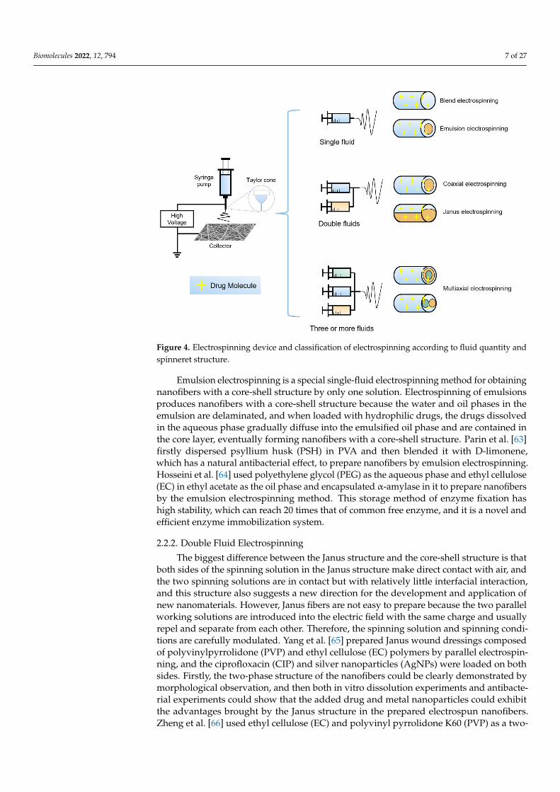
Electrospinning can be classified into single fluid electrospinning, double fluid elec-trospinning, and multi-fluid electrospinning based on the type of spinning solution usedin electrospinning. According to the geometry of the spinneret or nozzle that controlsthe production of nanofibers with different morphological structures, single-fluid electro-spinning can be divided into blend electrospinning and emulsion electrospinning, anddouble-fluid electrospinning into coaxial electrospinning and Janus electrospinning andmulti-fluid electrospinning. It is also possible to selectively load functional drugs duringthe preparation of spinning solutions, as shown in Figure 4.
2.2.1. Single Fluid Electrospinning
Single-fluid electrospinning is the most basic production method of electrospinning,usually one or several polymers used are co-blended and dissolved in a solvent, and then in-organic nanoparticles, drugs, etc., that need to be added are added forelectrospinning [58,59]. Single-fluid electrospinning is limited by the presence of onlyone spinning solution, thus also limiting the application of some good performance butnon-spinnable polymer solutions [60]. Nasser et al. [61] loaded the antibiotic gentamicinsulfate and the local anesthetic lidocaine hydrochloride on PLLA nanofibers using poly(L-lactic acid) (PLLA) as the spinning solution substrate. In addition, the loading of aluminumchloride with a hemostatic effect on PLLA nanofibers can better utilize the hemostaticeffect of nanofibers. Habiba et al. [62] co-blended chitosan, polyvinyl alcohol (PVA), andzeolite and performed electrospinning to obtain nanofibers with good adsorption anddesorption rates, which can be well applied to wastewater treatment for the adsorption andpurification of metal ions, etc. Single-fluid electrospinning is the most basic electrospinningmethod, on the basis of which two fluids and even multi-fluid electrospinning methodshave also been developed, extending the application of electrospinning in various fields.
Biomolecules 2022, 12, 794 7 of 27
Biomolecules 2022, 12, x FOR PEER REVIEW 7 of 27
Figure 4. Electrospinning device and classification of electrospinning according to fluid quantity and spinneret structure.
2.2.1. Single Fluid Electrospinning Single-fluid electrospinning is the most basic production method of electrospinning,
usually one or several polymers used are co-blended and dissolved in a solvent, and then inorganic nanoparticles, drugs, etc., that need to be added are added for electrospinning [58,59]. Single-fluid electrospinning is limited by the presence of only one spinning solu-tion, thus also limiting the application of some good performance but non-spinnable pol-ymer solutions [60]. Nasser et al. [61] loaded the antibiotic gentamicin sulfate and the local anesthetic lidocaine hydrochloride on PLLA nanofibers using poly(L-lactic acid) (PLLA) as the spinning solution substrate. In addition, the loading of aluminum chloride with a hemostatic effect on PLLA nanofibers can better utilize the hemostatic effect of nanofibers. Habiba et al. [62] co-blended chitosan, polyvinyl alcohol (PVA), and zeolite and per-formed electrospinning to obtain nanofibers with good adsorption and desorption rates, which can be well applied to wastewater treatment for the adsorption and purification of metal ions, etc. Single-fluid electrospinning is the most basic electrospinning method, on the basis of which two fluids and even multi-fluid electrospinning methods have also been developed, extending the application of electrospinning in various fields.
Emulsion electrospinning is a special single-fluid electrospinning method for obtain-ing nanofibers with a core-shell structure by only one solution. Electrospinning of emul-sions produces nanofibers with a core-shell structure because the water and oil phases in the emulsion are delaminated, and when loaded with hydrophilic drugs, the drugs dis-solved in the aqueous phase gradually diffuse into the emulsified oil phase and are con-tained in the core layer, eventually forming nanofibers with a core-shell structure. Parin et al. [63] firstly dispersed psyllium husk (PSH) in PVA and then blended it with D-limo-nene, which has a natural antibacterial effect, to prepare nanofibers by emulsion electro-spinning. Hosseini et al. [64] used polyethylene glycol (PEG) as the aqueous phase and ethyl cellulose (EC) in ethyl acetate as the oil phase and encapsulated α-amylase in it to
Figure 4. Electrospinning device and classification of electrospinning according to fluid quantity andspinneret structure.
Emulsion electrospinning is a special single-fluid electrospinning method for obtainingnanofibers with a core-shell structure by only one solution. Electrospinning of emulsionsproduces nanofibers with a core-shell structure because the water and oil phases in theemulsion are delaminated, and when loaded with hydrophilic drugs, the drugs dissolvedin the aqueous phase gradually diffuse into the emulsified oil phase and are contained inthe core layer, eventually forming nanofibers with a core-shell structure. Parin et al. [63]firstly dispersed psyllium husk (PSH) in PVA and then blended it with D-limonene,which has a natural antibacterial effect, to prepare nanofibers by emulsion electrospinning.Hosseini et al. [64] used polyethylene glycol (PEG) as the aqueous phase and ethyl cellulose(EC) in ethyl acetate as the oil phase and encapsulated α-amylase in it to prepare nanofibersby the emulsion electrospinning method. This storage method of enzyme fixation hashigh stability, which can reach 20 times that of common free enzyme, and it is a novel andefficient enzyme immobilization system.
2.2.2. Double Fluid Electrospinning
The biggest difference between the Janus structure and the core-shell structure is thatboth sides of the spinning solution in the Janus structure make direct contact with air, andthe two spinning solutions are in contact but with relatively little interfacial interaction,and this structure also suggests a new direction for the development and application ofnew nanomaterials. However, Janus fibers are not easy to prepare because the two parallelworking solutions are introduced into the electric field with the same charge and usuallyrepel and separate from each other. Therefore, the spinning solution and spinning condi-tions are carefully modulated. Yang et al. [65] prepared Janus wound dressings composedof polyvinylpyrrolidone (PVP) and ethyl cellulose (EC) polymers by parallel electrospin-ning, and the ciprofloxacin (CIP) and silver nanoparticles (AgNPs) were loaded on bothsides. Firstly, the two-phase structure of the nanofibers could be clearly demonstrated bymorphological observation, and then both in vitro dissolution experiments and antibacte-rial experiments could show that the added drug and metal nanoparticles could exhibitthe advantages brought by the Janus structure in the prepared electrospun nanofibers.Zheng et al. [66] used ethyl cellulose (EC) and polyvinyl pyrrolidone K60 (PVP) as a two-

Biomolecules 2022, 12, 794 8 of 27
phase solution and loaded with tamoxifen citrate (TAM) as the active drug component,and electrospinning was performed using eccentric spinneret. As shown in Figure 5A,a distinct Janus structure with one side round and one side crescent can be observed byscanning electron microscopy. Due to the presence of Janus nanostructure, it can releaseTAM rapidly in the first stage and slowly in the latter phase. This nanofiber with a Janusstructure provides a new drug release system, which has a promising future in the field ofcontrolled drug release and further applications.
Coaxial electrospinning technology was developed on the basis of electrospinning.The emergence of coaxial electrospinning technology can make up for certain polymersdue to its electrospinning limitations, which cannot be stretched into fibers by ordinaryelectrospinning defects. Coaxial electrospinning has the advantage of further molding, canbe easy to spin a polymer solution as the shell layer, cannot be spun or is difficult to spin apolymer solution as the core layer, in the electrospinning process, and the outer layer of thesolution. In the electrospinning process, the outer layer solution acts as a template and thecore layer solution is spun into fibers [67–69]. Coaxial electrospinning is mostly used in themedical field for the controlled release of drugs, many of which have poor water solubilityand low dissolution rates. Combined with the small diameter, large porosity, and largespecific surface area of nanofibers, it provides a rapid drug release method to promotethe rapid dissolution of insoluble drugs [70,71]. To improve the viability of lactobacillus,Yu et al. [72] used polylactic acid (PLA) as the main raw material to produce nanofibers bycoaxial electrospinning method, using PLA and fructooligosaccharides (FOS) as the shellsolution, and lactobacillus plantarum was cultured in Man–Rogosa–Sharpe (MRS) agarto obtain the nucleation solution, which successfully encapsulated Lactobacillus into thenanofibers. This method successfully improved the viability of lactobacillus and proposed anovel encapsulation method for active substances. Li et al. [73] designed and prepared core-shell nanofibers with an ultrathin shell layer using the coaxial electrospinning technique,as shown in Figure 5B, with polyvinylpyrrolidone (PVP) K90 or polycaprolactone (PCL)as the core solution and selected drug and PVPK10 as the shell solution. The in vitrodissolution test confirmed that the core-shell nanofibers with ultrathin shells have goodsolubility and the drug in the fibers can be released within 1 min. This study can be used toprepare structured nanocomposites with ultrathin shells to enhance the rapid dissolutionof insoluble drugs. Coaxial electrospinning has been intensively investigated in the fieldsof drug delivery and sustained release.
2.2.3. Multi-Fluid Electrospinning
To adapt to more complex applications and to exhibit more diverse functions, multi-fluid electrospinning is gradually being widely studied. In the multi-fluid electrospinningprocess, at least one solution is required for electrospinning, which greatly enriches thevariety and number of polymers involved in electrospinning and thus brings a new re-search boom [74,75]. One of the triaxial electrospinning processes is similar to coaxial butcan further extend the capability of electrospinning in creating complex nanostructures.Yang et al. [76] used a spinnable ibuprofen and alcohol soluble protein mixture as the coresolution, the nonspinnable cellulose acetate (CA) solution was used as the middle layer, anda nonspinnable acetone/acetic acid solvent as the outermost layer. The spinning solutionflowing from the core layer of the spinneret is spinnable and can be electrospun to formnanofibers, while the CA in the middle layer is a non-spinnable solution that is deposited asa thin “nano-coat” on the core solution, while the external solvent has the characteristic ofensuring a stable and continuous spinning process. The presence of multi-fluids providesthe possibility of more structural combinations for spinning, while the complex spinningconditions and spinning equipment require further in-depth study. Xu et al. [77] used amixture of acetone, DMAc, and ethanol as solvent and dissolved different concentrationsof metformin hydrochloride (MET) and CA as core and middle layer fluids, respectively,and the pure solvent was used as the outer layer fluid to prepare nanofibers by a modifiedthree-layer electrospinning process, as shown in Figure 5C. This nanofiber was able to

Biomolecules 2022, 12, 794 9 of 27
achieve a slow release of MET for drugs with water solubility and prolong the adminis-tration time of water-soluble drugs. Chang et al. [78] fabricated a novel tri-fluid spinninghead with a shell enveloping two separate cores, as shown in Figure 5D. One shell solutionconsists of Eudragit® E100 (EE) and paracetamol (PAR), and the two core solutions consistof Eudragit® L100-55 (EL), PAR and Eudragit® S100 (ES), PAR, respectively. Eudragit® E100(EE), Eudragit® L100-55 (EL) and Eudragit® S100 (ES) were purchased from Rohm GmbH& Co. KG (Darmstadt, Germany). This sheath-separate-core nano-structure combinesthe three spinning solutions, as shown in Figure 5E, and the characteristic structure canbe observed by SEM images. This new electrospinning structure is able to release drugsaccording to different pH and is an intelligent responsive three-phase drug release system,which has good prospects for application in the field of controlled drug release. Along thisway, some brand-new electrospun nanostructures can be further developed for biomedicalapplications [79].
Biomolecules 2022, 12, x FOR PEER REVIEW 10 of 27
Figure 5. Nanofibers and spinnerets with different structures. (A) SEM pictures of nanofibers with Janus structure [66]; (B) production process diagram of coaxial electrospinning [73]; (C) schematic diagram of three-layer coaxial electrospinning spinneret [77]; (D) new three-fluid electrospinning spinneret [78]; (E) SEM images of nanofibers with sheath-separate-core nano-structure [78].
2.3. Electrospinning Influence Factors The factors affecting electrospinning are mainly divided into the nature of the spin-
ning solution itself, spinning parameters, and environmental parameters. The nature of the spinning solution itself includes the molecular weight of the polymer, molecular struc-ture, the nature of the solvent, the viscosity of the spinning solution, electrical conductiv-ity, etc. [80–82], as shown in Table 1. The molecular weight of the polymer largely affects the viscosity and rheological properties of the polymer, which is intuitively reflected in the diameter of the nanofibers; generally speaking, the higher the molecular weight of the polymer, the larger the diameter of the nanofibers. A faster solvent evaporation rate will cause the fibers to not completely split and refine, and the fiber diameter will increase; while a slow solvent evaporation rate will also make the fibers stick to each other on the collection plate, and no complete nanofibers will be obtained; the faster the solvent evap-oration rate, the larger the fiber porosity and specific surface area; the slower the solvent evaporation rate, the less easy to remove the solvent residue. In a certain range, the higher the viscosity of the spinning solution, the more likely a bead structure will be produced and block the spinneret; the lower the viscosity, the smaller the fiber diameter, but too low will produce an electrospray. The dielectric constant of the spinning solution will also have an impact on the nanofibers; in general, the higher the dielectric constant the smaller the diameter of the nanofibers, while a dielectric constant that is too small is also likely to produce bead fibers, which is not conducive to the preparation of nanofibers [83–85].
Figure 5. Nanofibers and spinnerets with different structures. (A) SEM pictures of nanofibers withJanus structure [66]; (B) production process diagram of coaxial electrospinning [73]; (C) schematicdiagram of three-layer coaxial electrospinning spinneret [77]; (D) new three-fluid electrospinningspinneret [78]; (E) SEM images of nanofibers with sheath-separate-core nano-structure, as indicatedby the red (The PEL-PAR core) and blue arrows (The ES-PAR core) [78].
2.3. Electrospinning Influence Factors
The factors affecting electrospinning are mainly divided into the nature of the spinningsolution itself, spinning parameters, and environmental parameters. The nature of thespinning solution itself includes the molecular weight of the polymer, molecular structure,

Biomolecules 2022, 12, 794 10 of 27
the nature of the solvent, the viscosity of the spinning solution, electrical conductivity,etc. [80–82], as shown in Table 1. The molecular weight of the polymer largely affects theviscosity and rheological properties of the polymer, which is intuitively reflected in thediameter of the nanofibers; generally speaking, the higher the molecular weight of thepolymer, the larger the diameter of the nanofibers. A faster solvent evaporation rate willcause the fibers to not completely split and refine, and the fiber diameter will increase; whilea slow solvent evaporation rate will also make the fibers stick to each other on the collectionplate, and no complete nanofibers will be obtained; the faster the solvent evaporation rate,the larger the fiber porosity and specific surface area; the slower the solvent evaporationrate, the less easy to remove the solvent residue. In a certain range, the higher the viscosityof the spinning solution, the more likely a bead structure will be produced and block thespinneret; the lower the viscosity, the smaller the fiber diameter, but too low will producean electrospray. The dielectric constant of the spinning solution will also have an impact onthe nanofibers; in general, the higher the dielectric constant the smaller the diameter of thenanofibers, while a dielectric constant that is too small is also likely to produce bead fibers,which is not conducive to the preparation of nanofibers [83–85].
Table 1. Key parameters affecting electrospinning.
Spinningsolution
properties
Polymer molecular weight The fiber diameter increases with the increase in polymermolecular weight
Solvent evaporation rate
The faster the solvent evaporates, the greater the fiber diameter; the fasterthe solvent evaporates, the greater the fiber porosity and specific surface
area; the slower the solvent evaporates, the less easy to remove thesolvent residue
Spinning solution viscosityThe higher the viscosity of the spinning solution, the easier it is to blockthe spinneret; the lower the viscosity, the smaller the fiber diameter, but
too low will produce electrospray
Conductivity of spinning solution The larger the dielectric constant, the smaller the fiber diameter; thesmaller the dielectric constant, the easier it is to produce beads of fiber
Spinningparameters
Spinning voltageThe higher the spinning voltage, the smaller the fiber size; too much
voltage will lead to unstable spinning; too little voltage fiber diameterwill be coarse, or even produce droplets
Liquid feeding speed The larger the flow rate, the larger the fiber diameter, too large willproduce droplets; low feed rate spinning process is easy to interrupt
Collector Influence the three-dimensional structure and arrangement of the product
Distance between spinning head and collecting plateSpacing is too small solvent cannot be fully evaporated; spacing is too
large to affect the electric field strength, but also make the fiber is not easyto deposit and fly into the air
Environmental parametersSpinning environment temperature Increasing the temperature increases the rate of solvent volatilization, and
hollow nanofibers can be obtained by increasing the temperature
Spinning environment humidity Elevated humidity reduces the rate of solvent evaporation, andnanocrystalline films can be obtained by increasing humidity
Spinning parameters include spinning voltage, liquid feed rate, collection distance,etc. [86]. The higher the spinning voltage, the higher the tensile strain rate, and the smallerthe nanofiber size will be; too much voltage will increase the speed of the fiber ejection sothat the solvent cannot be fully volatilized, which will also lead to spinning instability; toolittle voltage will cause the appearance of solution droplets, and the fiber diameter will becoarse [87]. For the feed rate, generally speaking, the greater the flow rate the larger thefiber diameter; a feed rate that is too low will easily interrupt the spinning process; anda feed rate that is too large will produce an unstable Taylor cone, and the droplets willdirectly drop down. The distance from spinneret to collector also affects nanofibers, thefiber cannot be fully stretched, and the solvent cannot be completely evaporated; a distancethat is too large will affect the electric field strength and will also affect the tensile strain ofthe fiber, but is also not conducive to the collection of fibers.
The environmental parameters of electrospinning mainly include the temperature andhumidity of the spinning environment. With the increase in temperature, the evaporationrate of the solvent in the spinning solution will also increase; generally, the viscosity of

Biomolecules 2022, 12, 794 11 of 27
the spinning solution will also decrease with the increase in temperature, and it is alsopossible to obtain hollow nanofibers by increasing the temperature [88]. Humidity affectsthe evaporation rate of the solvent, and too high humidity will lead to incomplete dryingof the solvent in the nanofibers.
3. Amino Acid3.1. Introduction to Amino Acids
Amino acids are the basic unit of biological functional macromolecular proteins and thebasic material of proteins required by animal nutrition. Amino acids are classified accordingto their side-chain groups and can be divided into polar amino acids and non-polar aminoacids, of which polar amino acids can be divided into basic amino acids, acidic amino acids,and neutral amino acids [89], as shown in Figure 6. Poly amino acids are a kind of naturalprotein mimic, which have the same primary structure as natural proteins and polypeptidematerials, and some poly amino acids can also simulate their secondary structure. Withthe diversity of amino acid structures, unique self-assembly structures and conformationaltransformation, high bioactivity, and good biocompatibility, they are widely used in thefield of biomaterials. With the rapid increase in the application of polymer materials inthe biomedical field, amino acid-derived functional polymers with the characteristics ofdegradability and high biocompatibility have also been vigorously developed. Becauseamino acids have bioactive side groups and diverse functional groups, they can realize avariety of functional polymerization, so they also have potential applications in imaging,drug delivery, cell adhesion, biodegradation, and so on [90,91].
Biomolecules 2022, 12, x FOR PEER REVIEW 12 of 27
3. Amino Acid 3.1. Introduction to Amino Acids
Amino acids are the basic unit of biological functional macromolecular proteins and the basic material of proteins required by animal nutrition. Amino acids are classified ac-cording to their side-chain groups and can be divided into polar amino acids and non-polar amino acids, of which polar amino acids can be divided into basic amino acids, acidic amino acids, and neutral amino acids [89], as shown in Figure 6. Poly amino acids are a kind of natural protein mimic, which have the same primary structure as natural proteins and polypeptide materials, and some poly amino acids can also simulate their secondary structure. With the diversity of amino acid structures, unique self-assembly structures and conformational transformation, high bioactivity, and good biocompatibil-ity, they are widely used in the field of biomaterials. With the rapid increase in the appli-cation of polymer materials in the biomedical field, amino acid-derived functional poly-mers with the characteristics of degradability and high biocompatibility have also been vigorously developed. Because amino acids have bioactive side groups and diverse func-tional groups, they can realize a variety of functional polymerization, so they also have potential applications in imaging, drug delivery, cell adhesion, biodegradation, and so on [90,91].
Figure 6. Amino acids and classification.
3.2. Polar Amino Acids 3.2.1. Basic Amino Acid
Basic amino acids are amino acids with hydroxide negative ions generated by hy-drolysis with multiple hydrogen positive ions, including arginine, lysine, and histidine. The side chains of this class of amino acids contain protonatable basic groups
Lysine (Lys) is a common amino acid. Lys, which is water-soluble, biodegradable, and non-cytotoxic, is able to effectively promote cell adhesion and proliferation at the in-terface of biomaterials and improve tissue regeneration. In biological media, as the amine group on the Lys molecule is susceptible to protonation, it interacts with negatively charged cell membranes. Lys and its derivatives play an important role in biomedicine and tissue engineering [92–94]. Poly ε-lysine (ε-PL) can be produced by Streptomyces albus
Figure 6. Amino acids and classification.
3.2. Polar Amino Acids3.2.1. Basic Amino Acid
Basic amino acids are amino acids with hydroxide negative ions generated by hydrol-ysis with multiple hydrogen positive ions, including arginine, lysine, and histidine. Theside chains of this class of amino acids contain protonatable basic groups
Lysine (Lys) is a common amino acid. Lys, which is water-soluble, biodegradable, andnon-cytotoxic, is able to effectively promote cell adhesion and proliferation at the interfaceof biomaterials and improve tissue regeneration. In biological media, as the amine groupon the Lys molecule is susceptible to protonation, it interacts with negatively charged

Biomolecules 2022, 12, 794 12 of 27
cell membranes. Lys and its derivatives play an important role in biomedicine and tissueengineering [92–94]. Poly ε-lysine (ε-PL) can be produced by Streptomyces albus and has avariable number of L-lysine residues that are chemically bonded through an amide bondbetween the ε-amino and α-carboxyl groups. Broad-spectrum antibacterial activity is animportant characteristic of poly-L-lysine, which can act as an antibacterial agent against bac-teria, fungi, and even viruses. ε-PL was introduced into polyacrylic acid (PAA)/polyvinylalcohol (PVA) electrospun nanofibers by Amariei et al. [95] for electrospinning together.Based on the antibacterial properties of ε-PL, the bacterial colonization rate of the ε-PLwas reduced by several hundred times compared to unfunctionalized dressings, whichlater also showed good biocompatibility after cytotoxicity testing. Lin et al. [92] blendedpoly-L-lysine with gelatin and glycerol to make nanofiber mats by electrospinning, andthe preparation process is shown in Figure 7A, where the addition of glycerol improvedthe mechanical strength of gelatin nanofibers and the addition of poly-L-lysine not onlyinhibited the growth of microorganisms but also extended the shelf life of food products,which is a promising food packaging material. Hyperbranched poly-L-lysine (HPLys) withlow shrinkage can be used as a 3D scaffold because of its special properties, and it can beused in heart tissue engineering, promotion of cell adhesion, and good biocompatibility.As a scaffold material used in the heart, polyaniline nanofibers are widely used, but theirbiocompatibility is insufficient. Fernandes et al. [96] used HPLys to modify polyaniline andblended them together for electrospinning to prepare nanofibers.
Arginine is often used as an additive to produce a facilitative effect on wound healingrather than a polymeric scaffold. Studies have shown that wound dressings loaded witharginine produce proline, which is required for collagen synthesis, promote the productionof biologically active molecules in the body [97]. The biocompatibility and longevity ofwound dressings can be enhanced by doping with arginine and controlling its sustainedrelease. Hussein et al. [98] loaded arginine onto nanofibers made from a blend of PVAand hyaluronic acid to improve the uniformity of nanofiber diameter, and the SEM sizeand morphology of the prepared nanofibers are shown in Figure 7B. Exhibiting excellenthemocompatibility and outstanding proliferation and adhesion potential, especially againstKlebsiella pneumonia showing sufficient antibacterial activity, this composite nanofiber couldbe further developed as a promising multifunctional wound dressing.
3.2.2. Acidic Amino Acid
Poly(γ-glutamate) (γ-PGA) is an anionic poly(amino acid) and can be naturally pro-duced by microorganisms such as Bacillus subtilis. γ-glutamyl transpeptidase in the humanbody can degrade γ-PGA to glutamate, which is non-toxic to the human body and widelypresent in the human body. γ-PGA has been gradually studied in biomedical fields, suchas drug delivery, bioadhesion, and so on, because of its good biocompatibility, versatility,biodegradability, and high water retention [99–101]. However, the high solubility and fastdissolution rate of γ-PGA in water are not favorable for application in fields such as tissuerepair scaffolds. So, in this case, cross-linking agents are needed to enhance the physicalproperties of γ-PGA for better application in the human body and biomedical field. Severalcommon cross-linking agents such as ethylene glycol diglycidyl ether, cystamine [102],oxazoline (OXA) [103], and L-lysine [104] are able to cross-link γ-PGA and improve itsstability in water. In addition, several studies have shown that electrostatically spun γ-PGAor γ-PGA-based composite fiber mats have good cell adhesion and proliferation ability.Lee et al. [105] innovatively co-blended PGA, polyethylene glycol (PEG), and TritonX-100and were the first to successfully prepare PGA nanofibers by the electrospinning method.To overcome the water solubility of PGA, they chose to use butyl PGA for electrospinningand finally obtained water-insoluble nanofibers. Tajima et al. [103] chose OXA as thecross-linking agent; when the ratio of γ-PGA/OXA was 60/40% wt, the prepared nanofiberfilms could achieve skin-like tensile properties. Lower crosslinker blending ratios lead tohigher hygroscopicity or water absorption, and due to the good mechanical properties andhygroscopicity of γ-PGA, the nanofibers prepared from it can be used in agrochemicals to

Biomolecules 2022, 12, 794 13 of 27
biomedical products, which have a wide scope of application. Xu et al. [106] synthesizedγ-PGA with L-cysteine and norbornene to synthesize photocrosslinkable γ-PGA-Nor. Dueto the good mechanical properties and spinnability of PEO, it was introduced as a carrierpolymer to facilitate the formation of electrospun fibers. The non-crosslinked fibers wereprepared by electrospinning and finally crosslinked by UV irradiation to functionalize thefiber surface. This special nanofiber network has a structure and size similar to naturalECM, with ideal mechanical strength to support cell adhesion and promote growth on it,and has a significant inhibitory effect on proliferative scarring. The drug release curve ofginsenoside Rg3 (GS-Rg3) loaded on the fiber is shown in Figure 7F.
Poly(aspartic acid) (PASP) is synthesized by thermal polymerization of aspartic acid, apolyamino acid with carboxylic acid side chains, which is biodegradable, chelating, anddispersing. It has good solubility in most pH solutions, including the oral cavity withpH = 6.8, and is a degradable polymer with good water solubility [107]. It has been shownthat poly(aspartic acid) is a polymer that can replace acrylic acid and is greener and moreeconomical [108–110]. Due to its versatility such as water solubility and biodegradability,PASP has great applications in water treatment, wound dressing, stent material, drugdelivery and release, etc. In addition, PSAP has functionalized carboxylic acid residues,which can be grafted with some polymers to improve the surface properties of the materialsand can be applied to the fields of targeted regulation, antifouling, tissue engineering, andso on [111]. Figure 7C is a summary of some characteristics of PASP and its application areas.PASP nanofibers are generally prepared by choosing polysuccinimide (PSI), obtained fromL-Asp catalyzed by phosphoric acid, as the precursor, and then cross-linking PSI nanofibersby hydrolysis to obtain PASP nanofibers. Zhang et al. [112] used the excellent adsorptionof metal ions by PASP, combined with the high specific surface area of nanofibers, todesign a sensor that can detect metal ions and can be reused, as shown in Figure 7E, anddesigned an electrospun nanofiber hydrogel membrane (ENHM) using PASP, which can beused as an aqueous solution colorimetric sensor for applications such as water treatment.Monlar et al. [113] electrospun PSI to obtain homogeneous nanofibers, which were then putinto an imidazole-based buffer solution with pH = 8 for hydrolysis to obtain PASP gel fibers.This method produced PASP nanofibers similar to human ECM with desirable mechanicaland biological properties. In 2017, Monlar et al. [114] again used coaxial electrospinningto improve the crosslinking process, using PSI as the shell polymer and adding PEO co-blending to improve the viscosity of the spinning solution and improve the stability of thejet. The crosslinking of 2,2,4 (2,4,4)-trimethyl-1,6-hexanediamine (THD) in the core layer ofthe nozzle occurs when the two solutions come into contact at the nozzle tip, forming PASP.This core-shell structure of nanofibers can effectively control the crosslinking time of thetwo solutions and complete the crosslinking with maximum efficiency, without cloggingthe nozzle, which is an effective method to prepare PASP nanofibers. The SEM imageof nanofiber is shown in Figure 7D. This insoluble gel fiber is pH responsive and can befurther applied in the future in the fields of drug release and tissue engineering.

Biomolecules 2022, 12, 794 14 of 27Biomolecules 2022, 12, x FOR PEER REVIEW 15 of 28
Figure 7. Preparation and application of polar amino acid electrospun nanofibers. (A) Electrospun gelatin-glycerin-ε-poly-lysine nanofibers [95]; (B) SEM images of selected PVA/HA, PVA/HA/CNCs, and PVA/HA/CNCs/L-arginine NFs scaffolds [98]; (C) properties and biomedical applications of poly(aspartic acid) and its derivatives [111]; (D) SEM image of electrospun nanofiber [114]; (E) application of procedure of PASP [112]; (F) Release curve of GS-Rg3 from fiber [106].
3.2.3. Neutral Amino Acid Glycine is an important amino acid in the human body and is involved in the pro-
duction of many important reactions and substances in the human body, including the production of DNA, proteins, and heme, as well as playing an important role in lipid me-tabolism, immune regulation, and neurotransmission. For this reason, glycine is often used to improve immunity and anti-inflammation, promote wound healing, and even im-prove neurological function, and has good biocompatibility, biodegradability, and excel-lent mechanical properties [115]. Alazzawi et al. [116] prepared nanofibers by electrospin-ning by blending PVA with an aqueous glycine solution. Such nanofibers with high spe-cific surface area can be well used in applications such as bio-scaffolds and drug transport.
The electrospinning conditions and characteristics of several polar amino acids are shown in Table 2.
Table 2. Electrospinning conditions and characteristics of common polar amino acids.
Amino Acids Additional Polymer
Solvent Electrospun Technique
Characteristic Ref.
Lysine Gelatin/glycerin Acetic acid Blend Excellent antibacterial ability against Lis-teria monocytogenes, a promising food
packaging material [92]
Commented [M3]: Please provide sharper image of figure 7B if have
Commented [吉4R3]: Yes, many thanks! We have tried our best, but the original document does not have high sharpness.
Figure 7. Preparation and application of polar amino acid electrospun nanofibers. (A) Electrospungelatin-glycerin-ε-poly-lysine nanofibers [95]; (B) SEM images of selected PVA/HA, PVA/HA/CNCs,and PVA/HA/CNCs/L-arginine NFs scaffolds [98]; (C) properties and biomedical applicationsof poly(aspartic acid) and its derivatives [111]; (D) SEM image of electrospun nanofiber [114];(E) application of procedure of PASP [112]; (F) Release curve of GS-Rg3 from fiber [106].
3.2.3. Neutral Amino Acid
Glycine is an important amino acid in the human body and is involved in the pro-duction of many important reactions and substances in the human body, including theproduction of DNA, proteins, and heme, as well as playing an important role in lipidmetabolism, immune regulation, and neurotransmission. For this reason, glycine is oftenused to improve immunity and anti-inflammation, promote wound healing, and even im-prove neurological function, and has good biocompatibility, biodegradability, and excellentmechanical properties [115]. Alazzawi et al. [116] prepared nanofibers by electrospinningby blending PVA with an aqueous glycine solution. Such nanofibers with high specificsurface area can be well used in applications such as bio-scaffolds and drug transport.
The electrospinning conditions and characteristics of several polar amino acids areshown in Table 2.

Biomolecules 2022, 12, 794 15 of 27
Table 2. Electrospinning conditions and characteristics of common polar amino acids.
Amino Acids AdditionalPolymer Solvent Electrospun Technique Characteristic Ref.
Lysine
Gelatin/glycerin Acetic acid BlendExcellent antibacterial ability against Listeriamonocytogenes, a promising food packaging
material[92]
PAA/PVA Distilled water Blend Long-lasting antibacterial activity with goodbiocompatibility [95]
PAN DCM/DMF Blend High biocompatibility and potential for culturingheart cells [96]
PAA Distilled water Blend The addition of polylysine enhances the mechanicalstrength and stability of PAA [117]
PLLA/PPY HFIP Coaxial Stable electrical properties, good biocompatibility,high cell adhesion rate [118]
Glutamic acid
PLGA TFA Blend Promotes wound healing and prevents tissueadhesions [101]
Cystamine(aftertreatment) TFA Blend Good water stability [102]
OXA Ethanol/water/hydrochloric Blend Good mechanical properties and similar to skin,
with certain moisture absorption properties [103]
PEG Distilled water Blend Uniform nanofiber diameter [105]
PEO Distilled water Blend Promotes cell adhesion and proliferation andinhibits proliferative scarring [106]
PVA Distilled water Blend Promotes cell adhesion and can be used as a tissueengineering scaffold [119]
PCL HFIP Blend Improves the solubility of florfenicol (FF) andpromotes the in vitro release of the drug [120]
Aspartic acid
PSI — BlendStrong adsorption of metal ions and reduced water
solubility after cross-linking, can be used as acolorimetric sensor for aqueous solutions
[112]
PSI DMF Blend A biocompatible fiber scaffold [113]
PSI/PEO/THD DMF Coaxial pH sensitive for smart drug release applications [114]
Arginine PVA/HA Distilled water Blend Accelerates wound healing and tissue regeneration [98]
Glycine PVA Distilled water Blend High specific surface area for bio-scaffold and drugtransport applications [116]
3.3. Nonpolar Amino Acids
Phenylalanine is the most hydrophobic amino acid, and biodegradable superhy-drophobic nonwoven materials can be obtained by electrospinning polyphenylalanine.Poly(L-phenylalanine) (PolyPhe) has a stable chemical structure and exhibits good chemicalstability in both acidic and alkaline environments [121–123]. Sun et al. [124] electrospun chi-ral phenylalanine gels with PCL co-blended to prepare hybrid scaffolds, which can mimichuman vascular endothelial cells, enhance cell adhesion, and promote vascular endothelialremodeling, which is an innovative approach for cardiovascular therapy. Confocal laserscanning microscopy images of HUVECs cultured on a chiral hybrid scaffold are shown inFigure 8A. The good biocompatibility of this material can be observed by staining live anddead cells green and red. Yoshida et al. [125] chose polyphenylalanine to prepare nanofi-brous membranes by electrospinning. This electrospinning produced a nonwoven fabricthat is superhydrophobic and biodegradable, which is promising for further wastewatertreatment as well as biomedical applications. Chemical stability of PolyPhe nonwovensprepared by electrospinning into hexane/CHCl3 in basic (pH 12) conditions and the watercontact angle (CA) of a water droplet on the PolyPhe nonwovens after the alkali treatmentare shown in Figure 8B. In addition, chiral phenylalanine is often used as a gelling agent toenhance the cytocompatibility of polymers, and blending phenylalanine gels with polymerscan modulate the biocompatibility of polymers.

Biomolecules 2022, 12, 794 16 of 27
Biomolecules 2022, 12, x FOR PEER REVIEW 17 of 28
as a gelling agent to enhance the cytocompatibility of polymers, and blending phenylala-nine gels with polymers can modulate the biocompatibility of polymers.
Figure 8. Application effect of nonpolar amino acid electrospun nanofibers. (A) H&E staining of the wound section, NE: new epidermis, GT: granulation tissue, ND: new dermis [123]; (B) confocal laser scanning microscope images of HUVECs cultured on chiral hybrid scaffolds [124]; (C) morphology and CA of PolyPhe nonwoven fabric after alkali treatment [125].
Polyalanine acid, especially poly-b-alanine (PBA), contains amide bonds in its main chain, similar to protein molecules. Because PBA has a highly crystalline polymer struc-ture, it is excellent in terms of heat resistance and mechanical properties, so it has excellent results in biomedical applications, is biologically active, and supports cell adhesion [126]. Catiker et al. [127] blended PBA with an optically active, biocompatible biodegradable thermoplastic poly(3-hydroxybutyrate) (P3HB). The addition of PBA enhances the open-ing of functional groups on the surface of P3HB and increases the biocompatibility and scaffolding properties of P3HB. The nanofibers prepared by the electrospinning method have a porous structure and can be applied to soft tissues.
Tryptophan is an essential amino acid that is involved in protein synthesis and is a precursor to many biologically active components in many important physiological activ-ities. Tryptophan is also frequently used for medical diagnosis at the molecular level, in-cluding cataracts, colon tumors, renal cell carcinomas, etc. [128,129]. Li et al. [123] synthe-sized poly(esterurea)TP-PEU from tryptophan and L-phenylalanine, and then made nan-ofiber mats by electrospinning, loaded with S-nitrosoglutathione, which can release nitric oxide (NO) from S-nitrosoglutathione (GSNO). H&E staining of the wound section is shown in Figure 8C. The release of NO enables the treatment of wound infection and pro-motes wound cell proliferation and improves collagen deposition, which serves to pro-mote wound healing.
Commented [M5]: Please add the scale bar for fig-ure8 A if have.
Commented [吉6R5]: Yes, many thanks!
Figure 8. Application effect of nonpolar amino acid electrospun nanofibers. (A) H&E staining of thewound section, NE: new epidermis, GT: granulation tissue, ND: new dermis [123]; (B) confocal laserscanning microscope images of HUVECs cultured on chiral hybrid scaffolds [124]; (C) morphologyand CA of PolyPhe nonwoven fabric after alkali treatment [125].
Polyalanine acid, especially poly-b-alanine (PBA), contains amide bonds in its mainchain, similar to protein molecules. Because PBA has a highly crystalline polymer structure,it is excellent in terms of heat resistance and mechanical properties, so it has excellentresults in biomedical applications, is biologically active, and supports cell adhesion [126].Catiker et al. [127] blended PBA with an optically active, biocompatible biodegradablethermoplastic poly(3-hydroxybutyrate) (P3HB). The addition of PBA enhances the openingof functional groups on the surface of P3HB and increases the biocompatibility and scaf-folding properties of P3HB. The nanofibers prepared by the electrospinning method have aporous structure and can be applied to soft tissues.
Tryptophan is an essential amino acid that is involved in protein synthesis and isa precursor to many biologically active components in many important physiologicalactivities. Tryptophan is also frequently used for medical diagnosis at the molecularlevel, including cataracts, colon tumors, renal cell carcinomas, etc. [128,129]. Li et al. [123]synthesized poly(esterurea)TP-PEU from tryptophan and L-phenylalanine, and then madenanofiber mats by electrospinning, loaded with S-nitrosoglutathione, which can releasenitric oxide (NO) from S-nitrosoglutathione (GSNO). H&E staining of the wound sectionis shown in Figure 8C. The release of NO enables the treatment of wound infection and

Biomolecules 2022, 12, 794 17 of 27
promotes wound cell proliferation and improves collagen deposition, which serves topromote wound healing.
The electrospinning conditions and characteristics of several nonpolar amino acids areshown in Table 3.
Table 3. Electrospinning conditions and characteristics of common nonpolar amino acids.
Amino Acids AdditionalPolymer Solvent Electrospun
Technique Characteristic Ref.
PhenylalaninePCL HFIP Blend
Cell adhesion is good and can beapplied to vascular endothelial
remodeling[124]
/ TFA/CHCl3 Blend Super hydrophobic material toensure stable adhesion of droplets [125]
Alanine P3HB HFIP BlendGood biocompatibility and
mechanical properties, conducive tocell adhesion and proliferation
[127]
Tryptophan L-phenylalanine HFIP Blend Treat wound infection and promotewound healing [123]
4. Application4.1. Application in Hemostasis and Wound Healing
Wound injury caused by trauma or surgery is one of the most common clinical diseases,which has a great impact on the life of patients. Avoiding the serious consequences causedby excessive blood loss and wound infection has always been the focus of clinical medicalresearch. It can be seen from the above that electrospinning nanofibers are a good choicewith structural and performance advantages [130–134]. Electrospinning of poly(aminoacids) with high biocompatibility and loading of drugs or active substances with anti-inflammatory, antibacterial, and hemostatic functions can obtain electrospun nanofiberswith good medical effects and sustained drug release therapeutic effects [135–139].
The nanofiber membranes obtained by electrospinning of amino acid polymers ex-hibit strong biocompatibility and have important applications in hemostasis and woundhealing. Sun et al. [140] blended γ-PGA and cationic photosensitizer 5,10,15,20-tetrakis(1methylpyridinium-4-yl)porphyrin tetra (p-toluenesulfonate) (TMPyP) for electrospin-ning and stabilized them by chemical cross-linking. The treatment of wounds withthis material is shown in Figure 9A. Cheng et al. [141] prepared ultrafine nanofibers ofPVA/histidine/AgNPs for application in antimicrobial wound dressings. Since AgNPsare easily decomposed in polymer solutions, the researchers designed two separate tubesfor PVA and histidine/AgNPs and mixed them only near the spinning head to furtherreduce the degradation rate of AgNPs and maximize their antimicrobial activity, and theantimicrobial effect of this wound dressing is shown in Figure 9B. This composite nanofiberis a convenient and efficient low-cost method, which is important for wound antimicrobialas well as further drug delivery. Sequeira et al. [142] prepared electrospun nanofibersby co-blending PVA and lysine, and also blended the anti-inflammatory agent ibupro-fen (IBP) and antimicrobial agent lavender oil (LO) into the electrospun membrane bymixed electrospinning and surface physical adsorption methods. It was able to achievesustained release of IBU and initial burst release of LO, as shown in Figure 9C, which isimportant for initial antimicrobial and sustained anti-inflammatory in the healing processof wounds. It is a novel composite wound dressing and also provides the possibility offurther application of lysine and other amino acids. The prepared nanofiber mats can keepthe wound environment moist and release cytotoxic reactive oxygen species (ROS) undervisible light irradiation, which is amino acid material with bactericidal activity. It has goodapplication potential for wound infection and wound healing. It has good potential forapplication in wound infection and wound healing. Nemeth et al. [143] performed solventelectrospinning of polyaspartic acid using ethanol as a solvent to design polyaspartic acidnanofibers capable of rapid dissolution in the oral pH environment and was shown to be

Biomolecules 2022, 12, 794 18 of 27
a model with potential applications for sublingual drug delivery systems with potentialapplications in sublingual and oral drug delivery.
Ravichandran et al. [144] constructed a composite nanofiber scaffold composed ofPLLA and polyaspartic acid, which not only mimics the natural ECM structure but alsoeffectively mimics its biological function to enhance granulation tissue formation and guidenew cells into the wound area, as shown in Figure 9D. This composite nanofiber membraneshowed good biocompatibility on both days 10 and 15, which has a positive impact onpromotion. Murase et al. [122] co-blended phenylalanine, adipic acid, and butylene glycolto make ester amide, and then made nanofibers by electrospinning method. This materialis biocompatible and the resulting nanofiber mats degrade at a controlled rate. Due tothe highly hydrophobic benzyl side group of phenylalanine, this ester amide is able toexhibit enhanced enzymatic hydrolysis ability. In addition, this composite fiber has anadvantage in the field of drug delivery, as it was shown that this composite fiber canmodulate its antimicrobial activity by drug loading according to the loading colchicineprotease experiments.
4.2. Application in Special Trauma Repair
Burns are a major global public health problem, and burn trauma can be caused by heat(flame or scald), freezing, electricity, chemicals, radiation, or friction. Skin repair after burnsrequires a complex process following trauma, including coagulation, inflammation, cellproliferation, and tissue remodeling [145,146]. Burns can cause damage to the protectiveskin barrier and lead to pathogen colonization of the burn wound site. Burns can causedamage to body tissues and require polymers, bioactive molecules, or combinations thereofthat support tissue regeneration [147,148]. The focus of the response to severe skin damageis to develop tissue skin substitutes that are biocompatible, sufficiently biodegradable, andhydrophilic, while electrospun nanofibers are good materials with an ECM-like structure,appropriate mechanical properties, and sufficient porosity. Fang et al. [149] used γ-PGAas the core solution and PLA as the shell solution to prepare coaxial nanofibers with acore-shell structure. Due to the good biocompatibility and biodegradability of PLA, γ-PGAis easy to process, easy to form film, and has good plasticity; it is readily biodegradable,promotes cell adhesion, and has slow-release properties. The animal model evaluation ofthe wound healing-promoting ability of γ-PGA/PLA fiber mat showed that this core-shellstructured nanofiber has excellent wound healing-promoting ability and has great potentialin the field of wound repair, as shown in Figure 9E.
Diabetes is a chronic degenerative disease. The diabetic foot is a symptom of diabetes,including infection, ulceration, or destruction of foot tissue, which seriously affects thequality of life of diabetics and, in more severe cases, requires amputation. Even if thediabetic foot heals, the chances of recurrence are very high. Diabetic wounds are oftendifficult to treat because they can become trapped in a vicious inflammatory cycle, wherethe response to growth factors is impaired, resulting in poor circulation and the continuedrelease of inflammatory cytokines, which can produce excess amounts of reactive oxygenspecies, injuring microvasculature and creating other complications. Normal woundscan naturally terminate inflammation and heel, but diabetic wounds require appropriatetreatment to enter a normal healing process [150]. Traditional diabetic foot dressings areprimarily used to protect the wound from infection and do not aid in wound healing, and astechnology advances active wound dressings that provide bioactive substances have beenfound to be more effective in treating diabetic foot wounds. Based on the similar structureof electrospinning and ECM, electrospinning of bioactive substances with biocompatiblepolymers is a feasible approach for the treatment of diabetic foot [151,152]. Isela et al. [153]used γ-PGA and polyvinyl alcohol (PVA) blended to prepare electrospinning nanofibersfor wound dressing for diabetic foot treatment. The nature and fiber structure of γ-PGAand PVA provided a good base for the drug release system, which was able to sustainthe release in phosphate-buffered medium for more than 200 h. This composite fiber is

Biomolecules 2022, 12, 794 19 of 27
an effective model for the treatment of diabetic foot and also provides inspiration for thetreatment of wounds in other sites.
Bone is actually an active tissue and can be reconfigured. Conventional scaffolds,made primarily of metal, calcium phosphate ceramic, or glass, have osteoconductive prop-erties but not osteoinductive properties. They may also release toxic metal ions throughcorrosion or wear, leading to inflammation and allergic reactions. In addition, the non-biodegradable nature of such scaffolds in the natural environment further limits the repairof these challenging defects. An ideal bone regeneration scaffold should mimic ECM well,be biocompatible to promote cell growth, and have mechanical properties close to those ofbone. Bone regeneration materials made of inorganic and metallic materials are less biocom-patible and less flexible due to their high stiffness, in contrast to polymeric materials, whichare more suitable. Polymer fibers can also be specially tailored to meet clinical needs. Com-posite polymer fibers prepared by electrospinning have been widely used for the treatmentof bone, cartilage, and osteochondral defects because of their good bioactivity and superiormechanical properties. Bone regeneration membranes are commonly used in the treatmentof bone tissue defects, and they are particularly suitable for recovery in cases of large boneinjuries and massive bone grafts. Barrier membranes for bone regeneration applicationsplay a key role in preventing cell entry from surrounding epithelial and connective tissues,preventing the proliferation of bone progenitor cells and formation of new bone tissuewithin the implant [154,155]. Common bone regeneration membranes are divided into twocategories: resorbable membranes, which may cause damage to soft tissues and requiresecondary surgical removal, and non-resorbable membranes, which are generally chosento use naturally degradable components that are friendly to patients and wound tissuesbut need to overcome the limitations of the mechanical properties of naturally degradablematerials [156–158]. Liu et al. [159] used polyaspartic acid to modify maize proteins withgood film-forming and biocompatibility properties. The nanofibrous membrane was madeby electrospinning of maize protein, which has good film formation, biocompatibility, andmechanical properties. The nanofibrous membranes were implanted into intracranial bonedefects in rats, and their ability to promote bone growth and repair was evaluated, asshown in Figure 9F. The zeatin nanofibrous membrane modified with polyaspartic acidhas low cytotoxicity and provides a good microenvironment for promoting osteogenicity,which is a good biomaterial for coping with bone repair and has promising applications.Alazzawi et al. [116] prepared core-shell structured nanofibers using PCL as the shell andglycine and PVA as the core and wrapped glycine as the core part in them by coaxialelectrospinning. This core-shell structured nanofiber is able to guide bone regenerationand promote the growth of bone defects, which is an ideal membrane for bone regener-ation and can alleviate the rapid degradability of glycine. It is thus clear that compositematerials of glycine and PVA have great potential for biomedical applications, and thechoice of different additive materials may also have different effects on wound treatment.Poly(γ-glutamic acid)/β-tricalcium phosphate (γ-PGA/β-TCP) composite fiber mats wereprepared by Yao et al. [160] using the electrospinning technique. Later, in the presence of1-ethyl-(3-dimethylaminopropyl) carbodiimide (EDC), it reacted with cystamine to obtaina cross-linked product with improved water resistance, which can be better used as anew bone substitute. In addition, the cross-linked nanofibers tend to have higher alkalinephosphatase activity and are better able to promote cell adhesion, which are promising forapplications not only in bone repair but also in tissue engineering.

Biomolecules 2022, 12, 794 20 of 27Biomolecules 2022, 12, x FOR PEER REVIEW 21 of 27
Figure 9. Application effect of poly(amino acid) electrospun nanofibers. (A) Photographs of wounds at different time intervals after treatment with different environments and materials [140]; (B) bac-terial growth inhibition halos against Staphylococcus aureus and E. coli for PVA/L-H and PVA/L-H/Ag nanofibers [141]; (C) characterization of the IBP and LO in vitro release profile at pH 5 and pH 8 [142]; (D) SEM images showing the cell–biomaterial interactions on day 10 and day 15 on PLLA/PAA/Col I&III nanofibers [144]; (E) wound healing in the experimental and control groups at different times [149]; (F) three-dimensional micro-computed tomography reconstructed images of PAsp nanofibrous membrane treatment at 4 and 8 weeks postoperatively [159].
5. Summary and Outlook Nanofibers are widely researched and applied because of their nanoscale size which
can well mimic the human ECM structure, as well as their porosity, high specific surface area, and other characteristics with good air permeability and moisture retention. Electro-spinning, as an important method to prepare nanofibers, is also a relevant research hotspot in recent years. Single polymer electrospinning can no longer meet the requirements for he-mostatic dressings nowadays, and a variety of polymers with compatible or complementary properties are selected to be blended with inorganic nanoparticles, drugs, and bioactive sub-stances at the same time, and a variety of new electrospinning techniques are combined to design nanofibers with parallel, coaxial, and triaxial structures, which can be used in the fields of drug transport, drug controlled release, scar reduction, active substance encapsu-lation, etc. They have been applied in the fields of drug transport, drug release control, scar reduction, and active substance encapsulation, and can help wound healing in various as-pects, such as hemostasis, antibacterial, and accelerated healing.
Amino acids, as a kind of biological small molecules, are often used in biomedical applications. Common amino acids such as glutamic acid, lysine, and aspartic acid are considered biomedical materials with the potential for development due to their excellent biocompatibility and biodegradability. At present, wound hemostatic dressings prepared by electrospinning of various amino acids are still relatively few and are mostly used to
Figure 9. Application effect of poly(amino acid) electrospun nanofibers. (A) Photographs of woundsat different time intervals after treatment with different environments and materials [140]; (B) bac-terial growth inhibition halos against Staphylococcus aureus and E. coli for PVA/L-H and PVA/L-H/Ag nanofibers [141]; (C) characterization of the IBP and LO in vitro release profile at pH 5 andpH 8 [142]; (D) SEM images showing the cell–biomaterial interactions on day 10 and day 15 onPLLA/PAA/Col I&III nanofibers [144]; (E) wound healing in the experimental and control groups atdifferent times [149]; (F) three-dimensional micro-computed tomography reconstructed images ofPAsp nanofibrous membrane treatment at 4 and 8 weeks postoperatively, the diameter of calvarial-defect model is 6 mm [159].
5. Summary and Outlook
Nanofibers are widely researched and applied because of their nanoscale size whichcan well mimic the human ECM structure, as well as their porosity, high specific surfacearea, and other characteristics with good air permeability and moisture retention. Electro-spinning, as an important method to prepare nanofibers, is also a relevant research hotspotin recent years. Single polymer electrospinning can no longer meet the requirements forhemostatic dressings nowadays, and a variety of polymers with compatible or comple-mentary properties are selected to be blended with inorganic nanoparticles, drugs, andbioactive substances at the same time, and a variety of new electrospinning techniquesare combined to design nanofibers with parallel, coaxial, and triaxial structures, whichcan be used in the fields of drug transport, drug controlled release, scar reduction, activesubstance encapsulation, etc. They have been applied in the fields of drug transport, drugrelease control, scar reduction, and active substance encapsulation, and can help woundhealing in various aspects, such as hemostasis, antibacterial, and accelerated healing.

Biomolecules 2022, 12, 794 21 of 27
Amino acids, as a kind of biological small molecules, are often used in biomedicalapplications. Common amino acids such as glutamic acid, lysine, and aspartic acid areconsidered biomedical materials with the potential for development due to their excellentbiocompatibility and biodegradability. At present, wound hemostatic dressings preparedby electrospinning of various amino acids are still relatively few and are mostly usedto promote wound healing, and there are few amino acid composite fibers for woundhemostasis. From the above description and summary of amino acid wound dressings,it can be seen that the method of preparing electrospun nanofibers by blending aminoacids with polymers is feasible, and if further addition of hemostatic materials, drugs, andbioactive substances may be able to achieve the desired hemostatic effect. The researchon amino acid polymer materials in the field of electrospinning is not sufficient, and theelectrospinning conditions of most amino acid polymers need to be further clarified. Withthe increasing attention and research on biomaterials in recent years, nanofibers based onamino acid polymers are gaining more and more attention, and further applications ofamino acid polymers in electrospinning in various fields are also contemporary.
Author Contributions: Y.J. wrote the original draft; W.S. and L.X. reviewed and edited the manuscript;D.-G.Y. and S.W.A.B. critically reviewed and revised the manuscript. All authors have read andagreed to the published version of the manuscript.
Funding: This work was supported by the Hong Kong Research Grant Council (No. UGC/FDS11/P01/21) and the Natural Science Foundation of Shanghai (No. 20ZR1439000). SWAB thanks thesupport of Institutional Strategic Grant of the Caritas Institute of Higher Education (ISG190101).
Institutional Review Board Statement: Not applicable.
Informed Consent Statement: Not applicable.
Data Availability Statement: No applicable.
Conflicts of Interest: The authors declare no conflict of interest.
References1. Yang, X.; Liu, W.; Li, N.; Wang, M.; Liang, B.; Ullah, I.; Neve, A.L.; Feng, Y.; Chen, H.; Shi, C. Design and development of
polysaccharide hemostatic materials and their hemostatic mechanism. Biomater. Sci. 2017, 5, 2357–2368. [CrossRef] [PubMed]2. Heemskerk, J.W.M.; Mattheij, N.J.A.; Cosemans, J.M.E.M. Platelet-based coagulation: Different populations, different functions:
Platelet-based coagulation. J. Thromb. Haemost. 2013, 11, 2–16. [CrossRef]3. Kubícková, J.; Medek, T.; Husby, J.; Matonohová, J.; Vágnerová, H.; Marholdová, L.; Velebný, V.; Chmelar, J. Nonwoven textiles
from hyaluronan for wound healing applications. Biomolecules 2022, 12, 16. [CrossRef] [PubMed]4. Hsu, B.B.; Conway, W.; Tschabrunn, C.M.; Mehta, M.; Perez-Cuevas, M.B.; Zhang, S.; Hammond, P.T. Clotting mimicry from
robust hemostatic bandages based on self-assembling peptides. ACS Nano 2015, 9, 9394–9406. [CrossRef]5. Semple, J.W.; Italiano, J.E.; Freedman, J. Platelets and the immune continuum. Nat. Rev. Immunol. 2011, 11, 264–274. [CrossRef]
[PubMed]6. Martin, P. Wound healing–aiming for perfect skin regeneration. Science 1997, 276, 75–81. [CrossRef] [PubMed]7. Versteeg, H.H.; Heemskerk, J.W.M.; Levi, M.; Reitsma, P.H. New Fundamentals in Hemostasis. Physiol. Rev. 2013, 93, 327–358.
[CrossRef] [PubMed]8. Engelmann, B.; Massberg, S. Thrombosis as an intravascular effector of innate immunity. Nat. Rev. Immunol. 2013, 13, 34–45.
[CrossRef] [PubMed]9. Werner, S.; Grose, R. Regulation of wound healing by growth factors and cytokines. Physiol. Rev. 2003, 83, 835–870. [CrossRef]10. Ruggeri, Z.M. Platelets in atherothrombosis. Nat. Med. 2003, 8, 1227–1234. [CrossRef] [PubMed]11. Raziyeva, K.; Kim, Y.; Zharkinbekov, Z.; Kassymbek, K.; Jimi, S.; Saparov, A. Immunology of acute and chronic wound healing.
Biomolecules 2021, 11, 700. [CrossRef] [PubMed]12. Singh, A.; Maqsood, Z.; Iqubal, M.K.; Ali, J.; Baboota, S. Compendium of conventional and targeted drug delivery formulation
used for the treatment and management of the wound healing. Curr. Drug Deliv. 2022, 19, 192–211. [CrossRef]13. Wu, Y.Y.; Kumar, R.; Wong, C.C.; Reddy, D.N.K.; Huang, F.Y. Synthesis and characterization of curcumin incorporated multi
component nano-scaffold with enhanced anti-bacterial and wound healing properties. Curr. Drug Deliv. 2022, 19. [CrossRef][PubMed]
14. Wang, P.H.; Huang, B.S.; Horng, H.C.; Yeh, C.C.; Chen, Y.J. Wound healing. J. Chin. Med. Assoc. 2018, 81, 94–101. [CrossRef]15. Tavakoli, S.; Klar, A.S. Advanced hydrogels as wound dressings. Biomolecules 2020, 10, 1169. [CrossRef] [PubMed]

Biomolecules 2022, 12, 794 22 of 27
16. Zhang, Z.; Han, S.; Liu, P.; Yang, X.; Han, J.; Wang, A.; Zhang, J. Healing effects of curcumin nanoparticles in deep tissue injurymouse model. Curr. Drug Deliv. 2021, 18, 1003–1013. [CrossRef] [PubMed]
17. Tufail, S.; Siddique, M.I.; Sarfraz, M.; Sohail, M.F.; Shahid, M.N.; Omer, M.O.; Haliza, K.; Rasool, F. Simvastatin nanoparticlesloaded polymeric film as a potential strategy for diabetic wound healing: In vitro and in vivo evaluation. Curr. Drug Deliv. 2022,19, 534–546. [CrossRef]
18. El-Shanshory, A.A.; Agwa, M.M.; Abd-Elhamid, A.I.; Soliman, H.; Mo, X.; Kenawy, E.R. Metronidazole topically immobilizedelectrospun nanofibrous scaffold: Novel secondary intention wound healing accelerator. Polymers 2022, 14, 454. [CrossRef]
19. Gul, A.; Gallus, I.; Tegginamath, A.; Maryska, J.; Yalcinkaya, F. Electrospun antibacterial nanomaterials for wound dressingsapplications. Membranes 2021, 11, 908. [CrossRef]
20. Chen, S.; Liu, B.; Carlson, M.A.; Gombart, A.F.; Reilly, D.A.; Xie, J. Recent advances in electrospun nanofibers for wound healing.Nanomedicine 2017, 12, 1335–1352. [CrossRef] [PubMed]
21. Yuan, Z.; Sheng, D.; Jiang, L.; Shafiq, M.; Khan, A.R.; Hashim, R.; Chen, Y.; Li, B.; Xie, X.; Chen, J.; et al. Vascular endothelialgrowth factor-capturing aligned electrospun polycaprolactone/gelatin nanofibers promote patellar ligament regeneration. ActaBiomater. 2022, 140, 233–246. [CrossRef]
22. Kant, V.; Kumari, P.; Jitendra, D.K.; Ahuja, M.; Kumar, V. Nanomaterials of natural bioactive compounds for wound healing:Novel drug delivery approach. Curr. Drug Deliv. 2021, 18, 1406–1425. [CrossRef]
23. Li, J.; Guan, S.; Su, J.; Liang, J.; Cui, L.; Zhang, K. The development of hyaluronic acids used for skin tissue regeneration. Curr.Drug Deliv. 2021, 18, 836–846. [CrossRef] [PubMed]
24. Graça, M.F.; de Melo-Diogo, D.; Correia, I.J.; Moreira, A.F. Electrospun asymmetric membranes as promising wound dressings: Areview. Pharmaceutics 2021, 13, 183. [CrossRef]
25. Croitoru, A.M.; Ficai, D.; Ficai, A.; Mihailescu, N.; Andronescu, E.; Turculet, C.F. Nanostructured fibers containing natural orsynthetic bioactive compounds in wound dressing applications. Materials 2020, 13, 2407. [CrossRef] [PubMed]
26. Köse, M.D.; Ungun, N.; Bayraktar, O. Eggshell membrane based turmeric extract loaded orally disintegrating films. Curr. DrugDeliv. 2022, 19, 547–559. [CrossRef] [PubMed]
27. Minhas, M.U.; Ahmad, S.; Khan, K.U.; Sohail, M.; Abdullah, O.; Khalid, I.; Malik, N.S. Synthesis and evaluation of polyethyleneglycol-4000-co-poly (AMPS) based hydrogel membranes for controlled release of mupirocin for efficient wound healing. Curr.Drug Deliv. 2022, 19. [CrossRef]
28. Xu, H.; Zhang, F.; Wang, M.; Lv, H.; Yu, D.G.; Liu, X.; Shen, H. Electrospun hierarchical structural films for effective woundhealing. Biomater. Adv. 2022, 136, 212795. [CrossRef]
29. Guo, S.; Jiang, W.; Shen, L.; Zhang, G.; Gao, Y.; Yang, Y.; Yu, D.G. Electrospun hybrid films for fast and convenient delivery ofactive herb extracts. Membranes 2022, 12, 398. [CrossRef]
30. Dziemidowicz, K.; Sang, Q.; Wu, J.; Zhang, Z.; Zhou, F.; Lagaron, J.M.; Mo, X.; Parker, G.J.M.; Yu, D.G.; Zhu, L.M.; et al.Electrospinning for healthcare: Recent advancements. J. Mater. Chem. B 2021, 9, 939–951. [CrossRef]
31. Chen, J.; Zhang, G.; Zhao, Y.; Zhou, M.; Zhong, A.; Sun, J. Promotion of skin regeneration through co-axial electrospun fibersloaded with basic fibroblast growth factor. Adv. Compos. Hybrid Mater. 2022, 5, 1–15. [CrossRef]
32. Xu, L.; Liu, Y.; Zhou, W.; Yu, D. Electrospun medical sutures for wound healing: A review. Polymers 2022, 14, 1637. [CrossRef]33. Yang, X.; Li, L.; Yang, D.; Nie, J.; Ma, G. Electrospun core–shell fibrous 2D scaffold with biocompatible poly(glycerol sebacate)
and poly-l-lactic acid for wound healing. Adv. Fiber Mater. 2020, 2, 105–117. [CrossRef]34. Liu, H.; Jiang, W.; Yang, Z.; Chen, X.; Yu, D.-G.; Shao, J. Hybrid films prepared from a combination of electrospinning and casting
for offering a dual-phase drug release. Polymers 2022, 14, 2132. [CrossRef]35. Kang, S.; Zhao, K.; Yu, D.G.; Zheng, X.; Huang, C. Advances in biosensing and environmental monitoring based on electrospun
nanofibers. Adv. Fiber Mater. 2022, 4, 404–435. [CrossRef]36. Fusaro, L.; Gualandi, C.; Antonioli, D.; Soccio, M.; Liguori, A.; Laus, M.; Nadia, L.; Boccafoschi, F.; Focarete, M.L. Elastomeric
electrospun scaffolds of a biodegradable aliphatic copolyester containing PEG-like sequences for dynamic culture of humanendothelial cells. Biomolecules 2020, 10, 1620. [CrossRef]
37. Zhou, Y.; Liu, Y.; Zhang, M.; Feng, Z.; Yu, D.G.; Wang, K. Electrospun nanofiber membranes for air filtration: A review.Nanomaterials 2022, 12, 1077. [CrossRef] [PubMed]
38. Wang, Y.; Xu, H.; Wu, M.; Yu, D.G. Nanofibers-based food packaging. ES Food Agrofor. 2021, 7, 1–24. [CrossRef]39. Safonova, L.; Bobrova, M.; Efimov, A.; Davydova, L.; Tenchurin, T.; Bogush, V.; Agapova, O.; Agapov, I. Silk fibroin/spidroin
electrospun scaffolds for full-thickness skin wound healing in rats. Pharmaceutics 2021, 13, 1704. [CrossRef]40. Wang, M.; Tan, Y.; Li, D.; Xu, G.; Yin, D.; Xiao, Y.; Chen, X.; Zhu, X.; Shi, X. Negative isolation of circulating tumor cells using a
microfluidic platform integrated with streptavidin-functionalized PLGA nanofibers. Adv. Fiber Mater. 2021, 3, 192–202. [CrossRef]41. Zhang, P.; Zhang, X.; Li, B.; Xu, L.; Dang, F.; Li, B.W. Enhanced microwave absorption performance in an ultralight porous
single-atom Co–N–C absorber. Adv. Compos. Hybrid Mater. 2021, 4, 1292–1301. [CrossRef]42. Zhang, X.; Li, L.; Ouyang, J.; Zhang, L.; Xue, J.; Zhang, H.; Tao, W. Electroactive electrospun nanofibers for tissue engineering.
Nano Today 2021, 39, 101196. [CrossRef]43. Wang, K.; Wang, X.; Jiang, D.; Pei, Y.; Wang, Z.; Zhou, X.; Wu, J.; Mo, X.; Wang, H. Delivery of mRNA vaccines and anti-PDL1
siRNA through non-invasive transcutaneous route effectively inhibits tumor growth. Compos. B Eng. 2022, 233, 109648. [CrossRef]

Biomolecules 2022, 12, 794 23 of 27
44. Jiang, S.; Schmalz, H.; Agarwal, S.; Greiner, A. Electrospinning of ABS nanofibers and their high filtration performance. Adv. FiberMater. 2020, 2, 34–43. [CrossRef]
45. Luraghi, A.; Peri, F.; Moroni, L. Electrospinning for drug delivery applications: A review. J. Control. Release 2021, 334, 463–484.[CrossRef] [PubMed]
46. Liu, R.; Hou, L.; Yue, G.; Li, H.; Zhang, J.; Liu, J.; Miao, B.; Wang, N.; Bai, J.; Cui, Z.; et al. Progress of fabrication and applicationsof electrospun hierarchically porous nanofibers. Adv. Fiber Mater. 2022, 4, 1–27. [CrossRef]
47. Wang, L.; Cheng, W.; Zhu, J.; Li, W.; Li, D.; Yang, X.; Zhao, W.; Ren, M.; Mo, X.; Fu, Q.; et al. Electrospun nanoyarn and exosomesof adipose-derived stem cells for urethral regeneration: Evaluations in vitro and in vivo. Colloids Surf. B Biointerfaces 2022,209, 112218. [CrossRef]
48. Zhou, K.; Wang, M.; Zhou, Y.; Sun, M.; Xie, Y.; Yu, D.G. Comparisons of antibacterial performances between electrospunpolymer@drug nanohybrids with drug-polymer nanocomposites. Adv. Compos. Hybrid Mater. 2022, 5, 1–13. [CrossRef]
49. Zhang, X.; Guo, S.; Qin, Y.; Li, C. Functional electrospun nanocomposites for efficient oxygen reduction reaction. Chem. Res. Chin.Univ. 2021, 37, 379–393. [CrossRef]
50. Yu, D.G. Preface—Bettering drug delivery knowledge from pharmaceutical techniques and excipients. Curr. Drug Deliv. 2021, 18,2–3. [CrossRef]
51. Zhang, Y.; Li, S.; Xu, Y.; Shi, X.; Zhang, M.; Huang, Y.; Liang, Y.; Chen, Y.; Ji, W.; Kim, J.R.; et al. Engineering of hollow polymericnanosphere-supported imidazolium-based ionic liquids with enhanced antimicrobial activities. Nano Res. 2022, 15, 5556–5568.[CrossRef]
52. Varshosaz, J.; Choopannejad, Z.; Minaiyan, M.; Kharazi, A.Z. Rapid hemostasis by nanofibers of polyhydroxyethyl methacry-late/polyglycerol sebacic acid: An in vitro/in vivo study. J. Appl. Polym. Sci. 2021, 138, 49785. [CrossRef]
53. Adeli, H.; Khorasani, M.T.; Parvazinia, M. Wound dressing based on electrospun PVA/chitosan/starch nanofibrous mats:Fabrication, antibacterial and cytocompatibility evaluation and in vitro healing assay. Int. J. Biol. Macromol. 2019, 122, 238–254.[CrossRef]
54. Yu, D.G.; Wang, M.; Li, X.; Liu, X.; Zhu, L.M.; Annie Bligh, S.W. Multifluid electrospinning for the generation of complexnanostructures. WIREs Nanomed. Nanobiotechnol. 2020, 12, e1601. [CrossRef] [PubMed]
55. Song, X.; Jiang, Y.; Zhang, W.; Elfawal, G.; Wang, K.; Jiang, D.; Hong, H.; Wu, J.; He, C.; Mo, X.; et al. Transcutaneous tumorvaccination combined with anti-programmed death-1 monoclonal antibody treatment produces a synergistic antitumor effect.Acta Biomater. 2022, 140, 247–260. [CrossRef] [PubMed]
56. Liu, X.; Xu, H.; Zhang, M.; Yu, D.G. Electrospun medicated nanofibers for wound healing: Review. Membranes 2021, 11, 770.[CrossRef]
57. Yu, D.; Wang, M.; Ge, R. Strategies for sustained drug release from electrospun multi-layer nanostructures. WIREs Nanomed.Nanobiotechnol. 2021, 13, e1772. [CrossRef]
58. Song, Y.; Huang, H.; He, D.; Yang, M.; Wang, H.; Zhang, H.; Li, J.; Li, Y.; Wang, C. Gallic Acid/2-Hydroxypropyl-β-cyclodextrininclusion complexes electrospun nanofibrous webs: Fast dissolution, improved aqueous solubility and antioxidant property ofgallic acid. Chem. Res. Chin. Univ. 2021, 37, 450–455. [CrossRef]
59. Zhang, M.; Song, W.; Tang, Y.; Xu, X.; Huang, Y.; Yu, D. Polymer-based nanofiber–nanoparticle hybrids and their medicalapplications. Polymers 2022, 14, 351. [CrossRef]
60. Zhao, K.; Kang, S.X.; Yang, Y.Y.; Yu, D.G. Electrospun functional nanofiber membrane for antibiotic removal in water: Review.Polymers 2021, 13, 226. [CrossRef]
61. Nasser, S.; Ibrahim, M.; Atassi, Y. Hemostatic wound dressings based on drug loaded electrospun PLLA nanofibrous mats. Mater.Chem. Phys. 2021, 267, 124686. [CrossRef]
62. Habiba, U.; Afifi, A.M.; Salleh, A.; Ang, B.C. Chitosan/(polyvinyl alcohol)/zeolite electrospun composite nanofibrous membranefor adsorption of Cr6+, Fe3+ and Ni2+. J. Hazard. Mater. 2017, 322, 182–194. [CrossRef] [PubMed]
63. Parın, F.N.; Ullah, A.; Yesilyurt, A.; Parın, U.; Haider, M.K.; Kharaghani, D. Development of PVA–psyllium husk meshes viaemulsion electrospinning: Preparation, characterization, and antibacterial activity. Polymers 2022, 14, 1490. [CrossRef]
64. Hosseini, A.; Ramezani, S.; Tabibiazar, M.; Mohammadi, M.; Golchinfar, Z.; Mahmoudzadeh, M.; Jahanban-Esfahlan, A.Immobilization of α-amylase in ethylcellulose electrospun fibers using emulsion-electrospinning method. Carbohydr. Polym. 2022,278, 118919. [CrossRef] [PubMed]
65. Yang, J.; Wang, K.; Yu, D.G.; Yang, Y.; Bligh, S.W.A.; Williams, G.R. Electrospun Janus nanofibers loaded with a drug and inorganicnanoparticles as an effective antibacterial wound dressing. Mater. Sci. Eng. C Mater. Biol. Appl. 2020, 111, 110805. [CrossRef][PubMed]
66. Zheng, X.; Kang, S.; Wang, K.; Yang, Y.; Yu, D.G.; Wan, F.; Williams, G.R.; Bligh, S.W.A. Combination of structure-performanceand shape-performance relationships for better biphasic release in electrospun Janus fibers. Int. J. Pharm. 2021, 596, 120203.[CrossRef] [PubMed]
67. Lv, H.; Guo, S.; Zhang, G.; He, W.; Wu, Y.; Yu, D.G. Electrospun structural hybrids of acyclovir-polyacrylonitrile at acyclovir formodifying drug release. Polymers 2021, 13, 4286. [CrossRef] [PubMed]
68. Liu, Y.; Chen, X.; Liu, Y.; Gao, Y.; Liu, P. Electrospun coaxial fibers to optimize the release of poorly water-soluble drug. Polymers2022, 14, 469. [CrossRef]

Biomolecules 2022, 12, 794 24 of 27
69. Liu, Y.; Chen, X.; Yu, D.G.; Liu, H.; Liu, Y.; Liu, P. Electrospun PVP-core/PHBV-shell fibers to eliminate tailing off for an improvedsustained release of curcumin. Mol. Pharm. 2021, 18, 4170–4178. [CrossRef]
70. Ning, T.; Zhou, Y.; Xu, H.; Guo, S.; Wang, K.; Yu, D.G. Orodispersible membranes from a modified coaxial electrospinning for fastdissolution of diclofenac sodium. Membranes 2021, 11, 802. [CrossRef]
71. Ghazalian, M.; Afshar, S.; Rostami, A.; Rashedi, S.; Bahrami, S.H. Fabrication and characterization of chitosan-polycaprolactonecore-shell nanofibers containing tetracycline hydrochloride. Colloids Surf. A 2022, 636, 128163. [CrossRef]
72. Yu, H.; Liu, W.; Li, D.; Liu, C.; Feng, Z.; Jiang, B. Targeting delivery system for lactobacillus plantarum based on functionalizedelectrospun nanofibers. Polymers 2020, 12, 1565. [CrossRef]
73. Li, J.J.; Yang, Y.Y.; Yu, D.G.; Du, Q.; Yang, X.L. Fast dissolving drug delivery membrane based on the ultra-thin shell of electrospuncore-shell nanofibers. Eur. J. Pharm. Sci. 2018, 122, 195–204. [CrossRef] [PubMed]
74. Zhao, K.; Lu, Z.H.; Zhao, P.; Kang, S.X.; Yang, Y.Y.; Yu, D.G. Modified tri–axial electrospun functional core–shell nanofibrousmembranes for natural photodegradation of antibiotics. Chem. Eng. J. 2021, 425, 131455. [CrossRef]
75. Liu, Y.; Chen, X.; Gao, Y.; Yu, D.G.; Liu, P. Elaborate design of shell component for manipulating the sustained release behaviorfrom core–shell nanofibres. J. Nanobiotechnol. 2022, 20, 244. [CrossRef] [PubMed]
76. Yang, Y.; Li, W.; Yu, D.G.; Wang, G.; Williams, G.R.; Zhang, Z. Tunable drug release from nanofibers coated with blank celluloseacetate layers fabricated using tri-axial electrospinning. Carbohydr. Polym. 2019, 203, 228–237. [CrossRef] [PubMed]
77. Xu, H.; Xu, X.; Li, S.; Song, W.L.; Yu, D.G.; Annie Bligh, S.W. The effect of drug heterogeneous distributions within core-sheathnanostructures on Its sustained release profiles. Biomolecules 2021, 11, 1330. [CrossRef]
78. Chang, S.; Wang, M.; Zhang, F.; Liu, Y.; Liu, X.; Yu, D.G.; Shen, H. Sheath-separate-core nanocomposites fabricated using a trifluidelectrospinning. Mater. Des. 2020, 192, 108782. [CrossRef]
79. Liu, H.; Wang, H.; Lu, X.; Murugadoss, V.; Huang, M.; Yang, H.; Wan, F.; Yu, D.G.; Guo, Z. Electrospun structural nanohybridscombining three composites for fast helicide delivery. Adv. Compos. Hybrid Mater. 2022, 5, 1–13. [CrossRef]
80. Okutan, N.; Terzi, P.; Altay, F. Affecting parameters on electrospinning process and characterization of electrospun gelatinnanofibers. Food Hydrocoll. 2014, 39, 19–26. [CrossRef]
81. Guidotti, G.; Soccio, M.; Bondi, E.; Posati, T.; Sotgiu, G.; Zamboni, R.; Torreggiani, A.; Corticelli, F.; Lotti, N.; Aluigi, A. Effects ofthe blending ratio on the design of keratin/poly(butylene succinate) nanofibers for drug delivery applications. Biomolecules 2021,11, 1194. [CrossRef] [PubMed]
82. Ziyadi, H.; Baghali, M.; Bagherianfar, M.; Mehrali, F.; Faridi-Majidi, R. An investigation of factors affecting the electrospinning ofpoly (vinyl alcohol)/kefiran composite nanofibers. Adv. Compos. Hybrid Mater. 2021, 4, 768–779. [CrossRef]
83. Kang, S.; Hou, S.; Chen, X.; Yu, D.G.; Wang, L.; Li, X.; Williams, G.R. Energy-saving electrospinning with a concentric teflon-corerod spinneret to create medicated nanofibers. Polymers 2020, 12, 2421. [CrossRef]
84. Zhan, L.; Deng, J.; Ke, Q.; Li, X.; Ouyang, Y.; Huang, C.; Liu, X.; Qian, Y. Grooved fibers: Preparation principles throughelectrospinning and potential applications. Adv. Fiber Mater. 2022, 4, 203–213. [CrossRef]
85. Sivan, M.; Madheswaran, D.; Valtera, J.; Kostakova, E.K.; Lukas, D. Alternating current electrospinning: The impacts of varioushigh-voltage signal shapes and frequencies on the spinnability and productivity of polycaprolactone nanofibers. Mater. Des. 2022,213, 110308. [CrossRef]
86. Zare, M.; Dziemidowicz, K.; Williams, G.R.; Ramakrishna, S. Encapsulation of pharmaceutical and nutraceutical active ingredientsusing electrospinning processes. Nanomaterials 2021, 11, 1968. [CrossRef] [PubMed]
87. Bakar, S.S.S.; Fong, K.C.; Eleyas, A.; Nazeri, M.F.M. Effect of voltage and flow rate electrospinning parameters on polyacrylonitrileelectrospun fibers. In IOP Conference Series: Materials Science and Engineering; IOP Publishing: Bristol, UK, 2018; Volume 318,p. 012076.
88. Yang, G.Z.; Li, H.P.; Yang, J.H.; Wan, J.; Yu, D.G. Influence of working temperature on the formation of electrospun polymernanofibers. Nanoscale. Res. Lett. 2017, 12, 55. [CrossRef]
89. Gupta, S.S.; Mishra, V.; Mukherjee, M.D.; Mukherjee, M.D.; Saini, P.; Ranjan, K.R. Amino acid derived biopolymers: Recentadvances and biomedical applications. Int. J. Biol. Macromol. 2021, 188, 542–567. [CrossRef] [PubMed]
90. Khan, W.; Muthupandian, S.; Farah, S.; Kumar, N.; Domb, A.J. Biodegradable polymers derived from amino acids. Macromol.Biosci. 2011, 12, 1625–1636. [CrossRef]
91. Chen, D.; Liu, X.; Qi, Y.; Ma, X.; Wang, Y.; Song, H.; Zhao, Y.; Li, W.; Qin, J. Poly (aspartic acid) based self-healing hydrogelwith blood coagulation characteristic for rapid hemostasis and wound healing applications. Colloids Surf. B 2022, 214, 112430.[CrossRef]
92. Lin, L.; Gu, Y.; Cui, H. Novel electrospun gelatin-glycerin-ε-Poly-lysine nanofibers for controlling Listeria monocytogenes onbeef. Food Packag. Shelf Life 2018, 18, 21–30. [CrossRef]
93. Han, Y.; Shi, C.; Cui, F.; Chen, Q.; Tao, Y.; Li, Y. Solution properties and electrospinning of polyacrylamide and ε-polylysinecomplexes. Polymer 2020, 204, 122806. [CrossRef]
94. Mekhail, M.; Jahan, K.; Tabrizian, M. Genipin-crosslinked chitosan/poly-l-lysine gels promote fibroblast adhesion and prolifera-tion. Carbohydr. Polym. 2014, 108, 91–98. [CrossRef]
95. Amariei, G.; Kokol, V.; Vivod, V.; Boltes, K.; Letón, P.; Rosal, R. Biocompatible antimicrobial electrospun nanofibers functionalizedwith ε-poly-l-lysine. Int. J. Pharm. 2018, 553, 141–148. [CrossRef] [PubMed]

Biomolecules 2022, 12, 794 25 of 27
96. Fernandes, E.G.R.; Zucolotto, V.; De Queiroz, A.A.A. Electrospinning of hyperbranched poly-L-Lysine/polyaniline nanofibers forapplication in cardiac tissue engineering. J. Macromol. Sci. A 2010, 47, 1203–1207. [CrossRef]
97. Allur Subramaniyan, S.; Sheet, S.; Balasubramaniam, S.; Berwin Singh, S.V.; Rampa, D.R.; Shanmugam, S.; Kang, D.R.; Choe, H.S.;Shim, K.S. Fabrication of nanofiber coated with l-arginine via electrospinning technique: A novel nanomatrix to. Cell Commun.Adhes. 2018, 24, 19–32. [CrossRef] [PubMed]
98. Hussein, Y.; El-Fakharany, E.M.; Kamoun, E.A.; Loutfy, S.A.; Amin, R.; Taha, T.H.; Salim, S.A.; Amer, M. ElectrospunPVA/hyaluronic acid/L-arginine nanofibers for wound healing applications: Nanofibers optimization and in vitro bioevaluation.Int. J. Biol. Macromol. 2020, 164, 667–676. [CrossRef]
99. Park, S.B.; Sung, M.H.; Uyama, H.; Han, D.K. Poly(glutamic acid): Production, composites, and medical applications of thenext-generation biopolymer. Prog. Polym. Sci. 2021, 113, 101341. [CrossRef]
100. Ma, X.; Xu, T.; Chen, W.; Qin, H.; Chi, B.; Ye, Z. Injectable hydrogels based on the hyaluronic acid and poly (γ-glutamic acid) forcontrolled protein delivery. Carbohydr. Polym. 2018, 179, 100–109. [CrossRef]
101. Ko, Y.G.; Yoon, K.H.; Park, C.; Sung, M.H.; Kwon, O.K.; Ahn, C.H.; Kim, Y.J.; Kwon, O.H. Preparation and Evaluation ofPoly(γ-glutamic acid)-Based Anti-Adhesion Membranes. Key Eng. Mater. 2007, 342, 225–228. [CrossRef]
102. Wang, S.; Cao, X.; Shen, M.; Guo, R.; Bányai, I.; Shi, X. Fabrication and morphology control of electrospun poly(γ-glutamic acid)nanofibers for biomedical applications. Colloids Surf. B 2012, 89, 254–264. [CrossRef]
103. Tajima, T.; Ueno, S.; Yabu, N.; Sukigara, S.; Ko, F. Fabrication and characterization of poly-γ-glutamic acid nanofiber. J. Appl.Polym. Sci. 2011, 122, 150–158. [CrossRef]
104. Murakami, S.; Aoki, N.; Matsumura, S. Bio-based biodegradable hydrogels prepared by crosslinking of microbial poly(γ-glutamicacid) with L-lysine in aqueous solution. Polym. J. 2011, 43, 414–420. [CrossRef]
105. Lee, E.H.; Uyama, H.; Kwon, O.H.; Sung, M.H. Fabrication of ultrafine fibers of poly(γ-glutamic acid) and its derivative byelectrospinning. Polym. Bull. 2009, 63, 735–742. [CrossRef]
106. Xu, T.; Yang, R.; Ma, X.; Chen, W.; Liu, S.; Liu, X.; Cai, X.; Xu, H.; Chi, B. Bionic poly(γ-glutamic acid) electrospun fibrous scaffoldsfor preventing hypertrophic scars. Adv. Healthc. Mater. 2019, 8, 1900123. [CrossRef]
107. Li, S.; Zhang, C.; Yu, Q.; Jia, L. Functional polyaspartic acid fibers hydrogel membrane with enhanced mechanical performanceprepared by coaxial electrospinning. Mater. Res. Express 2021, 8, 036407. [CrossRef]
108. Shen, J.; Zhou, Z.; Chen, D.; Wang, Y.; He, Y.; Wang, D.; Qin, J. Poly(aspartic acid) based self-healing hydrogels with antibacterialand light-emitting properties for wound repair. Colloids Surf. B 2021, 200, 111568. [CrossRef] [PubMed]
109. Song, H.Q.; Pan, W.; Li, R.Q.; Yu, B.; Liu, W.; Yang, M.; Xu, F.J. Rodlike supramolecular nanoassemblies of degradablepoly(aspartic acid) derivatives and hydroxyl-rich polycations for effective delivery of versatile tumor-suppressive ncRNAs. Small2018, 14, 1703152. [CrossRef] [PubMed]
110. Kang, H.J.; Chen, N.; Dash, B.C.; Hsia, H.C.; Berthiaume, F. Self-assembled nanomaterials for chronic skin wound Healing. Adv.Wound Care 2021, 10, 221–233. [CrossRef]
111. Adelnia, H.; Tran, H.D.; Little, P.J.; Blakey, I.; Ta, H.T. Poly(aspartic acid) in biomedical applications: From polymerization,modification, properties, degradation, and biocompatibility to applications. ACS Biomater. Sci. Eng. 2021, 7, 2083–2105. [CrossRef]
112. Zhang, C.; Li, H.; Yu, Q.; Jia, L.; Wan, L.Y. Poly(aspartic acid) electrospun nanofiber hydrogel membrane-based reusablecolorimetric sensor for Cu(II) and Fe(III) detection. ACS Omega 2019, 4, 14633–14639. [CrossRef]
113. Molnar, K.; Juriga, D.; Nagy, P.M.; Sinko, K.; Jedlovszky-Hajdu, A.; Zrinyi, M. Electrospun poly(aspartic acid) gel scaffolds forartificial extracellular matrix: Electrospun poly(aspartic acid) gel scaffolds. Polym. Int. 2014, 63, 1608–1615. [CrossRef]
114. Molnar, K.; Jedlovszky-Hajdu, A.; Zrinyi, M.; Jiang, S.; Agarwal, S. Poly(amino acid)-based gel fibers with pH responsivity bycoaxial reactive electrospinning. Macromol. Rapid Commun. 2017, 38, 1700147. [CrossRef] [PubMed]
115. Wang, W.; Wu, Z.; Dai, Z.; Yang, Y.; Wang, J.; Wu, G. Glycine metabolism in animals and humans: Implications for nutrition andhealth. Amino Acids 2013, 45, 463–477. [CrossRef]
116. Alazzawi, M.; Kadim Abid Alsahib, N.; Turkoglu Sasmazel, H. Core/Shell glycine-polyvinyl alcohol/polycaprolactone nanofi-brous membrane intended for guided bone regeneration: Development and characterization. Coatings 2021, 11, 1130. [CrossRef]
117. Finn, N.; Carlinet, C.; Maurdev, G. Electrospun poly(acrylic acid)/lysine fibers and the interactive effects of moisture, heat, andcross-link density on their behavior. J Appl. Polym. Sci. 2015, 132, 41252. [CrossRef]
118. Zhang, H.; Wang, K.; Xing, Y.; Yu, Q. Lysine-doped polypyrrole/spider silk protein/poly(l -lactic) acid containing nerve growthfactor composite fibers for neural application. Mater. Sci. Eng. C. Mater. Biol. Appl. 2015, 56, 564–573. [CrossRef] [PubMed]
119. Yang, J.M.; Yang, J.H.; Tsou, S.C.; Ding, C.H.; Hsu, C.C.; Yang, K.C.; Yang, C.C.; Chen, K.S.; Chen, S.W.; Wang, J.S. Cell proliferationon PVA/sodium alginate and PVA/poly(γ-glutamic acid) electrospun fiber. Mater. Sci. Eng. C. Mater. Biol. Appl. 2016, 66, 170–177.[CrossRef]
120. Zhan, H.; Liu, S.; Hu, X.; Jiang, D. Performance and drug release studies of poly (ε-caprolactone)/γ-poly (glutamic acid) fibrousmembranes. Text. Res. J. 2019, 89, 1642–1657. [CrossRef]
121. Wang, B.; Dong, J.; Niu, L.; Chen, W.; Chen, D.; Shen, C.; Zhu, J.; Zhang, X. In vitro and in vivo degradation of potentialanti-adhesion materials: Electrospun membranes of poly(ester-amide) based on L-phenylalanine and p-(dioxanone): In Vitro andIn Vivo Degradation of PDPA Membranes. J. Biomed. Mater. Res. B Appl. Biomater. 2017, 105, 1369–1378. [CrossRef] [PubMed]
Biomolecules 2022, 12, 794 26 of 27
122. Murase, S.K.; del Valle, L.J.; Kobauri, S.; Katsarava, R.; Puiggali, J. Electrospun fibrous mats from a l-phenylalanine basedpoly(ester amide): Drug delivery and accelerated degradation by loading enzymes. Polym. Degrad. Stab. 2015, 119, 275–287.[CrossRef]
123. Li, M.; Qiu, W.; Wang, Q.; Li, N.; Wang, X.; Yu, J.; Li, X.; Li, F.; Wu, D. Nitric oxide-releasing L-Tryptophan and L-Phenylalaninebased Poly(ester urea)s electrospun composite mats as antibacterial and antibiofilm dressing for wound healing. Compos. Part. BEng. 2022, 229, 109484. [CrossRef]
124. Sun, B.; Hou, L.; Sun, B.; Han, Y.; Zou, Y.; Huang, J.; Zhang, Y.; Feng, C.; Dou, X.; Xu, F. Use of Electrospun phenylalanine/poly-ε-caprolactone chiral hybrid saffolds to promote endothelial remodeling. Front. Bioeng. Biotechnol. 2021, 9, 773635. [CrossRef][PubMed]
125. Yoshida, H.; Yanagisawa, K. Creation of superhydrophobic poly(L-phenylalanine) nonwovens by electrospinning. Polymers 2018,10, 1212. [CrossRef]
126. Srinath, D.; Lin, S.; Knight, D.K.; Rizkalla, A.S.; Mequanint, K. Fibrous biodegradable l-alanine-based scaffolds for vasculartissue engineering: Fibrous biodegradable l-alanine-based scaffolds for vascular TE. J. Tissue Eng. Regen. Med. 2014, 8, 578–588.[CrossRef]
127. Çatıker, E.; Konuk, E.; Gültan, T.; Gümüsderelioglu, M. Enhancement of scaffolding properties for poly(3-hydroxybutyrate):Blending with poly-β-alanine and wet electrospinning. Int. J. Polym. Mater. Polym. Biomater. 2019, 68, 338–349. [CrossRef]
128. Opitz, C.A.; Somarribas Patterson, L.F.; Mohapatra, S.R.; Dewi, D.L.; Sadik, A.; Platten, M.; Trump, S. The therapeutic potential oftargeting tryptophan catabolism in cancer. Br. J. Cancer 2020, 122, 30–44. [CrossRef]
129. Friedman, M. Analysis, nutrition, and health benefits of tryptophan. Int. J. Tryptophan Res. 2018, 11, 117864691880228. [CrossRef]130. Chen, W.; Zhao, P.; Yang, Y.Y.; Yu, D.G. Electrospun Beads-on-the-string Nanoproducts: Preparation and Drug Delivery
Application. Curr. Drug Deliv. 2022, 19. [CrossRef] [PubMed]131. Xu, X.; Zhang, M.; Lv, H.; Yang, Y.; Yu, D.G. Electrospun polyacrylonitrile-based lace nanostructures and their Cu(II) adsorption.
Sep. Purif. Technol. 2022, 288, 120643. [CrossRef]132. Ye, P.; Wei, S.; Luo, C.; Wang, Q.; Li, A.; Wei, F. Long-term effect against methicillin-resistant staphylococcus aureus of emodin
released from coaxial electrospinning nanofiber membranes with a biphasic profile. Biomolecules 2020, 10, 362. [CrossRef]133. Pavlou, E.G.; Georgatzakou, H.T.; Fortis, S.P.; Tsante, K.A.; Tsantes, A.G.; Nomikou, E.G.; Kapota, A.I.; Petras, D.I.; Venetikou,
M.S.; Papageorgiou, E.G.; et al. Coagulation abnormalities in renal pathology of chronic kidney disease: The interplay betweenblood cells and soluble factors. Biomolecules 2021, 11, 1309. [CrossRef]
134. Da Silva, J.; Leal, E.C.; Carvalho, E. Bioactive antimicrobial peptides as therapeutic agents for infected diabetic foot ulcers.Biomolecules 2021, 11, 1894. [CrossRef] [PubMed]
135. Feng, X.; Hao, J. Identifying new pathways and targets for wound healing and therapeutics from natural sources. Curr. DrugDeliv. 2021, 18, 1064–1084. [CrossRef] [PubMed]
136. Yu, D.G.; Lv, H. Preface-striding into nano drug delivery. Curr. Drug Deliv. 2022, 19, 1–3. [CrossRef]137. Zhang, Y.; Song, W.; Lu, Y.; Xu, Y.; Wang, C.; Yu, D.G.; Kim, I. Recent Advances in poly(α-L-glutamic acid)-based nanomaterials
for drug delivery. Biomolecules 2022, 12, 636. [CrossRef] [PubMed]138. He, H.; Wu, M.; Zhu, J.; Yang, Y.; Ge, R.; Yu, D.G. Engineered spindles of little molecules around electrospun nanofibers for
biphasic drug release. Adv. Fiber Mater. 2022, 4, 305–317. [CrossRef]139. Brimo, N.; Serdaroglu, D.Ç.; Uysal, B. Comparing antibiotic pastes with electrospun nanofibers as modern drug delivery systems
for regenerative endodontics. Curr. Drug Deliv. 2022, 19, 1567–2018. [CrossRef]140. Sun, L.; Song, L.; Zhang, X.; Zhou, R.; Yin, J.; Luan, S. Poly(γ-glutamic acid)-based electrospun nanofibrous mats with photody-
namic therapy for effectively combating wound infection. Mater. Sci. Eng. C Mater. Biol. Appl. 2020, 113, 110936. [CrossRef]141. Cheng, Z.; Zhang, F.; Liu, W.; Cuim, L.; Kang, L. A novel preparation for a PVA/L-histidine/AgNPs membrane and its
antibacterial property. RSC Adv. 2015, 5, 54182–54187. [CrossRef]142. Sequeira, R.S.; Miguel, S.P.; Cabral, C.S.; Moreira, A.F.; Ferreira, P.; Correia, I.J. Development of a poly(vinyl alcohol)/lysine
electrospun membrane-based drug delivery system for improved skin regeneration. Int. J. Pharm. 2019, 570, 118640. [CrossRef][PubMed]
143. Németh, C.; Gyarmati, B.; Gacs, J.; Salakhieva, D.V.; Molnár, K.; Abdullin, T.; László, K.; Szilágyi, A. Fast dissolving nanofibrousmatrices prepared by electrospinning of polyaspartamides. Eur. Polym. J. 2020, 130, 109624. [CrossRef]
144. Ravichandran, R.; Venugopal, J.R.; Sundarrajan, S.; Mukherjee, S.; Sridhar, R.; Ramakrishna, S. Composite poly-l-lactic acid/poly-(α,β)-dl-aspartic acid/collagen nanofibrous scaffolds for dermal tissue regeneration. Mater. Sci. Eng. C Mater. Biol. Appl. 2012, 32,1443–1451. [CrossRef]
145. Yerra, A.; Dadala, M.M. Silk fibroin electrospun nanofiber blends with antibiotics and polyvinyl alcohol for burn wound healing.J. Appl. Polym. Sci. 2022, 139, 51930. [CrossRef]
146. Talukder, M.E.; Hasan, K.M.; Wang, J.; Yao, J.; Li, C.; Song, H. Novel fibrin functionalized multilayered electrospun nanofibermembrane for burn wound treatment. J. Mater. Sci. 2021, 56, 12814–12834. [CrossRef]
147. Hermosilla, J.; Pastene-Navarrete, E.; Acevedo, F. Electrospun fibers loaded with natural bioactive compounds as a biomedicalsystem for skin burn treatment. A Review. Pharmaceutics 2021, 13, 2054. [CrossRef] [PubMed]

Biomolecules 2022, 12, 794 27 of 27
148. Hashemi, S.S.; Saadatjo, Z.; Mahmoudi, R.; Delaviz, H.; Bardania, H.; Rajabi, S.S.; Rafati, A.; Zarshenas, M.M.; Barmak, M.J.Preparation and evaluation of polycaprolactone/chitosan/Jaft biocompatible nanofibers as a burn wound dressing. Burns 2021,S0305417921003624. [CrossRef] [PubMed]
149. Fang, Y.; Zhu, X.; Wang, N.; Zhang, X.; Yang, D.; Nie, J.; Ma, G. Biodegradable core-shell electrospun nanofibers based on PLAand γ-PGA for wound healing. Eur. Polym. J. 2019, 116, 30–37. [CrossRef]
150. Mishra, S.C.; Chhatbar, K.C.; Kashikar, A.; Mehndiratta, A. Diabetic foot. BMJ 2017, 359, j5064. [CrossRef]151. Huang, K.; Khalaji, M.S.; Yu, F.; Xie, X.; Zhu, T.; Morsi, Y.; Zhao, J.; Mo, X. Multifunctional bioactive core-shell electrospun
membrane capable to terminate inflammatory cycle and promote angiogenesis in diabetic wound. Bioact. Mater. 2021, 6,2783–2800.
152. Armstrong, D.G.; Boulton, A.J.M.; Bus, S.A. Diabetic foot ulcers and their recurrence. N. Engl. J. Med. 2017, 376, 2367–2375.[CrossRef]
153. Sandoval-Herrera, I.; Romero-García, J.; Ledezma-Pérez, A.; Alvarado-Canché, C.; Torres-Lubian, R.; De-León, A. Controlledrelease of chlorogenic acid from polyvinyl alcohol/poly(γ-glutamic acid) blended electrospun nanofiber mats with potentialapplications in diabetic foot treatment. Polymers 2021, 13, 2943. [CrossRef]
154. Kim, Y.K.; Ku, J.K. Guided bone regeneration. J. Korean Assoc. Oral Maxillofac. Surg. 2020, 46, 361–366. [CrossRef] [PubMed]155. Aprile, P.; Letourneur, D.; Simon-Yarza, T. Membranes for guided bone regeneration: A road from bench to bedside. Adv. Healthc.
Mater. 2020, 9, 2000707. [CrossRef] [PubMed]156. Yang, C.; Shao, Q.; Han, Y.; Liu, Q.; He, L.; Sun, Q.; Ruan, S. Fibers by electrospinning and their emerging applications in bone
tissue engineering. Appl. Sci. 2021, 11, 9082. [CrossRef]157. Wang, Z.; Wang, H.; Xiong, J.; Li, J.; Miao, X.; Lan, X.; Liu, X.; Wang, W.; Cai, N.; Tang, Y. Fabrication and in vitro evaluation of
PCL/gelatin hierarchical scaffolds based on melt electrospinning writing and solution electrospinning for bone regeneration.Mater. Sci. Eng. C. Mater. Biol. Appl. 2021, 128, 112287. [CrossRef] [PubMed]
158. Dejob, L.; Toury, B.; Tadier, S.; Grémillard, L.; Gaillard, C.; Salles, V. Electrospinning of in situ synthesized silica-based and calciumphosphate bioceramics for applications in bone tissue engineering: A review. Acta Biomater. 2021, 123, 123–153. [CrossRef]
159. Liu, Y.; Miao, Y.L.; Qin, F.; Cao, C.; Yu, X.L.; Wu, Y.H.; Wang, T.L.; Xu, R.G.; Zhao, L.; Wu, F.; et al. Electrospun Poly (asparticacid)-modified zein nanofibers for promoting bone regeneration. Int. J. Nanomed. 2019, 14, 9497–9512. [CrossRef]
160. Yao, C.H.; Yang, S.P.; Chen, Y.S.; Chen, K.Y. Electrospun poly(γ–glutamic acid)/β–tricalcium phosphate composite fibrous matsfor bone regeneration. Polymers 2019, 11, 227. [CrossRef]
Copyright © 2022 FDOKUMEN


![3-[(2-HYDROXYBENZYLIDENE) AMINO]PHENYL}IMINO)](https://static.fdokumen.com/doc/165x107/631c6e3f7051d371800f7901/3-2-hydroxybenzylidene-aminophenylimino.jpg)