Preparation of inorganic silica nanofibers containing silver nanoparticles
10
Transcript of Preparation of inorganic silica nanofibers containing silver nanoparticles
Fibers and Polymers 2007, Vol.8, No.6, 591-600
591
Preparation of Inorganic Silica Nanofibers Containing Silver Nanoparticles
Kyung Dan Min, Ji Ho Youk1, Young-Je Kwark
2*, and Won Ho Park**
Department of Organic Materials and Textile System Engineering, Chungnam National University,
Daejeon 305-764, Korea1Department of Advanced Fiber Engineering, Intelligent Textile System Research Center,
Inha University, Incheon 402-751, Korea2Department of Organic Materials and Fiber Engineering, Soongsil University, Seoul 158-743, Korea
(Received August 17, 2007; Revised October 17, 2007; Accepted October 25, 2007)
Abstract: Silica nanofibers containing silver nanoparticles were successfully prepared using sol-gel chemistry and electro-spinning technique. Solution of tetraethly orthosilicate in ethanol containing silver nitrate was aged to have sufficient viscos-ity and electrospun to form nanofibers. Upon thermal treatment, the gelation reaction between silanols was completed in theprepared silica nanofibers, and at the same time, silver ions in the nanofiber changed to metallic silver or silver oxides. Thereduction of silver ions could be also achieved by UV irradiation, and the generated silver nanoparticles were present prefer-entially on the surface of the silica nanofibers. On testing release behavior of silver ions, it was found that most of silverremained in the silica nanofiber. The silica nanofibers containing silver nanoparticles exhibited excellent antibacterial anddeodorant properties.
Keywords: Silica nanofiber, Silver nanoparticle, Sol-gel chemistry, Electrospinning, Antibacterial activity
Introduction
Due to its low thermal conductivity, low thermal expansion
coefficient, good thermal stability, and good mechanical
properties, silica fiber has been widely used as a reinforcing
fiber in composites and heat insulating materials. The silica
fibers have been prepared from natural crystals. The molten
liquid of crystals at more than 2,000oC formed to glass rod,
and the end of the glass rod soften to draw fiber. This
conventional method has problems of requiring huge
amount of energy and insufficient purity of the prepared
fiber. To overcome these problems, sol-gel chemistry of
silicon alkoxides has been used [1]. Sol-gel chemistry of
silicon alkoxides could take place at low temperature to
form silicon oxides and, therefore, has been widely used to
prepare monoliths, powder, film coating, and fiber forms of
inorganic materials. The silica materials prepared in this
method have excellent thermal and mechanical properties
and have many potential applications.
Silver (Ag) ions and Ag compounds has been known to be
of antibacterial activity, non-toxic, thermal and chemical
stability [2-4]. Microorganisms with resistance to the anti-
microbial activity of Ag are exceedingly rare [5]. Because of
these properties, they have been widely used in various
biomedical fields, such as wound dressing materials, body
wall repairs, augmentation devices, tissue scaffolds, and
antimicrobial filters [6-13].
In this work, we prepared inorganic silica nanofiber webs
using electrospinning technique. Sol-gel chemistry of silicon
alkoxides was adapted to prepare spinning solution and
silver nitrate (AgNO3) was added to the spinning solution to
introduce Ag atoms into the silica nanofiber webs. Processing
conditions for electrospinning were optimized to prepare
nano-scale silica fibers, and the antibacterial properties and
air permeability of the prepared nanofibers were tested to
check the possibility of using these nanofiber webs in
filtration applications.
Experimental
Materials and Characterizations
Tetraethyl orthosilicate (TEOS, 95 %), nitric acid, ethanol,
and AgNO3 were purchased from commercial suppliers and
used as received without further purification.
Viscosity of silica solution during gelation process was
determined using Brookfield digital viscometer (Model
LVDV-E 230). Aliquot was extracted after pre-determined
time from the solution at 80 oC and cooled to room temperature
in a sealed vial before measuring viscosity. Fourier transformed
infrared spectroscopy (FT-IR) measurement was performed
using Nicolet Magma 560 with scanning number of 32 and
resolution of 9 cm−1. Sample films were prepared by pressing
powder of freeze dried nanofiber web with KBr. Wide angle
X-ray diffraction (XRD) was performed using wide angle X-
ray diffractormeter (Rigaku Co., D/MAX-2200) with Cu-Kα
radiation under conditions of 15 mA and 30 kV. Morphology
of the silica nanofiber was observed using scanning electron
microscopy (SEM, Hitachi S-2350). From the SEM images,
average diameter and its distribution of the nanofiber were
determined using SEM image analyzer (Scope Eye, VI
Technology). Thermogravimetric analysis (TGA) was performed
using Perkin-Elmer TGA-7 under nitrogen atmosphere in
the range of 30 ºC to 700 ºC with a heating rate of 10 ºC/**Corresponding author: [email protected]
**Corresponding author: [email protected]
592 Fibers and Polymers 2007, Vol.8, No.6 Kyung Dan Min et al.
min. Transmission electron microscopy (TEM) image of Ag
particles on silica nanofibers was obtained using JEM-2010
(JEOL Ltd.) in magnifications of 50,000 and 100,000. Sample
was prepared by mounting Ag containing silica nanofibers
on a copper grid coated with carbon. X-ray photoelectron
spectroscopy (XPS) analysis was performed using VG-
Scientific ESCALAB 250 with Al-Kα laser source. Elemental
analysis was performed using CE Instrument, EA 1110 to
determine the content of nitrogen atom of the Ag containing
silica nanofiber. Porosity of the nanofibers web was determined
using mercury porosimeter (Carlo Erba model 2000). UV-
visible (Perkin Elmer, Lambda 35) spectrophotometer was
used to follow the photoreduction of Ag+ ions to Ag
nanoparticles in silica nanofibers [14,15]. Inductively Coupled
Plasma (ICP, Perkin-Elmer Instruments, Optima 3300DV)
was used to check the releasing behavior of Ag+ ions. Air
permeability of nanofiber web was determined using TEXTEST
Instruments, FX 3300 following standard testing procedure
(ASTM D737-04).
Electrospinning of Silicates Using Sol-gel Chemistry
To a solution of TEOS in ethanol was added dropwise
nitric acid aqueous solution with continuous and vigorous
stirring (TEOS/water/ethanol/nitric acid=1/2/2/0.01) [16-18].
On adding nitric acid, hydrolysis and condensation reactions
take place while generating heat. When the mixture was
cooled to room temperature, AgNO3 (0.05~0.5 wt% to silicate)
was added. The homogeneous solution was aged for 70 min
at 80 oC with continuous stirring, and then cooled to room
temperature before spinning. The electrospinning setup used
in this study consisted of a syringe and needle (ID=0.84
mm), an aluminum collecting plate, and a high voltage
supply (Chungpa EMT). A syringe pump connected to the
syringe controlled the flow rate. The solutions were electrospun
at a positive voltage of 25 kV, a working distance of 10 cm
(the distance between the needle tip and the collecting plate),
and a solution flow rate of 3 ml/h. All electrospinning
procedures were carried out at room temperature.
Thermal Treatment and UV Irradiation of Silica/AgNO3
Nanofibers
The silica nanofibers were heated to pre-determined temper-
atures for 30 min and held at that temperature for another
30 min before cooling to room temperature. The treated
nanofiber was characterized with FT-IR, XRD, TGA, TEM,
and XPS. The silica/AgNO3 nanofibers on a glass slide were
irradiated with UV light at 254 nm using a 500 W high-
pressure Hg lamp system (StabiLight, NT-LS-HG50-SR).
The glass slide was washed with deionized water in an
ultrasonicator for 30 min and completely dried.
Antibacterial Properties Tests
The antibacterial activities of the silica nanofiber webs
with and without Ag nanoparticles were tested against
Staphylococcus aureus (ATCC 6538P) and Klebsiella
pneumonia (ATCC 4352) according to the (standard) testing
method for the antibacterial activity of textiles (KS K
0693:2001) [19]. The agar plates containing the test samples
and the control (blank) were incubated at 37 oC for 18 h. The
reductions of the bacteria were calculated according to the
following equation, Reduction (%)=(B−A)/B×100, where
A and B are the numbers of surviving cells (colony forming
units per milliliter) for the plates containing the test samples
and the control, respectively, after 1 h. Halo test was performed
following standard test procedure (AATCC 147-1998). Zone
of inhibition (W) was calculated as W=(T−D)/2, where T is
the total diameter of a sample and support and D is the
diameter of a sample. Deodorant test was performed following
Japan KAKEN test procedure. To a tetra bag containing 1 g
of sample was charged 3 l of ammonia gas. After 24 h, the
concentration of the gas was determined.
Results and Discussion
Electrospinning of Silica/AgNO3 Nanofibers
In electrospinning processes, the spinning solution should
have proper range of viscosity to form fibers in nano size.
Since the viscosity of the silicates solution increased as the
gelation between silicon alkoxides preceded, it was important
to precisely control the degree of the gelation reaction for the
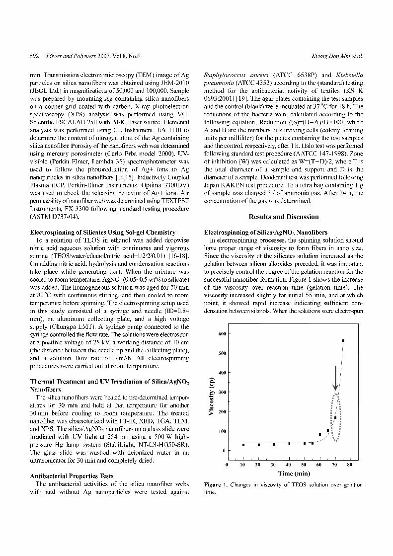
successful nanofiber formation. Figure 1 shows the increase
of the viscosity over reaction time (gelation time). The
viscosity increased slightly for initial 55 min, and at which
point, it showed rapid increase indicating sufficient con-
densation between silanols. When the solutions were electrospun
Figure 1. Changes in viscosity of TEOS solution over gelation
time.
Silica Nanofibers Containing Silver Nanoparticles Fibers and Polymers 2007, Vol.8, No.6 593
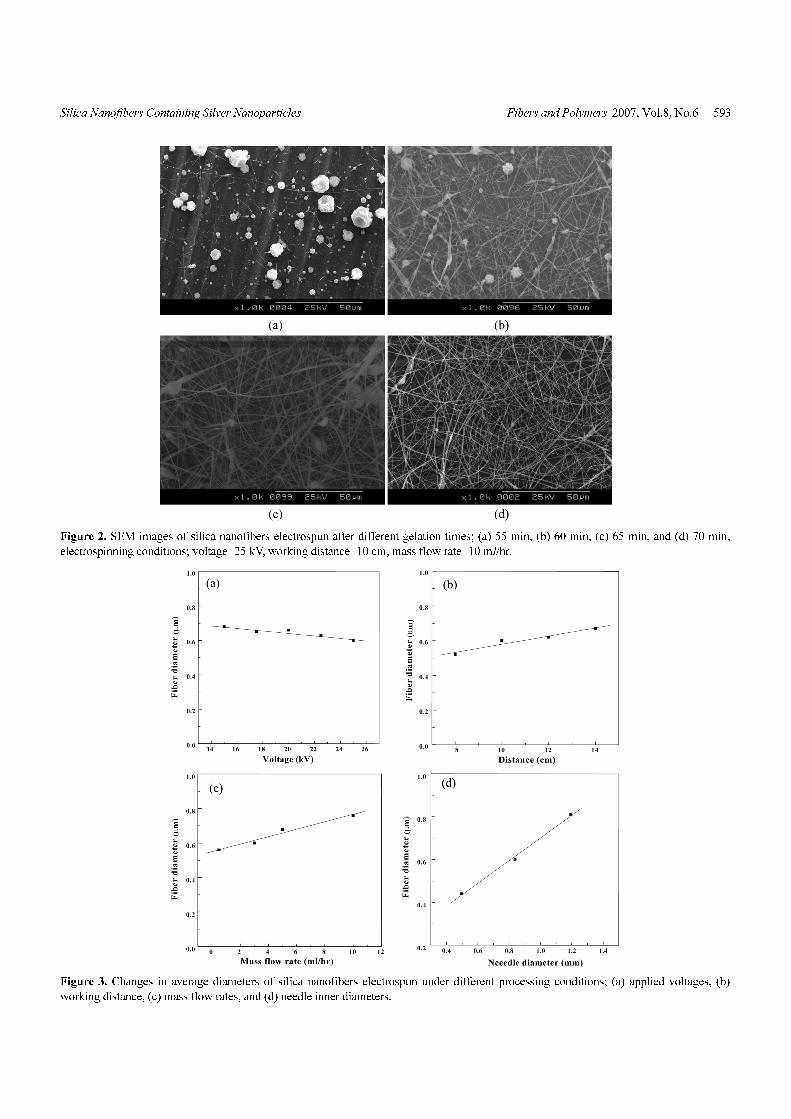
Figure 2. SEM images of silica nanofibers electrospun after different gelation times; (a) 55 min, (b) 60 min, (c) 65 min, and (d) 70 min,
electrospinning conditions; voltage=25 kV, working distance=10 cm, mass flow rate=10 ml/hr.
Figure 3. Changes in average diameters of silica nanofibers electrospun under different processing conditions; (a) applied voltages, (b)
working distance, (c) mass flow rates, and (d) needle inner diameters.
594 Fibers and Polymers 2007, Vol.8, No.6 Kyung Dan Min et al.
under a standard condition of a positive voltage of 25 kV, a
working distance of 10 cm, and a solution flow rate of 3 ml/h,
the prepared silica nanofibers had morphologies of sprayed
particles when the gelation time was 55 min, beaded fibers
when it was 60 and 65 min, and finally homogeneous
nanofibers after 70 min of gelation time (Figure 2). Figure 3
shows the relationship between diameter of the prepared
silica fiber and the processing parameters, such as applied
voltage, working distance, mass flow rate, and diameter of
the spinning tip. Similar to the cases of other organic polymers,
diameter of the electrospun fiber decreased with increasing
applied voltage, decreasing working distance, and decreasing
mass flow rate, while the dependencies on the parameters
were slightly higher. On the other hand, tip size affected to
the diameter of the nanofibers greater than other parameters.
Therefore, gelation time and spinning tip size were the two
most important parameters in electrospinning of silicate
solution using sol-gel chemistry.
When salt is added to the spinning solution, increased
charge density of the solution enhances repulsion between
the materials to give homogeneous nanofibers with smaller
diameter. AgNO3 was expected to behave as a salt to do such
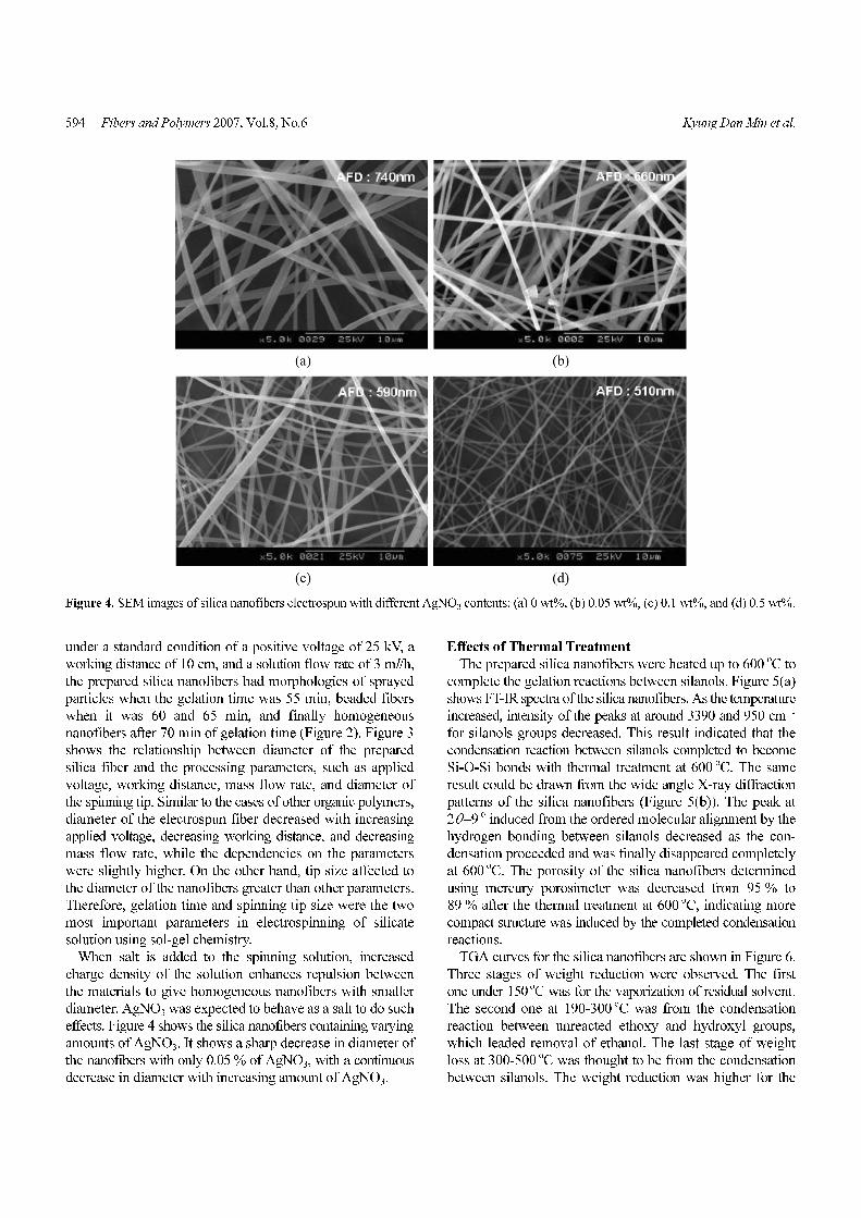
effects. Figure 4 shows the silica nanofibers containing varying
amounts of AgNO3. It shows a sharp decrease in diameter of
the nanofibers with only 0.05 % of AgNO3, with a continuous
decrease in diameter with increasing amount of AgNO3.
Effects of Thermal Treatment
The prepared silica nanofibers were heated up to 600oC to
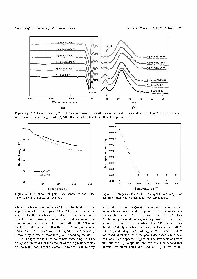
complete the gelation reactions between silanols. Figure 5(a)
shows FT-IR spectra of the silica nanofibers. As the temperature
increased, intensity of the peaks at around 3390 and 950 cm−1
for silanols groups decreased. This result indicated that the
condensation reaction between silanols completed to become
Si-O-Si bonds with thermal treatment at 600oC. The same
result could be drawn from the wide angle X-ray diffraction
patterns of the silica nanofibers (Figure 5(b)). The peak at
2θ=9o induced from the ordered molecular alignment by the
hydrogen bonding between silanols decreased as the con-
densation proceeded and was finally disappeared completely
at 600oC. The porosity of the silica nanofibers determined
using mercury porosimeter was decreased from 95 % to
89 % after the thermal treatment at 600 oC, indicating more
compact structure was induced by the completed condensation
reactions.
TGA curves for the silica nanofibers are shown in Figure 6.
Three stages of weight reduction were observed. The first
one under 150 oC was for the vaporization of residual solvent.
The second one at 190-300 oC was from the condensation
reaction between unreacted ethoxy and hydroxyl groups,
which leaded removal of ethanol. The last stage of weight
loss at 300-500oC was thought to be from the condensation
between silanols. The weight reduction was higher for the
Figure 4. SEM images of silica nanofibers electrospun with different AgNO3 contents; (a) 0 wt%, (b) 0.05 wt%, (c) 0.1 wt%, and (d) 0.5 wt%.
Silica Nanofibers Containing Silver Nanoparticles Fibers and Polymers 2007, Vol.8, No.6 595
silica nanofibers containing AgNO3, probably due to the
vaporization of nitro groups as NO or NO2 gases. Elemental
analysis for the nanofibers treated at various temperatures
revealed that nitrogen content decreased as increasing
temperature, and reached almost zero after 200oC (Figure
7). This result matched well with the TGA analysis results,
and implied that nitrate groups in AgNO3 could be easily
removed by thermal treatment to give reduced Ag metals.
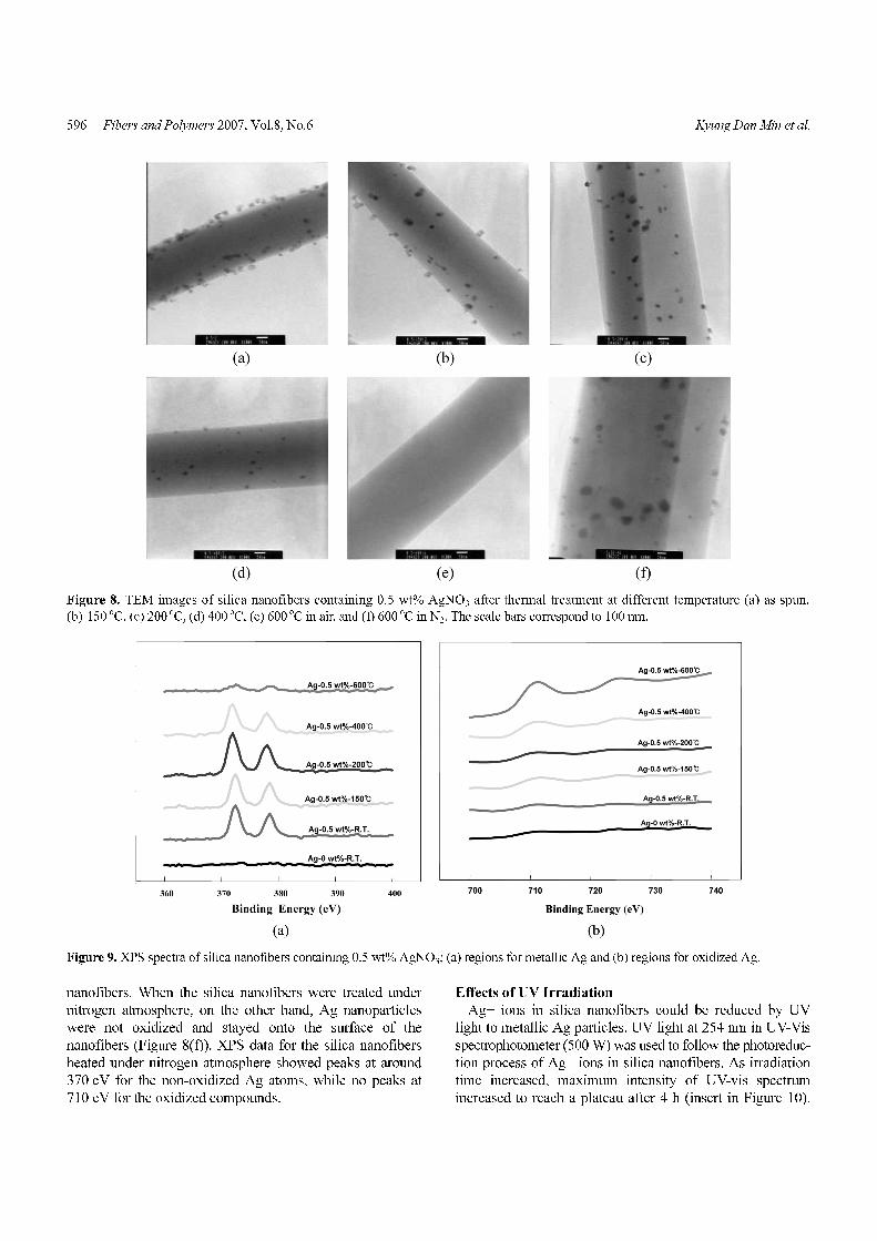
TEM images of the silica nanofibers containing 0.5 wt%
of AgNO3 showed that the amount of the Ag nanoparticles
on the nanofibers surface seemed decreased as increasing
temperature (Figure 8(a)-(e)). It was not because the Ag
nanoparticles disappeared completely from the nanofibers
surface, but because Ag metals were oxidized to AgO or
AgO2 and presented homogeneously inside of the silica
nanofibers. This could be confirmed by XPS analysis. For
the silica/AgNO3 nanofibers, there were peaks at around 370 eV
for 3d3/2 and 3d5/2 orbitals of Ag atoms. As temperature
increased, intensities of these peaks decreased while new
peak at 710 eV appeared (Figure 9). The new peak was from
the oxidized Ag compound, and this result evidenced that
thermal treatment under air oxidized Ag atoms in the
Figure 5. (a) FT-IR spectra and (b) X-ray diffraction patterns of pure silica nanofibers and silica nanofibers containing 0.5 wt% AgNO3 and
silica nanofibers containing 0.5 wt% AgNO3 after thermal treatments at different temperature in air.
Figure 6. TGA curves of pure silica nanofibers and silica
nanofibers containing 0.5 wt% AgNO3.
Figure 7. Nitrogen content of 0.5 wt% AgNO3-containing silica
nanofibers after heat treatment at different temperature.
596 Fibers and Polymers 2007, Vol.8, No.6 Kyung Dan Min et al.
nanofibers. When the silica nanofibers were treated under
nitrogen atmosphere, on the other hand, Ag nanoparticles
were not oxidized and stayed onto the surface of the
nanofibers (Figure 8(f)). XPS data for the silica nanofibers
heated under nitrogen atmosphere showed peaks at around
370 eV for the non-oxidized Ag atoms, while no peaks at
710 eV for the oxidized compounds.
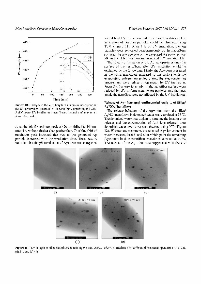
Effects of UV Irradiation
Ag+ ions in silica nanofibers could be reduced by UV
light to metallic Ag particles. UV light at 254 nm in UV-Vis
spectrophotometer (500 W) was used to follow the photoreduc-
tion process of Ag+ ions in silica nanofibers. As irradiation
time increased, maximum intensity of UV-vis spectrum
increased to reach a plateau after 4 h (insert in Figure 10).
Figure 8. TEM images of silica nanofibers containing 0.5 wt% AgNO3 after thermal treatment at different temperature (a) as spun,
(b) 150 oC, (c) 200 oC, (d) 400 oC, (e) 600 oC in air, and (f) 600 oC in N2. The scale bars correspond to 100 nm.
Figure 9. XPS spectra of silica nanofibers containing 0.5 wt% AgNO3; (a) regions for metallic Ag and (b) regions for oxidized Ag.
Silica Nanofibers Containing Silver Nanoparticles Fibers and Polymers 2007, Vol.8, No.6 597
Also, the initial maximum peak at 420 nm shifted to 444 nm
after 4 h, without further change after then. This blue shift of
maximum peak indicated that size of the generated Ag
particle increased with the irradiation time. These results
indicated that the photoreduction of Ag+ ions was completed
with 4 h of UV irradiation under the tested conditions. The
generation of Ag nanoparticles could be observed using
TEM (Figure 11). After 1 h of UV irradiation, the Ag
particles were generated heterogeneously on the nanofibers
surface. The average size of the generated Ag particles was
50 nm after 1 h irradiation and increased to 73 nm after 4 h.
The selective formation of the Ag nanoparticles onto the
surface of the nanofibers after UV irradiation could be
explained by the followings: Firstly, the Ag+ ions presented
in the silica nanofibers migrated to the surface with the
evaporating solvent molecules during the electrospinning
process, and were reduce to Ag metals by UV irradiation.
Secondly, the Ag+ ions only on the nanofiber surface were
reduced by UV to form metallic Ag particles, and the ones
inside the nanofiber were not affected by the UV irradiation.
Release of Ag+ Ions and Antibacterial Activity of Silica/
AgNO3 Nanofibers
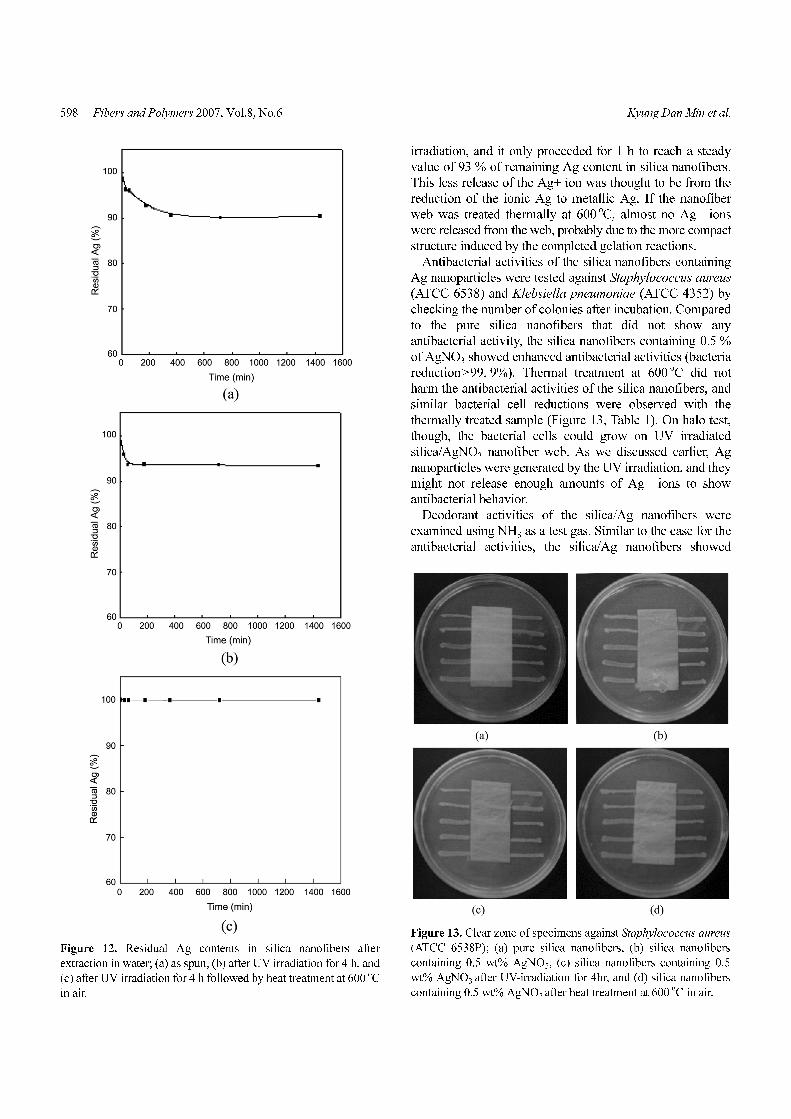
The release behavior of the Ag+ ions from the silica/
AgNO3 nanofibers in deionized water was examined at 37 oC.
The deionized water was shaken to simulate the local in vivo
release, and the concentration of Ag+ ions released onto
deionized water over time was checked using ICP (Figure
12). Without any treatment, the released Ag+ ion content in
water increased for 6 h, and after which point the remaining
Ag content in silica nanofibers was almost constant as 90 %.
The release of the Ag+ ions was suppressed with the UV
Figure 10. Changes in the wavelength of maximum absorption in
the UV absorption spectra of silica nanofibers containing 0.5 wt%
AgNO3 over UV-irradiation times (insert: intensity of maximum
absorption peak).
Figure 11. TEM images of silica nanofibers containing 0.5 wt% AgNO3 after UV-irradiation for different times; (a) as-spun, (b) 1 h, (c) 2 h,
(d) 3 h, and (e) 4 h.
598 Fibers and Polymers 2007, Vol.8, No.6 Kyung Dan Min et al.
irradiation, and it only proceeded for 1 h to reach a steady
value of 93 % of remaining Ag content in silica nanofibers.
This less release of the Ag+ ion was thought to be from the
reduction of the ionic Ag to metallic Ag. If the nanofiber
web was treated thermally at 600 oC, almost no Ag+ ions
were released from the web, probably due to the more compact
structure induced by the completed gelation reactions.
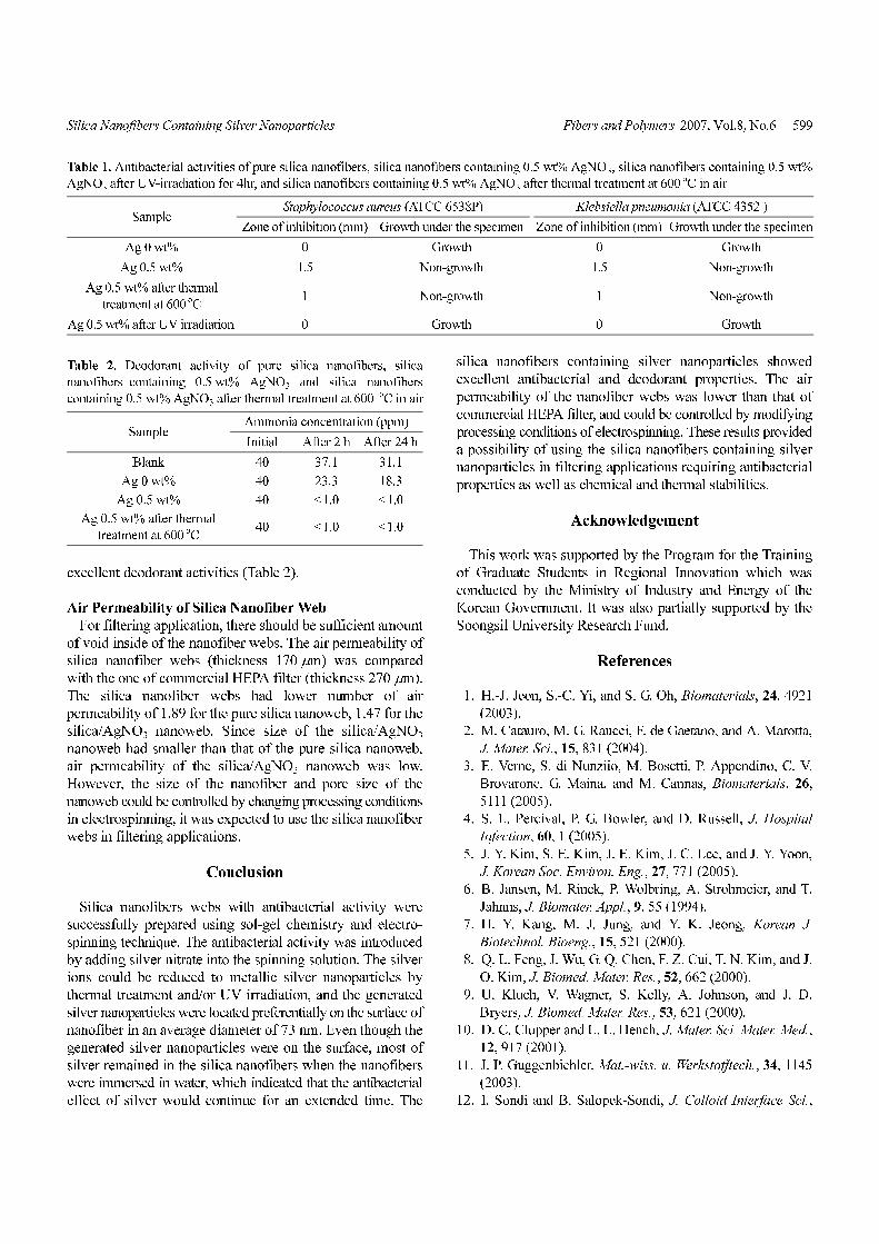
Antibacterial activities of the silica nanofibers containing
Ag nanoparticles were tested against Staphylococcus aureus
(ATCC 6538) and Klebsiella pneumoniae (ATCC 4352) by
checking the number of colonies after incubation. Compared
to the pure silica nanofibers that did not show any
antibacterial activity, the silica nanofibers containing 0.5 %
of AgNO3 showed enhanced antibacterial activities (bacteria
reduction>99. 9%). Thermal treatment at 600oC did not
harm the antibacterial activities of the silica nanofibers, and
similar bacterial cell reductions were observed with the
thermally treated sample (Figure 13, Table 1). On halo test,
though, the bacterial cells could grow on UV irradiated
silica/AgNO3 nanofiber web. As we discussed earlier, Ag
nanoparticles were generated by the UV irradiation, and they
might not release enough amounts of Ag+ ions to show
antibacterial behavior.
Deodorant activities of the silica/Ag nanofibers were
examined using NH3 as a test gas. Similar to the case for the
antibacterial activities, the silica/Ag nanofibers showed
Figure 12. Residual Ag contents in silica nanofibers after
extraction in water; (a) as spun, (b) after UV irradiation for 4 h, and
(c) after UV irradiation for 4 h followed by heat treatment at 600 oC
in air.
Figure 13. Clear zone of specimens against Staphylococcus aureus
(ATCC 6538P); (a) pure silica nanofibers, (b) silica nanofibers
containing 0.5 wt% AgNO3, (c) silica nanofibers containing 0.5
wt% AgNO3 after UV-irradiation for 4hr, and (d) silica nanofibers
containing 0.5 wt% AgNO3 after heat treatment at 600 oC in air.
Silica Nanofibers Containing Silver Nanoparticles Fibers and Polymers 2007, Vol.8, No.6 599
excellent deodorant activities (Table 2).
Air Permeability of Silica Nanofiber Web
For filtering application, there should be sufficient amount
of void inside of the nanofiber webs. The air permeability of
silica nanofiber webs (thickness 170 µm) was compared
with the one of commercial HEPA filter (thickness 270 µm).
The silica nanofiber webs had lower number of air
permeability of 1.89 for the pure silica nanoweb, 1.47 for the
silica/AgNO3 nanoweb. Since size of the silica/AgNO3
nanoweb had smaller than that of the pure silica nanoweb,
air permeability of the silica/AgNO3 nanoweb was low.
However, the size of the nanofiber and pore size of the
nanoweb could be controlled by changing processing conditions
in electrospinning, it was expected to use the silica nanofiber
webs in filtering applications.
Conclusion
Silica nanofibers webs with antibacterial activity were
successfully prepared using sol-gel chemistry and electro-
spinning technique. The antibacterial activity was introduced
by adding silver nitrate into the spinning solution. The silver
ions could be reduced to metallic silver nanoparticles by
thermal treatment and/or UV irradiation, and the generated
silver nanoparticles were located preferentially on the surface of
nanofiber in an average diameter of 73 nm. Even though the
generated silver nanoparticles were on the surface, most of
silver remained in the silica nanofibers when the nanofibers
were immersed in water, which indicated that the antibacterial
effect of silver would continue for an extended time. The
silica nanofibers containing silver nanoparticles showed
excellent antibacterial and deodorant properties. The air
permeability of the nanofiber webs was lower than that of
commercial HEPA filter, and could be controlled by modifying
processing conditions of electrospinning. These results provided
a possibility of using the silica nanofibers containing silver
nanoparticles in filtering applications requiring antibacterial
properties as well as chemical and thermal stabilities.
Acknowledgement
This work was supported by the Program for the Training
of Graduate Students in Regional Innovation which was
conducted by the Ministry of Industry and Energy of the
Korean Government. It was also partially supported by the
Soongsil University Research Fund.
References
1. H.-J. Jeon, S.-C. Yi, and S. G. Oh, Biomaterials, 24, 4921
(2003).
2. M. Catauro, M. G. Raucci, F. de Gaetano, and A. Marotta,
J. Mater. Sci., 15, 831 (2004).
3. E. Verne, S. di Nunziio, M. Bosetti, P. Appendino, C. V.
Brovarone, G. Maina, and M. Cannas, Biomaterials, 26,
5111 (2005).
4. S. L. Percival, P. G. Bowler, and D. Russell, J. Hospital
Infection, 60, 1 (2005).
5. J. Y. Kim, S. E. Kim, J. E. Kim, J. C. Lee, and J. Y. Yoon,
J. Korean Soc. Environ. Eng., 27, 771 (2005).
6. B. Jansen, M. Rinck, P. Wolbring, A. Strohmeier, and T.
Jahnns, J. Biomater. Appl., 9, 55 (1994).
7. H. Y. Kang, M. J. Jung, and Y. K. Jeong, Korean J.
Biotechnol. Bioeng., 15, 521 (2000).
8. Q. L. Feng, J. Wu, G. Q. Chen, F. Z. Cui, T. N. Kim, and J.
O. Kim, J. Biomed. Mater. Res., 52, 662 (2000).
9. U. Klueh, V. Wagner, S. Kelly, A. Johnson, and J. D.
Bryers, J. Biomed. Mater. Res., 53, 621 (2000).
10. D. C. Clupper and L. L. Hench, J. Mater. Sci. Mater. Med.,
12, 917 (2001).
11. J. P. Guggenbichler, Mat.-wiss. u. Werkstofftech., 34, 1145
(2003).
12. I. Sondi and B. Salopek-Sondi, J. Colloid Interface Sci.,
Table 1. Antibacterial activities of pure silica nanofibers, silica nanofibers containing 0.5 wt% AgNO3, silica nanofibers containing 0.5 wt%
AgNO3 after UV-irradiation for 4hr, and silica nanofibers containing 0.5 wt% AgNO3 after thermal treatment at 600 oC in air
SampleStaphylococcus aureus (ATCC 6538P) Klebsiella pneumonia (ATCC 4352 )
Zone of inhibition (mm) Growth under the specimen Zone of inhibition (mm) Growth under the specimen
Ag 0 wt% 0 Growth 0 Growth
Ag 0.5 wt% 1.5 Non-growth 1.5 Non-growth
Ag 0.5 wt% after thermal
treatment at 600 oC1 Non-growth 1 Non-growth
Ag 0.5 wt% after UV irradiation 0 Growth 0 Growth
Table 2. Deodorant activity of pure silica nanofibers, silica
nanofibers containing 0.5 wt% AgNO3 and silica nanofibers
containing 0.5 wt% AgNO3 after thermal treatment at 600 oC in air
SampleAmmonia concentration (ppm)
Initial After 2 h After 24 h
Blank 40 37.1 31.1
Ag 0 wt% 40 23.3 18.3
Ag 0.5 wt% 40 <1.0 <1.0
Ag 0.5 wt% after thermal
treatment at 600 oC40 <1.0 <1.0

600 Fibers and Polymers 2007, Vol.8, No.6 Kyung Dan Min et al.
275, 177 (2004).
13. J. H. Yeum, H. D. Ghim, and Y. Deng, Fibers and
Polymers, 6, 277 (2005).
14. V. Hornebecq, M. Antonietti, T. Cardinal, and M. Treguer-
Delapierre, Chem. Master., 15, 1993 (2003).
15. X. Li, J. Zhang, W. Xu, H. Jia, X. Wang, B. Yang, B. Zhao,
B. Li, and Y. Ozaki, Langmuir, 19, 4285 (2003).
16. T. Peltola, M. Jokinen, S. Veittola, H. Fahiala, and A.
Yliurpo, Biomaterials, 22, 589 (2001).
17. Y. Xu, W. Zhou, L. Zhang, and L. Cheng, J. Mater. Pro.
Tech., 101, 44 (2000).
18. G. P. Fotou, S. E. Pratsinis, and N. G. Pinto, J. Non-Crystal.
Solids, 183, 135 (1995).
19. S. Zhang, R. Fu, D. Wu, W. Xu, Q. Ye, and Z. Chen,
Carbon, 42, 3209 (2004).