Development of a chitosan–polyglutamate based injectable polyelectrolyte complex scaffold
7
Please cite this article in press as: Wu, H. -D., et al. Development of a chitosan–polyglutamate based injectable polyelectrolyte complex scaffold. Carbohydrate Polymers (2011), doi:10.1016/j.carbpol.2011.02.024 ARTICLE IN PRESS G Model CARP-5465; No. of Pages 7 Carbohydrate Polymers xxx (2011) xxx–xxx Contents lists available at ScienceDirect Carbohydrate Polymers journal homepage: www.elsevier.com/locate/carbpol Development of a chitosan–polyglutamate based injectable polyelectrolyte complex scaffold Hong-Da Wu a,1 , Jen-Chang Yang b,c,1 , Tsuimin Tsai d , Dain-Yu Ji a , Wei-Jen Chang a,e , Chien-Chung Chen d , Sheng-Yang Lee a,c,f,∗ a School of Dentistry, College of Oral Medicine, Taipei Medical University, Taipei 110, Taiwan, ROC b School of Dental Technology, College of Oral Medicine, Taipei Medical University, Taipei 110, Taiwan, ROC c Center for Teeth Bank and Dental Stem Cell Technology, Taipei Medical University, Taipei 110, Taiwan, ROC d Graduate Institute of Biomaterials and Engineering, College of Oral Medicine, Taipei Medical University, Taipei 110, Taiwan, ROC e Dental Department of Taipei Medical University-Shuang Ho Hospital, Jhonghe city, Taipei, Taiwan, ROC f Dental Department of Taipei Medical University Municipal Wan-Fang Hospital, Taipei 116, Taiwan, ROC article info Article history: Received 7 January 2011 Received in revised form 11 February 2011 Accepted 12 February 2011 Available online xxx Keywords: Polyelectrolyte complex Injectable scaffold Chitosan Polyglutamate acid Calcium sulfate dihydrate Bone regeneration abstract In this study, the chitosan–polyglutamate hydrogel was developed as injectable polyelectrolyte complex (PEC) scaffold for bone regeneration. In practice, oppositely charged polyelectrolytes were mixed using a static injection mixer, the gelation time was less than 1 min. The resorbable PEC scaffold can be in situ- formed as a scaffold for osteoblast ingrowth. The canine alveolar defect model was used to evaluate the efficacy of PEC implants. The PEC scaffold was highly effective in enhancing bone regeneration. Moreover, the addition of CaSO 4 ·2H 2 O (CSD) to PEC scaffold can further enhance the early stage healing of bone regeneration. © 2011 Elsevier Ltd. All rights reserved. 1. Introduction Bone grafts are commonly used in orthopedics and dentistry to repair bony defects due to trauma, disease, or surgery (Taylor, 1983). Autograft bone is most effective for bone repair; how- ever, the supply of autogenous bone grafts is limited. In addition, allografts are susceptible to immune responses and disease trans- mission. Therefore, numerous synthetic bone substitutes have been used in clinical practice because of their biodegradation, bio- compatibility, and osteoconductivity. However, it is difficult for prefabricated bone blocks to fit an irregular bony defect (Xu, Weir, Burguera, & Fraser, 2006). Although granular-type bone grafts can easily be filled into an irregular defect, it is difficult to maintain them in defect sites, especially with incomplete bony defects (Yao, Liu, Hsu, & Chen, 2005). To overcome these problems, injectable ∗ Corresponding author at: School of Dentistry, College of Oral Medicine, Taipei Medical University, 250 Wu-Hsing Street, Taipei 110, Taiwan, ROC. Tel.: +886 2 27361661x5112; fax: +886 2 27362295. E-mail address: [email protected] (S.-Y. Lee). 1 These authors contributed equally to this work. scaffolds are highly desirable in clinical applications (Wintermantel et al., 1996). After injection, a precursor solution can fill in an irreg- ular defect, and the gel can form in situ. In addition, injectable scaffolds can also serve as a carrier for cells and/or bioactive molecules applied via minimally invasive procedures (Kretlow, Klouda, & Mikos, 2007). Injectable scaffolds are formed via an in situ crosslinking method, which is a critical technique in developing, as it affects the character of the resulting gel (e.g., the gelling time, gel con- tent, porosity, degradation time, and biocompatibility) (Yu & Ding, 2008). Commonly used in situ crosslinking methods are classified into chemical and physical types. Chemical crosslinking (redox- or photo-initiated) are controlled by adjusting the initiator concen- tration or radiation dosage to obtain a desirable gelling time and degradability. However, chemical crosslinkers are usually toxic, which is a major obstacle to their applications (Hou, De Bank, & Shakesheff, 2004). In comparison, physical ionic crosslinking dis- plays much fewer biocompatibility concerns. For this reason, an ionically crosslinked polyelectrolyte complex (PEC) method was chosen for this study. The PEC consisted of a complex of oppositely charged (cationic and anionic) macromolecules that were bound by electrostatic interactions (Li et al., 2007). 0144-8617/$ – see front matter © 2011 Elsevier Ltd. All rights reserved. doi:10.1016/j.carbpol.2011.02.024
-
Upload
taipeimedical -
Category
Documents
-
view
0 -
download
0
Transcript of Development of a chitosan–polyglutamate based injectable polyelectrolyte complex scaffold
G
C
Dc
HWa
b
c
d
e
f
a
ARRAA
KPICPCB
1
t1eamucpBetL
MT
0d
ARTICLE IN PRESSModel
ARP-5465; No. of Pages 7
Carbohydrate Polymers xxx (2011) xxx–xxx
Contents lists available at ScienceDirect
Carbohydrate Polymers
journa l homepage: www.e lsev ier .com/ locate /carbpol
evelopment of a chitosan–polyglutamate based injectable polyelectrolyteomplex scaffold
ong-Da Wua,1, Jen-Chang Yangb,c,1, Tsuimin Tsaid, Dain-Yu Ji a,ei-Jen Changa,e, Chien-Chung Chend, Sheng-Yang Leea,c,f,∗
School of Dentistry, College of Oral Medicine, Taipei Medical University, Taipei 110, Taiwan, ROCSchool of Dental Technology, College of Oral Medicine, Taipei Medical University, Taipei 110, Taiwan, ROCCenter for Teeth Bank and Dental Stem Cell Technology, Taipei Medical University, Taipei 110, Taiwan, ROCGraduate Institute of Biomaterials and Engineering, College of Oral Medicine, Taipei Medical University, Taipei 110, Taiwan, ROCDental Department of Taipei Medical University-Shuang Ho Hospital, Jhonghe city, Taipei, Taiwan, ROCDental Department of Taipei Medical University Municipal Wan-Fang Hospital, Taipei 116, Taiwan, ROC
r t i c l e i n f o
rticle history:eceived 7 January 2011eceived in revised form 11 February 2011ccepted 12 February 2011vailable online xxx
a b s t r a c t
In this study, the chitosan–polyglutamate hydrogel was developed as injectable polyelectrolyte complex(PEC) scaffold for bone regeneration. In practice, oppositely charged polyelectrolytes were mixed usinga static injection mixer, the gelation time was less than 1 min. The resorbable PEC scaffold can be in situ-formed as a scaffold for osteoblast ingrowth. The canine alveolar defect model was used to evaluate theefficacy of PEC implants. The PEC scaffold was highly effective in enhancing bone regeneration. Moreover,
eywords:olyelectrolyte complexnjectable scaffoldhitosanolyglutamate acidalcium sulfate dihydrate
the addition of CaSO4·2H2O (CSD) to PEC scaffold can further enhance the early stage healing of boneregeneration.
© 2011 Elsevier Ltd. All rights reserved.
one regeneration
. Introduction
Bone grafts are commonly used in orthopedics and dentistryo repair bony defects due to trauma, disease, or surgery (Taylor,983). Autograft bone is most effective for bone repair; how-ver, the supply of autogenous bone grafts is limited. In addition,llografts are susceptible to immune responses and disease trans-ission. Therefore, numerous synthetic bone substitutes have been
sed in clinical practice because of their biodegradation, bio-ompatibility, and osteoconductivity. However, it is difficult forrefabricated bone blocks to fit an irregular bony defect (Xu, Weir,
Please cite this article in press as: Wu, H. -D., et al. Development of a chitosCarbohydrate Polymers (2011), doi:10.1016/j.carbpol.2011.02.024
urguera, & Fraser, 2006). Although granular-type bone grafts canasily be filled into an irregular defect, it is difficult to maintainhem in defect sites, especially with incomplete bony defects (Yao,iu, Hsu, & Chen, 2005). To overcome these problems, injectable
∗ Corresponding author at: School of Dentistry, College of Oral Medicine, Taipeiedical University, 250 Wu-Hsing Street, Taipei 110, Taiwan, ROC.
el.: +886 2 27361661x5112; fax: +886 2 27362295.E-mail address: [email protected] (S.-Y. Lee).
1 These authors contributed equally to this work.
144-8617/$ – see front matter © 2011 Elsevier Ltd. All rights reserved.oi:10.1016/j.carbpol.2011.02.024
scaffolds are highly desirable in clinical applications (Wintermantelet al., 1996). After injection, a precursor solution can fill in an irreg-ular defect, and the gel can form in situ. In addition, injectablescaffolds can also serve as a carrier for cells and/or bioactivemolecules applied via minimally invasive procedures (Kretlow,Klouda, & Mikos, 2007).
Injectable scaffolds are formed via an in situ crosslinkingmethod, which is a critical technique in developing, as it affectsthe character of the resulting gel (e.g., the gelling time, gel con-tent, porosity, degradation time, and biocompatibility) (Yu & Ding,2008). Commonly used in situ crosslinking methods are classifiedinto chemical and physical types. Chemical crosslinking (redox- orphoto-initiated) are controlled by adjusting the initiator concen-tration or radiation dosage to obtain a desirable gelling time anddegradability. However, chemical crosslinkers are usually toxic,which is a major obstacle to their applications (Hou, De Bank, &Shakesheff, 2004). In comparison, physical ionic crosslinking dis-
an–polyglutamate based injectable polyelectrolyte complex scaffold.
plays much fewer biocompatibility concerns. For this reason, anionically crosslinked polyelectrolyte complex (PEC) method waschosen for this study. The PEC consisted of a complex of oppositelycharged (cationic and anionic) macromolecules that were boundby electrostatic interactions (Li et al., 2007).
user
反白
user
反白
ING
C
2 ate Po
Nf(ihMgWhfwIcseemiif
hmnafdrlKi
lapocm
2
2
oP(mMdTItf
2
tswrmg
ARTICLEModel
ARP-5465; No. of Pages 7
H.-D. Wu et al. / Carbohydr
Chitosan is a polysaccharide constituted by N-glucosamine and-acetyl-glucosamine units, and it has been widely investigated
or bone regeneration (Muzzarelli, 2009, 2011). PolyglutamatePG), a natural polyanionic peptide produced by Bacillus subtilis,s a hydrophilic, biocompatible, and biodegradable material thatas been successfully used in medical applications (Richard &argaritis, 2001). A chitosan–PG was used to fabricate hydro-
els, and preformed scaffolds in several recent studies (Hsieh, Tsai,ang, Chang, & Hsieh, 2005; Tsao et al., 2010). However, no report
as so far used a chitosan–PG hydrogel to develop injectable scaf-old for bone regeneration. In this study, the PEC structure formedas mainly based on electrostatic interactions of chitosan and PG.
n order to improve the osteogenic potential of the designed PEC,alcium sulfate dihydrate (CSD) was added. CSD acts at a preribo-omal level and regulates the translation process to increase thexpression of bone-related proteins (Palmieri et al., 2008). Lazaryt al. (2007) also reported that CSD is a passive osteoconductiveaterial, and it might also have potential osteoinductivity due to
ts high calcium ion release. For these reasons, incorporation of CSDnto a PEC system may be a good combination to enhance new boneormation.
In the process of forming the PEC, it was difficult to achieveomogeneity of the hydrogel by a direct mixing method. Duringixing the two oppositely charged polyelectrolytes with a mag-
etic stirrer, the PEC usually formed at the interfaces as hugeggregates (Hsieh et al., 2005). To develop an in situ-formed scaf-old, the two oppositely charged polyelectrolytes had to be mixeduring the injection period. The static mixer is widely used in aange of applications such as for the continuous mixing of viscousiquids, blending, and chemical reactions (Paul, Atiemo-Obeng, &resta, 2004). Thus, the static mixing system was chosen as the
njection mixer in this experiment.The aims of this study were to develop a non-toxic PEC formu-
ation which can be injected into defects and solidified in situ withstatic injection mixer; moreover, CSD was added to the PEC to
rovide adequate calcium ions to enhance bone regeneration. Thesteogenetic ability of the designed specimens was evaluated in aanine alveolar defect model using histological and histomorpho-etric methods.
. Materials and methods
.1. Materials
Chitosan (MW 25,000–35,000) with a degree of deacetylationf 95% was obtained from Shin Era Technology (Taipei, Taiwan).olyglutamate (PG, MW 1000 kDa) was purchased from VedanTaipei, Taiwan). Phosphate-buffered saline (PBS), potassium bro-
ide, high-viscosity carboxy-methyl-cellulose (CM-cellulose), andTT were purchased by Sigma (St. Louis, MO, USA). Calcium sulfate
ihydrate (CSD) was purchased from Biotech One (Taipei, Taiwan).he decalcifying agent was purchased from Surgipath Medicalndustries (Richmond, IL, USA). Trypsin-EDTA, �-minimum essen-ial medium (MEM), and fetal bovine serum (FBS) were purchasedrom Gibco BRL Life Technology (Grand Island, NY, USA).
.2. Preparation of specimens (PEC and PEC/CSD)
Chitosan was dissolved in 1% acetic acid, and the final pH ofhe solution was adjusted to 6.2. PG and CM-cellulose were dis-
Please cite this article in press as: Wu, H. -D., et al. Development of a chitosCarbohydrate Polymers (2011), doi:10.1016/j.carbpol.2011.02.024
olved in double-distilled water (DDW), and the pH of the solutionas adjusted to 7.5 to neutralize the cationic polyelectrolytes. The
esults of the following gelation experiments were used to for-ulate and characterize the PEC. When preparing the PEC/CSD
roup, CSD was added to the polycationic and polyanionic solu-
PRESSlymers xxx (2011) xxx–xxx
tions, respectively. The final concentration of CSD in the PEC systemwas 20% (w/w). The cationic and anionic polyelectrolytes weremixed in a 1:1 (v/v) ratio with a static injection mixer (Sulzer, Salem,NH, USA) at room temperature.
2.3. Analytical determination of PEC
In the gelling time test, 2 ml of PEC was injected into a glassbeaker with a 5 ml capacity which contained a stirring bar (with adiameter of 5 mm and a length of 10 mm) at room temperature, andthen placed on a stirring plate (Corning, Painted Post, NY, USA) ata stirring speed of 45 rpm. The gelling time was noted as the timerequired for the stirring bar to stop (Balakrishnan & Jayakrishnan,2005; Hong, Mao, Wang, Gao, & Shen, 2006).
In the gel content test, specimens were lyophilized and weighed.Acetic acid (1%; a solvent for chitosan, PG, and CM-cellulose) wasused to extract non-crosslinked material from the specimen. Thegel content was calculated from the dry weight of a specimen beforeand after solvent extraction (Lee, Hung, Cheng, & Wang, 2005). Alltest specimens were extracted in the solvent for 24 h, washed withDDW, dried in an oven at 80 ◦C, and then weighed. The gel contentwas calculated using the equation:
gel content (%) = Wa
Wb× 100;
where Wb and Wa are the dry weights of the specimens before andafter extraction, respectively.
To analysis the equilibrium swelling, specimens were injectedinto the wells of culture plates (12-well) and dried in a freeze-drier.The dry weights (Wd) were immediately measured, and then thespecimens were immersed in PBS, maintained at 37 ◦C for 24 h, andweighed to obtain the wet weight (Ww). The equilibrium swelling(ES) of the matrices was calculated using the following equation:
equilibrium swelling value = Ww − Wd
Wd.
To examine the morphology of the PEC, cross-sections of thelyophilized PEC were directly sputter-coated with gold. Microstruc-tures of the lyophilized specimen and cell-containing specimenwere examined by SEM (Hitachi S2400, Tokyo, Japan) under anaccelerating voltage of 15–20 kV.
In vitro degradation test was conducted according to ISO10993-9 guidelines; specimens were incubated in 50 ml of PBS at 37 ◦Cwith agitation at 50 rpm. After 1, 2, 4, 12, and 26 weeks of incuba-tion, specimens were lyophilized. The amount of degradation wascalculated using the following equation:
weight retention (%) = Wt
Wd× 100;
where Wd is the weight of the initial dry specimen, and Wt is theweight of the dry specimen at time t.
2.4. In vitro cell compatibility and in vivo implantation test
In accordance with ISO10993-5 guidelines, the cell compati-bility of the PEC was evaluated on MC3T3E1 cell (pre-osteoblast)monolayer using the MTT method. The extracted medium for test-ing was prepared by incubating the specimen with 10% FBS at anextraction ratio of 0.2 g/ml for 24 h at 37 ◦C. Extracted medium ofthe PEC (experimental group), extracted medium of high-densitypolyethylene (negative control, NC), medium with 0.5% DMSO (pos-
an–polyglutamate based injectable polyelectrolyte complex scaffold.
itive control, PC), and normal culture medium (control, C) wereplaced on a monolayer of cells. After incubation at 37 ◦C for 24 h,the cellular responses were assessed by optical microscopy and theMTT assay. The absorbance of MTT in each well was immediatelyrecorded at 570 nm using a UV/visible absorbance spectroscopy
ARTICLE IN PRESSG Model
CARP-5465; No. of Pages 7
H.-D. Wu et al. / Carbohydrate Polymers xxx (2011) xxx–xxx 3
F G + 2%c
(w
c .
foa1i9uo
fCmpsawmsibiwafitAt
FP
ig. 1. Gelation times at different concentrations (A) chitosan (mixed with 3% Pellulose + 3% PG (mixed with 8% chitosan) at 25 ◦C (n = 4).
Bio-Tek Instruments, Winooski, VT, USA). The cell compatibilityas estimated using the following equation:
ell viability (% of control) = absorbance of the test materialabsorbance of the control
× 100
In the cell attachment test, the PEC was sterilized in 75% ethanolor 24 h. After washing with PBS, 5000 MC3T3E1 cells were seededn a gel and incubated at 37 ◦C in an incubator with a 0.5% CO2tmosphere for 72 h. Cell-containing specimens were fixed with awt% glutaraldehyde solution and 1 wt% OsO4 (post-fixed). Spec-
mens were dehydrated in a graded ethanol series (30, 50, 70, 80,0, and 100 wt%), and then dried by critical-point drying with liq-id CO2. Finally, cells containing PEC were coated with gold andbserved by SEM.
The canine alveolar bone defect implantation test was per-ormed in strict accordance with protocols approved by the Animalare Committee of Taipei Medical University. Eight beagles with aean age of 1 year were operated on under general anesthesia. All
re-molars in the mandible were extracted. When the extractionocket had healed (about 2 months), six defects (6 mm in depthnd 6 mm in diameter) on both sides of the mandible were createdith a trephine bur. The defects were filled with the following graftaterials: PEC, PEC/CSD, and CSD (particle size 500–1000 �m). The
ame procedure was performed in the empty control group. Todentifying the location of the defect, we recorded the distanceetween the canine teeth and each defect on alveolar ridge. Spec-
mens were harvested after 3 and 6 weeks. Each implantation siteas harvested with a trephine bur (with an inner diameter of 6 mm)
Please cite this article in press as: Wu, H. -D., et al. Development of a chitosCarbohydrate Polymers (2011), doi:10.1016/j.carbpol.2011.02.024
t 3 and 6 weeks after implantation. The harvested specimens werexed in 10% buffered formalin for 1 week, and a cross-section ofhe specimen was created with a low-speed cutter (Techcut 4TM,llied High Tech Products, Rancho Dominguez, CA, USA), and then
he specimen was decalcified until the specimen could be pene-
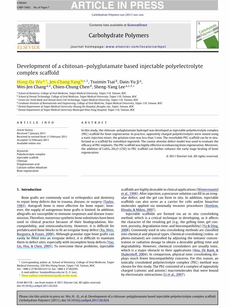
ig. 2. Gel content (GC) (�) and equilibrium swelling (ES) at 25 ◦C in PBS for 24 h (�) of coG + 2% CM-cellulose), (B) PG + 2% CM-cellulose (mixed with 8% chitosan), and (C) CM-cel
CM-cellulose), (B) PG + 2% CM-cellulose (mixed with 8% chitosan), and (C) CM-
trated with a needle. Finally, the decalcified sample was preparedto a paraffin section, and stained with hematoxylin and eosin (H&E).The stained section was observed under light microscopy (OlympusEX51, Tokyo, Japan). In this study, histomorphometric method wasused to quantify the amount of newly formed bone tissue withinthe grafted area, which was carried out with an optical micro-scope connected to a personal computer equipped with an imageanalysis system. The defect boundaries and cavity area were visu-ally determined, and all areas of the image were quantified usingImage J analysis software (National Institutes of Health, Bethesda,MD, USA). The amount of newly formed bone was calculated usingfollowing equation (Park, Jang, Bae, An, & Suh, 2009):
new bone (%) = area of regenerated bonearea of created defect
× 100.
2.5. Statistical analysis
Group means and standard deviations (SD) were calculated foreach measured parameter. Data were compared using a one-wayanalysis of variance (ANOVA) and post hoc Tukey’s test, and p valuesof <0.05 indicated statistical significance.
3. Results and discussion
3.1. Analytical determination of PEC (gelation property,micromorphology and degradation)
an–polyglutamate based injectable polyelectrolyte complex scaffold.
Gelation times of the specimens were estimated at room tem-perature. The concentrations of chitosan, PG, and CM-cellulosewere respectively controlled to 2.5–8%, 1–5%, and 1–5%, becauseof operative feasibility (e.g., solubility and viscosity). In our system,gelation times of specimens were between 40 s and 6 min (Fig. 1).
mplexes formed by mixing different concentrations of (A) chitosan (mixed with 3%lulose + 3% PG (mixed with 8% chitosan) (n = 4).
IN PRESSG
C
4 ate Polymers xxx (2011) xxx–xxx
TtdcCgaoawchtmJ(
inshftcstesttciKvrbfh(te
taAdwvccaiZw
otro4twtdoa
ARTICLEModel
ARP-5465; No. of Pages 7
H.-D. Wu et al. / Carbohydr
he gelation time could be adjusted by changing the concentra-ion of the polymer; however, the tendency of the gelation timeid not seem to be positively correlated with the polymer con-entration. At the respective concentrations of chitosan, PG, andM-cellulose of 8%, 3%, and 2% (w/w), the specimen had the fastestelation time (40 s). As an injectable scaffold system, an appropri-te gelling time is important; it is known that the setting timesf commercial injectable calcium phosphate cement (CPC) systemsre about 10–30 min, which usually cause the CPC to be wash-outith body fluids during operation (Xu & Simon, 2005). Generally,
hemical and ionic crosslinking injectable scaffold systems usuallyave less solidification periods compared to commercial CPC sys-ems. Our PEC system can gel within 1 min that is similar to Tan’s
odified chitosan–hyaluronic acid gel system (about 1 min) andin’s chitosan derivative enzymatic crosslinked gel system (10 s)Jin et al., 2009; Tan, Chu, Payne, & Marra, 2009).
The gel contents were related to the number of materials form-ng the PEC structure. Fig. 2 displays the gel contents which wereot positively correlated with the concentrations of materials. Thepecimen composed of 8% chitosan and 3% PG + 2% CM-cellulosead the maximum gel content (75%), this indicates that PEC
ormation is more efficient at nearly a stoichiometric composi-ion. The swelling ratio is an important parameter to characterizerosslinked structure. The effect of the polymer concentration onwelling is shown in Fig. 2. The equilibrium swelling values ofhe designed specimens were within 2.7–5.1. There was no lin-ar relationship between the concentrations of materials and thewelling values. The swelling value was inversely proportional tohe gel content. The gel content of the PEC was associated withhe swelling behavior; the higher gel content resulted in a strongerrosslinked structure which confines the macromolecules to a lim-ted spatial volume, so the swelling value is largely restricted. Sumi,ala, Praveen, and Prasada Rao (2008) reported that a low swellingalue was associated with a low drug release rate. Moreover, envi-onmental changes in pH and temperature also affect the swellingehavior (Karg et al., 2008). According to the above results, theormulation of 8% chitosan and 3% PG + 2% CM-cellulose had theighest gel content (75%), the lowest equilibrium swelling value2.7) and the fastest gelling time (40 s), which may have been closesto the stoichiometric formulation and was chosen for the followingxperiments.
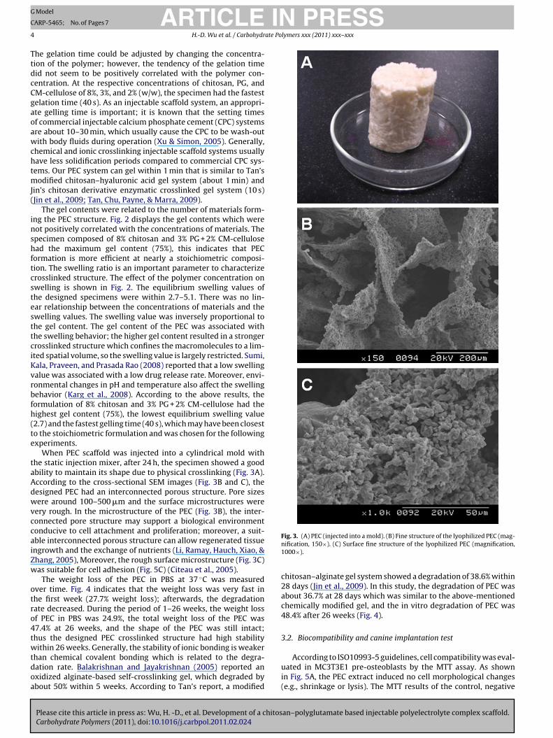
When PEC scaffold was injected into a cylindrical mold withhe static injection mixer, after 24 h, the specimen showed a goodbility to maintain its shape due to physical crosslinking (Fig. 3A).ccording to the cross-sectional SEM images (Fig. 3B and C), theesigned PEC had an interconnected porous structure. Pore sizesere around 100–500 �m and the surface microstructures were
ery rough. In the microstructure of the PEC (Fig. 3B), the inter-onnected pore structure may support a biological environmentonducive to cell attachment and proliferation; moreover, a suit-ble interconnected porous structure can allow regenerated tissuengrowth and the exchange of nutrients (Li, Ramay, Hauch, Xiao, &hang, 2005), Moreover, the rough surface microstructure (Fig. 3C)as suitable for cell adhesion (Fig. 5C) (Citeau et al., 2005).
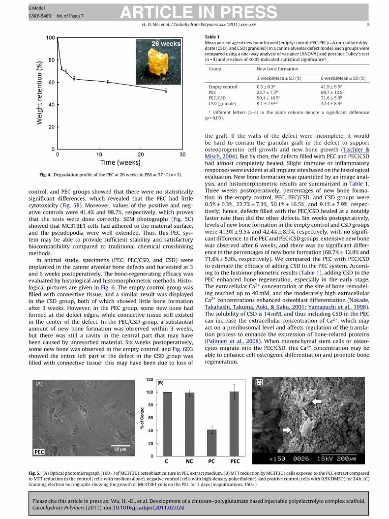
The weight loss of the PEC in PBS at 37 ◦C was measuredver time. Fig. 4 indicates that the weight loss was very fast inhe first week (27.7% weight loss); afterwards, the degradationate decreased. During the period of 1–26 weeks, the weight lossf PEC in PBS was 24.9%, the total weight loss of the PEC was7.4% at 26 weeks, and the shape of the PEC was still intact;hus the designed PEC crosslinked structure had high stability
Please cite this article in press as: Wu, H. -D., et al. Development of a chitosCarbohydrate Polymers (2011), doi:10.1016/j.carbpol.2011.02.024
ithin 26 weeks. Generally, the stability of ionic bonding is weakerhan chemical covalent bonding which is related to the degra-ation rate. Balakrishnan and Jayakrishnan (2005) reported anxidized alginate-based self-crosslinking gel, which degraded bybout 50% within 5 weeks. According to Tan’s report, a modified
Fig. 3. (A) PEC (injected into a mold). (B) Fine structure of the lyophilized PEC (mag-nification, 150×). (C) Surface fine structure of the lyophilized PEC (magnification,1000×).
chitosan–alginate gel system showed a degradation of 38.6% within28 days (Jin et al., 2009). In this study, the degradation of PEC wasabout 36.7% at 28 days which was similar to the above-mentionedchemically modified gel, and the in vitro degradation of PEC was48.4% after 26 weeks (Fig. 4).
3.2. Biocompatibility and canine implantation test
an–polyglutamate based injectable polyelectrolyte complex scaffold.
According to ISO10993-5 guidelines, cell compatibility was eval-uated in MC3T3E1 pre-osteoblasts by the MTT assay. As shownin Fig. 5A, the PEC extract induced no cell morphological changes(e.g., shrinkage or lysis). The MTT results of the control, negative
ARTICLE IN PRESSG Model
CARP-5465; No. of Pages 7
H.-D. Wu et al. / Carbohydrate Polymers xxx (2011) xxx–xxx 5
cscatsatbm
iaelfiiafiabbssfi
Table 1Mean percentage of new bone formed (empty control, PEC, PEC/calcium sulfate dihy-drate (CSD), and CSD (granules)) in a canine alveolar defect model, each groups werecompared using a one-way analysis of variance (ANOVA) and post hoc Tukey’s test(n = 4) and p values of <0.05 indicated statistical significancea.
Group New bone formation
3 weeksMean ± SD (%) 6 weeksMean ± SD (%)
Empty control 0.5 ± 0.3a 41.9 ± 9.5a
PEC 22.7 ± 7.3b 68.7 ± 12.8b
PEC/CSD 50.1 ± 16.5c 71.6 ± 5.9b
FtS
Fig. 4. Degradation profile of the PEC at 26 weeks in PBS at 37 ◦C (n = 3).
ontrol, and PEC groups showed that there were no statisticallyignificant differences, which revealed that the PEC had littleytotoxicity (Fig. 5B). Moreover, values of the positive and neg-tive controls were 41.4% and 98.7%, respectively, which proveshat the tests were done correctly. SEM photographs (Fig. 5C)howed that MC3T3E1 cells had adhered to the material surface,nd the pseudopodia were well extended. Thus, this PEC sys-em may be able to provide sufficient stability and satisfactoryiocompatibility compared to traditional chemical crosslinkingethods.In animal study, specimens (PEC, PEC/CSD, and CSD) were
mplanted in the canine alveolar bone defects and harvested at 3nd 6 weeks postoperatively. The bone-regenerating efficacy wasvaluated by histological and histomorphometric methods. Histo-ogical pictures are given in Fig. 6. The empty control group waslled with connective tissue, and a similar result was displayed
n the CSD group, both of which showed little bone formationfter 3 weeks. However, in the PEC group, some new bone hadormed at the defect edges, while connective tissue still existedn the center of the defect. In the PEC/CSD group, a substantialmount of new bone formation was observed within 3 weeks,
Please cite this article in press as: Wu, H. -D., et al. Development of a chitosCarbohydrate Polymers (2011), doi:10.1016/j.carbpol.2011.02.024
ut there was still a cavity in the central part that may haveeen caused by unresorbed material. Six weeks postoperatively,ome new bone was observed in the empty control, and Fig. 6D3howed the entire left part of the defect in the CSD group waslled with connective tissue; this may have been due to loss of
ig. 5. (A) Optical photomicrograph (100×) of MC3T3E1 osteoblast culture in PEC extracto MTT reduction in the control (cells with medium alone), negative control (cells with hicanning electron micrographs showing the growth of MC3T3E1 cells on the PEC for 3 da
CSD (granule) 9.1 ± 7.9a,b 42.4 ± 8.9a
a Different letters (a–c) in the same column denote a significant difference(p < 0.05).
the graft. If the walls of the defect were incomplete, it wouldbe hard to contain the granular graft in the defect to supportosteoprogenitor cell growth and new bone growth (Tischler &Misch, 2004). But by then, the defects filled with PEC and PEC/CSDhad almost completely healed. Slight immune or inflammatoryresponses were evident at all implant sites based on the histologicalevaluation. New bone formation was quantified by an image anal-ysis, and histomorphometric results are summarized in Table 1.Three weeks postoperatively, percentages of new bone forma-tion in the empty control, PEC, PEC/CSD, and CSD groups were0.5% ± 0.3%, 22.7% ± 7.3%, 50.1% ± 16.5%, and 9.1% ± 7.9%, respec-tively; hence, defects filled with the PEC/CSD healed at a notablyfaster rate than did the other defects. Six weeks postoperatively,levels of new bone formation in the empty control and CSD groupswere 41.9% ± 9.5% and 42.4% ± 8.9%, respectively, with no signifi-cant difference. In the PEC and PEC/CSD groups, extensive new bonewas observed after 6 weeks, and there was no significant differ-ence in the percentages of new bone formation (68.7% ± 12.8% and71.6% ± 5.9%, respectively). We compared the PEC with PEC/CSDto estimate the efficacy of adding CSD to the PEC system. Accord-ing to the histomorphometric results (Table 1), adding CSD to thePEC enhanced bone regeneration, especially in the early stage.The extracellular Ca2+ concentration at the site of bone remodel-ing reached up to 40 mM, and the moderately high extracellularCa2+ concentrations enhanced osteoblast differentiation (Nakade,Takahashi, Takuma, Aoki, & Kaku, 2001; Yamaguchi et al., 1998).The solubility of CSD is 14 mM, and thus including CSD in the PECcan increase the extracellular concentration of Ca2+, which mayact on a preribosomal level and affects regulation of the transla-tion process to enhance the expression of bone-related proteins
an–polyglutamate based injectable polyelectrolyte complex scaffold.
(Palmieri et al., 2008). When mesenchymal stem cells or osteo-cytes migrate into the PEC/CSD, this Ca2+ concentration may beable to enhance cell osteogenic differentiation and promote boneregeneration.
medium. (B) MTT reduction by MC3T3E1 cells exposed to the PEC extract comparedgh-density polyethylene), and positive control (cells with 0.5% DMSO) for 24 h. (C)ys (magnifications, 150×).

ARTICLE IN PRESSG Model
CARP-5465; No. of Pages 7
6 H.-D. Wu et al. / Carbohydrate Polymers xxx (2011) xxx–xxx
F cts fillA Sectio
4
smttiatrdb
A
f
R
B
C
H
H
ig. 6. Histological evaluation of defects without filler (empty control) (A), and defe1-2, B1-2, C1-2, and D1-2. Six weeks postoperatively: A3-4, B3-4, C3-4, and D3-4.
. Conclusions
In conclusion, a chitosan/PG based injectable PEC scaffold wasuccessfully developed. This PEC system is promptly gelled underild conditions without employing any toxic crosslinker, and it can
ake the shape of the cavity and fill irregular defects. At formula-ion of 8% chitosan and 3% PG + 2% CM-cellulose, the optimal resultn vitro was obtained. The canine alveolar bone defects test suggestsdding CSD to the PEC can promote osteogenic efficacy, howeverhe exact mechanism of CSD effect for enhancing bone regenerationequire further biochemical studies. In conclusion, these studiesemonstrated that the PEC/CSD may have high potential for use inone tissue engineering.
cknowledgement
This study was supported by a grant (NSC95-2314-B-038-043)rom the National Science Council, Taipei, Taiwan.
eferences
alakrishnan, B., & Jayakrishnan, A. (2005). Self-cross-linking biopolymers asinjectable in situ forming biodegradable scaffolds. Biomaterials, 26(18),3941–3951.
iteau, A., Guicheux, J., Vinatier, C., Layrolle, P., Nguyen, T. P., Pilet, P., et al. (2005).In vitro biological effects of titanium rough surface obtained by calcium phos-
Please cite this article in press as: Wu, H. -D., et al. Development of a chitosCarbohydrate Polymers (2011), doi:10.1016/j.carbpol.2011.02.024
phate grid blasting. Biomaterials, 26(2), 157–165.ong, Y., Mao, Z., Wang, H., Gao, C., & Shen, J. (2006). Covalently crosslinked chi-
tosan hydrogel formed at neutral pH and body temperature. Journal of BiomedicalMaterials Research – Part A, 79(4), 913–922.
ou, Q. P., De Bank, P. A., & Shakesheff, K. M. (2004). Injectable scaffolds for tissueregeneration. Journal of Materials Chemistry, 14, 1915–1923.
ed with PEC (B), PEC/CSD (C), and CSD (granular) (D). Three weeks postoperatively:ns were stained with hematoxylin and eosin (magnifications, 20× and 100×).
Hsieh, C. Y., Tsai, S. P., Wang, D. M., Chang, Y. N., & Hsieh, H. J. (2005). Preparationof gamma-PGA/chitosan composite tissue engineering matrices. Biomaterials,26(28), 5617–5623.
Jin, R., Moreira Teixeira, L. S., Dijkstra, P. J., Karperien, M., van Blitterswijk, C. A.,Zhong, Z. Y., et al. (2009). Injectable chitosan-based hydrogels for cartilage tissueengineering. Biomaterials, 30(13), 2544–2551.
Karg, M., Pastoriza-Santos, I., Rodriguez-Gonzalez, B., Von Klitzing, R., Wellert, S.,& Hellweg, T. (2008). Temperature, pH, and ionic strength induced changes ofthe swelling behavior of PNIPAM-poly(allylacetic acid) copolymer microgels.Langmuir, 24(12), 6300–6306.
Kretlow, J. D., Klouda, L., & Mikos, A. G. (2007). Injectable matrices and scaffolds fordrug delivery in tissue engineering. Advanced Drug Delivery Reviews, 59(4–5),263–273.
Lazary, A., Balla, B., Kosa, J. P., Bacsi, K., Nagy, Z., Takacs, I., et al. (2007). Effect ofgypsum on proliferation and differentiation of MC3T3-E1 mouse osteoblasticcells. Biomaterials, 28(3), 393–399.
Lee, M. W., Hung, C. L., Cheng, J. C., & Wang, Y. J. (2005). A new anti-adhesion film syn-thesized from polygalacturonic acid with 1-ethyl-3-(3-dimethylaminopropyl)carbodiimide crosslinker. Biomaterials, 26(18), 3793–3799.
Li, Q. L., Chen, Z. Q., Darvell, B. W., Liu, L. K., Jiang, H. B., Zen, Q., et al. (2007). Chitosan-phosphorylated chitosan polyelectrolyte complex hydrogel as an osteoblastcarrier. Journal of Biomedical Materials Research. Part B: Applied Biomaterials,82(2), 481–486.
Li, Z., Ramay, H. R., Hauch, K. D., Xiao, D., & Zhang, M. (2005). Chitosan–alginatehybrid scaffolds for bone tissue engineering. Biomaterials, 26(18), 3919–3928.
Muzzarelli, R. A. A. (2009). Chitins and chitosans for the repair of wounded skin,nerve, cartilage and bone. Carbohydrate Polymers, 76, 167–182.
Muzzarelli, R. A. A. (2011). Chitosan composites with inorganics, morphogeneticproteins and stem cells, for bone regeneration. Carbohydrate Polymers, 83,1433–1445.
Nakade, O., Takahashi, K., Takuma, T., Aoki, T., & Kaku, T. (2001). Effect of extra-cellular calcium on the gene expression of bone morphogenetic protein-2 and-4 of normal human bone cells. Journal of Bone and Mineral Metabolism, 19(1),13–19.
an–polyglutamate based injectable polyelectrolyte complex scaffold.
Palmieri, A., Pezzetti, F., Brunelli, G., Scapoli, L., Lo Muzio, L., Scarano, A., et al. (2008).Calcium sulfate acts on the miRNA of MG63E osteoblast-like cells. Journal ofBiomedical Materials Research. Part B: Applied Biomaterials, 84(2), 369–374.
Park, J. W., Jang, J. H., Bae, S. R., An, C. H., & Suh, J. Y. (2009). Bone formation withvarious bone graft substitutes in critical-sized rat calvarial defect. Clinical OralImplants Research, 20(4), 372–378.

ING
C
ate Po
P
R
S
T
T
T
T
ARTICLEModel
ARP-5465; No. of Pages 7
H.-D. Wu et al. / Carbohydr
aul, E. L., Atiemo-Obeng, V. A., & Kresta, S. M. (2004). Handbook of industrial mixing:Science and practice. In D. D. Todd (Ed.), Mixing of Highly Viscous Fluids, Polymers,and Pastes (pp. 987–1025). New Jersey: J. Wiley & Son, Hoboken.
ichard, A., & Margaritis, A. (2001). Poly(glutamic acid) for biomedical applications.Critical Reviews in Biotechnology, 21(4), 219–232.
umi, V. S., Kala, R., Praveen, R. S., & Prasada Rao, T. (2008). Imprinted polymersas drug delivery vehicles for metal-based anti-inflammatory drug. InternationalJournal of Pharmaceutics, 349(1–2), 30–37.
an, H., Chu, C. R., Payne, K. A., & Marra, K. G. (2009). Injectable in situ formingbiodegradable chitosan–hyaluronic acid based hydrogels for cartilage tissueengineering. Biomaterials, 30(13), 2499–2506.
aylor, G. I. (1983). The current status of free vascularized bone grafts. Clinics in
Please cite this article in press as: Wu, H. -D., et al. Development of a chitosCarbohydrate Polymers (2011), doi:10.1016/j.carbpol.2011.02.024
Plastic Surgery, 10(1), 185–209.ischler, M., & Misch, C. E. (2004). Extraction site bone grafting in general dentistry:
Review of applications and principles. Dentistry Today, 23(5), 1–7.sao, C. T., Chang, C. H., Lin, Y. Y., Wu, M. F., Wang, J. L., Han, J. L., et al. (2010).
Antibacterial activity and biocompatibility of a chitosan-�-poly(glutamic acid)polyelectrolyte complex hydrogel. Carbohydrate Research, 345(12), 1774–1780.
PRESSlymers xxx (2011) xxx–xxx 7
Wintermantel, E., Mayer, J., Blum, J., Eckert, K. L., Luscher, P., & Mathey, M.(1996). Tissue engineering scaffolds using superstructures. Biomaterials, 17(2),83–91.
Xu, H. H., & Simon, C. G., Jr. (2005). Fast setting calcium phosphate-chitosanscaffold: Mechanical properties and biocompatibility. Biomaterials, 26(12),1337–1348.
Xu, H. H., Weir, M. D., Burguera, E. F., & Fraser, A. M. (2006). Injectable and macrop-orous calcium phosphate cement scaffold. Biomaterials, 27(24), 4279–4287.
Yamaguchi, T., Chattopadhyay, N., Kifor, O., Butters, R. R., Jr., Sugimoto, T., & Brown,E. M. (1998). Mouse osteoblastic cell line (MC3T3-E1) expresses extracellu-lar calcium (Ca2+) sensing receptor and its agonists stimulate chemotaxis andproliferation of MC3T3-E1 cells. Journal of Bone and Mineral Research, 13(10),
an–polyglutamate based injectable polyelectrolyte complex scaffold.
1530–1538.Yao, C. H., Liu, B. S., Hsu, S. H., & Chen, Y. S. (2005). Calvarial bone response to a tri-
calcium phosphate-genipin crosslinked gelatin composite. Biomaterials, 26(16),3065–3074.
Yu, L., & Ding, J. (2008). Injectable hydrogels as unique biomedical materials. Chem-ical Society Reviews, 37(8), 1473–1481.