Application of Diene-Based Thermoplastic Polyurethanes in ...
Upload
independentCategory
view
2download
0
ARTICLE IN PRESS
0142-9612/$ - se
doi:10.1016/j.bi
�CorrespondLondon, Londo
E-mail addr
Biomaterials 28 (2007) 423–433
www.elsevier.com/locate/biomaterials
Synthesis of two-component injectable polyurethanes forbone tissue engineering
Ian C. Bonzania, Raju Adhikarib, Shadi Houshyarb, Roshan Mayadunneb,Pathiraja Gunatillakeb, Molly M. Stevensa,c,�
aDepartment of Materials, Imperial College London, London SW7 2AZ, UKbPolyNovo Biomaterials Pty Ltd., Bag 10, Bayview Avenue, Clayton South 3169, AustraliacInstitute of Biomedical Engineering, Imperial College London, London SW7 2 AZ, UK
Received 5 May 2006; accepted 16 August 2006
Available online 18 September 2006
Abstract
The advent of injectable polymer technologies has increased the prospect of developing novel, minimally invasive arthroscopic
techniques to treat a wide variety of ailments. In this study, we have synthesised and evaluated a novel polyurethane-based injectable, in
situ curable, polymer platform to determine its potential uses as a tissue engineered implant. Films of the polymers were prepared by
reacting two pentaerythritol-based prepolymers, and characterised for mechanical and surface properties, and cytocompatibility. This
polymer platform displayed mechanical strength and elasticity superior to many injectable bone cements and grafts. Cytotoxicity tests
using primary human osteoblasts, revealed positive cell viability and increased proliferation over a period of 7 days in culture. This
favourable cell environment was attributed to the hydrophilic nature of the films, as assessed by dynamic contact angle (DCA) analysis of
the sample surfaces. The incorporation of b-TCP was shown to improve mechanical properties, surface wettability, and cell viability and
proliferation, compared to the other sample types. SEM/EDX analysis of these surfaces also revealed physicochemical surface
heterogeneity in the presence of b-TCP. Based on preliminary mechanical analysis and cytotoxicity results, these injectable polymers may
have a number or potential orthopaedic applications; ranging from bone glues to scaffolds for bone regeneration.
r 2006 Elsevier Ltd. All rights reserved.
Keywords: Polyurethane; Bone tissue engineering; Cytotoxicity; Mechanical properties; Surface analysis; Wettability
1. Introduction
In recent years, skeletal reconstruction has become anincreasingly common and important procedure for theorthopaedic community. Problems associated with jointdisease and trauma may not always be severe, but theirmanagement can be complex. Treatments often involvechoices between non-operative management and varyingdegrees of surgical intervention [1]. Surgical treatments areextremely labour intensive, costing the patient both timeand money, and often require bone grafting to restorenormal function and physical integrity.
e front matter r 2006 Elsevier Ltd. All rights reserved.
omaterials.2006.08.026
ing author. Department of Materials, Imperial College
n SW7 2AZ, UK.
ess: [email protected] (M.M. Stevens).
The recent development of injectable, biodegradable,and in situ cross-linkable biomaterials seek to alleviatemany of the challenges associated with current surgicaltechniques and prefabricated tissue engineered implants[2,3]. Injectable systems have the advantage of arthroscopicuse in which a gel can be introduced into a defect withcomplex geometries to provide a strong bond withsurrounding tissues. Ideally, upon injection the materialshould: (i) polymerise without detrimental effects to thesurrounding tissue; (ii) have a cytocompatible environ-ment; (iii) provide mechanical and physical integrity forselected application; (iv) guide cell attachment and growth;and (v) allow for diffusion of nutrients to the cells [4,5]. Forthis purpose, synthetic polymer-based systems offer somedistinct advantages over metal, ceramic, and naturalbiomaterials. Degradable polymer implants can provide

ARTICLE IN PRESSI.C. Bonzani et al. / Biomaterials 28 (2007) 423–433424
initial structural integrity for damaged bone and subse-quently degrade within a timeframe that allows for naturalbone tissue in-growth and remodelling. The mechanicalproperties, degradation times, and chemical functionalitiesof synthetic polymers can be easily tailored to support thetissue type, providing issues such as cytocompatibility,sterilisation, and ease of handling are addressed [3]. Thus, akey aim when designing injectable in situ forming polymersystems is to select not only chemically and structurallyversatile polymers but also ones that are cytocompatible.
To date, very few synthetic polymer-based injectablesystems have been developed that possess suitable proper-ties for orthopaedic applications (e.g. high strength, andcontrolled degradation rates) [6,7]. The work of Temenoffand Mikos [8] and Burkoth and Anseth [6] and Poshustaet al. [9] has led to the development of two such in situpolymerisable systems, based on the chemistries of poly(propylene fumerate) (PPF) and polyanhydrides. Thesepolymer systems have shown the ability to control bothpolymerisation and degradation rates, form highly cross-linked (strong) structures upon polymerisation, minimiseexothermic temperatures during formation, and polymeriseinto three-dimensional complex structures [6,8,10–12].Additionally, these systems were shown to be bio-compatible in vivo and encourage cell attachment, pro-liferation, and differentiated osteoblastic function in vitro[8–10,13,14].
Polyurethanes can offer many advantages in the designof injectable and biodegradable polymer compositions.Polyurethanes have an established record of biocompat-ibility [15,16], the ability to be functionalised to improvecell growth and proliferation [16–20], and controllabledegradation kinetics [21]. Additionally, the mechanicalproperties of polyurethanes can be tailored for uses as hard[22–24] and soft tissue [25–28] biomaterials. Currently,novel non-toxic, biodegradable lysine-di-isocynate (LDI)-based polyurethanes are being developed for uses in tissueengineering. Zhang et al. [29], have developed a newgeneration of peptide (LDI)-based polyurethane foamsthat possess the versatility of polyurethanes, but lack thetoxicology of other urethane products. These peptide-based polymer systems can incorporate active moieties,such as ascorbic acid and glucose, which promote celladhesion, viability, and proliferation with no adverseeffects on the surrounding environment [16,30]. Thispolymer system, however, has been synthesised as spongyfoam, and therefore does not have the advantage of beingarthroscopically delivered and cured in situ.
This present study seeks to evaluate a novel two-com-ponent injectable and in situ curable polymer system ut-ilising LDI-based polyurethanes, developed by PolyNovoBiomaterials Pty Ltd. (Clayton South, Australia), forinitial orthopaedic applications. Here we have investigatedthe initial surface and mechanical properties of thesynthesised polymers, and analysed the cytocompatibilityand cell–surface interactions with primary human osteo-blasts. Preliminary results advocate for future investigation
into the use of this injectable polymer platform as a bonecement, as the polymers have suitable mechanical proper-ties and provide an adequate environment for osteoblastviability and proliferation.
2. Materials and methods
Pentaerythritol, Glycolic acid (70% w/v in water) and Stannous
Octoate were purchased from Aldrich (Sydney, Australia) and used as
received. Ethyl 2,6-diisocyanato hexanoate (ELDI) was purchased from
Kyowa Hakko Kogyo Co. Ltd., Japan and b-tricalcium phosphate from
Plasma Biotal Limited, UK.
2.1. Prepolymer synthesis
2.1.1. Prepolymer A
Pre-dried pentaerythritol (4.818 g) (Aldrich, Sydney, Australia) was
weighed into a dry three-neck flask equipped with a magnetic stirrer,
nitrogen inlet and drying tube. ELDI (30.04 g) was then added to the flask
followed by catalyst stannous 2-ethyl hexanoate (0.1wt%) under nitrogen.
The reaction mixture was stirred and heated to 50 1C for 72 h under
nitrogen atmosphere. In polymers 76-6, 76-6a (see Table 1), the
prepolymer A was prepared by replacing PE with a polyol prepared
according to the method described below.
2.1.2. Prepolymer B
Pre-dried pentaerythritol (27.23 g) was weighed in a dry three-neck
flask equipped with a magnetic stirrer, nitrogen inlet and air condenser
attached to the flask through a distillation head. Glycolic acid (136.21 g,
70% w/v) was then added to the flask. The flask was heated in an oil bath
to 80 1C for about 3 h until all pentaerythritol dissolved. The temperature
of the flask was then increased to 160 1C and heating continued. Nitrogen
was bubbled through the reaction mixture for fast removal of water
formed during the reaction. The reaction was stopped after 24 h and the
reaction flask removed from the oil bath after carefully turning off
nitrogen flow. The polyol was then transferred to a single neck RB flask
and degassed at 120 1C under vacuum (0.1 Torr) using Kuglerohor.
Polyols PE–LLA and PE–DLLA were prepared using the same procedure,
except L-lactic acid and DL-lactic acid, respectively, were used in place of
glycolic acid.
2.1.3. Prepolymer molecular properties
The viscosity of prepolymer A was measured using a Bohlin Rheometer
(CSR10) at 23 1C. Hydroxyl number of the polyols was determined by
p-toluene isocyanate method in accordance with ASTMmethod E1899-02.
The molecular weights of the prepolymers were determined using gel
permeation chromatography (GPC). Analysis was performed on a Waters
Associates Liquid Chromatography system (Waters 717, Rydalmere,
Australia) equipped with a differential refractometer and four m-Styragelcolumns (105, 104, 103 and 100 A). The mobile phase was tetrahydrofuran
(THF) at a flow rate of 1ml/min. Prepolymer was dissolved in THF by
warming the solution at 50 1C for 1 h and filtered through 0.45mm filter
before analysis. The system was calibrated with narrow disperse
polystyrene standards and molecular weights are reported as polystyrene
equivalents.
2.2. Sample preparation for mechanical testing
In a typical procedure, prepolymer A (1.0 g, 0.96mmol) was mixed with
prepolymer B (0.493 g, 0.96mmol) along with stannous 2-ethyl hexanoate
catalyst (0.1wt%) using a spatula for few minutes and degassed at
ambient temperature under a vacuum of 0.1 Torr in 5min. The degassed
viscous polymer mixture (0.33 g) was then injected into cylindrical cavities
[6mm (D)� 12mm (L)] of a Teflons mould and cured at 37 1C overnight.
The polymer sample with b-tricalcium phosphate granules (10wt%) was
ARTICLE IN PRESS
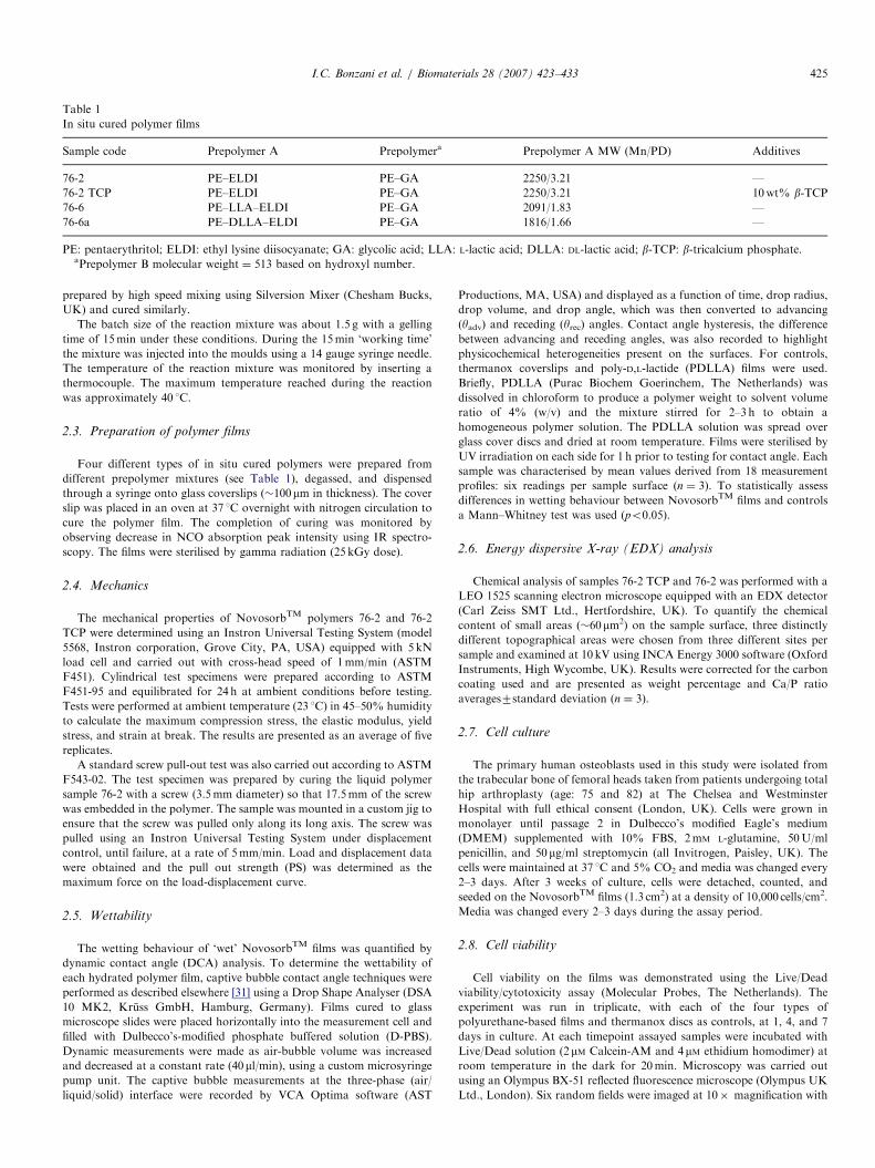
Table 1
In situ cured polymer films
Sample code Prepolymer A Prepolymera Prepolymer A MW (Mn/PD) Additives
76-2 PE–ELDI PE–GA 2250/3.21 —
76-2 TCP PE–ELDI PE–GA 2250/3.21 10wt% b-TCP76-6 PE–LLA–ELDI PE–GA 2091/1.83 —
76-6a PE–DLLA–ELDI PE–GA 1816/1.66 —
PE: pentaerythritol; ELDI: ethyl lysine diisocyanate; GA: glycolic acid; LLA: L-lactic acid; DLLA: DL-lactic acid; b-TCP: b-tricalcium phosphate.aPrepolymer B molecular weight ¼ 513 based on hydroxyl number.
I.C. Bonzani et al. / Biomaterials 28 (2007) 423–433 425
prepared by high speed mixing using Silversion Mixer (Chesham Bucks,
UK) and cured similarly.
The batch size of the reaction mixture was about 1.5 g with a gelling
time of 15min under these conditions. During the 15min ‘working time’
the mixture was injected into the moulds using a 14 gauge syringe needle.
The temperature of the reaction mixture was monitored by inserting a
thermocouple. The maximum temperature reached during the reaction
was approximately 40 1C.
2.3. Preparation of polymer films
Four different types of in situ cured polymers were prepared from
different prepolymer mixtures (see Table 1), degassed, and dispensed
through a syringe onto glass coverslips (�100mm in thickness). The cover
slip was placed in an oven at 37 1C overnight with nitrogen circulation to
cure the polymer film. The completion of curing was monitored by
observing decrease in NCO absorption peak intensity using IR spectro-
scopy. The films were sterilised by gamma radiation (25 kGy dose).
2.4. Mechanics
The mechanical properties of NovosorbTM polymers 76-2 and 76-2
TCP were determined using an Instron Universal Testing System (model
5568, Instron corporation, Grove City, PA, USA) equipped with 5 kN
load cell and carried out with cross-head speed of 1mm/min (ASTM
F451). Cylindrical test specimens were prepared according to ASTM
F451-95 and equilibrated for 24 h at ambient conditions before testing.
Tests were performed at ambient temperature (23 1C) in 45–50% humidity
to calculate the maximum compression stress, the elastic modulus, yield
stress, and strain at break. The results are presented as an average of five
replicates.
A standard screw pull-out test was also carried out according to ASTM
F543-02. The test specimen was prepared by curing the liquid polymer
sample 76-2 with a screw (3.5mm diameter) so that 17.5mm of the screw
was embedded in the polymer. The sample was mounted in a custom jig to
ensure that the screw was pulled only along its long axis. The screw was
pulled using an Instron Universal Testing System under displacement
control, until failure, at a rate of 5mm/min. Load and displacement data
were obtained and the pull out strength (PS) was determined as the
maximum force on the load-displacement curve.
2.5. Wettability
The wetting behaviour of ‘wet’ NovosorbTM films was quantified by
dynamic contact angle (DCA) analysis. To determine the wettability of
each hydrated polymer film, captive bubble contact angle techniques were
performed as described elsewhere [31] using a Drop Shape Analyser (DSA
10 MK2, Kruss GmbH, Hamburg, Germany). Films cured to glass
microscope slides were placed horizontally into the measurement cell and
filled with Dulbecco’s-modified phosphate buffered solution (D-PBS).
Dynamic measurements were made as air-bubble volume was increased
and decreased at a constant rate (40ml/min), using a custom microsyringe
pump unit. The captive bubble measurements at the three-phase (air/
liquid/solid) interface were recorded by VCA Optima software (AST
Productions, MA, USA) and displayed as a function of time, drop radius,
drop volume, and drop angle, which was then converted to advancing
(yadv) and receding (yrec) angles. Contact angle hysteresis, the difference
between advancing and receding angles, was also recorded to highlight
physicochemical heterogeneities present on the surfaces. For controls,
thermanox coverslips and poly-D,L-lactide (PDLLA) films were used.
Briefly, PDLLA (Purac Biochem Goerinchem, The Netherlands) was
dissolved in chloroform to produce a polymer weight to solvent volume
ratio of 4% (w/v) and the mixture stirred for 2–3 h to obtain a
homogeneous polymer solution. The PDLLA solution was spread over
glass cover discs and dried at room temperature. Films were sterilised by
UV irradiation on each side for 1 h prior to testing for contact angle. Each
sample was characterised by mean values derived from 18 measurement
profiles: six readings per sample surface (n ¼ 3). To statistically assess
differences in wetting behaviour between NovosorbTM films and controls
a Mann–Whitney test was used (po0.05).
2.6. Energy dispersive X-ray (EDX) analysis
Chemical analysis of samples 76-2 TCP and 76-2 was performed with a
LEO 1525 scanning electron microscope equipped with an EDX detector
(Carl Zeiss SMT Ltd., Hertfordshire, UK). To quantify the chemical
content of small areas (�60mm2) on the sample surface, three distinctly
different topographical areas were chosen from three different sites per
sample and examined at 10 kV using INCA Energy 3000 software (Oxford
Instruments, High Wycombe, UK). Results were corrected for the carbon
coating used and are presented as weight percentage and Ca/P ratio
averages7standard deviation (n ¼ 3).
2.7. Cell culture
The primary human osteoblasts used in this study were isolated from
the trabecular bone of femoral heads taken from patients undergoing total
hip arthroplasty (age: 75 and 82) at The Chelsea and Westminster
Hospital with full ethical consent (London, UK). Cells were grown in
monolayer until passage 2 in Dulbecco’s modified Eagle’s medium
(DMEM) supplemented with 10% FBS, 2mM L-glutamine, 50U/ml
penicillin, and 50 mg/ml streptomycin (all Invitrogen, Paisley, UK). The
cells were maintained at 37 1C and 5% CO2 and media was changed every
2–3 days. After 3 weeks of culture, cells were detached, counted, and
seeded on the NovosorbTM films (1.3 cm2) at a density of 10,000 cells/cm2.
Media was changed every 2–3 days during the assay period.
2.8. Cell viability
Cell viability on the films was demonstrated using the Live/Dead
viability/cytotoxicity assay (Molecular Probes, The Netherlands). The
experiment was run in triplicate, with each of the four types of
polyurethane-based films and thermanox discs as controls, at 1, 4, and 7
days in culture. At each timepoint assayed samples were incubated with
Live/Dead solution (2mM Calcein-AM and 4mM ethidium homodimer) at
room temperature in the dark for 20min. Microscopy was carried out
using an Olympus BX-51 reflected fluorescence microscope (Olympus UK
Ltd., London). Six random fields were imaged at 10� magnification with

ARTICLE IN PRESS
Fig. 2. Schematic of prepolymer B synthesis: condensation reaction.
I.C. Bonzani et al. / Biomaterials 28 (2007) 423–433426
an Olympus DP070 digital camera and analysed using Olympus DP
Controller software (Olympus Imaging, London, UK).
2.9. Cell metabolic activity
The effect of the polyurethane-based films on cell metabolic activity
was investigated using Cell Titer 96s Nonradioactive Cell Proliferation
Assay (MTS Assay, Promega, UK) after 1, 4, and 7 days. Metabolic
activity is assessed calorimetrically, using the tetrazolium compound MTS
[3-(4,5-dimethylthiazol-2-yl)-5-(3-carboxymethoxyphenyl)-2-(4-sulfophe-
nyl-)-2H-tetrazolium, innersalt], which is bioreduced by metabolically
active cells into a coloured formazan product that is soluble in tissue
culture medium. Metabolic activity was determined by optical density and
analysis of data was performed using a one-way ANOVA, with a Tukey
post hoc test to establish significance (po0.05).
3. Results
3.1. Prepolymer chemistry and properties
3.1.1. Prepolymer A
The reaction described in Section 2.1 yields a viscousprepolymer consisting of a mixture of star and hyper-branched structures (Fig. 1). The number average mole-cular weight and polydispersity of the prepolymer were1348 and 1.73, respectively, based on GPC analysis. Theprepolymer instantaneous viscosity was 8.7� 104 cSt.
3.1.2. Prepolymer B
In all cases Prepolymer B was a four arm polyolprepared form PE and glycolic acid (Fig. 2) usingcondensation polymerisation as described in Section 2.1.The molecular weight and polydispersity of the polyol wasdetermined to be 513.3 and 1.08, respectively, based onhydroxyl number determination.
The cured polymers are a cross-linked network formedby the reaction of terminal isocyanate and hydroxyl groupspresent, respectively, in prepolymers A and B. The main
Fig. 1. Schematic of prepolymer A synthesis—pentaerythritol end capped
with ethyl lysine diisocyanate.
functional groups present in the polymer network are esterand urethane/urea, all susceptible to hydrolytic, enzymaticand other types of degradation pathways present inbiological systems.
3.2. Mechanics
The mechanical properties of samples 76-2 and 76-2 TCPwere studied in compression and represented as stress,strain, and elastic modulus. In situ cross-linked polymernetworks 76-2 TCP and 76-2 exhibit excellent mechanicalstrength, with ultimate compressive strengths (UCS) of139711 and 136714MPa and elastic modulus (E) of2.370.03 and 2.070.01GPa, respectively. By comparison,these samples had a UCS significantly greater than, and anE within the range of cancellous bone (Table 2). To furtherhighlight the mechanical strength of the polymers, the UCSwere compared to those of European and FDA approvedacrylic bone cements (73–117MPa) and injectable, photo-cross-linkable polyanhydrides (30–50MPa), recently devel-oped for orthopaedic applications [6,32]. In measuringstrain resistance, sample 76-2 and 76-2 TCP were found tohave ultimate compressive strains of 440%, with recover-able elastic strains of �5% (Fig. 3).When compressed, the polymers demonstrated visco-
elastic-like properties, evidenced by an initial linear elasticregion and a plastic deformation region seen prior tofailure (Fig. 3). This type of viscoelastic behaviour is alsodemonstrated in natural bone [33]. Stress–strain plots(Fig. 3) revealed that the incorporation of 10wt% b-TCPparticles (5 mm) to sample 76-2 caused an increase (notsignificant) in E, yield strength, and UCS.Sample 76-2 was also tested for its pull out strength. The
pull out force of sample 76-2, 59547101N, was found tobe significantly higher compared to the pull out force ofscrews in healthy bone and curable PMMA cements,demonstrated to be 15407361N [34] and 23017283N[35], respectively.

ARTICLE IN PRESS
Table 2
Initial mechanical properties in longitudinal compression of samples 76-2 and 76-2 TCP compared with properties of natural bone, widely used calcium
phosphate-based bone cements, and injectable and photocross-linkable polyanhydrides
Material Longitudinal compression
Ultimate stress (MPa) Stress at yield (MPa) Modulus (GPa)
76-2 136 (14) 84 (15) 2.0 (0.3)
76-2 TCP 139 (11) 76 (13) 2.3 (0.01)
Cortical bonea 180–220 180 (13) 18.6 (3.5)
Cancellous bonea 5–10 — 6.1 (5.4)
Acrylic bone cementsb 73–117 — 0.05–0.1
Polyanhydridesc 30–50 — —
The bone properties reported are ranges for the human femur, tibia, radius, and ulna (44,72).
Values for mechanical properties were taken directly, or from information contained in these papers: a[44,72], b[43], c[73]. Some values were not reported in
literature (—). Values reported as mean (SD).
Fig. 3. Stress–strain curves of cylindrical specimens 76-2 and 76-2 TCP loaded in longitudinal compression and measured using an Instron Universal
Testing System.
I.C. Bonzani et al. / Biomaterials 28 (2007) 423–433 427
3.3. Wettability
The wetting behaviour of each of the polymer films wasassessed by DCA, previously evaluated as a sensitivemethod to detect interfacial changes at biomaterial/biosystem interfaces [36]. The combination of captivebubble techniques with Drop Shape Analysis imageprocessing, allowed for the accurate and objective mea-surement of dynamic contact angles in a wet, biologicallyrelevant environment. The contact angle measurements ofthe polyurethane films and control films (Fig. 4), arepresented as advancing angle, yadv, and receding angle, yrec.
In comparing receding contact angle results, we found allpolyurethane films to be significantly less hydrophilic thanthermanox (po0.05). The films, however were found to besignificantly more hydrophilic than control film PDLLA(po0.05). Cross-comparing contact angles of the polyur-ethane films, suggested that sample 76-2 TCP is the mosthydrophilic, followed by samples 76-2, 76-6a, and 76-6,although these differences were found not to be significant.
Additionally, all polyurethane films were found to havesimilar hysteresis values (�211), with sample 76-2 TCPhaving a slightly higher value (21.851). Whereas, therma-nox and PDLLA, expressed lower contact angle hysteresis,with values of 11.61 and 16.71, respectively. This signified amore physically or chemically heterogeneous surface on thepolyurethane films compared to controls.
3.4. EDX analysis
SEM/EDX analysis of unseeded samples 76-2 and 76-2TCP was employed to examine surface topographyand determine chemical composition of film surfaces.This enabled correlations between chemical content andtopographical heterogeneity on the sample surfaces.These two samples were compared because they haveidentical polymer chemistries and fabrication techniques,however sample 76-2 TCP was supplemented with b-TCPparticles during film fabrication. EDX results revealedthat calcium and phosphorus were solely concentrated at

ARTICLE IN PRESS
Fig. 4. The advancing (yadv) and receding (yrec) dynamic contact angles of captive bubbles on NovosorbTM films and controls in Dulbecco’s modified
phosphate buffered solution (D-PBS). The controls are poly-(DL-lactic acid) (PDLLA) films and thermanox. The results are displayed as means7SD and
samples are arranged in terms of decreasing hydrophilicity, seen as an increase in the receding angle. Significance was assigned using a Mann–Whitney test
(po0.05). Thermanox was found to have a yrec significantly less than all other samples (*), NovosorbTM films were found to have a yrec significantly less
than PDLLA (a–d) and comparable to one another.
I.C. Bonzani et al. / Biomaterials 28 (2007) 423–433428
topographically heterogeneous areas on the film surface ofsample 76-2 TCP (Fig. 5). These areas of heterogeneity(sample areas 1 and 3 in Fig. 5A) appeared as topogra-phically raised areas, with inter-dispersed pore-like struc-tures. In contrast, no calcium and phosphorus was detectedon the adjacent flat and nonporous area on the samesample surface (sample area 2 in Fig. 5A). Additionally, insample 76-2, no calcium and phosphorus was detected onall surface areas scanned (sample areas 1–3 in Fig. 5B). TheCa/P ratios of the heterogeneous areas on sample 76-2 TCPwere averaged to be 1.6170.27 (n ¼ 9).
3.5. Assessment of cell viability on NovosorbTM films
Primary human osteoblasts were cultured on thepolymer films for 1, 4, 7 days and evaluated for cytotoxicityusing Live/Dead assay. By this qualitative method, live anddead cells were stained green and red, respectively. Asindicated in Fig. 6, a majority of the cells remained viableon the samples for periods of up to 7 days, seen as positivelive to dead cell ratios (495%). The live cells adhered wellto the films surfaces, and exhibited normal, healthyosteoblastic spindle-like morphology for periods of up to4 days. In comparing the samples, it is seen that sample76-2 TCP maintained the best cell viability at longer timepoints. In samples 76-2, 76-6, and 76-6a, good cell viabilityis seen for periods of up to 4 days and is followed by aslight decrease in cell viability. Osteoblasts on thermanoxcontrols were found to have similar spindle-shapedmorphology as compared to the polyurethane films,however cells were found to be confluent after 4 days in
culture (data not shown). The images shown in Fig. 6 arerepresentative of the results seen for all polyurethane filmsat six different locations on the surface (n ¼ 3).
3.6. Assessment of cell metabolic activity on NovosorbTM
films
Changes in the metabolic activity of primary osteo-blasts cultured on the films were evaluated byMTS proliferation assay at 1, 4, and 7 days. MTSmeasures metabolic function in cells via mitochon-drial activity, where a higher absorbance signifies eitheran increase in cell proliferation or a higher rate ofmetabolism of the MTS. A reduction in cellular metabolicactivity is an early indication of cellular damage, whichcould potentially lead to cell death. Results for all films,presented in Fig. 7, show a general increase in cellmetabolism for a period of 7 days. This same cellproliferation profile was obtained on the thermanoxcontrol. In determining significance, it was found thatsample 76-2 exhibited significantly decreased metabolicactivity for each time point as compared to the thermanoxcontrol (po0.05), although an increase in metabolicactivity was noticed for a period of 7 days. Samples 76-6and 76-6a were found to have comparable increases in cellproliferation to cells on thermanox for a period of 4 days.Sample 76-2 (TCP) was comparable (no significantdifference) to cells on thermanox over the entire periodof 7 days. No metabolic activity was observed from any ofthe unseeded samples, signifying that the samples did notreact with the MTS reagents (data not shown).

ARTICLE IN PRESS
Fig. 5. SEM/EDX surface analysis of unseeded films 76-2 TCP (A) and
76-2 (B). Concentrations of calcium (Ca) and phosphorus (P) are found
strictly on the topographically heterogeneous areas of sample 76-2 TCP
(sample areas A1 and A3), with Ca/P ratios of 1.4 and 1.6, respectively.
Chemical spectral analysis of sample 76-2 surfaces reveals undetectable
concentrations of calcium and phosphorus present (sample areas B1–B3)
(scale ¼ 20 mm).
I.C. Bonzani et al. / Biomaterials 28 (2007) 423–433 429
4. Discussion
The preliminary evaluation of toxicity performed in thisstudy indicated that the films provided generally cytocom-patable surface characteristics that allowed for initial cellattachment, sustained viability, and increased cell prolif-eration profiles. Moreover, these results confirmed polymersynthesis and curing processes to be adequate for initialimplantation, since cell viability and increased proliferationwas noted for a 7-day period.
Results of Live/Dead and MTS suggested that in situsample 76-2 TCP provided an optimal environment forosteoblast attachment, sustained viability, and prolifera-tion as compared to the rest of the films. A possibleexplanation for this result was the incorporation of b-TCPgranules during polymer film synthesis. b-TCP is aresorbable bioceramic material and can be used in theform of blocks, granules, or cements in scaffolds for boneregeneration. Previous literature suggests that TCP mayenhance osteoblast function and overall bone formation
[37,38]. Although the exact mechanisms whereby thishappens presently cannot be elucidated, evidence suggeststhat TCP fillers increase the initial anchoring and spreadingof serum proteins on polymer surfaces [39]. Therefore, onecan assume that sample 76-2 TCP should have a moreosteoconductive environment compared to a similarsample film without b-TCP (i.e. sample 76-2). This wasobserved in this study, as sample 76-2 TCP demonstratedenhanced viability and proliferation of osteoblasts over a7-day period compared to sample 76-2. Additionally, b-TCP is also believed to act as a buffering agent,neutralising any acidic by-products and maintaining pH[12], which could be another explanation as to why betterviability and proliferation were found on this film.Initial mechanical support and bonding of an implant to
the surrounding tissue, is of particular importance tobiomaterials for orthopaedic applications. It is integral thatthese mechanical properties are analogous to the propertiesof the tissue being repaired. While many properties areimportant when characterising materials for orthopaedics,compressive properties are most consistently recorded inliterature, and therefore provide for a better comparisonbetween materials [40]. After cured, samples 76-2 and 76-2TCP attained compressive strengths of 136 and 139MPa,respectively. Although insignificant, the slightly enhancedmechanical properties found in sample 76-2 TCP (Table 2and Fig. 3) are most likely due to the incorporation ofb-TCP particles; a typical reinforcement effect notedpreviously [5]. Both samples, however, had compressivestrengths well above those for cancellous bone (5–10MPa)and are nearly within the range of cortical bone(180–220MPa) in compression [33]. These compressivestrengths are almost twice the critical minimum (70MPa)permitted under standard ISO 5833, regulating injectablebone cements [41]. Similarly, both polyurethane samplesexhibited elastic modulus (�2GPa) in accordance withpermitted minimums under the same standard (41.8GPa).Additionally, the polymers are found to exhibit mechanicalproperties better than those of a novel calcium phosphate-based bone cements and widely used acrylic cements, bothof which have gained FDA and European approval foruses in distal radius and tibial plateau fractures, cranio-facial applications, and screw fixation [32,42,43].Further comparisons of mechanical properties of the
polyurethane polymers to natural bone, revealed thatpolymers 76-2 and 76-2 TCP were designed to withstandthe peak compressive strains in limb bones incurred duringrigorous functional loading. In bone, principal compressivestrains have been found to reach peak magnitudes of2000 me [44], well within the elastically recoverable strainrange of polymers 76-2 and 76-2 TCP (o50,000 me).Undoubtedly, the result of plastic deformation to a weightbearing implant will lead to loss of form and function, andwill eventually fail, resulting in both pain and an additionalsurgical procedure for the patient. Moreover, the ratios ofultimate compressive strength to elastic modulus, arecomparable in both bone and our polymer constructs

ARTICLE IN PRESS
Fig. 6. Live/Dead analysis of NovosorbTM films seeded with primary human osteoblasts for 1, 4, and 7 days. Images show combined images of live (green)
and dead (red cells) cells on all samples. Non-cytotoxic surfaces are seen on all sample types for periods of up to 7 days, with images showing sustained cell
viability and extremely positive live to dead cell ratios (495%) (scale bar ¼ 200mm).
1Thermanox
*
*
* *
*76-2 TCP76-276-676-6a
0.9
0.8
0.7
0.6
0.5
0.4
0.3
0.2
0.1
01 day 4 day 7 day
Opt
ical
Den
sity
(49
2 nm
)
Fig. 7. MTS assay for primary human osteoblasts cultured on NovosorbTM films and thermanox controls for 1, 4, and 7 days. Significance is assessed
compared to thermanox control for each time point assayed (*po0.05).
I.C. Bonzani et al. / Biomaterials 28 (2007) 423–433430

ARTICLE IN PRESSI.C. Bonzani et al. / Biomaterials 28 (2007) 423–433 431
(�1/100) [32]. This analysis showed that the polyurethaneconstructs, although not mechanically identical to corticalbone, possess a similar balance between compressiveelasticity and strength found in natural bone.
Mechanical screw pull-out tests, showed sample 76-2 tohave a fixation strength greater than that of bone andPMMA bone cements, found to be 15407361 and23017283N, respectively [34,35]. This strong initial bond-ing strength possessed by polymer 76-2 may act to reduceinterfacial failure and bond loosening by strengthening thecement–implant and cement–bone interface [45]. Thestrength of this interface with the surrounding host bonetissue is of paramount importance to the clinical success ofinjectable polymer systems in orthopaedic applications.The strong initial bonding of this injectable polymer gel tothe host bone will allow for quick restoration of voids;leading to immediate fracture stability, an earlier returnto full-weight bearing, and faster restoration of tissuefunction.
The wetting behaviour was assessed by DCA, previouslyevaluated as a sensitive method to detect interfacialchanges at biomaterial/biosystem interfaces [36]. DCA isconsidered a valuable technique for characterising thewetting behaviour of rough biomaterial surfaces, becausethe three-phase contact angle is sensitive to the outermost3–20 A of a surface and its chemistry [46]. This same area isalso important to many cell–surface interactions includingadhesion, motility, and signalling. It is known that surfacewettability plays a vital role in the cell–biomaterialinterface, modulating both protein adsorption and celladhesion [47], where cell adhesion and motility increases onmore hydrophilic surfaces [48]. In this study an increase inthe value of the receding contact angle provided a reliableindication of a less hydrophilic surface [49]. All polyur-ethane films were found to have receding angles signifi-cantly more hydrophilic than PDLLA; a biodegradablepolymer approved for in vivo orthopaedic applications bythe Food and Drug Administration of the USA [50,51].This surface hydrophilicity, correlated with the favourablecell viability and proliferation results seen for all samples.
To better understand the wetting characteristics of thesefilms, hysteresis values obtained from DCA, were used toassess surface roughness. The heterogeneity of the surfacecan be observed indirectly when measuring contact angles.If a surface is not smooth, large deviations in attainablecontact angle measurements will be seen as the three-phasecontact line is moved, leading to large hysteresis values[52]. These irregular contact angles arise from a phenom-enon known as pinning (stick/slip). It was previouslyshown, that if size of the heterogeneity [53] or roughnessfeature [54] was larger than 0.1 mm, the three phase contactline had a tendency to be stopped by a pinning mechanismon local surface imperfections and could not move withoutintroducing additional external force (i.e. rapid influx of airinto the captive bubble). This creates a series of metastablestates, with contact angle hysteresis used as indicator as tothe extent of these surface heterogeneities. All polyurethane
films were seen to be rough, heterogeneous surfaces,evidenced by significantly larger values of contact anglehysteresis (�211) compared to smooth, homogeneousthermanox surface (11.61). Sample 76-2 TCP was foundto have a slightly higher hysteresis value as compared tothe other films. This was most likely due to the fact thatsample 76-2 TCP, was composed of two regions, havingdifferent wetting characteristics: the guest material (b-TCP5 mm particles) and the primary host material (LDI-basedpolyurethane). The different chemistries present on thissurface likely influence the wetting behaviour by causingboth morphological and chemical changes in the system[46]. Although not statistically significant, this suggestedthat sample 76-2 TCP had a rougher surface with moreheterogeneity, in support of SEM micrographs of thesurfaces of samples 76-2 and 76-2 TCP (Fig 5), whichshowed a more topographically heterogeneous surface onsample 76-2 TCP. Although surface roughness was notquantitatively assessed in this study, one can suggest thatthe surface roughness of sample 76-2 TCP has created amore hydrophilic environment, shown as a reduction incontact angle. This observation is in agreement withprevious investigations, which concluded that roughnessmakes hydrophilic surfaces (contact angleo901) morehydrophilic and hydrophobic surfaces (contact an-gles4901) more hydrophobic [55].EDX analysis confirmed the presence of calcium and
phosphorous on the surface of sample 76-2 TCP whilstthese elements were undetectable on sample 76-2. Further-more, results revealed calcium and phosphorus to be solelyconcentrated at topographically heterogeneous areas onthe film surface of sample 76-2 TCP (Fig. 5B). The fact thatno concentrations of calcium or phosphorous were foundon sample 76-2, suggested that b-TCP had a role increating topographical heterogeneity. This demonstrates apossible correlation between the b-TCP filler and increasedchemical and physical heterogeneity. These areas ofchemical heterogeneity were found to consist of Ca/Pratios (1.6170.27) similar to native bone (1.71) and naturalstoichiometric HA (�1.67), known to have osteoinductiveand osteoconductive capacities [56,57]. More studies,however, are needed to examine the extent of the relation-ship between physiochemical heterogeneity and osteoblastbehaviour in sample 76-2 TCP.
5. Conclusions
This study presents a novel two-component polyur-ethane platform that can be administered arthroscopically,polymerise at biological temperatures in situ, and provideappropriate bonding strength and mechanical support,with mechanical properties superior to widely used bonecements. These novel polymers provided a favourable invitro environment for initial cell adhesion, maintained cellviability, and normal rates of proliferation of humanosteoblasts for periods of 7 days. Physicochemically,this biocompatibility can in-part, be attributed to the

ARTICLE IN PRESSI.C. Bonzani et al. / Biomaterials 28 (2007) 423–433432
hydrophilic surface characteristics of these polymers, and,in the case of sample 76-2 TCP, the combined presence ofheterogeneous topographies and chemical moieties on thesurface. The addition of b-TCP filler during film fabrica-tion enhanced cell viability, increased surface heterogeneityand mechanical properties in compression, and signifi-cantly increased cell proliferation.
Acknowledgements
The authors would like to thank Laleh Safina, JonnyBlaker (Department of Chemical Engineering and Depart-ment of Materials) for their technical assistance and Dr.Alex Bismarck (Department of Chemical Engineering) forthe use of his DCA equipment. The authors also expresstheir gratitude to Dr. Michael Ball (Department ofmaterials) for his helpful discussions. Ian Bonzani wouldalso like to acknowledge the Marshall Aid Commemora-tion Commission for funding.
References
[1] Edwards ER, Graves SE, McNeil JJ, Williamson OD, Urquhart DM,
Cicuttini FM. Orthopaedic trauma: establishment of an outcomes
registry to evaluate and monitor treatment effectiveness. Injury
2006;37(2):95–6.
[2] Stevens MM, George JH. Exploring and engineering the cell–surface
interface. Science 2005;310(5751):1135–8.
[3] Gunatillake PA, Adhikari R. Biodegradable synthetic polymers for
tissue engineering. Eur Cell Mater 2003;5:1–16.
[4] Vacanti JP, Morse MA, Saltzman WM, Domb AJ, Perez-Atayde A,
Langer R. Selective cell transplantation using bioabsorbable artificial
polymers as matrices. J Pediatr Surg 1988;23(1 Part 2):3–9.
[5] Peter SJ, Miller MJ, Yasko AW, Yaszemski MJ, Mikos AG. Polymer
concepts in tissue engineering. J Biomed Mater Res 1998;43(4):422–7.
[6] Burkoth AK, Anseth KS. A review of photocrosslinked polyanhy-
drides: in situ forming degradable networks. Biomaterials
2000;21(23):2395–404.
[7] Burdick JA, Frankel D, Dernell WS, Anseth KS. An initial
investigation of photocurable three-dimensional lactic acid based
scaffolds in a critical-sized cranial defect. Biomaterials 2003;24(9):
1613–20.
[8] Temenoff JS, Mikos AG. Injectable biodegradable materials for
orthopedic tissue engineering. Biomaterials 2000;21(23):2405–12.
[9] Poshusta AK, Burdick JA, Mortisen DJ, Padera RF, Ruehlman D,
Yaszemski MJ, et al. Histocompatibility of photocrosslinked poly-
anhydrides: a novel in situ forming orthopaedic biomaterial.
J Biomed Mater Res A 2003;64(1):62–9.
[10] Muggli DS, Burkoth AK, Anseth KS. Crosslinked polyanhydrides
for use in orthopedic applications: degradation behavior and
mechanics. J Biomed Mater Res 1999;46(2):271–8.
[11] Peter SJ, Kim P, Yasko AW, Yaszemski MJ, Mikos AG. Cross-
linking characteristics of an injectable poly(propylene fumarate)/
beta-tricalcium phosphate paste and mechanical properties of the
crosslinked composite for use as a biodegradable bone cement.
J Biomed Mater Res 1999;44(3):314–21.
[12] Peter SJ, Miller ST, Zhu G, Yasko AW, Mikos AG. In vivo
degradation of a poly(propylene fumarate)/beta-tricalcium phosphate
injectable composite scaffold. J Biomed Mater Res 1998;41(1):1–7.
[13] Peter SJ, Miller ST, Zhu G, Yasko AW, Mikos AG. In vivo
degradation of a poly(propylene fumarate)/beta-tricalcium phosphate
injectable composite scaffold. J Biomed Mater Res 1998;41(1):1–7.
[14] Peter SJ, Lu L, Kim DJ, Mikos AG. Marrow stromal osteoblast
function on a poly(propylene fumarate)/[beta]-tricalcium phosphate
biodegradable orthopaedic composite. Biomaterials 2000;21(12):
1207–13.
[15] Galletti G, Gogolewski S, Ussia G, Farruggia F. Long-term patency
of regenerated neoaortic wall following the implant of a fully
biodegradable polyurethane prosthesis: experimental lipid diet model
in pigs. Ann Vasc Surg 1989;3(3):236–43.
[16] Zhang JY, Doll BA, Beckman EJ, Hollinger JO. Three-dimensional
biocompatible ascorbic acid-containing scaffold for bone tissue
engineering. Tissue Eng 2003;9(6):1143–57.
[17] Zhu Y, Gao C, Guan J, Shen J. Promoting the cytocompatibility of
polyurethane scaffolds via surface photo-grafting polymerization of
acrylamide. J Mater Sci Mater Med 2004;15(3):283–9.
[18] Zhu Y, Gao C, He T, Shen J. Endothelium regeneration on luminal
surface of polyurethane vascular scaffold modified with diamine and
covalently grafted with gelatin. Biomaterials 2004;25(3):423–30.
[19] Zhu Y, Sun Y. The influence of polyelectrolyte charges of
polyurethane membrane surface on the growth of human endothelial
cells. Colloids Surf B 2004;36(1):49–55.
[20] dal Pra I, Petrini P, Charini A, Bozzini S, Fare S, Armato U. Silk
fibroin-coated three-dimensional polyurethane scaffolds for tissue
engineering: interactions with normal human fibroblasts. Tissue Eng
2003;9(6):1113–21.
[21] Skarja GA, Woodhouse KA. In vitro degradation and erosion of
degradable, segmented polyurethanes containing an amino acid-
based chain extender. J Biomater Sci Polym Ed 2001;12(8):851–73.
[22] Grad S, Kupcsik L, Gorna K, Gogolewski S, Alini M. The use of
biodegradable polyurethane scaffolds for cartilage tissue engineering:
potential and limitations. Biomaterials 2003;24(28):5163–71.
[23] Saad B, Kuboki Y, Welti M, Uhlschmid GK, Neuenschwander P,
Suter UW. DegraPol-foam: a degradable and highly porous
polyesterurethane foam as a new substrate for bone formation. Artif
Organs 2000;24(12):939–45.
[24] Gorna K, Gogolewski S. Preparation, degradation, and calcification
of biodegradable polyurethane foams for bone graft substitutes.
J Biomed Mater Res A 2003;67(3):813–27.
[25] Xue L, Greisler HP. Biomaterials in the development and future of
vascular grafts. J Vasc Surg 2003;37(2):472–80.
[26] Zdrahala RJ. Small caliber vascular grafts. Part II: Polyurethanes
revisited. J Biomater Appl 1996;11(1):37–61.
[27] Guan J, Fujimoto KL, Sacks MS, Wagner WR. Preparation and
characterization of highly porous, biodegradable polyurethane
scaffolds for soft tissue applications. Biomaterials 2005;26(18):
3961–71.
[28] Bruin P, Smedinga J, Pennings AJ, Jonkman MF. Biode-
gradable lysine diisocyanate-based poly(glycolide-co-epsilon-capro-
lactone)-urethane network in artificial skin. Biomaterials 1990;11(4):
291–5.
[29] Zhang JY, Beckman EJ, Piesco NP, Agarwal S. A new peptide-based
urethane polymer: synthesis, biodegradation, and potential to
support cell growth in vitro. Biomaterials 2000;21(12):1247–58.
[30] Zhang JY, Beckman EJ, Hu J, Yang GG, Agarwal S, Hollinger JO.
Synthesis, biodegradability, and biocompatibility of lysine diisocya-
nate glucose polymers. Tissue Eng 2002;8(5):771–85.
[31] Safina L, Blaker JJ, Maquet V, Boccaccini AR, Mantalaris A,
Bismarck A. Characterisation of ‘wet’ polymer surfaces for tissue
engineering applications: are flat surfaces a suitable model for
complex structures? e-polymers 2005;10.
[32] Lewis G. Properties of acrylic bone cement: state of the art review.
J Biomed Mater Res 1997;38(2):155–82.
[33] Currey JD. Mechanical properties of vertebrate hard tissues. Proc
Inst Mech Eng [H] 1998;212(6):399–411.
[34] Yueheui H, Draughnm RA. Mechanical testing of bone and the
bone–implant interface. Boca Raton, FL: CRC Press LLC; 1999.
[35] Motzkin NE, Chao EY, An KN, Wikenheiser MA, Lewallen DG.
Pull-out strength of screws from polymethylmethacrylate cement.
J Bone Jt Surg Br 1994;76(2):320–3.
[36] Rupp F, Axmann D, Ziegler C, Geis-Gerstorfer J. Adsorption/
desorption phenomena on pure and Teflon AF-coated titania

ARTICLE IN PRESSI.C. Bonzani et al. / Biomaterials 28 (2007) 423–433 433
surfaces studied by dynamic contact angle analysis. J Biomed Mater
Res 2002;62(4):567–78.
[37] Gaasbeek RD, Toonen HG, van Heerwaarden RJ, Buma P.
Mechanism of bone incorporation of beta-TCP bone substitute in
open wedge tibial osteotomy in patients. Biomaterials 2005;26(33):
6713–9.
[38] Barralet JE, Grover L, Gaunt T, Wright AJ, Gibson IR. Preparation
of macroporous calcium phosphate cement tissue engineering
scaffold. Biomaterials 2002;23(15):3063–72.
[39] Hutmacher DW, Kirsch A, Ackermann KL, Huerzeler MB. Matrix
and carrier materials for bone growth factors—state of the art and
future perspectives. In: Stark GB, Horch R, Tancos E, editors.
Biological matrices and tissue reconstruction. Heidelberg, Germany:
Springer; 1998.
[40] Yaszemski MJ, Payne RG, Hayes WC, Langer R, Mikos AG.
Evolution of bone transplantation: molecular, cellular and tissue
strategies to engineer human bone. Biomaterials 1996;17(2):175–85.
[41] International Organisations for Standardization. I. Implants for
surgery—acrylic resin cements. Report no. ISO 5833, ISO, Geneva,
2002.
[42] Frankenburg EP, Goldstein SA, Bauer TW, Harris SA, Poser RD.
Biomechanical and histological evaluation of a calcium phosphate
cement. J Bone Jt Surg Am 1998;80(8):1112–24.
[43] Larsson S, Bauer TW. Use of injectable calcium phosphate cement
for fracture fixation: a review. Clin Orthop Relat Res 2002(395):
23–32.
[44] Burr DB, Milgrom C, Fyhrie D, ForwoodM, Nyska M, Finestone A,
et al. In vivo measurement of human tibial strains during vigorous
activity. Bone 1996;18(5):405–10.
[45] Spector M. Biomaterial failure. Orthop Clin North Am 1992;
23(2):211–7.
[46] Drelich J. Apparent and microscopic contact angles. The Nether-
lands: VSP; 2000.
[47] Anselme K, Bigerelle M, Noel B, Dufresne E, Judas D, Iost A, et al.
Qualitative and quantitative study of human osteoblast adhesion on
materials with various surface roughnesses. J Biomed Mater Res
2000;49(2):155–66.
[48] Altankov G, Grinnell F, Groth T. Studies on the biocompatibility of
materials: fibroblast reorganization of substratum-bound fibronectin
on surfaces varying in wettability. J Biomed Mater Res 1996;30(3):
385–91.
[49] Volpe CD, Penati A, Peruzzi R, Siboni S, Toniolo L, Colombo C.
The combined effect of roughness and heterogeneity on contact
angles: the case of polymer coating for stone protection. In: Drelich J,
Laskowski JS, Mittal KL, editors. Apparent and microscopic contact
angles. The Netherlands: VSP; 2000. p. 349–75.
[50] Cutright DE, Hunsuck EE, Beasley JD. Fracture reduction
using a biodegradable material, polylactic acid. J Oral Surg
1971;29(6):393–7.
[51] Yang J, Bei J, Wang S. Enhanced cell affinity of poly(D,L-lactide) by
combining plasma treatment with collagen anchorage. Biomaterials
2002;23(12):2607–14.
[52] Mobius D, Miller R. Drops and bubbles in interfacial research. The
Netherlands: Elsevier; 1998.
[53] Eick JD, Johnson LN, Fromer JR, Good RJ, Neumann AW. Surface
topography: its influence on wetting and adhesion in a dental
adhesive system. J Dent Res 1972;51(3):780–8.
[54] Eick JD, Good RJ, Neumann AW. Thermodynamics of contact
angles, Part 2, Rough solid surfaces. J Colloid Interfaces Sci 1975;53:
235–48.
[55] Bico J, Marzolin C, Quere D. Pearl drops. Europhys Lett
1999;47:220–6.
[56] Causa F, Netti PA, Ambrosio L, Ciapetti G, Baldini N,
Pagani S, et al. Poly-epsilon-caprolactone/hydroxyapatite composites
for bone regeneration: in vitro characterization and human
osteoblast response. J Biomed Mater Res A 2005;76A(1):
151–62.
[57] LeGeros RZ. Biological and synthetic apatites. In: Brown PW,
Constantz B, editors. Hydroxyapatite and related materials. Boca
Raton, FL: CRS Press; 1994. p. 1–28.
Copyright © 2022 FDOKUMEN