Strontium rich hybrid injectable system for bone regeneration
171
Strontium rich hybrid injectable system for bone regeneration NUNO NEVES TESE DE DOUTORAMENTO APRESENTADA À FACULDADE DE MEDICINA DA UNIVERSIDADE DO PORTO EM MEDICINA D 2017
-
Upload
khangminh22 -
Category
Documents
-
view
0 -
download
0
Transcript of Strontium rich hybrid injectable system for bone regeneration
Strontium rich hybrid injectable system for bone regeneration
NUNO NEVES
TESE DE DOUTORAMENTO APRESENTADA
À FACULDADE DE MEDICINA DA UNIVERSIDADE DO PORTO EM
MEDICINA
D 2017


Strontium rich hybrid injectable system for bone regeneration
NUNO NEVES
TESE DE DOUTORAMENTO APRESENTADA
À FACULDADE DE MEDICINA DA UNIVERSIDADE DO PORTO EM
MEDICINA
D 2017

Nuno Silva Morais Neves
Strontium rich hybrid injectable system for bone regeneration
Tese de Candidatura ao grau de Doutor em Medicina, submetida à Faculdade de Medicina da Universidade do Porto.
Orientador – Professor Doutor Gilberto Costa
Categoria – Professor Associado
Afiliação – Centro Hospitalar São João (CHSJ) e Faculdade de Medicina da Universidade do Porto (FMUP)
Coorientador – Professor Doutor Mário A. Barbosa
Categoria – Professor Catedrático
Afiliação – Instituto de Ciências Biomédicas Abel Salazar (ICBAS), Instituto de Investigação e Inovação em Saúde (i3S) e Instituto de Engenharia Biomédica (INEB), Universidade do Porto


3
Table of Contents
ACKNOWLEGMENTS 5 LIST OF PUBLICATIONS 7 ABSTRACT 9 RESUMO 13
CHAPTER I - GENERAL INTRODUCTION; AIMS OF THE THESIS 17 Bonegrafts 19Bonesubstitutes 20Strontium 23References 26AimsoftheThesis 25 CHAPTER II - IN VIVO AND CLINICAL APPLICATION OF STRONTIUM-ENRICHED BIOMATERIALS FOR BONE REGENERATION 35 Abstract 38Introduction 39MaterialsandMethods 41Results 44Discussion 51Articlefocus 57Keymessages 57StrengthsandLimitations 57SupplementaryMaterial 58References 63 CHAPTER III - STRONTIUM-RICH INJECTABLE HYBRID SYSTEM FOR BONE REGENERATION 71 Abstract 74Introduction 75MaterialsandMethods 77Results 85Discussion 92Conclusions 98Highlights 99StatementofSignificance 100GraphicalAbstract 101Acknowledgements 102References 103 CHAPTER IV - INJECTABLE HYBRID SYSTEM FOR STRONTIUM LOCAL DELIVERY PROMOTES BONE REGENERATION IN A RAT CRITICAL-SIZED DEFECT MODEL 111 Abstract 114Introduction 115Results 118Discussion 128

4
Conclusions 133MaterialsandMethods 134Acknowledgements 141SupplementaryMaterial 142References 143 CHAPTER V - CONCLUDING REMARKS AND FUTURE PERSPECTIVES 153 Conclusions 160References 161

5
ACKNOWLEDGEMENTS
This thesis would not have been possible without the collaboration and
involvement of several people to whom I am deeply grateful. I would particularly like
to thank the following:
Professor Gilberto Costa, supervisor of this thesis in very special
circumstances. I am grateful for your support and guidance. Already as your student
and since my early years in residency, you have been an example of a dedicated
doctor, surgeon, teacher, and friend;
Professor Mário Barbosa, co supervisor of this thesis, for setting up this
challenge and introducing me to a new world of laboratorial and scientific research.
In the difficult moments, your leadership, perseverance and ethics were inspiring;
Professor Cristina Ribeiro, my research companion. I am grateful for your
interest, commitment, rigor, opinions and suggestions;
All my colleagues at INEB and i3S, especially Filipa Lourenço, Daniel
Vasconcelos, Serafim Oliveira, Bruno Campos, Cláudia Carvalho, Susana Sousa,
Meriem Lamghari, Cristina Barrias, Susana Santos, Raquel Gonçalves, Pedro
Granja, for all the collaboration and support;
Dr. Rui Pinto, Chair of the Orthopedic Department of Centro Hospitalar de São
João, for the continuing confidence, support, and incentive for excellence;
Professor Manuel Gutierres, with whom I shared my first steps in Orthopedics,
for the true friendship that bonds us;
All my colleagues at the Orthopedic Department of Hospital de São João,
especially Manuel Ribeiro da Silva, Pedro Negrão, Rui Matos, Pedro Cacho
Rodrigues, Daniela Linhares and João Santos Carvalho, for all the cooperation,
insight, collaboration and friendship;

6
Professor Manuel Barbosa and Professor Dulce Madeira, with whom I started
my academic career, for the opportunity given, the example of leadership, mutual
respect, and scientific and educational standards.
The Radiology Department of FMUP, and its Chair Professor Isabel Ramos,
the Nefrology Department of FMUP, and its Chair Professor Manuel Pestana, the
Instituto de Medicina Legal – Delegação Norte, especially Professor Agostinho
Santos, the Department of Pharmaceutical Technology of FFUP, namely Professor
Paulo Costa and Professor Isabel Almeida, and the Department of Mechanics of
FEUP, particularly Professor Mário Vaz, for their contribution to the work developed;
My parents, José and Teresa, for all the love and unconditional support, and for
having transmitted the values that guide my life;
My wife, Patrícia, and my children, João and Luísa, always supportive, loving,
and caring, in spite of the professional and academic life that separates us so often.
You are always on my thoughts;
Lastly, Professor Abel Trigo Cabral, the inspirer of this thesis and its first
supervisor, unfortunately unable to see it come to an end. He was my mentor, my
master and my friend, an example of rectitude, honesty and professionalism. To him
I dedicate this work.

7
LIST OF PUBLICATIONS
The work performed in the frame of this thesis resulted in the following publications:
Neves N, Linhares D, Costa G, Ribeiro CC, Barbosa MA. In vivo and clinical
application of strontium-enriched biomaterials for bone regeneration. Bone Joint Res.
2017 Jun;6(6):366-375. doi: 10.1302/2046-3758.66.BJR-2016-0311.R1.
Neves N, Campos BB, Almeida IF, Costa PC, Trigo Cabral A, Barbosa MA, Ribeiro
CC. Strontium-rich injectable hybrid system for bone regeneration. Mater Sci Eng C
Mater Biol Appl. 2016 Feb;59:818-827. doi: 10.1016/j.msec.2015.10.038.
Lourenço AH*, Neves N*, Ribeiro-Machado C, Sousa SR, Lamghari M, Barrias C,
Trigo Cabral A, Barbosa MA, Ribeiro CC. Injectable hybrid system for strontium local
delivery promotes bone regeneration in a rat critical-sized defect model. Sci Rep.
2017 Jul 11;7(1):5098. doi: 10.1038/s41598-017-04866-4
* equal contribution

8

9
ABSTRACT
Bone tissue can spontaneously heal through a characteristic, complex, and
highly regulated process of consecutive and closely linked stages of inflammation,
repair and remodelling. Trauma, extensive bone loss, systemic inflammation, and
multiple diseases may disrupt this process, impair healing and result in non-union
and bone defects. Osteoporosis is increasing in the progressively older population of
western countries. As it results in loss of mineralization and subsequent changes in
bone architecture, related fractures and bone loss can be anticipated. The
management of fractures and bone defects, especially in osteoporotic conditions,
remains a significant surgical challenge. Autologous grafts are still considered the
current gold standard for bone tissue replacement but are also associated with
significant problems and insufficiencies. Accordingly, tissue engineering has been
focusing on developing bone substitutes that can withstand normal dynamic
physiological mechanical stresses and provide a matrix capable of supporting cell
migration and tissue ingrowth.
In this doctoral dissertation we aim to contribute to the development of
alternative injectable biomaterials to be used in the management of bone defects,
particularly adapted to the osteoporotic patient.
First we assessed the in vivo efficacy and safety of strontium (Sr) enriched
biomaterials in bone formation and/or remodelling. There is considerable evidence
that Sr has a beneficial influence in bone strength and architecture, and incorporating
Sr in biomaterials is a strategy to locally improve osteogenesis without inducing
detrimental side effects. With this purpose, we conducted a systematic review on the
effect of Sr-enriched biomaterials on bone remodelling, when compared to Sr-free
similar ones. 27 papers were included, and all showed similar or increased effect of
Sr in bone formation and/or regeneration, in both healthy and osteoporotic models,
an effect impacted by time and concentration. Only one article reported systemic

10
effects from Sr addition. This review, the first of its kind on this subject, confirmed the
safety and effectiveness of Sr-enriched biomaterials for stimulating bone formation
and remodelling in animal models. However, the wide range of study methods
impaired an adequate quantitative synthesis of the results.
In the second part we describe the development and characterization of a
hybrid polymer-ceramic injectable system that consists of an alginate matrix cross-
linked in situ in the presence of Sr, and Sr-rich hydroxyapatite microspheres.
Different formulations were tested for injectability and mechanical properties. A 3.5%
(w/v) ultrapure sodium alginate solution was used as the vehicle and its in situ
gelation was promoted by the addition of calcium or Sr carbonate, and Glucone-δ-
lactone. Compositions with 35% w of 555 µm microspheres presented the best
compromise between injectability and compression strength of the system. Micro-CT
analysis revealed a homogeneous distribution of the microspheres inside the vehicle,
and a mean inter-microspheres space of 220 µm, adequate for tissue and vascular
ingrowth.
Finally we evaluated the in vivo response to this Sr-enriched system and its
influence on new bone formation compared to a similar Sr-free material. Both
systems were injected in a critical-size metaphyseal femoral bone defect rat model
and results were assessed at 15 and 60 days post surgery. Micro-CT results show a
trend towards higher new bone formed in Sr-hybrid group. Greater cell invasion was
detected at the center of the defect of Sr-hybrid group after 15 days with earlier bone
formation. Higher material degradation with increase of collagen fibers and bone
formation in the center of the defect after 60 days was observed, as opposed to bone
formation restricted to the periphery of the defect in the control. These imaging and
histological findings support the evidence of an improved response with the Sr
enriched material. Moreover, no alterations were observed in the Sr levels in
systemic organs or serum.

11
In conclusion, this thesis: (I) confirms the effectiveness and safety of Sr-
enriched biomaterials for bone regeneration; (II) develops an alternative viscoelastic
biomaterial for the management of bone defects, which can be manually injected,
sets in situ, and offers structural support and a temporary scaffold for bone growth;
(III) provides additional evidence of enhanced bone formation with the incorporation
of Sr on the biomaterial, without detrimental systemic effects.

12

13
RESUMO
O tecido ósseo tem a capacidade de regeneração através de um processo
característico, complexo e altamente regulado, envolvendo fases consecutivas e
intimamente ligadas de inflamação, reparação e remodelação. O trauma, perda
óssea significativa, inflamação sistémica e múltiplas doenças podem alterar este
processo, impedir a reparação, e resultar em não união e defeitos ósseos. A
osteoporose está a aumentar na população cada vez mais envelhecida dos países
ocidentais. Uma vez que desta doença resulta uma perda da mineralização e
subsequentes alterações da arquitetura óssea, são de esperar fraturas e perdas
ósseas. O tratamento das fraturas e defeitos ósseos, especialmente em condições
osteoporóticas, permanece um desafio cirúrgico significativo. Os enxertos autólogos
são ainda considerados o padrão para a substituição do tecido ósseo, mas estão
associados a problemas e insuficiências relevantes. Consequentemente, a
engenharia de tecidos tem-se focado no desenvolvimento de substitutos ósseos que
possam suportar as forças mecânicas dinâmicas fisiológicas e fornecer uma matriz
capaz de suportar a migração celular e a invasão tecidular.
Nesta dissertação doutoral procuramos contribuir para o desenvolvimento de
biomateriais injetáveis alternativos para o tratamento de defeitos ósseos,
particularmente adaptados ao doente osteoporótico.
Começámos por avaliar a eficácia e segurança in vivo de materiais
enriquecidos em estrôncio (Sr) na formação e/ou remodelação ósseas. Há evidência
considerável que o Sr tem uma influência benéfica na resistência e arquitetura
ósseas, e a incorporação de Sr em biomateriais é uma estratégia para melhorar a
osteogénese localmente sem induzir efeitos laterais deletérios. Neste sentido,
realizámos uma revisão sistemática sobre a evidência que compara o efeito de um
material enriquecido em Sr na regeneração óssea quando comparado com um
material semelhante livre de Sr. Foram incluídas 27 publicações, e todos os estudos

14
mostraram um efeito similar ou acrescido do Sr na formação e/ou regeneração
óssea, tanto em modelos saudáveis como osteoporóticos. Este efeito é
positivamente influenciado pelo tempo e pela concentração. Apenas um artigo
reportou um efeito sistémico resultante da adição de Sr. Esta revisão, a primeira do
género, confirmou a segurança e eficácia de biomateriais enriquecidos em Sr na
formação e remodelação ósseas em modelos animais. Contudo, a elevada
variabilidade dos métodos de estudo impediu uma síntese quantitativa adequada
dos resultados.
Na segunda parte descrevemos o desenvolvimento e caracterização de um
sistema híbrido polímero-cerâmico injetável que é formado por uma matriz de
alginato reticulada in situ na presença de Sr, e microesferas de hidroxiapatite ricas
em Sr. Foram testadas diferentes formulações quanto à injectabilidade e
propriedades mecânicas. Uma solução de alginato de sódio ultrapuro a 35% (w/v) foi
usada como veículo e a gelação in situ foi promovida pela adição de carbonato de
cálcio ou Sr, e glucono-δ-lactona. As composições com 35% de microesferas de 555
µm apresentaram o melhor compromisso entre injectabilidade e força de
compressão do sistema. A análise por Micro-CT revelou uma distribuição
homogénea das microesferas dentro do veículo, e um espaçamento médio entre as
mesmas de 220 µm, adequado para invasão tecidular e vascular.
Por último, avaliámos a resposta in vivo a este sistema e a sua influência na
formação de novo osso comparadas com um material similar livre de Sr. Ambos
sistemas foram injetados num modelo de defeito ósseo metafisário femoral crítico de
rato, e os resultados avaliados aos 15 e 60 dias após a cirurgia. Os resultados do
Micro-CT mostraram uma tendência para formação acrescida de novo osso no
grupo do híbrido de Sr. Foi detetada uma maior invasão celular no centro do defeito
no grupo do híbrido de Sr aos 15 dias com formação óssea mais precoce. Foi
observada uma maior degradação do material com aumento de fibras de colagénio e

15
formação óssea no centro do defeito aos 60 dias, enquanto que no controlo a
formação óssea se limitou à periferia. Estes achados imagiológicos e histológicos
confirmam a evidência de uma melhor resposta com material enriquecido em Sr.
Adicionalmente, não foram observadas alterações nos níveis séricos de Sr ou em
órgãos sistémicos.
Em conclusão, esta tese: (I) confirma a eficácia e segurança de biomateriais
enriquecidos em Sr utilizados para regeneração óssea; (II) desenvolve um
biomaterial viscoelástico alternativo para o tratamento de defeitos ósseos que pode
ser injetado manualmente, reticula in situ e garante suporte estrutural e uma matriz
temporária para crescimento ósseo; (III) fornece evidência adicional de melhoria da
formação óssea com a incorporação de Sr no biomaterial, sem efeitos sistémicos
deletérios.

16

17
CHAPTER I
GENERAL INTRODUCTION AIMS OF THE THESIS

18

GeneralIntroduction
19
Bone tissue has the ability to spontaneously heal through a characteristic,
complex, and highly regulated process with consecutive and closely linked stages of
inflammation, repair and remodeling [1]. However, disruption of this process caused
by trauma, extensive bone loss, systemic inflammation, such as in diabetes mellitus,
sepsis and rheumatoid arthritis, and diseases like cancer and osteoporosis, can
impair healing and result in non-union and bone defects [1]. As the expectancy of life
rises in western countries, an increase in the prevalence of osteoporosis, related
fractures and bone loss can be anticipated. The management of fractures and bone
defects, especially in osteoporotic conditions, remains a significant surgical challenge
and there is a need for improved tissue engineering strategies with either biological
or synthetic bone substitutes [2, 3].
This introduction will focus the problems and drawbacks of currently used bone
grafts and substitutes, the experience with ceramics and hydrogels, and the potential
benefits of doping biomaterials with other elements, especially with strontium.
Bone grafts
Autologous grafts are considered the current gold standard for bone tissue
replacement because of easy harvest, osteogenic potential and long record of
successful use in clinical practice. Nevertheless, autografts are limited in supply,
need an additional time-consuming harvest surgery, with associated morbidity, pain
and poor cosmetic appearance at the donor site, and their properties, shape and size
do not exactly match those of the bone to be replaced [4-6]. Allografts and xenografts
have also been extensively studied but the risks of immune rejection and disease
transfer have limited their use [4].

GeneralIntroduction
20
Bone substitutes
In order to overcome the problems with bone grafts, alternative bone
substitutes, including hard and soft biomaterials, have been introduced for bone
tissue engineering. Ideally, a bone substitute should conform to Giannoudis’
‘‘Diamond theory’’ where osteogenic cells and vascularization, mechanical stability,
growth factors, and osteoconductive scaffolds, are a prerequisite for adequate bone
healing [7]. It should also be sterilizable, easily handled and molded into the bone
defect, radiolucent for prompt radiographic assessment, and readily available, with
minimal morbidity, at a reasonable cost [3, 4]. None of the bone substitutes available
fulfills yet all the necessary requirements for each clinical situation.
Ceramic bone substitutes
Ceramic substitutes have been used for decades and present several
advantages, notably adequate biological response, osteoconductivity and
mechanical properties [8-10]. Hydroxyapatite (HA), the primary mineral component of
teeth and bone, can be used either alone or in combination with tricalcium phosphate
(TCP) to produce blocks and granules [11, 12]. Combining the two allows an
adequate balance between the rapid degradation of TCP and the high mechanical
strength of HA, for optimal bone substitution [4].
Ceramic micro or nanoparticles can be suspended in appropriate vehicles to be
used as injectable materials [13-15]. They can be applied by minimally invasive
surgical procedures effectively filling irregularly shaped cavities, with reduced tissue
damage and limited exposure to infectious agents. Spherical particles are more
suitable for implantation than non-homogeneous granules, since they conform better
to irregular implant sites and present more predictable flowing properties during
injection [16-19]. Still, handling and brittleness of calcium phosphate cements are
frequently cited limitations [20, 21].

GeneralIntroduction
21
Scaffold porosity is essential to support differentiation of cells into bone forming
osteoblasts, migration of bone progenitor cells, and ingrowth of blood vessels and
nerves, to repair the defect site [22, 23]. It has been suggested that a minimum pore
size of ~100-150 microns is necessary for bone formation, and at least 300 microns
for vascular ingrowth [23]. Although increased porosity improves biological response,
at the same time mechanical properties may be compromised.
Another challenge for bone substitutes is the resorption rate, as they are
expected to be progressively substituted by newly formed bone [24]. A very fast
degradation rate will not allow sufficient support for adequate cell invasion, whereas
if too slow, this invasion may actually be impaired, both resulting in failure.
Hydrogels in bone tissue engineering
Because of their biocompatibility both natural and synthetic polymers have also
been used for bone tissue engineering, either as solid scaffolds or hydrogels.
Hydrogels are water-swollen, networked, hydrophilic polymeric materials, typically
formed via cross-linking or chain entanglement, that maintain a distinct three-
dimensional structure [25, 26]. They have been shown to support cell culture within
the gel and on its surface, and to increase angiogenesis and osteoconductivity in
different bone repair models [27-30]. However, hydrogels, as soft materials, have an
elastic modulus inferior to that of bone, significantly limiting their use in load bearing
areas [31]. Nevertheless, the number of cross-links, type of monomer, and local
environment can be adjusted to modify mechanical properties [32].
An emerging trend is combining hydrogels with other materials, resulting in
hybrid composites that mimic both the organic and inorganic phases of natural bone.
Combined with ceramic granulates they form moldable pastes and putties through
improved cohesion, and can also be used as carriers for injectable composites [31,
33]. Hydrogels, can thus form a three dimensional environment with the suspended

GeneralIntroduction
22
ceramic particles, providing support and modulating cell invasion for bone tissue
repair [31].
Alginate
Alginate is a natural polymer composed of guluronic and mannuronic acids,
found in seaweeds and typically extracted from brown algae (Phaeophyceae). It is
considered to be biocompatible, non-toxic, non-immunogenic and biodegradable,
and is easily extracted at low price, making it adequate for biomedical applications
[34, 35]. Alginate has the ability to form hydrogels under mild chemical conditions, in
the presence of divalent cations, such as Ca2+, Ba2+ and Sr2+, through a
cytocompatible physical gelation process. These cations bind homoguluronic blocks
in adjacent alginate chains in a cooperative manner (egg-box model) producing a
cross-linked hydrogel network [36].
Both biodegradability and mechanical properties can be tuned by using varied
ratios of different molecular weights [34, 37]. Besides, alginate can be combined with
inorganic materials like HA to improve mechanical strength and bone tissue
formation, or be used as a carrier for ceramic components, as an injectable system,
taking advantage of its in situ gel forming ability [38-40]. In situ gelation allows easy
injection through minimally invasive procedures, while providing cohesion and
resistance to washing-out at the implant site. Different particle size and shape,
polymer concentration, and sterilization can influence both injectability and
mechanical properties [31]. Low viscosity allows easier injectability, but induces
greater particle sedimentation, and filtering, with phase separation upon injection.
This can be minimized by smaller particle size, higher viscosity, high liquid to powder
ratio, quicker extrusion, small syringe size, and use of short cannula [31, 41].
However, carriers featuring excessive rigidity and lack of degradation exhibit
decreased bioactivity, delaying cellular colonization, impairing vascularization, and
directing bone healing away from an endochondral pathway [31, 42].

GeneralIntroduction
23
A strategy to promote cellular infiltration within the hydrogel and to direct de
novo bone formation at the local implant site is the incorporation of bio-adhesive
motifs derived from extracellular matrices into alginate. Covalently binding of the
RGD (arginine-glycine-aspartic acid) sequence onto alginate promotes osteogenic
cell adhesion, focal adhesion formation, proliferation, and differentiation [43].
Other advantages of alginate are its potential use as a controlled release
vehicle of enzymes, drugs and cytokines, and its ability to encapsulate and deliver
cells that support natural tissue regeneration [44-48].
Strontium
Strontium (Sr) is a trace element that plays a dual role in bone metabolism,
simultaneously stimulating bone formation and inhibiting bone resorption [49-54]. At
least three mechanisms have been implicated in these opposite effects of Sr:
activation of the calcium-sensing receptor (CaSR), nuclear factor of activated Tc
(NFATc)/Wnt signaling, and modulation of osteoprotegerin (OPG) and receptor
activator of nuclear factor κ-B ligand (RANKL) [49]. Several in vitro studies have
shown that Sr decreases bone resorption, by reducing osteoclast activity [55-57],
decreasing functional osteoclast markers expression [55], disrupting osteoclasts
cytoskeleton [56], and increasing osteoclast apoptosis [58]. Simultaneously, it
induces positive effects on osteoblastogenesis and osteoblast activity in different in
vitro models [59], namely by enhancing replication of preosteoblastic cells [56, 60-
63], increasing osteogenesis [56, 60, 64-66], decreasing osteoblast apoptosis [61,
67], and promoting terminal differentiation of osteoblasts into osteocytes [60].
Pre-clinical in vivo studies confirm in vitro data showing the beneficial effect of
Sr on bone formation and remodeling in both normal and osteopenic/osteoporotic
animal models [50, 68-73].

GeneralIntroduction
24
Sr, administered orally as Sr ranelate has been in clinical use as an effective
anti-osteoporotic drug in the prevention of both vertebral and non-vertebral
osteoporotic fractures [49, 74, 75].
However, cardiovascular safety of oral intake of Sr ranelate has been
questioned due to a small but significant increase in non-fatal myocardial infarctions
[76-78].
According to current strict guidelines [78], the drug is authorized for the
treatment of severe osteoporosis, in postmenopausal women and in adult men, at
high risk of fracture for whom treatment with other medicinal products approved for
the treatment of osteoporosis is not possible. Additionally, its use should be restricted
to patients with no past or current history of ischemic heart disease, peripheral
arterial disease and/or cerebrovascular disease or uncontrolled hypertension.
Doping calcium phosphate cements and other ceramics with bioactive drugs is
a recent trend to improve their regeneration potential. Incorporating Sr in biomaterials
is a strategy to achieve high concentrations in a local environment, using the Sr
osteoanabolic and anti-osteoclastic properties to enhance new bone formation, while
avoiding its systemic effects [79-81]. These composite hybrid enriched biomaterials
may lead to improved results in the management of bone defects in osteoporosis.

GeneralIntroduction
25
Aims of the Thesis
The main objective of this PhD thesis is the development of strontium enriched
injectable materials to be used in the management of bone defects, particularly
adapted to the osteoporotic patient.
Thereby, the following specific objectives were defined:
1) Assess the in vivo efficacy and safety of Sr enriched biomaterials in bone
formation and/or remodeling
A systematic review on evidence that compares the effect of a Sr-enriched and
a similar Sr-free biomaterial was conducted in PubMed and Scopus. Data showed
increased bone formation and remodeling with the use of biomaterials enriched with
Sr, an effect impacted by time and concentration, with adequate safety. These
results are presented in Chapter II.
2) Development and characterization of a new injectable biomaterial to be used for
bone regeneration
Following previous works carried out by our group, an injectable hybrid system
that consists of an alginate matrix cross-linked in situ with Sr, reinforced with Sr-rich
hydroxyapatite microspheres, was developed. Different formulations were tested for
injectability and mechanical properties. These results are described in Chapter III.
3) Evaluate the in vivo response to the Sr enriched hybrid system and its influence
on new bone formation compared to a similar Sr-free material
Both systems were injected in a critical-size metaphyseal femoral bone defect
rat model and results were assessed at 15 and 60 days post surgery. Micro CT and
histological findings support the evidence of an improved response with the Sr
enriched material. Moreover, no changes were observed in Sr levels in systemic
organs or serum. These findings are presented in Chapter IV

GeneralIntroduction
26
References
[1] Claes L, Recknagel S, Ignatius A. Fracture healing under healthy and
inflammatory conditions. Nat Rev Rheumatol. 2012;8:133-43.
[2] Goldhahn J, Scheele WH, Mitlak BH, Abadie E, Aspenberg P, Augat P, et al.
Clinical evaluation of medicinal products for acceleration of fracture healing in
patients with osteoporosis. Bone. 2008;43:343-7.
[3] Marmor M, Alt V, Latta L, Lane J, Rebolledo B, Egol KA, et al. Osteoporotic
Fracture Care: Are We Closer to Gold Standards? J Orthop Trauma. 2015;29 Suppl
12:S53-6.
[4] Campana V, Milano G, Pagano E, Barba M, Cicione C, Salonna G, et al. Bone
substitutes in orthopaedic surgery: from basic science to clinical practice. J Mater Sci
Mater Med. 2014;25:2445-61.
[5] Nandi S, Roy S, Mukherjee P, Kundu B, De D, Basu D. Orthopaedic applications
of bone graft & graft substitutes : a review. Indian Journal of Medical Research.
2010;132:15-30.
[6] Arner JW, Santrock RD. A historical review of common bone graft materials in
foot and ankle surgery. Foot Ankle Spec. 2014;7:143-51.
[7] Giannoudis PV, Einhorn TA, Marsh D. Fracture healing: the diamond concept.
Injury. 2007;38 Suppl 4:S3-6.
[8] Chai YC, Carlier A, Bolander J, Roberts SJ, Geris L, Schrooten J, et al. Current
views on calcium phosphate osteogenicity and the translation into effective bone
regeneration strategies. Acta Biomater. 2012;8:3876-87.
[9] Dorozhkin SV. Bioceramics of calcium orthophosphates. Biomaterials.
2010;31:1465-85.
[10] Larsson S. Calcium phosphates: what is the evidence? J Orthop Trauma.
2010;24 Suppl 1:S41-5.

GeneralIntroduction
27
[11] Damien E, Hing K, Saeed S, Revell PA. A preliminary study on the enhancement
of the osteointegration of a novel synthetic hydroxyapatite scaffold in vivo. J Biomed
Mater Res A. 2003;66:241-6.
[12] Burg KJ, Porter S, Kellam JF. Biomaterial developments for bone tissue
engineering. Biomaterials. 2000;21:2347-59.
[13] Dupraz A, Nguyen TP, Richard M, Daculsi G, Passuti N. Influence of a cellulosic
ether carrier on the structure of biphasic calcium phosphate ceramic particles in an
injectable composite material. Biomaterials. 1999;20:663-73.
[14] Maruyama M, Ito M. In vitro properties of a chitosan-bonded self-hardening
paste with hydroxyapatite granules. J Biomed Mater Res. 1996;32:527-32.
[15] Low KL, Tan SH, Zein SH, Roether JA, Mourino V, Boccaccini AR. Calcium
phosphate-based composites as injectable bone substitute materials. J Biomed
Mater Res B Appl Biomater. 2010;94:273-86.
[16] Qiu QQ, Ducheyne P, Ayyaswamy PS. New bioactive, degradable composite
microspheres as tissue engineering substrates. J Biomed Mater Res. 2000;52:66-76.
[17] Hsu FY, Chueh SC, Wang YJ. Microspheres of hydroxyapatite/reconstituted
collagen as supports for osteoblast cell growth. Biomaterials. 1999;20:1931-6.
[18] Sunny MC, Ramesh P, Varma HK. Microstructured microspheres of
hydroxyapatite bioceramic. J Mater Sci Mater Med. 2002;13:623-32.
[19] Sivakumar M, Rao KP. Preparation, characterization, and in vitro release of
gentamicin from coralline hydroxyapatite-alginate composite microspheres. J Biomed
Mater Res A. 2003;65:222-8.
[20] Xu HH, Quinn JB. Calcium phosphate cement containing resorbable fibers for
short-term reinforcement and macroporosity. Biomaterials. 2002;23:193-202.
[21] Friedman CD, Costantino PD, Takagi S, Chow LC. BoneSource hydroxyapatite
cement: a novel biomaterial for craniofacial skeletal tissue engineering and
reconstruction. J Biomed Mater Res. 1998;43:428-32.

GeneralIntroduction
28
[22] Guldberg RE, Duvall CL, Peister A, Oest ME, Lin AS, Palmer AW, et al. 3D
imaging of tissue integration with porous biomaterials. Biomaterials. 2008;29:3757-
61.
[23] Karageorgiou V, Kaplan D. Porosity of 3D biomaterial scaffolds and
osteogenesis. Biomaterials. 2005;26:5474-91.
[24] Koerten HK, van der Meulen J. Degradation of calcium phosphate ceramics. J
Biomed Mater Res. 1999;44:78-86.
[25] Kopecek J. Hydrogel biomaterials: a smart future? Biomaterials. 2007;28:5185-
92.
[26] Peppas NA, Bures P, Leobandung W, Ichikawa H. Hydrogels in pharmaceutical
formulations. Eur J Pharm Biopharm. 2000;50:27-46.
[27] Kim J, Kim IS, Cho TH, Lee KB, Hwang SJ, Tae G, et al. Bone regeneration
using hyaluronic acid-based hydrogel with bone morphogenic protein-2 and human
mesenchymal stem cells. Biomaterials. 2007;28:1830-7.
[28] Fellah BH, Weiss P, Gauthier O, Rouillon T, Pilet P, Daculsi G, et al. Bone repair
using a new injectable self-crosslinkable bone substitute. J Orthop Res.
2006;24:628-35.
[29] Short AR, Koralla D, Deshmukh A, Wissel B, Stocker B, Calhoun M, et al.
Hydrogels That Allow and Facilitate Bone Repair, Remodeling, and Regeneration. J
Mater Chem B Mater Biol Med. 2015;3:7818-30.
[30] Elisseeff J, McIntosh W, Anseth K, Riley S, Ragan P, Langer R.
Photoencapsulation of chondrocytes in poly(ethylene oxide)-based semi-
interpenetrating networks. J Biomed Mater Res. 2000;51:164-71.
[31] D'Este M, Eglin D. Hydrogels in calcium phosphate moldable and injectable
bone substitutes: Sticky excipients or advanced 3-D carriers? Acta Biomater.
2013;9:5421-30.
[32] Anseth KS, Bowman CN, Brannon-Peppas L. Mechanical properties of
hydrogels and their experimental determination. Biomaterials. 1996;17:1647-57.

GeneralIntroduction
29
[33] Bohner M. Design of ceramic-based cements and putties for bone graft
substitution. Eur Cell Mater. 2010;20:1-12.
[34] Venkatesan J, Bhatnagar I, Manivasagan P, Kang KH, Kim SK. Alginate
composites for bone tissue engineering: a review. Int J Biol Macromol. 2015;72:269-
81.
[35] Lee KY, Mooney DJ. Alginate: properties and biomedical applications. Prog
Polym Sci. 2012;37:106-26.
[36] Morch YA, Donati I, Strand BL, Skjak-Braek G. Effect of Ca2+, Ba2+, and Sr2+
on alginate microbeads. Biomacromolecules. 2006;7:1471-80.
[37] Oliveira SM, Almeida IF, Costa PC, Barrias CC, Ferreira MR, Bahia MF, et al.
Characterization of polymeric solutions as injectable vehicles for hydroxyapatite
microspheres. AAPS PharmSciTech. 2010;11:852-8.
[38] Olderoy MO, Xie M, Andreassen JP, Strand BL, Zhang Z, Sikorski P.
Viscoelastic properties of mineralized alginate hydrogel beads. J Mater Sci Mater
Med. 2012;23:1619-27.
[39] Oliveira SM, Barrias CC, Almeida IF, Costa PC, Ferreira MR, Bahia MF, et al.
Injectability of a bone filler system based on hydroxyapatite microspheres and a
vehicle with in situ gel-forming ability. J Biomed Mater Res B Appl Biomater.
2008;87:49-58.
[40] Ito T, Saito M, Uchino T, Senna M, Iafisco M, Prat M, et al. Preparation of
injectable auto-forming alginate gel containing simvastatin with amorphous calcium
phosphate as a controlled release medium and their therapeutic effect in
osteoporosis model rat. J Mater Sci Mater Med. 2012;23:1291-7.
[41] Habib M, Baroud G, Gitzhofer F, Bohner M. Mechanisms underlying the limited
injectability of hydraulic calcium phosphate paste. Part II: particle separation study.
Acta Biomater. 2010;6:250-6.

GeneralIntroduction
30
[42] Khanarian NT, Jiang J, Wan LQ, Mow VC, Lu HH. A hydrogel-mineral composite
scaffold for osteochondral interface tissue engineering. Tissue Eng Part A.
2012;18:533-45.
[43] Rowley JA, Madlambayan G, Mooney DJ. Alginate hydrogels as synthetic
extracellular matrix materials. Biomaterials. 1999;20:45-53.
[44] Krebs MD, Salter E, Chen E, Sutter KA, Alsberg E. Calcium phosphate-DNA
nanoparticle gene delivery from alginate hydrogels induces in vivo osteogenesis. J
Biomed Mater Res A. 2010;92:1131-8.
[45] Kolambkar YM, Dupont KM, Boerckel JD, Huebsch N, Mooney DJ, Hutmacher
DW, et al. An alginate-based hybrid system for growth factor delivery in the functional
repair of large bone defects. Biomaterials. 2011;32:65-74.
[46] Grellier M, Granja PL, Fricain JC, Bidarra SJ, Renard M, Bareille R, et al. The
effect of the co-immobilization of human osteoprogenitors and endothelial cells within
alginate microspheres on mineralization in a bone defect. Biomaterials.
2009;30:3271-8.
[47] Abbah SA, Lu WW, Chan D, Cheung KM, Liu WG, Zhao F, et al. In vitro
evaluation of alginate encapsulated adipose-tissue stromal cells for use as injectable
bone graft substitute. Biochem Biophys Res Commun. 2006;347:185-91.
[48] Chang SC, Chung HY, Tai CL, Chen PK, Lin TM, Jeng LB. Repair of large
cranial defects by hBMP-2 expressing bone marrow stromal cells: comparison
between alginate and collagen type I systems. J Biomed Mater Res A. 2010;94:433-
41.
[49] Marie PJ, Felsenberg D, Brandi ML. How strontium ranelate, via opposite effects
on bone resorption and formation, prevents osteoporosis. Osteoporos Int.
2011;22:1659-67.
[50] Grynpas MD, Hamilton E, Cheung R, Tsouderos Y, Deloffre P, Hott M, et al.
Strontium increases vertebral bone volume in rats at a low dose that does not induce
detectable mineralization defect. Bone. 1996;18:253-9.

GeneralIntroduction
31
[51] Bonnelye E, Chabadel A, Saltel F, Jurdic P. Dual effect of strontium ranelate:
stimulation of osteoblast differentiation and inhibition of osteoclast formation and
resorption in vitro. Bone. 2008;42:129-38.
[52] Yang F, Yang D, Tu J, Zheng Q, Cai L, Wang L. Strontium enhances osteogenic
differentiation of mesenchymal stem cells and in vivo bone formation by activating
Wnt/catenin signaling. Stem Cells. 2011;29:981-91.
[53] Baron R, Tsouderos Y. In vitro effects of S12911-2 on osteoclast function and
bone marrow macrophage differentiation. Eur J Pharmacol. 2002;450:11-7.
[54] Peng S, Liu XS, Huang S, Li Z, Pan H, Zhen W, et al. The cross-talk between
osteoclasts and osteoblasts in response to strontium treatment: involvement of
osteoprotegerin. Bone. 2011;49:1290-8.
[55] Baron R, Tsouderos Y. In vitro effects of S12911-2 on osteoclast function and
bone marrow macrophage differentiation. European Journal of Pharmacology.
2002;450:11-7.
[56] Bonnelye E, Chabadel A, Saltel F, Jurdic P. Dual effect of strontium ranelate:
Stimulation of osteoblast differentiation and inhibition of osteoclast formation and
resorption in vitro. Bone. 2008;42:129-38.
[57] Takahashi N, Sasaki T, Tsouderos Y, Suda T. S 12911-2 Inhibits Osteoclastic
Bone Resorption In Vitro. Journal of Bone and Mineral Research. 2003;18:1082-7.
[58] Hurtel-Lemaire AS, Mentaverri R, Caudrillier A, Cournarie F, Wattel A, Kamel S,
et al. The Calcium-sensing Receptor Is Involved in Strontium Ranelate-induced
Osteoclast Apoptosis: NEW INSIGHTS INTO THE ASSOCIATED SIGNALING
PATHWAYS. Journal of Biological Chemistry. 2009;284:575-84.
[59] Marie PJ, Felsenberg D, Brandi ML. How strontium ranelate, via opposite effects
on bone resorption and formation, prevents osteoporosis. Osteoporosis International.
2011;22:1659-67.

GeneralIntroduction
32
[60] Atkins GJ, Welldon KJ, Halbout P, Findlay DM. Strontium ranelate treatment of
human primary osteoblasts promotes an osteocyte-like phenotype while eliciting an
osteoprotegerin response. Osteoporosis International. 2009;20:653-64.
[61] Brennan TC, Rybchyn MS, Green W, Atwa S, Conigrave AD, Mason RS.
Osteoblasts play key roles in the mechanisms of action of strontium ranelate. British
Journal of Pharmacology. 2009;157:1291-300.
[62] Caverzasio J. Strontium ranelate promotes osteoblastic cell replication through
at least two different mechanisms. Bone. 2008;42:1131-6.
[63] Chattopadhyay N, Quinn SJ, Kifor O, Ye C, Brown EM. The calcium-sensing
receptor (CaR) is involved in strontium ranelate-induced osteoblast proliferation.
Biochemical Pharmacology. 2007;74:438-47.
[64] Barbara A, Delannoy P, Denis BG, Marie PJ. Normal matrix mineralization
induced by strontium ranelate in MC3T3-E1 osteogenic cells. Metabolism.
2004;53:532-7.
[65] Choudhary S, Halbout P, Alander C, Raisz L, Pilbeam C. Strontium Ranelate
Promotes Osteoblastic Differentiation and Mineralization of Murine Bone Marrow
Stromal Cells: Involvement of Prostaglandins. Journal of Bone and Mineral
Research. 2007;22:1002-10.
[66] Zhu L-L, Zaidi S, Peng Y, Zhou H, Moonga BS, Blesius A, et al. Induction of a
program gene expression during osteoblast differentiation with strontium ranelate.
Biochemical and Biophysical Research Communications. 2007;355:307-11.
[67] Fromigué O, Haÿ E, Barbara A, Marie PJ. Essential Role of Nuclear Factor of
Activated T Cells (NFAT)-mediated Wnt Signaling in Osteoblast Differentiation
Induced by Strontium Ranelate. Journal of Biological Chemistry. 2010;285:25251-8.
[68] Delannoy P, Bazot D, Marie PJ. Long-term treatment with strontium ranelate
increases vertebral bone mass without deleterious effect in mice. Metabolism.
2002;51:906-11.

GeneralIntroduction
33
[69] Buehler J, Chappuis P, Saffar JL, Tsouderos Y, Vignery A. Strontium ranelate
inhibits bone resorption while maintaining bone formation in alveolar bone in
monkeys (Macaca fascicularis). Bone. 2001;29:176-9.
[70] Ammann P, Shen V, Robin B, Mauras Y, Bonjour JP, Rizzoli R. Strontium
ranelate improves bone resistance by increasing bone mass and improving
architecture in intact female rats. J Bone Miner Res. 2004;19:2012-20.
[71] Ammann P, Badoud I, Barraud S, Dayer R, Rizzoli R. Strontium ranelate
treatment improves trabecular and cortical intrinsic bone tissue quality, a determinant
of bone strength. J Bone Miner Res. 2007;22:1419-25.
[72] Bain SD, Jerome C, Shen V, Dupin-Roger I, Ammann P. Strontium ranelate
improves bone strength in ovariectomized rat by positively influencing bone
resistance determinants. Osteoporos Int. 2009;20:1417-28.
[73] Peng S, Liu XS, Wang T, Li Z, Zhou G, Luk KD, et al. In vivo anabolic effect of
strontium on trabecular bone was associated with increased osteoblastogenesis of
bone marrow stromal cells. J Orthop Res. 2010;28:1208-14.
[74] Meunier PJ, Roux C, Seeman E, Ortolani S, Badurski JE, Spector TD, et al. The
effects of strontium ranelate on the risk of vertebral fracture in women with
postmenopausal osteoporosis. N Engl J Med. 2004;350:459-68.
[75] Reginster JY, Seeman E, De Vernejoul MC, Adami S, Compston J, Phenekos C,
et al. Strontium ranelate reduces the risk of nonvertebral fractures in
postmenopausal women with osteoporosis: Treatment of Peripheral Osteoporosis
(TROPOS) study. J Clin Endocrinol Metab. 2005;90:2816-22.
[76] Abrahamsen B, Grove EL, Vestergaard P. Nationwide registry-based analysis of
cardiovascular risk factors and adverse outcomes in patients treated with strontium
ranelate. Osteoporosis International. 2014;25:757-62.
[77] Cooper C, Fox KM, Borer JS. Ischaemic cardiac events and use of strontium
ranelate in postmenopausal osteoporosis: a nested case–control study in the CPRD.
Osteoporosis International. 2014;25:737-45.

GeneralIntroduction
34
[78] Reginster JY, Neuprez A, Dardenne N, Beaudart C, Emonts P, Bruyere O.
Efficacy and safety of currently marketed anti-osteoporosis medications. Best
Practice & Research Clinical Endocrinology & Metabolism. 2014;28:809-34.
[79] Thormann U, Ray S, Sommer U, Elkhassawna T, Rehling T, Hundgeburth M, et
al. Bone formation induced by strontium modified calcium phosphate cement in
critical-size metaphyseal fracture defects in ovariectomized rats. Biomaterials.
2013;34:8589-98.
[80] Baier M, Staudt P, Klein R, Sommer U, Wenz R, Grafe I, et al. Strontium
enhances osseointegration of calcium phosphate cement: a histomorphometric pilot
study in ovariectomized rats. J Orthop Surg Res. 2013;8:16.
[81] Liu P, Wang N, Hao Y, Zhao Q, Qiao Y, Li H, et al. Entangled titanium fibre balls
combined with nano strontium hydroxyapatite in repairing bone defects. Med Princ
Pract. 2014;23:264-70.

35
CHAPTER II
IN VIVO AND CLINICAL APPLICATION OF STRONTIUM-ENRICHED BIOMATERIALS FOR BONE REGENERATION
Article 1
Published in Bone and Joint Research. 2017 Jun;6(6):366-375.
doi: 10.1302/2046-3758.66.BJr- 2016-0311.r1

36

Invivoandclinicalapplicationofstrontium-enrichedbiomaterialsforboneregeneration
37
In vivo and clinical application of strontium-enriched biomaterials for bone regeneration
Nuno Nevesa,b,c,d, Daniela Linharesd,e, Gilberto Costac,d, Cristina C Ribeiroa,b,f, Mário
A Barbosaa,b,g
ai3S - Instituto de Investigação e Inovação em Saúde, Universidade do Porto, Rua Alfredo Allen, 208,
4200-135 Porto, Portugal
bINEB - Instituto de Engenharia Biomédica, Universidade do Porto, Rua Alfredo Allen, 208, 4200-135
Porto, Portugal
cFMUP - Faculdade de Medicina da Universidade do Porto, Departamento de Cirurgia, Serviço de
Ortopedia, Alameda Prof. Hernâni Monteiro, 4200-319 Porto, Portugal
dCHSJ - Centro Hospitalar de São João, Orthopedic Department, Alameda Prof. Hernâni Monteiro,
4200-319 Porto, Portugal
eMEDCIDS - Faculdade de Medicina da Universidade do Porto, Alameda Prof. Hernâni Monteiro, 4200-
319 Porto, Portugal
fISEP - Instituto Superior de Engenharia do Porto, Instituto Politécnico do Porto, Rua Dr. António
Bernardino de Almeida 431, 4249-015 Porto, Portugal
gICBAS-Instituto de Ciências Biomédicas Abel Salazar, Universidade do Porto, Rua de Jorge Viterbo
Ferreira 228, 4050-313 Porto, Portugal

Invivoandclinicalapplicationofstrontium-enrichedbiomaterialsforboneregeneration
38
Abstract
Objectives
This systematic review aimed to assess the in vivo and clinical effect of strontium
(Sr)- enriched biomaterials in bone formation and/or remodelling.
Methods
A systematic search was performed in PubMed, followed by a two-step selection
process. We included in vivo original studies on Sr-containing biomaterials used for
bone support or regeneration, comparing at least two groups that only differ in Sr
addition in the experimental group.
Results
A total of 572 references were retrieved and 27 were included. Animal models were
used in 26 articles, and one article described a human study. Osteoporotic models
were included in 11 papers. All articles showed similar or increased effect of Sr in
bone formation and/or regeneration, in both healthy and osteoporotic models. No
study found a decreased effect. Adverse effects were assessed in 17 articles, 13 on
local and four on systemic adverse effects. From these, only one reported a systemic
impact from Sr addition. Data on gene and/or protein expression were available from
seven studies.
Conclusions
This review showed the safety and effectiveness of Sr-enriched biomaterials for
stimulating bone formation and remodelling in animal models. The effect seems to
increase over time and is impacted by the concentration used. However, included
studies present a wide range of study methods. Future work should focus on
consistent models and guidelines when developing a future clinical application of this
element.
Keywords: Biomaterials, Strontium, Bone

Invivoandclinicalapplicationofstrontium-enrichedbiomaterialsforboneregeneration
39
Introduction
The treatment of fractures carries important challenges, especially when
impaired bone healing or bone loss is present. Osteoporosis is increasingly found in
Western countries as the population ages, leading to a significant rise in the
incidence of specific fractures, where these problems are particularly evident [1].
New treatment options are needed to overcome the challenges associated with
management of this condition.
Biological and synthetic bone grafts have been used to manage bone defects,
and autografts are the current benchmark [2]. However, due to their limited
availability, the morbidity associated with harvest surgery and the relatively poor
performance of synthetic materials, especially under osteoporotic conditions, there is
a need for the development of effective and safer alternatives [3, 4]. One proposed
strategy has been the addition of osteoinductive factors or osteoprogenitor cells to a
bone substitute, in order to improve osteogenesis, particularly when impaired healing
response is expected [5-7].
Strontium (Sr) is a trace element that simultaneously stimulates bone formation
and inhibits bone resorption [8-10]. Nevertheless, the cardiovascular safety of oral Sr
ranelate is still a matter of concern as a small but significant increase in non-fatal
myocardial infarctions has been reported [11-13], leading to strict indications and
restrictions for its use [11].
Several pre-clinical studies, performed in both normal and osteoporotic animal
models, were consistent with previous in vitro studies, showing the beneficial effect
of Sr ranelate in increasing bone architecture and bone strength [14-16]. Accordingly,
in order to enhance bone repair, Sr has recently been incorporated into different
bone substitutes. This strategy aims to achieve a safer use of its osteoanabolic and
anti-osteoclastic activity, as high concentrations are achieved locally, improving bone
formation with less systemic impact. However, in vivo studies are scarce, and most
reports do not present an adequate control group. Whether Sr-enriched materials are

Invivoandclinicalapplicationofstrontium-enrichedbiomaterialsforboneregeneration
40
effective and safe is still a matter of debate, and more information on local Sr use is
needed, with uniform criteria.
The aim of this study was to systematically review the in vivo effect of Sr in
bone formation and/or remodelling, when incorporated into biomaterials.

Invivoandclinicalapplicationofstrontium-enrichedbiomaterialsforboneregeneration
41
Materials and Methods
A systematic search was performed in Pubmed and Scopus, using as a search
strategy a combination of “Strontium”, “Bone regeneration”, “osteogenesis” (and
similar terms such as ("Bone Substitutes" or Bone) and “Biomaterials” (and
equivalents such as "Biocompatible materials" or "materials Testing" or "Tissue
Scaffolds" or "Biomimetic materials"). The search was limited by english language,
human or other animal species (in Pubmed) and articles published after 1990 until
July 2015. Additional papers were retrieved by non-systematic searches of relevant
sources and screening of all retrieved article references (Fig. 1).
Increased bone regeneration was defined based on increased bone formation
and/or increased bone remodelling. Two comparison groups were previously defined,
as an experimental group (E), which received a Sr-enriched biomaterial for
evaluation of bone support or regeneration, and a control group (C) that received a
similar Sr-free material. The groups had to differ only in Sr addition to the biomaterial
in order to be included. Subgroups were also defined when specific conditions were
present in both experimental and control groups (such as osteoporosis).
The study was conducted in two phases (Fig. 1). In each phase, two
independent reviewers (NN and DL) analysed the references and pooled according
to predefined inclusion – A) studies with original data, B) on Sr doped materials, C)
used for bone support or regeneration, D) performed in vivo – and exclusion criteria –
E) articles without a control group only different from experimental on Sr addition to
the biomaterial, F) on Sr usage only as a substitute on implant coating material, and
G) if full-text not available (Fig. 1). In phase one, titles and abstracts were screened,
and articles proceeded to the next phase upon inclusion of at least one reviewer. In
phase two, full texts were assessed and disagreements were discussed between
reviewers. When the full text was not available, authors were contacted and asked
for a full-text copy. There was no article excluded due to unavailability of its full text.

Invivoandclinicalapplicationofstrontium-enrichedbiomaterialsforboneregeneration
42
Fig 1. Flow diagram showing the study screening process.
Using an electronic form pre-developed by the authors, two investigators (NN
and DL) performed data extraction. Qualitative results on the effect of Sr on bone
regeneration were extracted independently of technique used for assessing Sr effect
in each group. General results on implant effect were retrieved from individual
papers, with data presented according to the amount of Sr in each biomaterial, time
between material implantation and the analysis, and presence of concomitant
conditions. The reported effect of Sr on bone formation, bone remodelling and its
adverse effects were converted in a graphic summary table. Increased bone
422 records after limits applied
272 of records after duplicates removed
272 Titles and Abstracts screened by 2
independent reviewers
Inclusion CriteriaA.Original articlesB.Articles on Sr doped materialsC.Articles that assess use of Sr-incorporated biomaterials for bone support or regenerationD.In vivo studies
Exclusion CriteriaE.Articles without a control group only different from experimental on Sr addition to the biomaterialF.Articles on Sr usage only as a substitute on implant coating materialsG.Full-text not available
41 Full-text articles assessed by 2
independent reviewers
27 Articles included in final synthesis
572 records identified through PubMed® and Scopus®
search
Hand search from additional sources
and included studies references
Limits-Published after 1990-Articles In English-Performed in humans or other animals-Not reviews, nor editorial, nor comments
Query(Strontium) AND ("Bone Substitutes" OR "Bone Regeneration" OR Osteogenesis OR Bone OR Bones) AND (Biomaterial OR Biomaterials OR "Biocompatible Materials" OR "Materials Testing" OR "Tissue Scaffolds" OR "Biomimetic Materials")

Invivoandclinicalapplicationofstrontium-enrichedbiomaterialsforboneregeneration
43
formation was considered as higher reported bone formation, total bone volume or
other similar reference. Enhanced bone remodelling was defined as advanced
maturation stage, higher biomaterial degradation, and central versus peripheral bone
formation or similar.
When available, data on significance from each study were also gathered, with
a statistically significant value defined as p<0.05. Data on gene or protein expression
were collected upon availability.
This systematic review was conducted based on Preferred reporting Items for
Systematic reviews and meta-analyses (PRISMA) statement guidelines [17]. The
PRISMA statement checklist is available as Supplementary material.

Invivoandclinicalapplicationofstrontium-enrichedbiomaterialsforboneregeneration
44
Results
A total of 572 references were retrieved after a literature search in Pubmed
(210 references), and in Scopus (362 references), downgraded to 272 records after
the application of exclusion criteria, the removal of duplicates, and the addition of
hand and reference searches. In the title and abstracts selection phase, 231 records
were excluded, mainly in vitro studies and studies on Sr’s usage as a coating
material, rather than as an implant for bone support or regeneration. In the full text
selection phase, 41 papers were included. Four full texts were not available but were
retrieved after contact with the authors. From these, 14 papers were excluded,
mostly due to absence of control groups that received a Sr-free material, otherwise
similar to the experimental material, and 27 articles were included in the final review
(Fig. 1) [15, 16, 18-42].
General article information is available in Table I and general results on implant
effect on Supplementary Table I. Rat models were used in 17 studies, nine were in
rabbits and one in humans. The population of included studies ranged from four to 72
animals (Table I). Apart from two articles, the primary goal of the papers was to
assess the effect of Sr-enriched materials in the models studied. The majority of
bone defects were created in long bones, mainly in the femur (n = 18). Most of the
defects were drilled and bilateral, with some studies using the same animal in the
control and experimental groups. Three studies used segmental defects (Table I).
The major concomitant condition studied in the animal models was
osteoporosis; two studies included models with and without osteoporosis, and nine
included only osteoporotic animals. One study was performed on animals with
osteonecrosis. In the remaining ten studies, all animals were healthy (Table I). The
single article concerning humans included subjects who underwent a craniotomy due
to different neoplastic and vascular conditions.
Time from implantation to analysis varied among the different studies, ranging
from six days to 12 months. The studies used different materials. (Table I). Two sets

Invivoandclinicalapplicationofstrontium-enrichedbiomaterialsforboneregeneration
45
of studies used similar materials; eight on bioactive glass and five on hydroxyapatite
(HA)/Calcium phosphate (CP) cements. Sr concentration in the E group ranged from
0.1% to 22% (Supplementary Table I). All but three studies performed histologic
and/or histomorphometric analysis. Radiological analysis such as micro-CT, PET
scan, radiograph or scintigraphy were used in 12 papers for imaging (Table I). Gene
analysis and immunohistochemistry were available in two [21, 31] and six [16, 31, 34,
38, 40, 41] papers, respectively. Data on protein or gene expression are displayed in
Figure 2.
ID
Animal n Concomitant conditions
Defect Material Analysis E C Type mm Location E C
Banarjee [18] Sprague Dawley rats
8 4 H Bilateral drilled
3 Distal Femur
ß-TCP-MgO/SrO cylinders
ß-TCP cylinders
Hist
Bose [19] Sprague Dawley rats
4 4 H Bilateral drilled
3 Distal Femur
ß-TCP-MgO/SrO cylinders
ß-TCP cylinders
Hist
Boyd [20] Wistar rats 12 12 O/H Unilateral drilled
1 Midshaft Femur
Sr-Bioactive glass
Bioactive glass
Hist
Cardemil [21] Sprague Dawley rats
32 32 O/H Bilateral drilled
2.3 Distal Femur
Sr-CP granules HA granules
Hist*; G
Cheng [22] Sprague Dawley rats
22 21 O Unilateral wedge
4 Distal Femur
Sr-CPC Xerogel particles Sr-Fe foam
CPC Xerogel particles Fe foam
PET
Cheng [23] Sprague Dawley rats
7 7 O Unilateral wedge
4 Distal Femur
Sr-CPC CPC PET
Dagang [24] New Zealand White rabbits
2 2 H Unilateral drilled
2.2 Distal Femur
Sr-HA cement HA cement
Hist
Gorustovich [25]
Wistar rats 15 15 H Bilateral drilled
1.5 Tibia Sr-Bioactive glass
Bioactive glass
Hist*
Gu [26] New Zealand White rabbits
12 12 H Unilateral segmental
15 Radius Sr-CPP scaffold Sr-CPP scaffold
Hist*
Lin [29] Fisher rats 3 3 O Drilled (2 defects)
5 Calvarius Sr-Ca Silicate scaffold
Ca Silicate scaffold
Hist*; µ-CT
Mohan [30] New Zealand White rabbits
6 6 H Unilateral segmental
15 Midshaft Ulna
Sr-CP cylinders HA cylinders
Hist*; µ-CT
Thormann [16]
Sprague Dawley rats
15 15 O Unilateral wedge
4 Distal Femur
Sr-CPC CPC Hist*; G; Immuno
Tian [31] New Zealand White rabbits
24 24 H Unilateral segmental
15 Radius Sr-CPP scaffold CPP scaffold
Hist*; X-ray; Immuno
Wei [32] Wistar rats 6 6 O Bilateral drilled
3 Distal Femur
Sr Bioactive glass
Bioactive glass
Hist*; µ-CT
Xie [33] New Zealand white rabbits
NI NI H Unilateral 15 Femur K/Sr-CPP scaffold
CPP scaffold
Hist*;
Zhao [35] Sprague Dawley rats
6 6 H Drilled (2 defects)
5 Calvarius Sr Bioactive glass
Sr Bioactive glass
Hist*; µ-CT
Zhang [34] Wistar rats NI NI O Bilateral drilled
3 Distal Femur
Sr Bioactive glass
Bioactive glass
Hist*; µ-CT; G; Immuno
Baier [15] Sprague Dawley rats
30 30 O Unilateral drilled
2 Distal Femur
Sr-CPC CPC Hist*
Izci [27] Humans 4 4 Other† Unilateral drilled
15 Cranium Si-Sr-HA peg Si-HA peg µ-CT; Scintigraphy
Li [28] Wistar rats 20 20 H Bilateral drilled
3 Proximal Tibia
Sr-CaS paste CaS paste Hist*; µ-CT; X-ray

Invivoandclinicalapplicationofstrontium-enrichedbiomaterialsforboneregeneration
46
ID
Animal n Concomitant conditions
Defect Material Analysis E C Type mm Location E C
Jebahi [36] Wistar rats 5 5 O Unilateral
drilled 3 Distal
Femur Sr Bioactive glass
Bioactive glass
Hist*
Jebahi [37] Wistar rats 5 5 O Unilateral drilled
3 Distal Femur
Sr Bioactive glass
Bioactive glass
Hist*
Kang [38] Japanese White rabbits
18 18 ON Unilateral drilled
3 Proximal Femur
Sr-CPP scaffold and MNCs
CPP scaffold and MNCs
Hist*; X-ray; Immuno
Tarafder [39] Sprague Dawley rats
4 4 H Bilateral drilled
3 Distal Femur
ß-TCP-MgO/SrO cylinders
ß-TCP cylinders
Hist*
Tarafder [40] New Zealand White rabbits
2 2 H Bilateral drilled
5.5 Distal Femur
ß-TCP-MgO/SrO cylinders
ß-TCP cylinders
Hist*; Immuno
Xie [41] New Zealand White rabbits
9 9 H Unilateral 15 Femur Shaft
K/Sr-CPP scaffold
CPP Hist*; X-ray; Immuno
Zhang [42] New Zealand White rabbits
6 6 H Unilateral drilled
6 Distal Femur
Sr-Borate Bioactive glass
Borate Bioactive glass
Hist*
Table I. Description of the sample and methods of the studies included
Results stated by each paper on the effect of the Sr-enriched biomaterial in
bone formation and/or remodelling are displayed in Supplementary Table I. Although
five studies reported analysis with multiple Sr concentrations, only in three did the
authors gather information on the comparison between materials with different Sr
content. In two studies, a significant superior overall effect was found with materials
enriched with higher Sr concentration; the other study that looked at various
concentrations found non-significant differences. Only one article reported the
differential effect between healthy specimens and those with osteoporosis, finding an
increased effect in osteoporotic animals (Supplementary Table I).

Invivoandclinicalapplicationofstrontium-enrichedbiomaterialsforboneregeneration
47
Fig. 2. Summary of study results on gene and protein expression. When different results from different study times were available, they were stated.
A graphic analysis on the result of the effect of the studied biomaterial in bone
formation and remodelling, along with a summary of adverse effects reported by
each paper, is presented in Table II. Two articles studying bone formation reported a
similar effect in the E and C groups; another one reported similar effects only in
osteoporotic animals but an increased effect in healthy ones [20]. Baier et al [15] only
found significant differences in the third month in both bone formation and
remodelling. Cheng et al [22] studied three materials, with different compositions, but
only one material, calcium phosphate cement (CPC), resulted in an increased effect
on the experimental group. Apart from these, all other studies with analysis on bone
formation found an increased effect of enriched material (Tables II and III). Of the
GeneExpression
ProteinExpression
IL-6(atday28)
CR(atday28)
TNF-αIL-6(atday6)Caspase(atday28)ALPCol1aOCOPGRANKLCR(atday6)CatKVEGFACol1a1RUN-X2RANK-LOPGCar2
OCNALPCol10VEGF
RANKL
BMP2OPGOCNCD31Col1Col1a1
Similar IncreasedDecreased

Invivoandclinicalapplicationofstrontium-enrichedbiomaterialsforboneregeneration
48
studies with reports on bone remodelling, four showed similar results in experimental
and control, four found an increased effect in experimental only in late study phases,
and 17 reported an increased effect in the experimental group in all studied times
(Table III).
Article Strontium content Time Bone formation
Bone remodelling
Adverse reaction
Inflammatory reaction
Bose, 2011 [18] 1 wt% 4 wks Increased NI 8 wks Increased NI 12 wks NI Increased 16 wks NI Increased
Tian, 2009 [31] 1 wt% 4 wks Increased * Similar L 1 No
12 wks Increased * Similar 16 wks Increased * Similar Xie, 2012 [33] 2 wt% 4 wks Increased Similar 8 wks Increased Similar 12 wks Increased Increased Overall Increased Increased L 1 No Dagang, 2008 [24] 5/10 wt% 4/8/12/24
wks Similar Increased L 1 No
Gorustovich, 2010 [25] 6 wt% 30 days NI Similar L 1 No
Gu, 2013 [26] 11.5 Ca/Sr MR 4 wks NI Increased L 1 No
8 wks NI Increased L 1 No
16 wks Increased Increased L 1 No
Overall Increased Increased Banarjee, 2010 [18] 0.25/1 wt% 4/16 wks NI Increased
Li, 2014 [28] 5/10 wt% 4 wks Increased Similar 8 wks Increased NI 12 wks Increased Increased Mohan, 2012 [30] 1.67 (Ca+Sr)/P MR 4/12 wks Increased * Increased * L 1 No
Zhao, 2015 [35] 10 wt% 8 wks Increased * Increased * Izci, 2013 [27] NI 3/6/12 mths Increased Similar L 1 No
Kang, 2015 [38] 11.5 Ca/Sr MR 4/8/12 wks Increased * Increased * L 1 No
Tarafder, 2013 [39] 1 wt% 4 wks Increased * Increased *
S (Similar Mg and Sr excretion in urine)
8 wks Increased * Increased *
12 wks Increased * Increased
16 wks Similar NI Tarafder, 2014 [40] 1 wt% 8/12 wks Increased * Increased *
Xie, 2013 [41] 11.5 Ca/Sr MR 4 wks Similar Increased L 1 No
8 wks Increased Increased L 1 No
12 wks Increased Increased L 1 No
Zhang, 2015 [42] 9 mol% SrO 4/8 wks Increased * Increased * L 1 No

Invivoandclinicalapplicationofstrontium-enrichedbiomaterialsforboneregeneration
49
Article Strontium content Time Bone formation
Bone remodelling
Adverse reaction
Inflammatory reaction
Boyd, 2009 [20] 0.14 SrO Mol Fract 4 wks NI Similar L 1 No
0.28 SrO Mol Fract 4 wks Increased NI L 1 No
Similar NI Cardemil, 2013 [21] NI 6 days Similar NI L 1 Yes
28 days Similar Increased *
6 days Similar NI 28 days Similar Increased * Wei, 2014 [32] 5 wt% 2 wks Similar Similar 4 wks Increased * NI 8 wks Increased * Increased
Thormann, 2013 [16] 0.123 Sr/Ca MR 6 wks Increased * Increased *
Zhang, 2013 [34] 2.5 wt% 2 wks Increased * Increased *
S Increased * (Significant Sr increase in blood and urine samples)
4 wks Increased * Increased *
8 wks Increased * Increased/ Similar
5 wt% 2 wks Increased * Increased *
4 wks Increased * Increased *
Baier, 2013 [15] NI 1 mth Similar Similar S (Similar spine and contralateral BMD) 3 mths Similar Similar
6 mths Increased* Increased *
Lin, 2013 [29] 10 wt% 4 wks Increased * Increased * Cheng, 2014 [22] CPC – 8.36 wt% 6 wks Increased NI Xerogel – 20 wt% Similar NI Iron Foam – 22 wt% Similar NI Cheng, 2014 [23] 8.36 wt% 6 wks Increased NI Jebahi, 2012 [36] 0.1% 60 days Increased Increased S (Similar blood cell counts and
similar Ca and P blood levels)
Jebahi, 2013 [37] 0.1% 90 days Increased * Increased * L 1 No
Table II. Summary of general results on bone formation and bone remodelling from individual studies. Results are presented according to the content of Sr used in the biomaterial and the average time from implantation to evaluation Shaded cells represent results from osteoporotic models. *Statistically significant difference between experimental and control wt%, weight percentage; MR, molar ratio; Mol Fract, Molar Fraction; d, days; w, weeks; m, months; NI, No Information; L, local; S, Systemic; CPC, Calcium Polyphosphate Cement; BMD, Bone mineral Density ↔ Similar in experimental and control
No study found a decreased effect of Sr addition in bone formation and/or
regeneration when compared with controls. Overall, two articles had no report on
bone remodelling, and another two did not report on bone formation.
Of the 17 articles with results on adverse effects of the implanted biomaterial,
13 reported similar local secondary effects in experimental and control. From these,

Invivoandclinicalapplicationofstrontium-enrichedbiomaterialsforboneregeneration
50
12 found no inflammatory reaction and one showed increased inflammation in both
experimental and control. One article reported increased systemic effects of Sr
application, with significantly raised levels of this ion in urine and blood samples, and
the other three articles found no differences in systemic effects of Sr application.
Time (wks)
Healthy Osteoporosis
No of articles
Bone formation Bone remodelling No of articles
Bone formation Bone remodelling
Increased* Similar† Increased* Similar† Increased* Similar† Increased* Similar† 1 1 1 0 0 0 1 1 0 0 0 2 0 0 0 0 0 2 1 1 1 1 4 14 3 8 5 8 6 3 3 1 3 6 0 0 0 0 0 3 2‡ 3‡ 0 1 8 10 1 8 1 7 3 0 3 0 3 12 10 1 8 2 7 2 1 1 1 1 16 5 1 2 1 3 0 0 0 0 0 24 2 1 1 1 1 1 0 1 0 1 48 1 0 1 1 0 0 0 0 0 0
Table III. Number of studies stating a specific result on bone formation and bone remodelling according to the time from implantation to evaluation. The single article on osteonecrosis was excluded from this analysis. †experimental versus control; *experimental versus control; ‡Cheng, 2014 [35] have different materials

Invivoandclinicalapplicationofstrontium-enrichedbiomaterialsforboneregeneration
51
Discussion
This is the first systematic review that summarizes the in vivo effect in bone
formation and remodelling of Sr-enriched biomaterials. Overall, Sr improves bone
formation and remodelling, leading to a higher response when compared with similar
Sr-free materials. Sr effect is present even in osteoporotic environments and some
studies report greater effects in these models (Supplementary Table I). Our results
are in agreement with other reviews on other enriching elements [43, 44] and with
previous in vitro results on Sr [9, 45-50].
Bone formation and remodelling: timing, models and health status
From the 25 articles with results on bone formation, 23 report some kind of
improvement in the experimental group. Although not all state a benefit in all study
points, we observed that the Sr effect appears mostly in the later stages of each
study. In fact, a tendency to an increase in the number of studies reporting a stronger
effect of Sr in bone formation in later study points is observed when analysing
studies according to the time of evaluation, as seen in Table III. Conclusions from
studies with earlier assessment points therefore may be premature to differentiate
the bone reaction between experimental and control. This may explain why Cardemil
et al [21] found no differences, since the study ended just four weeks after
implantation. However, some authors reported a significant improvement in
experimental even at weeks two and four. One can argue that response to Sr may be
influenced by the amount of time that the bone is exposed to this component.
Few studies report similar effects on bone formation in experimental and
control, and for each study time the number of studies reporting increased effect in
experimental versus control is at least similar to the number of studies stating equal
effects. When considering bone remodelling, fewer results are available, and the
number of studies reporting increased bone formation and remodelling in
experimental is only superior after six weeks. These results on bone formation and

Invivoandclinicalapplicationofstrontium-enrichedbiomaterialsforboneregeneration
52
remodelling are valid, independent of the model’s health status. This confirms
previous reports on Sr, as a stimulator of bone differentiation and osteogenesis [9,
45-50]. However, the optimal conditions for its usage are yet to be determined, in
order to maximize its beneficial effects. Moreover, no study showed decreased bone
formation or remodelling in experimental, in any time period, for either healthy or
diseased models. The presence of a beneficial effect of Sr even in osteoporotic
models, may enhance its therapeutic value, since osteogenesis impairment is a
major challenge in this condition [4].
Biomaterials
We decided to consider Strontium Calcium Phosphate (SrCaPO4) and Sr-HA
as similar materials, since SrCaPO4 results from incorporation of Sr into HA [40].
However, it is known that incorporation of Sr into HA may impact its solubility [21],
which may partially explain why only articles using biomaterials with Sr-HA showed
no improved effect in E. Although Mohan et al [30] found a significant improvement in
E using HA as a base material, we cannot make any comparison with the two other
articles using HA since different defect models were used.
The rate of Sr release from the biomaterial was also identified as a possible
factor impacting its activity, since osteoblast-like cells use the strontium released
from the biomaterial to synthesize their mineralised extracellular matrix [51].
Thormann et al [16] showed higher Sr concentrations in zones of increased bone
formation, supporting this finding. More studies on the relationship between Sr
concentrations and bone formation are needed to clarify this theory.
Sr content
Our study also showed that even small amounts of Sr might be enough to have
an impact on both bone formation and remodelling since some authors found
significant differences with only 0.1% of this component [36, 37]. However, two of the

Invivoandclinicalapplicationofstrontium-enrichedbiomaterialsforboneregeneration
53
three authors who specifically compared different Sr percentages found an increased
overall response with higher concentrations [23, 24], confirming previous in vitro
reports [52]. The other author reported similar results between E and C [28]. All three
authors agree that the optimum dose of Sr is yet to be discovered, since it can
impact both the bone response and material properties [24, 28, 34].
Gene expression
The available information on Sr impact on gene expression can be seen in
Figure 2. Broadly speaking, little variation was found but a decrease in the pro-
inflammatory cytokine Il-6, a stimulator of osteoclast recruitment and bone
reabsorption, related to altered bone metabolism, may help to explain the Sr effect as
a promoter of bone formation and remodelling [53]. The increase in genes and
proteins involved in these processes, such as osteocalcin and bone morphogenetic
protein supports our reported results of Sr’s bone forming effects [53].
Side effects
As previously stated, Sr systemic side effects were responsible for a
downgrading of its interest for the scientific community, and for a move to local
application of this component [11-13]. Our review found only four studies on the
systemic repercussion of local Sr application. While one study reported increased
levels of Sr in both urine and blood samples [34], another showed similar urinary
excretion of this element [39]. The other two studies found no difference between
experimental and control groups [15, 36]. One study reported increased values of
serum Sr, but no comparison with C was performed, and the authors state that the
concentration was 100 times lower than that needed to produce systemic effects
[15]. Nevertheless, the pathological effects of these findings are not well known and
still a matter of debate for future studies. A total of 13 reported on local effects, but
no differences were found from C, and only one study found an increased

Invivoandclinicalapplicationofstrontium-enrichedbiomaterialsforboneregeneration
54
inflammatory reaction, both in experimental and control. Also, the short follow-up of
most included studies impacts the ability to trace reliable conclusions on the adverse
long-term effect of Sr.
Methodologies and limitations
The methods of the included studies were highly variable. First, different animal
models were used, and only nine studies presented the same animal species. This
can impact the results since it is known that both bone architecture and the
regeneration process are different among species, posing problems when it comes to
comparisons between studies [54, 55]. Also, all studies in animals used small
species, with known differences in bone macroscopic, microscopic and remodelling
properties when compared with humans [55]. Although larger animals, like dog and
sheep, present a more reliable model, they may pose more ethical, housing, handling
and availability issues [54, 55]. These variations between species may, at least
partially, explain the different responses to the biomaterial found in the included
studies. Only one article studied the application of Sr-enriched biomaterials in
humans [27]. Although achieving an increased effect of Sr, the constraints of study
design impact our ability to draw reliable conclusions. Even in the same species,
defects differ in size, type and location. All studies that created a segmental defect
found an increased effect of Sr on both bone formation and remodelling independent
of the study time. This did not happen with other types of defect. One may suppose
that segmental defects, with a greater impact on bone macrostructure, may influence
either bone response, with the possibility of stimulation of a quicker reaction, or the
ability to retrieve reliable results in the early stages. Guidelines are not available
regarding the appropriate defect needed for each case, and a recent study pointed
out that differences in defect creation impact the bone response [54]. More accurate
and homogeneous lines of conduct are essential for the future design of comparable
and reproducible study models.

Invivoandclinicalapplicationofstrontium-enrichedbiomaterialsforboneregeneration
55
As stated before, time to healing evaluation is variable among studies.
Previous reports stated that fracture consolidation with a neocortex consisting of
woven and lamellar bone is usually completed in an average of five to six weeks.
However, different models, and even different bone defects, can alter this timeline
[56].
Different methods for evaluation of response were found. Qualitative
measurements, such as histology or imaging techniques, are observer-dependent
subjective analyses, introducing bias to the reported results [57]. This may explain
why Dagang, Kewei and Yong [24] found no significant differences between E and C
since this study performed only a histological analysis. Our review has other
limitations. Only 27 studies were retrieved. From these, 20 presented numeric results
from histological analysis, but a quantitative synthesis of data was not possible since
only a few performed comparable measurements of bone formation and/or
remodelling. These studies, along with those with only qualitative data, were included
in the review, allowing a broad qualitative assessment of published studies but no
meta-analysis was performed. The definition of bone formation and bone remodelling
was subjective and different in each study. Two reviewers performed the selection of
relevant results but the absence of strict definitions increases the risk of bias. Many
studies, especially those with only qualitative data assessment, did not present the
significance of the comparisons, increasing the subjectivity of their interpretation.
Future studies must follow appropriate protocols, and guidelines on results
assessment for each technique should be drafted. The application of Sr in larger
animal models, with longer follow-up times, is needed, with an appropriate monitoring
of long-term local and systemic side effects. More studies on the comparison
between diseased and healthy models, and between different Sr concentrations,
would be of great importance to better understand the potentiality of this element.
In conclusion, Sr is an apparently safe and effective doping material for
stimulating bone formation and remodelling. Its effect may be more pronounced and

Invivoandclinicalapplicationofstrontium-enrichedbiomaterialsforboneregeneration
56
variable over time according to the concentration applied. Additionally, its benefit in
osteoporotic models raises the possibility of its therapeutic value. However, the
plethora of methods, measurements and protocols found in individual studies
impacts the ability to perform a reliable data synthesis and analysis on Sr effect. It is
important to develop adequate models and follow consistent guidelines of research in
future studies, in order to better define the therapeutic application of this element.

Invivoandclinicalapplicationofstrontium-enrichedbiomaterialsforboneregeneration
57
Article focus
• Systematic review • Assess the in vivo effect of Sr-enriched biomaterials in bone formation and/or
remodelling
Key messages
• Increased bone formation and/or remodelling in biomaterials enriched with Sr • Sr effect is impacted by time and concentration
Strengths and Limitations
• First systematic review of Sr-enriched biomaterials • Inclusion of studies with heterogeneous methods may impact comparability

Invivoandclinicalapplicationofstrontium-enrichedbiomaterialsforboneregeneration
58
Supplementary Material
ID Strontium
content Time Results
Bose [19] 1 wt% 4 wks Increased bone formation in E versus C 8 wks Increased bone formation in E versus C 12 wks Presence of bone remodelling in E versus no bone remodelling in C 16 wks Bone remodelling with compact interface bone-implant in E versus fibrous
interface in C Tian [31] 1 wt% 4 wks Similar observation of new bone in the margins of the implant, with no new bone
formation in the centre in E and C. Increased number of osteoblasts (measurement of active bone formation) and presence of woven type of new bone in E versus C
12 wks Increased newly formed bone in E versus C; Presence of active osteoblasts in E and numerous osteoblasts in C; Presence of degradation of the scaffold in E
16 wks Presence of new bone regenerated and penetrated through the interconnective pores in E versus no new bone and only fibrous tissue in the centre of the scaffolds and increased quantity and density of the defected area in C
Overall Significantly increased percentage of new bone volume in E; Similar degradability and degradation rate in E and C Radiograph: Illegible bone boundary with implant in E and C
Xie [33] 2 wt% 4 wks Increased bone volume in E versus C; Clearly visible interfaces of all the scaffolds in E and C
8 wks Increased bone volume in E versus C; Presence of new bone formation in the centre and similar degradation in E and C
12 wks Increased bone volume in E versus C; Large new bone in the centre of scaffolds with trabecular structure encasing the scaffold in E versus new bone regenerated and penetrated through the interconnective pores to the margin in C
Overall Incorporation of Sr decreased significantly the number of osteoclasts; Dagang [24] 5 wt% 4/8/12/24
wks Higher degradability and mature bone (measured by haversian canals) in E versus C; similar/no inflammation reaction, no fibrous membrane on the interface, slight surface absorption in E and C. No significant differences in newly formed bone.
10 wt% Higher degradability, mature bone (measured by haversian canals) and surface absorption (obvious absorption pores) in E versus C; similar no inflammation reaction, no fibrous membrane on the interface in E and C. No significant differences in newly formed bone. Sr 10% versus Sr 5%: higher degradation and higher absorption pores
Gorustovich [25]
6 wt% 30 days Similar affinity index (direct contact area), no fibrous layer, no macrophage and no inflammatory cells in the interface in E and C; EDX analysis with similar bond to bone through a calcium-phosphorus interface in E and C
Gu [26] 11.5 Ca/Sr MR 4 wks Newly formed bone within and surrounding all the scaffolds in E versus only around the scaffolds in C
8 wks Presence of woven bone in E 16 wks Similar repair of most of the defect in E and C; greater ratio of newly formed
bone/residual materials in E versus C Overall Superior osteogenic capacity in E; Scaffold/bone boundary became illegible in E Banarjee [18] 0.25/1 wt% 4 to 16
wks Presence of new bone on the pre-existing cortical bone and on the implant in E versus only on the pre-existing cortical bone in C
Li [28] 5 wt% 4 wks Immature bone trabeculae and cancellous bone in E versus original repair in the defect area in C. Radiograph: presence of reabsorption in experimental and control
8 wks Well organised mature bone trabeculae in E versus immature bone trabeculae in C
12 wks Complete cortical repair in E versus cortical bone tissue with many voids in C. Radiograph: implant was not noted radiographically and the cortex closed in E and C; Slower resorption rates and more compact bone repair in E versus C
10 wt% 4 wks Well differentiated bone trabeculae in E versus original repair in the defect area in C. Radiograph: Presence of reabsorption in E and C
8 wks Regeneration of incomplete cortical surface at the same level of the adjacent cortical plate in E versus immature bone trabeculae in C
12 wks Complete cortical repair in E versus cortical bone tissue with many voids in C. Radiograph: Implant was not noted radiographically and the cortex closed in E and C; Slower resorption rates and more compact bone repair in E versus C
Overall Overall Micro-CT new bone formation and sporadic trabecular bone in the marrow canal in E and C; Improved new cortex formation in E versus C; significantly higher BMD and bone volume (relation BV/TV) in E versus C Sr 10% versus 5%: Non-significant difference in BMD and bone volume (relation BV/TV) in E versus C

Invivoandclinicalapplicationofstrontium-enrichedbiomaterialsforboneregeneration
59
ID Strontium content
Time Results
Mohan [30] 1.67 (Ca+Sr)/P MR
4 wks Significantly more newly formed bone in E versus C 12 wks Significantly more newly formed bone and material degradation in E versus C.
Increased prominence of mature lamellar bone in E versus C Overall Micro-CT: Increased bone volume, fraction trabecular number, trabecular
thickness and bone density in E versus C Zhao [35] 10 wt% 8 wks Significantly higher mineralisation levels and new bone area in E versus C;
Significantly lower material residual area in E versus C Micro-CT: Significantly superior bone volume (relation BV/TV), BMD and significantly higher blood vessel area and blood vessel number in E versus C
Izci [27] NI 3/6/12 mths
Scintigraphy: Superior osteoblastic activity in E versus C. Micro-CT: Osseo-integration in E and C
Kang [38] 11.5 Ca/Sr MR 4 wks Significantly more newly formed bone in E versus C
12 wks Significantly higher defect repair and newly formed bone in E versus C. Radiograph: significantly higher trapdoor cartilage and defect repair in E versus C
Tarafder [39] 1 wt% 4 wks Significantly higher bone area fraction (total newly formed bone area/total area) and osteoid area fraction (osteoid area/total area) in E versus C
8 wks Significantly higher bone area fraction and osteoid area fraction in E versus C 12 wks Significantly higher bone area fraction in E versus C. Completely mineralised
bone formation in E versus presence of osteoid in C 16wks Similar bone area fraction in E versus C Tarafder [40] 1 wt% 8 wks Osteoid-like new bone formation E versus no osteoid-like new bone formation in
C; Significantly higher bone area fraction (total newly formed bone area/total area) and osteoid area fraction (osteoid area/total area) in E versus C
12wks Significantly higher bone area fraction in E versus C; Significantly higher haversian canal area (haversian canal area/total area) in E versus C
Xie [41] 11.5 Ca/Sr MR 4 wks Similar newly formed bone in E versus C; Similar new bone formation in the margins with no new bone in the centre of the implant in E and C; Higher degradability in E versus C
8 wks Higher newly formed bone in E versus C; Sporadic new bone formation in the centre of the implant in E versus no new bone in the centre in C
12 wks Higher newly formed bone in E versus C; Similar close union between implant and host bone in E and C; Trabecular bone in the centre of the implant in E versus only on the margins in C
Zhang [42] 9 mol% SrO 4/8 wks Significantly higher bone-implant contact index in E versus C; Significantly lower Tb.Pf (Trabecular Bone Pattern Factor) in E versus C. Micro-CT: Significantly higher bone volume (relation BV/TV) in E versus C
Boyd [20] 0.14 SrO Mol Fract
4 wks Mixed response of bone and fibrous tissue and no medullary inflammation in E and C
0.28 SrO Mol Fract
4 wks Presence of direct bone formation and no medullary inflammation in E Cortical healing in 3 of 6 of E and 2 of 6 of C; Similar bone marrow bone
formation in E and C Cardemil [21] NI 6 days Presence of local inflammatory reaction, with more new blood vessels and
similar osteogenesis in E versus C 28 days Non-significantly higher proportion of bone at the centre of the defect in C versus
significantly higher proportion of bone at the periphery of the defect in E; Significantly greater decrease in percentage of granule area in E versus C; less easily distinguished bone-granule interface in E; Bone formation in E and C
6 days Idem 6d 28 days Significantly higher proportion of bone at the centre of the defect in C versus
significantly higher proportion of bone at the periphery of the defect in E; Significantly greater decrease in percentage of granule area in E versus C; less easily distinguished bone-granule interface in E; Bone formation in E and C Osteoporotic versus non-osteoporotic in E: trend to higher total-bone percentage; Significantly higher percentage of bone at the periphery
Wei [32] 5 wt% 2 wks Similar new bone formation, presence of bone regeneration and woven trabecula in E and C
4 wks Significantly more new bone in E versus C 8wks Significantly more new bone, and superior bone thickness in E versus C
Overall Higher degradation rate and rate of new bone formed in E versus C Micro-CT: Increased bone volume fraction in E versus C
Thormann [16] 0.123 Sr/Ca MR
6 wks Statistically higher new bone formation in E (also increased fragmentation and osteoid formation) versus C; Significantly more bone formation at the bone-biomaterial interface region in E versus C; Significantly higher TRAP-positive cells in E versus C

Invivoandclinicalapplicationofstrontium-enrichedbiomaterialsforboneregeneration
60
ID Strontium content
Time Results
Zhang [34] 2.5 wt% 2 wks Similar percentage of bone regeneration in E and C; Micro-CT: Significantly superior bone volume (relation BV/TV), trabecular number and trabecular thickness and significantly inferior trabecular separation in E versus C
4 wks Significantly higher percentage of bone regeneration in E versus C; Presence of maturing trabecula forming lamellar bone with osteoclasts involving the bone remodelling in E versus dispersed or scattered newly formed bone in C. Micro-CT: Significantly superior bone volume (relation BV/TV), trabecular number and trabecular thickness and significantly inferior trabecular separation in E versus C
8 wks Significantly higher percentage of bone regeneration in E versus C; Greater amount and thickness of trabecula, with plenty of osteocytes clearly visible in the mineralised bone matrix in E versus C Micro-CT: Significantly superior bone volume (relation BV/TV), and trabecular thickness, similar trabecular number and trabecular separation in E versus C
5 wt% 2 wks Similar percentage of bone regeneration in E and C Micro-CT: Significantly superior bone volume (relation BV/TV), trabecular number and trabecular thickness and significantly inferior trabecular separation in E versus C Sr 5% versus Sr 2.5%: Similar percentage of bone regeneration; Micro-CT: Significantly superior bone volume (relation BV/TV), trabecular number and trabecular thickness, and significantly inferior trabecular separation in E versus C
4 wks Significantly higher percentage of bone regeneration in E versus C. Micro-CT: Significantly superior bone volume (relation BV/TV), trabecular number and trabecular thickness, and significantly inferior trabecular separation in E versus C Sr 5% versus Sr 2.5%: Micro-CT: Significantly superior bone volume (relation BV/TV), trabecular number and trabecular thickness, and significantly inferior trabecular separation in E versus C
8 wks Significantly higher percentage of bone regeneration in E versus C. Micro-CT: Significantly superior bone volume (relation BV/TV), and trabecular thickness, similar trabecular number and significantly inferior trabecular separation in E versus C. Sr 5% versus Sr 2.5%: Significantly higher percentage of bone regeneration; Micro-CT: Significantly superior bone volume (relation BV/TV) and trabecular thickness, similar trabecular number and trabecular separation in E versus C
Baier [15] NI 1 mth Similar circumferential contact index, ingrowth index, implant discontinuities and implant discontinuities containing newly formed bone in E and C
3 mths Significantly higher circumferential contact index, ingrowth index, implant discontinuities and implant discontinuities containing newly formed bone in E versus C
6 mths Non-significantly higher circumferential contact index in E versus C; Significantly higher ingrowth index, implant discontinuities and implant discontinuities containing newly formed bone in E versus C
Lin [29] 10 wt% 4 wks Significantly higher mineralised tissue, newly formed bone and blood vessels in E versus C; Significantly lower remnant scaffold area in E versus C. Micro-CT: Increased newly formed bone area, significantly increased new bone mineral density, higher bone volume/total volume ratio and higher trabecular thickness in E versus C
Cheng [22] CPC – 8.36 wt%
6 wks PET scan: Significant increase in bone formation in the biomaterial-bone interface in E versus C; Non-significant differences in defect region in E versus C
Xerogel – 20 wt%
PET scan: No significant differences in bone formation in E versus C
Iron Foam – 22 wt%
PET scan: No significant differences in bone formation in E versus C
Cheng [23] 8.36 wt% 6 wks PET scan: Increased bone formation in E versus C Jebahi [36] 0.1 wt% 60 days Similar presence of newly formed bone in E and C; Highly cellular layer, more
advanced ossification and more bone regeneration in E versus sparser osteoid deposition in C
Jebahi [37] 0.1 wt% 90 days Significantly superior bone volume (relation BV/TV), osteoblast number and Ob.S/BS in E versus C; Significantly lower Oc.S/BS and OV/BV in E versus C; Similar mineralising surface (MS/CS) in E and C; EDX analysis showed higher bioactivity in bone-implant surface E versus C
Supplementary Table I. Studies showing individual results of Sr effect on bone formation and/or bone remodelling. Results are presented according to the content of Sr used in the biomaterial and the average time from implantation to evaluation. E, Experimental; C, Control; w, weeks; d, days; m, months; wt%, weight percentage; MR, Molar Ratio; Mol Fract, Molar Fraction; CT, Computed Tomography; PET, Positron-Emission Tomography; EDX, Energy-dispersive X-ray analysis; BMD, Bone Mineral Density; BV/TV, Bone Volume/Total Volume; Ob.S/BS, Osteoblast/Bone Surface; OV/BV, Osteoid/Bone Surface; Oc.S/BS, Osteoclast/Bone Surface; MS/CS, Mineralising Surface. When available, comparisons among study times or Sr doses are presented. Unless stated otherwise, results are from histology and/or histomorphometric analysis. Shaded cells represent results from osteoporotic models.

Invivoandclinicalapplicationofstrontium-enrichedbiomaterialsforboneregeneration
61
Section/topic # Checklist item Reported on page #
TITLE
Title 1 Identify the report as a systematic review, meta-analysis, or both. 1
ABSTRACT
Structured summary
2 Provide a structured summary including, as applicable: background; objectives; data sources; study eligibility criteria, participants, and interventions; study appraisal and synthesis methods; results; limitations; conclusions and implications of key findings; systematic review registration number.
1
INTRODUCTION
Rationale 3 Describe the rationale for the review in the context of what is already known. 1/2 Objectives 4 Provide an explicit statement of questions being addressed with reference to
participants, interventions, comparisons, outcomes, and study design (PICOS). 2
METHODS Protocol and registration
5 Indicate if a review protocol exists, if and where it can be accessed (e.g., Web address), and, if available, provide registration information including registration number.
N/A
Eligibility criteria 6 Specify study characteristics (e.g., PICOS, length of follow-up) and report characteristics (e.g., years considered, language, publication status) used as criteria for eligibility, giving rationale.
2/Figure 1
Information sources 7 Describe all information sources (e.g., databases with dates of coverage, contact with study authors to identify additional studies) in the search and date last searched.
2/Figure 1
Search 8 Present full electronic search strategy for at least one database, including any limits used, such that it could be repeated.
2/Figure 1
Study selection 9 State the process for selecting studies (i.e., screening, eligibility, included in systematic review, and, if applicable, included in the meta-analysis).
2/Figure 1
Data collection process
10 Describe method of data extraction from reports (e.g., piloted forms, independently, in duplicate) and any processes for obtaining and confirming data from investigators.
2/3
Data items 11 List and define all variables for which data were sought (e.g., PICOS, funding sources) and any assumptions and simplifications made.
2-4
Risk of bias in individual studies
12 Describe methods used for assessing risk of bias of individual studies (including specification of whether this was done at the study or outcome level), and how this information is to be used in any data synthesis.
N/A
Summary measures
13 State the principal summary measures (e.g., risk ratio, difference in means). 2/5
Synthesis of results 14 Describe the methods of handling data and combining results of studies, if done, including measures of consistency (e.g., I2) for each meta-analysis.
5
Risk of bias across studies
22 Present results of any assessment of risk of bias across studies (see Item 15). N/A
Additional analysis 23 Give results of additional analyses, if done (e.g., sensitivity or subgroup analyses, meta-regression [see Item 16]).
2
RESULTS Study selection 17 Give numbers of studies screened, assessed for eligibility, and included in the review,
with reasons for exclusions at each stage, ideally with a flow diagram. 2/3/Figure 1
Study characteristics
18 For each study, present characteristics for which data were extracted (e.g., study size, PICOS, follow-up period) and provide the citations.
3/4
Risk of bias within studies
19 Present data on risk of bias of each study and, if available, any outcome level assessment (see item 12).
N/A
Results of individual studies
20 For all outcomes considered (benefits or harms), present, for each study: (a) simple summary data for each intervention group (b) effect estimates and confidence intervals, ideally with a forest plot.
Table 2
Synthesis of results 21 Present results of each meta-analysis done, including confidence intervals and measures of consistency.
N/A
Risk of bias across studies
22 Present results of any assessment of risk of bias across studies (see Item 15). N/A
Additional analysis 23 Give results of additional analyses, if done (e.g., sensitivity or subgroup analyses, meta-regression [see Item 16]).
Table 3

Invivoandclinicalapplicationofstrontium-enrichedbiomaterialsforboneregeneration
62
Supplementary Table ii. Prisma Statement Checklist. N/A: Non-Applicable. From: Moher D, Liberati A, Tetzlaff J, Altman DG, The PRISMA Group (2009). Preferred Reporting Items for Systematic Reviews and Meta-Analyses: The PRISMA Statement. PLoS Med 6(7): e1000097. doi:10.1371/journal.pmed1000097.
Section/topic # Checklist item Reported on page #
DISCUSSION Summary of evidence
24 Summarize the main findings including the strength of evidence for each main outcome; consider their relevance to key groups (e.g., healthcare providers, users, and policy makers).
7/8
Limitations 25 Discuss limitations at study and outcome level (e.g., risk of bias), and at review-level (e.g., incomplete retrieval of identified research, reporting bias).
7/8
Conclusions 26 Provide a general interpretation of the results in the context of other evidence, and implications for future research.
7-9
FUNDING Funding 27 Describe sources of funding for the systematic review and other support (e.g., supply
of data); role of funders for the systematic review. 10

Invivoandclinicalapplicationofstrontium-enrichedbiomaterialsforboneregeneration
63
References
[1] Alves SM, Economou T, Oliveira C, Ribeiro AI, Neves N, Gomez-Barrena E, et al.
Osteoporotic hip fractures: bisphosphonates sales and observed turning point in
trend. A population-based retrospective study. Bone. 2013;53:430-6.
[2] Matassi F, Nistri L, Chicon Paez D, Innocenti M. New biomaterials for bone
regeneration. Clin Cases Miner Bone Metab. 2011;8:21-4.
[3] Lin K, Liu P, Wei L, Zou Z, Zhang W, Qian Y, et al. Strontium substituted
hydroxyapatite porous microspheres: Surfactant-free hydrothermal synthesis,
enhanced biological response and sustained drug release. Chemical Engineering
Journal. 2013;222:49-59.
[4] Goldhahn J, Scheele WH, Mitlak BH, Abadie E, Aspenberg P, Augat P, et al.
Clinical evaluation of medicinal products for acceleration of fracture healing in
patients with osteoporosis. Bone. 2008;43:343-7.
[5] Liu HY, Wu AT, Tsai CY, Chou KR, Zeng R, Wang MF, et al. The balance
between adipogenesis and osteogenesis in bone regeneration by platelet-rich
plasma for age-related osteoporosis. Biomaterials. 2011;32:6773-80.
[6] Cao L, Liu G, Gan Y, Fan Q, Yang F, Zhang X, et al. The use of autologous
enriched bone marrow MSCs to enhance osteoporotic bone defect repair in long-
term estrogen deficient goats. Biomaterials. 2012;33:5076-84.
[7] Cho SW, Sun HJ, Yang JY, Jung JY, Choi HJ, An JH, et al. Human adipose
tissue-derived stromal cell therapy prevents bone loss in ovariectomized nude
mouse. Tissue Eng Part A. 2012;18:1067-78.
[8] Marie PJ, Felsenberg D, Brandi ML. How strontium ranelate, via opposite effects
on bone resorption and formation, prevents osteoporosis. Osteoporos Int.
2011;22:1659-67.
[9] Bonnelye E, Chabadel A, Saltel F, Jurdic P. Dual effect of strontium ranelate:
Stimulation of osteoblast differentiation and inhibition of osteoclast formation and
resorption in vitro. Bone. 2008;42:129-38.

Invivoandclinicalapplicationofstrontium-enrichedbiomaterialsforboneregeneration
64
[10] Baron R, Tsouderos Y. In vitro effects of S12911-2 on osteoclast function and
bone marrow macrophage differentiation. Eur J Pharmacol. 2002;450:11-7.
[11] Reginster JY, Neuprez A, Dardenne N, Beaudart C, Emonts P, Bruyere O.
Efficacy and safety of currently marketed anti-osteoporosis medications. Best Pract
Res Clin Endocrinol Metab. 2014;28:809-34.
[12] Cooper C, Fox KM, Borer JS. Ischaemic cardiac events and use of strontium
ranelate in postmenopausal osteoporosis: a nested case-control study in the CPRD.
Osteoporos Int. 2014;25:737-45.
[13] Abrahamsen B, Grove EL, Vestergaard P. Nationwide registry-based analysis of
cardiovascular risk factors and adverse outcomes in patients treated with strontium
ranelate. Osteoporos Int. 2014;25:757-62.
[14] Hulsart-Billstrom G, Xia W, Pankotai E, Weszl M, Carlsson E, Forster-Horvath C,
et al. Osteogenic potential of Sr-doped calcium phosphate hollow spheres in vitro
and in vivo. J Biomed Mater Res A. 2013;101:2322-31.
[15] Baier M, Staudt P, Klein R, Sommer U, Wenz R, Grafe I, et al. Strontium
enhances osseointegration of calcium phosphate cement: a histomorphometric pilot
study in ovariectomized rats. Journal of Orthopaedic Surgery and Research.
2013;8:16-.
[16] Thormann U, Ray S, Sommer U, Elkhassawna T, Rehling T, Hundgeburth M, et
al. Bone formation induced by strontium modified calcium phosphate cement in
critical-size metaphyseal fracture defects in ovariectomized rats. Biomaterials.
2013;34:8589-98.
[17] Liberati A, Altman DG, Tetzlaff J, Mulrow C, Gotzsche PC, Ioannidis JP, et al.
The PRISMA statement for reporting systematic reviews and meta-analyses of
studies that evaluate healthcare interventions: explanation and elaboration. BMJ.
2009;339:b2700.

Invivoandclinicalapplicationofstrontium-enrichedbiomaterialsforboneregeneration
65
[18] Banerjee SS, Tarafder S, Davies NM, Bandyopadhyay A, Bose S.
Understanding the influence of MgO and SrO binary doping on the mechanical and
biological properties of beta-TCP ceramics. Acta Biomater. 2010;6:4167-74.
[19] Bose S, Tarafder S, Banerjee SS, Davies NM, Bandyopadhyay A.
Understanding in vivo response and mechanical property variation in MgO, SrO and
SiO(2) doped beta-TCP. Bone. 2011;48:1282-90.
[20] Boyd D, Carroll G, Towler MR, Freeman C, Farthing P, Brook IM. Preliminary
investigation of novel bone graft substitutes based on strontium-calcium-zinc-silicate
glasses. J Mater Sci Mater Med. 2009;20:413-20.
[21] Cardemil C, Elgali I, Xia W, Emanuelsson L, Norlindh B, Omar O, et al.
Strontium-doped calcium phosphate and hydroxyapatite granules promote different
inflammatory and bone remodelling responses in normal and ovariectomised rats.
PLoS One. 2013;8:e84932.
[22] Cheng C, Alt V, Pan L, Thormann U, Schnettler R, Strauss LG, et al. Application
of F-18-sodium fluoride (NaF) dynamic PET-CT (dPET-CT) for defect healing: a
comparison of biomaterials in an experimental osteoporotic rat model. Med Sci
Monit. 2014;20:1942-9.
[23] Cheng C, Alt V, Pan L, Thormann U, Schnettler R, Strauss LG, et al. Preliminary
evaluation of different biomaterials for defect healing in an experimental osteoporotic
rat model with dynamic PET-CT (dPET-CT) using F-18-sodium fluoride (NaF). Injury.
2014;45:501-5.
[24] Dagang G, Kewei X, Yong H. The influence of Sr doses on the in vitro
biocompatibility and in vivo degradability of single-phase Sr-incorporated HAP
cement. J Biomed Mater Res A. 2008;86:947-58.
[25] Gorustovich AA, Steimetz T, Cabrini RL, Porto Lopez JM. Osteoconductivity of
strontium-doped bioactive glass particles: a histomorphometric study in rats. J
Biomed Mater Res A. 2010;92:232-7.

Invivoandclinicalapplicationofstrontium-enrichedbiomaterialsforboneregeneration
66
[26] Gu Z, Zhang X, Li L, Wang Q, Yu X, Feng T. Acceleration of segmental bone
regeneration in a rabbit model by strontium-doped calcium polyphosphate scaffold
through stimulating VEGF and bFGF secretion from osteoblasts. Mater Sci Eng C
Mater Biol Appl. 2013;33:274-81.
[27] Izci Y, Secer HI, Ilica AT, Karacalioglu O, Onguru O, Timucin M, et al. The
efficacy of bioceramics for the closure of burr-holes in craniotomy: case studies on
14 patients. J Appl Biomater Funct Mater. 2013;11:e187-96.
[28] Li X, Xu CP, Hou YL, Song JQ, Cui Z, Wang SN, et al. A novel resorbable
strontium-containing alpha-calcium sulfate hemihydrate bone substitute: a
preparation and preliminary study. Biomed Mater. 2014;9:045010.
[29] Lin K, Xia L, Li H, Jiang X, Pan H, Xu Y, et al. Enhanced osteoporotic bone
regeneration by strontium-substituted calcium silicate bioactive ceramics.
Biomaterials. 2013;34:10028-42.
[30] Mohan BG, Shenoy SJ, Babu SS, Varma HK, John A. Strontium calcium
phosphate for the repair of leporine (Oryctolagus cuniculus) ulna segmental defect. J
Biomed Mater Res A. 2013;101:261-71.
[31] Tian M, Chen F, Song W, Song Y, Chen Y, Wan C, et al. In vivo study of porous
strontium-doped calcium polyphosphate scaffolds for bone substitute applications. J
Mater Sci Mater Med. 2009;20:1505-12.
[32] Wei L, Ke J, Prasadam I, Miron RJ, Lin S, Xiao Y, et al. A comparative study of
Sr-incorporated mesoporous bioactive glass scaffolds for regeneration of osteopenic
bone defects. Osteoporos Int. 2014;25:2089-96.
[33] Xie H, Wang Q, Ye Q, Wan C, Li L. Application of K/Sr co-doped calcium
polyphosphate bioceramic as scaffolds for bone substitutes. J Mater Sci Mater Med.
2012;23:1033-44.
[34] Zhang Y, Wei L, Chang J, Miron RJ, Shi B, Yi S, et al. Strontium-incorporated
mesoporous bioactive glass scaffolds stimulating in vitro proliferation and

Invivoandclinicalapplicationofstrontium-enrichedbiomaterialsforboneregeneration
67
differentiation of bone marrow stromal cells and in vivo regeneration of osteoporotic
bone defects. Journal of Materials Chemistry B. 2013;1:5711-22.
[35] Zhao S, Zhang J, Zhu M, Zhang Y, Liu Z, Tao C, et al. Three-dimensional
printed strontium-containing mesoporous bioactive glass scaffolds for repairing rat
critical-sized calvarial defects. Acta Biomater. 2015;12:270-80.
[36] Jebahi S, Oudadesse H, el Feki H, Rebai T, Keskes H, Pellen P, et al.
Antioxidative/oxidative effects of strontium-doped bioactive glass as bone graft. In
vivo assays in ovariectomised rats. Journal of Applied Biomedicine. 2012;10:195-
209.
[37] Jebahi S, Oudadesse H, Elleuch J, Tounsi S, Keskes H, pellen P, et al. The
potential restorative effects of strontium-doped bioactive glass on bone
microarchitecture after estrogen-deficieny induced osteoporosis: Physicochemical
and histomorphometric analyses. Journal of the Korean Society for Applied Biological
Chemistry. 2013;56:533-40.
[38] Kang P, Xie X, Tan Z, Yang J, Shen B, Zhou Z, et al. Repairing defect and
preventing collapse of femoral head in a steroid-induced osteonecrotic of femoral
head animal model using strontium-doped calcium polyphosphate combined BM-
MNCs. Journal of materials science Materials in medicine. 2015;26:80.
[39] Tarafder S, Davies NM, Bandyopadhyay A, Bose S. 3D printed tricalcium
phosphate bone tissue engineering scaffolds: Effect of SrO and MgO doping on in
vivo osteogenesis in a rat distal femoral defect model. Biomaterials Science.
2013;1:1250-9.
[40] Tarafder S, Dernell WS, Bandyopadhyay A, Bose S. SrO- and MgO-doped
microwave sintered 3D printed tricalcium phosphate scaffolds: Mechanical properties
and in vivo osteogenesis in a rabbit model. Journal of Biomedical Materials Research
- Part B Applied Biomaterials. 2014;103:679-90.

Invivoandclinicalapplicationofstrontium-enrichedbiomaterialsforboneregeneration
68
[41] Xie H, Wang J, Li C, Gu Z, Chen Q, Li L. Application of strontium doped calcium
polyphosphate bioceramic as scaffolds for bone tissue engineering. Ceramics
International. 2013;39:8945-54.
[42] Zhang Y, Cui X, Zhao S, Wang H, Rahaman MN, Liu Z, et al. Evaluation of
injectable strontium-containing borate bioactive glass cement with enhanced
osteogenic capacity in a critical-sized rabbit femoral condyle defect model. ACS
Applied Materials and Interfaces. 2015;7:2393-403.
[43] Bose S, Roy M, Bandyopadhyay A. Recent advances in bone tissue engineering
scaffolds. Trends in biotechnology. 2012;30:546-54.
[44] Qiu Z-Y, Noh I-S, Zhang S-M. Silicate-doped hydroxyapatite and its promotive
effect on bone mineralization. Frontiers of Materials Science. 2013;7:40-50.
[45] Chattopadhyay N, Quinn SJ, Kifor O, Ye C, Brown EM. The calcium-sensing
receptor (CaR) is involved in strontium ranelate-induced osteoblast proliferation.
Biochem Pharmacol. 2007;74:438-47.
[46] Atkins GJ, Welldon KJ, Halbout P, Findlay DM. Strontium ranelate treatment of
human primary osteoblasts promotes an osteocyte-like phenotype while eliciting an
osteoprotegerin response. Osteoporos Int. 2009;20:653-64.
[47] Brennan TC, Rybchyn MS, Green W, Atwa S, Conigrave AD, Mason RS.
Osteoblasts play key roles in the mechanisms of action of strontium ranelate. Br J
Pharmacol. 2009;157:1291-300.
[48] Choudhary S, Halbout P, Alander C, Raisz L, Pilbeam C. Strontium ranelate
promotes osteoblastic differentiation and mineralization of murine bone marrow
stromal cells: involvement of prostaglandins. J Bone Miner Res. 2007;22:1002-10.
[49] Zhu LL, Zaidi S, Peng Y, Zhou H, Moonga BS, Blesius A, et al. Induction of a
program gene expression during osteoblast differentiation with strontium ranelate.
Biochem Biophys Res Commun. 2007;355:307-11.

Invivoandclinicalapplicationofstrontium-enrichedbiomaterialsforboneregeneration
69
[50] Fromigue O, Hay E, Barbara A, Marie PJ. Essential role of nuclear factor of
activated T cells (NFAT)-mediated Wnt signaling in osteoblast differentiation induced
by strontium ranelate. J Biol Chem. 2010;285:25251-8.
[51] Kokesch-Himmelreich J, Schumacher M, Rohnke M, Gelinsky M, Janek J. ToF-
SIMS analysis of osteoblast-like cells and their mineralized extracellular matrix on
strontium enriched bone cements. Biointerphases. 2013;8:17.
[52] Christoffersen J, Christoffersen MR, Kolthoff N, Bärenholdt O. Effects of
strontium ions on growth and dissolution of hydroxyapatite and on bone mineral
detection. Bone. 1997;20:47-54.
[53] Rahman MS, Akhtar N, Jamil HM, Banik RS, Asaduzzaman SM. TGF-β/BMP
signaling and other molecular events: regulation of osteoblastogenesis and bone
formation. Bone Res. 2015;3:15005-.
[54] Li Y, Chen S-K, Li L, Qin L, Wang X-L, Lai Y-X. Bone defect animal models for
testing efficacy of bone substitute biomaterials. Journal of Orthopaedic Translation.
2015;3:95-104.
[55] Pearce AI, Richards RG, Milz S, Schneider E, Pearce SG. Animal models for
implant biomaterial research in bone: a review. Eur Cell Mater. 2007;13:1-10.
[56] Chakkalakal DA, Strates BS, Mashoof AA, Garvin KL, Novak JR, Fritz ED, et al.
Repair of segmental bone defects in the rat: an experimental model of human
fracture healing∗. Bone. 1999;25:321-32.
[57] Tadrous PJ. On the concept of objectivity in digital image analysis in pathology.
Pathology. 2010;42:207-11.

70

71
CHAPTER III
STRONTIUM-RICH INJECTABLE HYBRID SYSTEM FOR BONE REGENERATION
Article 2
Published in Materials Science and Engineering C: Materials for Biological Applications. 2016 Feb;59:818-827
doi: 10.1016/j.msec.2015.10.038

72

Strontium-richinjectablehybridsystemforboneregeneration
73
Strontium-rich injectable hybrid system for bone regeneration
Nuno Nevesa,b,c, Bruno B. Camposd, Isabel F. Almeidae, Paulo C. Costae, Abel Trigo
Cabralc, Mário A. Barbosaa,b,f, Cristina C. Ribeiroa,b,g, #
ai3S - Instituto de Investigação e Inovação em Saúde, Universidade do Porto, Portugal
bINEB - Instituto de Engenharia Biomédica, Universidade do Porto, Rua do Campo Alegre 823, 4150-
180 Porto, Portugal
cFMUP - Faculdade de Medicina da Universidade do Porto, Departamento de Cirurgia, Serviço de
Ortopedia, Alameda Prof. Hernâni Monteiro, 4200-319 Porto, Portugal
dFCUP - Faculdade de Ciências da Universidade do Porto, Centro de Investigação em Química,
Departamento de Química e Bioquímica, Rua do Campo Alegre 1021/1055, 4169-007 Porto, Portugal
eFFUP - Faculdade de Farmácia da Universidade do Porto, Laboratório de Tecnologia Farmacêutica,
Departamento de Ciências do Medicamento, Rua de Jorge Viterbo Ferreira 228, 4050-313 Porto,
Portugal
fICBAS - Instituto de Ciências Biomédicas de Abel Salazar, Universidade do Porto, Rua de Jorge
Viterbo Ferreira 228, 4050-313 Porto, Portugal
gISEP – Instituto Superior de Engenharia do Porto, Instituto Politécnico do Porto, Rua Dr. António
Bernardino de Almeida 431, 4249-015, Porto, Portugal

Strontium-richinjectablehybridsystemforboneregeneration
74
Abstract
Current challenges in the development of scaffolds for bone regeneration include the
engineering of materials that can withstand normal dynamic physiological mechanical
stresses exerted on the bone and provide a matrix capable of supporting cell
migration and tissue ingrowth. The objective of the present work was to develop and
characterize a hybrid polymer-ceramic injectable system that consists of an alginate
matrix cross-linked in situ in the presence of strontium (Sr), incorporating a ceramic
reinforcement in the form of Sr-rich microspheres. The incorporation of Sr in the
microspheres and in the vehicle relies on the growing evidence that Sr has beneficial
effects in bone remodeling and in the treatment of osteopenic disorders and
osteoporosis. Sr-rich porous hydroxyapatite microspheres with a uniform size and a
mean diameter of 555 µm were prepared, and their compression strength and
friability tested. A 3.5% (w/v) ultrapure sodium alginate solution was used as the
vehicle and its in situ gelation was promoted by the addition of calcium (Ca) or Sr
carbonate and Glucone-δ-lactone. Gelation times varied with temperature and cross-
linking agent, being slower for Sr than for Ca, but adequate for injection in both
cases. Injectability was evaluated using a device employed in vertebroplasty surgical
procedures, coupled to a texture analyser in compression mode. Compositions with
35% w of microspheres presented the best compromise between injectability and
compression strength of the system, the force required to extrude it being lower than
100 N. Micro CT analysis revealed a homogeneous distribution of the microspheres
inside the vehicle, and a mean inter-microspheres space of 220 µm. DMA results
showed that elastic behavior of the hybrid is dominant over the viscous one and that
the higher storage modulus was obtained for the 3.5%Alg-35%SrHAp-Sr formulation.
Keywords: Biomaterials; injectable bone substitute; strontium; alginate;
hydroxyapatite

Strontium-richinjectablehybridsystemforboneregeneration
75
Introduction
Osteoporosis is a systemic disease that affects a significant part of the aging
population in western countries. It results in progressive loss of mineralization and
consequent changes in bone architecture leading to increased susceptibility to
fractures. Because of the raise in expectancy of life, osteoporosis has become a
serious public health issue that will probably be worsened in the near future. The
treatment of bone defects in osteoporotic fractures remains a significant challenge.
The use of calcium phosphate ceramics in bone regeneration, either alone or in
combination with a polymeric phase, is now a common practice, since these
materials provide good biological responses, based on their osteoconductive
properties, and adequate mechanical properties [1-5]. The development of injectable
materials for filling bone defects allows for the use of minimally invasive techniques.
Most injectable ceramic materials consist of micro or nanoparticles suspended in
appropriate vehicles [6-11]. Spherical particles are more suitable for implantation
than non-homogeneous granules, since they conform better to irregular implant sites
and present more predictable flowing properties during injection [12-15]. The space
between particles is critical for the success of a scaffold for bone regeneration since
blood vessels and cells should be able to invade the inter-particle network to promote
bone formation throughout the filled defect. It is generally accepted that a size of
100-200 µm in diameter is suitable for bone in-growth [16].
The addition of inductive factors or osteoprogenitor cells is a current strategy to
improve osteogenesis in osteoporosis [17-20]. Sr is a trace element that plays a dual
role in bone metabolism, stimulating bone formation and inhibiting bone resorption
[21-26]. In vitro pre-clinical studies indicate that Sr decreases bone resorption by
directly inhibiting the differentiation, resorbing activity and apoptosis of osteoclasts
[21, 25-27]. It was also observed that it enhanced the replication of preosteoblastic
cells and osteogenic differentiation, reduced osteoblasts apoptosis and, secondarily,

Strontium-richinjectablehybridsystemforboneregeneration
76
promoted bone matrix synthesis [21, 24, 26, 28, 29]. At least three mechanisms are
involved in the opposite effects of Sr: activation of the calcium-sensing receptor
(CaSR), nuclear factor of activated Tc (NFATc)/Wnt signaling, and modulation of
osteoprotegerin (OPG) and receptor activator of nuclear factor κ-B ligand (RANKL)
[21]. The available in vivo data are consistent with the in vitro studies showing the
beneficial effect of Sr in increasing bone architecture and bone strength in both intact
and osteoporotic animal models [22, 30-35]. Sr has been used in clinical practice
against osteoporosis as oral Sr ranelate, and has shown effectiveness in the
prevention of both vertebral and non-vertebral osteoporotic fractures [21, 36, 37].
Incorporating Sr in calcium phosphate cements may be a strategy to achieve high Sr
concentrations not in a systemic but in a local environment, using the osteoanabolic
and anti-osteoclastic activity for enhancement of new bone formation [38-40].
Our group has developed an injectable system based on hydroxyapatite
microspheres and an alginate-based vehicle with gel-forming ability [41]. We have
extensively studied various types of microspheres, namely of calcium alginate [42],
hybrid calcium phosphate/alginate [43] and calcium phosphates [44, 45].
In the present work we propose an injectable hybrid system that consists of a
polymeric matrix cross-linked in situ with Sr, reinforced with Sr-rich calcium
phosphate microspheres, to be used for bone regeneration. The reasoning behind
this strategy is that our hybrid system will provide both a scaffold capable of
supporting cell migration and tissue ingrowth and incorporate two Sr release kinetics,
in order to guarantee an effective bone remodeling since the early stages of
implantation.

Strontium-richinjectablehybridsystemforboneregeneration
77
Materials and Methods
1. Microspheres preparation and characterization
1.1. Microspheres preparation
HAp-microspheres and Sr-HAp microspheres were prepared using the droplet
extrusion method combined with ionotropic gelation in the presence of Ca2+ and Sr2+
respectively. A homogeneous paste, obtained by dispersing the ceramic powder
(HAp, Plasma Biotal) in a 3% (w/v) sodium alginate solution, was extruded drop-wise
into a CaCl2 or SrCl2 cross-linking solution with a concentration of 0.1 M, where
spherical-shaped particles were instantaneously formed due to the rapid
establishment of Ca or Sr mediated junctions between polyguluronate chains on the
polymer backbone.
Different ceramic-to-polymer solution ratios were tested, the 0.2 w/w being the one
that resulted in more reproducible results. The size of the microspheres was
controlled by regulating the extrusion flow rate using a syringe pump, and by
applying a coaxial air stream (Encapsulation unit Var J1, Nisco Engineering AG). At
completion of the gelation period the microspheres were recovered and rinsed in
water. Finally, they were dried and sintered at 1100 ºC and 1200 ºC (Euroterm 2408
MLE, Termolab).
1.2. Microspheres characterization
HAp and Sr-HAp microspheres were characterized in terms of diameter (laser beam
diffraction - Coulter LS, Beckman Coulter), porosity (mercury porosimetry - AutoPore
IV, Micromeritics) and specific surface area (gas adsorption according to the
Brunauer, Emmel and Teller-BET method - ASAP 2000, Micromeritics).
Morphological and physic-chemical characterization of the microspheres was carried
out using scanning electron microscopy (SEM-EDS), X-ray diffraction (XRD) and
Fourier transform infrared spectroscopy (FTIR). The SEM/EDS exam was performed
using a high resolution (Schottky) Environmental Scanning Electron Microscope with

Strontium-richinjectablehybridsystemforboneregeneration
78
X-ray Microanalysis and Electron Backscattered Diffraction analysis: Quanta 400
FEG ESEM/EDAX Genesis X4M. Samples were coated with an Au/Pd thin film, by
sputtering, using the SPI Module Sputter Coater equipment. For FTIR and XRD
analysis, microspheres were reduced to powder and analysed respectively as KBr
pellets using a spectrophotometer (Spectrum 2000, Perkin Elmer) and as powder
particles using a diffractometer (Philips X-Pert). Zeta potential of hydroxyapatite
microspheres was determined from streaming potential measurements with a
commercial electrokinetic analyser (EKA, Anton Paar GmbH) using a special
cylindrical cell with a powder insert for granular or powder samples. The electrolyte
used was 1 mM KCl and the pH was slowly changed from 3 to 10. Mechanical
characterization of the microspheres (friability and rupture force) was performed
using a friability tester and a texture analyser. Friability tests were implemented
according to a procedure described in the European Pharmacopeia (7th edition) with
minor modifications. Briefly, 2 g of microspheres were loaded into a drum (F1, Sotax
AG,) operating at 25 rpm. The fall height was 150 mm and the same microspheres
were used for three cycles of 4 min each. After each cycle, the desegregated powder
was blown out and the microspheres were collected and weighted again [41].
Friability was reported as percentage of total weight lost. Microspheres rupture force
(hardness) was evaluated in a texture analyser (TA-XT2i, Stable Micro Systems).
The load was applied vertically to individual microspheres, using a load cell of 5 kg
and a cylindrical metallic probe with a diameter of 2 mm at a displacement rate of 0.1
mm/s. The rupture force was determined from the maximum force reached (breaking
point). In each experiment, 10 microspheres were assayed and the mean value from
at least three experiments was calculated.
2. Vehicle characterization
2.1. Alginate molecular weight determination
An ultra-pure sodium alginate (NovaMatrix, FMC Biopolymers) with >60% content of

Strontium-richinjectablehybridsystemforboneregeneration
79
guluronic vs. mannuronic acid units was used in the experiments. The molecular
weight of the alginate was calculated by gel permeation chromatography/size
exclusion chromatography (GPC/SEC). The analysis was performed at room
temperature (circa 23 ºC), using a modular system, composed by an isocratic pump,
a vacuum degasser and an autosampler module (GPC Max, Viscotek), a
viscometer/right angle laser light scattering (RALLS) and a dual detector (T60,
Viscotek), and a refractive index detector (K-5002, Knaeur), operating at the same
wavelength as the RALLS detector (670 nm). Separations were performed in a set of
PL aquagel- OH mixed columns. The mobile phase consisted of 0.1M NaNO3 with
0.02% w/v NaN3 and the flow-rate was maintained at 1.0 mL/min. Samples were
dissolved in the mobile phase at 1 mg/mL, filtered, and 100 µL of sample were
injected by the autosampler on an automatic injection valve equipped with a 200 µL
loop. All modules were controlled and sample data analysed by the OmniSEC Triple
Detection/Light Scattering GPC/SECModular GPCMax software. Samples were
analyzed in quadruplicate.
2.2. Gel formation
Internal gelation of the Na alginate solution was promoted using a method previously
described by Kuo and Ma [46] with minor modifications, described elsewhere [41].
Briefly, a Ca or Sr salt with limited solubility, in this case CaCO3 or SrCO3, was mixed
with the alginate solution and used as a source of Ca or Sr ions respectively. The
release of Ca or Sr into the solution was promoted by the generation of an acidic pH
with Glucone-δ-lactone (GDL, Sigma), a slowly dissociating acid, which was also
incorporated in the solution. Once released, Ca or Sr ions can participate in the
interchain ionic binding between carboxyl groups (COO-) of guluronic acids blocks in
the polymer chain, giving rise to a cross-linked gel. The CaCO3/GDL or SrCO3/GDL
ratio was set at 0.5 to yield a neutral pH value. All components were pre-equilibrated
at room temperature (set at 20 ºC) before being mixed. An aqueous CaCO3 or SrCO3

Strontium-richinjectablehybridsystemforboneregeneration
80
suspension previously mixed and vortexed for 1 min was added to the Na-alginate
solution (3.5% w/v). A Ca2+/COO- or Sr2+/COO- ratio of 0.288 was used, since
preliminary results showed that it promotes gelation in an adequate handling period,
giving the surgeon enough time to prepare, manipulate, and inject the system. To
initiate gel formation, a fresh GDL solution was vortexed for 20 s and subsequently
added to each mixture. The curing time of alginate was studied using a rheometer
(Kinexus Pro, Malvern Instruments), by monitoring the temporal evolution of storage
modulus (G’) and loss modulus (G’’). The storage modulus is an indicator of the
degree of elasticity of the material, and the loss modulus is a measure of the degree
of viscous behavior. The crossover of G' and G'', is defined as the gel point, the point
at which the material properties change from a more “liquid-like” to a more “solid-
like”. Parallel-plates measurement system was used, in shear-controlled oscillatory
mode. The plate’s distance (working gap) was set to 0.5 mm; the shear-strain applied
was 5% of the gap (according to the Linear Viscoelastic Region (LVR) of the alginate
hydrogels), at a frequency of 1 Hz. The temperature was set to 37 ºC and 20 ºC, and
the solvent pool was used to maintain the water-saturated environment and avoid
any water evaporation from the alginate solution during the tests.
3. Hybrid system preparation and characterization
3.1 Hybrid system preparation
For the hybrid system, alginate was combined with Ca or Sr microspheres and cross-
linked by internal gelation with Ca or Sr, respectively (hereafter designated as Ca- or
Sr-hybrid). This formulation and methodology were adapted and optimized from
previous works using ceramic microspheres and Ca-cross-linked alginate hydrogels
[41, 43, 44, 47, 48]. Alginate was thoroughly mixed with an aqueous suspension of
Ca carbonate (CaCO3, Fluka) or Sr carbonate (SrCO3, Sigma) at a CaCO3/COOH or
SrCO3/COOH molar ratio of 1.6. A fresh solution of GDL was added to trigger gel
formation. The CaCO3/GDL or SrCO3/GDL molar ratio was set at 0.125. The total

Strontium-richinjectablehybridsystemforboneregeneration
81
polymer concentration ranged from 3 to 4% w/v in deionized water. Microspheres
were added to the alginate solution to yield different weight percentages ranging from
10 to 35% w of the total solution and homogeneously suspended in the alginate
solution (Fig. 1).
Figure 1. Scheme of hybrid preparation: Alginate was mixed with an aqueous suspension of Ca or Sr carbonate. A fresh solution of GDL was added to trigger gel formation. Microspheres (10 to 35% w of the total solution) were added and homogeneously suspended in the alginate solution.
3.2 Hybrid system characterization
3.2.1 Injectability tests
In the procedure, an injection device commonly used in vertebroplasty surgical
procedures was used, which was coupled to a texture analyser [Fig. 2(A and B)]. The
device consisted of a plastic syringe (20 mm internal diameter), a cannula (2.7 mm
internal diameter, 173 mm length) and a polymeric connection tube. The syringe was
filled with extemporaneously prepared microspheres-vehicle mixtures and the whole
device was mounted on the texture analyser operating in the compression mode (TA-
XT2i, Stable Micro Systems). For extrusion, the load was applied vertically using a
load cell of 30 kg and a plunger displacement rate of 1 mm/s for a distance of 35
mm. Results were expressed as the force needed to extrude the mixture out from the
syringe at room temperature. All samples were assayed at least in triplicate.
3.2.2 Compression tests
The microspheres-vehicle mixtures were extruded into cylindrical shape molds,

Strontium-richinjectablehybridsystemforboneregeneration
82
transferred to an oven at 37 ºC under controlled humidity (to prevent dehydration)
and incubated for 24 h. At the end, cross-linked systems with a cylindrical shape
were obtained.
The compression strength of the systems obtained after 24 h of incubation was
evaluated with a texture analyser (TA-XT2i, Stable Micro Systems), with the load
applied vertically using a load cell of 30 kg and a disk shaped probe with a 50 mm
diameter covering a distance of 5 mm at a displacement rate of 1 mm/s [Fig. 2(C)].
For each hybrid composition, compression tests were performed in a minimum of 3
separate cylinders and the force profile curves were used to estimate the associated
compression strength (maximum force).
Figure 2. A – Injection device commonly used in vertebroplasty surgical procedures coupled to the texture analyser (A and B); Compression test of cylindrical shape sample (C).
3.2.3 DMA tests
The Dynamic Mechanical Analysis (DMA) supplies an oscillatory force, causing a
sinusoidal stress to be applied to the sample, which generates a sinusoidal strain. In
DMA, a complex modulus (E* = E´ + iE˝), an elastic modulus (E′) and an imaginary
(loss) modulus (E″) are calculated from the material response to the sine wave. The
E’ and E’’ gives information respectively, on the ability of the material to return and
lose energy. The ratio of these two effects (Tan δ = E˝/E´) is called damping.

Strontium-richinjectablehybridsystemforboneregeneration
83
The DMA compression mode (DMA 8000, Perkin Elmer) was used to study the
viscoelastic properties of the Ca and Sr vehicle (Alg-Ca and Alg-Sr, respectively) and
of the hybrid system cross-linked with Ca and with Sr. Cylinders of 5 mm in diameter
and 4 mm height were tested. The system comprises a parallel-plate measuring
system to compress the specimen between the upper and lower platen. A frequency
of oscillation of 1 Hz, and displacement amplitude of 1% of the thickness were used
throughout the experiments. The influence of cross-linking agent of the vehicle,
sintering temperature, percentage of microspheres, and time of curing were
investigated. All the measurements were carried out at controlled temperature (20
ºC), to prevent the dehydration of the samples. Five specimens were tested for each
condition.
3.2.4 Evaluation of the space between particles using micro-CT
The spatial distribution and size of the interstices between the microspheres was
evaluated from high-resolution 3D micro-computed tomography (µCT) data sets.
Micro-CT analysis was performed on a hybrid system cylinder composed of 3.5%Alg-
35%Sr-HAp cross-linked with Sr. Briefly, a cylinder (3x4 mm) prepared in the same
conditions described in sections 3.1 and 3.2.2. was incubated for 24h at 37 ºC under
controlled humidity, and sequentially analyzed, using a high-resolution µCT Skyscan
1072 scanner (Skyscan, Kontich, Belgium). The sample was scanned in high
resolution mode, using a pixel size of 8.79 µm and an integration time of 1.7 ms. The
X-ray source was set at 75 keV of energy and 132 µA of current. A 1 mm-thick
aluminium filter and a beam hardening correction algorithm were employed to
minimize beam-hardening artifacts (Skyscan hardware/software).
Representative datasets of 373 slices were used for morphometric analysis. Binary
images were created using two different thresholds, 50-255 and 150-255.
Additionally, 3D virtual models were created, visualized, and registered using image
processing software (ANT 3D Creator v2.4, Skyscan). Fully automated computer

Strontium-richinjectablehybridsystemforboneregeneration
84
algorithms were implemented for segmentation and analysis, using ITK and VTK
toolkits.
3.2.5 Statistical analysis
Microspheres diameter, rupture force and molecular weight were reported as
mean±standard deviation. Normality tests were performed with Shapiro-Wilk test in
order to assure normal distribution of the data obtained. The equality of variances
assumption was verified with the Levene's test. The effect of the microspheres
composition on rupture force was statistically evaluated by Student´s t-test. The
effect of different composition of vehicle and microspheres on the extrusion force and
on compression strength was statistically evaluated by one-way ANOVA. Post hoc
comparisons of the means of individual groups were performed using Tukey’s
Honestly Significant Difference test. A value of p<0.05 was taken to denote
significance. Statistical analysis was performed with IBM SPSS Statistics for
Windows, Version 22.0 (Armonk, NY, IBM Corp, USA).

Strontium-richinjectablehybridsystemforboneregeneration
85
Results
1. Microspheres characterization
Microspheres with mean final diameters of 553±1.97 µm and 556±3.91 µm were
obtained for the Sr-HAp and HAp formulations, respectively. Mercury porosimetry
results indicated that both microspheres formulations present similar porosity
percentage (29%) the median pore diameter for the Sr-HAp microspheres being
higher (0.58 µm) than the one observed for the HAp microspheres (0.28 µm).
Surface area measurements indicated a specific surface area of 2.860 m2g-1 for Sr-
HAp microspheres and 3.647 m2g-1 for the HAp microspheres. Scanning electron
microscopy revealed that in both cases microspheres were uniform in size and shape
(Fig. 3). At high magnifications, non-sintered microspheres presented a
homogeneous distribution of the ceramic phase in the alginate matrix. Upon sintering
the polymer phase was substituted by a porous network.
Figure 3. SEM images of Sr-HAp porous microspheres, with increasing magnification revealing the microstructure of the particles.
The isoelectric point of HAp microspheres occurred at approximately pH 4.5 whereas
the isoelectric point of Sr-HAp microspheres occurred at approximately pH 4. At
physiological pH both microspheres were negatively charged, the potential of Sr-HAp
microspheres being more negative than that of HAp microspheres. Subtle spectral
changes in the PO43- domains were observed in the FTIR spectra of the Sr-HAp
microspheres suggesting the incorporation of Sr in the HAp lattice. In what concerns
XRD results, after sintering no extraneous phases were observed in HAp and Sr-HAp
microspheres. EDS analysis showed that approximately 4.0% w (1.2% At) of Sr was

Strontium-richinjectablehybridsystemforboneregeneration
86
incorporated in the Sr-HAp microspheres. Significantly different rupture forces of
1.1±0.3 N and 0.5±0.2 N were observed for the Sr-HAp and HAp microspheres,
respectively, sintered at 1200 ºC (p<0.001). Lower values were obtained for the
particles sintered at 1100 ºC. Friability <0.1% was observed for the Sr-HAp and for
the HAp microspheres.
2. Vehicle characterization
A molecular weight of 131±13 kDa was obtained for the ultrapure alginate used
throughout the experiments.
The gel point occurred at a time of approximately 7 min for the Sr vehicle and 25 min
for the Ca vehicle at 37ºC, and 20 min for the Sr vehicle and 45 min for the Ca
vehicle at 22 ºC (normal temperature of an operating room) (Fig. 4).
Figure 4. Mean gelation times for 3.5%Alg-Ca and 3.5%Alg-Sr 3.5% gels at 37 ºC and 22 ºC (error bars represent the standard deviation - sd).

Strontium-richinjectablehybridsystemforboneregeneration
87
3. Hybrid system characterization
3.1 Injectability and compression tests
Using an extrusion rate of 1 mm/s, the maximum force required to extrude the hybrid
systems with a composition of 3%Alg-25%HAp and 3%Alg-30%HAp (vehicle cross-
linked with Ca) was well below the 100 N, limit commonly referred for manual
procedures [49]. Increasing the percentage of microspheres to 35% w, led to an
increase of the extrusion force needed, exceeding the 100 N limit.
When Sr was the reticulation agent of the alginate and Sr-HAp microspheres were
used, the extrusion force needed was smaller, not exceeding the 100 N limit.
When increasing the alginate concentration of the vehicle from 3 to 3.5% w/v and
using Sr as the cross-linking agent, there was an increase in the extrusion force
although not exceeding the 100 N limit (Fig. 5 A, 6 A). Keeping the same
concentration of alginate but varying the type of microspheres (Sr-HAp instead of
HAp microspheres) a higher force was needed to extrude the hybrid system.
Although no statistically significant differences were observed for the compression
strength values of the various formulations tested, the maximum mean value (42.6 N)
was obtained with 3.5%Alg-35%Sr-HAp-Sr composition (Fig. 5 B, 6 B).
Figure 5. A) Boxplot representing the extrusion force needed for the hybrid compositions of 3.5%Alg-35%Sr-HAp cross-linked with Sr, 3%Alg-30%HAp cross-linked with Ca and 3%Alg-30%Sr-HAp cross-linked with Ca in the extrusion test. B) Boxplot representing the force needed for the hybrid compositions of 3.5%Alg-35%Sr-HAp cross-linked with Sr, 3%Alg-30%HAp cross-linked with Ca and 3%Alg-30%Sr-HAp cross-linked with Ca in the compression test.

Strontium-richinjectablehybridsystemforboneregeneration
88
Figure 6. Typical stress-strain curves for injectability (A) and compression tests (B) for the hybrid composition of 3.5%Alg-35%Sr-HAp cross-linked with Sr.
3.2 DMA tests
When comparing the vehicle of alginate cross-linked with Ca with the one cross-
linked with Sr, it was observed a higher storage modulus for the former (Fig. 7).
Figure 7. Mean values of storage (E’), loss modulus (E’’), and tan δ for alginate cross-linked with Ca (Alg-Ca) and with Sr (Alg-Sr) (error bars represent the standard deviation).
The addition of microspheres to the polymer increased the storage modulus, the
increase being proportional to the percentage of microspheres. This is expected
because HAp-microspheres are much stiffer than alginate.

Strontium-richinjectablehybridsystemforboneregeneration
89
The tan δ increased proportionally to the increase of microspheres percentage due to
the increase of loss modulus as a consequence of particles friction (Fig. 8).
Figure 8. Storage (E’), loss modulus (E’’), and tan δ for Sr-hybrid with different microspheres percentages.
It was also observed that the sintering temperature of the microspheres influenced
the storage modulus of the hybrid. A sintering temperature of 1200 ºC led to
microspheres with higher compression strength, and consequently higher hybrid
stiffness.
The influence of time of curing in the mechanical properties of the Sr-HAp hybrid was
evaluated. The count up of the time began with the addition of GDL to the alginate
solution. The results showed an increase of storage modulus till a time of 24 hours, a
consequence of the increasing of cross-linking chains within the alginate, due to the
presence of the Sr cation.
In order to increase the compression strength of the hybrid, different vehicle and
hybrid preparations were investigated (Fig. 9).
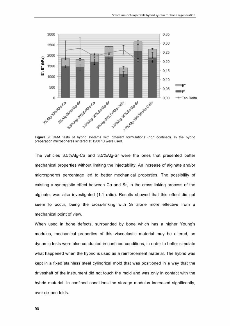
Strontium-richinjectablehybridsystemforboneregeneration
90
Figure 9. DMA tests of hybrid systems with different formulations (non confined). In the hybrid preparation microspheres sintered at 1200 ºC were used.
The vehicles 3.5%Alg-Ca and 3.5%Alg-Sr were the ones that presented better
mechanical properties without limiting the injectability. An increase of alginate and/or
microspheres percentage led to better mechanical properties. The possibility of
existing a synergistic effect between Ca and Sr, in the cross-linking process of the
alginate, was also investigated (1:1 ratio). Results showed that this effect did not
seem to occur, being the cross-linking with Sr alone more effective from a
mechanical point of view.
When used in bone defects, surrounded by bone which has a higher Young’s
modulus, mechanical properties of this viscoelastic material may be altered, so
dynamic tests were also conducted in confined conditions, in order to better simulate
what happened when the hybrid is used as a reinforcement material. The hybrid was
kept in a fixed stainless steel cylindrical mold that was positioned in a way that the
driveshaft of the instrument did not touch the mold and was only in contact with the
hybrid material. In confined conditions the storage modulus increased significantly,
over sixteen folds.

Strontium-richinjectablehybridsystemforboneregeneration
91
3.3 Evaluation of the space between particles using micro-CT
Figure 10 shows a reconstructed µCT image of a hybrid cylinder.
The µCT quantitative analysis of the Sr-hybrid materials showed that 49.4% of the
total cylinder volume is occupied by microspheres, with mean interparticle space of
220.7 µm. It was also observed the presence of denser particles distributed between
the microspheres in a percentage of 0.3%, possibly corresponding to non-dissolved
Sr-carbonate.
Figure 10. Micro-CT reconstructed image of Sr-hybrid system. Sr-HAp microspheres are represented in green. Denser particles are represented in red, possibly corresponding to non-dissolved Sr-carbonate.

Strontium-richinjectablehybridsystemforboneregeneration
92
Discussion
A wide variety of particle based fillers for bone regeneration has been described in
the literature, including multifaceted granules and smooth spherical particles of
various sizes, with or without pores, and with broad or uniform size distributions [3, 5,
6, 16]. The size and shape of the particles dictate their spatial rearrangement within
the implant site, playing a critical role in new bone formation. Particles diameter and
shape are also relevant parameters when considering injection procedures, since
uniform, spherical particles have more predictable flowing properties [12-15]. The
microspheres prepared according to the methodology herein described, were
uniform, both in terms of size and shape with mean diameters around 555 µm, an
appropriate value for effective bone substitution [50]. Adequate mechanical
properties of the microspheres are essential so they can resist damage during
transportation, packing, handling, and injection. Friability of both microspheres
composition (HAp and Sr-HAp) was below detectable levels, and rupture force was
adequate to withstand the pressure exerted during extrusion. The addition of Sr also
doubles the rupture force of the microspheres. The reason for this can be related to a
possible incorporation of Sr into the hydroxyapatite lattice by replacing Ca ions, as
supported by FTIR results. It is well known that a wide range of cations and anions,
may substitute into the structure of synthetic hydroxyapatite, replacing calcium,
phosphate or hydroxyl ions. Ca2+ can be substituted to some extent by divalent
cations, namely Sr2+. The substitutions alter the crystal lattice, inducing a structural
disorder, and consequently changing some of the material properties, including
phase stability, solubility, reactivity and mechanical properties. The ionic radius of Ca
is about 0.99 Å, while the ionic radius of Sr is higher (1.13 Å). The incorporation of
ions larger than Ca into the lattice of hydroxyapatite whereby an elongation of the
lattice parameters and, as a result, a deformation of the elementary crystalline cell
occurs. Chen et al. observed that the incorporation of low doses of Sr (atom ratio of
Sr/(Sr+Ca) adjusted to 5%) improved the mechanical properties of hydroxyapatite,

Strontium-richinjectablehybridsystemforboneregeneration
93
namely compressive strength, Knoop hardness and indirect tensile strength [51].
Alginate is a biomaterial that has found numerous applications in the biomedical field
due to its favorable properties such as biocompatibility, ease of gelation and
retention of structural similarity to the extracellular matrices in tissues [52]. Alginate is
a natural linear polysaccharide that contains 1,4-linked beta-D-mannuronic (M) and
alfa-L-guluronic (G) acid residues, arranged in a non-regular and block-wise fashion
along the chain. Alginate is well known for its high affinity towards Ca [53]. Ca binds
consecutive G-residues (G-blocks) in different alginate chains in a cooperative
manner (egg-box model) producing the cross-links of the hydrogel. The cross-linking
affects the resulting gel properties, such as stiffness, elasticity, and stability. Hence,
the functional properties of alginate gels can be tailored by the selection of alginate
composition. In general, alginate having a relatively high content of G (high-G
proportion alginates) gives stiff and stable gels with a high porosity, whereas alginate
with a low content of G (low-G or high-M proportion alginates) results in more elastic
and less stable Ca gels. Having in mind the envisaged application, in the present
work alginate with a content of more than 60% of guluronic vs mannuronic acid units
was used.
Alginate can also be cross-linked with other divalent ions, including Sr. Strontium
ions have very high affinities towards alginate, and have been shown to bind to G-
and M- blocks in the alginate [54].
Ca and Sr-alginate gels can be made by internal gelation, meaning that the
reticulation agents (Ca or Sr) are leaking at a low rate from an ion provider source
within the alginate. In the present work, Ca and Sr were used in the form of
carbonate and the release of the metal ions was caused by a reduction in pH due to
the addition of GDL. The gelation process is highly dependent upon diffusion of
gelation ions into the polymer network. An appropriate gelation time of the hybrid
material will be important in clinical practice. It should not be too short so there is
enough time to prepare and deliver the system, but not too long, to avoid an

Strontium-richinjectablehybridsystemforboneregeneration
94
excessively long surgical procedure. The gelation time for the Sr vehicle was
significantly shorter than that of the Ca vehicle, but adequate (20 min.), at a normal
operating room temperature.
Alginate has been reported to be biocompatible and is, therefore, a good candidate
for biomedical applications [47, 52]. Degradation of alginate is slow but can be tuned
by manipulating polymer molecular weights and composition [55]. In order to promote
bone regeneration, the injectable system that we propose should provide a scaffold
capable of supporting cell migration and tissue ingrowth. One well-known strategy to
promote cellular infiltration within a cell-free material and to direct de novo bone
formation at the local implant site is the incorporation of guidance cues such as bio-
adhesive motifs derived from extracellular matrices into the biomimetic material [56].
Here, the implanted material provides not only the mechanical support for tissue
regeneration but also appropriate guidance signals recognized by protein and
proteoglycan receptors on cell surfaces. Grafting of RGD peptides on alginate is
pertinent for the application envisaged in this work, since cell attachment is impaired
on unmodified alginate. Although in the present work, hybrid system was based on
non-functionalized alginate, tests were also performed using RGD functionalized
alginate. No significant differences were observed in what concerns to the viscosity
of the alginate and the injectability and mechanical properties of the complete
system.
The percentage and composition of microspheres, the alginate concentration and the
reticulating agent influence the maximum force needed to perform manual injections,
which should not exceed 100 N [49]. In a hybrid system of 3% alginate cross-linked
with Ca, an increase of the percentage of microspheres to 35% increased the force
needed for extrusion above the 100 N limit, and a filtering effect was observed, as
the alginate extrudes quicker than the microspheres. Using Sr instead, as the
reticulating agent, the extrusion force was smaller, allowing for an increase both in
alginate concentration and percentage of microspheres. With the 3.5%Alg-35%Sr-

Strontium-richinjectablehybridsystemforboneregeneration
95
HAp-Sr composition we obtained the higher compression strength value, still with an
extrusion force below the 100 N limit.
Also, the reticulation time when using Sr was smaller than when using Ca, as well as
the respective modulus obtained in DMA tests. This is in accordance with the
observation of Koo and Ma that alginate hydrogels formed with slower rates of
gelation tend to exhibit greater structural homogeneity and therefore larger modulus
than those gelled rapidly [46].
The viscoelastic properties of a bone defect filing material determine the capacity of
dissipation of energy by bone, and influence the relative response of surrounding
damaged and healthy tissue. DMA showed that for all tested materials E’ is higher
than E” which indicated that the elastic behavior of the hybrid is dominant over the
viscous one. Results also showed that the storage modulus was proportional to the
HAp microspheres content as indicated in Fig. 7. The obtained results confirmed that
the HAp particles acted as reinforcement filler by transferring the sustaining load
from the matrix to the rigid particles.
Although important features for bone repair, the mechanical properties of
hydrogel/calcium phosphate composites have rarely been reported [57]. Current
injectable bone defect fillers, like polymethylmetacrylate (PMMA) or calcium
phosphate cements, have maximum compression strength values, which are much
higher than that of cancellous bone. Besides, brittleness has limited the use of
calcium phosphate cements [58, 59]. The elastic modulus of this hybrid system is
below that of cancellous bone (20-5000 MPa) [60, 61], but when tested in a confined
setting (as would be the case in bone defects) the elastic mode increases
significantly. Low mechanical properties and the non-applicability in load-bearing
situations are commonly cited limitations of hydrogels in bone tissue engineering
[57]. However they can be used in non load-bearing sites, and, if necessary,
supplementary internal fixation devices may be applied [62]. Small size defects (up to
5-10 ml), where mechanical properties are not as critical, may be more adequate for

Strontium-richinjectablehybridsystemforboneregeneration
96
the use of this hybrid system.
Besides the phase composition, numerous parameters influence bone regeneration.
The internal structure of the particles, such as the porosity, the pore size distribution,
or the size distribution of the pore interconnections, is of particular relevance. It is
expected that the hybrid system that we propose will provide adequate mechanical
support in the early phases after surgery and gradual replacement of the artificial
scaffold by bone with adequate strength. The system, however, relies on the
maintenance of part of its constituents (the microspheres), taking into account that
full replacement by healthy, strong bone is not expected due to the age of the
patients. Alginate is inherently non-degradable in mammals, as they lack the enzyme
(i.e., alginase), which can cleave the polymer chains, but ionically cross-linked
alginate gels can be dissolved by release of the divalent ions cross-linking the gel
into the surrounding media due to exchange reactions with monovalent cations such
as sodium ions. In the model that we propose, it is expected an adequate
degradation behavior of alginate leaving a space between particles (220.7 µm)
adequate for the invasion of blood vessels and cells.
One advantage of the hybrid system developed in this project was the presence of Sr
in its composition. Strontium, in the form of oral strontium ranelate, has been in
clinical use as an anti-osteoporotic agent effective in the prevention of both vertebral
and non-vertebral osteoporotic fractures [36, 37]. However, cardiovascular safety has
been a concern regarding its use. Recently, a small but significant increase in non-
fatal myocardial infarctions was observed when analyzing all studies reporting the
effect of strontium ranelate in osteoporosis and osteoarthritis [63-65]. Currently, the
drug is authorized for the treatment of severe osteoporosis, in postmenopausal
women and in adult men, at high risk of fracture for whom treatment with other
medicinal products approved for the treatment of osteoporosis is not possible, and
the use of strontium ranelate should be restricted to patients with no past or current
history of ischemic heart disease, peripheral arterial disease and/or cerebrovascular

Strontium-richinjectablehybridsystemforboneregeneration
97
disease or uncontrolled hypertension.
The incorporation of Sr in both the microspheres and the vehicle, will allow Sr
release since the early stages of implantation, due to the different degradation
kinetics of the polymer network (faster) and the ceramic particles (slower). It will be
released locally to enhance bone formation, but clinically relevant systemic
concentrations are not expected [38, 39, 66]. Baier et al. [39] compared the
osteointegration of calcium phosphate cement implants containing Sr to calcium
phosphate cement implants in ovariectomized rats, and found faster osteointegration
and more new bone formation with the addition of Sr, both on the implant surface
and within the implant. Systemic release of Sr from the implants did not induce
significant effects on bone density in this rat model. Thormann et al. [38] studied the
effect of Sr modified calcium phosphate cement (SrCPC) in bone formation in critical-
size metaphyseal fracture defects in ovariectomized rats. Both higher new bone
formation in the SrCPC group compared to CPC group, and high Sr concentration in
the interface region of the SrCPC implant were observed. They concluded that Sr
release had most probably been responsible for these results, that local delivery of
Sr from Sr-loaded biomaterials is possible, and that strontium’s biological activity to
stimulate new bone formation is preserved within the CPC.

Strontium-richinjectablehybridsystemforboneregeneration
98
Conclusions
We have developed a strontium rich viscoelastic hybrid system, herein described,
that can be manually injected and sets in situ at body temperature, providing a
scaffold for cell migration and tissue ingrowth. When implanted, the hybrid system
will offer structural support while providing a temporary scaffold onto which new bone
can grow. The incorporation of two Sr release kinetics (from the alginate and from
the microspheres), may further improve effective bone regeneration, which can be
especially useful in osteoporotic conditions.

Strontium-richinjectablehybridsystemforboneregeneration
99
Highlights
• We developed a Sr rich viscoelastic hybrid system (alginate matrix cross-
linked in situ with Sr, and Sr-rich microspheres)
• It can be manually injected, sets in situ and offers structural support,
providing a temporary scaffold for bone growth
• The incorporation of Sr relies on the growing evidence of its beneficial effects
in bone remodeling and in osteoporosis
• Two Sr release kinetics may further improve effective bone regeneration,
which can be especially useful in osteoporosis

Strontium-richinjectablehybridsystemforboneregeneration
100
Statement of Significance
Current challenges in the development of scaffolds for bone regeneration include the
engineering of materials that can withstand physiological mechanical stresses and
provide a matrix capable of supporting cell migration and tissue ingrowth. We have
developed a strontium (Sr) rich viscoelastic hybrid system (alginate matrix cross-
linked in situ in the presence of Sr, and Sr-rich microspheres) that can be manually
injected and sets in situ, offering structural support while providing a temporary
scaffold onto which new bone can grow. The incorporation of Sr relies on the growing
evidence of its beneficial effects in bone remodeling and in the treatment of
osteoporosis. The two Sr release kinetics may further improve effective bone
regeneration, which can be especially useful in osteoporotic conditions.

Strontium-richinjectablehybridsystemforboneregeneration
101
Graphical Abstract

Strontium-richinjectablehybridsystemforboneregeneration
102
Acknowledgements
This work was financed by FEDER funds, through the Programa Operacional
Factores de Competitividade - COMPETE, and by Portuguese funds through FCT -
Fundação para a Ciência e a Tecnologia in the framework of the project
PTDC/CTM/103181/2008 and of project Pest-C/SAU/LA0002/2011.
The authors thankfully acknowledge the use of Micro-CT in the 3B’s Research Group
facilities at University of Minho, the use of microscopy services at Centro de
Materiais da Universidade do Porto, CEMUP and the technicians responsible for the
experimental techniques mentioned, namely Vítor Correlo and Daniela Silva.

Strontium-richinjectablehybridsystemforboneregeneration
103
References
[1] Zhou H, Lee J. Nanoscale hydroxyapatite particles for bone tissue engineering.
Acta Biomater 2011;7:2769-81.
[2] Dorozhkin SV. Amorphous calcium (ortho)phosphates. Acta Biomater
2010;6:4457-75.
[3] Dorozhkin SV. Bioceramics of calcium orthophosphates. Biomaterials
2010;31:1465-85.
[4] Dorozhkin SV. Nanosized and nanocrystalline calcium orthophosphates. Acta
Biomater 2010;6:715-34.
[5] Combes C, Rey C. Amorphous calcium phosphates: synthesis, properties and
uses in biomaterials. Acta Biomater 2010;6:3362-78.
[6] Low KL, Tan SH, Zein SH, Roether JA, Mourino V, Boccaccini AR. Calcium
phosphate-based composites as injectable bone substitute materials. J Biomed
Mater Res B Appl Biomater 2010;94:273-86.
[7] Grimandi G, Weiss P, Millot F, Daculsi G. In vitro evaluation of a new injectable
calcium phosphate material. J Biomed Mater Res 1998;39:660-6.
[8] Iooss P, Le Ray AM, Grimandi G, Daculsi G, Merle C. A new injectable bone
substitute combining poly(epsilon-caprolactone) microparticles with biphasic calcium
phosphate granules. Biomaterials 2001;22:2785-94.
[9] Dupraz A, Nguyen TP, Richard M, Daculsi G, Passuti N. Influence of a cellulosic
ether carrier on the structure of biphasic calcium phosphate ceramic particles in an
injectable composite material. Biomaterials 1999;20:663-73.
[10] Yaszemski MJ, Payne RG, Hayes WC, Langer R, Mikos AG. In vitro degradation
of a poly(propylene fumarate)-based composite material. Biomaterials 1996;17:2127-
30.
[11] Maruyama M, Ito M. In vitro properties of a chitosan-bonded self-hardening
paste with hydroxyapatite granules. J Biomed Mater Res 1996;32:527-32.

Strontium-richinjectablehybridsystemforboneregeneration
104
[12] Qiu QQ, Ducheyne P, Ayyaswamy PS. New bioactive, degradable composite
microspheres as tissue engineering substrates. J Biomed Mater Res 2000;52:66-76.
[13] Hsu FY, Chueh SC, Wang YJ. Microspheres of hydroxyapatite/reconstituted
collagen as supports for osteoblast cell growth. Biomaterials 1999;20:1931-6.
[14] Sunny MC, Ramesh P, Varma HK. Microstructured microspheres of
hydroxyapatite bioceramic. J Mater Sci Mater Med 2002;13:623-32.
[15] Sivakumar M, Rao KP. Preparation, characterization, and in vitro release of
gentamicin from coralline hydroxyapatite-alginate composite microspheres. J Biomed
Mater Res A 2003;65:222-8.
[16] Bohner M, Tadier S, van Garderen N, de Gasparo A, Dobelin N, Baroud G.
Synthesis of spherical calcium phosphate particles for dental and orthopedic
applications. Biomatter 2013;3.
[17] Liu HY, Wu AT, Tsai CY, Chou KR, Zeng R, Wang MF, et al. The balance
between adipogenesis and osteogenesis in bone regeneration by platelet-rich
plasma for age-related osteoporosis. Biomaterials 2011;32:6773-80.
[18] Cao L, Liu G, Gan Y, Fan Q, Yang F, Zhang X, et al. The use of autologous
enriched bone marrow MSCs to enhance osteoporotic bone defect repair in long-
term estrogen deficient goats. Biomaterials 2012;33:5076-84.
[19] Cho SW, Sun HJ, Yang JY, Jung JY, Choi HJ, An JH, et al. Human adipose
tissue-derived stromal cell therapy prevents bone loss in ovariectomized nude
mouse. Tissue Eng Part A 2012;18:1067-78.
[20] Wang Z, Goh J, Das De S, Ge Z, Ouyang H, Chong JS, et al. Efficacy of bone
marrow-derived stem cells in strengthening osteoporotic bone in a rabbit model.
Tissue Eng 2006;12:1753-61.
[21] Marie PJ, Felsenberg D, Brandi ML. How strontium ranelate, via opposite effects
on bone resorption and formation, prevents osteoporosis. Osteoporos Int
2011;22:1659-67.

Strontium-richinjectablehybridsystemforboneregeneration
105
[22] Grynpas MD, Hamilton E, Cheung R, Tsouderos Y, Deloffre P, Hott M, et al.
Strontium increases vertebral bone volume in rats at a low dose that does not induce
detectable mineralization defect. Bone 1996;18:253-9.
[23] Bonnelye E, Chabadel A, Saltel F, Jurdic P. Dual effect of strontium ranelate:
stimulation of osteoblast differentiation and inhibition of osteoclast formation and
resorption in vitro. Bone 2008;42:129-38.
[24] Yang F, Yang D, Tu J, Zheng Q, Cai L, Wang L. Strontium enhances osteogenic
differentiation of mesenchymal stem cells and in vivo bone formation by activating
Wnt/catenin signaling. Stem Cells 2011;29:981-91.
[25] Baron R, Tsouderos Y. In vitro effects of S12911-2 on osteoclast function and
bone marrow macrophage differentiation. Eur J Pharmacol 2002;450:11-7.
[26] Peng S, Liu XS, Huang S, Li Z, Pan H, Zhen W, et al. The cross-talk between
osteoclasts and osteoblasts in response to strontium treatment: involvement of
osteoprotegerin. Bone 2011;49:1290-8.
[27] Hurtel-Lemaire AS, Mentaverri R, Caudrillier A, Cournarie F, Wattel A, Kamel S,
et al. The calcium-sensing receptor is involved in strontium ranelate-induced
osteoclast apoptosis. New insights into the associated signaling pathways. J Biol
Chem 2009;284:575-84.
[28] Peng S, Zhou G, Luk KD, Cheung KM, Li Z, Lam WM, et al. Strontium promotes
osteogenic differentiation of mesenchymal stem cells through the Ras/MAPK
signaling pathway. Cell Physiol Biochem 2009;23:165-74.
[29] Caverzasio J. Strontium ranelate promotes osteoblastic cell replication through
at least two different mechanisms. Bone 2008;42:1131-6.
[30] Delannoy P, Bazot D, Marie PJ. Long-term treatment with strontium ranelate
increases vertebral bone mass without deleterious effect in mice. Metabolism
2002;51:906-11.

Strontium-richinjectablehybridsystemforboneregeneration
106
[31] Buehler J, Chappuis P, Saffar JL, Tsouderos Y, Vignery A. Strontium ranelate
inhibits bone resorption while maintaining bone formation in alveolar bone in
monkeys (Macaca fascicularis). Bone 2001;29:176-9.
[32] Ammann P, Shen V, Robin B, Mauras Y, Bonjour JP, Rizzoli R. Strontium
ranelate improves bone resistance by increasing bone mass and improving
architecture in intact female rats. J Bone Miner Res 2004;19:2012-20.
[33] Ammann P, Badoud I, Barraud S, Dayer R, Rizzoli R. Strontium ranelate
treatment improves trabecular and cortical intrinsic bone tissue quality, a determinant
of bone strength. J Bone Miner Res 2007;22:1419-25.
[34] Bain SD, Jerome C, Shen V, Dupin-Roger I, Ammann P. Strontium ranelate
improves bone strength in ovariectomized rat by positively influencing bone
resistance determinants. Osteoporos Int 2009;20:1417-28.
[35] Peng S, Liu XS, Wang T, Li Z, Zhou G, Luk KD, et al. In vivo anabolic effect of
strontium on trabecular bone was associated with increased osteoblastogenesis of
bone marrow stromal cells. J Orthop Res 2010;28:1208-14.
[36] Meunier PJ, Roux C, Seeman E, Ortolani S, Badurski JE, Spector TD, et al. The
effects of strontium ranelate on the risk of vertebral fracture in women with
postmenopausal osteoporosis. N Engl J Med 2004;350:459-68.
[37] Reginster JY, Seeman E, De Vernejoul MC, Adami S, Compston J, Phenekos C,
et al. Strontium ranelate reduces the risk of nonvertebral fractures in
postmenopausal women with osteoporosis: Treatment of Peripheral Osteoporosis
(TROPOS) study. J Clin Endocrinol Metab 2005;90:2816-22.
[38] Thormann U, Ray S, Sommer U, Elkhassawna T, Rehling T, Hundgeburth M, et
al. Bone formation induced by strontium modified calcium phosphate cement in
critical-size metaphyseal fracture defects in ovariectomized rats. Biomaterials
2013;34:8589-98.
Strontium-richinjectablehybridsystemforboneregeneration
107
[39] Baier M, Staudt P, Klein R, Sommer U, Wenz R, Grafe I, et al. Strontium
enhances osseointegration of calcium phosphate cement: a histomorphometric pilot
study in ovariectomized rats. J Orthop Surg Res 2013;8:16.
[40] Liu P, Wang N, Hao Y, Zhao Q, Qiao Y, Li H, et al. Entangled titanium fibre balls
combined with nano strontium hydroxyapatite in repairing bone defects. Med Princ
Pract 2014;23:264-70.
[41] Oliveira SM, Barrias CC, Almeida IF, Costa PC, Ferreira MR, Bahia MF, et al.
Injectability of a bone filler system based on hydroxyapatite microspheres and a
vehicle with in situ gel-forming ability. J Biomed Mater Res B Appl Biomater
2008;87:49-58.
[42] Barrias CC, Lamghari M, Granja PL, Sa Miranda MC, Barbosa MA. Biological
evaluation of calcium alginate microspheres as a vehicle for the localized delivery of
a therapeutic enzyme. J Biomed Mater Res A 2005;74:545-52.
[43] Ribeiro CC, Barrias CC, Barbosa MA. Calcium phosphate-alginate microspheres
as enzyme delivery matrices. Biomaterials 2004;25:4363-73.
[44] Ribeiro CC, Barrias CC, Barbosa MA. Preparation and characterisation of
calcium-phosphate porous microspheres with a uniform size for biomedical
applications. J Mater Sci Mater Med 2006;17:455-63.
[45] Barrias CC, Ribeiro CC, Lamghari M, Miranda CS, Barbosa MA. Proliferation,
activity, and osteogenic differentiation of bone marrow stromal cells cultured on
calcium titanium phosphate microspheres. J Biomed Mater Res A 2005;72:57-66.
[46] Kuo CK, Ma PX. Ionically cross-linked alginate hydrogels as scaffolds for tissue
engineering: part 1. Structure, gelation rate and mechanical properties. Biomaterials
2001;22:511-21.
[47] Evangelista MB, Hsiong SX, Fernandes R, Sampaio P, Kong HJ, Barrias CC, et
al. Upregulation of bone cell differentiation through immobilization within a synthetic
extracellular matrix. Biomaterials 2007;28:3644-55.

Strontium-richinjectablehybridsystemforboneregeneration
108
[48] Fonseca KB, Bidarra SJ, Oliveira MJ, Granja PL, Barrias CC. Molecularly
designed alginate hydrogels susceptible to local proteolysis as three-dimensional
cellular microenvironments. Acta Biomaterialia 2011;7:1674-82.
[49] Krebs J, Ferguson SJ, Bohner M, Baroud G, Steffen T, Heini PF. Clinical
measurements of cement injection pressure during vertebroplasty. Spine (Phila Pa
1976) 2005;30:E118-22.
[50] Gauthier O, Bouler JM, Weiss P, Bosco J, Aguado E, Daculsi G. Short-term
effects of mineral particle sizes on cellular degradation activity after implantation of
injectable calcium phosphate biomaterials and the consequences for bone
substitution. Bone 1999;25:71S-4S.
[51] Chen DF, YF. Evaluation on the mechanic properties of the solid solution of
strontium substituted hydroxyapatite. Chin J Stoma Mater Appar 2001;19:178-83.
[52] Lee KY, Mooney DJ. Alginate: properties and biomedical applications. Prog
Polym Sci 2012;37:106-26.
[53] Andersen T, Strand BL, Formo K, Alsberg E, Christensen BE. Chapter 9
Alginates as biomaterials in tissue engineering. Carbohydrate Chemistry: Volume
37: The Royal Society of Chemistry; 2012. p. 227-58.
[54] Morch YA, Donati I, Strand BL, Skjak-Braek G. Effect of Ca2+, Ba2+, and Sr2+
on alginate microbeads. Biomacromolecules 2006;7:1471-80.
[55] Augst AD, Kong HJ, Mooney DJ. Alginate hydrogels as biomaterials. Macromol
Biosci 2006;6:623-33.
[56] Sakiyama-Elbert SH, JA Functional biomaterials: Design of novel biomaterials. .
Annual Review of Materials Research 2001;31:183-201.
[57] D'Este M, Eglin D. Hydrogels in calcium phosphate moldable and injectable
bone substitutes: Sticky excipients or advanced 3-D carriers? Acta Biomater
2013;9:5421-30.

Strontium-richinjectablehybridsystemforboneregeneration
109
[58] Friedman CD, Costantino PD, Takagi S, Chow LC. BoneSource hydroxyapatite
cement: a novel biomaterial for craniofacial skeletal tissue engineering and
reconstruction. J Biomed Mater Res 1998;43:428-32.
[59] Xu HH, Quinn JB. Calcium phosphate cement containing resorbable fibers for
short-term reinforcement and macroporosity. Biomaterials 2002;23:193-202.
[60] Ouyang J, Yang GT, Wu WZ, Zhu QA, Zhong SZ. Biomechanical characteristics
of human trabecular bone. Clin Biomech (Bristol, Avon) 1997;12:522-4.
[61] Keaveny TM, Morgan EF, Niebur GL, Yeh OC. Biomechanics of trabecular bone.
Annu Rev Biomed Eng 2001;3:307-33.
[62] Willie BM, Petersen A, Schmidt-Bleek K, Cipitria A, Mehta M, Strube P, et al.
Designing biomimetic scaffolds for bone regeneration: why aim for a copy of mature
tissue properties if nature uses a different approach? Soft Matter 2010;6:4976-87.
[63] Reginster JY, Neuprez A, Dardenne N, Beaudart C, Emonts P, Bruyere O.
Efficacy and safety of currently marketed anti-osteoporosis medications. Best Pract
Res Clin Endocrinol Metab 2014;28:809-34.
[64] Cooper C, Fox KM, Borer JS. Ischaemic cardiac events and use of strontium
ranelate in postmenopausal osteoporosis: a nested case-control study in the CPRD.
Osteoporos Int 2014;25:737-45.
[65] Abrahamsen B, Grove EL, Vestergaard P. Nationwide registry-based analysis of
cardiovascular risk factors and adverse outcomes in patients treated with strontium
ranelate. Osteoporos Int 2014;25:757-62.
[66] Hulsart-Billstrom G, Xia W, Pankotai E, Weszl M, Carlsson E, Forster-Horvath C,
et al. Osteogenic potential of Sr-doped calcium phosphate hollow spheres in vitro
and in vivo. J Biomed Mater Res A 2013;101:2322-31.

110

111
CHAPTER IV
INJECTABLE HYBRID SYSTEM FOR STRONTIUM LOCAL DELIVERY PROMOTES BONE REGENERATION IN A RAT CRITICAL-SIZED DEFECT MODEL
Article 3
Published in Scientific Reports. 2017 Jul 11;7(1):5098
doi: 10.1038/s41598-017-04866-4

112

Injectablehybridsystemforstrontiumlocaldeliverypromotesboneregenerationinaratcritical-sizeddefectmodel
113
Injectable hybrid system for strontium local delivery promotes bone
regeneration in a rat critical-sized defect model
Ana Henriques Lourenço1,2,3 *, Nuno Neves1,2,4 *, Cláudia Ribeiro-Machado 1,2, Susana
R. Sousa1,2,5, Meriem Lamghari1,2, Cristina C. Barrias1,2, Abel Trigo Cabral4, Mário A.
Barbosa1,2,6, Cristina C. Ribeiro1,2,5 #
1i3S - Instituto de Investigação e Inovação em Saúde, Universidade do Porto, Rua Alfredo Allen, 208,
4200 - 135 Porto, Portugal
2INEB - Instituto de Engenharia Biomédica, Universidade do Porto, Rua Alfredo Allen, 208, 4200 - 135
Porto, Portugal
3Faculdade de Engenharia, Universidade do Porto, Rua Dr. Roberto Frias, s/n, 4200-465 Porto,
Portugal
4Faculdade de Medicina, Universidade do Porto, Serviço de Ortopedia, Alameda Prof. Hernâni
Monteiro, 4200-319 Porto, Portugal
5ISEP – Instituto Superior de Engenharia do Porto, Instituto Politécnico do Porto, Rua Dr. António
Bernardino de Almeida 431, 4249-015, Porto, Portugal
6ICBAS - Instituto de Ciências Biomédicas de Abel Salazar, Universidade do Porto, Rua de Jorge
Viterbo Ferreira n. 228, 4050-313 Porto, Portugal
* These authors contributed equally to this work.

Injectablehybridsystemforstrontiumlocaldeliverypromotesboneregenerationinaratcritical-sizeddefectmodel
114
Abstract
Strontium (Sr) has been described as having beneficial influence in bone strength
and architecture. However, negative systemic effects have been reported on oral
administration of Sr ranelate, leading to strict restrictions in clinical application. We
hypothesized that local delivery of Sr improves osteogenesis without eliciting
detrimental side effects. Therefore, the in vivo response to an injectable Sr-hybrid
system composed of RGD-alginate hydrogel cross-linked in situ with Sr and
reinforced with Sr-doped hydroxyapatite microspheres, was investigated. The system
was injected in a critical-size bone defect model and compared to a similar Sr-free
material. Micro-CT results show a trend towards higher new bone formed in Sr-hybrid
group and major histological differences were observed between groups. Higher cell
invasion was detected at the center of the defect of Sr-hybrid group after 15 days
with earlier bone formation. Higher material degradation with increase of collagen
fibers and bone formation in the center of the defect after 60 days was observed as
opposed to bone formation restricted to the periphery of the defect in the control.
These histological findings support the evidence of an improved response with the Sr
enriched material. Importantly, no alterations were observed in the Sr levels in
systemic organs or serum.
Keywords: strontium, bone regeneration, injectable bone substitute, alginate,
hydroxyapatite, ionic delivery system

Injectablehybridsystemforstrontiumlocaldeliverypromotesboneregenerationinaratcritical-sizeddefectmodel
115
Introduction
The management of fractures and bone defects remains a significant challenge, and
there is the need for improved therapeutic strategies [1]. Biological (autografts,
allografts and xenografts) and synthetic bone grafts are currently used in clinical
practice for bone repair. Because of their osteogenic potential and the absence of
risks of immune rejection or disease transfer, autografts are clinically preferred.
However, they are limited in supply, imply the additional morbidity of a harvest
surgery and their properties and shape do not match exactly those of the bone to be
replaced [2]. Intensive investigation is being carried out to produce synthetic bone
grafts in order to overcome these problems. The use of injectable materials in bone
regeneration, especially calcium phosphate based materials, presents several
advantages, namely due to their adequate biological responses, osteoconductivity
and mechanical properties [3-6]. These materials can be applied by minimally
invasive surgical procedures, to efficiently fill-in cavities of non-uniform shapes, with
no tissue damage and limited exposure to infectious agents, thus reducing patient
discomfort and procedure-associated health costs. The addition of osteoinductive
factors or osteoprogenitor/stem cells may improve bone repair, particularly in
osteoporotic conditions, characterized by an impaired healing response [7-10].
Oral administration of Strontium (Sr) ranelate has shown effectiveness in the
prevention of both vertebral and non-vertebral osteoporotic fractures [11, 12]. Unlike
other anti-osteoporotic agents widely used in clinical practice, such as
bisphosphonates, estrogen, selective estrogen-receptor modulators (SERMs) and
calcitonin, which inhibit bone resorption [9], Sr ranelate also promotes bone
formation [13-16]. Several in vitro studies show that Sr ranelate decreases bone
resorption, by reducing osteoclast activity [13, 14, 17], decreasing functional
osteoclast markers expression [13], disrupting osteoclasts cytoskeleton [14], and
increasing osteoclast apoptosis [18]. Simultaneously, it induces positive effects on
osteoblastogenesis and osteoblast activity in different in vitro models [19], namely by

Injectablehybridsystemforstrontiumlocaldeliverypromotesboneregenerationinaratcritical-sizeddefectmodel
116
enhancing replication of preosteoblastic cells [14, 20-23], increasing osteogenesis
[14, 20, 24-26], decreasing osteoblast apoptosis [21, 27], and promoting terminal
differentiation of osteoblasts into osteocytes [20]. Some pre-clinical studies
performed in both normal and osteopenic/osteoporotic animal models confirmed
these in vitro results, showing the beneficial effects of Sr ranelate on bone formation
and remodeling [28-32]. Despite these important effects, cardiovascular safety of
orally administered Sr ranelate has been questioned due to a small but significant
increase in non-fatal myocardial infarctions [12, 33, 34]. Currently, there are strict
indications and restrictions to its use [12].
Nevertheless, Sr incorporation into biomaterials for bone regeneration may improve
their regeneration potential. In vivo studies showed that doping calcium phosphate
cements and other ceramics with Sr promotes bone repair [35-37]. A sustained
delivery system for local release of Sr ions can obviate systemic complications with
similar rates of bone formation at the site of implantation.
Current injectable bone defect methacrylate-based fillers have compression
strengths much higher than that of cancellous bone, and the brittleness of calcium
phosphate cements is a limitation [38, 39]. We have previously developed various
types of injectable biomaterials for bone regeneration, namely calcium phosphate
[40-42] and calcium phosphate/alginate [43] microspheres, as well as different types
of bio-functional alginate hydrogels [44-47]. When combined, alginate can act as an
appropriate vehicle for ceramic microspheres delivery and immobilization at the injury
site. Alginate is a natural linear polysaccharide, biodegradable and biocompatible,
extensively studied for biomedical applications [48, 49]. Although generally regarded
as a bioinert material, since it does not elicit specific cell-matrix interactions, grafting
of alginate with arginine-glycine-aspartic acid (RGD) peptides is an effective strategy
to provide appropriate guidance signals to promote cell adhesion and facilitate cell
colonization [50]ENREF_49. Alginate forms hydrogels under mild chemical
conditions, in the presence of divalent cations, such as Ca and Sr, through a

Injectablehybridsystemforstrontiumlocaldeliverypromotesboneregenerationinaratcritical-sizeddefectmodel
117
cytocompatible physical gelation process. These cations bind homoguluronic blocks
in adjacent alginate chains in a cooperative manner (egg-box model) producing a
cross-linked hydrogel network [51, 52].
Recently, we developed an injectable hybrid system that consists of ~500 µm
diameter hydroxyapatite (HAp) microspheres doped with Sr, embedded in a
functionalized alginate matrix cross-linked in situ with Sr [53]. As a vehicle for the
microspheres delivery, functionalized alginate with RGD peptides was used,
providing a scaffold for cell adhesion and migration and allowing for the injectability
of the system. However, the use of hydrogels in bone tissue engineering is limited by
low mechanical properties and the non-applicability in load-bearing conditions [54].
The packing of the microspheres upon delivery raises the compression strength of
the material [53], and alginate creates an interconnected 3D network adequate for
the invasion of blood vessels and cells [55]. Moreover, the presence of Sr in both
components of the system provides two different release routes upon degradation of
the materials. This system presents a clinically relevant compromise between
adequate injectability and gelation time and final compression strength [53].
Moreover, in vitro studies showed that this Sr-hybrid scaffold promotes sustained
release of Sr2+, supports human mesenchymal stem cells adhesion, survival and
osteogenic differentiation, and inhibits osteoclasts differentiation and activity, as
compared to a similar Sr-free system [56].
In the current study we aim to evaluate the in vivo response to the designed Sr-rich
hybrid system and its influence on new bone formation using a rat metaphyseal
femoral critical-sized defect model, compared to a similar Sr-free material. The
proposed system is expected to provide an adequate therapeutic approach to fill-in
bone defects by minimally invasive surgery, while acting as a scaffold for local Sr2+
release to promote bone regeneration.

Injectablehybridsystemforstrontiumlocaldeliverypromotesboneregenerationinaratcritical-sizeddefectmodel
118
Results
Radiographical analysis of bone and biomaterial
A cylindrical defect with 3 mm diameter and depth of approximately 4 mm was drilled
in the lateral condyle of the right femur (Figure 1A). The defect was filled with the
biomaterial, injected using a 1 mL syringe (Figure 1B, C). A picture of the Sr-hybrid
material used is shown in Figure 1D, where spherical particles (Sr-HAp
microspheres) are embedded within a transparent hydrogel (Sr cross-linked RGD-
alginate).
Figure 1. In vivo intraoperative setting. Critical sized defect created in the distal femur (A). Injection of the hybrid material using a 1 mL syringe (B) and filled defect (C). Detail of the hybrid system, composed of HAp microspheres embedded in a RGD-alginate hydrogel (D).
Rat femurs were imaged by X-ray along the experimental period allowing for a follow-
up at 15 and 30 days. Representative images of defects filled with materials (Sr-
hybrid or Ca-hybrid) or empty defects are shown in Fig. 2. In hybrid-filled defects
(Fig. 2A to D), microspheres are located inside the created bone defect (arrows in
the images), where the higher radiopacity of the HAp microspheres allowed for the
easy monitoring using X-ray. Microspheres were homogeneously distributed within
the defects (Fig. 2A to D) and were still detected at day 30, a non-invasive mid-term
follow-up (Fig. 2B and D). Empty defects were also imaged (Fig. 2E and F) and the
defect could still be observed after 30 days, confirming it to be of a critical size.

Injectablehybridsystemforstrontiumlocaldeliverypromotesboneregenerationinaratcritical-sizeddefectmodel
119
Figure 2. Radiographic imaging follow-up. Representative lateral X-Ray images from right rat femurs of Sr-hybrid (A and B) or Ca-hybrid-filled (C and D) and empty (E and F) defects at 15 days (A, C and E) and 30 days (B ,D and F) post-surgery. Arrows pinpoint site of created defect, filled or empty. Microspheres can be observed as circular objects more radiopaque than bone.
Micro-CT morphometric 3D evaluation
Micro-CT analysis was performed 60 days post-implantation (Fig. 3) to evaluate new
bone formation at the defect site and to assess the spatial distribution of ceramic
microspheres within the lesion. In Sr-hybrid filled defects (Fig. 3A), microspheres
were homogeneously distributed inside the defect with no apparent degradation, with
preserved size and without modifications in shape. Similar results were found in Ca-
hybrid filled defects. Furthermore, and particularly in Sr-hybrid samples, centripetal
bone colonization could be observed by new bone formation surrounding the ceramic
microspheres and with the development of new bone trabeculae in the periphery of
the defect. 3D morphometric analysis was performed using five femurs per group,

Injectablehybridsystemforstrontiumlocaldeliverypromotesboneregenerationinaratcritical-sizeddefectmodel
120
where the ROI was defined in binary images (Fig. 3B) and the percentage of new
bone formed (bone volume fraction, BV/TV) was calculated. Values of (31.5±1.7) %
and (28.6±1.1) % (BV/TV (%), mean±SE) of new bone was measured in animals that
received the Sr-hybrid and Ca-hybrid materials, respectively (Fig. 3C).
Figure 3. Micro-CT analysis of distal femur after 60 days of implantation. 3D reconstructed image and respective orthogonal slices of micro-CT acquisition of the femur with Sr-hybrid filled defect (A). Morphometric analysis approach used to quantify new bone formation: transversal micro-CT slice of a femur injected with Sr-hybrid after 60 days of implantation highlighting ROI (B). C - New bone formed (BV/TV, %) after 60 days of implantation of Sr-hybrid and Ca-hybrid materials. Data presented as box-plot with median and min to max whiskers of n = 5 samples (n.s. – statistically non-significant).
Histological evaluation of bone/biomaterial interface
In Fig. 4A representative images of femurs with defects filled with Sr-hybrid or Ca-
hybrid materials, at days 15 and 60, are portrayed. A global view of the defects and
materials (Fig. 4A a to d), as well as a more detailed view of the periphery of the
defect (Fig. 4A a’ to d’ and magnifications a’’ to d’’), are given. Histological analysis at

Injectablehybridsystemforstrontiumlocaldeliverypromotesboneregenerationinaratcritical-sizeddefectmodel
121
day 15 post-implantation showed that the created defects exhibited similar diameter
and were filled with approximately 15 to 18 microspheres (Fig. 4A a, b). As early as
15 days post-implantation, all animals showed, to some extent, newly formed bone at
the periphery of the defect (Fig 4A a’, b’). Sr-hybrid implanted defects also showed
new bone formation in close contact with the microspheres, distant from the
periphery of native bone (Fig. 4A b’’, arrow). SEM images and EDS analysis of this
newly formed bone in close vicinity of the microspheres is shown in Fig. 4B. The
results confirmed the high content of calcium and phosphate, and a Ca/P ratio in
accordance with normal bone composition (Z1 in Fig. 4B), and different from the
elemental analysis of the microspheres (Z2 in Fig. 4B), where Sr was also identified.
After 60 days, new bone formation at the periphery was observed in both materials.
Sr-hybrid implanted defects exhibited a thicker trabecular bone structure at the
periphery of the defect (Fig 4A d’, d’’), when compared to the Ca-hybrid group (Fig.
4A c’, c’’). The quantification of new bone formed at the periphery of the defect
revealed a statistically significant thicker bone structure in Sr-hybrid group (662.4±48
µm) in contrast to a thinner bone formation (381.1±29 µm) in the Ca-hybrid group
(mean±SE, p<0.001, Fig. 4C).

Injectablehybridsystemforstrontiumlocaldeliverypromotesboneregenerationinaratcritical-sizeddefectmodel
122
Figure 4. Histological evaluation of bone/biomaterial interface in femurs implanted with Sr-hybrid and Ca-hybrid systems. Coronal histological sections of critical sized defect created in the distal femur, 15 and 60 days post-implantation (A). Global view of the created defects filled with Ca-hybrid (a and c) and Sr-hybrid (b and d) materials, at 15 (a and b) or 60 days (c and d) post-implantation, stained with MT (a to d, dashed yellow line circling the created defect area). Interface bone/biomaterial, 15 days (a’ and b’) and 60 days (c’ and d’) post-implantation, with Ca-hybrid (a’ and c’) and Sr-hybrid (b’ and d’) systems, and higher magnification of square (a’’ to d’’, collagen/bone in blue, yellow dashed line – bone/biomaterial interface, * – alginate, M – microspheres, black arrows – new bone). B - SEM image of histological section of Sr-hybrid filled defect 15 days post-implantation with EDS spectra of new bone (Z1) found near the microsphere and microsphere (Z2). C - Thickness of peripheral bone found around the defect (as represented with arrows in A c’’ and A d’’), 60 days post-implantation of Sr-hybrid and Ca-hybrid systems. Data presented as mean±SEM of 20 different random locations of trabecular bone found around the defect of n=5 animals/group, 3 sections/animal, Asterisks show statistically significant differences (*** p<0.001).

Injectablehybridsystemforstrontiumlocaldeliverypromotesboneregenerationinaratcritical-sizeddefectmodel
123
Histological evaluation of the center of the defect
Representative images of the center of the defect in both groups are shown in Fig.
5A. Although no evident differences were observed in the diameter of the
microspheres with time of implantation, alginate showed different behavior between
groups. After 15 days, some alginate was observed surrounding microspheres in
both experimental groups (Fig 5A, a and b). In Sr-hybrid group, higher cell invasion
at the center was observed, mainly of polymorphonuclear neutrophils (PMN) (Fig 5A,
b and b’).
Figure 5. Histological analysis of the center of the defect in femurs implanted with Sr-hybrid and Ca-hybrid systems. Detailed view of the center of the defect implanted with Ca-hybrid (a and c) and Sr-hybrid (b and d) materials, stained with H&E, after 15 (a and b) and 60 days (c and d) of implantation, with higher magnification of square (a’ to d’, A). B - Area of residual material (microspheres and alginate) found within the defect area, 60 days post-implantation of Sr-hybrid and Ca-hybrid systems. Measurements were performed by delimiting the area of material in MT stained sections using ImageJ software, n=5 animals/group, 3 sections/animal. Data presented as mean±SE and asterisks show statistically significant differences (*** p<0.001). C – TRAP-LG staining images of Ca-hybrid (a) and Sr-hybrid (b) systems filled defects, 60 days post-implantation. (M – microspheres, * – alginate, PMN – Polymorphonuclear neutrophils, GT – Granulation Tissue, BV – blood vessels, red arrows – Osteoclasts).

Injectablehybridsystemforstrontiumlocaldeliverypromotesboneregenerationinaratcritical-sizeddefectmodel
124
After 60 days, alginate was still present in both groups (Fig. 5A, c and d), although
higher degradation was observed in Sr-hybrid filled defects. A statistically significant
decrease in the area of residual material (alginate + microspheres) present on the
defect site was observed in Sr-hybrid group with an area of 1.77±0.2 x105 µm2
compared to 3.14± 0.1 x105 µm2 in Ca-hybrid group (mean±SE, p<0.001, Fig. 5B).
In a detailed microscopic analysis of the center of the Sr-hybrid filled defects (Fig.
5A, d and d’) granulation tissue could be observed, with the presence of blood
vessels (Fig. 5A d’) and osteoclasts (Fig. 5C b). In contrast, in Ca-hybrid group
osteoclasts were found only at the periphery of the defect (Fig. 5C a). Furthermore,
PSR staining images under conventional light (Fig. 6A a and c) showed the presence
of collagen (in red) within the area of the defect in Sr-hybrid (Fig. 6A c) at a higher
extent than in Ca-hybrid filled defect after 60 days (Fig. 6A a). The use of PSR-
polarization method (Fig. 6A b and d) allowed for the quantification of different types
of collagen fibrils, i.e. green and red, which are associated with thin/immature/type III
and thick/mature/type I collagen, respectively. The quantification was performed
within the central area of the defect using a fixed ROI (diameter=2.4 mm, yellow
circle in Fig. 6A b and d) and results are shown in Fig. 6B. As expected, an increase
in the percentage of red/type I collagen was observed from day 15 to day 60 in both
groups. However, 60 days post-implantation, a slightly higher percentage of red/type
I collagen was measured in the central defect region in Sr-hybrid group ((3.3±1.3) %,
mean±SE) compared to Ca-hybrid group ((1.9±0.3) %, mean±SE).

Injectablehybridsystemforstrontiumlocaldeliverypromotesboneregenerationinaratcritical-sizeddefectmodel
125
Figure 6. Quantification of birefringent collagen fibers by PSR-polarization method in the center of the defect. PSR staining visualized under conventional light (a and c) and polarized light (b and d) to identify different collagen fiber types, 60 days post-implantation of Ca-hybrid (a and b) and Sr-hybrid (c and d) systems (A). PSR stains collagen in red (conventional light) and collagen fibers are specifically birefringent in polarized light (green - thin fibers/type III; red - thick fibers/type I, MosaicX image, original magnification 20x). Fixed region of interest (ROI) used for quantification is demonstrated in b and d. B - Quantification of area of green or red birefringent collagen fibers in the center of the defect in Sr-hybrid and Ca-hybrid filled defects, 15 and 60 days post-implantation. Data presented as mean±SE of n=5 animals/group, 3 images/animal. Asterisks show statistically significant differences (p<0.05) between groups for red collagen data.

Injectablehybridsystemforstrontiumlocaldeliverypromotesboneregenerationinaratcritical-sizeddefectmodel
126
Evaluation of Sr systemic effect
Sr levels were quantified in serum (Fig. 7A) and organs (Fig. 7B) associated with
excretory/filtration functions, such as liver, spleen and kidneys, by ICP-AES analysis,
to evaluate the safety of the designed Sr-hybrid system. The Sr levels in serum of
animals that were subjected to Sr-hybrid implantation (27.05±2.7 µg/L after 15 days
and 20.61±1.3 µg/L after 60 days) were not statistically different from those in empty
defect animals (27.26±3.9 µg/L after 15 days and 23.31±1.9 µg/L after 60 days) or
non-operated animals (28.59±0.8 µg/L, mean±SD). Even after 60 days of
implantation, no increase in Sr was found in serum (Limit of quantification, LOQ – 5
µg/L).
Sr quantification in organs at 60 days post-implantation supports results from
measurements in serum. No statistical significant differences were observed
between empty defect animals (0.44±0.1 µg/g kidney, 0.50±0.1 µg/g spleen,
0.48±0.3 µg/g liver, mean±SD) and Sr-hybrid group (0.69±0.3 µg/g kidney, 0.59±0.2
µg/g spleen, 0.36±0.1 µg/g liver, mean±SD). Moreover, histomorphological analyses
were also performed in histological sections of organs after 60 days (Fig. 7C). In Sr-
hybrid implanted group no morphological alterations at macro or microscopic level
were observed, when compared to non-operated animals. Analysis of Ca-hybrid
group presented similar results, with no alterations observed.

Injectablehybridsystemforstrontiumlocaldeliverypromotesboneregenerationinaratcritical-sizeddefectmodel
127
Figure 7. Strontium systemic evaluation. Strontium concentration found in serum in non-operated, empty and Sr-hybrid implanted animals at days 15 and 60 post-surgery (A). Data presented as mean±SD of 6 samples/group/time point and 2 samples non-operated. Strontium concentration found in the liver, kidneys and spleen of animals implanted with Sr-hybrid and empty defect, 60 days post-surgery (B). Data presented as mean±SD of 4 samples/group/time point. Representative histological sections from liver (a to c), kidney (d to f) and spleen (g to i) of non-operated (a, d and g), Sr-hybrid (b, e and h) and Ca-hybrid implanted (c, f, and i) animals, 60 days post-surgery (C).

Injectablehybridsystemforstrontiumlocaldeliverypromotesboneregenerationinaratcritical-sizeddefectmodel
128
Discussion
In this study the in vivo response to an injectable Sr-rich hybrid system, composed of
Sr-doped HAp microspheres embedded in an Sr-cross-linked RGD-alginate
hydrogel, as compared to a similar Sr-free system (Ca-hybrid material), using a rat
metaphyseal femoral critical size defect model, is presented.
The designed hybrid system aims at providing adequate mechanical support in the
early phases of bone formation and gradual replacement of the artificial scaffold by
newly-formed bone with adequate function and mechanical properties. The use of
hydrogels is a promising approach in skeletal regenerative medicine [49, 57-59].
Alginate has been used due to its biocompatibility, low toxicity, and mild gelation in
the presence of divalent cations. Therefore alginate gels act as a natural extracellular
matrix mimic which can be tuned to deliver bioactive agents and cells to the desired
site, creating space for new tissue formation and control the structure and function of
the engineered tissue [49]. Other works have incorporated alginate in self-setting
cements, for improving injectability, cohesion and compression strength [60-62]. In
the present study, the ability of alginate to form hydrogels in situ acting as a carrier
for HAp microspheres under cytocompatible conditions, was explored. In agreement
with our previous results [53], the system showed to be adequate for minimally
invasive implantation. A conventional syringe can be used to manually inject the
material, perfectly filling complex defects and, once set, creating a 3D matrix with
homogeneous distribution of microspheres. Furthermore, alginate was modified with
RGD peptides to provide biological cues for promoting cell adhesion and colonization
[50, 63]. The main disadvantage regarding load-bearing application is alginate low
mechanical properties which can be overcome through the reinforcement with
ceramic components, application in non load-bearing areas, and the concomitant use
of fixation devices [54]. With an alginate-to-microspheres weight ratio of 0.35, and
microspheres with average diameter of 530 µm, the hybrid system allows for a good
compromise between mechanical resistance and adequate space between particles

Injectablehybridsystemforstrontiumlocaldeliverypromotesboneregenerationinaratcritical-sizeddefectmodel
129
(approximately 220 µm), which is expected to facilitate in situ cell colonization and
invasion by blood vessels [53, 55].
Deficient bone healing is expected to occur, especially in osteoporotic conditions.
The use of crystalline HAp in the microspheres, with low degradation rate, ensures
its permanency at the injury site for longer periods post-implantation, therefore
allowing for a mechanical reinforcement of the defect. In this work, the high
radiopacity of microspheres allowed for an easy follow-up in vivo of the material
using conventional radiological imaging. In clinical practice, this comes as highly
advantageous since regular X-rays are required to assess bone healing.
In this system we used alginate cross-linked by internal gelation with Sr2+ as a
vehicle for the Sr-doped HAp microspheres. Sr was incorporated both in the hydrogel
and the microspheres, which present different release kinetic profiles resulting in
sustained Sr2+ release for long periods of time (AH Lourenço et al, unpublished
results). In other in vivo studies, Sr has been found to enhance bone formation [35,
37, 64, 65]. For example, Banerjee et al. studied the effect of doping ß-TCP with
MgO/SrO on bone formation in Sprague-Dawley rats [64]ENREF_59. Doped ß-TCP
promoted more osteogenesis and faster bone formation than pure ß-TCP. In critical
calvaria defects of an ovariectomized rat model, macroporous Sr-substituted
scaffolds showed superior osteoinductive activity to enhance early bone formation,
and could also stimulate angiogenesis compared with calcium silicate scaffolds [65].
The current study showed that Sr-hybrid system presents bioactive properties,
promoting cell migration, implant vascularization and supports bone ingrowth. Newly
formed bone developed in close contact with the material, without any fibrous
interface, growing in a centripetal manner, in continuity with the surrounding host
trabecular bone, indicates a good integration with the host tissue. Newly formed bone
percentages is in agreement to those observed in other works testing similar
materials [66, 67] and a trend towards greater new bone formation was observed in
Sr-hybrid filled defects.

Injectablehybridsystemforstrontiumlocaldeliverypromotesboneregenerationinaratcritical-sizeddefectmodel
130
Major histological differences were observed between the two groups. Higher cell
invasion was seen at the center of the Sr-hybrid filled defects at 15 days post-
implantation, with the presence of polymorphonuclear neutrophils (PMN). Bone injury
elicits an inflammatory response that is beneficial to healing when acute and highly
regulated. Inflammatory cells are recruited to the site of injury for clearance of
pathogens and maintenance of bone homeostasis [68]. This higher cell invasion
correlates with higher material degradation and bone tissue formation seen after 60
days. We may assume that Sr induces faster bone healing, possibly due to a faster
resolution of inflammation, tissue repair and remodeling. This can be appealing since
the presence of Sr may be modulating the inflammatory response, a current trend in
bone regeneration strategies regarding the development of biomaterials [69-71].
Earlier bone formation was identified in close contact with the microspheres in Sr-
hybrid system, highlighting higher osteoinductivity when compared to Ca-hybrid
system which was evidenced by the thicker trabecular bone structure at the
periphery of the defect observed in Sr-hybrid group 60 days post-implantation.
Cardemill et al have also found major differences in the topological distribution of the
formed bone in association with Sr-doped calcium phosphate or HAp granules,
although both materials showed comparable overall bone formation, when implanted
in ovariectomized and non-ovariectomized rats [72]. A larger amount of mineralized
bone was observed in the center of the defect in HAp group, mainly in
ovariectomized rats, whereas at the periphery of the defect the bone area was higher
using Sr-doped granules, irrespective of ovariectomy.
Granulation tissue with blood vessels and increased collagen deposition were
observed at 60 days post-implantation in Sr-hybrid filled defects. Several studies
have correlated the color of birefringent collagen fibers under polarized light with
different collagen types [73, 74]. Our results show an increase in thicker red collagen
fibers in Sr-hybrid group. These fibers are associated with type I collagen, the main
type found in bone tissue. It has been shown that the incorporation of Sr in

Injectablehybridsystemforstrontiumlocaldeliverypromotesboneregenerationinaratcritical-sizeddefectmodel
131
biomaterials may decrease the number of osteoclasts significantly, but these were
nevertheless closely associated with newly formed trabeculae, indicating activated
bone remodeling [75]. A quantitative analysis of TRAP positive cells was not
performed. However, the presence of osteoclasts in the center of the defect supports
the higher bone remodeling found in the Sr-hybrid group. Although the microCT
calculations did not reveal a statistically different bone volume, these histological
findings sustain the evidence of an improved response with the Sr enriched material.
The use of methylmethacrylate embedding histological technique allowed for the
study of both bone and material without decalcification. The technique was optimized
in our lab [76] using an exothermic process. One of the disadvantages of the
procedure is the inability to perform immunohistochemistry studies due to loss of
antigenicity, which is worth exploring in future works.
With the use of Sr releasing systems, cardiovascular safety is of concern. Current
guidelines indicate that orally administered Sr ranelate should be avoided in patients
with past or present history of ischemic heart disease, peripheral arterial disease
and/or cerebrovascular disease or uncontrolled hypertension, due to an observed
increase of cardiovascular events [12]. Although previous reports have shown that
ionic Sr can be added to calcium phosphates and ceramics, potentially stimulating
bone formation locally, the risk of systemic adverse effects has been rarely reported.
Baier et al studied the addition of Sr to calcium-phosphate cement in a distal
methaphyseal femoral defect in ovariectomized rat model. Results have shown faster
osteointegration of the implant with the addition of Sr, and Sr serum concentrations
of 10.87 ± 4.16 µg/l were found 1 month post-implantation [35]. The systemic Sr
levels were very low when compared to those found upon oral Sr ranelate treatment
[77]. In the present study, Sr concentration was assessed both in the serum and
organs with excretory/filtration functions, as well as the histology of these organs.
Serum Sr concentration in operated animals was found to be similar to non-operated
and in the same range as previously reported [35]. No statistically significant

Injectablehybridsystemforstrontiumlocaldeliverypromotesboneregenerationinaratcritical-sizeddefectmodel
132
difference was observed between Sr-hybrid implanted and empty defects, both in
serum and organs. Similarly, Sr concentration levels do not seem to be increased
compared to normal levels found in the liver of Wistar rats (~0.2 ug/g of dry weight
[78]) . These results, together with the absence of morphological changes in
histological sections of the organs suggest that Sr release is restricted to the defect
site, corroborating the safety of this osteoinductive hybrid system.

Injectablehybridsystemforstrontiumlocaldeliverypromotesboneregenerationinaratcritical-sizeddefectmodel
133
Conclusions
We evaluated the in vivo response of an injectable Sr-rich hybrid system composed
of Sr-doped HAp microspheres embedded in Sr-cross-linked RGD-alginate hydrogel
intended for bone regeneration. Sr-hybrid system led to an increased bone formation
in both center and periphery of a critical size defect compared to a non Sr–doped
similar system, where new bone formation was restricted to the periphery. Besides
promoting earlier new bone formation, Sr-hybrid system was also found to stimulate
higher cell colonization with increased deposition of thick collagen fibers in the center
of the defect. Importantly, our results suggest that only local release of Sr from the
material was obtained, since no statistically significant differences on Sr
concentration were detected in retrieved organs or serum. Together, these data
demonstrate that the incorporation of Sr improved the osteoinductive properties of
the hybrid system leading to higher bone regeneration without inducing detrimental
side effects currently associated with other Sr-based therapeutic strategies. The Sr-
hybrid material stands as a promising approach for bone regeneration strategies
through minimally invasive procedures.

Injectablehybridsystemforstrontiumlocaldeliverypromotesboneregenerationinaratcritical-sizeddefectmodel
134
Materials and Methods
Preparation of the injectable hybrid materials
For the preparation of the hybrid system, RGD-alginate was combined with Sr-doped
hydroxyapatite (HAp) or HAp microspheres and cross-linked by internal gelation with
Sr or Ca carbonate, respectively (hereafter designated as Sr-hybrid or Ca-hybrid).
These formulations and methodologies were adapted and optimized from previous
works using Ca-cross-linked alginate hydrogels [47, 48, 63, 79].
Ultra-pure (UP) LVG Alginate (Pronova FMC Biopolymers, G content ≥60%, MW
131±13 kDa) was functionalized with RGD peptides as previously described [63],
filtered in 0.22 µm Steriflip units (Millipore), lyophilized and stored at -20 °C until
further use. Endotoxin levels were measured in RGD-modified and non-modified UP
alginate using the Food and Drug Administration (FDA) approved Endosafe™-PTS
system (Charles River). The analysis was performed and certified by an external
entity (Analytical Services Unit, IBET/ITQB) revealing endotoxin levels below 0.1
EU/mL (EU - unit of measurement for endotoxin activity), respecting the US
Department of Health and Human Services guidelines for implantable devices.
Sterile RGD-Alginate was dissolved in 0.9% (w/v) NaCl solution under sterile
conditions to yield a 4% (w/v) solution, which was thoroughly mixed with an aqueous
suspension of SrCO3 (Sigma) or CaCO3 (Fluka) at SrCO3/COOH or CaCO3/COOH
molar ratio of 1.6. A fresh solution of glucone delta-lactone (GDL, Sigma) was added
to trigger gel formation at a final polymer concentration of 3.5% (w/v) and a
carbonate/GDL molar ratio of 0.125.
HAp microspheres were prepared as described elsewhere [42]. Briefly, HAp powder
(Plasma Biotal) was dispersed in a 3% (w/v) alginate solution (FMC Biopolymers)
with a ceramic-to-polymer solution ratio of 0.25. The paste was extruded dropwise
into 0.1 M SrCl2 (Merck) or 0.1 M CaCl2 (Merck) cross-linking solution, to produce
Sr-HAp or HAp microspheres, respectively. Microspheres were allowed to reticulate
for 30 min in the cross-linking solution, and were then washed in deionized water,

Injectablehybridsystemforstrontiumlocaldeliverypromotesboneregenerationinaratcritical-sizeddefectmodel
135
dried and sintered at 1200 °C. Upon sintering, the polymer phase is burned out
giving rise to a porous network where Sr or Ca ions are incorporate in the ceramic
particles. Microspheres with spherical shape and diameter of 500-560 µm were
retrieved by sieving and autoclave sterilized for further use. Sterile microspheres
were promptly added to the gelling alginate solution to yield 35% in weight of the total
mixture, thoroughly homogenized and placed in a 1 mL syringe (Terumo) ready for
extrusion of the material.
Animal surgical procedure
All animal experiments were conducted following protocols approved by the Ethics
Committee of the Portuguese Official Authority on Animal Welfare and
Experimentation (DGAV) – reference no. 0420/000/000/2012. We used a critical size
metaphyseal bone defect model adapted from Le Guehennec et al [80], as previously
described [81]. Three months old Wistar Han male rats (Charles River Laboratories)
with weight ranging from 300 to 400 g were used. Two different experimental groups
(n=5 animals/group) were analysed: bone defect filled with Sr-hybrid material and
bone defect filled with Ca-hybrid material (control material). Animals with empty
defects (n=5) were used as a critical-sized defect model control. Two different time-
points were used, 15 days and 60 days, to evaluate the relationship of inflammation
and early bone formation, and new bone formation, respectively. Non-operated
animals (n=2, 60 days) and animas with empty defects were used as control for
serum Sr quantification and organ histological analysis. The analgesic buprenorphine
(0.05 mg per kg), was administrated subcutaneously, 30 minutes before surgery. The
animals were then subjected to volatile anesthesia with isofluorane, in a chamber,
according to standard procedures of the animal facility (inducing anesthesia with 900
cc O2 /min, 5% Isofluorane), confirmed by loss of posture and reflexes. Animals were
then moved to a clean surgery area and anesthesia was maintained along all time of
surgery with a face mask (300 cc O2 /min, 2.5% Isofluorane). The right knee of each

Injectablehybridsystemforstrontiumlocaldeliverypromotesboneregenerationinaratcritical-sizeddefectmodel
136
animal was shaved and skin cleaned and disinfected with 70% ethanol. A lateral
incision was performed and both skin and muscles were retracted to expose the
articular capsule. After arthrotomy, a cylindrical defect with 3 mm diameter and depth
of approximately 4 mm was drilled in the anterolateral wall of the lateral condyle of
the femur. The defect was washed with physiological saline solution and either filled
with a biomaterial or left empty. All materials were prepared in sterile conditions and
injected in the femur's critical defect using a 1 mL syringe. Skin and muscle were
sutured and the animal was placed back in its cage. Animals were observed until
regaining consciousness. Post-operative care was carried out for 48 hours, where
analgesics were given (Buprenorphine) in the same dose as before surgery, every 12
hours, with a subcutaneous injection. Behavior and wound healing were examined
along time.
Sample collection
Fifteen and sixty days post-surgery animals were sacrificed. Animals were kept
under volatile anesthesia (Isofluorane) and blood collection was performed by
cardiac puncture. Pentobarbital (Eutasil) was administered for euthanizing animals,
and femurs and organs (liver, left and right kidneys and spleen) were retrieved. Blood
was centrifuged and serum collected and stored at -80 1234°C until further use.
Femurs were cleaned from surrounding soft tissue and immediately placed in 10%
(v/v) formalin neutral solution for 4 days, rinsed in phosphate buffered saline (PBS)
solution and dehydrated in serial ethanol solutions (50-70%) for 3 days each. Femurs
were maintained in 70% ethanol at 4ºC until further use. Organs were also placed in
10% (v/v) formalin solution for 24h and further processed for paraffin embedding.
Radiographic analysis
Lateral X-ray of femurs retrieved from animals sacrificed at 15 days post-surgery
were obtained using a radiographic system (Owandy). For the remaining animals, an

Injectablehybridsystemforstrontiumlocaldeliverypromotesboneregenerationinaratcritical-sizeddefectmodel
137
in vivo lateral X-ray was also performed at 30 days post-surgery, to allow for a follow-
up of defects and materials.
Micro-computed tomography (micro-CT) analysis
Bone defects and adjacent areas were analyzed using a high-resolution micro-CT
(Skyscan 1072 scanner). Specimens (n=5, 60 days post-implantation) were scanned
in high resolution mode, using a pixel size of 19.13 µm and an integration time of 1.7
ms. The X-ray source was set at 91 keV of energy and 110 µA of current. A 1-mm-
thick aluminum filter and a beam hardening correction algorithm were employed to
minimize beam-hardening artefacts (SkyScan hardware/software).
For all scanned specimens, representative datasets of 1023 slices were used for
morphometric analysis. To quantify new bone formation, a volume of interest (VOI),
corresponding to the femoral defect volume, was delineated using CTAn software
(Skyscan Ltd), to enable quantitative analysis to be performed. Binary images were
created using two different thresholds, 50-255 (corresponding to particles and new
bone) and 90-255 (just particles), and the respective TV (Total volume) determined.
The difference between both TV corresponds to the volume of new bone formed
(Bone volume, BV). Additionally, 3D virtual models were generated using an image
processing software (ANT 3D Creator v 2.4, SkyScan). The micro-CT threshold was
first calibrated from a backscattered image with primarily determined quantitative
histological measurements, which was then applied equally to all samples.
Histological Analysis
Femurs were dehydrated in 100% ethanol for 3 days, at 4°C, followed by immersion
in xylol for 24 h and further embedding in methylmethacrylate and processed for
histological analysis as described elsewhere [76, 82].
Serial 7 µm coronal slides were retrieved and the intermediate region of the defect
(1200-1500 µm) was stained with Hematoxylin and Eosin (H&E), Masson’s
Injectablehybridsystemforstrontiumlocaldeliverypromotesboneregenerationinaratcritical-sizeddefectmodel
138
Trichrome (MT), Picrosirius Red (PSR) and Tartarate-resistant acid phosphatase
(TRAP)-light green (LG) staining. Briefly, for H&E, undeplastified sections were re-
hydrated in deionized water and incubated in Gill’s Hematoxylin for 6 min and
counterstained with alcoholic Eosin Y for 1 min. For MT staining, an MT kit (Sigma-
Aldrich) was used according to manufacturer’s instructions in undeplastified sections.
TRAP staining was performed according to manufacturer using a TRAP kit (Sigma)
and counterstained with LG 0.1% (v/v) in deplastified section with xylol overnight.
Sections were visualized under a light microscope (DP25, Olympus) and imaged. For
PSR staining, sections were deplastified, hydrated in decreasing ethanol gradient to
de-ionized water, stained for 6 min in Celestine blue and another 6 min in Gill’s
Hematoxylin. After a 10 min washing step in water, sections were stained with Sirius
Red for 1 h, washed with acidified water, dehydrated and mounted. Sections were
imaged through polarization lens under a light microscope (Axiovert 200M, Zeiss)
using MosaiX software.
Regarding the retrieved organs, paraffin sections of 3 µm thickness were sequentially
obtained and stained with H&E.
Peripheral bone thickness was determined as the average thickness of twenty
different random locations (arrow in Fig.4A c’’ and d’’) of bone found around the
defect area using MT stained sections. AxioVision software was used for the
measurements (n=5 animals/group). The area of residual material found within the
defect was measured by the same user, manually delimiting the area of the hybrid
(microspheres and alginate) in MT stained sections. Alginate and microspheres
retain staining and have a different texture, allowing for the easy identification of the
material. MosaicJ (ImageJ software, n=5 animals/group, 3 sections/animal) was used
for the assembly of microscopic images at 20x magnification and area values were
obtained in ImageJ.
Birefrigent green and red fibers were quantified as the percentage of thin/type III and
thicker/type I collagen fibers, respectively [73, 74]. The collagen area within the

Injectablehybridsystemforstrontiumlocaldeliverypromotesboneregenerationinaratcritical-sizeddefectmodel
139
central region of the defect (diameter=2.4 mm) was quantified in ImageJ software
(n=5 animals/group, 3 images/animal). Sections were stained simultaneously and
images acquired in the same day with the same parameters.
Serial 7 µm coronal slides were also analysed by Scanning Electron
Microscopy/Energy-dispersive X-ray spectroscopy (SEM/EDS) using a High
Resolution (Schottky) Environmental Scanning Electron Microscope with X-Ray
Microanalysis and Electron Backscattered Diffraction analysis: Quanta 400 FEG
ESEM / EDAX Genesis X4M. Samples were coated with an Au/Pd thin film, by
sputtering, using the SPI Module Sputter Coater equipment.
Systemic Sr quantification by Inductively Coupled Plasma – Atomic Emission
Spectroscopy (ICP-AES)
Sr levels in serum and organs (spleen, liver and kidneys) were quantified by ICP-
AES (Horiba Jobin-Yvon, Ultima spectrometer, generator RF of 40,68 MHz). Serum
samples (n=6 samples/group/time point and n=2 non-operated) were diluted 5 times
in 1% Suprapur nitric acid (Fluka) as described elsewhere [83]. Spleen, liver and
kidneys were digested in Suprapure nitric acid (n=4 samples/group/time point).
Before use, all glass materials were washed and then immersed in a 20% (v/v) nitric
acid solution for at least 1 day in order to eliminate possible contaminations with Sr
or other impurities from the vessels walls. Organs (~300 mg) were dried in a
microwave (MARS-X 1500 W, CEM) configured with a 14 position carousel. An
aliquot of 10 mL of Suprapur nitric acid was added and microwave digestion
proceeded during 55 min, according to microwave digestion program
(Supplementary - Table 1). The solutions were concentrated until 1 mL and
preserved at −20 °C until Sr determination. The limit of detection (LOD – 1 µg/L) and
limit of quantification (LOQ – 5 µg/L) for Sr were adequate for the expected
concentration range of the samples.

Injectablehybridsystemforstrontiumlocaldeliverypromotesboneregenerationinaratcritical-sizeddefectmodel
140
Statistical Analysis
Statistical analysis was performed using non-parametric Mann–Whitney test with
GraphPad Prism Program. A value of p<0.05 was considered statistically significant:
*p<0.05; **p<0.01; *** p<0.001.

Injectablehybridsystemforstrontiumlocaldeliverypromotesboneregenerationinaratcritical-sizeddefectmodel
141
Acknowledgements
The authors thankfully acknowledge the use of Micro-CT in the 3B’s Research Group
facilities at University of Minho, the use of microscopy services at Centro de
Materiais da Universidade do Porto (CEMUP) and the technicians responsible for the
experimental techniques mentioned, Vítor Correlo and Daniela Silva, respectively;
Carla Rodrigues from Requimte at FCT-UNL for the help with ICP-AES analysis. This
work was financed by FEDER - Fundo Europeu de Desenvolvimento Regional funds
through the COMPETE 2020 - Operational Programme for Competitiveness and
Internationalization (POCI), Portugal 2020, and by Portuguese funds through FCT -
Fundação para a Ciência e a Tecnologia/ Ministério da Ciência, Tecnologia e
Inovação in the framework of the project "Institute for Research and Innovation in
Health Sciences" (POCI-01-0145-FEDER-007274) and project
PTDC/CTM/103181/2008. AHL acknowledges FCT for PhD grant:
SFRH/BD/87192/2012, CCB acknowledges FCT for research position Investigator
FCT (IF2013).

Injectablehybridsystemforstrontiumlocaldeliverypromotesboneregenerationinaratcritical-sizeddefectmodel
142
Supplementary Material
Stages 1 2 3 Power (W) 600 600 600 Time (min) 5 10 10 Temperature (control, ºC) 50 100 175 Hold (min) 10 10 15 Table 1. Microwave digestion program

Injectablehybridsystemforstrontiumlocaldeliverypromotesboneregenerationinaratcritical-sizeddefectmodel
143
References
[1] Goldhahn J, Scheele WH, Mitlak BH, Abadie E, Aspenberg P, Augat P, et al.
Clinical evaluation of medicinal products for acceleration of fracture healing in
patients with osteoporosis. Bone. 2008;43:343-7.
[2] Campana V, Milano G, Pagano E, Barba M, Cicione C, Salonna G, et al. Bone
substitutes in orthopaedic surgery: from basic science to clinical practice. Journal of
Materials Science: Materials in Medicine. 2014;25:2445-61.
[3] Dupraz A, Nguyen TP, Richard M, Daculsi G, Passuti N. Influence of a cellulosic
ether carrier on the structure of biphasic calcium phosphate ceramic particles in an
injectable composite material. Biomaterials. 1999;20:663-73.
[4] Iooss P LRA, Grimandi G, Daculsi G, Merle C. . A new injectable bone substitute
combining poly(epsilon-caprolactone) microparticles with biphasic calcium phosphate
granules. Biomaterials. 2001:2785-94.
[5] Larsson S. Calcium Phosphates: What Is the Evidence? Journal of Orthopaedic
Trauma. 2010;24:S41-S5.
[6] Low KL, Tan SH, Zein SHS, Roether JA, Mouriño V, Boccaccini AR. Calcium
phosphate-based composites as injectable bone substitute materials. Journal of
Biomedical Materials Research Part B: Applied Biomaterials. 2010;94B:273-86.
[7] Cao L, Liu G, Gan Y, Fan Q, Yang F, Zhang X, et al. The use of autologous
enriched bone marrow MSCs to enhance osteoporotic bone defect repair in long-
term estrogen deficient goats. Biomaterials. 2012;33:5076-84.
[8] Cho SW, Sun HJ, Yang J-Y, Jung JY, Choi HJ, An JH, et al. Human Adipose
Tissue-Derived Stromal Cell Therapy Prevents Bone Loss in Ovariectomized Nude
Mouse. Tissue Engineering Part A. 2012;18:1067-78.
[9] Liu H-Y, Wu ATH, Tsai C-Y, Chou K-R, Zeng R, Wang M-F, et al. The balance
between adipogenesis and osteogenesis in bone regeneration by platelet-rich
plasma for age-related osteoporosis. Biomaterials. 2011;32:6773-80.

Injectablehybridsystemforstrontiumlocaldeliverypromotesboneregenerationinaratcritical-sizeddefectmodel
144
[10] Wang Z, Goh J, De SD, Ge Z, Ouyang H, Chong JSW, et al. Efficacy of bone
marrow-derived stem cells in strengthening osteoporotic bone in a rabbit model.
Tissue engineering 2006:1753-61. .
[11] Meunier PJ, Roux C, Seeman E, Ortolani S, Badurski JE, Spector TD, et al.
The Effects of Strontium Ranelate on the Risk of Vertebral Fracture in Women with
Postmenopausal Osteoporosis. New England Journal of Medicine. 2004;350:459-68.
[12] Reginster JY, Neuprez A, Dardenne N, Beaudart C, Emonts P, Bruyere O.
Efficacy and safety of currently marketed anti-osteoporosis medications. Best
Practice & Research Clinical Endocrinology & Metabolism. 2014;28:809-34.
[13] Baron R, Tsouderos Y. In vitro effects of S12911-2 on osteoclast function and
bone marrow macrophage differentiation. European Journal of Pharmacology.
2002;450:11-7.
[14] Bonnelye E, Chabadel A, Saltel F, Jurdic P. Dual effect of strontium ranelate:
Stimulation of osteoblast differentiation and inhibition of osteoclast formation and
resorption in vitro. Bone. 2008;42:129-38.
[15] Grynpas MD, Hamilton E, Cheung R, Tsouderos Y, Deloffre P, Hott M, et al.
Strontium increases vertebral bone volume in rats at a low dose that does not induce
detectable mineralization defect. Bone. 1996;18:253-9.
[16] Yang F, Yang D, Tu J, Zheng Q, Cai L, Wang L. Strontium Enhances
Osteogenic Differentiation of Mesenchymal Stem Cells and In Vivo Bone Formation
by Activating Wnt/Catenin Signaling. STEM CELLS. 2011;29:981-91.
[17] Takahashi N, Sasaki T, Tsouderos Y, Suda T. S 12911-2 Inhibits Osteoclastic
Bone Resorption In Vitro. Journal of Bone and Mineral Research. 2003;18:1082-7.
[18] Hurtel-Lemaire AS, Mentaverri R, Caudrillier A, Cournarie F, Wattel A, Kamel S,
et al. The Calcium-sensing Receptor Is Involved in Strontium Ranelate-induced
Osteoclast Apoptosis: NEW INSIGHTS INTO THE ASSOCIATED SIGNALING
PATHWAYS. Journal of Biological Chemistry. 2009;284:575-84.

Injectablehybridsystemforstrontiumlocaldeliverypromotesboneregenerationinaratcritical-sizeddefectmodel
145
[19] Marie PJ, Felsenberg D, Brandi ML. How strontium ranelate, via opposite effects
on bone resorption and formation, prevents osteoporosis. Osteoporosis International.
2011;22:1659-67.
[20] Atkins GJ, Welldon KJ, Halbout P, Findlay DM. Strontium ranelate treatment of
human primary osteoblasts promotes an osteocyte-like phenotype while eliciting an
osteoprotegerin response. Osteoporosis International. 2009;20:653-64.
[21] Brennan TC, Rybchyn MS, Green W, Atwa S, Conigrave AD, Mason RS.
Osteoblasts play key roles in the mechanisms of action of strontium ranelate. British
Journal of Pharmacology. 2009;157:1291-300.
[22] Caverzasio J. Strontium ranelate promotes osteoblastic cell replication through
at least two different mechanisms. Bone. 2008;42:1131-6.
[23] Chattopadhyay N, Quinn SJ, Kifor O, Ye C, Brown EM. The calcium-sensing
receptor (CaR) is involved in strontium ranelate-induced osteoblast proliferation.
Biochemical Pharmacology. 2007;74:438-47.
[24] Barbara A, Delannoy P, Denis BG, Marie PJ. Normal matrix mineralization
induced by strontium ranelate in MC3T3-E1 osteogenic cells. Metabolism.
2004;53:532-7.
[25] Choudhary S, Halbout P, Alander C, Raisz L, Pilbeam C. Strontium Ranelate
Promotes Osteoblastic Differentiation and Mineralization of Murine Bone Marrow
Stromal Cells: Involvement of Prostaglandins. Journal of Bone and Mineral
Research. 2007;22:1002-10.
[26] Zhu L-L, Zaidi S, Peng Y, Zhou H, Moonga BS, Blesius A, et al. Induction of a
program gene expression during osteoblast differentiation with strontium ranelate.
Biochemical and Biophysical Research Communications. 2007;355:307-11.
[27] Fromigué O, Haÿ E, Barbara A, Marie PJ. Essential Role of Nuclear Factor of
Activated T Cells (NFAT)-mediated Wnt Signaling in Osteoblast Differentiation
Induced by Strontium Ranelate. Journal of Biological Chemistry. 2010;285:25251-8.

Injectablehybridsystemforstrontiumlocaldeliverypromotesboneregenerationinaratcritical-sizeddefectmodel
146
[28] Ammann P, Badoud I, Barraud S, Dayer R, Rizzoli R. Strontium Ranelate
Treatment Improves Trabecular and Cortical Intrinsic Bone Tissue Quality, a
Determinant of Bone Strength. Journal of Bone and Mineral Research.
2007;22:1419-25.
[29] Ammann P, Shen V, Robin B, Mauras Y, Bonjour J-P, Rizzoli R. Strontium
Ranelate Improves Bone Resistance by Increasing Bone Mass and Improving
Architecture in Intact Female Rats. Journal of Bone and Mineral Research.
2004;19:2012-20.
[30] Bain SD, Jerome C, Shen V, Dupin-Roger I, Ammann P. Strontium ranelate
improves bone strength in ovariectomized rat by positively influencing bone
resistance determinants. Osteoporosis International. 2009;20:1417-28.
[31] Buehler J, Chappuis P, Saffar JL, Tsouderos Y, Vignery A. Strontium ranelate
inhibits bone resorption while maintaining bone formation in alveolar bone in
monkeys (Macaca fascicularis). Bone. 2001;29:176-9.
[32] Delannoy P, Bazot D, Marie PJ. Long-term treatment with strontium ranelate
increases vertebral bone mass without deleterious effect in mice. Metabolism.
2002;51:906-11.
[33] Abrahamsen B, Grove EL, Vestergaard P. Nationwide registry-based analysis of
cardiovascular risk factors and adverse outcomes in patients treated with strontium
ranelate. Osteoporosis International. 2014;25:757-62.
[34] Cooper C, Fox KM, Borer JS. Ischaemic cardiac events and use of strontium
ranelate in postmenopausal osteoporosis: a nested case–control study in the CPRD.
Osteoporosis International. 2014;25:737-45.
[35] Baier M, Staudt P, Klein R, Sommer U, Wenz R, Grafe I, et al. Strontium
enhances osseointegration of calcium phosphate cement: a histomorphometric pilot
study in ovariectomized rats. Journal of Orthopaedic Surgery and Research.
2013;8:1-8.

Injectablehybridsystemforstrontiumlocaldeliverypromotesboneregenerationinaratcritical-sizeddefectmodel
147
[36] Hulsart-Billström G, Xia W, Pankotai E, Weszl M, Carlsson E, Forster-Horváth C,
et al. Osteogenic potential of Sr-doped calcium phosphate hollow spheres in vitro
and in vivo. Journal of Biomedical Materials Research Part A. 2013;101A:2322-31.
[37] Thormann U, Ray S, Sommer U, ElKhassawna T, Rehling T, Hundgeburth M, et
al. Bone formation induced by strontium modified calcium phosphate cement in
critical-size metaphyseal fracture defects in ovariectomized rats. Biomaterials.
2013;34:8589-98.
[38] Friedman CD CP, Takagi S, Chow LC. BoneSource hydroxyapatite cement: a
novel biomaterial for craniofacial skeletal tissue engineering and reconstruction. J
Biomed Mater Res. 1998.
[39] Xu HH QJ. Calcium phosphate cement containing resorbable fibers for short-
term reinforcement and macroporosity. Biomaterials. 2002.
[40] Barrias CC, Ribeiro CC, Lamghari M, Miranda CS, Barbosa MA. Proliferation,
activity, and osteogenic differentiation of bone marrow stromal cells cultured on
calcium titanium phosphate microspheres. Journal of Biomedical Materials Research
Part A. 2005;72A:57-66.
[41] Oliveira SM, Barrias CC, Almeida IF, Costa PC, Ferreira MRP, Bahia MF, et al.
Injectability of a bone filler system based on hydroxyapatite microspheres and a
vehicle with in situ gel-forming ability. Journal of Biomedical Materials Research Part
B: Applied Biomaterials. 2008;87B:49-58.
[42] Ribeiro CC, Barrias CC, Barbosa MA. Preparation and characterisation of
calcium-phosphate porous microspheres with a uniform size for biomedical
applications. Journal of Materials Science: Materials in Medicine. 2006;17:455-63.
[43] Ribeiro CC, Barrias CC, Barbosa MA. Calcium phosphate-alginate microspheres
as enzyme delivery matrices. Biomaterials. 2004;25:4363-73.
[44] Barrias CC, Lamghari M, Granja PL, Sá Miranda MC, Barbosa MA. Biological
evaluation of calcium alginate microspheres as a vehicle for the localized delivery of

Injectablehybridsystemforstrontiumlocaldeliverypromotesboneregenerationinaratcritical-sizeddefectmodel
148
a therapeutic enzyme. Journal of Biomedical Materials Research Part A.
2005;74A:545-52.
[45] Fonseca KB, Gomes DB, Lee K, Santos SG, Sousa A, Silva EA, et al. Injectable
MMP-Sensitive Alginate Hydrogels as hMSC Delivery Systems. Biomacromolecules.
2014;15:380-90.
[46] Maia FR, Barbosa M, Gomes DB, Vale N, Gomes P, Granja PL, et al. Hydrogel
depots for local co-delivery of osteoinductive peptides and mesenchymal stem cells.
Journal of Controlled Release. 2014;189:158-68.
[47] Maia FR, Lourenço AH, Granja PL, Gonçalves RM, Barrias CC. Effect of Cell
Density on Mesenchymal Stem Cells Aggregation in RGD-Alginate 3D Matrices
under Osteoinductive Conditions. Macromolecular Bioscience. 2014;14:759-71.
[48] Evangelista MB, Hsiong SX, Fernandes R, Sampaio P, Kong H-J, Barrias CC, et
al. Upregulation of bone cell differentiation through immobilization within a synthetic
extracellular matrix. Biomaterials. 2007;28:3644-55.
[49] Lee KY, Mooney DJ. Alginate: properties and biomedical applications. Progress
in polymer science. 2012;37:106-26.
[50] Sakiyama-Elbert S, Hubbell J. Functional Biomaterials: Design of Novel
Biomaterials. Annual Review of Materials Research. 2001;31:183-201.
[51] Andersen T, Strand BL, Formo K, Alsberg E, Christensen BE. Alginates as
biomaterials in tissue engineering. Carbohydrate Chemistry: Volume 37: The Royal
Society of Chemistry; 2012. p. 227-58.
[52] Mørch ÝA, Donati I, Strand BL. Effect of Ca2+, Ba2+, and Sr2+ on Alginate
Microbeads. Biomacromolecules. 2006;7:1471-80.
[53] Neves N, Campos BB, Almeida IF, Costa PC, Cabral AT, Barbosa MA, et al.
Strontium-rich injectable hybrid system for bone regeneration. Materials Science and
Engineering: C. 2016;59:818-27.
[54] D'Este M ED. Hydrogels in calcium phosphate moldable and injectable bone
substitutes: Sticky excipients or advanced 3-D carriers? Acta Biomater. 2013.

Injectablehybridsystemforstrontiumlocaldeliverypromotesboneregenerationinaratcritical-sizeddefectmodel
149
[55] Bohner M, Tadier S, van Garderen N, de Gasparo A, Dobelin N, Baroud G.
Synthesis of spherical calcium phosphate particles for dental and orthopedic
applications. Biomatter. 2013;3.
[56] Lourenço A, Torres A, Santos S, Barbosa M, Barrias C, Ribeiro C. Sr-Hybrid
system for bone regeneration: an in vitro study. 4th Interrogations at the Biointerface
Advanced Summer School. Barcelona2014.
[57] Fedorovich NE, Alblas J, de Wijn JR, Hennink WE, Verbout AJ, Dhert WJ.
Hydrogels as extracellular matrices for skeletal tissue engineering: state-of-the-art
and novel application in organ printing. Tissue Eng. 2007;13:1905-25.
[58] Khademhosseini A, Langer R. Microengineered hydrogels for tissue engineering.
Biomaterials. 2007;28:5087-92.
[59] Patenaude M, Smeets NMB, Hoare T. Designing Injectable, Covalently Cross-
linked Hydrogels for Biomedical Applications. Macromolecular Rapid
Communications. 2014;35:598-617.
[60] Lee G-S, Park J-H, Won J-E, Shin US, Kim H-W. Alginate combined calcium
phosphate cements: mechanical properties and in vitro rat bone marrow stromal cell
responses. Journal of Materials Science: Materials in Medicine. 2011;22:1257-68.
[61] Xu C, Wang X, Zhou J, Huan Z, Chang J. Bioactive tricalcium silicate/alginate
composite bone cements with enhanced physicochemical properties. Journal of
Biomedical Materials Research Part B: Applied Biomaterials. 2017:n/a-n/a.
[62] Sprio S, Dapporto M, Montesi M, Panseri S, Lattanzi W, Pola E, et al. Novel
Osteointegrative Sr-Substituted Apatitic Cements Enriched with Alginate. Materials.
2016;9:763.
[63] Bidarra SJ, Barrias CC, Fonseca KB, Barbosa MA, Soares RA, Granja PL.
Injectable in situ crosslinkable RGD-modified alginate matrix for endothelial cells
delivery. Biomaterials. 2011;32:7897-904.

Injectablehybridsystemforstrontiumlocaldeliverypromotesboneregenerationinaratcritical-sizeddefectmodel
150
[64] Banerjee SS, Tarafder S, Davies NM, Bandyopadhyay A, Bose S.
Understanding the influence of MgO and SrO binary doping on the mechanical and
biological properties of β-TCP ceramics. Acta Biomaterialia. 2010;6:4167-74.
[65] Lin K, Xia L, Li H, Jiang X, Pan H, Xu Y, et al. Enhanced osteoporotic bone
regeneration by strontium-substituted calcium silicate bioactive ceramics.
Biomaterials. 2013;34:10028-42.
[66] Daculsi G, Uzel AP, Weiss P, Goyenvalle E, Aguado E. Developments in
injectable multiphasic biomaterials. The performance of microporous biphasic
calcium phosphate granules and hydrogels. Journal of Materials Science: Materials
in Medicine. 2010;21:855-61.
[67] Gauthier O, Müller R, von Stechow D, Lamy B, Weiss P, Bouler J-M, et al. In
vivo bone regeneration with injectable calcium phosphate biomaterial: A three-
dimensional micro-computed tomographic, biomechanical and SEM study.
Biomaterials. 2005;26:5444-53.
[68] Loi F, Córdova LA, Pajarinen J, Lin T-h, Yao Z, Goodman SB. Inflammation,
fracture and bone repair. Bone. 2016;86:119-30.
[69] Vasconcelos DP, Costa M, Amaral IF, Barbosa MA, Águas AP, Barbosa JN.
Modulation of the inflammatory response to chitosan through M2 macrophage
polarization using pro-resolution mediators. Biomaterials. 2015;37:116-23.
[70] Vasconcelos DP, Costa M, Amaral IF, Barbosa MA, Águas AP, Barbosa JN.
Development of an immunomodulatory biomaterial: Using resolvin D1 to modulate
inflammation. Biomaterials. 2015;53:566-73.
[71] Vasconcelos DM, Gonçalves RM, Almeida CR, Pereira IO, Oliveira MI, Neves N,
et al. Fibrinogen scaffolds with immunomodulatory properties promote in vivo bone
regeneration. Biomaterials. 2016;111:163-78.
[72] Cardemil C, Elgali I, Xia W, Emanuelsson L, Norlindh B, Omar O, et al.
Strontium-Doped Calcium Phosphate and Hydroxyapatite Granules Promote

Injectablehybridsystemforstrontiumlocaldeliverypromotesboneregenerationinaratcritical-sizeddefectmodel
151
Different Inflammatory and Bone Remodelling Responses in Normal and
Ovariectomised Rats. PLoS ONE. 2013;8:e84932.
[73] Montes GS, Junqueira LCU. The use of the Picrosirius-polarization method for
the study of the biopathology of collagen. Memórias do Instituto Oswaldo Cruz.
1991;86:1-11.
[74] Vieira AE, Repeke CE, Ferreira Junior SdB, Colavite PM, Biguetti CC, Oliveira
RC, et al. Intramembranous Bone Healing Process Subsequent to Tooth Extraction
in Mice: Micro-Computed Tomography, Histomorphometric and Molecular
Characterization. PLoS ONE. 2015;10:e0128021.
[75] Wei L, Ke J, Prasadam I, Miron RJ, Lin S, Xiao Y, et al. A comparative study of
Sr-incorporated mesoporous bioactive glass scaffolds for regeneration of osteopenic
bone defects. Osteoporosis International. 2014;25:2089-96.
[76] Carvalho C, Magalhães J, Pereira L, Simões-Silva L, Castro-Ferreira I, Frazão
JM. Evolution of bone disease after kidney transplantation: A prospective
histomorphometric analysis of trabecular and cortical bone. Nephrology. 2016;21:55-
61.
[77] Marie PJ, Hott M, Modrowski D, De Pollak C, Guillemain J, Deloffre P, et al. An
uncoupling agent containing strontium prevents bone loss by depressing bone
resorption and maintaining bone formation in estrogen-deficient rats. Journal of Bone
and Mineral Research. 1993;8:607-15.
[78] Takahashi S, Takahashi I, Sato H, Kubota Y, Yoshida S, Muramatsu Y.
Determination of major and trace elements in the liver of Wistar rats by inductively
coupled plasma-atomic emission spectrometry and mass spectrometry. Laboratory
Animals. 2000;34:97-105.
[79] Fonseca KB, Bidarra SJ, Oliveira MJ, Granja PL, Barrias CC. Molecularly
designed alginate hydrogels susceptible to local proteolysis as three-dimensional
cellular microenvironments. Acta Biomaterialia. 2011;7:1674-82.

Injectablehybridsystemforstrontiumlocaldeliverypromotesboneregenerationinaratcritical-sizeddefectmodel
152
[80] Le Guehennec L, Goyenvalle E, Aguado E, Houchmand-Cuny M, Enkel B, Pilet
P, et al. Small-animal models for testing macroporous ceramic bone substitutes.
Journal of Biomedical Materials Research Part B: Applied Biomaterials.
2005;72B:69-78.
[81] Santos SG, Lamghari M, Almeida CR, Oliveira MI, Neves N, Ribeiro AC, et al.
Adsorbed fibrinogen leads to improved bone regeneration and correlates with
differences in the systemic immune response. Acta Biomaterialia. 2013;9:7209-17.
[82] Sousa DM, Baldock PA, Enriquez RF, Zhang L, Sainsbury A, Lamghari M, et al.
Neuropeptide Y Y1 receptor antagonism increases bone mass in mice. Bone.
2012;51:8-16.
[83] Fuchs RK, Allen MR, Condon KW, Reinwald S, Miller LM, McClenathan D, et al.
Strontium ranelate does not stimulate bone formation in ovariectomized rats.
Osteoporosis International. 2008;19:1331-41.

153
CHAPTER V CONCLUDING REMARKS AND FUTURE PERSPECTIVES

154

ConcludingRemarksandFuturePerspectives
155
This PhD thesis aims to contribute to the development of alternative injectable
biomaterials, which may be particularly suitable for minimally invasive implantation in
bone defects of osteoporotic patients. Synthetic biomaterials have been used as
bone substitutes for decades, and advances in tissue engineering have enabled the
design of bioactive scaffolds capable of inducing bone formation and vascularization
[1, 2]. Following previous works of our group, we sought to develop an alternative
biomaterial suitable for this application. Our specific objectives were: 1) assess the
current evidence of the benefits and safety of incorporating Sr in bone substitutes, 2)
develop and characterize a new Sr-enriched injectable hybrid system, and 3)
evaluate the effect of Sr enrichment in vivo.
Recently, the focus turned onto the possibility of incorporating different growth
factors, drugs, genes or stem cells in biomaterials to accelerate and improve bone
healing [2]. Biomolecules such as growth hormone (GH), transforming growth factor-
β (TGF-β), bone morphogenetic proteins (BMP), insulin-like growth factor (IGF),
fibroblast growth factor (FGF), and vascular endothelial growth factor (VEGF), trace
elements (zinc (Zn), magnesium (Mg), silicate (Si), and Sr), drugs like zolendronate
and alendronate, and mesenchymal stem cells, have all shown a positive effect in
bone healing when added to CaP biomaterials [2-4]. This in situ administration allows
the scaffold to support new bone formation while the released doping agent induces
a local tissue specific response, potentially obviating systemic side effects [4, 5].
Sr is an obvious candidate for effective scaffold doping due to its dual
osteoanabolic and anti-osteoclastic activity. An increasing number of publications
reported on the beneficial effects of incorporating Sr in biomaterials [6-8]. However,
most are in vitro studies, and the few in vivo reports available generally do not
present an adequate control group. As such, current literature does not provide
sufficient evidence on the effectiveness and safety of adding Sr to biomaterials, and
its use is still a matter of debate. In the context of this thesis we have performed the
first systematic review that summarizes the in vivo effect of Sr-enriched biomaterials

ConcludingRemarksandFuturePerspectives
156
in bone formation and remodeling. In order to accurately define this specific role of
Sr, we restricted our selection to in vivo original studies comparing at least two
groups that solely differed in Sr addition to the experimental group biomaterial. Only
27 papers matched these inclusion criteria. None used a large animal model and a
single one was performed in humans. Moreover, follow up was variable, but usually
short, and less than half reported on osteoporosis. Nevertheless, all articles showed
similar or increased effect of Sr incorporation in bone formation and/or regeneration,
impacted by time and concentration, supporting the beneficial role of Sr as a doping
agent. Although there are current restrictions to the use of oral strontium ranelate in
the management of osteoporosis due to cardiovascular events, surprisingly only 4 of
the included studies have addressed the systemic side effects of local Sr release [7,
9-11]. In spite of the apparent absence of significant side effects, additional studies
are needed to provide an adequate clarification on this matter.
This review also points to the lack of specific guidelines for evaluation of
substitute biomaterials. Therefore, studies with different methodologies and
strategies to assess Sr effect compromised a reliable quantitative synthesis and
analysis of data. Different size, location and type of defect, animal species, and
measurement protocols can all impact results and preclude direct comparisons [12].
Relevant and consistent healthy as well as diseased bone defect models, and clearly
defined evaluation methodologies are needed for both translational preclinical and
clinical future studies [12, 13].
The hybrid injectable viscoelastic system presented in this thesis is based on
previous works from our group on the development of different types of microspheres
and hydrogels [14-22]. Unlike traditional block scaffolds, small particles can be
combined with appropriate vehicles to be administered through minimally invasive
surgery. These injectable hybrid biomaterials can easily conform to irregular defects,
while assuring space between particles for tissue and vascular ingrowth, as required
for effective healing. In previous works ceramic microspheres and biocompatible

ConcludingRemarksandFuturePerspectives
157
polymeric solutions were studied and optimized to produce an adequate injectable
bone filler [21, 23]. The injectability of this system, consisting of hydroxyapatite
microspheres and an alginate vehicle with in situ gel-forming ability, was also
evaluated [24].
Based on these results, we intended to develop an alternative biomaterial, not
only to be injected and fill defects of any size and shape, but also to elicit an
improved healing response. Sr was incorporated in both the microspheres and the
vehicle, allowing different release rates, due to both faster degradation kinetics of the
polymer network and a slower one of the ceramic particles. Accordingly, besides
offering structural support for cell migration and tissue ingrowth, the hybrid system
enables a continuous Sr release, that begins at the early stages of implantation. An
improvement in the effectiveness of bone regeneration is therefore expected,
especially useful in osteoporotic conditions.
In Chapter III we have described the process of development and
characterization of the biomaterial. Besides mechanical properties, we sought to
develop a system that could be easily handled in any operating room, with adequate
working time and manual injectability. We observed that the incorporation of Sr in the
microspheres almost doubles their rupture force. Also, when Sr is used as the
reticulating agent, the extrusion force is smaller, allowing for an increase in alginate
concentration and percentage of microspheres, leading ultimately to an improvement
in mechanical properties. The best compromise between injectability and
compression strength was obtained with a hybrid system of 3.5% alginate cross-
linked with Sr, and 35% of Sr enriched microspheres. However, the elastic modulus
is still below that of cancellous bone [25, 26]. Thus, the system appears more
adequate for use in non load-bearing regions, or combined with internal fixation
devices. Nevertheless, reports on the mechanical properties of hydrogel/calcium
phosphate composites are scarce, and our results are an important addition to the
existing literature [27]. Other adjustments, as the number of cross-links, type of

ConcludingRemarksandFuturePerspectives
158
monomer and local environment, are available strategies in order to fine-tune the
alginate hydrogel biochemical and viscoelastic properties. Additionally,
biomechanical tests were performed with unsterilized ultra pure alginate, and the
impact of sterilization in the biomaterial needs further assessment.
The use of alginate, combined with ceramic microspheres not only works as a
carrier but also allows improved cohesion and resistance to wash out as the polymer
forms a gel in situ [27, 28]. In our in vivo study, a conventional syringe was used to
manually inject the material, perfectly filling the defect. Radiographic analysis
showed a homogeneous distribution of microspheres, which remained in place for
the full duration of the experiment.
Microspheres have a uniform shape and diameter, adequate for effective bone
substitution [29], and are homogeneously distributed within the hybrid system.
Although alginate is a non cell-interactive polymer, RGD-grafting promotes
osteogenic cell adhesion, focal adhesion formation, proliferation, and differentiation,
and progressive degradation [30], leaving an appropriate space between particles for
blood vessels invasion and cell colonization [31].
In Chapter IV we investigated the in vivo response to this novel hybrid system
in a critical size defect model. Both Sr-hybrid and Sr-free hybrid systems induced
newly formed bone percentages comparable to those reported in other works testing
similar materials [32, 33]. However, there were significant histological differences
between these groups. Sr incorporation promoted earlier new bone formation and
greater cell colonization, with increased deposition of thick collagen fibers in the
center of the defect, leading to increased bone formation in both center and
periphery of a critical size defect. These results emphasize the impact of Sr-doping in
improving bone remodeling. One can speculate that this may be due to a role of Sr in
the modulation of the inflammatory response.
Another important finding was the absence of significant differences in Sr levels
both in serum and excretory/filtration organs between animals with Sr-hybrid

ConcludingRemarksandFuturePerspectives
159
implanted and empty defects. Although the evidence on the effectiveness of Sr-
doping is mounting, safety is still a concern, as previously stated. This work provides
additional evidence on this topic, but future studies should focus on this important
problem.
Testing our system in a clinical setting would be important. Due to logistic and
cost constrains an osteoporotic model was not tested in the present thesis. As
evidenced by our systematic review, osteoporosis may influence the impact of Sr
doping, and such study should be undertaken. Besides, no appropriate study has yet
been carried out in large animal models, and only one has been reported on humans
[34]. Further studies on these settings are of upmost importance in the translation of
this system to the daily clinical practice.

ConcludingRemarksandFuturePerspectives
160
Conclusions
In conclusion, this thesis presents an alternative viscoelastic hybrid system for
the management of bone defects, composed of an alginate matrix cross-linked in situ
with Sr, and Sr-rich HA microspheres. The reasoning for this strategy lies in the
growing evidence of the beneficial effect of Sr enrichment of biomaterials, which was
confirmed in the systematic review conducted. This system can be manually injected,
sets in situ and offers structural support, providing a temporary scaffold for bone
growth. In a critical-size rat femoral bone defect model, we observed that Sr
incorporation promoted earlier new bone formation and greater cell colonization, with
increased material degradation and deposition of thick collagen fibers in the center of
the defect, leading to increased bone formation in both center and periphery of the
defect. Moreover, no changes were observed in Sr levels both in systemic organs
and serum. This Sr-hybrid injectable biomaterial stands as a promising approach for
bone regeneration.

ConcludingRemarksandFuturePerspectives
161
References
[1] Marmor M, Alt V, Latta L, Lane J, Rebolledo B, Egol KA, et al. Osteoporotic
Fracture Care: Are We Closer to Gold Standards? J Orthop Trauma. 2015;29 Suppl
12:S53-6.
[2] Bose S, Roy M, Bandyopadhyay A. Recent advances in bone tissue engineering
scaffolds. Trends Biotechnol. 2012;30:546-54.
[3] Bose S, Tarafder S. Calcium phosphate ceramic systems in growth factor and
drug delivery for bone tissue engineering: A review. Acta biomaterialia. 2012;8:1401-
21.
[4] Verron E, Khairoun I, Guicheux J, Bouler JM. Calcium phosphate biomaterials as
bone drug delivery systems: a review. Drug Discov Today. 2010;15:547-52.
[5] Lee SH, Shin H. Matrices and scaffolds for delivery of bioactive molecules in bone
and cartilage tissue engineering. Adv Drug Deliv Rev. 2007;59:339-59.
[6] Hulsart-Billstrom G, Xia W, Pankotai E, Weszl M, Carlsson E, Forster-Horvath C,
et al. Osteogenic potential of Sr-doped calcium phosphate hollow spheres in vitro
and in vivo. J Biomed Mater Res A. 2013;101:2322-31.
[7] Baier M, Staudt P, Klein R, Sommer U, Wenz R, Grafe I, et al. Strontium
enhances osseointegration of calcium phosphate cement: a histomorphometric pilot
study in ovariectomized rats. J Orthop Surg Res. 2013;8:16.
[8] Thormann U, Ray S, Sommer U, Elkhassawna T, Rehling T, Hundgeburth M, et
al. Bone formation induced by strontium modified calcium phosphate cement in
critical-size metaphyseal fracture defects in ovariectomized rats. Biomaterials.
2013;34:8589-98.
[9] Zhang Y, Wei L, Chang J, Miron RJ, Shi B, Yi S, et al. Strontium-incorporated
mesoporous bioactive glass scaffolds stimulating in vitro proliferation and
differentiation of bone marrow stromal cells and in vivo regeneration of osteoporotic
bone defects. Journal of Materials Chemistry B. 2013;1:5711-22.

ConcludingRemarksandFuturePerspectives
162
[10] Jebahi S, Oudadesse H, Feki He, Rebai T, Keskes H, Pellen P, et al.
Antioxidative/oxidative effects of strontium-doped bioactive glass as bone graft. In
vivo assays in ovariectomised rats. Journal of Applied Biomedicine. 2012;10:195-
209.
[11] Tarafder S, Davies NM, Bandyopadhyay A, Bose S. 3D printed tricalcium
phosphate scaffolds: Effect of SrO and MgO doping on in vivo osteogenesis in a rat
distal femoral defect model. Biomater Sci. 2013;1:1250-9.
[12] Li Y, Chen S-K, Li L, Qin L, Wang X-L, Lai Y-X. Bone defect animal models for
testing efficacy of bone substitute biomaterials. Journal of Orthopaedic Translation.
2015;3:95-104.
[13] Goldhahn J, Scheele WH, Mitlak BH, Abadie E, Aspenberg P, Augat P, et al.
Clinical evaluation of medicinal products for acceleration of fracture healing in
patients with osteoporosis. Bone. 2008;43:343-7.
[14] Barrias CC, Lamghari M, Granja PL, Sa Miranda MC, Barbosa MA. Biological
evaluation of calcium alginate microspheres as a vehicle for the localized delivery of
a therapeutic enzyme. J Biomed Mater Res A. 2005;74:545-52.
[15] Ribeiro CC, Barrias CC, Barbosa MA. Calcium phosphate-alginate microspheres
as enzyme delivery matrices. Biomaterials. 2004;25:4363-73.
[16] Barrias CC, Ribeiro CC, Lamghari M, Miranda CS, Barbosa MA. Proliferation,
activity, and osteogenic differentiation of bone marrow stromal cells cultured on
calcium titanium phosphate microspheres. J Biomed Mater Res A. 2005;72:57-66.
[17] Ribeiro CC, Barrias CC, Barbosa MA. Preparation and characterisation of
calcium-phosphate porous microspheres with a uniform size for biomedical
applications. J Mater Sci Mater Med. 2006;17:455-63.
[18] Maia FR, Lourenco AH, Granja PL, Goncalves RM, Barrias CC. Effect of cell
density on mesenchymal stem cells aggregation in RGD-alginate 3D matrices under
osteoinductive conditions. Macromol Biosci. 2014;14:759-71.

ConcludingRemarksandFuturePerspectives
163
[19] Fonseca KB, Gomes DB, Lee K, Santos SG, Sousa A, Silva EA, et al. Injectable
MMP-sensitive alginate hydrogels as hMSC delivery systems. Biomacromolecules.
2014;15:380-90.
[20] Maia FR, Barbosa M, Gomes DB, Vale N, Gomes P, Granja PL, et al. Hydrogel
depots for local co-delivery of osteoinductive peptides and mesenchymal stem cells.
J Control Release. 2014;189:158-68.
[21] Oliveira SM, Barrias CC, Ribeiro CC, Almeida IF, Bahia MF, Barbosa MA.
Morphology and mechanical properties of injectable ceramic microspheres2009.
[22] Bidarra SJ, Barrias CC, Fonseca KB, Barbosa MA, Soares RA, Granja PL.
Injectable in situ crosslinkable RGD-modified alginate matrix for endothelial cells
delivery. Biomaterials. 2011;32:7897-904.
[23] Oliveira SM, Almeida IF, Costa PC, Barrias CC, Ferreira MR, Bahia MF, et al.
Characterization of polymeric solutions as injectable vehicles for hydroxyapatite
microspheres. AAPS PharmSciTech. 2010;11:852-8.
[24] Oliveira SM, Barrias CC, Almeida IF, Costa PC, Ferreira MR, Bahia MF, et al.
Injectability of a bone filler system based on hydroxyapatite microspheres and a
vehicle with in situ gel-forming ability. J Biomed Mater Res B Appl Biomater.
2008;87:49-58.
[25] Ouyang J, Yang GT, Wu WZ, Zhu QA, Zhong SZ. Biomechanical characteristics
of human trabecular bone. Clin Biomech (Bristol, Avon). 1997;12:522-4.
[26] Keaveny TM, Morgan EF, Niebur GL, Yeh OC. Biomechanics of trabecular bone.
Annu Rev Biomed Eng. 2001;3:307-33.
[27] D'Este M, Eglin D. Hydrogels in calcium phosphate moldable and injectable
bone substitutes: Sticky excipients or advanced 3-D carriers? Acta Biomater.
2013;9:5421-30.
[28] Bohner M. Design of ceramic-based cements and putties for bone graft
substitution. Eur Cell Mater. 2010;20:1-12.

ConcludingRemarksandFuturePerspectives
164
[29] Gauthier O, Bouler JM, Weiss P, Bosco J, Aguado E, Daculsi G. Short-term
effects of mineral particle sizes on cellular degradation activity after implantation of
injectable calcium phosphate biomaterials and the consequences for bone
substitution. Bone. 1999;25:71S-4S.
[30] Rowley JA, Madlambayan G, Mooney DJ. Alginate hydrogels as synthetic
extracellular matrix materials. Biomaterials. 1999;20:45-53.
[31] Bohner M, Tadier S, van Garderen N, de Gasparo A, Dobelin N, Baroud G.
Synthesis of spherical calcium phosphate particles for dental and orthopedic
applications. Biomatter. 2013;3.
[32] Daculsi G, Uzel AP, Weiss P, Goyenvalle E, Aguado E. Developments in
injectable multiphasic biomaterials. The performance of microporous biphasic
calcium phosphate granules and hydrogels. J Mater Sci Mater Med. 2010;21:855-61.
[33] Gauthier O, Muller R, von Stechow D, Lamy B, Weiss P, Bouler JM, et al. In vivo
bone regeneration with injectable calcium phosphate biomaterial: a three-
dimensional micro-computed tomographic, biomechanical and SEM study.
Biomaterials. 2005;26:5444-53.
[34] Izci Y, Secer HI, Ilica AT, Karacalioglu O, Onguru O, Timucin M, et al. The
efficacy of bioceramics for the closure of burr-holes in craniotomy: case studies on
14 patients. J Appl Biomater Funct Mater. 2013;11:e187-96.