Development of an EGFR-targeting Alginate-based Injectable ...

Hindawi Publishing CorporationBioMed Research InternationalVolume 2013, Article ID 526512, 8 pageshttp://dx.doi.org/10.1155/2013/526512
Research ArticleMagnetic Retraction of Bowel by Intraluminal InjectableCyanoacrylate-Based Magnetic Glue
Zhigang Wang,1 Andrew Brown,1 Pascal André,2 Stuart I. Brown,1
Gordon J. Florence,3 and Alfred Cuschieri1
1 Institute for Medical Science and Technology (IMSaT), Wilson House, University of Dundee, Dundee DD2 1FD, UK2 School of Physics and Astronomy, University of St Andrews, St Andrews KY16 9SS, UK3 School of Chemistry, University of St Andrews, St Andrews KY16 9ST, UK
Correspondence should be addressed to Alfred Cuschieri; [email protected]
Received 14 August 2013; Accepted 1 October 2013
Academic Editor: Elena Landi
Copyright © 2013 Zhigang Wang et al. This is an open access article distributed under the Creative Commons Attribution License,which permits unrestricted use, distribution, and reproduction in any medium, provided the original work is properly cited.
Magnetic retraction offers advantages over physical retraction by graspers because of reduced tissue trauma. The objectives of thisstudy are to investigate a novel method of magnetisation of bowel segments by intraluminal injection of magnetic glue and todemonstrate the feasibility of magnetic retraction of bowel with sufficient force during minimal access surgery. Following an initialmaterials characterisation study, selected microparticles of stainless steel (SS410-𝜇Ps) were mixed with chosen cyanoacrylate glue(Loctite 4014). During intraluminal injection of the magnetic glue using ex vivo porcine colonic segments, a magnetic probe placedat the injected site ensured that the SS410-𝜇Ps aggregated during glue polymerisation to form an intraluminal mucosally adherentcoagulum. The magnetised colonic segments were retracted by magnetic probes (5 and 10mm) placed external to the bowel wall.A tensiometer was used to record the retraction force. With an injected volume of 2mL in a particle concentration of 1 g/mL, thistechnique producedmaximalmagnetic retraction forces of 2.24± 0.23N and 5.11 ± 0.34N (𝑛 = 20), with use of 5 and 10mmprobes,respectively. The results indicate that the formation of an intraluminal coagulum based on SS410-𝜇Ps and Loctite 4014 producessufficient magnetic retraction for bowel retraction.
1. Introduction
Retraction of bowel loops during minimal access surgery(MAS) remains problematic because the low friction slipperysmooth andmoist surface resists grasping. For this reason, toprovide effective traction, the jaws of laparoscopic graspersfeature ridges or toothed profiles tominimize slip and for thisreason, the bowel is often grasped too tightly, increasing riskof trauma and associated complications, for example, delayedhealing, adhesion formation, and even perforation [1]. Thepotential bowel trauma during laparoscopic grasping is welldocumented. One study [2] confirmed the low incidenceof successful grasping (62%) and highlighted the need forimprovements in laparoscopic grasping. This is particularlypertinent to natural-orifice transluminal endoscopic surgery(NOTES) and single port or port laparoscopic surgery(SPLS).Thus exposure of the gallbladder [3] is difficult duringSPL cholecystectomy. Ryou and Thompson [4] described
the use of internal and external magnets for liver retractionduring experimental transcolonic (NOTES) cholecystectomyand found that the magnetic system provided effective liverretraction and significantly shortened the procedure time.
Magnetic interactions and magnetic force have attractedconsiderable research for both medical and surgical appli-cations [5, 6]. Magnetic microparticles and small magnetshave been used for MAS applications such as in magnetictissue retraction [4, 7, 8], magnetized islets separation fortransplantation [9], magnetic navigation of catheters [10] oruntethered devices (e.g., microrobots or magnetic capsules)[11–13],magnetic detection andmarker [14–16], andmagneticcompression anastomosis [17, 18]. Magnetic fields generatedby magnetic particles [14] or implanted magnets [16] havebeen used for the detection of tumor or lesion sites. Clinicallythe strong magnetic interaction between paired magnetshas been used to create a compression anastomosis forrevision of bilioenteric anastomotic stricture in a patient
2 BioMed Research International
after live-related hepatic transplantation [17] and to create acholedochojejunal anastomosis [18].
We have been investigating tissue magnetization bymagnetic nano/microparticles for MAS applications andhave previously reported two tissue ferromagnetisationapproaches for retraction: (i) surfacemagnetization by apply-ing a small volume of glue-based magnetic media to themucosal surface [7] and (ii) by interstitial injection ofphosphate-buffered saline (PBS) ferrofluids [8]. In theseexperiments injected ferromagnetisation was shown to besuperior to magnetisation by surface magnetic pellets as thelatter tended to peel off the tissue during retraction bymagnetic probes.
In the present study, we report a third novel approachfor the magnetization of bowel loops for magnetic retractionduring MAS and open surgery. This is based on intralu-minal injection of glue-based magnetic glue which bondson polymerization to the mucosal layer of the bowel wall.A magnetic probe is then inserted intraperitoneally andplaced on the serosal aspect to retract the magnetized bowel.This paper reports the development and characterization ofcyanoacrylate magnetic glues containing dispersed stainlesssteel microparticles.
2. Methods
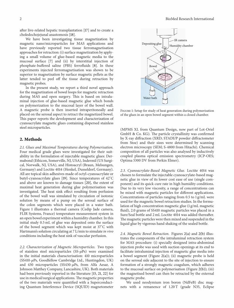
2.1. Glues and Maximal Temperatures during Polymerisation.Four medical grade glues were investigated for their suit-ability in the formulation of injectable magnetic glues: Der-mabond (Ethicon, Somerville, NJ, USA), Indermil (US Surgi-cal, Norwalk, NJ, USA), and Histoacryl (Braun, Melsungen,Germany) and Loctite 4014 (Henkel, Dusseldorf, Germany).All are topical skin adhesives made of octyl-cyanoacrylate orbutyl-cyanoacrylate glues [19]. Since temperatures of 42∘Cand above are known to damage tissues [20], the extent ofmaximal heat generation during glue polymerisation wasinvestigated. The heat sink effect resulting from perfusionof the bowel wall was simulated by circulation of isotonicsolution by means of a pump on the serosal surface ofthe colon segments which were placed in a water bath.Figure 1 illustrates a thermal camera (Cedip Jade camera,FLIR Systems, France) temperature measurement system inan open bowel experimentwithin a humidity chamber. In thisinitial study 0.5mL of glue was deposited onto the surfaceof the bowel segment which was kept moist at 37∘C withHartmann’s solution circulating at 7 L/min to simulate in vivoconditions including the heat sink of vascular perfusion.
2.2. Characterization of Magnetic Microparticles. Two typesof stainless steel microparticles (SS-𝜇Ps) were examinedin the initial materials characterisation: 410 microparticles(SS410-𝜇Ps, Goodfellow Cambridge Ltd., Huntingdon, UK)and 430 microparticles (SS430-𝜇Ps, from Alfa Aesar, AJohnsonMatthey Company, Lancashire, UK). Both materialshad been previously reported in the literature [15, 21, 22] foruse inmedical/surgical applications.Themagnetic propertiesof the two materials were quantified with a Superconduct-ing Quantum Interference Device (SQUID) magnetometer
IR camera
Humidity chamberDepositing glue
Bowel
Water bath
Figure 1: Setup for study of heat generation during polymerisationof the glues in an open bowel segment within a closed chamber.
(MPMS XL from Quantum Design, now part of Lot-OrielGmbH & Co. KG). The particle crystallinity was confirmedby X-ray diffraction (XRD; STADI/P powder diffractometerfrom Stoe) and their sizes were determined by scanningelectron microscopy (SEM; S-4800 from Hitachi). Chemicalcomposition of all particles was also analysed by inductivelycoupled plasma optical emission spectrometry (ICP-OES;Optima 5300 DV from Perkin Elmer).
2.3. Cyanoacrylate-Based Magnetic Glue. Loctite 4014 waschosen to formulate the injectable cyanoacrylate-based mag-netic glue in view of its lower cost ease of use (single com-ponent) and its quick cure rate in high humidity conditions.Due to its very low viscosity, a range of concentrations canbe mixed with magnetic particles for different applications.Concentrations of particles ranging from 0.5 to 1 g/mL wereused for the magnetic bowel retraction studies. In the formu-lation of high concentration magnetic glue (1 g/mL magneticfluid), 2.0 grams of SS410 magnetic particles was placed in aSure/Seal bottle and 2mL Loctite 4014 was added thereafter.Themagnetic particles were thenmixed and suspended in theliquid glue by vigorous hand shaking of the sealed bottle.
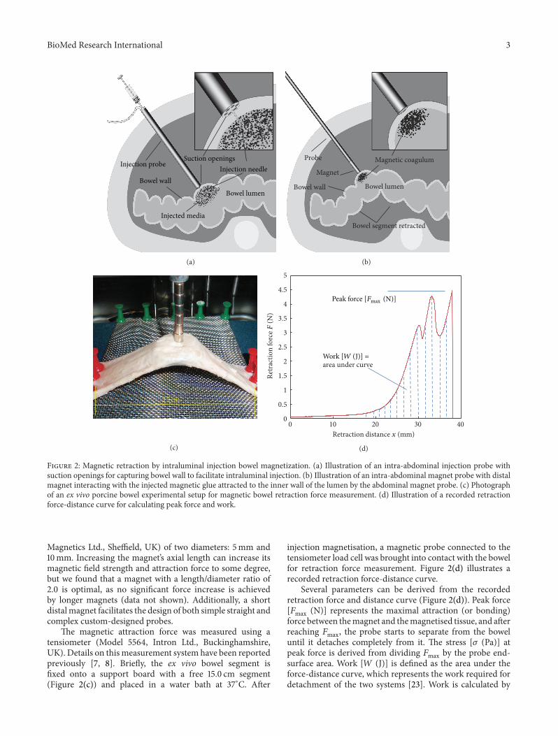
2.4. Magnetic Bowel Retraction. Figures 2(a) and 2(b) illus-trates the components of the intraluminal retraction systemfor MAS procedure: (i) specially designed intra-abdominalinjection probe was used with suction openings at its end tofacilitate intraluminal injection of magnetic glue media intoa bowel segment (Figure 2(a)); (ii) magnetic probe is heldon the serosal side adjacent to the site of injection to ensureformation of a strongly magnetic coagulum, which adheresto the mucosal surface on polymerisation (Figure 2(b)); (iii)the magnetised bowel can then be retracted by the externalmagnetic probe.
We used neodymium iron boron (NdFeB) disc mag-nets with a remanence of 1.20 T (grade N35, Eclipse
BioMed Research International 3
Injection probeSuction openings
Bowel wallInjection needle
Injected media
Bowel lumen
(a)
Probe
Bowel wall
Magnetic coagulum
Bowel segment retracted
Bowel lumen
Magnet
(b)
15 cm
(c)
0
0.5
1
1.5
2
2.5
3
3.5
4
4.5
5
0 10 20 30 40
Retr
actio
n fo
rce F
(N)
Retraction distance x (mm)
area under curve
Peak force [Fmax (N)]
Work [W (J)] =
(d)
Figure 2: Magnetic retraction by intraluminal injection bowel magnetization. (a) Illustration of an intra-abdominal injection probe withsuction openings for capturing bowel wall to facilitate intraluminal injection. (b) Illustration of an intra-abdominal magnet probe with distalmagnet interacting with the injected magnetic glue attracted to the inner wall of the lumen by the abdominal magnet probe. (c) Photographof an ex vivo porcine bowel experimental setup for magnetic bowel retraction force measurement. (d) Illustration of a recorded retractionforce-distance curve for calculating peak force and work.
Magnetics Ltd., Sheffield, UK) of two diameters: 5mm and10mm. Increasing the magnet’s axial length can increase itsmagnetic field strength and attraction force to some degree,but we found that a magnet with a length/diameter ratio of2.0 is optimal, as no significant force increase is achievedby longer magnets (data not shown). Additionally, a shortdistalmagnet facilitates the design of both simple straight andcomplex custom-designed probes.
The magnetic attraction force was measured using atensiometer (Model 5564, Intron Ltd., Buckinghamshire,UK). Details on thismeasurement system have been reportedpreviously [7, 8]. Briefly, the ex vivo bowel segment isfixed onto a support board with a free 15.0 cm segment(Figure 2(c)) and placed in a water bath at 37∘C. After
injection magnetisation, a magnetic probe connected to thetensiometer load cell was brought into contact with the bowelfor retraction force measurement. Figure 2(d) illustrates arecorded retraction force-distance curve.
Several parameters can be derived from the recordedretraction force and distance curve (Figure 2(d)). Peak force[𝐹max (N)] represents the maximal attraction (or bonding)force between themagnet and themagnetised tissue, and afterreaching 𝐹max, the probe starts to separate from the boweluntil it detaches completely from it. The stress [𝜎 (Pa)] atpeak force is derived from dividing 𝐹max by the probe end-surface area. Work [𝑊 (J)] is defined as the area under theforce-distance curve, which represents the work required fordetachment of the two systems [23]. Work is calculated by

4 BioMed Research International
Table 1: Characteristics of the magnetic particles.
Size (diameter) 𝑀𝑠
(emu/g) 𝐻𝑐
(kOe) Fe/Cr (w%)SS410-𝜇Ps Up to 50 𝜇mSEM 168.0 0.01 86.6 ± 6.8/12.6 ± 1.0SS430-𝜇Ps Up to 40 𝜇mSEM 110.0 0.02 82.2 ± 6.7/16.9 ± 1.4Magnetisation at saturation (𝑀
𝑠) and coercivity (𝐻
𝑐) measured with a SQUID magnetometer at 300K; iron and chromium percentage as deduced from ICP-
OES measurements and size as obtained by SEM characterizations.
36
37
38
39
40
41
42
43
44
Dermabond Histoacryl Indermil Loctite 4014
Max
imal
tem
pera
ture
(∘C)
Figure 3: Averaged maximum temperatures were measured by theIR thermal camera after 0.5mL of each glue was deposited ontothe surface of ex vivo porcine bowel (error bar: standard deviation,number of test 𝑛 = 7).
a custom-written MATLAB (MathWorks, Cambridge, UK)program using (1) which is based on linear trapezoidal rule[24]:
𝑊 =
𝑁−1
∑
𝑖=0
(𝑥𝑖+1− 𝑥𝑖) ∗(𝐹𝑖+ 𝐹𝑖+1)
2, (1)
where W is adhesion work, 𝑥 is retraction distance, 𝐹 isretraction force, 𝑖 is data sampling point, and 𝑁 is totalnumber of data points.
3. Results
3.1. Heat Generation by Glues. The maximal temperaturegenerated during glue polymerisation by the 4 medical gradecyanoacrylate glues studied was obtained from the recordedthermal camera image. Figure 3 plots the averaged maximaltemperaturesmeasured by the IR thermal camera after 0.5mLof each glue was deposited onto the surface of ex vivo porcinebowel (error bar: standard deviation, number of test 𝑛 = 7).All showed safe heat generation (i.e., below 42∘C) except forHistoacryl (maximal about 43.5∘C).
3.2. Characterisation of Magnetic Particles. Figures 4(a) and4(b) present the appearance of stainless steel microparticlesas observed by electron microscopy and Figure 4(c) thehysteresis curves at room temperature.
Results from XRD (not presented) and electronmicroscopy findings showed both SS410 and SS430 particles
Table 2: Magnetic bowel retraction data.
𝐹max (N) 𝑊 (mJ) 𝜎 (kPa)5mmmagnet probe 2.2 ± 0.2 34 ± 11 114 ± 11.710mmmagnet probe 5.1 ± 0.3 81 ± 17 65 ± 4.3SS410-𝜇Ps-based magnetic glues at concentration 1 g/mL: intraluminallyinjected volume of 2mL.
to be crystalline but polydispersed in size and shape withparticles’ diameters ranging from a few microns up to 50 𝜇m(Figures 4(a) and 4(b)). Magnetic properties in terms ofmagnetization as a function of applied magnetic field werequantified at room temperature and the resulting curves areshown in Figure 4(c). As expected the curves are symmetricand the magnetization at saturation, 𝑀
𝑠, is deduced from
the plateau, when the magnetization does not increase anyfurther with increasing magnetic field strength. SS410-𝜇Ps(Figure 4(c1)) exhibit a higher magnetization at saturationthan SS430-𝜇Ps, and this is likely due to their slightly higheriron content and a lower chromium doping (Table 1). Thecoercivity refers to the magnetic field which needs to beapplied to reduce the magnetization of a material down tozero after the magnetization of the sample has reached sat-uration. For both SS-𝜇Ps the remanence (i.e., the remainingmagnetisation after the field has been removed) has beenfound to be very small. These observations indicate thatboth stainless steel 𝜇Ps even though too large to form stablesuspension would not present difficulty in dispersion in thefluid due to magnetic interactions since the particles exhibitno mutual magnetic attraction unless placed in a magneticfield. SS410-𝜇Ps were chosen for the study in view of theirhigher magnetization and lower coercivity (or remanence).
3.3. Measurement of Magnetic Bowel Retraction and Forces.Harvested porcine colonic segments were used with injectionby a large gauge needle (16G or 1.7mm diameter × 50mm,B. Braun Melsungen AG) to facilitate rapid injection of highconcentrationmagnetic glue, although a smaller gauge needle(19G needle from BD Microlance 3, BD Drogheda, Ireland)can also be used for slower injection. After withdrawal of theneedle, the small puncture wound effectively sealed itself bythe injected glue with no visible leakage. The procedure tookaround 1 minute. Table 2 summarises the magnetic retractionof a magnetised bowel segments in 20 ex vivo experimentsusing both the 5mm and the 10mmmagnet probes.
The average pull force that surgeons use to provide suffi-cient tension to the bowel is 2.5N [1] with the maximal forcebeing just below 5N. The test results obtained in the present

BioMed Research International 5
10𝜇m 10𝜇m
(a1) (a2)
(a)
(b2)(b1)
250 nm 250 nm
(b)
−50 −25 0 25 50 −50 −25 0 25 50
−200
−100
0
100
200
H (kOe) H (kOe)
−100
−50
0
50
100
(c1) (c2)
MSS410
-𝜇Ps
(em
u/g)
MSS430
-𝜇Ps
(em
u/g)
(c)
Figure 4: (a)-(b) SEM images and (c) hysteresis curves completed at room temperature for two stainless steel microparticle of SS410-𝜇Ps (1)and SS430-𝜇Ps (2).
study (Table 2) indicate that SS410-𝜇Ps based bowel magne-tization is capable of providing retraction forces and work inthis range with use of 5 and 10mmdiameter permanent mag-net probes. One important advantage of magnetic retractionover conventional pull traction by teeth-like graspers is thatit inflicts less stress during high tension retraction with thetissues being not only retracted but compressed between theridges of the graspers. For example, average pressures at thetip of conventional graspers at the contact surface area withthe target tissue vary from210 kPa to 650 kPa [25]. In contrast,the current approach does not require such compression and
much lower pressures (65 kPa to 114 kPa) were observed inthe current experiments by the 10mm and 5mm diametermagnetic probes, respectively (Table 2).
3.4. On-Going Iron Oxide Nanoparticles Formulations. Mag-netic nanoparticles can potentially exhibit stronger magneticproperties than microparticles per unit mass. This arisesbecause bulkmagnetic materials present multiple domains ofmagnetisations. Within each domain, the magnetisation hasonly one direction, which however can vary fromone domainto another. The presence of several interacting domains in

6 BioMed Research International
one particle with potentially different orientations can resultin lowering the overall magnetisation of the particle. Incontrast, decrease of the size of the particles to a singledomain excludes the possibility of magnetic interactionsbetween multiple magnetic domains within a single particle.If the nanoparticles do not carry a magnetic “dead-layer” attheir interface, their magnetic properties can be enhancedcompared to microparticles when exposed to the samemagnetic field [26–28].
Magnetic nanoparticles have been successfully used fornumerous biomedical applications [29–34]; however wefound that iron oxide nPs could not be mixed easily withLoctite 4014 liquid for medical magnetic glue injection. Forthis reason the on-going studies by the group is exploringways to overcome this problem as it is related to the fastpolymerization of the glue when mixed with the iron oxidenanoparticles. This is likely associated with the surfacereactivity of the nanoparticles which needs to be adequatelytuned and possibly coated to enable development of efficientnanoparticle-based medical magneto-glues.
4. Discussion
Thenew system based on formation of intraluminalmagneticcoagulum provides highly effective atraumatic retraction andovercomes the problems reported with previous approachesbased on localised ferromagnetisation of tissues, that is,low injection volume during interstitial ferromagnetisation[8] and peeling of surface magnetic pellets beyond certainretraction forces [7]. Furthermore, it provides significantlygreater retraction forces, which meet all the requirementsfor uncompromised bowel retraction/manipulation duringMAS equivalent to bowel grasping without risk of trauma orslippage. Additionally, the results of the present experimentshave excluded thermal injury to the issues induced bymedicalgrade cyanoacrylate glues during polymerisation. Loctite4014 glue was selected because of its very low viscosity, singlecomponent nature, rapid cure rate, and ability to mix withstainless steel microparticles.
To date, ex vivo porcine bowel segments have been usedto validate the concept and measure the forces generated(Figure 5). To facilitate intraluminal injection at randomlyselected target bowel segment, an open abdominal modelwas used instead of a laparoscopic model. Two mL magneticgluewas injected intraluminally into targeted bowel segmentsand the 10mm diameter magnet probe used to retract themagnetised bowel (Figure 5(a)) which could be manipulatedandmoved around, and retracted (Figure 5(b)) withmaximalpull detachment force of 5N, which was in agreement withInstron bench test results using ex vivo bowel segments(Table 2).
The ex vivo experiments with the formulated magneticglue also demonstrated that even small volumes (2mL)injected intraluminally in target bowel segments enableeffective magnetic bowel retraction using a small (5 to 10mmdiameter) permanentmagnet probes. For the same retractionforce, the experimental data confirmed that the stress exertedon the target tissue by magnetic probes was significantly lesswhen compared with the force generated during retraction
(a)
(b)
Figure 5: Retraction of in situ magnetised bowel using an exvivo porcine model and a volume of 2mL magnetic media withconcentration of 1 g/mL was injected into a bowel segment: (a)photography showing the 10mm magnet probe inserted into a10mm port and engaged with the magnetised bowel segment; (b)photography showing retraction of themagnetised bowel toward theport direction, with maximal detachment force of 5N.
by grasping forceps (65 kPa versus 210 kPa). Furthermore,the magnetic probe does not gather the tissue into a fold butinstead creates a flat and smooth contact area with a widerdistribution of probe-tissue contact forces. This underliesthe atraumatic nature of magnetic retraction since the tissuefalls off when the force exceeds the magnetic pull. Activerelease of the magnetically retracted tissue can be obtainedby custom-designed probes which adjust the magneticattraction force through a controlled release mechanism.
Our experiments also confirmed that change of retrac-tion speed (from 0.1mm/s to 10mm/s) does not affect the

BioMed Research International 7
magnetic retraction force and work, suggesting that bowelretractedwith such probeswould not have to bemoved slowlyduring surgery. The experiments demonstrate that comparedto external (serosal) application, the intraluminal magneticcoagulum significantly increases themaximal retraction force(1.75 ± 0.86N versus 5.15 ± 0.98N) with the use of a 10mmmagnet probe. Once the external magnet probe is removed,the coagulum detaches from the mucosa and thus becomessusceptible to spontaneous elimination in the stools. Anadjustable magnetic force probe is desirable for controlledrelease of tissue whenever this is required. A simple andeffective design for such a probe has been designed anddeveloped in our lab and will be published elsewhere.
5. Conclusions
The results of the current experimental study confirm that,with the technology described, intraluminal magnetic coag-ula formed from medical-grade biocompatible cyanoacry-late (Loctite) and SS410-𝜇Ps constitutes a novel system forefficient and atraumatic magnetic retraction of bowel. Thesystem provides the retraction forces required for bowelmanipulation and handling during laparoscopic surgery. Thetechnology functions by producing a coagulum containinga sufficient mass of aggregated stainless steel microparticlesadherent to the mucosa. This is either removed with thespecimen in resectional bowel surgery or is expelled withreturn of bowel function. Further improvement is foreseenwith the use of polymer coated nanoparticles of iron oxidebeing developed in our laboratory instead of stainless steelmicroparticles. Evaluation of the fully developed technologyby in vivo large animal studies before translation to clinicalpractice is needed.
Conflict of Interests
The authors declare that there is no conflict of interestsregarding the publication of this paper.
Acknowledgment
Thisworkwas supported by the Engineering andPhysical Sci-ences Research Council (EPSRC), UK, under Grant EP/HO10033/1.
References
[1] E. A. M. Heijnsdijk, H. de Visser, J. Dankelman, and D. J.Gouma, “Slip and damage properties of jaws of laparoscopicgraspers,” Surgical Endoscopy and Other Interventional Tech-niques, vol. 18, no. 6, pp. 974–979, 2004.
[2] E. A. M. Heijnsdijk, J. Dankelman, and D. J. Gouma, “Effec-tiveness of grasping and duration of clamping using laparo-scopic graspers,” Surgical Endoscopy and Other InterventionalTechniques, vol. 16, no. 9, pp. 1329–1331, 2002.
[3] P.-O. Park, M. Bergstrom, K. Ikeda, A. Fritscher-Ravens, andP. Swain, “Experimental studies of transgastric gallbladdersurgery: cholecystectomy and cholecystogastric anastomosis
(videos),”Gastrointestinal Endoscopy, vol. 61, no. 4, pp. 601–606,2005.
[4] M. Ryou and C. C.Thompson, “Magnetic retraction in natural-orifice transluminal endoscopic surgery (NOTES): addressingthe problem of traction and countertraction,” Endoscopy, vol.41, no. 2, pp. 143–148, 2009.
[5] Q. A. Pankhurst, J. Connolly, S. K. Jones, and J. Dobson, “Appli-cations of magnetic nanoparticles in biomedicine,” Journal ofPhysics D, vol. 36, no. 13, pp. R167–R181, 2003.
[6] R. A. Frimpong and J. Z. Hilt, “Magnetic nanoparticles inbiomedicine: synthesis, functionalization and applications,”Nanomedicine, vol. 5, no. 9, pp. 1401–1414, 2010.
[7] Z.Wang, L.Wang, B. Tang, T. Frank, S. Brown, andA.Cuschieri,“Retraction by surface ferromagnetisation of target tissues:preliminary studies on feasibility of magnetic retraction forendoscopic surgery,” Surgical Endoscopy and Other Interven-tional Techniques, vol. 22, no. 8, pp. 1838–1844, 2008.
[8] Z. Wang, L. Wang, S. I. Brown, T. G. Frank, and A. Cuschieri,“Ferromagnetization of target tissues by interstitial injection offerrofluid: formulation and evidence of efficacy for magneticretraction,” IEEE Transactions on Biomedical Engineering, vol.56, no. 9, pp. 2244–2252, 2009.
[9] G. G. M. Pinkse, E. Steenvoorde, S. Hogendoorn et al., “Stabletransplantation results of magnetically retracted islets: a novelmethod,” Diabetologia, vol. 47, no. 1, pp. 55–61, 2004.
[10] D. C. Meeker, E. H. Maslen, R. C. Ritter, and F. M. Creighton,“Optimal realization of arbitrary forces in amagnetic stereotaxissystem,” IEEE Transactions onMagnetics, vol. 32, no. 2, pp. 320–328, 1996.
[11] S. Martel, J.-B. Mathieu, O. Felfoul et al., “Automatic navigationof an untethered device in the artery of a living animal usinga conventional clinical magnetic resonance imaging system,”Applied Physics Letters, vol. 90, no. 11, Article ID 114105, 2007.
[12] J. Cadeddu, R. Fernandez, M. Desai et al., “Novel magneti-cally guided intra-abdominal camera to facilitate laparoendo-scopic single-site surgery: initial human experience,” SurgicalEndoscopy and Other Interventional Techniques, vol. 23, no. 8,pp. 1894–1899, 2009.
[13] M. T. Gettman and P. Swain, “Initial experimental evaluationof wireless capsule endoscopes in the bladder: implications forcapsule cystoscopy,” European Urology, vol. 55, no. 5, pp. 1207–1212, 2009.
[14] T. Uchiyama, K. Mohri, M. Shinkai et al., “Position sensing ofmagnetite gel using MI sensor for brain tumor detection,” IEEETransactions on Magnetics, vol. 33, no. 5, pp. 4266–4268, 1997.
[15] J. M. Peeters, J.-H. Seppenwoolde, L. W. Bartels, and C. J. G.Bakker, “Development and testing of passive tracking markersfor different field strengths and tracking speeds,” Physics inMedicine and Biology, vol. 51, no. 6, pp. N127–N137, 2006.
[16] T. Ohdaira and H. Nagai, “Intraoperative localization of early-stage upper gastrointestinal tumors using a magnetic markingclip-detecting system,” Surgical Endoscopy and other Interven-tional Techniques, vol. 21, no. 5, pp. 810–815, 2007.
[17] D. Erdmann, R. Sweis, C. Heitmann et al., “Side-to-side suture-less vascular anastomosis with magnets,” Journal of VascularSurgery, vol. 40, no. 3, pp. 505–511, 2004.
[18] N. Muraoka, H. Uematsu, E. Yamanouchi et al., “Yamanouchimagnetic compression anastomosis for bilioenteric anastomoticstricture after living-donor liver transplantation,” Journal ofVascular and Interventional Radiology, vol. 16, no. 9, pp. 1263–1267, 2005.

8 BioMed Research International
[19] A. J. Singer, T. Zimmerman, J. Rooney, P. Cameau, G. Rudomen,and S. A. McClain, “Comparison of wound-bursting strengthsand surface characteristics of FDA-approved tissue adhesivesfor skin closure,” Journal of Adhesion Science and Technology,vol. 18, no. 1, pp. 19–27, 2004.
[20] M. A. Astrahan, M. D. Sapozink, D. Cohen et al., “Microwaveapplicator for transurethral hyperthermia of benign prostatichyperplasia,” International Journal of Hyperthermia, vol. 5, no.3, pp. 283–296, 1989.
[21] H. Chen, M. D. Kaminski, A. D. Ebner, J. A. Ritter, and A. J.Rosengart, “Magnetizable intravascular stents for sequestrationof systemically circulating magnetic nano- and microspheres,”in Proceedings of the 3rd IEEE/EMBS Special Topic Conferenceon Microtechnology in Medicine and Biology, pp. 286–289, May2005.
[22] H. Chen, A. D. Ebner, D. Bockenfeld et al., “A comprehensive invitro investigation of a portable magnetic separator device forhuman blood detoxification,” Physics in Medicine and Biology,vol. 52, no. 19, pp. 6053–6072, 2007.
[23] C. Eouani, P. Piccerelle, P. Prinderre, E. Bourret, and J.Joachim, “In-vitro comparative study of buccal mucoadhesiveperformance of different polymeric films,” European Journal ofPharmaceutics and Biopharmaceutics, vol. 52, no. 1, pp. 45–55,2001.
[24] Pharmacokinetics, Marcel Dekker, New York, NY, USA, 1982,edited by M. Gibaldi and M. Perrier.
[25] J. A. Cartmill, A. J. Shakeshaft, W. R. Walsht, and C. J. Martin,“High pressures are generated at the tip of laparoscopic grasp-ers,” Australian and New Zealand Journal of Surgery, vol. 69, no.2, pp. 127–130, 1999.
[26] M. Mikhaylova, D. K. Kim, N. Bobrysheva et al., “Superpara-magnetism of magnetite nanoparticles: dependence on surfacemodification,” Langmuir, vol. 20, no. 6, pp. 2472–2477, 2004.
[27] C. d. Montferrand, Y. Lalatonne, D. Bonnin et al., “Size-dependent nonlinear weak-field magnetic behavior ofmaghemite nanoparticles,” Small, vol. 8, no. 12, pp. 1945–1956, 2012.
[28] U. Jeong, X. Teng, Y. Wang, H. Yang, and Y. Xia, “Superpara-magnetic colloids: controlled synthesis and niche applications,”Advanced Materials, vol. 19, no. 1, pp. 33–60, 2007.
[29] G. A. O. Jinhao, G. U. Hongwei, and X. U. Bing, “Multifunc-tional magnetic nanoparticles: design, synthesis, and biomedi-cal applications,” Accounts of Chemical Research, vol. 42, no. 8,pp. 1097–1107, 2009.
[30] M. Oudkerk, A. G. van den Heuvel, P. A. Wielopolski, P. I.M. Schmitz, I. H. M. Borel Rinkes, and T. Wiggers, “Hepaticlesions: detection with ferumoxide-enhanced T1-weighted MRimaging,” Radiology, vol. 203, no. 2, pp. 449–456, 1997.
[31] R. C. Semelka andT. K.G.Helmberger, “State of the art: contrastagents for mr imaging of the liver,” Radiology, vol. 218, no. 1, pp.27–38, 2001.
[32] S. Chen, C. Hoskins, L. Wang, M. P. MacDonald, and P. Andre,“A water-soluble temperature nanoprobe based on a multi-modal magnetic-luminescent nanocolloid,” Chemical Commu-nications, vol. 48, no. 19, pp. 2501–2503, 2012.
[33] S. Chen, L. Wang, S. L. Duce et al., “Engineered biocompatiblenanoparticles for in vivo imaging applications,” Journal of theAmerican Chemical Society, vol. 132, no. 42, pp. 15022–15029,2010.
[34] L. Motte, F. Benyettou, C. de Beaucorps, M. Lecouvey, I.Milesovic, and Y. Lalatonne, “Multimodal superparamagneticnanoplatform for clinical applications: immunoassays, imaging& therapy,” Faraday Discussions, vol. 149, pp. 211–225, 2011.

Submit your manuscripts athttp://www.hindawi.com
Hindawi Publishing Corporation http://www.hindawi.com Volume 2013Hindawi Publishing Corporation http://www.hindawi.com Volume 2013
The Scientific World Journal
Hindawi Publishing Corporationhttp://www.hindawi.com
Nucleic AcidsJournal of
Volume 2013
ArchaeaHindawi Publishing Corporationhttp://www.hindawi.com Volume 2013
ISRN Biotechnology
Hindawi Publishing Corporationhttp://www.hindawi.com Volume 2013
Hindawi Publishing Corporationhttp://www.hindawi.com
GenomicsInternational Journal of
Volume 2013
Evolutionary BiologyInternational Journal of
Hindawi Publishing Corporationhttp://www.hindawi.com Volume 2013
Hindawi Publishing Corporationhttp://www.hindawi.com Volume 2013
Advances in
Virolog y
ISRN Microbiology
Hindawi Publishing Corporationhttp://www.hindawi.com Volume 2013
Marine BiologyJournal of
Hindawi Publishing Corporationhttp://www.hindawi.com Volume 2013
BioMed Research International
Hindawi Publishing Corporationhttp://www.hindawi.com Volume 2013
ISRN Zoology
Hindawi Publishing Corporationhttp://www.hindawi.com Volume 2013
Hindawi Publishing Corporationhttp://www.hindawi.com Volume 2013
Signal TransductionJournal of
ISRN Cell Biology
Hindawi Publishing Corporationhttp://www.hindawi.com Volume 2013
Hindawi Publishing Corporationhttp://www.hindawi.com Volume 2013
BioinformaticsAdvances in
PeptidesInternational Journal of
Hindawi Publishing Corporationhttp://www.hindawi.com Volume 2013
Hindawi Publishing Corporationhttp://www.hindawi.com Volume 2013
Enzyme Research
Hindawi Publishing Corporationhttp://www.hindawi.com Volume 2013
Biochemistry Research International
ISRN Molecular Biology
Hindawi Publishing Corporationhttp://www.hindawi.com Volume 2013
Stem CellsInternational
Hindawi Publishing Corporationhttp://www.hindawi.com Volume 2013
Copyright © 2022 FDOKUMEN