Microstructure and Fracture Toughness of Nitrided D2 Steels ...
Upload
independentCategory
view
0download
0
After online publication, subscribers (personal/institutional) to this journal will haveaccess to the complete article via the DOI using the URL:
If you would like to know when your article has been published online, take advantageof our free alert service. For registration and further information, go to:http://www.springerlink.com.
Due to the electronic nature of the procedure, the manuscript and the original figureswill only be returned to you on special request. When you return your corrections,please inform us, if you would like to have these documents returned.
Dear Author
Here are the proofs of your article.
• You can submit your corrections online, via e-mail or by fax.
• For online submission please insert your corrections in the online correction form.
Always indicate the line number to which the correction refers.
• You can also insert your corrections in the proof PDF and email the annotated PDF.
• For fax submission, please ensure that your corrections are clearly legible. Use a fine
black pen and write the correction in the margin, not too close to the edge of the page.
• Remember to note the journal title, article number, and your name when sending your
response via e-mail or fax.
• Check the metadata sheet to make sure that the header information, especially author
names and the corresponding affiliations are correctly shown.
• Check the questions that may have arisen during copy editing and insert your
answers/corrections.
• Check that the text is complete and that all figures, tables and their legends are included.
Also check the accuracy of special characters, equations, and electronic supplementary
material if applicable. If necessary refer to the Edited manuscript.
• The publication of inaccurate data such as dosages and units can have serious
consequences. Please take particular care that all such details are correct.
• Please do not make changes that involve only matters of style. We have generally
introduced forms that follow the journal’s style.
• Substantial changes in content, e.g., new results, corrected values, title and authorship are
not allowed without the approval of the responsible editor. In such a case, please contact
the Editorial Office and return his/her consent together with the proof.
• If we do not receive your corrections within 48 hours, we will send you a reminder.
• Your article will be published Online First approximately one week after receipt of your
corrected proofs. This is the official first publication citable with the DOI. Further
changes are, therefore, not possible.
• The printed version will follow in a forthcoming issue.
Please note
http://dx.doi.org/10.1007/s12253-011-9453-0
AUTHOR'S PROOF
Metadata of the article that will be visualized in OnlineFirst
1 Article Title Periacinar Retraction Clefting and D2-40 Expression in Prostatic
Adenocarcinoma
2 Article Sub- Title
3 Article Copyright -Year
Arányi Lajos Foundation 2011(This will be the copyright line in the final PDF)
4 Journal Name Pathology & Oncology Research
5
Corresponding
Author
Family Name Krušlin
6 Particle
7 Given Name Božo
8 Suffix
9 Organization Sestre milosrdnice University Hospital Centre
10 Division Ljudevit Jurak Department of Pathology
11 Address Vinogradska cesta 29, Zagreb , Croatia
12 Organization School of Medicine, University of Zagreb
13 Division Department of Pathology
14 Address Zagreb , Croatia
15 e-mail [email protected]
16
Author
Family Name Ulamec
17 Particle
18 Given Name Monika
19 Suffix
20 Organization Sestre milosrdnice University Hospital Centre
21 Division Ljudevit Jurak Department of Pathology
22 Address Vinogradska cesta 29, Zagreb , Croatia
23 e-mail
24
Author
Family Name Džombeta
25 Particle
26 Given Name Tihana
27 Suffix
28 Organization School of Medicine, University of Zagreb
29 Division Department of Pathology
30 Address Zagreb , Croatia
AUTHOR'S PROOF
31 e-mail
32
Author
Family Name Čupić
33 Particle
34 Given Name Hrv oje
35 Suffix
36 Organization Sestre milosrdnice University Hospital Centre
37 Division Ljudevit Jurak Department of Pathology
38 Address Vinogradska cesta 29, Zagreb , Croatia
39 Organization School of Medicine, University of Zagreb
40 Division Department of Pathology
41 Address Zagreb , Croatia
42 e-mail
43
Author
Family Name Leniček
44 Particle
45 Given Name Tanja
46 Suffix
47 Organization Sestre milosrdnice University Hospital Centre
48 Division Ljudevit Jurak Department of Pathology
49 Address Vinogradska cesta 29, Zagreb , Croatia
50 e-mail
51
Author
Family Name Tomas
52 Particle
53 Given Name Dav or
54 Suffix
55 Organization Sestre milosrdnice University Hospital Centre
56 Division Ljudevit Jurak Department of Pathology
57 Address Vinogradska cesta 29, Zagreb , Croatia
58 Organization School of Medicine, University of Zagreb
59 Division Department of Pathology
60 Address Zagreb , Croatia
61 e-mail
62
Schedule
Received 17 July 2011
63 Revised
64 Accepted 11 August 2011
65 Abstract Retraction clef ting is known to appear in v arious ty pes of tumors, but ithas only recently been recognized as a specif ic histological phenomenon.Prev iously , it was considered merely a laboratory procedure artif act, butlately , there hav e been some assumptions that peritumoral retractionsactually represent ly mphatic spaces. In our study , we analy zed neoplastic
AUTHOR'S PROOF
glands in 52 specimens of prostatic adenocarcinoma. Immunohistochemicalanaly sis was perf ormed using D2-40 antibody , to highlight ly mphaticendothelium and thereby dif f erentiate actual ly mph v essels orly mphv ascular inv asion f rom periacinar retractions. Our results showedthat the number of ly mph v essels was signif icantly lower in tumoroustissue compared to adjacent normal prostatic tissue. On the other hand, thenumber of ly mph v essels in tumorous tissue was signif icantly higher thanthe number of ly mph v essels mimicking periacinar retractions. Ov erall, thenumber of ly mph v essels mimicking periacinar clef ts was particularly low.These results are in accordance with our prev ious studies, which had shownthat periacinar clef ting appears due to lack of basal cells and stromalchanges around tumorous acini. Also, these results support our hy pothesisthat retractions do not represent ly mph v essels but should be considered adistinct entity , which is prov en to be helpf ul both as diagnostic andpredictiv e f actor.
66 Keywordsseparated by ' - '
D2 40 - Ly mphov ascular inv asion - Prostatic adenocarcinoma - Retractionclef ting
67 Foot noteinformation
AUTHOR'S PROOF
UNCORRECTEDPROOF
1
23 RESEARCH
4 Periacinar Retraction Clefting and D2-40 Expression5 in Prostatic Adenocarcinoma
6 Monika Ulamec & Tihana Džombeta & Hrvoje Čupić &
7 Tanja Leniček & Davor Tomas & Božo Krušlin
8 Received: 17 July 2011 /Accepted: 11 August 20119 # Arányi Lajos Foundation 2011
10
11 Abstract Retraction clefting is known to appear in various12 types of tumors, but it has only recently been recognized as13 a specific histological phenomenon. Previously, it was14 considered merely a laboratory procedure artifact, but15 lately, there have been some assumptions that peritumoral16 retractions actually represent lymphatic spaces. In our17 study, we analyzed neoplastic glands in 52 specimens of18 prostatic adenocarcinoma. Immunohistochemical analysis19 was performed using D2-40 antibody, to highlight lym-20 phatic endothelium and thereby differentiate actual lymph21 vessels or lymphvascular invasion from periacinar retrac-22 tions. Our results showed that the number of lymph vessels23 was significantly lower in tumorous tissue compared to24 adjacent normal prostatic tissue. On the other hand, the25 number of lymph vessels in tumorous tissue was signifi-26 cantly higher than the number of lymph vessels mimicking27 periacinar retractions. Overall, the number of lymph vessels28 mimicking periacinar clefts was particularly low. These29 results are in accordance with our previous studies, which30 had shown that periacinar clefting appears due to lack of31 basal cells and stromal changes around tumorous acini.32 Also, these results support our hypothesis that retractions33 do not represent lymph vessels but should be considered a34 distinct entity, which is proven to be helpful both as35 diagnostic and predictive factor.
36Keywords D2 40 . Lymphovascular invasion . Prostatic37adenocarcinoma . Retraction clefting
38Introduction
39Clefting is a well known histological phenomenon de-40scribed in various carcinoma types beside prostatic carci-41noma, mainly in basal cell carcinoma and breast carcinoma.42The neoplastic cells of prostatic cancer often appear pulled43away from the surrounding stroma, leaving empty spaces44that completely or partially encircle the acini; these are45called retraction clefts or retraction artifacts and were46described for the first time in 1960s in autopsy studies by47Halpert and co-workers [1, 2]. Periacinar retraction clefts48in prostatic carcinoma serve as a helpful additional49criterion in setting of pathohistological diagnosis, partic-50ularly in differentiating it from some benign conditions51(atrophy, postatrophic hyperplasia, atypical adenomatous52hyperplasia), which may mimic prostatic carcinoma [3–8].53They are mostly seen in Gleason grade 3, occasionally54appear in grades 2 and 4 but are uncommon in comedo55type Gleason grade 5 carcinoma [6]. The origin of clefting56is still not clear. At first considered a consequence of57inadequate laboratory procedure, it has also been sug-58gested that clefts actually represent pre-lymphatic spaces59or lymph vessel compartments [9]. However, in our60opinion they probably result from lack of basal cells and/61or stromal changes [10, 11]. Some authors suggest that the62presence and extent of clefts around tumorous tissue, not63only in prostatic adenocarcinoma but also in some other64tumors, especially breast carcinoma, can predict nodal65metastasis and patients’ outcome [12–14]. Moreover, the66presence of extensive retraction clefting in prostatic67carcinoma is associated with more aggressive tumor
M. Ulamec :H. Čupić : T. Leniček :D. Tomas :B. Krušlin (*)Ljudevit Jurak Department of Pathology,Sestre milosrdnice University Hospital Centre,Vinogradska cesta 29,Zagreb, Croatiae-mail: [email protected]
T. Džombeta :H. Čupić :D. Tomas : B. KrušlinDepartment of Pathology, School of Medicine,University of Zagreb,Zagreb, Croatia
Pathol. Oncol. Res.DOI 10.1007/s12253-011-9453-0
JrnlID 12253_ArtID 9453_Proof# 1 - 04/09/2011

AUTHOR'S PROOF
UNCORRECTEDPROOF
68 phenotype and a shorter biochemical recurrence-free69 interval [15].70 A few years ago D2-40, a novel monoclonal antibody71 directed against mucin-type transmembrane glycoprotein72 podoplanin, specifically expressed by lymphatic endothelial73 cells, was introduced [16]. D2-40 was originally raised74 against M2A antigen, a surface sialoglycoprotein first75 detected in association with germ cell neoplasia and fetal76 testicular gonocytes [16]. The main significance of D2-4077 lies in the fact that it is a selective marker of lymphatic78 endothelium, it is unreactive with vascular endothelium and79 can be used to identify lymphatic invasion of primary80 tumors [17, 18]. In our study, we used D2-40 to detect81 lymph vessels in prostatic cancer and compare their82 presence and distribution with periacinar retraction clefts.83 Aim was to confirm our hypothesis that clefting is a distinct84 entity unrelated to lymphatic system.
85 Materials and Methods
86 Neoplastic glands were analyzed in 52 consecutive speci-87 mens obtained from patients who underwent radical88 prostatectomy due to prostatic adenocarcinoma in the89 period from January 1st to June 30th 2006. Specimens90 were taken from the archive at the Ljudevit Jurak91 Department of Pathology, Sestre milosrdnice University92 Hospital Centre, Zagreb. Overall, 13 of 52 (25%) patients93 had extraprostatic extension and/or seminal vesicle inva-94 sion. Six (11.5%) patients had lymph node metastases.95 All available sections were examined on both96 hematoxylin and eosin (HE) and immunostained slides.97 Lymphatic endothelial lining was highlighted using D2-98 40 antibody, enabling the distinction between lympho-99 vascular invasion, defined as prostate adenocarcinoma100 cells within D2-40 positive compartments, and retracted101 periacinar stroma. Therefore, to evaluate the number of102 lymph vessels among periacinar clefts, each section was103 first examined under low magnification field (x100) on104 HE stained slide, than the same section was examined105 on D2-40 immunostained slide.106 The area with highest number of D2-40 positive107 compartments was selected under lower magnification108 (x100) and than lymph vessels were counted under high109 magnification (x400) on 10 high power fields (HPF), in110 both tumor and surrounding normal prostatic tissue.111 Similarly, 30 glands with most extensive clefts were112 selected on HE slides under low power magnification,113 marked, and than lymph vessels were counted in the same114 area on immunostained slides.115 The number of lymph vessels mimicking periacinar116 retraction clefts was counted on the whole mount section117 under high magnification (x400), on immunostained slides.
118That referred to lymph vessels appearing to be perigland-119ular retractions on HE slides, but as demonstrated by D2-12040, represented lymphovascular invasion.121Immunostained sections were examined by three inde-122pendent pathologists (U.M, T.D. and K.B.) and the final123score was determined as a mean value counted by124individuals.125Tissue samples were fixed in 10% buffered formalde-126hyde for 24 h after surgery. Following fixation, prostate127samples were cross sectioned through regions and placed128into tissue containers. Samples were fixed for another 24 h,129embedded in paraffin, cut at 5 μm and routinely stained130with hematoxylin and eosin. Each sample contained at least1315% of tumorous tissue and normal prostatic tissue that was132used as an internal control. Lymph vessels were demon-133strated with D2-40 antibody (Novocastra, mouse antibody,134clone 49, 1:100).135Immunohistochemical staining was performed using136standard procedures on DAKO TechMate Horizon auto-137mated immunostainer using antibodies to D2-40.138Briefly, 5 μm tissue sections were deparaffinized.139Antigen retrieval for D2-40 was performed in a steamer140using citrate buffer (pH 6.0) for 30 min. After blocking141with hydrogen peroxide and normal goat serum, the142sections were incubated with primary monoclonal mouse143anti-human antibodies against D2-40 (Novocastra, mouse144antibody, clone 49, 1:100) for 30 min at 37°C. The sections145were incubated sequentially with biotinylated goat anti-146mouse immunoglobulin and peroxidase-conjugated strepta-147vidin. Primary antibody was replaced by phosphate-148buffered saline in negative control sections. Color was149developed by incubation with 3, 3′-diaminobenzidine150tetrahydrochloride and slides were counterstained by151hematoxylin.152Statistical analysis was performed using Mann–Whitney153U test. Pearson X2 test was used to compare Gleason score154with the number of lymph vessels mimicking periacinar155retractions and number of lymph vessels in the area with156most extensive periacinar retractions. The level of signifi-157cance was set at p<0.001.
158Results
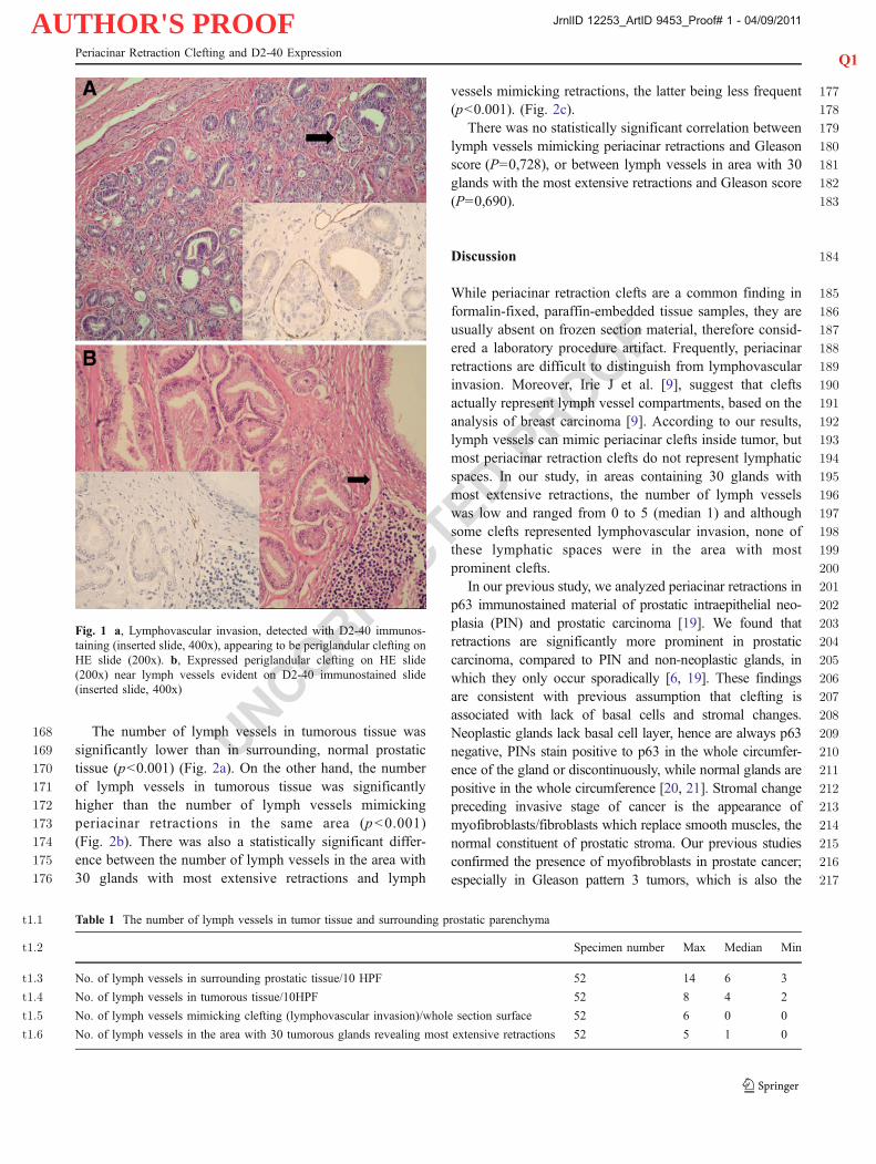
159The number of lymph vessels ranged between 2 and1608 (median value 4) in tumorous tissue and between 3 and16114 (median 6) in surrounding, normal prostatic tissue,162counted on 10 HPF. The number of lymph vessels163mimicking periacinar clefts ranged between 0 and 6 on164whole cross section (median 0) (Fig. 1a). In 30 tumorous165glands with most extensive retractions, the number of166lymph vessels ranged between 0 and 5 (median 1) (Fig. 1b).167The results are summarized in Table 1.
M. Ulamec et al.
JrnlID 12253_ArtID 9453_Proof# 1 - 04/09/2011
AUTHOR'S PROOF
UNCORRECTEDPROOF
168 The number of lymph vessels in tumorous tissue was169 significantly lower than in surrounding, normal prostatic170 tissue (p<0.001) (Fig. 2a). On the other hand, the number171 of lymph vessels in tumorous tissue was significantly172 higher than the number of lymph vessels mimicking173 periacinar retractions in the same area (p<0.001)174 (Fig. 2b). There was also a statistically significant differ-175 ence between the number of lymph vessels in the area with176 30 glands with most extensive retractions and lymph
177vessels mimicking retractions, the latter being less frequent178(p<0.001). (Fig. 2c).179There was no statistically significant correlation between180lymph vessels mimicking periacinar retractions and Gleason181score (P=0,728), or between lymph vessels in area with 30182glands with the most extensive retractions and Gleason score183(P=0,690).
184Discussion
185While periacinar retraction clefts are a common finding in186formalin-fixed, paraffin-embedded tissue samples, they are187usually absent on frozen section material, therefore consid-188ered a laboratory procedure artifact. Frequently, periacinar189retractions are difficult to distinguish from lymphovascular190invasion. Moreover, Irie J et al. [9], suggest that clefts191actually represent lymph vessel compartments, based on the192analysis of breast carcinoma [9]. According to our results,193lymph vessels can mimic periacinar clefts inside tumor, but194most periacinar retraction clefts do not represent lymphatic195spaces. In our study, in areas containing 30 glands with196most extensive retractions, the number of lymph vessels197was low and ranged from 0 to 5 (median 1) and although198some clefts represented lymphovascular invasion, none of199these lymphatic spaces were in the area with most200prominent clefts.201In our previous study, we analyzed periacinar retractions in202p63 immunostained material of prostatic intraepithelial neo-203plasia (PIN) and prostatic carcinoma [19]. We found that204retractions are significantly more prominent in prostatic205carcinoma, compared to PIN and non-neoplastic glands, in206which they only occur sporadically [6, 19]. These findings207are consistent with previous assumption that clefting is208associated with lack of basal cells and stromal changes.209Neoplastic glands lack basal cell layer, hence are always p63210negative, PINs stain positive to p63 in the whole circumfer-211ence of the gland or discontinuously, while normal glands are212positive in the whole circumference [20, 21]. Stromal change213preceding invasive stage of cancer is the appearance of214myofibroblasts/fibroblasts which replace smooth muscles, the215normal constituent of prostatic stroma. Our previous studies216confirmed the presence of myofibroblasts in prostate cancer;217especially in Gleason pattern 3 tumors, which is also the
Fig. 1 a, Lymphovascular invasion, detected with D2-40 immunos-taining (inserted slide, 400x), appearing to be periglandular clefting onHE slide (200x). b, Expressed periglandular clefting on HE slide(200x) near lymph vessels evident on D2-40 immunostained slide(inserted slide, 400x)
t1.1 Table 1 The number of lymph vessels in tumor tissue and surrounding prostatic parenchyma
t1.2 Specimen number Max Median Min
t1.3 No. of lymph vessels in surrounding prostatic tissue/10 HPF 52 14 6 3
t1.4 No. of lymph vessels in tumorous tissue/10HPF 52 8 4 2
t1.5 No. of lymph vessels mimicking clefting (lymphovascular invasion)/whole section surface 52 6 0 0
t1.6 No. of lymph vessels in the area with 30 tumorous glands revealing most extensive retractions 52 5 1 0
Q1Periacinar Retraction Clefting and D2-40 Expression
JrnlID 12253_ArtID 9453_Proof# 1 - 04/09/2011

AUTHOR'S PROOF
UNCORRECTEDPROOF
Fig. 2 Correlation between thenumber of lymph vessels intumor and adjacent prostatictissue (a), between lymphvessels mimicking periacinarclefting and number of lymphvessels in tumor (b) andbetween the number of lymphvessels within the area with 30neoplastic glands with mostextensive retractions and lymphvessels mimicking periacinarclefting (c)
M. Ulamec et al.
JrnlID 12253_ArtID 9453_Proof# 1 - 04/09/2011
AUTHOR'S PROOF
UNCORRECTEDPROOF
218 pattern with most prominent periacinar retractions [6], but219 myofibroblasts were not found in surrounding normal tissue220 [11]. Similar studies have been made in breast carcinoma.221 The study by Acs et al. [8] showed the presence of retractions222 in 60% of invasive carcinomas, and their rare occurence223 around in situ carcinomas or benign ducts and acini.224 Recent studies have also focused on potential prognostic225 significance of retraction clefting in various types of226 tumors, including prostatic adenocarcinoma. Acs and co-227 workers found that breast carcinomas associated with228 lymphovascular invasion and lymph node metastasis had229 significantly higher percentage of retraction artifacts com-230 pared to tumors without these features. Furthermore,231 extensive retractions were significantly associated with232 both poor overall prognosis and disease-free interval [12].233 Recently, same authors have confirmed these findings in234 core needle biopsy material of invasive ductal carcinoma235 [13]. Similar studies showed statistically significant corre-236 lation between extensive peritumoral retractions in esoph-237 ageal squamous cell carcinoma and lymph node metastasis238 [14]. In our previous study, we examined whether extensive239 retractions could predict biochemical reccurence-free sur-240 vival in prostatic carcinoma. The extent of retraction241 showed a statistically significant positive correlation with242 preoperative PSA and negative correlation with biochemi-243 cal disease free survival [15]. Also, tumors associated with244 seminal vesicle invasion and/or extracapsular extension245 showed significantly higher percentage of retraction artifact246 than tumors without these features [15].247 Lymphangiogenesis has been associated with poor248 prognosis in a number of human cancers. Its prognostic249 significance in prostate cancer is uncertain. Some studies250 suggest that lymph vessel density inside intratumoral251 compartment of prostatic carcinoma is reduced [22, 23] as252 it is in our study. Roma et al. [22] associated higher253 peritumoral lymphatic vessel density with higher Gleason254 score and peritumoral invasion with frequent lymph node255 metastasis. Cheng et al. [23] did not correlate these findings256 and concluded that quantification of lymphangiogenesis in257 prostate adenocarcinoma does not offer useful prognostic258 information. According to our study, the number of lymph259 vessels is significantly lower in tumorous tissue compared260 to peritumoral compartment and it does not correlate with261 Gleason score. We have not found any statistically262 significant correlation between lymph vessel density and263 nodal metastasis, but any possible conclusions in this264 regard are hampered by the fact that only six patients in265 our study group had nodal metastasis, which is insufficient266 number for reliable statistical analysis. Number of lymph267 vessels mimicking clefts (lymphovascular invasion) is268 particularly low. These results support our hypothesis that269 clefts appear due to lack of basal cells and stromal changes270 around tumorous acini and that clefts do not represent pre-
271lymphatic or lymphatic spaces but should be considered a272distinct entity.273274Acknowledgements This work was supported in part by the275Ministry of Science, Education and Sports, Croatia, project numbers276108-1081870-1884 and 134-0000000-3381.277We would like to thank Dr. Milan Milošević from School of Public278Health “Andrija Štampar”, Zagreb, Croatia, for the statistical analysis.
279References 280
2811. Halpert B, Schmalhorst WR (1996) Carcinoma of the prostate in282patients 70–79 years old. Cancer 1919:695–6982832. Halpert B, Sheehan EA, Schmalhorst WR et al (1963) Carcinoma284of the prostate: a survey of 5000 autopsies. Cancer 16:736–7422853. Kruslin B, Tomas D, Mikuz G (2011) Periacinar retraction artifact286of prostate. Front Biosci 3:226–352874. Srigley JR (2004) Benign mimickers of prostatic adenocarcinoma.288Mod Pathol 17:328–482895. Herawi M, Parwani AV, Irie J et al (2005) Small glandular290proliferations on needle biopsies: most common benign mimickers291of prostatic adenocarcinoma sent in for expert second opinion.292Am J Surg Pathol 29:874–802936. Ulamec M, Tomas D, Ensinger C et al (2007) Periacinar retraction294clefting in proliferative prostatic atrophy and prostatic carcinoma.295J Clin Pathol 60:1098–11012967. Kruslin B, Tomas D, Rogatsch H et al (2003) Periacinar retraction297clefting in the prostatic needle core biopsies: an important298diagnostic criterion or a simple artifact? Virchows Arch299443:524–73008. Kruslin B, Tomas D, Rogatsch H et al (2005) Correlation of301periacinar retraction clefting in needle core biopsies and302corresponding prostatectomy specimens of patients with prostatic303adenocarcinoma. Int J Surg Pathol 13:67–723049. Irie J, Manucha V, Ioffe OB et al (2007) Artefact as the305pathologist’s friend: peritumoral retraction in in situ and infiltrating306duct carcinoma of the breast. Int J Surg Pathol 15:53–930710. Tomas D, Kruslin B (2004) The potential value of (myo)308fibroblastic stromal reaction in the diagnosis of prostatic309adenocarcinoma. Prostate 61:324–33131011. Tomas D, Ulamec M, Hudolin T et al (2006) Myofibroblastic311stromal reaction and expression of tenascin-C and laminin in312prostate cancer. Prostate Cancer Prostatic Dis 9:414–41931312. Acs G, Dumoff KL, Solin LJ et al (2007) Extensive retraction314artifact correlates with lymphatic invasion and nodal metastasis315and predicts poor outcome in early stage breast carcinoma. Am J316Surg Pathol 31:129–4031713. Acs G, Paragh G, Chuang ST et al (2009) The presence of318micropapillary features and retraction artifact in core needle319biopsy material predicts lymph node metastasis in breast carcinoma.320Am J Surg Pathol 33:202–1032114. Bujas T, Pavić I, Lenicek T et al (2008) Peritumoral retraction322clefting correlates with advanced stage squamous cell carcinoma323of the esophagus. Pathol Oncol Res 14:443–732415. Tomas D, Spajic B, Milosevic M et al (2011) Extensive retraction325artifacts predict biochemical recurrence-free survival in prostatic326carcinoma. Histopathology 58:447–5432716. Marks A, Sutherland DR, Bailey D et al (1999) Characterization328and distribution of an oncofetal antigen (M2A antigen) expressed329on testicular germ cell tumors. Br J Cancer 80:569–57833017. Kahn HJ, Bailey D, Marks A (2002) Monoclonal antibody D2-40,331a new marker of lymphatic endothelium, reacts with Kaposi’s and332a subset of angiosarcomas. Mod Pathol 15:434–440
Q1Periacinar Retraction Clefting and D2-40 Expression
JrnlID 12253_ArtID 9453_Proof# 1 - 04/09/2011
AUTHOR'S PROOF
UNCORRECTEDPROOF
333 18. Kahn HJ, Marks A (2002) A new monoclonal antibody, D2-40,334 for detection of lymphatic invasion in primary tumors. Lab Invest335 82:1255–1257336 19. Kruslin B, Tomas D, Cviko A et al (2006) Periacinar clefting and337 p63 immunostaining in prostatic intraepithelial neoplasia and338 prostatic carcinoma. Pathol Oncol Res 12:205–9339 20. Weinstein MH, Signoretti S, Loda M (2002) Diagnostic utility of340 immunohistochemical staining for p63, a sensitive marker of341 prostatic basal cells. Mod Pathol 15:1302–8
34221. Davis LD, Zhang W, Merseburger A et al (2002) p63 expression343profile in normal and malignant prostate epithelial cells. Anticancer344Res 22:3819–2534522. Roma AA, Magi-Galluzzi C, Kral MA et al (2006)346Peritumoral lymphatic invasion is associated with regional347lymph node metastases in prostate adenocarcinoma. Mod348Pathol 19:392–834923. Cheng L, Bishop E, Zhou H et al (2008) Lymphatic vessel density350in radical prostatectomy specimens. Hum Pathol 39:610–5
351
M. Ulamec et al.
JrnlID 12253_ArtID 9453_Proof# 1 - 04/09/2011

AUTHOR'S PROOF
UNCORRECTEDPROOF
AUTHOR QUERY
AUTHOR PLEASE ANSWER QUERY.
Q1. Please check the suggested running page title if appropriate. Otherwise, please provide shortrunning title with maximum of 65 characters including spaces.
Copyright © 2022 FDOKUMEN