Desmosomal Plakophilins in the Prostate and Prostatic Adenocarcinomas
11
Tumorigenesis and Neoplastic Progression Desmosomal Plakophilins in the Prostate and Prostatic Adenocarcinomas: Implications for Diagnosis and Tumor Progression Sonja Breuninger,* Sonja Reidenbach,* Christian Georg Sauer, † Philipp Stro ¨bel, † Jesco Pfitzenmaier, § Lutz Trojan, ‡ and Ilse Hofmann* From the Joint Research Division Vascular Biology of the Medical Faculty Mannheim, Heidelberg University, and the German Cancer Research Center (DKFZ-ZMBH-Alliance),* Center for Biomedicine and Medical Technology Mannheim (CBTM), Mannheim; Institute of Pathology, † and Clinic for Urology, ‡ University Medical Center Mannheim, Heidelberg University, Mannheim; and Department of Urology, § Medical Faculty Heidelberg, Heidelberg University, Heidelberg, Germany The plakophilins , members of the armadillo-repeat family , consist of three different proteins (PKP1-3) that are specifically recruited to desmosomal plaques in a highly cell type-specific manner. Using immunofluores- cence , immunoelectron microscopy , and immunoblot , we found that all three plakophilins occurred in lumi- nal and basal cells of the pseudostratified prostate epi- thelium. The analysis of 135 cases of prostatic adeno- carcinomas grouped into tumors with low (Gleason score < 6) , intermediate (Gleason score 7) , and high Gleason score (8 < Gleason score < 10) showed that the expression of PKP1 was reduced or lost in adenocarci- nomas with high Gleason scores. The expression of PKP2 was unchanged in all prostatic adenocarcinomas analyzed. In contrast , PKP3 expression was increased in carcinomas with high Gleason scores in comparison with carcinomas with low Gleason scores. In DU 145 cell lines with either overexpression or knockdown of PKP3 , both imbalances resulted in fewer desmosomal cell contacts. In addition , overexpression of PKP3 in DU 145 cells led to an augmentation in proliferation rate. Our data imply that both loss of PKP1 and up-regulation of PKP3 expression are biologically important events in prostate cancer and are associated with a more ag- gressive phenotype. (Am J Pathol 2010, 176:000 – 000; DOI: 10.2353/ajpath.2010.090737) Cell-cell adhesion is particularly important for the forma- tion and maintenance of normal tissue architecture. The human prostate epithelium is composed of luminal cells with secretory activity and basal cells forming a pseudostratified epithelium with dispersed neuroendo- crine cells. Different types of junctions such as adherens junctions, desmosomes, and tight junctions mediate the contact between epithelial cells. 1,2 Various transmem- brane proteins are complexed with plaque proteins spe- cific for each type of junction (eg, in desmosomes, the desmogleins and desmocollins act as Ca 2 -dependent transmembrane glycoproteins, which at their cytoplasmic portion, are complexed with desmoplakin, plakoglobin, and plakophilins). Several junctional plaque proteins belong to the arma- dillo ( arm)-repeat protein family, characterized by tandem arrays of a 42-amino acid repeat motif. This family in- cludes -catenin, plakoglobin, and the members of the more closely related p120 subfamily, namely a subgroup localized to adherens junctions, consisting of the name- giving protein p120, Armadillo repeat gene deleted in velo-cardio-facial syndrome (ARVCF), protein p0071, neurojungin, and the desmosomal plakophilins. 3,4 In ad- dition to their junctional localization, most of these pro- teins also occur at varying degrees in the cyto- and nucleoplasm, and it has been suggested that this may reflect a dual function common to most arm-repeat pro- teins with key roles in both cell adhesion and signaling. 5,6 For -catenin and protein p120, cytoplasmic and nuclear binding partners have been identified and their imple- mentation in signaling cascades (eg, the wnt signaling pathway) is well characterized. 7 The plakophilins make up three different proteins (PKP1-3) that are specifically recruited to desmosomal Supported by a project grant from the Deutsche Krebshilfe (107861) to I.H. Accepted for publication January 19, 2010. Supplemental material for this article can be found on http://ajp. amjpathol.org. Address reprint requests to Dr. Ilse Hofmann, Joint Research Division Vascular Biology of the Medical Faculty Mannheim, University of Heidel- berg, and the German Cancer Research Center, CBTM, Ludolf-Krehl- Strasse 13-17, 68167 Mannheim, Germany. E-mail: [email protected]. The American Journal of Pathology, Vol. 176, No. 5, May 2010 Copyright © American Society for Investigative Pathology DOI: 10.2353/ajpath.2010.090737 1 Uncorrected Version. Published on March 26, 2010 as DOI:10.2353/ajpath.2010.090737 Copyright 2010 by the American Society for Investigative Pathology.
-
Upload
independent -
Category
Documents
-
view
3 -
download
0
Transcript of Desmosomal Plakophilins in the Prostate and Prostatic Adenocarcinomas
Tumorigenesis and Neoplastic Progression
Desmosomal Plakophilins in the Prostate andProstatic Adenocarcinomas: Implications forDiagnosis and Tumor Progression
Sonja Breuninger,* Sonja Reidenbach,*Christian Georg Sauer,† Philipp Strobel,†
Jesco Pfitzenmaier,§ Lutz Trojan,‡
and Ilse Hofmann*From the Joint Research Division Vascular Biology of the Medical
Faculty Mannheim, Heidelberg University, and the German Cancer
Research Center (DKFZ-ZMBH-Alliance),* Center for Biomedicine
and Medical Technology Mannheim (CBTM), Mannheim; Institute
of Pathology,† and Clinic for Urology,‡ University Medical Center
Mannheim, Heidelberg University, Mannheim; and Department
of Urology,§ Medical Faculty Heidelberg, Heidelberg University,
Heidelberg, Germany
The plakophilins, members of the armadillo-repeatfamily, consist of three different proteins (PKP1-3) thatare specifically recruited to desmosomal plaques in ahighly cell type-specific manner. Using immunofluores-cence, immunoelectron microscopy, and immunoblot,we found that all three plakophilins occurred in lumi-nal and basal cells of the pseudostratified prostate epi-thelium. The analysis of 135 cases of prostatic adeno-carcinomas grouped into tumors with low (Gleasonscore < 6), intermediate (Gleason score 7), and highGleason score (8 < Gleason score < 10) showed that theexpression of PKP1 was reduced or lost in adenocarci-nomas with high Gleason scores. The expression ofPKP2 was unchanged in all prostatic adenocarcinomasanalyzed. In contrast, PKP3 expression was increasedin carcinomas with high Gleason scores in comparisonwith carcinomas with low Gleason scores. In DU 145cell lines with either overexpression or knockdown ofPKP3, both imbalances resulted in fewer desmosomalcell contacts. In addition, overexpression of PKP3 in DU145 cells led to an augmentation in proliferation rate.Our data imply that both loss of PKP1 and up-regulationof PKP3 expression are biologically important eventsin prostate cancer and are associated with a more ag-gressive phenotype. (Am J Pathol 2010, 176:000–000; DOI:
10.2353/ajpath.2010.090737)
Cell-cell adhesion is particularly important for the forma-tion and maintenance of normal tissue architecture. Thehuman prostate epithelium is composed of luminalcells with secretory activity and basal cells forming apseudostratified epithelium with dispersed neuroendo-crine cells. Different types of junctions such as adherensjunctions, desmosomes, and tight junctions mediate thecontact between epithelial cells.1,2 Various transmem-brane proteins are complexed with plaque proteins spe-cific for each type of junction (eg, in desmosomes, thedesmogleins and desmocollins act as Ca2�-dependenttransmembrane glycoproteins, which at their cytoplasmicportion, are complexed with desmoplakin, plakoglobin,and plakophilins).
Several junctional plaque proteins belong to the arma-dillo (arm)-repeat protein family, characterized by tandemarrays of a 42-amino acid repeat motif. This family in-cludes �-catenin, plakoglobin, and the members of themore closely related p120 subfamily, namely a subgrouplocalized to adherens junctions, consisting of the name-giving protein p120, Armadillo repeat gene deleted invelo-cardio-facial syndrome (ARVCF), protein p0071,neurojungin, and the desmosomal plakophilins.3,4 In ad-dition to their junctional localization, most of these pro-teins also occur at varying degrees in the cyto- andnucleoplasm, and it has been suggested that this mayreflect a dual function common to most arm-repeat pro-teins with key roles in both cell adhesion and signaling.5,6
For �-catenin and protein p120, cytoplasmic and nuclearbinding partners have been identified and their imple-mentation in signaling cascades (eg, the wnt signalingpathway) is well characterized.7
The plakophilins make up three different proteins(PKP1-3) that are specifically recruited to desmosomal
Supported by a project grant from the Deutsche Krebshilfe (107861) to I.H.
Accepted for publication January 19, 2010.
Supplemental material for this article can be found on http://ajp.amjpathol.org.
Address reprint requests to Dr. Ilse Hofmann, Joint Research DivisionVascular Biology of the Medical Faculty Mannheim, University of Heidel-berg, and the German Cancer Research Center, CBTM, Ludolf-Krehl-Strasse 13-17, 68167 Mannheim, Germany. E-mail: [email protected].
The American Journal of Pathology, Vol. 176, No. 5, May 2010
Copyright © American Society for Investigative Pathology
DOI: 10.2353/ajpath.2010.090737
1
Uncorrected Version. Published on March 26, 2010 as DOI:10.2353/ajpath.2010.090737
Copyright 2010 by the American Society for Investigative Pathology.

plaques in a highly cell type-specific manner. Proteinp0071, also called PKP4, is more closely related to othermembers of the p120 subfamily and is localized as anondesmosomal plaque protein in adherens junctions.8,9
Only PKP2 has been detected in all desmosome-produc-ing cell types, including simple and stratified epithelia aswell as nonepithelial tissues such as myocardium andlymph node follicles.5,10 In contrast, PKP1 is restricted tostratified and complex epithelia and urothelium,11 whereasPKP3 is located in almost all desmosome-bearing cells, withthe exception of desmosomes between hepatocytes andbetween cardiomyocytes.12,13
Prostate cancer is the most frequent malignancy inmen of the western civilization and early detection isessential to reduce mortality and increase survival. Inrecent years, evidence has been accumulated that sev-eral junctional proteins play important roles in carcino-genesis, tumor invasion and metastasis. As a prototypicexample, �-catenin mutations in colorectal carcinomasresult in impaired �-catenin degradation14 with profoundeffects on downstream signaling pathways. Loss, down-regulation, or mislocalization of another important junctionalprotein, protein p120, has been found to correlate withtumor progression.15 In contrast, the potential role of plako-philins in human tumorigenesis and progression is largelyunexplored. A few immunohistochemical studies on squa-mous cell carcinoma suggested a correlation with clinicalparameters16–18 and point to a potentially important role.
In the past, the occurrence of PKPs in normal prostateepithelium and in prostatic adenocarcinoma has notbeen analyzed. Therefore, we performed immunofluores-cence and immunoelectron microscopy to study the dis-tribution of PKP1-3 in human prostate. All three PKPswere detectable in normal luminal and basal cells ofglandular prostatic epithelium. The analysis of adenocar-cinomas of the prostate together with observations inprostatic cell lines with either overexpression or knock-down of PKPs implies that loss of PKP1 and up-regulationof PKP3 expression are biologically important events inprostate cancer.
Table 1. Primary Antibodies Used
Antigen Antibody Source
PlakophilinsPKP1 mAb, m Clone PP1-5C2 (Progen
Biotechnik, Heidelberg,Germany)11
As, gp PP1 B6-4 (ProgenBiotechnik)6
PKP2 mAb, m Clones PP2/62, PP2/86, PP2/150 (Progen Biotechnik)5
As, gp Head a5
As, gp NTb5
As, gp HP1�5
PKP3 mAb, m Clone PKP3-270.6.2 (ProgenBiotechnik)
mAb, m Clone 23E3/4 (AcrisAntibodies, Herford,Germany)
mAb, m Biomol, Hamburg, GermanyAs, gp PP3-1 (Progen Biotechnik)13
Desmosomalproteins
Desmoplakin mAb, m DP1&2-2.15; DP1-2.17;DP1&2-2.20 (ProgenBiotechnik)
As, gp DP no. 49522
Desmoglein 2 As, rb rb5 (Progen Biotechnik)Desmoglein 2 As, rb rb8, (provided by L.
Langbein, German CancerResearch Center,Heidelberg, Germany)
Desmoglein 1/2 mAb, m DG 3.10 (Progen Biotechnik)Plakoglobin
(�-catenin)mAb, m Clone 11E45
mAb, m InvitrogenArm-repeat
proteins ofadherensjunctions
Protein p120 mAb, m BD Biosciences Pharmingen,Heidelberg, Germany
As, rb Sigma-Aldrich, Deisenhofen,Germany
Protein p0071 mAb, m Clone 7.7.9 (ProgenBiotechnik)8,9
mAb, m Clone 406.3.1 (ProgenBiotechnik)8,9
As, gp hp0071 (ProgenBiotechnik)8,9
ARVCF As, gp 2� hARVCF (ProgenBiotechnik)23
As, gp 3� hARVCF39
mAb, m Clone 1056 (ProgenBiotechnik)23
mAb, m Clone 97823
Neurojungin(�-catenin)
mAb, m Clone J19 (ProgenBiotechnik)
mAb, m Clone 7A24
mAb, m BD Biosciences PharmingenAs, rb KE20 (Sigma-Aldrich)As, rb YV19 (Sigma-Aldrich)
�-Catenin mAb, m BD Biosciences PharmingenAs, rb Sigma-Aldrich
Classicalcadherins
E-Cadherin mAb, m BD Biosciences PharmingenmAb, rb Biomol
N-Cadherin mAb, m A-CAM (Sigma-Aldrich)mAb, m Clone 32, BD Biosciences
Pharmingen(table continues)
Table 1. Continued
Antigen Antibody Source
Marker proteinsKeratin 5 As, gp GP5.2 (Progen Biotechnik)Keratin 8 As, gp K8.2 (Progen Biotechnik)Keratin 8 � 18 As, gp K8/K18 (Progen Biotechnik)Keratin 14 mAb, m LL002 (Natutec, Frankfurt,
Germany)As, gp GP CK 14.2 (Progen
Biotechnik)Keratin 18 As, gp GP CK18.2 (Progen
Biotechnik)Others
�-Catenin mAb, m InvitrogenAs, rb Sigma-Aldrich
�-Actin mAb, m Novus Biological, AcrisAntibodies
myc-tag Rb ab9106 (Abcam,Cambridge, UK)
mAb, monoclonal antibody; As, conventionally prepared antiserum;rb, rabbit; m, mouse; gp, guinea pig.
2 Breuninger et alAJP May 2010, Vol. 176, No. 5
Materials and Methods
Cultured Cells and Tissues
Cell cultures derived from prostatic hyperplasia and me-tastases used in this study were the benign cell lineBPH-119 and the malignant cell lines DU 145 and LNCaP(for further information on cell lines, see the catalog of theAmerican Tissue Culture Collection, Manassas, VA). TheBPH-1 cell line was cultivated in RPMI 1640 medium(Invitrogen, Karlsruhe, Germany) with 1% glutamine (In-
vitrogen), 10% fetal bovine serum (Biochrom, Berlin, Ger-many), 1 �g/ml insulin (Sanofi-Aventis, Frankfurt, Ger-many), 20 ng/ml testosterone (Sigma-Aldrich, Steinheim,Germany), 5 �g/ml transferrin (Sigma-Aldrich), and 10ng/ml hydrocortisone (Fluka, Steinheim, Germany). Thecell lines DU 145 and LNCaP were cultivated in RPMI1640 medium with 10% fetal bovine serum and 1% glu-tamine. Tumor-free human prostatic tissue samples wereobtained from tumor prostatectomies at the Departmentof Urology, Medical Faculty Heidelberg and Medical Fac-
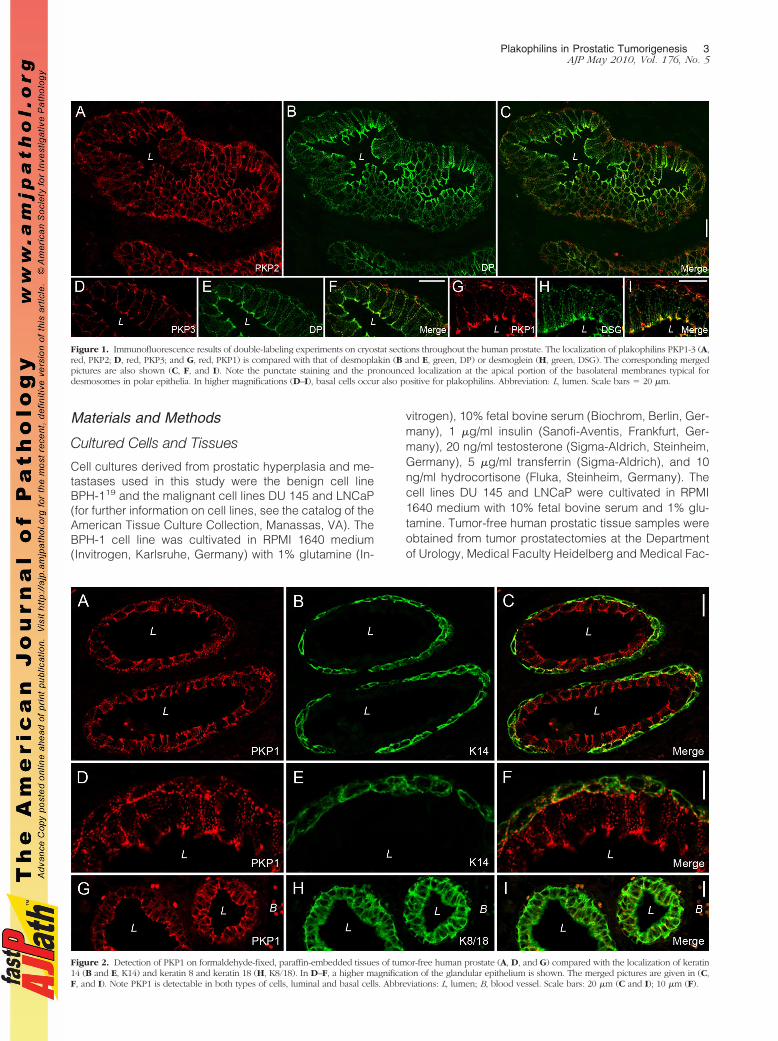
Figure 1. Immunofluorescence results of double-labeling experiments on cryostat sections throughout the human prostate. The localization of plakophilins PKP1-3 (A,red, PKP2; D, red, PKP3; and G, red, PKP1) is compared with that of desmoplakin (B and E, green, DP) or desmoglein (H, green, DSG). The corresponding mergedpictures are also shown (C, F, and I). Note the punctate staining and the pronounced localization at the apical portion of the basolateral membranes typical fordesmosomes in polar epithelia. In higher magnifications (D–I), basal cells occur also positive for plakophilins. Abbreviation: L, lumen. Scale bars � 20 �m.
Figure 2. Detection of PKP1 on formaldehyde-fixed, paraffin-embedded tissues of tumor-free human prostate (A, D, and G) compared with the localization of keratin14 (B and E, K14) and keratin 8 and keratin 18 (H, K8/18). In D–F, a higher magnification of the glandular epithelium is shown. The merged pictures are given in (C,F, and I). Note PKP1 is detectable in both types of cells, luminal and basal cells. Abbreviations: L, lumen; B, blood vessel. Scale bars: 20 �m (C and I); 10 �m (F).
Plakophilins in Prostatic Tumorigenesis 3AJP May 2010, Vol. 176, No. 5
ulty Mannheim, Heidelberg University (Heidelberg, Ger-many), with the permission of the ethics committee of theMedical Faculty. Formaldehyde-fixed, paraffin-embed-ded prostatic adenocarcinomas were provided from theNational Center for Tumor Diseases (Heidelberg, Ger-many) and the Department of Pathology, Medical FacultyMannheim, Heidelberg University.
Patients
A total of 135 tissue samples were analyzed: 44 caseswith low Gleason scores of 3 to 6, 38 cases with anintermediate Gleason score of 7, and 53 cases with highGleason scores of 8 to 10.20 The patients’ age was be-tween 52 and 77 years, and their prostate-specific anti-gen blood levels ranged between 0.5 and 97.6 ng/ml. Atotal of 71 tissue samples were investigated by paraffinmultitissue arrays that consisted of five representativepunches of tumor and three nonneoplastic prostate tis-sues per case (for technical details, see Ref. 21). Withboth, full-sized and microarray samples comparable re-sults were obtained.
Antibodies
The primary antibodies used in this study are listed inTable 1.5,6,8,9,11,13,22–24,39 Details on references and dis-tributors can also be found in Table 1.5,6,8,9,11,13,22–24,39
Secondary antibodies used for immunofluorescence mi-croscopy were species-specific goat antibodies againstIgs of guinea pig, mouse, or rabbit conjugated to Cy3(BioTrend, Cologne, Germany) or Alexa 488 (MoBiTec,Gottingen, Germany). For immunoblot analysis, horse-radish peroxidase-conjugated secondary antibodies
were applied in combination with the enhanced chemilu-minescence system (AceGlow-Solution; PEQLAB Bio-technologie, Erlangen, Germany).
Immunofluorescence Microscopy andImmunoelectron Microscopy
The methods used for single- and double-label antibodyimmunofluorescence microscopy on sections of frozentissues and cultured cells were described in detail else-where.8 For sectioning of paraffin-embedded tissues (3to 5 �m), a microtome (HM 430; Microm, Walldorf, Ger-many) was used. Briefly, sections were deparaffinizedaccording to standard techniques and thereafter pre-treated to retrieve masked antigens (0.05 M Tris-HClbuffer, pH 10.2, 12 minutes, 120°C for detection of PKP1,PKP2, and ARVCF and 0.1 M Tris buffer containing 10%urea, pH 11, 15 minutes, 120°C for detection of PKP3,protein p0071, and protein p120) with a multifunctionalmicrowave processor (RHS-1; Milestone, Sorisole, Italy).Alternatively, the samples were heated in their appropri-ate buffer either with a standard microwave (Panasonic,Hamburg, Germany) at 600 W or a steamer (Braun, Kro-nberg, Germany) for 25 minutes. Sections were thentreated with 0.2% Triton X-100 in PBS for 5 minutes,washed with PBS (twice 5 minutes), blocked with 5% goatserum and 2% milk powder in PBS (10 minutes), and thenprocessed for immunofluorescence microscopy. ForPKP1 and PKP2 staining sections were additionally incu-
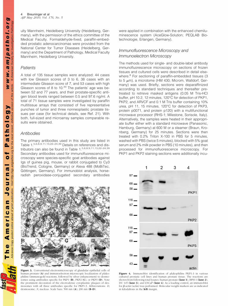
Figure 3. Conventional electronmicroscopy of glandular epithelial cells ofhuman prostate (A) and immunoelectron microscopic localization of plako-philins (immunogold reactions, followed by silver enhancement) to desmo-somes using antibodies specific for PKP1 (B), PKP2 (C), or PKP3 (D). Notethe prominent decoration of the electrodense cytoplasmic plaques of des-mosomes with all three antibodies specific for PKP1-3. Abbreviations: D,desmosome; N, nucleus. Scale bars: 500 nm (A); 200 nm (B–D).
Figure 4. Immunoblot identification of plakophilins PKP1-3 in variouscultured prostatic cell lines and human prostate tissue. The reactions areshown from following total lysates: human prostate (lane 1), BPH-1 (lane 2),DU 145 (lane 3), and LNCaP (lane 4). As a loading control, an immunoblotfor �-actin (actin) was performed. Molecular weight markers are as indicatedin kilodaltons in the left margin.
4 Breuninger et alAJP May 2010, Vol. 176, No. 5

bated before blocking with 2 �g/ml proteinase K (Dako-Cytomation, Hamburg, Germany) for 8 minutes at 37°C,followed by 8 minutes at room temperature. The PKP1-3-specific antibodies were incubated for 4 hours at roomtemperature (PKP1 and PKP2) or overnight at 4°C(PKP3), the secondary antibodies for 1 hour. As controlfor unspecific binding, incubation with primary antibodywas omitted, and the specimens were incubated withsecondary antibody only. Micrographs were taken withan Axiophot microscope (Zeiss, Jena, Germany). Confo-cal laser scanning immunofluorescence microscopy wasdone on a Zeiss LSM 510 UV instrument (Zeiss) as de-scribed before.25 Procedures used for conventional elec-tron microscopy and immunoelectron microscopy wereas described in detail elsewhere.26
Interpretation of Immunfluorescence Signal
A representative section of each tumor sample wasstained with H&E to verify tumor classification and toproof the quality of the tissue sample by light microscopy.The extent of immunofluorescence signal was subclassi-fied as homogeneous (staining of the whole tumor) orinhomogeneous (staining only in defined tumor areas).Immunofluorescence experiments were conducted with anegative control for each tumor sample by omitting pri-mary antibody. In addition, an internal control represent-ing nonneoplastic prostate tissue was included. Depend-ing on the percentage of positive cells, the immunereactions were scored into negative (�5% positive stain-ing), inhomogeneous (5% � positive staining � 90%),and positive (�90% positive staining). Immunofluores-cence evaluation was performed and photographs weretaken with an Axiophot microscope (Zeiss).
Statistical Analyses
Statistical differences between categoric variables weredetermined using Yates corrected �2 test. Correlationsbetween parameters in ordinal scales were analyzed bySpearman rank R test, as provided by the SPSS software(version 2000; SPSS, Chicago, IL). For all analyses, avalue of P � 0.05 was considered significant, unlessotherwise stated.
Gel Electrophoresis and Immunoblotting
Lysates were generated by the addition of Laemmlisample buffer substituted with Benzonase (Merck,Darmstadt, Germany) to cells or tissues. For immuno-blot, proteins decorated with specific primary and sec-ondary antibodies were detected by applying the en-hanced chemiluminescence system to the membranes.Light emission was recorded by a photomultiplier in agel documentation system (ChemiSmart 5000; PEQLABBiotechnologie).
RNA Isolation and Real-Time PCR
RNA from cells was isolated using a RNA isolation kit(RNAeasy; Qiagen, Hilden, Germany). Afterward, a RT-PCRwas performed to transcribe RNA into cDNA (High CapacitycDNA Reverse Transcription kit; Applied Biosystems,Darmstadt, Germany). The obtained cDNA was diluted 1/5for real-time PCR and added to the Power SYBR Green PCR
Figure 5. Occurrence of plakophilins (PKP1, PKP2, and PKP3) in prostaticadenocarcinoma in correlation with Gleason scores (low: Gleason score 3 to6; intermediate: Gleason score 7; high: Gleason score 8 to 10). The percent-ages of negative (white color), inhomogeneous (light gray color), and pos-itive (dark gray color) cases are given.
Table 2. Immunofluorescence Results for the Plakophilins, PKP1-3 in Low, Intermediate, And High Gleason Scores of ProstaticAdenocarcinoma
Tissue samples
Total Low Gleason score Intermediate Gleason score High Gleason score
PKP1Positive 55/135 (41%) 33/44 (75%) 10/38 (26%) 12/53 (23%)Inhomogeneous 68/135 (50%) 5/44 (11%) 26/38 (69%) 37/53 (70%)Negative 12/135 (9%) 6/44 (14%) 2/38 (5%) 4/53 (7%)
PKP2Positive 106/132 (80%) 34/41 (83%) 32/38 (84%) 40/53 (75%)Negative 26/132 (20%) 7/41 (17%) 6/38 (16%) 13/53 (25%)
PKP3Positive 108/132 (82%) 26/41 (63%) 35/38 (92%) 47/53 (89%)Negative 24/132 (18%) 15/41 (37%) 3/38 (8%) 6/53 (11%)
Plakophilins in Prostatic Tumorigenesis 5AJP May 2010, Vol. 176, No. 5
Master Mix (Applied Biosystems). Primers against PKP1(sequence forward: 5�-GACCAGGACAACTCCACGTT-3�,reverse: 5�-CTGCTGGTGGTCCCATAGTT-3�), PKP2 (se-quence forward: 5�-GCAAATGGTTTGCTCGATTT-3�, reverse:5�-GGCTGGTAATCTGCAATGGT-3�), and PKP3 (sequenceforward: 5�-TGATGAGCTTCGCAAAAATG-3�, reverse: 5�-CT-GAGAGGCTGAGCTGAGGT-3�) were used, and the occur-rence of corresponding DNA fragments was analyzed viaStepOnePlus Real-Time PCR System (Applied Biosystems)and controlled by electrophoresis in 1% agarose gels. Hy-poxanthine-guanine phosphoribosyltransferase RNA levelswere used as internal reference to evaluate RNA recoveryand to exclude variations between the different cell clones.
Generation of cDNA Constructs, Transfections,and Generation of Stable Cell Lines
The PKP3-pEGFP-N1 construct27 was cloned into thepLenti6.2/V5-DEST plasmid (Invitrogen) via the pENTR3Cplasmid using restriction sites EcoRI and EcoRV, followed
by Clonase reaction as specified by the manufacturer. As anegative control a pLenti-IRES-EGFP plasmid containingenhanced green fluorescent protein (EGFP) only was used(provided by A. Fischer, CBTM). Additionally, a PKP3-myctag construct was created by replacing the EGFP tag withannealed oligonucleotides coding for the myc tag (5�-CG-GAGCAGAAACTCATCTCAGAAGAGGATCTGTGGT-3� and5�-CTAGATTACAGATCCTCTTCTGAGATGAGTTTCTGCTC-CGGTAC-3�) in the PKP3-pEGFP-N1 vector.
To generate PKP3-specific short hairpin (sh)RNAs, oligo-nucleotides against different PKP3-mRNA targeting siteswere synthesized (shPKP3-9: 5�-GTACCTCGGGACAG-GACGTGAAGATAGTTCAAGAGACTATCTTCACGTCCTG-TCCCTTTTTGGAAA-3� and 5�-AGCTTTTCCAAAAAGGG-ACAGGACGTGAAGATAGTCTCTTGAACTATCTTCACGTC-CTGTCCCGAG-3�; shPKP3-1350: 5�-GTACCTCGGATCC-TGTGGAACCTTTCATTCAAGAGATGAAAGGTTCCACAG-GATCCTTTTTGGA-3�; and 5�-AGCTTTTCCAAAAAGGAT-CCTGTGGAACCTTTCATCTCTTGAATGAAAGGTTCCAC-AGGATCC-3�).
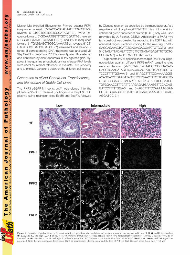
Figure 6. Detection of plakophilins on formaldehyde-fixed, paraffin-embedded tissue of prostatic adenocarcinoma grouped in low (A, D, G, and J), intermediate(B, E, H, and K), and high (C, F, I, and L) Gleason score by immunofluorescence. H&E is shown for a representative example of low (A, Gleason score 3 to 6),intermediate (B, Gleason score 7), and high (C, Gleason score 8 to 10) Gleason score. Immunolocalizations of PKP1 (D–F), PKP2 (G–I), and PKP3 (J–L) arepresented. Note the heterogeneous detection of PKP1 in intermediate Gleason score and the loss of PKP1 in high Gleason score. Scale bars � 50 �m.
6 Breuninger et alAJP May 2010, Vol. 176, No. 5
The annealed oligonucleotides were ligated directlyinto the psiRNA-h7SKZeo plasmid (InvivoGen, Toulouse,France) using the unique Acc65I and HindIII restrictionsites. As a negative control, a shRNA construct againstLuciferase, shLuc, was used (InvivoGen).
The pLenti-PKP3-EGFP plasmid and the pLenti-EGFPplasmid were stably transduced into DU 145 cells ac-cording to the protocol of the manufacturer (Invitrogen),and cells were selected with 10 �g/ml blasticidin (Invitro-gen). For generation of stable PKP3 knockdown cellclones and of the negative control cell clone, shLucif-erase, DU 145 cells were transfected with the corre-sponding shRNA plasmids using Fugene 6 transfectionreagent (Roche, Mannheim, Germany), followed by se-lection with 100 �g/ml Zeocin (Invitrogen). For selectionof plasmid containing cell clones, the expression of EGFPwas analyzed. A transient transfection with the PKP3-mycconstruct was also performed with Fugene 6 transfectionreagent.
Proliferation Assay
DU 145 wild-type, PKP3 knockdown (shPKP3-1350), andPKP3 overexpressing (PKP3-EGFP) cells were seeded in a96-well plate (6 � 102 cells/well). 5-Bromo-2�-deoxyuridine(BrdU) was added 48 hours later. Incorporation of BrdUwas determined after another 24 hours, according to theprotocol of the manufacturer (Cell Proliferation ELISA, BrdU(colorimetric); Roche). Absorbance at 400 nm (referencewavelength, 492 nm) was measured using an ELISA reader(Tecan, Crailsheim, Germany). Statistical differences in pro-liferation between cell culture lines were analyzed usingStudent’s t-test for unpaired experiments.
Results
The Presence of Plakophilins in Normal Prostate
To characterize the occurrence of arm-repeat proteins ofthe p120 subfamily in normal prostate, we systematicallyscreened human tumor-free prostate by immunofluores-cence on cryosections using antibodies specific for pro-tein p120, ARVCF, protein p0071, neurojungin, and pla-kophilins 1-3. Thereby, we noticed a strong staining of theglandular epithelium for all members of the p120 subfam-ily with the exception of neurojungin (data not shown).According to previous reports,2,5,11,13 the occurrence ofall three plakophilins in the glandular epithelium of theprostate, especially of PKP1, was unexpected, and there-fore, we followed this observation in more detail. Wecompared the localization PKP1-3 with that of other des-mosomal plaque proteins (eg, desmoplakin) and desmo-somal transmembrane proteins (eg, desmoglein) andfound for all three plakophilins colocalization with desmo-somal marker proteins (Figure 1). All proteins showed apunctuate staining at cell borders and a pronouncedaccumulation at the apical portion of basolateral mem-branes typical for polar epithelia. The glandular epithe-lium of the prostate consists of luminal cells revealingsecretory activity and stretched basal cells. By careful
inspection at higher magnification, the basal cells wereseen to be labeled by antibodies specific for PKP1-3. Tounequivocally clarify if all three plakophilins occur inbasal cells, we used antibodies specific for keratin 14, amarker for basal cells, and keratin 8 and keratin 18,markers for luminal and basal cells,28 and compared thelocalization with that of PKP1-3. To obtain better structuralpreservation, we used immunofluorescence on formalde-hyde-fixed and paraffin-embedded tumor-free humanprostate samples. Indeed, antibodies against all threeplakophilins convincingly showed a decoration of bothbasal and luminal cells (Figure 2 and Supplemental Fig-ure 1, see http://ajp.amjpathol.org). We also applied immu-noelectron microscopy to verify the observation that allthree plakophilins occur in the glandular epithelium of theprostate at the ultrastructural level (Figure 3). Antibodiesagainst all three plakophilins decorated desmosomalplaques of neighboring epithelial cells in the glandular ep-ithelium of prostate showing the typical appearance of des-mosomes with a cytoplasmic electrodense plaque whereintermediate filaments are inserted.
The Detection of Plakophilins in Cultured CellsDerived from Prostatic Tissue
We compared our results obtained on human prostaticnormal tissue with cultured cells derived from humanprostate. Cell cultures derived from prostatic hyperplasia(eg, BHP-1 cells) and metastases (eg, LNCaP cells iso-lated from lymph node metastases and DU 145 cellsderived from a brain metastasis) were used, and immu-noblotting using antibodies specific for the plakophilins,PKP1-3, was performed. Although all three plakophilinswere detectable in lysates from prostatic tissue and fromthe benign cell line (BPH-1), only PKP3 showed a prom-inent reaction in lysates from both malignant cell lines,LNCaP and DU 145 (Figure 4). In contrast, PKP2 was
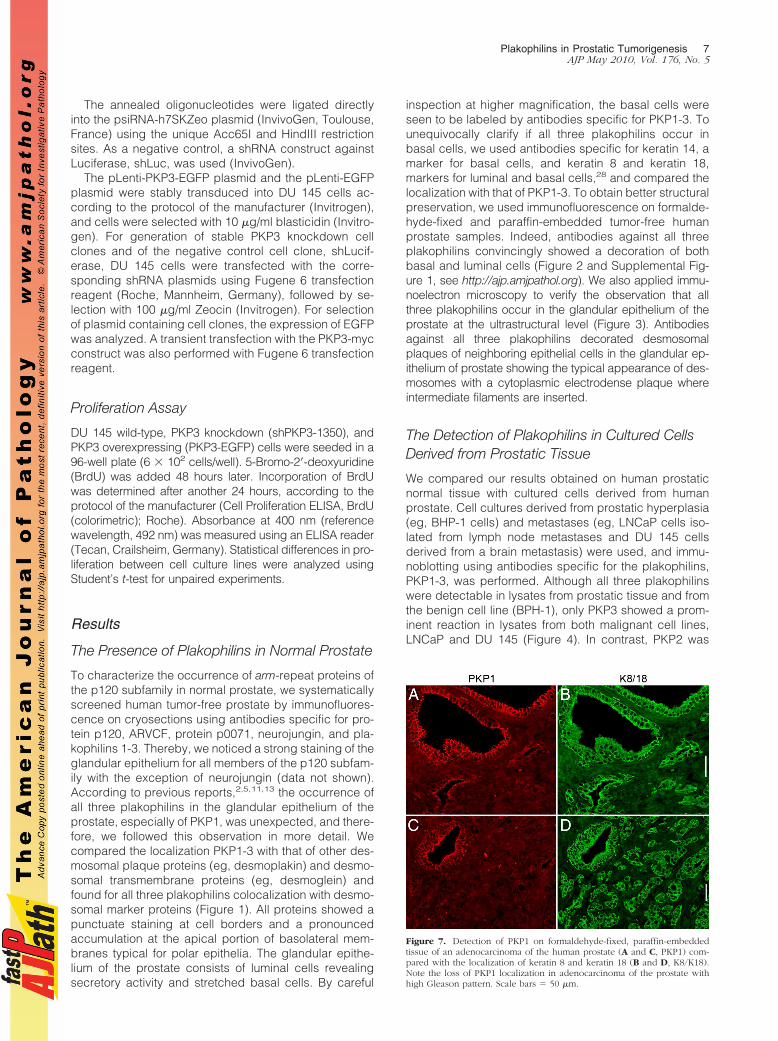
Figure 7. Detection of PKP1 on formaldehyde-fixed, paraffin-embeddedtissue of an adenocarcinoma of the human prostate (A and C, PKP1) com-pared with the localization of keratin 8 and keratin 18 (B and D, K8/K18).Note the loss of PKP1 localization in adenocarcinoma of the prostate withhigh Gleason pattern. Scale bars � 50 �m.
Plakophilins in Prostatic Tumorigenesis 7AJP May 2010, Vol. 176, No. 5
barely traceable in LNCaP and DU 145 cells, and PKP1was not detectable in these cell lines. We verified theseobservations by immunofluorescence microscopy andfound that PKP3 and PKP2 were detectable in desmo-somes in all cell lines, whereas PKP1 was only observedin the benign cell line BPH-1 (data not shown). Moreover,we performed RT-PCR on DU 145 cells and found mRNAexpression of PKP3 (Supplemental Figure 2, see http://ajp.amjpathol.org) and PKP2 but not of PKP1 (data notshown).
The Plakophilins in Adenocarcinomas of theProstate
Because not all three plakophilins were detectable inmalignant prostatic cell lines, we wondered whether theloss of plakophilins correlates with different tumor char-acteristics. Therefore, we systematically analyzed 135prostatic adenocarcinomas with different Gleason scores
(n � 44 with a low Gleason score between 3 and 6; n �38 with an intermediate Gleason score 7; and n � 53 witha high Gleason score 8 to 10) for expression of PKP1-3 byimmunofluorescence. The findings are summarized inTable 2 and Figure 5. In summary, PKP1 expression wasfound less frequently and was generally lower in tumorswith intermediate and high Gleason scores than in tumorswith low Gleason scores, which was statistically highlysignificant (P � 0.00006). In contrast, PKP3 expressionwas significantly more frequent and generally higher inadenocarcinomas with intermediate or high Gleasonscores than in tumors with low Gleason scores, of which37% were completely negative (P � 0.0002). No majordifferences between the single groups were noted withregard to PKP2. There was no statistical correlation be-tween PKP expression and prostate-specific antigen val-ues (data not shown).
Representative immunofluorescence images for thepresence of plakophilins in adenocarcinoma with low,
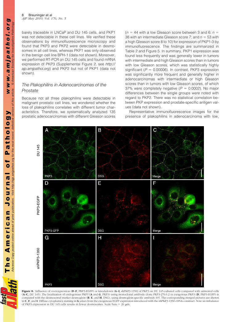
Figure 8. Influence of overexpression (D–F, PKP3-EGFP) or knockdown (G–I, shPKP3-1350) of PKP3 on DU 145-cultured cells compared with untreated cells(A–C, DU 145). The localization of endogenous PKP3 (A and G, PKP3) using monoclonal antibody clone PKP3-270.6.2 or exogenous PKP3 (D, PKP3-EGFP) iscompared with the desmosomal marker desmoglein (B, E, and H, DSG), using desmoglein-specific antibody rb5. The corresponding merged pictures are shownin C, F, and I. Diffuse cytoplamics staining in G arises from the exogenous EGFP expression introduced with the shPKP3-1350 cDNA construct. Note an imbalanceof PKP3 expression in DU 145 cells results in fewer desmosomes. Scale bars � 20 �m.
8 Breuninger et alAJP May 2010, Vol. 176, No. 5
intermediate, and high Gleason score are presented inFigure 6. In tumors with a low Gleason score, PKP1 andPKP2 were localized between neighboring cells, andPKP3 was additionally noticed in the cytoplasm of glan-dular epithelial cells. In tumors with intermediate Gleasonscores, PKP1 occurred only in larger, more regularshaped glands, whereas in smaller glands with a ten-dency of the epithelial cells to spread in the surroundingstroma, the PKP1 signal was lost, whereas PKP2 andPKP3 were still detectable. This observation was evenmore prominent in carcinomas with a high Gleason score:colocalization of PKP1 with keratin 8/18, as an epithelialcell marker, revealed large tumor areas negative forPKP1 (Figure 7).
Effects of PKP3 Overexpression andKnockdown on DU 145 Cells
To analyze the potential functional consequences ofoverexpression of PKP3 in more aggressive tumors, wegenerated stable cell lines with modified expression ofPKP3. We used the malignant cell line DU 145 and intro-duced cDNA constructs yielding either the overexpres-sion of the fusion protein PKP3-EGFP (“gain of function”)or reduced expression (“loss of function”) through ashRNA interference approach. To knock down the ex-pression of PKP3, two different target sequences werechosen yielding the cell clones shPKP3-9 and shPKP3-1350 with different RNA and protein levels of PKP3 (Sup-plemental Figure 2, see http://ajp.amjpathol.org). In con-trast, a cell clone stably expressing shRNA specific forluciferase showed RNA and protein levels comparablewith untreated DU 145 cells. The cell clone, shPKP3-1350,with the higher knockdown efficiency was used for furtheranalyses. When PKP3-EGFP was expressed in DU 145cells, this protein was detected at cell-cell contacts in des-mosomes as shown by colocalization with desmosomalproteins (eg, desmoglein) but also in a decent amount in thecytoplasm (Figure 8). In addition, a high portion of uncou-pled cells was observed in stably transfected cells wherethe portion of cytoplasmic PKP3-EGFP was even higher. Asa control, a PKP3-myc construct was introduced in DU 145cells, and here, PKP3 fused to a smaller tag also occurredin desmosomes and in the cytoplasm (Supplemental Figure3, see http://ajp.amjpathol.org). In cell lines with knockdownof PKP3, the distribution of desmosomal proteins (eg, des-moglein) was also disturbed (Figure 8). Desmoglein wasonly rarely detectable in desmosomes and instead mostlylocalized in the cytoplasm in different sized structures.Taken together, our findings clearly showed that overex-pression and knockdown of PKP3 interfered with the forma-tion of desmosomes and thus with cell-cell contacts.
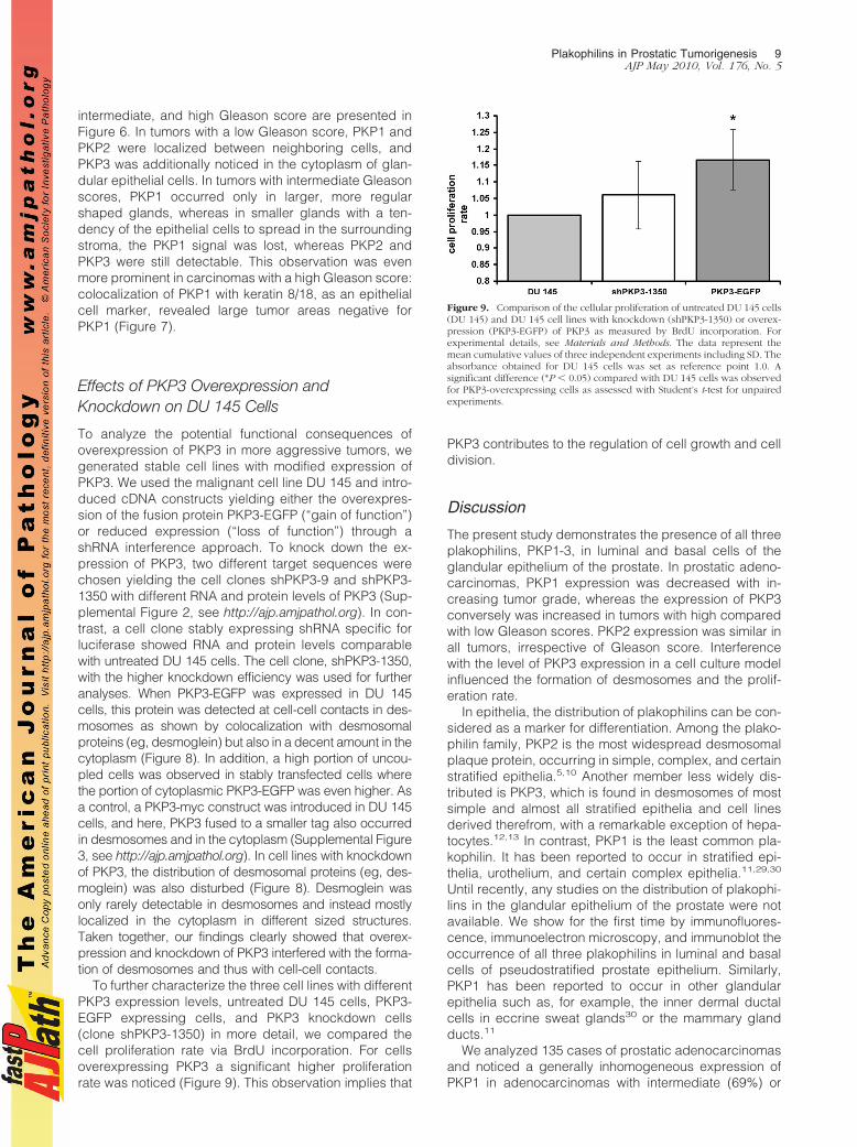
To further characterize the three cell lines with differentPKP3 expression levels, untreated DU 145 cells, PKP3-EGFP expressing cells, and PKP3 knockdown cells(clone shPKP3-1350) in more detail, we compared thecell proliferation rate via BrdU incorporation. For cellsoverexpressing PKP3 a significant higher proliferationrate was noticed (Figure 9). This observation implies that
PKP3 contributes to the regulation of cell growth and celldivision.
Discussion
The present study demonstrates the presence of all threeplakophilins, PKP1-3, in luminal and basal cells of theglandular epithelium of the prostate. In prostatic adeno-carcinomas, PKP1 expression was decreased with in-creasing tumor grade, whereas the expression of PKP3conversely was increased in tumors with high comparedwith low Gleason scores. PKP2 expression was similar inall tumors, irrespective of Gleason score. Interferencewith the level of PKP3 expression in a cell culture modelinfluenced the formation of desmosomes and the prolif-eration rate.
In epithelia, the distribution of plakophilins can be con-sidered as a marker for differentiation. Among the plako-philin family, PKP2 is the most widespread desmosomalplaque protein, occurring in simple, complex, and certainstratified epithelia.5,10 Another member less widely dis-tributed is PKP3, which is found in desmosomes of mostsimple and almost all stratified epithelia and cell linesderived therefrom, with a remarkable exception of hepa-tocytes.12,13 In contrast, PKP1 is the least common pla-kophilin. It has been reported to occur in stratified epi-thelia, urothelium, and certain complex epithelia.11,29,30
Until recently, any studies on the distribution of plakophi-lins in the glandular epithelium of the prostate were notavailable. We show for the first time by immunofluores-cence, immunoelectron microscopy, and immunoblot theoccurrence of all three plakophilins in luminal and basalcells of pseudostratified prostate epithelium. Similarly,PKP1 has been reported to occur in other glandularepithelia such as, for example, the inner dermal ductalcells in eccrine sweat glands30 or the mammary glandducts.11
We analyzed 135 cases of prostatic adenocarcinomasand noticed a generally inhomogeneous expression ofPKP1 in adenocarcinomas with intermediate (69%) or
Figure 9. Comparison of the cellular proliferation of untreated DU 145 cells(DU 145) and DU 145 cell lines with knockdown (shPKP3-1350) or overex-pression (PKP3-EGFP) of PKP3 as measured by BrdU incorporation. Forexperimental details, see Materials and Methods. The data represent themean cumulative values of three independent experiments including SD. Theabsorbance obtained for DU 145 cells was set as reference point 1.0. Asignificant difference (*P � 0.05) compared with DU 145 cells was observedfor PKP3-overexpressing cells as assessed with Student’s t-test for unpairedexperiments.
Plakophilins in Prostatic Tumorigenesis 9AJP May 2010, Vol. 176, No. 5
high (70%) Gleason score. In tumor areas with high Glea-son pattern, PKP1 was no longer detectable, whereas theexpression of the two other PKPs, PKP2 and PKP3, waseither unchanged (PKP2) or even increased (PKP3). Oneprevious study31 reported the presence of all three pla-kophilins in prostate carcinomas but did not address theassociation with Gleason grading. In other human tumors,the expression of plakophilins seems to differ between dif-ferent entities. In oropharyngeal tumors, a reduction or anabsence of PKP1 and PKP3 in desmosomes has beennoticed.17 In contrast, non-small cell lung carcinomas werereported to show up-regulation of PKP3.16 An inhomoge-neous expression of PKP1 in tumors has been described inseveral reports,18,30,32,33 and it has been suggested thatPKP1 functions as a differentiation marker for carcinomas.30
In glandular epithelium, the detection of PKP1 might be avaluable indicator of well-differentiated glandular structures.Differences in the occurrence of PKPs (eg, loss, up-regula-tion, or inhomogeneous expression) could reflect tumor pro-gression, as already noticed for protein p120.15
By using DU 145 cultured cancer cells as a cell culturemodel, deduced from a brain metastasis of a prostaticadenocarcinoma, we interfered with the expression levelof PKP3. Both the knockdown (loss of function) and theoverexpression (gain of function) resulted in formation offewer desmosomes, and the overexpression led to a higherproliferation rate. The essential importance of all three PKPshas been shown in vitro in cultured cells via knockdownapproaches.18,34,35 The down-regulation of PKP1 andPKP3 resulted in a decreased formation of desmosomesand increased cell motility and invasion.18,35 PKPs do notonly contribute to formation and maintenance of desmo-somes22,34,36 but also function in the cyto- and nucleo-plasm.27,37 PKP2 has been found in nuclear particles con-taining RNA polymerase III.37 Moreover, cytoplasmic PKP1and PKP3 are components of RNA-containing particles andcan be recruited to stress granules.27 The overexpressionof PKP3 led to a higher portion of PKP3 in the cytoplasmicpool and thereby may regulate cell growth and cell divisionand effect the formation of desmosomes. A deficiency inone of the PKPs has a tremendous impact on epithelialintegrity in animals and humans. Ablation of PKP1 results inskin fragility and hypohidrotic ectodermal dysplasia.38
Knockout PKP2 mice exhibit lethal alterations in heart mor-phogenesis.39 PKP3-deficient mice develop hair coat ab-normalities and are prone to cutaneous inflammation.40
Clearly, future studies will have to reveal the function ofPKP1 and 3 in the cytoplasm and how exchange of thejunctional and nonjunctional form is regulated in healthyand diseased states such as, for example, cancer.
Acknowledgments
We thank Prof. Dr. Werner W. Franke (Helmholtz Group forCell Biology, German Cancer Research Center) and Prof.Dr. Hellmut G. Augustin (Joint Research Division VascularBiology of the Medical Faculty Mannheim for continuousand generous support).
References
1. Hartsock A, Nelson WJ: Adherens and tight junctions: structure,function and connections to the actin cytoskeleton. Biochim BiophysActa 2008, 1778:660–669
2. Getsios S, Huen AC, Green KJ: Working out the strength and flexibilityof desmosomes. Nat Rev Mol Cell Biol 2004, 5:271–281
3. Hatzfeld M: The p120 family of cell adhesion molecules. Eur Cell Biol2005, 84:205–214
4. Schmidt A, Jager S: Plakophilins—hard work in the desmosome,recreation in the nucleus? Eur J Cell Biol 2005, 84:189–204
5. Mertens C, Kuhn C, Franke WW: Plakophilins 2a and 2b constitutiveproteins of dual location in the karyoplasm and the desmosomalplaque. J Cell Biol 1996, 13:1009–1025
6. Schmidt A, Langbein L, Rode M, Pratzel S, Zimbelmann R, FrankeWW: Plakophilins 1a and 1b: widespread nuclear proteins recruited inspecific epithelial cells as desmosmal plaque components. Cell Tis-sue Res 1997, 290:481–499
7. Nelson WJ, Nusse R: Convergence of Wnt, beta-catenin, and cad-herin pathways. Science 2004, 303:1483–1487
8. Hofmann I, Kuhn C, Franke WW: Protein p0071, a major plaqueprotein of non-desmosomal adhering junctions, is a selective cell typemarker. Cell Tissue Res 2008, 334:381–399
9. Hofmann I, Schlechter T, Kuhn C, Hergt M, Franke WW: Proteinp0071, an armadillo plaque protein that characterizes a specificsubtype of adherens junctions. J Cell Sci 2009, 122:21–24
10. Mertens C, Kuhn C, Moll R, Schwetlick I, Franke WW: Desmosomalplakophilin 2 as a differentiation marker in normal and malignanttissues. Differentiation 1999, 64:277–290
11. Heid HW, Schmidt A, Zimbelmann R, Schafer S, Winter-SimanowskiS, Stumpp S, Keith M, Figge U, Schnolzer M, Franke WW: Celltype-specific desmosomal plaque proteins of the plakoglobin family:plakophilin 1 (band 6 protein). Differentiation 1994, 58:113–131
12. Bonne S, van Hengel J, Nollet F, Kools P, van Roy F: Plakophilin-3, anovel armadillo-like protein present in nuclei and desmosomes ofepithelial cells. J Cell Sci 1999, 112:2265–2276
13. Schmidt A, Langbein L, Pratzel S, Rode M, Rackwitz H-R, Franke WW:Plakophilin 3—a novel cell-type-specific desmosomal plaque protein.Differentiation 1999, 64:291–306
14. Korinek V, Barker N, Morin PJ, van Wichen D, de Weger R, KinzlerKW, Vogelstein B, Clevers H: Constitutive transcriptional activation bya �-catenin-Tcf complex in APC�/� colon carcinoma. Science 1997,275:1784–1787
15. van Hengel J, van Roy F: Diverse functions of p120ctn in tumors.Biochem Biophys Acta 2007, 1773:78–88
16. Furukawa C, Daigo Y, Ishikawa N, Kato T, Ito T, Tsuchiya E, Sone S,Nakamura Y: Plakophilin 3 oncogene as prognostic marker and ther-apeutic target for lung cancer. Cancer Res 2005, 65:7102–7110
17. Papagerakis S, Shabana A-H, Depondt J, Gehanno P, Forest N:Immunohistochemical localization of plakophilins (PKP1. PKP2, PKP3and p0071) in primary oropharyngeal tumors: correlation with clinicalparameters. Hum Pathol 2003, 34:565–572
18. Sobolik-Delmaire T, Katafiasz D, Keim SA, Mahoney MG, Wahl JK III:Decreased plakophilin-1 expression promotes increased motility inhead and neck squamous cell carcinoma cells. Cell Commun Adhe-sion 2007, 14:99–109
19. Hayward SW, Dahiya R, Cunha GR, Bartek J, Deshpande N, NarayanP: Establishment and characterization of an immortalized but non-transformed human prostate epithelial cell line: bPH-1. In Vitro CellDev Biol Anim 1995, 31:14–24
20. Epstein JI, Allsbrook WC Jr, Amin MB, Egevad LL, ISUP GradingCommittee: The 2005 International Society of Urological Pathology(ISUP) Consensus Conference on Gleason Grading of Prostatic Car-cinoma. Am J Surg Pathol 2005, 29:1228–1242
21. Zettl A, Meister S, Katzenberger T, Kalla J, Ott MM, Muller-HermelinkH-K, Ott G: Immunohistochemical analysis of B cell lymphoma usingtissue microarrays identifies particular phenotypic profiles of B celllymphomas. Histopathology 2003, 432:209–219
22. Koeser J, Troyanovsky SM, Grund C, Franke WW: De novo formationof desmosomes in cultured cells upon transfection of genes encodingspecific desmosomal components. Exp Cell Res 2003, 285:114–130
23. Walter B, Schlechter T, Hergt M, Berger I, Hofmann I: Differentialexpression pattern of protein ARVCF in nephron segments of humanand mouse kidney. Histochem Cell Biol 2008, 130:943–956
10 Breuninger et alAJP May 2010, Vol. 176, No. 5

24. Paffenholz R, Kuhn C, Grund C, Stehr S, Franke WW: The arm-repeatprotein NPRAP (neurojungin) is a constituent of the plaques of theouter limiting zone in the retina, defining a novel type of adheringjunction. Exp Cell Res 1999, 250:452–464
25. Hofmann I, Schnolzer M, Kaufmann I, Franke WW: Symplekin, a consti-tutive protein of karyo- and cytoplasmic particles involved in mRNAbiogenesis in Xenopus laevis oocytes. Mol Biol Cell 2002, 13:1665–1676
26. Peitsch WK, Hofmann I, Pratzel S, Grund C, Kuhn C, Moll I, LangbeinL, Franke WW: Drebrin particles: components in the ensemble regu-lating actin dynamics of lamellipodia and filopodia. Eur J Cell Biol2001, 80:567–579
27. Hofmann I, Casella M, Schnolzer M, Schlechter T, Spring H, FrankeWW: Identification of the junctional plaque protein plakophilin 3 incytoplasmic particles containing RNA-binding proteins and the re-cruitment of plakophilins 1 and 3 to stress granules. Mol Biol Cell2006, 17:1388–1398
28. van Leenders GJLH, Schalken JA: Epithelial cell differentiation in thehuman prostate epithelium: implications for the pathogenesis andtherapy of prostate cancer. Crit Rev Oncol Hematol 2003, 46:S3–S10
29. Kapprell HP, Owaribe K, Franke WW: Identification of a basic proteinof Mr 75,000 as an accessory desmosomal plaque protein in stratifiedand complex epithelia. J Cell Biol 1988, 106:1679–1691
30. Moll I, Kurzen H, Langbein L, Franke WW: The distribution of thedesmosomal protein, Plakophilin 1, in human skin and skin tumors.J Invest Dermatol 1997, 139–146
31. Schwarz J, Ayim A, Schmidt A, Jager S, Koch S, Baumann R, DunneAA, Moll R: Differential expression of desmosomal plakophilins invarious types of carcinomas: correlation with cell type and differen-tiation. Hum Pathol 2006, 37:613–622
32. Kurzen H, Munzing I, Hartschuh W: Expression of desmosomal pro-teins in squamous cell carcinomas of the skin. J Cutan Pathol 2003,30:621–630
33. Schmidt-Graeff A, Koeninger A, Olschewski M, Haxelmans S,Nitschke R, Bocahton-Piallat M-L, Lifschitz-Mercer B, Gabbiani G,Langbein L, Czernobilsky B: The Ki67� proliferation index correlateswith increased retinol-binding protein-1 and the coordinated loss ofplakophilin-1 and desmoplakin during progression of cervical squa-mous lesions. Histopathology 2007, 51:87–97
34. Bass-Zubeck AE, Hobbs RP, Amargo EV, Garcia NJ, Hsieh SN, ChenX, Wahl III JK, Denning MF, Green KJ: Plakophilin 2: a critical scaffoldfor PKC� that regulates intercellular assembly. J Cell Biol 2008,181:605–613
35. Kundu ST, Gosave P, Khapare N, Patel R, Hosing AS, Maru GB, IngleA, DeCaprio JA, Dalal SN: Plakophilin 3 downregulation leads to adecrease in cell adhesion and promotes metastasis. Int J Cancer2008, 123:2303–2314
36. Wahl III JK: A role for plakophilin-1 in the initiation of desmosomeassembly. J Cell Biochem 2005, 96:390–403
37. Mertens C, Hofmann I, Wang Z, Teichmann M, Sepehri Chong S,Schnolzer M, Franke WW: Nuclear particles containing RNA polymer-ase III complexes associated with junctional plaque protein plakophi-lin 2. Proc Natl Acad Sci USA 2001, 98:7795–7800
38. McGrath JA, Hoeger PH, Christiano AM, McMillan JR, Mellerio JE,Ashton GHS, Dopping-Hepenstal PJC, Lake BD, Leigh IM, Harper JI,Eady RAJ: Skin fragility and hypohidroitic ectodermal dysplasia resultingfrom ablation of plakophilin 1. Br J Dermatol 1999, 140:297–307
39. Grossmann KS, Grund C, Huelsken J, Behrend M, Erdmann B,Franke WW, Birchmeier W: Requirement of plakophilin 2 for heartmorphogenesis and cardiac junction formation. J Cell Biol 2004,167:149–160
40. Sklyarova T, Bonne S, D’hooge P, Denecker G, Gossens S, De RyckeR, Borgonie G, Bosl M, van Roy F, van Hengel J: Plakophilin-3-deficient mice develop hair coat abnormalities and are prone tocutaneous inflammation. J Invest Dermatol 2007, 128:1375–1385
Plakophilins in Prostatic Tumorigenesis 11AJP May 2010, Vol. 176, No. 5