Recurrent gene fusions in prostate cancer
29
Recurrent Gene Fusions in Prostate Cancer Chandan Kumar-Sinha 1,3 , Scott A. Tomlins 1,3 , and Arul M. Chinnaiyan 1,2,3,4,5,* 1 Michigan Center for Translational Pathology, University of Michigan Medical School, Ann Arbor, Michigan 48109 2 Howard Hughes Medical Institute, University of Michigan Medical School, Ann Arbor, Michigan 48109 3 Department of Pathology, University of Michigan Medical School, Ann Arbor, Michigan 48109 4 Department of Urology, University of Michigan Medical School, Ann Arbor, Michigan 48109 5 The Comprehensive Cancer Center, University of Michigan Medical School, Ann Arbor, Michigan 48109 Abstract The discovery of recurrent gene fusions in a majority of prostate cancers has important clinical and biological implications in the study of common epithelial tumors. Gene fusion and chromosomal rearrangements were previously thought to be the primary oncogenic mechanism of hematological malignancies and sarcomas. The prostate cancer gene fusions that have been identified thus far are characterized by 5’ genomic regulatory elements, most commonly controlled by androgen, fused to members of the ETS family of transcription factors, leading to the over-expression of oncogenic transcription factors. ETS gene fusions likely define a distinct class of prostate cancer which may have a bearing on diagnosis, prognosis and rational therapeutic targeting. Introduction Prostate cancer is one of the most prevalent malignancies affecting men worldwide, and is the most frequent cancer among American men with an estimated incidence of approximately 220,000 (29% of all cancers in men) and a mortality estimated to be over 27,000 (9% of all male cancer deaths) in 2007 1 . An array of treatment modalities are available, including active surveillance, prostatectomy, radiation therapy and androgen ablation therapy, all influenced *Correspondence to A.M.C.: E-mail: [email protected]. Competing Interest Statement The University of Michigan has filed for a patent on the detection of gene fusions in prostate cancer, on which S.A.T. and A.M.C. are co-inventors. The diagnostic field of use has been licensed to GenProbe Inc. GenProbe has had no role in the design or experimentation of this study, nor has it participated in the writing of the manuscript. Oncomine and the MCM are freely available to the academic community. The commercial rights to Oncomine and MCM have been licensed to Compendia Bioscience, which A.M.C. co-founded. A summary of the published literature on the characterization of recurrent gene fusions in prostate cancer. The columns in order from left to right detail the gene fusion, sample cohort the data is derived from, the frequency of the gene fusion in the sample setobserved gene fusion/ total number of samples examined (% gene fusion in the sample cohort), the assay used for detection and characterization of the gene fusions, and the reference. Databases Mitelman Database: http://cgap.nci.nih.gov/Chromosomes/Mitelman Oncomine: http://www.oncomine.org/ Atlas of Genetics and Cytogenetics: http://atlasgeneticsoncology.org/index.html Entrez gene: http://www.ncbi.nlm.nih.gov/sites/entrez?db=gene COPA package in R: http://www.bioconductor.org COPA package integrated into SAM: http://www-stat.stanford.edu/~tibs/SAM NIH Public Access Author Manuscript Nat Rev Cancer. Author manuscript; available in PMC 2009 July 16. Published in final edited form as: Nat Rev Cancer. 2008 July ; 8(7): 497–511. doi:10.1038/nrc2402. NIH-PA Author Manuscript NIH-PA Author Manuscript NIH-PA Author Manuscript
-
Upload
independent -
Category
Documents
-
view
1 -
download
0
Transcript of Recurrent gene fusions in prostate cancer
Recurrent Gene Fusions in Prostate Cancer
Chandan Kumar-Sinha1,3, Scott A. Tomlins1,3, and Arul M. Chinnaiyan1,2,3,4,5,*1Michigan Center for Translational Pathology, University of Michigan Medical School, Ann Arbor,Michigan 481092Howard Hughes Medical Institute, University of Michigan Medical School, Ann Arbor, Michigan481093Department of Pathology, University of Michigan Medical School, Ann Arbor, Michigan 481094Department of Urology, University of Michigan Medical School, Ann Arbor, Michigan 481095The Comprehensive Cancer Center, University of Michigan Medical School, Ann Arbor, Michigan48109
AbstractThe discovery of recurrent gene fusions in a majority of prostate cancers has important clinical andbiological implications in the study of common epithelial tumors. Gene fusion and chromosomalrearrangements were previously thought to be the primary oncogenic mechanism of hematologicalmalignancies and sarcomas. The prostate cancer gene fusions that have been identified thus far arecharacterized by 5’ genomic regulatory elements, most commonly controlled by androgen, fused tomembers of the ETS family of transcription factors, leading to the over-expression of oncogenictranscription factors. ETS gene fusions likely define a distinct class of prostate cancer which mayhave a bearing on diagnosis, prognosis and rational therapeutic targeting.
IntroductionProstate cancer is one of the most prevalent malignancies affecting men worldwide, and is themost frequent cancer among American men with an estimated incidence of approximately220,000 (29% of all cancers in men) and a mortality estimated to be over 27,000 (9% of allmale cancer deaths) in 20071. An array of treatment modalities are available, including activesurveillance, prostatectomy, radiation therapy and androgen ablation therapy, all influenced
*Correspondence to A.M.C.: E-mail: [email protected] Interest StatementThe University of Michigan has filed for a patent on the detection of gene fusions in prostate cancer, on which S.A.T. and A.M.C. areco-inventors. The diagnostic field of use has been licensed to GenProbe Inc. GenProbe has had no role in the design or experimentationof this study, nor has it participated in the writing of the manuscript. Oncomine and the MCM are freely available to the academiccommunity. The commercial rights to Oncomine and MCM have been licensed to Compendia Bioscience, which A.M.C. co-founded.A summary of the published literature on the characterization of recurrent gene fusions in prostate cancer. The columns in order fromleft to right detail the gene fusion, sample cohort the data is derived from, the frequency of the gene fusion in the sample setobservedgene fusion/ total number of samples examined (% gene fusion in the sample cohort), the assay used for detection and characterizationof the gene fusions, and the reference.DatabasesMitelman Database: http://cgap.nci.nih.gov/Chromosomes/MitelmanOncomine: http://www.oncomine.org/Atlas of Genetics and Cytogenetics: http://atlasgeneticsoncology.org/index.htmlEntrez gene: http://www.ncbi.nlm.nih.gov/sites/entrez?db=geneCOPA package in R: http://www.bioconductor.orgCOPA package integrated into SAM: http://www-stat.stanford.edu/~tibs/SAM
NIH Public AccessAuthor ManuscriptNat Rev Cancer. Author manuscript; available in PMC 2009 July 16.
Published in final edited form as:Nat Rev Cancer. 2008 July ; 8(7): 497–511. doi:10.1038/nrc2402.
NIH
-PA Author Manuscript
NIH
-PA Author Manuscript
NIH
-PA Author Manuscript

by the use of serum prostate specific antigen (PSA) levels2. Even as clinically localized prostatecancer has become highly curable the overall death toll remains high due to recurrence of“cured” cases and progression to hormone refractory metastatic disease, which remainsuncurable3. Conversely, nonspecific PSA tests result in a large number of false positives forprostate cancer, leading to a faux-cancer burden and repeated biopsies4. More specificdiagnostic modalities, prognostic indicators of progression and a better understanding ofprostate cancer biology for treatment of hormone refractory disease are high priorities inprostate cancer research.
Approximately two years ago, our group identified recurrent genomic rearrangements inprostate cancer resulting in the fusion of the 5’ untranslated end of TMPRSS2 (a prostatespecific, androgen responsive gene) to ETS family genes (oncogenic transcription factors)5.The original findings have been rapidly corroborated by several independent groups worldwideand now the focus is on the functional and clinical correlates of prostate cancer in the contextof the gene fusions. Emerging experimental evidence suggests that these fusions are keymolecular entities driving the development and progression of a unique class of prostatecancers, providing potential avenues for targeted therapy, similar to the BCR-ABL1 gene fusionin chronic myeloid leukemia (CML).
Gene fusions resulting from chromosomal rearrangements represent the most prevalent formof genetic alterations known in cancers6 and, as exemplified by the archetype gene fusionBCR-AbL1 in CML7, 8 they can serve as ideal diagnostic markers9–11, provide insight intotumor biology12, and most importantly serve as specific therapeutic targets13, 14. Intriguingly,while numerous gene fusions have been described in rare hematological malignancies and evenrarer bone and soft tissue sarcomas15, they are much rarer among epithelial cancers. Genefusions described among epithelial cancers so far have included RET-NTRK1 fusions inpapillary thyroid carcinoma, PAX8-PPARG in follicular thyroid carcinoma, MECT1-MAML2 in mucoepidermoid carcinoma, the TFE3-TFEB in kidney carcinomas, and BRD4-NUT in midline carcinomas etc (reviewed16). Remarkably, recurrent gene fusions have notpreviously been detected in the most prevalent carcinomas including prostate, breast (with theexception of rare, secretory breast cancers), lung, gastrointestinal and gynecologic tumors17,despite compelling arguments that predict their occurence15, 18, 19. The absence of genefusions in common solid tumors has been attributed to the technical difficulties associated withtheir cytogenetic analysis. Also, epithelial cancers are thought to be clonally heterogeneous,with causal chromosomal aberrations co-habiting the tissues with clinically irrelevant ones.While cytogenetic analyses help identify ‘physical’ genomic aberrations, recurrent genefusions in prostate cancer were identified based on gene expression data, bypassing thetechnical limitations of cytogenetics in solid cancers. This strategy led to the identification ofrecurrent gene fusions in common solid cancers, close to 50 years after the discovery ofPhiladelphia chromosome in 1960s.
In this review, we appraise recent progress in the characterization of recurrent gene fusions inprostate cancer. We will highlight the clinical implications of new discoveries, emergingcontroversies and challenges, as well as future research directions. In addition to serving aspotential diagnostic/prognostic markers and therapeutic candidates for a unique class ofprostate cancer, the discovery of recurrent rearrangements in prostate cancer affirms a moregeneralized role for similar chromosomal aberrations in other common epithelial cancers.
Discovering gene fusions with bioinformaticsCancers are, for the most part, phenotypically and molecularly heterogeneous entities. Thus,characterization of distinct molecular classes with an overarching influence of a single geneor two is clinically and therapeutically significant. For example, in one-quarter to one-third of
Kumar-Sinha et al. Page 2
Nat Rev Cancer. Author manuscript; available in PMC 2009 July 16.
NIH
-PA Author Manuscript
NIH
-PA Author Manuscript
NIH
-PA Author Manuscript
all breast cancer cases, amplification and over-expression of the oncogene HER2 defines anaggressive class that is more likely to metastasize, develop hormone resistance, and respondsignificantly to HER2 targeted therapy. Likewise, Philadelphia chromosome positive chronicmyelogenous leukemia (CML) typifies 10% of all leukemia cases, where the underlyingaberration is a BCR-ABL1 gene fusion that becomes the focal point of diagnosis, classification,prognostication, therapy, as well as follow up and recurrence monitoring (BOX 1). Other well-defined cancer classes include less than 5% of all breast cancers harboring BRCA1 orBRCA2 mutations20, 6% of colon cancers with microsatellite instability or germline mutationscharacterizing specific clinical classes such as hereditary non-polyposis colorectal cancer(HNPCC), or familial adenomatus polyposis (FAP)21, 22 and 10% of non small cell lungcancers harboring sensitizing mutations in EGFR that respond to the EGFR targeting druggefitinib23.
We initiated a systematic identification of candidate oncogenes activated by chromosomalrearrangements or high level copy number changes based on gene expression signatures usingan unconventional analytical approach. Cancer gene expression data sets were queried forgenes that are highly over-expressed in a subset of samples rather than focusing on those thatare widely shared in all samples5. To identify such “outlier” genes, we applied a datatransformation algorithm, cancer outlier profile analysis (COPA), to all of the 132 gene-expression data sets (comprising >10,000 microarray experiments from various cancers)available in Oncomine (www.oncomine.org24–27), our gene expression compendium (FIG.1). COPA transformation effectively compresses typical biomarker profiles characterized bya general overexpression of genes in all cancer samples, while accentuating ‘outlier gene’profiles, characterized by general low expression with marked overexpression in a fraction ofthe samples. Prioritized outlier genes identified by our systematic approach included severalwell-known cancer genes involved in recurrent chromosomal aberrations or high level copynumber changes associated with their specific cancers. Surprisingly, two genes known to beinvolved in gene fusions in Ewing’s sarcoma28, 29, namely ERG (ETS-related gene) andETV1 (ETS variant gene 1), were scored as high ranking outliers in several independentprostate-cancer profiling studies. Akin to Ewing’s sarcoma, where Ewing sarcoma breakpointregion 1 (EWSR1)–ERG and EWSR1–ETV1 fusion genes are mutually exclusive in differentcases28, ERG and ETV1 overexpression were mutually exclusive in prostate cancer. Thissuggested that elevated expression of ERG/ETV1 may be a key molecular event in a subset ofprostate cancers.
Analysis of prostate cancer samples that overexpress ERG and ETV1 (“outliers”) using exon-walking quantitative PCR revealed overexpression of only their 3’ regions, suggestive of agenetic rearrangement (FIG. 1). This lead us to characterize the 5’ ends of ERG and ETV1transcripts using a 5’ RNA-ligase-mediated rapid amplification of cDNA ends (RACE)methodology (FIG 1). We discovered that the 5’ ends of ERG and ETV1 are replaced with the5’ untranslated region of a prostate-specific, androgen responsive, transmembrane serineprotease gene (TMPRSS2) (21q22.2) in the outlier cases5. Fusion transcripts were confirmedby quantitative PCR, and rearrangments at the genomic loci were then assessed in multiplesamples by fluorescence in situ hybridization (FISH) on tissue microarrays (FIG. 1). FISHdemonstrated that a majority of prostate cancer samples harbor these aberrations. It bears notingthat the TMPRSS2-ETS fusions are analogous to the IgH-myc30, 31 and IgH-bcl232 genefusions in lymphomas. In these cases, no fusion protein is generated but there is massiveoverexpression of an oncogenic factor under the control of a subverted promoter element12.
Interestingly, while almost all ERG overexpressing tumors were found to harborTMPRSS2:ERG fusion, subsequent investigations identified fewer TMPRSS2:ETV1 cases thanexpected based on the frequency of ETV1 outlier expression. This paradox was investigatedwith the use of 5’ RACE (FIG. 1) on ETV1 outlier samples, leading to the characterization of
Kumar-Sinha et al. Page 3
Nat Rev Cancer. Author manuscript; available in PMC 2009 July 16.
NIH
-PA Author Manuscript
NIH
-PA Author Manuscript
NIH
-PA Author Manuscript
multiple upstream fusion partners of ETV1. These differed in their prostate specificity andandrogen responsiveness, presenting a novel level of complexity in the molecular biology ofprostate cancers33. The upstream partners of ETV1 identified so far include: a prostate specificandrogen-induced gene (SLC45A3) and an endogenous retroviral element (HERV-K_22q11.23) that are functionally analogous to TMPRSS2; a prostate-specific, but androgen-repressed gene (C15orf21) and a strongly expressed housekeeping gene (HNRPA2B1) with noprostate specificity or androgen responsiveness33. Different rearrangements werecharacterized in two prostate cancer cell lines with high level ETV1 expression; LNCaP andMDA-PCa 2B cells were found to have the ETV1 gene at 7p21 rearranged to a prostate specificregion at chromosome 14q13.3–14q21.1. Recently,
Subsequently, we have characterized prostate cancer samples with COPA defined outlierexpression of other ETS family genes and discovered more gene fusions such asTMPRSS2:ETV434, TMPRSS2:ETV5 and SLC45A3-ETV535. Most recently, Hermans et al.identified the prostate specific, androgen induced genes KLK2 and CANT1 as additional 5’partners of ETV4 (cite pubmed ID: 18451133). The COPA methodology of identifying geneexpression outliers is available at www.oncomine.org. This algorithm has been implementedin an R package (termed COPA package; available at http://www.bioconductor.org)36 andbeen integrated into Significance Analysis of Microarrays (SAM) methodology by Tibshiraniet al. (http://www-stat.stanford.edu/∼tibs/SAM)37. Recently, COPA has been used to identifygenomic alterations associated with the NFκB pathway in multiple myeloma38, supporting theapplicability of this methodology for identifying candidates for chromosomal aberration. Thisgene expression based methodology effectively overcomes the barriers to cytogeneticidentification of recurrent chromosomal aberrations associated with solid cancers.
Diversity and frequency of gene fusions in prostate cancerTMPRSS2:ERG gene fusions have been reported in approximately 50% of over 1500 clinicallylocalized prostate cancer samples analyzed in over two dozen reports published thus far(TABLE 1), reflecting the prevalence of such fusions in PSA screened patient cohorts, with alower frequency (15%) reported for a population-based cohort (cite Swedish watchful paper).Overall, early- and mid-stage localized prostate cancers and hormone refractory metastaticcancers display TMPRSS2:ERG rearrangements in 50% or more cases, whereas high-gradeprostatic intraepithelial neoplasia (HGPIN) appear to have a lower frequency of the genefusions (TABLE 1)39–43. It bears noting that gene fusions involving other ETS familymembers, primarily ETV1 but also including ETV4 and ETV5, together likely constitute lessthan 10% of prostate cancer samples.
Considering the annual incidence (220,000 cases) of prostate cancer in the United States alone,the number of patients with ETS gene fusions likely surpasses the number of patients withBCR-ABL1 (∼7000 patients per year) and several bone and soft tissue sarcomas with individualincidences ranging from a few dozen to a few hundred per year 1, 15.
While high throughput FISH assays using tissue microarray sections have been useful inscreening large numbers of samples to determine the prevalence of recurrent gene fusions inprostate cancer, the characterization of fusion transcripts by RACE and RT-PCR has revealeda spectrum of fusion transcript variants (FIG. 2; TABLE 1). The most common variants involveTMPRSS2 exon 1 or 2 fused to ERG exon 2, 3, 4 or 5(ref 5, 39, 40, 44–49). Less frequentcombinations include TMPRSS2 exon 4 or 5 fused to ERG exon 4 or 5(ref 48), and in one case,TMPRSS2 exon 2 was found fused to inverted ERG exon 6–445. These variant fusiontranscripts most likely represent alternative splicing variants50, and unpublished work fromother groups suggests that there are distinct phenotypic effects produced by different isoforms.Lapointe et al. have described two isoforms of TMPRSS2 involved in fusion with ERG: the
Kumar-Sinha et al. Page 4
Nat Rev Cancer. Author manuscript; available in PMC 2009 July 16.
NIH
-PA Author Manuscript
NIH
-PA Author Manuscript
NIH
-PA Author Manuscript
reference sequence TMPRSS2 (exon 1), involved in about 50% of the fusions, and an alternativeTMPRSS2 isoform, mapping 4 kb upstream of the reference sequence, found in 10% of thefusions; approximately 44% of the samples reportedly expressing both the variants46. Earlyefforts to chart the molecular variants have lead one group to employ nested RT-PCR tocharacterize as many as 14 fusion variants40 and another 850, with many common variantsidentified. While the clinical significance of these variants is undefined, someTMPRSS2:ERG fusion variants have been associated with prognostic outcomes. For example,Wang et al50 observed that TMPRSS2 exon 2 fused with ERG exon 4 is associated withaggressive disease, and the fusion transcript with the first in-frame ATG codon present inERG exon 3 was associated with seminal vesicle invasion (which is correlated with pooroutcome). Further, other isoforms that highly express fusion mRNAs were also associated withearly prostate-specific antigen recurrence.
BCR-ABL1 has well characterized breakpoint clusters in both the genes that determine thenature of fusion genes (BOX 1). With respect to TMPRSS2:ERG, Yoshimoto et al point to alikely involvement of regions of microhomology (about 300 bp stretches sharing up to 90%homology) in the introns adjacent to fusion exons49. Through a systematic mapping of genomicrearrangements in and around the TMPRSS2 and ERG loci using SNP arrays, Liu et alcorrelated the presence of consensus sequences resembling human Alu repeats within theintrons of the two genes with the TMPRSS2:ERG gene fusion51.
Most of the fusion genes and transcripts characterized thus far have the protein coding regionderived largely from the ETS family gene and in all fusion variants, the ETS domain appearsto be retained. Recently, we identified one ‘fusion protein’, with the first three coding exons(103 amino acids) of an RNA helicase DDX5, fused in frame to ETV4 exon 5, in one prostatecancer with ETV4 outlier expression (manuscript submitted). However the functionalsignificance of residues from DDX5 remains unclear.
Clinical, Histological, and Molecular FeaturesSimilar to BCR-ABL1 positive leukemias7, 52, colon cancers with microsatelliteinstability53, 54, or breast cancers with BRCA mutations55, 56, ETS gene fusions in prostatecancer are associated with specific morphological features and prognoses, as well as specificmolecular signatures. A particularly interesting picture is emerging in multi-focal prostatecancers, where different tumor foci in a patient sample have different gene fusion status;however different sites of metastatic prostate cancer from the same patient are uniformly fusionpositive or negative.
Association with MorphologyMosquera et al identified five morphological features- blue-tinged mucin, cribriform growthpattern, macronucleoli, intraductal tumour spread, and signet-ring cell- to be significantlyassociated with prostate tumor samples with TMPRSS2:ERG fusion57. Samples whichharbored three or more of these features were almost always (93%) fusion positive and only24% of fusion positive samples did not display any of these morphological features57. Tu etal. also noted a significantly higher frequency of TMPRSS2:ETS gene fusions in mucin-positivecarcinomas than mucin-negative tumors58. Larger studies may help establish the phenotypicassociations of the gene fusions (and their variants). A molecular connection between the gene-fusions and specific morphological features will also require follow up studies.
Prognostic AssociationTMPRSS2:ERG has been frequently but not unequivocally associated with more aggressiveprostate cancers and a poorer prognosis. TMPRSS2 gene rearrangement has been variously
Kumar-Sinha et al. Page 5
Nat Rev Cancer. Author manuscript; available in PMC 2009 July 16.
NIH
-PA Author Manuscript
NIH
-PA Author Manuscript
NIH
-PA Author Manuscript

associated with high pathologic stage59 and higher rate of recurrence60 in independent cohortsof surgically treated localized prostate cancer cases and the presence of gene fusion has beenscored as the single most important prognostic factor 60, 61. In a FISH based analysis of 445cancer cases, not having an ERG fusion was found to be a good prognostic factor (90% survivalat 8 years) compared to cancers with duplication of TMPRSS2:ERG in combination withdeletion of 5'-ERG (2+Edel) that exhibited very poor cause-specific survival (25% survival at8 years)62. This novel category of ‘2+Edel’ status was found to be an independent of otherprognostic factors including Gleason score and serum levels of prostate-specific antigen. Inthe assessment of gene fusion status in a population-based “watchful waiting” cohort of menwith localized prostate cancer- mostly comprising of good or uncertain prognoses, tumors fromjust 15% of the patients were found to harbor TMPRSS2:ERG fusion. Remarkably, this fusionpositive subset was significantly associated with prostate cancer specific death63. In anotherstudy involving primary prostate cancers and hormone-naïve lymph node metastases, asignificant association was observed between TMPRSS2:ERG rearranged tumors and highertumor stage, as well as the presence of metastatic disease involving pelvic lymph nodes41.Similarly, Rajput et al observed more frequent TMPRSS2:ERG fusions in moderate to poorlydifferentiated tumors as compared to well-differentiated tumors47. In a cohort of patientstreated for clinically localized prostate cancer Nam et al observed that TMPRSS2:ERG fusionpositive subgroup of patients had a significantly higher risk of recurrence (58.4% at 5 years)than fusion negative patients (8.1%)61. On the other hand many studies have reported anabsence of such clinical correlation between the fusions and prognosis. In a report precedinggene fusions discovery, Petrovics et al had associated ERG overexpressing prostate cancerswith several positive prognosticators such as longer recurrence-free survival, well andmoderately differentiated stages, lower pathological stage, and negative surgical margins64.Similar observations were recently made by Winnes et al with respect to gene fusion positiveprostate cancers where they report “a clear tendency” for fusion positive tumors to be associatedwith lower Gleason grade and better survival than fusion negative tumors65. Yoshimoto etal found no correlation between clinical outcome and presence of gene fusions49 and Lapointeet al found no significant association between presence of a TMPRSS2:ERG fusion and tumorstage, Gleason grade or recurrence-free survival46. It may be noted that many of the negativereports have small sample sizes: Yoshimoto et al, 15 cases, Winnes et al, 50, Liu et al, 41,Lapointe et al, 63, as compared to the reports of an association: Rajput 196, Perner 136, Nami26, Nam 165, Mehra 96, Demischelis 111, Attard 445, (TABLE 2). Clearly, more studies, withlarger patient cohorts would help resolve specific prognostic association of the gene fusions.
Multifocal prostate cancerLocalized prostate cancer is typically multi-focal with different foci displaying histologicaland molecular heterogeneity67. When the status of gene rearrangements in 93 tumor foci from43 radical prostatectomy resections were analyzed, 70% of the cases were found rearranged atTMPRSS2 locus, higher than accounted for by TMPRSS2:ERG fusions seen in 55% of thecases68. Our study showed that a small percentage of prostate cancers cases might harboruncharacterized fusion partners driven by the upstream regulatory elements of TMPRSS259,68. Further, attesting to the heterogeneity of multi-focal cancers, 70% of the cases showeddivergent gene rearrangements in the different foci68. In another study of TMPRSS2:ERGfusions in multi-focal prostate cancers, Barry et al. reported that 41% of foci harbored differentgene rearrangements69. Further, Clark et al have reported the detection of TMPRSS2:ERG andTMPRSS2:ETV1 in two different foci of a prostate cancer70.
Metastatic prostate cancerWhen we surveyed the different metastatic sites of hormone refractory prostate cancer for ETSgene rearrangements, we made two unexpected observations: (1) All the metastatic sites froma patient display identical ETS rearrangement status (fusion positive or negative), and (2) All
Kumar-Sinha et al. Page 6
Nat Rev Cancer. Author manuscript; available in PMC 2009 July 16.
NIH
-PA Author Manuscript
NIH
-PA Author Manuscript
NIH
-PA Author Manuscript
of the metastatic prostate cancer sites harboring TMPRSS2:ERG were found to be a result ofinterstitial deletion (EDel) suggesting that Edel is a lethal sub-type of prostate cancer associatedwith androgen-independent disease71. Earlier, Attard et al. have correlated EDel gene fusionswith poorer prognosis62 and Perner et al., with high tumor stage and ‘metastases to pelviclymph nodes’41. Together, these observations strongly suggest that metastatic cancer arisesthrough clonal expansion of malignant cells from a unique primary focus of dissemination,which argues for a careful assessment of different tumor foci of a patient for an accurateprognostication71 (FIG. 3).
Other Clinical associationsAlthough gene fusions are prima facie acquired somatic mutations, we have observed samplesfrom hereditary prostate cancer (HPC) patients to be three times more likely to harbor ERGrearrangements than those from sporadic prostate cancers patients (unpublished observations).Similar familial associations have also been reported with respect to gene-fusion richmyeloproliferative disorders72. Our observations could have important clinical implicationsfor the 5–10% of hereditary (as well as 10–20% of familial) prostate cancer patients73.Interestingly, our preliminary observations do not suggest any racial differences in theprevalence of the gene fusions (unpublished observations). Any future studies dealing withthese important assessments would need to factor in the multi-focal heterogeneity of therearrangements, as well as include broader ETS family gene rearrangement screens. Anotherarea of clinical interest would be the assessment of rearrangement status in response to salvageradiotherapy that could facilitate the identification of patients at the risk for recurrence.
Molecular Aberrations associated with Gene FusionsTo elucidate the role of ETS gene fusions in prostate cancer biology (FIG. 4), attempts havebeen made to define gene expression signatures of ETS-gene over-expressing prostate cancers.A meta-analysis of multiple expression data from 410 human prostate tissue samples (178normal and 232 tumors and metastases) was performed to identify genes correlated withERG overexpression45. A strong association was made with high expression of the HDAC1gene and (presumably, consequently) low expression of its target genes. In the same study, anincreased expression of WNT-associated pathways and down-regulation of TNF and cell deathpathways was also noted. Gene expression profiling of TMPRSS2:ERG positive samples leadSetlur et al. to define an 87-gene signature-enriched for Estrogen Receptor signaling pathwaygenes, associated with the gene fusion positive prostate cancer tissues74. Follow-up functionalstudies corroborated the regulation of TMPRSS2:ERG expression by estrogenic compounds inan androgen non-responsive prostate cancer cell line harboring the fusion. Increased expressionof ERα has been associated with prostate cancer progression, metastasis and androgen resistantphenotype75. Taken together, these observations suggest an attractive new therapeuticapproach for metastatic prostate cancers that fail androgen ablation therapy (discussed in moredetail in a later section).
Since ETS gene fusions appear to account for 50–60% of all prostate cancers, non-ETS fusioncancers may constitute a clinically relevant group as well. We carried out a meta-analysis ofthree independent gene expression profiling studies76–78 for comparison of ETS fusionpositive prostate cancers with non-ETS cancers. Importantly, ETS positive and negative tumorshad distinct expression, signatures that were maintained across studies and platforms. Using amolecular concepts based analysis (Molecular Concepts Map24), we observed a relative under-expression of genes on chromosomal region 6q21 (for example FOXO3A and CCNC) in non-ETS prostate cancers77. In a subsequent array-CGH analysis from our group, we noted a lossof 6q21 in >45% of non-ETS samples79. Recently, Lapointe et al. described a deletion at anearby locus, 6q15 in non-ETS prostate cancer samples80. Together, these results suggest that
Kumar-Sinha et al. Page 7
Nat Rev Cancer. Author manuscript; available in PMC 2009 July 16.
NIH
-PA Author Manuscript
NIH
-PA Author Manuscript
NIH
-PA Author Manuscript

ETS positive and negative tumors may be fundamentally different types of prostate cancer,driven by distinct genetic aberrations whose effects can be seen in expression signatures.
PIN studiesTMPRSS2:ERG rearrangements have been observed in the prostate cancer precursor tissue,high grade prostatic intraepithelial neoplasia (HGPIN), albeit at a lower frequency. Using aPCR based approach, Cerveira et al reported TMPRSS2:ERG fusion in 4/19 (21%) of samplesexamined39, while Perner et al reported a very comparable incidence at 5/26 (19.2%) using aFISH based approach42. The latter report also indicated that fusion positive HGPIN is almostalways present in close proximity of fusion positive cancer tissue. A recent study by Clark etal reports HGPIN, as well as some foci resembling low grade PIN (LGPIN), closely associatedwith fusion positive prostate cancer that harbor ERG rearrangements, in as many as six out ofnine prostate cancer samples analyzed70. Together, these observations strongly suggest thatthe ETS gene rearrangements may be early events in prostate carcinogenesis.
Functional studies of ETS Gene Fusions in Prostate CancerFunctional characterization of the role of ETS gene fusions in prostate cancer has primarilyfocused on assessment of prostate specificity and androgen responsiveness of the fusion genesand the mechanistic role of fusion genes in carcinogenesis. As TMPRSS2 is prostate specificand strongly induced by androgen,81–83 we tested if the TMPRSS2:ERG fusion gene isandrogen regulated as well. Androgen treatment induced ERG expression in prostate cancercell line VCaP harboring TMPRSS2:ERG fusion, but not in LNCaP cells, which are androgenresponsive but do not have this fusion5. Likewise, there was no induction of ERG orTMPRSS2:ERG expression in the androgen insensitive NCI-H660 prostate cancer cell linecarrying the TMPRSS2:ERG fusion84. Similarly, Hermans et al. found ERG expressionrestricted to the androgen-sensitive human prostate cancer xenografts carrying theTMPRSS2:ERG fusion, but not in androgen insensitive samples nor in the samples without thefusion44.
Although only TMPRSS2 has been reported as a 5’ fusion partner of ERG, additional 5’ partnershave been identified for ETV1, ETV4 and ETV5. These 5’ partners include TMPRSS2,SLC45A3, HERV-K_22q11.23, C15orf21, CANT1 and KLK2, which are prostate-specific,while HNRPA2B1 has a ubiquitous housekeeping expression. With respect to androgenregulation, TMPRSS2, SLC45A3, HERV-K_22q11.23, CANT1 and KLK2 contribute androgen-inducible sequences, while C15orf21 is repressed by androgen treatment and HNRPA2B1 isinsensitive to androgens. As androgen ablation therapy is central to advanced prostate cancermanagement, the divergent androgen responsiveness driving the fusion genes could affectresponse to therapy and disease progression. This may provide important clues into the biologyof hormone insensitive samples as well.
While the upstream regulatory elements of fusion partners dictate prostate-specific, androgen-responsive expression of the ETS genes, an obvious next question is whether these gene fusionsare carcinogenic in the prostate. This aspect has been investigated by our group, as well asothers, and the results, though not definitive, are very compelling.
To recapitulate the biological effects of aberrant over-expression of ETV1 in the prostate,truncated ETV1 was ectopically over-expressed in prostate epithelial cell lines RWPE andPrEC. Surprisingly, we found it did not cause cell transformation and had no effect on cellproliferation or anchorage independent growth; instead, it made the epithelial cells invasivethrough matrigel assays33. In a complementary observation, LnCaP cells lost theirinvasiveness when ETV1 expression was knocked down33, 85. Consistent with thesephenotypic observations, the gene expression signature of ETV1-overexpresssing RWPE cells
Kumar-Sinha et al. Page 8
Nat Rev Cancer. Author manuscript; available in PMC 2009 July 16.
NIH
-PA Author Manuscript
NIH
-PA Author Manuscript
NIH
-PA Author Manuscript
shows an enrichment of molecular concepts (biologically related genes) involved ininvasion33.
To investigate the effects of the gene fusions in vivo, we generated transgenic mice over-expressing ETV1 in prostate epithelium (using a prostate specific, androgen regulated, probasin(Pb) promoter86). Six out of eight (75%) transgenic mice developed mouse prostaticintraepithelial neoplasia (mPIN), which was variably present in all three prostatic lobes(anterior, ventral and dorsolateral), by 12–14 weeks of age. Consistent with our in vitroobservations, none of the mPIN foci developed into tumors, which strongly suggests thatadditional genetic lesions/ environmental factors are required for the development ofcarcinoma. Virtually identical observations were made in ERG over-expression studies invitro as well as in transgenic Pb-ERG mice by two independent groups87, 88, and in ETV5over-expression studies in vitro35.
That transgenic Pb-ERG and Pb-ETV1 mice do not develop frank carcinoma is consistent withclinical observations in human prostate cancer development and progression. ComprehensiveFISH analysis showed no ERG rearrangements in benign prostate glands, ∼20% of PIN lesions(but only intermingling with cancer foci that harbored similar ERG rearrangements), ∼50% oflocalized prostate cancers, and ∼30% of metastatic prostate cancers42. Thus, preceding geneticlesions likely function to deregulate cellular proliferation resulting in PIN, while ETS fusionsdrive the transition to carcinoma. As metastatic foci from a single case are homogeneous inregards to ETS status, ETS fusions clearly occur before metastasis. We33, 87 and others89have speculated that loss of tumor suppressors such as PTEN or NKX3-1 may precede andcooperate with ETS gene fusions to drive cancer development; compound transgenic micerecapitulating these lesions will likely represent an ideal model of human prostate cancer.
Diagnostic, Prognostic, and Predictive Implications of the Gene FusionsThe application of serum measurement of prostate specific antigen (PSA) in prostate cancerdiagnosis, prognosis and disease follow-up has had a dramatic impact on prostate cancermanagement by providing a simple and sensitive primary screening modality to diagnoseprostate cancers at a curable stage (reviewed4). However, the PSA test exhibits poor specificity,and is often elevated in benign conditions such as prostatitis and benign prostatichyperplasia90, 91. The diagnosis of indolent prostate cancers has also added to an avoidablecancer burden (reviewed92). Therefore, there is an urgent need for better biomarkers that candistinguish indolent from clinically significant prostate cancer and can reduce the number ofunnecessary biopsies (estimated at 70–80%92).
As described in this review, ETS positive and negative tumors have distinct morphologicalfeatures, unique expression signatures, and distinct clinical outcomes. These results suggestthat ETS negative tumors may harbor other classes of gene fusions, may be driven by distinctmolecular mechanisms, or may occur at a different stage of prostate cell differentiation. Asprostate cancer is recognized as being heterogenous in both biological and clinical phenotypes,we propose that current data supports two classes of prostate cancer: ETS gene fusion positiveand ETS gene fusion negative. Thus, a sensitive and specific screening test for ETS genefusions, which do not occur in benign prostate tissue or in ETS negative cancers, will likelyoutperform serum PSA for detection of this class of prostate cancer.
Detection of fusions is envisaged from needle biopsies or prostate cells isolated from blood.Recently, Mao et al reported the detection of TMPRSS2-ERG fusion from circulating tumorcells93. Since prostate tumor cells are also shed in the urine, PCR based detection of fusiontranscripts from urine sediments could provide a noninvasive adjunct to diagnose this class ofprostate cancer. We reported sensitive and specific detection of TMPRSS2:ERG fusion
Kumar-Sinha et al. Page 9
Nat Rev Cancer. Author manuscript; available in PMC 2009 July 16.
NIH
-PA Author Manuscript
NIH
-PA Author Manuscript
NIH
-PA Author Manuscript
transcript from the urine sediments of prostate cancer patients94, as well as analyzed the fusiontranscript in combination with other putative prostate cancer biomarkers in urine sedimentsusing qPCR95. In this discovery study, multiplexed detection of GOLPH2, SPINK1, PCA3and TMPRSS2:ERG fusion transcripts outperformed serum PSA or PCA3 alone in detectingprostate cancer. Combined detection of TMPRSS2:ERG and PCA3 transcripts in urine wasreported to provide improved sensitivity of detection of prostate cancer in another study96.Future refinement of these tests may lead to a clinical supplement to serum PSA for detectingETS positive prostate cancer.
As discussed earlier, the inter-focal heterogeneity of prostate cancer gene fusions predicatesassessment of multiple needle biopsies68. With the continual refinement of clinical associationstudies, we expect that identification of specific gene fusions will assist in a molecularclassification of prostate cancers. For example, as specific fusion subtypes (such as EDel) arecorrelated with poorer outcome, patients harboring these events could be considered for moreaggressive treatment modalities. Importantly, as indicated by multiple regulatory partners ofEJV133, it is surmised that this information may eventually become integral to therapeuticdecision making.
Therapeutic Implications of Prostate Cancer Gene FusionsAndrogen signaling is pivotal to prostate cancer development and survival. “Androgenablation” therapy continues to be a core treatment modality since the pioneering applicationof castration and estrogen administration by Charles Huggins in early 1940s3, 97, 98, followedby use of LHRH agonists by Andrew Schally99, and more recently replaced by variousandrogen receptor blockers100, like cyproterone101, and cyproterone acetate102 followed bynon-steroidal anti-androgens like flutamide103, bicalutamide and nilutamide100.Unfortunately, although androgen ablation provides considerable palliative relief for mostpatients, it is almost never completely curative, either as a mono-therapy or in combinedandrogen blockade modalities, and eventually, most recurrent tumors progress to hormoneindependent disease104, 105.
TMPRSS2:ERG fusion-bearing tumors have been associated with aggressive tumors with lethalphenotype, so it follows that targeting ERG activity in fusion positive samples might offernovel therapeutic avenues for prostate cancer. Discovery of estrogen signaling pathwaysinduced in TMPRSS2:ERG positive androgen non-responsive cases may be significant in thisrespect. Efforts are also underway to identify small molecule inhibitors to ERG activity, aswell as to identify downstream targets of ERG protein, that could provide additional therapeutictargets.
Implications for the FutureGenomic rearrangements creating ‘gene fusions’ arguably represent the most commonmutation class in human cancers, even though until recently they have been largelycharacterized in rare hematological and soft tissue malignancies6. In this respect, the presenceof similar gene fusions in cancers of epithelial origin is remarkable for having eluded discoverytill recently. Identification of recurrent gene fusions in prostate cancer by the ‘non-cytogenetic’approach of gene expression analysis followed by characterization of ‘outlier’ genes representsa novel, albeit one of many possible, technique to explore solid cancers for similar genomicaberrations. The recent report of the presence of EML4-ALK fusion transcript in 6.7% (5 outof 75) of non small cell lung cancer (NSCLC) patients, revealed by functional screening of thecancer tissue transcriptome (cDNA expression library)106, and the discovery of ALK andROS fusion proteins in NSCLC through an outlier analysis of the phosphoproteome of cancertissues and cell lines107, represent other successful approaches. Genome-wide search for“fusion-transcripts” by next generation sequencing approaches108, 109 followed by high
Kumar-Sinha et al. Page 10
Nat Rev Cancer. Author manuscript; available in PMC 2009 July 16.
NIH
-PA Author Manuscript
NIH
-PA Author Manuscript
NIH
-PA Author Manuscript
throughput TMA-FISH are likely to be used for future gene-fusion explorations in othercommon carcinoma. We envision a wider prevalence of gene fusions in prostate cancer,possibly involving other regulatory elements and non-ETS genes that are yet to be discovered(FIG. 5).
An understanding of the diverse regulatory “on/off switches” driving oncogene expression inprostate cancer tissues might lead to clarification of the molecular heterogeneity of multifocalprostate cancer in tandem with the clonally homogenous nature of disseminated foci ofmetastatic tumors. Recurrent gene fusions appear to be correlated with prostate cancerdevelopment and progression, and more studies with larger patient cohorts should identifyspecific clinical correlates. Continued studies on the relationship between gene fusions,androgen responsiveness, and hormone refractory metastatic disease is crucial as the varyingandrogen responsiveness of different regulatory elements may drive markedly differentresponses with androgen deprivation therapy. Considering the research summarized in thisreview, we propose a molecular classification of prostate cancers--ETS fusion positive andnegative--that will likely serve to guide future diagnostic, prognostic and therapeutic advancesin prostate cancer.
At a glance• Approximately 50% prostate cancers from serum PSA-screened cohorts harbor
recurrent gene fusions.
• The gene fusions in prostate cancer are characterized by 5’ genomic regulatoryelements, most commonly controlled by androgen, fused to members of the ETSfamily of transcription factors.
• TMPRSS2:ERG is the most common gene fusion, present in about half of all localizedprostate cancers analyzed. TMPRSS2 also fuses to other ETS family genes such asETV1, ETV4 and ETV5 in a small percentage of prostate cancers.
• ETV1, ETV4 and ETV5 have additional 5’ fusion partners, that differ in their prostatespecificity and response to androgen.
• Many ETS gene fusion transcript variants have been idetnfied with different 5’ and3’ partner sequences, likely with prognostic/ diagnostic implications.
• Prostate cancers harboring TMPRSS2-ERG gene fusion display characteristicmorphological features of prostate cancers such as macronucleoli, intraductal tumourspread as well as rare blue-tinged mucin, cribriform growth pattern and signet-ringcell.
• ERG overexpression imparts invasiveness to prostate cells in vitro and inducesplasminogen activation, and matrix metalloprotease pathways.
• ETS gene fusion positive and negative prostate cancers have distinct chromosomalaberrations, expression signatures, morphological features and clinical outcomes,suggesting that they represent fundamentally different classes of prostate cancer.
• Sensitive and specific diagnostic tests and targeted therapeutics will impact thedetection and management of ETS positive prostate cancer.
Side Bars• PIN: Prostatic Intraepithelial Neoplasia defines foci of rapidly dividing prostate
epithelial cells, believed to be the precursor of prostate cancer. PINs are typicallyclassified as high grade, medium grade and low grade, depending on their level of
Kumar-Sinha et al. Page 11
Nat Rev Cancer. Author manuscript; available in PMC 2009 July 16.
NIH
-PA Author Manuscript
NIH
-PA Author Manuscript
NIH
-PA Author Manuscript
differentiation. Approximately one third of men with high grade PIN (HGPIN)develop prostate cancer.
• Mitelman Database of Chromosome Aberrations in Cancer: A manually curateddatabase of all published cases of cytogenetic aberrations in cancer along with theirclinical associations has been collated by Mitelman et al110. This database currentlyhas information on 53,946 cancer cases (November 26, 2007) involving more than350 gene fusions and other cytogenetic aberrations documented in cancers.(http://cgap.nci.nih.gov/Chromosomes/Mitelman).
• Gene fusion: A physical linking of two genes (typically accompanied with deletionof portions of the two partner genes) such that they come to share a common regulatoryelement and/or open reading frames (ORFs), the latter encoding chimeric proteins,for example BCR-ABL1 or TMPRSS2-ERG.
• ETS: The E26 Transformation Specific family of genes encodes nuclear transcriptionfactors, characterized by DNA binding ETS domains and various protein interactiondomains. ETS family proteins are involved in cell growth, signal transduction, cellcycle regulation, apoptosis, hematopoietic, neuronal and myogenic differentiation,and in several human malignancies, such as Ewing’s tumors, leukemias and in prostatecancer.
• TMPRSS2: An androgen-regulated type II transmembrane serine protease (TTSP)expressed highly in normal prostate epithelium and prostate cancer cells. TMPRSS2null mice develop to normal, fertile adulthood111.
• PLAU: Urokinase Plasminogen Activator is a serine protease that degradesextracellular matrix involved in tumor cell migration and proliferation. ERG inducesPLAU overexpression and inhibition of PLAU blocks ERG mediated invasion.
• Androgen ablation: A core prostate cancer treatment modality that involves severingandrogen signaling in prostate cancer either by physical means (castration) orbiochemically, by injecting estrogens, or anti Androgens, or Androgen Receptoragonists/ antagonists. Charles Huggins was awarded the Nobel Prize in Physiologyor Medicine, 1966 for this development in 1941.
• Molecular Concepts Map (MCM): Gene expression signatures of sets ofbiologically connected genes, such as ‘lipid biosynthesis genes’ or ‘androgenactivated genes’ etc have been defined as 'molecular concepts' and over 40,000 suchconcepts have been compiled in Oncomine. The MCM involves interrogating a set ofuser defined genes (such as an expression signature or genes identified in a functionalscreen) against all Concepts in the MCM to identify molecular networks enriched inthe query set.
BOX 1 Gene fusions and Cancer
• Recurrent gene fusions in cancer: Gene fusions represent the most common classof somatic mutations associated with cancer6. These may involve the regulatoryelements of one gene (often tissue specific) aberrantly apposed to a proto-oncogene, for example, immunoglobulin and T cell receptor regulatory regionsfused to MYC oncogene in B and T cell malignancies, respectively112.Alternatively, coding regions of two genes get juxtaposed, resulting in a chimericprotein with a new or altered activity; for example the BCR-ABL1 gene fusion inchronic myelogenous leukemia (CML)12, 112 and a subset of acute lymphoblasticleukemia (ALL)113, 114.
Kumar-Sinha et al. Page 12
Nat Rev Cancer. Author manuscript; available in PMC 2009 July 16.
NIH
-PA Author Manuscript
NIH
-PA Author Manuscript
NIH
-PA Author Manuscript
• BCR-ABL1 Paradigm: BCR-ABL1 gene fusion on the Philadelphia chromosome(aberrant Chromosome 22) discovered by Nowell and Hungerford in 196110,115 results from a translocation of the proto-oncogene c-abl1 from chromosome9 to the bcr gene on chromosome 22; the fusion gene encoding a fusion proteinBCR-ABL1, in CML11, 12.
a. BCR-ABL1 is a diagnostic marker for CML: Detection of BCR-ABL1fusion transcript in peripheral blood is used to confirm CML diagnosis,monitor cytogenetic remission and residual disease116.
b. BCR-ABL1 fusion gene has many molecular variants: A wide variety offusion variants of BCR-ABL1 are known, as a result of alternativebreakpoint regions in BCR and in ABL1. Depending on the location ofbreakpoint regions, BCR-ABL1 protein may be 210 kDa (MBcr), 190kDa (mBcr) or µBcr (230kDa). Different fusions have been associatedwith different disease phenotypes52.
c. BCR-ABL1 is pathognomonic for CML: The BCR-ABL1 fusion proteinhas tyrosine kinase activity117 which is essential for initiation,maintenance and progression of CML7. Transgenic BCR-ABL1 micedisplay CML-like myeloproliferative disorders118–120. TransgenicBCR-ABL1 expression in hematopoietic stem cells induces chronic phaseCML in mice118, 121.
d. BCR-ABL1 is a specific therapeutic target: Imatinib, a small moleculeinhibitor of ABL1 tyrosine kinase activity is used as the standardtreatment for chronic phase CML122–124.
• Gene Fusions in Carcinoma- The missing Link: While leukemias/ lymphomasand bone/soft tissue sarcomas, which together represent only 10% of all humancancers account for more than 80% of all known gene fusions, common epithelial,cancers which account for 80% of cancer related deaths, account for only about10% of recurrent gene fusions15, 19. This paradox has been challenged by therecent discoveries of gene fusions in prostate and lung cancers.
References1. American Cancer Society: Cancer Facts & Figures 2007. 2007. www.acs.org.2. Lilja H, Ulmert D, Vickers AJ. Prostate-specific antigen and prostate cancer: prediction, detection and
monitoring. Nat Rev Cancer 2008;8:268–278. [PubMed: 18337732]3. Denmeade SR, Isaacs JT. A history of prostate cancer treatment. Nat Rev Cancer 2002;2:389–396.
[PubMed: 12044015]4. Loeb S, Catalona WJ. Prostate-specific antigen in clinical practice. Cancer Lett 2007;249:30–39.
[PubMed: 17258389]5. Tomlins SA, et al. Recurrent fusion of TMPRSS2 and ETS transcription factor genes in prostate cancer.
Science 2005;310:644–648. [PubMed: 16254181]6. Futreal PA, et al. A census of human cancer genes. Nat Rev Cancer 2004;4:177–183. [PubMed:
14993899]7. Ren R. Mechanisms of BCR-ABL in the pathogenesis of chronic myelogenous leukaemia. Nat Rev
Cancer 2005;5:172–183. [PubMed: 15719031]8. Goldman JM, Melo JV. Chronic myeloid leukemia--advances in biology and new approaches to
treatment. N Engl J Med 2003;349:1451–1464. [PubMed: 14534339]9. Nowell PC, Hungerford DA. Chromosome studies on normal and leukemic human leukocytes. J Natl
Cancer Inst 1960;25:85–109. [PubMed: 14427847]
Kumar-Sinha et al. Page 13
Nat Rev Cancer. Author manuscript; available in PMC 2009 July 16.
NIH
-PA Author Manuscript
NIH
-PA Author Manuscript
NIH
-PA Author Manuscript

10. Nowell PC, Hungerford DA. Chromosome studies in human leukemia. II. Chronic granulocyticleukemia. J Natl Cancer Inst 1961;27:1013–1035. [PubMed: 14480645]
11. Rowley JD. Letter: A new consistent chromosomal abnormality in chronic myelogenous leukaemiaidentified by quinacrine fluorescence and Giemsa staining. Nature 1973;243:290–293. [PubMed:4126434]
12. Rowley JD. Chromosome translocations: dangerous liaisons revisited. Nat Rev Cancer 2001;1:245–250. [PubMed: 11902580]
13. Druker BJ, et al. Activity of a specific inhibitor of the BCR-ABL tyrosine kinase in the blast crisisof chronic myeloid leukemia and acute lymphoblastic leukemia with the Philadelphia chromosome.N Engl J Med 2001;344:1038–1042. [PubMed: 11287973]
14. Druker BJ, et al. Efficacy and safety of a specific inhibitor of the BCR-ABL tyrosine kinase in chronicmyeloid leukemia. N Engl J Med 2001;344:1031–1037. [PubMed: 11287972]
15. Mitelman F, Mertens F, Johansson B. Prevalence estimates of recurrent balanced cytogeneticaberrations and gene fusions in unselected patients with neoplastic disorders. Genes ChromosomesCancer 2005;43:350–366. [PubMed: 15880352]
16. Kumar-Sinha C, Tomlins SA, Chinnaiyan AM. Evidence of recurrent gene fusions in commonepithelial tumors. Trends Mol Med. 2006
17. Mitelman F. Recurrent chromosome aberrations in cancer. Mutat Res 2000;462:247–253. [PubMed:10767636]
18. Mitelman F, Johansson B, Mandahl N, Mertens F. Clinical significance of cytogenetic findings insolid tumors. Cancer Genet Cytogenet 1997;95:1–8. [PubMed: 9140447]
19. Mitelman F, Johansson B, Mertens F. Fusion genes and rearranged genes as a linear function ofchromosome aberrations in cancer. Nat Genet 2004;36:331–334. [PubMed: 15054488]
20. Narod SA, Foulkes WD. BRCA1 and BRCA2: 1994 and beyond. Nat Rev Cancer 2004;4:665–676.[PubMed: 15343273]
21. Jeter JM, Kohlmann W, Gruber SB. Genetics of colorectal cancer. Oncology (Williston Park)2006;20:269–276. [PubMed: 16629258]discussion 285–6, 288–9
22. Rowley PT. Inherited susceptibility to colorectal cancer. Annu Rev Med 2005;56:539–554. [PubMed:15660526]
23. Lynch TJ, et al. Activating mutations in the epidermal growth factor receptor underlyingresponsiveness of non-small-cell lung cancer to gefitinib. N Engl J Med 2004;350:2129–2139.[PubMed: 15118073]
24. Rhodes DR, et al. Molecular concepts analysis links tumors, pathways, mechanisms, and drugs.Neoplasia 2007;9:443–454. [PubMed: 17534450]
25. Rhodes DR, et al. Oncomine 3.0: genes, pathways, and networks in a collection of 18,000 cancergene expression profiles. Neoplasia 2007;9:166–180. [PubMed: 17356713]
26. Rhodes DR, et al. Mining for regulatory programs in the cancer transcriptome. Nat Genet2005;37:579–583. [PubMed: 15920519]
27. Rhodes DR, et al. ONCOMINE: a cancer microarray database and integrated data-mining platform.Neoplasia 2004;6:1–6. [PubMed: 15068665]
28. Oikawa T, Yamada T. Molecular biology of the Ets family of transcription factors. Gene 2003;303:11–34. [PubMed: 12559563]
29. Sorensen PH, et al. A second Ewing's sarcoma translocation, t(21;22), fuses the EWS gene to anotherETS-family transcription factor, ERG. Nat Genet 1994;6:146–151. [PubMed: 8162068]
30. Adams JM, et al. The c-myc oncogene driven by immunoglobulin enhancers induces lymphoidmalignancy in transgenic mice. Nature 1985;318:533–538. [PubMed: 3906410]
31. Taub R, et al. Translocation of the c-myc gene into the immunoglobulin heavy chain locus in humanBurkitt lymphoma and murine plasmacytoma cells. Proc Natl Acad Sci U S A 1982;79:7837–7841.[PubMed: 6818551]
32. Tsujimoto Y, Finger LR, Yunis J, Nowell PC, Croce CM. Cloning of the chromosome breakpoint ofneoplastic B cells with the t(14;18) chromosome translocation. Science 1984;226:1097–1099.[PubMed: 6093263]
Kumar-Sinha et al. Page 14
Nat Rev Cancer. Author manuscript; available in PMC 2009 July 16.
NIH
-PA Author Manuscript
NIH
-PA Author Manuscript
NIH
-PA Author Manuscript
33. Tomlins SA, et al. Distinct classes of chromosomal rearrangements create oncogenic ETS genefusions in prostate cancer. Nature 2007;448:595–599. [PubMed: 17671502]
34. Tomlins SA, et al. TMPRSS2:ETV4 gene fusions define a third molecular subtype of prostate cancer.Cancer Res 2006;66:3396–3400. [PubMed: 16585160]
35. Helgeson BE, et al. Characterization of TMPRSS2:ETV5 and SLC45A3:ETV5 Gene Fusions inProstate Cancer. Cancer Research. 2007in Press
36. MacDonald JW, Ghosh D. COPA--cancer outlier profile analysis. Bioinformatics 2006;22:2950–2951. [PubMed: 16895932]
37. Tibshirani R, Hastie T. Outlier sums for differential gene expression analysis. Biostatistics 2007;8:2–8. [PubMed: 16702229]
38. Annunziata CM, et al. Frequent engagement of the classical and alternative NF-kappaB pathways bydiverse genetic abnormalities in multiple myeloma. Cancer Cell 2007;12:115–130. [PubMed:17692804]
39. Cerveira N, et al. TMPRSS2-ERG gene fusion causing ERG overexpression precedes chromosomecopy number changes in prostate carcinomas and paired HGPIN lesions. Neoplasia 2006;8:826–832.[PubMed: 17032499]
40. Clark J, et al. Diversity of TMPRSS2-ERG fusion transcripts in the human prostate. Oncogene2007;26:2667–2673. [PubMed: 17043636]
41. Perner S, et al. TMPRSS2:ERG Fusion-Associated Deletions Provide Insight into the Heterogeneityof Prostate Cancer. Cancer Res 2006;66:8337–8341. [PubMed: 16951139]
42. Perner S, et al. TMPRSS2-ERG fusion prostate cancer: an early molecular event associated withinvasion. Am J Surg Pathol 2007;31:882–888. [PubMed: 17527075]
43. Mehra R, et al. Characterization of TMPRSS2-ETS Gene Aberrations in Androgen-IndependentMetastatic Prostate Cancer. Cancer Res 2008;68:3584–3590. [PubMed: 18483239]
44. Hermans KG, et al. TMPRSS2:ERG fusion by translocation or interstitial deletion is highly relevantin androgen-dependent prostate cancer, but is bypassed in late-stage androgen receptor-negativeprostate cancer. Cancer Res 2006;66:10658–10663. [PubMed: 17108102]
45. Iljin K, et al. TMPRSS2 fusions with oncogenic ETS factors in prostate cancer involve unbalancedgenomic rearrangements and are associated with HDAC1 and epigenetic reprogramming. CancerRes 2006;66:10242–10246. [PubMed: 17079440]
46. Lapointe J, et al. A variant TMPRSS2 isoform and ERG fusion product in prostate cancer withimplications for molecular diagnosis. Mod Pathol 2007;20:467–473. [PubMed: 17334351]
47. Rajput AB, et al. Frequency of the TMPRSS2:ERG gene fusion is increased in moderate to poorlydifferentiated prostate cancers. J Clin Pathol. 2007
48. Soller MJ, et al. Confirmation of the high frequency of the TMPRSS2/ERG fusion gene in prostatecancer. Genes Chromosomes Cancer 2006;45:717–719. [PubMed: 16575875]
49. Yoshimoto M, et al. Three-color FISH analysis of TMPRSS2/ERG fusions in prostate cancer indicatesthat genomic microdeletion of chromosome 21 is associated with rearrangement. Neoplasia2006;8:465–469. [PubMed: 16820092]
50. Wang J, Cai Y, Ren C, Ittmann M. Expression of Variant TMPRSS2/ERG Fusion Messenger RNAsIs Associated with Aggressive Prostate Cancer. Cancer Res 2006;66:8347–8351. [PubMed:16951141]
51. Liu W, et al. Multiple genomic alterations on 21q22 predict various TMPRSS2/ERG fusion transcriptsin human prostate cancers. Genes Chromosomes Cancer 2007;46:972–980. [PubMed: 17654723]
52. Melo JV. The diversity of BCR-ABL fusion proteins and their relationship to leukemia phenotype.Blood 1996;88:2375–2384. [PubMed: 8839828]
53. Lynch HT, Smyrk T, Lynch JF. Overview of natural history, pathology, molecular genetics andmanagement of HNPCC (Lynch Syndrome). Int J Cancer 1996;69:38–43. [PubMed: 8600057]
54. Halvarsson B, et al. Phenotypic heterogeneity in hereditary non-polyposis colorectal cancer: identicalgermline mutations associated with variable tumour morphology and immunohistochemicalexpression. J Clin Pathol 2007;60:781–786. [PubMed: 16901974]
Kumar-Sinha et al. Page 15
Nat Rev Cancer. Author manuscript; available in PMC 2009 July 16.
NIH
-PA Author Manuscript
NIH
-PA Author Manuscript
NIH
-PA Author Manuscript

55. Lakhani SR, et al. Multifactorial analysis of differences between sporadic breast cancers and cancersinvolving BRCA1 and BRCA2 mutations. J Natl Cancer Inst 1998;90:1138–1145. [PubMed:9701363]
56. Lakhani SR. The pathology of familial breast cancer: Morphological aspects. Breast Cancer Res1999;1:31–35. [PubMed: 11250680]
57. Mosquera JM, et al. Morphological features of TMPRSS2-ERG gene fusion prostate cancer. J Pathol2007;212:91–101. [PubMed: 17385188]
58. Tu JJ, et al. Gene fusions between TMPRSS2 and ETS family genes in prostate cancer: frequencyand transcript variant analysis by RT-PCR and FISH on paraffin-embedded tissues. Mod Pathol.2007
59. Mehra R, et al. Comprehensive assessment of TMPRSS2 and ETS family gene aberrations in clinicallylocalized prostate cancer. Mod Pathol 2007;20:538–544. [PubMed: 17334343]
60. Nami RK, et al. Expression of TMPRSS2:ERG gene fusion in prostate cancer cells is an importantprognostic factor for cancer progression. Cancer Biol Ther 2007;6:40–45. [PubMed: 17172822]
61. Nam RK, et al. Expression of the TMPRSS2:ERG fusion gene predicts cancer recurrence after surgeryfor localised prostate cancer. Br J Cancer. 2007
62. Attard G, et al. Duplication of the fusion of TMPRSS2 to ERG sequences identifies fatal humanprostate cancer. Oncogene. 2007
63. Demichelis F, et al. TMPRSS2:ERG gene fusion associated with lethal prostate cancer in a watchfulwaiting cohort. Oncogene 2007;26:4596–4599. [PubMed: 17237811]
64. Petrovics G, et al. Frequent overexpression of ETS-related gene-1 (ERG1) in prostate cancertranscriptome. Oncogene 2005;24:3847–3852. [PubMed: 15750627]
65. Winnes M, Lissbrant E, Damber JE, Stenman G. Molecular genetic analyses of the TMPRSS2-ERGand TMPRSS2-ETV1 gene fusions in 50 cases of prostate cancer. Oncol Rep 2007;17:1033–1036.[PubMed: 17390040]
66. Nam RK, et al. Expression of TMPRSS2:ERG gene fusion in prostate cancer cells is an importantprognostic factor for cancer progression. Cancer Biol Ther 2007;6:40–45. [PubMed: 17172822]
67. Arora R, et al. Heterogeneity of Gleason grade in multifocal adenocarcinoma of the prostate. Cancer2004;100:2362–2366. [PubMed: 15160339]
68. Mehra R, et al. Heterogeneity of TMPRSS2 Gene Rearrangements in Multifocal ProstateAdenocarcinoma: Molecular Evidence for an Independent Group of Diseases. Cancer Res2007;67:7991–7995. [PubMed: 17804708]
69. Barry M, Perner S, Demichelis F, Rubin MA. TMPRSS2-ERG fusion heterogeneity in multifocalprostate cancer: clinical and biologic implications. Urology 2007;70:630–633. [PubMed: 17991527]
70. Clark J, et al. Complex patterns of ETS gene alteration arise during cancer development in the humanprostate. Oncogene 2008;27:1993–2003. [PubMed: 17922029]
71. Mehra R, et al. Characterization of TMPRSS2-ETS Gene Aberrations in Androgen IndependentMetastatic Prostate Cancer. Cancer Res. 2008in press
72. Skoda R, Prchal JT. Lessons from familial myeloproliferative disorders. Semin Hematol2005;42:266–273. [PubMed: 16210040]
73. Langeberg WJ, Isaacs WB, Stanford JL. Genetic etiology of hereditary prostate cancer. Front Biosci2007;12:4101–4110. [PubMed: 17485361]
74. Setlur SR, et al. TMPRSS2-ERG fusion prostate cancer is a molecularly distinct estrogen-sensitivesubclass of aggressive prostate cancer. Journal of National Cancer Institute. 2007in press
75. Bonkhoff H, Fixemer T, Hunsicker I, Remberger K. Estrogen receptor expression in prostate cancerand premalignant prostatic lesions. Am J Pathol 1999;155:641–647. [PubMed: 10433957]
76. Glinsky GV, Glinskii AB, Stephenson AJ, Hoffman RM, Gerald WL. Gene expression profilingpredicts clinical outcome of prostate cancer. J Clin Invest 2004;113:913–923. [PubMed: 15067324]
77. Tomlins SA, et al. Integrative molecular concept modeling of prostate cancer progression. Nat Genet2007;39:41–51. [PubMed: 17173048]
78. Lapointe J, et al. Gene expression profiling identifies clinically relevant subtypes of prostate cancer.Proc Natl Acad Sci U S A 2004;101:811–816. [PubMed: 14711987]
Kumar-Sinha et al. Page 16
Nat Rev Cancer. Author manuscript; available in PMC 2009 July 16.
NIH
-PA Author Manuscript
NIH
-PA Author Manuscript
NIH
-PA Author Manuscript
79. Kim JH, et al. Integrative analysis of genomic aberrations associated with prostate cancer progression.Cancer Res 2007;67:8229–8239. [PubMed: 17804737]
80. Lapointe J, et al. Genomic profiling reveals alternative genetic pathways of prostate tumorigenesis.Cancer Res 2007;67:8504–8510. [PubMed: 17875689]
81. Lin B, et al. Prostate-localized and androgen-regulated expression of the membrane-bound serineprotease TMPRSS2. Cancer Res 1999;59:4180–4184. [PubMed: 10485450]
82. Afar DE, et al. Catalytic cleavage of the androgen-regulated TMPRSS2 protease results in its secretionby prostate and prostate cancer epithelia. Cancer Res 2001;61:1686–1692. [PubMed: 11245484]
83. Vaarala MH, Porvari K, Kyllonen A, Lukkarinen O, Vihko P. The TMPRSS2 gene encodingtransmembrane serine protease is overexpressed in a majority of prostate cancer patients: detectionof mutated TMPRSS2 form in a case of aggressive disease. Int J Cancer 2001;94:705–710. [PubMed:11745466]
84. Mertz KD, et al. Molecular characterization of TMPRSS2-ERG gene fusion in the NCI-H660 prostatecancer cell line: a new perspective for an old model. Neoplasia 2007;9:200–206. [PubMed:17401460]
85. Cai C, et al. ETV1 Is a Novel Androgen Receptor-Regulated Gene That Mediates Prostate CancerCell Invasion. Mol Endocrinol. 2007
86. Ellwood-Yen K, et al. Myc-driven murine prostate cancer shares molecular features with humanprostate tumors. Cancer Cell 2003;4:223–238. [PubMed: 14522256]
87. Tomlins SA, et al. Role of the TMPRSS2-ERG gene fusion in prostate cancer. Neoplasia2008;10:177–188. [PubMed: 18283340]
88. Klezovitch O, et al. A causal role for ERG in neoplastic transformation of prostate epithelium. ProcNatl Acad Sci U S A 2008;105:2105–2110. [PubMed: 18245377]
89. Shaffer DR, Pandolfi PP. Breaking the rules of cancer. Nat Med 2006;12:14–15. [PubMed: 16397543]90. Crawford ED. PSA testing: what is the use? The Lancet 365:1447–1449.91. Stamey TA, et al. The prostate specific antigen era in the United States is over for prostate cancer:
what happened in the last 20 years? J Urol 2004;172:1297–1301. [PubMed: 15371827]92. Frankel S, Smith GD, Donovan J, Neal D. Screening for prostate cancer. Lancet 2003;361:1122–
1128. [PubMed: 12672328]93. Mao X, et al. Detection of TMPRSS2:ERG fusion gene in circulating prostate cancer cells. Asian J
Androl 2008;10:467–473. [PubMed: 18385909]94. Laxman B, et al. Noninvasive detection of TMPRSS2:ERG fusion transcripts in the urine of men
with prostate cancer. Neoplasia 2006;8:885–888. [PubMed: 17059688]95. Laxman B, et al. A First Generation Multiplex Biomarker Analysis of Urine for the Early Detection
of Prostate Cancer. Cancer Research. 2007in Press96. Hessels D, et al. Detection of TMPRSS2-ERG Fusion Transcripts and Prostate Cancer Antigen 3 in
Urinary Sediments May Improve Diagnosis of Prostate Cancer. Clin Cancer Res 2007;13:5103–5108.[PubMed: 17785564]
97. Huggins C, Hodges CV. Studies on Prostatic Cancer I. The Effect of Castration, of Estrogen and ofAndrogen Injection on Serum Phosphatases in Metastatic Carcinoma of the Prostate. Cancer Res1941;1:5.
98. Huggins C, Stephens RC, Hodges CV. Studies on prostatic cancer: 2. The effects of castration onadvanced carcinoma of the prostate gland. Arch. Surg 1941;43
99. Schally AV, Kastin AJ, Arimura A. Hypothalamic FSH and LH-regulating hormone. Structure,physiology and clinical studies. Fertil. Steril 1971;22:703–721. [PubMed: 4941683]
100. Denmeade SR, Isaacs JT. 2000:765–776.101. Steinberg GD, Isaacs JT. 1993:322–341.102. Varenhorst E, Wallentin L, Carlstrom K. The effects of orchidectomy, estrogens, and cyproterone
acetate on plasma testosterone, LH, and FSH concentrations in patients with carcinoma of theprostate. Scand. J. Urol. Nephrol 1982;16:31–36. [PubMed: 6211762]
103. Liao S, Howell DK, Chang TM. Action of a nonsteroidal antiandrogen, flutamide, on the receptorbinding and nuclear retention of 5 [alpha]-dihydrotestosterone in rat ventral prostate. Endocrinology1974;94:1205–1209. [PubMed: 4362044]
Kumar-Sinha et al. Page 17
Nat Rev Cancer. Author manuscript; available in PMC 2009 July 16.
NIH
-PA Author Manuscript
NIH
-PA Author Manuscript
NIH
-PA Author Manuscript
104. Maximum androgen blockage in advanced prostate cancer: an overview of 22 randomised trials with3,283 deaths in 5,710 patients. Lancet 1995;346:265–269. [PubMed: 7630245]
105. Laufer M, Denmeade SR, Sinibaldi V, Carducci M, Eisenberger MA. Complete androgen blockadefor prostate cancer: What went wrong? J. Urol 2000;164:3–9. [PubMed: 10840412]
106. Soda M, et al. Identification of the transforming EML4-ALK fusion gene in non-small-cell lungcancer. Nature 2007;448:561–566. [PubMed: 17625570]
107. Rikova K, et al. Global Survey of Phosphotyrosine Signaling Identifies Oncogenic Kinases in LungCancer. Cell 2007;131:14.
108. Korbel JO, et al. Paired-end mapping reveals extensive structural variation in the human genome.Science 2007;318:420–426. [PubMed: 17901297]
109. Ruan Y, et al. Fusion transcripts and transcribed retrotransposed loci discovered throughcomprehensive transcriptome analysis using Paired-End diTags (PETs). Genome Res 2007;17:828–838. [PubMed: 17568001]
110. Mitelman, F.; Johansson, B.; Mertens, F. The information in the Mitelman Database of ChromosomeAberrations in Cancer relates chromosomal aberrations to tumor characteristics, based either onindividual cases or associations. All the data have been manually culled from the literature by FelixMitelman, Bertil Johnansson, and Fredrik Mertens. The Cancer Genome Anatomy Project (CGAP),National Cancer Institute; 2007.
111. Kim TS, Heinlein C, Hackman RC, Nelson PS. Phenotypic analysis of mice lacking the Tmprss2-encoded protease. Mol Cell Biol 2006;26:965–975. [PubMed: 16428450]
112. Rabbitts TH. Chromosomal translocations in human cancer. Nature 1994;372:143–149. [PubMed:7969446]
113. Gleissner B, et al. Leading prognostic relevance of the BCR-ABL translocation in adult acute B-lineage lymphoblastic leukemia: a prospective study of the German Multicenter Trial Group andconfirmed polymerase chain reaction analysis. Blood 2002;99:1536–1543. [PubMed: 11861265]
114. Ribeiro RC, et al. Clinical and biologic hallmarks of the Philadelphia chromosome in childhoodacute lymphoblastic leukemia. Blood 1987;70:948–953. [PubMed: 3307953]
115. Nowell PC, Hungerford DA. A minute chromosome in human chronic granulocytic leukemia.Science 1960;132:1497–1501.
116. Hughes T, et al. Monitoring CML patients responding to treatment with tyrosine kinase inhibitors:review and recommendations for harmonizing current methodology for detecting BCR-ABLtranscripts and kinase domain mutations and for expressing results. Blood 2006;108:28–37.[PubMed: 16522812]
117. Lugo TG, Pendergast AM, Muller AJ, Witte ON. Tyrosine kinase activity and transformationpotency of bcr-abl oncogene products. Science 1990;247:1079–1082. [PubMed: 2408149]
118. Daley GQ, Van Etten RA, Baltimore D. Induction of chronic myelogenous leukemia in mice by theP210bcr/abl gene of the Philadelphia chromosome. Science 1990;247:824–830. [PubMed:2406902]
119. Elefanty AG, Hariharan IK, Cory S. bcr-abl, the hallmark of chronic myeloid leukaemia in man,induces multiple haemopoietic neoplasms in mice. Embo J 1990;9:1069–1078. [PubMed: 1691092]
120. Kelliher MA, McLaughlin J, Witte ON, Rosenberg N. Induction of a chronic myelogenous leukemia-like syndrome in mice with v-abl and BCR/ABL. Proc Natl Acad Sci U S A 1990;87:6649–6653.[PubMed: 2204061]
121. Koschmieder S, et al. Inducible chronic phase of myeloid leukemia with expansion of hematopoieticstem cells in a transgenic model of BCR-ABL leukemogenesis. Blood 2005;105:324–334.[PubMed: 15331442]
122. Druker BJ, et al. Five-year follow-up of patients receiving imatinib for chronic myeloid leukemia.N Engl J Med 2006;355:2408–2417. [PubMed: 17151364]
123. Hughes TP, et al. Frequency of major molecular responses to imatinib or interferon alfa pluscytarabine in newly diagnosed chronic myeloid leukemia. N Engl J Med 2003;349:1423–1432.[PubMed: 14534335]
124. O'Brien SG, et al. Imatinib compared with interferon and low-dose cytarabine for newly diagnosedchronic-phase chronic myeloid leukemia. N Engl J Med 2003;348:994–1004. [PubMed: 12637609]
Kumar-Sinha et al. Page 18
Nat Rev Cancer. Author manuscript; available in PMC 2009 July 16.
NIH
-PA Author Manuscript
NIH
-PA Author Manuscript
NIH
-PA Author Manuscript

AcknowledgementsWe apologize to all the authors whose work could not be included in this manuscript due to space constraints.
We thank Jill Granger for her scientific editorial assistance. We thank Rohit Mehra, Nallasivam Palanisamy, andSaravana Mohan Dhanasekharan for useful discussions. We thank Robin Kunkel for help with the art-work for figures.This work was supported in part by Department of Defense, the National Institutes of Health, the Early DetectionResearch Network, and the Prostate Cancer Foundation, to A.M.C. A.M.C. is supported by a Clinical TranslationalResearch Award from the Burroughs Wellcome Foundation. S.A.T. is a Fellow of the Medical Scientist TrainingProgram and is supported by the GPC Biotech Young Investigator Award from the Prostate Cancer Foundation.
Kumar-Sinha et al. Page 19
Nat Rev Cancer. Author manuscript; available in PMC 2009 July 16.
NIH
-PA Author Manuscript
NIH
-PA Author Manuscript
NIH
-PA Author Manuscript

FIGURE 1. Cytological and Molecular Techniques used in Gene Fusion StudiesA. Cancer Outlier Profile Analysis (COPA) (described in the main text) was applied tomicroarray data available Oncomine (B.), and the top “outlier” genes formed our primarycandidates to test for potential genomic rearrangements using cytogenetic and moleculartechniques. C. The downstream workflow begins with quantitative real time PCR specific toexons from all parts of the gene (Exon-walk PCR), whereby a sharply divergent expressionpattern from different parts of the gene suggests that a part of the gene may be split/ rearranged.D. Fusion partners of candidate split genes are identified by cloning the beginning and end ofthe candidate transcript using rapid amplification of cDNA ends (RACE). 5’ RACE is used toidentify the 5’ fusion partner, and involves ligating an RNA adaptor sequence to the test RNAand carrying out RT-PCR using RNA adaptor specific forward primer and candidate genespecific reverse primer (located close to the split exon). The RACE PCR products are clonedand sequenced to confirm the identity of fusion genes. E. Fusion specific RT-PCR is used toconfirm the presence of fusion transcript. F. Finally, the chromosomal rearrangements involvedin the gene fusion are characterized by fluorescence in-situ hybridization (FISH) on interphasenuclei. FISH analysis involves hybridization of fluorescently labeled DNA probescorresponding to the candidate target sequences. Since it allows visualization of specificregions of the chromosome based on DNA sequence, it is the method of choice to investigate
Kumar-Sinha et al. Page 20
Nat Rev Cancer. Author manuscript; available in PMC 2009 July 16.
NIH
-PA Author Manuscript
NIH
-PA Author Manuscript
NIH
-PA Author Manuscript

the genomic rearrangements like insertion, deletion, and translocation that can split or fusegenes.In the ‘split probe’ or ‘break apart’ assay strategy, two fluorescent probes (for example, greenand red) are hybridized to sequences less than 1Mb apart, so that in normal interphase nucleusthey appear co-localized. If a genetic rearrangement involves the region spanned by the twoprobes, the signals are separated and appear distinct (translocation), or a single color signal isseen and the other signal is lost (deletion). In the alternative ‘fusion probe’ strategy, two probesare localized far apart on the chromosome- or on two separate chromosomes. Two pairs ofseparate signals are seen in the normal diploid nucleus and in the event of a chromosomalrearrangement leading to gene fusion, the two signals are co-localized. Both these techniqueshave been used in the gene fusion studies, often in combination.
Kumar-Sinha et al. Page 21
Nat Rev Cancer. Author manuscript; available in PMC 2009 July 16.
NIH
-PA Author Manuscript
NIH
-PA Author Manuscript
NIH
-PA Author Manuscript

FIGURE 2. Anatomy of Gene fusions in Prostate CancerA cartoon representation of the gene fusions characterized in prostate cancers so far. The 5’fusion partners are depicted on the left side and corresponding 3’ partners on the right. Lightcolors at the ends of the genes depict untranslated exons. The dark colored boxes depict codingexons. The numbers on the boxes identify the base positions of the exons. The arrows representandrogen responsiveness of the fusion genes, arrows pointing up (red) signify androgenmediated upregulation; arrows pointing down (green) represent androgen mediated downregulation of the corresponding gene; the horizontal arrows (blue) represent absence ofandrogen action on the fusion genes’ expression. TMPRSS2-ETS gene fusions have beengrouped as Type I; other gene fusions which are androgen inducible have been grouped as
Kumar-Sinha et al. Page 22
Nat Rev Cancer. Author manuscript; available in PMC 2009 July 16.
NIH
-PA Author Manuscript
NIH
-PA Author Manuscript
NIH
-PA Author Manuscript

Type II, androgen repressed fusion genes make up Type III, Androgen insensitive fusion genes,Type IV, and lastly, the novel situation in prostate cancer cell lines, with ETS genes rearrangedto an androgen sensitive location (without the generation of classical gene fusions) has beenclassified as Type V.
Kumar-Sinha et al. Page 23
Nat Rev Cancer. Author manuscript; available in PMC 2009 July 16.
NIH
-PA Author Manuscript
NIH
-PA Author Manuscript
NIH
-PA Author Manuscript

FIGURE 3. Genesis and Progression of Gene Fusions in Prostate CancerETS gene fusions can first be observed in a subset of high grade prostatic intraepithelialneoplasia (HGPINs). Intriguingly, these foci are nearly invariably associated withintermingling foci of carcinoma, suggesting that ETS gene fusions may drive the progressionto localized prostate cancer. Alternative lesions likely drive the development of ETS genefusion negative HGPIN and localized carcinoma. While the same prostate may harbor bothfusion positive and negative tumor foci, only a single foci metastasizes to distant organs, asdifferent metastatic sites in the same patient are uniformly fusion positive or negative.
Kumar-Sinha et al. Page 24
Nat Rev Cancer. Author manuscript; available in PMC 2009 July 16.
NIH
-PA Author Manuscript
NIH
-PA Author Manuscript
NIH
-PA Author Manuscript
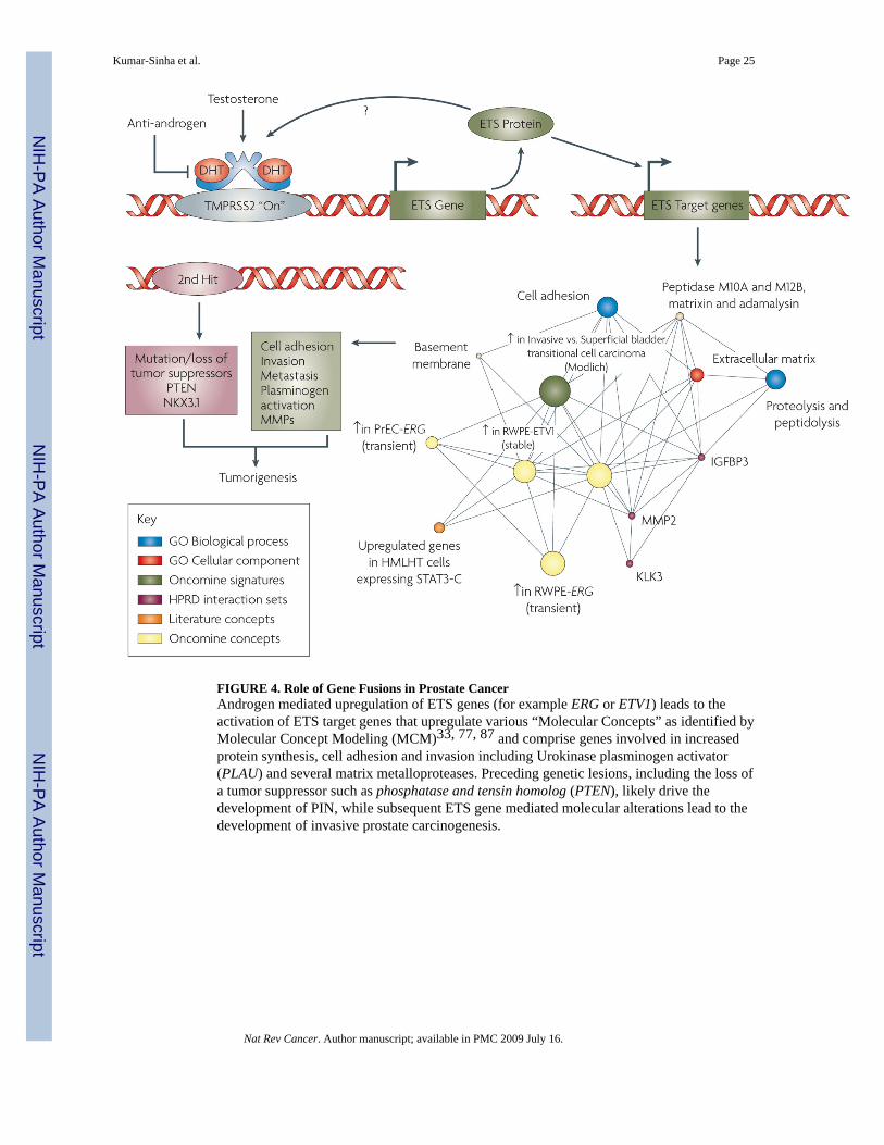
FIGURE 4. Role of Gene Fusions in Prostate CancerAndrogen mediated upregulation of ETS genes (for example ERG or ETV1) leads to theactivation of ETS target genes that upregulate various “Molecular Concepts” as identified byMolecular Concept Modeling (MCM)33, 77, 87 and comprise genes involved in increasedprotein synthesis, cell adhesion and invasion including Urokinase plasminogen activator(PLAU) and several matrix metalloproteases. Preceding genetic lesions, including the loss ofa tumor suppressor such as phosphatase and tensin homolog (PTEN), likely drive thedevelopment of PIN, while subsequent ETS gene mediated molecular alterations lead to thedevelopment of invasive prostate carcinogenesis.
Kumar-Sinha et al. Page 25
Nat Rev Cancer. Author manuscript; available in PMC 2009 July 16.
NIH
-PA Author Manuscript
NIH
-PA Author Manuscript
NIH
-PA Author Manuscript

FIGURE 5. Gamut of Gene Fusions in Prostate CancerSix major classes of pathognomic gene fusions in prostate cancer are envisioned based on thetypes of upstream regulatory partners and the nature of the fused genes. These includeregulatory regions that are either androgen responsive or androgen repressed or androgeninsensitive (housekeeping) fused to ETS family genes or other, yet to be discovered, non-ETSgenes
Kumar-Sinha et al. Page 26
Nat Rev Cancer. Author manuscript; available in PMC 2009 July 16.
NIH
-PA Author Manuscript
NIH
-PA Author Manuscript
NIH
-PA Author Manuscript
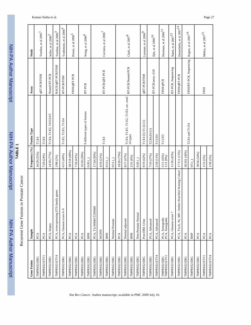
NIH
-PA Author Manuscript
NIH
-PA Author Manuscript
NIH
-PA Author Manuscript
Kumar-Sinha et al. Page 27TA
BLE
1R
ecur
rent
Gen
e Fu
sion
s in
Pros
tate
Can
cer
Gen
e Fu
sion
Sam
ple
Freq
uenc
y (%
)Fu
sion
Typ
eA
ssay
Stud
y
TMPR
SS2:
ERG
PCA
16/2
9 (5
5%)
T1:E
4qR
T-PC
R/F
ISH
Tom
lins,
et a
l 200
51TM
PRSS
2:ET
V1
PCA
7/29
(24%
)T1
:E4
TMPR
SS2:
ERG
PCA
, bio
psy
14/1
8 (7
7%)
T1:E
4, T
1:E2
, T4/
5:E4
/5N
este
d R
T-PC
RSo
ller,
et a
l 200
62
TMPR
SS2:
ETV
4PC
A, o
vere
xpre
ssin
g ET
S fa
mily
gen
es2/
98 (2
%)
RA
CE/
qRT-
PCR
/FIS
HTo
mlin
s, et
al 2
0063
TMPR
SS2:
ERG
PCA
, Gle
ason
scor
e 6–
96/
15 (4
0%)
T1:E
5, T
2:E5
, T1:
E4R
T-PC
R/F
ISH
Yos
him
oto,
et a
l 200
64
TMPR
SS2:
ERG
PCA
58/1
18 (4
9%)
FISH
/qR
T-PC
RPe
rner
, et a
l 200
65TM
PRSS
2:ER
GPC
A7/
18 (4
1%)
TMPR
SS2:
ERG
PCA
35/5
9 (5
9%)
8 di
ffer
ent t
ypes
of f
usio
nsR
T-PC
RW
ang,
et a
l 200
66TM
PRSS
2:ER
GB
PH0/
20 (_
)
TMPR
SS2:
ERG
PCA
, T1c
N0M
0/T2
N0M
017
/34
(50%
)
T1:E
4R
T-PC
R/q
RT-
PCR
Cer
vier
a, e
t al 2
0067
TMPR
SS2:
ERG
HG
PIN
4/19
(21%
)
TMPR
SS2:
ERG
BPH
0/13
(_)
TMPR
SS2:
ERG
Nor
mal
Pro
stat
e0/
11 (_
)
TMPR
SS2:
ERG
PCA
19/2
6 (7
3%)
T1:E
4, T
1:E5
. T1:
E2, T
1:E3
, etc
.-tot
al14
type
sR
T-PC
R/N
este
d-PC
RC
lark
, et a
l 200
78TM
PRSS
2:ER
GN
orm
al a
djac
ent
8/17
(47%
)
TMPR
SS2:
ERG
BPH
2/31
(6%
)
TMPR
SS2:
ERG
Non
-Pro
stat
e N
orm
al0/
20 (_
)
TMPR
SS2:
ERG
Post
:DR
E U
rine
8/19
(42%
)T1
:E4
(5) T
1:32
(1)
qRT-
PCR
/FIS
HLa
xman
, et a
l 200
69
TMPR
SS2:
ERG
PCA
, Adv
ance
d7/
19 (3
7%)
T2:E
6;E4
(1)
RT-
PCR
/arr
ay c
GH
Iljin
, et a
l 200
610TM
PRSS
2:ET
V4
PCA
, Adv
ance
d2/
18 (1
1%)
T1/2
:E4
TMPR
SS2:
ERG
TMPR
SS2:
ETV
1PC
A, X
enog
rafts
PCA
, Xen
ogra
fts5/
11 (4
5%)
1/11
(9%
)T1
/2:E
5FI
SH/q
PCR
Her
man
s, et
al 2
00611
TMPR
SS2:
ERG
PCA
, Gle
ason
scor
e 7
11/2
6 (4
2%)
RT-
PCR
, Seq
uenc
ing
Nam
i, et
al 2
00712
TMPR
SS2:
ERG
PCA
, T1a
:b, N
s, M
0′, O
rebr
o W
atch
ful W
aitin
g C
ohor
t17
/111
(15%
)FI
SH/q
RT-
PCR
Dem
iche
lis, e
t al 2
00713
TMPR
SS2:
ERG
PCA
36/1
01 (3
6%)
T2:E
4 an
d T1
:E4
FISH
/RT-
PCR
, Seq
uenc
ing
Raj
put,
et a
l 200
714TM
PRSS
2:ER
GB
HP
0/5
(_)
TMPR
SS2:
ERG
PCA
30/5
6 (5
4%)
FISH
Meh
ra, e
t al 2
00715
TMPR
SS2:
ETV
1PC
A1/
53 (2
%)
TMPR
SS2:
ETV
4PC
A1/
58 (2
%)
Nat Rev Cancer. Author manuscript; available in PMC 2009 July 16.
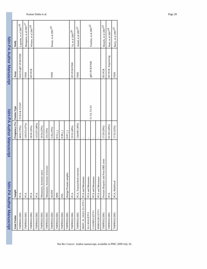
NIH
-PA Author Manuscript
NIH
-PA Author Manuscript
NIH
-PA Author Manuscript
Kumar-Sinha et al. Page 28G
ene
Fusi
onSa
mpl
eFr
eque
ncy
(%)
Fusi
on T
ype
Ass
aySt
udy
TMPR
SS2:
ERG
PCA
44/6
3 (7
0%)
T2:E
4 &
T2:
E4*
RA
CE/
qRT-
PCR
/FIS
HLa
poin
te, e
t al 2
00716
TMPR
SS2:
ERG
PCA
120/
253
(47%
)FI
SHM
osqu
era,
et a
l 200
717
TMPR
SS2:
ERG
PCA
18/5
0 (3
6%)
RT-
PCR
Win
nes,
et a
l 200
718
TMPR
SS2:
ERG
PCA
115/
237
(48%
)
FISH
Pern
er, e
t al 2
00719
TMPR
SS2:
ERG
Met
asta
ses,
horm
one
naiv
e10
/34
(29%
)
TMPR
SS2:
ERG
Met
asta
ses,
horm
one
refr
acto
ry3/
9 (3
3%)
TMPR
SS2:
ERG
HG
PIN
5/26
(19%
)
TMPR
SS2:
ERG
BPH
0/15
(_)
TMPR
SS2:
ERG
PIA
0/38
(_)
TMPR
SS2:
ERG
Ben
ign
Pros
tate
sam
ples
0/47
(_)
TMPR
SS2:
ERG
PCA
25/5
2 (4
8%)
RT-
PCR
/FIS
HTu
, et a
l 200
720
TMPR
SS2:
ERG
PCA
, Tra
nsur
ethr
al re
sect
ion
134/
445
(30%
)FI
SHA
ttard
, et a
l 200
721
HER
V_K
_22Q
11.2
3: E
TV1
PCA
and
Met
asta
ses
-
T1:T
4, T
2:T4
qRT-
PCR
/FIS
HTo
mlin
s, et
al 2
00722
SLC
45A
3:ET
V1
PCA
and
Met
asta
ses
-
C15
OR
F21:
ETV
1PC
A a
nd M
etas
tase
s-
HN
RPA
2B1:
ETV
1PC
A a
nd M
etas
tase
s-
TMPR
SS2:
ERG
Pros
tate
Bio
psie
s and
Pos
t-DR
E ur
ine
17/2
9 (5
9%)
RT-
PCR
Hes
sels
, et a
l 200
723
TMPR
SS2:
ERG
PCA
81/1
65 (4
9%)
RT-
PCR
, Seq
uenc
ing
Nam
, et a
l 200
724
TMPR
SS2:
ERG
PCA
, Mul
tifoc
al17
/32
(53%
)FI
SHB
arry
, et a
l 200
725
Nat Rev Cancer. Author manuscript; available in PMC 2009 July 16.
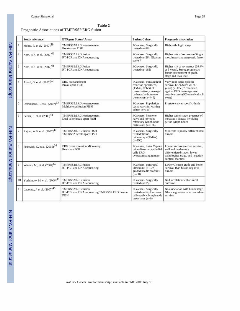
NIH
-PA Author Manuscript
NIH
-PA Author Manuscript
NIH
-PA Author Manuscript
Kumar-Sinha et al. Page 29
Table 2Prognostic Associations of TMPRSS2:ERG fusion
Study reference ETS gene Status/ Assay Patient Cohort Prognostic association
1 Mehra, R. et al. (2007)59 TMPRSS2/ERG rearrangementBreak-apart FISH
PCa cases, Surgicallytreated (n=96)
High pathologic stage
2 Nam, R.K. et al. (2007)66 TMPRSS2:ERG fusionRT-PCR and DNA sequencing
PCa cases, Surgicallytreated (n=26), Gleasonscore 7
Higher rate of recurrence Singlemost important prognostic factor
3 Nam, R.K. et al. (2007)61 TMPRSS2:ERG fusionRT-PCR and DNA sequencing
PCa cases, Surgicallytreated (n=165)
Higher risk of recurrence (58.4%at 5 years). Strong prognosticfactor independent of grade,stage and PSA level.
4 Attard, G. et al. (2007)62 ERG rearrangementBreak-apart FISH
PCa cases, transurethralresection specimens,(TMAs, Cohort ofconservatively managedpatients (no hormonetreatment) (n=445)
Very poor cause-specificsurvival (25% Survival at 8years) (2+Edel)* comparedagainst ERG rearrangementnegative cases (90% survival at 8years)
5 Demichelis, F. et al. (2007)63 TMPRSS2:ERG rearrangementMulticolored fusion FISH
PCa cases, Populationbased watchful waitingcohort (n=111)
Prostate cancer specific death
6 Perner, S. et al. (2006)41 TMPRSS2:ERG rearrangementDual color break-apart FISH
PCa cases, hormone-naïve and hormone-refractory lymph nodemetastases (n=136)
Higher tumor stage, presence ofmetastatic disease involvingpelvic lymph nodes
7 Rajput, A.B. et al. (2007)47 TMPRSS2:ERG fusion FISHTMPRSS2 Break-apart FISH
PCa cases, Surgicallytreated Tissuemicroarrays (TMAs)(n=196)
Moderate to poorly differentiatedtumors
8 Petrovics, G. et al. (2005)64 ERG overexpression Microarray,Real-time PCR
PCa cases, Laser Capturemicrodissected epithelialcells ERGoverexpressing tumors
Longer recurrence-free survival,well and moderatelydifferentiated stages, lowerpathological stage, and negativesurgical margins
9 Winnes, M., et al. (2007)65 TMPRSS2:ERG fusionRT-PCR and DNA sequencing
PCa cases, transrectalultrasound (TRUS)guided needle biopsies(n=50)
Lower Gleason grade and bettersurvival than fusion-negativetumors
10 Yoshimoto, M. et al. (2006)49 TMPRSS2:ERG fusionRT-PCR and DNA sequencing
PCa cases, Surgicallytreated (n=15)
No Correlation with clinicaloutcome
11 Lapointe, J. et al. (2007)46 TMPRSS2:ERG fusionRT-PCR and DNA sequencing TMPRSS2:ERG FusionFISH
PCa cases, Surgicallytreated (n=54) Hormonenaiive pelvic lymph nodemetastases (n=9)
No association with tumor stage,Gleason grade or recurrence-freesurvival
Nat Rev Cancer. Author manuscript; available in PMC 2009 July 16.