Gastrointestinal Microbiota Dysbiosis Associated with SARS ...
Upload
independentCategory
view
4download
0
Protein Expression and PuriWcation xxx (2005) xxx–xxx
www.elsevier.com/locate/yprep
YPREP 2461 No. of Pages 11; DTD=5.0.1ARTICLE IN PRESS
23 February 2005 Disk Used Vimala (CE) / Chandrasekar (TE)
CORRECTED PROOF
Expression and puriWcation of SARS coronavirus proteins using SUMO-fusions
Xun Zuo a, Michael R. Mattern a, Robin Tan b, Shuisen Li c, John Hall a, David E. Sterner a, Joshua Shoo b, Hiep Tran a, Peter Lim b, Stefan G. SaraWanos d,
Lubna Kazi e, Sonia Navas-Martin e, Susan R. Weiss e, Tauseef R. Butt a,¤
a LifeSensors, Inc., 271 Great Valley Parkway, Malvern, PA 19355, USAb Department of Biological Sciences, National University of Singapore, 10 Kent Ridge Crescent, Singapore 119260, Singapore
c Drexel University, School of BioMedical Engineering, 3141 Chestnut Street, Philadelphia, PA 19104, USAd Center for Advanced Biotechnology and Medicine (CABM), Rutgers University, Department of Chemistry and Chemical Biology,
679 Hoes Lane, Piscataway, NJ 08854-5638, USAe Department of Microbiology, University of Pennsylvania School of Medicine, 203A Johnson Pavilion, 36th Street and Hamilton Walk,
Philadelphia, PA 19104, USA
Received 14 January 2005, and in revised form 8 February 2005
Abstract
Severe acute respiratory syndrome coronavirus (SARS-CoV) proteins belong to a large group of proteins that is diYcult toexpress in traditional expression systems. The ability to express and purify SARS-CoV proteins in large quantities is critical for basicresearch and for development of pharmaceutical agents. The work reported here demonstrates: (1) fusion of SUMO (small ubiquitin-related modiWer), a 100 amino acid polypeptide, to the N-termini of SARS-CoV proteins dramatically enhances expression in Esche-richia coli cells and (2) 6£ His-tagged SUMO-fusions facilitate rapid puriWcation of the viral proteins on a large scale. We haveexploited the natural chaperoning properties of SUMO to develop an expression system suitable for proteins that cannot beexpressed by traditional methodologies. A unique feature of the system is the SUMO tag, which enhances expression, facilitates puri-Wcation, and can be eYciently cleaved by a SUMO-speciWc protease to generate native protein with a desired N-terminus. We havepuriWed various SARS-CoV proteins under either native or denaturing conditions. These puriWed proteins have been used to gener-ate highly speciWc polyclonal antibodies. Our study suggests that the SUMO-fusion technology will be useful for enhancing expres-sion and puriWcation of the viral proteins for structural and functional studies as well as for therapeutic uses. 2005 Published by Elsevier Inc.
Keywords: SARS-CoV 3CL protease; SARS-CoV Nucleocapsid; SARS-CoV Spike protein; SUMO; SUMO-fusion system; SUMO protease; Pro-tein expression; Ni–NTA aYnity puriWcation; Escherichia coli culture
Severe acute respiratory syndrome (SARS)1 is a respi- the outbreak spread quickly to about 35 countries on Wve
UNratory illness that has only recently been reported in Asia,North America, and Europe. After the Wrst case of the dis-ease in humans was found in Southern China late 2002,
1046-5928/$ - see front matter 2005 Published by Elsevier Inc.doi:10.1016/j.pep.2005.02.004
¤ Corresponding author. Fax: +1 610 644 8616.E-mail address: [email protected] (T.R. Butt).
1 Abbreviations used: SARS, severe acute respiratory syndrome; SARS-Co-D-thiogalactopyranoside; Ni–NTA, nickel–nitrilotriacetic acid; SDS–PAGitin; SUMO, small ubiquitin-related modiWer; PMSF, phenylmethylsulfony
continents, resulting in more than 8000 cases and 800deaths. At present, there is no eYcacious treatment regimefor SARS. The need for both a reliable diagnostic assay
34444
V, SARS coronavirus; DUB, deubiquitinating enzyme; IPTG, isopropylE, sodium dodecyl sulfate–polyacrylamide gel electrophoresis; Ub, ubiql Xuoride; FPLC, fast performance liquid chromatography.
�u
1
2
3
4
5
6789101112
13
14
151617181920212223242526
2728
29303132333435363738
--
90123
44454647

2 X. Zuo et al. / Protein Expression and PuriWcation xxx (2005) xxx–xxx
YPREP 2461 No. of Pages 11; DTD=5.0.1ARTICLE IN PRESS
23 February 2005 Disk Used Vimala (CE) / Chandrasekar (TE)
UNCORREC
and a therapeutic agent (antiviral or vaccine) is obvious. Apreviously unknown coronavirus has been identiWed asthe causative agent of SARS. Scientists at the CDC andother laboratories determined the genomic sequence ofthis coronavirus and named it SARS-CoV [1–3].
Coronavirus, a genus within the family Coronaviridae,contains a group of large, positive stranded, enveloped,pathogenic RNA viruses that infect many species of ani-mals, including humans. They cause respiratory, enteric,and central nervous system diseases [4]. The genomicsequence of SARS-CoV provides important informationfor the development of diagnostic tests and vaccines.This information aVords the opportunity to express anySARS-CoV protein of choice for recombinant subunitvaccines. Development of protein-based diagnostic andtherapeutic methods would be greatly facilitated by theability to produce viral proteins of high quality in tracta-ble amounts, which requires protein engineering, expres-sion, and puriWcation. Six proteins of SARS-CoV,namely Spike (S), Nucleocapsid (Nc), Envelope (E),SARS polymerase (RdRp), SARS protease (3CL), andmembrane (M), have become the focus of eVorts to pro-duce antiviral agents and vaccines against SARS. TheSARS-CoV proteins investigated in this study aredescribed brieXy below.
SARS-CoV 3CL protease (3CL, 3CLpro or Mpro) isthe principal coronavirus protease utilized by the virusto process its replicase proteins into mature forms. Thefull length of the 3CL has 306 amino acids (molecularweight »33.8 kDa). The protease cleaves the replicasepolyproteins (pp1a and pp1ab) to generate RNA-depen-dent polymerase (RdRp), 3CL, and helicase, all crucialfor viral replication [5–7]. Therefore, 3CL represents anattractive target for the design and discovery of corona-virus antiviral agents, as does the polymerase [8]. SARS-CoV Nucleocapsid protein (N or Nc) is a phosphopro-tein containing 423 amino acids (molecular weight»46 kDa) [9]. Large quantities of the protein are trans-lated on free polysomes in the cytoplasm, where somemolecules are rapidly phosphorylated. It is known thatthe protein binds the viral RNA and forms the nucleo-capsid, but its exact mechanisms and role in replicationare not yet clear. The Nc protein is known to have B andT cell epitopes and to elicit host protective immuneresponses [10,11]. Spike protein (S or Spk) is a glycopro-tein containing 1255 amino acids [12]. Upon translation,it is inserted into the rough endoplasmic reticulum andglycosylated with N-linked glycans [13]. Some of theproteins accumulate in the Golgi apparatus, and a frac-tion of oligomeric spike protein is transported to themembrane, where it mediates cell–cell fusion. Like thoseof other coronaviruses, the SARS-CoV spike proteinlikely contains many of the neutralizing antibody epi-topes as well as T cell epitopes [14].
A supply of puriWed SARS-CoV proteins would bevaluable for both clinical and investigational purposes.
TED PROOF
Although several strategies have been developed overthe years to express heterologous recombinant proteinsin bacterial, yeast, mammalian, and insect cells, theexpression of heterologous genes in bacteria is by far thesimplest and most inexpensive means available forresearch or commercial purposes. However, heterolo-gous gene products often fail to attain their correctthree-dimensional (3-D) conformation, or are simplyexpressed very poorly in Escherichia coli. Selection ofORFs for structural genomics projects has shown thatonly »20% of all heterologous genes expressed in E. colirender soluble or correctly folded proteins [15,16]. Sev-eral gene-fusion systems, such as NusA, maltose bindingprotein (MBP), glutathione-S-transferase (GST), ubiqui-tin (UB), and thioredoxin (Trx), have been developed[17,18]. All of these conventional methods have short-comings, primarily ineYcient expression and/or incon-sistent cleavage.
Small ubiquitin-related modiWer (SUMO) is a ubiqui-tin-related protein that functions by covalent attachmentto other proteins. SUMO and its associated enzymes arepresent in all eukaryotes and are highly conserved fromyeast to humans [19–21]. SUMO has 18% sequence iden-tity with ubiquitin [22]. The yeast Saccharomyces cerevi-siae has only a single SUMO gene (SMT3) that is essentialfor viability [20]. In contrast to yeast SMT3, three mem-bers of SUMO have been described in vertebrates:SUMO-1, SUMO-2, and SUMO-3. Human SUMO-1, a101 amino acid polypeptide, shares 50% sequence identitywith human SUMO-2/SUMO-3 [23], which are closehomologues. Yeast SUMO shares 47% sequence identitywith mammalian SUMO-1. Although overall sequenceidentity between ubiquitin and SUMO is only 18%, struc-ture determination by NMR reveals that they share acommon three-dimensional structure characterized by atightly packed globular fold with �-sheets wrappedaround a single �-helix [24,25]. It is known that SUMO,fused at the N-terminus with other proteins, can fold andprotect the protein by its chaperoning properties, makingit a useful tag for heterologous expression [26]. All SUMOgenes encode precursor proteins with a short C-terminalsequence that extends from the conserved C-terminalGly–Gly motif. SUMO proteases remove SUMO fromproteins, by cleaving the C-termini of SUMO (-GGATY)in yeast to the mature form (-GG) or deconjugating itfrom lysine side chains [27,28]. The former activity (prote-ase) is useful for removal of SUMO as an expression tag.There are 2 SUMO proteases in yeast [27,28] and at least 6in humans, the human enzymes ranging from 238 to 1112amino acid residues [22,29–31].
We have developed a novel SUMO-fusion systemthat provides increased levels of expression of heterolo-gous proteins in E. coli and allows rapid puriWcation ofproteins of interest [26,32]. We report here the applica-tion of SUMO-fusion technology to the expression andpuriWcation of major SARS-CoV proteins.
11111111111111111111111111111111111111111111111111111111
48495051525354555657585960616263646566676869707172737475767778798081828384858687888990919293949596979899100101102103
0405060708091011121314151617181920212223242526272829303132333435363738394041424344454647484950515253545556575859

X. Zuo et al. / Protein Expression and PuriWcation xxx (2005) xxx–xxx 3
YPREP 2461 No. of Pages 11; DTD=5.0.1ARTICLE IN PRESS
23 February 2005 Disk Used Vimala (CE) / Chandrasekar (TE)
NCORREC
Materials and methods
SARS-CoV 3CL Protease (3CL), SARS-CoV Nucle-ocapsid (Nc), and SARS-CoV Spike C-terminal frag-ment protein (Spk C) were fused with SUMO andexpressed in E. coli. For expression of the proteins,SARS-CoV cDNA was derived from infected cell RNA,provided by the CDC, Atlanta, to S.R.W. (University ofPennsylvania).
Construction of SUMO-SARS-CoV-fusion protein expression vectors
Expression constructs encoding the SUMO-fusionproteins all utilized the pET24d plasmid (Novagen,Madison, WI) as the backbone. The pET24 derivativecarrying the SMT3 gene of S. cerevisiae, which encodesthe yeast SUMO protein, has been described previously[26]. It contains an N-terminal hexahistidine (6£ His)tag, introduced by PCR into the SUMO codingsequence, as well as a unique BsaI site at the C-terminus.The cloning strategy to express fusion proteins employedthis BsaI site to insert the SARS-CoV protein codingsequences in frame with SUMO. PCR primers (Table 1)incorporating this site or Esp 3I were used to amplify theSARS-CoV coding sequences from cDNA clones carriedin pTOPO vectors. The 3� primers carried a BamHI sitefor insertion into the multiple cloning site of pET24d.The primer pairs used to PCR amplify the SARS-CoVprotein genes are listed in Table 1. Because of its largesize, Spike protein was designed as two half-molecules,S1 (N-terminal fragment, amino acids 1–667, SpK N)and S2 (C-terminal fragment, amino acids 668–1193,SpK C) domains and the Spk C was tested for expres-sion and puriWcation in this study. For PCR ampliWca-tion of the genes of interest, a proofreading polymerasewas used (Platinum Taq, Invitrogen, Carlsbad, CA).PCR fragments were subcloned into pET24-6£ His-SUMO or pET24-6£ His (a parallel vector that does notcarry the SUMO sequence) to produce parallel sets ofconstructs encoding 6£ His-SUMO and 6£ His fusedversions of the proteins of interest. All plasmids wereroutinely sequenced.
TED PROOF
Expression of SARS proteins using SUMO-fusion
To test and compare expression of the SARS pro-teins, a single colony of the E. coli strain BL21 (DE3)containing each of the plasmids described above wasinoculated into 5 ml of either Luria–Bertani (LB) or M9minimal (MM) media. The antibiotic kanamycin wasalso included at 30�g/ml in all media. The cells weregrown at 37 °C overnight with shaking at 250 rpm. Thenext morning the overnight culture was transferred into50 ml fresh medium to permit exponential growth. Whenthe OD600 value reached »0.6–0.7, protein expressionwas induced by addition of 1 mM IPTG (isopropyl-�-D-thiogalactopyranoside), followed by prolonged growthat either 37 or 20 °C to determine optimal induction con-ditions. For protein puriWcation, cultures were scaled upto 0.5–1.0 L LB medium.
Sodium dodecyl sulfate–polyacrylamide gel electro-phoresis (SDS–PAGE) was used to verify expression ofthe protein. BrieXy, 1.5ml samples of culture wereremoved just before expression was induced and afterinduction, and cells were collected by centrifugation at6000rpm for 5min. The cell pellets were suspended in50�l of distilled water, and the samples were freeze–thawed once to facilitate disruption of the cells. The cellsuspensions were treated with RNAse and DNAse (bothat 40�g/ml) to digest nucleic acids. After mixing withSDS–PAGE sample buVer containing SDS and �-mercaptoethanol, samples were heated at 95°C for 5minto facilitate denaturation and reduction of proteins. Pro-teins were detected using SDS–polyacrylamide gels withTris–glycine running buVer and Coomassie blue staining.
Western blots
Proteins separated by SDS–PAGE were transferredonto nitrocellulose membranes at 42 V (»150 mA) for2.5 h. Membranes were then incubated with 30 ml ofTTBS buVer (pH 8.0), containing 5% nonfat dry milk for1 h at room temperature. The expressed proteins wereprobed with either monoclonal anti-His-tag or poly-clonal antibodies obtained from rabbits immunizedagainst individual SUMO-SARS-CoV-fusion proteins
22222222222222222222222222222222222222222222
UTable 1PCR primers for amplifying the SARS-CoV protein genes
Restriction enzyme recognition sites used for cloning are indicated in uppercase letters. Owing to the presence of a BsaI site within the 3CL proteasecoding sequences, a diVerent restriction enzyme, Esp3I, was used at the 5� end to join the BsaI site in the pET-SUMO vector.
Proteins Region of genes Primers
Spike protein—C terminal fragment AA 668–1193 tttGGTCTCaaggtatgagtactagccaaaaatctattgtggccgcGGATCCtcatttaatatattgctcatattttc
Nucleocapsid protein Entire gene tttGGTCTCaaggtatgtctgataatggaccccaatccgcGGATCCtcatgcctgagttgaatcagcag
CL Protease Entire gene tttCGTCTCaaggtagtggttttaggaaaatggcattcccgcgcGGATCCtcattggaaggtaacaccagagc
222222222222
160161162163164165166167168169170171172173174175176177178179180181182183184185186187188189190191192193194195196197198199200201202203204205206207208209210211212213214215
1617181920212223242526272829303132333435363738394041424344454647484950515253545556575859606162636465666768697071

4 X. Zuo et al. / Protein Expression and PuriWcation xxx (2005) xxx–xxx
YPREP 2461 No. of Pages 11; DTD=5.0.1ARTICLE IN PRESS
23 February 2005 Disk Used Vimala (CE) / Chandrasekar (TE)
UNCORREC
(Rockland Immunochemicals) by incubating overnightat 4 °C with 1: 1000 dilution of the primary antibodies.After the membranes were washed with TTBS buVer for5 min, they were incubated with a secondary antibody(Peroxidase-conjugated goat anti-rabbit IgG, RocklandImmunochemicals, diluted 1000-fold) for 45 min. Themembranes were Wnally washed with TTBS for 10 minbefore the chemiluminescent Western blot substrateswere applied (Roche, Mannheim, Germany), and visual-ized on Wlms (Kodak BioMax).
PuriWcation of SARS-CoV proteins
Because the SUMO constructs bear an N-terminal 6£His tag, expressed SARS-CoV proteins fused withSUMO can be rapidly puriWed by Ni–NTA aYnitychromatography. In this study, the soluble proteins fromE. coli cell lysates and the insoluble proteins from the cellinclusion bodies were puriWed under native and denatur-ing conditions, respectively. A typical procedure forpuriWcation of the SARS-CoV proteins is illustrated inFig. 1. Protein concentrations were determined using theBradford color-reaction assay (Bio-Rad) measured spec-trophotometrically at 595 nm with bovine serum albuminas a standard, according to the manufacturer’s instruc-tions. SDS–PAGE and Coomassie blue staining wereused to evaluate the eVectiveness of the puriWcations andcleavage of SUMO-SARS-CoV protein fusions.
Fig. 1. The procedure for puriWcation of SARS-CoV proteinsexpressed with SUMO-fusion system in E. coli and cleavage of 6£His-SUMO-tagged proteins.
TED PROOF
Preparation of soluble and insoluble protein samples from E. coli cells
The E. coli cells expressing the SARS-CoV proteinswere harvested from LB medium (typically, 1.0 L) bycentrifugation (8000g for 10 min at 4 °C). Typically, thewet weights of the E. coli cells harvested from 1 L culturewere 10–15 g. The cell pellets were resuspended in lysisbuVer (PBS containing additional 150 mM NaCl, 10 mMimidazole, 1% Triton X-100, and 1 mM PMSF, pH 8.0)at 3 ml for 1 g of the cells, resulting in »4 mg protein perml after the proteins were extracted. The cells were lysedby sonication (50% output for 5 £ 30 second pulses).Sonication was conducted with the tube jacketed in wetice and observing 1 min intervals between pulse cycles toprevent heating. After the lysates were incubated withDNase and RNase (each at 40 �g/ml) for 20 min, theywere centrifuged at 20,000g for 30 min at 4 °C, andsupernatants (soluble protein fractions) were collected.The pellets containing inclusion bodies were washedthree times in buVer (PBS containing 25% sucrose, 5 mMEDTA, and 1% Trition X-100, pH 7.5) followed by cen-trifugation, as described above. The washed inclusionbodies were resuspended in denaturing solubilizationbuVer (Novagen), which contained 50 mM Caps (pH11.0), 0.3% N-lauryl sarcosine, and 1 mM DTT, andincubated for 30 min at room temperature with shakingto extract the insoluble proteins. Because debris frominclusion bodies was much smaller than that in the celllysate, the extract for the insoluble proteins wasobtained by high-speed centrifugation (80,000g for30 min at 4 °C).
PuriWcation of 6£ His-tagged SUMO-SARS-CoV proteins
The soluble proteins extracted from E. coli cells werepuriWed under native conditions and a BioLogic Duo-Flow FPLC system (Bio-Rad) was used for fractiona-tions. BrieXy, the cell lysate (typically, 20–40 ml contain-ing 0.2–0.5 g proteins) was loaded onto a columncontaining »10 ml Ni–NTA superXow resin (Qiagen,Valencia, CA) and the samples of Xow-through contain-ing unbound proteins were collected for subsequentanalysis. The resin was extensively washed with »50–100 ml of wash buVer (PBS containing 20 mM imidazoleand additional 150 mM NaCl, pH 8.0) until OD280reached or fell below the base line (UV value D 0).Finally, the 6£ His-tagged SUMO-fusion proteins wereeluted with elution buVer (PBS containing 300 mM imid-azole and additional 150 mM NaCl, pH 8.0). The puri-Wed SUMO-fused proteins eluted as a single isolated UVpeak. The proteins with high OD280 values were collectedin 4 ml fractions that were checked on SDS-gels andpooled.
The insoluble proteins extracted from the E. coliinclusion bodies were puriWed under denaturing condi-tions, which were similar to the native conditions
33333333333333333333333333333333333333333333333333333333
272273274275276277278279280281282283284285286287288289290291292293294295296297298299300301302303304305306307308309310311312313314315316317318319320321322323324325326327
2829303132333435363738394041424344454647484950515253545556575859606162636465666768697071727374757677787980818283

X. Zuo et al. / Protein Expression and PuriWcation xxx (2005) xxx–xxx 5
YPREP 2461 No. of Pages 11; DTD=5.0.1ARTICLE IN PRESS
23 February 2005 Disk Used Vimala (CE) / Chandrasekar (TE)
UNCORREC
described above except for the use of highly alkaline pHbuVer containing detergent. BrieXy, an insoluble proteinsample (»20–40 ml) prepared in the denaturing buVer(50 mM Caps, 0.3% N-lauryl sarcosine, and 1 mM DTT,pH 11.0) was incubated with »10 ml of Ni–NTAsuperXow resin at 4 °C for 1 h with shaking for eVectivebinding of the 6£ His-tagged proteins to the resin. Themixture was then loaded into an empty column and theXow-through sample was collected. Subsequently, theresin was continually washed with denaturing washbuVer that contained 20 mM imidazole, 0.3% N-lauroylsarcosine, 0.3 M NaCl, and 50 mM Caps, pH 11, untilOD280 fell below the base line. The 6£ His-taggedSUMO-fusion proteins were Wnally eluted using dena-turing elution buVer that contained the same compo-nents as in the denaturing wash buVer, except that theconcentration of imidazole was increased to 300 mM.
Cleavage of SUMO-fusion by the SUMO proteaseThe SUMO protease used in this study (yeast Ulp1
catalytic domain) was produced in our laboratory asdescribed [26], and a unit of the protease activity wasdeWned as the amount of SUMO protease that cleaves15 �g of SUMO-Met-GFP-fusion substrate at 25 °C in1 h in buVer containing 20 mM Tris–HCl, pH 8.0, and5 mM �-mercaptoethanol [26]. Before adding the enzymefor cleavage, the puriWed SUMO-fusion proteins (solublefraction) were dialyzed with 3.5 kDa cutoV membranesagainst PBS (pH 7.4) for 12–15 h at 4 °C to remove highsalt and imidazole, while the puriWed sample in denatur-ing buVer were refolded by extensive dialysis for at least24 h against 20 mM Tris–HCl (pH 8.0) containing 10%glycerol. No protein precipitation was observed duringthe dialysis. The minimum amount of SUMO proteaserequired for complete cleavage of a given SUMO-fusionwas variable. Typically, for most of the puriWed SUMO-SARS-CoV proteins we added the enzyme at a ratio of1 U to 15�g substrates and incubated in either PBS (pH7.4) or 20 mM Tris buVer (pH 8.0), containing 5 mM �-mercaptoethanol, at 30 °C for 1 h. In this study, cleavageof the SUMO-SARS-CoV Nc protein was achieved witha lower amount of the SUMO protease after checkingeVectiveness of the enzyme in serial dilution (see Fig. 8).
Removal of SUMO and SUMO protease for Wnal puriWcation of SARS-CoV proteins
Since both SUMO and SUMO protease had 6£ Histags, but SARS-CoV proteins did not, the cleavedSUMO-fusion samples could be re-applied to the nickelcolumn to obtain the puriWed membrane proteins bysubtracting the 6£ His-tagged proteins. BrieXy, after theSUMO-fusions were cleaved by the SUMO protease, thesample was loaded onto a nickel column with Ni–NTAresin. Most of the SARS-CoV protein without 6£ Histags was eluted in the Xow-through (unbound) fractions,and the rest was recovered by washing the resin with
TED PROOF
PBS. The eluted and washed proteins appearing in frac-tions with high-UV values at OD280 were pooled as theWnal puriWed sample. The puriWed proteins were checkedon SDS-gels and the samples were stored at ¡80 °C afterglycerol was added to 10%.
Results
Enhanced expression of SARS-CoV proteins with SUMO-fusion
SARS-CoV proteins 3CL, Nc, and Spike C, in ver-sions fused to either 6£ His-SUMO or 6£ His, wereexpressed in E. coli cells under various conditions. Theexpressed proteins were readily identiWed by their migra-tion positions in SDS-gels based on their molecularweights, and were further conWrmed by immunologicalreactions with their respective antibodies on Westernblots.
The expressed SARS-CoV 3CL protease (3CL) wasdetected in lysates of E. coli cells under several cultureand induction conditions (Fig. 2); induced cell lysatesamples showed appropriate protein bands (approxi-mately 35 kDa for 3CL and 47 kDa for SUMO-3CL-fusion) on the SDS-gels (the sequence-predicted sizes of3CL and SUMO-3CL are 33.8 and 45.8 kDa, respec-tively). When fused to the 3CL, SUMO signiWcantlyenhanced expression of its partner protein in both LBand MM media under all the conditions tested, com-pared to the 3CL expressed without SUMO-fusion (Fig.2). Overnight growth (»15 h) at 20 °C resulted in anincreased yield of SUMO-fused 3CL compared to a 6 hculture at the same temperature and a 3 h culture at37 °C (Fig. 2).
Expressed SARS-CoV Nucleocapsid (Nc) wasdetected in either unfused (»46 kDa) or SUMO-fused(»60 kDa) versions from IPTG-induced E. coli cellsunder various culture and induction conditions (Fig. 3).Notably, much higher yields of the expressed proteinswere observed from rich medium (LB) than from mini-mal medium (MM) (Fig. 3), suggesting the formershould be better for large-scale production and puriWca-tion of the proteins. Similar to the 3CL results, expres-sion enhancement was seen when Nc was fused toSUMO and expressed in minimal medium, but in LBmedium there were no signiWcant diVerences between theexpression of Nc without SUMO and Nc fused withSUMO (Fig. 3).
The SUMO-fusion also greatly increased the level ofexpression of the C-terminal half of the SARS-CoVSpike protein (Spk C) compared to that of unfused SpkC in LB media (Fig. 4). Only a very weak protein band(»58 kDa) of unfused Spk C could be seen in the SDS-gel and no band was seen in the Western blot probedwith anti-His-tag antibodies, indicating that Spk C was
44444444444444444444444444444444444444444444444444444444
384385386387388389390391392393394395396397398399400401402403404405406407408409410411412413414415416417418419420421422423424425426427428429430431432433434435436437438439
4041424344454647484950515253545556575859606162636465666768697071727374757677787980818283848586878889909192939495

6 X. Zuo et al. / Protein Expression and PuriWcation xxx (2005) xxx–xxx
YPREP 2461 No. of Pages 11; DTD=5.0.1ARTICLE IN PRESS
23 February 2005 Disk Used Vimala (CE) / Chandrasekar (TE)
poorly expressed without SUMO-fusion under the con-ditions tested. In contrast, an intense protein band wasobserved at the SUMO-Spk C migration position(»68 kDa) on the SDS-gel (Fig. 4, left panel) when SpKC was fused with SUMO and the identity of the fusionprotein was conWrmed by reactions with anti-His-tagantibody (Fig. 4, right panel).
PuriWcation of SARS proteins
PuriWcation of SARS-CoV 3CL proteaseFig. 5 shows detection of the proteins from a repre-
sentative puriWcation of soluble SARS-CoV 3CL undernative conditions. The cell lysate containing solubleSUMO-3CL was used for this puriWcation, because a
66666666
PROOF
Fig. 2. Enhanced expression of SARS-CoV 3CL protease (3CL) by SUMO-fusion in E. coli. Cells grown in either Luria–Bertani (LB) or M9 minimal(MM) medium were induced at the temperatures and for the lengths of time indicated. Just before expression was induced and after induction wascompleted the cells from a 1.5 ml aliquot of culture were lysed. Samples of whole cell lysates (»7.5 �l) from the various expression conditions wereresolved in 12% SDS-gels and stained with Coomassie blue. Molecular weights were as indicated, and arrowheads highlight expected/observed posi-tions of respective expressed protein bands.
5555555555555555555555555
UNCORRECTED
Fig. 3. Enhanced expression of SARS-CoV Nucleocapsid protein (Nc) by SUMO-fusion in E. coli. The conditions for cell culture, protein expression,and gel detection were the same as in Fig. 2. The yields of expressed SUMO-Nc proteins were higher than the Nc expressed without SUMO in mini-mal media, but there were no signiWcant diVerences in their expression in LB media.
5555555555555555555555
496497498499500501502503504505506507508509510511512513514515516517518519520521522523524525526527528529530531532533534535536537538539540541542543544545546547548549550551
5253545556575859606162636465666768697071727374757677787980818283848586878889909192939495969798
5990001020304050607

X. Zuo et al. / Protein Expression and PuriWcation xxx (2005) xxx–xxx 7
YPREP 2461 No. of Pages 11; DTD=5.0.1ARTICLE IN PRESS
23 February 2005 Disk Used Vimala (CE) / Chandrasekar (TE)
UNCORREC
Fig. 5. Detection of proteins in samples from various steps of a typicalpuriWcation of SARS-CoV 3CL protease. Aliquots of the samples(each containing »5 �g protein) were separated on a 12% SDS-gel andstained with Coomassie blue. The migration positions of the SUMO-fusion and the proteins resulting from the cleavage are as indicated.
Table 2
TED PR
majority of the expressed protein (>80%) was presentin the soluble fraction (data not shown). Proteins with-out 6£ His tags were removed from the Ni–NTA resinusing wash buVer containing 20 mM imidazole, and the6£ His-tagged SUMO-3CL-fusion was eluted usingelution buVer containing 300 mM imidazole. After theSUMO-3CL fractions were pooled, the sample was dia-lyzed extensively against PBS (pH 7.4) at 4 °C toremove high salt and imidazole, which would interferewith the cleavage reaction. The SUMO-fusion wascleaved by addition of SUMO protease at 30 °C for 1 hunder the conditions described in Materials and meth-ods. The completeness of cleavage was conWrmed bychecking the proteins on a 12% SDS-gel, since the bandof the SUMO-3CL disappeared and two new bandscorresponding to the expected molecular weights ofSUMO and 3CL were detected. After the cleaved sam-ple was re-applied to a Ni–NTA column to subtract 6£His-tagged SUMO and SUMO protease, Wnal puriWed3CL was obtained (Fig. 5); the protein from the sub-tracted sample ran as a single, intense band (»34 kDa),indicating that 3CL had been puriWed successfully(>95% purity). In this experiment, a high yield (totally»56 mg) of the pure 3CL was achieved from 1 L of E.coli cultured and induced at 20 °C overnight (Table 2).
666666666666666677777777777
OOFFig. 4. Enhanced expression of SARS-CoV Spike C protein (Spk C) by SUMO-fusion in E. coli. The cells were cultured in LB media and the condi-tions for cell culture, protein expression, and gel detection were the same as in Fig. 2. The Western blot (right panel) was performed with anti-His-tagprimary antibody.
66666666666666666666
Summary of the SARS-CoV proteins resulting from representative puriWcations of 1 L E. coli culture
The SARS-CoV proteins fused with SUMO were expressed in E. coli and induced at 20 °C overnight. The wet weights of the cells harvested from 1 Lof E. coli culture for the 3CL, Nc, and Spk C were 14, 13, and 10 g, respectively. The samples were prepared and puriWed as described in Materials andmethods.
Proteins 3CL Nc Spk C
Starting samples for puriWcation Soluble fraction (224 mg) Soluble fraction (189 mg) Insoluble fraction (66 mg)PuriWed SUMO-fusions 101 mg 66 mg 24 mgPuriWed SARS-CoV proteins 56 mg 26 mg 12 mgPurity >95% >95% »30%
777777777
608609610611612613614615616617618619620621622623624625626627628629630631632633634635636637638639640641642643644645646647648649650651652653654655656657658659660661662663
6465666768697071727374757677787980818283848586878889909192939495969798990001020304050607080910111213141516171819

8 X. Zuo et al. / Protein Expression and PuriWcation xxx (2005) xxx–xxx
YPREP 2461 No. of Pages 11; DTD=5.0.1ARTICLE IN PRESS
23 February 2005 Disk Used Vimala (CE) / Chandrasekar (TE)
UNCORREC
We used the anti-SUMO-3CL-fusion antibody to iden-tify the puriWed 3CL protein, since the antibody couldreact with the SUMO-3CL-fusion and their cleavedpartners. The puriWed protein was conWrmed to be theSARS-CoV 3CL by the Western blot probed with theanti-SUMO-3CL antibody (see below and Fig. 7).
PuriWcation of SARS-CoV Nucleocapsid proteinSimilar to the SARS-CoV 3CL protease, most of the
expressed SUMO-Nc protein was found in the soluble
Fig. 6. Detection of proteins from various steps of a typical puriWca-tion of SAR-CoV Nucleocapsid proteins. Aliquots (»5 �g proteins) ofthe samples were resolved in a 12% SDS-gel and stained with Coo-massie blue. The migration positions of molecular weight markers,SUMO-fusion, and proteins resulting from the cleavage are as indi-cated.
Fig. 7. Detection of reactions of the puriWed Nc and 3CL proteins withthe respective SUMO-fusion antibodies. The puriWed SARS-CoV pro-teins were detected on the Coomassie blue stained 12% SDS-gel (A)and the Western blots were probed with the SUMO-3CL antibody (B)and with the SUMO-Nc antibody (C). The amounts of the puriWedproteins loaded for the SDS-gel and the Western blots were 4 and 2�g, respectively. Arrowheads highlight observed positions of respec-tive SARS protein bands.
TED PROOF
fraction from E. coli cells, and therefore the superna-tant of the cell lysate was used for puriWcation of theSARS-CoV Nc protein. The proteins resulting fromvarious steps in the puriWcation procedure weredetected using SDS–PAGE (Fig. 6). Using Ni–NTAaYnity to purify the 6£ His-tagged SUMO-fusion wasan eYcient method, since only a single, high-densityprotein band was detected in the eluted fractions (Fig.6). After the puriWed sample was dialyzed and theSUMO protease added under the conditions describedabove, complete cleavage of the fusion was achieved. Asingle, highly intense band (»46 kDa) was detected inthe Wnal puriWed sample, indicating that >95% pureSARS-CoV Nc was obtained (Fig. 6). In this experi-ment, approximately 26 mg of the Nc was puriWed fromthe 1 L E. coli culture (Table 2). The protein’s identitywas conWrmed by its reaction with the anti-SUMO-Ncantibody (see Fig. 7).
Detection of the puriWed SARS-CoV 3CL and Nc proteins using Western blots
Fig. 7 shows that the SUMO-3CL-fusion antibodyreacted speciWcally with the puriWed 3CL, with a littlecross-reactivity with Nc; likewise, SUMO-Nc-fusionantibody had a highly speciWc reaction to puriWed Nc,without any cross-reaction with 3CL. The results notonly conWrmed the identities of the SARS-CoV proteinsbut also suggested that the puriWed SARS proteinsmaintained their immunity response properties.
EVects of variations in the amount SUMO protease on cleavage of SUMO-Nc-fusion proteins
To evaluate the eVectiveness of SUMO protease onthe cleavage of SUMO-SARS-CoV proteins, serial 1:1dilutions of the enzyme (starting at 2.0 U) were used todigest aliquots (10 �g) of puriWed SUMO-Nc in PBS (pH7.4) containing 5 mM �-mercaptoethanol at 30 °C for 1 h(Fig. 8). Since it is known that SUMO has a molecularmass of 11.5 kDa (although it migrates »20 kDa in anSDS–polyacrylamide gel), and the Nc band is »46 kDa,cleavage is judged to be successful if the protein bandrepresenting full-length substrate fusion (e.g.,20 + 46 D 66 kDa in the case of SUMO-Nc) disappearsand new bands corresponding to the expected molecularweights of the hydrolysis products are detected. Fig. 8shows that as little as 0.063 U of the enzyme cleaved>95% of 10 �g of SUMO-Nc-fusion (lane 6) and 0.008 Ucleaved »50% of the substrate (Lane 9) under the testedconditions.
PuriWcation of SARS-CoV Spike C proteinWhen fused with SUMO, the C-terminal half of
SARS-CoV Spike protein (Spk C) was expressed at highlevels in E. coli (Fig. 9A). Because approximately 60% ofthe total fusion protein expressed was in the bacterialinclusion bodies, the insoluble protein sample extracted
77777777777777777777777788888888888888888888888888888888
720721722723724725726727728729730731732733734735736737738739740741742743744745746747748749750751752753754755756757758759760761762763764765766767768769770771772773774775
7677787980818283848586878889909192939495969798990001020304050607080910111213141516171819202122232425262728293031
X. Zuo et al. / Protein Expression and PuriWcation xxx (2005) xxx–xxx 9
YPREP 2461 No. of Pages 11; DTD=5.0.1ARTICLE IN PRESS
23 February 2005 Disk Used Vimala (CE) / Chandrasekar (TE)
UNCORREC
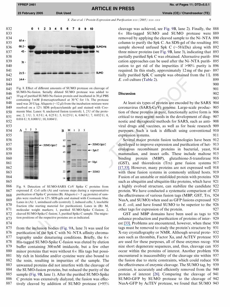
from the inclusion bodies (Fig. 9A, lane 3) was used forpuriWcation of the Spk C with Ni–NTA aYnity chroma-tography under denaturing conditions. BrieXy, the 6£His-tagged SUMO-Spike C-fusion was eluted by elutionbuVer containing 300 mM imidazole, but a few otherminor proteins that were without 6£ His tags but possi-bly rich in histidine and/or cysteine were also bound tothe resin, resulting in impurities of the sample. Theunwanted proteins did not interfere with the cleavage ofthe SUMO-fusion proteins, but reduced the purity of thesample (Fig. 9B, lane 1). After the puriWed SUMO-SpikeC protein was extensively dialyzed, the fusion was eVec-tively cleaved by addition of SUMO protease (>95%
Fig. 8. EVect of diVerent amounts of SUMO protease on cleavage ofSUMO-Nc-fusion. Serially diluted SUMO protease was added to10 �g of puriWed SUMO-Nc-fusion protein and incubated in 30 �l PBScontaining 5 mM �-mercaptoethanol at 30 °C for 1 h. The proteaseused was 20 U/�g. Aliquots (»12 �l) from the incubation mixture wereresolved on a 12% SDS–polyacrylamide gel and stained with Coo-massie blue. Lanes: 0, uncleaved fusion (control); 1, 2 U of the prote-ase; 2, 1 U; 3, 0.5 U; 4, 0.25 U; 5, 0.125 U; 6, 0.063 U; 7, 0.032 U; 8,0.016 U; 9, 0.008 U; 10, 0.004 U.
Fig. 9. Detection of SUMO-SARS CoV Spike C proteins fromexpressed E. Coli cells (A) and various steps during a representativepuriWcation of Spike C proteins (B). Aliquots (» 5 �g protein) of sam-ples were resolved in 12% SDS-gels and stained with Coomassie blue.Lanes in (A): 1, uninduced cells (control); 2, induced cells; 3, insolublefraction (the starting material for puriWcation). Lanes in (B): M,molecular weight markers; 1, puriWed SUMO-Spike C-fusion; 2,cleaved SUMO-Spike C-fusion; 3, puriWed Spike C sample. The migra-tion positions of the respective proteins are as indicated.
TED PROOF
cleavage was achieved, see Fig. 9B, lane 2). Finally, the6£ His-tagged SUMO and SUMO protease wereremoved by applying the cleaved sample to the Ni–NTAcolumn to purify the Spk C. An SDS-gel of the resultingsample showed unfused Spk C (»58 kDa) along withthree minor proteins (see Fig. 9B, lane 3), indicating thatpartially puriWed Spk C was obtained. Alternative puriW-cation approaches can be used after the Ni–NTA puriW-cation to get rid of the impurities if >90% purity isrequired. In this study, approximately 12 mg of the par-tially puriWed SpK C sample was obtained from the 1 LE. coli culture (Table 2).
Discussion
At least six types of protein are encoded by the SARScoronavirus (SARS-CoV) genome. Large-scale produc-tion of these proteins in pure, functionally active form iscritical to meet urgent needs in the development of diag-nostic and therapeutic methods for SARS, such as anti-viral drugs and vaccines, as well as for basic researchpurposes. Such a task is diYcult using conventionalexpression systems.
Several major protein fusion technologies have beendeveloped to improve expression and puriWcation of het-erologous recombinant proteins in bacterial, yeast,mammalian, and insect cells. These include maltosebinding protein (MBP), glutathione-S-transferase(GST), and thioredoxin (Trx) gene fusion systems[17,33]. However, many proteins are not expressed wellwith these fusion systems in commonly utilized hosts.Fusion of an unstable or misfolded protein with proteinssuch as ubiquitin and ubiquitin-like proteins, which havea highly evolved structure, can stabilize the candidateprotein. We have conducted a systematic comparison ofthe eVectiveness of various fusion tags (MBP, GST, Trx,NusA, and SUMO) when used as GFP fusions expressedin E. coli, and have found SUMO to be superior to theother tags for expression of the protein.
GST and MBP domains have been used as tags toenhance production and puriWcation of proteins of inter-est [33]. Problems are encountered, however, when thesetags must be removed to study the protein’s structure byX-ray crystallography or NMR. Although several prote-ases such as thrombin, Factor Xa, and AcTEV proteaseare used for these purposes, all of these enzymes recog-nize short degenerate sequences, and, thus, cleavage canoccur within the proteins of interest. Another problemencountered is inaccessibility of the cleavage site withinthe fusion due to steric constraints, which could reducethe eVectiveness of enzymic cleavage. The SUMO tag, bycontrast, is accurately and eYciently removed from theprotein of interest [26]. Comparing the cleavage ofSUMO-GFP by SUMO protease to the cleavage ofNusA-GFP by AcTEV protease, we found that SUMO
88888888888899999999999999999999999999999999999999999999
832833834835836837838839840841842843844845846847848849850851852853854855856857858859860861862863864865866867868869870871872873874875876877878879880881882883884885886887
8889909192939495969798990001020304050607080910111213141516171819202122232425262728293031323334353637383940414243

10 X. Zuo et al. / Protein Expression and PuriWcation xxx (2005) xxx–xxx
YPREP 2461 No. of Pages 11; DTD=5.0.1ARTICLE IN PRESS
23 February 2005 Disk Used Vimala (CE) / Chandrasekar (TE)
UNCORREC
protease had a 64-fold higher activity than AcTEV pro-tease when the same amount of enzyme was used(unpublished results).
Ubiquitin has been reported to exert chaperoningeVects on fused proteins, thus increasing expression ofproteins in E. coli and yeast [34–36]. The fused proteinscan be cleaved by Ub-proteases (both UCH and UBPclasses), but the enzymes are unstable, diYcult toproduce, and often must be used in large quantities (anenzyme to substrate ratio of 1:1), making this technologyimpractical for large-scale protein production. Our labo-ratory has exploited the chaperoning properties of sev-eral ubiquitin-like proteins including SUMO, and theextreme robustness of SUMO protease has allowed us todevelop a technology that provides both enhancedexpression and cleavage of the fusion protein. A numberof diYcult proteins have been expressed in our labora-tory in both unfused and SUMO-fused forms and com-pared side-by-side to demonstrate that SUMO-fusiondramatically enhances the expression of many types ofproteins, including membrane proteins, and that SUMOprotease cleaves a variety of SUMO-fusions with highspeciWcity and eYciency over a wide range of variousconditions, including pH (5.5–10.5) and temperature (4–37 °C) [26]. Non-speciWc cleavage of the substrate wasnot observed, even when the amount of enzyme wasdeliberately increased to a 1:1 ratio [26]. In this study,titration of the hydrolytic capacity of SUMO proteaseon the puriWed SARS-CoV Nc proteins conWrmed thatSUMO protease is an extremely potent enzyme (Fig. 8).The predicted molecular weight of the SUMO proteaseis 26.7 kDa, though it usually runs at »31 kDa positionon a SDS-polyacrylamide gel.
After evaluating and comparing various SARS-CoVproteins expressed with or without SUMO-fusion in E.coli under several culture and induction conditions, wefound that SUMO-fusion signiWcantly increased expres-sion of SARS proteins under nearly all conditions tested.We established a batch production protocol employing20 °C overnight growth for large-scale expression ofSUMO-SARS-CoV proteins, since a shorter time (e.g.,6 h) or higher temperature (37 °C) resulted in loweryields, especially for soluble proteins (data not shown).Although in most cases, cells growing in rich medium(LB) produced more SUMO-fused protein than cellsgrowing in minimal medium (MM), we will use MM toinvestigate secreted SARS-CoV proteins in future stud-ies, since rich medium contains a large number of inter-fering proteins.
In addition to producing SARS-CoV proteins in largequantities for basic research and for development ofanti-SARS pharmaceutical agents, it is important toproduce pure proteins that retain biological activity. Theexpressed and puriWed SARS-CoV proteins had immu-nological activity, but the question remains concerningtheir functional activities. It appears, at least in the case
TED PROOF
of one SARS-CoV protein, that SUMO-enhancedexpression and puriWcation from E. coli results in activeprotein. In a study to be published, SUMO-fusionenhanced expression of the SARS-CoV RNA-dependentRNA polymerase (RdRp), and the puriWed solubleRdRp was biologically active (S.G.S., manuscript inpreparation). Finally, we recently observed that SUMO-fusion signiWcantly enhanced expression and puriWcationof SARS-CoV membrane protein (M) as well. Using theSUMO-fusion technology described here, the expressionlevel of SARS-CoV M protein in E. coli was greatlyimproved, and the insoluble proteins extracted from thebacterial inclusion bodies were puriWed [32]. Applicationof the various puriWed SARS-CoV proteins to the devel-opment of SARS vaccines and functional assays areunderway.
Acknowledgments
The work described here was supported in part by agrant (R43 GM067271-01) from the NIH/NIGMS toT.R.B. and a grant (RO1-AI 17418) from NIH/NIAIDto S.R.W.
References
[1] T.G. Ksiazek, D. Erdman, C.S. Goldsmith, S.R. Zaki, T. Peret, S.Emery, S. Tong, C. Urbani, J.A. Comer, W. Lim, P.E. Rollin, S.F.Dowell, A.E. Ling, C.D. Humphrey, W.J. Shieh, J. Guarner, C.D.Paddock, P. Rota, B. Fields, J. DeRisi, J.Y. Yang, N. Cox, J.M.Hughes, J.W. LeDuc, W.J. Bellini, L.J. Anderson, A novel corona-virus associated with severe acute respiratory syndrome, N. Engl.J. Med. 348 (2003) 1953–1966.
[2] P.A. Rota, M.S. Oberste, S.S. Monroe, W.A. Nix, R. Campagnoli,J.P. Icenogle, S. Penaranda, B. Bankamp, K. Maher, M.H. Chen,S. Tong, A. Tamin, L. Lowe, M. Frace, J.L. DeRisi, Q. Chen, D.Wang, D.D. Erdman, T.C. Peret, C. Burns, T.G. Ksiazek, P.E.Rollin, A. Sanchez, S. LiYck, B. Holloway, J. Limor, K.McCaustland, M. Olsen-Rasmussen, R. Fouchier, S. Gunther,A.D. Osterhaus, C. Drosten, M.A. Pallansch, L.J. Anderson, W.J.Bellini, Characterization of a novel coronavirus associated withsevere acute respiratory syndrome, Science 300 (2003) 1394–1399.
[3] E. Snijder, P.J. Bredenbeek, J.C. Dobbe, V. Thiel, L.L. Ziebuhr, Y.Poon, Y. Guan, M. Rozanov, W.J. Spaan, A.E. Gorbalenya,Unique and conserved features of genome and proteome ofSARS-coronavirus, an early split-oV from the coronavirus group2 lineage, J. Mol. Biol. 331 (2003) 991–1004.
[4] K. McIntosh, Coronaviruses: a comparative review, Curr. Top.Microbiol. Immunol. 63 (1974) 85–129.
[5] J. Herold, T. Raabe, B. Schelle-Prinz, S.G. Siddell, Nucleotidesequence of the human coronavirus 229E RNA polymerase locus,Virology 195 (1993) 680–691.
[6] J. Herold, T. Raabe, S. Siddell, Molecular analysis of the humancoronavirus (strain 229E) genome, Arch. Virol. Suppl. 7 (1993) 63–74.
[7] J. Herold, T. Raabe, S.G. Siddell, Characterization of the humancoronavirus 229E (HCV 229E) gene 1, Adv. Exp. Med. Biol. 342(1993) 75–79.
1111111111111111
1
1111
1111111111111111111111111111111111111
944945946947948949950951952953954955956957958959960961962963964965966967968969970971972973974975976977978979980981982983984985986987988989990991992993994995996997998999
000001002003004005006007008009010011012013014015
016
017018019020
021022023024025026027028029030031032033034035036037038039040041042043044045046047048049050051052053054055056057

X. Zuo et al. / Protein Expression and PuriWcation xxx (2005) xxx–xxx 11
YPREP 2461 No. of Pages 11; DTD=5.0.1ARTICLE IN PRESS
23 February 2005 Disk Used Vimala (CE) / Chandrasekar (TE)
CORREC
[8] X. Xu, Y. Liu, S. Weiss, E. Arnold, S.G. SaraWanos, J. Ding,Molecular model of SARS coronavirus polymerase: implicationsfor biochemical functions and drug design, Nucleic Acids Res. 31(2003) 7117–7130.
[9] M. Surjit, B. Liu, P. Kumar, V.T. Chow, S.K. Lal, The nucleocap-sid protein of the SARS coronavirus is capable of self-associationthrough a C-terminal 209 amino acid interaction domain, Bio-chem. Biophys. Res. Commun. 317 (2004) 1030–1036.
[10] Y.D. Wang, W.Y. Sin, G.B. Xu, H.H. Yang, T.Y. Wong, X.W.Pang, X.Y. He, H.G. Zhang, J.N. Ng, C.S. Cheng, J. Yu, L. Meng,R.F. Yang, S.T. Lai, Z.H. Guo, Y. Xie, W.F. Chen, T-cell epitopesin severe acute respiratory syndrome (SARS) coronavirus spikeprotein elicit a speciWc T-cell immune response in patients whorecover from SARS, J. Virol. 78 (2004) 5612–5618.
[11] S. Xiong, Y.F. Wang, M.Y. Zhang, X.J. Liu, C.H. Zhang, S.S. Liu,C.W. Qian, J.X. Li, J.H. Lu, Z.Y. Wan, H.Y. Zheng, X.G. Yan, M.J.Meng, J.L. Fan, Immunogenicity of SARS inactivated vaccine inBALB/c mice, Immunol. Lett. 95 (2004) 139–143.
[12] X. Xiao, S. Chakraborti, A.S. Dimitrov, K. GramatikoV, D.S.Dimitrov, The SARS-CoV S glycoprotein: expression and func-tional characterization, Biochem. Biophys. Res. Commun. 312(2003) 1159–1164.
[13] C. Schwegmann-Wessels, M. Al-Falah, D. Escors, Z. Wang, G.Zimmer, H. Deng, L. Enjuanes, H.Y. Naim, G. Herrler, A novelsorting signal for intracellular localization is present in the S pro-tein of a porcine coronavirus but absent from severe acute respira-tory syndrome-associated coronavirus, J. Biol. Chem. 279 (2004)43661–43666.
[14] H. Zhang, G. Wang, J. Li, Y. Nie, X. Shi, G. Lian, W. Wang, X.Yin, Y. Zhao, X. Qu, M. Ding, H. Deng, IdentiWcation of an anti-genic determinant on the S2 domain of the severe acute respira-tory syndrome coronavirus spike glycoprotein capable of inducingneutralizing antibodies, J. Virol. 78 (2004) 6938–6945.
[15] G.S. Waldo, B.M. Standish, J. Berendzen, T.C. Terwilliger, Rapidprotein-folding assay using green Xuorescent protein, Nat. Bio-technol. 17 (1999) 691–695.
[16] L.C. Wright, J. Seybold, A. Robichaud, I.M. Adcock, P.J. Barnes,Phosphodiesterase expression in human epithelial cells, Am. J.Physiol. 275 (1998) L694–L700.
[17] J. Nilsson, S. Stahl, J. Lundeberg, M. Uhlen, P.A. Nygren, AYnityfusion strategies for detection, puriWcation, and immobilization ofrecombinant proteins, Protein Expr. Purif. 11 (1997) 1–16.
[18] A. Hershko, A. Ciechanover, The ubiquitin system, Annu. Rev.Biochem. 67 (1998) 425–479.
[19] S. Muller, C. Hoege, G. Pyrowolakis, S. Jentsch, SUMO, ubiqui-tin’s mysterious cousin, Nat. Rev. Mol. Cell Biol. 2 (2001) 202–210.
[20] S. Jentsch, G. Pyrowolakis, Ubiquitin and its kin: how close arethe family ties?, Trends Cell Biol. 10 (2000) 335–342.
[21] F. Melchior, SUMO—nonclassical ubiquitin, Annu. Rev. CellDev. Biol. 16 (2000) 591–626.
UN
TED PROOF
[22] E.T. Yeh, L. Gong, T. Kamitani, Ubiquitin-like proteins: newwines in new bottles, Gene 248 (2000) 1–14.
[23] H. Saitoh, J. Hinchey, Functional heterogeneity of small ubiqui-tin-related protein modiWers SUMO-1 versus SUMO-2/3, J. Biol.Chem. 275 (2000) 6252–6258.
[24] S. Goettsch, P. Bayer, Structural attributes in the conjugation ofubiquitin, SUMO and RUB to protein substrates, Front Biosci. 7(2002) a148–a162.
[25] P. Bayer, A. Arndt, S. Metzger, R. Mahajan, F. Melchior, R. Jae-nicke, J. Becker, Structure determination of the small ubiquitin-related modiWer SUMO-1, J. Mol. Biol. 280 (1998) 275–286.
[26] M.P. Malakhov, M.R. Mattern, O.A. Malakhova, M. Drinker,S.D. Weeks, T.R. Butt, SUMO fusions and SUMO-speciWc prote-ase for eYcient expression and puriWcation of proteins, J. Struct.Funct. Genom. 5 (2004) 75–86.
[27] S.J. Li, M. Hochstrasser, A new protease required for cell-cycleprogression in yeast, Nature 398 (1999) 246–251.
[28] S.J. Li, M. Hochstrasser, The yeast ULP2 (SMT4) gene encodes anovel protease speciWc for the ubiquitin-like Smt3 protein, Mol.Cell. Biol. 20 (2000) 2367–2377.
[29] T. Suzuki, A. Ichiyama, H. Saitoh, T. Kawakami, M. Omata, C.H.Chung, M. Kimura, N. Shimbara, K. Tanaka, A new 30-kDa ubiq-uitin-related SUMO-1 hydrolase from bovine brain, J. Biol. Chem.274 (1999) 31131–31134.
[30] K.I. Kim, S.H. Baek, C.H. Chung, Versatile protein tag, SUMO: itsenzymology and biological function, J. Cell Physiol. 191 (2002)257–268.
[31] K.I. Kim, S.H. Baek, Y.J. Jeon, S. Nishimori, T. Suzuki, S. Uchida,N. Shimbara, H. Saitoh, K. Tanaka, C.H. Chung, A new SUMO-1-speciWc protease, SUSP1, that is highly expressed in reproduc-tive organs, J. Biol. Chem. 275 (2000) 14102–14106.
[32] X. Zuo, S. Li, J. Hall, M.R. Mattern, H. Tran, J. Shoo, R. Tan, S.R.Weiss, T.R. Butt, Enhanced expression and puriWcation of mem-brane proteins by SUMO fusion in Escherichia coli, J. Struct.Funct. Genom. (2005) (in press).
[33] P. Jonasson, S. Liljeqvist, P.A. Nygren, S. Stahl, Genetic design forfacilitated production and recovery of recombinant proteins inEscherichia coli, Biotechnol. Appl. Biochem. 35 (2002) 91–105.
[34] D.P. McDonnell, J.W. Pike, D.J. Drutz, T.R. Butt, B.W. O’Malley,Reconstitution of the vitamin D-responsive osteocalcin transcrip-tion unit in Saccharomyces cerevisiae, Mol. Cell. Biol. 9 (1989)3517–3523.
[35] D.J. Ecker, J.M. Stadel, T.R. Butt, J.A. Marsh, B.P. Monia, D.A.Powers, J.A. Gorman, P.E. Clark, F. Warren, A. Shatzman,Increasing gene expression in yeast by fusion to ubiquitin, J. Biol.Chem. 264 (1989) 7715–7719.
[36] T.R. Butt, S. Jonnalagadda, B.P. Monia, E.J. Sternberg, J.A.Marsh, J.M. Stadel, D.J. Ecker, S.T. Crooke, Ubiquitin fusion aug-ments the yield of cloned gene products in Escherichia coli, Proc.Natl. Acad. Sci. USA 86 (1989) 2540–2544.
11111111111111111111111111111111111111111111111111
10581059106010611062106310641065106610671068106910701071107210731074107510761077107810791080108110821083108410851086108710881089109010911092109310941095109610971098109911001101110211031104110511061107
108109110111112113114115116117118119120121122123124125126127128129130131132133134135136137138139140141142143144145146147148149150151152153154155156157
Copyright © 2022 FDOKUMEN