“Spinal Fusions Advanced Applications” will begin shortly.
53
UASI Lunch and Learn: “Spinal Fusions Advanced Applications” will begin shortly. Service Stats ▪ 4 out of 5 UASI clients request ongoing or return services following an initial engagement • UASI works for top hospitals delivering value tailored to our client’s specific needs • CONSULTANTS average 18 years in HIM/Coding • MANAGERS average 26 years in HIM/coding
-
Upload
khangminh22 -
Category
Documents
-
view
5 -
download
0
Transcript of “Spinal Fusions Advanced Applications” will begin shortly.

UASI Lunch and Learn: “Spinal Fusions Advanced Applications” will begin shortly.
Service Stats
▪ 4 out of 5 UASI clients
request ongoing or return
services following an initial
engagement
• UASI works for top
hospitals delivering value
tailored to our client’s
specific needs
• CONSULTANTS average 18
years in HIM/Coding
• MANAGERS average 26
years in HIM/coding

June 2021
Spinal Fusions Advanced Applications
Presented By: Natalie Sartori M.Ed., RHIA, CCS

Course Description:• This presentation is designed to provide a thorough review of ICD-10-PCS coding for spinal
fusions.
• Content will include a review of pertinent anatomy and physiology, coding guidelines and important coding clinic references that provide guidance in correct code assignment.
• Finally, we’ll examine a sample operative report to determine what documentation to look for when coding spinal fusions.
Learning Objectives:• Review spinal and vertebral anatomy
• Analyze coding guidelines for spinal fusion
• Examine Coding Clinic references applicable to spinal fusion coding
• Assess documentation requirements for PCS code assignment
Introduction:• A spinal fusion surgery is designed to stop the motion at a painful vertebral segment, which
in turn should decrease pain generated from the joint.
• Spinal fusion surgery comes in many forms: lumbar spinal fusion, cervical spinal fusion, and PLIFs just to name a few.
• They are all designed to help limit pain caused by the joints, though each surgery is different depending on whether you are trying to treat degenerative disc disease, spondylolisthesis, or another condition.

A&P Vertebral Column:• The vertebrae in the human vertebral column are divided into different regions, which
correspond to the curves of the spinal column.
• The articulating vertebrae are named according to their region of the spine.
• There are seven cervical vertebrae, twelve thoracic vertebrae and five lumbar vertebrae.
• The Sacrum is located behind the pelvis. Five bones (abbreviated S1 through S5) fused into a triangular shape, form the sacrum. The sacrum fits between the two hipbones connecting the spine to the pelvis. The last lumbar vertebra (L5) articulates (moves) with the S1 sacral bone.
• Immediately below the sacrum are five additional bones, fused together to form the Coccyx (tailbone).

A&P Vertebral Column:

Vertebral Anatomy:• Each vertebral segment is comprised of a cylinder-shaped bone in the front of the spine,
called the vertebral body, a soft cartilage disc between each vertebra, and paired facet joints in the back.
• Each segment is named for its upper and lower vertebra, such as the C6-C7 segment, or the L4-L5 segment.
• The bones in the spinal column surround and protect the spinal cord, which runs behind the vertebral bodies in a canal from the neck down to the top of the lumbar spine.
• At each segment, spinal nerve roots exit the spine through holes in the back of the vertebrae called foramina.
• The entire spine is knit together by a series of interconnected ligaments and tendons that help support and stabilize the spine while allowing a great deal of flexibility.

Vertebrae Anatomy:

Vertebral Ligaments:

Fusion Procedures of the Spine:B3.10a
• The body part coded for a spinal vertebral joint(s) rendered immobile by a spinal fusion procedure is classified by the level of the spine (e.g. thoracic).
• There are distinct body part values for:• a single vertebral joint – for example C6-C7
• for multiple vertebral joints at each spinal level – for example T3-T11
• transitioning levels – for example L5-S1
Example: Body part values specify Lumbar Vertebral Joint, Lumbar Vertebral Joints, 2 or More and Lumbosacral Vertebral Joint and lumboarsacral joint.

Fusion Procedures of the Spine:B3.10b
• If multiple vertebral joints are fused, a separate procedure is coded for each vertebral joint that uses a different device and/or qualifier.
Example: Fusion of lumbar vertebral joint, posterior approach, anterior column and fusion of lumbar vertebral joint, posterior approach, posterior column are coded separately.

Fusion Procedures of the Spine:Body Part Approach Device Qualifier
0 Occipital-cervical Joint
1 Cervical Vertebral Joint
2 Cervical Vertebral Joints, 2 or more
4 Cervicothoracic Vertebral Joint
6 Thoracic Vertebral Joint
7 Thoracic Vertebral Joints, 2 to 7
8 Thoracic Vertebral Joints, 8 or more
A Thoracolumbar Vertebral Joint
0 Open
3 Percutaneous
4 Percutaneous
Endoscopic
7 Autologous Tissue
Substitute
J Synthetic Substitute
K Nonautologous
Tissue Substitute
0 Anterior Approach,
Anterior Column
1 Posterior Approach,
Posterior Column
J Posterior Approach,
Anterior Column
0 Occipital-cervical Joint
1 Cervical Vertebral Joint
2 Cervical Vertebral Joints, 2 or more
4 Cervicothoracic Vertebral Joint
6 Thoracic Vertebral Joint
7 Thoracic Vertebral Joints, 2 to 7
8 Thoracic Vertebral Joints, 8 or more
A Thoracolumbar Vertebral Joint
0 Open
3 Percutaneous
4 Percutaneous
Endoscopic
A Interbody Fusion
Device
0 Anterior Approach,
Anterior Column
J Posterior Approach,
Anterior Column
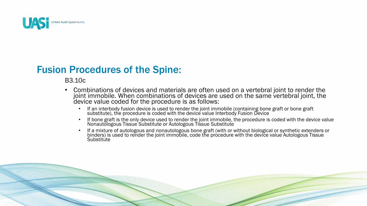
Fusion Procedures of the Spine:B3.10c
• Combinations of devices and materials are often used on a vertebral joint to render the joint immobile. When combinations of devices are used on the same vertebral joint, the device value coded for the procedure is as follows: • If an interbody fusion device is used to render the joint immobile (containing bone graft or bone graft
substitute), the procedure is coded with the device value Interbody Fusion Device
• If bone graft is the only device used to render the joint immobile, the procedure is coded with the device value Nonautologous Tissue Substitute or Autologous Tissue Substitute
• If a mixture of autologous and nonautologous bone graft (with or without biological or synthetic extenders or binders) is used to render the joint immobile, code the procedure with the device value Autologous Tissue Substitute

Fusion Procedures of the Spine:B3.10c
Examples:
• Fusion of a vertebral joint using a cage style interbody fusion device containing morselized bone graft is coded to the device Interbody Fusion Device.
• Fusion of a vertebral joint using a bone dowel interbody fusion device made of cadaver bone and packed with a mixture of local morselized bone and demineralized bone matrix is coded to the device Interbody Fusion Device.
• Fusion of a vertebral joint using both autologous bone graft and bone bank bone graft is coded to the device Autologous Tissue Substitute.

Excision for Graft:B3.9
• If an autograft is obtained from a different procedure site in order to complete the objective of the procedure, a separate procedure is coded.
Examples:
• Harvesting a bone graft from the iliac crest for a cervical fusion would require separate code.
• Using small pieces of vertebral bone from the fusion procedure does not require a separate code.

Release Procedures:B3.13
• In the root operation Release, the body part value coded is the body part being freed and not the tissue being manipulated or cut to free the body part.
Example: Removing a portion of the posterior longitudinal ligament to decompress the nerve at C6-C7.

Body System:Spinal Fusions are coded to one of two Body Systems depending on the level(s) being fused:
• R = Upper Joints – Includes the occipital-cervical, cervical, cervicothoracic, thoracic and thoracolumbar joints.
• S =Lower Joints – Includes the lumbar and lumbosacral joints.

Root Operation:• Fusion (G) - Joining together portions of an articular body part rendering the articular body
part immobile
• Explanation: The body part is joined together by fixation device, bone graft, or other means

Body Part:The correct body part character reflects:
• the level of the vertebrae (cervical, thoracic, lumbar and/or sacral)
• the number of vertebral joints fused
The intervertebral joint is the space that is located between any two adjacent vertebrae.
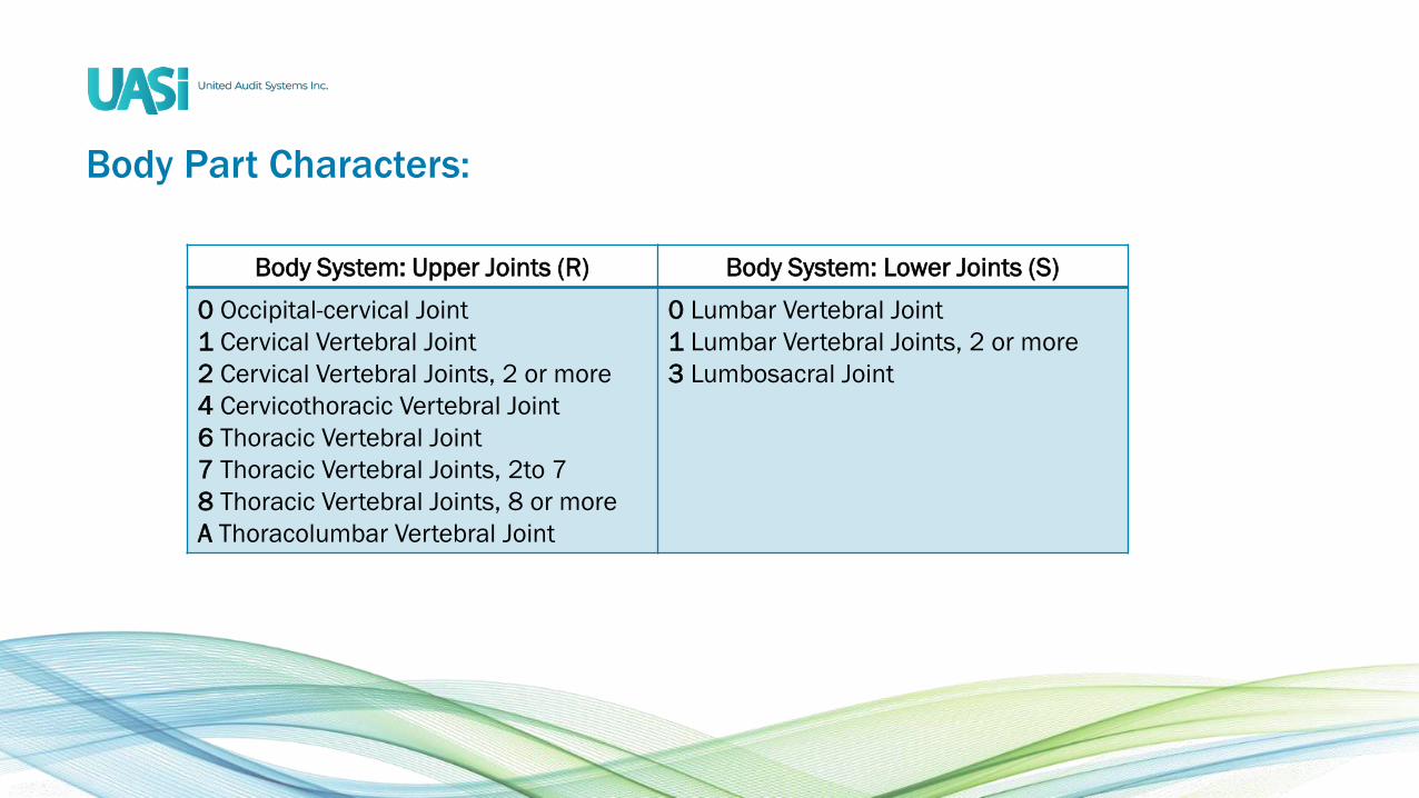
Body Part Characters:
Body System: Upper Joints (R) Body System: Lower Joints (S)
0 Occipital-cervical Joint
1 Cervical Vertebral Joint
2 Cervical Vertebral Joints, 2 or more
4 Cervicothoracic Vertebral Joint
6 Thoracic Vertebral Joint
7 Thoracic Vertebral Joints, 2to 7
8 Thoracic Vertebral Joints, 8 or more
A Thoracolumbar Vertebral Joint
0 Lumbar Vertebral Joint
1 Lumbar Vertebral Joints, 2 or more
3 Lumbosacral Joint

Approach:• The most common approach for spinal fusion is open (0).
• However, percutaneous endoscopic (4) approach procedures are becoming more common.

Device:Spinal fusion can be performed using several different techniques. These techniques include:
• Interbody fusion devices (A) - Stabilize and fuse the disc spaces and provide an immediately stable segment for the fusion. These devices are also known as interbody fusion cages, BAK cage, synthetic cage, or bone dowels.
• Autologous Tissue Substitute (7) – A bone graft obtained from the patient during the procedure. Bone grafts may be harvested locally using the same incision or from another part of the body requiring a separate incision. Harvesting of the bone requires a separate procedure code when it is performed through a separate incision. (Guideline B3.9) Morselized bone fragments harvested from the same incision during the approach to operative site does not require a separate code.
• Nonautologous Tissue Substitute (K) – The bone is harvested by a tissue bank from a cadaver.
• Synthetic Substitute (J) – these types of grafts are synthetic or a manipulated naturally occurring product.

Common Interbody Devices:
PEEK Cage Optimesh Bone Dowel

Common Interbody Devices:
VERTE-STACK

Fusion Procedure Devices:• For FY 2019 revisions were made to tables ORG Fusion of Upper Joints and OSG Fusion of
Lower Joints by removing the device character “No Device (Z)” from all rows for all body parts.
• Because a fusion procedures, including spinal fusion, always requires some type of device codes with the device character “Z- No Device” are considered clinically invalid.

FY 2018 Updates
The FY 2018 PCS updates created separate row for the Device character “A Interbody Fusion Device”.
This row only has clinically valid choices for the qualifiers specifying Anterior Column – see example
below.
Section 0 Medical and Surgical
Body System S Lower Joints
Operation G Fusion: Joining together portions of an articular body part rendering the articular body part
immobile
Body Part Approach Device Qualifier
0 Lumbar Vertebral Joint
1 Lumbar Vertebral Joints, 2 or more
3 Lumbosacral Joint
0 Open
3 Percutaneous
4 Percutaneous
Endoscopic
A Interbody Fusion
Device
0 Anterior Approach,
Anterior Column
J Posterior Approach,
Anterior Column

Qualifier:
The qualifier characters identify the portion of the spine being fused (anterior or posterior) and if the
surgical approach is from the front or back of the body.
Anterior Approach,
Anterior Column (0)
Posterior Approach,
Posterior Column (1)
Posterior Approach,
Anterior Column (J)
Look for supine (face up)
Positioning
Look for prone (back up)
positioning
Look for prone (back up)
Positioning
Look for an incision made
on the front or side of the
body
Look for an incision made
on the back
Look for an incision made
on the back
The vertebral body will be
fused (Interbody fusion)
Structures on the posterior
spine are fused.
The vertebral body will be
fused (Interbody fusion)

Qualifier Approach:
Supine - the position of a person lying on the back. This position is normally utilized for anterior
approach.
Prone - is a body position in which one lies flat with the chest down and the back up. In anatomical
terms of location, the dorsal side is up, and the ventral side is down. This position is normally utilized for
posterior approach.

Qualifier Approach:

Qualifier Column:
Anterior
Column
Posterior
Column

Common Abbreviations Anterior Column Fusions:
Procedure Approach Qualifier
Anterior lumbar interbody
fusion (ALIF)
Incision made in front of the spine
through a minilaparotomy or
Laparoscopy
0 Anterior Approach,
Anterior Column
Posterior lumbar interbody
fusion (PLIF)
Incision made through a midline
incision in the back
J Posterior Approach,
Anterior Column
Extreme lateral interbody
fusion (XLIF)
Incision made in the patient’s side 0 Anterior Approach,
Anterior Column
Direct lateral interbody fusion
(DLIF)
Incision made in the patient’s side 0 Anterior Approach,
Anterior Column
Transforaminal lumbar
interbody fusion (TLIF)
Incision made through a midline
incision in the back
J Posterior Approach,
Anterior Column
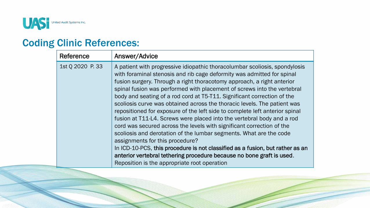
Coding Clinic References:
Reference Answer/Advice
1st Q 2020 P. 33 A patient with progressive idiopathic thoracolumbar scoliosis, spondylosis
with foraminal stenosis and rib cage deformity was admitted for spinal
fusion surgery. Through a right thoracotomy approach, a right anterior
spinal fusion was performed with placement of screws into the vertebral
body and seating of a rod cord at T5-T11. Significant correction of the
scoliosis curve was obtained across the thoracic levels. The patient was
repositioned for exposure of the left side to complete left anterior spinal
fusion at T11-L4. Screws were placed into the vertebral body and a rod
cord was secured across the levels with significant correction of the
scoliosis and derotation of the lumbar segments. What are the code
assignments for this procedure?
In ICD-10-PCS, this procedure is not classified as a fusion, but rather as an
anterior vertebral tethering procedure because no bone graft is used.
Reposition is the appropriate root operation

Coding Clinic References:
Reference Answer/Advice
3rd Q 2019 P. 35 An interbody fusion device must contain bone graft material or other material
like bone graft material in order to immobilize and join the vertebrae. Guideline
B3.10 refers to the hierarchy in reporting the combinations of devices that may
be used for fusion of the vertebral joint. When an interbody fusion device is
used, assign the device value "A, Interbody fusion device." "Alone" in the
guideline refers to interbody fusion devices such as cortical bone dowels or
intervertebral body spacers that are composed of bone. Other interbody fusion
devices are made of metal or plastic and are packed with bone or bone-like
material that is placed in or around the implant to join the vertebrae to stabilize
the spinal column and prohibit movement. "A, Interbody fusion device" is
appropriate in both instances.

Coding Clinic References:Reference Answer/Advice
2nd Q 2019 P.
35
The DTRAX Spinal System is a set of instruments intended and indicated for access
and preparation of a spinal joint to aid in fusion. When assigning ICD-10-PCS codes
for procedures using DTRAX spinal instruments, coding professionals should code
the procedure based on what was done, rather than the device used. In this case, a
posterior fusion between the facet (interfacet) was done, not an interbody fusion. If
the documentation is unclear, query the physician for clarification.
1st Q 2019 P.
30
In this case, the decompressive laminectomy was performed to treat a separately
documented diagnosis of lumbar spinal stenosis. Since there is a distinct objective,
it is appropriate to code decompressive laminectomy even though it was performed
at the same level as the lumbar spinal fusion. The root operation Release is coded
separately when decompression is documented, and there is a distinct surgical
objective, not just incidental removal of the lamina to reach the site of the
procedure. If the laminectomy is done as an operative approach to prepare for the
spinal fusion, it is not coded separately.

Coding Clinic References:
Reference Answer/Advice
3rd Q 2018 CC P. 30 Decompression was performed bilaterally from L2-L3, L3-L4 and L4-L5. The
thecal sac was then free of compression. For this type of procedure, the surgeon
will typically document release of "thecal sac." The appropriate body part for the
decompressive laminectomies is lumbar spinal cord.
1st Q 2018 CC P. 22 The root operation of “fusion” does not require the use of bone graft however,
the spinal fusion guideline indicates that a spinal fusion requires bone graft.
1st Q 2018 CC P. 8 Reporting a code for the placement of BMP is optional and facilities may code it,
if desired. When an open approach is used, assign the following ICD- 10-PCS
code:
3E0U0GB Introduction of recombinant bone morphogenetic
protein into joints, open approach

Coding Clinic References:
Reference Answer/Advice
2nd Q 2016 P. 17 During an interbody fusion at C6-C7 with anterior discectomy and anterior
neural foraminal decompression, a rongeur was used to remove the right
third of the posterior longitudinal ligament at the C6-C7 level. In addition to
coding the interbody fusion and discectomy a code is assigned separately
for the removal of the ligament “Release”. The root operation “Release”
meets the objective of removing the ligament in order to fully decompress
the C7 nerve root.
2nd Q 2015 P. 21 A patient with cervical myelopathy and the surgeon performed an open
total decompressive laminectomy of C3-C6. The laminectomy procedure to
release the spinal cord is coded only once because the cervical spinal cord
is classified as a single body part. Each spinal and or nerve root has a
distinct body part character for each level so a code should be reported for
each level.

Coding Clinic References:
Reference Answer/Advice
3rd Q 2014 P.
30
Fixation devices (rods, plates, screws) used in conjunction with a
spinal fusion procedure is included in the fusion root operation and
no additional code is assigned.
3rd Q 2014 P.
36;
2nd Q 2014 P.
6
The correct codes for a posterior lumbar interbody fusion of L3-L5
using autologous bone graft harvested from the right iliac crest
and pedicle screw instrumentation , L3-L5 discectomy are:
0SG107J – Fusion of 2 or more lumbar vertebrae with autologous
tissue, anterior column, posterior approach
0SB20ZZ – Excision of lumbar vertebral disc, open
0QB20ZZ – Excision of right pelvic bone, open

Coding Clinic References:Reference Answer/Advice
2nd Q 2014 P. 7 The correct codes for an anterior cervical-thoracic (C7-T1) spinal fusion, using
interbody cage packed with autograft and demineralized bone matrix,
placement of plates and screws with total discectomy are:
0RG40A0 – Fusion of cervicothoracic vertebral joint with interbody
fusion device, anterior column anterior approach, open
0RT50ZZ – Resection of cervicothoracic vertebral disc, open
Spinal fusion using an interbody cage with demineralized bone matrix and
autograft is coded to the device value “Interbody Fusion Device” (PCS Guideline
B3.10c). The fixation instrumentation is included in the fusion root operation
and no additional code is assigned.
3rd Q 2013 P. 25 A 360 degree fusion involves fusing both the anterior and posterior column and
there fore each procedure is going to have a different Qualifier (PCS Guideline
B3.10b). 360 degree fusions often utilize different devices for the anterior and
posterior column.

Coding Clinic References:
Reference Answer/Advice
1st Q 2013 P. 21 Fusion of T6 – L2 requires three fusion codes based on distinct body part
values for the vertebrae fused. (PCS Guidelines B3.2a and B3.10a). All fusions
performed with autologous tissue substitute, posterior approach posterior
column
0RG7071 – Fusion 2-7 thoracic (T6-T12) vertebral joints
0RGA071 – Fusion thoracolumbar (T12-L1) vertebral joints
0SG0071 – Fusion one lumbar (L1-L2) vertebral joint
1st Q 2013 P. 21 Autografts, allografts and BMP placement are identified by the device
character in the fusion code
1st Q 2013 P. 29 A Smith-Robinson approach to a C7-T1 fusion involves an anterior approach
anterior column.

Report Additional Procedure Codes for:• “Excision” or “Resection” of intervertebral disc for each vertebral level (cervical, thoracic,
lumbar) not individual discs.
• Removal of previously placed interbody fusion devices that are removed due to pseudoarthrosis and/or complication.
• Decompression of spinal nerves and/or spinal cord using the root operation “Release”.
• Harvesting of bone or bone marrow if harvested from a different site requiring a new incision or approach.
• Fixation devices placed above or below the level of the spinal fusion.

Description of Procedure:
The patient was placed prone onto a Jackson spinal frame. Her back was prepped and draped in usual sterile fashion and a straight midline approach to the lumbar spine was performed through a 4.5 inch incision. Subperiosteal exposure was carried out from the tip of the transverse process from L2-L5 bilaterally and decortication was carried our using a rongeur. Transpedicular screws were then placed in the pedicles at L2, L3, L4 and L5 bilaterally. Segmental decompression was then carried out by using a rongeur to take down supraspinous and interspinous ligaments. Rongeurs and Kerrison were utilized to perform bilateral hemilaminectomies from L2-L5. Direct decompression of bilateral nerve roots was carried out through foraminotomies and pedicle to pedicle decompression. Complete decompression of the nerve root was achieved. Probe was passed into each neural foramen and confirmed wide decompression. Annulotomy was then made in the disks between L2-L5. Using intradiscal scrapers I was able to distract and reduce the spondylolisthesis at L4-L5 and endplates were curetted down to bleeding subchrondal bone. The Optimesh mechanical body device was then selected, appropriately sized, and packed with structural allograft bone, thereby providing a rigid anterior column support and biomechanical device at each of the 3 levels. Two rods were cut and contoured into lordosis, placed into the screws from L2-5, reduction maneuvers performed, which thereby reduced the degenerative scoliosis. Set screws were placed and torqued using manufacturers’ provided torque wrench. Two cross connectors were placed and torqued and final Xrays were taken. A separate skin incision had been made over the right iliac crest, Jamshidi needle introduced and bone marrow was aspirated. This bone marrow was processed in the arterial site system, returned back to the patient as packed cells and the bone marrow concentrate was retained for bone grafting and mixed with allograft bone demineralized bone matrix and a gram of vancomycin. This bone graft was packed in direct continuity at the transverse process from L2-5 bilaterally. This was done after the wound was pulsatile lavagedout with 3 L of antibiotic containing saline plus 1 additional liter of normal saline. Final check of the wound was performed. There was no active bleeding. Devitalized muscular tissue was debrided sharply with pickups and scissors, and deep fascia was closed using interrupted and running #1 Vicryl sutures.

PCS Coding Example Answers:Answers: Rationale for each individual code is on subsequent slides followed by a copy of the Op Note with documentation color coded based on the codes below.
• 0SG10AJ – Fusion 2-4 Lumbar Joints, Posterior Approach, Anterior Column
• 0SG1071 – Fusion 2-4 Lumbar Joints, Posterior Approach, Posterior Column
• 07DR3ZZ – Extraction of Iliac Bone Marrow, Percutaneous Approach
• 01NB0ZZ - Release of Lumbar Nerve, Open

0SG10AJ – Fusion 2-4 Lumbar Joints, Posterior Approach, Anterior ColumnPCS Code Rationale
0 Medical & Surgical Section
S Lower Joints – the lumbar and sacral vertebrae are considered lower joints
G Fusion –
1 Lumbar vertebral joints 2 or more - the number of joints fused is three and include L2-3, L3-4 and L4-5
0 Open - the Op Note states “Her back was prepped and draped in usual sterile fashion and a straight midline
approach to the lumbar spine was performed through a 4.5 inch incision. Subperiosteal exposure was carried
out from the tip of the transverse process from L2-L5 bilaterally and decortication was carried our using a
rongeur.
A Interbody fusion device - The Optimesh mechanical body device was then selected, appropriately sized, and
packed with structural allograft bone, thereby providing a rigid anterior column support and biomechanical
device at each of the 3 levels.
J Posterior Approach Anterior Column – the Op notes states the patient was placed in “prone’ or face down
position which represents a posterior approach. Interbody devices are placed between 2 vertebral bodies
which is the anterior column.

0SG1071 – Fusion 2-4 Lumbar Joints, Posterior Approach, Posterior ColumnPCS Code Rationale
0 Medical & Surgical Section
S Lower Joints – the lumbar and sacral vertebrae are considered lower joints
G Fusion –
1 Lumbar vertebral joints 2 or more - the number of joints fused is three and include L2-3, L3-4 and L4-5
0 Open - the Op Note states “Her back was prepped and draped in usual sterile fashion and a straight midline
approach to the lumbar spine was performed through a 4.5 inch incision. Subperiosteal exposure was carried
out from the tip of the transverse process from L2-L5 bilaterally and decortication was carried our using a
rongeur.
7 Autologous Tissue Substitute – the Op Note states “the bone marrow concentrate was retained for bone
grafting and mixed with allograft bone demineralized bone matrix and a gram of vancomycin” as directed in
PCS Guideline B3.10c
1 Posterior Approach Posterior Column - the Op notes states the patient was placed in “prone’ or face down
position which represents a posterior approach. This bone graft was packed in direct continuity at the
transverse process from L2-5 bilaterally – the transverse processes are part of the posterior column.

07DR3ZZ – Extraction of Iliac Bone Marrow, Percutaneous Approach
PCS Code Rationale
0 Medical & Surgical Section
7 Lymphatic & Hemic System – in this case bone marrow is being used not actual bone
D Extraction – 4th Q CC P.111 states biopsy of bone marrow is coded to the root operation “Extraction” the
technique to retrieve the bone marrow whether for biopsy or other reason is the same.
R Bone Marrow Iliac Op Note states “A separate skin incision had been made over the right iliac crest, Jamshidi
needle introduced and bone marrow was aspirated.”
3 Percutaneous – Although the Op Note documents an incision was made the iliac crest is a superficial bone that
can easily be palpated through the skin it would only require a minor incision to expose the bone. The definition
of “Percutaneous” includes entry by minor incision. Without further documentation to support an Open
approach Percutaneous is the correct approach.
Z No Device
Z No Qualifier
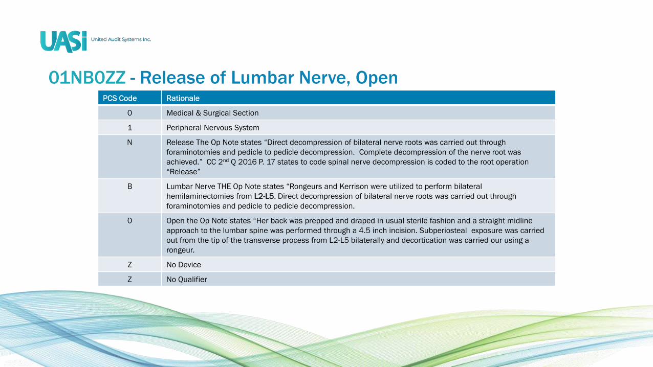
01NB0ZZ - Release of Lumbar Nerve, OpenPCS Code Rationale
0 Medical & Surgical Section
1 Peripheral Nervous System
N Release The Op Note states “Direct decompression of bilateral nerve roots was carried out through
foraminotomies and pedicle to pedicle decompression. Complete decompression of the nerve root was
achieved.” CC 2nd Q 2016 P. 17 states to code spinal nerve decompression is coded to the root operation
“Release”
B Lumbar Nerve THE Op Note states “Rongeurs and Kerrison were utilized to perform bilateral
hemilaminectomies from L2-L5. Direct decompression of bilateral nerve roots was carried out through
foraminotomies and pedicle to pedicle decompression.
0 Open the Op Note states “Her back was prepped and draped in usual sterile fashion and a straight midline
approach to the lumbar spine was performed through a 4.5 inch incision. Subperiosteal exposure was carried
out from the tip of the transverse process from L2-L5 bilaterally and decortication was carried our using a
rongeur.
Z No Device
Z No Qualifier

Description of Procedure:
The patient was placed prone onto a Jackson spinal frame. Her back was prepped and draped in usual sterile fashion and a straight midline approach to the lumbar spine was performed through a 4.5 inch incision. Subperiosteal exposure was carried out from the tip of the transverse process from L2-L5 bilaterally and decortication was carried our using a rongeur. Transpedicular screws were then placed in the pedicles at L2, L3, L4 and L5 bilaterally. Segmental decompression was then carried out by using a rongeur to take down supraspinous and interspinous ligaments. Rongeurs and Kerrison were utilized to perform bilateral hemilaminectomies from L2-L5. Direct decompression of bilateral nerve roots was carried out through foraminotomies and pedicle to pedicle decompression. Complete decompression of the nerve root was achieved. Probe was passed into each neural foramen and confirmed wide decompression. Annulotomy was then made in the disks between L2-L5. Using intradiscal scrapers I was able to distract and reduce the spondylolisthesis at L4-L5 and endplates were curetted down to bleeding subchrondal bone. The Optimesh mechanical body device was then selected, appropriately sized, and packed with structural allograft bone, thereby providing a rigid anterior column support and biomechanical device at each of the 3 levels. Two rods were cut and contoured into lordosis, placed into the screws from L2-5, reduction maneuvers performed, which thereby reduced the degenerative scoliosis. Set screws were placed and torqued using manufacturers’ provided torque wrench. Two cross connectors were placed and torqued and final Xrays were taken. A separate skin incision had been made over the right iliac crest, Jamshidi needle introduced and bone marrow was aspirated. This bone marrow was processed in the arterial site system, returned back to the patient as packed cells and the bone marrow concentrate was retained for bone grafting and mixed with allograft bone demineralized bone matrix and a gram of vancomycin. This bone graft was packed in direct continuity at the transverse process from L2-5 bilaterally. This was done after the wound was pulsatile lavagedout with 3 L of antibiotic containing saline plus 1 additional liter of normal saline. Final check of the wound was performed. There was no active bleeding. Devitalized muscular tissue was debrided sharply with pickups and scissors, and deep fascia was closed using interrupted and running #1 Vicryl sutures.

Questions?
Natalie Sartori, M.Ed., RHIA, CCS
Corporate Trainer

Community Involvement:UASI has always provided a way for the company and our employees to give back through charitable events such as:
• In 2020 UASI employees donated-more than $5,000 to an employee who lost their home in Hurricane Laura
• UASI’s Virtual Giving Tree-UASI employees donated gifts to St. Joseph Orphanage
• Habitat for Humanity-UASI employees worked to rehab a house in Cincinnati
• UASI’s We Fight Together-campaign to raise awareness and money for breast cancer research
• UASI’s Red Cross Donation Drive-Raised over $5,000 to benefit victims of the tornadoes
• UASI’s Summer Give Back-Our employees shared their stories of volunteerism in over 22 charities
• UASI 30Thirty30 Program-In celebration of 30 years in business UASI donated $30,000 to selected charities
• Adopt A Class Program-serving local schools

Employee Recognition:As a company with a remote workforce, we recognize the importance of feeling connected. Below are some of the great ways we recognize our staff:
• President’s Award
• Years of Service Awards
• Holiday and Summer recognition gifts
• Recognition through “professional weeks”
• Birthday Card (signed by President)
• Health and Wellness Challenges
• Quality Award Program
• Community Involvement

How do I apply?
Holly Saylor
Director, Human Resources
Tracy Knox
Recruiter
Ann Schmidt
Recruiter
Holly Sheward
Manager, Recruitment
Apply directly on the Careers page at
www.uasisolutions.com/careers
Ask a question to our HR and
Recruitment Team by emailing

At A Glance:Since 1984, UASI has set the industry standard in
coding, health information management, clinical
documentation and revenue integrity.
Founded in 1984
Cincinnati, Ohio
37 yearsIn business
400+Employees including AHIMA/AAPC-certified
coders, HIM and clinical documentation specialists
200+Hospitals/Health Systems nationwide
40 ClientsIn the US News & World Report’s
Best Regional and Honor Roll Hospitals