The role of gene fusions in the evolution of metabolic pathways: the histidine biosynthesis case
17
BioMed Central Page 1 of 17 (page number not for citation purposes) BMC Evolutionary Biology Open Access Research The role of gene fusions in the evolution of metabolic pathways: the histidine biosynthesis case Renato Fani* 1 , Matteo Brilli 1 , Marco Fondi 1 and Pietro Lió 2 Address: 1 Dept. of Animal Biology and Genetics, via Romana 17, 50125 Florence, Italy and 2 Computer Laboratory, University of Cambridge, CB3 0FD, Cambridge, UK Email: Renato Fani* - [email protected]; Matteo Brilli - [email protected]; Marco Fondi - [email protected]; Pietro Lió - [email protected] * Corresponding author Abstract Background: Histidine biosynthesis is one of the best characterized anabolic pathways. There is a large body of genetic and biochemical information available, including operon structure, gene expression, and increasingly larger sequence databases. For over forty years this pathway has been the subject of extensive studies, mainly in Escherichia coli and Salmonella enterica, in both of which details of histidine biosynthesis appear to be identical. In these two enterobacteria the pathway is unbranched, includes a number of unusual reactions, and consists of nine intermediates; his genes are arranged in a compact operon (hisGDC [NB]HAF [IE]), with three of them (hisNB, hisD and hisIE) coding for bifunctional enzymes. We performed a detailed analysis of his gene fusions in available genomes to understand the role of gene fusions in shaping this pathway. Results: The analysis of HisA structures revealed that several gene elongation events are at the root of this protein family: internal duplication have been identified by structural superposition of the modules composing the TIM-barrel protein. Several his gene fusions happened in distinct taxonomic lineages; hisNB originated within γ- proteobacteria and after its appearance it was transferred to Campylobacter species (ε- proteobacteria) and to some Bacteria belonging to the CFB group. The transfer involved the entire his operon. The hisIE gene fusion was found in several taxonomic lineages and our results suggest that it probably happened several times in distinct lineages. Gene fusions involving hisIE and hisD genes (HIS4) and hisH and hisF genes (HIS7) took place in the Eukarya domain; the latter has been transferred to some δ-proteobacteria. Conclusion: Gene duplication is the most widely known mechanism responsible for the origin and evolution of metabolic pathways; however, several other mechanisms might concur in the process of pathway assembly and gene fusion appeared to be one of the most important and common. from Second Congress of Italian Evolutionary Biologists (First Congress of the Italian Society for Evolutionary Biology) Florence, Italy. 4–7 September 2006 Published: 16 August 2007 BMC Evolutionary Biology 2007, 7(Suppl 2):S4 doi:10.1186/1471-2148-7-S2-S4 <supplement> <title> <p>Second Congress of Italian Evolutionary Biologists (First Congress of the Italian Society for Evolutionary Biology)</p> </title> <editor>Renato Fani, David Caramelli, Pietro Liò</editor> <sponsor> <note>The supplement organisers would like to acknowledge the following organisations for their financial support of the meeting: Ente Cassa di Risparmio di Firenze, Sarstedt, CelBio, Università degli Studi di Firenze.</note> </sponsor> <note>Research</note> <url>http://www.biomedcentral.com/content/pdf/1471-2148-7-S2-info.pdf</url> </supplement> This article is available from: http://www.biomedcentral.com/1471-2148/7/S2/S4 © 2007 Fani et al; licensee BioMed Central Ltd. This is an open access article distributed under the terms of the Creative Commons Attribution License (http://creativecommons.org/licenses/by/2.0 ), which permits unrestricted use, distribution, and reproduction in any medium, provided the original work is properly cited.
Transcript of The role of gene fusions in the evolution of metabolic pathways: the histidine biosynthesis case

BioMed CentralBMC Evolutionary Biology
ss
Open AcceResearchThe role of gene fusions in the evolution of metabolic pathways: the histidine biosynthesis caseRenato Fani*1, Matteo Brilli1, Marco Fondi1 and Pietro Lió2Address: 1Dept. of Animal Biology and Genetics, via Romana 17, 50125 Florence, Italy and 2Computer Laboratory, University of Cambridge, CB3 0FD, Cambridge, UK
Email: Renato Fani* - [email protected]; Matteo Brilli - [email protected]; Marco Fondi - [email protected]; Pietro Lió - [email protected]
* Corresponding author
AbstractBackground: Histidine biosynthesis is one of the best characterized anabolic pathways. There isa large body of genetic and biochemical information available, including operon structure, geneexpression, and increasingly larger sequence databases. For over forty years this pathway has beenthe subject of extensive studies, mainly in Escherichia coli and Salmonella enterica, in both of whichdetails of histidine biosynthesis appear to be identical. In these two enterobacteria the pathway isunbranched, includes a number of unusual reactions, and consists of nine intermediates; his genesare arranged in a compact operon (hisGDC [NB]HAF [IE]), with three of them (hisNB, hisD and hisIE)coding for bifunctional enzymes. We performed a detailed analysis of his gene fusions in availablegenomes to understand the role of gene fusions in shaping this pathway.
Results: The analysis of HisA structures revealed that several gene elongation events are at theroot of this protein family: internal duplication have been identified by structural superposition ofthe modules composing the TIM-barrel protein.
Several his gene fusions happened in distinct taxonomic lineages; hisNB originated within γ-proteobacteria and after its appearance it was transferred to Campylobacter species (ε-proteobacteria) and to some Bacteria belonging to the CFB group. The transfer involved the entirehis operon. The hisIE gene fusion was found in several taxonomic lineages and our results suggestthat it probably happened several times in distinct lineages.
Gene fusions involving hisIE and hisD genes (HIS4) and hisH and hisF genes (HIS7) took place in theEukarya domain; the latter has been transferred to some δ-proteobacteria.
Conclusion: Gene duplication is the most widely known mechanism responsible for the origin andevolution of metabolic pathways; however, several other mechanisms might concur in the processof pathway assembly and gene fusion appeared to be one of the most important and common.
from Second Congress of Italian Evolutionary Biologists (First Congress of the Italian Society for Evolutionary Biology)Florence, Italy. 4–7 September 2006
Published: 16 August 2007
BMC Evolutionary Biology 2007, 7(Suppl 2):S4 doi:10.1186/1471-2148-7-S2-S4
<supplement> <title> <p>Second Congress of Italian Evolutionary Biologists (First Congress of the Italian Society for Evolutionary Biology)</p> </title> <editor>Renato Fani, David Caramelli, Pietro Liò</editor> <sponsor> <note>The supplement organisers would like to acknowledge the following organisations for their financial support of the meeting: Ente Cassa di Risparmio di Firenze, Sarstedt, CelBio, Università degli Studi di Firenze.</note> </sponsor> <note>Research</note> <url>http://www.biomedcentral.com/content/pdf/1471-2148-7-S2-info.pdf</url> </supplement>
This article is available from: http://www.biomedcentral.com/1471-2148/7/S2/S4
© 2007 Fani et al; licensee BioMed Central Ltd. This is an open access article distributed under the terms of the Creative Commons Attribution License (http://creativecommons.org/licenses/by/2.0), which permits unrestricted use, distribution, and reproduction in any medium, provided the original work is properly cited.
Page 1 of 17(page number not for citation purposes)
BMC Evolutionary Biology 2007, 7(Suppl 2):S4 http://www.biomedcentral.com/1471-2148/7/S2/S4
BackgroundHistidine biosynthesis is one of the best characterizedanabolic pathways. There is a large body of genetic andbiochemical information available, mainly for Escherichiacoli and Salmonella enterica, including operon structure,gene expression, and growing sequence data [1]. In thesetwo enterobacteria, the pathway is the same, unbranched,includes a number of unusual reactions, and consists ofnine intermediates; his genes are arranged in a compactoperon (hisGDC [NB]HAF [IE]), with three of them(hisNB, hisD and hisIE) coding for bifunctional enzymes(Figure 1) [2,3].
Histidine biosynthesis is a metabolic cross-road and playsan important role in cellular metabolism being intercon-nected to both the de novo synthesis of purines and tonitrogen metabolism. The connection to purine biosyn-thesis results from an enzymatic step catalyzed by imida-zole glycerol phosphate (IGP) synthase, a heterodimeric
protein composed by one subunit each of the hisH andhisF products [2]. This heterodimeric enzyme catalyzes thetransformation of N'-(5'-phosphoribosyl)-formimino-5-aminoimidazol-4-carboxamide ribonucleotide (PRFAR)into 5'-(5-aminoimidazole-4-carboxamide) ribonucle-otide (AICAR), which is recycled into the de novo purinebiosynthetic pathway, and IGP, which leads to histidine.The important connection to nitrogen metabolism is dueto a glutamine molecule, the source of the final nitrogenatom of the imidazole ring of IGP. Chemical and biologi-cal evidences suggest that histidine was formed during thelong period of chemical abiotic synthesis of organic com-pounds and the monophyly of the three cell domains inphylogenetic trees of concatenated His proteins, suggeststhat this biosynthetic route is ancient. The chemical syn-theses of histidine [4], prebiotic analogues of histidine[5], and of histidyl-histidine under primitive conditionshas been reported [6], as well as the role of the latter in theenhancement of some possible prebiotic oligomerization
Summary of Histidine biosynthesisFigure 1Summary of Histidine biosynthesis. Schematic representation of the histidine biosynthetic pathway and the organization of his gene in Escherichia coli. Genes and proteins in color are those involved in fusion events.
de novo synthesis of purines
E
A
HF
B
C
N
ATP +PRPPPRATP
PRAMP
5’ProFAR
5’PRFAR
AICAR
+
IGP
IAL
HOL
G
I
IENB
Histidine
HAL
Nitrogen metabolism
Glutamine
D
Glutamate
KetoglutarateHOL-P
I
his G BD C N EH A F
Page 2 of 17(page number not for citation purposes)

BMC Evolutionary Biology 2007, 7(Suppl 2):S4 http://www.biomedcentral.com/1471-2148/7/S2/S4
reactions involving amino acids [7] and nucleotides [8]. Itis therefore reasonable to assume that His-containingsmall peptides could have been involved in the prebioticformation of other peptides and nucleic acid molecules,once these monomers accumulated in primitive tidallagoons or ponds. If primitive catalysts required histidine,then the eventual exhaustion of the prebiotic supply ofhistidine and histidine-containing peptides [4,6,8]imposed a selective pressure favoring those organismscapable of synthesizing histidine. Hence this metabolicpathway might have been assembled long before theappearance of the Last Universal Common Ancestor(LUCA) and the wet-lab and bioinformatics work carriedout by our group in the last 15 years strongly supportedthis thesis [2,9-14]. A wide variety of clustering strategiesof his genes have been documented [10]; moreover, animpressive series of well characterized duplication, elon-gation, and fusion events has shaped this pathway. There-fore, the histidine biosynthetic pathway represents a verygood model for understanding the molecular mecha-nisms driving the assembly and refining of metabolicroutes.
It is worth noting that at least seven genes, namely hisD,hisB, hisN, hisI, hisE, hisF, and hisH underwent fusionevents in different phylogenetic lineages [11-13,15]. Genefusions provide a mechanism for the physical associationof different protein domains that might be catalytic or reg-ulatory [16]. Moreover, fusions frequently involve genescoding for proteins that function in a concerted manner,such as enzymes catalyzing sequential steps within a met-abolic pathway [17]. Fusion of such catalytic centers likelyfacilitates the channeling of intermediates [16]; the highfitness of gene fusions can also rely on the tight regulationof the expression of the fused domains.
Besides, a special case of gene fusion has played a key rolein the evolution of ancestral genes: several proteins havebeen shown to be the outcome of coupled "duplicationand fusion" events (gene elongation). The outcome ofsuch an event is a gene with two paralogous moieties(modules) that might undergo further duplication events,leading to a gene with several internal repetitions. Geneelongation events allow improving a protein's function byincreasing the number of active sites and/or the acquisi-tion of an additional function by modifying a redundantsegment. The most documented example pertains two hisgenes, hisA and hisF, encoding two (β/α)8 barrel (TIM-bar-rel) proteins [18].
The aim of this work is to evaluate the overall role thatgene fusion(s) might have had in the context of the assem-bly and evolution of histidine biosynthetic route, and tounderstand the biological significance of each fusion. Forthis purpose, the structure and organization of all the
available his genes that underwent fusion event(s) wereanalyzed using statistical and bioinformatics methods.
Results and discussionA cascade of gene elongations and duplications: hisA and hisFThe two genes hisA and hisF code for a [N-(5'-phosphori-bosyl) formimino]-5-aminoimidazole-4-carboxamideribonucleotide (ProFAR) isomerase and a cyclase, respec-tively, which catalyze two central and sequential reactions(the fourth and fifth ones) of the pathway (Figure 1) andbelong to the TIM-barrel family of proteins. The compar-ative analysis of the HisA and HisF proteins from differentarchaeal, bacterial, and eukaryotic (micro)organismsrevealed that they are paralogous and share a similar inter-nal organization into two paralogous modules half thesize of the entire sequence [18]. Comparison of thesemodules led to the suggestion that hisA and hisF are theresult of two ancient successive duplications, the first oneinvolving an ancestral module half the size of the present-day hisA gene and leading (by a gene elongation event) tothe ancestral hisA gene, which in turn underwent a dupli-cation that gave rise to the ancestor of hisF [18].
The barrel structure is composed by eight concatenated(β-strand)-loop-(α-helix) units. The β-strands are locatedin the interior of the protein, forming the staves of a bar-rel, whereas the α-helices pack around them facing theexterior. According to the model proposed [18,19] theancestral half-barrel gave a functional enzyme by homo-dimerization. The elongation event leading to the ances-tor of hisA/hisF genes resulted in the covalent fusion oftwo half-barrels producing a protein whose function wasrefined and optimized by mutational changes; onceassembled, the "whole-barrel gene" underwent geneduplication, leading to the ancestor of hisA and hisF[18,19].
The structural symmetry of the TIM barrel has promptedus to investigate the possibility of an even older gene elon-gation event involving (β/α)-mers smaller than the (β/α)4units of the ancestral "half-barrel" precursor. To this pur-pose an extensive analysis of all the available HisA andHisF sequences was carried out. This analysis was per-formed by splitting each HisA sequence into four modules(HisA1-HisA4) following secondary structures successionin the corresponding protein from Thermotoga maritima,whose three-dimensional structure is available (we willrefer to these four regions as the "quarters"). The align-ments concerning Methanocaldococcus jannaschii areshown in Figure 2 (identity and similarity values are inTable 1). The degree of sequence similarity is not very highbut it might be used to support an evolutionary modelsuggesting that the present day situation could have beenreached after two gene elongation events, each one dou-
Page 3 of 17(page number not for citation purposes)

BMC Evolutionary Biology 2007, 7(Suppl 2):S4 http://www.biomedcentral.com/1471-2148/7/S2/S4
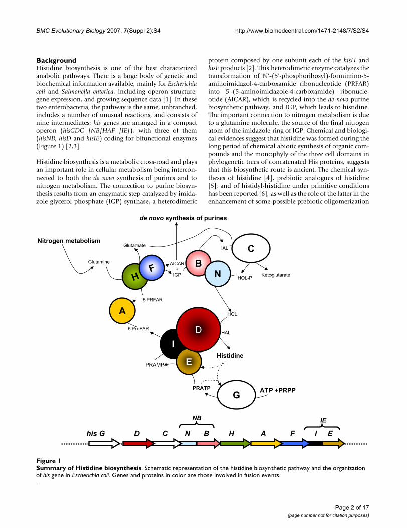
bling the length of the ancestral gene and the number of(β/α)-modules in the product. Thus the HisA TIM-barrelwould be the result of a cascade of two consecutive geneelongations (Figure 3). This model is supported by thestructures shown in Figure 3 (upper left panel). We usedthe T. maritima HisA structure (1Q02) to obtain the coor-dinates of each atom composing the "quarters" of barreland then performed a structural superposition using swissPDB viewer. Lang et al., [19] compared the structure of thetwo HisA half-barrels and obtained a root-mean-square
deviation (rms) ranging from 1.5 to 2.0 Å using all mainchain, non hydrogen atoms. Our results concerning"quarters" structural superpositions showed for the firstand the second quarters an average rms of 1.21 Å using allalpha carbons, strongly supporting the model proposed.We showed examples from these two organism, becauseM. jannaschii with several other Archaea showed the bestoverall degree of conservation of internal repetitions,while the choice of T. maritima followed the availability ofHisA tridimensional structure [19].
HisA "quarters" alignmentsFigure 2HisA "quarters" alignments. Pairwise alignments of Methanocaldococcus jannaschii (mj) HisA subregions corresponding to "quarters" of barrels (named HisA1, HisA2, HisA3, HisA4). Symbols: *,: correspond to identical or similar aminoacids, respec-tively.
10 20 30 40 50 60
| | | | | |
mjHisA1 MIIIPAVDLKDKKCVQLIQGDPNKKHLELNNVE-VAKKFVDEGAE-YLHIIDLDAAFG-TGNN
mjHisA2 RDVIKN--IIKEVNVPVEVGGGIRNLEIAKELISLGVDRVIVGTKAILEPKFIDDLNKEIGKD
mjHisA3 KIVLA----VECKEGKVVIKG-WKEKVDKTPIEVIKEFEDKVGYILFTN-VDVEGLLKGIN--
mjHisA4 VDIIK--ELIEKTDIPIIYSGGITTLEDIKALKELGIYGVVIGSALYKGLIDLKKALEIVKN-
:: :::: : :: * :: : : : : * :*: : :*:: : :
mjHisA1 MIIIPAVDLKDKKCVQLIQGDPNKKHLELNNP-VEVAKKFVDEGAE-YLHIIDLDAAFG-TGNN
mjHisA2 RDVIKNI-IKEVNVPVEVGG--GIRNLEIAKELISLGVDRVIVGTKAILEPKFIDDLNKEIGKD
:* : :*: : : * ::**: : : : * *:: * :* *::
mjHisA1 MIIIPAVDLKDKKCVQLIQGDPNKKHLELNNPVEVAKKFVDE-GAEYLHIIDLDAAFGTGNN
mjHisA3 -KIVLAVECKEGKVV--IKG--WKEKVD-KTPIEVIKEFEDKVGYILFTNVDVEGLL-KGIN
*: **: *: * * *:* *:::: : *:** *:* *: : :*:: : * *
mjHisA1 MIIIPAVDLKDKKCVQLIQGDPNKKHLE-LNNPVEVAKKFVDEGAE-YLHIIDLDAAFG-TGNN
mjHisA4 VDIIK--ELIEKTDIPIIYSG-GITTLEDIKALKELGIYGVVIGSALYKGLIDLKKALEIVKN-
: ** :* :* : :* ** :: *: * *: * :*** *: *
mjHisA2 RDVIKNI-IKEVNVPVEVGG--GIRNLEIAKELISLGVDRVIVGTKAILEPKFIDDLNKEIGKD
mjHisA3 -KIVLAVECKEGKVVIK-GWKEKVDKTPI--EVIKEFEDK--VG-YILFTNVDVEGLLKGIN--
:: : ** :* :: * : : * *:* *: ** :: :: * * *
mjHisA2 RDVIKNIIKEVNVPVEVGGGIRNLEIAKELISLGVDRVIVGTKAILEPKFIDDLNKEIGKD
mjHisA4 VDIIKELIEKTDIPIIYSGGITTLEDIKALKELGIYGVVIGS-ALYKGLIDLKKALEIVKN
*:**::*:: ::*: *** ** * * **: *::*: *: : : ** *:
mjHisA3 -KIVLAVECK-EGKVVIKGWKEKV-DKTPIEVIKEFEDKVGYILFTN-VDVEGLLKGIN-
mjHisA4 VDIIKELIEKTDIPIIYSGGITTLEDIKALKELGIYGVVIGSALYKGLIDLKKALEIVKN
*: : * : :: * : * :: : : :* *: :*:: *: ::
Page 4 of 17(page number not for citation purposes)
BMC Evolutionary Biology 2007, 7(Suppl 2):S4 http://www.biomedcentral.com/1471-2148/7/S2/S4
The symmetry of the TIM-barrel structure suggests to testa further ancestral duplication in which the original genecoded for a single (β/α)-module, capable of forming ahomo-octamer to form a complete barrel. Although thealignments constructed from the eight single (β/α)-mod-ules are very short, they still contain a non negligibleamount of sequence and secondary structure similaritiesnot expected from random distribution of amino acidsand by visual comparison of their structure is in agree-ment with this hypothesis (Figure 3, lower left panel).
Thereby, the ancestral forms of life might have expandedtheir coding abilities and their genomes by duplicating asmall number of mini-genes, i.e. the "starter types". Weare completely aware that the evidence of the ancientduplications involving an ancestral mini-gene encodingthe "quarter" of barrel and the single (β/α)-module isbased on very limited amount of sequence and structuralsimilarities; in spite of this, in our opinion, the hypothesismentioned above remains valid. Moreover, further studiesare needed to clarify the presence of these additional gene
Table 1: Identity and similarity values for the HisA quarters comparisons.
HisA quarters comparisons
hisA1 hisA2 HisA3 hisA4
HisA1 15.8 29 25.4HisA2 42.9 20.6 32.2HisA3 58.1 42.9 13HisA4 42.9 58.1 41.9
Identity (upper diagonal) and similarity percentages calculated for the alignments shown in Figure 2.
An evolutionary model for hisA and hisF genesFigure 3An evolutionary model for hisA and hisF genes. Right-most panel is the evolutionary model that we propose and discuss in the text concerning hisA and hisF origin and evolution. Panels on the left are: the first and the second quarters (top) and two single (β/α) modules of the HisA protein from Thermotoga maritima which illustrates the structural similarities from which we derived our model. The quarters have a structural alignment with only 1.2 Å of RMS on 104 alpha carbons.
a
c
A1
A1 A2
his A his F
his A
Gene elongations
Duplication and
divergence
Fusion
Gene duplication
Half-Barrel
Intermediate stage
EntireBarrel
Half-Barrel
Intermediate stage
Duplication anddivergence
Fusion
a
c
A1
A1 A2
his A his F
his A
Gene elongations
Duplication and
divergence
Fusion
Gene duplication
Half-Barrel
Intermediate stage
EntireBarrel
Half-Barrel
Intermediate stage
Duplication anddivergence
Fusion
Quarter of
Barrel
b
c
Page 5 of 17(page number not for citation purposes)

BMC Evolutionary Biology 2007, 7(Suppl 2):S4 http://www.biomedcentral.com/1471-2148/7/S2/S4
elongation events and to integrate them into a more gen-eral picture of the evolution of the very diversified TIM-barrel family of proteins.
The hisNB fusionThe eighth and sixth steps of histidine biosynthesis arecatalyzed by histidinol-phosphate phosphatase (EC3.1.3.15) (HOL-Pase) and IGP dehydratase (EC 4.2.1.19),respectively [1]. Distinct HOL-Pases have been character-ized in different organisms, whereas IGP dehydratase isthe same in all known histidine synthesizing organisms.In E. coli the two activities are coded for by a single gene,referred to as hisNB [11]: the N-terminal domain (HisN)is a phosphatase belonging to the DDDD family [11] andthe C-terminal domain is responsible for IGP dehydrataseactivity. The evolutionary history of the hisNB gene hasbeen recently reported by [11] who showed that hisNBgene fusions are present in most γ-proteobacteria and in
the ε-proteobacterium Campylobacter jejuni; phylogeneticanalysis allowed to trace the fusion event in an ancestor ofthe γ-subdivision and its later horizontal transfer to C.jejuni. Moreover, hisN is paralogous to gmhB (E. colinomenclature), catalyzing the dephosphorylation of D-α-D-heptose 1,7-PP for surface Lipopolysaccharide produc-tion [20,21].
Since the former hisNB evolutionary model was based ona limited number of genomes, we update it including allavailable genomes (April, 1: 41 Archaea, 759 Bacteria and135 Eukarya).
By combining results obtained with several queries, weretrieved 131 orthologous bifunctional HisNB sequences:104 come from γ-proteobacteria, 9 from ε-proteobacteria,1 to a α-proteobacterium and 17 from the CFB group(Table 2 and Additional file 1). No archaeal and eukaryo-
Table 2: Phylogenetic distribution of HisIE and HisNB genes.
Domain Phylum Class # His+ % HisIE % HisNB
Bacteria Acidobacteria Acidobacteria 1 0 0Solibacteres 1 0 0
Actinobacteria Actinobacteria 36 2.8 0Aquificae Aquificae 1 100 0
Bacteroidetes Bacteroidetes 2 100 50Flavobacteria 1 100 100
Sphingobacteria 1 100 100Chlorobi Chlorobia 4 0 0
Chloroflexi Dehalococcoidetes 2 0 0Cyanobacteria Chroococcales 10 90 0
Gloeobacteria 1 100 0Nostocales 2 100 0
Oscillatoriales 1 100 0Prochlorales 10 100 0
Firmicutes Bacilli 37 48.7 0Clostridia 10 80 0
Planctomycetes Planctomycetacia 1 0 0Proteobacteria α 52 1.9 1.85
β 39 0 0δ 12 16.7 0ε 7 85.7 82γ 89 68.5 95
Spirochaetes Spirochaetes 4 0 0TD group Deinococci 4 100 0
Thermotogae Thermotogae 1 100 0Archaea Crenarchaeota Thermoprotei 6 0 0
Euryarchaeota Archaeoglobi 1 0 0Halobacteria 4 0 0
Methanobacteria 2 0 0Methanococci 3 0 0
Methanomicrobia 8 0 0Methanopyri 1 0 0Thermococci 2 100 0
Thermoplasmata 1 100 0
Phylogenetic distribution analysis of the fusions HisIE and HisNB, along with the percentage of species possessing one of them (two right – most columns) on the number of histidine producing organisms (as assessed by the presence of his genes in their genome). Eukarya are treated separately (see HIS4 section) and never possess the hisNB gene fusion.
Page 6 of 17(page number not for citation purposes)
BMC Evolutionary Biology 2007, 7(Suppl 2):S4 http://www.biomedcentral.com/1471-2148/7/S2/S4
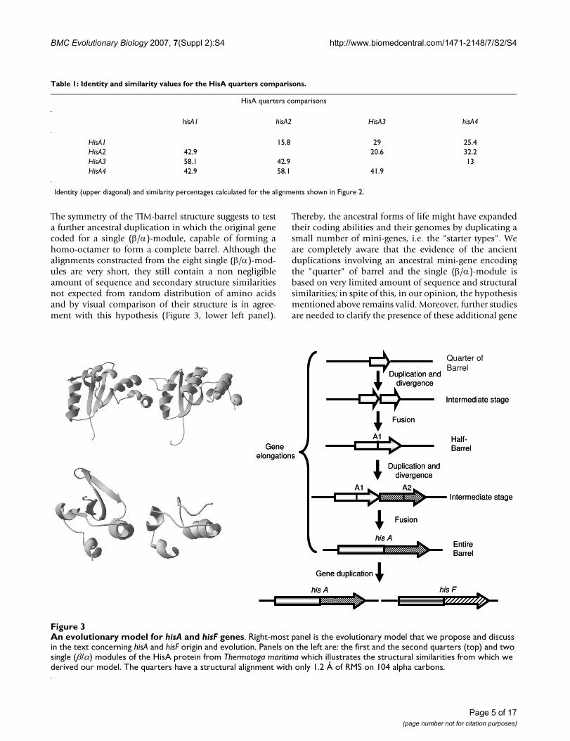
tic bifunctional sequence was retrieved although they pos-sess genes encoding DDDD-type phosphatases, or, moregenerally HAD hydrolases [11]. These data confirmed thenarrow phylogenetic distribution of the hisNB fusion,which is mostly present in γ-proteobacteria. However, theoccurrence of a fused hisNB gene in other lineagesenlarged its distribution raising the question of the originof this fusion in these phyla, i.e. if it is either the outcomeof convergent evolution or a horizontal gene transferevent (HGT). To discern between these two different sce-narios, a phylogenetic analysis of HisNB sequences wascarried out. A phylogenetic tree obtained using a repre-sentative set of HisNB sequences is reported in Figure 4,which shows that HisNB sequences from α- and ε-proteo-bacteria, and CFB bacteria are intermixed with γ-proteo-bacterial sequences and do not reflect the 16S rDNAphylogeny. This result strongly suggests that the hisNBgene has been horizontally transferred from some γ-pro-teobacteria to the other microorganisms. It is also quitepossible that the transfer event might have involved theentire his operon or part thereof, as evidenced by phyloge-netic trees of other his genes (e.g. Additional File 2). Thisstatement relies on the analysis of the organization of hisgenes in the 131 genomes harboring the hisNB fusion,
which revealed (Additional file 3) that all of them arelocalized within more or less compact operons.
The hisIE fusionThe hisI and hisE genes code for a phosphoribosyl-ATPphosphohydrolase and a phosphoribosyl-AMP cyclohy-drolase that are responsible for the third and second stepsin histidine biosynthesis, respectively. In E. coli and S.enterica the two genes are fused to form the last gene of thehis operon (Figure 1).
The phylogenetic distribution of hisIE genes was obtainedby retrieving homologous sequences using the E. coliHisIE amino acid sequence as a query to probe genomedatabase. The data are reported in Table 2 and can be sum-marized as follows (see also Additional file 4):
1. The hisIE fusion is not universally distributed;
2. Bifunctional hisIE genes were found in all eukaryotes(see section regarding HIS4);
3. Most of the archaeal genomes harbor monofunctionalhisI and hisE genes; the occurrence of hisIE in Thermococciand Thermoplasmata is very likely the outcome of a HGTevent from a bacterium donor [15]. Moreover, when thehisI and hisE genes are not fused in Archaea, they do notbelong to operons and are separated on the chromosome.The only exceptions are represented by Sulfolobus species,where the two genes are in a compact operon but sepa-rated by the hisH gene;
4. The hisIE gene fusion is present in 100% of the histidineproducing organisms belonging to Aquificae, Deinococci,Bacteroidetes, Cyanobacteria, and Thermotogae. Moreo-ver, a bifunctional hisIE gene was found in all γ-proteobac-teria that branched off after the separation ofPseudomonadales from the main branch. The presence ofthe fusion in ε-proteobacteria can be explained by meansof a HGT of the entire operon [13] from γ-proteobacteria;the same appears to be true for Bacteria belonging to theCFB group and possessing the hisNB gene fusion (see thecorresponding paragraph). In spite of the high number ofgenomes sequenced (39), no hisIE fusion was found in β-proteobacteria, which represent the key-point for thecompacting of his genes during the construction of pro-teobacterial his operon [13].
5. Firmicutes show a complex scenario: we have foundfused and stand-alone genes in very closely related speciesof Bacillus (i.e. Bacillus subtilis possesses the gene fusionwhile B. thuringiensis and B. anthracis do not; in these caseshisI and hisE are contiguous and very close on the chromo-some); the same is true for Clostridia. The presence of thefusion in model organisms such as Bacillus subtilis but not
HisNB phylogenetic analysisFigure 4HisNB phylogenetic analysis. HisNB Phylogenetic tree. Organisms (groups) in red are bacteria not belonging to γ-proteobacteria and harboring the HisNB fusion.
Escherichia/Shigella/Salmonella
Erwinia carotovoraSerratia proteamaculans
YersiniaPhotorhabdus luminescens
Sodalis glossinidiusBaumannia cicadellinicola
Blochmannia pennsylvanicusBlochmannia floridanus
Buchnera
Vibrio/Photobacterium
Psychromonas ingrahamiiPsychromonas sp. CNPT3
Aeromonas hydrophilaAlteromonas macleodiiPseudoalteromonas atlanticaalpha proteobacterium HTCC2255
Colwellia psychrerythraea 34HIdiomarina loihiensis L2TRIdiomarina baltica OS145
ShewanellaActinobacillus succinogenes
Actinobacillus pleuropneumoniaePasteurella multocida
Mannheimia succiniciproducensHaemophilus
AlteromonadalesCampylobacter
LegionellaXanthomonas
Stenotrophomonas maltophiliaXylella
CFB group100
10065
100
100
100100
100
100
97
100
99
48
100
33
100
49
96
58
100
99
5863
79
37
70
47
83
46
55
65
2828
10048
6299
97
64
52
100
0.05
Page 7 of 17(page number not for citation purposes)

BMC Evolutionary Biology 2007, 7(Suppl 2):S4 http://www.biomedcentral.com/1471-2148/7/S2/S4
in some of the recently sequenced genomes of the samegenus suggests that sequencing artifacts, probably favoredby the gene organization of these two genes, mightexplain this situation.
6. Actinobacteria lack this gene fusion.
7. Apparently there is no correlation between the occur-rence of hisIE fusion and his genes organization. However,it is interesting that during Proteobacteria evolution, wewitness a progressive approaching of two initially far hisIand hisE cistrons, starting from δ- and ε-proteobacteria.The distance between them decreases in bacteria belong-ing to the α-branch; then, they partially overlap in β-pro-teobacteria, a molecular event which is coincident withthe formation of a complete his operon, which very oftenincludes genes apparently not involved in histidine bio-synthesis. Finally, the two genes fused in the ancestor of γ-proteobacteria, where the compactness of his operon isvery high [13].
Despite of the proposed HGTs, the current phylogeneticdistribution of the HisIE bifunctional enzyme evokes ascenario of convergent evolution and independent genefusions/splitting of the two cistrons in different lineages.However, phylogenetic analyses are not of great help toconfirm this view (data not shown) because these pro-teins are very short (less than 100 residues each) and theinformative sites in a multialignment of sequences com-ing from complete genomes are extremely reduced bring-ing to unreliable trees (i.e. very low bootstrap support, startopologies, data not shown). On a different perspective,the analysis of the linker region connecting the twodomains might help in understanding the evolutionaryhistory of these fusions, but we have found that they arevery short, giving no information on this issue (data notshown). If the idea on convergent evolution of these genefusions will hold future works, it might turn out that thereis a strong selective pressure favoring their appearance indifferent lineages. Lastly, the finding that the gene order inall the bifunctional genes is always hisI, hisE, suggests thata different arrangement of the two domains should be dis-advantageous for the enzymatic activity and that struc-tural and/or functional constraints might be responsiblefor the extant arrangement.
From metabolons to multifunctional enzymes: the eukaryotic HIS7 and HIS4 fusionsTwo fusions involving his genes were disclosed in the yeastSaccharomyces cerevisiae: HIS7, corresponding to the bacte-rial hisH and hisF genes [12], and HIS4. The latter genecodes for a tetrafunctional enzyme consisting of about800 residues and containing three regions homologous tothe prokaryotic hisI, hisE and hisD genes, arranged in thisorder in the yeast gene, whose products perform the sec-
ond, the third and the last two steps of histidine biosyn-thesis, respectively.
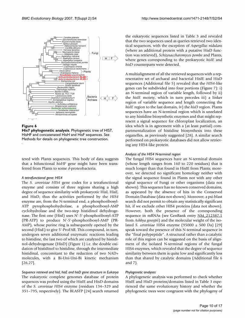
The IGP synthase coding gene: HIS7The bifunctional enzyme IGP synthase catalyzes the fifthstep of histidine biosynthesis, generating the imidazolering of the histidine precursor, IGP. The overall reaction isthe conversion of PRFAR to IGP and AICAR [2] via aglutamine molecule, and with no free intermediate (Fig-ure 1). This represents the central step of the pathway,which connects histidine biosynthesis to nitrogen metab-olism and the de novo synthesis of purines, throughAICAR. The active form of the E. coli IGP synthase is a het-erodimer of the hisH and hisF products i.e. a glutamineamidotransferase (GAT) and a cyclase, respectively [22].The requirement for a direct interaction between GAT andthe cyclase was confirmed by the discovery of the structureof the S. cerevisiae HIS7 gene coding for IGP synthase [23];the analysis of the encoded enzyme demonstrated that itis constituted by two domains, an N-terminal and a C-ter-minal one, sharing a high degree of sequence similaritywith known HisH and HisF, respectively. Previous workssuggested that the eukaryotic HIS7 gene is the outcome ofa fusion event involving two monofunctional, bacterial-like genes [12]. According to the model proposed, theeukaryotic lineage inherited two monofunctional genes,hisH and hisF, that underwent gene fusion. The alternativescenario, that is the possibility of a HGT event from aprokaryote harboring a fused hisHF gene to eukaryoteswas excluded on the basis of the absence of the HisHFfusion in prokaryotes. However, the increasing number ofsequence in databases opens the possibility to modify thismodel. To this purpose the S. cerevisiae HIS7 aminoacidsequence was used in a BLASTP search [24], allowing toretrieve 21 bifunctional sequences. Whilst most (18)come from Eukarya, three of them belonged to two δ-pro-teobacteria, raising the possibility that the bacterial andthe eukaryotic HisHF sequences share a common ances-try, even though a phenomenon of convergent evolutioncould not be ruled out. Thus, all the available HisHFbifunctional sequences and a set of bacterial and archaealconcatenated HisH and HisF sequences were alignedusing the program ClustalW [25]. The multialignment(partially shown in Figure 5) allowed to detect severalconserved insertions that distinguish bifunctional pro-teins. This speaks towards a common origin of the eukary-otic and bacterial hisHF genes rather than a phenomenonof convergent evolution. The phylogenetic tree obtainedusing the above mentioned multialignment showed inFigure 6 supports this view: it can be splitted into twomain clusters, one containing all the monofunctionalsequences including the one coming from the δ-proteo-bacterium Geobacter sulfurreducens, the second one com-prising all the bifunctional eukaryotic and bacterialsequences; the three bifunctional bacterial sequences clus-
Page 8 of 17(page number not for citation purposes)

BMC Evolutionary Biology 2007, 7(Suppl 2):S4 http://www.biomedcentral.com/1471-2148/7/S2/S4
Page 9 of 17(page number not for citation purposes)
His7 multialignmentFigure 5His7 multialignment. A multialignment of HIS7, HisHF and a set of representative concatenated HisH and HisF sequences from E. coli, S. solfataricus, A. fulgidus and G. sulfurreducens. Yellow shading represent insertions shared only by bifunctional HIS7 and HisHF proteins. Shading of the multialignment has been made with PAM250 matrix.
110 120 130 140 150 160 170 180 190 200 ....|....|....|....|....|....|....|....|....|....|....|....|....|....|....|....|....|....|....|....| S. cerevisiae 41 GTSRLILPGVGNYGHFVD-NLFNRGFEKPIREYIES-GKPIMGICVGLQALFAGSVESPKSTGLNYIDFKLSRFDDSEK--------PVPEIGWNSCIPS 130S. pombe 38 KAECLIFPGVGNFGFVCD-SLAKQGFLEPLRRYALS-GKPFMAVCVGIQALFEGSVEAPHSKGLGVFPGLVQRFDNDDK--------TVPHIGWNSCAVR 127A. thaliana 99 NADRLIFPGVGAFAPAMD-VLNRTGMAEALCKYIEN-DRPFLGICLGLQLLFDSSEENGPVKGLGVIPGIVGRFDASAGI-------RVPHIGWNALQVG 189Delta MLMS-1 37 QAEKLIFPGVGNFGSAME-SLQKRGWLAPLLQYLQA-GRPYLGICLGLQTLFEASTEAPGVAGLGLIKGVIQRFGTPDGVAGDSFPLAVPQIGWNGLAPR 134D. psychrophila 38 TADKLIFPGVGSFGSVMH-TLRDRGYIEPLKRRIEE-DKPFLGICVALQALFEGSEETPGVAGLGILPGQIKKFVRSAL--------SVPQIGWNGIHLL 127E. coli 38 LADKLFLPGVGTAQAAMD-QVRERELFDLIKAC----TQPVLGICLGMQLLGRRSEESNGVDLLGIIDEDVPKMTDFGL--------PLPHMGWNRVYPQ 124S. solfataricus 36 DYDLIVFPGVGAFSAVAEFILRYRELFNDLRRS----GTNFLGVCLGMQIMFEKGTEGKESNGLGWFKGIVDKINANV---------KLPHIGWDLVFEV 122A. fulgidus 37 CASGVVFPGVGAFKSAIE-KLN------LIRDVIDSLEVPILGICLGMQLFATESTEGGVYRGLDYIPGRVVRFPPSVG--------KVPHMGWNTLKIT 121G. sulfurreducens 39 EAEKIVLPGVGAFRDCMR-NLEQGGFVEPILRVIRE-GRPFLGICVGMQLLLTDSVEFGLYQGLNVIPGHVLRFPEGMREGGEEL--KVPHMGWNQLSIK 134
210 220 230 240 250 260 270 280 290 300 ....|....|....|....|....|....|....|....|....|....|....|....|....|....|....|....|....|....|....|....|
S. cerevisiae 131 EN---LFFGLDPYKRYYFVHSFAAILNSEKKKNLENDGWKIAKAKYGSEEFIAAVNKNNIFATQFHPEKSGKAGLNVIENFLKQQ-----SPPIPNYSAE 222S. pombe 128 SDTSKEFFGMRPHDKFYFVHSYMIPEKGL----ILPPEFKIATTKYGNETFVGAIVKNNFLATQFHPEKSGSAGLRCLKAFLTGN----YEQPI---SGE 216A. thaliana 190 KD-SEILDDVGN-RHVYFVHSYRAIPSDE------NKDWISSTCNYG-ESFISSIRRGNVHAVQFHPEKSGEVGLSVLRRFLHPK-LPATQKPM------ 273Delta MLMS-1 135 QD-SPLLADCRQ-EKFYFVHSYRAAASPE------NCDWVLAETTYG-EDFISAVQRGAVAACQFHPEKSGPAGLALLGNFLRAEKLAVTKRPASPGKER 225D. psychrophila 128 KE-SPSFAGYEE-EKLYFVHSYHAPLDTV------PEDWALTATDYG-TRFISAVEKGNVTAFQFHPEKSGQAGLNLLQNFLSSQ-----KRPS-----Q 208E. coli 125 AG-NRLFQGIEDGAYFYFVHSYAMPVNP----------WTIAQCNYG-EPFTAAVQKDNFYGVQFHPERSGAAGAKLLKNFLEM---------------- 196S. solfataricus 123 KDSCELTYGLDK-KYVYYVHSYVAYPTSG--------DYVYMKSQYG-IEYPALVCDKNVVGTQFHPEKSSNTGKIFLENLKGWI--------------- 197A. fulgidus 122 RE-AEILDGVESGEFVYFVHSYYMQTDDE---------FVISKTDYG-IDFPSGVERENYIGFQFHPEKSGKVGLRILENFVNIV--------------- 195G. sulfurreducens 135 RR-PPAFAEVEDGANVYFVHSYYEMPDDE--------SVIAATCTYG-VEFCAAIWKDNIVATQFHPEKSQAVGLSILKNFGEMK--------------- 209
310 320 330 340 350 360 370 380 390 400 ....|....|....|....|....|....|....|....|....|....|....|....|....|....|....|....|....|....|....|....|
S. cerevisiae 223 EKELLMNDYSNYGLTRRIIACLDVRTNDQGDLVVTKGDQYDVREKSDGKGVRNLGKPVQLAQKYYQQGADEVTFLNITSFRDCPLKDTPMLEVLKQAAKT 322S. pombe 217 ASKLIENSFG--GLTKRIIACLDVRSNDAGDLVVTKGDQYDVREKSSGSEVRNLGKPVELCQRYFQEGADEVVFLNITSFRNCPMADAPMLQVLEKAAQT 314A. thaliana 273 -------EGKASKLAKRVIACLDVRTNDKGDLVVTKGDQYDVREQSNENEVRNLGKPVDLAGQYYKDGADEISFLNITGFRDFPLGDLPMIQVLRQTSKN 366Delta MLMS-1 226 LPAGALFPRAATRPAKRIIACLDVRNNDQGDLVVTKGDQYDVRE-AGHGAVRNLGKPVELAERYYHEGADEITFLNITAFRDFPLEDQPMLEVLERASER 324D. psychrophila 209 TPIVPIEIEKKTEHAKRVIACLDVRSNDNGDLVVTKGDQYDVRQ---EGEVRNLGKPVDLARRYYQEGADEVTFLNITGFRDFPMEDQPMIEVLQKASEN 305E. coli 196 ------------MLAKRIIPCLDV----RDGQVV-KGVQF--------RNHEIIGDIVPLAKRYAEEGADELVFYDITASSD---GRVVDKSWVSRVAEV 268S. solfataricus 197 ----------KRMTTKRIIACLDV----KDGNVV-KGVNF--------LNLQLKGDPVSLASLYEEEGADEIVFLDITATIE---ARKALYNVIKDTASV 271A. fulgidus 195 ----------KRMLAKRIIPCLDVTLDESEARVV-KGVEF--------VNLRDAGDPVELAKRYDEEGADELVFLDITASPE---GRRTMIDVIERTAEQ 273G. sulfurreducens 209 ------------MLTKRIIPCLDV----KGGRVV-KGVQF--------LELRDAGDPVEIAELYDRQGADELTFLDITASSD---ERSIIIDVVRRTAER 281
410 420 430 440 450 460 470 480 490 500 ....|....|....|....|....|....|....|....|....|....|....|....|....|....|....|....|....|....|....|....|
S. cerevisiae 323 VFVPLTVGGGIKDIVDVDGTKIPALEVASLYFRSGADKVSIGTDAVYAAEKYYELGNRGDGTSPIETISKAYGAQAVVISVDPKRVYVNSQADTKNK-VF 421S. pombe 315 VFVPLTVGGGIRDVSDPDGTFHPAVEVAGIYFRSGADKVSIGSDAVYAAEKYYENGKKLSGKTAIETISKAYGNQAVVISVDPKRQYVKVPEDTKHH-VV 413A. thaliana 367 VFVPLTVGGGIRDFTDASGRYYSSLEVAAEYFRSGADKISIGSDAVSAAEEFIKSGV-KTGKSSLEQISRVYGNQAVVVSIDPRRVYVNHPDDVPYK-VI 464Delta MLMS-1 325 IFVPLTIGGGIREFTDAHGRFYSALEVAARYFRAGADKISIGSDAVAVVEDYLARGRQKDGKSSLEQIAHLYGSQAVVISIDPRRVYVSGPAAAPGKQVI 424D. psychrophila 306 VFVPLTIGGGIRDFTDSNGKYYSALDVAAQYFRSGADKISIGSDAVAIVEEYLATGK-KSGKSSIEQISAVYGVQAVVISVDPRRVYVQSPDETTHT-CV 403E. coli 269 IDIPFCVAGGIKSLED-----------AAKILSFGADKISINSPALADPT-------------LITRLADRFGVQCIVVGIDTWY--------------- 329S. solfataricus 272 LSIPLTVGGGIRTPDD-----------VSMALRSGADKVSINTAAVESSQ-------------IVKKSAEEFGSQAVVVAIDVKKV-------------- 333A. fulgidus 274 VFIPFTVGGGIKSIED-----------INTILSAGADKVSINTAAVKNPE-------------FVREAADIFGSQCIVVAIDCRRNF----DLSKGEYIV 345G. sulfurreducens 282 VFMPLTVGGGVRTVDD-----------IRNLLNAGADKVSINTAAVHRPE-------------FVREAAERFGSQCTVVAIDARQV-------------- 343
510 520 530 540 550 560 570 580 590 600 ....|....|....|....|....|....|....|....|....|....|....|....|....|....|....|....|....|....|....|....|
S. cerevisiae 422 ETEYPGPNGEKYCWYQCTIKGGRESR----DLGVWELTRACEALGAGEILLNCIDKDGSNSGYDLELIEHVKDAVKIPVIASSGAGVPEHFEEAFLKTRA 517S. pombe 414 KTSRLGPNGEAYCWYQCTVKGGREYR----DIDVVELTRACEAMGAGEVLLNCMDQDGSNAGYDIELVRLVKNSVNIPVIASSGAGIPQHFEEVFKETDC 509A. thaliana 465 RVTNPGPNGEEYAWYQCTVSGGREGR----PIGAFELAKAVEELGAGEILLNCIDCDGQGKGFDIDLVKLISDSVGIPVIASSGAGTPDHFSEVFEKTNA 560Delta MLMS-1 425 PTAFPGPAGEEYCWYQCTVKGGREGR----DLDAATLAAACQELGAGEILLNCIDKDGTNSGFDHELIKMVKEAVTIPVIASSGAGKPEHFSEVFIATGA 520D. psychrophila 404 KTGSLGPNGEEYCWYQCTIKGGREGS----KLDAVQLVQGCEALGAGEILLNCIDKDGTNDGFELELISLVKKAVSIPVIASSGAGSARDFVEVFRETDA 499E. coli 329 -------DAETGKYH-VNQYTGDESRTRVTQWETLDWVQEVQKRGAGEIVLNMMNQDGVRNGYDLEQLKKVREVCHVPLIASGGAGTMEHFLEAFRDADV 421S. solfataricus 333 ----------SGNWI-VFTKSGTYNT----RLDAIKWAKKVEELGAGEILLTSIDRDGTRLGYDLELTRKIVDSVNIPVIASGGAGKMEHFYEVFSLAKA 418A. fulgidus 346 EL-----EDGTKAWYEVVIYGGRKPV----GIDAVWWAKRVEELGAGEILLTSMNRDGTKDGFDIPITRKISEEVNIPVIASGGAGTKEHFYEGFVEGKA 436G. sulfurreducens 343 --------PGENRWE-VYTHGGRNPT----GIDAVEWARRMEEYGAGEILLTSMDRDGTKDGYDIPLTRAIVDAVSIPVIASGGVGNLEHLYDGFVKAGA 430
610 620 630 ....|....|....|....|....|....|....|
S. cerevisiae 518 DACLGAGMFHRGEFTVNDVKEYLLEHGLKVRMDEE 552S. pombe 510 DAALAAGIFHRQTCRIEDVKEYLAIHDVLVRT--- 541A. thaliana 561 SAALAAGIFHRKEVPIQSVKEHLQEERIEVRI--- 592Delta MLMS-1 521 EAALAAGIFHRREVEIGEVKQHLKRHGIEVRG--- 552D. psychrophila 500 DAALAAGIFHREEVPISEVKTTLMQAGISCR---- 530E. coli 422 DGALAASVFHKQIINIGELKAYLATQGVEIRIC-- 454S. solfataricus 419 DAALAAGIFHDGIIKIKDLKSYLSQKGIEVRM--- 450A. fulgidus 437 DACLAASIFHYREIGIREIKEYLAERGVQVRL--- 468G. sulfurreducens 431 SACLAASIFHYKEYTIGEAKEYLRQRGVPVRL--- 462
BMC Evolutionary Biology 2007, 7(Suppl 2):S4 http://www.biomedcentral.com/1471-2148/7/S2/S4
tered with Plants sequences. This body of data suggeststhat a bifunctional hisHF gene might have been trans-ferred from Plants to some δ-proteobacteria.
A tetrafunctional gene: HIS4The S. cerevisiae HIS4 gene codes for a tetrafunctionalenzyme and consists of three regions sharing a highdegree of sequence similarity with prokaryotic HisI, HisE,and HisD, thus the activities performed by the HIS4enzyme are, from the N-terminal end, a phosphoribosyl-ATP pyrophosphohydrolase, a phosphoribosyl-AMPcyclohydrolase and the two-step histidinol dehydroge-nase. The first one (HisI) uses N'-5'-phosphoribosyl-ATP(PR-ATP) to produce N'-5'-phosphoribosyl-AMP (PR-AMP), whose purine ring is subsequently opened by thesecond (HisE) to give 5'-ProFAR. This compound, in turn,undergoes seven additional enzymatic reactions leadingto histidine, the last two of which are catalyzed by histidi-nol-dehydrogenase (HisD) (Figure 1) i.e. the double oxi-dation of histidinol to histidine, through the intermediatehistidinal, concomitant to the reduction of two NAD+molecules, with a Bi-Uni-Uni-Bi kinetic mechanism[26,27].
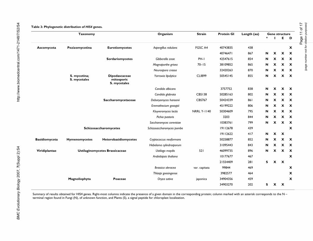
Sequence retrieval and hisI, hisE and hisD gene structure in EukaryaThe eukaryotic complete genomes database of proteinsequences was probed using the HisIE and HisD domainsof the S. cerevisiae HIS4 enzyme (residues 134–329 and351–795, respectively). The BLASTP [24] search returned
the eukaryotic sequences listed in Table 3 and revealedthat the two sequences used as queries retrieved two iden-tical sequences, with the exception of Aspergillus nidulans(where an additional protein with a putative HisD func-tion was retrieved), Schizosaccharomyces pombe and Plants,where genes corresponding to the prokaryotic hisIE andhisD counterparts were detected.
A multialignment of all the retrieved sequences with a rep-resentative set of archaeal and bacterial HisIE and HisDsequences (Additional file 5) revealed that the HIS4-likegenes can be subdivided into four portions (Figure 7): i)an N-terminal region of variable length, followed by ii)the hisIE moiety, which in turn precedes iii) a linkerregion of variable sequence and length connecting thehisIE region to the last domain, iv) the hisD region. Plantssequences have an N-terminal region which is unrelatedto any histidine biosynthetic enzymes and that might rep-resent a signal sequence for chloroplast localization, anidea which is in agreement with a (at least partial) com-partmentalization of histidine biosynthesis into theseorganelles, as previously suggested [28]. A similar searchperformed on prokaryotic databases did not allow retriev-ing any HIS4-like protein.
Analysis of the HIS4 N-terminal regionThe fungal HIS4 sequences have an N-terminal domain(whose length ranges from 160 to 220 residues) that ismuch longer than that found in HisIE from Plants; more-over, we detected no significant homology neither withthe signal sequence found in Plants nor with any othersignal sequence of Fungi or other organisms (data notshown). This sequence has no known conserved domains,as appeared by the absence of hits in the ConservedDomain Database (data not shown). Moreover, a psi-blastsearch did not permit to obtain any statistically significanthit, if we exclude other HIS4 proteins (data not shown).However, both the presence of the correspondingsequence in mRNAs (see GenBank entry NM_212387.1from Ashbya gossypii) and the molecular weight of the iso-lated S. cerevisiae HIS4 enzyme (95000 ± 500 Da) [29]speak toward the presence of this N-terminal sequence inthe "final polypeptide". A structural rather than a catalyticrole of this region can be suggested on the basis of align-ment of the isolated N-terminal regions of the fungalHIS4 enzymes, which revealed that the degree of sequencesimilarity between them is quite low and significantly lessthan that shared by catalytic domains (Additional file 6and 7).
Phylogenetic analysesA phylogenetic analysis was performed to check whetherHisIE and HisD proteins/domains listed in Table 3 expe-rienced the same evolutionary history and whether thephylogenetic trees were congruent with the phylogeny of
His7 phylogenetic analysisFigure 6His7 phylogenetic analysis. Phylogenetic tree of HIS7, HisHF and concatenated HisH and HisF sequences. See Methods for details on phylogenetic tree construction.
F
Fungi
Candida glabrata
Saccharomyces cerevisiae
Kluyveromyces lactis
Ashbya gossypii
Debaryomyces hansenii
Candida albicans
Yarrowia lipolytica
Neurospora crassa
Gibberella zeae
Aspergillus nidulans
Aspergillus terreus
Aspergillus fumigatus
Schizosaccharomyces pombe
Ustilago maydis
Coprinopsis cinerea
Cryptococcus neoformans
Oryza sativa
Arabidopsis thaliana
Desulfotalea psychrophiladelta proteobacterium MLMS-1delta proteobacterium MLMS-1
Escherichia coli
Campylobacter jejuni
Xylella fastidiosa
Bacteroides fragilis
Thermotoga maritima
Bacillus subtilis
Nostoc sp. PCC 7120
Lactococcus lactis
Clostridium acetobutylicum
Archaeoglobus fulgidus
Aquifex aeolicus
Sulfolobus solfataricus
Streptomyces coelicolor
Geobacter sulfurreducensCaulobacter crescentus
Neisseria meningitidis
Nitrosomonas europaea
100100
100
100
97
91
100
100
99
100
99
98100
84
81100
51
98
65
99
97
69
100
82
87
46
3833
65
46
38
0.1
Plants
Bacteria
Archaea
H
H F
Page 10 of 17(page number not for citation purposes)
BMC
Evo
lutio
nary
Bio
logy
200
7, 7
(Sup
pl 2
):S4
http
://w
ww
.bio
med
cent
ral.c
om/1
471-
2148
/7/S
2/S
4
Page
11
of 1
7(p
age
num
ber n
ot fo
r cita
tion
purp
oses
)
Table 3: Phylogenetic distribution of HIS4 genes.
Taxonomy Organism Strain Protein GI Length (aa) Gene structure* I E D
Ascomycota Pezizomycotina Eurotiomycetes Aspergillus nidulans FGSC A4 40743835 438 X
40746471 867 N X X X
Sordariomycetes Gibberella zeae PH-1 42547615 854 N X X X
Magnaporthe grisea 70–15 38109852 865 N X X X
Neurospora crassa 32420263 870 N X X X
S. mycotina;S. mycetales
Dipodascaceaemitosporic
S. mycetales
Yarrowia lipolytica CLIB99 50545145 855 N X X X
Candida albicans 3757752 838 N X X X
Candida glabrata CBS138 50285163 802 N X X X
Saccharomycetaceae Debaryomyces hansenii CBS767 50424339 861 N X X X
Eremothecium gossypii 45199222 806 N X X X
Kluyveromyces lactis NRRL Y-1140 50304609 795 N X X X
Pichia pastoris 3203 844 N X X X
Saccharomyces cerevisiae 10383761 799 N X X X
Schizosaccharomycetes Schizosaccharomyces pombe 19112678 439 X
19112622 417 N X X
Basidiomycota Hymenomycetes Heterobasidiomycetes Cryptococcus neoformans 50258877 852 N X X X
Hebeloma cylindrosporum 31095443 843 N X X X
Viridiplantae Ustilaginomycetes Brassicaceae Ustilago maydis 521 46099735 896 N X X X
Arabidopsis thaliana 10177677 467 X
21554409 281 S X X
Brassica oleracea var. capitata 99844 469 X
Thlaspi goesingense 3982577 464 X
Magnoliophyta Poaceae Oryza sativa japonica 34904356 459 X
34903270 202 S X X
Summary of results obtained for HIS4 genes. Right-most columns indicate the presence of a given domain in the corresponding protein; column marked with an asterisk corresponds to the N – terminal region found in Fungi (N), of unknown function, and Plants (S), a signal peptide for chloroplast localization.

BMC Evolutionary Biology 2007, 7(Suppl 2):S4 http://www.biomedcentral.com/1471-2148/7/S2/S4
Eukarya based on other molecular markers, such as 18SrDNA. According to the eukaryotic phylogeny based on18S rDNA [30] Viridiplantae branched off from the majoreukaryotic line earlier than Fungi, that represent a sistergroup of Metazoa. Moreover, within Fungi, Basidiomy-cota appear to branch off earlier than Ascomycota; the lat-ter are rooted by Archiascomycotina, includingSchizosaccharomycetes. However, discordant phylogenieshave been obtained using different sequences and thismight be mainly due to HGTs or to the transfer of organel-lar genes to the nucleus [31,32]. The analysis of the HisDphylogenetic tree obtained using all the available eukary-otic sequences with their prokaryotic counterpartsrevealed that it is consistent with the eukaryotic speciesphylogeny and supported by very high bootstrap values(Figure 8a), suggesting a vertical inheritance for hisD inFungi and Plants. The phylogenetic analyses also revealedthat the second Aspergillus nidulans hisD gene has beenacquired from a prokaryote, probably a Cyanobacterium.The interpretation of the phylogenetic tree obtained usingthe HisIE sequences is quite different (Figure 8b): two dis-tinct clusters can be recognized, the first one comprisingsequences from Fungi, and the second one includingprokaryotic and Plants sequences. HisIE from Fungi nowis separated from Plants, and Eukarya are not mono-phyletic. A possible explanation is that hisIE genes ofFungi and Plants have not been vertically inherited from acommon ancestor; however HisIE proteins often gave riseto 'strange' phylogenetic trees for their short length.
Concerning the splitting of the two moieties (HisIE andHisD) in S. pombe, the presence of the N-terminalunknown sequence in the hisIE gene product (Table 3)
and the phylogenetic trees reported in Figures 7 suggest aHIS4 gene fission event rather than its primary absence.Moreover, the fission yeast belongs to the Ascomycota, allof which possess the canonical S. cerevisiae-like HIS4enzyme, and the same (Table 3) is true for species whichbranch off earlier from the fungal lineage (as the Basidio-mycota Cryptococcus neoformans and other).
An evolutionary model for the origin and evolution of HIS4 geneA possible evolutionary model for the origin and evolu-tion of HIS4 predicts that (at least) one copy of hisD wasdonated from prokaryotes to the ancestor of Eukarya andthis sequence was vertically inherited by Fungi and Plants.Concerning hisIE, it is quite possible that the ancestor ofeukaryotes received a bifunctional hisIE gene fromprokaryotes rather than two monofunctional genes thatthen underwent a gene fusion. These ideas are consistentwith both the structure of hisIE genes in known Eukaryaand with the phylogenetic trees shown in Figure 8. How-ever, the ancestor of Fungi and Plants might have receivedthe HisIE gene from different prokaryotes, as shown bythe HisIE phylogenetic tree (Additional file 8). After thedivergence of Fungi from Plants the fusion between thetwo bifunctional genes (hisIE and hisD) occurred in Fungileading to the extant HIS4 tetrafunctional gene, which wasmaintained during the evolution with the exception of S.pombe, where it was split.
ConclusionIn this paper we have analyzed the fusions involving his-tidine biosynthetic genes. At least eight out of ten hisgenes, i.e., hisA, B, D, E, F, H, I, and N underwent differentfusion events strongly supporting a major role of thismechanism in both the assembly and evolution of histi-dine biosynthesis. Each of the five his fusions detected sofar, i.e. hisA/hisF, hisIE, hisHF (HIS7), hisNB, and hisIED(HIS4) has been analyzed for: i) gene structure, ii) phylo-genetic distribution, iii) timing of appearance, iv) hori-zontal gene transfer, v) correlation with geneorganization, and vi) biological significance. Our resultsmight be summarized as follows:
1. The only "universal" gene fusion concerns hisA and hisFgenes, which are the outcome of a cascade of (at least) twogene elongation events followed by a paralogous geneduplication. The structure of hisA and hisF, that is the pres-ence of two paralogous modules half the size of the entiregene, is the same in all histidine-synthesizing organismsand no correlation with his genes organization exists, inthe sense that the two genes maintain the same structureindependently from his gene organization (complete orpartial clustering or scattering). It is also interesting thatthe traces of the two elongation events are detectable atthe primary sequence level in only a few species, mainlyArchaea.
HIS4, hisIE and hisD genesFigure 7HIS4, hisIE and hisD genes. Schematic representation of the archaeal, bacterial and eukaryotic genes coding for phos-phoribosyl-ATP pyrophosphohydrolase (hisI), phosphoribo-syl-AMP cyclohydrolase (hisE) and histidinol dehydrogenase (hisD). The HisI and HisE proteins are coded by a bifunctional gene or two separate cistrons in both Archaea and Bacteria and by a bifunctional gene in all Eukarya. Homologus regions are represented by the same hatching.
hisDhisI hisE
Prokaryotes(Archaea and Bacteria)
hisIE hisD
HIS4
Fungi
Plants
hisIE hisD
Page 12 of 17(page number not for citation purposes)

BMC Evolutionary Biology 2007, 7(Suppl 2):S4 http://www.biomedcentral.com/1471-2148/7/S2/S4
This suggests that the two elongation events are veryancient i.e. they date before LUCA. The analysis of thesequence and structure of HisA and HisF depicts a likelyscenario for divergent evolution of (at least) some of theproteins belonging to the TIM-barrel family; interestinglyHisA is the only one maintaining an almost perfect subdi-vision in two modules half the size of the entire gene andsharing a high degree of sequence similarity. In other TIM-barrels, such as HisF and TrpF, the common origin of thetwo halves has been obscured by point mutations and/orlarger rearrangements due to functional and/or structuralconstraints. Therefore it is possible that HisA mightresembles the ancestral TIM-barrel enzyme. By structuralcomparisons of fragments of the T. maritima HisA proteinwe obtained indications on the paralogy between quartersof the barrel (each correspondingto a (β/α)2 module).
2. No fusion involving his genes has been disclosed inArchaea, with the exception of hisIE in some Euryarchae-
ota. However, the hisIE bifunctional genes are very likelynot native of those Archaea, but are the outcome of a HGTevent involving an entire bacterial his operon [15].
3. The fusion between hisI and hisE occurred more thanonce in Bacteria, speaking towards a phenomenon of con-vergent evolution; in many cases it has been preceded bythe progressive approaching and overlapping of the genes(e.g. in proteobacteria). Sometimes, the fusion was con-comitant with the formation of compact operons. Moreo-ver, this gene might have been horizontally transferred.
4. The hisNB fusion is a relatively recent evolutionaryevent that happened in the ε-branch of proteobacteria.This fusion was parallel to the introgression of hisN intoan already formed and more or less compact his operon.Once occurred, the fusion was fixed and transferred toother proteobacteria and/or CFB group along with theentire operon or part thereof.
HisD and HisIE phylogenetic analysisFigure 8HisD and HisIE phylogenetic analysis. Phylogenetic tree obtained using a multialignment of HisD (a) and HisIE (b) proteins and domains from HIS4 proteins and MrBayes program. Values above nodes are posterior probabilities*100 (for details on phy-logenetic tree construction see Methods). See text for details.
C.neoformans var. neoformans B3501A
N.crassa
G.zeae PH1
M.grisea 7015
A.nidulans FGSC A4
S.pombe
Y.lipolytica CLIB99
K.lactis
S.cerevisiae
C.glabrata
A.gossypii ATCC 10895
P.pastoris
C.albicans
D.hansenii CBS767
U.maydis 521
H.cylindrosporum
H.hepaticus ATCC 51449
W.succinogenes DSM 1740
A.aeolicus VF5
T.maritima MSB8
D.radiodurans R1
T.thermophilus HB27
M.acetivorans str. C2A
M.thermautotrophicus
M.jannaschii DSM 2661
M.kandleri AV19
N. meningitidis MC58
G.sulfurreducens PCA
M.tuberculosis CDC1551
Nostoc sp. PCC 7120
Synechococystis PCC 6803
P.aerophilum str. IM2
B.subtilis
L.lactis subsp. lactis Il1403
S.aureus subsp. aureus Mu50
E.coli K12
A.thaliana
O.sativa
57
77
100
99
100
100100
94
87
51
73
100
100
98
98
92
79100
87
64
10027
77
75
68
39
100
56
81
15
23
44
39
89
Plants
Fungi
Bacteria andArchaea
D.hansenii CBS767
C.albicans
A.gossypii ATCC 10895
K.lactis
C.glabrata
S.cerevisiae
P.pastoris
Y.lipolytica CLIB99
M.grisea 7015
G.zeae PH1
N.crassa
A.nidulans FGSC A4
S.pombe
H.cylindrosporum
C.neoformans var. neoformans B3501A
U.maydis 521
B. oleracea
A.thaliana
T.goesingense
O.sativa
E.coli K12
Synechocystis sp. PCC 6803
A.nidulans FGSC A4
N.meningitidis MC58
A.aeolicus VF5
W.succinogenes DSM 1740
H.hepaticus ATCC 51449
T.maritima MSB8
B.subtilis
G.sulfurreducens PCA
B.japonicum USDA 110
L.lactis subsp. lactis Il1403
M.tuberculosis CDC1551
P.aerophilum str. IM2
S.solfataricus P2
T.thermophilus HB27
D.radiodurans R1
Prochlorococcus sp. 1
A.fulgidus DSM 4304
M.kandleri AV19
M.acetivorans C2A
M.burtonii DSM 6242
M.jannaschii DSM 2661
M.thermautotrophicus str. Delta H
100
100
100
84
70
92
88
100
70
46
85
38
68
47
99
97
63
10045
54
25
38
100
100
7596
100
100
96
100
100
93
100
100
100
53
56
100
100
100
0.2
Plants
Fungi
Bacteria andArchaea
a b
Page 13 of 17(page number not for citation purposes)
BMC Evolutionary Biology 2007, 7(Suppl 2):S4 http://www.biomedcentral.com/1471-2148/7/S2/S4
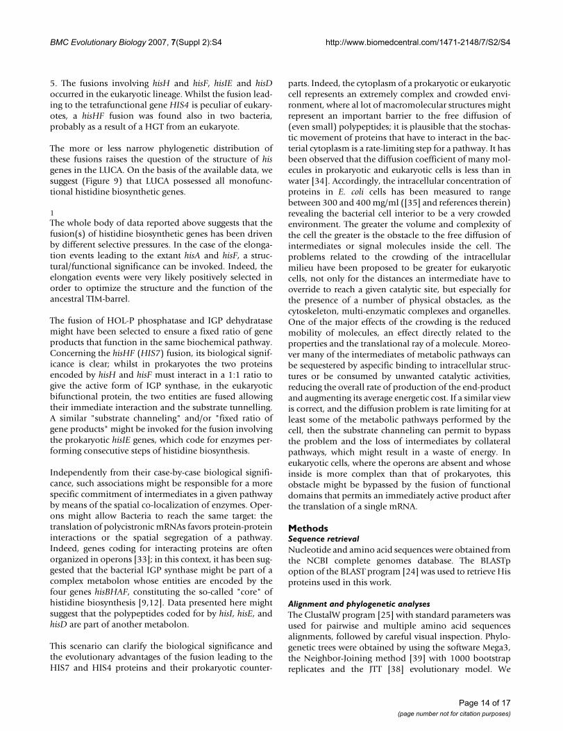
5. The fusions involving hisH and hisF, hisIE and hisDoccurred in the eukaryotic lineage. Whilst the fusion lead-ing to the tetrafunctional gene HIS4 is peculiar of eukary-otes, a hisHF fusion was found also in two bacteria,probably as a result of a HGT from an eukaryote.
The more or less narrow phylogenetic distribution ofthese fusions raises the question of the structure of hisgenes in the LUCA. On the basis of the available data, wesuggest (Figure 9) that LUCA possessed all monofunc-tional histidine biosynthetic genes.
1The whole body of data reported above suggests that thefusion(s) of histidine biosynthetic genes has been drivenby different selective pressures. In the case of the elonga-tion events leading to the extant hisA and hisF, a struc-tural/functional significance can be invoked. Indeed, theelongation events were very likely positively selected inorder to optimize the structure and the function of theancestral TIM-barrel.
The fusion of HOL-P phosphatase and IGP dehydratasemight have been selected to ensure a fixed ratio of geneproducts that function in the same biochemical pathway.Concerning the hisHF (HIS7) fusion, its biological signif-icance is clear; whilst in prokaryotes the two proteinsencoded by hisH and hisF must interact in a 1:1 ratio togive the active form of IGP synthase, in the eukaryoticbifunctional protein, the two entities are fused allowingtheir immediate interaction and the substrate tunnelling.A similar "substrate channeling" and/or "fixed ratio ofgene products" might be invoked for the fusion involvingthe prokaryotic hisIE genes, which code for enzymes per-forming consecutive steps of histidine biosynthesis.
Independently from their case-by-case biological signifi-cance, such associations might be responsible for a morespecific commitment of intermediates in a given pathwayby means of the spatial co-localization of enzymes. Oper-ons might allow Bacteria to reach the same target: thetranslation of polycistronic mRNAs favors protein-proteininteractions or the spatial segregation of a pathway.Indeed, genes coding for interacting proteins are oftenorganized in operons [33]; in this context, it has been sug-gested that the bacterial IGP synthase might be part of acomplex metabolon whose entities are encoded by thefour genes hisBHAF, constituting the so-called "core" ofhistidine biosynthesis [9,12]. Data presented here mightsuggest that the polypeptides coded for by hisI, hisE, andhisD are part of another metabolon.
This scenario can clarify the biological significance andthe evolutionary advantages of the fusion leading to theHIS7 and HIS4 proteins and their prokaryotic counter-
parts. Indeed, the cytoplasm of a prokaryotic or eukaryoticcell represents an extremely complex and crowded envi-ronment, where al lot of macromolecular structures mightrepresent an important barrier to the free diffusion of(even small) polypeptides; it is plausible that the stochas-tic movement of proteins that have to interact in the bac-terial cytoplasm is a rate-limiting step for a pathway. It hasbeen observed that the diffusion coefficient of many mol-ecules in prokaryotic and eukaryotic cells is less than inwater [34]. Accordingly, the intracellular concentration ofproteins in E. coli cells has been measured to rangebetween 300 and 400 mg/ml ([35] and references therein)revealing the bacterial cell interior to be a very crowdedenvironment. The greater the volume and complexity ofthe cell the greater is the obstacle to the free diffusion ofintermediates or signal molecules inside the cell. Theproblems related to the crowding of the intracellularmilieu have been proposed to be greater for eukaryoticcells, not only for the distances an intermediate have tooverride to reach a given catalytic site, but especially forthe presence of a number of physical obstacles, as thecytoskeleton, multi-enzymatic complexes and organelles.One of the major effects of the crowding is the reducedmobility of molecules, an effect directly related to theproperties and the translational ray of a molecule. Moreo-ver many of the intermediates of metabolic pathways canbe sequestered by aspecific binding to intracellular struc-tures or be consumed by unwanted catalytic activities,reducing the overall rate of production of the end-productand augmenting its average energetic cost. If a similar viewis correct, and the diffusion problem is rate limiting for atleast some of the metabolic pathways performed by thecell, then the substrate channeling can permit to bypassthe problem and the loss of intermediates by collateralpathways, which might result in a waste of energy. Ineukaryotic cells, where the operons are absent and whoseinside is more complex than that of prokaryotes, thisobstacle might be bypassed by the fusion of functionaldomains that permits an immediately active product afterthe translation of a single mRNA.
MethodsSequence retrievalNucleotide and amino acid sequences were obtained fromthe NCBI complete genomes database. The BLASTpoption of the BLAST program [24] was used to retrieve Hisproteins used in this work.
Alignment and phylogenetic analysesThe ClustalW program [25] with standard parameters wasused for pairwise and multiple amino acid sequencesalignments, followed by careful visual inspection. Phylo-genetic trees were obtained by using the software Mega3,the Neighbor-Joining method [39] with 1000 bootstrapreplicates and the JTT [38] evolutionary model. We
Page 14 of 17(page number not for citation purposes)
BMC Evolutionary Biology 2007, 7(Suppl 2):S4 http://www.biomedcentral.com/1471-2148/7/S2/S4
obtained different topologies for the tree correspondingto HIS4 domains and we compared them with thoseobtained with bayesian inference, i.e. MrBayes [36]. Wedefined the following parameters (not cited if default set-tings were used): evolutionary models of amino acidsequences were the WAG [37] and JTT [38] with characterfrequencies estimated from dataset (-F); topologiesobtained were identical for the two model; we report theshortest trees (in both cases corresponding to the oneobtained with WAG); we used heterogeneous rates amongsites, distributed as a Gamma distribution with shapeparameter free of variate from 0 to 50 (average obtainedfor these datasets 0.85); MCMC settings were as follows:2,500,000 and 1,500,000 generations, respectively forHisIE and HisD domains, and five chains. No startingtrees were used, with the idea in mind that convergence tovery similar values of the five chains would have beenmore significant than starting each chain from the sametree. Trees were sampled every 250 generations, for a totalset of 10,000 and 6,000 trees. Burnin was used to excludefrom following analysis those trees which were sampledbefore convergence of the chains; this was assessed case-by-case by calculating average, standard deviation andvariance. Convergence was reached after about 50,000generations (200 trees discarded). The resulting datasetswere used to obtain the shown trees with the allcompatoption.
Structural alignmentsStructural alignments and Root-means-squares calcula-tions were performed using Swiss pdb viewer [40]. We iso-
lated modules corresponding to (β/α)2 and (β/α)accordingly to the analysis performed by [19] showingsecondary elements belonging and not belonging to thebarrel structure. These modules were then used as inde-pendent molecules and structurally aligned by using the'magic fit' option.
Competing interestsThe authors declare that they have no competing interests.
Authors' contributionsAll authors contributed equally to the work and manu-script preparation.
Additional material
Additional file 1Phylogenetic distribution of hisNB genes. Histogram showing the per-centage of organisms possessing a hisNB gene for taxonomic groups rep-resented in NCBI genomes database and taking into account only histidine producing organisms.Click here for file[http://www.biomedcentral.com/content/supplementary/1471-2148-7-S2-S4-S1.pdf]
Additional file 2HisD phylogenetic tree of organisms possessing hisNB. A NJ phyloge-netic tree (evolutionary model: Dayhoff, 500 bootstrap replicates) obtained from a HisD multialignment. The topology is congruent with those obtained with other His proteins; it illustrates that hisNB has been probably transferred together with a complete histidine biosynthetic operon. See also Additional File 3 concerning gene organization. Red: ε-proteobacteria possessing (upper group) and not possessing (bottom group) the hisNB gene fusion; Green: CFB group bacteria possessing (upper group) and not possessing (bottom group) the hisNB gene fusion.Click here for file[http://www.biomedcentral.com/content/supplementary/1471-2148-7-S2-S4-S2.pdf]
Additional file 3HisNB and gene organization. Several features concerning HisNB pro-teins from Bacteria belonging to species not available at the time of our previous analysis concerning hisNB genes [11].Click here for file[http://www.biomedcentral.com/content/supplementary/1471-2148-7-S2-S4-S3.pdf]
Additional file 4Phylogenetic distribution of hisIE genes. Histogram showing the per-centage of organisms possessing a hisIE gene for taxonomic groups repre-sented in NCBI genomes database and taking into account only histidine producing organisms.Click here for file[http://www.biomedcentral.com/content/supplementary/1471-2148-7-S2-S4-S4.pdf]
A global view of his gene fusions appearanceFigure 9A global view of his gene fusions appearance. Schemat-ical representation of his gene fusion appearance and hori-zontal transfer. Abbreviations used: A, B, E correspond to Archaea, Bacteria and Eukarya, respectively. LUCA stands for the Last Universal Common Ancestor.
hisIEDGene fusion
E
hisAGene
elongation(s)
LUCA
Monofunctional his geneshis ABCDEFGHIN
hisHFGene fusion
hisNBGene fusion BhisIE
Gene fusionhisA – hisF
Gene duplication
A
Horizontal gene transfer
Evolutionary time
Page 15 of 17(page number not for citation purposes)

BMC Evolutionary Biology 2007, 7(Suppl 2):S4 http://www.biomedcentral.com/1471-2148/7/S2/S4
AcknowledgementsThis article has been published as part of BMC Evolutionary Biology Volume 7 Supplement 2, 2007: Second Congress of Italian Evolutionary Biologists (First Congress of the Italian Society for Evolutionary Biology). The full con-tents of the supplement are available online at http://www.biomedcen tral.com/1471-2148/7?issue=S2
References1. Winkler ME: Biosynthesis of histidine. In Escherichia coli and Sal-
monella typhimurium: cellular and molecular biology Volume 1. Edited by:Neidhardt FC, Ingraham JL, Low KB, Magasanik B, Schaechter M, Hum-barger HD. Washington DC: ASM Press; 1987:395-411.
2. Alifano P, Fani R, Lió P, Lazcano A, Bazzicalupo M, Carlomagno MS,Bruni CB: Histidine biosynthetic pathway and genes: struc-ture, regulation and evolution. Microbiol Rev 1996, 60:44-69.
3. Carlomagno MS, Chiarotti L, Alifano P, Nappo AG, Bruni CB: Struc-ture of the Salmonella typhimurium and Escherichia coli K-12histidine operons. J Mol Biol 1988, 203:585-606.
4. Shen C, Yang L, Miller SL, Oró J: Prebiotic synthesis of histidine.J Mol Evol 1990, 31:167-74.
5. Maurel MC, Ninio J: Catalysis by a prebiotic nucleotide analogof histidine. Biochimie 1987, 69:551-553.
6. Shen C, Mills T, Oró J: Prebiotic synthesis of histidyl-histidine.J Mol Evol 1990, 31:175-9.
7. White DH, Erickson JC: Catalysis of peptide bond formation byhistidyl-histidine in a fluctuating clay environment. J Mol Evol1980, 16:279-290.
8. Shen C, Lazcano A, Oró J: The enhancement activities of histi-dyl-histidine in some prebiotic reactions. J Mol Evol 1990,31:445-52.
9. Fani R, Lió P, Lazcano A: Molecular evolution of the histidinebiosynthetic pathway. J Mol Evol 1995, 41:760-774.
10. Fani R, Mori E, Tamburini E, Lazcano A: Evolution of the structureand chromosomal distribution of histidine biosyntheticgenes. Orig Life Evol Biosph 1998, 28:555-570.
11. Brilli M, Fani R: Molecular evolution of hisB genes. J Mol Evol2004, 58:225-237.
12. Brilli M, Fani R: Origin and evolution of eucaryal HIS7 genes:from metabolons to bifunctional proteins? Gene 2004,339:149-160.
13. Fani R, Brilli M, Lió P: The origin and evolution of operons: thepiecewise building of the proteobacterial histidine operon. JMol Evol 2005, 60:378-390.
14. Fani R: Gene duplication and gene loading. In Microbial evolution:gene establishment, survival and exchange Edited by: Miller RV, Day MJ.Washington DC: ASM Press; 2004:67-81.
15. Fani R, Brilli M, Lió P: Inference from proteobacterial operonsshows piecewise organization: a reply to price et Al. J Mol Evol2006, 63:577-580.
16. Jensen R: Evolution of metabolic pathways in enteric bacteriaIn Escherichia coli and Salmonella typhimurium. In Escherichia coliand Salmonella typhimurium: cellular and molecular biology Volume 1.Edited by: Neidhardt FC, Ingraham JL, Low KB, Magasanik B, SchaechterM, Humbarger HD. Washington DC: ASM Press; 1987:2649-2662.
17. Yanai I, Wolf YI, Koonin EV: Evolution of gene fusions: horizon-tal transfer versus independent events. Genome Biol 2002,3:research0024-.
18. Fani R, Lió P, Chiarelli I, Bazzicalupo M: The evolution of the his-tidine biosynthetic genes in prokaryotes: a common ances-tor for the hisA and hisF genes. J Mol Evol 1994, 38:489-495.
19. Lang D, Thoma R, Henn-Sax M, Sterner R, Wilmanns M: Structuralevidence for evolution of the β/α barrel scaffold by geneduplication and fusion. Science 2000, 289:1546-1550.
20. Kneidinger B, Marolda C, Graninger M, Zamyatina A, McArthur F,Kosma P, Valvano MA, Messner P: Biosynthesis pathway of ADP-L-glycero-beta-D-manno-heptose in Escherichia coli. J Bacteriol2002, 184:363-9.
21. Kneidinger B, Graninger M, Puchberger M, Kosma P, Messner P: Bio-synthesis of nucleotide-activated D-glycero-D-manno-hep-tose. J Biol Chem 2001, 276:20935-44.
22. Klem TJ, Davisson VJ: Imidazole glycerol phosphate synthase:the glutamine amidotransferase in histidine biosynthesis.Biochemistry 1993, 32:5177-5186.
23. Kuenzler M, Balmelli T, Egli CM, Paravicini G, Braus GH: Cloning,primary structure, and regulation of the HIS7 gene encodinga bifunctional glutamine amidotransferase: cyclase from Sac-charomyces cerevisiae. J Bacteriol 1993, 175:5548-5558.
24. Altschul SF, Madden TL, Schaffer AA, Zhalg J, Zhalg Z, Miller W, Lip-man DJ: Gapped BLAST and PSI-BLAST: a new generation ofprotein database search programs. Nucl Acids Res 1997,25:3389-3402.
25. Thompson JD, Higgins DG, Gibson TJ: CLUSTAL W: improvingthe sensitivity of progressive multiple sequence alignmentthrough sequence weighting, position-specific gap penaltiesand weight matrix choice. Nucleic Acids Res 1994, 22:4673-4680.
26. Bürger E, Görisch H: Evidence for an essential lysine at theactive site of L-Histidinol:NAD + oxidoreductase; a bifunc-tional dehydrogenase. Eur J Biochem 1981, 118:125-130.
27. Kheirolomoom A, Mano J, Nagai A, Ogawa A, Iwasaki G, Ohta D:Steady-state kinetics of cabbage Histidinol dehydrogenase.Arch Biochem Biophys 1994, 312:493-450.
28. Fujimori K, Ohta D: Isolation and characterization of a histi-dine biosynthetic gene in Arabidopsis encoding a polypeptidewith two separate domains for phosphoribosyl-ATP pyro-phosphohydrolase and phosphoribosyl-AMP cyclohydrolase.Plant Physiology 1998, 118:275-283.
29. Keesey JK Jr, Bigelis R, Fink GR: The product of the his4 genecluster in Saccharomyces cerevisiae. A trifunctional polypep-tide. J Biol Chem 1979, 254:7427-7433.
30. Van de Peer Y, De Wachter R: Evolutionary relationships amongthe eucaryotic crown taxa taking into account site-to-siterate variation in 18S rRNA. J Mol Evol 1997, 45:619-630.
31. Brandvain Y, Barker MS, Wade MJ: Gene co-inheritance and genetransfer. Science 2007, 315:1685.
32. Reyes-Prieto A, Hackett JD, Soares MB, Bonaldo MF, Bhattacharya D:Cyanobacterial contribution to algal nuclear genomes is pri-
Additional file 5HIS4 multialignments with concatenated prokaryotic sequences. A multialignment of fungal HIS4 sequences and the corresponding proteins from Plants and from a selected number of Prokaryotes.Click here for file[http://www.biomedcentral.com/content/supplementary/1471-2148-7-S2-S4-S5.pdf]
Additional file 6Entropy plot of a multialignment of HIS4 sequences. Entropy plot of the multialignment of HIS4 proteins. The regions of the protein are also indi-cated showing their different degree of conservation. Entropy was calcu-lated with the following formula: H(I) = S [f(b, I) * ln(f(b, I))], where b is a residue found in in column I and f(b, I) its frequency in I and the summation extends over all residues in column I.Click here for file[http://www.biomedcentral.com/content/supplementary/1471-2148-7-S2-S4-S6.pdf]
Additional file 7Pairwise identity values within HIS4 domains. Identity values for the pairwise comparison of the different domains composing HIS4 proteins (the standard deviation is also shown).Click here for file[http://www.biomedcentral.com/content/supplementary/1471-2148-7-S2-S4-S7.pdf]
Additional file 8Identifiers of sequences used in this work.Click here for file[http://www.biomedcentral.com/content/supplementary/1471-2148-7-S2-S4-S8.txt]
Page 16 of 17(page number not for citation purposes)
http://www.ncbi.nlm.nih.gov/entrez/query.fcgi?cmd=Retrieve&db=PubMed&dopt=Abstract&list_uids=8852895
http://www.ncbi.nlm.nih.gov/entrez/query.fcgi?cmd=Retrieve&db=PubMed&dopt=Abstract&list_uids=8852895
http://www.ncbi.nlm.nih.gov/entrez/query.fcgi?cmd=Retrieve&db=PubMed&dopt=Abstract&list_uids=3062174
http://www.ncbi.nlm.nih.gov/entrez/query.fcgi?cmd=Retrieve&db=PubMed&dopt=Abstract&list_uids=3062174
http://www.ncbi.nlm.nih.gov/entrez/query.fcgi?cmd=Retrieve&db=PubMed&dopt=Abstract&list_uids=2445388
http://www.ncbi.nlm.nih.gov/entrez/query.fcgi?cmd=Retrieve&db=PubMed&dopt=Abstract&list_uids=2445388
http://www.ncbi.nlm.nih.gov/entrez/query.fcgi?cmd=Retrieve&db=PubMed&dopt=Abstract&list_uids=7205965
http://www.ncbi.nlm.nih.gov/entrez/query.fcgi?cmd=Retrieve&db=PubMed&dopt=Abstract&list_uids=7205965
http://www.ncbi.nlm.nih.gov/entrez/query.fcgi?cmd=Retrieve&db=PubMed&dopt=Abstract&list_uids=8587121
http://www.ncbi.nlm.nih.gov/entrez/query.fcgi?cmd=Retrieve&db=PubMed&dopt=Abstract&list_uids=8587121
http://www.ncbi.nlm.nih.gov/entrez/query.fcgi?cmd=Retrieve&db=PubMed&dopt=Abstract&list_uids=9742729
http://www.ncbi.nlm.nih.gov/entrez/query.fcgi?cmd=Retrieve&db=PubMed&dopt=Abstract&list_uids=9742729
http://www.ncbi.nlm.nih.gov/entrez/query.fcgi?cmd=Retrieve&db=PubMed&dopt=Abstract&list_uids=9742729
http://www.ncbi.nlm.nih.gov/entrez/query.fcgi?cmd=Retrieve&db=PubMed&dopt=Abstract&list_uids=8028028
http://www.ncbi.nlm.nih.gov/entrez/query.fcgi?cmd=Retrieve&db=PubMed&dopt=Abstract&list_uids=8494895
http://www.ncbi.nlm.nih.gov/entrez/query.fcgi?cmd=Retrieve&db=PubMed&dopt=Abstract&list_uids=8494895
http://www.ncbi.nlm.nih.gov/entrez/query.fcgi?cmd=Retrieve&db=PubMed&dopt=Abstract&list_uids=8366040
http://www.ncbi.nlm.nih.gov/entrez/query.fcgi?cmd=Retrieve&db=PubMed&dopt=Abstract&list_uids=9254694
http://www.ncbi.nlm.nih.gov/entrez/query.fcgi?cmd=Retrieve&db=PubMed&dopt=Abstract&list_uids=9254694
http://www.ncbi.nlm.nih.gov/entrez/query.fcgi?cmd=Retrieve&db=PubMed&dopt=Abstract&list_uids=7984417
http://www.ncbi.nlm.nih.gov/entrez/query.fcgi?cmd=Retrieve&db=PubMed&dopt=Abstract&list_uids=7984417
http://www.ncbi.nlm.nih.gov/entrez/query.fcgi?cmd=Retrieve&db=PubMed&dopt=Abstract&list_uids=7984417
http://www.ncbi.nlm.nih.gov/entrez/query.fcgi?cmd=Retrieve&db=PubMed&dopt=Abstract&list_uids=6793363
http://www.ncbi.nlm.nih.gov/entrez/query.fcgi?cmd=Retrieve&db=PubMed&dopt=Abstract&list_uids=6793363
http://www.ncbi.nlm.nih.gov/entrez/query.fcgi?cmd=Retrieve&db=PubMed&dopt=Abstract&list_uids=6793363
http://www.ncbi.nlm.nih.gov/entrez/query.fcgi?cmd=Retrieve&db=PubMed&dopt=Abstract&list_uids=8037463
http://www.ncbi.nlm.nih.gov/entrez/query.fcgi?cmd=Retrieve&db=PubMed&dopt=Abstract&list_uids=8037463
http://www.ncbi.nlm.nih.gov/entrez/query.fcgi?cmd=Retrieve&db=PubMed&dopt=Abstract&list_uids=9733547
http://www.ncbi.nlm.nih.gov/entrez/query.fcgi?cmd=Retrieve&db=PubMed&dopt=Abstract&list_uids=9733547
http://www.ncbi.nlm.nih.gov/entrez/query.fcgi?cmd=Retrieve&db=PubMed&dopt=Abstract&list_uids=9419239
http://www.ncbi.nlm.nih.gov/entrez/query.fcgi?cmd=Retrieve&db=PubMed&dopt=Abstract&list_uids=9419239

BMC Evolutionary Biology 2007, 7(Suppl 2):S4 http://www.biomedcentral.com/1471-2148/7/S2/S4
Publish with BioMed Central and every scientist can read your work free of charge
"BioMed Central will be the most significant development for disseminating the results of biomedical research in our lifetime."
Sir Paul Nurse, Cancer Research UK
Your research papers will be:
available free of charge to the entire biomedical community
peer reviewed and published immediately upon acceptance
cited in PubMed and archived on PubMed Central
yours — you keep the copyright
Submit your manuscript here:http://www.biomedcentral.com/info/publishing_adv.asp
BioMedcentral
marily limited to plastid functions. Curr Biol 2006,16:2320-2325.
33. Dandekar T, Snel B, Huynen M, Bork P: Conservation of geneorder: a fingerprint of proteins that physically interact.Trends Biochem Sci 1998, 23:324-328.
34. Dauty E, Verkman AS: Molecular crowding reduces to a similarextent the diffusion of small solutes and macromolecules:measurement by fluorescence correlation spectroscopy. JMol Recognit 2004, 17:441-447.
35. Zimmerman SB, Minton AP: Estimation of macromolecule con-centrations and excluded volume effects for the cytoplasmof Escherichia coli. J Mol Biol 1991, 222:599-620.
36. Huelsenbeck JP, Ronquist F: MrBayes: Bayesian inference of phy-logenetic trees. Bioinformatics 2001, 17:754-755.
37. Whelan S, Goldman N: A general empirical model of proteinevolution derived from multiple protein families using amaximum-likelihood approach. Mol Biol Evol 2001, 18:691-699.
38. Jones DT, Taylor WR, Thornton JM: A new approach to proteinfold recognition. Nature 1992, 358:86-89.
39. Kumar S, Tamura K, Jakobsen IB, Nei M: MEGA2: molecular evo-lutionary genetics analysis software. Bioinformatics 2001,17:1244-1245.
40. Kaplan W, Littlejohn TG: Swiss-PDB Viewer (Deep View). Brief Bioinform2001, 2:195-197.
Page 17 of 17(page number not for citation purposes)
http://www.ncbi.nlm.nih.gov/entrez/query.fcgi?cmd=Retrieve&db=PubMed&dopt=Abstract&list_uids=9787636
http://www.ncbi.nlm.nih.gov/entrez/query.fcgi?cmd=Retrieve&db=PubMed&dopt=Abstract&list_uids=9787636
http://www.ncbi.nlm.nih.gov/entrez/query.fcgi?cmd=Retrieve&db=PubMed&dopt=Abstract&list_uids=1748995
http://www.ncbi.nlm.nih.gov/entrez/query.fcgi?cmd=Retrieve&db=PubMed&dopt=Abstract&list_uids=1614539