Elusive structural changes of New Delhi metallo-β-lactamase ...
Upload
independentCategory
view
1download
0
Investigating the Elusive Mechanism of GlycosaminoglycanBiosynthesis*□S
Received for publication, July 9, 2009 Published, JBC Papers in Press, July 23, 2009, DOI 10.1074/jbc.M109.043208
Xylophone V. Victor‡, Thao K. N. Nguyen§, Manivannan Ethirajan‡, Vy M. Tran§, Khiem V. Nguyen‡,and Balagurunathan Kuberan‡§1
From the Departments of ‡Medicinal Chemistry and §Bioengineering, University of Utah, Salt Lake City, Utah 84112
Glycosaminoglycan (GAG) biosynthesis requires numerousbiosynthetic enzymes and activated sulfate and sugar donors.Although the sequence of biosynthetic events is resolved usingreconstituted systems, little is known about the emergence ofcell-specific GAG chains (heparan sulfate, chondroitin sulfate,and dermatan sulfate) with distinct sulfation patterns. We haveutilized a library of click-xylosides that have various aglyconesto decipher the mechanism of GAG biosynthesis in a cellularsystem. Earlier studies have shown that both the concentrationof the primers and the structure of the aglycone moieties canaffect the composition of the newly synthesized GAG chains.However, it is largely unknown whether structural features ofaglycone affect the extent of sulfation, sulfation pattern, disac-charide composition, and chain length of GAG chains. In thisstudy, we show that aglycones can switch not only the type ofGAG chains, but also their fine structures. Our findings providesuggestive evidence for the presence of GAGOSOMES that havedifferent combinations of enzymes and their isoforms regulat-ing the synthesis of cell-specific combinatorial structures. Wesurmise that click-xylosides are differentially recognized by theGAGOSOMES to generate distinct GAG structures as observedin this study. These novel click-xylosides offer new avenues toprofile the cell-specificGAGchains, elucidate themechanismofGAG biosynthesis, and to decipher the biological actions ofGAG chains in model organisms.
Proteoglycans play amajor role in various cellular/physiolog-ical processes, including blood clotting, growth factor signaling,embryogenesis, axon growth and guidance, angiogenesis, andothers (1–4). Proteoglycans consists of a core protein and gly-cosaminoglycan (GAG)2 chains.GAGchains account for�50%of the total molecular weight and are primarily responsible forphysiological activity of the proteoglycans (5, 6). GAG chainsare composed of repeating disaccharide units of a hexosamine
residue and a hexuronic acid residue. The three major types ofGAG chains found in the proteoglycans are heparan sulfate(HS), chondroitin sulfate (CS) and dermatan sulfate (DS).These GAG chains are differentiated by the type of hexosamine(glucosamine/galactosamine), the percentage of uronic acidepimers (glucuronic/iduronic acid), the extent of sulfation, andthe nature of glycosidic linkage (�-/�-). One of the key steps inthe proteoglycan biosynthesis is the xylosylation of certain spe-cific serine residues of the core protein (7–10), which occurs inthe late endoplasmic reticulum and/or cis-Golgi compartments(11–13). This key event is an essential prelude for the construc-tion of the proteoglycan linkage region (14) that is followed bysequence of events resulting in the assembly of mature GAGchains by alternative addition of hexosamine and glucuronicacid residues. The maturation of GAG chains occurs in themedial and trans-Golgi compartments and involves the follow-ing events: N-sulfation of glucosamine units by N-deacetylase-N-sulfotransferases (for HS only), epimerization of glucuronicacids to iduronic acids by C-5 epimerase, and sulfation of therepeating disaccharide units by a variety of sulfotransferasesand their isoforms.The position, extent, and pattern of sulfation attribute enor-
mous diversity toGAG chains, which confer specificity in bind-ing to a vast array of proteins. These diverse structural featuresare very tightly regulated in a spatio-temporal manner duringand beyond the development of an organism, and these featuresdictate differential interactions with various growth factors andreceptors, and numerous protein targets leading to an array ofphysiological functions (15, 16).The presence of free GAG chains has been known to disrupt
the interaction of endogenous GAG components of proteogly-cans with protein ligands thereby altering the physiologicalactivities. Consequently, they have been used asmolecular toolsin the elucidation of the role of GAG chains in the activation ofcellular events (17–19). Free GAG chains can be synthesized invitro in cell culture by providing exogenous xylosides contain-ing various hydrophobic aglyconemoieties. Thus, the xylosidescan act as false acceptors for initiation of linkage region and thesubsequent elongation of GAG chains. Xylosides have beenused for over three decades both in vitro (20–28) and in vivo(25, 29–31) to probe the functional significance of GAG chainsin various dynamic systems under different conditions. Thequantity and type of GAG chains synthesized depends onthe systemwhere itwas tested andon the structureof the aglyconemoiety of the xylosides (32–34). Most of these studies haveutilized a few O-xylosides that are inherently less stable. Fur-thermore, synthesis ofO-xylosides requires very stringent reac-
* This work was supported, in whole or in part, by National Institutes of HealthGrant GM075168 (to B. K.). This work was also supported by Human Fron-tiers Science Program (to B. K.) and the Vietnam Education Fund (toT. K. N. N. and K. V. N.).
This work is dedicated to Prof. Robert D. Rosenberg.□S The on-line version of this article (available at http://www.jbc.org) contains
supplemental Figs. S1–S6.1 To whom correspondence should be addressed: Dept. of Medicinal Chem-
istry, Skaggs Hall, Rm#307, 30 South 2000 East, University of Utah, Salt LakeCity, UT 84112. Tel.: 801-587-9474; Fax: 801-585-9119; E-mail: [email protected].
2 The abbreviations used are: GAG, glycosaminoglycan; HS, heparan sulfate;CS, chondroitin sulfate; DS, dermatan sulfate; CHO, Chinese hamster ovary;HPLC, high-pressure liquid chromatography.
THE JOURNAL OF BIOLOGICAL CHEMISTRY VOL. 284, NO. 38, pp. 25842–25853, September 18, 2009© 2009 by The American Society for Biochemistry and Molecular Biology, Inc. Printed in the U.S.A.
25842 JOURNAL OF BIOLOGICAL CHEMISTRY VOLUME 284 • NUMBER 38 • SEPTEMBER 18, 2009
by guest on July 22, 2016http://w
ww
.jbc.org/D
ownloaded from
by guest on July 22, 2016
http://ww
w.jbc.org/
Dow
nloaded from
by guest on July 22, 2016http://w
ww
.jbc.org/D
ownloaded from
by guest on July 22, 2016
http://ww
w.jbc.org/
Dow
nloaded from
by guest on July 22, 2016http://w
ww
.jbc.org/D
ownloaded from
by guest on July 22, 2016
http://ww
w.jbc.org/
Dow
nloaded from
by guest on July 22, 2016http://w
ww
.jbc.org/D
ownloaded from
by guest on July 22, 2016
http://ww
w.jbc.org/
Dow
nloaded from
by guest on July 22, 2016http://w
ww
.jbc.org/D
ownloaded from
by guest on July 22, 2016
http://ww
w.jbc.org/
Dow
nloaded from
by guest on July 22, 2016http://w
ww
.jbc.org/D
ownloaded from
by guest on July 22, 2016
http://ww
w.jbc.org/
Dow
nloaded from
by guest on July 22, 2016http://w
ww
.jbc.org/D
ownloaded from
by guest on July 22, 2016
http://ww
w.jbc.org/
Dow
nloaded from
by guest on July 22, 2016http://w
ww
.jbc.org/D
ownloaded from
by guest on July 22, 2016
http://ww
w.jbc.org/
Dow
nloaded from
by guest on July 22, 2016http://w
ww
.jbc.org/D
ownloaded from
by guest on July 22, 2016
http://ww
w.jbc.org/
Dow
nloaded from
by guest on July 22, 2016http://w
ww
.jbc.org/D
ownloaded from

tion conditions, toxic Lewis acids, and at times leads to insepa-rable � and � mixtures with unpredictable yields. As a result, itis tedious to generate diverse xylosides in a rapid fashion andutilize them in biological systems.We envisioned that synthesisof metabolically stable xylosides will advance our knowledge ofglycosaminoglycan biosynthesis and how they regulate variouspathophysiological processes.In our earlier communication, we outlined a simple strategy,
utilizing click chemical methodology that addresses the abovelimitations ofO-xylosides, to generate a library of xylosides in arobust manner (35). Several studies have shown that the con-centration of the primers and the aglycone moieties influencethe composition of GAG chains produced (32). In the currentstudy, we show that the aglycone moieties of click-xylosidesmay not only influence the composition and quantity of GAGchains but also the extent of sulfation, sulfation pattern, disac-charide composition, and chain length using pgsA-745 Chinesehamster ovary (CHO) cell line as a model cellular system. Ourfindings provide new insights in to the mechanism of GAGbiosynthesis and offer new avenues to decipher the biologicalactions of GAG chains in model organisms.
EXPERIMENTAL PROCEDURES
Materials andMethods—ThemutantCHOcell line (pgsA-745)wasobtained fromAmericanTypeCultureCollection (36).All cellculture reagentswereobtained fromHyClone.Theradiochemical,[35S]Na2SO4 was purchased from MP Biomedicals, and D-[-6-3H]glucosamine and flow scintillationmixture, Ultima-FloAP, forFlow radiometric analysis were obtained from PerkinElmer LifeSciences. All other chemicals and biochemicals were obtainedfrom Sigma. The click-xylosides, reported here, were synthe-sized as described previously (35). The 6�Pronase solutionwasprepared using Streptomyces griseus protease type XIV (1mg/ml). The DEAE-Sepharose gel was purchased from Amer-sham Biosciences. The HPLC columns utilized were DEAE-3SW (7.5 mm � 7.5 cm, 10-�m particle size) for anion-ex-change chromatography and G3000SWXL (7.8 mm � 30 cm,5-�mparticle size) for size-exclusion chromatography column,purchased from Tosoh Bioscience LLC. Commercially avail-able unsaturated HS and CS standards were used to determinemigration positions and elucidate disaccharide composition ofprimed GAG chains. Following standard unsaturated heparansulfate disaccharideswere purchased fromSigma-Aldrich: 2-acet-amido-2-deoxy-4-O-(4-deoxy-�-L-threo-hexenepyranosyluronicacid)-D-glucose (�UA-GlcNAc), 2-deoxy-2-sulfamido-4-O-(4-deoxy-�-L-threo-hexenepyranosyluronicacid)-D-glucose(�UA-GlcNS), 2-deoxy-2-sulfamido-4-O-(4-deoxy-2-O-sulfo-�-L-threo-hexenepyranosyluronic acid)-6-O-sulfo-D-glucose(�UA-GlcNS6S), 2-deoxy-2-sulfamido-4-O-(4-deoxy-2-O-sulfo-�-L-threo-hexenepyranosyluronic acid)-D-glucose (�UA2S-GlcNS), and 2-deoxy-2-sulfamido-4-O-(4-deoxy-2-O-sulfo-�-L-threo-hexenepyranosyluronic acid)-6-O-sulfo-D-glucose(�UA2S-GlcNS6S). CS disaccharide standards were also pur-chased for calibration. GAG disaccharide standards and3H-labeled lyase disaccharide products were detected at 232nm using inline UV detector and flow scintillation analyzer,respectively.
Screening of Click-xylosides in Cell Culture—The priming ofthe xylosides in xylosyl transferase-deficient CHO cell linepgsA-745 was performed as described in our earlier communi-cation (35). Briefly, 1 � 105 cells were plated per well, contain-ing the appropriate complete growthmedium, in a 24-well plateand incubated at 37 °C in a humidified incubator for 24 h toreach a confluency of about 50%. The cells were then washedwith sterile PBS and replaced with 450 �l appropriate mediumcontaining 10% dialyzed FBS. A serial dilution of the primers at100X the final concentration was prepared and 5 �l of appro-priate 100X primer was added to various wells to yield a finalconcentration of 0.1 1, 10, 100 and 1000�M respectively. 50�Ciof [35S]Na2SO4 or D-[6-3H]glucosaminewas then added to eachwell for radiolabeling of the GAG chains. The 24-well plateswere placed in the incubator for 24 h before the addition of 6XPronase solution (100 �l) followed by incubation at 37 °Covernight.Purification andQuantification of GAGChains—After treat-
ing each well with Pronase solution overnight, the entire con-tents of the wells were transferred to a microcentrifuge tubeand subjected to centrifugation at 16,000 � g for 5 min. Thesupernatant were transferred to a fresh tube and half-a-volumeof 0.016% Triton X-100 was added. The diluted supernatantwas loaded on to aDEAE-Sepharose column (0.2ml) pre-equil-ibrated with 10 column volumes of wash buffer (20mMNaOAcbuffer, pH 6.0), containing 0.1 MNaCl and 0.01% Triton X-100)and the column was washed with 20 column volumes of washbuffer. The bound GAG was eluted with 6 column volumes ofelution buffer (20 mM NaOAc (pH 6.0) containing 1 M NaCl).The amount of GAG chains primed by various click-xylosideswas determined by quantifying the 35S- or 3H-radioactivityincorporated in the purified GAG eluate. 50 �l of the variouseluates was diluted with 5 ml of scintillation mixture and trip-licate samples were measured using a scintillation counter fortotal radioactivity.Analysis of GAG Chains by Anion-exchange Chro-
matography—The homogeneity and extent of sulfation of theGAG chains synthesized on the various primers were deter-mined by measuring migration time on anion-exchange col-umn using high-pressure liquid chromatography (HPLC) withinline flow scintillation analyzer. 25 �l of eluate was diluted10-fold with 10 mM KH2PO4 (pH 6.0) containing 0.2% CHAPS,and loaded on to a HPLC-DEAE column and eluted with alinear NaCl gradient of 0.2 M to 1 M over 80 min.Extent of GAG Sulfation in the Presence of Chlorate—Conflu-
ent 745-CHO cell cultures containing [3H]GlcNH2 weretreated with xyloside 5 (100 �M) in the presence sodium chlo-rate at various concentrations. GAG chains were recoveredfrom the conditioned media by DEAE-Sepharose chromatog-raphy, followed by isolation of [3H]labeled GAG as described inthe previous section. To monitor the effect of chlorate concen-tration on GAG sulfation, samples of [3H]GAG from controland chlorate-treated cells were analyzed on an anion-exchangeHPLC column as described in the previous section.Co-priming of Two Different Xylosides in Cell Culture—The
co-priming of the xylosides was performed as described abovein the “screening of click-xylosides section” by adding two dif-ferent xylosides to xylosyl transferase-deficient CHO cells in
GAGOSOMES and Glycosaminoglycan Biosynthesis
SEPTEMBER 18, 2009 • VOLUME 284 • NUMBER 38 JOURNAL OF BIOLOGICAL CHEMISTRY 25843
by guest on July 22, 2016http://w
ww
.jbc.org/D
ownloaded from

the same well of a 24-well plate. Briefly, a solution containingtwo xylosides, 22 and 24, at 100� the final concentration wasprepared, and 5 �l of the premixed xylosides was added to yielda final concentration of 100 �M for each xyloside. 50 �Ci of[35S]Na2SO4 or D-[6-3H]glucosamine was then added to eachwell for radiolabeling the GAG chains synthesized. The 24-wellplates were placed in the incubator for 24 h before the additionof 6X Pronase solution (100 �l) followed by incubation at 37 °Covernight. The GAG chains were then purified from the solu-tion as described in the earlier section.Analysis of GAG Chains by Size-exclusion Chro-
matography—The chain length of the GAG chains synthesizedon various primers was determined by measuring their migra-tion time on size exclusion columnusingHPLCwith inline flowscintillation analyzer. The GAG chains were loaded on to twoG3000SWXL columns (Tosoh, 7.8 mm � 30 cm), which wereconnected in tandem, and eluted over 60 min with phosphatebuffer (100 mM KH2PO4, 100 mM NaCl, pH 6). The migrationtime of theGAG chains was determined bymeasuring the aver-age of peak-width at half maximum, and the average molecularweight was calculated based on the migration time of polysty-rene sulfonate standards.Analysis of GAG Disaccharide Composition—To determine
the disaccharide composition of GAG, the total GAG chainswere fractionated using size exclusion chromatography asdescribed above and the fractions containing the GAG chainswere pooled and dialyzed using a benzoylated dialysis mem-brane with molecular mass 2000 Da in 10 mM ammoniumbicarbonate. To concentrate and remove volatile ammoniumbicarbonate, dialyzed GAG samples were placed in a speed-vacconcentration system under reduced pressure. The HS in thetotal GAG was digested with heparitinases I, II, and III over-night at 37 °C and analyzed using strong anion-exchangeHPLCusing a Phenosphere SAX column (Phenomenex, 250 � 4.6mm) with inline radiodetection. Solvent A was double-distilledwater (pH 3.5). Solvent B was 2 M NaCl (pH 3.5). The disaccha-rides were eluted at a flow rate of 1 ml/min with the followingprofile: 100% solvent A for 5 min; linear gradient of 0 to 40%solvent B for 35min; 100% solvent B for 10min and finally 100%solvent A for 25 min to equilibrate the column. In a similarfashion, to determine the disaccharide composition of xyloside-primed CS and DS chains, the GAG chains were digested withchondroitinase ABC (ACCI) overnight at 37 °C, and the result-ing disaccharides were analyzed as described above.
RESULTS
Priming Activity of Click-xylosides—We have previouslyreported the synthesis of the click-xylosides and have shownthat these molecules prime GAG chains in a cell line that lacksproduction of endogenous GAG chains due to deficiency of akey enzyme, xylosyl transferase, involved in the biosynthesis ofproteoglycans (35). This synthetic methodology allowed us toconstruct a library of novel xylosides with a wide variety ofaglycone moieties in a rapid manner. In this study, we havesynthesized additional compounds and expanded the xylosidelibrary to probe the effect of aglycones on the priming ability(Table 1). At the outset, we have examinedwhether the concen-tration of xylosides would affect the priming ability and deter-
mined the optimal primer concentration of each xyloside forthe maximal production of GAG chains.The priming ability of the xylosides was determined as
described under “Experimental Procedures” utilizing themutant CHO cell line (pgsA-745) at the following five concen-trations: 0.1 �M, 1 �M, 10 �M, 100 �M, and 1 mM. The resultsindicate that the priming is concentration-dependent, and sig-nificant priming for most xylosides was observed at a concen-tration of 100 �M or higher (Fig. 1). From the screening exper-iments with the above concentrations, we were able todetermine the optimal concentration range for each xylosidefor its maximal priming activity. For a selected group of xylo-sides, experiments were performed at intermediate concentra-tions, which showed that click-xylosides can act as acceptorseffectively at concentrations as low as 30 �M (data not shown).At 1 mM concentration, the triazolyl xyloside 1, which has nohydrophobic aglycone attached to the triazole group, was ableto participate in GAG synthesis, whereas the addition of ahydrophilic -CH2OH group, as shown in xyloside 2, leads to
TABLE 1Structures of click-xylosidesA library of metabolically stable xylosides containing a variety of hydrophobicgroups was synthesized using click-chemistry (see Ref. 35 for details). This librarywas employed in the current study to elucidate the mechanism of glycosaminogly-can biosynthesis.
GAGOSOMES and Glycosaminoglycan Biosynthesis
25844 JOURNAL OF BIOLOGICAL CHEMISTRY VOLUME 284 • NUMBER 38 • SEPTEMBER 18, 2009
by guest on July 22, 2016http://w
ww
.jbc.org/D
ownloaded from

dramatic decrease in the priming activity (Fig. 1). However, thepriming activity of hydrophobic xylosides 12, 13, 14, 15, 16,and 17 was higher at 100 �M concentration than at 1 mM con-centration (Fig. 1).Effect of Substituents on Priming—The role of various substit-
uent groups attached to the phenyl aglyconemoiety of the xylo-side modulating GAG biosynthesis was assessed. Substituentssuch as -OCH3 group in 6 and the -NO2 group in 11 have amarginal effect on the priming activity at 100 �M concentra-tion. We also found that these substitutions do not have anyinfluence at higher concentrations (Fig. 1). The analysis of thehalogen substitutions, fluoro-, chloro-, bromo-, and iodo-sub-stituents in 7, 8, 9, and 10, respectively, show no tangible effectin the amount of GAG chains produced when examined at 100�M concentration (Fig. 1). Interestingly, the amount of GAGchains primed decreases with increasing atomic weight of thehalogen substitutions at 1 mM concentration (Fig. 2A).Effect of Substituent Positions on Priming Activity—Three
xylosides, 24, 26, and 27, were synthesized to investigate theeffect of the position of -OCH3 substituent on GAG biosynthe-sis. At 100 �M, xyloside 26 (ortho) primed nearly two times theGAG chains compared with the unsubstituted xyloside 24,whereas xyloside 27 (para) primed 50% to that of unsubstitutedxyloside24. Interestingly, thepara-substituted xyloside primedthree times more GAG chains than the ortho-substituted xylo-
side at 1 mM concentration. Thus, the position of substituenthas a dramatic influence on the priming ability of the givenxyloside even though the activity trend is different at differentconcentrations (Fig. 2B).Extent of Sulfation and Migration Time—We utilized DEAE
anion-exchange chromatography to compare the extent of sul-fation of primed GAG chains. However, there are no commer-cially available GAG standards with various extent of sulfationto correlate the migration time and extent of sulfation. There-fore, we generated GAG chains with different extent of sulfa-tion by incubating xyloside 5with cells in the presence of chlo-rate, an inhibitor of 3�-phosphoadenosine 5�-phosphosulfatesynthetase that affects GAG sulfation, at various concentra-tions (37, 38). pgsA-745-CHO cells were treated with 100 �M
xyloside 5 and metabolically radiolabeled with [3H]GlcNH2 inthe presence of various sodium chlorate concentrations, 1 mM,5 mM, and 25 mM. GAG chains were recovered from the condi-tioned media by DEAE-Sepharose chromatography, followedby isolation of [3H]GAG chains as described under “Experi-mental Procedures.” To monitor the effect of chlorate concen-tration on GAG sulfation, samples of [3H]GAG from controland chlorate-treated cells were analyzed by an anion-exchangeHPLC column chromatography and subsequently analyzed forradioactivity with the aid of inline flow scintillation analyzer.The GAG chains were quantitatively retained by the column,
FIGURE 1. Priming activity of various xylosides. The novel xylosides were examined for their priming ability using xylosyl transferase-deficient CHO cells(pgsA-745). 100,000 cells were seeded per well of 24-well plates and treated with various xylosides at 0.1, 1, 10, and 100 �M and 1 mM concentrations for 24 hin the presence of 50 �Ci of [35S]O4
2� or D-[6-3H]glucosamine. The GAG chains were then purified and quantified as described under “Experimental Procedures.”Xylosides with different aglycones primed different amounts of GAG chains at different concentrations suggesting the decisive role played by aglycones. Thedata indicate the average of three independent experiments.
GAGOSOMES and Glycosaminoglycan Biosynthesis
SEPTEMBER 18, 2009 • VOLUME 284 • NUMBER 38 JOURNAL OF BIOLOGICAL CHEMISTRY 25845
by guest on July 22, 2016http://w
ww
.jbc.org/D
ownloaded from

but they required different concentrations of NaCl for theirelution (Fig. 3). These findings served as a basis to attribute thedifferences in the elution profile/migration time of variousGAG chains on the HPLC anion-exchange column to theextent of sulfation of GAG chains primed by various xylosidesas described. The chain length of theGAGchains can also affectthe migration time albeit to a lesser extent than sulfation.Comparison of Elution Profiles of GAG Chains—We focused
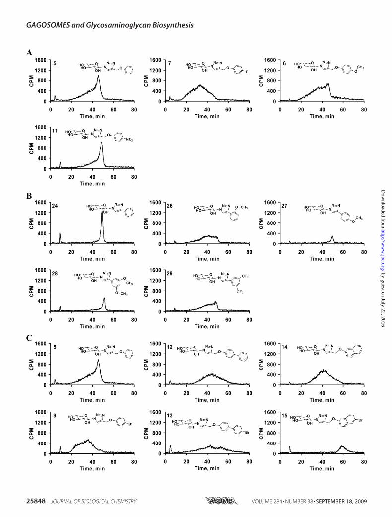
our investigation on HPLC analysis of the GAG chains primedby the exogenous xylosides at the optimal concentration (100�M). Using anion-exchange HPLC, we compared the elutionprofile of xyloside primed GAG chains, which is an indicator ofextent of sulfation. GAG chains that have less sulfation elute atlower ionic strength,whereas heavily sulfatedGAGchains eluteat higher ionic strength. First, we compared the HPLC profilesof GAG chains primed by xylosides of the simplest form, xylo-se-linked to triazole only (1), cyclohexyl (3), or phenyl (5) asshown in Fig. 5. The GAG chains synthesized from the above
compounds showed a very similar profile although the quanti-ties of the GAG chains were different for each xyloside. Theyshowed a predominantly homogenous peak along with a non-separable highly heterogeneous minor portion eluting at lowersalt concentration. Xylosides that differ in their aglycone aro-matic structure, 5 (phenyl), 12 (biphenyl), 14 (naphthyl), and17 (phenanthryl), were analyzed for the extent of sulfation byusing the DEAE column. GAG chains primed by xyloside 5eluted from 20 to 52 min with a peak maximum at 45 min,whereasGAGchains primed by 12, 14, and 17were elutedwithan even distribution from 20 to 60 min and with a peak maxi-mum of �42–44 min (Fig. 4). The differences in the elutionprofiles of the primed GAG chains indicate differences in theextent of sulfation among these GAG chains. The phenan-threne containing xyloside 17 primed fewer GAG chains. Itmay possibly be due to the inability of the enzymes to recognizea highly hydrophobic xyloside even though itmay cross cell andGolgi membranes more effectively, or alternatively this mayreflect the inability of the xyloside17 to reach the enzyme activesite.Effect of Spacer on Sulfation Patterns of GAG Chains—The
xylosides were attached to the phenyl moiety with many differ-ent spacers to confer conformational flexibility and were thenexamined for their influence on priming activity. The first set ofcompounds that we compared differs by spacer length. Thephenyl ring was linked directly to the triazole in 24 or throughthe spacers such as -CH2- in 25, -CH2-O- in 5, -CH2-O-CH2- in18, or -CH2-S- in 22. The elution profiles of the GAG chainsprimed by these xylosides are shown in Fig. 5A. It was interest-ing to find that the direct linking of a phenyl group in xyloside24 led to the synthesis of GAG chains that were highly homog-enous and eluted as a very narrow single peak at 48 min. Intro-ducing one carbon unit (-CH2-) between the phenyl and tria-zole groups in 25 led to loss of the homogeneity of GAG chainssynthesized, and these eluted between 18 and 45min. Introduc-
FIGURE 2. Effect of substituents and their positions on priming activity.Cells were treated with various xylosides for 24 h in the presence of 50 �Ci of[35S]O4
2� or D-[6-3H]glucosamine as described under “Experimental Proce-dures.” Primed GAG chains were then purified from the supernatant usinganion-exchange column chromatography and quantified using liquid scintil-lation. A, effect of various halogen substituents on the priming activity wasexamined. Fluoro-, chloro-, bromo-, and iodo-substituted xylosides (7, 8, 9,and 10, respectively) were compared against the unsubstituted xyloside 5 at1 mM concentration. B, effect of methoxy-substituent and its position on thephenyl ring was examined for priming activity. Unsubstituted xyloside 24 wascompared with ortho-substituted xyloside 26 and para-substituted xyloside27 at 100 �M (unshaded) and 1 mM (shaded) concentrations. The data indicatethe average of three independent experiments.
FIGURE 3. Correlation between extent of sulfation and migration time onHPLC anion-exchange chromatography. Cells containing D-[6-3H]glucosa-mine were treated with 100 �M xyloside 5 in the presence of chlorate atvarious concentrations. GAG chains were then isolated and quantified asdescribed under “Experimental Procedures.” The chlorate treatment did notaffect the amount of GAG chains produced. 1,000,000 cpm were applied toHPLC DEAE anion-exchange chromatography and eluted with a linear NaClgradient as described under “Experimental Procedures.” GAG species fromchlorate-treated cells were eluted earlier than those from control cells. Theelution profiles of GAG chains from control cells (gray tracer), cells treatedwith 5 mM chlorate (dark tracer), and cells treated with 25 mM chlorate (brokentracer) are shown. The elution profiles are representative of at least two inde-pendent experiments.
GAGOSOMES and Glycosaminoglycan Biosynthesis
25846 JOURNAL OF BIOLOGICAL CHEMISTRY VOLUME 284 • NUMBER 38 • SEPTEMBER 18, 2009
by guest on July 22, 2016http://w
ww
.jbc.org/D
ownloaded from

tion of various other spacers such as -CH2-O- in 5, -CH2-O-CH2- in 18, and -CH2-S- in 22 resulted in the production ofGAG chains with different elution profiles and failed to re-establish the narrow peak that was observed for the xyloside24 (Fig. 5A). In contrast to xyloside 24, xyloside 30 that has
a naphthyl group directly attached to triazole produced GAGchains with broad elution profile. When the naphthyl groupwas separated from the triazole group by a -CH2-S- spacer(23) narrow GAG chains production was restored. However,xyloside 14 that has the -CH2-O- spacer produced heteroge-
FIGURE 4. Effect of various hydrophobic moieties on the DEAE elution profile of GAG chains. GAG chains primed by various xylosides (1, 3, 5, 12, 14, and17) eluted from small anion-exchange column with 1 M NaCl, were diluted 5-fold and analyzed by anion-exchange HPLC column as described under “Exper-imental Procedures.” The variations in the elution profiles and migration times indicate differences in both sulfation pattern and extent of sulfation of theprimed GAG chains. The elution profiles are representative of at least two independent experiments.
FIGURE 5. Spacers between the triazole and aromatic ring in the aglycone moiety alter the sulfation pattern and extent of sulfation. Purified GAG chainswere diluted 5-fold and analyzed by anion-exchange HPLC column as described under “Experimental Procedures.” A gradient of 0.2 to 1.0 M NaCl for 80 min wasused for elution of the GAG chains. The differential elution profile and migration time indicates variations in sulfation pattern and extent of sulfation of GAGchains primed by phenyl group containing xylosides 5, 18, 22, 24, and 25 (A) and naphthyl group containing xylosides 14, 23, and 30 having various spacerswere examined (B). The elution profiles are representative of at least two independent experiments.
GAGOSOMES and Glycosaminoglycan Biosynthesis
SEPTEMBER 18, 2009 • VOLUME 284 • NUMBER 38 JOURNAL OF BIOLOGICAL CHEMISTRY 25847
by guest on July 22, 2016http://w
ww
.jbc.org/D
ownloaded from
GAGOSOMES and Glycosaminoglycan Biosynthesis
25848 JOURNAL OF BIOLOGICAL CHEMISTRY VOLUME 284 • NUMBER 38 • SEPTEMBER 18, 2009
by guest on July 22, 2016http://w
ww
.jbc.org/D
ownloaded from

neous GAG chains with a broad elution profile (Fig. 5B). Thisstudy has revealed that conformational flexibility likelyinfluences the biosynthesis of GAG chains with differentialsulfation pattern.The GAG chains primed by compounds 5, 6 (-OCH3), 7
(fluoro), 8 (chloro), 9 (bromo), 10 (iodo), and 11 (-NO2) wereanalyzed by HPLC to determine the effect of various substitu-ents on the extent of GAG sulfation. The halogen substituentssignificantly altered the extent of sulfation causing an increasein the heterogeneity of the GAG chains with little differenceamong the halogen containing xylosides (Fig. 6A; data notshown for xylosides 8–10). It was interesting to note that halo-gen substitution resulted in the production of GAG chains thatelute earlier from the DEAE column than those from the xylo-side 5. The xylosides containing -OCH3 group in 6 and -NO2group in 11 primed GAG chains that had different elution pro-files and reaffirmed that the type of substituent influences theGAGbiosynthetic pathways (Fig. 6A). All of the above substitu-ents were located at the para position of the phenyl ring. Wethen examined whether the position of substituent influencesthe sulfation patterns of the GAG chains. We examined thesulfation patterns of the GAG chains produced by xylosidesthat were substituted at various positions on the phenyl ringwith the -OCH3 group. The xylosides with a single substituentat the ortho position in 26 and at the para position in 27resulted in different elution profiles for the primedGAG chains(Fig. 6B). The para-substituted xyloside 27 primed homoge-nousGAGchains that eluted at 50min, albeit in lower quantity.On the other hand, the GAG chains primed by the ortho-sub-stituted xyloside 26 eluted between 20 and 50 min indicatingthese chains have various sulfation patterns (Fig. 6B). Thehighly electron withdrawing substituent in the place of -OCF329 also resulted in broad GAG chains. In addition, we investi-
gated the effect of the same substituent on different aglyconemoieties by comparing the bromo substitution of phenyl moi-ety in 9, biphenyl moiety in 13, and naphthyl moiety in 15. Wehad observed that a bromo substitution on the phenyl ring ledto a decrease in the extent of sulfation of GAG chains, whereasbromo substitution on biphenyl and naphthyl groups in theaglycone moieties led to a significant increase in the extent ofsulfation (Fig. 6C).Co-priming Experiment—Investigation of the extent of sulfa-
tion and the sulfation pattern of the GAG chains that weresynthesized by various xylosides showed distinct elution profileon anion-exchange column. These differences among GAGchains primed by xylosides are attributed to the presence ofdifferent aglycones on these xylosides. Hence, it is imperativethat we understand the molecular mechanisms that can gener-ate such differential priming of GAG chains. Xylosides 22 and24 primed GAG chains with different profiles in anion-ex-change chromatography. Xyloside 22 stimulates GAG chainsthat elute as a narrow peak with high sulfate density, whereasxyloside 24 produces GAG chains that elute as a broad peakwith less sulfate density. To determine whether distinct GAGchains can be primed by cells at the same time, xylosides 22 and24, were co-primed by addition to the same well, and theiranion-exchange profiles were compared with individuallyprimed GAG chains. The resulting GAG chains were isolatedand analyzed by anion-exchange chromatography (Fig. 7). Theindividually primed GAG chains have distinctive elution pro-files due to differences in their extent of sulfation (Fig. 7A).Also, an analysis was performed using GAG chains that wereprimed individually but mixed together before the anion-ex-change chromatography. The mixing of individually primedGAG chains results in less resolution of the peaks that haveotherwise very distinct profile (Fig. 7B). On the other hand,
FIGURE 6. Effect of substituents and their positions on GAG fine structures. Purified GAG chains were diluted 5-fold and analyzed by HPLC DEAE anion-exchange chromatography as described under “Experimental Procedures.” The differential elution profiles and migration times indicate variations in sulfationpattern and extent of sulfation. A, effect of various substituents at the para position of the phenyl ring was examined. The elution suggests that differentsubstituents on xylosides, 5, 6, 7, and 11 affect the elution profile of the GAG chains suggesting changes in the fine structures. B, influence of position ofthe substituent on the fine structures was examined. Unsubstituted xyloside 24 primed GAG chains that elute as a narrow peak, whereas ortho methoxy-substituted xyloside 26 produced GAG chains that elute as a broad peak. On the other hand, para-substituted xyloside 27 and dimethoxy-substituted xyloside28 produce GAG chains that elute as narrow peaks. C, bromo-substitution on phenyl, biphenyl, and naphthyl moieties was found to affect the extent ofsulfation and sulfation pattern. Unsubstituted xylosides 5, 12, and 14 were compared with their corresponding bromo-substituted xylosides 9, 13, and 15 forthe change in the extent of sulfation and sulfation pattern. The elution profiles are representative of at least two independent experiments.
FIGURE 7. Co-priming experiment with xylosides that prime distinct GAG chains. A, xylosides 22 and 24 were primed at 100 �M concentration in pgsA-745cells for 24 h individually, and GAG chains were purified. Individually primed GAG chains were then analyzed on HPLC DEAE anion-exchange chromatographyunder similar conditions in separate runs. The solid tracer indicates GAG chains primed by xyloside 22, and the broken tracer indicates the narrow elution profilesof the GAG chains primed by xyloside 24. B, individually primed GAG chains from xylosides 22 and 24 were mixed together and analyzed under similarconditions as described in A. C, 100 �M each of xylosides 22 and 24 were primed together for 24 h in cell culture experiments. The resulting GAG chains werepurified and analyzed by DEAE anion-exchange chromatography as described earlier. The elution profile suggests further loss of resolution of two distinct GAGchains when xylosides 22 and 24 are co-primed. The elution profiles are representative of at least two independent experiments.
GAGOSOMES and Glycosaminoglycan Biosynthesis
SEPTEMBER 18, 2009 • VOLUME 284 • NUMBER 38 JOURNAL OF BIOLOGICAL CHEMISTRY 25849
by guest on July 22, 2016http://w
ww
.jbc.org/D
ownloaded from

GAG chains that were co-primed in the presence of 22 and 24show a very broad peak due to overlap of the individual peaks.The slope of the front of peak increases gradually similar to thatobserved for GAG chains from 22 while the tail of the peakdrops off rather sharply comparable to that of GAG chainsprimed by 24 (Fig. 7C). This suggests that the GAG chains withdifferent extent of sulfation were primed by two xylosides thatwere co-primed even though the peaks of the different GAGchains cannot be completely resolved using anion-exchangechromatography.Chain Length Analysis of GAG Chains—To examine the
effect of various aglycones on GAG chain length, primed GAGchains were analyzed by size-exclusion chromatography asdescribed under “Experimental Procedures.” GAG chains hadan average molecular weight in the range of 6,000 to 34,000(Table 2). A significant number of xylosides in this studyprimed GAG chains that have molecular weight greater than10,000, whereas some generated GAG chains with lowermolecular weight. Only a few studies report chain length anal-ysis for xyloside-primed GAG chains (39, 40). In our study,most of the click-xylosides prime significantly long GAGchains. Xylosides that predominantly prime HS chains, 5 and22, have shorter chain lengths in the range of 6,000 to 12,000.Also, heterogeneous GAG chains with different extent of sulfa-tion observed from theDEAE elution profile have shorter chainlengths. Significantly, GAG chains that have narrow elutionprofile with high extent of sulfation, primed by 19, 20, 23, and24, have higher molecular weights (Table 2).Disaccharide Composition of GAGChains—The influence of
various aglycones on the disaccharide compositions of GAGchains was not rigorously examined in earlier studies. To deter-mine the GAG disaccharide composition, we chose variousxylosides that prime HS (5 and 22), DS (19 and 20), and CS (7and 14). To determine disaccharide composition, the purifiedGAG chains were digested with heparitinases I, II, and III, orchondroitinase ABC lyase, and analyzed using a strong anion-exchange HPLC column as described under “ExperimentalProcedures.” Radiolabeled disaccharides were identified by
comparison of their elution positions relative to those of disac-charide standards. Disaccharide analysis revealed the followingHS disaccharides in HS chains obtained from xylosides: �UA-GlcNAc, �UA-GlcNS, �UA-GlcNS(6S), �UA(2S)-GlcNS,�UA(2S)-GlcNAc(6S), and �UA(2S)-GlcNS(6S), see supple-mental Figs. S3A and S3B. Disaccharide analysis of xyloside-primed DS and CS showed two disaccharides, �UA-GalNAcand �UA-GalNAc(6S). Xyloside-primed CS chains contained�50% unsulfated disaccharides, whereas xyloside-primed DSchains contained �75% 6-O-sulfated disaccharide (see supple-mental Figs. S4A, S4B, S5A, and S5B). These results suggest thataglycones may aid in the selective transport of xylosides to dif-ferent Golgi compartments that have different combinations ofbiosynthetic enzymes and isoforms resulting in the generationof GAG chains with distinct sulfation patterns.
DISCUSSION
Most of the previous investigations used O-xylosides and afew S-xylosides, which are susceptible to degradation, to deter-mine their ability to prime GAG chains with the exception oftwo studies that have examined stable C-xylosides (29, 41). Wehave synthesized metabolically stable click-xylosides, usingsimple click chemistry. We have also determined that theseclick-xylosides can continuously prime GAG chains for at least5 days suggesting their stability (see supplemental Fig. S6).The priming activity of the xylosides in this study shows that
most of the primers generate significant quantity of GAGchains, although a few are not effective primers. One possibleexplanation is that the diffusion rates of the primers depend onthe aglycone and lead to differential biosynthesis of the GAGchains. The biosynthesis of GAG chains is a very complex proc-ess as the priming activity depends on many enzymatic reac-tions in the synthesis of the linkage region as well as polymeri-zation. Therefore, we carefully examined the priming activity ofvarious xylosides at different concentrations. It is interesting tonote that the xyloside 15, containing bromonaphthyl aglyconemoiety, primed more effectively at 10 �M than it did at 100 �M.If diffusion is the only factor that governs priming activity, thepriming activity would be higher at 100 �M. In a similar man-ner, the xylosides 26 and 29 primed very effectively at 100 �M
but primed few GAG chains at 1 mM.We also found that naph-thyl xyloside 30 primed better at 100 �M than it did at the 1mM
concentration. This inhibition of priming at higher concen-trations of xylosides might possibly be due to substrate levelinhibition of enzymes involved assembly of linkage regionthat would lead to reduction in the amount of GAG chainsprimed. These data clearly suggest that differential primingactivity among various xylosides does not solely depend onthe diffusion but also on various factors that are influencedby the aglycones.Several in vitro studies earlier examined the various charac-
teristics of the GAG chains primed by xylosides such as extentof sulfation and the sulfation pattern using anion-exchangechromatography (39, 42), the chain lengths (39), HS/CS com-position (32), and disaccharide composition (32). All of thesestudies have utilized a single xyloside and compared the xylo-side-primed GAG chains to that of endogenous GAG chains. Itis known that xylosides tend to make 5–20 times more GAG
TABLE 2The molecular mass of GAG chains primed by various xylosidesThe molecular mass of the GAG chains synthesized on various primers was deter-mined by measuring their migration time on size-exclusion column as describedunder “Experimental Procedures.” The average migration time was determinedusing peak-width at half-maximum. The average molecular mass (MM) was calcu-lated using themigration time in comparison to that of polystyrene sulfonate stand-ards performed under similar conditions.
Xyloside Average MM Kav
Da3 21,600 0.335 10,500 0.506 10,500 0.507 7,800 0.578 8,700 0.5411 31,000 0.2518 15,000 0.4219 33,900 0.2320 33,900 0.2322 5,800 0.6523 31,000 0.2524 25,800 0.2925 11,100 0.4927 37,100 0.2129 23,600 0.31
GAGOSOMES and Glycosaminoglycan Biosynthesis
25850 JOURNAL OF BIOLOGICAL CHEMISTRY VOLUME 284 • NUMBER 38 • SEPTEMBER 18, 2009
by guest on July 22, 2016http://w
ww
.jbc.org/D
ownloaded from

chains than endogenous core proteins and, therefore, areexpected to see the differences between the endogenous andxyloside-primed GAG chains (43, 44). On the other hand, inthis study we have exhaustively analyzed the GAG chains stim-ulated by the click-xylosides for their priming activity, sulfationpattern, extent of sulfation, chain lengths, and disaccharideprofile and compared the above characteristics as a function ofthe aglycone moieties.
Influence of Aglycone on Fine Structures—Our experimentaldata have shown that most xylosides with aromatic aglyconemoieties are good primers of GAG chains. The quantity of totalGAG chains, HS/CS/DS composition, extent of sulfation, andsulfation pattern were distinct for each xyloside in our study.The priming ability of the synthetic xylosides depends not onlyon the aglycone (Fig. 4), type of aromatic ring (Fig. 4), and sub-stitutions (Figs. 6), but also on the distance between the xyloseand aglycone (Fig. 5) in addition to the hydrophilic nature of thespacer (Fig. 5). It is intriguing that the wide variety of xylosidesprimed GAG chains with enormous diversity in theirHS/CS/DS composition and the chain length. It is also notablethat xyloside-primed HS chains have subtle differences in theirdisaccharide composition. We plan to examine the fine struc-ture of these HS chains using state-of-the-art techniques,nuclear magnetic resonance and mass spectrometry, to furtherdecipher the difference between the HS chains stimulated bythe corresponding xyloside.Existence ofGAGOSOMES—GAGbiosynthesis is amultistep
process, and the sequence of biosynthetic events is somewhatresolved using reconstituted systems such as recombinantenzymes and microsomal fractions (45–48). However, the fac-tors that regulate the emergence of cell-specific GAG chainswith distinct sulfation patterns remain largely unknown. It hasbeen proposed that glycosaminoglycans are synthesized in theGolgi by two different widely debated mechanisms (6, 49, 50).In one mechanism, most enzymes are anchored to the Golgimembrane at different locations and randomly modify the nas-
cent chains resulting in diversestructures. In the second proposedmechanism, enzymes are predictedto be physically interacting within amacromolecular complex calledGAGOSOME and concurrentlycoordinate elongation, epimeriza-tion, and sulfation (6). The secondmechanism requires channeling ofsubstrates so that the chains areassembled with specific sulfationpatterns. Evidence for the firstmodel comes from the fact thatGAG chains isolated from a specificcell type or a specific tissue arealways found to be polydisperse innature. This is amore relaxedmodelthat can account for emergence ofdiverse structures from a given cell.There are various evidences avail-able that also suggest the existenceof GAGOSOMES as proposed inthe second model. For example,ext1 and ext2 are found to be moreactive when they are co-expressedthan either is expressed alone (51–54). Recent studies have also shownthat the relative concentrations ofext1, ext2, andNDST1 influence thesulfation pattern ofHS (55). In addi-
FIGURE 8. Chondroitinase B treatment of GAG chains primed by xyloside20. Xyloside 20 was primed at 100 �M concentration in pgsA-745 cells for24 h. The GAG chains were then purified as described under “ExperimentalProcedures.” The purified GAG chains were digested with chondroitinase B,analyzed by HPLC DEAE anion-exchange chromatography, and comparedwith the undigested GAG chains. The elution profiles of the undigested (bro-ken tracer) and digested (solid tracer) GAG chains are shown.
FIGURE 9. Regulation of GAG biosynthesis by GAGOSOME model. Each GAGOSOME can have differentcombination of enzymes that generate cell-specific combinatorial GAG structures with differential sulfationpattern required for binding to diverse proteins. While some xylosides (blue and red) are selectively primed bya specific GAGOSOME to generate distinct fine structures, other xylosides (black) are promiscuously primed bymore than one GAGOSOME resulting in heterogeneous GAG chains.
GAGOSOMES and Glycosaminoglycan Biosynthesis
SEPTEMBER 18, 2009 • VOLUME 284 • NUMBER 38 JOURNAL OF BIOLOGICAL CHEMISTRY 25851
by guest on July 22, 2016http://w
ww
.jbc.org/D
ownloaded from

tion,HSC-5 epimerase and 2-OST are shown to be co-localizedin the medial Golgi (56). However, there is no direct evidencefor interaction among all of these enzymes.In this study, we observed a wide variation in the sulfation
patterns of GAG chains primed by various xylosides. Thesevariations in the sulfation patterns should be attributed to thepresence of discrete enzyme complexes in different Golgi sub-compartments that may differentially regulate the biosynthesisof GAG chains. Several proteoglycans have been found to har-bor a conserved motif. A chimeric core protein containing thisconserved motif was able to regulate the level of the epimeriza-tion of D-glucuronosyl residues to L-iduronosyl residues (57).Thus, this conserved motif is suggested to direct the chimericprotein to a specific sub-cellular compartment, enriched withspecific enzymes that can lead to the differential modificationof the GAG chains. In a similar fashion, we anticipate that thecharacteristics of aglyconemoietiesmay dictate the localizationof different primers to specific sub-compartments, binding dif-ferentially to the biosynthetic enzymes, to generateGAGchainswith distinct sulfation patterns or specific type of GAG chain(HS, CS, or DS). In our study, some click-xylosides, 5 and 22,stimulated predominantly HS (�90%) as determined by theirsusceptibility to digestionwith heparitinases I, II, and III. This isthe first report to shows that HS can be synthesized exclusivelyon an exogenous GAG acceptor. In contrast, xylosides 7, 8, 14,and 25 synthesized up to 80% CS, whereas xylosides 19, 20, 23,and 24, exclusively synthesized DS chains that elute as sharpnarrow peaks in anion-exchange column. We have analyzedthese GAG chains that elute as narrow peaks by digestion ofGAG chains with chondroitinase B enzyme that selectivelycleaves GalNAc�(1–4)IdoA linkage and determined these nar-row peaks are predominantly DS (Fig. 8). Interestingly, the DSchains that elute as narrow peaks have fewer unsulfated disac-charides. In contrast, CS chains that elute as broad peaks have asignificant amount of unsulfated disaccharides (see supple-mental Figs. S4 and S5).We surmise that these xylosidesmay bepreferentially partitioned into a specific sub-compartment thatresults in the synthesis of DS. Thus, selective compartmental-ization of certain xylosideswould likely result in the synthesis ofhomogenous and distinct populations of the GAG chains.However, other xylosides may be targeted to more than onesub-cellular compartment resulting in the production of hetero-geneous GAG chains with different composition of HS/CS/DS,chain length, and disaccharide composition. A number ofmod-ifications have been identified in the linkage region of the pro-teoglycans that may determine the type of GAG chain synthe-sized (58–63). Thesemodificationsmay be selectivelymade oncertain xylosides, which have different aglycones, leading to thedramatic change in the synthesis of specific GAG chains. How-ever, the influence of these modifications in the selective trans-port of linkage oligosaccharides to specific compartments thatmay result in unique sulfation pattern is not known.We plan toexamine in detail the type of modifications found in the linkageregions of GAG chains primed by these xylosides and theireffect on the class switching in addition to specific sulfationpatterns.Our findings provide further suggestive evidence for pres-
ence of GAGOSOMES that regulate the generation of cell-spe-
cific combinatorial structures that are often erroneouslyreferred to as heterogeneous structures. We surmise that thesexylosides with different aglycones can either selectively parti-tion into a specific GAGOSOMEor be recognized differentiallyby various GAGOSOMES to generate distinct GAG structuresas observed in this study (Fig. 9). Furthermore, the extent ofdiversity of GAG structures synthesized in a given cell is likelyinfluenced by the number and composition of GAGOSOMES.These enzyme complexes themselves may also be spatio-tem-porally regulated both at the transcriptional and translationallevel and are thus expected to dynamically synthesize variousunique GAG chains throughout the lifetime of the cell.In conclusion, we have demonstrated for the first time that
the aglycone moieties of the xylosides influence sulfation pat-tern, extent of sulfation, chain length, disaccharide composi-tion, and type of GAG chains. These findings compel us topropose a GAGOSOME model for the GAG biosynthesis.Therefore, we predict that these xylosides would become animportant glycobiological tool to diligently probe how the sta-tus of the cell would affect the biosynthetic machinery, GAGO-SOMES, and also to probe cell-specific dynamic changes bothin fine structures and amounts of GAG chains produced duringand beyond the developmental stages of an organism.
Acknowledgment—We thank Prof. Darrell R. Davis for critical read-ing of the manuscript.
REFERENCES1. Sasisekharan, R., Shriver, Z., Venkataraman, G., and Narayanasami, U.
(2002) Nat. Rev. Cancer 2, 521–5282. Lander, A. D. (1993) Curr. Opin. Neurobiol. 3, 716–7233. Capila, I., and Linhardt, R. J. (2002) Angew. Chem. Int. Ed. Engl. 41,
391–4124. Powell, A. K., Yates, E. A., Fernig, D. G., and Turnbull, J. E. (2004) Glyco-
biology 14, 17R–30R5. Salmivirta,M., Lidholt, K., and Lindahl, U. (1996) FASEB J. 10, 1270–12796. Esko, J. D., and Selleck, S. B. (2002) Annu. Rev. Biochem. 71, 435–4717. Lindahl, U., Cifonelli, J. A., Lindahl, B., and Roden, L. (1965) J. Biol. Chem.
240, 2817–28208. Bourdon, M. A., Krusius, T., Campbell, S., Schwartz, N. B., and Ruoslahti,
E. (1987) Proc. Natl. Acad. Sci. U.S.A. 84, 3194–31989. Muir, H. (1958) Biochem. J. 69, 195–20410. Gregory, J. D., Laurent, T. C., and Roden, L. (1964) J. Biol. Chem. 239,
3312–332011. Vertel, B. M., Walters, L. M., Flay, N., Kearns, A. E., and Schwartz, N. B.
(1993) J. Biol. Chem. 268, 11105–1111212. Lohmander, L. S., Hascall, V. C., Yanagishita, M., Kuettner, K. E., and
Kimura, J. H. (1986) Arch. Biochem. Biophys. 250, 211–22713. Lohmander, L. S., Shinomura, T., Hascall, V. C., and Kimura, J. H. (1989)
J. Biol. Chem. 264, 18775–1878014. Lindahl, U., and Roden, L. (1965) J. Biol. Chem. 240, 2821–282615. Kuberan, B., Lech, M., Borjigin, J., and Rosenberg, R. D. (2004) J. Biol.
Chem. 279, 5053–505416. Sugumaran, G., Katsman, M., and Silbert, J. E. (1998) Biochem. J. 329,
203–20817. Platt, J. L., Brown, D. M., Granlund, K., Oegema, T. R., and Klein, D. J.
(1987) Dev. Biol. 123, 293–30618. Yost, H. J. (1990) Development 110, 865–87419. Rapraeger, A. (1989) J. Cell Biol. 109, 2509–251820. Kanwar, Y. S., Hascall, V. C., Jakubowski, M. L., and Gibbons, J. T. (1984)
J. Cell Biol. 99, 715–72221. Hopwood, J. J., and Dorfman, A. (1977) J. Biol. Chem. 252, 4777–4785
GAGOSOMES and Glycosaminoglycan Biosynthesis
25852 JOURNAL OF BIOLOGICAL CHEMISTRY VOLUME 284 • NUMBER 38 • SEPTEMBER 18, 2009
by guest on July 22, 2016http://w
ww
.jbc.org/D
ownloaded from

22. Okayama,M., Kimata, K., and Suzuki, S. (1973) J. Biochem. 74, 1069–107323. Levitt, D., and Dorfman, A. (1973) Proc. Natl. Acad. Sci. U.S.A. 70,
2201–220524. Galligani, L., Hopwood, J., Schwartz, N. B., and Dorfman, A. (1975) J. Biol.
Chem. 250, 5400–540625. Gibson, K. D., Segen, B. J., and Audhya, T. K. (1977) Biochem. J. 162,
217–23326. Handley, C. J., and Lowther, D. A. (1977) Biochim. Biophys. Acta 500,
132–13927. Sandy, J. D., Brown, H. L., and Lowther, D. A. (1980) Biochem. J. 188,
119–13028. Morriss-Kay, G. M., and Crutch, B. (1982) J. Anat. 134, 491–50629. Sobue, M., Habuchi, H., Ito, K., Yonekura, H., Oguri, K., Sakurai, K., Ka-
mohara, S., Ueno, Y., Noyori, R., and Suzuki, S. (1987) Biochem. J. 241,591–601
30. Gibson, K. D., and Segen, B. J. (1977) Biochem. J. 168, 65–7931. Hjelle, J. T., and Gibson, K. D. (1979) J. Embryol. Exp. Morphol. 53,
179–20232. Fritz, T. A., Lugemwa, F. N., Sarkar, A. K., and Esko, J. D. (1994) J. Biol.
Chem. 269, 300–30733. Lugemwa, F. N., Sarkar, A. K., and Esko, J. D. (1996) J. Biol. Chem. 271,
19159–1916534. Mani, K., Belting, M., Ellervik, U., Falk, N., Svensson, G., Sandgren, S.,
Cheng, F., and Fransson, L. A. (2004) Glycobiology 14, 387–39735. Kuberan, B., Ethirajan, M., Victor, X. V., Tran, V., Nguyen, K., and Do, A.
(2008) Chembiochem 9, 198–20036. Esko, J. D., Stewart, T. E., and Taylor, W. H. (1985) Proc. Natl. Acad. Sci.
U.S.A. 82, 3197–320137. Safaiyan, F., Kolset, S. O., Prydz, K., Gottfridsson, E., Lindahl, U., and
Salmivirta, M. (1999) J. Biol. Chem. 274, 36267–3627338. Greve, H., Cully, Z., Blumberg, P., and Kresse, H. (1988) J. Biol. Chem. 263,
12886–1289239. Silbert, J. E., Sugumaran, G., and Cogburn, J. N. (1993) Biochem. J. 296,
119–12640. Carrino, D. A., and Caplan, A. I. (1994)Matrix Biol. 14, 121–13341. Malmberg, J.,Mani, K., Sawen, E.,Wiren, A., andEllervik,U. (2006)Bioorg.
Med. Chem. 14, 6659–666542. Nishimoto, S. K., Kajiwara, T., Ledger, P. W., and Tanzer, M. L. (1982)
J. Biol. Chem. 257, 11712–1171643. Cheng, F., Havsmark, B., Sakurai, K., Habuchi, H., Suzuki, S., Yoshida, K.,
and Fransson, L. A. (1997) Glycoconj. J. 14, 297–30544. Coster, L., Hernnas, J., and Malmstrom, A. (1991) Biochem. J. 276,
533–53945. Kuberan, B., Lech, M. Z., Beeler, D. L., Wu, Z. L., and Rosenberg, R. D.
(2003) Nat. Biotechnol. 21, 1343–134646. Kuberan, B., Beeler, D. L., Lawrence, R., Lech, M., and Rosenberg, R. D.
(2003) J. Am. Chem. Soc. 125, 12424–1242547. Lidholt, K., Riesenfeld, J., Jacobsson, K. G., Feingold, D. S., and Lindahl, U.
(1988) Biochem. J. 254, 571–57848. Jacobsson, I., Lindahl, U., Jensen, J.W., Roden, L., Prihar, H., and Feingold,
D. S. (1984) J. Biol. Chem. 259, 1056–106349. Sasisekharan, R., and Venkataraman, G. (2000) Curr. Opin. Chem. Biol. 4,
626–63150. Ledin, J., Ringvall, M., Thuveson, M., Eriksson, I., Wilen, M., Kusche-
Gullberg, M., Forsberg, E., and Kjellen, L. (2006) J. Biol. Chem. 281,35727–35734
51. Kobayashi, S., Morimoto, K., Shimizu, T., Takahashi, M., Kurosawa, H.,and Shirasawa, T. (2000) Biochem. Biophys. Res. Commun. 268, 860–867
52. McCormick, C., Duncan, G., Goutsos, K. T., and Tufaro, F. (2000) Proc.Natl. Acad. Sci. U.S.A. 97, 668–673
53. Kim, B. T., Kitagawa, H., Tanaka, J., Tamura, J., and Sugahara, K. (2003)J. Biol. Chem. 278, 41618–41623
54. Senay, C., Lind, T., Muguruma, K., Tone, Y., Kitagawa, H., Sugahara, K.,Lidholt, K., Lindahl, U., and Kusche-Gullberg, M. (2000) EMBO Rep. 1,282–286
55. Presto, J., Thuveson, M., Carlsson, P., Busse, M., Wilen, M., Eriksson, I.,Kusche-Gullberg, M., and Kjellen, L. (2008) Proc. Natl. Acad. Sci. U.S.A.105, 4751–4756
56. Pinhal, M. A., Smith, B., Olson, S., Aikawa, J., Kimata, K., and Esko, J. D.(2001) Proc. Natl. Acad. Sci. U.S.A. 98, 12984–12989
57. Seidler, D. G., Breuer, E., Grande-Allen, K. J., Hascall, V. C., and Kresse, H.(2002) J. Biol. Chem. 277, 42409–42416
58. Sugahara, K., Ohi, Y., Harada, T., de Waard, P., and Vliegenthart, J. F.(1992) J. Biol. Chem. 267, 6027–6035
59. Oegema, T. R., Jr., Kraft, E. L., Jourdian, G.W., andVanValen, T. R. (1984)J. Biol. Chem. 259, 1720–1726
60. Sugahara, K., Mizuno, N., Okumura, Y., and Kawasaki, T. (1992) Eur.J. Biochem. 204, 401–406
61. Sugahara, K., Yamada, S., Yoshida, K., deWaard, P., and Vliegenthart, J. F.(1992) J. Biol. Chem. 267, 1528–1533
62. Fransson, L. A., Silverberg, I., and Carlstedt, I. (1985) J. Biol. Chem. 260,14722–14726
63. Shibata, S., Midura, R. J., and Hascall, V. C. (1992) J. Biol. Chem. 267,6548–6555
GAGOSOMES and Glycosaminoglycan Biosynthesis
SEPTEMBER 18, 2009 • VOLUME 284 • NUMBER 38 JOURNAL OF BIOLOGICAL CHEMISTRY 25853
by guest on July 22, 2016http://w
ww
.jbc.org/D
ownloaded from

S1
Investigating the Elusive Mechanism of Glycosaminoglycan Biosynthesis
Xylophone V. Victor1, Thao K. N. Nguyen2, Manivannan Ethirajan1, Vy M. Tran2, Khiem
V. Nguyen1 and Balagurunathan Kuberan1,2*
1Department of Medicinal Chemistry and 2Department of Bioengineering, University of Utah, Salt Lake City, UT 84112
Supplemental Section:
Structural data:
For xylosides 1 through 25, the structural data can be found in the following reference B.
Kuberan et. al., (2008) Chembiochem, 9, 198-200. The data for remaining xylosides are provided
below.
Xyloside 26: 1H NMR (CD3OD): δ 8.436 (1H, s, triazolyl H), 8.111 ( 1H, d, J = 7.815, Ar-H),
7.346 (1H, dd, J = 7.03, 8.55 Hz, Ar-H), 7.109 (1H, d, J = 8.2 Hz, Ar-H), 7.055 (1H, dd, J = 7.42,
8.59 Hz, Ar-H), 5.556 (1H, d, J = 9.38 Hz, H-1), 4.057-4.004 (2H, m, H-2, H-5a), 3.962 (3H, s),
3.745-3.682 (1H, m, H-4), 3.540-3.468 (2H, m, H-3, H-5b); Mass (EI): calculated for
C14H17N3O5+H 308.12465, found 307.9333
Xyloside 27: 1H NMR (CD3OD): δ 8.408 (1H, s, triazolyl H), 7.754 ( 2H, d, J = 8.99 Hz, Ar-H),
6.993 (2H, d, J = 8.98 Hz, Ar-H), 5.533 (1H, d, J = 9.37 Hz, H-1), 4.033 (1H, dd, J = 5.47, 11.33
Hz, H-5a), 3.937 (1H, d, J = 9.37 Hz, H-2), 3.824 (3H, s), 3.735-3.673 (1H, m, H-4), 3.574-3.464
(2H, m, H-3, H-5b); Mass (EI): calculated for C14H17N3O5 +H 308.12465, found 308.0667
Xyloside 28: 1H NMR (CD3OD): δ 8.544 (1H, s, triazolyl H), 7.022 ( 2H, d, J = 2.34 Hz, Ar-H),
6.477 (1H, t, J = 2.34 Hz, Ar-H), 5.540 (1H, d, J = 9.37 Hz, H-1), 4.034 (1H, dd, J = 5.47, 11.33
Hz, H-5a), 3.937 (1H, d, J = 9.37 Hz, H-2), 3.824 (6H, s), 3.709-3.669 (1H, m, H-4), 3.541-3.465
(2H, m, H-3, H-5b); Mass (EI): calculated for C15H19N3O6 +H 338.13521, found 338.0000
Xyloside 29: 1H NMR (CD3OD): δ 8.871 (1H, s, triazolyl H), 8.470 ( 2H, s, Ar-H), 7.949 (1H, s,
Ar-H), 5.591 (1H, d, J = 9.38 Hz, H-1), 4.051 (1H, dd, J = 5.47, 11.32 Hz, H-5a), 3.937 (1H, d, J

S2
= 9.38 Hz, H-2), 3.740-3.677 (1H, m, H-4), 3.554-3.482 (2H, m, H-3, H-5b); Mass (EI):
calculated for C15H13F6N3O4 +H 414.08885, found 413.9333
Xyloside 30: 1H NMR (CD3OD): δ 8.50 (1H, s, triazolyl H), 8.25 (1H, dd, J = 3.51, 6.25 Hz, Ar-
H), 7.93-7.95 (2H, m, Ar-H), 7.70 (1H, dd, J = 1.1, 7.0 Hz, Ar-H), 7.52-7.57 (3H, m, Ar-H),
5.64 (1H, d, J = 9.3 Hz, H-1), 4.07 (1H, dd, J = 5.47, 11.13 Hz, H-5a), 4.03 (1H, t, J
= 9.37, H-2), 3.71-3.77 (1H, m, H-4), 3.51-3.58 (2H, m, H-3, H-5b); Mass (EI): calculated
for C17H17N3O4 +H 328.12, found 327.93

S3
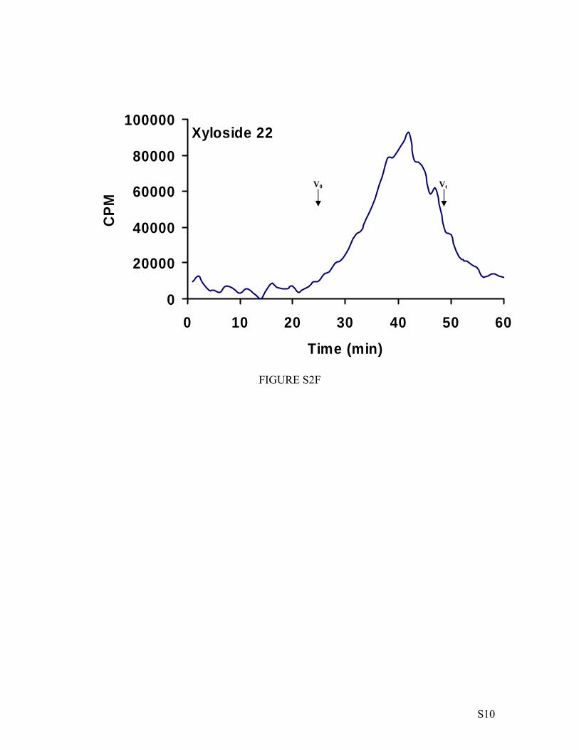
Figure Legends Figure S1. Calibration of size exclusion column with polystyrene sulfonate standards. Polystyrene sulfonate standards of various molecular weights, 65000 Da, were analyzed by size exclusion chromatography as described in the “Material and Methods” section. The migration times of the various polystyrene sulfonate species were plotted against the molecular weight to obtain a calibration curve. The migration time of GAG chains primed by various xylosides were compared to the calibration curve to determine the molecular weight. Figure S2. Chain length analysis of xyloside-primed GAG chains. The molecular weight of the GAG chains synthesized on various primers was determined by measuring their migration time on size exclusion column as described in the “Material and Methods” section. V0 and Vt represent the void volume and total volume, respectively. The average migration time was determined by using peak width at half maximum. The average molecular weight was determined using the migration time in comparison to the calibration curve obtained for polystyrene sulfonate standards performed under similar conditions. Figure S3. Disaccharide profiles of xyloside-primed HS chains. GAG chains (~500,000 cpm) were digested with heparitinases and resulting disaccharides were analyzed by SAX-HPLC with inline flow scintillation analyzer as described in the Experimental section. The SAX elution chromatograms of representative of two independent experiments. I: ∆UA-GlcNAc; II: ∆UA-GlcNS; III: ∆UA-GlcNS6S; IV: ∆UA2S-GlcNS; V: ∆UA2S-GlcNAc6S and IV: ∆UA2S-GlcNS6S. Figure S4. Disaccharide profiles of xyloside-primed DS chains. GAG chains (~500,000 cpm) were digested with chondroitinase ABC enzyme and resulting disaccharides were analyzed by SAX-HPLC with inline flow scintillation analyzer as described in the Experimental section. The SAX elution chromatograms of representative of two independent experiments. I: ∆UA-GalNAc; and II: ∆UA-GalNAc6S. Figure S5. Disaccharide profiles of xyloside-primed CS chains. GAG chains (~500,000 cpm) were digested with chondroitinase ABC enzyme and resulting disaccharides were analyzed by SAX-HPLC with inline flow scintillation analyzer as described in the Experimental section. The SAX elution chromatograms of representative of two independent experiments. I: ∆UA-GalNAc; and II: ∆UA-GalNAc6S. Figure S6. Long term priming ability of GAG chains by xyloside. The long term priming ability of click-xyloside was examined using xylosyl transferase deficient CHO cells (pgsA-745). 100,000 cells were seeded per well of 24-well plates and treated with xyloside 5 at 100 µM concentration in the presence of 50 µCi 35S-SO4
2- or D-[6-3H]-glucosamine. The medium was removed from the well at 24, 48, 96 and 120 h, GAG chains were purified and quantified as described under “Experimental Methods”.

S4
y = 6E+06e-0.1784x
R2 = 0.9908
100
1000
10000
100000
20 25 30 35 40 45 50
Migration time (min)
MW
(Da)
FIGURE S1
Vt (2000 Da) V0
(70000 Da)

S5
Xyloside 5
0
20000
40000
60000
80000
100000
0 10 20 30 40 50 60
Time (min)
CPM
FIGURE S2A
Vt V0

S6
Xyloside 6
0
40000
80000
120000
160000
0 10 20 30 40 50 60
Time (min)
CPM
FIGURE S2B
Vt V0

S7
Xyloside 7
0
20000
40000
60000
80000
100000
0 10 20 30 40 50 60
Time (min)
CPM
FIGURE S2C
Vt V0

S8
Xyloside 19
0
40000
80000
120000
160000
0 10 20 30 40 50 60
Time (min)
CPM
FIGURE S2D
Vt V0

S9
Xyloside 20
0
20000
40000
60000
80000
100000
0 10 20 30 40 50 60
Time (min)
CPM
FIGURE S2E
Vt V0
S10
Xyloside 22
0
20000
40000
60000
80000
100000
0 10 20 30 40 50 60
Time (min)
CPM
FIGURE S2F
Vt V0

S11
5
0
5000
10000
15000
20000
25000
0 10 20 30 40 50
Time (min)
CPM
FIGURE S3A
I
II
III V
VI
IV

S12
22
02000
40006000
800010000
1200014000
0 10 20 30 40 50
Time (min)
CPM
FIGURE S3B
I
II
IIIVI
IV

S13
FIGURE S4A
19
0
20000
40000
60000
80000
0 10 20 30 40 50
Time (min)
CPM
I
II

S14
FIGURE S4B
20
0
30000
60000
90000
120000
0 10 20 30 40 50
Time (min)
CPM
I
II

S15
FIGURE S5A
7
0
30000
60000
90000
120000
150000
0 10 20 30 40 50
Time (min)
CPM
I
II

S16
FIGURE S5B
14
0
4000
8000
12000
16000
0 10 20 30 40 50
Time (min)
CPM
I II

S17
0
400000
800000
1200000
1600000
0 6 12 24 48 72 96 120
Time (h)
FIGURE S6

V. Nguyen and Balagurunathan KuberanXylophone V. Victor, Thao K. N. Nguyen, Manivannan Ethirajan, Vy M. Tran, Khiem
Investigating the Elusive Mechanism of Glycosaminoglycan Biosynthesis
doi: 10.1074/jbc.M109.043208 originally published online July 23, 20092009, 284:25842-25853.J. Biol. Chem.
10.1074/jbc.M109.043208Access the most updated version of this article at doi:
Alerts:
When a correction for this article is posted•
When this article is cited•
to choose from all of JBC's e-mail alertsClick here
Supplemental material:
http://www.jbc.org/content/suppl/2009/07/23/M109.043208.DC1.html
http://www.jbc.org/content/284/38/25842.full.html#ref-list-1
This article cites 63 references, 45 of which can be accessed free at
by guest on July 22, 2016http://w
ww
.jbc.org/D
ownloaded from
Copyright © 2022 FDOKUMEN