Biosynthesis of Smaller-Sized Platinum Nanoparticles Using ...
Upload
khangminh22Category
view
0download
0
R E V I E W A R T I C L E
Thebiosynthesis of peptidoglycan lipid-linked intermediatesAhmed Bouhss1, Amy E. Trunkfield2, Timothy D.H. Bugg2 & Dominique Mengin-Lecreulx1
1Laboratoire des Enveloppes Bacteriennes et Antibiotiques, Institut de Biochimie et Biophysique Moleculaire et Cellulaire, UMR 8619 CNRS,
Univ Paris-Sud, Orsay, France; and 2Department of Chemistry, University of Warwick, Coventry, UK
Correspondence: Dominique Mengin-
Lecreulx, Laboratoire des Enveloppes
Bacteriennes et Antibiotiques, IBBMC, UMR
8619 CNRS, Universite Paris-Sud, Bat. 430,
91405 Orsay Cedex, France. Tel.: 133 1 69 15
48 41; fax: 133 1 69 85 37 15; e-mail:
Received 10 July 2007; revised 20 September
2007; accepted 24 September 2007.
First published online 11 December 2007.
DOI:10.1111/j.1574-6976.2007.00089.x
Editor: Jacques Coyette
Keywords
peptidoglycan; undecaprenyl phosphate; UppS
synthase; UppP phosphatases; MraY
translocase; MurG transferase.
Abstract
The biosynthesis of bacterial cell wall peptidoglycan is a complex process involving
many different steps taking place in the cytoplasm (synthesis of the nucleotide
precursors) and on the inner and outer sides of the cytoplasmic membrane
(assembly and polymerization of the disaccharide-peptide monomer unit, respec-
tively). This review summarizes the current knowledge on the membrane steps
leading to the formation of the lipid II intermediate, i.e. the substrate of the
polymerization reactions. It makes the point on past and recent data that have
significantly contributed to the understanding of the biosynthesis of undecaprenyl
phosphate, the carrier lipid required for the anchoring of the peptidoglycan
hydrophilic units in the membrane, and to the characterization of the MraY and
MurG enzymes which catalyze the successive transfers of the N-acetylmuramoyl-
peptide and N-acetylglucosamine moieties onto the carrier lipid, respectively.
Enzyme inhibitors and antibacterial compounds interfering with these essential
metabolic steps and interesting targets are presented.
Introduction
Peptidoglycan (murein) is a major heteropolymer of bacter-
ial cell walls that consists in long glycan chains made of
alternating units of N-acetylmuramoyl-peptides (MurNAc-
peptides) and N-acetylglucosamine (GlcNAc) that are cross-
linked together via the short peptide chains (Rogers et al.,
1980; Park, 1996; Vollmer et al., 2008). The main essential
function of this giant cell-sized macromolecule is to protect
cells against the deleterious effects of the internal osmotic
pressure. It also contributes to the maintenance of the
characteristic cell shape and serves as a platform for the
anchoring of other cell envelope components including
proteins (Braun & Sieglin, 1970; Marraffini et al., 2006;
Dramsi et al., 2008) and polysaccharides (Neuhaus &
Baddiley, 2003).
The biosynthesis of peptidoglycan is a complex process
involving many different cytoplasmic and membranes steps
(van Heijenoort, 2001b). The first stage consists in the
formation of the soluble nucleotide precursors, from UDP-
GlcNAc to UDP-MurNAc-pentapeptide. In particular, the
synthesis of the peptide moiety is performed by a series of
enzymes designated as the Mur ligases (MurC, MurD, MurE
and MurF) which are responsible for the respective additions
of L-alanine, D-glutamic acid, meso-diaminopimelic acid
(A2pm) or L-lysine, and D-alanyl-D-alanine to UDP-MurNAc
(Barreteau et al., 2008). As reported by Schleifer & Kandler
(1972) and exemplified in the accompanying reviews
(Barreteau et al., 2008; Vollmer et al., 2008), the structure of
this peptide and the substrate specificity of these enzymes
exhibit some variations in the bacterial world. The membrane
steps then begin with the transfer of the phospho-MurNAc-
pentapeptide moiety from the cytoplasmic precursor to the
membrane acceptor undecaprenyl phosphate (C55-P), a
reaction catalyzed by the transferase MraY (also named
translocase) yielding undecaprenyl-pyrophosphoryl-Mur-
NAc-pentapeptide (lipid I) (Fig. 1). Thereafter, the transferase
MurG catalyzes the transfer of the GlcNAc moiety from UDP-
GlcNAc to lipid I yielding undecaprenyl-pyrophosphoryl-
MurNAc-(pentapeptide)-GlcNAc (lipid II) which, after its
passage through the membrane by a yet unknown mechan-
ism, will be used as the substrate for the polymerization
reactions (van Heijenoort, 2001a, b) (Fig. 1). The C55-P
carrier lipid plays a central role in these steps and is also
FEMS Microbiol Rev 32 (2008) 208–233c� 2007 Federation of European Microbiological SocietiesPublished by Blackwell Publishing Ltd. All rights reserved
Dow
nloaded from https://academ
ic.oup.com/fem
sre/article/32/2/208/2683939 by guest on 21 July 2022
required for the synthesis of other cell-wall polymers. This
lipid as well as all the enzymes participating in peptidoglycan
synthesis are essential, specific for the bacterial world and
therefore constitute interesting potential targets to be
exploited for the discovery of new antibacterials.
This review summarizes recent advances in the under-
standing of the membrane steps of peptidoglycan synthesis.
In particular, it will focus on recent significant contributions
to the knowledge of the metabolism of the carrier lipid and
on progress made toward the biochemical and structural
characterization of the MraY and MurG activities and the
search of inhibitors of these enzymes.
Undecaprenyl phosphate metabolism
Undecaprenyl phosphate (C55-P), also referred to as bacto-
prenol, is a key lipid involved in the biosynthesis of
peptidoglycan and a variety of other cell-wall polysaccharide
components such as lipopolysaccharides, the enterobacterial
common antigen, capsule polysaccharides, and teichoic
acids (Wright et al., 1967; Scher et al., 1968; Troy et al.,
1971, 1975; Watkinson et al., 1971; Johnson & Wilson, 1977;
Rohr et al., 1977; Reeves et al., 1996; Rick et al., 1998; van
Heijenoort, 2001b; Raetz & Whitfield, 2002; Neuhaus &
Baddiley, 2003). C55-P-linked saccharides are also used for
N-linked protein glycosylation that occurs in certain pro-
karyotes (Glover et al., 2005; Szymanski & Wren, 2005).
That a single lipid participates in the synthesis of various
wall polymers has been earlier considered as a potential
site of control that prevents an imbalance in the formation
of the cell envelope as a whole (Anderson et al., 1972).
Although C55-P remains the classical carrier lipid form
encountered in bacterial world, the lower-size homologs
decaprenyl phosphate (C50-P) and nonaprenyl phosphate
(C45-P) were shown to fulfill this essential function
in mycobacterial species (Kaur et al., 2004; Mahapatra
et al., 2005) and Paracoccus denitrificans (Ishii et al., 1986),
respectively.
In the peptidoglycan pathway, C55-P is needed for the
synthesis and transport of hydrophilic GlcNAc-MurNAc-
peptide monomeric structures across the hydrophobic en-
vironment of the cytoplasmic membrane to the externally
located sites of polymerization. Although this function is
essential, the knowledge of the metabolism of C55-P was still
very limited recently and based on fragmentary data ob-
tained from various bacterial species (Fig. 2). In particular,
only few data on the genes and enzymes involved in steps 2,
3 and 4 were available (Higashi et al., 1970a; Sandermann &
Strominger, 1972; Willoughby et al., 1972; Poxton et al.,
1974; Kalin & Allen, 1979).
Fig. 1. Membrane steps of peptidoglycan
biosynthesis. M, G and the five colored beads
linked to M represent MurNAc, GlcNAc and the
pentapeptide, respectively. C55-PP and C55-P
are for undecaprenyl pyrophosphate and
undecaprenyl phosphate, respectively.
Fig. 2. Metabolism of undecaprenyl phosphate in bacteria. The site
of action of bacitracin, an antibiotic which acts by sequestering of
undecaprenyl pyrophosphate, is indicated. Steps 1–4 are catalyzed
by undecaprenyl pyrophosphate synthase, undecaprenyl pyrophosphate
phosphatase, undecaprenol phosphokinase and undecaprenyl
phosphate phosphatase activities, respectively.
FEMS Microbiol Rev 32 (2008) 208–233 c� 2007 Federation of European Microbiological SocietiesPublished by Blackwell Publishing Ltd. All rights reserved
209Biosynthesis of peptidoglycan lipid intermediates
Dow
nloaded from https://academ
ic.oup.com/fem
sre/article/32/2/208/2683939 by guest on 21 July 2022

Biosynthesis and recycling of undecaprenylpyrophosphate
The precursor for C55-P, undecaprenyl pyrophosphate
(C55-PP), is synthesized by addition of eight C5 isopentenyl
units (cis[Z]-configuration) onto C15 (all-trans[E])-farnesyl
pyrophosphate (FPP) (Fig. 2). This reaction is catalyzed by
the undecaprenyl pyrophosphate synthase UppS (di-trans,-
poly-cis-decaprenylcistransferase; EC 2.5.1.31), which
belongs to the family of cis-prenyltransferases of group IV
(Ogura & Koyama, 1998). Farnesyl pyrophosphate itself
results from head-to-tail condensation of isopentenyl pyr-
ophosphate with dimethylallyl pyrophosphate, generating
C10 geranyl pyrophosphate, followed by a second condensa-
tion of isopentenyl pyrophosphate, a reaction catalyzed by
the farnesyl pyrophosphate synthase that is a prototype of
the trans-prenyltransferase family (Ogura & Koyama, 1998;
Liang et al., 2002). The structure and mixed E,Z stereo-
chemistry of the C55-prenyl product (Fig. 3), as deduced
from the recent knowledge of the reaction mechanism of the
UppS synthase, confirmed earlier data of mass and nuclear
magnetic resonance spectrometry of the undecaprenol iso-
lated from bacterial membranes (Scher et al., 1968; Gough
et al., 1970).
The UppS synthase had been partially purified and
characterized from several bacteria including Salmonella
newington, Micrococcus luteus, Lactobacillus plantarum,
Bacillus subtilis and Escherichia coli (Christenson et al.,
1969; Baba & Allen, 1978, 1980; Takahashi & Ogura, 1982;
Baba et al., 1985; Fujisaki et al., 1986). Like the enzymes
involved in fatty acid synthesis, UppS is a soluble cyto-
plasmic enzyme that generates a membrane embedded
product (undecaprenyl pyrophosphate). The first uppS gene
was identified much more recently, in 1998 by Ogura’s
group, using a genomic DNA library of M. luteus B-P 26
constructed in E. coli and a screening of recombinant clones
for overexpression of the synthase activity (Shimizu et al.,
1998). Orthologs were subsequently identified in various
Gram-positive and Gram-negative bacterial species (Apfel
et al., 1999) and this gene was demonstrated to be essential
in E. coli (Kato et al., 1999) and Streptococcus pneumoniae
(Apfel et al., 1999). Interestingly, these newly identified
proteins did not exhibit significant sequence similarity with
members of the trans-prenyltransferase family and in parti-
cular they did not carry the characteristic aspartate-rich
DDXXD motif that is involved in substrate binding via a
Mg21 bridge in the latter enzyme family (Chen et al., 1994).
The construction of expression vectors allowed the purifica-
tion of mg quantities of various UppS in either wild-type or
histidine-tagged form (Shimizu et al., 1998; Apfel et al.,
1999; Pan et al., 2000). The enzyme activity showed both a
detergent (Triton X-100) and MgCl2 dependency (Apfel
et al., 1999), the latter cation being required for the binding
and subsequent condensation of isopentenyl pyrophosphate
(Chen et al., 2002b). UppS has now been characterized in
great detail, both biochemically and structurally, by different
groups. The crystal structure of M. luteus and E. coli
enzymes have been solved, either in apo form or in complex
with Mg21, isopentenyl phosphate, FPP, and product analo-
gues (Fujihashi et al., 2001; Ko et al., 2001; Chang et al.,
2004; Guo et al., 2005). The data showed that not only the
primary but also the three-dimensional (3-D) structure of
cis-prenyltransferases was totally different from that of
trans-prenyltransferases (Takahashi & Koyama, 2006). The
enzyme is a homodimer of 29-kDa subunits and each
monomer is composed of six parallel b-strands forming a
Fig. 3. Structure of the polyprenyl carrier lipid
involved in cell wall biosynthesis.
FEMS Microbiol Rev 32 (2008) 208–233c� 2007 Federation of European Microbiological SocietiesPublished by Blackwell Publishing Ltd. All rights reserved
210 A. Bouhss et al.
Dow
nloaded from https://academ
ic.oup.com/fem
sre/article/32/2/208/2683939 by guest on 21 July 2022

central b-sheet core, which is surrounded by five of the seven
a-helices (Fig. 4). A largely hydrophobic 30 A depth cleft was
observed at the protein surface whose entrance carries
several positively charged residues as well as a ‘structural P
loop’ motif that is characteristic of phosphate recognition
enzymes. A flexible domain located in the vicinity of the H3
a-helix and cleft entrance was identified as an important
region for the catalytic process and the two subunits were
shown to alternate between two different ‘closed’ and ‘open’
conformations (Ko et al., 2001; Chen et al., 2002b), the
‘closed’ form being catalytically active (Chang et al., 2003).
Based on the 3-D structure and site-directed mutagenesis
experiments, a model for substrate binding and a catalytic
mechanism were proposed (Fujikura et al., 2000, 2003; Pan
et al., 2000; Kharel et al., 2001; Takahashi & Koyama, 2006).
How the ultimate product chain length of cis-prenyltrans-
ferases is determined was also investigated in some detail
(Ko et al., 2001; Kharel et al., 2006; Takahashi & Koyama,
2006). Site-directed mutagenesis of E. coli and M. luteus
UppS and a sequence comparison with the C70�120 product
synthesizing eukaryotic cis-prenyltransferases highlighted
some residues that play a critical role in the determination
of the product chain length. It was proposed that charged
residues present at the hinge region of the H3 a-helix might
control the bending direction of the growing hydrophobic
prenyl chain along the hydrophobic interior of the H3 helix
so that the hydrophobic cleft could accommodate the bulk
of the prenyl chain to fit a suitable size during enzymatic
elongation. As the substrate specificity and catalytic proper-
ties of the FPP synthase and cis-prenyltransferases are
varying to some extent in the bacterial world, the size and
stereochemistry of the ultimate polyprenyl product, i.e. the
carrier lipid, are bacterial species specific: undecaprenyl
phosphate in most cases, rarely nonaprenyl phosphate
(C45) (Ishii et al., 1986), both with the classical o,di-
trans,poly-cis conformation, or decaprenyl phosphate (C50)
with an unusual o,trans,octa-cis conformation (Wolucka
et al., 1994; Kaur et al., 2004; Mahapatra et al., 2005) (Fig. 3).
Trace amounts of nona-, deca-, and dodeca-prenyl alcohol
derivatives were always detected together with the predomi-
nant C55-P lipid in membrane extracts, confirming the high
but not absolute specificity of the corresponding UppS
synthase activities observed during in vitro assays (Thorne
& Kodicek, 1966; Higashi et al., 1967, 1970b; Scher et al.,
1968; Gough et al., 1970; Umbreit et al., 1972; Umbreit &
Strominger, 1972b; Apfel et al., 1999).
The bacitracin antibiotic has been shown to inhibit
bacterial cell wall biosynthesis through sequestration of
C55-PP, the product of the UppS synthase, thereby provok-
ing the loss of the cell integrity and lysis (Siewert &
Strominger, 1967; Stone & Strominger, 1971; Storm &
Strominger, 1973). This potent antibiotic produced as a
mixture of related cyclic polypeptides by some strains of
Bacillus licheniformis and B. subtilis is extensively used for
prophylaxis and therapy in food animals. It is clinically
used for treatments of surface tissue infections in combina-
tion with other antimicrobial drugs. Its oral use had
been earlier suggested for the control of vancomycin-
Fig. 4. Crystal structure of undecaprenyl
pyrophosphate synthase dimer (UppS) from
Escherichia coli. The figure was prepared using
PyMol and the atomic coordinates (1JP3)
deposited by Ko et al. (2001). The seven
a-helices and six b-strands are shown in red and
green for subunit A and pink and blue for
subunit B, respectively. The flexible loop
without observable electron densities (residues
72–83) is represented as a dotted line in one
subunit. The hydrophobic cleft at the molecular
surface of each subunit is indicated by an
arrow.
FEMS Microbiol Rev 32 (2008) 208–233 c� 2007 Federation of European Microbiological SocietiesPublished by Blackwell Publishing Ltd. All rights reserved
211Biosynthesis of peptidoglycan lipid intermediates
Dow
nloaded from https://academ
ic.oup.com/fem
sre/article/32/2/208/2683939 by guest on 21 July 2022

resistant enterococci (VRE) (O’Donovan et al., 1994)
although without evident success (Mondy et al., 2001;
Hachem & Raad, 2002).
Formation of undecaprenyl phosphate
The dephosphorylation of C55-PP (step 2 in Fig. 2) is
required before the lipid carrier becomes available for use
in the various biosynthetic pathways. This reaction must
also occur after each cycle of polymerization of cell wall
components (e.g. of peptidoglycan) and the release of the
linked saccharides, because the lipid carrier is in most cases
liberated in the pyrophosphate form. However, some excep-
tions exist: for instance, the transfer of 4-amino-4-deoxy-L-
arabinose (L-Ara4N) units to lipid A catalyzed by the ArnT
membrane protein uses undecaprenyl-phosphoryl-L-Ara4N
as the donor substrate and releases the lipid in the C55-P
form (Trent et al., 2001).
The membrane-bound phosphatase catalyzing this reac-
tion had been partially purified from M. luteus by Goldman
& Strominger (1972) about 30 years ago and some of its
properties were investigated. Its optimal pH for activity was
near 7.5, the enzyme did not require any cation, was
stimulated by nonionic Triton detergents, and failed to
hydrolyze isopentenyl pyrophosphate. The gene for this
activity, however, remained to be identified.
In 1993, an E. coli gene whose overexpression resulted in a
decreased susceptibility to bacitracin had been identified
and the authors hypothesized that this gene should encode
an undecaprenol phosphokinase (Cain et al., 1993). This
hypothesis was based on the assumption that a significant
pool of free C55-OH may exist in E. coli membranes, as
earlier demonstrated in some Gram-positive bacteria (Higa-
shi et al., 1970b), that would be directed towards the
formation of C55-P by the overproduced kinase, thereby
reducing cell requirements for C55-PP molecules and the cell
sensitivity to bacitracin. In fact, this question was recently
revisited and the bacA gene product was finally unambigu-
ously proved to be a C55-PP phosphatase (El Ghachi et al.,
2004). The overproduction of the BacA protein, which
allowed E. coli cells to resist to high concentrations of
bacitracin, was correlated with a large (280-fold) increase
of C55-PP phosphatase activity in membranes (El Ghachi
et al., 2004). The increased level of C55-PP phosphatase
activity likely accelerated the conversion of the pool of C55-
PP, the bacitracin target, to C55-P, resulting in an increased
cell resistance to the antibiotic. The 30 kDa protein BacA was
predicted to be an integral membrane protein with eight
transmembrane segments. It was successfully extracted
from cell membranes by the n-dodecyl-b-D-maltoside de-
tergent and purified to near homogeneity in the histidine-
tagged form (El Ghachi et al., 2004). The E. coli enzyme
exhibited a high C55-PP phosphatase activity of ca.
2200 nmol min�1 mg�1 of protein, a value about 7300-fold
higher than the basal activity detected in wild-type cell
membranes. It did not show any detectable C55-OH phos-
phokinase activity. Considering this newly identified func-
tion, it was proposed to rename the bacA gene uppP (El
Ghachi et al., 2004), for undecaprenyl pyrophosphate phos-
phatase (also named undecaprenyl-diphosphatase; EC
3.6.1.27), to follow the nomenclature previously adopted
with uppS that encodes the C55-PP synthase (Apfel et al.,
1999).
The dephosphorylation of C55-PP was predicted to be an
essential metabolic step. The finding that the bacA gene
could be deleted from the chromosome of E. coli (El Ghachi
et al., 2004), Mycobacterium smegmatis (Rose et al., 2004),
Staphylococcus aureus and Streptococcus pneumoniae (Chalk-
er et al., 2000), without loss of viability or any apparent
effect on growth rate or morphology, was therefore quite
surprising. Its deletion however resulted in impaired biofilm
and smegma formation in M. smegmatis, and attenuated
virulence in mouse models of infection in S. aureus and
Streptococcus pneumoniae. All the bacA deletion mutants
showed enhanced susceptibility to bacitracin (Chalker et al.,
2000; El Ghachi et al., 2004; Rose et al., 2004). The
unexpected viability of the bacA deletion mutants suggested
the existence of other cell proteins with C55-PP phosphatase
activity. The detection of a 25% residual phosphatase
activity in the membranes of the E. coli mutant was
consistent with this hypothesis (El Ghachi et al., 2004). No
bacA homologue was found in the genomes of the afore-
mentioned species, indicating that these putative phospha-
tases belonged to a distinct protein family. Although a copy
of bacA gene was found in most bacterial genomes se-
quenced to date (up to three putative bacA orthologs were
found in some species, e.g. Bacillus cereus), it was apparently
absent in some bacteria such as Helicobacter pylori (unpub-
lished data).
A search in databases for putative membrane phospha-
tases activities identified three proteins of unknown func-
tion that formed the so-called BcrR family: BcrC from
B. licheniformis, YbjG from E. coli, and YwoA from B. subtilis
(El Ghachi et al., 2005). Interestingly, the bcrC gene product
was described as one of the three components of the ABC
transporter system responsible for the protection of
B. licheniformis against the antibiotic it produces, bacitracin
(Podlesek et al., 1995, 2000), and the overexpression of the
E. coli ybjG and B. subtilis ywoA genes were reported to
increase bacitracin resistance in the corresponding bacterial
species (Harel et al., 1999; Cao & Helmann, 2002; Bernard
et al., 2003). Two other E. coli genes displaying similarity
with ybjG that encoded members of the PAP2 phosphatidic
acid-phosphatase family were also identified: yeiU, of un-
known function, and pgpB, encoding one of the two
phosphatidylglycerolphosphate phosphatases (Icho & Raetz,
FEMS Microbiol Rev 32 (2008) 208–233c� 2007 Federation of European Microbiological SocietiesPublished by Blackwell Publishing Ltd. All rights reserved
212 A. Bouhss et al.
Dow
nloaded from https://academ
ic.oup.com/fem
sre/article/32/2/208/2683939 by guest on 21 July 2022

1983). All of these candidate proteins were predicted to be
integral membrane proteins and contained a sequence
identical or quite similar to the characteristic phosphatase
signature KX6RP-(X12�54)-PSGH-(X31�54)-SRX5HX3D (El
Ghachi et al., 2005) that had been identified previously by
Stukey & Carman (1997) and Neuwald (1997). Interestingly,
this conserved phosphatase motif was not detected in the
sequence of BacA and the absence of significant sequence
homology between BacA and the above mentioned proteins
clearly indicated that they belonged to two distinct protein
families. The chromosomal ybjG, yeiU and pgpB genes could
be disrupted individually without apparent effect on cell
growth but the coinactivation of the three genes bacA, ybjG
and pgpB was shown to be lethal (El Ghachi et al., 2005). A
thermosensitive conditional triple mutant strain was gener-
ated which lysed at the restrictive temperature due to the
depletion of C55-PP phosphatase activity and arrest of
peptidoglycan synthesis (El Ghachi et al., 2005). It con-
firmed the implication of at least the three proteins BacA,
YbjG and PgpB in the formation of the C55-P carrier lipid in
vivo. As observed with bacA, the overexpression of the
individual ybjG, yeiU and pgpB genes was correlated to an
increased resistance to bacitracin and an increased level of
C55-PP phosphatase activity in membranes. The B. subtilis
ywoA gene product was also subsequently purified and
proved to be a C55-PP phosphatase (Bernard et al., 2005).
Therefore, two different classes of integral membrane
proteins which belong to the BacA and PAP2 phosphatase
families, respectively, could catalyze the dephosphorylation
of C55-PP into C55-P in bacteria. The number of these
proteins could apparently vary from one bacterial species
to another. The situation in E. coli is one BacA protein and at
least two members of the PAP2 family. Most bacterial species
have only one copy of bacA in their genome but some
species seem to express several bacA orthologs (e.g. Bacillus
anthracis, B. cereus) and some others apparently do not
express any (e.g. H. pylori). The situation is similar for
members of the PAP2 family but the precise number of those
proteins that effectively exhibit C55-PP phosphatase activity
remains to be determined for each species.
Interestingly, genes belonging to either of these two
classes were also found in gene clusters conferring antibiotic
resistance in the bacitracin-producing and bacitracin-resis-
tant species. For instance, bcrC and ywoA genes that encode
phosphatases from the PAP2 family were located within
clusters expressing bacitracin efflux systems in B. lichenifor-
mis and B. subtilis strains, respectively (Podlesek et al., 1995;
Cao & Helmann, 2002; Bernard et al., 2003), and one bacA
ortholog was recently found to play a similar essential role
in acquired bacitracin resistance in Enterococcus faecalis
(Manson et al., 2004). C55-PP phosphatase encoding genes
are thus used by bacteria either for generating the essential
carrier lipid C55-P or for depleting cells of the pool of C55-
PP, the bacitracin target, as a mechanism of resistance to this
antibiotic.
The purification and further biochemical characteriza-
tion of these different proteins is now required to discern
phosphatase activities specifically involved in C55-P meta-
bolism from non- or less-specific phosphatases, of distinct
metabolic function, that can also use C55-PP as a substrate.
C55-PP is synthesized at the inner side of the cytoplasmic
membrane but is released at the outer side of the membrane
by the peptidoglycan polymerization machinery. Whether
the dephosphorylation of C55-PP occurs on both membrane
sides or only on one side is at present unknown. A
topological analysis of these different membrane proteins
will help to answer this question. The involvement of either
different phosphatases with catalytic sites orientated to-
wards the cytoplasm and the periplasm or a single phospha-
tase could be envisaged, but in both cases a trans-bilayer
movement of the carrier lipid should occur, suggesting here
also the involvement of a putative flippase (see ‘The possible
existence of a flippase’).
The reaction of dephosphorylation of C55-PP was con-
sidered as an interesting potential target in a search for new
antibiotics. The recent discovery that two classes of enzymes
and multiple orthologs in each class could participate in
this process could render this search more problematic.
However, one efficient way to inhibit this step remains the
sequestration of the C55-PP substrate, as demonstrated with
bacitracin (Siewert & Strominger, 1967; Stone & Strominger,
1971; Storm & Strominger, 1973). An attack of the pyropho-
sphate moiety of C55-PP was also suggested to be part of the
mechanism of action of nisin, a lantibiotic whose primary
target is lipid II (Bonev et al., 2004). It was earlier hypothe-
sized that the reaction of dephosphorylation of C55-PP
could be the site of action of colicin M, a bacteriolytic toxin
produced by some E. coli strains that kills sensitive E. coli
strains and related species (Harkness & Braun, 1989a, b).
This question was recently revisited and colicin M was in
fact identified as an enzyme catalyzing the specific degrada-
tion of lipids I and II peptidoglycan intermediates (El
Ghachi et al., 2006).
Undecaprenol: a storage form of lipid carrier?
The intriguing presence of free undecaprenol (C55-OH) in
bacterial membranes had been earlier reported in Gram-
positive species. More than 90% of the endogenous
C55-isoprenyl lipid of S. aureus was found in this non-
functional alcohol form (Higashi et al., 1970b) and
a similar situation was observed in Enterococcus faecalis
(Umbreit et al., 1972), Listeria plantarum (Thorne & Kodi-
cek, 1966; Gough et al., 1970), and Listeria monocytogenes
(Vilim et al., 1973). It could represent a reserve pool for the
regulation of the C55-P pool, an hypothesis that seemed to
FEMS Microbiol Rev 32 (2008) 208–233 c� 2007 Federation of European Microbiological SocietiesPublished by Blackwell Publishing Ltd. All rights reserved
213Biosynthesis of peptidoglycan lipid intermediates
Dow
nloaded from https://academ
ic.oup.com/fem
sre/article/32/2/208/2683939 by guest on 21 July 2022

be corroborated by the detection of two membrane-
associated enzyme activities, undecaprenol phosphokinase
and undecaprenyl phosphate phosphatase, catalyzing
the interconversion of C55-OH and C55-P (steps 3 and 4
in Fig. 2), in some of the latter species (Higashi et al.,
1970a; Sandermann & Strominger, 1972; Willoughby
et al., 1972; Poxton et al., 1974; Kalin & Allen, 1979).
The corresponding genes however remained to be
identified.
Only one report on the characterization of a C55-P
phosphatase had been published to date (Willoughby et al.,
1972) but a potential involvement of nonspecific phospha-
tases that could explain the generation of the large pool of
C55-OH was also suggested (Bohnenberger & Sandermann,
1976). The C55-P phosphatase activity detected in particu-
late fractions from S. aureus did not require any cation, was
optimal at pH 5, and had an apparent Km for C55-P of
1.5mM (Willoughby et al., 1972). All attempts to extract it
from membranes were unsuccessful and the enzyme was
consequently not purified nor further characterized. In the
same report, the authors mentioned the failure to detect a
similar activity in particulate fractions from B. subtilis,
Enterococcus faecalis, M. luteus and E. coli. This activity
observed in S. aureus probably accounted for the stimula-
tion by ATP of the overall rate of peptidoglycan synthesis
observed in vitro with enzyme preparations from this
organism (Anderson et al., 1966).
A C55-OH phosphokinase activity was shown to be
extractable by butanol from S. aureus and Klebsiella aero-
genes membranes (Higashi et al., 1970a; Poxton et al., 1974).
Its activity was optimal at pH around 8.5 and was dependent
on the presence of Mg21 (Higashi et al., 1970a). The enzyme
from S. aureus was purified to near homogeneity and its
molecular weight (MW) was estimated at 14 kDa (Sander-
mann & Strominger, 1972). The estimated Km value was
57mM for both C55-OH and the nucleotide cosubstrate ATP,
and ADP was confirmed as a reaction product. The phos-
phokinase from L. plantarum was partially solubilized by a
variety of methods utilizing Triton X-100 and was charac-
terized in some detail (Kalin & Allen, 1979). Its apparent Km
values for C55-OH and ATP were estimated at 14 mM and
2 mM, respectively. No other nucleoside triphosphate was
shown to substitute for ATP. Interestingly, it was very
recently suggested that the diacylglycerol kinase (DGK)
from Streptococcus mutans could also use C55-OH as an
alternative substrate (Lis & Kuramitsu, 2003). Although this
was not unambiguously established, the physiological sig-
nificance of this putative undecaprenol kinase activity of
DGK was further supported by an increased susceptibility to
bacitracin of the dgk mutant strain as compared with that of
the parental strain. It could thus be hypothesized that the
C55-OH phosphokinase activity that had been purified from
S. aureus membranes about 30 years ago was due, at least in
part, to the DGK enzyme. The S. aureus dgk gene codes for a
114-residues protein with a MW of 12.97 kDa, a value close
to that (14 kDa) earlier estimated for the C55-OH phospho-
kinase from this organism (Sandermann & Strominger,
1972), but no evidence that this protein also exhibits C55-
OH phosphokinase activity has been provided. It should be
noted that the DGK from E. coli was proved to be inactive on
C55-OH (Bohnenberger & Sandermann, 1979; Lis & Kur-
amitsu, 2003). Moreover, C55-OH has never been detected
in E. coli cells membranes, except in nonphysiological
conditions, e.g. following enzymatic degradation of lipid
intermediates by colicin M (El Ghachi et al., 2006). The
existence of a pool of C55-OH and expression of the couple
of kinase and phosphatase that catalyze its interconversion
with C55-P have thus only been demonstrated in a restricted
number of bacterial species. Their effective implication in
the regulation of the pool of C55-P and of its use for the
synthesis of the different cell-wall polymers remain to be
demonstrated.
Lipid I biosynthesis
The lipid I is an essential intermediate molecule in the
peptidoglycan biosynthesis pathway (Fig. 1). Its existence
was reported for the first time by Chatterjee & Park (1964)
and Struve & Neuhaus (1965). Afterwards, it was identified
as a lipid intermediate produced by transfer of the phospho-
MurNAc-pentapeptide moiety from UDP-MurNAc-penta-
peptide onto a lipid fraction (Anderson et al., 1965) with
concomitant release of UMP. The elucidation of the lipid
structure had been achieved by Higashi et al. (1967), who
found that the acceptor was a C55 isoprenoid alcohol
phosphate, undecaprenyl phosphate (C55-P). However, as
detailed in ‘Undecaprenyl phosphate metabolism’, the length
and stereochemistry of this carrier lipid appeared to be
slightly different in certain bacterial species, such as
M. smegmatis (Mahapatra et al., 2005) and P. denitrificans
(Ishii et al., 1986). In E. coli, the pool of the lipid I was
estimated at about 700 molecules cell�1 (van Heijenoort
et al., 1992). Such an extremely low pool level was explained
by the fact that this compound is an intermediate in the
pathway whose synthesis and utilization reactions are effi-
ciently coupled.
Translocase I reaction and identification of themraY gene
In 1965, Strominger and Neuhaus laboratories demon-
strated for the first time the transfer of the phospho-
MurNAc-pentapeptide moiety from the soluble UDP
nucleotide precursor onto the C55-P carrier lipid, using
membrane preparations from S. aureus and M. luteus
FEMS Microbiol Rev 32 (2008) 208–233c� 2007 Federation of European Microbiological SocietiesPublished by Blackwell Publishing Ltd. All rights reserved
214 A. Bouhss et al.
Dow
nloaded from https://academ
ic.oup.com/fem
sre/article/32/2/208/2683939 by guest on 21 July 2022

(Anderson et al., 1965; Struve & Neuhaus, 1965).
C55-Pþ UDP-MurNAc-pentapeptide2UMPþ C55-PP-MurNAc-pentapeptide ðlipid IÞ
This reaction, which is generally referred to as the
‘transfer reaction’, does not lead to any modification of the
basal MurNAc-pentapeptide structure synthesized by the
Mur synthetases in the cytoplasm. It essentially consists in its
translocation onto the C55 carrier lipid present in the
cytoplasmic membrane. This anchoring is required before
subsequent steps could occur, i.e. the addition of the GlcNAc
residues by the MurG transferase, the passage of the
peptidoglycan monomeric structures through the mem-
brane and finally their polymerization on the outer side of
the cytoplasmic membrane. The enzyme catalyzing this first
transfer/translocation reaction, the phospho-MurNAc-pen-
tapeptide translocase or MraY (E.C. 2.7.8.13), therefore
insures the link between the cytoplasmic and periplasmic
steps of peptidoglycan biosynthesis (van Heijenoort, 2001b;
Bugg et al., 2006).
MraY was also found to be able to exchange radio-
labelled UMP for the unlabelled UMP moiety of UDP-
MurNAc-pentapeptide, consistent with the overall reaction
referred to as the ‘exchange reaction’:
½14C�UMPþUDP-MurNAc-pentapeptide2UMP
þ ½14C�UDP-MurNAc-pentapeptide
The mraY gene had been identified by Ikeda et al. (1991)
within a large cluster of genes, named mra for ‘murein region
A’, located at 2 min on the chromosome map of E. coli. In this
region the genes are tightly packed and appear in the order:
pbpB-murE-murF-mraY-murD-ftsW-murG-murC-ddlB-ftsQ-
ftsA-ftsZ-envA. They all code for proteins involved in pepti-
doglycan biosynthesis and cell division (Mur synthetases
MurC/D/E/F, penicillin-binding protein PBP3; division pro-
teins FtsW/Q/A/Z, MurG transferase, and D-Ala-D-Ala ligase
DdlB). A quite similar organization of this cluster was found
in other bacterial species. Ikeda et al. (1991) showed that the
overexpression of the E. coli mraY gene resulted in an increase
of the UDP-N-acetylmuramoyl-pentapeptide: undecaprenyl-
phosphate phospho-N-acetylmuramoyl-pentapeptide trans-
ferase activity in membranes, demonstrating that this gene
encoded the latter activity. Expression of the mraY gene was
shown to be dependent on the Pmra promoter (Hara et al.,
1997; Mengin-Lecreulx et al., 1998). This gene is essential for
the bacterial viability (Boyle & Donachie, 1998; Thanassi
et al., 2002) and a conditional mraY mutant strain was shown
to accumulate peptidoglycan nucleotide precursors under
restrictive growth conditions (Lara et al., 2005). One copy of
the mraY gene was found in all bacterial genomes sequenced
to date but was not detectable in eukaryotic organisms and
archaebacteria which both lack peptidoglycan. Interestingly,
however, the presence of an mraY gene orthologue was
recently demonstrated in the plant Arabidopsis thaliana
(Mondego et al., 2003). These authors postulated that this
MraY-like product could participate in the biosynthesis of
specific proteoglycan arabinogalactan proteins that reach
peak expression during late flower bud development. It was
also reported that the mraY gene product from A. thaliana
had putative plastid-targeting signals (Machida et al., 2006).
MraY protein, a member of the polyprenyl-phosphate N -acetyl hexosamine 1-phosphatetransferase superfamily
The alignment of MraY orthologue sequences from both
Gram-negative and Gram-positive species available in data-
bases allowed Bouhss et al. (1999) to identify a set of five
well-conserved hydrophilic sequences (I–V) containing 34
invariant amino acid residues (Fig. 5). In the E. coli MraY
sequence these domains were defined as H65-L80 (I), I111-
K133 (II), N189-L200 (III), L251-G275 (IV) and V296-F342
(V). The size of the MraY protein appears to be fairly well
conserved in the bacterial world, the sequences from Gram-
negative species being generally longer than those found in
Gram-positive species (�360 vs. 320 amino acid residues),
owing mainly to an extension at the N-terminal extremity.
Unusually large MraY proteins (�420 residues) are found,
however, in some bacteria such as Bacteroides (Price &
Momany, 2005). The presence of alternating hydrophobic
and hydrophilic segments in its primary structure clearly
suggested that MraY was an integral membrane protein
spanning the cytoplasmic membrane several times (Ikeda
et al., 1991). Moreover, a lipid microenvironment was
shown to be required for the MraY activity (Heydanek &
Neuhaus, 1969; Umbreit & Strominger, 1972a; Geis & Plapp,
1978; Weppner & Neuhaus, 1979). More recently, Bouhss
et al. (1999) determined the two-dimensional membrane
topology of both the E. coli and S. aureus MraY translocases,
using b-lactamase fusion experiments. A common topolo-
gical model was proposed which contained ten transmem-
brane segments joining four periplasmic loops and five
cytoplasmic sequences corresponding to the conserved
hydrophilic patterns I–V (Fig. 5). Both the N- and C-
terminal extremities were located in the periplasmic space.
This model was in agreement with many structural features
predicted from a sequence comparison of MraY ortholo-
gues, strongly suggesting its validity for all eubacterial MraY
proteins (Bouhss et al., 1999).
The cytoplasmic sequences II, III and IV of MraY were
also found in other prokaryotic and eukaryotic proteins
catalyzing the same kind of reaction, namely the transfer of a
lipid phosphate to the b-phosphate of an UDP-linked
hexosamine. These three conserved patterns thus defined
a superfamily of enzymes termed polyprenyl-phosphate
FEMS Microbiol Rev 32 (2008) 208–233 c� 2007 Federation of European Microbiological SocietiesPublished by Blackwell Publishing Ltd. All rights reserved
215Biosynthesis of peptidoglycan lipid intermediates
Dow
nloaded from https://academ
ic.oup.com/fem
sre/article/32/2/208/2683939 by guest on 21 July 2022

N-acetylhexosamine-1-phosphate transferases or UDP-Hex-
NAc : polyprenyl-P HexNAc-1-P transferases. In addition to
MraY, this superfamily contained the prokaryotic enzymes
WecA, TagO, WbcO, WbpL and RgpG involved in the
biosynthesis of different cell envelope polymers (enterobac-
terial common antigen, lipopolysaccharide O-antigen, tei-
choic acids or rhamnose-glucose polysaccharide) (Soldo
et al., 2002; Lehrer et al., 2007) and a eukaryotic paralogue,
GPT, involved in protein N-glycosylation (Lehrman, 1994;
Dal Nogare et al., 1998; Burda & Aebi, 1999). These enzymes
share a common membrane-bound acceptor substrate, un-
decaprenyl phosphate in bacteria or dolichyl phosphate in
eukaryotes, but they differ in their selectivity for the soluble
UDP-N-acetyl-hexosamine substrate. The sugar nucleotide
donors are UDP-MurNAc-pentapeptide and UDP-GlcNAc
for MraY and WecA, TagO and GPT, respectively, the MraY
substrate carrying an additional 3-O-lactoyl-pentapeptide
group.
The cytoplasmic sequences I and V are highly specific for
all MraY orthologues and are also present in some bacterial
paralogues with some differences. However, they are not
found in the eukaryotic paralogue sequences (GPTs). Se-
quence and membrane topology analyses revealed that all of
the invariant or highly-conserved residues identified within
MraY, and/or WecA and the eukaryotic GPTs, were located
on the cytoplasmic side of the membrane, consistent with
the active site being orientated towards the cytoplasm
(Bouhss et al., 1999; Lloyd et al., 2004; Lehrer et al., 2007).
The patterns II, III and IV would be involved in the substrate
binding and/or the catalytic process that are common
features in the enzyme superfamily, while the patterns I and
V would be involved in the substrate specificity and in
particular the recognition of the sugar nucleotide substrate.
Purification and biochemical characterization ofMraY
Solubilization, expression, and purification ofMraY
The availability of a pure, stable, soluble and active prepara-
tion of the integral membrane MraY protein was a prere-
quisite to the development of detailed biochemical
investigations. Since 1969, many soluble and active prepara-
tions of MraY were described, extracted from membranes of
various bacteria such as micrococci, S. aureus and E. coli
using detergents and in particular Triton X-100 or CHAPS
(Heydanek & Neuhaus, 1969; Umbreit & Strominger, 1972a;
Pless & Neuhaus, 1973; Brandish et al., 1996a; Breukink
et al., 2003; Lloyd et al., 2004; Stachyra et al., 2004). Upon
detergent extraction, the S. aureus MraY was shown to
require the presence of a phospholipid, either phosphatidyl-
choline, dioleoyl phosphatidyl choline or phosphatidylgly-
cerol (Pless & Neuhaus, 1973). Brandish et al. (1996a)
reported that overexpressed E. coli MraY protein was
preferentially activated by phosphatidylglycerol. All pre-
vious attempts to overexpress significantly and purify any
MraY protein had been unsuccessful. Thus, only partially
purified enzyme preparations were generally used for enzy-
matic assays (Brandish et al., 1996a; Zawadzke et al., 2003;
Lloyd et al., 2004; Stachyra et al., 2004). Recently, a
comparative study performed with recombinant MraY
proteins from E. coli, S. aureus, B. subtilis and Thermotoga
maritima, expressed in the E. coli C43(DE3) host strain,
identified n-dodecyl-b-D-maltoside and N-lauroyl-sarcosine
as the most efficient detergents for the extraction of this
protein from cell membranes (Bouhss et al., 2004). In the
Fig. 5. Membrane topology of the MraY
translocase (Escherichia coli). The topological
model consists in ten transmembrane
segments, four periplasmic loops and five
cytoplasmic sequences corresponding to the
conserved hydrophilic patterns I–V (Bouhss
et al., 1999). Conserved residues by identity
(red) and by similarity (blue) are indicated.
FEMS Microbiol Rev 32 (2008) 208–233c� 2007 Federation of European Microbiological SocietiesPublished by Blackwell Publishing Ltd. All rights reserved
216 A. Bouhss et al.
Dow
nloaded from https://academ
ic.oup.com/fem
sre/article/32/2/208/2683939 by guest on 21 July 2022

same report, conditions allowing the high-level overexpres-
sion of a MraY protein and, for the first time, its purification
to homogeneity in milligram quantities were described. The
specific activity of the pure B. subtilis MraY protein was
estimated at 1900 nmol min�1 mg�1 (Bouhss et al., 2004).
Substrate specificity and kinetic properties
MraY has two substrates: C55-P and UDP-MurNAc-penta-
peptide. The structure of the lipid substrate is expected to be
conserved in most bacterial species but a few exceptions
exist, as mentioned in ‘Undecaprenyl phosphate metabo-
lism’. The structure of the sugar nucleotide substrate shows
important variations in the bacterial world, particularly in
the peptide moiety (Schleifer & Kandler, 1972; Barreteau
et al., 2008; Vollmer et al., 2008). This peptide is a pentapep-
tide that generally contains at the third position either a
meso-A2pm residue in Gram-negative bacteria (as E. coli)
and Bacillus species or a lysine residue (more rarely
an ornithine) in most Gram-positive bacteria (as S. aureus).
In vivo complementation experiments have shown that the
S. aureus MraY was functional in E. coli and restored growth
of a mraY thermosensitive mutant, indicating that it accepts
the A2pm-containing sugar nucleotide (Bouhss et al., 1999).
Similarly, the overexpression of the S. aureus MurE synthe-
tase in E. coli (which introduces lysine instead of A2pm at
the third position of the nucleotide substrate) resulted in a
massive incorporation of lysine into the peptidoglycan,
demonstrating the efficient utilization of the lysine-contain-
ing UDP-MurNAc-pentapeptide by the E. coli MraY enzyme
(Mengin-Lecreulx et al., 1999). The relatively low specificity
of the MraY translocase and its tolerance toward the
variability of the peptide chain was a well-known character-
istic (Hammes & Neuhaus, 1974) that has been confirmed in
various other circumstances both in vivo and in vitro.
Shorter or longer peptides as well as modified peptides were
shown to be accepted: dipeptides (Ornelas-Soares et al.,
1994), tripeptides (Hammes & Neuhaus, 1974; Pisabarro
et al., 1986; van Heijenoort et al., 1992), tetrapeptides
(Hammes & Neuhaus, 1974), acetylated and dansylated
pentapeptides (Ward & Perkins, 1974; Weppner & Neuhaus,
1977; Brandish et al., 1996a; Stachyra et al., 2004), as well as
hexa- and heptapeptides (Billot-Klein et al., 1997). More
recently, the MraY enzyme from T. maritima was shown to
act on two different substrates in vivo: a D-lysine-containing
UDP-MurNAc-tripeptide and an L-lysine-containing UDP-
MurNAc-pentapeptide, with similar efficiencies (Boniface
et al., 2006).
The replacement of the L-Ala and D-Ala residues at
positions 1 and 4 of the pentapeptide chain, respectively, by
glycine reduced the S. aureus MraY catalytic efficiency by
135-fold (Hammes & Neuhaus, 1974). The use of UDP-
MurNAc-tetrapeptide (lacking one D-Ala) and UDP-Mur-
NAc-tripeptide (lacking two D-Ala) as substrates reduced
the MraY catalytic efficiency by fourfold and
80-fold, respectively, as compared with the nucleotide
pentapeptide. Stickgold & Neuhaus (1967) determined that
5-fluorouracil-substituted UDP-MurNAc-pentapeptide was
utilized at o 2% of the rate of the native substrate and was a
competitive inhibitor (Ki = 0.12 mM) of the transfer reac-
tion, just as 5-fluoro-UMP is a competitive inhibitor
(Ki = 50 mM) of the exchange reaction. However, 20-deoxy
UMP was accepted as a good substrate in the exchange
reaction (Neuhaus, 1971).
Various MraY assays using radiolabeled or fluorescent
substrates, resolution of reaction mixtures by paper or
thin-layer chromatography, or direct measurement using
microplates, have been developed for analyzing the kinetic
properties of this enzyme and for the screening of inhibitors
(Weppner & Neuhaus, 1977; Brandish et al., 1996a, b;
Bouhss et al., 2004; Stachyra et al., 2004). Typical Michae-
lis–Menten kinetics were observed with both purified and
partially purified enzyme preparations (Anderson et al.,
1966; Struve et al., 1966; Stickgold & Neuhaus, 1967;
Hammes & Neuhaus, 1974; Brandish et al., 1996a; Bouhss
et al., 2004; Stachyra et al., 2004). Km values observed for
UDP-MurNAc-pentapeptide were generally in the 5–30 mM
range with nonpurified enzyme preparations. However, a
much higher value of 1 mM was determined with the
purified MraY enzyme from B. subtilis (Bouhss et al., 2004).
The structure and size of the C55-P lipid acceptor
substrate present in the membranes is essentially determined
by the substrate specificity and catalytic properties of the
undecaprenyl pyrophosphate synthase UppS. However,
the E. coli MraY translocase was shown to accept in vitro
heptaprenyl (C35) as well as dodecaprenyl (C60) phosphate
as alternative substrates, with Km values (10–20 mM) similar
to that of the natural substrate (Brandish et al., 1996a).
Breukink et al. (2003) have reported that the Micrococcus
flavus MraY was active in vitro on polyprenyl substrates
containing from two to 25 isoprenyl units, with a maximal
efficiency observed for substrates bigger than C35. The
purified B. subtilis MraY exhibited a Km for C55-P about 10-
fold higher than that of the nonpurified MraYs from other
bacterial species (Bouhss et al., 2004). It could thus be
assumed that the membrane environment affects the recog-
nition of the lipid substrate by the enzyme.
The MraY activity is dependent on the presence of both
mono- and divalent metal ions. The activity of the E. coli
and B. subtilis enzymes was stimulated (two- to fourfold) in
presence of 10–100 mM of potassium or sodium (Brandish
et al., 1996a; Bouhss et al., 2004). Heydanek et al. (1970) also
observed that the S. aureus MraY was stimulated by many
monovalent metal ions such as K1, Rb1, Cs1 and NH41. The
MraY enzyme has an absolute requirement for a divalent
metal ion, particularly Mg21, at 5–40 mM concentrations
FEMS Microbiol Rev 32 (2008) 208–233 c� 2007 Federation of European Microbiological SocietiesPublished by Blackwell Publishing Ltd. All rights reserved
217Biosynthesis of peptidoglycan lipid intermediates
Dow
nloaded from https://academ
ic.oup.com/fem
sre/article/32/2/208/2683939 by guest on 21 July 2022

(Heydanek et al., 1970; Bouhss et al., 2004). Mn21 can
replace Mg21 but the activity is decreased by two orders
of magnitude (Bouhss et al., 2004). Lloyd et al. (2004)
proposed that the D115 or D116 aspartate residues of
E. coli MraY, which are located in the conserved hydro-
philic sequence II (Fig. 5) and whose replacement by
Asn leads to loss of catalytic activity, are involved in
Mg21 chelation.
Catalytic mechanism of MraY
In 1969, on the basis of kinetic evidences, Heydanek et al.
(1969) proposed a two-step catalytic mechanism for the
MraY reaction (Fig. 6). It consisted in an attack by a
nonidentified nucleophile residue of MraY on the b phos-
phate of UDP-MurNAc-pentapeptide, generating a covalent
enzyme-phospho-MurNAc-pentapeptide intermediate with
concomitant release of UMP. The second step corresponded
to the attack by an oxyanion from C55-P on the phosphate of
the covalent intermediate, resulting in the formation of lipid I
and regeneration of the native enzyme form. Recently, Lloyd
et al. (2004) proposed that the D267 aspartate residue of
E. coli MraY, which is located in the conserved hydrophilic
sequence III, plays the role of the catalytic nucleophile. Three
observations supported the two-step mechanism: (1) phos-
pho-MurNAc-pentapeptide was formed during the transfer
reaction that likely resulted from the hydrolysis of the
enzyme-linked phospho-MurNAc-pentapeptide intermedi-
ate; (2) the enzyme could catalyze the exchange of UMP with
the UMP moiety of UDP-MurNAc-pentapeptide in the
presence or absence of the cosubstrate C55-P (Pless &
Neuhaus, 1973); (3) dodecylamine inhibited the synthesis of
lipid I and caused the release of the phospho-MurNAc-
pentapeptide.
A major criticism of the experiments performed by
Heydanek et al. that could have resulted in a misinterpreta-
tion of the results was the use of a nonpurified MraY
enzyme. The different observations they made could effec-
tively be attributed to the presence of contaminating en-
zymes (phosphatases) and C55-P lipid acceptor in the
preparation used. An alternative mechanism consists in a
direct attack of the phosphate oxyanion of C55-P onto the bphosphate of UDP-MurNAc-pentapeptide. This would lead
to the formation of lipid I and UMP in only one step (Fig.
6). The recent availability of pure MraY protein (Bouhss
et al., 2004) will allow to revisit this question and elucidate
the catalytic mechanism.
Recent progress on inhibitors of theMraY-catalysed reaction
MraY is the target for several classes of natural product
antibiotics, which in some cases have been studied in detail
through structure/function studies and/or detailed kinetic
or mechanistic studies. The structures of the MraY inhibi-
tors have been reviewed in detail (Kimura & Bugg, 2003;
Dini, 2005; Bugg et al., 2006). This section will briefly survey
the different inhibitor classes, and small molecule inhibitors
arising from structure/function studies.
There are five different classes of uridine-containing
nucleoside natural antibiotic products that target MraY,
illustrated in Fig. 7. The tunicamycins (related to the
streptovirudins and corynetoxins) contain a uridine disac-
charide, attached to a fatty-acyl chain. Tunicamycin is a
reversible, competitive inhibitor of MraY, with Ki 0.6 mM
(Brandish et al., 1996b), but is not useful as an antibacterial
agent, since it is toxic to mammals, through potent inhibi-
tion of GlcNAc-1-P transferase in the dolichol cycle of N-
linked glycoprotein biosynthesis (Heifetz et al., 1979). The
Fig. 6. Proposed MraY translocase reaction mechanisms. The two-step mechanism involves a covalent enzyme-phospho-MurNAc-pentapeptide
intermediate and the single-step mechanism consists in a direct attack of the phosphate oxyanion of C55-P onto the b-phosphate of the nucleotide
substrate.
FEMS Microbiol Rev 32 (2008) 208–233c� 2007 Federation of European Microbiological SocietiesPublished by Blackwell Publishing Ltd. All rights reserved
218 A. Bouhss et al.
Dow
nloaded from https://academ
ic.oup.com/fem
sre/article/32/2/208/2683939 by guest on 21 July 2022

mureidomycins (related to the pacidamycins and napsamy-
cins) are peptidyl nucleoside natural products, containing a
30-deoxyuridine sugar attached via an enamide linkage to an
unusual peptide chain. The peptide chain contains an
N-methyl 2,3-diaminobutyric acid residue, and a urea
linkage to a C-terminal aromatic amino acid, which can be
meta-tyrosine, Trp, or Phe. Mureidomycin A is a slow-
binding inhibitor of E. coli MraY, with K�i = 2.2 nM (Brand-
ish et al., 1996a). The enamide functional group is not
essential for inhibition, and a range of dihydro-pacidamycin
analogues have been synthesized and tested for biological
activity (Boojamra et al., 2001). The mechanism of action of
mureidomycin A has been studied, and it was proposed that
the amino-terminus binds to the Mg21 cofactor-binding
site, and is positioned by an N-methyl amide cis-amide bond
(Howard & Bugg, 2003). A uridinyl dipeptide analogue of
mureidomycin A retained biological activity against Pseudo-
monas putida (Howard & Bugg, 2003).
The liposidomycins are fatty acyl nucleosides, whose
structures contain a sulfated aminoglycoside residue. Lipo-
sidomycin B is a slow-binding inhibitor (K�i = 80 nM)
of solubilized E. coli MraY (Brandish et al., 1996b).
A synthetic analogue containing the aminoribofuranose
monosaccharide attached to the 50 position of uridine
showed moderate levels of inhibition (IC50 = 50mM)
against translocase I when assayed in toluene-permeabilized
E. coli cells (Dini et al., 2000; Stachyra et al., 2004). Further
structure–function studies in this series of compounds
(riburamycins) gave more active inhibitors, and a synthetic
analogue containing a C12-fatty-acyl chain showed antibac-
terial activity against S. aureus (Dini et al., 2002; Stachyra
et al., 2004).
The muraymycins, reported by Wyeth Research in 2002,
contain an aminoribofuranoside monosaccharide, attached
to a short peptide chain, containing a urea linkage to a
C-terminal amino acid (McDonald et al., 2002). Members of
this family were potent inhibitors of MraY in vitro
(IC50 = 0.027mg mL�1), showed antibacterial activity
against S. aureus (MIC = 2–16mg mL�1) and enterococci
(MIC = 16–64mg mL�1) and were able to protect mice
against S. aureus infection (ED50 = 1.1 mg kg�1) (McDonald
et al., 2002). Synthetic analogues lacking the aminoribofur-
anoside showed reduced activity in vitro, but retained some
antibacterial activity (Yamashita et al., 2003).
Capuramycin, a further uridine nucleoside antibiotic, is a
potent inhibitor of MraY in vitro (IC50 = 0.017mg mL�1),
and a methylated derivative showed antibacterial activity
against M. smegmatis (MIC = 2–16mg mL�1) (Muramatsu
et al., 2003). Acylation derivatives of capuramycin showed
very potent activity (MIC = 0.06 mg mL�1) against several
Mycobacterium strains (Hotoda et al., 2003).
Genetic studies by Young and coworkers showed that
MraY was also the target for the bacteriolytic E protein from
bacteriophage fX174 (Bernhardt et al., 2000). The E protein
is a 91-amino acid protein, containing a transmembrane
domain. The killing action of E also required a host protein
Fig. 7. Structure of MraY translocase inhibitors.
FEMS Microbiol Rev 32 (2008) 208–233 c� 2007 Federation of European Microbiological SocietiesPublished by Blackwell Publishing Ltd. All rights reserved
219Biosynthesis of peptidoglycan lipid intermediates
Dow
nloaded from https://academ
ic.oup.com/fem
sre/article/32/2/208/2683939 by guest on 21 July 2022

SlyD, a peptidyl-prolyl isomerase. Recent studies on
the mechanism of action of E showed that a 37-amino
acid peptide containing the transmembrane domain of
E was a potent inhibitor of membrane-bound MraY
(IC50 = 0.8 mM), but did not inhibit solubilized MraY, unlike
the small molecule inhibitors (Mendel et al., 2006). It has
been proposed that E inhibits MraY via a protein–protein
interaction, blocking the formation of protein–protein
interactions between MraY and other cell wall assembly
proteins in the cytoplasmic membrane at cell division
(Mendel et al., 2006).
Interestingly, it was recently demonstrated that colicin M
was an enzyme acting by specifically targeting and destroy-
ing the peptidoglycan lipid intermediates, thereby provok-
ing the arrest of peptidoglycan synthesis and cell lysis (El
Ghachi et al., 2006). The cleavage site was located between
the C55 and pyrophosphoryl groups, as demonstrated
in vitro by appropriate assays and in vivo by the observation
that colicin M-treated cells accumulated C55-OH, a lipid
form that is normally not detectable in E. coli cell mem-
branes (El Ghachi et al., 2006). To the authors’ knowledge
this is the first example of a mechanism of inhibition of
peptidoglycan biosynthesis occurring via enzymatic degra-
dation of its precursors, in this case the MraY and MurG
lipid reaction products.
Lipid II biosynthesis
The translocase II reaction and identification ofthe murG gene
The translocase II (MurG) catalyses the second membrane
associated step of peptidoglycan synthesis (Fig. 1). This
glycosyl transferase of the GT-B superfamily transfers the
GlcNAc moiety from UDP-GlcNAc to the C4 hydroxyl
group of lipid I to form a b-linked disaccharide (lipid II). It
was originally identified in E. coli but has since been
recognized in all other bacteria that make peptidoglycan.
The murG gene was first discovered by Salmond et al.
(1980) who identified, cloned and mapped the gene within
the mra region of E. coli. The nucleotide sequence of the
E. coli murG coding region (Mengin-Lecreulx et al., 1990)
revealed an ORF of 1065 nucleotides theoretically coding for
a moderately hydrophobic 37.8 kDa protein. When cultures
of a thermosensitive murG mutant strain (GS58) growing
exponentially at 30 1C were shifted to the nonpermissive
temperature of 42 1C, cells progressively lost their rod shape,
became ovoid with a greatly increased volume and finally
lysed (Mengin-Lecreulx et al., 1991). This mutant accumu-
lated significant amounts of UDP-MurNAc-pentapeptide,
UDP-GlcNAc and to a lesser extent a lipid compound as
labelled A2pm-containing precursors, suggesting that the
mutational block was in a membrane step. The fact that the
ratio of the two lipid intermediates I and II, which had
previously been reported to be between 0.3 : 1 and 0.6 : 1 in
E. coli strains (Ramey & Ishiguro, 1978), was reversed and
reached the considerably high value of 8.2 : 1 in this mutant
clearly indicated that it was the second membrane step
that was altered. It was consequently deduced that murG
encoded the UDP-GlcNAc : MurNAc-(pentapeptide)-
pyrophosphoryl-undecaprenol GlcNAc transferase (EC
2.4.1.227) which catalyses the formation of lipid II from
lipid I. This protein was recently shown to interact with the
MraYand MreB proteins and was suggested to participate in
two multi-protein complexes involved in cell elongation and
cell division, respectively (Aaron et al., 2007; Mohammadi
et al., 2007; den Blaauwen et al., 2008).
Purification and biochemical characterizationof MurG
Crouvoisier et al. (1999) first reported the overproduction,
solubilization and purification of the protein. MurG was
solubilized using salt or detergent from the cell membranes
and purified using anion exchange for the wild-type protein
or Ni21 affinity for the His-tagged protein. Ha et al. (2000)
also reported a similar procedure where the protein was
solubilized with detergent directly from cell pellets of E. coli
and purified using cation exchange followed by gel filtration.
van den Brink-van der Laan et al. (2003) showed that the
cardiolipin content of E. coli cell membranes was increased
following overexpression of MurG and that lipid vesicles
copurified with MurG. The activity of MurG was increased
in the presence of cardiolipin, suggesting specific interac-
tions of the protein with phospholipids and in particular
with cardiolipin.
Using optimal conditions in HEPES buffer, pH 7.9,
supplemented with 5 mM MgCl2 or MnCl2, Ha et al.
(1999) determined the kinetic parameters for E. coli MurG.
Because lipid I is almost impossible to isolate from bacterial
cells and difficult to handle, a synthetic lipid I analogue was
used. The assay used radiolabelled UDP-[14C]GlcNAc and a
biotin-labelled lipid I analogue. A biotin capture technique
was used to sequester products from the reaction which
were then counted for bound radioactivity. The parameters
were KUDP-GlcNAc = 58� 30 mM, Klipid I analogue = 37� 4 mM,
kcat = 16� 2 min�1 in the presence of MgCl2. Using a
different (C35) lipid I analogue and a new assay based
on HPLC, Auger et al. (2003) showed that a high con-
centration (35%) of dimethylsulfoxide was necessary
for maximal enzyme activity. The kinetic constants
they determined in these conditions were: Km UDP-GlcNAc
= 150� 20mM, Km lipid I analogue = 2.8� 1 mM, kcat = 56
� 5 min�1. Ha et al. (1999) also investigated the acceptor
and donor substrate specificity for MurG. The results
showed that a biotin-labelled UDP-MurNAc-pentapeptide
FEMS Microbiol Rev 32 (2008) 208–233c� 2007 Federation of European Microbiological SocietiesPublished by Blackwell Publishing Ltd. All rights reserved
220 A. Bouhss et al.
Dow
nloaded from https://academ
ic.oup.com/fem
sre/article/32/2/208/2683939 by guest on 21 July 2022

was also a substrate for the enzyme with a relative rate of c.
20% when compared with the biotin-labelled lipid I analo-
gue. The results also revealed that the enzyme was sensitive
to the identity of the nucleotide, and required the presence
of the diphosphate linkage. The enzyme was inhibited by
UDP, but not by UMP, nor any other nucleotide dipho-
sphate. The enzyme also showed high specificity for the
equatorial stereochemistry at the C4 position of the donor.
Therefore, UDP-N-acetylgalactosamine failed to show in-
hibitory activity even at millimolar concentrations and
showed very little donor activity. Liu et al. (2003) reported
the development of a continuous fluorescence coupled
enzymatic assay in which the formation of UDP was coupled
to a pyruvate kinase-lactate dehydrogenase assay and the
fluorescence signal of NADH monitored. This group tested a
variety of lipid I analogues, with various substituents repla-
cing the undecaprenyl moiety, as substrates. MurG accepted
all of them with Km values of around 20–50 mM but there
were, however, large differences in the specific activity (kcat).
Acceptor substrates with long saturated linear alkyl chains
were better substrates than the natural lipid I demonstrating
an increase in kcat from 11� 2 min�1 for the natural
substrate to up to 180� 13 min�1 for an analogue with a
C14H29 chain.
Using synthetic substrate analogues and products con-
taining different length lipid chains, as well as a synthetic
dead-end acceptor analogue, Chen et al. (2002a) showed
that MurG follows an ordered Bi-Bi mechanism in which
UDP-GlcNAc binds first (Fig. 8). The enzyme likely utilizes a
mechanism that involves partial participation of the lone
pair on the sugar ring oxygen and therefore an oxocarbe-
nium-ion-like transition state. Evidence exists that supports
the procedure of glycosyltransferase-mediated reactions
through an oxocarbenium-ion-like transition state, similar
to that proposed for similar enzymes (Singh et al., 1987; Kim
et al., 1988; Takayama et al., 1999). Using a synthetic,
radiolabelled-analogue of lipid II, Auger et al. (2003)
reported for the first time that the reaction catalyzed by
MurG was reversible.
3-D structure of the MurG protein
The 1.9 A crystal structure of E. coli MurG (Ha et al., 2000)
revealed that the free enzyme consists of two protein
molecules in an asymmetric unit. Each protein chain has
two domains separated by a cleft which is c. 20 A deep and
18 A across at its widest point. Each domain adopts a
a/b open sheet motif which is characteristic of domains that
bind nucleotides. The N-terminal domain contains seven
parallel b-strands and six a-helices. The C-terminal domain
contains six parallel b-strands and eight a-helices including
one irregular bipartite helix that connects the N-domain to
the first b-strand of the C-domain. The structural homology
between the two domains is high, despite low sequence
homology. A subsequent, 2.5 A crystal structure of
E. coli MurG complexed with the donor substrate, UDP-
GlcNAc, was reported in 2003 (Hu et al., 2003a) (Fig. 9).
Before this, no X-ray crystal structures containing intact
substrates had been obtained for any of the NDP-glycosyl-
transferase superfamily. The substrate bound enzyme was
reported to have only a 16 A wide cleft, c. 2 A narrower than
the free enzyme. UDP-GlcNAc binds in both protein sub-
units along with four glycerol molecules and 121 water
molecules. Only one of the UDP-GlcNAc molecules is in
Fig. 8. Proposed MurG reaction mechanism. The model predicts a
deprotonation of the C4 hydroxyl group of the MurNAc moiety of lipid I
by a residue of MurG (probably His18). The oxyanion thus generated
then attacks the C1 of GlcNAc of the nucleotide substrate to form the
oxocarbenium-ion-like transition state. Finally, UDP is released generat-
ing lipid II. R = �C(CH3)CO-L-Ala-g-D-Glu-meso-A2pm-D-Ala-D-Ala,
Un = undecaprenyl.
FEMS Microbiol Rev 32 (2008) 208–233 c� 2007 Federation of European Microbiological SocietiesPublished by Blackwell Publishing Ltd. All rights reserved
221Biosynthesis of peptidoglycan lipid intermediates
Dow
nloaded from https://academ
ic.oup.com/fem
sre/article/32/2/208/2683939 by guest on 21 July 2022

the correct orientation for catalytic activity and is presumed
to be the Michaelis complex where the UDP is displaced by
the incoming nucleophile. MurG contains a sequence motif
that is found in most members of the GT-B superfamily.
This a/b/a subunit has two a-helices located near to the cleft
between the domains. The UDP-GlcNAc substrate makes
several contacts to these helices and also to the loops that
connect them to the adjacent b-strands. The GlcNAc moiety
of the donor substrate makes contacts with the invariant
residues of MurG through hydrogen bonding interactions
between the backbone amide of the A263 residue, the side
chain amides of N291 and N127 and the C4 hydroxyl group
of GlcNAc. Interactions also exist between the side chain
amide of Q287 to both the C3 and C4 hydroxyl groups of
GlcNAc. The catalytic base appears to be histidine H18
situated 9.52 A across the domain cleft in line with the
anomeric bond. The contacts between the enzyme and the
diphosphate are purely hydrogen bonding. S191 has been
shown to be an important residue whose mutation to
alanine affects all kinetic parameters including an increase
in the Km for lipid I binding from 0.053 to 0.179 mM. This
serine residue is located on a GGS loop that is conserved in
all MurG homologs which moves up towards the donor
substrate as it binds. Because MurG is believed to utilise a
sequential mechanism in which lipid I binds following UDP-
GlcNAc (Chen et al., 2002a), it is thought that conforma-
tional changes in the GGS loop may play a role in the
adjustments required for lipid I binding, as well as directly
contributing to UDP-GlcNAc binding. The uracil nucleotide
is anchored in a pocket by hydrogen bonds from the back-
bone amide of I244 to the N3H and O4 atoms. There is also a
possible interaction between the uracil O2 atom and R163.
The F243 residue also rotates, as the substrate binds, to cap
the binding pocket. Contacts between the enzyme and the
ribose 20 and 30 hydroxyl groups exist through hydrogen
bonds from E268. To evaluate their role in the activity of the
E. coli MurG enzyme, 13 residues that are invariant or highly
conserved in the MurG enzyme family (T15, H18, Y105,
H124, E125, N127, N134, S191, N198, R260, E268, Q288,
N291) and located within or very close to the active site
were recently submitted to site-directed mutagenesis
(Hu et al., 2003a; Crouvoisier et al., 2007). Most of these
mutations resulted in a great loss of enzyme activity, con-
sistent with the interactions observed in the crystal structures
of MurG alone or in complex with UDP-GlcNAc (Ha
et al., 2000; Hu et al., 2003a). The important role of these
residues either in substrate binding/catalysis or maintenance
of the protein’s conformation was confirmed (Hu et al.,
2003a; Crouvoisier et al., 2007). Interestingly, a hydro-
phobic patch has been identified in the MurG structure,
consisting of I74, L78, F81, W84 and W115 residues, which
appears to be the site of interaction between this extrinsic
membrane protein and the phospholipid bilayer (Ha et al.,
2000).
Recent progress on inhibitors of the MurGreaction
The cyclic lipoglycodepsipeptide antibiotic ramoplanin has
excellent activity against a wide range of Gram-positive
bacteria. It was demonstrated that ramoplanin forms com-
plexes with both lipids I and II (Reynolds & Somner, 1990;
Fig. 9. Structure of Escherichia coli MurG
complexed with UDP-GlcNAc. This structure,
drawn with Swiss PDB Viewer, shows the
interactions between UDP-GlcNAc and the
enzyme active site. Binding of the lipid I
substrate is presumed to take place in the
cleft between the two domains of the MurG
structure. From Hu et al. (2003a).
FEMS Microbiol Rev 32 (2008) 208–233c� 2007 Federation of European Microbiological SocietiesPublished by Blackwell Publishing Ltd. All rights reserved
222 A. Bouhss et al.
Dow
nloaded from https://academ
ic.oup.com/fem
sre/article/32/2/208/2683939 by guest on 21 July 2022
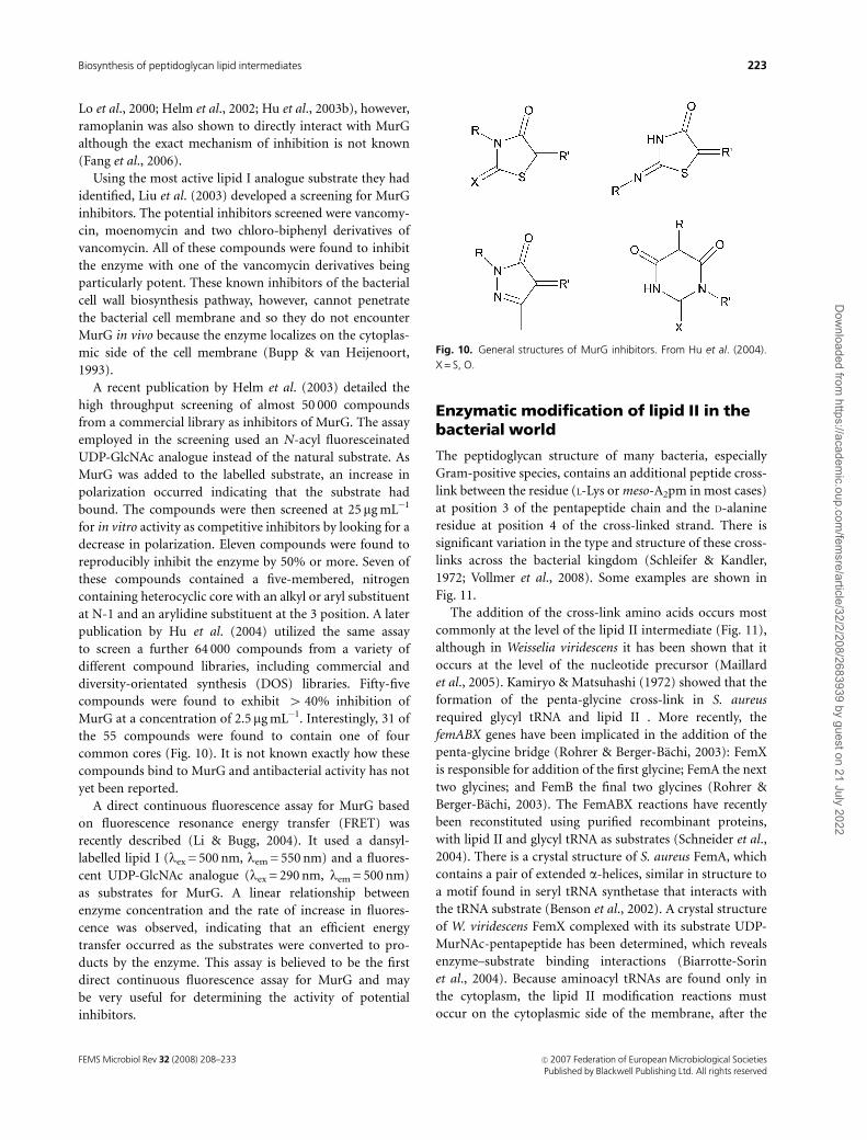
Lo et al., 2000; Helm et al., 2002; Hu et al., 2003b), however,
ramoplanin was also shown to directly interact with MurG
although the exact mechanism of inhibition is not known
(Fang et al., 2006).
Using the most active lipid I analogue substrate they had
identified, Liu et al. (2003) developed a screening for MurG
inhibitors. The potential inhibitors screened were vancomy-
cin, moenomycin and two chloro-biphenyl derivatives of
vancomycin. All of these compounds were found to inhibit
the enzyme with one of the vancomycin derivatives being
particularly potent. These known inhibitors of the bacterial
cell wall biosynthesis pathway, however, cannot penetrate
the bacterial cell membrane and so they do not encounter
MurG in vivo because the enzyme localizes on the cytoplas-
mic side of the cell membrane (Bupp & van Heijenoort,
1993).
A recent publication by Helm et al. (2003) detailed the
high throughput screening of almost 50 000 compounds
from a commercial library as inhibitors of MurG. The assay
employed in the screening used an N-acyl fluoresceinated
UDP-GlcNAc analogue instead of the natural substrate. As
MurG was added to the labelled substrate, an increase in
polarization occurred indicating that the substrate had
bound. The compounds were then screened at 25 mg mL�1
for in vitro activity as competitive inhibitors by looking for a
decrease in polarization. Eleven compounds were found to
reproducibly inhibit the enzyme by 50% or more. Seven of
these compounds contained a five-membered, nitrogen
containing heterocyclic core with an alkyl or aryl substituent
at N-1 and an arylidine substituent at the 3 position. A later
publication by Hu et al. (2004) utilized the same assay
to screen a further 64 000 compounds from a variety of
different compound libraries, including commercial and
diversity-orientated synthesis (DOS) libraries. Fifty-five
compounds were found to exhibit 4 40% inhibition of
MurG at a concentration of 2.5mg mL�1. Interestingly, 31 of
the 55 compounds were found to contain one of four
common cores (Fig. 10). It is not known exactly how these
compounds bind to MurG and antibacterial activity has not
yet been reported.
A direct continuous fluorescence assay for MurG based
on fluorescence resonance energy transfer (FRET) was
recently described (Li & Bugg, 2004). It used a dansyl-
labelled lipid I (lex = 500 nm, lem = 550 nm) and a fluores-
cent UDP-GlcNAc analogue (lex = 290 nm, lem = 500 nm)
as substrates for MurG. A linear relationship between
enzyme concentration and the rate of increase in fluores-
cence was observed, indicating that an efficient energy
transfer occurred as the substrates were converted to pro-
ducts by the enzyme. This assay is believed to be the first
direct continuous fluorescence assay for MurG and may
be very useful for determining the activity of potential
inhibitors.
Enzymatic modification of lipid II in thebacterial world
The peptidoglycan structure of many bacteria, especially
Gram-positive species, contains an additional peptide cross-
link between the residue (L-Lys or meso-A2pm in most cases)
at position 3 of the pentapeptide chain and the D-alanine
residue at position 4 of the cross-linked strand. There is
significant variation in the type and structure of these cross-
links across the bacterial kingdom (Schleifer & Kandler,
1972; Vollmer et al., 2008). Some examples are shown in
Fig. 11.
The addition of the cross-link amino acids occurs most
commonly at the level of the lipid II intermediate (Fig. 11),
although in Weisselia viridescens it has been shown that it
occurs at the level of the nucleotide precursor (Maillard
et al., 2005). Kamiryo & Matsuhashi (1972) showed that the
formation of the penta-glycine cross-link in S. aureus
required glycyl tRNA and lipid II . More recently, the
femABX genes have been implicated in the addition of the
penta-glycine bridge (Rohrer & Berger-Bachi, 2003): FemX
is responsible for addition of the first glycine; FemA the next
two glycines; and FemB the final two glycines (Rohrer &
Berger-Bachi, 2003). The FemABX reactions have recently
been reconstituted using purified recombinant proteins,
with lipid II and glycyl tRNA as substrates (Schneider et al.,
2004). There is a crystal structure of S. aureus FemA, which
contains a pair of extended a-helices, similar in structure to
a motif found in seryl tRNA synthetase that interacts with
the tRNA substrate (Benson et al., 2002). A crystal structure
of W. viridescens FemX complexed with its substrate UDP-
MurNAc-pentapeptide has been determined, which reveals
enzyme–substrate binding interactions (Biarrotte-Sorin
et al., 2004). Because aminoacyl tRNAs are found only in
the cytoplasm, the lipid II modification reactions must
occur on the cytoplasmic side of the membrane, after the
Fig. 10. General structures of MurG inhibitors. From Hu et al. (2004).
X = S, O.
FEMS Microbiol Rev 32 (2008) 208–233 c� 2007 Federation of European Microbiological SocietiesPublished by Blackwell Publishing Ltd. All rights reserved
223Biosynthesis of peptidoglycan lipid intermediates
Dow
nloaded from https://academ
ic.oup.com/fem
sre/article/32/2/208/2683939 by guest on 21 July 2022

MurG-catalysed reaction, but before flipping to the cell
surface.
Dipeptide Ala-Ala cross-links are found in Enterococcus
faecalis (Bouhss et al., 2001), and in strains of highly
penicillin-resistant Streptococcus pneumoniae (Garcia-Bus-
tos & Tomasz, 1990), in the latter case linked to the murMN
genes (Filipe & Tomasz, 2000). The murMN genes are found
in all pneumococci, but specific sequences of murM are
found in highly resistant strains (Filipe et al., 2000). Two
transferases involved in the formation of the Ala–Ala cross-
link in Enterococcus faecalis have also been identified, which
are able to add Ala-tRNA to UDP-MurNAc-pentapeptide
in vitro (Bouhss et al., 2002). The Streptococcus pneumoniae
MurM reaction has recently been reconstituted in vitro using
recombinant MurM, lipid II, and alanyl-tRNA (Lloyd et al.,
2007). MurM from a penicillin-resistant strain was found to
show 80-fold higher kcat/Km than MurM from a sensitive
strain, providing an explanation for the higher level of cross-
links in the resistant strains (Lloyd et al., 2007). The ATP-
dependent enzyme responsible for addition of D-aspartic
acid to L-lysine residue in Enterococcus faecium has also been
identified (Bellais et al., 2006).
In a number of bacterial strains, the D-glutamic acid
residue at position 2 of the pentapeptide chain is amidated,
to form D-isoglutamine. In S. aureus, it is known that this
amidation is dependent upon ATP and ammonia, and that
either lipid I or II can be amidated in vitro (Siewert &
Strominger, 1968). A membrane-bound amidating activity
has also been observed in Bacillus stearothermophilus that is
able to amidate the nucleotide precursor (Linnett & Stromin-
ger, 1974). Lipid II isolated from M. smegmatis has been
found to contain several modifications: the MurNAc residue
is modified as N-glycolylmuramic acid, or as de-acylated
muramic acid; the diaminopimelic acid residue is amidated;
and the terminal D-alanine residue is methylated (Mahapatra
et al., 2005). It appears that a number of modification
reactions occur in particular bacterial strains, most com-
monly at the level of the lipid-linked intermediates (Fig. 11).
In S. aureus, the acceptor substrate of sortase A that catalyzes
anchoring of surface proteins to peptidoglycan was identified
as lipid II (Perry et al., 2002). These different modifications
should be taken into account when considering the lipid II as
a potential target for new antibacterial agents (Breukink & de
Kruijff, 2006).
Chemical and enzymatic synthesis oflipids I and II and analogues
In order to reconstitute in vitro the lipid-linked steps of
peptidoglycan biosynthesis, it was necessary to prepare the
Fig. 11. Structure of lipid II and its main modifications in bacterial world. R = H or COOH for Lys and A2pm, respectively; D-Asx = D-Asp or D-Asn.
FEMS Microbiol Rev 32 (2008) 208–233c� 2007 Federation of European Microbiological SocietiesPublished by Blackwell Publishing Ltd. All rights reserved
224 A. Bouhss et al.
Dow
nloaded from https://academ
ic.oup.com/fem
sre/article/32/2/208/2683939 by guest on 21 July 2022
lipid-linked intermediates. Because they are present natu-
rally in very low abundance, it was not feasible to isolate
them from bacterial cells in useful quantities (van Heije-
noort et al., 1992; Guan et al., 2005). Chemical syntheses
were published for lipid I (VanNieuwenhze et al., 2001) and
for lipid II (Ye et al., 2001; VanNieuwenhze et al., 2002),
however the synthetic routes were long and proceeded in
fairly low overall yield. Auger et al., (1997, 2003) first
succeeded for the hemi-synthesis of functional analogues of
both lipids I and II. Chemical synthesis was used to prepare
soluble analogues of lipid I containing 10-carbon (Ha et al.,
1999) and 20-carbon chains (Cudic et al., 2001), which were
used as soluble substrates for MurG in vitro (Lazar & Walker,
2002).
Alternatively, lipids I and II can be prepared enzymati-
cally. The cytoplasmic precursor UDP-MurNAc-pentapep-
tide (containing meso-A2pm at position 3) can be
accumulated in B. subtilis using a cell wall synthesis medium
(Lugtenberg et al., 1972), and isolated in 50 mg quantities.
High level expression and purification of enzymes MurA-F
from E. coli or Pseudomonas aeruginosa and their use for the
preparation of the cytoplasmic intermediates in vitro have
been reported (Reddy et al., 1999; El Zoeiby et al., 2001;
Bouhss et al., 2004). An efficient procedure for the
conversion of UDP-MurNAc-pentapeptide into lipid I or
lipid II was published by Breukink et al. (2003), that used
membranes prepared from M. flavus, a species which
has elevated levels of MraY and MurG enzyme activities.
Using this procedure, they reported the preparation of
up to 50 mg quantities of lipids I or II. The availability of
purified C55-PP synthase UppS, C55-PP phosphatase
BacA, MraY and MurG proteins also facilitated the synthesis
of different forms of lipids I and II, with a labelling in
either the C55 or the pentapeptide moiety (El Ghachi et al.,
2006).
The e-amino group of meso-A2pm or L-Lys at position
3 of the pentapeptide chain can be chemically modified
by fluorophores such as dansyl. The N-dansyl analogue was
used to develop a fluorescence assay for MraY, for enzyme
kinetic studies (Brandish et al., 1996a), and for high
throughput inhibition assays (Stachyra et al., 2004). The
N-dansylated lipid II analogue was prepared and was shown
to give rise to fluorescence changes upon treatment with
E. coli PBP1b (Schwartz et al., 2002). An alternative
labelling strategy was recently published, in which the
D-alanine residue at position 4 or 5 can be replaced by
D-cysteine, by enzymatic synthesis from D-Ala-D-Cys or
D-Cys-D-Ala, and the thiol side chain of D-Cys can be
labelled by thiol-selective reagents such as pyrene maleimide
(Schouten et al., 2006). The S-labelled UDP-MurNAc-pen-
tapeptide analogues prepared in this way were converted
into fluorescent lipid I and II analogues, using M. flavus
membranes.
Possible existence of a flippase
Lipid II, biosynthesized on the cytoplasmic face of the
membrane, must somehow be flipped onto the extracellular
face of the membrane (Fig. 1). The possible existence of a
‘flippase’ protein capable of catalysing this process has been
discussed for many years (Weppner & Neuhaus, 1978; Ehlert
& Holtje, 1996) but no gene has been identified to date.
Analysis of fluorescently labelled peptidoglycan precursors
in S. aureus membranes by fluorescence energy transfer have
earlier concluded that the rate of unassisted flipping was
much too low to account for the rate of peptidoglycan
synthesis required at cell division (Weppner & Neuhaus,
1978), therefore it was generally assumed that there must be
some protein assistance for the flipping of lipid II across the
membrane. In fact, the biochemical process was only
recently demonstrated experimentally, in particular by ana-
lysis of the transmembrane transport of fluorescent lipid II
through model and bacterial membranes (Breukink et al.,
1999; van Dam et al., 2007). It was shown that lipid II flop
did not occur spontaneously and was not obligatory coupled
to lipid II synthesis. This suggested the involvement of one
or more integral membrane proteins or membrane-asso-
ciated proteins in this process. Based on their results, the
authors further hypothesized a coupling of the translocation
of lipid II with the subsequent reactions of polymerization
(transglycosylation) catalyzed by the penicillin-binding pro-
teins (van Dam et al., 2007).
The existence of ABC transporters that are able to catalyse
similar types of membrane transport events suggests that the
putative flippase might be similar to this protein family, for
which there is now structural information (Chang & Roth,
2001). The bacterial wzx gene was suggested as a candidate
flippase gene in lipopolysaccharide biosynthesis, on the basis
of gene knockout studies (Liu et al., 1996). A putative
flippase gene involved in N-linked protein glycosylation in
eukaryotic cells was identified, based upon gene knockout
studies (Helenius et al., 2002). The pglK gene encoding an
ABC transporter responsible for N-linked protein glycosyla-
tion in Campylobacter jejuni was shown to complement a
wzx deficiency in E. coli O-antigen biosynthesis, although
the corresponding gene products showed no sequence
similarity (Alaimo et al., 2006). In contrast, van Dam et al.
(2007) found that the flipping of lipid II was not dependent
upon ATP, which would argue against the involvement of an
ABC transporter. These different studies suggest that there is
a flippase protein for peptidoglycan biosynthesis, but its
identity and precise mechanism of action are yet to be
determined.
Concluding remarks
In summary, the understanding of the biochemistry of the
lipid-linked intermediates of bacterial peptidoglycan
FEMS Microbiol Rev 32 (2008) 208–233 c� 2007 Federation of European Microbiological SocietiesPublished by Blackwell Publishing Ltd. All rights reserved
225Biosynthesis of peptidoglycan lipid intermediates
Dow
nloaded from https://academ
ic.oup.com/fem
sre/article/32/2/208/2683939 by guest on 21 July 2022

biosynthesis has increased considerably in the last few years.
Several genes encoding the phosphatase and kinase enzymes
involved in undecaprenyl phosphate metabolism have now
been identified. The corresponding enzymes are potential
new targets for antibacterial action. Translocase MraY has
been successfully purified, and is the target for a number of
natural product and synthetic inhibitors. There is an X-ray
crystal structure for glycosyltransferase MurG, which will
facilitate the development of novel inhibitors for this
enzyme. Both MraY and MurG are attractive targets for
antibacterial development, and methods for high-through-
put screening of both enzymes have been developed. The
putative ‘flippase’ activity responsible for transport of lipid
intermediate II across the cytoplasmic membrane remains
an interesting target which has not yet been identified. A
number of enzymatic modifications of lipid II by the
FemABX/MurMN proteins have now been reconstituted;
their role in bacterial cell growth and antibiotic resistance
mechanisms can now be studied. Further modifications of
lipid-linked intermediates take place in certain microorgan-
isms, and the ability to prepare lipids I and II in useful
quantities will allow more detailed studies on new modifica-
tion reactions. Availability of lipid II will also allow more
detailed biochemical studies on the transglycosylase and
transpeptidase reactions catalysed by the penicillin-binding
proteins in future years. How all these different membrane
proteins are organized in the membrane and possibly
interact together in relation to the cell elongation and cell
division processes also remains to be elucidated.
Acknowledgements
The authors thank Didier Blanot and Thierry Touze for
critical reading of the manuscript. This work was supported
by the Centre National de la Recherche Scientifique and by
the European Community (FP6 projects COBRA, LSHM-
CT-2003-503335, and EUR-INTAFAR, LSHM-CT-2004-
512138). AET was supported by a PhD studentship from
the Engineering and Physical Sciences Research Council.
References
Aaron M, Charbon G, Lam H, Schwarz H, Vollmer W & Jacobs-
Wagner C (2007) The tubulin homologue FtsZ contributes to
cell elongation by guiding cell wall precursor synthesis in
Caulobacter crescentus. Mol Microbiol 64: 938–952.
Alaimo C, Catrein I, Morf L, Marolda CL, Callewaert N, Valvano
MA, Feldman MF & Aebi M (2006) Two distinct but
interchangeable mechanisms for flipping of lipid-linked
oligosaccharides. EMBO J 25: 967–976.
Anderson JS, Matsuhashi M, Haskin MA & Strominger JL (1965)
Lipid-phosphoacetylmuramyl-pentapeptide and lipid-
phosphodisaccharide-pentapeptide: presumed membrane
transport intermediates in cell wall synthesis. Proc Natl Acad
Sci USA 53: 881–889.
Anderson JS, Meadow PM, Haskin MA & Strominger JL (1966)
Biosynthesis of the peptidoglycan of bacterial cell walls. I.
Utilization of uridine diphosphate acetylmuramyl
pentapeptide and uridine diphosphate acetylglucosamine for
peptidoglycan synthesis by particulate enzymes from
Staphylococcus aureus and Micrococcus lysodeikticus. Arch
Biochem Biophys 116: 487–515.
Anderson RG, Hussey H & Baddiley J (1972) The mechanism of
wall synthesis in bacteria. The organization of enzymes and
isoprenoid phosphates in the membrane. Biochem J 127:
11–25.
Apfel CM, Takacs B, Fountoulakis M, Stieger M & Keck W (1999)
Use of genomics to identify bacterial undecaprenyl
pyrophosphate synthetase: cloning, expression, and
characterization of the essential uppS gene. J Bacteriol 181:
483–492.
Auger G, Crouvoisier M, Caroff M, van Heijenoort J & Blanot D
(1997) Synthesis of an analogue of the lipoglycopeptide
membrane intermediate I of peptidoglycan biosynthesis. Lett
Pept Sci 4: 371–376.
Auger G, van Heijenoort J, Mengin-Lecreulx D & Blanot D (2003)
A MurG assay which utilises a synthetic analogue of lipid I.
FEMS Microbiol Lett 219: 115–119.
Baba T & Allen CM Jr (1978) Substrate specificity of
undecaprenyl pyrophosphate synthetase from Lactobacillus
plantarum. Biochemistry 17: 5598–5604.
Baba T & Allen CM (1980) Prenyl transferases from Micrococcus
luteus: characterization of undecaprenyl pyrophosphate
synthetase. Arch Biochem Biophys 200: 474–484.
Baba T, Muth J & Allen CM (1985) Photoaffinity labeling of
undecaprenyl pyrophosphate synthetase with a farnesyl
pyrophosphate analogue. J Biol Chem 260: 10467–10473.
Barreteau H, Kovac A, Boniface A, Sova M, Gobec S & Blanot D
(2008) Cytoplasmic steps of peptidoglycan biosynthesis. FEMS
Microbiol Rev, in press.
Bellais S, Arthur M, Dubost L, Hugonnet JE, Gutmann L, van
Heijenoort J, Legrand R, Brouard JP, Rice L & Mainardi JL
(2006) Aslfm, the D-aspartate ligase responsible for the
addition of D-aspartic acid onto the peptidoglycan precursor
of Enterococcus faecium. J Biol Chem 281: 11586–11594.
Benson TE, Prince DB, Mutchler VT, Curry KA, Ho AM, Sarver
RW, Hagadorn JC, Choi GH & Garlick RL (2002) X-ray crystal
structure of Staphylococcus aureus FemA. Structure 10:
1107–1115.
Bernard R, Joseph P, Guiseppi A, Chippaux M & Denizot F
(2003) YtsCD and YwoA, two independent systems that confer
bacitracin resistance to Bacillus subtilis. FEMS Microbiol Lett
228: 93–97.
Bernard R, El Ghachi M, Mengin-Lecreulx D, Chippaux M &
Denizot F (2005) BcrC from Bacillus subtilis acts as an
undecaprenyl pyrophosphate phosphatase in bacitracin
resistance. J Biol Chem 280: 28852–28857.
FEMS Microbiol Rev 32 (2008) 208–233c� 2007 Federation of European Microbiological SocietiesPublished by Blackwell Publishing Ltd. All rights reserved
226 A. Bouhss et al.
Dow
nloaded from https://academ
ic.oup.com/fem
sre/article/32/2/208/2683939 by guest on 21 July 2022

Bernhardt TG, Roof WD & Young R (2000) Genetic evidence that
the bacteriophage fX174 lysis protein inhibits cell wall
synthesis. Proc Natl Acad Sci USA 97: 4297–4302.
Biarrotte-Sorin S, Maillard AP, Delettre J, Sougakoff W, Arthur M
& Mayer C (2004) Crystal structures of Weissella viridescens
FemX and its complex with UDP-MurNAc-pentapeptide:
insights into FemABX family substrates recognition. Structure
12: 257–267.
Billot-Klein D, Shlaes D, Bryant D, Bell D, Legrand R, Gutmann L
& van Heijenoort J (1997) Presence of UDP-N-
acetylmuramyl-hexapeptides and -heptapeptides in
enterococci and staphylococci after treatment with
ramoplanin, tunicamycin, or vancomycin. J Bacteriol 179:
4684–4688.
Bohnenberger E & Sandermann H Jr (1976) Dephosphorylation
of C55-isoprenyl-monophosphate by non-specific
phosphatases. FEBS Lett 67: 85–89.
Bohnenberger E & Sandermann H Jr (1979) Diglyceride kinase
from Escherichia coli. Purification in organic solvent and some
properties of the enzyme. Eur J Biochem 94: 401–407.
Bonev BB, Breukink E, Swiezewska E, De Kruijff B & Watts A
(2004) Targeting extracellular pyrophosphates underpins the
high selectivity of nisin. FASEB J 18: 1862–1869.
Boniface A, Bouhss A, Mengin-Lecreulx D & Blanot D (2006) The
MurE synthetase from Thermotoga maritima is endowed with
an unusual D-lysine adding activity. J Biol Chem 281:
15680–15686.
Boojamra CG, Lemoine RC, Lee JC et al. (2001) Stereochemical
elucidation and total synthesis of dihydropacidamycin D, a
semisynthetic pacidamycin. J Am Chem Soc 123: 870–874.
Bouhss A, Mengin-Lecreulx D, Le Beller D & van Heijenoort J
(1999) Topological analysis of the MraY protein catalysing the
first membrane step of peptidoglycan synthesis. Mol Microbiol
34: 576–585.
Bouhss A, Josseaume N, Allanic D, Crouvoisier M, Gutmann L,
Mainardi JL, Mengin-Lecreulx D, van Heijenoort J & Arthur
M (2001) Identification of the UDP-MurNAc-pentapeptide:
L-alanine ligase for synthesis of branched peptidoglycan
precursors in Enterococcus faecalis. J Bacteriol 183: 5122–5127.
Bouhss A, Josseaume N, Severin A, Tabei K, Hugonnet JE, Shlaes
D, Mengin-Lecreulx D, van Heijenoort J & Arthur M (2002)
Synthesis of the L-alanyl-L-alanine cross-bridge of Enterococcus
faecalis peptidoglycan. J Biol Chem 277: 45935–45941.
Bouhss A, Crouvoisier M, Blanot D & Mengin-Lecreulx D (2004)
Purification and characterization of the bacterial MraY
translocase catalyzing the first membrane step of
peptidoglycan biosynthesis. J Biol Chem 279: 29974–29980.
Boyle DS & Donachie WD (1998) mraY is an essential gene for
cell growth in Escherichia coli. J Bacteriol 180: 6429–6432.
Brandish PE, Burnham MK, Lonsdale JT, Southgate R, Inukai M
& Bugg TD (1996a) Slow binding inhibition of phospho-N-
acetylmuramyl-pentapeptide-translocase (Escherichia coli) by
mureidomycin A. J Biol Chem 271: 7609–7614.
Brandish PE, Kimura KI, Inukai M, Southgate R, Lonsdale JT &
Bugg TD (1996b) Modes of action of tunicamycin,
liposidomycin B, and mureidomycin A: inhibition of
phospho-N-acetylmuramyl-pentapeptide translocase from
Escherichia coli. Antimicrob Agents Chemother 40: 1640–1644.
Braun V & Sieglin U (1970) The covalent murein-lipoprotein
structure of the Escherichia coli cell wall. The attachment site of
the lipoprotein on the murein. Eur J Biochem 13: 336–346.
Breukink E & de Kruijff B (2006) Lipid II as a target for
antibiotics. Nat Rev Drug Discov 5: 321–332.
Breukink E, Wiedemann I, van Kraaij C, Kuipers OP, Sahl H &
de Kruijff B (1999) Use of the cell wall precursor lipid II by a
pore-forming peptide antibiotic. Science 286: 2361–2364.
Breukink E, van Heusden HE, Vollmerhaus PJ, Swiezewska E,
Brunner L, Walker S, Heck AJ & de Kruijff B (2003) Lipid II is
an intrinsic component of the pore induced by nisin in
bacterial membranes. J Biol Chem 278: 19898–19903.
Bugg TD, Lloyd AJ & Roper DI (2006) Phospho-MurNAc-
pentapeptide translocase (MraY) as a target for antibacterial
agents and antibacterial proteins. Infect Disord Drug Targets 6:
85–106.
Bupp K & van Heijenoort J (1993) The final step of peptidoglycan
subunit assembly in Escherichia coli occurs in the cytoplasm.
J Bacteriol 175: 1841–1843.
Burda P & Aebi M (1999) The dolichol pathway of N-linked
glycosylation. Biochim Biophys Acta 1426: 239–257.
Cain BD, Norton PJ, Eubanks W, Nick HS & Allen CM (1993)
Amplification of the bacA gene confers bacitracin resistance to
Escherichia coli. J Bacteriol 175: 3784–3789.
Cao M & Helmann JD (2002) Regulation of the Bacillus subtilis
bcrC bacitracin resistance gene by two extracytoplasmic
function sigma factors. J Bacteriol 184: 6123–6129.
Chalker AF, Ingraham KA, Lunsford RD, Bryant AP, Bryant J,
Wallis NG, Broskey JP, Pearson SC & Holmes DJ (2000) The
bacA gene, which determines bacitracin susceptibility in
Streptococcus pneumoniae and Staphylococcus aureus, is also
required for virulence. Microbiology 146: 1547–1553.
Chang G & Roth CB (2001) Structure of MsbA from E. coli: a
homolog of the multidrug resistance ATP binding cassette
(ABC) transporters. Science 293: 1793–1800.
Chang SY, Chen YK, Wang AH & Liang PH (2003) Identification
of the active conformation and the importance of length of the
flexible loop 72–83 in regulating the conformational change of
undecaprenyl pyrophosphate synthase. Biochemistry 42:
14452–14459.
Chang SY, Ko TP, Chen AP, Wang AH & Liang PH (2004)
Substrate binding mode and reaction mechanism of
undecaprenyl pyrophosphate synthase deduced from
crystallographic studies. Protein Sci 13: 971–978.
Chatterjee AN & Park JT (1964) Biosynthesis of cell wall
mucopeptide by a particulate fraction from Staphylococcus
aureus. Proc Natl Acad Sci USA 51: 9–16.
Chen A, Kroon PA & Poulter CD (1994) Isoprenyl diphosphate
synthases: protein sequence comparisons, a phylogenetic tree,
and predictions of secondary structure. Protein Sci 3: 600–607.
FEMS Microbiol Rev 32 (2008) 208–233 c� 2007 Federation of European Microbiological SocietiesPublished by Blackwell Publishing Ltd. All rights reserved
227Biosynthesis of peptidoglycan lipid intermediates
Dow
nloaded from https://academ
ic.oup.com/fem
sre/article/32/2/208/2683939 by guest on 21 July 2022

Chen L, Men H, Ha S, Ye XY, Brunner L, Hu Y & Walker S (2002a)
Intrinsic lipid preferences and kinetic mechanism of
Escherichia coli MurG. Biochemistry 41: 6824–6833.
Chen YH, Chen AP, Chen CT, Wang AH & Liang PH (2002b)
Probing the conformational change of Escherichia coli
undecaprenyl pyrophosphate synthase during catalysis using
an inhibitor and tryptophan mutants. J Biol Chem 277:
7369–7376.
Christenson JG, Gross SK & Robbins PW (1969) Enzymatic
synthesis of the antigen carrier lipid. J Biol Chem 244:
5436–5439.
Crouvoisier M, Mengin-Lecreulx D & van Heijenoort J (1999)
UDP-N-acetylglucosamine: N-acetylmuramoyl-
(pentapeptide) pyrophosphoryl undecaprenol N-
acetylglucosamine transferase from Escherichia coli:
overproduction, solubilization, and purification. FEBS Lett
449: 289–292.
Crouvoisier M, Auger G, Blanot D & Mengin-Lecreulx D (2007)
Role of the amino acid invariants in the active site of MurG as
evaluated by site-directed mutagenesis. Biochimie 89: 1498–1508.
Cudic P, Behenna DC, Yu MK, Kruger RG, Szewczuk LM &
McCafferty DG (2001) Synthesis of P1-citronellyl-P2-a-D-
pyranosyl pyrophosphates as potential substrates for the E. coli
undecaprenyl-pyrophosphoryl-N-acetylglucoseaminyl
transferase MurG. Bioorg Med Chem Lett 11: 3107–3110.
Dal Nogare AR, Dan N & Lehrman MA (1998) Conserved
sequences in enzymes of the UDP-GlcNAc/MurNAc family are
essential in hamster UDP-GlcNAc:dolichol-P GlcNAc-1-P
transferase. Glycobiology 8: 625–632.
den Blaauwen T, Nguyen-Disteche M, de Pedro MA & Ayala JA
(2008) Morphogenesis of rod shaped sacculi. FEMS Microbiol
Rev, in press.
Dini C (2005) MraY inhibitors as novel antibacterial agents. Curr
Top Med Chem 5: 1221–1236.
Dini C, Collette P, Drochon N, Guillot JC, Lemoine G, Mauvais P
& Aszodi J (2000) Synthesis of the nucleoside moiety of
liposidomycins: elucidation of the pharmacophore of this
family of MraY inhibitors. Bioorg Med Chem Lett 10:
1839–1843.
Dini C, Didier-Laurent S, Drochon N, Feteanu S, Guillot JC,
Monti F, Uridat E, Zhang J & Aszodi J (2002) Synthesis of sub-
micromolar inhibitors of MraY by exploring the region
originally occupied by the diazepanone ring in the
liposidomycin structure. Bioorg Med Chem Lett 12: 1209–1213.
Dramsi S, Davison S, Magnet S & Arthur M (2008) Surface
proteins covalently attached to peptidoglycan: examples from
both Gram-positive and Gram-negative bacteria. FEMS
Microbiol Rev, in press.
Ehlert K & Holtje JV (1996) Role of precursor translocation in
coordination of murein and phospholipid synthesis in
Escherichia coli. J Bacteriol 178: 6766–6771.
El Ghachi M, Bouhss A, Blanot D & Mengin-Lecreulx D (2004)
The bacA gene of Escherichia coli encodes an undecaprenyl
pyrophosphate phosphatase activity. J Biol Chem 279:
30106–30113.
El Ghachi M, Derbise A, Bouhss A & Mengin-Lecreulx D (2005)
Identification of multiple genes encoding membrane proteins
with undecaprenyl pyrophosphate phosphatase (UppP)
activity in Escherichia coli. J Biol Chem 280: 18689–18695.
El Ghachi M, Bouhss A, Barreteau H, Touze T, Auger G, Blanot D
& Mengin-Lecreulx D (2006) Colicin M exerts its bacteriolytic
effect via enzymatic degradation of undecaprenyl phosphate-
linked peptidoglycan precursors. J Biol Chem 281:
22761–22772.
El Zoeiby A, Sanschagrin F, Havugimana PC, Garnier A &
Levesque RC (2001) In vitro reconstruction of the biosynthetic
pathway of peptidoglycan cytoplasmic precursor in
Pseudomonas aeruginosa. FEMS Microbiol Lett 201: 229–235.
Fang X, Tiyanont K, Zhang Y, Wanner J, Boger D & Walker S
(2006) The mechanism of action of ramoplanin and
enduracidin. Mol Biosyst 2: 69–76.
Filipe SR & Tomasz A (2000) Inhibition of the expression of
penicillin resistance in Streptococcus pneumoniae by
inactivation of cell wall muropeptide branching genes. Proc
Natl Acad Sci USA 97: 4891–4896.
Filipe SR, Pinho MG & Tomasz A (2000) Characterization of the
murMN operon involved in the synthesis of branched
peptidoglycan peptides in Streptococcus pneumoniae. J Biol
Chem 275: 27768–27774.
Fujihashi M, Zhang YW, Higuchi Y, Li XY, Koyama T & Miki K
(2001) Crystal structure of cis-prenyl chain elongating enzyme,
undecaprenyl diphosphate synthase. Proc Natl Acad Sci USA
98: 4337–4342.
Fujikura K, Zhang YW, Yoshizaki H, Nishino T & Koyama T
(2000) Significance of Asn-77 and Trp-78 in the catalytic
function of undecaprenyl diphosphate synthase of Micrococcus
luteus B-P 26. J Biochem (Tokyo) 128: 917–922.
Fujikura K, Zhang YW, Fujihashi M, Miki K & Koyama T (2003)
Mutational analysis of allylic substrate binding site of
Micrococcus luteus B-P 26 undecaprenyl diphosphate synthase.
Biochemistry 42: 4035–4041.
Fujisaki S, Nishino T & Katsuki H (1986) Isoprenoid synthesis in
Escherichia coli. Separation and partial purification of four
enzymes involved in the synthesis. J Biochem (Tokyo) 99:
1327–1337.
Garcia-Bustos J & Tomasz A (1990) A biological price of
antibiotic resistance: major changes in the peptidoglycan
structure of penicillin-resistant pneumococci. Proc Natl Acad
Sci USA 87: 5415–5419.
Geis A & Plapp R (1978) Phospho-N-acetylmuramoyl-
pentapeptide-transferase of Escherichia coli K12. Properties of
the membrane-bound and the extracted and partially purified
enzyme. Biochim Biophys Acta 527: 414–424.
Glover KJ, Weerapana E & Imperiali B (2005) In vitro assembly of
the undecaprenylpyrophosphate-linked heptasaccharide for
prokaryotic N-linked glycosylation. Proc Natl Acad Sci USA
102: 14255–14259.
Goldman R & Strominger JL (1972) Purification and properties
of C55-isoprenylpyrophosphate phosphatase from Micrococcus
lysodeikticus. J Biol Chem 247: 5116–5122.
FEMS Microbiol Rev 32 (2008) 208–233c� 2007 Federation of European Microbiological SocietiesPublished by Blackwell Publishing Ltd. All rights reserved
228 A. Bouhss et al.
Dow
nloaded from https://academ
ic.oup.com/fem
sre/article/32/2/208/2683939 by guest on 21 July 2022
Gough DP, Kirby AL, Richards JB & Hemming FW (1970) The
characterization of undecaprenol of Lactobacillus plantarum.
Biochem J 118: 167–170.
Guan Z, Breazeale SD & Raetz CR (2005) Extraction and
identification by mass spectrometry of undecaprenyl
diphosphate-MurNAc-pentapeptide-GlcNAc from Escherichia
coli. Anal Biochem 345: 336–339.
Guo RT, Ko TP, Chen AP, Kuo CJ, Wang AH & Liang PH (2005)
Crystal structures of undecaprenyl pyrophosphate synthase in
complex with magnesium, isopentenyl pyrophosphate, and
farnesyl thiopyrophosphate: roles of the metal ion and
conserved residues in catalysis. J Biol Chem 280: 20762–20774.
Ha S, Chang E, Lo M-C, Men H, Park P, Ge M & Walker S (1999)
The kinetic characterization of Escherichia coli MurG using
synthetic substrate analogues. J Am Chem Soc 121: 8415–8426.
Ha S, Walker D, Shi Y & Walker S (2000) The 1.9 A crystal
structure of Escherichia coli MurG, a membrane-associated
glycosyltransferase involved in peptidoglycan biosynthesis.
Protein Sci 9: 1045–1052.
Hachem R & Raad I (2002) Failure of oral antimicrobial agents in
eradicating gastrointestinal colonization with vancomycin-
resistant enterococci. Infect Control Hosp Epidemiol 23: 43–44.
Hammes WP & Neuhaus FC (1974) On the specificity of
phospho-N-acetylmuramyl-pentapeptide translocase. The
peptide subunit of uridine diphosphate-N-acetylmuramyl-
pentapeptide. J Biol Chem 249: 3140–3150.
Hara H, Yasuda S, Horiuchi K & Park JT (1997) A promoter for
the first nine genes of the Escherichia coli mra cluster of cell
division and cell envelope biosynthesis genes, including ftsI
and ftsW. J Bacteriol 179: 5802–5811.
Harel YM, Bailone A & Bibi E (1999) Resistance to bacitracin as
modulated by an Escherichia coli homologue of the bacitracin
ABC transporter BcrC subunit from Bacillus licheniformis.
J Bacteriol 181: 6176–6178.
Harkness RE & Braun V (1989a) Colicin M inhibits
peptidoglycan biosynthesis by interfering with lipid carrier
recycling. J Biol Chem 264: 6177–6182.
Harkness RE & Braun V (1989b) Inhibition of lipopolysaccharide
O-antigen synthesis by colicin M. J Biol Chem 264:
14716–14722.
Heifetz A, Keenan RW & Elbein AD (1979) Mechanism of action
of tunicamycin on the UDP-GlcNAc:dolichyl-phosphate
GlcNAc-1-phosphate transferase. Biochemistry 18: 2186–2192.
Helenius J, Ng DT, Marolda CL, Walter P, Valvano MA & Aebi M
(2002) Translocation of lipid-linked oligosaccharides across
the ER membrane requires Rft1 protein. Nature 415: 447–450.
Helm JS, Chen L & Walker S (2002) Rethinking ramoplanin: the
role of substrate binding in inhibition of peptidoglycan
biosynthesis. J Am Chem Soc 124: 13970–13971.
Helm JS, Hu Y, Chen L, Gross B & Walker S (2003) Identification
of active-site inhibitors of MurG using a generalizable, high-
throughput glycosyltransferase screen. J Am Chem Soc 125:
11168–11169.
Heydanek MG Jr & Neuhaus FC (1969) The initial stage in
peptidoglycan synthesis. IV. Solubilization of phospho-N-
acetylmuramyl-pentapeptide translocase. Biochemistry 8:
1474–1481.
Heydanek MG Jr, Struve WG & Neuhaus FC (1969) On the initial
stage in peptidoglycan synthesis. III. Kinetics and uncoupling
of phospho-N-acetylmuramyl-pentapeptide translocase
(uridine 50-phosphate). Biochemistry 8: 1214–1221.
Heydanek MG Jr, Linzer R, Pless DD & Neuhaus FC (1970) Initial
stage in peptidoglycan synthesis. Mechanism of activation of
phospho-N-acetylmuramyl-pentapeptide translocase by
potassium ions. Biochemistry 9: 3618–3623.
Higashi Y, Strominger JL & Sweeley CC (1967) Structure of a lipid
intermediate in cell wall peptidoglycan synthesis: a derivative
of a C55 isoprenoid alcohol. Proc Natl Acad Sci USA 57:
1878–1884.
Higashi Y, Siewert G & Strominger JL (1970a) Biosynthesis of the
peptidoglycan of bacterial cell walls. XIX. Isoprenoid alcohol
phosphokinase. J Biol Chem 245: 3683–3690.
Higashi Y, Strominger JL & Sweeley CC (1970b) Biosynthesis of
the peptidoglycan of bacterial cell walls. XXI. Isolation of free
C55-isoprenoid alcohol and of lipid intermediates in
peptidoglycan synthesis from Staphylococcus aureus. J Biol
Chem 245: 3697–3702.
Hotoda H, Furukawa M, Daigo M et al. (2003) Synthesis and
antimycobacterial activity of capuramycin analogues Part 1:
substitution of the azepan-2-one moiety of capuramycin.
Bioorg Med Chem Lett 13: 2829–2832.
Howard NI & Bugg TD (2003) Synthesis and activity of 50-
uridinyl dipeptide analogues mimicking the amino terminal
peptide chain of nucleoside antibiotic mureidomycin A. Bioorg
Med Chem 11: 3083–3099.
Hu Y, Chen L, Ha S, Gross B, Falcone B, Walker D, Mokhtarzadeh
M & Walker S (2003a) Crystal structure of the MurG:UDP-
GlcNAc complex reveals common structural principles of a
superfamily of glycosyltransferases. Proc Natl Acad Sci USA
100: 845–849.
Hu Y, Helm JS, Chen L, Ye XY & Walker S (2003b) Ramoplanin
inhibits bacterial transglycosylases by binding as a dimer to
lipid II. J Am Chem Soc 125: 8736–8737.
Hu Y, Helm JS, Chen L, Ginsberg C, Gross B, Kraybill B, Tiyanont
K, Fang X, Wu T & Walker S (2004) Identification of selective
inhibitors for the glycosyltransferase MurG via high-
throughput screening. Chem Biol 11: 703–711.
Icho T & Raetz CR (1983) Multiple genes for membrane-bound
phosphatases in Escherichia coli and their action on
phospholipid precursors. J Bacteriol 153: 722–730.
Ikeda M, Wachi M, Jung HK, Ishino F & Matsuhashi M (1991)
The Escherichia coli mraY gene encoding UDP-N-
acetylmuramoyl-pentapeptide: undecaprenyl-phosphate
phospho-N-acetylmuramoyl-pentapeptide transferase.
J Bacteriol 173: 1021–1026.
Ishii K, Sagami H & Ogura K (1986) A novel prenyltransferase
from Paracoccus denitrificans. Biochem J 233: 773–777.
Johnson JG & Wilson DB (1977) Role of a sugar-lipid
intermediate in colanic acid synthesis by Escherichia coli.
J Bacteriol 129: 225–236.
FEMS Microbiol Rev 32 (2008) 208–233 c� 2007 Federation of European Microbiological SocietiesPublished by Blackwell Publishing Ltd. All rights reserved
229Biosynthesis of peptidoglycan lipid intermediates
Dow
nloaded from https://academ
ic.oup.com/fem
sre/article/32/2/208/2683939 by guest on 21 July 2022

Kalin JR & Allen CM Jr (1979) Characterization of undecaprenol
kinase from Lactobacillus plantarum. Biochim Biophys Acta
574: 112–122.
Kamiryo T & Matsuhashi M (1972) The biosynthesis of the cross-
linking peptides in the cell wall peptidoglycan of
Staphylococcus aureus. J Biol Chem 247: 6306–6311.
Kato J, Fujisaki S, Nakajima K, Nishimura Y, Sato M & Nakano A
(1999) The Escherichia coli homologue of yeast RER2, a key
enzyme of dolichol synthesis, is essential for carrier lipid
formation in bacterial cell wall synthesis. J Bacteriol 181:
2733–2738.
Kaur D, Brennan PJ & Crick DC (2004) Decaprenyl diphosphate
synthesis in Mycobacterium tuberculosis. J Bacteriol 186:
7564–7570.
Kharel Y, Zhang YW, Fujihashi M, Miki K & Koyama T (2001)
Identification of significant residues for homoallylic substrate
binding of Micrococcus luteus B-P 26 undecaprenyl
diphosphate synthase. J Biol Chem 276: 28459–28464.
Kharel Y, Takahashi S, Yamashita S & Koyama T (2006)
Manipulation of prenyl chain length determination
mechanism of cis-prenyltransferases. FEBS J 273: 647–657.
Kim SC, Singh AN & Raushel FM (1988) Analysis of the
galactosyltransferase reaction by positional isotope exchange
and secondary deuterium isotope effects. Arch Biochem
Biophys 267: 54–59.
Kimura K & Bugg TD (2003) Recent advances in antimicrobial
nucleoside antibiotics targeting cell wall biosynthesis. Nat Prod
Rep 20: 252–273.
Ko TP, Chen YK, Robinson H, Tsai PC, Gao YG, Chen AP, Wang
AH & Liang PH (2001) Mechanism of product chain length
determination and the role of a flexible loop in Escherichia coli
undecaprenyl-pyrophosphate synthase catalysis. J Biol Chem
276: 47474–47482.
Lara B, Mengin-Lecreulx D, Ayala JA & van Heijenoort J (2005)
Peptidoglycan precursor pools associated with MraYand FtsW
deficiencies or antibiotic treatments. FEMS Microbiol Lett 250:
195–200.
Lazar K & Walker S (2002) Substrate analogues to study cell-wall
biosynthesis and its inhibition. Curr Opin Chem Biol 6:
786–793.
Lehrer J, Vigeant KA, Tatar LD & Valvano MA (2007) Functional
characterization and membrane topology of Escherichia coli
WecA, a sugar-phosphate transferase initiating the
biosynthesis of enterobacterial common antigen and O-
antigen lipopolysaccharide. J Bacteriol 189: 2618–2628.
Lehrman MA (1994) A family of UDP-GlcNAc/MurNAc:
polyisoprenol-P GlcNAc/MurNAc-1-P transferases.
Glycobiology 4: 768–771.
Li JJ & Bugg TD (2004) A fluorescent analogue of UDP-N-
acetylglucosamine: application for FRET assay of
peptidoglycan translocase II (MurG). Chem Commun
(Cambridge) 182–183.
Liang PH, Ko TP & Wang AH (2002) Structure, mechanism and
function of prenyltransferases. Eur J Biochem 269: 3339–3354.
Linnett PE & Strominger JL (1974) Amidation and cross-linking
of the enzymatically synthesized peptidoglycan of Bacillus
stearothermophilus. J Biol Chem 249: 2489–2496.
Lis M & Kuramitsu HK (2003) The stress-responsive dgk gene
from Streptococcus mutans encodes a putative undecaprenol
kinase activity. Infect Immun 71: 1938–1943.
Liu D, Cole RA & Reeves PR (1996) An O-antigen processing
function for Wzx (RfbX): a promising candidate for O-unit
flippase. J Bacteriol 178: 2102–2107.
Liu H, Ritter TK, Sadamoto R, Sears PS, Wu M & Wong CH
(2003) Acceptor specificity and inhibition of the bacterial cell-
wall glycosyltransferase MurG. Chembiochem 4: 603–609.
Lloyd AJ, Brandish PE, Gilbey AM & Bugg TDH (2004) Phospho-
N-acetyl-muramyl-pentapeptide translocase from Escherichia
coli: catalytic role of conserved aspartic acid residues.
J Bacteriol 186: 1747–1757.
Lloyd AJ, Gilbey AM, Blewett AM et al. (2007) Characterization
of tRNA-dependent peptide bond formation by MurM in the
synthesis of Streptococcus pneumoniae peptidoglycan. J Biol
Chem, in press.
Lo MC, Men H, Branstrom A, Helm JS, Yao N, Goldman R &
Walker S (2000) A new mechanism of action proposed for
ramoplanin. J Am Chem Soc 122: 3540–3541.
Lugtenberg EJ, De Haas-Menger L & Ruyters WH (1972) Murein
synthesis and identification of cell wall precursors of
temperature-sensitive lysis mutants of Escherichia coli.
J Bacteriol 109: 326–335.
Machida M, Takechi K, Sato H et al. (2006) Genes for the
peptidoglycan synthesis pathway are essential for chloroplast
division in moss. Proc Natl Acad Sci USA 103: 6753–6758.
Mahapatra S, Yagi T, Belisle JT, Espinosa BJ, Hill PJ, McNeil MR,
Brennan PJ & Crick DC (2005) Mycobacterial lipid II is
composed of a complex mixture of modified muramyl and
peptide moieties linked to decaprenyl phosphate. J Bacteriol
187: 2747–2757.
Maillard AP, Biarrotte-Sorin S, Villet R, Mesnage S, Bouhss A,
Sougakoff W, Mayer C & Arthur M (2005) Structure-based
site-directed mutagenesis of the UDP-MurNAc-pentapeptide-
binding cavity of the FemX alanyl transferase from Weissella
viridescens. J Bacteriol 187: 3833–3838.
Manson JM, Keis S, Smith JM & Cook GM (2004) Acquired
bacitracin resistance in Enterococcus faecalis is mediated by an
ABC transporter and a novel regulatory protein, BcrR.
Antimicrob Agents Chemother 48: 3743–3748.
Marraffini LA, Dedent AC & Schneewind O (2006) Sortases and
the art of anchoring proteins to the envelopes of gram-positive
bacteria. Microbiol Mol Biol Rev 70: 192–221.
McDonald LA, Barbieri LR, Carter GT, Lenoy E, Lotvin J,
Petersen PJ, Siegel MM, Singh G & Williamson RT (2002)
Structures of the muraymycins, novel peptidoglycan
biosynthesis inhibitors. J Am Chem Soc 124: 10260–10261.
Mendel S, Holbourn JM, Schouten JA & Bugg TD (2006)
Interaction of the transmembrane domain of lysis protein E
from bacteriophage fX174 with bacterial translocase MraY
FEMS Microbiol Rev 32 (2008) 208–233c� 2007 Federation of European Microbiological SocietiesPublished by Blackwell Publishing Ltd. All rights reserved
230 A. Bouhss et al.
Dow
nloaded from https://academ
ic.oup.com/fem
sre/article/32/2/208/2683939 by guest on 21 July 2022
and peptidyl-prolyl isomerase SlyD. Microbiology 152:
2959–2967.
Mengin-Lecreulx D, Texier L & van Heijenoort J (1990)
Nucleotide sequence of the cell-envelope murG gene of
Escherichia coli. Nucleic Acids Res 18: 2810.
Mengin-Lecreulx D, Texier L, Rousseau M & van Heijenoort J
(1991) The murG gene of Escherichia coli codes for the UDP-
N-acetylglucosamine: N-acetylmuramyl-(pentapeptide)
pyrophosphoryl-undecaprenol N-acetylglucosamine
transferase involved in the membrane steps of peptidoglycan
synthesis. J Bacteriol 173: 4625–4636.
Mengin-Lecreulx D, Ayala J, Bouhss A, van Heijenoort J, Parquet
C & Hara H (1998) Contribution of the Pmra promoter to
expression of genes in the Escherichia coli mra cluster of cell
envelope biosynthesis and cell division genes. J Bacteriol 180:
4406–4412.
Mengin-Lecreulx D, Falla T, Blanot D, van Heijenoort J, Adams
DJ & Chopra I (1999) Expression of the Staphylococcus aureus
UDP-N-acetylmuramoyl-L-alanyl-D-glutamate:L-lysine ligase
in Escherichia coli and effects on peptidoglycan biosynthesis
and cell growth. J Bacteriol 181: 5909–5914.
Mohammadi T, Karczmarek A, Crouvoisier M, Bouhss A,
Mengin-Lecreulx D & den Blaauwen T (2007) The essential
peptidoglycan glycosyltransferase MurG forms a complex with
proteins involved in lateral growth as well as with proteins
involved in cell division in Escherichia coli. Mol Microbiol 65:
1106–1121.
Mondego JM, Simoes-Araujo JL, de Oliveira DE & Alves-Ferreira
M (2003) A gene similar to bacterial translocase I (mra Y)
identified by cDNA-AFLP is expressed during flower bud
development of Arabidopsis thaliana. Plant Science 164:
323–331.
Mondy KE, Shannon W & Mundy LM (2001) Evaluation of zinc
bacitracin capsules versus placebo for enteric eradication of
vancomycin-resistant Enterococcus faecium. Clin Infect Dis 33:
473–476.
Muramatsu Y, Ishii MM & Inukai M (2003) Studies on novel
bacterial translocase I inhibitors, A-500359s. II. Biological
activities of A-500359 A, C, D and G. J Antibiot (Tokyo) 56:
253–258.
Neuhaus FC (1971) Initial translocation reaction in the
biosynthesis of peptidoglycan by bacterial membranes. Acc
Chem Res 4: 297–303.
Neuhaus FC & Baddiley J (2003) A continuum of anionic charge:
structures and functions of D-alanyl-teichoic acids in gram-
positive bacteria. Microbiol Mol Biol Rev 67: 686–723.
Neuwald AF (1997) An unexpected structural relationship
between integral membrane phosphatases and soluble
haloperoxidases. Protein Sci 6: 1764–1767.
O’Donovan CA, Fan-Havard P, Tecson-Tumang FT, Smith SM &
Eng RH (1994) Enteric eradication of vancomycin-resistant
Enterococcus faecium with oral bacitracin. Diagn Microbiol
Infect Dis 18: 105–109.
Ogura K & Koyama T (1998) Enzymatic aspects of isoprenoid
chain elongation. Chem Rev 98: 1263–1276.
Ornelas-Soares A, de Lencastre H, de Jonge BL & Tomasz A
(1994) Reduced methicillin resistance in a new Staphylococcus
aureus transposon mutant that incorporates muramyl
dipeptides into the cell wall peptidoglycan. J Biol Chem 269:
27246–27250.
Pan JJ, Yang LW & Liang PH (2000) Effect of site-directed
mutagenesis of the conserved aspartate and glutamate on
E. coli undecaprenyl pyrophosphate synthase catalysis.
Biochemistry 39: 13856–13861.
Park JT (1996) The murein sacculus. Escherichia Coli and
Salmonella: Cellular and Molecular Biology, Vol. 1 (Neidhardt
FC, Curtiss R III, Ingraham JL, Lin ECC, Low KB, Magasanik
B, Reznikoff WS, Riley M, Schaechter M & Umbarger HE, eds),
pp. 48–57. ASM Press, Washington, DC.
Perry AM, Ton-That H, Mazmanian SK & Schneewind O (2002)
Anchoring of surface proteins to the cell wall of Staphylococcus
aureus. III. Lipid II is an in vivo peptidoglycan substrate for
sortase-catalyzed surface protein anchoring. J Biol Chem 277:
16241–16248.
Pisabarro AG, Prats R, Vazquez D & Rodriguez-Tebar A (1986)
Activity of penicillin-binding protein 3 from Escherichia coli.
J Bacteriol 168: 199–206.
Pless DD & Neuhaus FC (1973) Initial membrane reaction in
peptidoglycan synthesis. Lipid dependence of phospho-N-
acetylmuramyl-pentapeptide translocase (exchange reaction).
J Biol Chem 248: 1568–1576.
Podlesek Z, Comino A, Herzog-Velikonja B, Zgur-Bertok D,
Komel R & Grabnar M (1995) Bacillus licheniformis bacitracin-
resistance ABC transporter: relationship to mammalian
multidrug resistance. Mol Microbiol 16: 969–976.
Podlesek Z, Comino A, Herzog-Velikonja B & Grabnar M (2000)
The role of the bacitracin ABC transporter in bacitracin
resistance and collateral detergent sensitivity. FEMS Microbiol
Lett 188: 103–106.
Poxton IR, Lomax JA & Sutherland IW (1974) Isoprenoid alcohol
kinase: a third butanol-soluble enzyme in Klebsiella aerogenes
membranes. J Gen Microbiol 84: 231–233.
Price NP & Momany FA (2005) Modeling bacterial UDP-
HexNAc: polyprenol-P HexNAc-1-P transferases. Glycobiology
15: 29R–42R.
Raetz CR & Whitfield C (2002) Lipopolysaccharide endotoxins.
Annu Rev Biochem 71: 635–700.
Ramey WD & Ishiguro EE (1978) Site of inhibition of
peptidoglycan biosynthesis during the stringent response in
Escherichia coli. J Bacteriol 135: 71–77.
Reddy SG, Waddell ST, Kuo DW, Wong KK & Pompliano DL
(1999) Preparative enzymatic synthesis and characterization of
the cytoplasmic intermediates of murein biosynthesis. J Am
Chem Soc 121: 1175–1178.
Reeves PR, Hobbs M, Valvano MA et al. (1996) Bacterial
polysaccharide synthesis and gene nomenclature. Trends
Microbiol 4: 495–503.
Reynolds PE & Somner EA (1990) Comparison of the target sites
and mechanisms of action of glycopeptide and
FEMS Microbiol Rev 32 (2008) 208–233 c� 2007 Federation of European Microbiological SocietiesPublished by Blackwell Publishing Ltd. All rights reserved
231Biosynthesis of peptidoglycan lipid intermediates
Dow
nloaded from https://academ
ic.oup.com/fem
sre/article/32/2/208/2683939 by guest on 21 July 2022
lipoglycodepsipeptide antibiotics. Drugs Exp Clin Res 16:
385–389.
Rick PD, Hubbard GL, Kitaoka M, Nagaki H, Kinoshita T, Dowd
S, Simplaceanu V & Ho C (1998) Characterization of the lipid-
carrier involved in the synthesis of enterobacterial common
antigen (ECA) and identification of a novel phosphoglyceride
in a mutant of Salmonella typhimurium defective in ECA
synthesis. Glycobiology 8: 557–567.
Rogers HJ, Perkins HR & Ward JB (1980) Microbial Cell Walls and
Membranes. Chapman and Hall, London.
Rohr TE, Levy GN, Stark NJ & Anderson JS (1977) Initial
reactions in biosynthesis of teichuronic acid of Micrococcus
lysodeikticus cell walls. J Biol Chem 252: 3460–3465.
Rohrer S & Berger-Bachi B (2003) FemABX peptidyl transferases:
a link between branched-chain cell wall peptide formation and
b-lactam resistance in gram-positive cocci. Antimicrob Agents
Chemother 47: 837–846.
Rose L, Kaufmann SH & Daugelat S (2004) Involvement of
Mycobacterium smegmatis undecaprenyl phosphokinase in
biofilm and smegma formation. Microbes Infect 6: 965–971.
Salmond GP, Lutkenhaus JF & Donachie WD (1980)
Identification of new genes in a cell envelope-cell division gene
cluster of Escherichia coli: cell envelope gene murG. J Bacteriol
144: 438–440.
Sandermann H Jr & Strominger JL (1972) Purification and
properties of C55-isoprenoid alcohol phosphokinase from
Staphylococcus aureus. J Biol Chem 247: 5123–5131.
Scher M, Lennarz WJ & Sweeley CC (1968) The biosynthesis of
mannosyl-1-phosphoryl-polyisoprenol in Micrococcus
lysodeikticus and its role in mannan synthesis. Proc Natl Acad
Sci USA 59: 1313–1320.
Schleifer KH & Kandler O (1972) Peptidoglycan types of bacterial
cell walls and their taxonomic implications. Bacteriol Rev 36:
407–477.
Schneider T, Senn MM, Berger-Bachi B, Tossi A, Sahl HG &
Wiedemann I (2004) In vitro assembly of a complete,
pentaglycine interpeptide bridge containing cell wall precursor
(lipid II-Gly5) of Staphylococcus aureus. Mol Microbiol 53:
675–685.
Schouten JA, Bagga S, Lloyd AJ, de Pascale G, Dowson CG, Roper
DI & Bugg TD (2006) Fluorescent reagents for in vitro studies
of lipid-linked steps of bacterial peptidoglycan biosynthesis:
derivatives of UDPMurNAc-pentapeptide containing
D-cysteine at position 4 or 5. Mol Biosyst 2: 484–491.
Schwartz B, Markwalder JA, Seitz SP, Wang Y & Stein RL (2002) A
kinetic characterization of the glycosyltransferase activity of
Escherichia coli PBP1b and development of a continuous
fluorescence assay. Biochemistry 41: 12552–12561.
Shimizu N, Koyama T & Ogura K (1998) Molecular cloning,
expression, and purification of undecaprenyl diphosphate
synthase. No sequence similarity between E- and Z-prenyl
diphosphate synthases. J Biol Chem 273: 19476–19481.
Siewert G & Strominger JL (1967) Bacitracin: an inhibitor of the
dephosphorylation of lipid pyrophosphate, an Intermediate in
the biosynthesis of the peptidoglycan of bacterial cell walls.
Proc Natl Acad Sci USA 57: 767–773.
Siewert G & Strominger JL (1968) Biosynthesis of the
peptidoglycan of bacterial cell walls. XI. Formation of the
isoglutamine amide group in the cell walls of Staphylococcus
aureus. J Biol Chem 243: 783–790.
Singh AN, Hester LS & Raushel FM (1987) Examination of the
mechanism of sucrose synthetase by positional isotope
exchange. J Biol Chem 262: 2554–2557.
Soldo B, Lazarevic V & Karamata D (2002) tagO is involved in the
synthesis of all anionic cell-wall polymers in Bacillus subtilis
168. Microbiology 148: 2079–2087.
Stachyra T, Dini C, Ferrari P, Bouhss A, van Heijenoort J,
Mengin-Lecreulx D, Blanot D, Biton J & Le Beller D (2004)
Fluorescence detection-based functional assay for high-
throughput screening for MraY. Antimicrob Agents Chemother
48: 897–902.
Stickgold RA & Neuhaus FC (1967) On the initial stage in
peptidoglycan synthesis. Effect of 5-fluorouracil substitution
on phospho-N-acetylmuramyl-pentapeptide translocase
(uridine 50-phosphate). J Biol Chem 242: 1331–1337.
Stone KJ & Strominger JL (1971) Mechanism of action of
bacitracin: complexation with metal ion and C55-isoprenyl
pyrophosphate. Proc Natl Acad Sci USA 68: 3223–3227.
Storm DR & Strominger JL (1973) Complex formation between
bacitracin peptides and isoprenyl pyrophosphates. The
specificity of lipid-peptide interactions. J Biol Chem 248:
3940–3945.
Struve WG & Neuhaus FC (1965) Evidence for an initial acceptor
of UDP-Nac-muramyl-pentapeptide in the synthesis of
bacterial mucopeptide. Biochem Biophys Res Commun 18:
6–12.
Struve WG, Sinha RK & Neuhaus FC (1966) On the initial stage
in peptidoglycan synthesis. Phospho-N-acetylmuramyl-
pentapeptide translocase (uridine monophosphate).
Biochemistry 5: 82–93.
Stukey J & Carman GM (1997) Identification of a novel
phosphatase sequence motif. Protein Sci 6: 469–472.
Szymanski CM & Wren BW (2005) Protein glycosylation in
bacterial mucosal pathogens. Nat Rev Microbiol 3: 225–237.
Takahashi I & Ogura K (1982) Prenyltransferases of Bacillus
subtilis: undecaprenyl pyrophosphate synthetase and
geranylgeranyl pyrophosphate synthetase. J Biochem (Tokyo)
92: 1527–1537.
Takahashi S & Koyama T (2006) Structure and function of
cis-prenyl chain elongating enzymes. Chem Rec 6: 194–205.
Takayama S, Chung SJ, Igarashi Y, Ichikawa Y, Sepp A, Lechler RI,
Wu J, Hayashi T, Siuzdak G & Wong CH (1999) Selective
inhibition of b-1,4- and a-1,3-galactosyltransferases: donor
sugar-nucleotide based approach. Bioorg Med Chem 7:
401–409.
Thanassi JA, Hartman-Neumann SL, Dougherty TJ, Dougherty
BA & Pucci MJ (2002) Identification of 113 conserved essential
genes using a high-throughput gene disruption system in
Streptococcus pneumoniae. Nucleic Acids Res 30: 3152–3162.
FEMS Microbiol Rev 32 (2008) 208–233c� 2007 Federation of European Microbiological SocietiesPublished by Blackwell Publishing Ltd. All rights reserved
232 A. Bouhss et al.
Dow
nloaded from https://academ
ic.oup.com/fem
sre/article/32/2/208/2683939 by guest on 21 July 2022
Thorne KJ & Kodicek E (1966) The structure of bactoprenol, a
lipid formed by lactobacilli from mevalonic acid. Biochem J 99:
123–127.
Trent MS, Ribeiro AA, Lin S, Cotter RJ & Raetz CR (2001) An
inner membrane enzyme in Salmonella and Escherichia coli
that transfers 4-amino-4-deoxy-L-arabinose to lipid A:
induction on polymyxin-resistant mutants and role of a novel
lipid-linked donor. J Biol Chem 276: 43122–43131.
Troy FA, Frerman FE & Heath EC (1971) The biosynthesis of
capsular polysaccharide in Aerobacter aerogenes. J Biol Chem
246: 118–133.
Troy FA, Vijay IK & Tesche N (1975) Role of undecaprenyl
phosphate in synthesis of polymers containing sialic acid in
Escherichia coli. J Biol Chem 250: 156–163.
Umbreit JN & Strominger JL (1972a) Complex lipid requirements
for detergent-solubilized phosphoacetylmuramyl-pentapeptide
translocase from Micrococcus luteus. Proc Natl Acad Sci USA 69:
1972–1974.
Umbreit JN & Strominger JL (1972b) Isolation of the lipid
intermediate in peptidoglycan biosynthesis from Escherichia
coli. J Bacteriol 112: 1306–1309.
Umbreit JN, Stone KJ & Strominger JL (1972) Isolation of
polyisoprenyl alcohols from Streptococcus faecalis. J Bacteriol
112: 1302–1305.
van Dam V, Sijbrandi R, Kol M, Swiezewska E, de Kruijff B &
Breukink E (2007) Transmembrane transport of peptidoglycan
precursors across model and bacterial membranes. Mol
Microbiol 64: 1105–1114.
van den Brink-van der Laan E, Boots JW, Spelbrink RE, Kool GM,
Breukink E, Killian JA & de Kruijff B (2003) Membrane
interaction of the glycosyltransferase MurG: a special role for
cardiolipin. J Bacteriol 185: 3773–3779.
van Heijenoort J (2001a) Formation of the glycan chains in the
synthesis of bacterial peptidoglycan. Glycobiology 11:
25R–36R.
van Heijenoort J (2001b) Recent advances in the formation of the
bacterial peptidoglycan monomer unit. Nat Prod Rep 18:
503–519.
van Heijenoort Y, Gomez M, Derrien M, Ayala J & van Heijenoort
J (1992) Membrane intermediates in the peptidoglycan
metabolism of Escherichia coli: possible roles of PBP 1b and
PBP 3. J Bacteriol 174: 3549–3557.
VanNieuwenhze MS, Mauldin SC, Zia-Ebrahimi M, Aikins JA &
Blaszczak LC (2001) The total synthesis of lipid I. J Am Chem
Soc 123: 6983–6988.
VanNieuwenhze MS, Mauldin SC, Zia-Ebrahimi M, Winger BE,
Hornback WJ, Saha SL, Aikins JA & Blaszczak LC (2002) The
first total synthesis of lipid II: the final monomeric intermediate
in bacterial cell wall biosynthesis. J Am Chem Soc 124:
3656–3660.
Vilim A, Woods MC & Carroll KK (1973) Polyprenols of Listeria
monocytogenes. Can J Biochem 51: 939–941.
Vollmer W, Blanot D & de Pedro MA (2008) Peptidoglycan
structure and architecture. FEMS Microbiol Rev, in press.
Ward JB & Perkins HR (1974) Peptidoglycan biosynthesis by
preparations from Bacillus licheniformis: cross-linking of newly
synthesized chains to preformed cell wall. Biochem J 139:
781–784.
Watkinson RJ, Hussey H & Baddiley J (1971) Shared lipid
phosphate carrier in the biosynthesis of teichoic acid and
peptidoglycan. Nat New Biol 229: 57–59.
Weppner WA & Neuhaus FC (1977) Fluorescent substrate for
nascent peptidoglycan synthesis. Uridine diphosphate-
N-acetylmuramyl-(Ne-5-dimethylaminonaphthalene-
1-sulfonyl)pentapeptide. J Biol Chem 252: 2296–2303.
Weppner WA & Neuhaus FC (1978) Biosynthesis of peptidoglycan.
Definition of the microenvironment of undecaprenyl diphos-
phate-N-acetylmuramyl-(5-dimethylaminonaphthalene-
1-sulfonyl) pentapeptide by fluorescence spectroscopy.
J Biol Chem 253: 472–478.
Weppner WA & Neuhaus FC (1979) Initial membrane reaction in
peptidoglycan synthesis. Interaction of lipid with phospho-
N-acetylmuramyl-pentapeptide translocase. Biochim Biophys
Acta 552: 418–427.
Willoughby E, Highasi Y & Strominger JL (1972) Enzymatic
dephosphorylation of C55-isoprenylphosphate. J Biol Chem
247: 5113–5115.
Wolucka BA, McNeil MR, de Hoffmann E, Chojnacki T &
Brennan PJ (1994) Recognition of the lipid intermediate for
arabinogalactan/arabinomannan biosynthesis and its relation
to the mode of action of ethambutol on mycobacteria.
J Biol Chem 269: 23328–23335.
Wright A, Dankert M, Fennessey P & Robbins PW (1967)
Characterization of a polyisoprenoid compound functional in
O-antigen biosynthesis. Proc Natl Acad Sci USA 57:
1798–1803.
Yamashita A, Norton E, Petersen PJ, Rasmussen BA, Singh G,
Yang Y, Mansour TS & Ho DM (2003) Muraymycins, novel
peptidoglycan biosynthesis inhibitors: synthesis and SAR of
their analogues. Bioorg Med Chem Lett 13: 3345–3350.
Ye XY, Lo MC, Brunner L, Walker D, Kahne D & Walker S (2001)
Better substrates for bacterial transglycosylases. J Am Chem Soc
123: 3155–3156.
Zawadzke LE, Wu P, Cook L, Fan L, Casperson M, Kishnani M,
Calambur D, Hofstead SJ & Padmanabha R (2003) Targeting
the MraY and MurG bacterial enzymes for antimicrobial
therapeutic intervention. Anal Biochem 314: 243–252.
FEMS Microbiol Rev 32 (2008) 208–233 c� 2007 Federation of European Microbiological SocietiesPublished by Blackwell Publishing Ltd. All rights reserved
233Biosynthesis of peptidoglycan lipid intermediates
Dow
nloaded from https://academ
ic.oup.com/fem
sre/article/32/2/208/2683939 by guest on 21 July 2022
Copyright © 2022 FDOKUMEN