Peptidoglycan recognition proteins kill bacteria by activating protein-sensing two-component systems
17
Peptidoglycan Recognition Proteins Kill Bacteria by Inducing Oxidative, Thiol, and Metal Stress Des Raj Kashyap 1 , Annemarie Rompca 1 , Ahmed Gaballa 2 , John D. Helmann 2 , Jefferson Chan 3 , Christopher J. Chang 3 , Iztok Hozo 4 , Dipika Gupta 1 , Roman Dziarski 1 * 1 Indiana University School of Medicine–Northwest, Gary, Indiana, United States of America, 2 Department of Microbiology, Cornell University, Ithaca, New York, United States of America, 3 Departments of Chemistry and Molecular and Cell Biology and the Howard Hughes Medical Institute, University of California, Berkeley, Berkeley, California, United States of America, 4 Department of Mathematics, Indiana University Northwest, Gary, Indiana, United States of America Abstract Mammalian Peptidoglycan Recognition Proteins (PGRPs) are a family of evolutionary conserved bactericidal innate immunity proteins, but the mechanism through which they kill bacteria is unclear. We previously proposed that PGRPs are bactericidal due to induction of reactive oxygen species (ROS), a mechanism of killing that was also postulated, and later refuted, for several bactericidal antibiotics. Here, using whole genome expression arrays, qRT-PCR, and biochemical tests we show that in both Escherichia coli and Bacillus subtilis PGRPs induce a transcriptomic signature characteristic of oxidative stress, as well as correlated biochemical changes. However, induction of ROS was required, but not sufficient for PGRP killing. PGRPs also induced depletion of intracellular thiols and increased cytosolic concentrations of zinc and copper, as evidenced by transcriptome changes and supported by direct measurements. Depletion of thiols and elevated concentrations of metals were also required, but by themselves not sufficient, for bacterial killing. Chemical treatment studies demonstrated that efficient bacterial killing can be recapitulated only by the simultaneous addition of agents leading to production of ROS, depletion of thiols, and elevation of intracellular metal concentrations. These results identify a novel mechanism of bacterial killing by innate immunity proteins, which depends on synergistic effect of oxidative, thiol, and metal stress and differs from bacterial killing by antibiotics. These results offer potential targets for developing new antibacterial agents that would kill antibiotic-resistant bacteria. Citation: Kashyap DR, Rompca A, Gaballa A, Helmann JD, Chan J, et al. (2014) Peptidoglycan Recognition Proteins Kill Bacteria by Inducing Oxidative, Thiol, and Metal Stress. PLoS Pathog 10(7): e1004280. doi:10.1371/journal.ppat.1004280 Editor: Dana J. Philpott, University of Toronto, Canada Received April 14, 2014; Accepted June 13, 2014; Published July 17, 2014 Copyright: ß 2014 Kashyap et al. This is an open-access article distributed under the terms of the Creative Commons Attribution License, which permits unrestricted use, distribution, and reproduction in any medium, provided the original author and source are credited. Data Availability: The authors confirm that all data underlying the findings are fully available without restriction. All relevant data are within the paper and its Supporting Information files. Data for the entire whole genome expression arrays have been deposited in NCBI GEO database (accession numbers GSE44211 and GSE44212). Funding: This work was supported by USPHS Grants AI073290 and AI028797 (to RD), GM047446 (to JDH), and GM079465 (to CJC) from NIH; JC is an HFSP postdoctoral fellow and CJC is an Investigator with the Howard Hughes Medical Institute. The funders had no role in study design, data collection and analysis, decision to publish, or preparation of the manuscript. Competing Interests: The authors have declared that no competing interests exist. * Email: [email protected] Introduction Mammalian Peptidoglycan Recognition Proteins (PGRPs) are a family of four evolutionary conserved antibacterial innate immu- nity proteins [1–3]. Three PGRPs (PGLYRP1, PGLYRP3, and PGLYRP4) are directly bactericidal [4,5] and one PGRP (PGLYRP2) is a peptidoglycan-lytic amidase [6]. PGRPs kill both Gram-positive and Gram-negative bacteria [4,5] by a novel mechanism [7]. PGRPs activate envelope stress responses in bacteria, which results in membrane depolarization and intracel- lular production of toxic hydroxyl radicals (HO N ), which leads to energy depletion and inhibition of intracellular synthesis of peptidoglycan, proteins, RNA, and DNA, and cell death [7]. Bactericidal PGRPs do not inhibit extracellular peptidoglycan synthesis, do not hydrolyze the cell wall, and do not kill by permeabilizing bacterial membranes, or by osmotic lysis [4,5,7]. The induction of envelope stress by PGRPs in two model Gram- positive and Gram-negative bacteria is to a large extent dependent on the inappropriate over-activation of two-component systems that normally function to detect and dispose of misfolded proteins in bacteria, CssRS in Bacillus subtilis, and CpxRA in Escherichia coli [7]. The exact nature of the signal that activates CssRS and CpxRA is not known, because these two-component systems respond to many other types of stress besides misfolded proteins, including pH, osmolarity, Cu, and Zn [8]. In this study we investigate the down-stream events that are responsible for PGRP-induced bacterial killing. We first focused on the role of oxidative stress and reactive oxygen species (ROS) in PGRP bacterial killing, because we could inhibit bacterial killing by inhibiting PGRP-induced HO N production [7]. Detailed evaluation of the role of ROS in PGRP-induced killing was important, because the previously reported antibiotic-induced killing of E. coli that was also based on CpxRA-dependent induction of HO N [9,10] was called into question by recent reports showing that antibiotic-mediated killing of E. coli does not depend on ROS, as bactericidal antibiotics did not induce H 2 O 2 production or corresponding oxidative stress responses that would signal the presence of elevated levels of H 2 O 2 [11–13]. Our results presented here show remarkably similar responses to PGRPs in both E. coli and B. subtilis. Both model organisms PLOS Pathogens | www.plospathogens.org 1 July 2014 | Volume 10 | Issue 7 | e1004280
-
Upload
independent -
Category
Documents
-
view
0 -
download
0
Transcript of Peptidoglycan recognition proteins kill bacteria by activating protein-sensing two-component systems

Peptidoglycan Recognition Proteins Kill Bacteria byInducing Oxidative, Thiol, and Metal StressDes Raj Kashyap1, Annemarie Rompca1, Ahmed Gaballa2, John D. Helmann2, Jefferson Chan3,
Christopher J. Chang3, Iztok Hozo4, Dipika Gupta1, Roman Dziarski1*
1 Indiana University School of Medicine–Northwest, Gary, Indiana, United States of America, 2 Department of Microbiology, Cornell University, Ithaca, New York, United
States of America, 3 Departments of Chemistry and Molecular and Cell Biology and the Howard Hughes Medical Institute, University of California, Berkeley, Berkeley,
California, United States of America, 4 Department of Mathematics, Indiana University Northwest, Gary, Indiana, United States of America
Abstract
Mammalian Peptidoglycan Recognition Proteins (PGRPs) are a family of evolutionary conserved bactericidal innateimmunity proteins, but the mechanism through which they kill bacteria is unclear. We previously proposed that PGRPs arebactericidal due to induction of reactive oxygen species (ROS), a mechanism of killing that was also postulated, and laterrefuted, for several bactericidal antibiotics. Here, using whole genome expression arrays, qRT-PCR, and biochemical tests weshow that in both Escherichia coli and Bacillus subtilis PGRPs induce a transcriptomic signature characteristic of oxidativestress, as well as correlated biochemical changes. However, induction of ROS was required, but not sufficient for PGRPkilling. PGRPs also induced depletion of intracellular thiols and increased cytosolic concentrations of zinc and copper, asevidenced by transcriptome changes and supported by direct measurements. Depletion of thiols and elevatedconcentrations of metals were also required, but by themselves not sufficient, for bacterial killing. Chemical treatmentstudies demonstrated that efficient bacterial killing can be recapitulated only by the simultaneous addition of agentsleading to production of ROS, depletion of thiols, and elevation of intracellular metal concentrations. These results identify anovel mechanism of bacterial killing by innate immunity proteins, which depends on synergistic effect of oxidative, thiol,and metal stress and differs from bacterial killing by antibiotics. These results offer potential targets for developing newantibacterial agents that would kill antibiotic-resistant bacteria.
Citation: Kashyap DR, Rompca A, Gaballa A, Helmann JD, Chan J, et al. (2014) Peptidoglycan Recognition Proteins Kill Bacteria by Inducing Oxidative, Thiol, andMetal Stress. PLoS Pathog 10(7): e1004280. doi:10.1371/journal.ppat.1004280
Editor: Dana J. Philpott, University of Toronto, Canada
Received April 14, 2014; Accepted June 13, 2014; Published July 17, 2014
Copyright: � 2014 Kashyap et al. This is an open-access article distributed under the terms of the Creative Commons Attribution License, which permitsunrestricted use, distribution, and reproduction in any medium, provided the original author and source are credited.
Data Availability: The authors confirm that all data underlying the findings are fully available without restriction. All relevant data are within the paper and itsSupporting Information files. Data for the entire whole genome expression arrays have been deposited in NCBI GEO database (accession numbers GSE44211 andGSE44212).
Funding: This work was supported by USPHS Grants AI073290 and AI028797 (to RD), GM047446 (to JDH), and GM079465 (to CJC) from NIH; JC is an HFSPpostdoctoral fellow and CJC is an Investigator with the Howard Hughes Medical Institute. The funders had no role in study design, data collection and analysis,decision to publish, or preparation of the manuscript.
Competing Interests: The authors have declared that no competing interests exist.
* Email: [email protected]
Introduction
Mammalian Peptidoglycan Recognition Proteins (PGRPs) are a
family of four evolutionary conserved antibacterial innate immu-
nity proteins [1–3]. Three PGRPs (PGLYRP1, PGLYRP3, and
PGLYRP4) are directly bactericidal [4,5] and one PGRP
(PGLYRP2) is a peptidoglycan-lytic amidase [6]. PGRPs kill both
Gram-positive and Gram-negative bacteria [4,5] by a novel
mechanism [7]. PGRPs activate envelope stress responses in
bacteria, which results in membrane depolarization and intracel-
lular production of toxic hydroxyl radicals (HON), which leads to
energy depletion and inhibition of intracellular synthesis of
peptidoglycan, proteins, RNA, and DNA, and cell death [7].
Bactericidal PGRPs do not inhibit extracellular peptidoglycan
synthesis, do not hydrolyze the cell wall, and do not kill by
permeabilizing bacterial membranes, or by osmotic lysis [4,5,7].
The induction of envelope stress by PGRPs in two model Gram-
positive and Gram-negative bacteria is to a large extent dependent
on the inappropriate over-activation of two-component systems
that normally function to detect and dispose of misfolded proteins
in bacteria, CssRS in Bacillus subtilis, and CpxRA in Escherichia coli
[7]. The exact nature of the signal that activates CssRS and
CpxRA is not known, because these two-component systems
respond to many other types of stress besides misfolded proteins,
including pH, osmolarity, Cu, and Zn [8].
In this study we investigate the down-stream events that are
responsible for PGRP-induced bacterial killing. We first focused
on the role of oxidative stress and reactive oxygen species (ROS) in
PGRP bacterial killing, because we could inhibit bacterial killing
by inhibiting PGRP-induced HON production [7]. Detailed
evaluation of the role of ROS in PGRP-induced killing was
important, because the previously reported antibiotic-induced
killing of E. coli that was also based on CpxRA-dependent
induction of HON [9,10] was called into question by recent reports
showing that antibiotic-mediated killing of E. coli does not depend
on ROS, as bactericidal antibiotics did not induce H2O2
production or corresponding oxidative stress responses that would
signal the presence of elevated levels of H2O2 [11–13].
Our results presented here show remarkably similar responses to
PGRPs in both E. coli and B. subtilis. Both model organisms
PLOS Pathogens | www.plospathogens.org 1 July 2014 | Volume 10 | Issue 7 | e1004280

displayed similar transcriptomic signatures upon treatment with
PGRPs, including induction of oxidative, thiol, and metal stress
responses, along with corresponding increases in intracellular
H2O2 and metals and depletion of thiols. We demonstrate that all
these three responses are required, but individually are not
sufficient for bacterial killing by PGRPs. We further show that
bacterial killing can be efficiently reconstituted by the simulta-
neous treatment with chemicals that lead to production of ROS,
depletion of thiols, and elevation of intracellular metal concentra-
tions. These results indicate that killing of bacteria by PGRPs
involves synergistic effects of oxidative, thiol, and metal stress and
is different than killing by antibiotics.
Results
PGRPs induce oxidative, thiol, and metal stress genes inbacteria
To gain further insights into the mechanism(s) of PGRP-
mediated killing of bacteria, we used the unbiased approach of
whole genome expression arrays to identify stress response
pathways activated in PGRP-treated bacteria. We treated bacteria
with human PGRP and after 30 min we isolated RNA (before the
numbers of viable bacteria recovered by colony counts began to
significantly decrease). We used albumin as a negative control, and
we used two well-characterized bactericidal compounds as
controls. The first was gentamicin, an antibiotic that activates
the same misfolded protein-sensing two-component systems as
PGRP [7,10], but which was also recently shown not to induce
H2O2 production or oxidative stress responses in E. coli [12]. The
second was CCCP (carbonyl cyanide 3-chlorophenylhydrazone), a
membrane potential de-coupler, which, similar to PGRP, induces
membrane depolarization in bacteria [7].
Using whole genome expression arrays in three independent
experiments we detected expression of 5,531 probes in E. coli and
3,355 probes in B. subtilis, of which 1,510 and 536 probes were
expressed significantly higher in PGRP-treated E. coli and B. subtilis,
respectively, than in albumin-treated bacteria, and 1,988 and 617
probes were expressed significantly lower in PGRP-treated E. coli
and B. subtilis, respectively, than in albumin-treated bacteria (as
determined by one-tailed t-test at P#0.05). Further calculation of
FDR (false discovery rate) q values identified 2,733 and 795 probes
in E. coli and B. subtilis, respectively, whose expression was
significantly changed in PGRP-treated compared with albumin-
treated bacteria at q#0.05. In E. coli 2,008 genes and in B. subtilis
1,236 genes were either up-regulated or down-regulated more
than 3 times by any of the three treatments (PGRP, gentamicin,
and CCCP, Figures S1 and S2). We confirmed increased
expression of representative 25 E. coli and 28 B. subtilis up-
regulated genes using quantitative real time PCR (qRT-PCR,
Tables S1 and S2).
The results showed remarkably similar effects of PGRP on gene
expression in E. coli and B. subtilis. Virtually all top PGRP-induced
genes were involved in defense against oxidative, thiol (disulfide),
and metal stress, or in repair of the cellular damage in bacteria
caused by these stresses (Figure 1 and Tables 1 and S1, S2, S3, S4).
They included: (i) peroxide detoxification genes (oxyS, ahpF, katG in
E. coli, and katA, katE, ohrB, ahpF, ahpC in B. subtilis) induced by
peroxide-responsive OxyR in E. coli, and PerR in B. subtilis; (ii)
genes involved in detoxification of ROS and epoxides (paa operon
in E. coli controlled by Crp and Ihf); (iii) genes involved in efflux
and detoxification of copper, zinc, arsenite, and other metals
induced by metal-responsive or stress-responsive regulators (CueR,
ArsR, SoxR, RcnR in E. coli, and CsoR, CzrA, ArsR in B. subtilis);
(iv) genes coding for chaperones and protein, RNA, and DNA
quality control induced by stress-responsive regulators (sH, Ihf,
CpxRA in E. coli, and sB, CtsR, CssRS in B. subtilis); and (v) genes
for repair and synthesis of Fe-S clusters (controlled by IscR in E.
coli). The remaining groups of highly induced genes also reflect
bacterial response to oxidative, thiol, and metal stress and function
in energy generation, synthesis or uptake of methionine and
histidine, and defense against general stress (Figure 1 and Tables 1,
S1 and S2).
The majority of genes highly up-regulated by PGRP were not
induced or induced less by gentamicin and CCCP (Figures 1, S1
and S2, Tables S1 and S2). Many oxidative stress, energy
acquisition, and methionine and histidine biosynthesis genes (in
both E. coli and B. subtilis), and some metal detoxification and Fe-S
biosynthesis genes (in E. coli) and genes for transporters, envelope
remodeling, and general stress response (in B. subtilis) were induced
less (or not at all) by gentamicin compared with PGRP. However,
both PGRP and gentamicin induced SoxR-regulated soxS and
marRAB genes (which control drug resistance in E. coli) and several
genes for protein quality control (in both E. coli and B. subtilis). The
gene induction patterns by CCCP in E. coli and B. subtilis were also
unique and different from the pattern induced by PGRP or
gentamicin, with induction of several oxidative stress genes and
energy acquisition genes, and some metal detoxification genes
(Figure 1 and Table S1).
Different patterns of gene activation by PGRP and other
antibacterial compounds and also overlapping activation of genes
by PGRP for oxidative, thiol, metal, and also envelope stress were
further revealed by hierarchical cluster analysis by comparing
PGRP-activated genes with previously published gene array data
in bacteria exposed to H2O2, diamide (thiol-oxidizing agent), Zn,
and vancomycin (inhibitor of peptidoglycan synthesis). This
analysis revealed clusters of genes induced primarily by PGRP
(e.g., several OxyR-induced and DNA repair genes), and several
clusters of PGRP-induced genes overlapping with genes induced
either by H2O2, or diamide, or Zn, or vancomycin (Figure S3).
Altogether, our gene expression results suggest simultaneous
induction of multiple stress responses by PGRP.
Author Summary
Bacterial infections are still a major cause of morbidity andmortality because of increasing antibiotic resistance. Newtargets for developing new approaches to antibacterialtherapy are needed, because discovering new or improv-ing current antibiotics have become increasingly difficult.One such approach is developing new antibacterial agentsbased on the antibacterial mechanisms of bactericidalinnate immunity proteins, such as human peptidoglycanrecognition proteins (PGRPs). Thus, our aim was todetermine how PGRPs kill bacteria. We previously pro-posed that PGRPs kill bacteria by inducing toxic oxygenby-products (‘‘reactive oxygen species’’, ROS) in bacteria. Itwas also previously proposed, but recently refuted, thatbactericidal antibiotics kill bacteria by inducing ROSproduction in bacteria. These findings prompted us toevaluate in greater detail the mechanism of PGRP-inducedbacterial killing, including the role of ROS in PGRP killing.We show here that PGRPs kill bacteria through synergisticinduction of ROS, depletion of thiols, and increasingintracellular concentration of metals, which are allrequired, but individually not sufficient for bacterial killing.Our results reveal a novel bactericidal mechanism of innateimmunity proteins, which differs from killing by antibioticsand offers alternative targets for developing new antibac-terial therapies for antibiotic-resistant bacteria.
PGRPs Kill by Oxidative, Thiol, and Metal Stress
PLOS Pathogens | www.plospathogens.org 2 July 2014 | Volume 10 | Issue 7 | e1004280

Figure 1. Top up-regulated groups of genes in PGRP-treated E. coli and B. subtilis. Bacteria were treated with PGRP (PGLYRP4), gentamicin, orCCCP as in Tables 1, S1 and S2 and the fold increase in gene expression was determined by whole genome expression arrays. The results are means of 3experiments and the quantitative expression data, the significance of differences, and the gene functions and regulators are shown in Tables S1 and S2.doi:10.1371/journal.ppat.1004280.g001
PGRPs Kill by Oxidative, Thiol, and Metal Stress
PLOS Pathogens | www.plospathogens.org 3 July 2014 | Volume 10 | Issue 7 | e1004280
Inspection of genes down-regulated after PGRP treatment was
also informative. The most down-regulated genes in both E. coli
and B. subtilis were for: (i) Fe uptake, controlled by the Fur
regulator in both bacteria; (ii) motility, controlled by CpxRA in E.
coli; and (iii) phosphate utilization, controlled by PhoPR in B.
subtilis (Figures S1 and S2, Tables 1, S3 and S4).
Thus, our gene expression results indicate that PGRPs induce
oxidative stress, thiol stress, and metal stress in bacteria, and our
next experiments were designed to verify these responses
biochemically and to determine which of these responses are
involved in bacterial killing.
PGRPs induce production of H2O2
In both E. coli and B. subtilis, PGRPs induced expression of
genes typical of oxidative stress, including genes regulated by
intracellular peroxide sensors, OxyR and PerR (Tables 1, S1 and
S2). We therefore tested the hypothesis that PGRPs induce
production of H2O2 in bacteria. Oxidative stress can arise from
the intracellular production of superoxide anion (O22), which is
then converted into hydrogen peroxide (H2O2) and then into HON,
which are collectively known as ROS [14]. Thus, our hypothesis
was also consistent with our previous results showing induction of
HON by PGRPs in bacteria [7]. To directly verify this hypothesis,
we measured production of H2O2, because H2O2 is more stable
than other ROS (O22 and HON) and diffuses readily across
membranes facilitating its detection. To detect H2O2 production,
we used mutants, designated Hpx2, deficient in the major H2O2
degrading enzymes catalase (kat) and alkyl hydroperoxide reduc-
tase (ahp) (E. coli DkatGDkatEDahpCF and B. subtilis DkatADahpCF)
[12,15–17].
Treatment of bacteria with human recombinant PGRP [4,5,7]
or paraquat (an O22 and H2O2-inducing positive control) [12,18]
strongly induced intracellular H2O2 production in both E. coli and
B. subtilis, which was maximal at 15 min (Figure 2A), remained
equally high at 30 min, and began to decline after 60 min, likely
due to instability of H2O2 (data not shown). H2O2 was not induced
by albumin (negative control) or diamide (thiol-oxidizing disulfide
stress-inducing agent as another control) (Figure 2A).
ROS are required, but not sufficient for PGRP-inducedkilling
To determine whether PGRP-induced ROS are required for
PGRP-induced killing, we determined the requirement for oxygen
for PGRP-induced bacterial killing, as ROS cannot be formed in
the absence of oxygen. In the presence of oxygen, PGRP reduced
the numbers of E. coli and B. subtilis by nearly 4 logs in 4 hrs.
However, in the absence of oxygen (90% N2, 5% H2, 5% CO2),
PGRP did not kill E. coli, and under microaerophilic conditions
(1% O2) PGRP did not kill B. subtilis either (Figure 2B). However,
under anaerobic or microaerophilic conditions, PGRP was still
bacteriostatic for both bacteria. These results show that oxygen is
required for PGRP-induced killing, and also indicate additional
oxygen-independent antibacterial mechanisms of PGRPs.
Oxidative damage of DNA by ROS greatly contributes to their
toxicity, and mutants deficient in the excision or recombinational
repair of oxidative DNA lesions are especially sensitive to oxidative
stress [12,14,17]. Accordingly, a DrecA E. coli mutant was
significantly more sensitive to PGRP than WT bacteria
(Figure 2C). These results are consistent with the hypothesis that
oxidative DNA damage significantly contributes to the bactericidal
effect of PGRPs.
To further determine the role of ROS in bacterial killing, we
evaluated killing of WT and Hpx2 E. coli and B. subtilis by PGRP,
paraquat (which directly induces intracellular H2O2 production),
and exogenously added H2O2. PGRP readily killed WT and
Hpx2 E. coli and B. subtilis, and Hpx2 mutants were more sensitive
to PGRP killing than WT E. coli and B. subtilis (Figure 3A, E).
However, the concentrations of paraquat (5–250 mM) that induce
comparable amounts of H2O2 production as PGRP (,1.5 mM
H2O2 induced by 5 mM paraquat, compared with 1.2–2.2 mM
H2O2 induced by PGRP in Figure 2A) were only bacteriostatic
and did not kill WT E. coli and B. subtilis (Figure 3B, E). Although
Hpx2 mutants were more sensitive to paraquat than WT bacteria,
they were still not killed (E. coli) or killed inefficiently (B. subtilis) by
5–250 mM paraquat (Figure 3B, D, E). Only high concentration of
paraquat (500 mM) was bactericidal for Hpx2 mutants, but still
not for WT bacteria (Figure 3D). Similarly, only very high
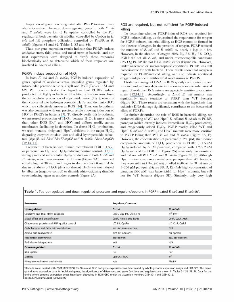
Table 1. Top up-regulated and down-regulated processes and regulons/operons in PGRP-treated E. coli and B. subtilisa.
Processes Regulons/Operons
Up-regulated E. coli B. subtilis
Oxidative and thiol stress response OxyR, Crp, Ihf, SoxR, Fnr sB, PerR
Metal efflux and detoxification CueR, ArsR, SoxR, RcnR CsoR, CzrA, ArsR
Chaperones, protein and RNA quality control sH, Ihf, CpxRA sB, CtsR, CssRS
Carbohydrate and fatty acid metabolism fad, fuc, fum operons N/A
Amino acid biosynthesis met, his operons his operon
Nucleotide biosynthesis deo operon pyr, pur operons
Fe-S cluster biosynthesis IscR N/A
Down-regulated E. coli B. subtilis
Iron uptake Fur Fur
Motility CpxRA, FlhDC sD
Phosphate utilization and uptake N/A PhoPR
aBacteria were treated with PGRP (PGLYRP4) for 30 min at 37uC and gene expression was determined by whole genome expression arrays and qRT-PCR. The meanquantitative expression data for individual genes, the significance of differences, and gene functions and regulators are shown in Tables S1, S2, S3, S4. Data for theentire whole genome expression arrays have been deposited in NCBI GEO under the accession numbers GSE44211 and GSE44212.doi:10.1371/journal.ppat.1004280.t001
PGRPs Kill by Oxidative, Thiol, and Metal Stress
PLOS Pathogens | www.plospathogens.org 4 July 2014 | Volume 10 | Issue 7 | e1004280

concentrations of exogenously added H2O2 (200–640 mM) were
bactericidal for Hpx2 mutants, but still not for WT bacteria
(Figure 3C, F). These results indicate that the amounts of H2O2
induced by PGRP or by 5–250 mM paraquat (,2 mM H2O2,
Figure 2A) are not sufficient to kill bacteria.
Altogether, these results demonstrate that ROS are induced by
PGRPs and are required for their bactericidal activity, but that
physiologically relevant concentrations of ROS induced by PGRPs
are not sufficient for bacterial killing. Therefore, these results
suggest that other killing mechanisms work together with O2-
dependent generation of ROS in eliciting the bactericidal activity
of PGRP.
PGRPs induce thiol depletion, which is required, but notsufficient for PGRP killing
We then tested the hypothesis that PGRPs cause thiol (disulfide)
stress by inducing depletion of intracellular thiols, because the
pattern of gene induction by PGRP was similar to the previously
reported pattern of gene induction by diamide (a thiol-depleting
electrophile), including activation of genes for the same metal
detoxification systems, chaperones, protein quality control, and
thiol stress responses [19,20]. PGRP, similar to diamide, depleted
over 90% of intracellular thiols in E. coli and B. subtilis within
30 min of exposure (Figure 4A), and these low levels of thiols were
maintained for at least 2 hrs both in PGRP- and diamide-treated
bacteria (data not shown). Paraquat only minimally reduced
intracellular thiols (Figure 4A) at a concentration that strongly
induced H2O2 production, comparable to PGRP-induced H2O2
production (Figure 2A). Altogether, our results show that PGRPs
induce both H2O2 production and thiol depletion, whereas
paraquat and diamide selectively induce either H2O2 production
or thiol depletion, respectively.
We next tested the role of intracellular thiols in PGRP killing.
Exogenous thiourea (a membrane-permeable thiol that inhibits
depletion of thiols and counteracts the effects of thiol and oxidative
stress) significantly diminished bactericidal activity of PGRP for
both E. coli and B. subtilis (Figure 4B), consistent with our previous
data [7]. These results suggest that depletion of thiols is required
for bactericidal activity of PGRPs. We next tested whether
depletion of thiols was sufficient for bacterial killing. Diamide, at
the concentration that induces similar depletion of thiols as PGRP
(Figure 4A), was bacteriostatic, but not bactericidal (Figure 4C).
Thus, this level of thiol depletion is not sufficient for bacterial
killing.
Glutathione and bacillithiol are the major low molecular weight
thiols in E. coli and B. subtilis, respectively, that protect against
oxidative and thiol stress [21–23]. Accordingly, glutathione-
deficient DgshA E. coli and bacillithiol-deficient DbshC B. subtilis
mutants had reduced total thiols by ,45% and ,30%,
respectively (Figure S4). Also, thiol depletion by PGRP or diamide
Figure 2. PGRP induces H2O2 production, requires O2 for killing, and the killing involves DNA damage. (A) Hpx2 E. coli or B. subtiliswere incubated aerobically with albumin (50 mg/ml), PGRP (PGLYRP3, 50 mg/ml), paraquat (5 mM), or diamide (250 mM) for 15 min and H2O2
production was measured. (B) E. coli or B. subtilis were incubated aerobically (+O2) or anaerobically (E. coli) or under microaerophilic conditions (B.subtilis) (no O2) with 50 mg/ml PGRP (PGLYRP4) or albumin and the numbers of bacteria were determined. (C) WT or DrecA E. coli were incubatedaerobically with 30 mg/ml PGRP (PGLYRP3) or albumin and the numbers of bacteria were determined. The results are means 6 SEM of 3 experiments(SEM were within symbols, if not visible); each experiment was repeated once with rMSA and another PGRP with similar results (A and B, PGLYRP4; B,PGLYRP3; not shown); *, P,0.05; **, P,0.001; albumin vs treated (A); PGRP + O2 vs no O2 (B); PGRP DrecA vs WT (C).doi:10.1371/journal.ppat.1004280.g002
PGRPs Kill by Oxidative, Thiol, and Metal Stress
PLOS Pathogens | www.plospathogens.org 5 July 2014 | Volume 10 | Issue 7 | e1004280

was less efficient in DgshA and DbshC mutants (79% and 74%
depletion) than in WT bacteria (97% and 90% depletion) (Figure
S4), suggesting that glutathione and bacillithiol are major targets of
PGRP-induced thiol depletion in E. coli and B. subtilis, respectively.
However, DgshA E. coli and DbshC B. subtilis were only somewhat
more sensitive to PGRP and diamide than WT strains (Figure 4C),
which indicates that these thiols play a modest role in protecting
against PGRP and that other cellular thiols in these mutants are
still able to maintain nearly sufficient reducing environment in the
cytoplasm. Altogether, these results suggest that although thiol
depletion likely contributes to bacterial killing, by itself it is not
sufficient for strong bactericidal activity.
PGRP induces increases in intracellular Zn and CuPGRP treatment highly induced genes for detoxification and
efflux of Cu, Zn, and other metals (Figure 1 and Tables 1, S1 and
S2). We therefore tested whether treatment with PGRP increased
intracellular concentrations of free Zn and Cu (also known as
‘‘labile’’ Zn and Cu, because no metal is truly free in cellular
context). Indeed, PGRP induced a large increase in intracellular
Figure 3. H2O2 is not sufficient for PGRP killing of bacteria. WT or Hpx2 E. coli or B. subtilis were incubated aerobically with PGRP (A, PGLYRP3;B, PGLYRP3:PGLYRP4), albumin, paraquat, or H2O2 for 1, 2, or 4 hrs (A, B, E) or 2 hrs (C, D, F) and the numbers of bacteria were determined. The resultsare means 6 SEM of 3 experiments (SEM were within symbols, if not visible); *, P,0.05; **, P,0.001; WT vs Hpx2.doi:10.1371/journal.ppat.1004280.g003
PGRPs Kill by Oxidative, Thiol, and Metal Stress
PLOS Pathogens | www.plospathogens.org 6 July 2014 | Volume 10 | Issue 7 | e1004280

free (labile) Zn2+ in both E. coli and B. subtilis, based on 60- to
100-fold increase in fluorescence of Zn2+-specific membrane
permeable Zynpyr-1 probe, measured by flow cytometry
(Figures 5A, 5B and S5A). This increase in Zn2+ was significant
at 30 and 60 min (data not shown) and was maximal at 2 hrs
(Figures 5A and S5A). Detection of intracellular Zn2+ was
completely suppressed by the membrane permeable Zn(II)
chelator, TPEN [24] (Figure S5A). PGRP also induced a large
increase in intracellular free (labile) Cu+ in B. subtilis, but not in E.
coli, based on 20-fold increase in fluorescence of Cu+-specific
membrane permeable CF4 probe, measured by flow cytometry
after 2-hr exposure to PGRP (Figures 5A and S5B). Paraquat and
diamide, used at the concentrations that caused similar increase
in H2O2 or depletion of thiols as PGRP, did not induce any
increases in intracellular free Zn2+ or Cu+ (Figure 5A). These
results suggest that PGRP-induced increases in intracellular
H2O2 or depletion of thiols are not responsible (or at least not
sufficient) for the PGRP-induced increases in intracellular Zn2+
and Cu+. The slower kinetics of increase in Zn2+ and Cu+ than
accumulation of H2O2 and depletion of thiols may be related to
slower kinetics of transport of exogenous metals into the cell.
Thus, these results further suggest that these three effects of
PGRPs (oxidative, thiol and metal stress) are independent and do
not induce each other.
Antibiotics induced different patterns of changes in intracellular
metals than PGRP-induced pattern. Gentamicin treatment led to
large increases of both intracellular Zn2+ and Cu+ in B. subtilis, and
low, but still significant, increase of Zn2+ and a moderate increase
of Cu+ in E. coli. Ciprofloxacin, used here as a known positive
control for induction of intracellular Cu+ in E. coli [25], caused
high increase in Cu+ in both E. coli and B. subtilis, but did not lead
to increased Zn2+ levels (Figures 5A and S5).
Metal toxicity is required, but not sufficient for PGRP-induced killing
Motivated by the high induction of genes for detoxification of
both Cu and Zn (Figure 1 and Tables 1, S1 and S2), and the
observed increase in intracellular metal concentrations in both E.
coli and B. subtilis (Figures 5A and S5), we next tested whether Zn2+
and Cu+ were required for bactericidal activity of PGRPs. Indeed,
chelating Zn2+ with TPEN completely inhibited the bactericidal
activity of PGRP in both E. coli and B. subtilis (Figure 5C).
Chelating Cu+ with Cu(I) chelator bathocuprione sulfonate (BCS)
[26] also completely inhibited bactericidal activity of PGRP in
both E. coli and B. subtilis (Figure 5C). These effects were selective
for PGRP, because TPEN and BCS did not inhibit killing by a
bactericidal antibiotic, gentamicin, and BCS even enhanced
Figure 4. PGRP induces thiol depletion, which is required but not sufficient for bacterial killing. (A) E. coli or B. subtilis were incubatedaerobically with albumin (50 mg/ml), PGRP (PGLYRP3, 50 mg/ml), diamide (250 mM), or paraquat (5 mM) for 30 min and intracellular thiols weremeasured. (B) E. coli or B. subtilis were incubated aerobically with albumin or PGRP (PGLYRP4, 50 mg/ml) without or with thiourea (150 mM, aninhibitor of thiol oxidation), and the numbers of bacteria were determined. (C) WT and glutathione-deficient DgshA E. coli or bacillithiol-deficientDbshC B. subtilis mutants were incubated aerobically with albumin (50 mg/ml), or diamide (250 mM), or PGRP (PGLYRP4, 50 mg/ml), and the numbersof bacteria were determined. The results are means 6 SEM of 3–6 experiments (SEM were within symbols, if not visible); each experiment wasrepeated once with rMSA and another PGRP with similar results (A, PGLYRP4; B and C, PGLYRP3:PGLYRP4; not shown); *, P,0.05; **, P,0.001; albuminvs treated (A); 2 vs + thiourea (B); WT vs DgshA or DbshC mutants (C).doi:10.1371/journal.ppat.1004280.g004
PGRPs Kill by Oxidative, Thiol, and Metal Stress
PLOS Pathogens | www.plospathogens.org 7 July 2014 | Volume 10 | Issue 7 | e1004280
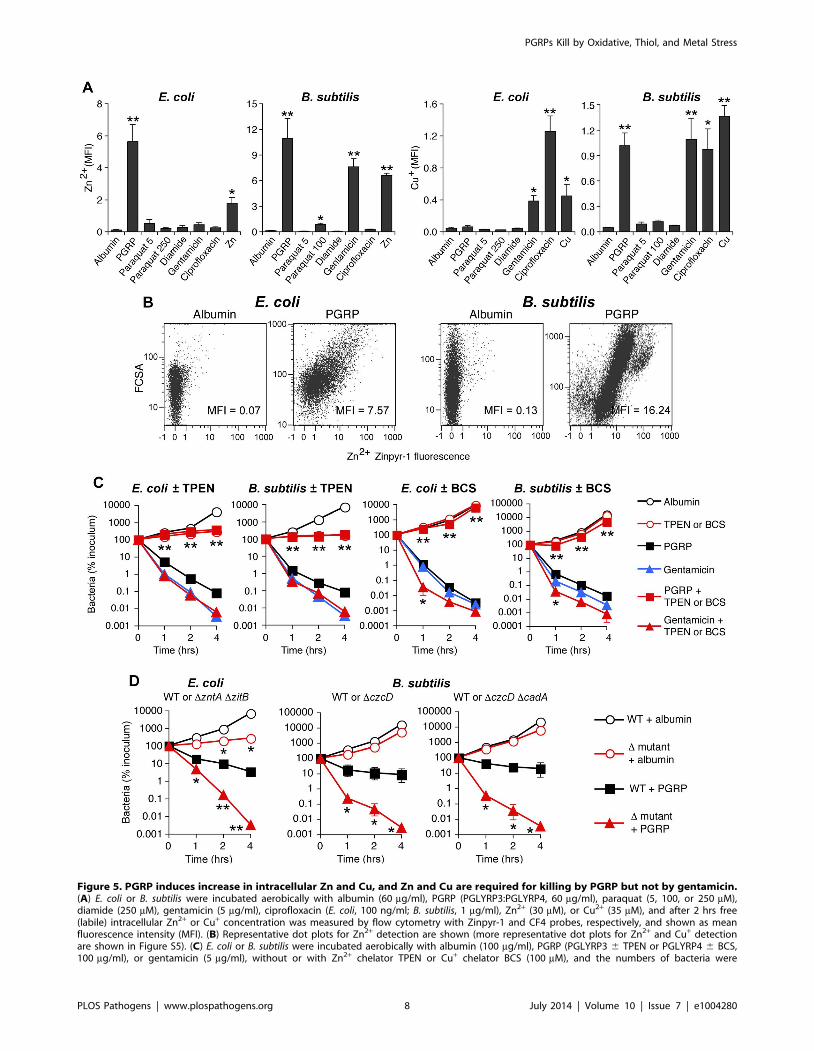
Figure 5. PGRP induces increase in intracellular Zn and Cu, and Zn and Cu are required for killing by PGRP but not by gentamicin.(A) E. coli or B. subtilis were incubated aerobically with albumin (60 mg/ml), PGRP (PGLYRP3:PGLYRP4, 60 mg/ml), paraquat (5, 100, or 250 mM),diamide (250 mM), gentamicin (5 mg/ml), ciprofloxacin (E. coli, 100 ng/ml; B. subtilis, 1 mg/ml), Zn2+ (30 mM), or Cu2+ (35 mM), and after 2 hrs free(labile) intracellular Zn2+ or Cu+ concentration was measured by flow cytometry with Zinpyr-1 and CF4 probes, respectively, and shown as meanfluorescence intensity (MFI). (B) Representative dot plots for Zn2+ detection are shown (more representative dot plots for Zn2+ and Cu+ detectionare shown in Figure S5). (C) E. coli or B. subtilis were incubated aerobically with albumin (100 mg/ml), PGRP (PGLYRP3 6 TPEN or PGLYRP4 6 BCS,100 mg/ml), or gentamicin (5 mg/ml), without or with Zn2+ chelator TPEN or Cu+ chelator BCS (100 mM), and the numbers of bacteria were
PGRPs Kill by Oxidative, Thiol, and Metal Stress
PLOS Pathogens | www.plospathogens.org 8 July 2014 | Volume 10 | Issue 7 | e1004280

gentamicin killing at 1 hr (Figure 5C), consistent with the recent
report of Cu+-mediated induction of antibiotic resistance regulator
in E. coli [25]. The results with metal chelators, however, need to
be interpreted with caution, because chelators are not 100%
specific and may chelate to some extent other metals. This could
explain the inhibition of PGRP-induced E. coli killing by BCS
(Figure 5C), when there was no significant PGRP-induced increase
in intracellular Cu+ in E. coli (Figure 5A), because although BCS is
a Cu+ chelator [26], it can also form dimers with Cu2+ and
possibly with other divalent metals and chelate them [27]. Our
current results are also consistent with our previous data showing
that chelating Zn2+ with EGTA (whose log stability constant for
Zn2+ is 12.9) inhibits bactericidal activity of PGRPs, and that
5 mM Zn2+ is required for PGRP killing [5]. Our previous results
also show that chelating Fe2+ with dipyridyl inhibits bactericidal
activity of PGRPs [7]. Cu2+ and Zn2+ at low physiologic
concentrations were only bacteriostatic, but not bactericidal
(Figure 6), which indicates that at these concentrations Cu2+ and
Zn2+ are not sufficient for bacterial killing.
To further determine which metal ions are the most critical for
PGRP-induced killing, we then compared the sensitivity to PGRP
and metal killing of WT bacteria and their mutants deficient in
various metal efflux and detoxification systems. We show that both
E. coli DzntADzitB mutant, deficient in two Zn2+ efflux systems
[28], and B. subtilis DczcD mutants deficient in the Zn2+, Cu2+,
Co2+, and Ni2+ efflux system [29] were substantially more sensitive
to PGRP-induced killing than WT bacteria (Figure 5D). Similarly,
DzntADzitB mutant was substantially more sensitive to killing by
extracellular Zn2+ than WT bacteria (Figure S6A). E. coli and B.
subtilis mutants deficient in Cu efflux and detoxification systems (E.
coli DcopADcueODcusCFBA and B. subtilis DcadA and DcopZA) were
not more sensitive to PGRP-induced killing than WT bacteria
(Figure S6C), and the E. coli DcopADcueODcusCFBA mutant was
even more resistant to PGRP killing. Similarly, E. coli DcopAD-cueODcusCFBA mutant was also more resistant to killing by
extracellular Cu2+ than WT bacteria (Figure S6A), perhaps
because increased intracellular Cu level protects E. coli from
oxidative Fe toxicity [30], and only at higher concentrations Cu
becomes bactericidal. B. subtilis DcopZA mutant had similar
sensitivity to killing by extracellular Cu2+ as WT bacteria, whereas
B. subtilis DczcDDcadA mutant was more sensitive to killing by
extracellular Cu2+ than WT bacteria (Figure S6B). Higher
sensitivity of Zn efflux-deficient than Cu efflux-deficient mutants
to PGRP is consistent with high increase of intracellular Zn2+ in
both PGRP-treated bacteria.
Altogether, these results indicate that Zn2+ and Cu+ are
required for bactericidal activity of PGRPs, and that Zn2+ is
more important than Cu+ for this bactericidal activity, especially in
E. coli. Our results also indicate that these metals are not required
for bactericidal activity of antibiotics. Indeed, PGRPs have the
same bactericidal activity towards antibiotic-sensitive bacteria and
clinical isolates resistant to multiple antibiotics (Figure S7).
Synergistic effect of ROS, thiol depletion, and metals isrequired for bacterial killing
We next tested the hypothesis that production of ROS,
depletion of thiols, and metal toxicity have a synergistic
bactericidal effect, because these three stress responses were all
induced in PGRP-treated bacteria and each was required, but not
individually sufficient, for bacterial killing. To induce intracellular
ROS production we used paraquat, which is reduced by Complex
I to radical cations, which react with O2 to generate O22, which
then generate H2O2 and then OHN [14,18]. To induce thiol stress,
we used diamide, which directly depletes intracellular thiols by
inducing formation of disulfide bonds and S-thiolations (which are
disulfide bonds between proteins and low molecular weight thiols,
such as glutathione, bacillithiol, and free cysteine) [14,20,31]. To
induce metal toxicity, we used exogenous Zn2+, or Cu2+ (which is
transported into the cell and reduced to more toxic Cu+), or
arsenite (AsO22).
Indeed, treatment of E. coli or B. subtilis with the doses of
paraquat that induce the amounts of H2O2 comparable with the
amounts of H2O2 induced by PGRP were not bactericidal
(Figure 6). Also, treatment of E. coli or B. subtilis with the doses
of diamide that deplete thiols to a comparable extent as PGRPs
were not bactericidal, and low concentrations of Zn2+, Cu2+, or As
(AsO22) by themselves were also not bactericidal (Figure 6).
Moreover, the combination of any two of these stresses was also
not bactericidal (except for a combination of paraquat plus Zn2+ or
Cu2+, which had low killing activity for B. subtilis). However, when
all three stress conditions were simultaneously imposed, the
resulting combination was strongly bactericidal for both E. coli
and B. subtilis, although Zn2+ was less efficient in E. coli than in B.
subtilis (Figure 6). These results validate our hypothesis and show
that ROS production, thiol depletion, and metal toxicity act
synergistically to kill bacteria.
To further verify that oxidative, thiol, and metal stress are
responsible for the bactericidal activity of PGRPs, we abolished
bactericidal activity of PGRP by de-glycosylation, which we
previously showed to be required for bactericidal activity of
PGRPs for both Gram-positive and Gram-negative bacteria [4,5].
De-glycosylation abolished 90–95% of the ability of PGRP to
induce (i) intracellular production of H2O2 (Figure 7A), (ii)
depletion of cellular thiols (Figure 7B), and (iii) increases in
intracellular Zn2+ (Figure 7C) in both E. coli and B. subtilis. These
results further validate the requirement of oxidative, thiol, and
metal stress for the bactericidal activity of PGRPs.
Discussion
Analysis of the global transcriptional responses of both E. coli
and B. subtilis to PGRP revealed stress responses involving
increased production of H2O2, depletion of thiols, and increases
in intracellular Zn2+ and Cu+, which were also verified by direct
measurements. Using selective chemical treatments (paraquat to
generate ROS, diamide to oxidize thiols, and exogenous metal
ions) and specific inhibitors, we demonstrated that ROS produc-
tion, thiol depletion, and increased intracellular Zn2+ or Cu+ are
all required, but individually are not sufficient, for bacterial killing,
and that combined action of oxidative, thiol, and metal stress kills
bacteria.
PGRP treatment induced oxidative stress through rapid
induction of H2O2 production. Oxidative stress results from
excessive production of ROS (O22, H2O2, and HON). Both O2
2
and H2O2 oxidize solvent-exposed [4Fe-4S] enzyme clusters,
causing release of Fe and cluster collapse to inactive [3Fe-4S]+.
determined. (D) E. coli or B. subtilis (WT or indicated mutants) were incubated aerobically with albumin or PGRP (PGLYRP4 for E. coli or PGLYRP3 for B.subtilis, 25 mg/ml), and the numbers of bacteria were determined. The results are means 6 SEM of 3–5 experiments (SEM were within symbols, if notvisible); experiments in C and D were repeated once with rMSA and another PGRP (PGLYRP4 or PGLYRP3, not shown) with similar results; *, P,0.05;**, P,0.005 (A) or **, P,0.001 (C, D); treated vs albumin (A); no TPEN or BCS vs with TPEN or BCS (C); or WT vs mutant (D).doi:10.1371/journal.ppat.1004280.g005
PGRPs Kill by Oxidative, Thiol, and Metal Stress
PLOS Pathogens | www.plospathogens.org 9 July 2014 | Volume 10 | Issue 7 | e1004280

PGRPs Kill by Oxidative, Thiol, and Metal Stress
PLOS Pathogens | www.plospathogens.org 10 July 2014 | Volume 10 | Issue 7 | e1004280

O22 and H2O2 also inactivate mononuclear iron enzymes by
oxidizing Fe-coordinating cysteines or by replacing Fe2+ with Zn2+
[16,21,32–34]. Moreover, H2O2 reacts with Fe2+ to generate HON
via Fenton reaction. HON is the most reactive and most toxic ROS
and it irreversibly damages DNA, proteins, and other organic
molecules [14,17].
PGRP treatment also depleted over 90% of cellular thiols. Thiol
stress results from oxidation of thiols, which maintain the redox
state in the cells and protect from oxidative damage. Oxidative
and thiol stress not only directly damage cells, but also release Fe
from proteins, increase intracellular concentration of Zn and Cu,
and increase toxicity of most metals [19,21,35–37]. Thiols bind
free metal ions and protect cells from metal toxicity [38], and for
this reason thiol stress induces the same genes for metal
detoxification and protein refolding and repair [19,20,31] as the
genes induced by PGRP (Tables 1, S1, and S2).
PGRP treatment also induced a drastic increase in intracellular
free (labile) Zn2+ in both E. coli and B. subtilis and intracellular free
(labile) Cu+ in B. subtilis (but not E. coli), which is the likely reason
for increased expression of metal detoxification and efflux genes.
These increases in free metals are required for PGRP toxicity,
because chelating intracellular Zn2+ with TPEN (Figure 5C) or
extracellular Zn2+ with EGTA [5], or chelating Cu+ with BCS
(Figure 5C) or Fe2+ with dipyridyl [7] also inhibits bacterial killing
by PGRP. Zn2+ seems the most important for PGRP killing, as
revealed by the highest sensitivity of Zn2+ efflux mutants to PGRP
killing (Figure 5D). However, the increased concentrations of
metals alone that are induced by PGRP are not sufficient for
bacterial killing.
The origins of metal toxicity are complex. Zn, a redox-inert
metal, is more abundant in the cytosol than Cu and at low
concentrations it may protect bacteria from oxidative and thiol
stress, likely by binding to thiols and preventing their further
oxidation [39]. However, high levels of Zn are toxic and up-
regulate the expression of genes for Zn efflux (zntA in E. coli and
czcD and cadA in B. subtilis, also observed in our arrays). Zn
toxicity, similar to Cu, results in part from inactivation of solvent-
exposed Fe-S clusters; and although this activity of Zn2+ is lower
than Cu+ [40], it is likely compensated by higher concentrations of
Zn2+ than Cu+. In oxidative stress, Zn2+ also inactivates
mononuclear enzymes by replacing Fe2+ in their active sites
[32]. Cu is toxic because it causes loss of Fe from solvent-exposed
Fe-S clusters, which inactivates enzymes, and also because this
release of Fe makes it available for enhanced production of HON
via Fenton reaction [14,21,35–37,41–44]. Cu also causes thiol
oxidation and sulfhydryl depletion, which contribute to thiol stress
and protein damage [21,35,37,42]. Fe toxicity results primarily
from generation of HON, which damages DNA, proteins, and lipids
[14,29]. HON is induced by PGRPs and chelating intracellular Fe
with dipyridyl inhibits both HON production and PGRP killing [7].
Many of the genes induced by PGRPs reflect direct or indirect
bacterial responses to the resulting oxidative, thiol, and metal
stress. The genes for repair of damaged proteins and DNA and
Ihf-regulated genes (which help to maintain DNA architecture) are
induced because ROS oxidize proteins and nucleic acids, because
oxidation of thiols damages proteins, and because increased
concentrations of intracellular metals also damages proteins
[14,16,17,19,21,33,34,41–44]. Genes for transition to fermenta-
tion and anaerobic growth (e.g., members of Fnr regulon in E. coli)
are a likely attempt to reduce the use of oxygen to limit further
production of ROS. Genes for energy generation are induced
because of possible oxidative damage to respiratory chain enzymes
and because a decrease in membrane potential [7] may cause a
decrease in ATP production by membrane potential-driven ATP
synthase [45,46]. This is also the likely reason why bacteria down-
regulate genes for high energy-requiring non-essential processes,
Figure 6. H2O2 production, thiol depletion, and Zn, Cu, or As synergistically kill E. coli and B. subtilis. E. coli or B. subtilis were incubatedaerobically with paraquat (50–100 mM), diamide (250 mM), and Zn2+ (13 mM E. coli or 20 mM B. subtilis), or Cu2+ (30–35 mM), or As (AsO2
2, 1 mM)individually or together (as indicated), and the numbers of bacteria were determined. The results are means 6 SEM of 3–5 experiments (SEM werewithin symbols, if not visible); *, P,0.05 three compounds together vs two compounds.doi:10.1371/journal.ppat.1004280.g006
Figure 7. De-glycosylation abolishes the ability of PGRP to induce intracellular production of H2O2, depletion of cellular thiols, andincreases in intracellular Zn2+. E. coli or B. subtilis were incubated with 50 mg/ml of bovine serum albumin (BSA), recombinant mouse serumalbumin (rMSA), PGRP (mock treated), or de-glycosylated PGRP, and assayed for H2O2 (A), thiols (B), and intracellular free (labile) Zn2+ (C) as describedin Figures 2, 4, and 5, respectively. The results are averages of duplicates from one experiment with PGLYRP3 (shown), which was repeated once withPGLYRP4 with similar results (not shown).doi:10.1371/journal.ppat.1004280.g007
PGRPs Kill by Oxidative, Thiol, and Metal Stress
PLOS Pathogens | www.plospathogens.org 11 July 2014 | Volume 10 | Issue 7 | e1004280

such as motility, which are controlled by CpxRA [8], one of the
regulators of envelope stress response activated by PGRP [7].
The genes for methionine and histidine synthesis may be
induced for several reasons. These amino acids are essential metal-
binding components abundant in metal detoxification proteins,
e.g., methionine shuttle is used for Cu efflux and histidine is used
for coordination of metals in metal detoxification proteins, such as
CusA and CopA Cu efflux and AraA and ArsD As efflux
transporters [47–49]. Also, histidine shares biosynthetic interme-
diates with nucleotides, whose synthesis is needed to repair
damaged DNA. Moreover, likely oxidation of the thiol group in
homocysteine may deplete this methionine biosynthesis interme-
diate. Methionine is also needed for initiation of translation and
DNA replication, and methionine synthase is highly sensitive to
thiol stress [50].
The genes for Fe-S cluster assembly (isc in E. coli) are likely
induced due to the damage to Fe-S clusters by oxidative, thiol, and
metal stress, and most likely Cu+-induced release of Fe2+ from Fe-
S clusters. Cu+ also damages Isc proteins, which may further
contribute to the induction of isc genes. Damage to DNA could be
either direct by Cu+, or more likely by Cu+-induced release of Fe2+
from Fe-S clusters and Fe-driven enhancement of HON production
from H2O2 [21]. This mechanism is supported by the ability to
inhibit PGRP killing by chelating either Fe2+ with dipyridyl [7] or
Cu+ with BCS (Figure 5C). Concurrently bacteria down-regulate
the expression of genes for Fe uptake, which also suggests an
increase in cytoplasmic free Fe2+, likely due to release of Fe2+ from
Fe-S clusters, caused by oxidative and thiol stress and Cu+. Down-
regulation of Fe uptake is controlled by the envelope stress
response regulator CpxRA, which is activated by PGRPs [7], and
by increased Cu and Zn [8,51–53].
How do PGRPs induce oxidative, thiol, and metal stress in
bacteria? PGRPs have a specific peptidoglycan-binding grove that
binds disaccharide-pentapeptide fragment of peptidoglycan
[2,3,54,55]. However, this PGRP-binding site on peptidoglycan
is not easily accessible on the surface of Gram-positive bacteria,
because of extensive peptidoglycan cross-linking and its substitu-
tion with polysaccharides and proteins. Thus, in Gram-positive
bacteria PGRPs preferentially bind to the separation sites of the
newly formed daughter cells, created by dedicated peptidoglycan-
lytic endopeptidases, which separate daughter cells after cell
division. We assume that these cell-separating endopeptidases
expose PGRP-binding muramyl peptides, because PGRP bound
to bacteria co-localizes with cell-separating endopeptidases and
PGRPs do not bind to other regions of the cell wall with highly
cross-linked peptidoglycan [7]. This localization is necessary for
bacterial killing, because mutants that lack these endopeptidases
and do not separate after cell division (DlytEDlytF B. subtilis) do not
bind PGRPs and are not killed by PGRPs [7]. In Gram-negative
bacteria, PGRPs bind uniformly to the entire outer membrane [7],
which is composed of lipopolysaccharide (LPS) and covers a thin
peptidoglycan layer. This is possible, because in addition to
binding peptidoglycan, PGRPs also bind LPS using binding sites
outside the peptidoglycan-binding groove [56,57]. This binding to
bacterial envelope is required for PGRP killing, because exoge-
nous peptidoglycan or LPS inhibit PGRP killing of Gram-positive
or Gram-negative bacteria, respectively, by blocking peptidogly-
can or LPS binding sites on PGRP [4,5,56]. It is not known
whether after binding to LPS in Gram-negative bacteria PGRPs
also bind to peptidoglycan, located in the periplasmic space
beneath the outer membrane. In both Gram-positive and Gram-
negative bacteria, after binding to peptidoglycan or LPS, PGRPs
do not enter the cytoplasm [7], but probably form oligomeric
ribbon-like structures [2,55] and induce envelope stress by
activating stress response two component systems, CpxRA in E.
coli and CssRS in B. subtlis, which are typically activated by
misfolded or aggregated proteins exported from the cells [3,7,58].
This activation ultimately results in membrane depolarization,
inhibition of all biosynthetic reactions, and cell death [7].
However, the exact initial mechanism through which PGRPs
activate envelope stress response and oxidative, thiol, and metal
stress is unknown, as this mechanism is also unknown for other
envelope stressors [8], and is currently under investigation.
Furthermore, based on induction of multiple stress response
regulons by PGRP (Tables 1, and S1, S2, S3, S4) and on
incomplete resistance of DcpxRA and DcssRS mutants to PGRP [7],
it is likely that PGRPs activate other stress sensors that induce
these multiple stress responses.
Other investigators previously proposed that oxidative stress is
involved in killing of E. coli by antibiotics [9,10]. However, recent
results do not support this conclusion [12,13] and are consistent
with our results. Our data clearly indicate that the mechanisms of
killing by PGRPs and antibiotics are different for the following
reasons. (i) PGRPs kill bacteria resistant to multiple antibiotics
(Figure S7) [4]. (ii) PGRP killing requires O2 and PGRPs do not
kill anaerobically (Figure 2B), whereas many antibiotics kill both
aerobically and anaerobically [12,13]. (iii) PGRPs very strongly
induce peroxide-responsive genes (e.g. the OxyR regulon in E. coli)
indicating endogenous H2O2 production, but antibiotics do not
(Tables S1 and S2) [12]. (iv) PGRPs strongly induce H2O2
production in bacteria (Figure 2A), but antibiotics do not [12]. (v)
DrecA mutant is more sensitive than wild type strain to PGRPs
(Figure 2B), but not to antibiotics [12]. (vi) PGRP-induced killing is
inhibited by chelating Zn2+ or Cu+, whereas killing by antibiotics is
not affected by chelating Zn2+ and is enhanced by chelating Cu+
(Figure 5C). These results are consistent with induction of the
antibiotic resistance regulator MarR by CpxRA [59] and by Cu+
[25], which are induced by both PGRP and antibiotics. However,
MarR confers resistance only to antibiotics [25], but not to PGRP.
(vii) The patterns of gene expression induced in E. coli and B.
subtilis by bactericidal concentrations of PGRP and by gentamicin
are different: more than half of the top 100 genes strongly induced
by PGRPs are not induced by gentamicin, e.g., genes for oxidative
stress, energy production, Fe-S cluster repair and assembly, Fe-S-
containing enzymes (e.g., edd), amino acid synthesis, and other
stress responses. (viii) We could prevent bacterial killing by cell wall
synthesis-inhibiting antibiotics, but not by PGRPs, using hyperos-
motic medium [7], which should not happen if the main
mechanism of killing by these antibiotics was due to oxidative
stress and was the same as for PGRPs. (ix) Antibiotics selectively
inhibit one biosynthetic reaction and other biosynthetic reactions
are not inhibited for several hours until bacteria die, whereas
exposure to PGRPs results in simultaneous and rapid inhibition of
all biosynthetic reactions in bacteria [7].
PGRPs, bactericidal innate immunity proteins, by combining
oxidative stress with thiol depletion and release of intracellular
metals, have evolved a powerful antibacterial defense strategy.
This strategy is consistent with recent evidence that phagocytic
cells, upon phagocytosis of bacteria, in addition to oxidative
killing, pump Cu and Zn into phagolysosomes to enhance
bacterial killing [41–44,60]. Indeed, the most abundant PGRP,
PGLYRP1, is present in neutrophil, eosinophil, and macrophage
granules [1,56,61–64], and other PGRPs (PGLYRP2, PGLYRP3,
and PGLYRP4) are produced on the skin and mucous
membranes, and in sweat, sebum, and saliva [1,4,5]. These body
secretions also contain significant amounts of Cu and Zn [5],
which is consistent with the requirement for Zn (Figure 4B) [5], Fe
[7], and Cu (Figure 4B) for bactericidal activity of PGRPs. In
PGRPs Kill by Oxidative, Thiol, and Metal Stress
PLOS Pathogens | www.plospathogens.org 12 July 2014 | Volume 10 | Issue 7 | e1004280

response to PGRPs bacteria up-regulate expression of Cu and Zn
exporters (CopA, ZntA, CadA, and CzcD). However, PGRPs
defeat this bacterial Cu and Zn defense, because PGRP-induced
oxidative, thiol, and metal stress likely damage respiratory chain
enzymes and depolarize bacterial membranes [7], which likely
reduces ATP production and proton motive force needed to drive
bacterial Cu and Zn efflux. Furthermore, because Cu tolerance
increases bacterial virulence [41–44], targeting Cu tolerance will
both increase bacterial killing and decrease bacterial virulence,
which should additionally improve host defense against infection.
In vivo PGRPs are present at concentrations similar to the
concentrations used in our experiments: PGLYRP1 is present in
milk at 120 mg/ml [65] and in polymorphonuclear leukocytes’
granules at 2.9 mg/109 cells [64], PGLYRP2 is present in serum
at 100 mg/ml [66,67], and PGLYRP3 and PGLYRP4 are secreted
on mucous membranes, likely reaching similar local concentra-
tions [1,4]. In this study we investigated the mechanism of
bactericidal activity of PGRPs in vitro, but the following evidence
indicates that PGRPs also have antibacterial activity in vivo: (i) local
application of PGRPs into upper respiratory tract protects mice
against lung infection [4,58]; (ii) Pglyrp12/2 mice are more
sensitive to some infections than wild type mice [62]; (iii)
neutrophils from Pglyrp12/2 mice are less efficient in bacterial
killing than neutrophils from wild type mice [62]; (iv) PGRPs
protect zebrafish embryos from bacterial infections [68]; (v)
PGRPs are required for maintenance of normal intestinal
microbiome in mice [69]; and (vi) PGRPs also have several anti-
microbial and microbiome-regulating functions in invertebrates
[3]. Our results indicate that PGRPs have bactericidal activity in
an aerobic environment, which is consistent with the highest
expression of PGRPs in phagocytic cells and on the skin and
mucous membranes, especially in the mouth, throat, esophagus,
and salivary glands [1–4,56,61–64,69]. Lower PGRP expression in
the stomach and small and large intestine is again consistent with
their bactericidal activity in an aerobic environment, although
anaerobically PGRPs are still bacteriostatic. Bactericidal activity of
PGRPs both in vitro and in vivo is enhanced by antimicrobial
peptides [5,58], also expressed in phagocytic cells and on mucous
membranes and skin, which likely further strengthens antibacterial
defenses of the host.
In conclusion, innate immunity proteins, PGRPs, induce
oxidative, thiol, and metal stress in E. coli and B. subtilis, which
act synergistically to kill bacteria. Because this bactericidal
mechanism differs from killing by antibiotics and because PGRPs
kill antibiotic-resistant bacteria, synergistic targeting of oxidative,
thiol, and metal stress can be used for the development of new
approaches to treatment of antibiotic resistant bacteria.
Materials and Methods
MaterialsBacterial strains are listed in Table S5. Disruption of Bacillus
genes was achieved by transformation with PCR products to
amplify DNA fragments flanking each target gene and an
intervening antibiotic cassette as previously described [70].
Human PGRPs (PGLYRP3, PGLYRP4, and PGLYRP3:P-
GLYRP4 heterodimer) were expressed in S2 cells and purified
as previously described [4,5] in a buffer containing 10 mM TRIS
(pH 7.6), with 150 mM NaCl, 10 mM ZnSO4, and 10% glycerol.
The experiments were done using PGLYRP3, PGLYRP4, and/or
PGLYRP3:PGLYRP4 (as indicated in Figure legends and Table
footnotes), and all key experiments were performed with at least
two PGRPs with similar results. Note that when expressed
individually, PGLYRP3 and PGLYRP4 form disulfide-linked
homodimers, and when co-expressed in the same cells, they form
disulfide-linked PGLYRP3:PGLYRP4 heterodimers [4]. For some
experiments PGRP was de-glycosylated by treatment with 0.67
units of N-glycosidase/mg PGRP (PNGase F from Elizabethkingia
miricola, Sigma) for 2 hr at 37uC, and we verified that this
treatment abolished PGRP’s bactericidal activity for E. coli and B.
subtilis, as previously described [4,5]. For non-de-glycosylated
PGRP in these experiments, PGRP was similarly incubated in the
same buffer, but without PNGase. Purified bovine serum albumin
(BSA, Sigma) was used as a negative control, and key experiments
were repeated with recombinant mouse serum albumin (rMSA) as
an additional control, which was cloned, expressed, and purified
by the same methods as PGRPs, as described [7], with similar
results, as indicated in figure legends. Paraquat (methyl viologen)
was from Acros Organics, Zinpyr-1 and TPEN were from Santa
Cruz. Bathocuprione disulfonate (BCS), CCCP (carbonyl cyanide
3-chlorophenyl-hydrazone), ciprofloxacin, diamide, gentamicin,
and other reagents were from Sigma-Aldrich, unless otherwise
indicated. Arsenite (AsO22) was prepared fresh from arsenic
trioxide at pH 8.2; CuSO4 was used as Cu2+, and ZnSO4 as Zn2+.
Gene expression arraysOvernight bacterial cultures were diluted 1:100 in LB, grown
aerobically with 250 rpm shaking to OD660 = 0.1–0.3, suspended
in fresh warm medium (E. coli MG1655 at OD660 = 0.3 or B.
subtilis 168 at OD660 = 0.1), and incubated aerobically with
100 mg/ml albumin (control), or 100 mg/ml PGRP (human
recombinant PGLYRP4), or 5 mg/ml gentamicin for 30 min, or
with 800 mM CCCP for 15 min, in 2 ml of 5 mM TRIS (pH 7.6)
with 150 mM NaCl, 5 mM ZnSO4, with addition of 2% of 100%
LB (E. coli), or in 1 ml of TRIS-Schaeffer medium with 0.05%
NH4Cl, 5 mM ZnSO4, 0.2% glucose, with addition of 2% of
100% LB (B. subtilis) at 37uC with 250 rpm shaking (these
optimum incubation times and concentrations for induction of
stress response genes were determined in preliminary experiments
using qRT-PCR). Because 5 mM Zn2+ is required for bactericidal
activity of PGRP and corresponds to the average concentration of
Zn2+ found in saliva, sweat, and other body fluids, where PGRPs
are present [5], we confirmed that Zn2+ is not depleted or
increased by additions of our proteins and bacteria, by measuring
the concentration of free Zn2+ in our incubation mixtures at the
initiation of our experiments (time 0) using Zn2+-specific probe,
Zinpyr-1, and fluorescence spectroscopy (with Molecular Devices
Gemini EM Spectrofluorometer). Our incubation mixtures
containing 100 mg/ml of either PGRP (PGLYRP3 or PGLYRP4)
or recombinant mouse albumin, and with or without addition of
bacteria, all contained similar amounts of free Zn2+ (4.1–4.4 mM).
Moreover, substituting the addition of 5 mM free Zn2+ with the
addition of 25 mM Zn2+ plus 20 mM EDTA (a divalent cation
chelator with high affinity for Zn2+, log dissociation constant 16.6)
yielded the same concentrations of free Zn2+ as in our reaction
mixture without EDTA, measured by Zinpyr-1 fluorescence.
Based on these results we concluded that addition of our control
protein or PGRPs and/or bacteria does not substantially change
the free Zn2+ concentration in our experiments.
To obtain RNA from each culture, bacteria were harvested and
RNA was extracted using Ambion RiboPure-bacteria RNA
extraction kit according to the manufacturer’s instructions. For
B. subtilis, before RNA extraction, bacteria were disrupted by
shaking with Zirconia beads. cDNA was synthesized with random
hexamer primers, fragmented, labeled with terminal transferase
and biotin, and hybridized to whole genome Affymetrix E. coli
Genome 2.0 Array GPL3154 or custom whole genome Affymetrix
900513 GeneChip B. subtilis Genome Array using Affymetrix
PGRPs Kill by Oxidative, Thiol, and Metal Stress
PLOS Pathogens | www.plospathogens.org 13 July 2014 | Volume 10 | Issue 7 | e1004280

Hybridization Oven 640 and Affymetrix GeneChip Fluidics
Station 450 and protocols provided by Affymetrix GeneChip
Technical Manual. Scanning and data extraction were done using
Affymetrix GeneChip Scanner 3000 and protocols provided by
Affymetrix GeneChip Technical Manual. cDNA synthesis, label-
ing, hybridization, and scanning were performed at the Genomic
and RNA Profiling Core facility, Baylor College of Medicine,
Houston, TX. The entire experiment was repeated 3 times both
for E. coli and B. subtilis.
Hybridization intensity data signals were analyzed, normalized,
and corrected for batch effect using Affymetrix GeneChip
Command Console Software. Signal average, noise average,
scaling factor, % present, and % absent were calculated for each
probe. From this analysis, for E. coli, signal intensity of $39 was
calculated as reliable expression, and using this cutoff, 5,531
probes were classified as present out of total 10,208 probes on the
array. For B. subtilis, signal intensity of $78 was calculated as
reliable expression, and using this cutoff, 3,355 probes were
classified as present out of total 5,039 probes on the array. The
probes were classified as expressed when at least one experiment in
one group showed the signal intensity $39 for E. coli or $78 for B.
subtilis. Signal intensities from 3 experiments were used to calculate
fold increases or decreases in gene expression between treated and
control groups, with signal intensity of 39 for E. coli or 78 for B.
subtilis used as a minimum intensity (i.e., for these calculations all
signal intensities of ,39 for E. coli and ,78 for B. subtilis were
replaced with 39 or 78, respectively). The fold changes in gene
expression were calculated using the formula: intensity in treated
group/geometric mean of intensity in control (albumin) groups,
and reported as means 6 SEM in Tables S1, S2, S3, S4. This
method yields conservative fold increases or decreases in gene
expression and avoids erroneous and unrealistically large fold
changes in gene expression, which would have been obtained if
signal intensities below the reliable expression thresholds were
used for these calculations. Transformed Ln(signal intensity) values
were used for direct statistical comparisons of expression signals
between treated and control (albumin) groups. We deposited all
whole genome expression arrays data in NCBI GEO (accession
numbers GSE44211 and GSE44212).
We also compared by hierarchical cluster analysis [71] our
whole genome expression results with published data on E. coli
exposed to H2O2 [72] and Zn (NCBI GEO GSE26187), and on B.
subtilis exposed to vancomycin [73], diamide [19], H2O2 [74], and
Zn [29].
Annotation of gene functions and regulationThe functions of genes, gene operons, and gene regulons
were annotated using the following web databases: for E. coli:
PrFEcT (http://www.prfect.org/index.php?option = com_content
&view = frontpage&Itemid = 1), GenExpDB (http://www.prfect.
org/index.php?option = com_wrapper&view = wrapper&Itemid =
38), and RegulonDB (http://regulondb.ccg.unam.mx/index.jsp);
and for B. subtilis: SubtilisWiki (http://subtiliswiki.net/wiki/index.
php/Main_Page) and SubtiWiki (http://subtiwiki.uni-goettingen.
de/).
qRT-PCRE. coli or B. subtilis (300 ml each) were incubated with albumin
(control), PGRP, gentamicin, or CCCP, and RNA was extracted
as described above for gene expression arrays. The amounts of
mRNA were measured using quantitative reverse transcription
real-time PCR (qRT-PCR) as previously described [7,63]. cDNA
was synthesized from 100 ng of RNA using RT2 PCR Array First
Strand Kit (Qiagen/SA Biosciences). Gene expression was
quantified by qRT-PCR using the ABI 7000 Sequence Detection
System with 1 cycle 10-min at 95uC and 40 cycles 15 sec at 95uCand 1 min at 60uC using Qiagen/SA Biosciences SYBR Green
Master Mix and the gene-specific primers (listed in Table S6) or
common primers for 16S rRNA from all Eubacteria (ACTCC-
TACGGGAGGCAGCAGT and ATTACCGCGGCTGCTGG-
C) as a housekeeping gene. For each gene, DCt was calculated
followed by normalization to the housekeeping gene, followed by
calculation of DDCt for each gene: DDCt =DCt12DCt2, where
DCt1 is the PGRP- or gentamicin- or CCCP-treated bacteria and
DCt2 is albumin-treated bacteria. This calculation gives the fold
increase in expression of each gene in PGRP- or gentamicin- or
CCCP-treated bacteria versus albumin-treated bacteria. The entire
experiment was repeated 3 times both for E. coli and B. subtilis.
Assays for H2O2 and thiolsTo measure production of H2O2, Hpx2 strains DkatGDkatE-
DahpCF E. coli and DkatADahpCF B. subtilis were used, which allow
accumulation and measurement of H2O2 [12,15–17]. Bacteria
(50 ml) were incubated as for gene expression arrays with albumin
(control), PGRP, paraquat, or diamide (at concentrations given in
Results), for 15–120 min (15 min was the optimum time for the
highest induction of H2O2, determined in preliminary experi-
ments), and total amount of H2O2 was determined using
fluorescent Amplex Red Hydrogen Peroxide/Peroxidase Assay
Kit (InVitrogen/Molecular Probes) according to the manufactur-
er’s instructions. To measure depletion of thiols, 50 ml of E. coli
MG1655 or B. subtilis 168 were incubated as above for H2O2
production for 30–120 min (30 min was the optimum time for
depletion of thiols, determined in preliminary experiments), and
the total amount of reduced thiols was determined using
fluorescent Measure-iT Thiol Assay Kit (InVitrogen/Molecular
Probes) [35] according to the manufacturer’s instructions.
Bactericidal assayFor bactericidal assays, overnight bacterial cultures were diluted
1:100 in LB, grown aerobically at 37uC with 250 rpm shaking to
OD660 = 0.1, suspended at ,2–46106 bacteria/ml in 50 ml of
fresh warm medium, for E. coli in 5 mM TRIS (pH 7.6) with
150 mM NaCl, 5 mM ZnSO4, 5% glycerol, with addition of 2% of
100% LB [5] or for B. subtilis in TRIS-Schaeffer medium with
0.05% NH4Cl, 5 mM ZnSO4, 5% glycerol, 0.2% glucose, with
addition of 2% of 100% LB [4,7], incubated at 37uC aerobically
with 250 rpm shaking, and the numbers of bacteria were
determined by colony counts [4]. Assays on killing under
anaerobic conditions were done in the same medium in complete
absence of oxygen (90% N2, 5% H2, 5% CO2) for E. coli or under
microaerophilic conditions (1% O2) for B. subtilis (because B. subtilis
grows very poorly under strict anaerobic conditions) in Anaerobe
Systems AS-580 Anaerobic Chamber for growing the cultures
before the assay, during the killing assay, and during incubation of
plates for colony counts. Bactericidal activity is defined as an at
least 100-fold decrease in the number of inoculated bacteria in
4 hrs.
Assays for intracellular Zn2+ and Cu+
E. coli MG1655 or B. subtilis 168 were prepared and incubated
aerobically as for bactericidal assays at ,26107 bacteria/ml for
0.5, 1, 2, or 4 hrs with albumin, PGRP, paraquat, diamide,
gentamicin, ciprofloxacin, Zn2+, or Cu2+ (at concentrations
indicated in Results), and without or with 100 mM TPEN for
Zn2+ assay or with 8 mM (E. coli) or 2 mM (B. subtilis) CuSO4 for
Cu+ assay. Bacteria were washed, incubated with Zinpyr-1
(dissolved in DMSO with 20% Pluronic F-127, 5 mM final
PGRPs Kill by Oxidative, Thiol, and Metal Stress
PLOS Pathogens | www.plospathogens.org 14 July 2014 | Volume 10 | Issue 7 | e1004280

concentration) for 15 min at 37uC for free (labile) intracellular
Zn2+ determination [24], or with Copperfluor-4 (CF4, dissolved in
DMSO, 2 mM final concentration) for 15 min at 37uC for Cu+
determination. CF4 is a 4th generation membrane permeable
fluorescent probe that specifically detects free (labile) intracellular
Cu+, with improved fluorescent signal, compared with previous
CS1–CS3 probes [75]. Bacteria were washed and analyzed by
flow cytometry using MACSQuant (Miltenyi) flow cytometer and
FITC excitation and emission settings. The maximum increases in
Zinpyr-1 and CF4 fluorescence were seen after 2 hrs of incubation
and these results are reported as mean fluorescence intensity (MFI)
6 SEM. Representative dot plots are also shown in some figures.
Statistical analysesQuantitative results are presented as means 6 SEM, with
statistical significance of the differences between groups deter-
mined by the two-sample one-tailed Student’s t-test using
Microsoft Excel; P#0.05 was considered significant. The n and
P values are indicated in the figures and tables. Some gene
expression results are presented as heat maps generated using Java
TreeView. For microarray data statistical significance of differ-
ences in gene expression was also analyzed by calculating P values
using two-sample two-tailed Student’s t-test, followed by calcula-
tion of p0(l) and then FDR (false discovery rate) q values, with
significance threshold of q#0.05, as described [76].
Supporting Information
Figure S1 E. coli genes up-regulated or down-regulatedmore than 3 times by PGRP, gentamicin, or CCCP. The
results are heat-maps of mean ratios of the gene expression signals
in PGRP-, gentamicin-, or CCCP-treated to control albumin-
treated bacteria determined by whole genome expression arrays
from 3 experiments (performed as described in Tables S1 and S3),
with maximum and minimum signal intensity set at +10 and 210,
and arranged from the highest to the lowest fold induction in
PGRP-treated group. The mean expression data, the significance
of differences, and the gene functions and regulators of top up- and
down-regulated genes are shown in Tables S1 and S3 (all data
deposited in NCBI GEO under the accession number GSE44211).
(TIF)
Figure S2 B. subtilis genes up-regulated or down-regulated more than 3 times by PGRP, gentamicin, orCCCP. The results are heat-maps of mean ratios of the gene
expression signals in PGRP-, gentamicin-, or CCCP-treated to
control albumin-treated bacteria determined by whole genome
expression arrays from 3 experiments (performed as described in
Tables S2 and S4), with maximum and minimum signal intensity
set at +10 and 210, and arranged from the highest to the lowest
fold induction in PGRP-treated group. The mean expression data,
the significance of differences, and the gene functions and
regulators of top up- and down-regulated genes are shown in
Tables S2 and S4 (all data deposited in NCBI GEO under the
accession number GSE44212).
(TIF)
Figure S3 Hierarchical cluster display of 605 (E. coli)and 594 (B. subtilis) most up-regulated or down-regulated genes in PGRP, gentamicin, CCCP, H2O2,Zn, vancomycin, or diamide treated bacteria. Log2-
transfomed gene expression data were clustered based on
expression level. (A) E. coli: Cluster I contains genes induced
mainly by PGRP (e.g., OxyR-induced: gor, hemH, yaaA, ahpF,
katG, and some metal-stress response genes: arsC, ndh). Cluster
II contains genes strongly induced by PGRP and also induced
by gentamicin and CCCP (e.g., energy acquisition genes and
other stress genes). Cluster III contains genes induced by
PGRP and gentamicin, including protein and RNA quality
control (rttR, clpB, htpG, dnaK), Fe-S cluster repair (iscS, iscR), and
some oxidative and metal stress genes (oxyS, arsB, arsR). ClusterIV contains genes induced by PGRP and CCCP (mostly genes
for alternative energy sources). Cluster V contains genes
induced by PGRP, CCCP, and Zn (genes for several transport-
ers). (B) B. subtilis: Cluster I contains genes induced by
PGRP only (e.g., uvrA, addA for DNA repair, and cysS, yrk
operons), genes induced by PGRP and vancomycin (his operon),
and genes induced by PGRP and gentamicin (ycgM, ycgN for
proline utilization, and pur operon for purine synthesis). ClusterII contains genes common to several treatments, including some
of the ROS-induced PerR regulon-controlled genes (mrgA, katA,
ykvW = zosA) and CymR-rgulated genes for obtaining cysteine
and methionine. Cluster III contains several metal-stress
induced genes, including CzrA and ArsR regulons. ClusterIV contains envelope stress genes, including YtrA regulon (ABC
transporter), induced only by PGRP and vancomycin. ClusterV contains several thiol stress-induced oxidoreductases (trxB,
yfmJ, ycnH, yvrD) and sB-controlled stress response genes. The
results for E. coli Zn are from NCBI GEO GSE26187, and E. coli
for H2O2 and B. subtilis for vancomycin, diamide, H2O2, and Zn
are from references 19, 29, 72–74. In some of these studies there
was general low induction of gene expression (especially H2O2
groups).
(TIF)
Figure S4 Glutathione- and bacillithiol-deficient mu-tants have reduced total thiols. WT and glutathione-deficient
DgshA E. coli or bacillithiol-deficient DbshC B. subtilis mutants were
incubated aerobically with albumin (50 mg/ml), or diamide
(250 mM), or PGRP (PGLYRP3, 50 mg/ml), and after 30 min
intracellular thiols were measured. The results are means 6 SEM
of 3 experiments (SEM were within symbols, if not visible); the
experiment was repeated once with PGLYRP3:PGLYRP4 with
similar results (not shown); *, P,0.05 WT vs DgshA or DbshC
mutants.
(TIF)
Figure S5 PGRP induces increase in intracellular Zn inE. coli and B. subtilis and in intracellular Cu in B.subtilis. E. coli or B. subtilis were incubated with the indicated
compounds without or with Zn2+ chelator TPEN (100 mM) as
indicated, and assayed for free (labile) intracellular Zn2+
concentration with Zinpyr-1 probe (A) or for free (labile)
intracellular Cu+ concentration with CF4 probe (B) by flow
cytometry, as described in Figure 5A. Representative dot plots
are shown; MFI, mean fluorescence intensity; FCSA, forward
scatter.
(TIF)
Figure S6 DzntADzitB Zn efflux mutant is highly sensi-tive to killing by Zn, but Cu efflux mutants have noincreased sensitivity to killing by Cu and PGRP. (A) E. coli
or (B) B. subtilis (WT or indicated mutants) were incubated
aerobically without or with ZnSO4 (13 mM) or CuSO4 (E. coli,
60–75 mM; B. subtilis, 300 mM), as indicated, and the numbers of
bacteria were determined. (C) E. coli or B. subtilis (WT or
indicated mutants) were incubated aerobically with albumin or
PGRP (PGLYRP4, E. coli, 50 mg/ml; B. subtilis, 25 mg/ml) as
indicated, and the numbers of bacteria were determined. The
results are means 6 SEM of 3 experiments (SEM were
PGRPs Kill by Oxidative, Thiol, and Metal Stress
PLOS Pathogens | www.plospathogens.org 15 July 2014 | Volume 10 | Issue 7 | e1004280

within symbols, if not visible); *, P,0.05; **, P,0.001; WT vs
mutant.
(TIF)
Figure S7 PGRP equally kills antibiotic-sensitive andantibiotic-resistant bacteria. WT (antibiotic sensitive) or
antibiotic-resistant clinical isolates (E. coli NDM-1, resistant to all
b-lactams and aminoglycosides, or S. aureus MRSA, resistant to
penicillins, cephalosporins, macrolides, and aminoglycosides)
were incubated aerobically with PGRP (PGLYRP3, 50 mg/ml
for E. coli; or PGLYRP4, 100 mg/ml for S. aureus) or albumin, and
the numbers of bacteria were determined. The results are means
6 SEM of 3 experiments (SEM were within symbols, if not
visible); there were no significant differences between the
numbers of surviving antibiotic-sensitive and antibiotic-resistant
bacteria.
(TIF)
Table S1 Top E. coli genes up-regulated by PGRP,gentamicin, and CCCP.
(PDF)
Table S2 Top B. subtilis genes up-regulated by PGRP,gentamicin, and CCCP.
(PDF)
Table S3 Top E. coli genes down-regulated by PGRP,gentamicin, and CCCP.(PDF)
Table S4 Top B. subtilis genes down-regulated byPGRP, gentamicin, and CCCP.(PDF)
Table S5 Bacterial strains used in this study.(PDF)
Table S6 E. coli and B. subtilis primers for qRT-PCR.(PDF)
Acknowledgments
We are grateful to James A. Imlay for E. coli strains, protocols, helpful
comments, and communicating to us his unpublished results, to
Christopher Rensing for E. coli strains, to Pete Chandrangsu for help in
assigning regulons, and to Sue Paul for help in performing experiments.
Author Contributions
Conceived and designed the experiments: DRK AG JDH DG RD.
Performed the experiments: DRK AR RD. Analyzed the data: DRK AG
JDH IH DG RD. Contributed reagents/materials/analysis tools: AG JDH
JC CJC. Contributed to the writing of the manuscript: RD.
References
1. Liu C, Xu Z, Gupta D, Dziarski R (2001) Peptidoglycan recognition proteins: anovel family of four human innate immunity pattern recognition molecules.
J Biol Chem 276: 34686–34694.2. Royet J, Dziarski R (2007) Peptidoglycan recognition proteins: pleiotropic
sensors and effectors of antimicrobial defenses. Nature Rev Microbiol 5: 264–
277.3. Royet J, Gupta D, Dziarski R (2011) Peptidoglycan recognition proteins:
modulators of the microbiome and inflammation. Nature Rev Immunol 11:
837–851.4. Lu X, Wang M, Qi J, Wang H, Li X, et al. (2006) Peptidoglycan recognition
proteins are a new class of human bactericidal proteins. J Biol Chem 281: 5895–
5907.5. Wang M, Liu LH, Wang S, Li X, Lu X, et al. (2007) Human peptidoglycan
recognition proteins require zinc to kill both Gram-positive and Gram-negativebacteria and are synergistic with antibacterial peptides. J Immunol 178: 3116–
3125.
6. Wang Z-M, Li X, Cocklin RR, Wang M, Wang M, et al. (2003) Humanpeptidoglycan recognition protein-L is an N-acetylmuramoyl-L-alanine amidase.
J Biol Chem 278: 49044–49052.
7. Kashyap DR, Wang M, Liu LH, Boons GJ, Gupta D, et al. (2011) Peptidoglycanrecognition proteins kill bacteria by activating protein-sensing two-component
systems. Nature Med 17: 676–683.
8. Raivio TL (2013) Everything old is new again: An update on current research onthe Cpx envelope stress response. Biochim Biophys Acta pii: S0167-
4889(13)00363-7.
9. Kohanski MA, Dwyer DJ, Hayete B, Lawrence CA, Collins JJ (2007) A commonmechanism of cellular death induced by bactericidal antibiotics. Cell 130: 797–
810.
10. Kohanski MA, Dwyer DJ, Wierzbowski J, Cottarel G, Collins JJ (2008)Mistranslation of membrane proteins and two-component system activation
trigger antibiotic-mediated cell death. Cell 135: 679–690.
11. Ezraty B, Vergnes A, Banzhaf M, Duverger Y, Huguenot A, et al. (2013) Fe-Scluster biosynthesis controls uptake of aminoglycosides in a ROS-less death
pathway. Science 340: 1583–1587.
12. Liu Y, Imlay JA (2013) Cell death from antibiotics without the involvement ofreactive oxygen species. Science 339: 1210–1213.
13. Keren I, Wu Y, Inocencio J, Mulcahy LR, Lewis K (2013) Killing by bactericidalantibiotics does not depend on reactive oxygen species. Science 339: 1213–1216.
14. Imlay JA (2013) The molecular mechanisms and physiological consequences of
oxidative stress: lessons from a model bacterium. Nat Rev Microbiol 11: 443–454.
15. Seaver LC, Imlay JA (2004) Are respiratory enzymes the primary sources of
intracellular hydrogen peroxide? J Biol Chem 279: 48742–48750.
16. Jang S, Imlay JA (2007) Micromolar intracellular hydrogen peroxide disruptsmetabolism by damaging iron-sulfur enzymes. J Biol Chem 282: 929–937.
17. Park S, You X, Imlay JA (2005) Substantial DNA damage from submicromolarintracellular hydrogen peroxide detected in Hpx2 mutants of Escherichia coli. Proc
Natl Acad Sci USA 102: 9317–9322.
18. Cocheme HM, Murphy MP (2008) Complex I is the major site of mitochondrialsuperoxide production by paraquat. J Biol Chem 283: 1786–1798.
19. Leichert LIO, Scharf C, Hecker M (2003) Global characterization of disulfidestress in Bacillus subtilis. J Bacteriol 185: 1967–1975.
20. Muller A, Hoffmann JH, Meyer HE, Narberhaus F, Jakob U, et al. (2013) Nonnative disulfide bond formation activates the s32-dependent heat shock response
in Escherichia coli. J Bacteriol 195: 2807–2816.
21. Macomber L, Imlay JA (2009) The iron-sulfur clusters of dehydratases areprimary intracellular targets of copper toxicity. Proc Natl Acad Sci USA 106:
8344–8349.
22. Helmann JD (2011) Bacillithiol, a new player in bacterial redox homeostasis.Antioxid Redox Signal 15: 123–133.
23. Gaballa A, Newton GL, Antelmann H, Parsonage D, Upton H, et al. (2010)
Biosynthesis and functions of bacillithiol, a major low-molecular-weight thiol inBacilli. Proc Natl Acad Sci USA 107: 6482–6486.
24. Haase H, Hebel S, Engelhardt G, Rink L (2013) Application of Zinpyr-1 for the
investigation of zinc signals in Escherichia coli. Biometals 26: 167–177.25. Hao Z, Lou H, Zhu R, Zhu J, Zhang D, et al. (2014) The multiple antibiotic
resistance regulator MarR is a copper sensor in Escherichia coli. Nat Chem Biol
10: 21–28.26. Rapisarda VA, Volentini SI, Farıas RN, Massa EM (2002) Quenching of
bathocuproine disulfonate fluorescence by Cu(I) as a basis for copperquantification. Anal Biochem 307: 105–109.
27. Sayre LM (1996) Alzheimer’s precursor protein and the use of bathocuproine for
determining reduction of copper(II). Science 274: 1933–1934.
28. Grass G, Fan B, Rosen BP, Franke S, Nies DH, et al. (2001) ZitB (YbgR), amember of the cation diffusion facilitator family, is an additional zinc transporter
in Escherichia coli. J Bacteriol 183: 4664–4667.
29. Moore CM, Gaballa A, Hui M, Ye RW, Helmann JD (2005) Genetic andphysiological responses of Bacillus subtilis to metal ion stress. Mol Microbiol 57:
27–40.30. Macomber L, Resing C, Imlay JA (2007) Intracellular copper does not catalyze
the formation of oxidative DNA damage in Escherichia coli. J Bacteriol 189: 1616–
1626.31. Pother DC, Liebeke M, Hochgrafe F, Antelmann H, Becher D, et al. (2009)
Diamide triggers mainly S thiolations in the cytoplasmic proteomes of Bacillus
subtilis and Staphylococcus aureus. J Bacteriol 191: 7520–7530.32. Gu M, Imlay JA. (2013) Superoxide poisons mononuclear iron enzymes by
causing mismetallation. Mol Microbiol 89: 123–134.
33. Jang S, Imlay JA (2010) Hydrogen peroxide inactivates the Escherichia coli Isciron-sulphur assembly system, and OxyR induces the Suf system to compensate.
Mol Microbiol 78: 1448–1467.
34. Anjem A, Imlay JA (2012) Mononuclear iron enzymes are primary targets ofhydrogen peroxide stress. J Biol Chem 287: 15544–15556.
35. Harrison JJ, Tremaroli V, Stan MA, Chan CS, Vacchi-Suzzi C, et al. (2009)
Chromosomal antioxidant genes have metal ion-specific roles as determinants ofbacterial metal tolerance. Environ Microbiol 11: 2491–2509.
36. Faulkner MJ, Helmann JD (2011) Peroxide stress elicits adaptive changes in
bacterial metal ion homeostasis. Antioxid Redox Signal 15: 175–189.37. Chillappagari S, Seubert A, Trip H, Kuipers OP, Marahiel MA, et al. (2010)
Copper stress affects iron homeostasis by destabilizing iron-sulfur clusterformation in Bacillus subtilis. J Bacteriol 192: 2512–2524.
PGRPs Kill by Oxidative, Thiol, and Metal Stress
PLOS Pathogens | www.plospathogens.org 16 July 2014 | Volume 10 | Issue 7 | e1004280

38. Helbig K, Bleuel C, Krauss GJ, Nies DH (2008) Glutathione and transition-
metal homeostasis in Escherichia coli. J Bacteriol 190: 5431–5438.
39. Gaballa A, Helmann JD (2002) A peroxide-induced zinc uptake system plays an
important role in protection against oxidative stress in Bacillus subtilis. Mol
Microbiol 45: 997–1005.
40. Xu FF, Imlay JA (2012) Silver(I), mercury(II), cadmium(II), and zinc(II) target
exposed enzymic iron-sulfur clusters when they toxify Escherichia coli. Appl
Environ Microbiol 78: 3614–3621.
41. Hood MI, Skaar EP (2012) Nutritional immunity: transition metals at the
pathogen-host interface. Nat Rev Microbiol 10: 525–537.
42. Wakeman CA, Skaar EP (2012) Metalloregulation of Gram-positive pathogen
physiology. Curr Opin Microbiol 15: 169–174.
43. Hodgkinson V, Petris MJ (2012) Copper homeostasis at the host-pathogen
interface. J Biol Chem 287: 13549–13555.
44. Samanovic MI, Ding C, Thiele DJ, Darwin KH (2012) Copper in microbial
pathogenesis: meddling with the metal. Cell Host Microbe 11: 106–115.
45. Deckers-Hebestreit G, Altendorf K (1996) The F0F1-type ATP synthases of
bacteria: structure and function of the F0 complex. Annu Rev Microbiol 50:
791–824.
46. Dimroth P, Kaim G, Matthey U (2000) Crucial role of the membrane potential
for ATP synthesis by F1Fo ATP synthases. J Exp Biol 203: 51–59.
47. Solioz M, Abicht HK, Mermod M, Mancini S (2010) Response of gram-positive
bacteria to copper stress. J Biol Inorg Chem 15: 3–14.
48. Su CC, Long F, Yu EW (2011) The Cus efflux system removes toxic ions via a
methionine shuttle. Protein Sci 20: 6–18.
49. Lin YF, Yang J, Rosen BP (2007) ArsD: an As(III) metallochaperone for the
ArsAB As(III)-translocating ATPase. J Bioenerg Biomembr 39: 453–458.
50. Hondorp ER, Matthews RG (2009) Oxidation of cysteine 645 of cobalamin-
independent methionine synthase causes a methionine limitation in Escherichia
coli. J Bacteriol 191: 3407–3410.
51. Lee LJ, Barrett JA, Poole RK (2005) Genome-wide transcriptional response of
chemostat-cultured Escherichia coli to zinc. J Bacteriol 187: 1124–1134.
52. Yamamoto K, Ishihama A (2006) Characterization of copper-inducible
promoters regulated by CpxA/CpxR in Escherichia coli. Biosci Biotechnol
Biochem 70: 1688–1695.
53. Slamti L, Waldor MK (2009) Genetic analysis of activation of the Vibrio cholerae
Cpx pathway. J Bacteriol 191: 5044–5056.
54. Guan, R. Roychowdhury A, Ember B, Kumar S, Boons GJ, et al. (2004)
Structural basis for peptidoglycan binding by peptidoglycan recognition
proteins. Proc Natl Acad Sci USA 101: 17168–17173.
55. Lim JH, Kim MS, Kim HE, Yano T, Oshima Y, et al. (2006) Structural basis for
preferential recognition of diaminopimelic acid-type peptidoglycan by a subset of
peptidoglycan recognition proteins. J Biol Chem 281: 8286–8295.
56. Tydell CC, Yuan J, Tran P, Selsted ME (2006) Bovine peptidoglycan
recognition protein-S: antimicrobial activity, localization, secretion and binding
properties. J Immunol 176: 1154–1162.
57. Sharma P, Dube D, Singh A, Mishra B, Singh N, et al. (2011) Structural basis of
recognition of pathogen-associated molecular patterns and inhibition of
proinflammatory cytokines by camel peptidoglycan recognition protein. J Biol
Chem 286: 16208–16217.
58. Dziarski R, Kashyap DR, Gupta D (2012) Mammalian peptidoglycan
recognition proteins kill bacteria by activating two-component systems and
modulate microbiome and inflammation. Microb Drug Resist 18: 280–285.
59. Rosner JL, Martin RG (2013) Reduction of cellular stress by TolC-dependent
efflux pumps in Escherichia coli indicated by BaeSR and CpxARP activation of spy
in efflux mutants. J Bacteriol 195: 1042–1050.
60. German N, Doyscher D, Rensing C (2013) Bacterial killing in macrophages and
amoeba: do they all use a brass dagger? Future Microbiol 8: 1257–1264.61. Liu C, Gelius E, Liu G, Steiner H, Dziarski R (2000) Mammalian peptidoglycan
recognition protein binds peptidoglycan with high affinity, is expressed inneutrophils and inhibits bacterial growth. J Biol Chem 275: 24490–24499.
62. Dziarski R, Platt KA, Gelius E, Steiner H, Gupta D (2003) Defect in neutrophil
killing and increased susceptibility to infection with non-pathogenic Gram-positive bacteria in peptidoglycan recognition protein-S (PGRP-S)-deficient
mice. Blood 102: 689–697.63. Park SY, Jing X, Gupta D, Dziarski R (2013) Peptidoglycan recognition protein
1 enhances experimental asthma by promoting Th2 and Th17 and limitingregulatory T cell and plasmacytoid dendritic cell responses. J Immunol 190:
3480–3492.
64. Tydell CC, Yount N, Tran D, Yuan J, Selsted ME (2002) Isolation,characterization, and antimicrobial properties of bovine oligosaccharide-binding
protein. A microbicidal granule protein of eosinophils and neutrophils. J BiolChem 277: 19658–19664.
65. Kappeler SR, Heuberger C, Farah Z, Puhan Z (2004) Expression of the
peptidoglycan recognition protein, PGRP, in the lactating mammary gland.J Dairy Sci 87: 2660–2668.
66. Hoijer MA, Melief MJ, Keck W, Hazenberg MP (1996) Purification andcharacterization of N-acetylmuramyl-L-alanine amidase from human plasma
using monoclonal antibodies. Biochim Biophys Acta 1289: 57–64.67. Zhang Y, van der Fits L, Voerman JS, Melief MJ, Laman JD, et al. (2005)
Identification of serum N-acetylmuramoyl-l-alanine amidase as liver peptido-
glycan recognition protein 2. Biochim Biophys Acta 1752: 34–46.68. Li X, Wang S, Qi J, Echtenkamp SF, Chatterjee R, et al. (2007) Zebrafish
peptidoglycan recognition proteins are bactericidal amidases essential for defenseagainst bacterial infections. Immunity 27: 518–529.
69. Saha S, Jing X, Park SY, Wang S, Li X, et al. (2010) Peptidoglycan Recognition
Proteins protect mice from experimental colitis by promoting normal gut floraand preventing induction of interferon-c. Cell Host Microbe 8: 147–162.
70. Wach A (1996) PCR-synthesis of marker cassettes with long flanking homologyregions for gene disruptions in S. cerevisiae. Yeast 12: 259–265.
71. Eisen MB, Spellman PT, Brown PO, Botstein D (1998) Cluster analysis anddisplay of genome-wide expression patterns. Proc Natl Acad Sci USA 95:
14863–14868.
72. Wang S, Deng K, Zaremba S, Deng X, Lin C, et al. (2009) Transcriptomicresponse of Escherichia coli O157:H7 to oxidative stress. Appl Environ Microbiol
75: 6110–6123.73. Cao M, Wang T, Ye R, Helmann JD (2002) Antibiotics that inhibit cell wall
biosynthesis induce expression of the Bacillus subtilis sW and sM regulons. Mol
Microbiol 45: 1267–1276.74. Helmann JD, Wu MF, Gaballa A, Kobel PA, Morshedi MM, et al. (2003) The
global transcriptional response of Bacillus subtilis to peroxide stress is coordinatedby three transcription factors. J Bacteriol 185: 243–253.
75. Dodani SC, Domaille DW, Nam CI, Miller EW, Finney LA, et al. (2011)Calcium-dependent copper redistributions in neuronal cells revealed by a
fluorescent copper sensor and X-ray fluorescence microscopy. Proc Natl Acad
Sci USA 108: 5980–5985.76. Storey JD, Tibshirani R (2003) Statistical significance for genomewide studies.
Proc Natl Acad Sci USA 100: 9440–9445.
PGRPs Kill by Oxidative, Thiol, and Metal Stress
PLOS Pathogens | www.plospathogens.org 17 July 2014 | Volume 10 | Issue 7 | e1004280