The reticular-activating hypofrontality (RAH) model of acute exercise

The
Jour
nal o
f G
ener
al P
hysi
olo
gy
A RT I C L E
The Rockefeller University Press $30.00J. Gen. Physiol. Vol. 132 No. 5 521–535www.jgp.org/cgi/doi/10.1085/jgp.200810030 521
I N T R O D U C T I O N
Abnormal activity of the epithelial sodium channel (ENaC)
is implicated in diseases of the cortical collecting ducts of
the kidney ( Liddle et al., 1963 ), in the airways ( Boucher,
2004 ; Schild, 2004 ), and in the middle ear ( Guipponi
et al., 2002 ). Novel therapies for these disorders may fol-
low from better understanding of ENaC regulation. Work
from several groups identify proteases as important ENaC
regulators ( Vallet et al., 1997, 2002 ; Vuagniaux et al., 2000,
2002 ; Donaldson et al., 2002 ; Hughey et al., 2003, 2004 ;
Caldwell et al., 2004, 2005 ; Harris et al., 2007 ). The results
of these studies suggest that ENaC is activated by its partial
and selective proteolysis during channel assembly and
processing or while channels are resident in the plasma
membrane. Despite this progress, detailed knowledge
of the molecular mechanism(s) underlying proteolytic
activation of ENaC is limited.
Vallet et al. (1997) obtained the fi rst evidence of an epi-
thelial membrane protease – activating ENaC in an auto-
crine fashion. They cloned a channel-activating protease
(CAP), or CAP1 (prostasin), from a Xenopus kidney epi-
thelial cell line (A6) and established that it activates ENaC
when coexpressed in Xenopus oocytes ( Vallet et al., 1997 ).
Subsequently, two additional serine proteases, mCAP2
Correspondence to Agust í n Garc í a-Caballero: a c a b a l l e @ m e d . u n c . e d u
Abbreviations used in this paper: CAP, channel-activating protease;
ENaC, epithelial sodium channel; HA-NT, HA – N-terminal; hNE, human
neutrophil elastase; MBS, modifi ed Barth ’ s solution; MTSET, [2-(trimethyl-
ammonium)ethyl] methanethiosulfonate bromide; P O , open probability;
V5-CT, V5 – C-terminal; WT, wild-type.
(homologue of human transmembrane protease serine
4 [TMPRSS4]) and mCAP3 (MT-SP1/Matriptase or epi-
thin), were identifi ed by homology cloning and found to
increase the activity of ENaC coexpressed in Xenopus oo-
cytes ( Vuagniaux et al., 2002 ). These studies found no ef-
fects of CAPs on the number of channels at the surface,
suggesting that CAPs increase ENaC activity by changing
open probability (P O ) ( Vallet et al., 2002 ; Vuagniaux et al.,
2002 ; Andreasen et al., 2006 ). Direct evidence that prote-
ases can increase ENaC P O was fi rst provided by Caldwell
et al. (2004, 2005) . They found that trypsin or human neu-
trophil elastase (hNE) added to the outer face of outside-
out patches of NIH-3T3 ENaC cells increased P O of “ near
silent ” ENaCs by up to 28-fold ( Caldwell et al., 2004 , 2005 ).
Subsequently, a 65-kD � -ENaC fragment generated at the
surface by hNE was linked to hNE-stimulated ENaC cur-
rent ( Harris et al., 2007 ). Adebamiro et al. (2007) identi-
fi ed specifi c residues in � -ENaC that were required for
elastase to stimulate ENaC current.
ENaC is a multimeric channel consisting of topologi-
cally similar � -, � -, and � -subunits ( Canessa et al., 1994 ).
A key link between protease-mediated cleavage of ENaC
and channel activity was discovered when Hughey et al.
(2003, 2004) detected minimal consensus cleavage
ENaC Proteolytic Regulation by Channel-activating Protease 2
Agust í n Garc í a-Caballero , Yan Dang , Hong He , and M. Jackson Stutts
Cystic Fibrosis/Pulmonary Research and Treatment Center, University of North Carolina, Chapel Hill, NC 27599
Epithelial sodium channels (ENaCs) perform diverse physiological roles by mediating Na + absorption across epi-thelial surfaces throughout the body. Excessive Na + absorption in kidney and colon elevates blood pressure and in the airways disrupts mucociliary clearance. Potential therapies for disorders of Na + absorption require better un-derstanding of ENaC regulation. Recent work has established partial and selective proteolysis of ENaCs as an im-portant means of channel activation. In particular, channel-activating transmembrane serine proteases (CAPs) and cognate inhibitors may be important in tissue-specifi c regulation of ENaCs. Although CAP2 (TMPRSS4) requires catalytic activity to activate ENaCs, there is not yet evidence of ENaC fragments produced by this serine protease and/or identifi cation of the site(s) where CAP2 cleaves ENaCs. Here, we report that CAP2 cleaves at multiple sites in all three ENaC subunits, including cleavage at a conserved basic residue located in the vicinity of the degenerin site ( � -K561, � -R503, and � -R515). Sites in � -ENaC at K149/R164/K169/R177 and furin-consensus sites in � -ENaC (R205/R231) and � -ENaC (R138) are responsible for ENaC fragments observed in oocytes coexpressing CAP2. However, the only one of these demonstrated cleavage events that is relevant for the channel activation by CAP2 takes place in � -ENaC at position R138, the previously identifi ed furin-consensus cleavage site. Replacement of arginine by alanine or glutamine ( � , � , � R138A/Q) completely abolished both the Na + current (I Na ) and a 75-kD � -ENaC fragment at the cell surface stimulated by CAP2. Replacement of � -ENaC R138 with a conserved basic resi-due, lysine, preserved both the CAP2-induced I Na and the 75-kD � -ENaC fragment. These data strongly support a model where CAP2 activates ENaCs by cleaving at R138 in � -ENaC.
© 2008 García-Caballero et al. This article is distributed under the terms of an Attribution–Noncommercial–Share Alike–No Mirror Sites license for the fi rst six months after the publi-cation date (see http://www.jgp.org/misc/terms.shtml). After six months it is available under a Creative Commons License (Attribution–Noncommercial–Share Alike 3.0 Unported license, as described at http://creativecommons.org/licenses/by-nc-sa/3.0/).

522 Identifi cation of Cleavage Sites on ENaC by CAP2
K156Q, K168Q, K170Q, R172Q, R178Q/K179Q/R180Q/K181Q, K185Q, K189Q, K200Q, K201Q, K504A, K510A, and R515A; CAP2: S387A) were generated by PCR and cloned into pCR-BluntII-TOPO (Invitrogen), linearized (HindIII), and in vitro transcribed using T7 RNA polymerase. A PolyA tail was added after transcription (Ambion). Mutations were performed with the quickchange multisite-directed mutagenesis kit (Stratagene). The WT ENaC plasmids were provided by B. Rossier (Univer-sité de Lausanne, Lausanne, Switzerland). The sequence of all plasmids was verifi ed at the University of North Carolina se-quencing facility.
Functional Studies of Na + Channels in Xenopus Oocytes V – VI-stage healthy oocytes were harvested as described previously ( Donaldson et al., 2002 ) and maintained in modifi ed Barth ’ s solution (MBS) at 18 ° C. Animals were maintained and studied under protocols approved by the University of North Carolina Institutional Animal Care and Use Committee. cRNAs encoding WT of both untagged and HA-NT – and V5-CT – tagged subunits or mutant HA-NT plus V5-CT – tagged subunits rat � -, � -, and � -ENaC (0.3 ng each) and CAP2 (TMPRSS4) cRNA (1 ng) were coin-jected into oocytes. 24 h after injection, two-electrode voltage clamping was performed using a Genclamp amplifi er (MDS Ana-lytical Technologies) in a constant perfusion system. Currents were measured in the presence and absence of 10 μ M amiloride, with membrane voltage clamped to � 100 mV. Currents were digi-tized and recorded using a Digidata 1200 A/D converter (MDS Analytical Technologies) and Axoscope software. 2 μ g/ml trypsin was perfused for 5 min after fi rst measuring the basal amiloride – sensitive current (I Na ) of each egg. Some eggs were incubated overnight with 50 μ g/ml aprotinin, as indicated. All results are expressed as the mean ± SE or as fold stimulation by CAP2 or trypsin. The means of two groups were tested for signifi cant dif-ference using an unpaired Student ’ s t test, and differences be-tween three or more groups were evaluated using ANOVA analysis (GraphPad Software, Inc.). Proteins extracted from control and injected oocytes were analyzed by Western blots to verify expression of ENaC and actin.
Surface Labeling Xenopus oocytes were injected with desired combinations of WT or mutant double epitope – tagged � , � , and � rat ENaC subunits (0.3 ng each) and with or without CAP2 or CAP1 cRNA (1 ng). After 24 h, 70 oocytes per experimental condition were pre-chilled on ice for 30 min and labeled with 0.7 mg/ml sulfo-NHS-biotin in MBS-Ca 2+ (mM), 85 NaCl, 1 KCl, 2.4 NaHCO 3 , 0.82 MgSO 4 , 0.41 CaCl, 0.33 Ca(NO 3 ), 16.3 Hepes titrated to pH 8.0 with NaOH, while tumbling gently for 20 min at 4 ° C. Oocytes were washed twice with chilled MBS-Ca 2+ buffer and incubated in MBS-Ca 2+ buffer with 100 mM glycine for 10 min at 4 ° C to quench free biotin. Oocytes were washed again three times with chilled MBS-Ca 2+ buffer, and then lysed with lysis buffer (in mM: 20 Tris, 50 NaCl, 50 NaF, 10 � -glycerophosphate, 5 Na 4 P 2 O 7 pyro-phosphate, 1 EDTA, pH 7.5, containing protease inhibitors [com-plete; Roche], aprotinin [Sigma-Aldrich]). Cell lysates were prepared by passing oocytes through a 27G1/2 needle twice and by centrifugation at 3,600 rpm for 10 min at 4 ° C. Supernatants were transferred to new tubes, and samples were spun at 14,000 rpm for 20 min at 4 ° C. Supernatants were discarded and pellets were solubilized in solubilization buffer (in mM: 50 Tris, 100 NaCl, 1% Triton X-100, 1% NP-40, 0.2% SDS, 0.1% Na deoxycholate, 20 NaF, 10 Na 4 P 2 O 7 pyrophosphate, 10 EDTA plus protease inhibitor cocktail, pH 7.5). Total inputs were taken from whole cell samples representing 4% of total protein. Solubilized proteins were incu-bated with 100 μ l of neutravidin beads (Thermo Fisher Scientifi c) overnight while tumbling at 4 ° C. Samples were washed twice with 500 mM NaCl, 50 mM Tris, pH 7.5, buffer and once with 150 mM
sequences for convertases of the furin family in the
� - and � -ENaC subunits. They demonstrated that muta-
genesis of these sites eliminated specifi c fragments of � -
and � -ENaC and caused a reduced basal ENaC current
that was recovered by application of exogenous trypsin.
Although furin family convertases are known to cleave
proteins during traffi cking to the cell membrane, they
are also active at the cell surface ( Thomas, 2002 ), leav-
ing some doubt as to the subcellular location of furin
action on ENaC. In addition, whereas mutagenesis of
furin consensus sequences in � -ENaC had the largest
effect on basal ENaC current ( Hughey et al., 2004 ), the
precise roles of furin-mediated cleavage of the two � -
and single � -ENaC furin sites are not known.
Based on these observations, a simple model for ENaC
regulation by proteases emerged ( Planes and Caughey,
2007 ). Cleavage of � - and � -subunits by furin or related
convertases is assumed to occur during channel assem-
bly and delivery to the cell membrane, but to a variable
extent. This results in channels arriving at the surface
with a range of proteolytic activation. Uncleaved or per-
haps mixed multimers of cleaved and uncleaved sub-
units are thought to be susceptible to further cleavage
and stimulation by surface or soluble proteases ( Planes
and Caughey, 2007 ).
CAPs are the main candidates identifi ed thus far as
surface-bound proteases that may activate ENaC at the
cell surface. A polybasic stretch of residues in � -ENaC
was recently identifi ed by mutagenesis as being required
for CAP1 stimulation of ENaC ( Bruns et al., 2007 ). How-
ever, studies comparing wild-type (WT) and catalytically
inactive CAP1-3 mutants have shown that catalytically
inactive CAP1 fully stimulates ENaC, suggesting it has
an indirect role in activating ENaC ( Andreasen et al.,
2006 ). Thus, the mechanism of CAP1 regulation is not
entirely clear. In contrast to CAP1, both CAP2 and
CAP3 must possess catalytic activity to stimulate ENaC
( Andreasen et al., 2006 ). Here, we have focused on the
activation of ENaC by CAP2. Previously, the sites within
ENaC where CAP2 cleaves were not known, nor had
CAP2-mediated cleavage at a specifi c ENaC residue been
shown to cause an increase in ENaC P O . Here, we report
that CAP2 cleaves all three ENaC subunits, both with
and without associated stimulation.
M AT E R I A L S A N D M E T H O D S
Plasmid Preparation For biochemical analyses of ENaC subunit proteolysis, cDNAs encoding rat � -, � -, and � -ENaC with HA – N-terminal (HA-NT) and V5 – C-terminal (V5-CT) epitope tags were generated. WT and mutant constructs ( � -ENaC: K149A/R164A/K169A/R177A, R205A/R231A, K486A, K501A, R503A, K504A, K512A, R519A, K524A, K544A, R545A, K550A, and K561A; � -ENaC: K411A, R416A, R435A, K443A, K452A, R477A, R487A, K488A, K492A, R503A, and S518C; � -ENaC: R138A, R138K, R138Q, R153Q,

Garc í a-Caballero et al. 523
and � -subunits and a mutant � S518C subunit. The cys-
teine-reactive compound [2-(trimethylammonium)ethyl]
methanethiosulfonate bromide (MTSET) locks such
channels in the open state (P O = 1.0) and has been used
as a means of estimating channel density as well as resting
P O ( Kellenberger et al., 2002 ). I Na of WT ENaC was not
stimulated by 1 mM MTSET (not depicted). However,
NaCl, 50 mM Tris, pH 7.5, buffer. Laemmli buffer was added and samples were loaded on a 4 – 12% gradient Tris-glycine gel after incubation for 10 min at 96 ° C. Samples were transferred to 0.45 μ m polyvinylidene difl uoride membranes (Millipore), and Western blot analysis was performed using an anti-V5 (Invitro-gen), anti-HA (Covance), and anti-actin (Millipore) monoclonal antibodies. Surface ENaC-biotinylated fragments were quantifi ed using the metamorph imaging 4.5 program (Hooker Microscopy Facility, University of North Carolina). Densitometry of selected bands was performed using uninjected oocyte samples as back-ground signal.
Western Blot Analysis Proteins were extracted from oocytes as described above. Biotin-y lated and total proteins were solubilized by boiling in Laemmli sample buffer for 10 min before loading onto 4 – 12% SDS-PAGE gels. Western blots were performed with anti-V5 (Invitrogen), anti-HA (Covance), anti-express (Invitrogen), and anti-actin (Mil-lipore) antibodies.
R E S U LT S
We applied functional and biochemical approaches
in parallel to assess the effects of CAP2 on the activity,
number, and cleavage of coexpressed ENaC in Xenopus oocytes. To establish that our experimental conditions
were informative for these analyses, we fi rst observed
that coexpression of CAP2 with WT ENaC elevated
basal I Na by � 2.5-fold over WT ENaC alone ( Fig. 1 A ).
Whereas the basal I Na of WT ENaC alone was stimulated
� 3.5-fold by the application of trypsin, the already ele-
vated I Na of oocytes coexpressing CAP2 and ENaC was
not stimulated further by exogenous trypsin. We rou-
tinely observed a reduction in I Na after 5 min of trypsin
exposure of oocytes coexpressing ENaC and CAP2 ( Fig. 1,
A and B ). This apparent inhibitory effect of trypsin is
mainly due to rundown of the CAP2-stimulated chan-
nels during the interval of trypsin application.
This result indicates that ENaC at the surface of oo-
cytes expressing CAP2 had undergone a proteolytic acti-
vation process that was not shared by all channels in the
control (ENaC alone) oocytes. To biochemically detect
ENaC cleavage concomitant with CAP2 coexpression, we
used modifi ed � -, � -, and � -ENaC subunits with HA-NT –
and V5-CT – tagged subunits ( Hughey et al., 2003 ). With
this approach, one injected subunit was the double epi-
tope – tagged version of � -, � -, or � -ENaC, combined with
the appropriate WT versions. We fi rst confi rmed that
CAP2 stimulated ENaC containing the HA-NT – /V5-CT –
tagged subunits similarly to WT ENaC (e.g., Fig. 1, B ).
The effects of CAP2 coexpression and trypsin on I Na me-
diated by these tagged channels were virtually identical
to its actions on WT ENaC. Thus, epitope-tagged ENaC
subunits can be used to follow the proteolytic actions of
CAP2 on ENaC (compare A and B in Fig. 1 ).
The stimulation of ENaC by active trypsin or elastase
is due to an increase in ENaC P O ( Caldwell et al., 2004,
2005 ). To test if CAP2 coexpression increased ENaC P O ,
we coexpressed CAP2 with ENaC consisting of WT � -
Figure 1. CAP2 and trypsin effects on amiloride-sensitive I Na of WT or HA-NT – and V5-CT – tagged or � -S518C mutant chan-nels in oocytes. WT � -, � -, � -, or double tagged (HA-NT/V5-CT) and mutant � -, � -S518C, and � -cRNA (0.3 ng each) ENaC sub-units and 1 ng CAP2 cRNA were injected into oocytes. 24 h after injection, two-electrode voltage clamp assays were conducted. Currents measured in the presence and absence of 10 μ M amil-oride, while clamping the membrane voltage to � 100 mV, were digitized and recorded. (A) Stimulation of ENaC WT channels by coexpression of CAP2 and 2 μ g/ml of exogenous trypsin ( n = 24). (B) CAP2 and trypsin (2 μ g/ml) stimulation of ENaC � -, � -, and � -HA-NT/V5-CT – tagged subunits ( n = 18). (C) 1 mM MTSET activation of � -, � -S518C, and � mutant untagged chan-nels with or without CAP2 ( n = 24). (A – C) Batches of oocytes were extracted from four to fi ve different frogs. Results are ex-pressed as the means ± SE. * and **, P < 0.0001, signifi cant dif-ference when CAP2- or trypsin-stimulated I Na is compared with control basal I Na . Statistical signifi cance was determined using an unpaired Student ’ s t test.

524 Identifi cation of Cleavage Sites on ENaC by CAP2
in Fig. 2 B , mutation of the catalytic triad of CAP2 abol-
ished the protease ’ s ability to stimulate basal I Na .
The observation that trypsin and MTSET do not stim-
ulate the I Na of ENaC coexpressed with active CAP2
indicates that ENaC at the surface of these oocytes are
maximally proteolytically stimulated to a high P O . To
identify ENaC cleavage(s) in the presence of CAP2 that
could increase P O , we examined the fragmentation pat-
tern of ENaC when coexpressed with CAP2 using HA-
NT – and V5-CT – tagged subunits ( Hughey et al., 2003 )
( Fig. 3 ). With ENaC alone, we typically observed a pat-
tern of fragments similar to that reported ( Hughey et al.,
2003 ). Western blots stained for epitope-tagged � -ENaC
or � -ENaC each variably demonstrated fragments ( � -
ENaC HA-NT, � 32 kD; � -ENaC HA-NT, � 18 kD; � -ENaC
V5-CT, � 66 kD; � -ENaC V5-CT, � 75 kD), consistent with
cleavage at identifi ed consensus cleavage sites for mem-
bers of the protein convertase family, whereas Western
blots of � -ENaC, as reported previously, did not indicate
cleavage under basal conditions ( Hughey et al., 2003 ).
CAP2 coexpression decreased the amount of full-length
protein corresponding to all three ENaC subunits in
whole cell lysates ( Fig. 3 ). The loss of full-length ENaC
coincided with the somewhat variable appearance of
smaller fragments. CAP2 coexpression consistently en-
hanced � 32-kD HA-NT and complementary � 66-kD
V5-CT staining bands of � -ENaC, suggesting increased
cleavage at the furin-consensus sites ( Fig. 3, A and B ).
In some experiments, CAP2 coexpression with � -ENaC
generated an HA-NT band of � 82 kD ( Fig. 3 A ), and
more consistently yielded an � 17-kD V5-CT band ( Fig.
3 B ). These complementary � -ENaC fragments predict
MTSET typically stimulated I Na of eggs coexpressing � ,
� S518C, and � -ENaC by three- to sixfold ( Fig. 1 C ). In
eggs injected with � , � S518C, � -ENaC plus CAP2, basal
I Na was stimulated � 2.7-fold as before. The addition of
MTSET to these eggs did not change I Na ( Fig. 1 C ). The
acute effect of MTSET on the I Na of � , � S518C, � -ENaC
was previously attributed to the net of MTSET ’ s actions
of simultaneously increasing P O to near 1.0 and reduc-
ing single channel conductance by 30% ( Kellenberger
et al., 2002 ). Therefore, the absence of change in I Na in-
duced by MTSET in eggs coexpressing � , � S518C , � -ENaC
and CAP2 indicates that the P O of ENaC at the surface
of oocytes expressing CAP2 is in the range of 0.7, in
good agreement with Caldwell ’ s estimation of the P O of
trypsin-treated ENaC ( Caldwell et al., 2004 ).
To establish cleavage of ENaC in the presence of CAP2
as a potential mechanism for stimulation of I Na , we next
asked if CAP2 catalytic activity was required for stimula-
tion of coexpressed ENaC. We addressed this question in
two ways. Because CAP2-mediated proteolysis is inhib-
ited by aprotinin, we asked if aprotinin blocked CAP2
regulation of ENaC. Overnight incubation of oocytes
with 50 μ g/ml aprotinin prevented the stimulation of
currents typically seen from coexpressing ENaC and CAP2
( Fig. 2 A ). This result does not rule out indirect action of
CAP2 on ENaC through another aprotinin-sensitive pro-
tease, as has been proposed for CAP1 ( Andreasen et al.,
2006 ). Therefore, we constructed an inactive CAP2
control by mutating serine 387 in its catalytic triad
(CAP2s387a) and tested its effect on ENaC. This muta-
tion alters the HDS triad at the catalytic site of CAP2 re-
sulting in inactivation ( Andreasen et al., 2006 ). As shown
Figure 2. Catalytic activity of CAP2 is required to stimulate ENaC. (A) I Na stimulated by CAP2 is sen-sitive to aprotinin. Eggs coexpressing ENaC plus CAP2 were preincubated or not with 50 μ g/ml apro-tinin overnight ( n = 18). ENaC WT plus CAP2 versus ENaC WT plus CAP2 plus aprotinin. *, P < 0.0001. (B) Inactive CAP2 s387a (CAP2s387a) does not acti-vate ENaC. Amiloride-sensitive I Na stimulated by WT but not by inactive CAP2 ( n = 30). (A and B) Batches of oocytes were extracted from four to fi ve different frogs. Results are expressed as the means ± SE. *, P < 0.0001. Statistical signifi cance was determined us-ing an unpaired Student ’ s t test. (C) Inactive CAP2 is expressed as a full-length precursor. Western blots of WT and inactive express-tagged CAP2. Represen-tative experiment is shown.

Garc í a-Caballero et al. 525
CAP2 coexpression induced the formation of HA-NT
� -ENaC fragments of � 47 and � 18 kD ( Fig. 3 A ). We
did not pursue the � 47-kD fragment due to a lack of
complementarity with a corresponding V5-CT fragment
of the expected size. The HA-NT band at 18 kD matches
a fragment reported to be generated by a convertase
family member, furin ( Hughey et al., 2003 ). CAP2 induced
a complementary V5-CT fragment of � -ENaC at � 75
kD, also consistent with cleavage at the � -ENaC furin site.
There was also an � 15-kD V5-CT � -ENaC band in ly-
sates of oocytes expressing active CAP2 ( Fig. 3 B ). This
band suggests cleavage at a site near the pre-M2 region
of � -ENaC ( Fig. 3 B ).
To identify those ENaC fragments at the cell surface
that could have derived from the catalytic activity of CAP2,
we compared fragments detected when ENaC was coex-
pressed with CAP2 or a catalytically inactive CAP2 mu-
tant. With coexpression of inactive CAP2 (CAP2 S387A),
we observed a migration pattern of ENaC on gels, cor-
responding to full-length subunits and ENaC “ furin
fragments ” indicative of endogenous furin-like activity,
indistinguishable from WT ENaC alone ( Fig. 3, C and D ).
In addition, I Na due to ENaC coexpressed with CAP2
S387A was similar to that of ENaC alone ( Fig. 2 B ).
cleavage close to the second transmembrane, or pre-
M2, region. The relationship between CAP2-stimulated
cleavage at the � -ENaC furin and pre-M2 regions to
CAP2-stimulated I Na is addressed in detail below. In ad-
dition, unique HA-NT fragments of � -ENaC of � 17 and
� 19 kD were induced by CAP2, consistent with cleavage
just distal of the fi rst transmembrane segment ( Fig. 3 A ).
Mutagenesis of four conserved basic residues in this
region to alanine (K149A/R164A/K169A/R177A) pre-
vented � 17 and � 19 kD N-terminal fragments with
CAP2 coexpression, but it did not affect stimulation of
I Na ( Fig. 4 ). Thus, we conclude that CAP2-mediated
cleavage in the proximal region of the extracellular
loop of the � -ENaC subunit is not required for CAP2 to
stimulate ENaC.
Previous work with other proteases revealed no cleav-
age of � -ENaC ( Hughey et al., 2003 ; Harris et al., 2007 ).
To our surprise, CAP2 induced � 90-kD HA-NT and
� 20-kD V5-CT fragments ( Fig. 3, A and B ) of double
epitope – tagged � -ENaC, which complement each other,
as the expected size of full-length � -ENaC protein is
� 110 kD. These � -ENaC fragments indicate that, simi-
lar to its action on � -ENaC, CAP2 promotes cleavage of
� -ENaC at the pre-M2 region.
Figure 3. Cleavage of � -, � -, and � -ENaC – tagged sub-units by WT but not inactive CAP2 S387A in oocytes. WT � -, � -, and � -ENaC – double tagged (HA-NT/V5-CT) and � -, � -, or � -ENaC – untagged subunits (0.3 ng each) and 1 ng CAP2 cRNA were injected into oocytes. Total protein lysates were prepared from oocytes, and Western blots analysis were conducted using anti-HA (A) and anti-V5 (B) monoclonal anti-bodies. Western blots of surface biotinylated and to-tal pools of � -ENaC HA-NT (C) and � -ENaC V5-CT (D) fragments caused by WT but not inactive CAP2 (CAP2 S387A). Actin expression was detected with an anti-actin monoclonal antibody as control (D). A � -ENaC 90-kD HA-NT fragment was detected at the surface pool (not depicted). NS, nonspecifi c. Batches of oocytes were extracted from three dif-ferent frogs. Representative experiments are shown ( n = 3). (E) Linear diagram of ENaC residues cleaved by CAP2. NH2, amino terminus; COOH, carboxy ter-minus; TM1, transmembrane domain 1; TM2, trans-membrane domain 2.

526 Identifi cation of Cleavage Sites on ENaC by CAP2
� 66-kD V5-CT band; for � -ENaC, an HA-NT band of � 18
kD complemented by a V5-CT band of � 75 kD ( Fig. 3 ).
The surface pool also contained bands (HA-NT of � 90
kD and V5-CT of � 20 kD) of � -ENaC consistent with
cleavage in the pre-M2 region (not depicted).
Thus, active CAP2 induces fragments in all three sub-
units at the oocyte surface. Because these fragments rep-
resent novel cleavage events at the pre-M2 region in � -, � -,
and � -ENaC, and apparent increased cleavage at the
convertase consensus site in � - and � -ENaC, we directed
additional studies to these regions. We used site-directed
mutagenesis to locate the specifi c residues cleaved and
to link specifi c cleavages to stimulation of I Na .
We fi rst sought to identify specifi c residues necessary
for CAP2-stimulated fragments in the pre-M2 regions
of � -, � -, and � -ENaC. We analyzed � -, � -, and � -ENaC
sequences corresponding to the extracellular loop at the
pre-M2 region with a cleavage prediction model for CAP3/
matriptase available at http://pops.csse.monash.edu.au/
pops.html. Although there is no established model to
predict cleavage sites for CAP2, we have observed that
CAP2 and CAP3/matriptase generate an identical C ter-
minal – fragmentation pattern in the � -ENaC subunit
(not depicted). From this observation, we reasoned that
these CAPs could share some substrate affi nity. Based on
the apparent molecular mass of the � 15 – 17-kD C termi-
nal fragments generated by CAP2 in all three subunits,
we surveyed a stretch of 40 amino acids in the pre-M2
region of � -, � -, and � -ENaC. Whereas multiple basic res-
idues in this region had high predictive scores for mat-
riptase cleavage, we noted an aligned basic residue at
position K561 in � -ENaC, R503 in � -ENaC, and R515 in
� -ENaC that was conserved across all three subunits of
human, mouse, and rat ENaCs ( Fig. 5 B ). When we
replaced basic residues at this residue with alanines
by site-directed mutagenesis, the appearance of the pre-
M2 fragments with CAP2 coexpression was eliminated
( Fig. 5 A ). Interestingly, in the mutants there was a trend
toward lighter intensity of bands associated with cleav-
age at the furin sites in � - and � -ENaC. Mutagenesis of
other basic residues ( � -ENaC: K486A, K501A, R503A,
K504A, K512A, R519A, K524A, K544A, R545A, K550A,
and K556A; � -ENaC: K411A, R416A, R435A, K443A,
K452A, R477A, R487A, K488A, and K492A; � -ENaC:
K504A and K510A) in the 80-residue stretch had no ef-
fects on CAP2-mediated fragments (not depicted).
The highly conserved residue we found to be required
for pre-M2 CAP2 fragments is 15 residues upstream of the
degenerin site present in each subunit ( Fig. 5 B ). To assess
the functional effect of this cleavage event in the pre-M2
region of the channel, we coexpressed a triple mutant
channel ( � -K561A, � -R503A, and � -R515A) in oocytes with
or without CAP2 and measured I Na . We used trypsin to
evaluate the proteolytic state of the WT or mutant chan-
nels, given previous reports by Caldwell et al. (2004) that
trypsin activates near-silent channels in excised patches.
To be sure the lack of any effect was not due to poor
expression of the mutant CAP2, we performed Western
blot analyses of WT and mutant CAP2 expression. These
results indicated that the mutant was effi ciently expressed
( Fig. 2 C ). As previously observed ( Andreasen et al.,
2006 ), WT CAP2 appears as the glycosylated precursor
( � 55 kD) and cleaved N-terminus glycosylated forms
( � 30 and � 20 kD), whereas mutant CAP2 S387A ap-
pears as a single precursor band of � 55 kD ( Fig. 2 C ). In
paired experiments with active CAP2, the surface pool
contained the fragments of � - and � -ENaC described
above, which indicated cleavage in the pre-M2 region.
In addition, we routinely observed increased intensity
of the bands believed to represent furin fragments: In � -
ENaC, an HA-NT band of � 32 kD, complemented by an
Figure 4. Effect of � -ENaC K149A/R164A/K169A/R177A mu-tant on CAP2-induced I Na and � -ENaC N-terminal fragments. (A) Western blots of � -ENaC N-terminal 17- and 19-kD novel frag-ments at the surface (top) and total pools (bottom). Lane 1, unin-jected eggs; lane 2, ENaC alone; lane 3, ENaC plus CAP2; lane 4, � -ENaC mutant alone; lane 5, � -ENaC mutant plus CAP2. FL, full-length. A representative experiment is shown ( n = 3). (B) I Na of WT and mutant � -ENaC stimulated by CAP2. Batches of oocytes were extracted from three different frogs ( n = 18). * and **, P < 0.0001. Amiloride-sensitive currents were measured as described in Fig. 1 .

Garc í a-Caballero et al. 527
expression on WT and ENaC channels mutagenized to
prevent cleavage by convertases. Although furin frag-
ments were not prominent in every control (WT ENaC
Although the basal I Na of the triple mutant channel was
signifi cantly diminished (751 ± 56 nA) compared with the
basal I Na of WT channels (1,652 ± 184 nA) ( Fig. 6 ), subse-
quent trypsin exposure activated the I Na of mutant chan-
nels (3.9-fold) and WT channels (3.9-fold) similarly. CAP2
coexpression increased basal I Na of triple mutant channels
by 3.43-fold, and trypsin had no further effect. In compari-
son, WT channels were activated by CAP2 (2.43-fold), and
no further trypsin response was observed ( Fig. 6 ). Thus,
we identifi ed a specifi c and highly conserved residue in
the pre-M2 region of � -, � -, and � -ENaC that is cleaved
when the channels are expressed with CAP2. However,
this cleavage event is not required for the stimulation
of ENaC by coexpressed CAP2.
Previous fi ndings by Hughey et al. (2003) ascribed
HA-NT � - and � -ENaC fragments ( � : 30 kD; � : 18 kD)
and complementary V5-CT � - and � -ENaC fragments
( � : 65 kD; � : 75 kD) to cleavage by furin family propro-
tein convertases ( Hughey et al., 2003 ). Prevention of
these fragments by mutagenesis of the furin consensus
cleavage sites diminished basal proteolytic activation of
ENaC ( Hughey et al., 2003, 2004 ). Because CAP2 con-
sistently increased the intensity bands corresponding to
� - and � -ENaC furin fragments, we asked if this appar-
ent enhanced cleavage contributed to CAP2 stimula-
tion of basal I Na . First, we compared the effects of CAP2
Figure 5. CAP2 cleaves � -, � -, and � -ENaC at a conserved basic residue in the pre-M2 region. WT � -, � -, and � -ENaC – untagged or – double tagged (HA-NT/V5-CT) or � -K561A, � -R503A, and � -R515A ENaC – tagged subunits (0.3 ng each) and 1 ng CAP2 cRNA were injected into oocytes. Total protein lysates were prepared from oocytes and Western blot analysis was conducted us-ing anti-V5 monoclonal antibodies (A). Black arrows indicate full-length (FL) and cleaved ENaC subunits. Batches of oocytes were extracted from three different frogs. Representative experi-ments are shown ( n = 3). (B) � -, � -, and � -ENaC human, mouse, and rat pre-M2 region sequences alignment. TMII, transmembrane 2.
Figure 6. Amiloride-sensitive I Na of triple mutant � -K561A, � -R503A, and � -R515A channels activated by CAP2. WT � -, � -, � -, or mutant � -K561A, � -R503A, and � -R515A ENaC cRNA (0.3 ng each) and 1 ng CAP2 cRNA were injected into oocytes. 24 h af-ter injection, two-electrode voltage clamp assays were conducted. Currents measured in the presence and absence of 10 μ M amil-oride, while clamping the membrane voltage to � 100 mV, were digitized and recorded. Batches of oocytes were extracted from four different frogs ( n = 24). Results are expressed as the means ± SE. Statistical signifi cance was determined using an unpaired Stu-dent ’ s t test. Basal WT I Na versus WT trypsin-stimulated I Na : *, P < 0.0001; basal WT I Na versus basal triple mutant I Na : **, P < 0.0011; basal triple mutant I Na versus triple mutant trypsin-stimulated I Na : #, signifi cant P < 0.0001.

528 Identifi cation of Cleavage Sites on ENaC by CAP2
on the 32-kD band (taking into account background
staining in the overexposed gel and slightly unequal
loading of the lanes). The 66-kD V5-CT fragment was
visible at the surface pool of WT ENaC eggs and en-
hanced when CAP2 was coexpressed; however, this frag-
ment was not detectable in oocytes expressing � -R205A/
R231A, � -, and � -ENaC channels with/without CAP2
( Fig. 7 B ). Not only did coexpression of CAP2 with
� -R205A/R231A, � -, and � -ENaC channels not increase
the furin fragments signifi cantly, but it also did not gener-
ate the smaller novel � 17- and � 19-kD � -ENaC N-terminal
fragments seen in WT ENaC plus CAP2 experiments.
( Fig. 7 A , lane 5 vs. lane 3). Nonetheless, the fold in-
crease ratio of I Na due CAP2 coexpression with the
mutant channels was very similar ( � 2.2-fold) to that of
WT channels ( � 2.0-fold) ( Fig. 7 C ) despite the absence
of CAP2-induced fragments of � -ENaC ( Fig. 7, A and B ).
Importantly, the elevated basal I Na of oocytes coexpress-
ing � -R205A/R231A, � , and � -ENaC with CAP2 was un-
responsive to exogenous trypsin, suggesting that CAP2
achieved full proteolytic activation of these mutant chan-
nels ( Fig. 7 C ). Basal I Na of oocytes expressing � -R205A/
R231A, � , and � -ENaC was signifi cantly smaller than I Na
of oocytes expressing WT channels, but responded briskly
to trypsin, as reported by Hughey et al. (2004) . These
results do not support a strong correlation between ap-
parent increased generation of � -ENaC furin fragments
by CAP2 and stimulated I Na . The elimination of the
other CAP2-induced fragments by mutating the furin
sites in � -ENaC suggests that they may have been gener-
ated secondarily to cleavage at the furin sites.
We next tested the role of � -ENaC ’ s single furin con-
sensus site (135 – 138 RKRR) in CAP2 stimulation by
substituting alanine for the arginine at residue 138 in
� -ENaC (135 – 138 RKRA). This change in the critical
P1 site in the furin consensus cleavage site is expected
to eliminate cleavage by furin-like convertases, and in-
deed, � -R138A in the surface pool appeared on West-
ern blot predominantly as the full-length channel, with
the � 75-kD C-terminal band that has been termed
a furin fragment nearly eliminated ( Fig. 8 A , compare
lanes 2 [WT � ] and 4 [ � -R138A]). For WT � -ENaC at
the surface of CAP2-expressing oocytes, a broad, � 75-
kD band was markedly increased in intensity ( Figs. 8 A
and 3 D ). In ENaC containing � -R138A, most staining
represented the full-length subunit, and CAP2 co-
expression produced no marked increase in the � 75-
kD band ( Fig. 8 A , lanes 4 and 5). An � 73-kD band was
also visible in lysates of � -furin mutants and enhanced
by CAP2 coexpression. Densitometric quantification
of the 75-kD fragment of WT � -ENaC showed an � 2.4-
fold increase induced by CAP2 coexpression ( Fig. 8 C ),
which correlates with a routinely two- to threefold in-
crease in I Na observed with CAP2 coexpression ( Fig. 8 B ).
Basal I Na and fold trypsin stimulation for ENaC con-
taining � -R138A was not much different from WT ENaC.
alone) experiment, coexpression with CAP2 reliably in-
creased intensity of the HA-NT 32-kD and V5-CT 66-kD
bands (furin fragments), and induced the novel HA-NT
bands of 17, 19 ( Fig. 7, A and B , compare lanes 2 and 3),
and 82 kD ( Fig. 3 A ). For example, in the gel shown, the
32-kD HA-NT band (furin fragment) was hardly notice-
able above background in the surface pool of control
oocytes (WT ENaC alone) ( Fig. 7 A , lane 2). This band
is similarly indistinct in � -R205A/R231A, � -, and � -
ENaC – expressing oocytes ( Fig.7 A , lane 4). CAP2 co-
expression with WT ENaC robustly increased the intensity
of the 32-kD band, as well as generating the novel 17-
and 19-kD bands ( Fig. 7 A , lane 3). However, this effect
of CAP2 coexpression was not seen in oocytes express-
ing � -R205A/R231A, � -, and � -ENaC ( Fig. 7 A , lane 5).
Although Western blot sensitivity does not permit the
presence of fragments to be ruled out, it is clear even
from overexposed gels that CAP2 has minimal effects
Figure 7. Effect of � -R205A/R231A, � -, and � -ENaC mutant on I Na and fragments stimulated by CAP2. (A and B) Surface biotin-ylated � -ENaC N-terminal and C-terminal fragments were visual-ized by Western blot analysis using anti-HA or anti-V5 monoclonal antibodies. (A) Many nonspecifi c (*, NS) bands evident in fi lm overexposed to reveal the fate of the furin (32 kD) and novel frag-ments (19 and 17 kD). FL, full-length. Lane 1, uninjected eggs; lane 2, WT ENaC alone; lane 3, WT ENaC plus CAP2; lane 4, � -R205A/R231A, � -, and � -ENaC alone; lane 5, � -R205A/R231A, � -, and � -ENaC plus CAP2. (C) CAP2-mediated I Na of WT or mu-tant channels were measured as described above. Batches of oo-cytes were extracted from fi ve different frogs ( n = 31). Results are expressed as the means ± SE. * and **, P < 0.0001. Statistical signifi cance was determined using an unpaired Student ’ s t test.
Garc í a-Caballero et al. 529
with WT ENaC promoted the contribution of a slightly
faster migrating band of � -ENaC than the 75-kD band
associated with furin cleavage, and under different ex-
posure conditions, this staining resolved into distinct
bands of � 75 and � 73 kD ( Figs. 3 D and 8 A ). Thus, we
suspected that CAP2 was cleaving � -ENaC downstream
from the furin site. Bruns et al. (2007) had observed a
similar fragment due to prostasin coexpression, which
they concluded arose from prostasin cleavage of mouse
� -ENaC after the polybasic tract 183 – 186 RKRK (178 – 181
RKRK in rat � -ENaC). In the previous study, both gen-
eration of the 73-kD fragment and the stimulation of I Na
by prostasin were eliminated by replacing RKRK with
QQQQ ( Bruns et al., 2007 ). This, and other results, led
the authors to conclude that prostasin and furin worked
in concert to fully activate ENaC by excising the resi-
dues between the furin site and the end of the polybasic
tract ( Bruns et al., 2007 ).
To determine if the polybasic tract 178 – 181 RKRK in
rat � -ENaC was important for CAP2 stimulation of rat
ENaC, we replaced 178 – 181 RKRK with 178 – 181 QQQQ
and determined the effect of CAP2 coexpression on I Na
and rat � -ENaC fragments. We found that CAP2-stimu-
lated I Na in oocytes expressing this quadruple � -ENaC
mutant were indistinguishable from I Na activated by
CAP2 coexpressed with WT ENaC ( Fig. 9 A ). In addi-
tion, the fragment pattern induced by CAP2 in either
WT or mutant � -ENaC was identical ( Fig. 9 B ). These
data demonstrate that CAP2 does not require the clus-
ter of arginine and lysines (178 – 181) to activate rat
ENaC, in contrast to the effect of CAP1 on mouse ENaC
( Bruns et al., 2007 ). Interestingly, in our hands, CAP1/
prostasin coexpression stimulated the QQQQ mutant
as well as WT ENaC (threefold for WT and 2.8-fold for
� -ENaC 178 – 181 QQQQ; not depicted). Like Bruns
et al. (2007) , we conjectured that CAP2 was able to
cleave the QQQQ mutant at nearby basic residues, and
we therefore studied the effect of additional mutations
(178 – 181 QQQQ plus K185Q, K189Q, K200Q, and
K201Q). Both CAP1 and CAP2 stimulated basal cur-
rents of these mutant channels by 2.2- and 1.8-fold, re-
spectively, and the stimulated basal I Na was not further
increased by trypsin ( Fig. 9 C ). Thus, other than the
� -ENaC furin site, we did not locate a second site contain-
ing basic residues that was required for stimulation of
ENaC by CAP1 or CAP2 coexpression.
Our results with previously validated furin mutants
indicate that cleavage at R138 in � -ENaC is largely re-
sponsible for the stimulation of I Na by coexpressed CAP2.
Because this residue occupies the critical P1 site for furin
and similar convertases, we next asked if CAP2 was actu-
ally cleaving after R138 or if CAP2 stimulated furin or
another convertase to cleave there. The minimal P4-P1
recognition sequence for furin is R/K-X-X-R, and the
requirement for arginine at P1 is stringent ( Thomas,
2002 ; Wheatley and Holyoak, 2007 ). Interestingly, recent
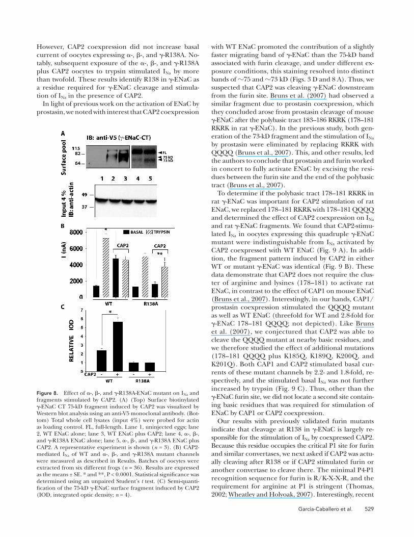
However, CAP2 coexpression did not increase basal
current of oocytes expressing � -, � -, and � -R138A. No-
tably, subsequent exposure of the � -, � -, and � -R138A
plus CAP2 oocytes to trypsin stimulated I Na by more
than twofold. These results identify R138 in � -ENaC as
a residue required for � -ENaC cleavage and stimula-
tion of I Na in the presence of CAP2.
In light of previous work on the activation of ENaC by
prostasin, we noted with interest that CAP2 coexpression
Figure 8. Effect of � -, � -, and � -R138A-ENaC mutant on I Na and fragments stimulated by CAP2. (A) (Top) Surface biotinylated � -ENaC CT 75-kD fragment induced by CAP2 was visualized by Western blot analysis using an anti-V5 monoclonal antibody. (Bot-tom) Total whole cell lysates (input 4%) were probed for actin as loading control. FL, full-length. Lane 1, uninjected eggs; lane 2, WT ENaC alone; lane 3, WT ENaC plus CAP2; lane 4, � -, � -, and � -R138A ENaC alone; lane 5, � -, � -, and � -R138A ENaC plus CAP2. A representative experiment is shown ( n = 3). (B) CAP2-mediated I Na of WT and � -, � -, and � -R138A mutant channels were measured as described in Results. Batches of oocytes were extracted from six different frogs ( n = 36). Results are expressed as the means ± SE. * and **, P < 0.0001. Statistical signifi cance was determined using an unpaired Student ’ s t test. (C) Semi-quanti-fi cation of the 75-kD � -ENaC surface fragment induced by CAP2 (IOD, integrated optic density; n = 4).

530 Identifi cation of Cleavage Sites on ENaC by CAP2
vations indicate that CAP1 and CAP3 prefer an argi-
nine, but tolerate a lysine in the P1 position. We speculated
that if CAP2 shared this relaxed stringency at P1, CAP2
coexpression would stimulate the furin-resistant ENaC
mutant � -, � -, and � -R138K. We also tested the effect of
CAP2 coexpression on � -, � -, and � -R138Q ENaC, in
which � residue 138 should not be a substrate for either
furin or CAP2. We found that basal I Na of ENaC con-
taining � -R138A or � -R138Q was not increased by coex-
pressed CAP2, but was robustly stimulated by exogenous
trypsin ( Fig.10 A ). In contrast, when ENaC contained
� -R138K, CAP2 coexpression signifi cantly increased basal
I Na . Importantly, trypsin did not further stimulate I Na of
oocytes expressing � -, � -, and � -R138K plus CAP2, in-
dicating that ENaC in these oocytes had already been
proteolytically activated . Thus, CAP2 stimulated ENaC
when the critical 138 (P1) residue in the � -subunit was
a lysine. These results strongly suggest that cleavage in-
duced by CAP2 at R138 in � -ENaC is responsible for
CAP2 stimulation of I Na .
In agreement with the functional results, we found that
CAP2 coexpression generated a 75-kD V5-CT � -ENaC
fragment when coexpressed with � -, � -, and � -R138 or
� -, � -, and � -R138K, but not when coexpressed with � -, � -,
and � -R138A or � -, � -, and � -R138Q ( Fig. 10, B and C ).
This is again consistent with cleavage at position 138 in
� -ENaC – mediating CAP2 stimulation of I Na . The appear-
ance or disappearance of the 75-kD fragment consis-
tently correlates with ENaC activation by CAP2. We
routinely detected a faster migrating fragment ( � 73
kD) with variable intensity, more evident in the R138A/
Q mutants ( Figs. 8 – 10 ). The fact that this faster migrat-
ing form appears in these mutants strongly suggests that
this cleavage event by itself is not linked to CAP2-stimu-
lated I Na .
To further test the importance of � -ENaC residue 138
in CAP2 stimulation of ENaC, we coexpressed either
WT � or � -R138A or � -R138K with � -R205A/R231A and
WT � -ENaC. I Na was stimulated by CAP2 coexpression
only when an arginine (WT) or a lysine was present at
position 138 in � -ENaC ( Fig.11 A ). We further isolated
the action of CAP2 by coexpressing CAP2 with WT � -
and � -ENaC combined with � -178-181 QQQQ mutants
that contained either alanine or lysine at � -ENaC resi-
due 138. These mutant � -subunits are not expected to be
cleaved by convertases acting at the � -ENaC furin site,
and they lack the polybasic tract implicated in CAP1 stim-
ulation of mouse ENaC. CAP2 did not stimulate basal
I Na of ENaC with the double mutant � -subunit contain-
ing alanine at residue 138; however, I Na was markedly
stimulated by subsequent trypsin ( Fig. 11 B ). In con-
trast, CAP2 coexpression clearly enhanced basal I Na of
ENaC that contained the mutant � -ENaC with lysine at
residue 138; moreover, subsequent trypsin exposure did
not signifi cantly add to the elevated basal I Na ( Fig. 11 B ).
These results indicate that ability of CAP2 coexpression
crystallization and other work reveal a similar, but not
identical, optimal sequence for matriptase binding and
cleavage, R-X-X-K/R ( Friedrich et al., 2002 ; Netzel-
Arnett et al., 2003 ; Desilets et al., 2006 ). Furthermore,
a similar P4-P1 preferred sequence for CAP1/prostasin,
R/K-H/R/K-X-R/K, was identifi ed by screening peptide
libraries ( Shipway et al., 2004 ). These published obser-
Figure 9. Effect of � -, � -, and � -178-181 QQQQ or � -, � -, and � -178-181 QQQQ , K185Q, K189Q, K200Q, and K201Q ENaC mutants on I Na stimulated by CAP2 and CAP1. (A) CAP2 in-duced I Na of WT and 178 – 181 QQQQ ENaC mutant channels. Amiloride-sensitive currents were measured as described in Fig. 1 . Batches of oocytes were extracted from three different frogs ( n = 12). Results are expressed as the means ± SE. * and **, P < 0.0001. Statistical signifi cance was determined using an unpaired Student ’ s t test. (B) Western blot of surface pool of WT and 178 – 181 QQQQ � -ENaC mutant channels. Lane 1, un-injected eggs; lane 2, WT ENaC alone; lane 3, WT ENaC plus CAP2; lane 4, � -, � -, and � -178-181 QQQQ ENaC alone; lane 5, � -, � -, and � -178-181 QQQQ ENaC plus CAP2. A representative experiment is shown ( n = 3). (C) CAP2 and CAP1 induced I Na of WT and 178 – 181 QQQQ, K185Q, K189Q, K200Q, and K201Q ENaC mutant channels. Amiloride-sensitive currents were mea-sured as described in Fig. 1 . Batches of oocytes were extracted from three different frogs ( n = 12). Results are expressed as the means ± SE. * and **, difference P < 0.05. Statistical signifi cance was determined using ANOVA.

Garc í a-Caballero et al. 531
teolytically active, we thought it was important to exam-
ine how CAP2 regulates ENaC coexpressed in Xenopus oocytes. In particular, we sought to identify the origin of
CAP2-induced ENaC fragments at the cell surface and
to determine the functional consequence of each iden-
tifi ed cleavage event.
We found that CAP2 induced cleavage of all three
ENaC subunits, and that resulting fragments appear at
the cell surface. Fragments were used to map cleavage
of ENaC subunits: (1) in � -ENaC, novel post-M1 frag-
ments required the basic conserved residues K149, R164,
K169, and R177; (2) in all three subunits in the pre-M2
region at � -K561, � -R503, and � -R515, a highly conserved
basic residue just before the important degenerin site;
and (3) at the conserved convertase consensus sites in
� -ENaC (R205/R231) and � -ENaC (R138). Somewhat
surprisingly, of these demonstrated sites of proteolysis,
only cleavage at R138 of � -ENaC could be linked to CAP2-
mediated activation of ENaC. Our results add to a grow-
ing body of work that identifi es � -ENaC as a critical target
of proteases that increase ENaC P O ( Adebamiro et al.,
2007 ; Bruns et al., 2007 ; Harris et al., 2007 ).
As our work unfolded, it became clear that some
CAP2-mediated cleavages of ENaC do not contribute to
proteolytic activation of ENaC. Using site-directed mu-
tagenesis, we modifi ed four basic residues found in the
post-M1 region of � -ENaC, which prevented the appear-
ance of the short N-terminal fragments at the cell surface
but had no effect on the CAP2 regulation of ENaC cur-
rent. Due to lack of a role of this cleavage in CAP2 stim-
ulation of ENaC, we did not identify the precise residues
to stimulate ENaC I Na is exquisitely dependent on the
residue at position 138 in � -ENaC.
D I S C U S S I O N
CAP2/TMPRSS4 is a member of a family of S1 serine
proteases that are anchored directly to cell surfaces, with
their catalytic domains located extracellularly ( Netzel-
Arnett et al., 2003 ). The activity of such proteases on
the cell surface is controlled by an activation step and
by interaction with native inhibitors, potentially permit-
ting regulation of events on the cell surface. Because
CAP2, as well as related serine proteases CAP1/prosta-
sin and CAP3/matriptase, regulate ENaC ( Andreasen
et al., 2006 ), it seems reasonable to assume that pro-
teolytic activation of ENaC is among those events. The
notion of near-silent ENaC being delivered to the cell
surface and activated in proportion to a balance be-
tween surface proteases and protease inhibitors fi ts well
with emerging views of proteolytic regulation ENaC
( Rossier, 2004 ; Tarran et al., 2006 ). However, we do not
yet possess a detailed understanding of how CAPs regu-
late ENaC. For example, CAP1/prostasin stimulates ENaC
and appears from knockdown studies to play the domi-
nant role in proteolytic regulation of ENaC ( Tong et al.,
2004 ). Yet, Andreasen et al. (2006) reported that prosta-
sin regulation of ENaC does not require prostasin itself
to be catalytically active, suggesting that its mode of reg-
ulation may involve other surface proteases. Because CAP2
is highly expressed in some epithelia, and regulates
ENaC through a mechanism that requires it to be pro-
Figure 10. Effect of replacement of � -ENaC R138 (P1) by alanine, lysine, or glutamine on I Na and fragments stimu-lated by CAP2. (A) CAP2 effect on I Na of WT and � -, � -, and � -R138A or � -, � -, and � -R138K or � -, � -, and � -R138Q ENaC mutant channels. Amiloride-sensitive currents were measured as described in Fig. 1 . Batches of oocytes were extracted from three different frogs ( n = 18). Results are expressed as the means ± SE. * and **, P < 0.0001. (B) West-ern blots of total whole cell extracts. CAP2-induced frag-ment pattern of WT and � -ENaC mutant channels (top) and actin as loading control (bottom). Lane 1, uninjected eggs; lane 2, WT ENaC plus CAP2; lane 3, � -, � -, and � -R138A ENaC plus CAP2; lane 4, � -, � -, and � -R138K ENaC plus CAP2; lane 5, � -, � -, and � -R138Q ENaC plus CAP2. A representative experiment is shown ( n = 3). (C) Western blots of surface biotinylated (top), total � -ENaC pools (bot-tom), and actin. Lane 1, uninjected eggs; lane 2, WT ENaC alone; lane 3, WT ENaC plus CAP2; lane 4, � -, � -, and � -R138K ENaC alone; lane 5, � -, � -, and � -R138K ENaC plus CAP2. A representative experiment is shown ( n = 3).

532 Identifi cation of Cleavage Sites on ENaC by CAP2
at the pre-M2 region of � -ENaC has also been seen by
others ( Planes et al., 2002 ; Ergonul et al., 2006 ; Myerburg
et al., 2006 ), but its role in proteolytic regulation of ENaC
has not been studied. Our data expand upon these pre-
vious observations. We sporadically detected a similar
sized N-terminal � -ENaC fragment at the surface and
total cellular pools when CAP2 was coexpressed with
ENaC ( Figs. 3 and 7 ). We speculate that the 82-kD HA-NT
band is missing in some experiments due to a variable
extent of additional cleavage. However, we consistently
detected a CAP2-induced � 17-kD C-terminal fragment
in � -ENaC that fully complemented the � 82-kD N-ter-
minal band. We unequivocally found this fragment at
the cell surface. Interestingly, we detected similar sized
C-terminal – tagged fragments of � - ( � 19 kD) and
� -ENaC ( � 15 kD) at the surface of oocytes coexpress-
ing catalytically active CAP2 and ENaC. To the best of
our knowledge, this is the fi rst documented cleavage of
� -ENaC by a coexpressed protease. Specifi c basic resi-
dues in each ENaC subunit were required for CAP2 to
stimulate the appearance of these pre-M2 fragments.
These basic residues were conserved across all ENaC
subunits and species, aligned 15 amino acids upstream
of the degenerin site in each subunit, and embedded in
a region, the palm domain ( � sheet 12), homologous to
that described for the acid-sensing ion channel 1 struc-
ture ( Jasti et al., 2007 ). Due to the critical role of the de-
generin site in ENaC gating, we were surprised when
triple mutant channels engineered with alanines in place
of the conserved basic residues were normally stimulated
by CAP2 coexpression. Again, we considered the possi-
bility that the lower maximum current of CAP2 coex-
pressing oocytes, compared with ENaC-alone controls,
could result from cleavage/degradation of active (surface)
ENaC by CAP2. However, the triple mutant channels that
were not cleaved in the pre-M2 region exhibit lower
rather than greater stimulated current. Unexpectedly,
we noticed that the mutant � -K561A, � -R503A, and � -
R515A not only completely abolished the small V5-CT
pre-M2 fragments, but also notably reduced the furin
� - and � -ENaC fragments ( Fig. 5 ).
CAP2 coexpression with WT ENaC consistently in-
creased staining intensity of bands that Hughey et al.
(2003) attributed to cleavage at the furin sites. Logi-
cally, increased cleavage at furin sites should increase
basal current and decrease trypsin responsiveness, which
adequately describes the effects of CAP2 on ENaC-medi-
ated currents. Therefore, we designed studies to deter-
mine if cleavage at recognized furin sites was indeed
required for CAP2 stimulation of ENaC. Using � -ENaC
in which the furin sites had been mutated through sub-
stitution of alanines for the critical P1 arginines, we
found that CAP2-enhanced cleavage at the � -furin sites
was effectively suppressed, without affecting CAP2 stim-
ulation of I Na . Interestingly, the 17 – 19-kD post-M1 and
the � 82-kD pre-M2 � -ENaC fragments produced by
involved. Although the appearance of these fragments
at the oocyte surface was dependent on catalytically ac-
tive CAP2, any role for cleavage in the early extracellu-
lar loop in regulating ENaC remains obscure. We did
note a tendency for trypsin-stimulated I Na of coexpress-
ing ENaC and CAP2 to be smaller than typsin-stimulated
WT control currents ( Figs. 1, 4, and 6 – 8 ). Therefore, we
considered the possibility that these small N-terminal
fragments might arise from CAP2-mediated degrada-
tion of active channels at the cell surface. However, our
functional study of the post-M1 mutants offers no hint
that preventing these � -ENaC fragments counters a de-
crease of I Na that is part of mixed stimulatory and inhib-
itory effects of CAP2. Nonetheless, if these fragments
are shown in the future to be directly generated by CAP2,
it may help in mapping the physical association between
ENaC and serine surface proteases.
We next turned our attention to CAP2-mediated cleav-
age of the pre-M2 region of ENaC. An � 82-kD N-termi-
nally tagged fragment predicted to arise from cleavage
Figure 11. CAP2 stimulated � -R205A/R231A, � -, and � -138K or � -, � -, and � -138K plus 178 – 181 QQQQ but not � -R205A/R231A, � -, and � -138A or � -, � -, and � -138A plus 178 – 181 QQQQ mutant channels. (A) CAP2 induced I Na of WT and � -R205A/R231A, � -, � -, or � -R205A/R231A, � -, and � -R138A or � -R205A/R231A, � -, and � -R138K ENaC mutant channels ( n = 12). (B) CAP2 induced I Na of � -, � -, and � -R138A, R178Q, K179Q, R180Q, and K181Q or � -, � -, and � -R138K, R178Q, K179Q, R180Q, and K181Q ENaC mutant channels. Amiloride-sensitive currents were measured as described in Fig. 1 . Batches of oocytes were extracted from three different frogs ( n = 12). Results are expressed as the means ± SE. * and **, P < 0.0001.

Garc í a-Caballero et al. 533
completely eliminated the fragment indicative of some
cleavage downstream from the furin site. Although our
study focuses on CAP2 stimulation of rat ENaC, these
results differ signifi cantly from the fi nding that conversion
of the RKRK tract to QQQQ eliminated both a smaller
fragment and activation of mouse ENaC by CAP1/pros-
tasin ( Bruns et al., 2007 ). We cannot account for this
discrepancy. However, we found that CAP1/prostasin
continued to stimulate the RKRK to QQQQ mutant of
rat � -ENaC. Thus, in our hands, CAP1/prostasin and
CAP2 behave similarly, i.e., they activate ENaC inde-
pendently of the polybasic tract 178 – 181 RKRK in
� -ENaC ( Fig. 9 ).
Faced with these observations, the apparent increase
in cleavage at the furin site of � -ENaC assumed greater
signifi cance. We noted that furin or other convertases
of the constitutive secretory pathway have a stringent
requirement for arginine in the P1 position of their cleav-
age site ( Thomas, 2002 ; Wheatley and Holyoak, 2007 ).
In the context of the P4-P1 residues present in the
� -furin site (135 – 138 RKRR), converting 138R to 138K
effectively eliminated the furin fragment of � -ENaC in
eggs expressing ENaC alone ( Fig. 10 C ). In contrast to
its effect on the furin fragment, conversion of this key
residue to lysine had no effect on generation of a 75-kD
fragment and activation of ENaC by CAP2. CAP1 and
CAP3/matriptase are reported to accept a lysine in the
P1 position ( Friedrich et al., 2002 ; Netzel-Arnett et al.,
2003 ; Shipway et al., 2004 ; Desilets et al., 2006 ). Our
data indicate that CAP2 may share this fl exibility for ba-
sic residues at P1, and importantly, strongly suggest that
CAP2 cleaves � -ENaC directly.
Our results raise an important question: Is a single
cleavage event in the � subunit at R138 suffi cient for
CAP2 to activate ENaC? Whereas cleavage at � -R138 is
clearly essential for CAP2 to stimulate ENaC, our data
do not rule out a requirement for additional cleavage
downstream of the furin site. Although en masse substi-
tution of glutamines for the 178 – 181 RKRK polybasic
sequence downstream of the furin site and other nearby
basic residues did not prevent activation of rat ENaC by
CAP2, the maneuver also did not eliminate the occur-
rence of the � 73-kD fragment. It is a formal possibility
that a different protease with conceivably a nonbasic
amino acid requirement, endogenously expressed in oo-
cytes, cleaves in this region of � -ENaC and that such cleav-
age contributes to CAP2 stimulation of ENaC.
The results of our study add to a body of evidence
identifying � -ENaC as the ENaC subunit that is targeted
by proteases that increase ENaC P O by cleavage after spe-
cifi c residues. Thus, it appears that CAP2, human neu-
trophil, and pancreatic elastases are now demonstrated
to cleave either at the furin site or in a region � 55 resi-
dues downstream of the furin site of � -ENaC. In addi-
tion, thrombin activates ENaC with a thrombin cleavage
sequence (LVPRG) engineered into � -hENaC at position
CAP2 coexpression with WT � -ENaC were also not gen-
erated when the � -ENaC double furin mutant was used
( Fig. 7 A ). Thus, the numerous cleavages of � -ENaC ob-
served with CAP2 coexpression may occur in a sequence
that begins with cleavage at the furin sites, but also ap-
pear to be unrelated to the ability of CAP2 to stimulate
ENaC basal current by two- to threefold.
In contrast to the lack of consequence of � -ENaC
furin site mutations, the R138A or R138Q mutations of
the P1 residue in the � -ENaC furin site completely abro-
gated CAP2 stimulation of ENaC, and dramatically sup-
pressed the appearance of fragments associated with
cleavage at the � -ENaC furin site. These mutants clearly
produced channels that traffi cked to the surface and
could be stimulated by trypsin, but they were not cleaved
at the furin site or activated when coexpressed with
CAP2. Cleavage of � -ENaC downstream of the docu-
mented furin site (rat residues 135 – 138) has been linked
to activation of ENaC by hNE (rat residues 180 – 200)
and by prostasin (rat residues 178 – 181) ( Adebamiro et al.,
2007 ; Bruns et al., 2007 ). These observations focus at-
tention on this region of � -ENaC as critical to stimula-
tion of ENaC by proteases.
Accordingly, we performed extensive mutagenesis of
the region beginning with the � -furin site and extend-
ing downstream � 70 residues to determine if CAP2 was
directly cleaving ENaC in this region. Published work
suggests that cleavage at V182 and V193 in human � -
ENaC accounts for activation of ENaC by neutrophil
elastase, whereas porcine pancreatic elastase cleaves at
A190 ( Adebamiro et al., 2007 ). Consistent with this ob-
servation, neutrophil elastase generates a slightly faster
migrating fragment than the furin fragment on SDS gels
( Harris et al., 2007 ). Moreover, the polybasic tract 183 –
186 RKRK in mouse ENaC was required for CAP1/pros-
tasin to both activate mouse ENaC and to generate a faster
migrating fragment consistent with cleavage at that site
( Bruns et al., 2007 ). CAP2 increased the intensity of
bands at the size of the furin fragment and at a slightly
smaller molecular mass, suggesting that CAP2 cleaved
in this elastase and CAP1-sensitive region of � -ENaC. Ini-
tially, we were drawn to the polybasic tract 183 – 186 RKRK
because of the CAP1/prostasin results ( Bruns et al., 2007 ).
Surprisingly, mutation of the homologous site (178 – 181
RKRK) in rat � -ENaC to QQQQ did not abolish CAP2
regulation of ENaC, nor did it eliminate a specifi c band
that migrated ahead of the furin fragment. Based on the
persistence of fragments slightly smaller than the furin
fragment with the RKRK tract converted to QQQQ, we
explored the contribution of fl anking basic amino ac-
ids. Despite converting multiple basic residues (R153Q,
K156Q, K168Q, K170Q, R172Q, R178Q, K179Q, R180Q,
K181Q, K185Q, K189Q, K200Q, and K201Q) to gluta-
mines in this region and generating different mutant
combinations ( Fig. 9 C ), we never generated a mutant
� -subunit that was not activated by CAP2, and we never

534 Identifi cation of Cleavage Sites on ENaC by CAP2
tase, a matrix-degrading transmembrane serine proteinase.
J. Biol. Chem. 277 : 2160 – 2168 .
Guipponi , M. , G. Vuagniaux , M. Wattenhofer , K. Shibuya , M.
Vazquez , L. Dougherty , N. Scamuffa , E. Guida , M. Okui , C. Rossier ,
et al . 2002 . The transmembrane serine protease (TMPRSS3) mu-
tated in deafness DFNB8/10 activates the epithelial sodium chan-
nel (ENaC) in vitro. Hum. Mol. Genet. 11 : 2829 – 2836 .
Harris , M. , D. Firsov , G. Vuagniaux , M.J. Stutts , and B.C. Rossier .
2007 . A novel neutrophil elastase inhibitor prevents elastase ac-
tivation and surface cleavage of the epithelial sodium channel
expressed in Xenopus laevis oocytes. J. Biol. Chem. 282 : 58 – 64 .
Hughey , R.P. , G.M. Mueller , J.B. Bruns , C.L. Kinlough , P.A. Poland , K.L.
Harkleroad , M.D. Carattino , and T.R. Kleyman . 2003 . Maturation
of the epithelial Na+ channel involves proteolytic processing of the
alpha- and gamma-subunits. J. Biol. Chem. 278 : 37073 – 37082 .
Hughey , R.P. , J.B. Bruns , C.L. Kinlough , K.L. Harkleroad , Q. Tong ,
M.D. Carattino , J.P. Johnson , J.D. Stockand , and T.R. Kleyman .
2004 . Epithelial sodium channels are activated by furin-dependent
proteolysis. J. Biol. Chem. 279 : 18111 – 18114 .
Jasti , J. , H. Furukawa , E.B. Gonzales , and E. Gouaux . 2007 . Structure
of acid-sensing ion channel 1 at 1.9 A resolution and low pH.
Nature . 449 : 316 – 323 .
Kellenberger , S. , I. Gautschi , and L. Schild . 2002 . An external site
controls closing of the epithelial Na+ channel ENaC. J. Physiol. 543 : 413 – 424 .
Liddle, G.W. , T. Bledsoe, and W.S. Coppage Jr. 1963 . A familial renal
disorder simulating primary aldosteronism but with negligible al-
dosterone secretion. Trans. Assoc. Am. Physicians . 76 : 199 – 213 .
Myerburg , M.M. , M.B. Butterworth , E.E. McKenna , K.W. Peters , R.A.
Frizzell , T.R. Kleyman , and J.M. Pilewski . 2006 . Airway surface liq-
uid volume regulates ENaC by altering the serine protease-prote-
ase inhibitor balance: a mechanism for sodium hyperabsorption
in cystic fi brosis. J. Biol. Chem. 281 : 27942 – 27949 .
Netzel-Arnett , S. , J.D. Hooper , R. Szabo , E.L. Madison , J.P. Quigley ,
T.H. Bugge , and T.M. Antalis . 2003 . Membrane anchored serine pro-
teases: a rapidly expanding group of cell surface proteolytic enzymes
with potential roles in cancer. Cancer Metastasis Rev. 22 : 237 – 258 .
Planes , C. , and G.H. Caughey . 2007 . Regulation of the epithelial
Na+ channel by peptidases. Curr. Top. Dev. Biol. 78 : 23 – 46 .
Planes , C. , M. Blot-Chabaud , M.A. Matthay , S. Couette , T. Uchida ,
and C. Clerici . 2002 . Hypoxia and beta 2-agonists regulate cell
surface expression of the epithelial sodium channel in native
alveolar epithelial cells. J. Biol. Chem. 277 : 47318 – 47324 .
Rossier , B.C. 2004 . The epithelial sodium channel: activation by
membrane-bound serine proteases. Proc. Am. Thorac. Soc. 1 : 4 – 9 .
Schild , L. 2004 . The epithelial sodium channel: from molecule to
disease. Rev. Physiol. Biochem. Pharmacol. 151 : 93 – 107 .
Shipway , A. , H. Danahay , J.A. Williams , D.C. Tully , B.J. Backes , and J.L.
Harris . 2004 . Biochemical characterization of prostasin, a channel
activating protease. Biochem. Biophys. Res. Commun. 324 : 953 – 963 .
Stockand , J.D. , A. Staruschenko , O. Pochynyuk , R.E. Booth , and
D.U. Silverthorn . 2008 . Insight toward epithelial Na(+) channel
mechanism revealed by the acid-sensing ion channel 1 structure.
IUBMB Life . 60:620 – 628.
Tarran , R. , L. Trout , S.H. Donaldson , and R.C. Boucher . 2006 .
Soluble mediators, not cilia, determine airway surface liquid vol-
ume in normal and cystic fi brosis superfi cial airway epithelia.
J. Gen. Physiol. 127 : 591 – 604 .
Thomas , G. 2002 . Furin at the cutting edge: from protein traffi c to
embryogenesis and disease. Nat. Rev. Mol. Cell Biol. 3 : 753 – 766 .
Tong , Z. , B. Illek , V.J. Bhagwandin , G.M. Verghese , and G.H.
Caughey . 2004 . Prostasin, a membrane-anchored serine peptidase,
regulates sodium currents in JME/CF15 cells, a cystic fi brosis
airway epithelial cell line. Am. J. Physiol. Lung Cell. Mol. Physiol. 287 : L928 – L935 .
186 ( Adebamiro et al., 2007 ). All of these proteases can
be inferred to act at the extracellular surface of the cell;
elastases and thrombin because they act acutely after
addition to the cell surface, and CAP2 because CAPs are
thought to be activated at the cell surface. This suggests
that in the ENaC heteromer, the furin site of � -ENaC
and a region downstream of the furin site at � 55 resi-
dues from it are exposed to proteases at the cell surface.
Future structural studies to reveal the molecular basis of
this access, and to provide insight as to how cleavage in
this region increases ENaC P O , await more defi nitive rec-
onciliation of ENaC primary sequence with the proposed
ASIC1 crystal structure ( Stockand et al., 2008 ).
We thank Dr. Ray Caldwell, Dr. Scott Donaldson, and Dr. Martina Gentzsch for critical reading of this manuscript.
This work is supported by grant P01-HL034322-II.
Lawrence G. Palmer served as editor.
Submitted: 18 April 2008 Accepted: 29 September 2008
R E F E R E N C E S Adebamiro , A. , Y. Cheng , U.S. Rao , H. Danahay , and R.J. Bridges .
2007 . A segment of � ENaC mediates elastase activation of Na +
transport. J. Gen. Physiol. 130 : 611 – 629 .
Andreasen , D. , G. Vuagniaux , N. Fowler-Jaeger , E. Hummler , and
B.C. Rossier . 2006 . Activation of epithelial sodium channels
by mouse channel activating proteases (mCAP) expressed in
Xenopus oocytes requires catalytic activity of mCAP3 and mCAP2
but not mCAP1. J. Am. Soc. Nephrol. 17 : 968 – 976 .
Boucher , R.C. 2004 . New concepts of the pathogenesis of cystic fi -
brosis lung disease. Eur. Respir. J. 23 : 146 – 158 .
Bruns , J.B. , M.D. Carattino , S. Sheng , A.B. Maarouf , O.A. Weisz , J.M.
Pilewski , R.P. Hughey , and T.R. Kleyman . 2007 . Epithelial Na+
channels are fully activated by furin- and prostasin-dependent
release of an inhibitory peptide from the gamma-subunit.
J. Biol. Chem. 282 : 6153 – 6160 .
Caldwell , R.A. , R.C. Boucher , and M.J. Stutts . 2004 . Serine protease
activation of near-silent epithelial Na+ channels. Am. J. Physiol. Cell Physiol. 286 : C190 – C194 .
Caldwell , R.A. , R.C. Boucher , and M.J. Stutts . 2005 . Neutrophil elas-
tase activates near-silent epithelial Na+ channels and increases
airway epithelial Na+ transport. Am. J. Physiol. Lung Cell. Mol. Physiol. 288 : L813 – L819 .
Canessa , C.M. , L. Schild , G. Buell , B. Thorens , I. Gautschi , J.D.
Horisberger , and B.C. Rossier . 1994 . Amiloride-sensitive epithe-
lial Na+ channel is made of three homologous subunits. Nature . 367 : 463 – 467 .
Desilets , A. , J.M. Longpre , M.E. Beaulieu , and R. Leduc . 2006 .
Inhibition of human matriptase by eglin c variants. FEBS Lett. 580 : 2227 – 2232 .
Donaldson , S.H. , A. Hirsh , D.C. Li , G. Holloway , J. Chao , R.C.
Boucher , and S.E. Gabriel . 2002 . Regulation of the epithelial so-
dium channel by serine proteases in human airways. J. Biol. Chem. 277 : 8338 – 8345 .
Ergonul , Z. , G. Frindt , and L.G. Palmer . 2006 . Regulation of matu-
ration and processing of ENaC subunits in the rat kidney. Am. J. Physiol. Renal Physiol. 291 : F683 – F693 .
Friedrich , R. , P. Fuentes-Prior , E. Ong , G. Coombs , M. Hunter , R.
Oehler , D. Pierson , R. Gonzalez , R. Huber , W. Bode , and E.L.
Madison . 2002 . Catalytic domain structures of MT-SP1/matrip-

Garc í a-Caballero et al. 535
by the serine protease mCAP1 expressed in a mouse cortical col-
lecting duct cell line. J. Am. Soc. Nephrol. 11 : 828 – 834 .
Vuagniaux , G. , V. Vallet , N.F. Jaeger , E. Hummler , and B.C. Rossier .
2002 . Synergistic activation of ENaC by three membrane-bound
channel-activating serine proteases (mCAP1, mCAP2, and mCAP3)
and serum- and glucocorticoid-regulated kinase (Sgk1) in Xenopus
oocytes. J. Gen. Physiol. 120 : 191 – 201 .
Wheatley , J.L. , and T. Holyoak . 2007 . Differential P1 arginine
and lysine recognition in the prototypical proprotein convertase
Kex2. Proc. Natl. Acad. Sci. USA . 104 : 6626 – 6631 .
Vallet , V. , A. Chraibi , H.P. Gaeggeler , J.D. Horisberger , and B.C.
Rossier . 1997 . An epithelial serine protease activates the amilo-
ride-sensitive sodium channel. Nature . 389 : 607 – 610 .
Vallet , V. , C. Pfi ster , J. Loffi ng , and B.C. Rossier . 2002 . Cell-surface
expression of the channel activating protease xCAP-1 is required
for activation of ENaC in the Xenopus oocyte. J. Am. Soc. Nephrol. 13 : 588 – 594 .
Vuagniaux , G. , V. Vallet , N.F. Jaeger , C. Pfi ster , M. Bens , N. Farman ,
N. Courtois-Coutry , A. Vandewalle , B.C. Rossier , and E. Hummler .
2000 . Activation of the amiloride-sensitive epithelial sodium channel
Copyright © 2022 FDOKUMEN