The neutrophil-activating protein of Helicobacter pylori promotes Th1 immune responses
10
of June 13, 2013. This information is current as Promote Neutrophil Adhesion In Vivo Helicobacter pylori Crosses Endothelia to The Neutrophil-Activating Protein of Marina de Bernard Cassatella, Cesare Montecucco, Francesco Tedesco and Durigutto, Anna Cabrelle, Nicola Tamassia, Marco A. Alessandra Polenghi, Fleur Bossi, Fabio Fischetti, Paolo http://www.jimmunol.org/content/178/3/1312 2007; 178:1312-1320; ; J Immunol References http://www.jimmunol.org/content/178/3/1312.full#ref-list-1 , 28 of which you can access for free at: cites 58 articles This article Subscriptions http://jimmunol.org/subscriptions is online at: The Journal of Immunology Information about subscribing to Permissions http://www.aai.org/ji/copyright.html Submit copyright permission requests at: Email Alerts http://jimmunol.org/cgi/alerts/etoc Receive free email-alerts when new articles cite this article. Sign up at: Print ISSN: 0022-1767 Online ISSN: 1550-6606. Immunologists All rights reserved. Copyright © 2007 by The American Association of 9650 Rockville Pike, Bethesda, MD 20814-3994. The American Association of Immunologists, Inc., is published twice each month by The Journal of Immunology by guest on June 13, 2013 http://www.jimmunol.org/ Downloaded from
Transcript of The neutrophil-activating protein of Helicobacter pylori promotes Th1 immune responses

of June 13, 2013.This information is current as
Promote Neutrophil Adhesion In VivoHelicobacter pylori Crosses Endothelia to The Neutrophil-Activating Protein of
Marina de BernardCassatella, Cesare Montecucco, Francesco Tedesco andDurigutto, Anna Cabrelle, Nicola Tamassia, Marco A. Alessandra Polenghi, Fleur Bossi, Fabio Fischetti, Paolo
http://www.jimmunol.org/content/178/3/13122007; 178:1312-1320; ;J Immunol
Referenceshttp://www.jimmunol.org/content/178/3/1312.full#ref-list-1
, 28 of which you can access for free at: cites 58 articlesThis article
Subscriptionshttp://jimmunol.org/subscriptions
is online at: The Journal of ImmunologyInformation about subscribing to
Permissionshttp://www.aai.org/ji/copyright.htmlSubmit copyright permission requests at:
Email Alertshttp://jimmunol.org/cgi/alerts/etocReceive free email-alerts when new articles cite this article. Sign up at:
Print ISSN: 0022-1767 Online ISSN: 1550-6606. Immunologists All rights reserved.Copyright © 2007 by The American Association of9650 Rockville Pike, Bethesda, MD 20814-3994.The American Association of Immunologists, Inc.,
is published twice each month byThe Journal of Immunology
by guest on June 13, 2013http://w
ww
.jimm
unol.org/D
ownloaded from

The Neutrophil-Activating Protein of Helicobacter pyloriCrosses Endothelia to Promote Neutrophil Adhesion In Vivo1
Alessandra Polenghi,2*† Fleur Bossi,2‡ Fabio Fischetti,§ Paolo Durigutto,‡ Anna Cabrelle,†
Nicola Tamassia,¶ Marco A. Cassatella,¶ Cesare Montecucco,� Francesco Tedesco,‡
and Marina de Bernard3*†
Helicobacter pylori induces an acute inflammatory response followed by a chronic infection of the human gastric mucosa char-acterized by infiltration of neutrophils/polymorphonuclear cells (PMNs) and mononuclear cells. The H. pylori neutrophil-activat-ing protein (HP-NAP) activates PMNs, monocytes, and mast cells, and promotes PMN adherence to the endothelium in vitro. Byusing intravital microscopy analysis of rat mesenteric venules exposed to HP-NAP, we demonstrated, for the first time in vivo, thatHP-NAP efficiently crosses the endothelium and promotes a rapid PMN adhesion. This HP-NAP-induced adhesion depends on theacquisition of a high affinity state of �2 integrin on the plasma membrane of PMNs, and this conformational change requires afunctional p38 MAPK. We also show that HP-NAP stimulates human PMNs to synthesize and release a number of chemokines,including CXCL8, CCL3, and CCL4. Collectively, these data strongly support a central role for HP-NAP in the inflammationprocess in vivo: indeed, HP-NAP not only recruits leukocytes from the vascular lumen, but also stimulates them to producemessengers that may contribute to the maintenance of the flogosis associated with the H. pylori infection. The Journal of Im-munology, 2007, 178: 1312–1320.
H elicobacter pylori is a Gram-negative bacterium, whichcolonizes the human gastric mucosa and chronically in-fects more than half of the human population. This in-
fection has been associated with various gastroduodenal diseasesand gastric cancers (1–4). H. pylori colonization is typically fol-lowed by infiltration of the gastric mucosa by polymorphonuclearleukocytes, macrophages, and lymphocytes (5, 6). A strong corre-lation exists between gastric infiltration by neutrophils (polymor-phonuclear cell; PMNs),4 mucosal damage, and development ofduodenal ulcer disease in H. pylori infections (7, 8). However, themechanism underlying the sustained recruitment of PMNs to theH. pylori-infected tissue in vivo still remains to be fully under-
stood. It has been shown that the bacterium activates endothelialcells to express several adhesion molecules and PMNs recruitingCXC chemokines, such as CXCL8 (9), known to trigger integrinactivation in PMNs (10). Gastric epithelial cells are also likely tocontribute to the generation of a chemoattractant milieu (11). Al-though it seems that one or more protein(s) encoded by the 30-kbDNA region present in the most virulent bacteria (the cag patho-genicity island) has a central role in promoting leukocyte recruit-ment, by inducing secretion of chemokines (11, 12), other reportssuggest that mediators encoded by cag pathogenicity island mayplay a role in strain potency, but are not the only bacterial productsinvolved in endothelial cell activation and in PMN extravasation(9, 13).
A major proinflammatory factor produced by H. pylori is H.pylori neutrophil-activating protein (HP-NAP) (14). It is a do-decameric protein of 150 kDa with a structure similar to bacterio-ferritins, including a central cavity for iron accumulation (15, 16).It was originally defined as PMN-activating protein because itstimulates PMNs to produce reactive oxygen radicals (17). In ad-dition, HP-NAP crosses the epithelia to contact inflammatory cellsalready resident in the tissue (such as macrophages and mast cells)or recruited during the inflammation (18). HP-NAP stimulates thesynthesis of tissue factor and the secretion of type 2 plasminogenactivator inhibitor by monocytes/macrophages (14, 19). Finally,HP-NAP promotes Th1 responses (20) by inducing the productionof cytokines, such as IL-12 and IL-23, in cells of the innate im-mune system, such as monocytes and machrophages. HP-NAPstimulates mast cells and monocytes/macrophages to produceTNF-� and CXCL8 (19, 20). TNF-� is a pleiotropic cytokine ableto stimulate adhesivity of endothelial cells by up-regulating adhe-sion molecules, such as VCAM-1 and ICAM-1 (21). Moreover,TNF-� can induce activation of integrins on PMNs, directly or bystimulating the secretion of CXCL8 from the endothelium (10, 22).
According to these observations, during H. pylori infection it isvery likely that HP-NAP, probably together with other bacterial orhost-derived factors, may trigger in vivo the PMN accumulation
*Department of Biology, University of Padua, Padua, Italy; †Venetian Institute ofMolecular Medicine, Padua, Italy; ‡Department of Physiology and Pathology, Uni-versity of Trieste, Trieste, Italy; §Department of Medicine and Neurology, Universityof Trieste, Cattinara Hospital, Trieste, Italy; ¶Department of Pathology, Division ofGeneral Pathology, University of Verona, Verona, Italy; and �Department of Biomed-ical Sciences, University of Padua, Padua, Italy
Received for publication August 8, 2006. Accepted for publication November16, 2006.
The costs of publication of this article were defrayed in part by the payment of pagecharges. This article must therefore be hereby marked advertisement in accordancewith 18 U.S.C. Section 1734 solely to indicate this fact.1 This work was supported by Italian Association for Cancer Research Regional GrantProposal 2005 Veneto and Ministero dell’Istruzione, dell’Universita e della Ricerca(Grant 2004064334_006; to M.d.B.); Ministero dell’Istruzione, dell’Universita e dellaRicerca Grant 2005060371_001 and Fondi sulla Ricerca di Base (to M.A.C.); Min-istero dell’Istruzione, dell’Universita e della Ricerca Grant 2005060371_005 (toF.T.); and Ministero dell’Istruzione, dell’Universita e della Ricerca Grant2005060371_004 (to C.M.).2 A.P. and F.B. contributed equally to this work.3 Address correspondence and reprint requests to Dr. Marina de Bernard, VenetianInstitute of Molecular Medicine, Via Orus 2, 35121 Padua, Italy. E-mail address:[email protected] Abbreviations used in this paper: PMN, polymorphonuclear cell; AO, acridine or-ange; HP-NAP, H. pylori neutrophil-activating protein; PAF, platelet-activating fac-tor; PET, polycarbonate; RPA, RNase protection assay; RT, room temperature;VEGF, vascular endothelial growth factor.
Copyright © 2007 by The American Association of Immunologists, Inc. 0022-1767/07/$2.00
The Journal of Immunology
www.jimmunol.org
by guest on June 13, 2013http://w
ww
.jimm
unol.org/D
ownloaded from

within the tissue, evoking the adhesive properties of both PMNsand endothelium. The possibility that HP-NAP also directly stim-ulates PMNs to adhere derives from several studies (13, 17); in theformer study, however, protein and PMNs were assayed togetherwithout evaluating whether HP-NAP was actually capable ofcrossing the endothelium to come in contact with PMNs. The latterstudy demonstrated that HP-NAP activates PMNs even when theyare separated by an endothelium grown on a Transwell support, butdid not consider the underflow conditions that are of great rele-vance in vivo.
In this study, we demonstrate for the first time that HP-NAP isable to cross the endothelium and to stimulate PMNs to adhere invivo, in underflow conditions. This effect is directly mediated byHP-NAP, which induces a high affinity state of integrins on PMNs,by recruiting a signaling cascade that involves p38-MAPK. More-over, we addressed the question as to whether HP-NAP was ablenot only to rapidly recruit a first defense line, but also to sustain aprolonged inflammatory state by acting on the recruited cells; in-deed, very little is known about the direct effects of HP-NAP onthe production of neutrophil-derived proinflammatory mediators.We show in this study that HP-NAP up-regulates the mRNA ex-pression and the protein release of several proinflammatory che-mokines, including CXCL8, CCL3, and CCL4.
Materials and MethodsReagents
HP-NAP was cloned, expressed, and purified from Bacillus subtilis toavoid LPS contamination, as described previously (15). SB203580, tetra-methyl benzidine, FITC-conjugated BSA, streptavidin-alkaline phospha-tase, p-nitrophenyl phosphate, nonfat milk powder, acridine orange (AO),platelet-activating factor (PAF), HBSS, and biotin were purchased bySigma-Aldrich. Na-citrate solution at 3.8% was from Diagnostic Merk.Dextran and Ficoll-Hypaque were from Amersham Biosciences. mAb forthe active conformation of CD11/CD18 (anti-24Ag) was a gift from N.Hogg (Imperial Cancer Research Fund, London, U.K.). Goat FITC-F(ab�)2
anti-mouse IgG and an irrelevant mouse IgG1 control were from CaltagLaboratories. Streptavidin-Texas Red was from DakoCytomation, andstreptavidin was from Pierce.
All of the procedures were done under sterile conditions, and used re-agents were prepared in endotoxin-free water for clinical use.
Intravital microscopy analysis
Male Wistar Kyoto rats, weighing 250–270 g, were anesthetized with so-dium thiobarbital (80 mg/kg, i.p.) and prepared for direct microscopy anal-ysis following previously described procedures (23). After surgical setup,the ileal mesentery was exposed and the microvascular trafficking of flu-orescent labeled leukocytes (AO, i.v. infused at a concentration of 0.025
mg/kg per min, at a rate of 0.5 ml/h) was examined. Segments of three tofive unbranched postcapillary venules (25- to 40-�m diameter, 200-�mlength) were selected for the analysis. Direct digital recordings of the mi-croscopy images were acquired at different phases, and further analyzedoff-line. AO-labeled leukocytes were classified as rolling (whenever theybecame visible, because of moving more slowly than RBCs) or adherent (ifthey remained stationary, for at least 30 s, upon the endothelial surface).
In each experimental setting, after excluding vascular lesions or unstablehemodynamic pattern, baseline evaluation was acquired. Then, HP-NAP (1or 5 �M, diluted in 200 �l of sterile saline) was topically applied to themesentery for 10 min, and image sequences were further recorded inter-mittently up to 120 min. Control animals were treated with comparablevolumes of either sterile saline alone or boiled HP-NAP. All of the exper-imental procedures were performed in compliance with the guidelines ofEuropean (86/609/EEC) and Italian (D.L.116/92) laws and approved by theItalian Ministry of University and Research as well as by the Administra-tion of the University Animal House.
Purification of PMNs and endothelial cell culture
Human PMNs were prepared from healthy donors, as previously de-scribed (24).
HUVECs were isolated from three to five normal umbilical cords bycollagenase digestion and grown in tissue culture plates (Costar) coatedwith 2% endotoxin-free gelatin (25). HUVECs were used at their firstpassage. The cells were kept in culture in medium 199 supplemented with20% newborn calf serum (Invitrogen Life Technologies), 50 �g/ml hepa-rin, 100 U/ml penicillin, 100 �g/ml streptomycin (Sigma-Aldrich), and 50�g/ml endothelial growth supplement (26).
Evaluation of the HP-NAP crossing through the endothelium
HP-NAP was biotinylated, according to the manufactor’s instructions. Thebiological activity of the biotinylated material was checked by evaluatingcytosolic calcium increase in PMNs exposed to HP-NAP (27).
HUVECs (7 � 104) were seeded onto 2% gelatin-coated polycarbonate(PET) inserts of a 12-well Transwell system (12-mm diameter, 0.4-�mpores; Costar) and were used 5 days after plating.
The formation of intact monolayer on the insert was evaluated by addingFITC-BSA (1 mg/ml) to the upper chamber and measuring after 5 min theamount of labeled BSA passed into the lower chamber by a Fluostar mi-croplate reader (SLT Labinstruments). Transwells were used only when theintensity of fluorescence in the lower chamber was negligible.
Biotinylated HP-NAP was added to the lower chamber at a final con-centration of 1 �M, and the amount of HP-NAP passed in the top well wasevaluated by ELISA.
Briefly, a 96-well microtiter maxisorp plate (Nunc) was coated by over-night incubation with streptavidin (10 �g/ml) in 0.1 M NaHCO3 (pH 9.6)at 4°C. After washing with PBS-0.1% Tween 20, the residual-free siteswere blocked with PBS-2% nonfat milk for 1 h at 37°C. After extensivewashing with PBS-0.1% Tween 20, samples of the medium were put in thecoated wells and left for 30 min at 37°C. The amount of biotinylated
FIGURE 1. Leukocyte adherence to endotheliumand extravasation. Video photomicrographs show time-dependent leukocyte adherence to endothelium of ratmesenteric microvessels in response to 1 �M (B), 5 �MHP-NAP (C), or control saline (A) applied to ileal mes-entery. Leukocytes labeled in vivo with AO were madevisible by fluorescence epi-illumination and appeared asbright spheres. Magnifications, �400 (A and C) and�100 (B).
1313The Journal of Immunology
by guest on June 13, 2013http://w
ww
.jimm
unol.org/D
ownloaded from

HP-NAP bound to the substrate was revealed incubating with streptavidin-alkaline phosphatase 1/3000, followed by p-nitrophenyl phosphate, as re-ported previously (25). Calibration curves were set up with increasing con-centrations of biotinylated HP-NAP directly added to streptavidin-coatedwells.
Evaluation of endothelium-bound HP-NAP by whole cell ELISA
Biotinylated HP-NAP was added to the lower chamber of a HUVEC en-dothelial monolayer at a final concentration of 1 �M. At different time
points, cells were washed with HBSS containing 1% BSA. The amount ofHP-NAP bound to the surface of the endothelium was revealed by wholecell ELISA by incubating the endothelium with streptavidin-alkaline phos-phatase 1/3000, followed by p-nitrophenyl phosphate, as reported previ-ously (25).
Evaluation of the HP-NAP effect on endothelial permeability
HUVECs were seeded onto PET inserts of a 12-well Transwell system, andeach Transwell was checked for the formation of intact monolayer by add-ing FITC-BSA to the upper chamber, as described above. Stimuli 1 �MHP-NAP or 1 �M PAF was added to the lower chamber. FITC-BSA flu-orescence was evaluated in the lower chamber at various time intervals.Calibration curves were set up measuring the fluorescence intensity ofincreasing concentrations of FITC-BSA.
PMN adhesion assay
PMNs (105) were added to HUVECs grown to confluence in 96-well tissueculture plates either in medium alone or in 100 �l of the upper chambermedium collected from the experiment of HP-NAP endothelium crossing.After 30-min incubation at 37°C, unbound leukocytes were removed bywashing, whereas the number of adherent cells was evaluated by a color-imetric assay using tetramethylbenzidine as a substrate for myeloperoxi-dase, as previously described (23). Percentage of PMN adherence wascalculated using a calibration curve. For the experiments with p38-MAPKinhibitor, PMNs were preincubated with 30 �M SB203580 for 30 min at37°C before exposing to 1 �M HP-NAP. After a further 30-min incubation,PMNs were washed and added to HUVEC confluent monolayers in 96-wellplates. After 30 min at 37°C, adherent PMNs were quantified, as above.
Expression of the 24 active conformational epitope
A total of 2.5 � 105 PMNs suspended in 200 �l of PBS 0.1% BSA, 1 mMCa2�, and 1 mM Mg2� was incubated with anti-24Ag or with an irrelevantmouse IgG1 for 10 min at 37°C and subsequently exposed to 1 �M HP-NAP for 15, 30, and 60 min. Cells were washed in ice-cold PBS, 0.2%BSA, incubated with FITC-F(ab�)2 of goat anti-mouse IgG at 4°C for 30min, washed again, and then analyzed for fluorescence on a BD Bio-sciences FACSCalibur flow cytometer. To evaluate whether the differencesbetween the peaks of cells were statistically significant with respect tocontrol, the Kolmogorov-Smirnov test for analysis of histograms was used,according to the CellQuest software guide (BD Biosciences).
When required, PMNs were preincubated with 30 �M SB203580 for 30min before adding Abs.
Scanning electron microscopy
An approach similar to that described for the HP-NAP passage across theendothelium was followed, except that HUVECs (2 � 104) were seededonto inserts of a 24-well Transwell system (6.5-mm diameter, 0.4-�mpores; Costar). HP-NAP (1 �M) was added to the lower chamber, and after45 min of incubation at 37°C, the PMNs were added to the endotheliallayer and left for 30 min at 37°C. The adherent PMNs were fixed andprocessed for scanning microscopy, as described previously (28).
Immunoelectron microscopy
To analyze the intracellular localization of HP-NAP, 1 �M biotinylatedprotein was added to an endothelial monolayer grown on a Transwell sys-tem, as described above. After 45 min at 37°C, cells were fixed in 0.5%glutharaldehyde dissolved in 0.1 M cacodylate buffer for 5 min at roomtemperature (RT), and then washed in cacodylate buffer and permeabilizedwith 0.1% Triton X-100 in PBS for 5 min at RT. Cells were incubated with20-nm colloidal gold-labeled streptavidin (British Biocell International; di-lution 1/100). After several washings in cacodylate buffer, cells were fixedin 2% glutaraldehyde dissolved in PBS and processed, as previouslydescribed (29).
FIGURE 2. Passage of HP-NAP through the endothelium. HUVECsgrown to confluence onto the Transwell inserts were exposed to biotinyl-ated HP-NAP (1 �M) added to the lower chamber. The time-dependentaccumulation of HP-NAP after endothelium crossing was measured in themedium, collected in the upper chamber at different time points, by ELISA(A); same samples were tested for the capability of inducing PMN adhesion(B). The amount of crossed HP-NAP bound to the luminal surface of en-dothelium was evaluated by whole cell ELISA (C). Results represent val-ues � SD obtained in a representative of three consecutive experiments.�, p � 0.05; ��, p � 0.01 vs unconditioned medium.
FIGURE 3. Scanning electron microscopy of humanPMNs adherent to HUVECs. HUVECs grown to con-fluence onto the Transwell inserts were exposed for 45min either to medium alone (A) or to 1 �M HP-NAP (B)added in the lower chamber. PMNs were then added ontop of the monolayers. After 30 min of incubation at37°C, endothelial cells were processed for scanningelectron microscopy analysis. Bars, 2 �m.
1314 HP-NAP STIMULATES PMN ADHESION IN VIVO
by guest on June 13, 2013http://w
ww
.jimm
unol.org/D
ownloaded from

Confocal microscopy analysis
HUVECs grown on PET Transwell and treated with biotinylated HP-NAPfor 0, 10, and 45 min were washed twice in PBS with 1 mM Ca2� and 1mM Mg2� and fixed in 1.5% paraformaldehyde for 15 min at RT. The PETmembrane supporting the cells was cut and inserted into a multiwell plate,where the cells were permeabilized according to the method reported pre-viously (30). Biotinylated HP-NAP was stained with streptavidin TexasRed conjugated in TBST-3% BSA for 1 h. After three washings withTBST-1% BSA, PET membranes were mounted on glass slides with el-vanol mounting medium. Images were obtained with a Confocal Ultraviewmicroscope (PerkinElmer).
Real-time PCR and RNase protection assay (RPA) studies
Real-time PCR was performed, as described (31), using gene-specificprimers (purchased from Invitrogen Life Technologies) available in thepublic database RTPrimerDB (http://medgen.ugent.be/rtprimerdb/) underthe following entry codes: CXCL8 (3553), CXCL10/IP-1 (3537), CCL2(3533), CCL3 (3599), CCL4 (3535), IL-6 (3545), vascular endothelialgrowth factor (VEGF) (3598), IFN-� (3542), TRAIL (3552), �2-micro-globulin (3534), and GAPDH (3539). The reaction conditions were identicalfor all primer sets, as follows: 50°C for 2 min, 95°C for 2 min, 40 cycles of95°C for 15 s, and 60°C for 1 min. �2-Microglobulin was selected as a nor-malizing gene, according to its stable expression levels in leukocytes (32).Data were calculated with Q-Gene software (www.BioTechniques.com) andare expressed as n-fold of the normalized amount of mRNA from untreatedcells (1 AU � mRNA cytokine concentration (fmol/�l)/mRNA �2-micro-globulin (fmol/�l)/mRNA �2-microglobulin (fmol/�l)). For the RPA ex-periments, the RiboQuantTM hcK-5 Human MultiProbe Template Setswere used according to the manufacturer’s instructions (BD Pharmingen),as already described (33).
Detection of CXCL8 and other cytokines in culture supernatants
Culture supernatants of stimulated PMNs were collected, as indicated, andthe amounts of CXCL8, CCL4, CXCL10, and VEGF were measured in thecell supernatants by commercial ELISA kits from BioSource Internationaland R&D Systems.
Statistical analysis
Group means were compared by Student’s t test, and difference was ac-cepted at p � 0.05.
ResultsHP-NAP induces in vivo leukocyte adhesion to endothelium
We first sought to determine whether HP-NAP was able to stim-ulate leukocyte endothelium adhesion in vivo. Intravital micros-copy analysis of rat mesenteric venules exposed to HP-NAP for 15min before image acquisition showed the ability of AO-labeledleukocytes (the bright spheres in Fig. 1, B and C) to adhere to theendothelium. Leukocyte accumulation, already visible at 10 min,was more evident at 30 min, and reached the maximum after 60min (Fig. 1). Accumulation remained constant until 120 min (data
not shown). No significant cell accumulation was observed in an-imals treated with saline (Fig. 1A) or with heat-inactivated HP-NAP (data not shown). Of note, 1 �M HP-NAP did not causesignificant leukocyte extravasation at any time point, whereas 5�M HP-NAP, a concentration that greatly enhances leukocyte ad-hesion, caused the appearance of a significant proportion of AO-labeled cells in the extravascular space after 60 min of treatment.
HP-NAP crosses the endothelium and induces PMNs to adhere
Considering that addition of HP-NAP to the extravascular side of theendothelium promoted leukocyte adhesion, we next addressed thequestion as to whether the protein was able to cross the endothelium,exerting a direct effect on leukocyte-endothelium interaction. For this
FIGURE 4. Kinetic analysis of BSA leakage through HUVECs. Cellswere grown to confluence onto the inserts of Transwell and exposed to 1�M HP-NAP, 1 �M PAF, or medium added to the upper chamber. Thepassage of BSA into the lower chamber at various time intervals was eval-uated by Fluostar. Values as means � SD of duplicate determinations ofthree separate experiments.
FIGURE 5. Accumulation of HP-NAP inside HUVECs. HUVECs weregrown to confluence onto the inserts of Transwell. Biotinylated HP-NAP (1�M) was added to the lower chamber, and the inserts were cut immediately(A), after 10 min (B), and after 45 min (C), and then processed for fluo-rescent image acquisition by confocal microscopy. Bar, 10 �m. D, Showsimmunogold labeling of HP-NAP after 45 min of internalization. Accu-mulation of intracellular HP-NAP is visible in a vesicle (boxed) approach-ing to the plasma membrane (arrowheads). Magnification, �28,000.
1315The Journal of Immunology
by guest on June 13, 2013http://w
ww
.jimm
unol.org/D
ownloaded from
purpose, we evaluated the passage of a biotinylated HP-NAPthrough a HUVEC confluent monolayer grown onto a Transwellinsert. HP-NAP was applied to the lower chamber, and the me-dium of the upper chamber was collected at different time pointsand the amount of HP-NAP was quantified by an ELISA. Asshown in Fig. 2A, HP-NAP was detected in the upper chamberafter 15 min, and the amount of the protein progressively in-creased, reaching the maximum at 45 min. Measurements ofmonolayer permeability indicated that after this time point themonolayer began to be not selectively permeable to proteins, asmonitored by the passage of FITC-BSA. Thus, we decided to limitour analysis of HP-NAP passage to the first 45 min.
To evaluate whether the protein passed into the upper chamberwas sufficient to induce leukocyte adhesion, the same mediumused for the ELISA was also applied to a naive endothelium in thepresence of PMNs for 30 min. As shown in Fig. 2B, adhesion wasalready observed with the conditioned medium collected in theupper chamber after 15 min. The extent of cell adhesion increasedwith the progressive accumulation of HP-NAP in the upper cham-ber (compare with A). We also investigated whether a proportionof HP-NAP remained associated to the endothelium followingtranscytosis. To this end, the amount of HP-NAP bound to endo-thelial cells, which had been exposed to the protein in a transcy-tosis assay, was measured by whole cell ELISA and found to beessentially similar to that of the released protein (Fig. 2, A and C).
Analysis of adherent PMNs by scanning electron microscopy(Fig. 3) showed that PMNs bound to HUVECs exposed to HP-NAP (Fig. 3B) had the morphologic appearance of flattened cellswith large protrusions extending on the surfaces of endothelialcells, whereas PMNs added to untreated endothelial monolayermaintained a rounded shape (Fig. 3A).
HP-NAP does not alter endothelial permeability and does notcross the endothelium via the paracellular route
We assessed the possibility that the exposure of endothelium toHP-NAP may cause an increase in permeability that could permitthe movement of the protein via the paracellular route. For thispurpose, we monitored the passage of FITC-BSA, applied in theupper chamber, through endothelial monolayers in the presence ofHP-NAP. The transport of native albumin across continuous en-dothelium occurs via noncoated plasmalemmal vesicles or caveo-lae rather than via the paracellular route (34, 35), making BSA agood marker for the evaluation of endothelial integrity (29). Asshown in Fig. 4, in the presence of HP-NAP, the amount of FITC-
BSA retrieved in the lower chamber was comparable to that of thecontrol experiment. We noticed a slight time-dependent increase,compatible with the transcellular transport of BSA. On the con-trary, PAF, a known permeabilizing agent, added to the endothe-lium caused a significant passage of BSA in the lower chamber.These observations suggest that unlike PAF, HP-NAP does notalter the endothelial barrier. Moreover, it is unlikely that the do-decameric HP-NAP, which is much larger than BSA, movesthrough the interendothelial clefts. In other words, these data sug-gest that HP-NAP crosses the endothelium layer via an intracel-lular route.
HP-NAP is transported within the endothelial cells
To obtain further evidence for an intracellular route of HP-NAPmovement through the endothelium, we performed a kinetic anal-ysis of internalization of biotinylated HP-NAP by HUVECs grownonto the insert of Transwells. Cells were labeled with Texas Red-conjugated straptavidin at different time points, and confocal mi-croscopy analysis clearly demonstrated that HP-NAP was inter-nalized as early as 10 min following its administration and that thisprocess increased with time, reaching its maximum after 45 min(Fig. 5, A–C). These data are kinetically compatible with the time-dependent accumulation of HP-NAP in the apical medium (seeFig. 2) and further substantiate the transport of HP-NAP within theendothelial cells. To further analyze the intracellular distribution ofHP-NAP, HUVECs exposed to the biotinylated protein for 45 minwere incubated with gold-labeled streptavidin and examined byelectron microscopy. The microphotograph shown in Fig. 5D re-veals that HP-NAP localizes into structures resembling vesicles.
HP-NAP induces a conformational change of �2 integrins
Previous reports suggested that HP-NAP may directly stimulatePMNs to adhere to the endothelial cells (13, 17). We thereforeevaluated the ability of HP-NAP to modulate the adhesion ofPMNs mediated by �2 integrin, the molecule responsible for thearrest of rolling cells on the endothelial surface (10). It has beendemonstrated that one modality of activation of �2 integrin is theacquisition of a conformation with high affinity for the ligand (10).This structural change can be monitored following the exposure ofnew epitopes such as the 24Ag (36).
To investigate whether the increased adhesion of PMNs stimu-lated by HP-NAP was due to the induction of an integrin highaffinity state, PMNs were incubated with HP-NAP, and the ex-pression of the 24Ag was evaluated by flow cytometry at different
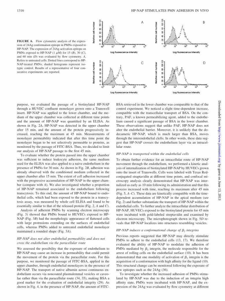
FIGURE 6. Flow cytometric analysis of the expres-sion of 24Ag conformation epitope in PMNs exposed toHP-NAP. The expression of 24Ag activation epitope onPMNs exposed to HP-NAP (1 �M) for 15 (B), 30 (C),and 60 min (D) was evaluated by flow cytometry. A,Refers to untreated cells. Dotted lines correspond to HP-NAP-treated PMNs; shaded histograms represent iso-typic control. Results of a representative of four con-secutive experiments are reported.
1316 HP-NAP STIMULATES PMN ADHESION IN VIVO
by guest on June 13, 2013http://w
ww
.jimm
unol.org/D
ownloaded from

time points. As illustrated in Fig. 6, PMNs express significant lev-els of the epitope after 15 min of treatment (Fig. 6B) as comparedwith basal levels (Fig. 6A). Exposure of this epitope remainedconstant at 30 min (Fig. 6C) and then started to decrease after 1 hof treatment, as shown by the appearance of a cellular populationwith a fluorescence comparable to that of untreated cells (Fig. 6D).No increase in 24Ag expression was revealed on untreated PMNsover all the time points considered (data not shown).
HP-NAP-induced �2 integrin activation involves p38-MAPK
Several studies suggest that HP-NAP may interact with at least tworeceptors on the plasma membrane of leukocytes. The engagementof TLR-2 (20) seems to be related to the production of cytokinesby monocytes, whereas the interaction with a G protein-coupledreceptor is linked to burst activation, adhesion, and chemotaxis ofPMNs (27). These latter effects are abrogated by inhibition of p38-MAPK, suggesting a role for this kinase in the signaling cascade(37). As p38-MAPK triggers �2 integrin activation in TNF-�-stim-ulated PMNs (38), we decided to ascertain whether expression of24Ag induced by HP-NAP also required p38-MAPK. PMNs werepretreated with the specific p38-MAPK inhibitor SB203580 beforebeing exposed to HP-NAP. Evaluation of the 24Ag expressionafter 30 min is illustrated in Fig. 7A. As compared with the valuesof PMNs exposed to HP-NAP alone (solid line), inhibition of p38-MAPK completely abolished the exposure of the 24Ag epitope(compare the dotted line with the shadowed histogram represent-ing the basal level). To definitely confirm that the integrin highaffinity state was the main one responsible for the adhesion ofPMNs exposed to HP-NAP, we examined the effect of the p38-MAPK inhibition in an adhesion assay. Fig. 7B shows that whenp38-MAPK was inhibited, the extent of adhesion was reduced tovalues comparable with those of untreated cells, despite stimula-tion of PMNs with HP-NAP. Of note, the inhibitor SB203580 didnot affect adhesivity per se, further confirming that p38-MAPK ispositioned downstream of HP-NAP in the promotion of the adhe-sivity state of PMNs.
HP-NAP activate PMNs to synthesize and release chemokines
Several studies reported that infection by H. pylori is associatedwith an increased expression of chemokines, and that endothelialand gastric epithelial cells are believed to contribute to this in-crease (9, 11, 39). Although it has been demonstrated that factorsother than HP-NAP stimulate endothelial cells to produce CXCL8(13), the release of CXCL8 or other chemokines from PMNs stim-ulated by HP-NAP has not been investigated yet. To shed lightupon this aspect, total RNA was extracted from PMNs incubatedwith HP-NAP and examined by RPA and real-time PCR. Asshown in Fig. 8A, treatment of PMNs with HP-NAP resulted in arapid induction of CXCL8, CCL3, and CCL4 mRNAs that reachedmaximum levels at 3 h for all the chemokines and slowly declinedthereafter. These observations were substantially confirmed by
real-time PCR studies (Fig. 8B), which also revealed that themRNAs for the CXCL10 and CCL2 chemokines, or for othercytokines, such as VEGF, TRAIL, and IFN-�, were not induced
FIGURE 7. Effects of p38-MAPK inhibition inHP-NAP-treated PMNs. Expression of 24Ag wasevaluated in PMNs treated with 30 �M SB203580before exposing to HP-NAP (A). Solid lines cor-respond to HP-NAP-stimulated neutrophils; dottedlines to HP-NAP-stimulated neutrophils pretreatedwith SB203580; and shaded histograms representuntreated cells. Effect of p38-MAPK inhibition onHP-NAP-neutrophil adhesion was determined (B).Results of a representative of four consecutive ex-periments are reported. ��, p � 0.01.
FIGURE 8. Time-dependent mRNA expression for various chemokines inHP-NAP-treated neutrophils. Total RNA was extracted from neutrophils cul-tured with HP-NAP and analyzed by either RPA (A) or real-time PCR (B) forCXCL8, CCL3, and CCL4 mRNA expression. B, Expression levels are de-picted as n-folds of the normalized amount of mRNA from untreated cells (1AU � mRNA cytokine concentration (fmol/�l)/mRNA �2-microglobulin(fmol/�l)/mRNA �2-microglobulin (fmol/�l) of triplicate reactions for eachsample. Data show representative experiments of three performed for eachpanel.
1317The Journal of Immunology
by guest on June 13, 2013http://w
ww
.jimm
unol.org/D
ownloaded from

in HP-NAP-treated PMNs (data not shown). In line with the geneexpression data, culture supernatants of PMNs stimulated withHP-NAP showed a remarkable extracellular production of bothCXCL8 and CCL4, which began, respectively, as early as after 3and 5 h ( p � 0.001, n � 5), and continued to progressively in-crease up to 21 h ( p � 0.005, n � 6) (Fig. 9). Under the sameconditions, neither CXCL10 nor VEGF was found to be releasedby activated neutrophils, even if PMNs were costimulated withIFN-� (data not shown).
DiscussionThe infiltration of neutrophils and mononuclear inflammatory cellswithin the H. pylori-infected stomach mucosa is a common find-ing, and the degree of mucosal damage is correlated with neutro-phil infiltration. Several protein components in H. pylori extractswere found to attract and activate neutrophils and other inflamma-tory cells (17, 40–49).
Among these molecules, a 150-kDa oligomeric protein iso-lated from H. pylori water extracts was found to promote neu-trophil adhesion to endothelial cells (17, 50), and the proteinwas named HP-NAP. HP-NAP is released in the medium, mostlikely after cell lysis, and binds to the bacterial surface, whereit can act as an adhesin, mediating binding to mucin (51) or toPMN sphingomyelin (52).
Until recently, all of the experiments illustrating the capacity ofHP-NAP of inducing leukocytes to adhere to the endothelium wereconducted, putting PMNs and HP-NAP directly in contact, withoutconsidering that in vivo the protein has to cross the endothelium tostimulate neutrophils. A recent report, however, showed that HP-NAP promoted transendothelial neutrophil migration also whenadded into a lower chamber of a Transwell system consisting of acultured monolayer of human endothelial cells as barrier betweentwo chambers (13). These findings, although strongly suggestingthat HP-NAP was able to cross the endothelium to contact leuko-cytes, were not reinforced by data, providing direct evidence forthe transendothelial passage of HP-NAP. Moreover, the authorsdid not consider that in vivo the underflow conditions may bias theeffects observed in vitro.
In this study, we report for the first time that HP-NAP promotesleukocyte adhesion to the endothelium in vivo. Intravital micros-copy, used in our study to analyze the mobilization of leukocytesin response to HP-NAP, allowed us to show that leukocytes cir-culating in the rat mesentery begin to adhere rather rapidly to theendothelium of the postcapillary venules after the topical applica-
tion of HP-NAP. Adhesion is detectable already after 10 min andreaches a maximum at 60 min. With 5 �M HP-NAP, a significantleukocyte extravasation is observed after 60 min of treatment.These findings are at variance with previous reports showing thatlower concentrations of HP-NAP induced PMN chemotaxis in arapid and efficient way (13, 27, 37). This discrepancy can be ex-plained by the different complexity of the systems used, whichemphasizes the need of using integrated models as close as pos-sible to the in vivo situation. Furthermore, it is possible thatCXCL8, secreted by HP-NAP-stimulated peritoneal macrophages,contributed to the accumulation of leukocytes in the tissue. In sup-port of this hypothesis, there is a recent finding that HP-NAP stim-ulated monocytes/macrophages to secrete CXCL8 (20).
In this study, we have also shown that HP-NAP is effectivelytransported across the endothelium via intracellular route withoutany major contribution of the paracellular route. The amount ofprotein recovered after crossing was small, but biologically rele-vant, because it was sufficient to stimulate PMNs to adhere toendothelial cells. HP-NAP failed to alter the endothelial perme-ability in such a way to move through interendothelial clefts, butit was transported within endothelial cells through vesicles, asclearly demonstrated by protein immunogold labeling. The inter-nalization process of HP-NAP was found to be very efficient, assuggested by the number of fluorescent spots visible after labelingof biotinylated HP-NAP with streptavidin Texas Red. This wasapparently in contrast with the limited amount of protein that wasreleased after endothelial crossing. A possible explanation is that asignificant proportion of HP-NAP remained bound to the endothe-lium after transcytosis, similarly to what was observed for the che-mokine CXCL8 (53). This possibility is supported by our findingthat a substantial amount of HP-NAP was detected on the endo-thelium, and this, in turn, promoted a significant adhesion of PMNs(data not shown).
�2 integrins on the surface of leukocytes need to be activated toacquire their ligand-binding capacity. Modulation of integrinproadhesive activity involves, in some instances, heterodimer lat-eral mobility leading to accumulation in discrete areas of theplasma membrane, a process called clustering, which has beenproposed as an essential event regulating integrin activation (54,55). Alternatively, or in addition, integrins may potently increasetheir affinity for ligand by altering their three-dimensional confor-mational state (10, 56). In the last few years, the importance ofthese conformational changes has been highlighted. Conforma-tional changes upon �2 integrin activation can be evidenced bymonitoring the exposure of activation epitopes such as the 24Ag(36). Using an Ab specific for the 24Ag, exposed after the acqui-sition of the integrin high affinity state (36), we show in this studythat HP-NAP induced the conformational change of the adhesionmolecule. A parallel evaluation of HP-NAP stimulation of endo-thelium to become more adhesive showed, in agreement with arecent report (13), that there was no increased expression ofICAM-1, VCAM-2, and endothelial leukocyte adhesion moleculeafter 4 and 24 h of stimulation (data not shown). These results,together with our previous observation illustrating that HP-NAPup-regulated the expression of �2 integrins (27), definitively con-firm that HP-NAP has a direct role in stimulating neutrophils toadhere. Finally, we demonstrated the involvement of the p38-MAPK along the signaling cascade responsible for the integrinactivation, as already reported for TNF-� (38).
The results of this study also establish that human PMNs re-spond to stimulation with HP-NAP by expressing and producingseveral chemokines, including CXCL8, CCL3, and CCL4. Expres-sion of other proinflammatory molecules, such as CXCL10,
FIGURE 9. Kinetics of CXCL8 and CCL4 extracellular production byHP-NAP-treated neutrophils. PMNs were stimulated with 1 �M HP-NAPfor the times indicated, and CXCL8 (A) and CCL4 (B) levels were thendetermined in the cell supernatants by ELISA. For each cytokine, the figureshows the mean value � the SD of duplicate assays for each time point,each obtained from three experiments performed under the sameconditions.
1318 HP-NAP STIMULATES PMN ADHESION IN VIVO
by guest on June 13, 2013http://w
ww
.jimm
unol.org/D
ownloaded from

IFN-�, and TRAIL, was not induced by HP-NAP, which is con-sistent with the fact that HP-NAP acts via TLR-2, and conse-quently through the intracellular Myd88-dependent pathway (57).Importantly, neither IL-6 nor CCL2 mRNAs were induced in HP-NAP-treated PMNs, indicating that the effects attributed to HP-NAP were direct and not mediated by contaminating monocytes oreosinophils (58). Because neutrophils rapidly migrate in largenumbers at the infection sites, the fact that they also serve as achemokine source may contribute to the generation of the con-ditions necessary for both the recruitment and activation notonly of additional neutrophils via CXCL8, but also of mono-cytes, dendritic cells, and lymphocytes through CCL3 andCCL4. The capacity of neutrophils to produce chemokines inresponse to HP-NAP, as shown in this study, provides newinsights for a better understanding of the cellular mechanismsinvolved in the H. pylori-induced inflammation.
AcknowledgmentsWe thank Federica Calzetti, Tito Ubaldini, and Claudio Gamboz for tech-nical assistance, and Dr. M. M. D’Elios for helpful discussion and criticalreading of the manuscript.
DisclosuresThe authors have no financial conflict of interest.
References1. Marshall, B. J., and J. R. Warren. 1984. Unidentified curved bacilli on gastric
epithelium in active chronic gastritis. Lancet 1: 1311–1315.2. Blaser, M. J., and J. C. Atherton. 2004. Helicobacter pylori persistence: biology
and disease. J. Clin. Invest. 113: 321–323.3. Goodwin, C. S. 1997. Helicobacter pylori gastritis, peptic ulcer and gastric can-
cer: clinical and molecular aspects. Clin. Infect. Dis. 25: 1017–1019.4. Parsonnet, J., S. Hansen, L. Rodriguez, A. B. Gelb, R. A. Warnke, E. Jellum,
N. Orentreich, J. H. Vogelman, and G. D. Friedman. 1994. Helicobacter pyloriinfection and gastric lymphoma. N. Engl. J. Med. 330: 1267–1271.
5. Dixon, M. F., R. M. Genta, J. H. Yardley, and P. Correa. 1996. Classification andgrading of gastritis. Am. J. Surg. Pathol. 20: 1161–1181.
6. D’Elios, M. M., M. Manghetti, M. De Carli, F. Costa, C. T. Baldari, D. Burroni,J. L. Telford, S. Romagnani, and G. Del Prete. 1997. Th1 effector cells specificfor Helicobacter pylori in the gastric antrum of patients with peptic ulcer disease.J. Immunol. 158: 962–967.
7. Davies, G. R., N. Banatvala, C. E. Collins, M. T. Sheaff, Y. Abdi, L. Clements,and D. S. Rampton. 1994. Relationship between infective load of Helicobacterpylori and reactive oxygen metabolite production in antral mucosa. Scand.J. Gastroenterol. 29: 419–424.
8. Hamlet, A., A. C. Thoreson, O. Nilsson, A. M. Svennerholm, and L. Olbe. 1999.Duodenal Helicobacter pylori infection differs in cagA genotype between asymp-tomatic subjects and patients with duodenal ulcers. Gastroenterology 116:259–268.
9. Innocenti, M., A. C. Thoreson, R. L. Ferrero, E. Stromberg, I. Bolin, L. Eriksson,A. M. Svennerholm, and M. Quiding-Jarbrink. 2002. Helicobacter pylori-inducedactivation of human endothelial cells. Infect. Immun. 70: 4581–4590.
10. Laudanna, C., J. Y. Kim, G. Constantin, and E. Butcher. 2002. Rapid leukocyteintegrin activation by chemokines. Immunol. Rev. 186: 37–46.
11. Crabtree, J. E., D. Kersulyte, S. D. Li, I. J. Lindley, and D. E. Berg. 1999.Modulation of Helicobacter pylori induced interleukin-8 synthesis in gastric ep-ithelial cells mediated by cag PAI encoded VirD4 homologue. J. Clin. Pathol. 52:653–657.
12. Crabtree, J. E., A. Covacci, S. M. Farmery, Z. Xiang, D. S. Tompkins, S. Perry,I. J. Lindley, and R. Rappuoli. 1995. Helicobacter pylori induced interleukin-8expression in gastric epithelial cells is associated with CagA positive phenotype.J. Clin. Pathol. 48: 41–45.
13. Brisslert, M., K. Enarsson, S. Lundin, A. Karlsson, J. G. Kusters, A. M.Svennerholm, S. Backert, and M. Quiding-Jarbrink. 2005. Helicobacter pyloriinduce neutrophil transendothelial migration: role of the bacterial HP-NAP.FEMS Microbiol. Lett. 249: 95–103.
14. Montecucco, C., and M. de Bernard. 2003. Molecular and cellular mechanisms ofaction of the vacuolating cytotoxin (VacA) and neutrophil-activating protein (HP-NAP) virulence factors of Helicobacter pylori. Microbes Infect. 5: 715–721.
15. Tonello, F., W. G. Dundon, B. Satin, M. Molinari, G. Tognon, G. Grandi,G. Del Giudice, R. Rappuoli, and C. Montecucco. 1999. The Helicobacter pyloriPMNs-activating protein is an iron binding protein with dodecameric structure.Mol. Microbiol. 34: 238–246.
16. Zanotti, G., E. Papinutto, W. G. Dundon, R. Battistuta, M. Seveso, G. DelGiudice, R. Rappuoli, and C. Montecucco. 2002. Structure of the neutrophil-activating protein from Helicobacter pylori. J. Mol. Biol. 323: 125–130.
17. Evans, D. J., Jr., D. G. Evans, T. Takemura, H. Nakano, H. C. Lampert, D. Y.Graham, D. N. Granger, and P. R. Kvietys. 1995. Characterization of a Helico-bacter pylori PMNs-activating protein. Infect. Immun. 63: 2213–2220.
18. Montemurro, P., H. Nishioka, W. G. Dundon, M. de Bernard, G. Del Giudice,R. Rappuoli, and C. Montecucco. 2002. The neutrophil-activating protein (HP-NAP) of Helicobacter pylori is a potent stimulant of mast cells. Eur. J. Immunol.32: 671–676.
19. Montemurro, P., G. Barbuti, W. G. Dundon, G. Del Giudice, R. Rappuoli,M. Colucci, P. De Rinaldis, C. Montecucco, N. Semeraro, and E. Papini. 2001.Helicobacter pylori neutrophil activating protein stimulates tissue factor and plas-minogen activator inhibitor-2 production by human blood mononuclear cells.J. Infect. Dis. 183: 1055–1062.
20. Amedei, A., A. Cappon, G. Codolo, A. Cabrelle, A. Polenghi, M. Benagiano,E. Tasca, A. Azzurri, M. M. D’Elios, G. Del Prete, and M. de Bernard. 2006. Theneutrophil-activating protein of Helicobacter pylori promotes Th1 immune re-sponses. J. Clin. Invest. 116: 1092–1101.
21. Silverstein, R., J. G. Wood, Q. Xue, M. Norimatsu, D. L. Horn, and D. C.Morrison. 2000. Differential host inflammatory responses to viable versus anti-biotic-killed bacteria in experimental microbial sepsis. Infect. Immun. 68:2301–2308.
22. Gamble, J. R., J. M. Harlan, S. J. Klebanoff, and M. A. Vadas. 1985. Stimulationof the adherence of neutrophils to umbilical vein endothelium by human recom-binant tumor necrosis factor. Proc. Natl. Acad. Sci. USA 82: 8667–8671.
23. Dobrina, A., R. Menegazzi, T. M. Carlos, E. Nardon, R. Cramer, T. Zacchi, J. M.Harlan, and P. Patriarca. 1991. Mechanisms of eosinophil adherence to culturedvascular endothelial cells: eosinophils bind to the cytokine-induced ligand vas-cular cell adhesion molecule-1 via the very late activation antigen-4 integrinreceptor. J. Clin. Invest. 88: 20–26.
24. Rossi, F., V. Della Bianca, M. Grzeskowiak, and F. Buzzoni. 1989. Studies onmolecular regulation of phagocytosis in PMNs: Con A-mediated ingestion andassociated respiratory burst independent of phosphoinositide turnover, rise in[Ca2�], and arachidonic acid release. J. Immunol. 142: 1652–1660.
25. Tedesco, F., M. Pausa, E. Nardon, M. Introna, A. Mantovani, and A. Dobrina.1997. The cytolytically inactive terminal complement complex activates endo-thelial cells to express adhesion molecules and tissue factor procoagulant activity.J. Exp. Med. 85: 1619–1627.
26. Maciag, T., J. Cerundolo, S. Ilsley, P. R. Kelley, and R. Forand. 1979. An en-dothelial cell growth factor from bovine hypothalamus: identification and partialcharacterization. Proc. Natl. Acad. Sci. USA 76: 5674–5678.
27. Satin, B., G. Del Giudice, V. Della Bianca, S. Dusi, C. Laudanna, F. Tonello,D. Kelleher, R. Rappuoli, C. Montecucco, and F. Rossi. 2000. The neutrophil-activating protein (HP-NAP) of Helicobacter pylori is a protective antigen and amajor virulence factor. J. Exp. Med. 191: 1467–1476.
28. Dobrina, A., M. Pausa, F. Fischetti, R. Bulla, E. Vecile, E. Ferrero, A. Mantovani,and F. Tedesco. 2002. Cytolytically inactive terminal complement complexcauses transendothelial migration of polymorphonuclear leukocytes in vitro andin vivo. Blood 99: 185–192.
29. Bossi, F., F. Fischetti, V. Pellis, R. Bulla, E. Ferrero, T. E. Mollnes, D. Regoli,and F. Tedesco. 2004. Platelet-activating factor and kinin-dependent vascularleakage as a novel functional activity of the soluble terminal complement com-plex. J. Immunol. 173: 6921–6927.
30. Bar-Shavit, R., V. Sabbah, M. G. Lampugnani, P. C. Marchisio, J. W. Fenton, II,I. Vlodavsky, and E. Dejana. 1991. An Arg-Gly-Asp sequence within thrombinpromotes endothelial cell adhesion. J. Cell Biol. 112: 335–344.
31. Cassatella, M. A. 2006. On the production of TNF-related apoptosis-inducing ligand(TRAIL/Apo-2L) by human neutrophils. J. Leukocyte Biol. 79: 1140–1149.
32. Hayashi, F., T. K. Means, and A. D. Luster. 2003. Toll-like receptors stimulatehuman neutrophil function. Blood 102: 2660–2669.
33. Lapinet, J. A., P. Scapini, F. Calzetti, O. Perez, and M. A. Cassatella. 2000. Geneexpression and production of tumor necrosis factor �, interleukin-1� (IL-1�),IL-8, macrophage inflammatory protein 1� (MIP-1�), MIP-1�, and � interferon-inducible protein 10 by human neutrophils stimulated with group B meningo-coccal outer membrane vesicles. Infect. Immun. 68: 6917–6923.
34. Milici, A. J., N. E. Watrous, H. Stukenbrok, and G. E. Palade. 1987. Transcytosisof albumin in capillary endothelium. J. Cell Biol. 105: 2603–2612.
35. Ghitescu, L., and M. Bendayan. 1992. Transendothelial transport of serum albu-min: a quantitative immunocytochemical study. J. Cell Biol. 117: 745–755.
36. Dransfield, I., and N. Hogg. 1989. Regulated expression of Mg2� binding epitopeon leukocyte integrin � subunits. EMBO J. 8: 3759–3765.
37. Nishioka, H., I. Baesso, G. Semenzato, L. Trentin, R. Rappuoli, G. Del Giudice,and C. Montecucco. 2003. The neutrophil-activating protein of Helicobacter py-lori (HP-NAP) activates the MAPK pathway in human neutrophils. Eur. J. Im-munol. 33: 840–849.
38. Bouaouina, M., E. Blouin, L. Halbwachs-Mecarelli, P. Lesavre, and P. Rieu.2004. TNF-induced �2 integrin activation involves Src kinases and a redox-regulated activation of p38-MAPK. J. Immunol. 173: 1313–1320.
39. Shimoyama, T., and J. E. Crabtree. 1997. Mucosal chemokines in Helicobacterpylori infection. J. Physiol. Pharmacol. 48: 315–323.
40. Karttunen, R., G. Andersson, K. Poikonen, T. U. Kosunen, T. Karttunen, K.Juutinen, and S. Niemela. 1990. Helicobacter pylori induces lymphocyte activa-tion in peripheral blood cultures. Clin. Exp. Immunol. 82: 485–488.
41. Mai, U. E., G. I. Perez-Perez, L. M. Wahl, S. M. Wahl, M. J. Blaser, and P. D.Smith. 1991. Soluble surface proteins from Helicobacter pylori activate mono-cytes/macrophages by lipopolysaccharide-independent mechanism. J. Clin. In-vest. 87: 894–900.
42. Mai, U. E., G. I. Perez-Perez, J. B. Allen, S. M. Wahl, M. J. Blaser, and P. D.Smith. 1992. Surface proteins from Helicobacter pylori exhibit chemotactic ac-tivity for human leukocytes and are present in gastric mucosa. J. Exp. Med. 175:517–525.
1319The Journal of Immunology
by guest on June 13, 2013http://w
ww
.jimm
unol.org/D
ownloaded from

43. Craig, P. M., M. C. Territo, W. E. Karnes, and J. H. Walsh. 1992. Helicobacterpylori secretes a chemotactic factor for monocytes and neutrophils. Gut 33:1020–1023.
44. Nielsen, H., and L. P. Andersen. 1992. Chemotactic activity of Helicobacterpylori sonicate for human polymorphonuclear leukocytes and monocytes. Gut 33:738–742.
45. Nielsen, H., and L. P. Andersen. 1992. Activation of human phagocyte oxidativemetabolism by Helicobacter pylori. Gastroenterology 103: 1747–1753.
46. Kozol, R., B. McCurdy, and R. Czanko. 1993. A neutrophil chemotactic factorpresent in H. pylori but absent in H. mustelae. Dig. Dis. Sci. 38: 137–141.
47. Reymunde, A., J. Deren, I. Nachamkin, D. Oppenheim, and G. Weinbaum. 1993.Production of chemoattractant by Helicobacter pylori. Dig. Dis. Sci. 38:1697–1694.
48. Marchetti, M., B. Arico, D. Burroni, N. Figura, R. Rappuoli, and P. Ghiara. 1995.Development of a mouse model of Helicobacter pylori infection that mimicshuman disease. Science 267: 1655–1658.
49. Betten, A., J. Bylund, T. Christophe, F. Boulay, A. Romero, K. Hellstrand, andC. Dahlgren. 2001. A proinflammatory peptide from Helicobacter pylori acti-vates monocytes to induce lymphocyte dysfunction and apoptosis. J. Clin. Invest.108: 1221–1228.
50. Yoshida, N., D. N. Granger, D. J. Evans, Jr., D. G. Evans, D. Y. Graham, D. C.Anderson, R. E. Wolf, and P. R. Kvietys. 1993. Mechanisms involved in Heli-cobacter pylori-induced inflammation. Gastroenterology 105: 1431–1440.
51. Namavar, F., M. Sparrius, E. C. Veerman, B. J. Appelmelk, and C. M.Vandenbroucke-Grauls. 1998. PMNs-activating protein mediates adhesion ofHelicobacter pylori to sulfated carbohydrates on high-molecular-weight salivarymucin. Infect. Immun. 66: 444–447.
52. Teneberg, S., H. Miller-Podraza, H. C. Lampert, D. J. Evans, Jr., D. G. Evans,D. Danielsson, and K. A. Karlsson. 1997. Carbohydrate binding specificity ofthe PMNs-activating protein of Helicobacter pylori. J. Biol. Chem. 272:19067–19071.
53. Middleton, J., S. Neil, J. Wintle, I. Clark-Lewis, H. Moore, C. Lam, M. Auer,E. Hub, and A. Rot. 1997. Transcytosis and surface presentation of IL-8 byvenular endothelial cells. Cell 91: 385–395.
54. Stewart, M. P., A. McDowall, and N. Hogg. 1998. LFA-1-mediated adhesion isregulated by cytoskeletal restraint and by a Ca2�-dependent protease, calpain.J. Cell Biol. 140: 699–707.
55. van Kooyk, Y., and C. G. Figdor. 2000. Avidity regulation of integrins: thedriving force in leukocyte adhesion. Curr. Opin. Cell Biol. 12: 542–547.
56. Plow, E. F., T. A. Haas, L. Zhang, J. Loftus, and J. W. Smith. 2000. Ligandbinding to integrins. J. Biol. Chem. 275: 21785–21788.
57. Akira, S., S. Uematsu, and O. Takeuchi. 2006. Pathogen recognition and innateimmunity. Cell 124: 783–801.
58. Cassatella, M. A. 1999. Neutrophil-derived proteins: selling cytokines by thepound. Adv. Immunol. 73: 369–509.
1320 HP-NAP STIMULATES PMN ADHESION IN VIVO
by guest on June 13, 2013http://w
ww
.jimm
unol.org/D
ownloaded from