Evolution of neutrophil apoptosis in septic shock survivors and nonsurvivors
11
Evolution of neutrophil apoptosis in septic shock survivors and nonsurvivors ☆,☆☆ Eduardo Tamayo MD, PhD a,1 , Esther Gómez MD, PhD a,1 , Juan Bustamante MD, PhD b, ⁎ ,1 , José I. Gómez-Herreras MD, PhD c,1 , Rosalba Fonteriz MD, PhD d,1 , Felipe Bobillo MD, PhD e,1 , Jesús F. Bermejo-Martín MD, PhD f,1 , Javier Castrodeza MD, PhD g,1 , Maria Heredia MD a,1 , Inma Fierro MD h,1 , Francisco Javier Álvarez MD, PhD h,1 a Department of Anaesthesiology and Intensive Care, Hospital Clínico Universitario de Valladolid, 47005 Valladolid, Spain b Department of Cardiovascular Surgery. Hospital Universitario La Princesa, 28006 Madrid, Spain c Department of Anaesthesiology and Intensive Care, Hospital Universitario Rio Hortega, 47012 Valladolid, Spain d Institute of Biology and Molecular Genetics (IBGM/CSIC), Universidad de Valladolid, 47005 Valladolid, Spain e Medical Intensive Care Unit, Hospital Clínico Universitario de Valladolid, 47005 Valladolid, Spain f Infection & Immunity Unit, Hospital Clinico Universitario de Valladolid -IECSCYL, 47005 Valladolid, Spain g Direction of Public Health, Investigation, Development and Innovation, SACYL, 47007 Valladolid, Spain h Department of Pharmacology and Therapeutics, Facultad de Medicina, Universidad de Valladolid, 47005 Valladolid, Spain Keywords: Apoptosis; Neutrophil; Septic shock; Mortality; Survivors Abstract Purpose: The aims were to analyze the temporal evolution of neutrophil apoptosis, to determine the differences in neutrophil apoptosis among 28-day survivors and nonsurvivors, and to evaluate the use of neutrophil apoptosis as a predictor of mortality in patients with septic shock. Materials and Methods: Prospective multicenter observational study carried out between July 2006 and June 2009. The staining solution study included 80 patients with septic shock and 25 healthy volunteers. Neutrophil apoptosis was assessed by fluorescein isothiocyanate (FITC)–conjugated annexin V and aminoactinomycin D staining. Results: The percentage of neutrophil apoptosis was significantly decreased at 24 hours, 5 days, and 12 days after the diagnosis of septic shock (14.8% ± 13.4%, 13.4% ± 8.4%, and 15.4% ± 12.8%, respectively; P b .0001) compared with the control group (37.6% ± 12.8%). The difference in apoptosis between 28-day surviving and nonsurviving patients was nonsignificant (P N .05). The mortality rate at 28 days was 53.7%. ☆ Conflict of Interest: None declared. ☆☆ Funding Sources: This work was supported in part by a grant from the “Gerencia de Salud, Consejería de Sanidad, Junta de Castilla y Leon” (GRS 143/ A/07) and Instituto de Salud Carlos III, Fondo Europeo de Desarrollo Regional FEDER, Red de Trastornos Adictivos RD06/0001/0020. ⁎ Corresponding author. Tel.: +34 915202268; fax: +34 915202201. E-mail addresses: [email protected] (E. Tamayo), [email protected] (E. Gómez), [email protected] (J.I. Gómez-Herreras), [email protected], [email protected] (J. Bustamante), [email protected] (R. Fonteriz), [email protected] (F. Bobillo), [email protected] (J. Castrodeza), [email protected] (M. Heredia), [email protected] (I. Fierro), [email protected] (F.J. Álvarez). 1 Valladolid Sepsis Study Group. 0883-9441/$ – see front matter © 2012 Elsevier Inc. All rights reserved. doi:10.1016/j.jcrc.2011.09.001 Journal of Critical Care (2012) 27, 415.e1–415.e11
Transcript of Evolution of neutrophil apoptosis in septic shock survivors and nonsurvivors
Journal of Critical Care (2012) 27, 415.e1–415.e11
Evolution of neutrophil apoptosis in septic shock survivorsand nonsurvivors☆,☆☆
Eduardo Tamayo MD, PhD a,1, Esther Gómez MD, PhD a,1,Juan Bustamante MD, PhD b,⁎,1, José I. Gómez-Herreras MD, PhD c,1,Rosalba Fonteriz MD, PhD d,1, Felipe Bobillo MD, PhD e,1,Jesús F. Bermejo-Martín MD, PhD f,1, Javier Castrodeza MD, PhD g,1,Maria Heredia MD a,1, Inma Fierro MD h,1, Francisco Javier Álvarez MD, PhD h,1
aDepartment of Anaesthesiology and Intensive Care, Hospital Clínico Universitario de Valladolid, 47005 Valladolid, SpainbDepartment of Cardiovascular Surgery. Hospital Universitario La Princesa, 28006 Madrid, SpaincDepartment of Anaesthesiology and Intensive Care, Hospital Universitario Rio Hortega, 47012 Valladolid, SpaindInstitute of Biology and Molecular Genetics (IBGM/CSIC), Universidad de Valladolid, 47005 Valladolid, SpaineMedical Intensive Care Unit, Hospital Clínico Universitario de Valladolid, 47005 Valladolid, SpainfInfection & Immunity Unit, Hospital Clinico Universitario de Valladolid -IECSCYL, 47005 Valladolid, SpaingDirection of Public Health, Investigation, Development and Innovation, SACYL, 47007 Valladolid, SpainhDepartment of Pharmacology and Therapeutics, Facultad de Medicina, Universidad de Valladolid, 47005 Valladolid, Spain
A
bc
0d
Keywords:Apoptosis;Neutrophil;Septic shock;Mortality;Survivors
AbstractPurpose: The aims were to analyze the temporal evolution of neutrophil apoptosis, to determine thedifferences in neutrophil apoptosis among 28-day survivors and nonsurvivors, and to evaluate the use ofneutrophil apoptosis as a predictor of mortality in patients with septic shock.Materials and Methods: Prospective multicenter observational study carried out between July 2006 andJune 2009. The staining solution study included 80 patients with septic shock and 25 healthy volunteers.Neutrophil apoptosis was assessed by fluorescein isothiocyanate (FITC)–conjugated annexin V andaminoactinomycin D staining.Results: The percentage of neutrophil apoptosis was significantly decreased at 24 hours, 5 days, and 12days after the diagnosis of septic shock (14.8% ± 13.4%, 13.4% ± 8.4%, and 15.4% ± 12.8%, respectively;P b .0001) compared with the control group (37.6% ± 12.8%). The difference in apoptosis between 28-daysurviving and nonsurviving patients was nonsignificant (P N .05). Themortality rate at 28 days was 53.7%.
☆ Conflict of Interest: None declared.☆☆ Funding Sources: This work was supported in part by a grant from the “Gerencia de Salud, Consejería de Sanidad, Junta de Castilla y Leon” (GRS 143/
/07) and Instituto de Salud Carlos III, Fondo Europeo de Desarrollo Regional FEDER, Red de Trastornos Adictivos RD06/0001/0020.⁎ Corresponding author. Tel.: +34 915202268; fax: +34 915202201.E-mail addresses: [email protected] (E. Tamayo), [email protected] (E. Gómez), [email protected] (J.I. Gómez-Herreras),
[email protected], [email protected] (J. Bustamante), [email protected] (R. Fonteriz), [email protected] (F. Bobillo),[email protected] (J. Castrodeza), [email protected] (M. Heredia), [email protected] (I. Fierro), [email protected] (F.J. Álvarez).
1 Valladolid Sepsis Study Group.
883-9441/$ – see front matter © 2012 Elsevier Inc. All rights reserved.oi:10.1016/j.jcrc.2011.09.001
415.e2 E. Tamayo et al.
The crude hazard ratio for mortality in patients with septic shock did not differ according to the percentageof apoptosis (hazard ratio, 1.006; 95% confidence interval, 0.98-1.03; P = .60).Conclusions: During the first 12 days of septic shock development, the level of neutrophil apoptosisdecreases and does not recover normal values. No differences were observed between surviving andnonsurviving patients.© 2012 Elsevier Inc. All rights reserved.
1. Introduction
Sepsis affects more than 500 000 patients annually in theUnited States [1], with an estimated incidence in Europe of 240to 400 cases per 100 000 inhabitants per year [2], and theincidence rate continues to increase [1,2]. Of the patientsadmitted to intensive care units (ICUs), the incidence of severesepsis or septic shock is between 9% and 37% [1-4]. In spite ofrecent advances in treatment with antibiotics and critical caretherapy, sepsis still results in a highmortality rate; it is typicallybetween 40% and 60% in the case of septic shock [1-5]. Septicshock and multiple organ dysfunction syndrome (MODS) arethe most common causes of death in ICUs [4].
The lack of apoptosis regulation in patients with severeinjuries contributes to the pathogenesis of MODS [6]. Thesystemic inflammatory response can harm the patient througheither an inadequate or excessive inflammatory reaction [7].Apoptosis is vital to the regulation of neutrophils, monocyteeosinophils, and macrophages. With sepsis, a delay inneutrophil apoptosis [8] and increased apoptosis in hemato-poietic tissues, such as the thymus, Peyer patch, spleen, andbone marrow, is frequently observed [9]. The prolongedneutrophil survival may allow a more robust inflammatoryresponse and, thus, better protect the individual. However,neutrophil survival may also cause harm to the host, leadingto the development of MODS and increasing mortality byincreasing the number of leukocytes or the production ofproinflammatory mediators and free radicals [10-12].
Although the significant reduction in neutrophil apoptosishas been sufficiently established in patients with sepsis[8,10], the temporal course of this phenomenon during theclinical evolution of septic shock has not been studied ineither surviving or nonsurviving patients. The use ofneutrophil apoptosis as a predictor of survival in patientswith septic shock has also not been studied.
Based on the fact that the reduction in neutrophilapoptosis during septic shock is related to the developmentof MODS [6,10,11], we hypothesize that the levels ofneutrophil apoptosis in patients who survive septic shockwill continue to increase throughout its evolution untilnormal levels, values equivalent to those found in healthycontrols, are reached, and these levels will be different (ie,higher) than those in nonsurviving patients.
The aim of this study was to analyze the following inpatients with septic shock: (1) temporal changes inneutrophil apoptosis, (2) differences in neutrophil apoptosisbetween 28-day survivors and 28-day nonsurvivors, (3) the
use of neutrophil apoptosis as a predictor of mortality, and(4) evaluation of the factors that may influence the course ofthe apoptosis.
2. Materials and methods
2.1. Study design, patients, and healthy volunteers
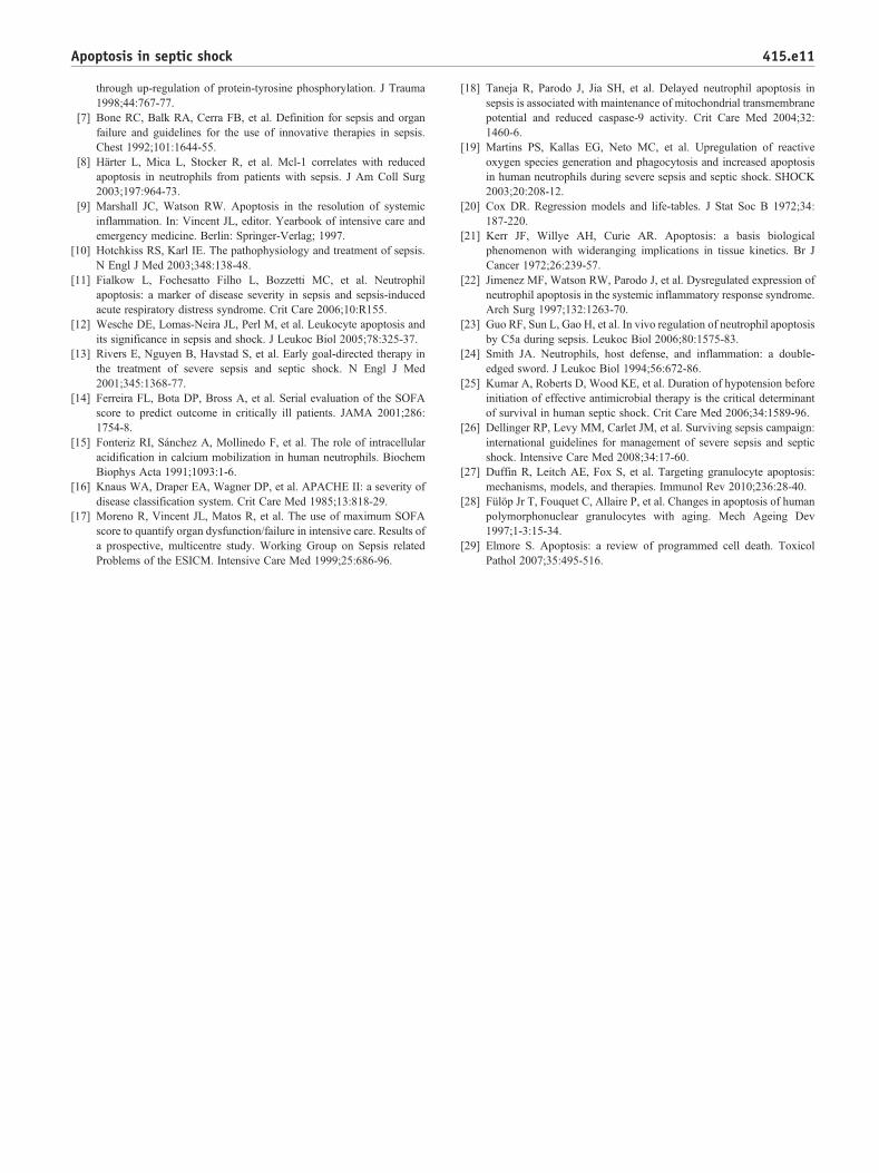
From July 2006 through June 2009, eligible patients wereenrolled in this open-label, prospective, nonrandomized,multicenter, nondrug interventional study, which was con-ducted at 4 medical/surgical ICUs in Valladolid, Spain:postoperative general, postoperative cardiac surgery, andmedical ICUs of the Hospital Clinico Universitario and thepostoperative general ICU of the Hospital Universitario RíoHortega. The study protocol was reviewed and approved bythe Human Subjects Review Committee and Research EthicsBoard of the Hospital Clínico Universitario of Valladolid, andinformed consent was obtained from each study participant.
The study only included patients older than 18 years whowere in septic shock with at least 2 dysfunctional organs orsystems at the time of enrollment, as previously described byBone et al [7] (Appendix A). The patients were enrolled inthe first 24 hours after the diagnosis of septic shock. Patientswere excluded if they had a known metastatic malignantdisease or HIV, were receiving immunosuppressive drugs orsystemic corticosteroids, or were enrolled in a concomitantinterventional study.
Over the course of the study, a total of 92 patients met thecriteria for inclusion; however, 12 patients had incompletefollow-up and were withdrawn from the study. In total, thestudy included 80 patients and 25 healthy volunteers, whowere included as normal controls (Appendix B). The 80septic shock patients had a mean age (±SD) of 68.7 ± 12.4years and consisted of 52 men (65.0%) and 28 women(35.0%). The mean age (±SD) of the 25 healthy controls was68.5 ± 17.3 years and consisted of 15 men (60.0%) and 10women (40.0%). No significant differences were observedbetween the 2 groups with respect to age or sex (P N .05).
The septic shock patients included in the study weredivided into 2 groups: 28-day nonsurvivors and 28-daysurvivors (Appendix B). A follow-up period of 12 days wasestablished for the evolution of apoptosis based on data fromprevious studies concerning the duration of stay in thehospital and ICU. Rivers et al [13] reported a mean hospital
415.e3Apoptosis in septic shock
stay of 13.0 ± 13.7 days; Angus et al [1] reported a mean ICUstay of 15.7 ± 7 days; Esteban et al [2] reported a mean ICUstay of 9 days (range, 4-25 days); and Ferreira et al [14]reported a mean ICU stay of 4.0 days (range, 1-56 days).
Patients were treated according to the best standard of carefor severe sepsis. The therapeutic measures were thosehabitually used in the ICUs in which the studywas conducted.
2.2. Blood samples, neutrophil isolation, andneutrophil apoptosis
2.2.1. Blood samplesA blood sample of 10 mL was obtained from each patient
immediately after providing informed consent (24 hours), at5 days, and at 12 days. A single peripheral blood extractionof 10 mL was obtained from each healthy control. The bloodsamples were placed in tubes containing citrate and glucose,and the samples were sent to the laboratory of the ValladolidInstitute of Biology and Molecular Genetics for analysis.
2.2.2. Isolation of neutrophilsNeutrophils were isolated as described previously [14].
Fresh bloodwasmixed 6:1 (vol/vol) with acid-citrate-dextrose.Dextran (T500 Pharmacy) (Pharmacosmos A/S, Holbaek,Denmark) was added to a final concentration of 1.3%. After 45minutes at room temperature, the upper layer containing no redblood cells was centrifuged (300g, 10 minutes). The cell pelletwas resuspended and layered on a Ficoll gradient (LymphocyteSeparation Medium; Flow Laboratories, Irvine, UK). After 40minutes of centrifugation (400g), the pellet containingneutrophils was recovered and contaminating red cellsdisrupted by hypotonic lysis. Neutrophils were resuspendedin 1-mL phosphate-buffered saline and counted by trypan blueexclusion. Viability was more than 95%. To confirm cellviability, we measured cytosolic calcium in parallel experi-ments. Cells were able to respond with calcium increases inresponse to fMet-Leu-Phe: formal-methionyl-leucyl-phenylal-anine and platelet-activating factor.
2.3. Neutrophil apoptosis
Neutrophil apoptosis was detected using a techniquecombining fluorescein isothiocyanate (FITC)–conjugatedannexin V (annexin V-FITC) and 7-aminoactinomycin D (7-ADD) (Molecular probes; Invitrogen; Paisley, Renfrewshire,UK) staining solution (Annexin V-FITC Apoptosis Detectionkit I; BDPharMingen, SanDiego,CA). The polymorphonuclearsuspension (100 μL) in cold binding buffer (200 000 cells/mL)was transferred to 4 culture tubes to which 5 μL of annexin V(BD PharMingen) was added. After incubation in the dark atroom temperature for 15minutes, 400μL of annexinV–bindingbuffer (BD PharMingen) and 0.4 μL of a 1 mg/mL stock of 7-ADD were added to each tube for flow cytometry (FACSCa-libur; Becton Dickinson, Fullerton, CA). Gating based onforward and side-scatter detection was applied to eliminate celldebris. A minimum of 10 000 events were acquired per sample.
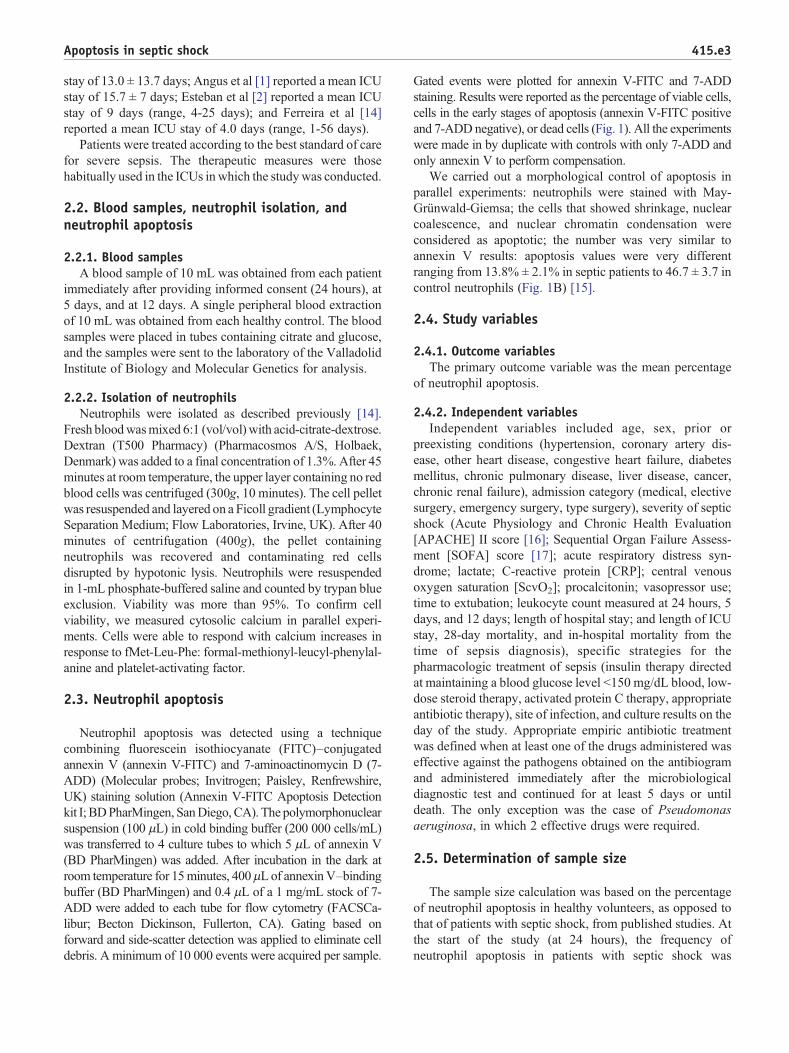
Gated events were plotted for annexin V-FITC and 7-ADDstaining. Results were reported as the percentage of viable cells,cells in the early stages of apoptosis (annexin V-FITC positiveand 7-ADDnegative), or dead cells (Fig. 1). All the experimentswere made in by duplicate with controls with only 7-ADD andonly annexin V to perform compensation.
We carried out a morphological control of apoptosis inparallel experiments: neutrophils were stained with May-Grünwald-Giemsa; the cells that showed shrinkage, nuclearcoalescence, and nuclear chromatin condensation wereconsidered as apoptotic; the number was very similar toannexin V results: apoptosis values were very differentranging from 13.8% ± 2.1% in septic patients to 46.7 ± 3.7 incontrol neutrophils (Fig. 1B) [15].
2.4. Study variables
2.4.1. Outcome variablesThe primary outcome variable was the mean percentage
of neutrophil apoptosis.
2.4.2. Independent variablesIndependent variables included age, sex, prior or
preexisting conditions (hypertension, coronary artery dis-ease, other heart disease, congestive heart failure, diabetesmellitus, chronic pulmonary disease, liver disease, cancer,chronic renal failure), admission category (medical, electivesurgery, emergency surgery, type surgery), severity of septicshock (Acute Physiology and Chronic Health Evaluation[APACHE] II score [16]; Sequential Organ Failure Assess-ment [SOFA] score [17]; acute respiratory distress syn-drome; lactate; C-reactive protein [CRP]; central venousoxygen saturation [ScvO2]; procalcitonin; vasopressor use;time to extubation; leukocyte count measured at 24 hours, 5days, and 12 days; length of hospital stay; and length of ICUstay, 28-day mortality, and in-hospital mortality from thetime of sepsis diagnosis), specific strategies for thepharmacologic treatment of sepsis (insulin therapy directedat maintaining a blood glucose level b150 mg/dL blood, low-dose steroid therapy, activated protein C therapy, appropriateantibiotic therapy), site of infection, and culture results on theday of the study. Appropriate empiric antibiotic treatmentwas defined when at least one of the drugs administered waseffective against the pathogens obtained on the antibiogramand administered immediately after the microbiologicaldiagnostic test and continued for at least 5 days or untildeath. The only exception was the case of Pseudomonasaeruginosa, in which 2 effective drugs were required.
2.5. Determination of sample size
The sample size calculation was based on the percentageof neutrophil apoptosis in healthy volunteers, as opposed tothat of patients with septic shock, from published studies. Atthe start of the study (at 24 hours), the frequency ofneutrophil apoptosis in patients with septic shock was
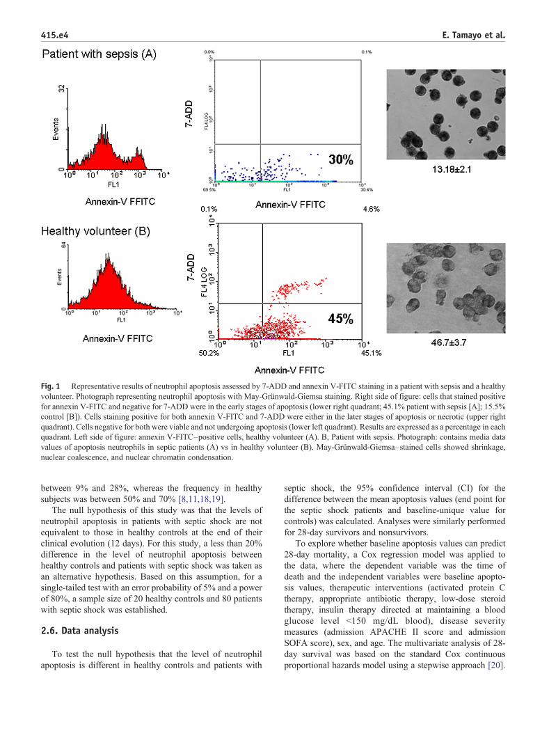
Fig. 1 Representative results of neutrophil apoptosis assessed by 7-ADD and annexin V-FITC staining in a patient with sepsis and a healthyvolunteer. Photograph representing neutrophil apoptosis with May-Grünwald-Giemsa staining. Right side of figure: cells that stained positivefor annexin V-FITC and negative for 7-ADD were in the early stages of apoptosis (lower right quadrant; 45.1% patient with sepsis [A]; 15.5%control [B]). Cells staining positive for both annexin V-FITC and 7-ADD were either in the later stages of apoptosis or necrotic (upper rightquadrant). Cells negative for both were viable and not undergoing apoptosis (lower left quadrant). Results are expressed as a percentage in eachquadrant. Left side of figure: annexin V-FITC–positive cells, healthy volunteer (A). B, Patient with sepsis. Photograph: contains media datavalues of apoptosis neutrophils in septic patients (A) vs in healthy volunteer (B). May-Grünwald-Giemsa–stained cells showed shrinkage,nuclear coalescence, and nuclear chromatin condensation.
415.e4 E. Tamayo et al.
between 9% and 28%, whereas the frequency in healthysubjects was between 50% and 70% [8,11,18,19].
The null hypothesis of this study was that the levels ofneutrophil apoptosis in patients with septic shock are notequivalent to those in healthy controls at the end of theirclinical evolution (12 days). For this study, a less than 20%difference in the level of neutrophil apoptosis betweenhealthy controls and patients with septic shock was taken asan alternative hypothesis. Based on this assumption, for asingle-tailed test with an error probability of 5% and a powerof 80%, a sample size of 20 healthy controls and 80 patientswith septic shock was established.
2.6. Data analysis
To test the null hypothesis that the level of neutrophilapoptosis is different in healthy controls and patients with
septic shock, the 95% confidence interval (CI) for thedifference between the mean apoptosis values (end point forthe septic shock patients and baseline-unique value forcontrols) was calculated. Analyses were similarly performedfor 28-day survivors and nonsurvivors.
To explore whether baseline apoptosis values can predict28-day mortality, a Cox regression model was applied tothe data, where the dependent variable was the time ofdeath and the independent variables were baseline apopto-sis values, therapeutic interventions (activated protein Ctherapy, appropriate antibiotic therapy, low-dose steroidtherapy, insulin therapy directed at maintaining a bloodglucose level b150 mg/dL blood), disease severitymeasures (admission APACHE II score and admissionSOFA score), sex, and age. The multivariate analysis of 28-day survival was based on the standard Cox continuousproportional hazards model using a stepwise approach [20].
415.e5Apoptosis in septic shock
The explanatory variables were used in a categorized ordichotomized form.
The relative hazard (RR = exp(βi)) was used as a measureof the risk of death in different groups, where βi is the basicparameter in the Cox model. Kaplan-Meier estimates ofmortality, along with risk ratios and 95% CIs were used todescribe the relative risk of death. These curves werecompared using the log-rank test.
Multiple linear regression models were used to assess theassociation of the percentage of neutrophil apoptosis from theonset of sepsis with the following variables: age, sex, indexes
Table 1 Characteristics of the study population
Characteristics Nonsur
Age (y), mean ± SD 70.1 ±Sex, M/F, n (%) 28 (53.Prior or preexisting conditions, n (%)Hypertension 22 (51.Coronary artery disease 5 (11.6Other heart disease 10 (23.Congestive heart failure 8 (18.6Diabetes mellitus 12 (27.Chronic pulmonary disease 7 (16.3Liver disease 7 (16.3Cancer 8 (18.6Chronic renal failure 11 (25.Admission category, n (%)Medical 10 (23.Elective surgery 27 (62.Emergency surgery 24 (55.Type surgeryCardiac surgery 10 (23.Abdominal surgery or peritonitis 18 (41.Cerebral trauma or brain surgery 1 (2.3)Thoracic surgery 0 (0.0)Vascular surgery 3 (6.9)Others surgery 0 (0.0)
Severity of septic shockAPACHE II points, mean ± SD 24.9 ±SOFA points, mean ± SD 9.8 ± 2ARDS, mean ± SD 40 (93)Arterial lactate (mEq/L), mean ± SD 3.7 ± 3CRP (mg/L), mean ± SD 249.9 ±ScvO2 (%) 76.3 (1Procalcitonin (ng/mL), mean ± SD 28.6 ±Use of vasopressors, n (%) 43 (100Time to extubation (d) 15.1 ±Leukocyte count (cells/mm3) at 24 h, mean ± SD 18 480Leukocyte count (cells/mm3) at 5 d, mean ± SD 17 930Leukocyte count (cells/mm3) at 12 d, mean ± SD 19 441Length of ICU stay (d), mean ± SD 23.3 ±Length of hospital stay (d), mean ± SD 28.2 ±Specific strategies for pharmacologic treatment in sepsis, n (%)Maintain blood glucose level b150 mg/dL, n (%) 28 (65.Low-dose steroid therapy, n (%) 24 (55.Activated protein C therapy, n (%) 13 (30.Appropriate antibiotic therapy, n (%) 18 (41.
ARDS indicates acute respiratory distress syndrome. P ≤ .05 was considered si
of severity (SOFA, APACHE II, lactate level, ScvO2),pharmacologic treatment of sepsis (blood glucose level, low-dose steroid therapy, activated protein C therapy, appropriateantibiotic therapy), procalcitonin, and bacteriemia.
Data are presented as mean ± SD. Statistical analyseswere performed using the SPSS software program (version15.0, Chicago, IL). We used the χ2 test and Fisher exact test,to compare categorical variables, and the Student t test, tocompare continuous variables, as appropriate.
The difference from baseline and between the groups wasevaluated by 2-way analysis of variance for repeated
vivors, n = 43 Survivors, n = 37 P
11.4 66.9 ± 13.6 .268)/15 (35.4) 24 (53.6)/13 (46.4) 1.00
2) 19 (51.3) 1.00) 6 (16.2) .532) 6 (16.2) .57) 4 (10.8) .369) 9 (24.3) .80) 13 (35.1) .06) 5 (13.5) 1.00) 9 (24.3) .586) 3 (8.1) .07
2) 10 (27.0) .478) 33 (89.2) .958) 18 (48.6) .65
3) 7 (18.9) .649) 15 (40.5) .90
1 (2.7) .911 (2.7) .282 (5.4) .773 (8.1) .06
5.1 21.1 ± 5.2 .03.6 8.9 ± 2.9 .16
32 (86.5) .52.5 3.1 ± 2.1 .43123.6 269.8 ± 125.9 .50
1.0) 77.6 (10.8) .4445.1 48.7 ± 73.4 .18) 37 (100) 1.0014.2 19.9 ± 14.2 .13.7 ± 8436.2 18 606.9 ± 9472.6 .95.3 ± 9889.9 15 536.8 ± 7482.3 .29.9 ± 11028.9 13 822.6 ± 5877.9 .0425.0 16. ± 5.4 .00020.0 16.4 ± 13.6 .003
1) 13 (35.1) .028) 16 (43.2) .642) 8 (21.6) .619) 27 (72.9) .004
gnificant.
415.e6 E. Tamayo et al.
measurements (analysis of variance followed by Scheffetest). Correlation analysis between variables was performedusing Pearson correlation coefficient. A probability value ofP ≤ .05 was considered significant.
3. Results
3.1. Patient demographics and clinicalcharacteristics
Table 1 shows the characteristics of the septic shock patientsand their distribution according to 28-day mortality. Atbaseline, the demographics and severity of septic shock weresimilar in the 28-day nonsurvivor group (n = 43) and the 28-daysurvivor group (n = 37). Notably, in the nonsurviving group, theAPACHEscore (P= .03), length of ICU stay (P= .0001), lengthof hospital stay (P = .003), and leukocyte count at 12 days (P =.04) were increased compared with survivors, whereas thenumber of patients undergoing appropriate antibiotic therapy(P = .004) or receiving insulin therapy to maintain bloodglucose less than 150 mg/dL (P = .02) was less (Table 1).
The lungs and the abdomen were the most common sitesof infection, occurring in 50.0% and 26.2% of the patients inthe 2 groups, respectively (Table 2). The incidence of gram-positive, gram-negative infections and fungus was similar inthe 2 groups (Table 2).
Table 2 Sites of infection and types of microorganisms causing infe
Variable Total, n = 80 No
Site of infection, a n (%)Lung 40 (50.0) 19Intra-abdominal 21 (26.2) 15Urinary tract 7 (8.7) 2Other b 12 (15.0) 5Positive blood culture 34 (42.5) 19Type of organism, c n (%)Gram-positive cocci 31 (38.7) 17Aureus staphylococcus 9 (11.2) 5Methicillin resistant 6 (7.5) 4Methicillin susceptible 3 (3.7) 1Epidermidis staphylococcus 8 (10.0) 4Streptococcus species 5 (6.2) 3Enterococcus species 9 (11.2) 5
Gram-negative bacilli 55 (68.7) 26Pseudomonas aeruginosa 9 (11.2) 3Klebsiella pneumoniae 15 (18.7) 7Acinetobacter baumannii 4 (5.0) 3Other gram-negative 27 (33.7) 13Fungus 15 (18.7) 8Candida albicans 8 (10.0) 5Other Candida species 7 (8.7) 3
P ≤ .05 was considered significant.a The site of infection was either documented or presumed based on clinicab Other sites of infection included the blood, skin, central nervous system,c Patients may have had more than 1 organism cultured.
3.2. Neutrophil apoptosis
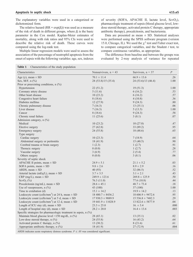
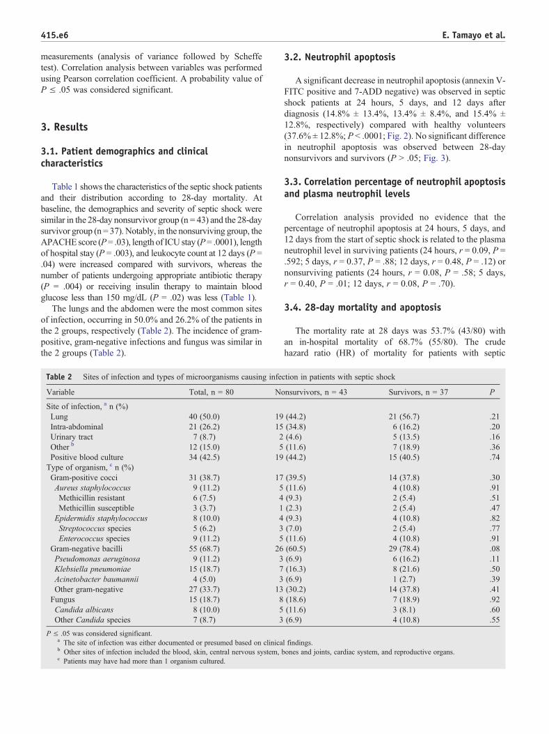
A significant decrease in neutrophil apoptosis (annexin V-FITC positive and 7-ADD negative) was observed in septicshock patients at 24 hours, 5 days, and 12 days afterdiagnosis (14.8% ± 13.4%, 13.4% ± 8.4%, and 15.4% ±12.8%, respectively) compared with healthy volunteers(37.6% ± 12.8%; P b .0001; Fig. 2). No significant differencein neutrophil apoptosis was observed between 28-daynonsurvivors and survivors (P N .05; Fig. 3).
3.3. Correlation percentage of neutrophil apoptosisand plasma neutrophil levels
Correlation analysis provided no evidence that thepercentage of neutrophil apoptosis at 24 hours, 5 days, and12 days from the start of septic shock is related to the plasmaneutrophil level in surviving patients (24 hours, r = 0.09, P =.592; 5 days, r = 0.37, P = .88; 12 days, r = 0.48, P = .12) ornonsurviving patients (24 hours, r = 0.08, P = .58; 5 days,r = 0.40, P = .01; 12 days, r = 0.08, P = .70).
3.4. 28-day mortality and apoptosis
The mortality rate at 28 days was 53.7% (43/80) withan in-hospital mortality of 68.7% (55/80). The crudehazard ratio (HR) of mortality for patients with septic
ction in patients with septic shock
nsurvivors, n = 43 Survivors, n = 37 P
(44.2) 21 (56.7) .21(34.8) 6 (16.2) .20(4.6) 5 (13.5) .16(11.6) 7 (18.9) .36(44.2) 15 (40.5) .74
(39.5) 14 (37.8) .30(11.6) 4 (10.8) .91(9.3) 2 (5.4) .51(2.3) 2 (5.4) .47(9.3) 4 (10.8) .82(7.0) 2 (5.4) .77(11.6) 4 (10.8) .91(60.5) 29 (78.4) .08(6.9) 6 (16.2) .11(16.3) 8 (21.6) .50(6.9) 1 (2.7) .39(30.2) 14 (37.8) .41(18.6) 7 (18.9) .92(11.6) 3 (8.1) .60(6.9) 4 (10.8) .55
l findings.bones and joints, cardiac system, and reproductive organs.
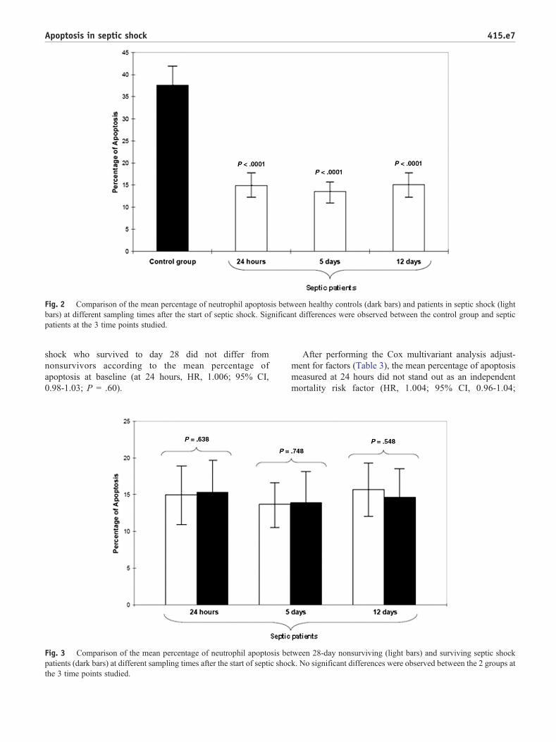
Fig. 2 Comparison of the mean percentage of neutrophil apoptosis between healthy controls (dark bars) and patients in septic shock (lightbars) at different sampling times after the start of septic shock. Significant differences were observed between the control group and septicpatients at the 3 time points studied.
415.e7Apoptosis in septic shock
shock who survived to day 28 did not differ fromnonsurvivors according to the mean percentage ofapoptosis at baseline (at 24 hours, HR, 1.006; 95% CI,0.98-1.03; P = .60).
Fig. 3 Comparison of the mean percentage of neutrophil apoptosis bepatients (dark bars) at different sampling times after the start of septic shocthe 3 time points studied.
After performing the Cox multivariant analysis adjust-ment for factors (Table 3), the mean percentage of apoptosismeasured at 24 hours did not stand out as an independentmortality risk factor (HR, 1.004; 95% CI, 0.96-1.04;
tween 28-day nonsurviving (light bars) and surviving septic shockk. No significant differences were observed between the 2 groups at
Table 3 Independent variables selected by the stepwise Coxregression analysis model as associated with 28-day ICUmortality
Characteristics P Adjusted HR(95.0% CI)
Sex (female, 1) .95 0.98 (0.45-2.11)Age (y) .47 1.01 (0.97-1.05)APACHE II .04 1.13 (1.003-1.28)SOFA .63 0.95 (0.78-1.15)Blood glucose level b150 mg/dL .51 0.99 (0.98-1.008)Low-dose steroid therapy .38 1.40 (0.65-3.03)Activated protein C therapy .30 1.48 (0.70-3.11)Appropriate antibiotic therapy .001 3.46 (1.64-7.26)Apoptosis .83 1.004 (0.96-1.04)
P ≤ .05 was considered significant.
415.e8 E. Tamayo et al.
P = .83). The only identifiable independent mortality riskfactors were appropriate antibiotic therapy and the APACHEII score. Thus, the mortality rate at 28 days was 3.46 timesgreater (95% CI, 1.64-7.26; P = .001) in patients whoreceived an incorrect antibiotic treatment compared withpatients who received the correct treatment. The mortalityrate was also greater with a greater APACHE II score(adjusted HR, 1.13; 95% CI, 1.002-1.28; P = .04).
3.5. Determinant apoptosis factors
Analyzing, by means of multiple linear regression, theeffect of the different factors on the percentage ofneutrophil apoptosis, from day 1 of the onset of sepsis,revealed that age was inversely associated with apoptosis(β = .40; t = 2.34; P = .02; 95% CI, 0.06-0.93),whereas blood glucose level (β = .44; t = 2.52; P = .01;95% CI, 0.01-0.12) and lactate level (β = .48; t = 2.36;P = .02; 95% CI, 0.05-0.79) were positively associatedwith it (Table 4).
Table 4 Multiple linear regression analyses showing the relationship
Variable SE β
Constant 35.41Age (y) 0.21 0.40Sex 4.22 0.22SOFA 1.04 0.12APACHE II 0.57 0.17Lactate level 0.17 0.48ScvO2 0.20 0.14Procalcitonin 0.09 0.31Blood glucose level 0.02 0.44Low-dose steroid therapy 4.47 0.09Activated protein C therapy 4.57 0.07Appropriate antibiotic therapy 4.49 0.18Bacteriemia 4.56 0.13
β indicates standardized regression coefficient; t, corresponding t values. P ≤ .0
4. Discussion
The 2 most relevant findings of this study are thatneutrophil apoptosis is decreased in patients with septicshock for at least 12 days with no significant differencesbetween 28-day nonsurvivors and survivors and thatneutrophil apoptosis is not an independent predictor ofmortality risk. In addition, it was observed that apoptosis wasgreater in relationship with higher levels of blood glucoselevel and lactate level and lower as age increased.
The term apoptosis (from the Greek “fall of the leaves”)was introduced by Kerr et al [21] in 1971 to describe aphysiologic cell death distinct from necrosis or pathologiccell death. In recent years, the study of apoptosis in sepsis hasgained interest due to the fact that it can help explain part ofthe enigmatic pathophysiology of this illness. Most studiescarried out along these lines come from preclinical research,using experimental models of sepsis, that are often based onlethal bacterial toxin-based studies, monospecific intrave-nous microbial challenge, or pretreatment approaches, whichdo not replicate septic patients' status adequately in clinicalpractice [10].
Delayed neutrophil apoptosis has been documented inanimals and septic patients only during the first 24 hoursafter the diagnosis of sepsis [18,22,23]. Guo et al [23]demonstrated that neutrophils in septic mice survive longercompared with nonseptic mice [23]. In septic patients, thedecrease in neutrophil apoptosis was also documented byTaneja et al [18], reporting the percent neutrophil apoptosisin healthy controls as 52% ± 7.8% compared with 6.2% ±1.1% in septic patients; Martins et al [19], reporting themedian neutrophil apoptosis in healthy controls as 11.3% vs9.1% in septic patients; Härter et al [8], reporting a medianpercent neutrophil apoptosis of 64% in healthy controls vs28.8% in septic patients; and Fialkow et al [11], whoreported a median percent neutrophil apoptosis in healthycontrols of 69% ± 1.1% vs 38% ± 3.7% in septic patients. To
between variables and apoptosis
t P 95% CI
−2.97 .006 (−178.17, −32.60)2.34 .02 (0.06, 0.93)1.51 .14 (−2.26, 15.10)0.75 .45 (−1.35, 2.93)0.86 .39 (−0.68, 1.68)2.36 .02 (0.05, 0.79)0.83 .41 (−0.25, 0.60)1.67 .10 (−0.03, 0.35)2.52 .01 (0.01, 0.12)0.58 .56 (−6.57, 11.84)0.47 .64 (−7.24, 11.55)1.14 .26 (−4.08, 14.37)0.80 .43 (−5.72, 13.03)
5 was considered significant.
415.e9Apoptosis in septic shock
the contrary of that previously stated, our results confirmdecreased neutrophil apoptosis not only during the first 24hours after the onset of sepsis but also that this reduction ismaintained over a period of at least 12 days.
In the present study, we detected no differences inneutrophil apoptosis between nonsurviving and survivingpatients. This finding may be due to the fact that septic shockis the most serious form of sepsis and produces a moreintense inhibition of apoptosis [11]. Consequently, the returnto normal values of apoptosis in surviving patients happenslater. Similarly, the present study found no correlationbetween decreased plasma neutrophil apoptosis and leuko-cytosis in surviving and nonsurviving patients. This fact issurprising because an important mechanism of leukocytosisthat can be observed in septic shock is explained by thestimulation of leukocyte production in the bone marrow [24].On the other hand, we did observe lower blood leukocytelevels in surviving patients on day 12 of septic shock.
The 28-day mortality observed in the present study wassimilar to that reported in other studies including patientswith the same clinical risk [25]. The patients included in ourstudy had an average APACHE score of 23.2 and SOFAscore of 9.4, and Cox multiple regression analysis did notshow that the mean percentage of apoptosis is anindependent predictor of mortality in patients with septicshock. We must point out that, of the independent variablesselected by Cox regression analysis, we only identifiedcorrect antibiotic treatment as an independent risk factor formortality. This result strengthens the recommendations ofthe Surviving Sepsis Campaign [26], which showed thatcorrect, early, wide-spectrum antibiotherapy is a fundamen-tal pillar in handling sepsis.
Different factors have been cited as being able toinfluence neutrophil apoptosis (age, blood glucose level,corticoids, hypoxia, tobacco, etc) [27,28]; however, theyhave not been analyzed in patients with sepsis. In our study,carried out on patients with sepsis, we observed greaterneutrophil apoptosis in those with higher levels of bloodglucose level and lactate level and lesser as age increased.These findings are concordant with those observed in
medical literature and described under different conditions[27,28]. We believe that, as this study was not designed toclarify these issues, other studies should be carried out aimedat analyzing them in more detail.
The present study has 2 main limitations. First, theevaluation of neutrophil apoptosis was carried out during thefirst 12 days after the onset of the clinical symptoms of septicshock. However, the study lasting until the normalization ofapoptosis would possibly have been of interest. Furthermore,because apoptosis occurs via a complex signaling cascadethat is tightly regulated at multiple points, there are manyopportunities to evaluate the activity of the proteins involved(cytomorphological alterations, DNA fragmentation, detec-tion of caspases, membrane alterations, detection ofapoptosis in whole mounts, mitochondrial assays) [29].There are a large variety of assays available, but each assayhas advantages and disadvantages [29]. We have usedannexin V-FITC labeling to measure apoptosis. Theadvantage is sensitivity (it can detect a single apoptoticcell). The disadvantage is that the membranes of necroticcells are labeled as well. To avoid that and to demonstrate themembrane integrity of the phosphatidylserine-positive cells,neutrophils were stained simultaneously with 7-ADD and inparallel with May-Grünwald-Giemsa. The cells that showedshrinkage, nuclear coalescence, and nuclear chromatincondensation were considered as apoptotic.
5. Conclusions
In conclusion, over the first 12 days of septic shockdevelopment, the level of neutrophil apoptosis decreased anddid not return to normal values, with no difference betweensurviving and nonsurviving patients. Moreover, the deter-minant factors for apoptosis levels were age, blood glucoselevel, and lactate level. Consequently, we concluded thatapoptosis in sepsis is a phenomenon that requires furtherstudy to clearly determine its role before proposing that newmedical treatments for sepsis have antiapoptotic effects.
415.e10 E. Tamayo et al.
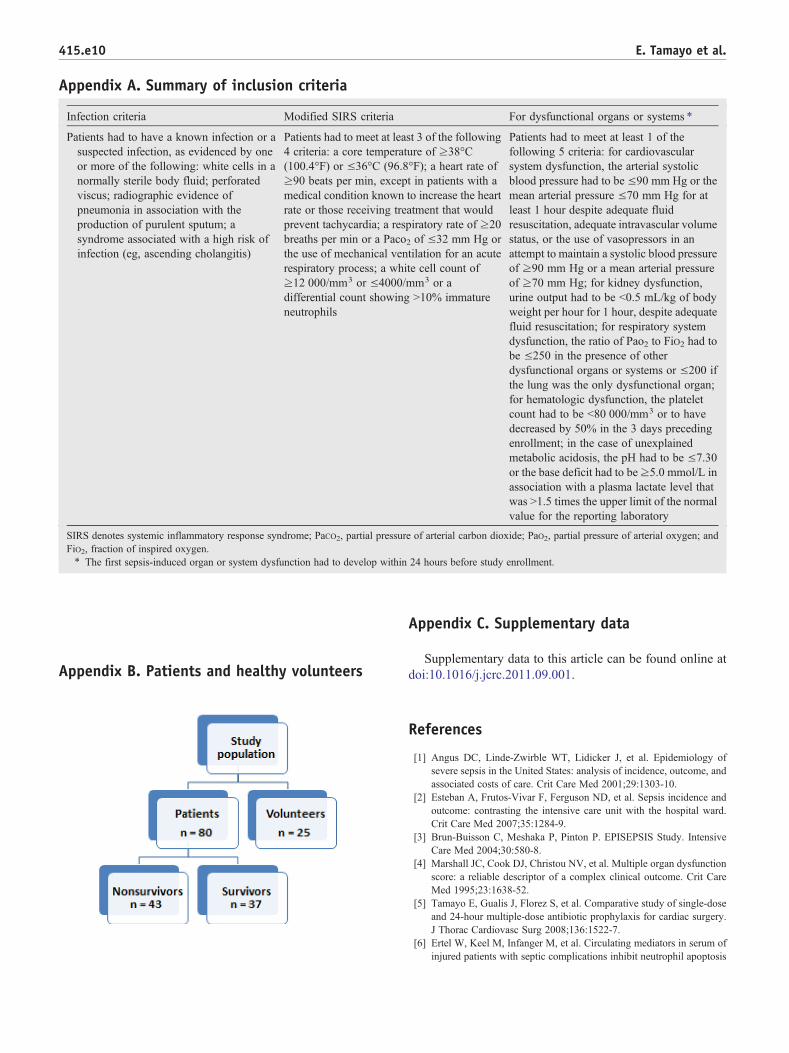
Appendix A. Summary of inclusion criteria
Infection criteria
Modified SIRS criteria For dysfunctional organs or systems ⁎Patients had to have a known infection or asuspected infection, as evidenced by oneor more of the following: white cells in anormally sterile body fluid; perforatedviscus; radiographic evidence ofpneumonia in association with theproduction of purulent sputum; asyndrome associated with a high risk ofinfection (eg, ascending cholangitis)
Patients had to meet at least 3 of the following4 criteria: a core temperature of ≥38°C(100.4°F) or ≤36°C (96.8°F); a heart rate of≥90 beats per min, except in patients with amedical condition known to increase the heartrate or those receiving treatment that wouldprevent tachycardia; a respiratory rate of ≥20breaths per min or a Paco2 of ≤32 mm Hg orthe use of mechanical ventilation for an acuterespiratory process; a white cell count of≥12 000/mm3 or ≤4000/mm3 or adifferential count showing N10% immatureneutrophils
Patients had to meet at least 1 of thefollowing 5 criteria: for cardiovascularsystem dysfunction, the arterial systolicblood pressure had to be ≤90 mm Hg or themean arterial pressure ≤70 mm Hg for atleast 1 hour despite adequate fluidresuscitation, adequate intravascular volumestatus, or the use of vasopressors in anattempt to maintain a systolic blood pressureof ≥90 mm Hg or a mean arterial pressureof ≥70 mm Hg; for kidney dysfunction,urine output had to be b0.5 mL/kg of bodyweight per hour for 1 hour, despite adequatefluid resuscitation; for respiratory systemdysfunction, the ratio of Pao2 to FiO2 had tobe ≤250 in the presence of otherdysfunctional organs or systems or ≤200 ifthe lung was the only dysfunctional organ;for hematologic dysfunction, the plateletcount had to be b80 000/mm3 or to havedecreased by 50% in the 3 days precedingenrollment; in the case of unexplainedmetabolic acidosis, the pH had to be ≤7.30or the base deficit had to be≥5.0 mmol/L inassociation with a plasma lactate level thatwas N1.5 times the upper limit of the normalvalue for the reporting laboratory
SIRS denotes systemic inflammatory response syndrome; PaCO2, partial pressure of arterial carbon dioxide; PaO2, partial pressure of arterial oxygen; andFiO2, fraction of inspired oxygen.⁎ The first sepsis-induced organ or system dysfunction had to develop within 24 hours before study enrollment.
Appendix B. Patients and healthy volunteers
Appendix C. Supplementary data
Supplementary data to this article can be found online atdoi:10.1016/j.jcrc.2011.09.001.
References
[1] Angus DC, Linde-Zwirble WT, Lidicker J, et al. Epidemiology ofsevere sepsis in the United States: analysis of incidence, outcome, andassociated costs of care. Crit Care Med 2001;29:1303-10.
[2] Esteban A, Frutos-Vivar F, Ferguson ND, et al. Sepsis incidence andoutcome: contrasting the intensive care unit with the hospital ward.Crit Care Med 2007;35:1284-9.
[3] Brun-Buisson C, Meshaka P, Pinton P. EPISEPSIS Study. IntensiveCare Med 2004;30:580-8.
[4] Marshall JC, Cook DJ, Christou NV, et al. Multiple organ dysfunctionscore: a reliable descriptor of a complex clinical outcome. Crit CareMed 1995;23:1638-52.
[5] Tamayo E, Gualis J, Florez S, et al. Comparative study of single-doseand 24-hour multiple-dose antibiotic prophylaxis for cardiac surgery.J Thorac Cardiovasc Surg 2008;136:1522-7.
[6] Ertel W, Keel M, Infanger M, et al. Circulating mediators in serum ofinjured patients with septic complications inhibit neutrophil apoptosis
415.e11Apoptosis in septic shock
through up-regulation of protein-tyrosine phosphorylation. J Trauma1998;44:767-77.
[7] Bone RC, Balk RA, Cerra FB, et al. Definition for sepsis and organfailure and guidelines for the use of innovative therapies in sepsis.Chest 1992;101:1644-55.
[8] Härter L, Mica L, Stocker R, et al. Mcl-1 correlates with reducedapoptosis in neutrophils from patients with sepsis. J Am Coll Surg2003;197:964-73.
[9] Marshall JC, Watson RW. Apoptosis in the resolution of systemicinflammation. In: Vincent JL, editor. Yearbook of intensive care andemergency medicine. Berlin: Springer-Verlag; 1997.
[10] Hotchkiss RS, Karl IE. The pathophysiology and treatment of sepsis.N Engl J Med 2003;348:138-48.
[11] Fialkow L, Fochesatto Filho L, Bozzetti MC, et al. Neutrophilapoptosis: a marker of disease severity in sepsis and sepsis-inducedacute respiratory distress syndrome. Crit Care 2006;10:R155.
[12] Wesche DE, Lomas-Neira JL, Perl M, et al. Leukocyte apoptosis andits significance in sepsis and shock. J Leukoc Biol 2005;78:325-37.
[13] Rivers E, Nguyen B, Havstad S, et al. Early goal-directed therapy inthe treatment of severe sepsis and septic shock. N Engl J Med2001;345:1368-77.
[14] Ferreira FL, Bota DP, Bross A, et al. Serial evaluation of the SOFAscore to predict outcome in critically ill patients. JAMA 2001;286:1754-8.
[15] Fonteriz RI, Sánchez A, Mollinedo F, et al. The role of intracellularacidification in calcium mobilization in human neutrophils. BiochemBiophys Acta 1991;1093:1-6.
[16] Knaus WA, Draper EA, Wagner DP, et al. APACHE II: a severity ofdisease classification system. Crit Care Med 1985;13:818-29.
[17] Moreno R, Vincent JL, Matos R, et al. The use of maximum SOFAscore to quantify organ dysfunction/failure in intensive care. Results ofa prospective, multicentre study. Working Group on Sepsis relatedProblems of the ESICM. Intensive Care Med 1999;25:686-96.
[18] Taneja R, Parodo J, Jia SH, et al. Delayed neutrophil apoptosis insepsis is associated with maintenance of mitochondrial transmembranepotential and reduced caspase-9 activity. Crit Care Med 2004;32:1460-6.
[19] Martins PS, Kallas EG, Neto MC, et al. Upregulation of reactiveoxygen species generation and phagocytosis and increased apoptosisin human neutrophils during severe sepsis and septic shock. SHOCK2003;20:208-12.
[20] Cox DR. Regression models and life-tables. J Stat Soc B 1972;34:187-220.
[21] Kerr JF, Willye AH, Curie AR. Apoptosis: a basis biologicalphenomenon with wideranging implications in tissue kinetics. Br JCancer 1972;26:239-57.
[22] Jimenez MF, Watson RW, Parodo J, et al. Dysregulated expression ofneutrophil apoptosis in the systemic inflammatory response syndrome.Arch Surg 1997;132:1263-70.
[23] Guo RF, Sun L, Gao H, et al. In vivo regulation of neutrophil apoptosisby C5a during sepsis. Leukoc Biol 2006;80:1575-83.
[24] Smith JA. Neutrophils, host defense, and inflammation: a double-edged sword. J Leukoc Biol 1994;56:672-86.
[25] Kumar A, Roberts D, Wood KE, et al. Duration of hypotension beforeinitiation of effective antimicrobial therapy is the critical determinantof survival in human septic shock. Crit Care Med 2006;34:1589-96.
[26] Dellinger RP, Levy MM, Carlet JM, et al. Surviving sepsis campaign:international guidelines for management of severe sepsis and septicshock. Intensive Care Med 2008;34:17-60.
[27] Duffin R, Leitch AE, Fox S, et al. Targeting granulocyte apoptosis:mechanisms, models, and therapies. Immunol Rev 2010;236:28-40.
[28] Fülöp Jr T, Fouquet C, Allaire P, et al. Changes in apoptosis of humanpolymorphonuclear granulocytes with aging. Mech Ageing Dev1997;1-3:15-34.
[29] Elmore S. Apoptosis: a review of programmed cell death. ToxicolPathol 2007;35:495-516.