Systemic Neutrophil Depletion Modulates the Migration and ...
19
Development/Plasticity/Repair Systemic Neutrophil Depletion Modulates the Migration and Fate of Transplanted Human Neural Stem Cells to Rescue Functional Repair X Hal X. Nguyen, 1,2,4 X Mitra J. Hooshmand, 1,2,3 Hirokazu Saiwai, 1,2,4 Jake Maddox, 1,5 X Arjang Salehi, 1,5 X Anita Lakatos, 1,2 X Rebecca A. Nishi, 2 X Desiree Salazar, 2,3,4 Nobuko Uchida, 6 and X Aileen J. Anderson 1,2,3,4 1 Sue and Bill Gross Stem Cell Research Center, 2 Institute for Memory Impairments and Neurological Disorders, and Departments of 3 Anatomy and Neurobiology, 4 Physical Medicine and Rehabilitation, and 5 Molecular Biology and Biochemistry, University of California, Irvine, Irvine, California 92697, and 6 StemCells, Inc., Palo Alto, California 94304 The interaction of transplanted stem cells with local cellular and molecular cues in the host CNS microenvironment may affect the potential for repair by therapeutic cell populations. In this regard, spinal cord injury (SCI), Alzheimer’s disease, and other neurological injuries and diseases all exhibit dramatic and dynamic changes to the host microenvironment over time. Previously, we reported that delayed transplantation of human CNS-derived neural stem cells (hCNS-SCns) at 9 or 30 d post-SCI (dpi) resulted in extensive donor cell migration, predominantly neuronal and oligodendrocytic donor cell differentiation, and functional locomotor improvements. Here, we report that acute transplantation of hCNS-SCns at 0 dpi resulted in localized astroglial differentiation of donor cells near the lesion epicenter and failure to produce functional improvement in an all-female immunodeficient mouse model. Critically, specific immu- nodepletion of neutrophils (polymorphonuclear leukocytes) blocked hCNS-SCns astroglial differentiation near the lesion epicenter and rescued the capacity of these cells to restore function. These data represent novel evidence that a host immune cell population can block the potential for functional repair derived from a therapeutic donor cell population, and support targeting the inflammatory microen- vironment in combination with cell transplantation after SCI. Key words: cell therapy; functional repair; immunotherapy; neuroinflammation; neutrophils; spinal cord injury Introduction The interaction of transplanted cell populations with local cellu- lar and molecular cues in the host microenvironment is a key variable that remains to be elucidated and may shape the trans- lational application of therapeutic cell populations for a clinical spinal cord injury (SCI) population. A principle factor altering the molecular and cellular composition of the host niche is time after injury, which is in part characterized by the recruitment of inflammatory mediators in distinct stages and distributions be- ginning at the acute [7 d post-SCI (dpi)] and persisting through Received Sept. 1, 2016; revised June 15, 2017; accepted July 30, 2017. Author contributions: H.X.N., M.J.H., and A.J.A. designed research; H.X.N., M.J.H., H.S., J.M., A.S., R.A.N., and D.S. performed research; N.U. contributed unpublished reagents/analytic tools; H.X.N., M.J.H., H.S., J.M., A.S., A.L., and R.A.N. analyzed data; H.X.N., M.J.H., and A.J.A. wrote the paper. This study was funded by National Institutes of Health (NIH) Grant NS-49885 (to A.J.A.), California Institute for Regenerative Medicine (CIRM) Grant RB2-01496 (to H.X.N. and A.J.A.), Paralysis Project of America Grant PPA-32574 (to H.X.N.), CIRM Stem Cell Training Award T1-00008 (to H.X.N.), Christopher and Dana Reeve Foundation Grant CRF-AAC 2005 (to A.J.A.), National Center for Research Resources and the National Center for Advancing Transla- tional Sciences (NIH) Grant UL1-TR-001414), The Morton Cure Paralysis Fund MCPF-204464 (to H.X.N.), and Stem- Cells, Inc. We thank the technical staff at the Christopher and Dana Reeve Foundation Animal Injury Core: Hongli Liu and Jovanny Lucero, as well as Gabriella Funes, Joel Trushinsky, and Dena Yassin. N.U. has a financial interest in StemCells, Inc. The authors declare no other competing financial interests. Correspondence should be addressed to either of the following: Hal X. Nguyen, 2101 Sue and Bill Gross Hall, University of California, Irvine, Irvine, CA 92697, E-mail: [email protected]; or Aileen J. Anderson, 2026 Sue and Bill Gross Hall, University of California, Irvine, Irvine, CA 92697, E-mail: [email protected]. DOI:10.1523/JNEUROSCI.2785-16.2017 Copyright © 2017 the authors 0270-6474/17/379269-19$15.00/0 Significance Statement The interaction of transplanted cells with local cellular and molecular cues in the host microenvironment is a key variable that may shape the translation of neurotransplantation research to the clinical spinal cord injury (SCI) human population, and few studies have investigated these events. We show that the specific immunodepletion of polymorphonuclear leukocyte neutrophils using anti-Ly6G inhibits donor cell astrogliosis and rescues the capacity of a donor cell population to promote locomotor improvement after SCI. Critically, our data demonstrate novel evidence that a specific host immune cell population can block the potential for functional repair derived from a therapeutic donor cell population. The Journal of Neuroscience, September 20, 2017 • 37(38):9269 –9287 • 9269
-
Upload
khangminh22 -
Category
Documents
-
view
10 -
download
0
Transcript of Systemic Neutrophil Depletion Modulates the Migration and ...

Development/Plasticity/Repair
Systemic Neutrophil Depletion Modulates the Migration andFate of Transplanted Human Neural Stem Cells to RescueFunctional Repair
X Hal X. Nguyen,1,2,4 X Mitra J. Hooshmand,1,2,3 Hirokazu Saiwai,1,2,4 Jake Maddox,1,5 X Arjang Salehi,1,5
X Anita Lakatos,1,2 X Rebecca A. Nishi,2 X Desiree Salazar,2,3,4 Nobuko Uchida,6 and X Aileen J. Anderson1,2,3,4
1Sue and Bill Gross Stem Cell Research Center, 2Institute for Memory Impairments and Neurological Disorders, and Departments of 3Anatomy andNeurobiology, 4Physical Medicine and Rehabilitation, and 5Molecular Biology and Biochemistry, University of California, Irvine, Irvine, California 92697,and 6StemCells, Inc., Palo Alto, California 94304
The interaction of transplanted stem cells with local cellular and molecular cues in the host CNS microenvironment may affect thepotential for repair by therapeutic cell populations. In this regard, spinal cord injury (SCI), Alzheimer’s disease, and other neurologicalinjuries and diseases all exhibit dramatic and dynamic changes to the host microenvironment over time. Previously, we reported thatdelayed transplantation of human CNS-derived neural stem cells (hCNS-SCns) at 9 or 30 d post-SCI (dpi) resulted in extensive donor cellmigration, predominantly neuronal and oligodendrocytic donor cell differentiation, and functional locomotor improvements. Here, wereport that acute transplantation of hCNS-SCns at 0 dpi resulted in localized astroglial differentiation of donor cells near the lesionepicenter and failure to produce functional improvement in an all-female immunodeficient mouse model. Critically, specific immu-nodepletion of neutrophils (polymorphonuclear leukocytes) blocked hCNS-SCns astroglial differentiation near the lesion epicenter andrescued the capacity of these cells to restore function. These data represent novel evidence that a host immune cell population can blockthe potential for functional repair derived from a therapeutic donor cell population, and support targeting the inflammatory microen-vironment in combination with cell transplantation after SCI.
Key words: cell therapy; functional repair; immunotherapy; neuroinflammation; neutrophils; spinal cord injury
IntroductionThe interaction of transplanted cell populations with local cellu-lar and molecular cues in the host microenvironment is a key
variable that remains to be elucidated and may shape the trans-lational application of therapeutic cell populations for a clinicalspinal cord injury (SCI) population. A principle factor alteringthe molecular and cellular composition of the host niche is timeafter injury, which is in part characterized by the recruitment ofinflammatory mediators in distinct stages and distributions be-ginning at the acute [�7 d post-SCI (dpi)] and persisting through
Received Sept. 1, 2016; revised June 15, 2017; accepted July 30, 2017.Author contributions: H.X.N., M.J.H., and A.J.A. designed research; H.X.N., M.J.H., H.S., J.M., A.S., R.A.N., and D.S.
performed research; N.U. contributed unpublished reagents/analytic tools; H.X.N., M.J.H., H.S., J.M., A.S., A.L., andR.A.N. analyzed data; H.X.N., M.J.H., and A.J.A. wrote the paper.
This study was funded by National Institutes of Health (NIH) Grant NS-49885 (to A.J.A.), California Institute forRegenerative Medicine (CIRM) Grant RB2-01496 (to H.X.N. and A.J.A.), Paralysis Project of America Grant PPA-32574(to H.X.N.), CIRM Stem Cell Training Award T1-00008 (to H.X.N.), Christopher and Dana Reeve Foundation GrantCRF-AAC 2005 (to A.J.A.), National Center for Research Resources and the National Center for Advancing Transla-tional Sciences (NIH) Grant UL1-TR-001414), The Morton Cure Paralysis Fund MCPF-204464 (to H.X.N.), and Stem-Cells, Inc. We thank the technical staff at the Christopher and Dana Reeve Foundation Animal Injury Core: Hongli Liuand Jovanny Lucero, as well as Gabriella Funes, Joel Trushinsky, and Dena Yassin.
N.U. has a financial interest in StemCells, Inc. The authors declare no other competing financial interests.Correspondence should be addressed to either of the following: Hal X. Nguyen, 2101 Sue and Bill Gross Hall,
University of California, Irvine, Irvine, CA 92697, E-mail: [email protected]; or Aileen J. Anderson, 2026 Sue and BillGross Hall, University of California, Irvine, Irvine, CA 92697, E-mail: [email protected].
DOI:10.1523/JNEUROSCI.2785-16.2017Copyright © 2017 the authors 0270-6474/17/379269-19$15.00/0
Significance Statement
The interaction of transplanted cells with local cellular and molecular cues in the host microenvironment is a key variable that mayshape the translation of neurotransplantation research to the clinical spinal cord injury (SCI) human population, and few studieshave investigated these events. We show that the specific immunodepletion of polymorphonuclear leukocyte neutrophils usinganti-Ly6G inhibits donor cell astrogliosis and rescues the capacity of a donor cell population to promote locomotor improvementafter SCI. Critically, our data demonstrate novel evidence that a specific host immune cell population can block the potential forfunctional repair derived from a therapeutic donor cell population.
The Journal of Neuroscience, September 20, 2017 • 37(38):9269 –9287 • 9269

the subacute (7–14 dpi) and chronic (�14 dpi) phases of trauma(Popovich et al., 1997; Jones et al., 2005; Fleming et al., 2006;Kigerl et al., 2006; Beck et al., 2010). In particular, a multiphasiccellular response has been identified in a rodent SCI model thatincludes an early neutrophil [polymorphonuclear leukocytes(PMNs) at 1 dpi] infiltration, and a biphasic microglia/macro-phage (7 and 60 dpi) response (Beck et al., 2010; Nguyen et al.,2011) Accumulating evidence supporting a dual role for infiltrat-ing immune cells has suggested both detrimental (Popovich et al.,1999; Weaver et al., 2000; Jones et al., 2005) and beneficial (Becket al., 2010; Rapalino et al., 1998; Schwartz and Yoles 2005; Davidand Kroner, 2011) effects on regeneration and repair after SCI.
A number of studies have suggested that inflammatory cyto-kines can modulate neural stem cell (NSC) proliferation and dif-ferentiation in vitro (Butovsky et al., 2006; Kokaia et al., 2012). Inparallel, microglia have been shown to induce migration and/orenhance neural lineage selection in mouse neural progenitors invitro (Aarum et al., 2003) and in endogenous hippocampal progen-itors in vivo (Monje et al., 2003). However, the effect of modulationof the inflammatory or immune microenvironment on the migra-tion, differentiation, or therapeutic efficacy of a transplanted cellpopulation has not previously been tested. Critically, the studiespresented here are thus unique, demonstrating novel evidence that ahost immune cell population blocks the potential for functional re-pair derived from a transplanted therapeutic cell population.
In the present study, we used human CNS-derived NSCs(hCNS-SCns) propagated as neurospheres (Uchida et al., 2000),which are capable of differentiation into human neurons, oligo-dendrocytes, and astrocytes in vitro and in vivo (Tamaki et al.,2002), and retain multipotency for �20 passages. We investi-gated the survival and engraftment of hCNS-SCns in animalsreceiving transplants acutely (0 dpi) after SCI and comparedthese results relative to animals transplanted at a delayed time of30 dpi in an otherwise identical paradigm. Contrary to conven-tional predictions (Nakamura and Okano 2013), the results dem-onstrated comparable engraftment of transplanted cells at bothtime points. However, in comparison with delayed transplants,animals receiving hCNS-SCns at 0 dpi exhibited a clear shift indonor cell localization at the injury epicenter, which was associ-ated with a striking increase in astroglial lineage selection, andfailure to exhibit recovery of function. Because the acute injurymicroenvironment is associated with robust activation of the innateinflammatory response, including transient early accumulation ofPMNs at the lesion epicenter (Beck et al., 2010), we next investigatedwhether specific PMN depletion could alter the fate and migration oftransplanted hCNS-SCns and restore the potential for donor cells toimprove locomotor function. We demonstrate that PMN depletionvia anti-Ly6G treatment was specific and sustained and resulted inthe release of donor human cell localization to the injury epicenter,the inhibition of human astrocyte differentiation, and the rescueof the capacity of transplanted hCNS-SCns to improve locomotorrecovery. Together, these data demonstrate a potential therapeuticstrategy to modulate the host CNS microenvironment and promotefunctional repair by a donor cell population.
Materials and MethodsExclusions, final numbers, and experimental blinding. All surgical, behav-ioral, histological, and quantitative analyses were performed by observersblinded to groups. Preinjury and postinjury animal exclusions, Grubbstest exclusions, and final animal numbers for statistical analysis are de-tailed below. There were no animal exclusions due to engraftment failureor at the stage of histological analysis. The number of animals that re-ceived SCI and cell transplant and the number of animals that were used
for stereological analysis and behavioral assessments for all experimentsare summarized in Table 1.
Acute versus delayed cell transplantation studiesContusion SCI and cell transplantation. Female NOD-scid mice (N �16/group, 10 weeks old; The Jackson Laboratory) were anesthetized us-ing Avertin (0.5 ml/20 g tribromo-ethanol) and received a laminectomyat the thoracic vertebrae 9 (T9) using a surgical microscope. The animalswere secured in a spinal stereotactic apparatus to receive 50 kilodyn(1 dyn � 10 �N) contusion injuries using the Infinite Horizon (IH)Impactor (Precision Systems and Instrumentation) at T9. Before SCI,animals were randomly assigned to receive either hCNS-SCns or vehicleinjections. A priori exclusion criteria were as follows: evidence of cord bruis-ing during laminectomy, less than �10% of 50 kilodyn force based on force/displacement curve, or evidence of unilateral cord bruising after SCI.Exclusion of animals was determined by investigators blinded to groups.
hCNS-SCns were provided by StemCells and are derived via fluo-rescence-activated cell sorting from donated fetal brain tissue and ex-panded as neurospheres based on expression of the stem cell markerCD133 �, lack of the hematopoietic markers CD34 � and CD45 �, andlow levels of CD24 lo. Sorting by these markers has previously been shownto result in a highly enriched population of human NSCs (hNSCs), andthe development and transplantation of these cells has been described indetail previously (Uchida et al., 2000). Polished siliconized beveled glasspipettes (bevel: inner diameter, 70 �m; outer diameter, 100 –110 �m;Sutter Instruments) were loaded with freshly triturated Passage 9 –10multipotent hCNS-SCns (75,000 cells/�l injection buffer) or vehicle in-jection buffer (50% X-Vivo, 50% HBSS), which were injected into theintact parenchyma at T9 using a NanoInjector system and microposi-tioner (WPI Instruments). Each site received 250 nl of cells or vehicle,delivered in 50 nl volumes over 75 s, followed by a 2 min delay beforewithdrawal of the pipette (Cummings et al., 2005). Previous studies inour laboratory have demonstrated that this volume does not exacerbatedamage to the spinal cord. In an acute cell transplant model, mice re-ceived hCNS-SCns immediately after SCI. In a delayed cell transplantmodel, animals were reanesthetized at 30 dpi, sutures were removed, andthe laminectomy/injured site at T9 was re-exposed. In both paradigms,animals received four injections of hCNS-SCns or vehicle into the intactparenchyma adjacent to the contusion site (Cummings et al., 2005). Thesurvival time for all cell-transplanted and vehicle-injected animals was12–16 weeks post-SCI.
Table 1. Number of animals
Vehicle (N) Acute Tx (N) Delayed Tx (N)
Acute vs delayed TxSCI and cell Tx 16 16 16SC121 human cell stereology N/A 6 6SC123 human astrocyte stereology N/A 6 N/AFibronectin lesion volume 6 6 N/ACatWalk gait analysis 16 15 N/A
IgG2a (N) Anti-Ly6G (N)
PMN depletion studySCI only 8 8
Acute TxSCI and cell Tx 12 12Fibronectin lesion volume 5 5SC121 human cell stereology 5 5SC123 human astrocyte stereology 5 5SC121 cluster volume 5 5SC123 cluster volume 5 5CatWalk gait analysis 10 10Hargreaves 10 10
VehicleSCI and vehicle injection 12 12CatWalk gait analysis 12 10Hargreaves 12 10
Tx, Transplantation.
9270 • J. Neurosci., September 20, 2017 • 37(38):9269 –9287 Nguyen et al. • Anti-Ly6G Rescues Donor Stem Cell Potential
After cell or vehicle injections, a small piece of gel-foam was placedover the site of laminectomy and the musculature was closed with su-tures. Animals received lactated Ringer’s solution (50 ml/kg, s.c.) imme-diately after surgery and for 3–5 d postsurgery, as well as buprenorphine(0.5 mg/kg, s.c.) immediately after injury and for 2 d thereafter. Due tothe immunodeficient nature of NOD-scid mice, and to avoid the possi-bility of bladder infections, antibiotics were administered daily through-out the 12–16 week duration of the study, rotating the use of Baytril,ciproflaxacin, and ampicillin (2.5 mg/kg, s.c., dose for all drugs) every2 weeks.
Tissue collection and processing. Mice were anesthetized at 12–16 weekspostinjury and transcardially perfused with PBS followed by 4% PBS-buffered paraformaldehyde. Spinal cord regions corresponding to dorsalroots at T2–T5, T6 –T12, and T13–L2 were dissected and postfixed over-night in 4% PBS-buffered paraformaldehyde, flash frozen at �65°C inisopentane, and stored at �80°C. The T6 –T12 spinal cords were embed-ded in Tissue-Tek optimal cutting temperature (OCT) compound andsectioned parasagittally at 30 �m on a sliding microtome (Microm).Parasagittal sections were collected and stored in 96-well tissue cultureplates filled with 100 mM Tris with 0.02% sodium azide and kept at 4°Cuntil processed for immunostaining. For diaminobenzidine (DAB) immu-nohistochemistry, tissue sections were incubated with 10% MeOH/3%H2O2 in 100 mM Tris to inactivate endogenous peroxide activity andimmunostained as described in the study by Hooshmand et al. (2009).Methyl green counterstain was used to visualize nuclei (Sigma-Aldrich).Fluorescence-conjugated immunohistochemistry was performed as de-scribed in the study Hooshmand et al. (2009). All antibodies and dilu-tions are listed in Table 2.
Stereological analysis. Animals containing less than three sections witha visible injury epicenter (minimum number required for obtaining re-liable results) were excluded from stereological analyses. Stereology of arandomly selected subgroup of animals (N � 6/group) was conductedusing an Olympus BX51 Microscope with a motorized stage and Stereo-Investigator software (version 7.00.3; MBF Biosciences). The OpticalFractionator probe (Joelving et al., 2006) was used to count the numberof SC121 human cells and SC123 human astrocytes. The counting areaswere determined by drawing contours at every 1 mm region (for SC121 �
cells) and around the entire spinal cord (for SC123 � cells) using a 4�objective. SC121 or SC123 cells associated with a visible methyl greennuclear stain were counted using a 100� objective in the correspondingareas. Grid size measurements were determined based on preliminaryexperimentation to establish low coefficient of error and sufficient totalestimated counts per frame (3–5 cells/counting frame). A minimum ofthree sections where the injury epicenter was clearly visible was analyzedfor each animal. Quantification in StereoInvestigator yielded the totalnumber of SC121 or SC123/methyl-green double-labeled cells withineach region or the entire spinal cord, respectively. Total SC121 count wascalculated as the sum of cells in all 1 mm regions. Migration was reportedas the total number of cells at each region.
Stereological data for SC123 � and SC121 � donor cells was also usedto determine the proportion of SC123 � cells near the lesion (within1 mm of the epicenter) versus away from the lesion (�1 mm distal toepicenter), based on the identification of the lesion using fibronectinimmunoreactivity. For this analysis, the lesion epicenter was identified asthe center line of fibronectin immunoreactivity in alternate parasagittalsections, and the total number of cells within 2 mm of this center linewere determined to have proximal localization using StereoInvestigatorreconstruction of optical fractionator data.
Volume analyses were performed using the Cavalieri estimator probein StereoInvestigator. Lesion volume analysis was performed using fi-
bronectin immunostaining. The fibronectin � region was identified asthe lesion epicenter, and a point-grid of known spacing (100 �m apart)was randomly overlayed on the section image. Points were counted usinga 20� objective within the region of interest. This analysis yielded theestimated lesion volume.
PMN depletion studiesContusion SCI and cell transplantation. The effect of PMN depletion wasonly tested in an acute cell transplant paradigm post-SCI. As describedabove, female NOD-scid mice (N � 12/group, 10 weeks old; The JacksonLaboratory) received a laminectomy at the T9 and 50 kilodyn contusioninjuries using the IH Impactor (Precision Systems and Instrumentation)at T9. Before SCI, animals were randomly assigned to receive eitheranti-Ly6G or IgG2a intraperitoneal injections, described below, followedby hCNS-SCns transplants or vehicle injections (Table 1). On the day oftransplantation, Passage 9 multipotent hCNS-SCns grown as neurosphereswere dissociated into a single-cell suspension, and cells (75,000/�l) or vehiclewere injected into the intact parenchyma at T9. Immediately after SCI, eachmouse received a total of four injections of hCNS-SCns or vehicle in theintact parenchyma adjacent to the contusion site (Cummings et al.,2005). The survival time for all animals was 90 dpi (12 weeks post-SCI).
Anti-Ly6G intraperitoneal injections and flow cytometric analysis. PMNdepletion was accomplished using anti-Ly6G (i.p. injections, 100 �l vol-ume; Clone 1A8, Bio X Cell) according to a modified injection schemefrom a previous study (Saiwai et al., 2013). One day before SCI and celltransplantation, mice (N � 12/group) received 200 �g of either anti-Ly6G or IgG2a isotype (Bio X Cell) control. Mice also received additionalinjections (100 �g) of anti-Ly6G or IgG2a immediately following SCI/transplantation and every 3 d after for up to 15 dpi. Anti-Ly6G injectionsdid not result in any fatality or adverse effect/toxicity. Furthermore, anadditional subset of animals (N � 8 per group) that received SCI only(without cell transplant) was used to confirm short-term PMN depletionby anti-Ly6G in the blood and spinal cord. These animals were sacrificedat 1 dpi (N � 4) or 14 dpi (N � 4), while the rest of the animals thatreceived cell transplant and either anti-Ly6G or IgG2 were sacrificedat 90 dpi (Table 1).
PMN depletion in the blood and spinal cord was assessed using flowcytometry. Briefly, blood samples (20 �l) from the tail vein were collectedin 1.5 ml Eppendorf Tubes containing 1 ml of cold PBS with 2 mM EDTA.Spinal cord tissues from T8 –T10 from randomly selected anti-Ly6G- andIgG2a-treated animals were removed, and dissociated mechanically andenzymatically according to previous study (Beck et al., 2010). Spinal cordcell solution was further purified from myelin debris using Myelin Re-moval Magnetic Beads II Kit on an autoMACS Pro Separator (MiltenyiBiotec) according to the manufacturer instructions.
Collected cells isolated from blood or spinal cord tissues were sus-pended in 0.85% NH4Cl2 lysis solution and washed with 1 ml of PBSwith 2 mM EDTA. A master mix of antibodies (all were obtained fromBioLegend and diluted at 1:100 in PBS) was prepared containing PE/Cy7anti-mouse CD45 (catalog #103114; RRID:AB_312979), APC anti-mouse CD11b (catalog #101211; RRID:AB_312794), PE anti-mouseLy6G (catalog #127607; RRID:AB_1186104), and FITC anti-mouse Ly6C(catalog #128005; RRID:AB_1186134). A volume of 100 ml of antibodymaster mix was distributed to each cell sample, incubated for 20 min,washed with 1 ml of PBS, and analyzed on an LSII (BD Biosciences) flowcytometer. Cell viability detected by propidium iodide (Sigma-Aldrich)was typically �90%. Ly6G and Ly6C have been used previously to reli-ably identify PMNs and monocytes in the blood and spinal cord tissue(Saiwai et al., 2013; Daley et al., 2008). For all samples, the CD45 �/CD11b � leukocyte cell population was gated to assess for Ly6G �/Ly6C �
PMNs and Ly6G �/Ly6C � monocytes. The mean values of PMNs andmonocytes as determined by flow cytometry were expressed as percent-age (�SE) relative to CD45 �/CD11b � leukocytes.
RNA sequencing and analysis. This analysis compared animals thatreceived no SCI � no administration of IgG2a or anti-Ly6G (naive, N �4), animals that received SCI � IgG2a (N � 4), and animals that receivedSCI � anti-Ly6G (N � 4). Mice were sacrificed 1 dpi, and total RNA wascollected from whole T8 –T10 vertebral spinal cord.
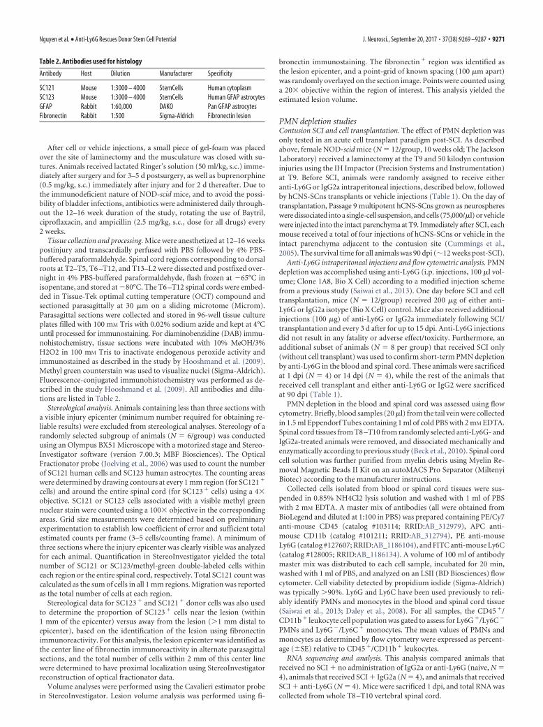
Table 2. Antibodies used for histology
Antibody Host Dilution Manufacturer Specificity
SC121 Mouse 1:3000 – 4000 StemCells Human cytoplasmSC123 Mouse 1:3000 – 4000 StemCells Human GFAP astrocytesGFAP Rabbit 1:60,000 DAKO Pan GFAP astrocytesFibronectin Rabbit 1:500 Sigma-Aldrich Fibronectin lesion
Nguyen et al. • Anti-Ly6G Rescues Donor Stem Cell Potential J. Neurosci., September 20, 2017 • 37(38):9269 –9287 • 9271

RNA was extracted using TRIzol (Life Technologies), and DNase-treated and purified using a NucleoSpin RNA II kit (Macherey-Nagel).Purified RNA was quantified using a Qubit 2.0 Fluorometer (Life Tech-nologies) and checked for quality using an Agilent Technologies Bioana-lyzer 2100. Library construction and sequencing were performed at theUniversity of California, Irvine, Genomics High-Throughput Facility.cDNA libraries were made with a TruSeq RNA Library Prep Kit version 2(Illumina) according to manufacturer instruction and sequenced on anIllumina HiSeq 4000 platform producing 30 million paired-end readswith a read length of 100. Before read mapping, each sample wassubjected to qualitative control analysis by FastQC tool (http://www.bioinformatics.babraham.ac.uk/projects/fastqc/). Subsequently, poten-tial ribosomal contamination was removed by the SortMeRNA tool (Ko-pylova et al., 2012) and low-quality bases (Q � 30) were filtered out bythe Trimmomatic tool (Bolger et al., 2014). Then, RNA sequencing(RNAseq) reads were mapped to mouse reference genome GRCm38version 87 using STAR aligner, and the uniquely mapped reads weresummarized at the gene level using featureCounts (Liao et al., 2014).Differential gene expression analysis was performed on TMM normal-ized counts implemented in EdgeR (McCarthy et al., 2012). A falsediscovery rate-corrected p value of �0.05 was used in determining sig-nificant differentially expressed genes for respective comparisons. Prin-cipal component analysis (PCA) was performed using R package “stats”and plotted using JMP statistical tool (version 11, SAS Institute). Hier-archical clustering was performed and visualized using the R packageggplot2_2.2.1. Tissue-specific expression was determined in FunRich(Pathan et al., 2015). The functionally relevant biological information fordifferentially expressed genes (DEGs) were identified using geneSCF tool(Subhash and Kanduri 2016; extended data Fig. 7-1 available at https://10.1523/JNEUROSCI.2785-16.2017.f7-1). The top enriched gene ontol-ogy (GO) terms were depicted in Figure 7F using R package ggplot2_2.2.1.
Tissue collection, processing, and analysis. Mice were anesthetized at 12weeks postinjury and transcardially perfused with PBS followed by 4%PBS-buffered paraformaldehyde. Spinal cord regions corresponding todorsal roots at T2–T5, T6 –T12, and T13–L2 were dissected and postfixedovernight in 4% PBS-buffered paraformaldehyde, flash frozen at �65°Cin isopentane, and stored at �80°C. The T6 –T12 spinal cords were em-bedded in Tissue-Tek OCT compound and sectioned coronally at 30 �musing a CryoJane tape transfer system (Leica Biosystems). A subset ofanimals (IgG2a, N � 2; anti-Ly6G, N � 2) was randomly selected forsectioning in the parasagittal plane on a sliding microtome (Microm)using the same method described above. CryoJane sections were stored at�20°C until processing for immunohistochemical or other staining. Forimmunohistochemistry, frozen CryoJane sections were brought to roomtemperature. Sections were processed for antigen retrieval using BufferA, pH 6, in the Retriever 2100 System (PickCell Laboratories).
All primary antibodies and dilutions that were used for immunolabel-ing are listed in Table 2, including human cytoplasm SC121, humanGFAP SC123, and fibronectin. The secondary antibodies used were ei-ther biotin-conjugated or fluorescence-conjugated affinipure F(ab)2fragment secondary antibodies (Jackson ImmunoResearch). DAB, methylgreen counterstaining, and fluorescence-conjugated immunohistochemis-try were performed as described above and in a previous study (Hooshmandet al., 2009). Fluorescent images were captured on the Zeiss LSM510 Ultra-view VOX Spinning Disk Confocal Microscope; saved as red, green, andblue; and final images were merged using Volocity 5.0 software.
Stereological analysis. As above, stereology of a randomly selected sub-group of animals (N � 5/group) was conducted using an Olympus BX51Microscope with motorized stage and StereoInvestigator to estimate un-biased total SC121 human cell and SC123 human astrocyte numbers, andvolumes of fibronectin lesion SC121 and SC123 clustering. Uniform ran-dom sampling of the tissue was performed according to standard stereo-logical principles. Sampling parameters (i.e., grid size and countingframe size) were empirically determined to arrive at low coefficients oferror. Briefly, total numbers of SC121 � human cells were determined byunbiased stereology in 1 of 12 intervals from coronal spinal cord sections0.36 mm apart using systematic random sampling with an optical frac-tionator probe and StereoInvestigator version 9 (MicroBrightField). Op-tical fractionator grid size (SC121, 350 � 350 �m; SC123, 200 � 200 �m)
and counting frame size (SC121, 35 � 35 �m; SC123, 50 � 50 �m) wereempirically determined to yield average Gundersen cumulative error val-ues of �0.1.
As described above, stereological data for SC123 � cells was used todetermine the number of SC123 � cells near the lesion (within 1 mm ofthe epicenter) versus away from the lesion (�1 mm distal to epicenter),based on the identification of the lesion using Fibronectin immunoreac-tivity. For this analysis, the lesion was identified as the volume occupiedby fibronectin immunoreactivity in alternate parasagittal sections. Thetotal number of cells within 2 mm of this lesion was determined to haveproximal localization using StereoInvestigator reconstruction of opticalfractionator data. Furthermore, the distribution/migration of SC121 hu-man cells and SC123 human astrocytes was represented as the number ofcells that were binned at distance (in millimeters) away from the sparedtissue region.
A Cavalieri probe (grid size, 20 � 20 �m; SI9, MicroBrightField) wasused to analyze total volumes of spinal cord, fibronectin lesion, SC121clustering, and S123 clustering in 1 of 12 intervals from coronal spinalcord sections 0.36 mm apart, at 10� magnification for spinal cord and20� magnification for fibronectin lesion and clustering volumes.
Behavioral tasks and assessmentsLocomotor recovery in mice (N � 10 –16/group; Table 1) was assessedusing CatWalk gait analysis to assess kinematic parameters (Koopmanset al., 2005). CatWalk testing was conducted by blinded technicians 1 dbefore SCI and every 4 weeks until sacrifice.
StatisticsStatistics were performed using Prism (version 5, GraphPad). Compari-sons between acute versus delayed cell transplants, cell transplant versusvehicle, and IgG2a versus anti-Ly6G treatments for flow cytometric anal-ysis, stereological quantification, and behavioral assay data (CatWalk)were analyzed using individual two-tailed Student’s t tests. Human celldistribution/migration of SC121 human cells and SC123 human astro-cytes was analyzed using two-way ANOVA followed by Bonferroni posthoc t tests. Time courses of blood PMNs and spinal cord-infiltratedPMNs were analyzed using one-way ANOVA followed by Bonferronipost hoc t tests. Significance was defined as p � 0.05.
Study approvalAll animal housing conditions, surgical procedures, and postoperativecare was conducted according to the Institutional Animal Care and UseCommittee guidelines at the University of California, Irvine.
ResultsAcute transplantation of hCNS-SCns altered donor cellastroglial differentiation and distribution and did notimprove recovery after SCIBecause these studies involved xenogeneic transplantation, weused a constitutively immunodeficient mouse model of SCI thatwe have previously shown to enable sufficient human donor cellengraftment success to accurately assess long-term cell integra-tion and effects on locomotor recovery of function (Anderson etal., 2011). While the adaptive immune response is absent in theseanimals, this NOD-scid mouse model has an intact cellular innateimmune response including PMN and macrophage/microglialrecruitment, and histopathological characteristics comparable toother laboratory mouse strains following SCI (Luchetti et al.,2009). Accordingly, vertebral level T9 contusion-injured NOD-scid mice received bilateral hCNS-SCns transplants or vehicleinto the spared parenchyma 1 mm above and below the SCIepicenter either immediately post-SCI (acute, 0 dpi) or 30 d post-SCI (delayed, 30 dpi). All animals received 75,000 hCNS-SCns ina total injection volume of 1 �l distributed across four injectionsites, as described under Materials and Methods.
Histological analyses using a human-specific cytoplasmic an-tibody (Fig. 1A,B, SC121/STEM121, brown label) demonstrated
9272 • J. Neurosci., September 20, 2017 • 37(38):9269 –9287 Nguyen et al. • Anti-Ly6G Rescues Donor Stem Cell Potential

donor human cell engraftment in 100% of animals (N � 16) thatreceived hCNS-SCns in either acute or delayed transplantationpost-SCI. Blinded, unbiased stereological quantification at 12–16weeks post-transplantation using the optical fractionator proberevealed 210,000 � 31,023 hCNS-SCns for mice receiving acutetransplantation and 215,700 � 48,980 for mice receiving delayedtransplantation (Fig. 1C), demonstrating no differences in donorhuman cell engraftment. Surprisingly, however, mice receivingacute transplantation exhibited dense clustering of hCNS-SCnsproximal to the lesion that was not apparent after delayed trans-plantation (Fig. 1A,B). This observation was verified by stereo-logical quantification, which demonstrated a significantly highernumber of hCNS-SCns localized in the spared parenchyma nearthe lesion (within 1 mm of the epicenter, as described in Materialsand Methods) in acute versus delayed transplantation animals
(Fig. 1D; 93,260 � 15,760 in acute vs 38,380 � 7780 in delayed).The reverse was apparent for hCNS-SCns that migrated awayfrom the lesion (�1 mm distal to the epicenter), with signifi-cantly higher cell numbers distal to the epicenter in delayed ver-sus acute transplants (Fig. 1D). Together, these data show nodifference in total hCNS-SCns engraftment between transplantparadigms; however, there was a striking difference in the distri-bution/localization of hCNS-SCns within the injured spinal cord.
hCNS-SCns do not alter lesion volume after delayed trans-plantation (Hooshmand et al., 2009; Salazar et al., 2010). Here,we assessed the localization of hCNS-SCns at the lesion epicenterand tested the effect of donor human cells on lesion volume afteracute transplantation using fluorescent immunolabeling forSC121 and fibronectin. Although large numbers of hCNS-SCnswere densely packed adjacent to the fibronectin� epicenter 12 weeks
Figure 1. hCNS-SCns transplanted at acute and delayed time points post-SCI engrafted similarly but differed in their distribution post-SCI. A, a�, B, b�, Immunostaining using a human-specificmarker (SC121, brown) demonstrated hCNS-SCns survival in acute (A, a�) and delayed (B, b�) transplants. A, B, While most human cells were localized away from the lesion by 12 weeks after delayedtransplantation (B), following acute transplantation (A), many human cells formed dense clusters near the lesion. C, Stereological quantification of human cells revealed equivalent numbers ofengrafted hCNS-SCns (N � 6/group; Student’s t test, p � 0.94). The dashed line indicates the number of cells originally transplanted (75,000/animal). D, Significantly more hCNS-SCns werelocalized proximal (1 mm rostral and caudal) to the lesion in animals receiving acute compared with delayed transplantation. In contrast, significantly more hCNS-SCns were localized distal (�1 mmrostral and caudal) to the lesion in animals receiving delayed versus acute transplantation. For this analysis, the lesion epicenter was identified as the center line of fibronectin immunoreactivity inalternate sections. The total number of cells within 2 mm of this center line was determined to have proximal localization using StereoInvestigator reconstruction of optical fractionator data.***p � 0.001, Student’s t test. Scale bars: A, B, 250 �m; a� and b�, 25 �m. Values are shown as the mean � SEM.
Nguyen et al. • Anti-Ly6G Rescues Donor Stem Cell Potential J. Neurosci., September 20, 2017 • 37(38):9269 –9287 • 9273

after acute transplantation, few were local-ized within the fibronectin� epicenteritself (Fig. 2A, hCNS-SCns, red; fibronec-tin, green; colocalization, yellow), suggestingthat acutely transplanted cells migrated to-ward the epicenter but avoided the epicentercore. Blinded, unbiased stereological quan-tification using the Cavalieri probe revealedno significant differences between fibronec-tin� regions in mice receiving acute hCNS-SCns transplants versus vehicle controls(Fig. 2B), demonstrating that acutely trans-planted hCNS-SCns neither increased nordecreased lesion volume.
hCNS-SCns transplantation at delayedtime points promotes locomotor recovery(Cummings et al., 2005; Salazar et al., 2010).In the present study, despite comparable en-graftment (Fig. 1), acute transplantationwas not associated with functional recoveryas determined by open-field locomotorassessment or ladderbeam analysis (datanot shown). Furthermore, CatWalk kine-matic gait analysis (Hamers et al., 2006), amore sensitive assessment of the fineaspects of locomotion, did not reveal sig-nificant differences between acute hCNS-SCns transplant versus vehicle groups inany parameters including hindpaw printarea, hindpaw maximum contact area,mean swing speed, and the percentage ofstep sequence Ab (Fig. 2C–F). Collectively,our data suggest that, despite evidence of ef-ficacy after delayed transplantation, acutehCNS-SCns transplantation at 0 dpi failedto promote locomotor recovery.
hCNS-SCns transplantation at delayedtime points, 9 and 30 dpi, respectively, re-sults in predominant differentiation intooligodendrocytes (68% and 50%), someneurons (25% and 40%), and very few as-trocytes (3% and 8%; Cummings et al.,2005; Salazar et al., 2010). In contrast, in
Figure 2. Acute transplantation of hCNS-SCns resulted in human astroglial differentiation and localization near the lesionepicenter but did not affect lesion volume or functional recovery after SCI. A, Minimal overlap of SC121 � human cells (red) andfibronectin � lesion (green) revealed few hCNS-SCns entering the lesion epicenter. B, Stereological quantification for the volume
4
of fibronectin � lesions demonstrated no significant differ-ences between vehicle and hCNS-SCns groups (N � 6/group;Student’s t test, N.S. � p � 0.05), suggesting that hCNS-SCnstransplantation did not alter the size of the lesion. C–F, hCNS-SCns engraftment was not associated with improvements ordecrements in recovery of function assessed by CatWalk gaitanalysis, including hindpaw print area (C), hindpaw maximumcontact area (D), swing speed mean (E), and the percentage ofstep sequence Abs (F). N�16/group, Student’s t tests, N.S.�p �0.05. G, A majority of human-specific astrocytes (SC123 �
cells, brown) formed dense clusters near the lesion. g�, High-power image of SC123 � labeling from G. H, Near the injuryepicenter, hCNS-SCns exhibited predominant colocalizationwith both pan-GFAP (red) and SC123 (green). I, Stereologicalquantification of SC123 � cells revealed that 86.3% of donorhuman cells exhibiting astrocytic differentiation were local-ized near the lesion. N � 6/group, Student’s t test, ***p �0.001. Scale bars: g�, 25 �m; G, 250 �m. Values are given asthe mean � SEM.
9274 • J. Neurosci., September 20, 2017 • 37(38):9269 –9287 Nguyen et al. • Anti-Ly6G Rescues Donor Stem Cell Potential

the present acute transplant study, bright-field and confocal im-aging using a human-specific astrocytic marker (SC123/STEM123) revealed the presence of many SC123� humanastrocytes proximal to the injury epicenter (Fig. 2G,H), corre-sponding to regions of dense donor human cell clusters. Thespecificity of SC123 for GFAP was validated, as SC123� cellsadjacent to the lesion exhibited nearly complete overlap withpan-GFAP� cells (data not shown), suggesting that the majorityof cells in this region were GFAP� astrocytes and human in ori-gin. Blinded, unbiased stereological quantification using theoptical fractionator probe demonstrated that 86.3% of SC123�
human astrocytes in the spinal cords of mice receiving acutehCNS-SCns transplantation were localized in the spared paren-chyma near the lesion (within 1 mm of the epicenter; Fig. 2I),suggesting that human donor cells were recruited toward the epicen-ter, where they differentiated into astrocytes. While studies have sug-gested that the injured spinal cord niche is gliogenic (Shihabuddin etal., 2000; Cao et al., 2001), there has been a paucity of empiricalstudies to determine whether variations in host niche and/or cell-intrinsic properties drive fate decisions of transplanted cells (Monjeet al., 2003). Because PMNs are specifically and selectively recruitedto the epicenter of the acute post-SCI microenvironment in humans(Fleming et al., 2006), rats (Beck et al., 2010), and mice (Saiwai et al.,2013), we next investigated the role of PMNs in modulating donorhuman cell astroglial fate, migration, and associated functional re-covery in the acute SCI niche.
Anti-Ly6G administration specifically depleted PMNsafter SCIAs the first immune cells to populate the site of injury, PMNshave been suggested to affect injury and subsequent processesthrough physical PMN-to-cell contact (Dinkel et al., 2004) andthe production of free radicals (Yagisawa et al., 1996), proteases(de Castro et al., 2000), and proinflammatory cytokines (Len-zlinger et al., 2001). As described above, NOD-scid mice have acomparable innate cellular immune response to other mousestrains. Here, we verified the presence of PMNs (Ly6G�/Ly6C�)using flow cytometry, demonstrating that NOD-scid mice have arobust and transient PMN infiltration that peaked at 1 dpi (Fig.3B) and was localized to the spinal cord lesion epicenter (data notshown). The timing and localization of this response were con-sistent with those determined in other studies in rats and mice
without adaptive immune system (T- and B-cell) deficiencies(Luchetti et al., 2009; Stirling et al., 2009; Beck et al., 2010). Fur-ther, a robust presence of PMNs was detected in the blood ofNOD-scid mice after SCI, showing a peak (90% of leukocytes) ofPMNs at 4 h post-SCI and gradually returning to preinjury levels(60% of leukocytes) after 10 dpi (Fig. 3A).
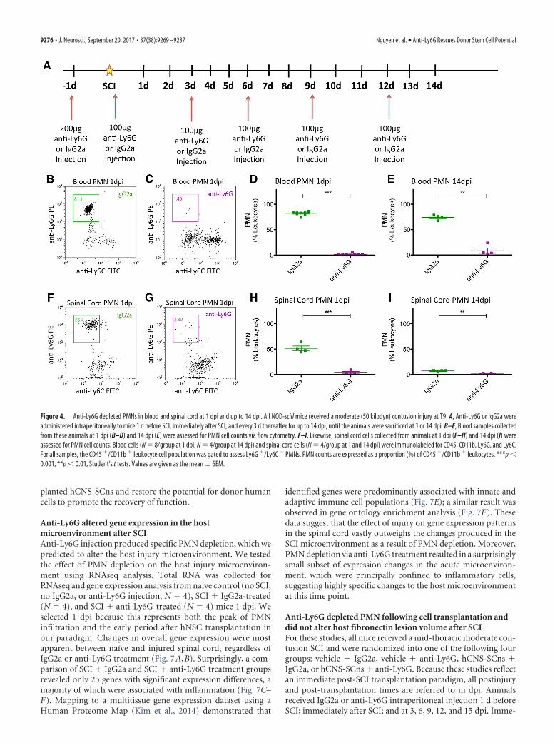
Having validated the retention of an innate cellular immuneresponse after SCI in NOD-scid mice, we next tested the specific-ity and efficiency of anti-Ly6G to deplete PMNs in the blood andspinal cords of SCI mice. All mice received a mid-thoracic mod-erate contusion SCI as described in Materials and Methods, andeither IgG2a or anti-Ly6G administration via intraperitoneal in-jection 1 d before SCI, as well as immediately after SCI, and every3 d until animals were sacrificed at 1 or 14 dpi (Fig. 4A). Flowcytometric analysis demonstrated that, compared with IgG2a-treated animals, anti-Ly6G-treated animals exhibited efficientdepletion of PMNs in the blood at 1 dpi (Fig. 4B–D; 1.23 � 0.65%of leukocytes; N � 8), which was sustained through 14 dpi (Fig.4E; 8.46 � 5.17% of leukocytes; N � 4). Furthermore, anti-Ly6G-treated animals exhibited comparable PMN depletion efficiencyin the spinal cord at 1 dpi (Fig. 4F–H; 4.25 � 1.78% of leukocytes;N � 4) and up to 14 dpi (Fig. 4I; 2.46 � 0.43% of leukocytes; N �4). Interestingly, anti-Ly6G treatment also resulted in an increas-ing proportion of monocytes (Ly6G�/Ly6C�) detected in bloodat 1 and 14 dpi (Fig. 5A–D), and in spinal cord tissue at 1 dpi butnot 14 dpi (Fig. 5E–H). We hypothesized that this change inproportion was not the result of an increase in monocytes, butrather the result of a shift due to the depleted PMN population inanti-Ly6G-treated animals. We tested this possibility by analyz-ing the absolute number of PMNs or monocytes per 5000 eventscounted via flow cytometry. Anti-Ly6G-treated animals exhib-ited a dramatic reduction in PMN number counted via thismethod compared with IgG2a-treated animals, at 1 and 14 dpi inboth blood (Fig. 6A,C) and spinal cord (Fig. 6E,G). In contrast,there was no difference in the monocyte number counted be-tween IgG2a and anti-Ly6G-treated groups at 1 and 14 dpi ineither blood (Fig. 6B,D) or spinal cord (Fig. 6F,H). These datademonstrate that anti-Ly6G efficiently and specifically depletedPMNs and did not affect monocyte absolute cell number afterSCI. We next investigated whether the specific PMN depletion viaanti-Ly6G injections could alter the fate and migration of trans-
Figure 3. After SCI, the number of PMNs increased acutely in the blood and spinal cord of mice. A, NOD-scid mice were given a moderate (50 kilodyn) contusion injury at T9, and blood sampleswere collected before injury and daily from 4 h to 14 dpi. B, For spinal cord PMN infiltration, another set of animals was given a moderate (50 kilodyn) contusion injury at T9 and were sacrificed at2 h, 1 dpi, 3 dpi, 7 dpi, 9 dpi, 14 dpi, 30 dpi, or 60 dpi. Blood (N � 4 – 8/group) and spinal cord (N � 4/group) cells were immunolabeled for CD45, CD11b, Ly6G, and Ly6C. For all samples, theCD45 �/CD11b � leukocyte cell population was gated to assess for Ly6G �/Ly6C � PMNs. The percentage of PMNs is expressed as the number of PMNs relative to CD45 �/CD11b � leukocytes. *p �0.05; ***p � 0.001, one-way ANOVA followed by Bonferroni post hoc t tests, N � 4 – 8/group. Values are given as the mean � SEM.
Nguyen et al. • Anti-Ly6G Rescues Donor Stem Cell Potential J. Neurosci., September 20, 2017 • 37(38):9269 –9287 • 9275
planted hCNS-SCns and restore the potential for donor humancells to promote the recovery of function.
Anti-Ly6G altered gene expression in the hostmicroenvironment after SCIAnti-Ly6G injection produced specific PMN depletion, which wepredicted to alter the host injury microenvironment. We testedthe effect of PMN depletion on the host injury microenviron-ment using RNAseq analysis. Total RNA was collected forRNAseq and gene expression analysis from naive control (no SCI,no IgG2a, or anti-Ly6G injection, N � 4), SCI � IgG2a-treated(N � 4), and SCI � anti-Ly6G-treated (N � 4) mice 1 dpi. Weselected 1 dpi because this represents both the peak of PMNinfiltration and the early period after hNSC transplantation inour paradigm. Changes in overall gene expression were mostapparent between naïve and injured spinal cord, regardless ofIgG2a or anti-Ly6G treatment (Fig. 7A,B). Surprisingly, a com-parison of SCI � IgG2a and SCI � anti-Ly6G treatment groupsrevealed only 25 genes with significant expression differences, amajority of which were associated with inflammation (Fig. 7C–F). Mapping to a multitissue gene expression dataset using aHuman Proteome Map (Kim et al., 2014) demonstrated that
identified genes were predominantly associated with innate andadaptive immune cell populations (Fig. 7E); a similar result wasobserved in gene ontology enrichment analysis (Fig. 7F). Thesedata suggest that the effect of injury on gene expression patternsin the spinal cord vastly outweighs the changes produced in theSCI microenvironment as a result of PMN depletion. Moreover,PMN depletion via anti-Ly6G treatment resulted in a surprisinglysmall subset of expression changes in the acute microenviron-ment, which were principally confined to inflammatory cells,suggesting highly specific changes to the host microenvironmentat this time point.
Anti-Ly6G depleted PMN following cell transplantation anddid not alter host fibronectin lesion volume after SCIFor these studies, all mice received a mid-thoracic moderate con-tusion SCI and were randomized into one of the following fourgroups: vehicle � IgG2a, vehicle � anti-Ly6G, hCNS-SCns �IgG2a, or hCNS-SCns � anti-Ly6G. Because these studies reflectan immediate post-SCI transplantation paradigm, all postinjuryand post-transplantation times are referred to in dpi. Animalsreceived IgG2a or anti-Ly6G intraperitoneal injection 1 d beforeSCI; immediately after SCI; and at 3, 6, 9, 12, and 15 dpi. Imme-
Figure 4. Anti-Ly6G depleted PMNs in blood and spinal cord at 1 dpi and up to 14 dpi. All NOD-scid mice received a moderate (50 kilodyn) contusion injury at T9. A, Anti-Ly6G or IgG2a wereadministered intraperitoneally to mice 1 d before SCI, immediately after SCI, and every 3 d thereafter for up to 14 dpi, until the animals were sacrificed at 1 or 14 dpi. B–E, Blood samples collectedfrom these animals at 1 dpi (B–D) and 14 dpi (E) were assessed for PMN cell counts via flow cytometry. F–I, Likewise, spinal cord cells collected from animals at 1 dpi (F–H) and 14 dpi (I) wereassessed for PMN cell counts. Blood cells (N � 8/group at 1 dpi; N � 4/group at 14 dpi) and spinal cord cells (N � 4/group at 1 and 14 dpi) were immunolabeled for CD45, CD11b, Ly6G, and Ly6C.For all samples, the CD45 �/CD11b � leukocyte cell population was gated to assess Ly6G �/Ly6C � PMNs. PMN counts are expressed as a proportion (%) of CD45 �/CD11b � leukocytes. ***p �0.001, **p � 0.01, Student’s t tests. Values are given as the mean � SEM.
9276 • J. Neurosci., September 20, 2017 • 37(38):9269 –9287 Nguyen et al. • Anti-Ly6G Rescues Donor Stem Cell Potential

diately after SCI, mice also received bilateral hCNS-SCns trans-plants or vehicle into the spared parenchyma 1 mm above andbelow the SCI epicenter, as described above. Blood was collectedto confirm PMN depletion efficiency at 1 and 14 dpi. All animalswere sacrificed at 90 dpi (Fig. 8A). Figure 8 shows the results ofgroups that received cell transplant (open circles) and vehicleinjection (filled squares), for both IgG2a (green) and anti-Ly6G
(purple) groups, in the same graphs for reference. No significantdifferences were detected between groups that received cells ver-sus vehicle within either the IgG2a or Ly6G treatment groups,demonstrating that cell transplantation did not alter the cellularresponse within animals that received IgG2a or anti-Ly6G.
Compared with the robust PMN levels in IgG2a-treatedanimals, complete PMN (Ly6G�/Ly6C�) depletion was evident
Figure 5. PMN depletion via anti-Ly6G increased the proportion of monocytes in the blood and spinal cord at 1 dpi and up to 14 dpi. A, B, E, F, Representative dot plots demonstrated anincreased percentage of monocytes in blood (A, B) and spinal cords (E, F) of anti-Ly6G-treated animals (purple box), compared with that of IgG2a-treated animals (green box) at 1 dpi. C, D, G, H, Flowcytometric data analysis demonstrated that anti-Ly6G treatment increased the percentage of monocytes in blood at both 1 dpi (C) and 14 dpi (D), and in spinal cord at 1 dpi (G) but not at 14 dpi (H)when PMNs are known to be minimally detectable in the cord. Blood cells (N � 8/group at 1 dpi; N � 4/group at 14 dpi) and spinal cord cells (N � 4/group at 1 and 14 dpi) were immunolabeledfor CD45, CD11b, Ly6G, and Ly6C. For all samples, the CD45 �/CD11b � leukocyte cell population was gated to assess Ly6G �/Ly6C � monocytes. Monocyte counts are expressed as a proportion (%)of CD45 �/CD11b � leukocytes. *p � 0.05, Student’s t test. Values are given as the mean � SEM.
Figure 6. PMN depletion via anti-Ly6G did not affect the absolute number of monocytes in the blood and spinal cord at 1 dpi and up to 14 dpi. The quantitation of PMN cell numbers per 5000events demonstrated that anti-Ly6G treatment effectively depleted the number of PMNs (compared with IgG2a treatment) in blood at 1 dpi (A) and 14 dpi (C), and in the spinal cord at 1 dpi (E), butnot at 14 dpi (G) when PMNs are known to be minimally detectable in the cord. B, D, F, H, Interestingly, the quantitation of monocyte cell numbers per 5000 events demonstrated no difference inthe absolute number of monocytes between IgG2a- and anti-Ly6G-treated animals in the blood (B, D) and spinal cord (F, H) at either 1 or 14 dpi. Blood cells (N � 8/group at 1 dpi; N � 4/group at14 dpi) and spinal cord cells (N � 4/group at 1 and 14 dpi) were immunolabeled for CD45, CD11b, Ly6G, and Ly6C, and were assessed for PMN or monocyte cell number per 5000 events via flowcytometry. For all samples, the CD45 �/CD11b � leukocyte cell population was gated to assess for Ly6G �/Ly6C � PMN or Ly6G �/Ly6C � monocytes. All values are expressed as the absolutenumber of PMNs or monocytes per 5000 events. ***p � 0.001, Student’s t test. Values are given as the mean � SEM.
Nguyen et al. • Anti-Ly6G Rescues Donor Stem Cell Potential J. Neurosci., September 20, 2017 • 37(38):9269 –9287 • 9277

Figure 7. PMN depletion via anti-Ly6G altered gene expression associated with inflammation and cell fate in the spinal cord. All NOD-scid mice received a moderate (50 kilodyn) contusion injuryat T9. Anti-Ly6G or IgG2a was administered intraperitoneally to mice as described above, and they were sacrificed at 1 dpi. Total RNAs from anti-Ly6G-treated (N � 4), IgG2a-treated (N � 4), andnaive (N � 4) mice were isolated from spinal cord tissues (T8 –T10), and gene expression analysis was compared between naive and SCI IgG2a, naive and SCI anti-Ly6G, and SCI IgG2a and SCIanti-Ly6G groups. A, PCA was performed on the whole gene expression matrix of all samples. Each dot in the graph represents a sample color coded by experimental group (Figure legend continues.)
9278 • J. Neurosci., September 20, 2017 • 37(38):9269 –9287 Nguyen et al. • Anti-Ly6G Rescues Donor Stem Cell Potential

in anti-Ly6G-treated animals at both 1 dpi (Fig. 8B–D) and 14 dpi(Fig. 8E). As above, an increased proportion of monocytes (Ly6G�/Ly6C�) was detected in blood at 1 and 14 dpi (Fig. 9A–D), and thespecificity of PMN depletion by anti-Ly6G was confirmed by quan-tification of the absolute number of PMNs or monocytes per 5000events counted via flow cytometry (Fig. 9E–H).
Lesion volume and astroglial scar formation after SCI are re-portedly unaltered by treatment with anti-Ly6G (Saiwai et al.,2013). As a necessary step for histological comparisons of cellengraftment, we confirmed that anti-Ly6G treatment did not al-ter histological lesion volume in groups receiving cell transplantsor vehicle as assessed by fibronectin volume 90 dpi (Fig. 10A,B;hCNS-SCns � IgG2a � 0.14 � 0.02 mm 3; hCNS-SCns � anti-Ly6G � 0.13 � 0.02 mm 3; Student’s t test, p � 0.49).Accordingly, blinded unbiased stereological analysis of human-specific hCNS-SCns engraftment was conducted via immunohis-tochemistry for the human-specific cytoplasmic marker SC121 todetermine whether treatment with anti-Ly6G altered donor hu-man cell number, localization, or lineage selection.
Anti-Ly6G modulated localization but not total donor humancell engraftment after SCIStereological analysis was conducted as described in Materialsand Methods using the optical fractionator probe and contourdefinitions to identify the perimeter of the spinal cord and thespared spinal cord parenchyma. This analysis represents an un-biased and density/volume-adjusted estimate of the total numberof engrafted donor human cells within the anatomical regionsampled. Cell engraftment was observed in 100% of hCNS-SCns �IgG2a (N � 12) and hCNS-SCns � anti-Ly6G-treated (N � 12)animals. A subset of animals was randomly selected from eachgroup for optical fractionator quantification of donor SC121�
human cells and SC123� human astrocytes (N � 5). The numberof donor SC121� human cells (Fig. 11A) showed no significantdifferences in engraftment between IgG2a- and anti-Ly6G-treated groups 90 dpi (Fig. 11B; hCNS-SCns � IgG2a �654,500 � 55,050; hCNS-SCns � anti-Ly6G � 474,600 �118,000; Student’s t test, p � 0.10). However, dramatic differ-ences were observed in the localization and distribution of donor
SC121� human cells (Fig. 11C,D). While IgG2a-treated animalsfrequently exhibited dense clusters of SC121� human cells inregions adjacent to the lesion epicenter (Fig. 11C; 3D reconstruc-tion with lesion epicenter in red and SC121� human cell clustersin blue in Fig. 11c), anti-Ly6G-treated animals exhibited eithersignificantly smaller clusters or the absence of clustered cells en-tirely (Fig. 11D; 3D reconstruction with lesion epicenter in redand SC121� human cell clusters in blue in Fig. 11d). Accord-ingly, anti-Ly6G-treated animals exhibited a significant reduc-tion in the number of SC121� cells compared with IgG2a-treatedanimals in the spared tissue adjacent to the lesion (Fig. 11E). Thedensity of SC121� human cells in IgG2a-treated animals pre-cluded accurate stereological sampling of individual cells, sug-gesting that the quantitative cell number comparison in Figure11, B and E, represents an underestimation of donor human cellsat these dense regions. We therefore confirmed this observationusing unbiased stereological quantification of SC121� humancell cluster volume (Fig. 11F), which also demonstrated that anti-Ly6G-treated animals exhibited a loss of donor human cell cluster-ing near the lesion epicenter. Together, these data show thatanti-Ly6G treatment altered the localization of donor human cellsnear the lesion site and suggest a novel role for PMNs in modulatingdonor human cell behavior in the SCI microenvironment.
Anti-Ly6G inhibited astroglial differentiation of donorhuman hCNS-SCns proximal to the lesion epicenterAs characterized above, clusters of donor human cells proximalto the lesion, in the region typically occupied by reactive astro-cytes and associated with the glial scar post-SCI, are a key featureobserved in an acute cell transplantation paradigm. Accordingly,human astrocyte lineage selection was assessed via immunohis-tochemistry for the human-specific anti-GFAP antibody SC123.Donor human cells proximal to the lesion epicenter in hCNS-SCns � IgG2a-treated animals were predominantly SC123� hu-man astrocytes, with extensive accumulation in dense clusters(Fig. 12A; 3D reconstruction with lesion epicenter in red andSC123� human astrocyte clusters in blue in Fig. 12a). In strikingcontrast, engrafted cells in hCNS-SCns � anti-Ly6G-treated an-imals were not retained in clusters, and few human astrocyteclusters were observed proximal to the lesion (Fig. 12B; 3D re-construction with lesion epicenter in red and SC123� humanastrocyte clusters in blue in Fig. 12b).
Anti-Ly6G-mediated reduction of human astrocyte clustersnear the lesion was supported by blinded unbiased stereologicalquantification of the total number of SC123� human astrocytesusing the optical fractionator probe, which demonstrated a sig-nificant decrease in hCNS-SCns � anti-Ly6G-treated animalscompared with hCNS-SCns � IgG2a-treated animals (Fig.12C; hCNS-SCns � anti-Ly6G � 23,980 � 8105; hCNS-SCns �IgG2a � 48,410 � 6955; Student’s t test, p � 0.05). Furtheranalysis of the number of SC123� human astrocytes localized tothe region proximal versus distal to the epicenter revealed thatthis difference was specific to the region near the lesion (within1 mm) and not observed in regions away (�1 mm) from thelesion (Fig. 12D; Student’s t tests, p � 0.05 near lesion, p � 0.28away from lesion). An associated decrease in the number ofSC123� human astrocytes was observed in the anti-Ly6G-treatedanimals when stereological data were plotted in a distributioncurve (Fig. 12E; two-way ANOVA followed by Bonferroni posthoc t tests, p � 0.001 at the spared tissue region adjacent to thelesion). As was the case for SC121 quantification, the density ofSC123� cells near the lesion epicenter in IgG2a-treated animalswas so high as to preclude accurate cell counting, resulting in a
4
(Figure legend continued.) and plotted in three dimensions using the first three principal com-ponents (PC1–PC3). The analysis revealed a distinct cluster of the naive group (blue) and moresimilarities in the overall gene expression between the anti-Ly6G (red) and IgG2a (green)groups. B, Hierarchical clustering of the expression of 15,627 genes (rows) and samples (col-umns) ordered by experimental groups. Heatmap visualization of clustering revealed clear col-lections of genes that were systematically upregulated (red) or downregulated (blue) in naiveand IgG2a or anti-Ly6G treatment groups, respectively. In contrast, there was an apparentsimilar gene expression pattern in IgG2a versus anti-Ly6G groups, substantiating PCA findings.C, The table listed 25 statistically significant genes (DE) found in IgG2a compared with anti-Ly6Ggroups by edgeR (logFC, log fold change; logCPM, log count per million; LR, likelihood ratio;Pvalue, unadjusted p value; FDR, false discovery rate). D, Heatmap of unsupervised bidirectionalclustering of 25 DE values of all samples indicated three distinct clusters classified by experi-mental group. Overall, 21 of the 25 DE genes were upregulated, and 4 genes (Cxcr2, Mettl7a1,Csfr3, and Rimkla) were downregulated in a comparison of the anti-Ly6G- and IgG2a-treatedgroups. E, Heatmap of tissue-specific expression for the significant genes was generated usingHuman Proteome Map (Kim et al., 2014) quantitative expression dataset implemented in Fun-rich 2.1.2. The magnitude of relative expression values across tissues was colored coded. Thehigher expression values were represented by an intense red color relative to the lower expres-sion values. F, The top significantly enriched GO terms associated with DEG are depicted inextended data Figure 7-1 available at https://10.1523/JNEUROSCI.2785-16.2017.f7-1. Thex-axis of the bar graph indicated the �log 10 scale of the p values, and the y-axis designates GOterm identifications and names. The lengths of the bars positively correlated with the statisticalsignificance.
Nguyen et al. • Anti-Ly6G Rescues Donor Stem Cell Potential J. Neurosci., September 20, 2017 • 37(38):9269 –9287 • 9279

Figure 8. Anti-Ly6G depleted blood PMNs following hCNS-SCns transplantation at 1 dpi and up to 14 dpi after SCI. A, Immediately after SCI, animals receiving intraperitoneal injections ofanti-Ly6G or IgG2a were transplanted with either hCNS-SCns (75,000/animal) or vehicle. Blood cells (N � 10 –12) were immunolabeled for CD45, CD11b, Ly6G, and Ly6C. The CD45 �/CD11b �
leukocyte cell population was gated to assess for Ly6G �/Ly6C � PMNs. B, C, hCNS-SCns � anti-Ly6G-treated but not hCNS-SCns � IgG2a-treated animals exhibited depletion of the PMNpopulation in blood at 1 and 14 dpi. D, E, Animals that received hCNS-SCns (open circles) or vehicle injection (filled squares) for both IgG2a (green) and anti-Ly6G (purple) are shown in the same graphfor reference. D, No significant differences were detected in blood PMNs 1 dpi between groups that received cells versus vehicle in either IgG2a (hCNS-SCns � IgG2a � 85.3 � 2.6% vs vehicle �IgG2a � 86.1 � 1.9%; N � 10 –12, Student’s t test, p � 0.80) or anti-Ly6G (hCNS-SCns � anti-Ly6G � 0.21 � 0.11% vs vehicle � anti-Ly6G � 0.09 � 0.05%; N � 10 –12, Student’s t test,p � 0.43) treatment groups. E, Similarly, no significant differences were detected in blood PMNs 14 dpi between groups that received cells versus vehicle in either IgG2a (hCNS-SCns � IgG2a �62.18�2.5% vs vehicle� IgG2a�70.95�3.3%; N �10 –12, Student’s t test, p �0.05) or anti-Ly6G (hCNS-SCns�anti-Ly6G�0.1691�0.05% vs vehicle�anti-Ly6G�0.2839�0.11%;N � 10 –12, Student’s t test, p � 0.33) treatment groups. ***p � 0.001, Student’s t test. Values are given as the mean � SEM.
9280 • J. Neurosci., September 20, 2017 • 37(38):9269 –9287 Nguyen et al. • Anti-Ly6G Rescues Donor Stem Cell Potential

likely underestimate of SC123� human astrocytes in this region.To provide a second measure and quantitatively assess cell clus-tering, the volume of SC123 clusters was also determined by ste-reology using the Calvalieri probe and sequentially revealed asignificant reduction in anti-Ly6G-treated versus IgG2a-treatedanimals (Fig. 12F; p � 0.05). In all, these data demonstrate aprofound change in donor human cell distribution and astrocytenumber/clustering in response to anti-Ly6G treatment, demon-strating a novel role for PMNs to modulate donor human celllineage selection and distribution. Accordingly, we next assessed theimpact of these fundamental changes in donor human cell localiza-tion and fate on functional locomotor recovery.
Anti-Ly6G restores the capacity of hCNS-SCns to promote therecovery of function after SCIPMN infiltration has been suggested to exert a detrimental rolepost-SCI, and inhibition of early acute inflammation (up to sev-
eral days post-SCI) has been shown to im-prove locomotor recovery (Tonai et al.,2001; Saville et al., 2004; Gorio et al., 2007;Bao et al., 2011); however, these studieswere not PMN specific in depletion. Infact, only Saiwai et al. (2013) have testedthe role of PMNs in SCI using a specificPMN depletion paradigm (Saiwai et al.,2013); in that study, PMN depletion up to7 dpi with anti-Ly6G did not improve loco-motor recovery in mice assessed through42 dpi. Accordingly, we first sought to testwhether anti-Ly6G treatment modulatedlong-term functional recovery after SCI(through 90 dpi) in our paradigm usingNOD-scid mice, independent of hCNS-SCns transplant. To address this question,mice received vehicle instead of hCNS-SCnstransplants and were assessed for locomotor
recovery of function using CatWalk kinematic gait analysis. Consis-tent with Saiwai et al. (2013), mice that received vehicle � IgG2aversus vehicle � anti-Ly6G in our paradigm exhibited no significantchange in hindpaw print area, hindpaw maximum contact area,mean swing speed, and the percentage of step sequence Abs (Fig.13A–D). These data suggest that early PMN infiltration does notexert dramatic effects on long-term recovery of function after SCI.
Although PMN depletion with anti-Ly6G did not alter loco-motor performance in animals that received vehicle, animals thatreceived donor cell transplants exhibited improved locomotorrecovery in CatWalk kinematic analysis, including the restora-tion of hindpaw print area (Fig. 13A), hindpaw maximumcontact area (Fig. 13B), mean swing speed (Fig. 13C), and thepercentage of step sequence Abs (Fig. 13D) when normalizedto preinjury levels (Fig. 13D, dotted line indicates preinjuryperformance normalization to 100% for each parameter; one-
Figure 9. PMN depletion via anti-Ly6G increased the proportion of monocytes but did not affect the absolute number of blood monocytes at 1 dpi and up to 14 dpi following hCNS-SCnstransplantation. A–D, hCNS-SCns � anti-Ly6G treatment (B) had increased blood monocyte proportion at 1 dpi compared with hCNS-SCns � IgG2a treatment (A), demonstrating an increase in thepercentage of monocytes in blood at 1 dpi (C) and 14 dpi (D). E, G, Quantitation of PMN absolute cell numbers per 5000 events demonstrated that the hCNS-SCns � anti-Ly6G treatment group haddepleted numbers of blood PMNs at 1 dpi (E) and 14 dpi (G). F, H, In contrast, the quantitation of monocyte absolute cell number per 5000 events demonstrated no difference in the number of bloodmonocytes between hCNS-SCns � IgG2a and hCNS-SCns � anti-Ly6G groups at either 1 dpi (F) or 14 dpi (H). C–H, Quantitative flow cytometry results for groups that received injections withhCNS-SCns (open circles) or vehicle (filled squares) for both IgG2a (green) and anti-Ly6G (purple) in the same graphs for reference. No significant differences in the percentage or absolute numberof PMNs or monocytes were detected between groups that received hCNS-SCns versus vehicle within either the IgG2a or anti-Ly6G treatment groups. N � 10 –12, ***p � 0.001, Student’s t test.Values are given as the mean � SEM.
Figure 10. Anti-Ly6G did not alter fibronectin lesion volume following hCNS-SCns transplantation post-SCI. A, Spinal cordsections of animals that received IgG2a or anti-Ly6G following hCNS-SCns transplantation 12 weeks post-SCI were immunolabeledwith fibronection to identify the lesion, which is marked by the dotted line. B, Stereological quantification for the volume of thefibronectin � lesion demonstrated no significant differences between hCNS-SCns � IgG2a and hCNS-SCns � anti-Ly6G (N � 5randomly selected animals/group, Student’s t test, p � 0.17). Scale bars, 250 �m. Values are given as the mean � SEM.
Nguyen et al. • Anti-Ly6G Rescues Donor Stem Cell Potential J. Neurosci., September 20, 2017 • 37(38):9269 –9287 • 9281

Figure 11. Anti-Ly6G did not alter human cell engraftment but did reduce human cell localization and cluster volume near the lesion epicenter. A, Human-specific SC121 cells (brown) detectedhCNS-SCns survival at 12 weeks post-SCI. Nuclei were identified by methyl green counterstain (green). B, Stereological quantification of human cells revealed no difference in cell engraftmentbetween hCNS-SCns � IgG2a and hCNS-SCns�anti-Ly6G groups (N �5; Student’s t test, p �0.10). The dashed line indicates the original transplant dose of 75,000 cells. C, hCNS-SCns� IgG2a treatmentresulted in dense clusters of SC121 � donor human cells (brown) near the lesion epicenter (marked by dashed border), which is consistent with the data from Figure 1A. D, In contrast, in hCNS-SCns�anti-Ly6Gmice, a dispersed distribution of SC121 � cells was apparent. c�, d�, Three-dimensional reconstructions generated by stereological quantification of the lesion (red), SC121 � clusters (blue), and individualSC121 �cells. E, Stereological quantification of SC121 �cells revealed a reduction of SC121 �cell numbers in the spared tissue adjacent to the lesion in hCNS-SCns�anti-Ly6G-treated animals, compared withthat of hCNS-SCns � IgG2a-treated animals (N � 5; two-way ANOVA with Bonferroni post hoc t test, p � 0.05). F, Additional analysis of SC121 � volume near the lesion of hCNS-SCns � anti-Ly6G-treatedanimals revealed a significant reduction in comparison with hCNS-SCns � IgG2a-treated animals (N � 5; Student’s t test, p � 0.05). Values are given as the mean � SEM.
9282 • J. Neurosci., September 20, 2017 • 37(38):9269 –9287 Nguyen et al. • Anti-Ly6G Rescues Donor Stem Cell Potential

Figure 12. Anti-Ly6G inhibited human astroglial differentiation and human astrocyte localization near the SCI epicenter. A, In hCNS-SCns � IgG2a-treated animals, a dense cluster ofhuman-specific astrocytes (SC123; brown) was observed near the lesion (dashed border). B, In contrast, hCNS-SCns � anti-Ly6G-treated animals exhibited robust reduction of SC123 � cells nearthe lesion. a�, b�, Three-dimensional reconstructions of the lesion (red), SC123 � clusters (blue areas), and individual SC123 � cells (blue dots). C, Unbiased stereological quantification revealed asignificant reduction in the number of SC123 � cells in hCNS-SCns � anti-Ly6G vs hCNS-SCns � IgG2a-treated animals (N � 5; Student’s t test, p � 0.05). D, Analysis of the number of SC123 �
cells near the lesion (1 mm rostral and caudal to epicenter) vs away from the lesion (�1 mm distal to epicenter) revealed that changes in the number of SC123 � cells were restricted to regions nearthe lesion (N � 5; Student’s t test, p � 0.05) and that there was no compensatory increase of SC123� cell number in distal regions (N � 5; Student’s t test, N.S. � p � 0.05). E, Stereology alsorevealed a significant reduction in the number of SC123 � cells within spared tissue (proximal to lesion) of hCNS-SCns � anti-Ly6G vs hCNS-SCns � IgG2a-treated animals (N � 5; two-way ANOVAwith Bonferroni post hoc t tests, p � 0.001). F, The SC123 � cells volume near the lesion of hCNS-SCns � anti-Ly6G-treated animals was significantly less than that in hCNS-SCns � IgG2a-treatedanimals (N � 5; Student’s t test, p � 0.05). Values are given as the mean � SEM.
Nguyen et al. • Anti-Ly6G Rescues Donor Stem Cell Potential J. Neurosci., September 20, 2017 • 37(38):9269 –9287 • 9283
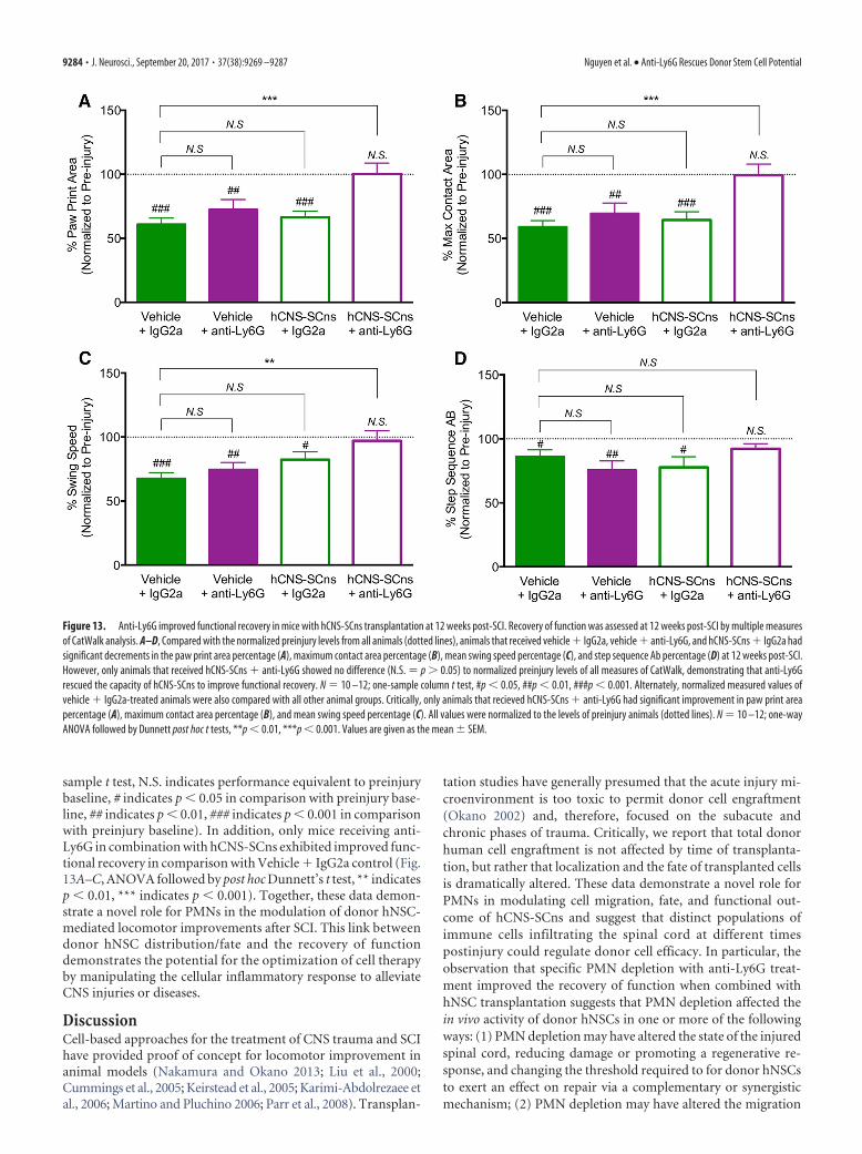
sample t test, N.S. indicates performance equivalent to preinjurybaseline, # indicates p � 0.05 in comparison with preinjury base-line, ## indicates p � 0.01, ### indicates p � 0.001 in comparisonwith preinjury baseline). In addition, only mice receiving anti-Ly6G in combination with hCNS-SCns exhibited improved func-tional recovery in comparison with Vehicle � IgG2a control (Fig.13A–C, ANOVA followed by post hoc Dunnett’s t test, ** indicatesp � 0.01, *** indicates p � 0.001). Together, these data demon-strate a novel role for PMNs in the modulation of donor hNSC-mediated locomotor improvements after SCI. This link betweendonor hNSC distribution/fate and the recovery of functiondemonstrates the potential for the optimization of cell therapyby manipulating the cellular inflammatory response to alleviateCNS injuries or diseases.
DiscussionCell-based approaches for the treatment of CNS trauma and SCIhave provided proof of concept for locomotor improvement inanimal models (Nakamura and Okano 2013; Liu et al., 2000;Cummings et al., 2005; Keirstead et al., 2005; Karimi-Abdolrezaee etal., 2006; Martino and Pluchino 2006; Parr et al., 2008). Transplan-
tation studies have generally presumed that the acute injury mi-croenvironment is too toxic to permit donor cell engraftment(Okano 2002) and, therefore, focused on the subacute andchronic phases of trauma. Critically, we report that total donorhuman cell engraftment is not affected by time of transplanta-tion, but rather that localization and the fate of transplanted cellsis dramatically altered. These data demonstrate a novel role forPMNs in modulating cell migration, fate, and functional out-come of hCNS-SCns and suggest that distinct populations ofimmune cells infiltrating the spinal cord at different timespostinjury could regulate donor cell efficacy. In particular, theobservation that specific PMN depletion with anti-Ly6G treat-ment improved the recovery of function when combined withhNSC transplantation suggests that PMN depletion affected thein vivo activity of donor hNSCs in one or more of the followingways: (1) PMN depletion may have altered the state of the injuredspinal cord, reducing damage or promoting a regenerative re-sponse, and changing the threshold required to for donor hNSCsto exert an effect on repair via a complementary or synergisticmechanism; (2) PMN depletion may have altered the migration
Figure 13. Anti-Ly6G improved functional recovery in mice with hCNS-SCns transplantation at 12 weeks post-SCI. Recovery of function was assessed at 12 weeks post-SCI by multiple measuresof CatWalk analysis. A–D, Compared with the normalized preinjury levels from all animals (dotted lines), animals that received vehicle � IgG2a, vehicle � anti-Ly6G, and hCNS-SCns � IgG2a hadsignificant decrements in the paw print area percentage (A), maximum contact area percentage (B), mean swing speed percentage (C), and step sequence Ab percentage (D) at 12 weeks post-SCI.However, only animals that received hCNS-SCns � anti-Ly6G showed no difference (N.S. � p � 0.05) to normalized preinjury levels of all measures of CatWalk, demonstrating that anti-Ly6Grescued the capacity of hCNS-SCns to improve functional recovery. N � 10 –12; one-sample column t test, #p � 0.05, ##p � 0.01, ###p � 0.001. Alternately, normalized measured values ofvehicle � IgG2a-treated animals were also compared with all other animal groups. Critically, only animals that recieved hCNS-SCns � anti-Ly6G had significant improvement in paw print areapercentage (A), maximum contact area percentage (B), and mean swing speed percentage (C). All values were normalized to the levels of preinjury animals (dotted lines). N � 10 –12; one-wayANOVA followed by Dunnett post hoc t tests, **p � 0.01, ***p � 0.001. Values are given as the mean � SEM.
9284 • J. Neurosci., September 20, 2017 • 37(38):9269 –9287 Nguyen et al. • Anti-Ly6G Rescues Donor Stem Cell Potential

and fate cues received by donor hNSCs in the injured spinal cord,restoring their potential to directly effect repair; or (3) a combi-nation of these activities.
Although previous studies have not tested the role of PMNs inrelation to cell transplantation, the effect of PMN infiltration onSCI pathogenesis and the state of the injured spinal cord is un-clear. Several studies have suggested a detrimental role for PMNsin the postinjury spinal cord. PMN inhibition (up to several dayspost-SCI) with CXCR2 inhibitors (Gorio et al., 2007), elastaseinhibitors (Tonai et al., 2001), phosphodiesterase inhibitors (Baoet al., 2011), and monoclonal anti-CD11 d antibodies (Saville etal., 2004) improved SCI and locomotor recovery. However, theneutralizing antibodies and inhibitors used were not PMN spe-cific and also inhibit or deplete early infiltrating monocytes,therefore affecting monocyte-derived macrophages, which maypromote tissue repair (David and Kroner, 2011; Saiwai et al.,2013).
Conversely, other studies have suggested that PMNs can pro-mote regeneration and initiate inflammation-associated tissuerepair. PMN depletion via anti-Gr-1 (clone RB6-8C5) antibodyworsened injury and locomotor recovery at 28 or up to 42 dpost-SCI (Stirling et al., 2009; Saiwai et al., 2013). Again, how-ever, anti-Gr-1 acts on monocytes as well as PMNs; hence, thesedata could actually support a reparative role for monocytes afterSCI, as noted above. In parallel, PMNs express the regenerationpromoting growth factor oncomodulin, and the specific immu-nodepletion of PMNs via anti-Ly6G (clone 1A8) treatment di-minished oncomodulin levels and suppressed axon regenerationafter inflammation-induced optic nerve injury (Kurimoto et al.,2013). However, anti-Ly6G antibody treatment did not alter ei-ther the lesion or locomotor recovery 42 d after contusion SCI ineither previous reports (Saiwai et al., 2013) or in our currentstudy, although it should be noted that there was a trend for a dec-rement in one CatWalk measure, regularity index, in vehicle � anti-Ly6G versus vehicle � IgG2a control at 12 weeks post-SCI. Thisobservation could suggest a more complex role for PMN infiltra-tion in long-term recovery after SCI. However, analysis of theRNA expression profile in naive, SCI � IgG-treated, and SCI �Ly6G-treated spinal cord 1 dpi demonstrated surprisingly fewgene changes, which were principally focused on innate andadaptive immune responses at this early time point. Further stud-ies of the peripheral and CNS immune response will be necessaryto establish the relationships among PMN infiltration, activation/infiltration of other immune populations, and the recovery of CNSfunction.
A second possibility is that PMN depletion altered the mi-croenvironment of the injured spinal cord and thereby the mi-gration and differentiation cues received by donor hNSCs,changing their capacity to directly effect repair. PMN infiltrationpost-SCI could modulate direct hNSC-mediated repair throughseveral mechanisms. PMNs can synthesize cytokines/factors(e.g., TNF-�, IL-6, ROS, complement proteins) that could affectdonor hNSC fate and migration. Indeed, several studies haveshown that the administration of TNF-� or IL-6, but not IL-1�,increases GFAP� cell proliferation in culture (Selmaj et al., 1990;Pannu et al., 2005), suggesting that these PMN-synthesized cyto-kines could contribute to donor hNSC astrocytic fate and migra-tion toward the injury epicenter after SCI. Furthermore, we haveshown that PMN-generated complement components C1q andC3a are chemotactic for hNSCs, promote astroglial lineage selec-tion, and impair oligodendroglial lineage selection in vitro, andthat blockade of C1q and C3a at the SCI epicenter reduces in vivo
astroglial differentiation of acutely transplanted hNSCs (Hoosh-mand et al., 2017).
In accordance with these data, a principal change in donorhNSC behavior observed following PMN depletion was the re-duction in recruitment of transplanted cells toward the injuryepicenter and the decrease in astroglial fate in these cells. Thesedata suggest that PMN-driven astrocytic fate and the migration ofdonor hNSCs toward the injury epicenter could contribute to theblockade of repair capacity, for example, by limiting the rostral—caudal “reach” of donor cells and diverting donor cells away fromspecific cell fates or phenotypes (e.g., oligodendroglia or forma-tion of beneficial astrocytes). Regarding the latter, under normalphysiological conditions astrocytes support neuronal growth.Moreover, delayed transplantation of astrocytes derived fromBMP-stimulated embryonic glial-restricted precursors promotesbehavioral recovery, axonal growth, and cell survival in rodentmodels of Parkinson’s disease (Proschel et al., 2014) and SCI(Shih et al., 2014), suggesting that phenotype is a key variable indefining the effect of astroglial fate on repair and regeneration. Inagreement with this concept, recent data suggest that phenotypemay also play a role in the net effects of endogenous astroglialpopulations within the CNS (Hamby et al., 2012; Anderson andMaes 2014; Liddelow et al., 2017).
The data presented here are the first to demonstrate that thespecific immunodepletion of an immune cell population canmodulate the efficacy of a therapeutic cell population. However,it is important to note that the converse has been previouslyshown, suggesting that transplanted stem cell populations canaffect the host inflammatory microenvironment to modulate re-pair and alter the survival/fate and recruitment of host cells. Inparticular, transplanted mesenchymal stromal/stem cells havebeen suggested to suppress the immune response through a num-ber of direct cell-to-cell interactions and soluble factors (Bassi etal., 2011; Holubova et al., 2014; Kode et al., 2009). Furthermore,several studies have suggested an immunoprotective effect byengrafted undifferentiated neural precursor cells, either via apo-ptotic induction of blood-borne CNS-infiltrating encephalito-genic T cells or TL-R4 activation (Koutsoudaki et al., 2016).Although transplanted cells can potentially affect the host mi-croenvironment (e.g., inflammation/astrogliosis), transplantedhCNS-SCns in our paradigm did not alter the level of PMNs ormonocytes in the blood or spinal cord, as shown by flow cytometry.
It has frequently been assumed that acute transplantation oftherapeutic stem cell populations would be problematic due topoor cell survival/engraftment (Okano 2002). In contrast, theacute versus delayed data presented here demonstrate equivalentengraftment 12 weeks after transplantation and suggest that themodulation of migration and lineage selection play a key role inthe potential for repair. In this regard, PMN depletion or manip-ulation of other immune populations and inflammatory mole-cules may have clinical relevance as an approach to expandtherapeutic window. Critically, differences in the immune re-sponse between rodents and humans may have a dramatic impactin considering the meaning of a therapeutic window in a clinicalcontext. Although our study tested an early time window basedon the timing of PMN infiltration in rodent SCI models (Beck etal., 2010), PMN infiltration in human SCI patients can persist forweeks, can be detected from months to years after SCI, and, as inrodent models, remains localized in the epicenter (Fleming et al.,2006). Future studies should consider strategies by which anti-Ly6G could be packaged into biodegradable materials [e.g.,poly(lactic-co-glycolic acid)] for sustained local delivery at thelesion, as well as small-molecule inhibitors of PMN infiltration
Nguyen et al. • Anti-Ly6G Rescues Donor Stem Cell Potential J. Neurosci., September 20, 2017 • 37(38):9269 –9287 • 9285
(e.g., sivelestat), which have been shown to be safely tolerated inthe clinical setting (Ryugo et al., 2006; Aikawa et al., 2011). Inparticular, strategies that can avoid systemic PMN depletion orimpairment of the overall systemic immune response would ad-dress key issues for polytrauma associated with SCI. In all, thesedata support the potential for clinical application of strategies totarget the inflammatory microenvironment in combination withcell transplantation after SCI.
ReferencesAarum J, Sandberg K, Haeberlein SL, Persson MA (2003) Migration and
differentiation of neural precursor cells can be directed by microglia. ProcNatl Acad Sci U S A 100:15983–15988. CrossRef Medline
Aikawa N, Ishizaka A, Hirasawa H, Shimazaki S, Yamamoto Y, Sugimoto H,Shinozaki M, Taenaka N, Endo S, Ikeda T, Kawasaki Y (2011) Reevalu-ation of the efficacy and safety of the neutrophil elastase inhibitor, Sive-lestat, for the treatment of acute lung injury associated with systemicinflammatory response syndrome; a phase IV study. Pulm PharmacolTher 24:549 –554. CrossRef Medline
Anderson AJ, Haus DL, Hooshmand MJ, Perez H, Sontag CJ, Cummings BJ(2011) Achieving stable human stem cell engraftment and survival in theCNS: is the future of regenerative medicine immunodeficient? Regen Med6:367– 406. CrossRef Medline
Anderson G, Maes M (2014) Reconceptualizing adult neurogenesis: role forsphingosine-1-phosphate and fibroblast growth factor-1 in co-ordinatingastrocyte-neuronal precursor interactions. CNS Neurol Disord Drug Tar-gets 13:126 –136. CrossRef Medline
Bao F, Fleming JC, Golshani R, Pearse DD, Kasabov L, Brown A, Weaver LC(2011) A selective phosphodiesterase-4 inhibitor reduces leukocyte infil-tration, oxidative processes, and tissue damage after spinal cord injury.J Neurotrauma 28:1035–1049. CrossRef Medline
Bassi EJ, Aita CA, Camara NO (2011) Immune regulatory properties ofmultipotent mesenchymal stromal cells: where do we stand? World J StemCells 3:1– 8. CrossRef Medline
Beck KD, Nguyen HX, Galvan MD, Salazar DL, Woodruff TM, Anderson AJ(2010) Quantitative analysis of cellular inflammation after traumatic spi-nal cord injury: evidence for a multiphasic inflammatory response in theacute to chronic environment. Brain 133:433– 447. CrossRef Medline
Bolger AM, Lohse M, Usadel B (2014) Trimmomatic: a flexible trimmer forIllumina sequence data. Bioinformatics 30:2114 –2120. CrossRef Medline
Butovsky O, Ziv Y, Schwartz A, Landa G, Talpalar AE, Pluchino S, Martino G,Schwartz M (2006) Microglia activated by IL-4 or IFN-gamma differen-tially induce neurogenesis and oligodendrogenesis from adult stem/pro-genitor cells. Mol Cell Neurosci 31:149 –160. CrossRef Medline
Cao QL, Zhang YP, Howard RM, Walters WM, Tsoulfas P, Whittemore SR(2001) Pluripotent stem cells engrafted into the normal or lesioned adultrat spinal cord are restricted to a glial lineage. Exp Neurol 167:48 –58.CrossRef Medline
Cummings BJ, Uchida N, Tamaki SJ, Salazar DL, Hooshmand M, SummersR, Gage FH, Anderson AJ (2005) Human neural stem cells differentiateand promote locomotor recovery in spinal cord-injured mice. Proc NatlAcad Sci U S A 102:14069 –14074. CrossRef Medline
Daley JM, Thomay AA, Connolly MD, Reichner JS, Albina JE (2008) Use ofLy6G-specific monoclonal antibody to deplete neutrophils in mice. J Leu-koc Biol 83:64 –70. CrossRef Medline
David S, Kroner A (2011) Repertoire of microglial and macrophage re-sponses after spinal cord injury. Nat Rev Neurosci 12:388 –399. CrossRefMedline
de Castro RC Jr, Burns CL, McAdoo DJ, Romanic AM (2000) Metallopro-teinase increases in the injured rat spinal cord. Neuroreport 11:3551–3554. CrossRef Medline
Dinkel K, Dhabhar FS, Sapolsky RM (2004) Neurotoxic effects of polymor-phonuclear granulocytes on hippocampal primary cultures. Proc NatlAcad Sci U S A 101:331–336. CrossRef Medline
Fleming JC, Norenberg MD, Ramsay DA, Dekaban GA, Marcillo AE, SaenzAD, Pasquale-Styles M, Dietrich WD, Weaver LC (2006) The cellularinflammatory response in human spinal cords after injury. Brain 129:3249 –3269. CrossRef Medline
Gorio A, Madaschi L, Zadra G, Marfia G, Cavalieri B, Bertini R, Di Giulio AM(2007) Reparixin, an inhibitor of CXCR2 function, attenuates inflamma-
tory responses and promotes recovery of function after traumatic lesionto the spinal cord. J Pharmacol Exp Ther 322:973–981. CrossRef Medline
Hamby ME, Coppola G, Ao Y, Geschwind DH, Khakh BS, Sofroniew MV(2012) Inflammatory mediators alter the astrocyte transcriptome andcalcium signaling elicited by multiple G-protein-coupled receptors.J Neurosci 32:14489 –14510. CrossRef Medline
Hamers FP, Koopmans GC, Joosten EA (2006) CatWalk-assisted gait anal-ysis in the assessment of spinal cord injury. J Neurotrauma 23:537–548.CrossRef Medline
Holubova M, Lysak D, Vlas T, Vannucci L, Jindra P (2014) Expanded cryo-preserved mesenchymal stromal cells as an optimal source for graft-versus-host disease treatment. Biologicals 42:139 –144. CrossRef Medline
Hooshmand MJ, Sontag CJ, Uchida N, Tamaki S, Anderson AJ, Cummings BJ(2009) Analysis of host-mediated repair mechanisms after human CNS-stem cell transplantation for spinal cord injury: correlation of engraft-ment with recovery. PLoS One 4:e5871. CrossRef Medline
Hooshmand MJ, Nguyen HX, Piltti KM, Benavente F, Hong S, Flanagan L,Uchida N, Cummings BJ, Anderson AJ (2017) Neutrophils induce as-troglial differentiation and migration of human neural stem cells via C1qand C3a synthesis. J Immunol 199:1069 –1085. CrossRef Medline
Joelving FC, Billeskov R, Christensen JR, West M, Pakkenberg B (2006)Hippocampal neuron and glial cell numbers in Parkinson’s disease–astereological study. Hippocampus 16:826 – 833. CrossRef Medline
Jones TB, McDaniel EE, Popovich PG (2005) Inflammatory-mediated in-jury and repair in the traumatically injured spinal cord. Curr Pharm Des11:1223–1236. CrossRef Medline
Karimi-Abdolrezaee S, Eftekharpour E, Wang J, Morshead CM, Fehlings MG(2006) Delayed transplantation of adult neural precursor cells promotesremyelination and functional neurological recovery after spinal cord in-jury. J Neurosci 26:3377–3389. CrossRef Medline
Keirstead HS, Nistor G, Bernal G, Totoiu M, Cloutier F, Sharp K, Steward O(2005) Human embryonic stem cell-derived oligodendrocyte progenitorcell transplants remyelinate and restore locomotion after spinal cord in-jury. J Neurosci 25:4694 – 4705. CrossRef Medline
Kigerl KA, McGaughy VM, Popovich PG (2006) Comparative analysis oflesion development and intraspinal inflammation in four strains of micefollowing spinal contusion injury. J Comp Neurol 494:578 –594. CrossRefMedline
Kim MS, Pinto SM, Getnet D, Nirujogi RS, Manda SS, Chaerkady R, Madug-undu AK, Kelkar DS, Isserlin R, Jain S, Thomas JK, Muthusamy B, Leal-Rojas P, Kumar P, Sahasrabuddhe NA, Balakrishnan L, Advani J, GeorgeB, Renuse S, Selvan LD, et al (2014) A draft map of the human pro-teome. Nature 509:575–581. CrossRef Medline
Kode JA, Mukherjee S, Joglekar MV, Hardikar AA (2009) Mesenchymalstem cells: immunobiology and role in immunomodulation and tissueregeneration. Cytotherapy 11:377–391. CrossRef Medline
Kokaia Z, Martino G, Schwartz M, Lindvall O (2012) Cross-talk betweenneural stem cells and immune cells: the key to better brain repair? NatNeurosci 15:1078 –1087. CrossRef Medline
Koopmans GC, Deumens R, Honig WM, Hamers FP, Steinbusch HW,Joosten EA (2005) The assessment of locomotor function in spinal cordinjured rats: the importance of objective analysis of coordination. J Neu-rotrauma 22:214 –225. CrossRef Medline
Kopylova E, Noe L, Touzet H (2012) SortMeRNA: fast and accurate filteringof ribosomal RNAs in metatranscriptomic data. Bioinformatics 28:3211–3217. CrossRef Medline
Koutsoudaki PN, Papastefanaki F, Stamatakis A, Kouroupi G, Xingi E, Styl-ianopoulou F, Matsas R (2016) Neural stem/progenitor cells differenti-ate into oligodendrocytes, reduce inflammation, and ameliorate learningdeficits after transplantation in a mouse model of traumatic brain injury.Glia 64:763–779. CrossRef Medline
Kurimoto T, Yin Y, Habboub G, Gilbert HY, Li Y, Nakao S, Hafezi-Moghadam A, Benowitz LI (2013) Neutrophils express oncomodulinand promote optic nerve regeneration. J Neurosci 33:14816 –14824.CrossRef Medline
Lenzlinger PM, Morganti-Kossmann MC, Laurer HL, McIntosh TK (2001)The duality of the inflammatory response to traumatic brain injury. MolNeurobiol 24:169 –181. CrossRef Medline
Liao Y, Smyth GK, Shi W (2014) featureCounts: an efficient general purposeprogram for assigning sequence reads to genomic features. Bioinformat-ics 30:923–930. CrossRef Medline
Liddelow SA, Guttenplan KA, Clarke LE, Bennett FC, Bohlen CJ, Schirmer L,
9286 • J. Neurosci., September 20, 2017 • 37(38):9269 –9287 Nguyen et al. • Anti-Ly6G Rescues Donor Stem Cell Potential

Bennett ML, Munch AE, Chung WS, Peterson TC, Wilton DK, Frouin A,Napier BA, Panicker N, Kumar M, Buckwalter MS, Rowitch DH, DawsonVL, Dawson TM, Stevens B, et al (2017) Neurotoxic reactive astrocytes areinduced by activated microglia. Nature 541:481–487. CrossRef Medline
Liu S, Qu Y, Stewart TJ, Howard MJ, Chakrabortty S, Holekamp TF, McDon-ald JW (2000) Embryonic stem cells differentiate into oligodendrocytesand myelinate in culture and after spinal cord transplantation. Proc NatlAcad Sci U S A 97:6126 – 6131. CrossRef Medline
Luchetti S, Beck KD, Galvan MD, Silva R, Cummings BJ, Anderson AJ (2010)Comparison of immunopathology and locomotor recovery in C57BL/6,BUB/BnJ and NOD-SCID mice after contusion spinal cord injury. J Neu-rotrauma 27:411– 421. CrossRef Medline
Martino G, Pluchino S (2006) The therapeutic potential of neural stem cells.Nat Rev Neurosci 7:395– 406. CrossRef Medline
McCarthy DJ, Chen Y, Smyth GK (2012) Differential expression analysis ofmultifactor RNA-Seq experiments with respect to biological variation.Nucleic Acids Res 40:4288 – 4297. CrossRef Medline
Monje ML, Toda H, Palmer TD (2003) Inflammatory blockade restores adulthippocampal neurogenesis. Science 302:1760–1765. CrossRef Medline
Nakamura M, Okano H (2013) Cell transplantation therapies for spinalcord injury focusing on induced pluripotent stem cells. Cell Res 23:70 –80. CrossRef Medline
Nguyen HX, Beck KD, Anderson AJ (2011) Quantitative assessment of im-mune cells in the injured spinal cord tissue by flow cytometry: a novel usefor a cell purification method. J Vis Exp (5):2698. CrossRef Medline
Okano H (2002) Neural stem cells: progression of basic research and perspec-tive for clinical application. Keio J Med 51:115–128. CrossRef Medline
Pannu R, Singh AK, Singh I (2005) A novel role of lactosylceramide in theregulation of tumor necrosis factor alpha-mediated proliferation of ratprimary astrocytes. Implications for astrogliosis following neurotrauma.J Biol Chem 280:13742–13751. CrossRef Medline
Parr AM, Kulbatski I, Zahir T, Wang X, Yue C, Keating A, Tator CH (2008)Transplanted adult spinal cord-derived neural stem/progenitor cells pro-mote early functional recovery after rat spinal cord injury. Neuroscience155:760 –770. CrossRef Medline
Pathan M, Keerthikumar S, Ang CS, Gangoda L, Quek CY, Williamson NA,Mouradov D, Sieber OM, Simpson RJ, Salim A, Bacic A, Hill AF, StroudDA, Ryan MT, Agbinya JI, Mariadason JM, Burgess AW, Mathivanan S(2015) FunRich: an open access standalone functional enrichment andinteraction network analysis tool. Proteomics 15:2597–2601. CrossRefMedline
Popovich PG, Wei P, Stokes BT (1997) Cellular inflammatory response afterspinal cord injury in Sprague-Dawley and Lewis rats. J Comp Neurol377:443– 464. CrossRef Medline
Popovich PG, Guan Z, Wei P, Huitinga I, van Rooijen N, Stokes BT (1999)Depletion of hematogenous macrophages promotes partial hindlimb re-covery and neuroanatomical repair after experimental spinal cord injury.Exp Neurol 158:351–365. CrossRef Medline
Proschel C, Stripay JL, Shih CH, Munger JC, Noble MD (2014) Delayedtransplantation of precursor cell-derived astrocytes provides multiplebenefits in a rat model of Parkinsons. EMBO Mol Med 6:504 –518.CrossRef Medline
Rapalino O, Lazarov-Spiegler O, Agranov E, Velan GJ, Yoles E, Fraidakis M,Solomon A, Gepstein R, Katz A, Belkin M, Hadani M, Schwartz M (1998)Implantation of stimulated homologous macrophages results in partialrecovery of paraplegic rats. Nat Med 4:814 – 821. CrossRef Medline
Ryugo M, Sawa Y, Takano H, Matsumiya G, Iwai S, Ono M, Hata H, Yamau-
chi T, Nishimura M, Fujino Y, Matsuda H (2006) Effect of a polymor-phonuclear elastase inhibitor (sivelestat sodium) on acute lung injuryafter cardiopulmonary bypass: findings of a double-blind randomizedstudy. Surg Today 36:321–326. CrossRef Medline
Saiwai H, Kumamaru H, Ohkawa Y, Kubota K, Kobayakawa K, Yamada H,Yokomizo T, Iwamoto Y, Okada S (2013) Ly6C� Ly6G- myeloid-derived suppressor cells play a critical role in the resolution of acuteinflammation and the subsequent tissue repair process after spinal cordinjury. J Neurochem 125:74 – 88. CrossRef Medline
Salazar DL, Uchida N, Hamers FP, Cummings BJ, Anderson AJ (2010) Hu-man neural stem cells differentiate and promote locomotor recovery in anearly chronic spinal cord injury NOD-scid mouse model. PLoS One5:e12272. CrossRef Medline
Saville LR, Pospisil CH, Mawhinney LA, Bao F, Simedrea FC, Peters AA,O’Connell PJ, Weaver LC, Dekaban GA (2004) A monoclonal antibodyto CD11d reduces the inflammatory infiltrate into the injured spinal cord:a potential neuroprotective treatment. J Neuroimmunol 156:42–57.CrossRef Medline
Schwartz M, Yoles E (2005) Macrophages and dendritic cells treatment ofspinal cord injury: from the bench to the clinic. Acta Neurochir Suppl93:147–150. CrossRef Medline
Selmaj KW, Farooq M, Norton WT, Raine CS, Brosnan CF (1990) Prolifer-ation of astrocytes in vitro in response to cytokines. A primary role fortumor necrosis factor. J Immunol 144:129 –135. Medline
Shihabuddin LS, Horner PJ, Ray J, Gage FH (2000) Adult spinal cord stemcells generate neurons after transplantation in the adult dentate gyrus.J Neurosci 20:8727– 8735. Medline
Shih CH, Lacagnina M, Leuer-Bisciotti K, Proschel C (2014) Astroglial-derived periostin promotes axonal regeneration after spinal cord injury.J Neurosci 34:2438 –2443. CrossRef Medline
Stirling DP, Liu S, Kubes P, Yong VW (2009) Depletion of Ly6G/Gr-1 leu-kocytes after spinal cord injury in mice alters wound healing and worsensneurological outcome. J Neurosci 29:753–764. CrossRef Medline
Subhash S, Kanduri C (2016) GeneSCF: a real-time based functional enrich-ment tool with support for multiple organisms. BMC Bioinformatics 17:365. CrossRef Medline
Tamaki S, Eckert K, He D, Sutton R, Doshe M, Jain G, Tushinski R, ReitsmaM, Harris B, Tsukamoto A, Gage F, Weissman I, Uchida N (2002) En-graftment of sorted/expanded human central nervous system stem cellsfrom fetal brain. J Neurosci Res 69:976 –986. CrossRef Medline
Tonai T, Shiba K, Taketani Y, Ohmoto Y, Murata K, Muraguchi M, Ohsaki H,Takeda E, Nishisho T (2001) A neutrophil elastase inhibitor (ONO-5046) reduces neurologic damage after spinal cord injury in rats. J Neu-rochem 78:1064 –1072. CrossRef Medline
Uchida N, Buck DW, He D, Reitsma MJ, Masek M, Phan TV, Tsukamoto AS,Gage FH, Weissman IL (2000) Direct isolation of human central ner-vous system stem cells. Proc Natl Acad Sci U S A 97:14720 –14725.CrossRef Medline
Weaver KD, Branch CA, Hernandez L, Miller CH, Quattrocchi KB (2000)Effect of leukocyte-endothelial adhesion antagonism on neutrophil mi-gration and neurologic outcome after cortical trauma. J Trauma 48:1081–1090. CrossRef Medline
Yagisawa M, Yuo A, Yonemaru M, Imajoh-Ohmi S, Kanegasaki S, Yazaki Y,Takaku F (1996) Superoxide release and NADPH oxidase componentsin mature human phagocytes: correlation between functional capacityand amount of functional proteins. Biochem Biophys Res Commun 228:510 –516. CrossRef Medline
Nguyen et al. • Anti-Ly6G Rescues Donor Stem Cell Potential J. Neurosci., September 20, 2017 • 37(38):9269 –9287 • 9287