SUMOylome Modulates Intra agellar Transport Machinery and ...
26
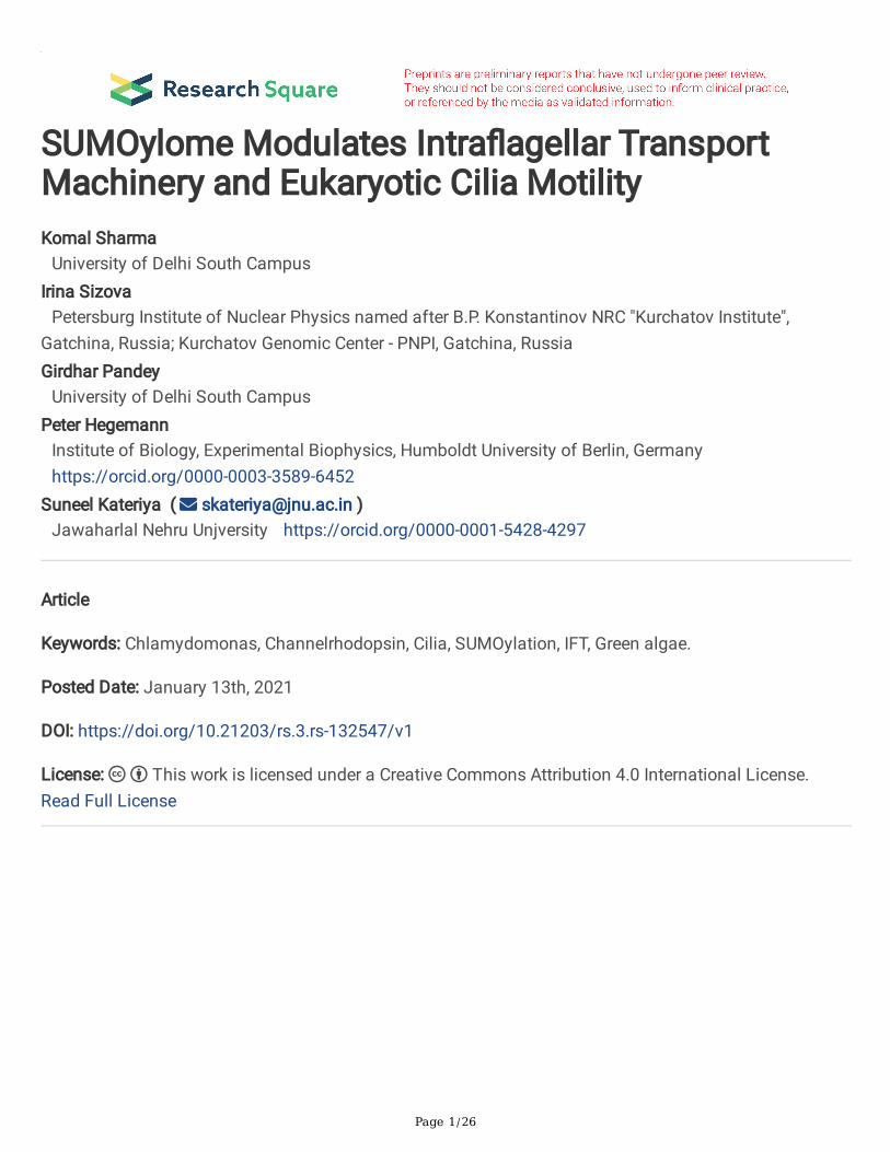
Page 1/26 SUMOylome Modulates Intraagellar Transport Machinery and Eukaryotic Cilia Motility Komal Sharma University of Delhi South Campus Irina Sizova Petersburg Institute of Nuclear Physics named after B.P. Konstantinov NRC "Kurchatov Institute", Gatchina, Russia; Kurchatov Genomic Center - PNPI, Gatchina, Russia Girdhar Pandey University of Delhi South Campus Peter Hegemann Institute of Biology, Experimental Biophysics, Humboldt University of Berlin, Germany https://orcid.org/0000-0003-3589-6452 Suneel Kateriya ( [email protected] ) Jawaharlal Nehru Unjversity https://orcid.org/0000-0001-5428-4297 Article Keywords: Chlamydomonas, Channelrhodopsin, Cilia, SUMOylation, IFT, Green algae. Posted Date: January 13th, 2021 DOI: https://doi.org/10.21203/rs.3.rs-132547/v1 License: This work is licensed under a Creative Commons Attribution 4.0 International License. Read Full License
-
Upload
khangminh22 -
Category
Documents
-
view
0 -
download
0
Transcript of SUMOylome Modulates Intra agellar Transport Machinery and ...
Page 1/26
SUMOylome Modulates Intra�agellar TransportMachinery and Eukaryotic Cilia MotilityKomal Sharma
University of Delhi South CampusIrina Sizova
Petersburg Institute of Nuclear Physics named after B.P. Konstantinov NRC "Kurchatov Institute",Gatchina, Russia; Kurchatov Genomic Center - PNPI, Gatchina, RussiaGirdhar Pandey
University of Delhi South CampusPeter Hegemann
Institute of Biology, Experimental Biophysics, Humboldt University of Berlin, Germanyhttps://orcid.org/0000-0003-3589-6452
Suneel Kateriya ( [email protected] )Jawaharlal Nehru Unjversity https://orcid.org/0000-0001-5428-4297
Article
Keywords: Chlamydomonas, Channelrhodopsin, Cilia, SUMOylation, IFT, Green algae.
Posted Date: January 13th, 2021
DOI: https://doi.org/10.21203/rs.3.rs-132547/v1
License: This work is licensed under a Creative Commons Attribution 4.0 International License. Read Full License

Page 2/26
AbstractTranslocation of channelrhodopsins (ChRs) is mediated by intra�agellar transport (IFT) machinery.However, the functional role of the network containing photoreceptors, IFT and other proteins incontrolling cilia motility of the alga is still not fully delineated. In the current study, we identi�ed twoimportant motifs at the C-terminus of ChR1. One of them is similar to a known ciliary targeting sequencethat speci�cally interacts with a small GTPase, and the other is a SUMOylation site. For the �rst time,experimental data provide an insight into the role of SUMOylation in the modulation of IFT & ChR1.Blocking of SUMOylation affected the phototaxis of C. reinhardtii cells. This implies SUMOylation basedregulation of protein network controlling photomotility. The conservation of SUMOylation site pattern asanalyzed for the relevant photoreceptors, IFT and its associated signaling proteins in other ciliated greenalgae suggested SUMOylation based photobehavioural response across the microbes. This reportestablishes a link between evolutionary conserved SUMOylation and ciliary machinery for themaintenance and functioning of cilia across the eukaryotes. Our enriched SUMOylome of C. reinhardtiicomprehends the proteins related to ciliary development and, photo-signaling, along with homologue(s)associated to human ciliopathies as SUMO targets.
IntroductionRhodopsins, a major class of photoreceptors for responding to several light-associated environmentalcues, exists in all the three domains of life i.e., archaea, bacteria and eukaryote (1, 2). Microbial type andanimal type rhodopsins are classi�ed based on different bound retinal in their dark state. Animal typerhodopsins are mainly involved in visual and non-visual phototransduction in vertebrates andinvertebrates (3). Rhodopsin functioning is associated with its dynamic localization and modi�cationsduring the photochemical signaling in metazoans (4-8). Chlamydomonas reinhardtii, comprises probablyten microbial type rhodopsins (named as Chlamyopsins, Cop3-12), among which Cop3(Channelrhodopsin 1, ChR1) and Cop4 (Channelrhodopsin 2, ChR2) are light-gated cation channels. ChR1and ChR2 are primarily responsible for the photo-orientation of this alga (9-12). Photoactivation of ChRsgenerates two types of photocurrents, a “photoreceptor current” in the eyespot followed by a “�agellarcurrent” that results in calcium (Ca2+) �ux across the ciliary membrane (12-16) leading to the ciliarybeating pattern that help cells to move towards ambient light (14). ChR1 and Cop8 were found to belocalized to the eyespot and cilia in a light dependent manner (17). It is known that the tra�cking ofproteins to specialized compartments like chloroplast, mitochondria, nucleus or cilia is dependent onsome speci�c signature motif(s) encoded in the relevant cargo proteins (18). In case of cilia, thesesignature motifs include ciliary targeting sequences (CTS) or a SUMOylation motif. CTS includessequences like VXPX and Ax[S/A] xQ, and the mutations in these motifs could alter their localizationpattern leading to ciliary tra�cking and associated disorders (19-22). Vertebrate rhodopsins, containingthe CTS motifs (VXPX and FR), are targeted to the modi�ed cilium of rod cells by polarized vesiculartra�cking using several GTPases (23, 24). These CTS interacts with a small GTPase, ARF4 and itsactivator in the vesicles while budding off from Golgi bodies, which recruits other protein complexes

Page 3/26
required in the transport (24, 25). CTS assisted tra�cking of rhodopsins in mammalian system is wellstudied (26).
Recently, SUMOylation mediated ciliary localization of proteins like small GTPase, ARL13 and anolfactory signaling protein, adenylyl cyclase 3 (AC3) have also been reported (27, 28). SUMOylation isknown for modulating the expression and organellar localization of the targets (29). SUMO (smallubiquitin like modi�er) modi�cation also regulates expression and function of many cytoskeletal proteinsand hence it is emerging as a current interest for its involvement in ciliary protein tra�cking and relateddisorders (30, 31).
In the presented study, a CTS motif, VMPS, has been identi�ed in the C-terminus of ChR1, which isrequired for its interaction with a small GTPase. This is also the �rst report demonstrating a microbialtype rhodopsin (i.e., ChR1), conserved IFT components, ciliary targeting machinery are modulated bycellular SUMOylation. Blocking of SUMOylation partially affects the photomotility of C. reinhardtii.Proteins related to ciliary maintenance, ciliary signaling and ciliopathies associated candidates alongwith photoreceptors were identi�ed from SUMOylome using Nano-LC-MS/MS with the eluents ofimmunoprecipitant sample(s). Orthologues of channelrhodopsin and its tra�cking associated machinerycomponents in other ciliated algae found to possess a conserved SUMOylation pattern. In essence, thesimilar pattern of SUMOylation in evolutionarily conserved IFT machinery and its associated componentsindicates its functional importance across the relevant eukaryotic organisms, and a defect of the samemay lead to certain ciliopathy disorder in human.
Materials And MethodsBio-informatics analysis
The Phytozome (https://phytozome.jgi.doe.gov/pz/portal.html#!search?show=BLAST) BLAST tool wasused to identify sequences orthologous to the human ARF4 sequence. The SUMOylation sites and theSUMO interaction motif (SIM) were predicted with SUMOplot (http://www.abcepta.com/sumoplot) andGPS-SUMO (http://sumosp.biocuckoo.org/online.php) (33, 34). Clustal W was used for multiple sequencealignment and edited with the BioEdit tool (35). The protein-protein interaction network was predictedusing STRING (string-db.org/) and was edited and analyzed with the Cytoscape_3.7.2 (36).
Constructs used and site directed mutagenesis
ChR1Ct (960-2136 bp) and ChR2Ct (945-2211 bp) cloned in the pETSUMO vector (Champion pET SUMOExpression System, Thermo Fisher Scienti�c) were provided by Dr. Mayanka Awasthi. The CrARL11GSTfused construct cloned in pGEX4T1 was provided by Dr. Peeyush Ranjan. ChR1Ct was mutated atV643XP645X site to M643XS645X and ChR2Ct at V628XP630X site to M628XS630X using QuickChangeII Site-Directed Mutagenesis kit, Agilent. SDM-PCR cycling parameters were adjusted according to theinstruction of manual. The sequence of the primers used for SDM are as follows:

Page 4/26
ChR1CtV643M;P645S Fw: CATGCAGGCCATGGGTGGCATGATGTCCAGCCCCGCCCCC, Rev:GGGGGCGGGGCTGGACATCATGCCACCCATGGCCTGCATG and
ChR2CtV628M;P630S Fw: ATGAGCTCCGGCGTGGTGGCCAACATGACGTCCTCCGCCG
Rev: CGGCGGAGGGCGTCACGTTGGCCACCACGCCGGAGCTCAT.
Chlamydomonas reinhardtii cell culture
Chlamydomonas reinhardtii wild type strain CC-124 was procured from Chlamydomonas resource center(www.chlamycollection.org). Culture was grown using TAP (tris-acetate-phosphate media, pH 7.4 andsupplemented with Hutner trace elements) at 25 ˚C in a shaker incubator (120 rpm) in a lightsynchronized manner (14 hrs light and 10 hrs dark condition). Light exposure was provided using white�uorescent light (2300 lux).
Preparation of Chlamydomonas total cell lysate (CrTCL) under different experimental conditions andimmunoblotting
The total cell lysate of C. reinhardtii culture (CC-124 strain) was prepared using exponential phase cellsthat were harvested by centrifugation at 5000 rpm for 5 min. The cell pellet was resuspended in 10 ml of1X TBS (50mM Tris-Cl pH 8.0, 150mM NaCl) supplemented with 1 mM PMSF and recommended amountof protease inhibitor cocktail (Protease inhibitor cocktail, for plant cell extracts, Sigma Aldrich) in thepresence or absence of 20 mM n-ethylmaleimide (NEM, Cat. No. E3876, Sigma Aldrich), as required forthe SUMOylation detection experiments. NEM has been used for the detection of SUMO conjugatedproteins in C. reinhardtii as described earlier (37, 38). The cell suspension was homogenized using thefollowing sonication parameters: 35% amplitude, 8 sec. ON/OFF cycle lasting for 3 min. After sonication,the concentration of protein in the CrTCL was estimated by the Bradford assay. CrTCL was alsoincubated with a puri�ed ULP-1 enzyme (Concentration: 8µg/µl) preparation as per the mentionedexperimental design: Different concentrations of the ULP-1 enzyme (1:100, 1:500 and 1:1000) wereincubated with 30 µg of CrTCL in the absence or presence of NEM. The reaction was incubated at twotemperatures, 22°C (Optimal temperature for growth of C. reinhardtii cells) for 2 hrs and 37°C for 1 hr.Finally, samples were solubilized in standard 2X Laemmli buffer and subsequently analyzed with SDS-PAGE. Then, western blotting using ChR1Ct or SUMO antibody, was done according the protocolestablished earlier (17).
Co-immunoprecipitation (Co-IP) and Nano LC-MS/MS
C. reinhardtii cells were harvested in their exponential phase (O.D.700: 0.6-0.7) and resuspended in IP lysisbuffer [50 mM Tris-Cl pH 8.0, 150 mM NaCl, 0.5% Sodium deoxycholate, 1% NP-40, 1 mM PMSF (addedfreshly), and PIC (Protease inhibitor cocktail)] in the presence or absence of 20 mM NEM. Sonication wasperformed for 3 min. (8 sec. ON/OFF cycle with 35% amplitude) to homogenize the algal cells and celldebris was removed by centrifugation at 14000 rpm for 10 min. The supernatant from the previous spin
Page 5/26
was precleared with pre-immune serum (rabbit) along with protein A beads (Protein A Sepharose beads,Invitrogen, USA). 1 ml of the precleared lysate was then incubated overnight at 4°C with 1 µg/ml ofprimary antibody with end to end rotation. The treated lysate was then incubated with 25 µl protein ASepharose beads for 2 hours at 4°C with rotation. After removing the CrTCL supernatant, the antibody-antigen complex bound beads were washed once with 1X PBS and 3 times with IP lysis buffer. For Nano-LC-MS/MS analysis, the complex was eluted from the bound protein A beads with 0.1 M glycine, pH 2.8(pre-sterilized with 0.2-micron �lter), which were then neutralized with 1 M Tris-Cl pH 8.0. The elutionswere then solubilized in a buffer containing 6 M guanidinium hydrochloride and 50 mM ammoniumbicarbonate. The reduction and alkylation were done with 10 mM Tris(2-carboxyethyl) phosphinehydrochloride (TCEP) and 55 mM iodoacetamide (IAA) in 25 mM ammonium bicarbonate, respectively.These samples were trypsinized overnight and then desalted. After speed vac, these samples wereresuspended in 0.1% formic acid and loaded on C18 reverse phase column (Central Instrument Facility,UDSC). Raw data was then analyzed using the Proteome Discoverer 2.2 (Thermo Scienti�c, USA). Fordetecting co-immunoprecipitation of a speci�c protein in the IP elution using immunoblotting, the protein-antibody complex was eluted from the beads by directly adding 2X Laemmli buffer and heating at 95°Cfor 10 min. Western blotting was done with the protein speci�c antibody (annotation and dilution ofantibody have been provided in the respective Fig. legend).
Expression and puri�cation of the recombinant proteins
Expression constructs containing the target genes (ChR1Ct, ChR2Ct, CrARL11GST, ChR1CtVP mutant,GST Only) were transformed into E. coli strain BL21 (DE3λ). Luria-Bertani broth (LB) was supplementedwith 50 µg/ml of kanamycin (for pET-Sumo constructs) or 100 µg/ml ampicillin (for pGEX4T1constructs), during bacterial cultivation. Cultures were incubated overnight at 37°C with shaking at 200rpm. The culture was then scaled up (secondary culture) using the overnight grown primary culture, in TBmedia (Terri�c Broth) with appropriate antibiotic, incubated at 37°C with shaking till an O.D600 of 0.6-0.8was attained. Flasks were incubated on ice for 45 minutes prior to induction. Recombinant proteinexpression was induced with 0.3 mM IPTG (Isopropyl β-D-1-thiogalactopyranoside) at 16°C with shakingat 200 rpm. Cells were harvested after 48 hrs of induction by centrifugation at 6000 rpm for 10 min. andfurther resuspended in 1XPBS containing 50 µg/ml lysozyme and 200 µM PMSF. The solubilized cellwere homogenized by sonication at 35% amplitude and 30 sec. ON/OFF pulse for 5 min. Lysed cells werethen centrifuged at 12000 rpm for 55 min. The total cell lysate was separated into soluble and insolublefraction. The soluble fraction, containing the protein of interest, was �ltered using a 0.45 µm �lter. Theprotein with a histidine (His)-tag (in case of pET-SUMO based constructs) was puri�ed using a Co2+
immobilized talon beads column (TALON® Metal A�nity Resins, Clontech Laboratories, Inc., USA) byimmobilized metal ion a�nity chromatography (IMAC). Protein with a GST tag (for pGEX-4T1 basedconstructs) was puri�ed using Glutathione Sepharose beads 4 Fast Flow (GE Healthcare, USA). Theprotein of interest was eluted with 250 mM Imidazole (Fisher Scienti�c, USA) in case of Co2+ immobilizedtalon beads. The GST beads bound protein complex was eluted with 30 mM reduced glutathionesolubilized in 50 mM Tris-Cl pH 8.5 [Glutathione reduced (GSH), SRL].

Page 6/26
GST pull-down assay
Recombinant protein of CrARL11GST, GST only, ChR1Ct, ChR2Ct and ChR1CtVP mutant variants wereexpressed into E. coli strain BL21 (DE3λ). Cells were pelleted, homogenized and fractionated bycentrifugation as described in the previous section of materials and methods. GST beads boundCrARL11GST was used as a bait to pull down ChR1Ct, ChR2Ct and ChR1CtVP mutant variants (prey forthe experiment) from their respective soluble fractions. GST protein was used as a negative controlduring this experiment. For the pull down, 1 ml soluble fractions of the prey protein(s) were �rstprecleared with 20 µg GST protein and 20 µl glutathione Sepharose beads. Soluble fraction was thenspun at 13000 rpm, 4°C and the supernatant was taken for GST pull-down assay. Each prey protein wasincubated with glutathione Sepharose beads bound to 20 µg GST as negative control and to 20µgARL11GST, separately, for 1hr at 4°C with end-to-end mixing. Beads were washed 3 times with chilled1XPBS. Bound proteins were eluted with GST elution buffer in subsequent fractions of 15 µl each. Elutedfractions were mixed with 2X Laemmli buffer containing 10% β-mercaptoethanol and heated at 95°C for10 min. Eluents were analyzed using SDS-PAGE followed by immunoblotting of the elutions with theprotein speci�c antibodies (Anti-ChR1Ct and Anti-ChR2Ct). The same western blot was stripped withstripping buffer (60 mM Tris-Cl pH 6.8, 2% SDS and 50mM β-mercaptoethanol) and then probed againwith the HRP conjugated primary GST antibody as a loading control.
Treatment of Chlamydomonas cells with the SUMOylation blocker, 2D08
The SUMOylation blocker, 2D08 (Cat. No. SML1052, Sigma Aldrich, Merck), has been used for severalSUMO related studies (39-41). It is a cell permeable synthetic chemical reagent that blocks the SUMO tagtransfer from E2 enzyme (UBC9) to the substrate. C. reinhardtii cell wall mutant strain, CC3403, wasgrown till O.D. reached 0.6 under 14:10 hours light-dark cycle. Cells were then harvested bycentrifugation at 3000 rpm and resuspended in lower volume (1/20) of fresh TAP media and 3 ml ofresuspended cells was poured in two Petri dishes. Cells in one Petri dish were incubated for 8 hours with100 µM of 2D08 (dissolved in DMSO), while another Petri dish containing equal number of cells wasincubated with equal volume of DMSO as of 2D08, as negative control. For dish assay, the Petri dishescontaining the 2D08 incubated cells and DMSO incubated as negative control were exposed to white light(Cool Fluorescent Lights) from one side of the Petri dishes. The movement of cell population wasobserved in both the Petri dishes and the images were captured accordingly. In order to detect the ChR1 inthe 2D08 incubated cells, lysis of the cells were carried out using an 8:4:3 (v/v) solution ofmethanol:chloroform:water with the required volume of the cells to precipitate all the proteins (32). Theprecipitated proteins were then pelleted down and resuspended in 100 µl of 50 mM Tris-Cl buffer (pH 8.0),150 mM NaCl supplemented with 20 mM NEM and PIC. Samples were then prepared from theresuspended and precipitated protein using 2X Laemmli buffer and heating at 65°C for 30 min. Sampleswere resolved on 8% SDS-PAGE and then immunoblotting was done with anti-ChR1Ct antibody followedby HRP-conjugated anti-rabbit secondary antibody.
Results

Page 7/26
ChR1 ciliary targeting sequence interacts with CrARL11
Deatiled sequence analysis of ChR1 from C. reinhardtii revealed that it contains motifs, “VMPS” (ValineMethionine Proline Serine) and “FR” (Phenylalanine Arginine), at its C-terminus (ChR1C-terminus,ChR1Ct), similar to the known CTS, VXPX and FR (Fig. 1A). The VXPX sequence of vertebrate rhodopsinspeci�cally interacts with a small GTPase, ARF4, for its vesicular targeting towards cilia (24). This led tothe search for the orthologue of ARF4 GTPase in the C. reinhardtii genome that might be interacting withthe VMPS of ChR1. Mining of the C. reinhardtii genome using Phytozome [plant genomics resourceplatform, (phytozome.jgi.doe.gov)] was carried out with the human ARF4 sequence as a query sequence.CrARL11 showed maximum homology to the ARF4 sequence, and sequence alignment of ARF4 withCrARL11 revealed homology in all the characteristic G-protein domains (Fig. 1B). CrARL11 showedaround 77% homology to ARF4. The interaction between CrARL11 and ChR1 was �rst con�rmed byimmunoprecipitation (IP) using CrARL11 and ChR1Ct antibodies in C. reinhardtii total cell lysate (CrTCL).CrARL11 antibody co-immunoprecipitated the ChR1 from CrTCL and vice-versa. The detection of ChR1 inCrARL11 IP eluent and CrARL11 in ChR1 IP eluent by immunoblotting showed interaction between ChR1and CrARL11 (Fig. 1C). Recombinant ChR1Ct protein (Fig. 1D) and ChR1 from CrTCL (Fig. 1E) werepulled-down by GST-tagged-CrARL11 protein. The results demonstrated the direct physical interactionbetween ChR1 and CrARL11, and that the ChR1Ct is su�cient for this interaction. ChR2 and Chlamyopsin5 (Cop5) were also found to contain the CTS similar to VXPX motif in their C-terminus (Fig. S2A). IP wascarried with ChR2Ct and Cop5 speci�c antibody in the CrTCL to con�rm their interaction with CrARL11. IPelutions of ChR2Ct and Cop5 showed the co-immunoprecipitation of CrARL11 as detected byimmunoblotting with CrARL11 antibody (Fig. S2B). The result showed that both ChR2 and Cop5 alsointeract with CrARL11. CrARL11-GST was able to pull-down recombinant ChR2Ct (Fig. S2C). The obtainedresults con�rmed the direct interaction between ChR2 and CrARL11 through its C-terminus.
The VMPS motif of ChR1 is crucial for the ChR1-CrARL11 interaction
It is well documented that the mutations V345M and P347S are responsible for disrupting ARF4interaction with rhodopsin leading to autosomal dominant retinitis pigmentosa (ADRP) in vertebrates (24,42-44). To determine the VMPS mediated interaction between ChR1 and CrARL11, the VMPS sequence inthe ChR1Ct was mutated to MMSS. CrARL11-GST was used as a bait to pull-down wild type ChR1Ct andChR1CtVP mutant (V and P residues in VMPS were mutated and the mutated protein was annotated asChR1CtVP mutant). Detection of ChR1 with ChR1Ct antibody by immunoblotting of pull-down eluentsrevealed the presence of wild type ChR1Ct and absence of ChR1CtVP mutant (Fig. 1F). This speci�es thatthe interaction of ChR1 and CrARL11 is mediated by VMPS motif present at the C-terminus of ChR1.
The C-terminus of ChR1 contains a functional SUMOylation site
The SUMOylation machinery consists of a series of enzymes for the conjugation of SUMO protein via anisopeptide bond to the ε-amino group of lysine residues within the consensus SUMOylation motif, ΨKXE,of the target proteins (45). An alternate mechanism of SUMO conjugation to the target protein is throughSUMO interaction motif (SIM), a small motif of amino acids that binds directly to SUMO or SUMO tagged

Page 8/26
to target proteins. ChRs from different green algae were contained ΨKXE as shown in the multiplesequence alignment of their C-termini (Fig. 2A). Supplementary table 1 annotates the predicted positionof SUMO tagged lysine residue and SIM motif within the ChRs of diverse algae. However, Pleodorinastarrii (PstChR1 and PstChR2) and ChR from Klebsormidium �accidum was found to contain SIM motifonly (Supplementary table 1). ChR1 was predicted with a single SUMOylation site at the end of its C-terminus. Experiments were performed using SUMO tag protectors, the schematic illustration of the sameis provided in Fig. 2B. NEM (n-ethylmaleimide) is an isopeptidase inhibitor that inhibits SUMO tagcleaving proteases, ubiquitin-like proteases (ULPs), and hence protects the target from deSUMOylation.NEM has been used extensively for the detection of SUMO and SUMO conjugating proteins in CrTCL (37).CrTCL was incubated with NEM and a protease inhibitor cocktail (PIC). Immunoblotting of the sampleswith ChR1Ct antibody showed bands with higher intensity at the expected molecular weight of ChR1 anda higher molecular weight band, in comparison to the negative control (with no NEM and PIC) as shownin Fig. 2C. Quanti�cation of the ChR1 speci�c band by densitometry revealed a comparatively signi�cantincrease in the ChR1 accumulation (Fig. 2D). It can be inferred from the results that ChR1 was stabilizedin the presence of NEM and PIC. This indicates an involvement of SUMOylation machinery in theregulation of the ChR1.
ULP-1 enzyme mediated deSUMOylation of ChR1 in Chlamydomonas
ULP-1 (Ubiquitin-like protease 1) is involved in deSUMOylation of the SUMOylated protein targets andform an integral part of the reversible SUMOylation machinery (25, 38). CrTCL samples (with or withoutNEM), were incubated with different dilutions of the puri�ed yeast ULP-1 for 2 hrs at 22°C (optimumtemperature for C. reinhardtii growth). Samples containing only ULP-1 showed lower intensity of ChR1speci�c bands upon immunoblotting with ChR1Ct antibody, in comparison to the samples incubated withboth ULP-1 and NEM (Fig. 2E). Densitometry analysis of the ChR1 speci�c bands revealed that theamount of ChR1 signi�cantly decreased in the presence of ULP-1 (Fig. 2F). This decreased ChR1 amountis expected to be associated with the ULP-1 mediated deSUMOylation of ChR1, which further possibly ledto degradation of the ChR1. It indicates that SUMOylation of ChR1 probably is related to its stability,protecting it from ULP dependent degradation. Similar set of experiment was carried at 37°C for optimalactivity of the ULP1. It also showed results similar to those obtained at 22°C, however no highermolecular weight band accumulation was seen (Fig. S3).
SUMOylation controls phototaxis of Chlamydomonas
C. reinhardtii cells were incubated for 8 hours with the SUMOylation blocker 2D08, which inhibitsSUMOylation by blocking the transfer of SUMO from E2 (UBC9) enzyme to the substrate (41). Theaccumulation of ChR1 was apparent in comparison to the control experiment with DMSO only uponimmunoblotting in the CrTCL (Fig. 3A). Quanti�cation of the immunoblot bands speci�c to ChR1 incontrol vs 2D08 treated samples revealed a relative increase in the abundance upon treatment with 2D08(Fig. 3B). In vivo experimental data support functional signi�cance of ChR1 SUMOylation associated withChR1 turnover or degradation. Phototaxis of C. reinhardtii cells incubated with or without SUMOylation
Page 9/26
blocker was compared by the disc assay (Fig. 3C). The phototaxis of the cells was observed to beaffected in the presence of SUMOylation blocker. Mean intensity from different regions of the plate wasmeasured by ImageJ software to compare the phototaxis of the cells in terms of the cell populationdensity (Fig. 3D). Comparatively, a greater number of cells showed no phototaxis in the presence ofSUMOylation blocker. Despite of the increased ChR1 amount in the presence of SUMOylation blocker, thephototaxis of the C. reinhardtii cell population was partially compromised. This suggests that theSUMOylated form of ChR1 plays an important role in phototaxis, and/or blocking the SUMOylationmachinery is preventing SUMOylation dependent perturbation of protein homeostasis of �agellar proteinsrequired for the maintenance of photo-movement of C. reinhardtii.
SUMOylation machinery of C. reinhardtii modulates ciliary proteins, centriole associated proteins andphotoreceptor associated network
This report also presents the enrichment and identi�cation of endogenous SUMOylated proteins(SUMOylome) of C. reinhardtii. The SUMOylome of C. reinhardtii was immunoprecipitated using anti-SUMO2 antibody followed by identi�cation of the peptides using Nano-LC-MS/MS. SUMO and otherSUMO conjugated proteins were detected in the CrTCL by immunoblotting with SUMO2 antibody (Fig.4A). SUMO2 IP followed by Nano-LC-MS/MS detected 520 proteins in total upon relevant databasesearch. Of these, 66 proteins were also present in rabbit pre-immune serum IP Nano LC-MS/MS, taken asnegative control and the same were excluded from SUMOylome. Proteins speci�c to SUMO2 Nano-LC-MS/MS showed association with vesicular and transporters (9%), photoreceptors and miscellaneousproteins (11%), cell cycle associated proteins, DNA binding proteins (6%), housekeeping and othermiscellaneous proteins (62%). Interestingly, numerous identi�ed proteins represented the ciliary/FAPs/IFTgroup [(12%) (Fig. 4B)]. Proteins, corresponding to which at least two peptides were identi�ed in the MSdata, were selected for further analysis and discussion. Several proteins were selected containing singlepeptide in proteomics data based on the established protein-protein interactions as mentioned in therelevant publications. An interactome was predicted with the proteins identi�ed in the Nano-LC-MS/MSusing STRING program. The acquired protein-protein interaction network data was visualized inCytoscape v3.7 using the degree centrality tool. The degree of a protein centrality is shown by varyingsize of the nodes in the network that enlarged with increase in protein’s biological importance in thecellular system (Fig. 4C). Various �agellar associated proteins (e.g., FAP148, FAP78 etc.) were in thecenter of interactome connecting motor proteins, vesicular proteins (Dynamin like GTPase), ion channels(VGKC) and proteasome machinery (26S). Table 1 lists selected proteins from SUMO Nano-LC-MS/MS.Detailed analysis of the proteins identi�ed in the Nano-LC-MS/MS data are listed in supplementary table4 including their SUMOylation sites predicted using online SUMOylation program. Proteins from bothSUMOylation and ubiquitination machinery like E3 ubiquitin ligase, SUMO activating enzyme E1B (SAE1)and UBCs were present. ChR1, Phototropin and Cop5 photoreceptors were also identi�ed. Theexperimental results obtained have revealed ChR1 as a SUMOylation target (Fig. 2). Among the �agellarelated proteins, most were the FAPs, RSPs (radial spoke proteins) and IFT machinery components [motorproteins, kinesin & dynein (DHC); IFT complex proteins]. Proteins related to centriole biology were alsoidenti�ed such as CEP290, Centrin-3 and microtubules associated tubulin tyrosine

Page 10/26
ligase/polyglutamylase (TTL) etc. Various FAP’s and RSP proteins were found to have a high degree ofcentrality indicating their importance in controlling the �agellar protein network. Occurrence of theseproteins in SUMO2 IP followed by Nano-LC-MS/MS supported the functional relatedness of SUMOylationto IFT machinery and several �agellar associated proteins. Further prediction of SUMOylation sites or SIMmotif in the IFT machinery components (from C. reinhardtii) using SUMOylation detecting onlineprograms, corroborates the possible involvement of SUMOylation in the regulation of IFT machinery(Supplementary Table 2). IFT complex proteins were containing SUMOylation site and SIM motifconsensus amino acid sequence, except a few like IFT80 and IFT140, which were lacking SUMOylationsite and SIM, respectively. IFT machinery is known to be evolutionarily conserved from lower to highereukaryotes including humans (46). It is essential for the maintenanc and development of cilia or �agella.This aspect led to compare the SUMOylation site pattern in IFT complex proteins from selected modelorganisms representing lower to higher eukaryotes including human, and pathogenic or non-pathogenicorganisms. Identi�ed homologues of IFT proteins from the respective model organism genome with thehomology search, predicted to contain the SUMOylation site. The comparison of the SUMO site locationrevealed similarity in the position of the lysine residue within the SUMOylation motif (SupplementaryTable 3). This conserved feature of the SUMOylation pattern within the IFT proteins postulates its crucialimportance in the regulation of IFT machinery and related physiology and associated human ciliopathies.The SUMOylation pattern of ChR tra�cking related protein machinery that includes kinesin, dynein, IFTcomplexes, ARF, BBSome, SMC, DnaJ as identi�ed in different �agellated green algae containing ChRswas also analyzed and has been presented in Supplementary Table 5. It was observed that except IFT27and BBS2, all other proteins showed similar SUMOylation pattern suggesting that SUMOylation is anessential post-translational regulation of possible ChR tra�cking machinery in other �agellated/ciliatedalgal systems.
DiscussionChRs are light-gated cation channels involved in the photomotile response of C. reinhardtii forscrutinizing an ambient light condition for the survival of the organism (10-12). Their potential ofmembrane depolarization, successful engineering for variable ion channel kinetics and spectralproperties led to the emergence of the �eld of optogenetics speci�cally to study neural signaling (47-50).The physiological relevance of ChRs in C. reinhardtii is an important aspect for exploring the evolutionarylink of several rhodopsin signaling pathways through C. reinhardtii (51). The physiological function ofboth ChR1 and ChR2 has been proven by siRNA antisense approach with subsequent photocurrent andphotoorientation studies (9, 12, 13, 52). However, the detailed knowledge of the tra�cking, processing,signaling and fate of ChRs from their synthesis to the functional sites is still obscure. Moreover, the studyof the fate of microbial rhodopsins in C. reinhardtii would allow researchers to compare this system withother organisms containing microbial type rhodopsins. Like in �agellated fungi, Blastocladiella emersonii,microbial type rhodopsins (rhodopsin cyclases) are responsible for the photoorientation of theirzoospores (53-55).

Page 11/26
ChR1 has been reported to be transported into the eyespot and �agella in a light dependent manner,which involves IFT machinery and other accessory components like guanylate cyclase, DnaJ and SMC(17). IFT is involved in the tra�cking of most of the proteins into the �agella or cilia (46). Ciliarytra�cking of proteins utilizes other proteins or complexes in association with the IFT components likeBBSome complex, small GTPases like Rabs, Arfs, Arls, and ciliary components (22, 56-58). The transportof vertebrate rhodopsin to the outer segment of rod cells requires the involvement of small GTPases likeArf, Arl and Rab proteins in their vesicular targeting to the base of the inner segment (24, 59). Severalciliary proteins possess CTS with which these protein complexes are known to interact for migrationtowards cilia. In addition to vertebrate rhodopsins, other transmembrane proteins like the heterotetramericchannel, CNGB1b (olfactory cyclic nucleotide-gated channel subunit 1b); ciliary cation channelresponsible for ADPKD and PC-1 (polycystin-1 channel) have also been reported to contain a C-terminusVXPX motif for their ciliary localization (18, 60). Interestingly, few ciliary proteins contain CTS motif attheir N-terminus like ADPKD associated PC-2 (polycystin-2 channel) and the cation transporter in sperm�agella, Na+-K+ ATPase (61, 62). GPCRs such as SR3 and Htr6 lack VXPX but are composed of aAx[S/A]xQ motif in the loop region of the third intracellular helix for their ciliary localization (22). Wehypothesized that similar machinery might be operational for the vesicular sorting of ChR1 to the �agellaor eyespot along the microtubules in C. reinhardtii (17, 63). Our results showed a similar rhodopsin-GTPase based interaction between the CTS of Chlamyopsins (ChR1, ChR2, Cop5) and CrARL11 (Fig. 1,Fig. S3). However, further in vivo experiments would be required to establish its functional relevance. Invertebrates, the loss of VXPX sequence in rhodopsins causes the exposure of the mis-tra�cking signal(CCGKN) within its C-terminus leading to mis-localization and degradation of the rhodopsin (20, 21).ChR1 variant lacking both N-terminus and C-terminus reported to be required for proper ligand-gated ionchannel activity and photochemical property (64). The indispensable requirement of the VMPS motif ofChR1 C-terminus for mediating ChR1-CrARL11 interaction supports its importance in related phenomena.Certain eyespot mutants of Chlamydomonas exhibit photo-perception, which was explained by lightdependent ciliary localization of ChR1, hypothesized to be responsible for this phototransduction throughcilia (17). ChR1 interaction with the small GTPase, CrARL11, as shown in this report and IFT20 asreported by Awasthi et al. in 2016, probably suggests the possible tra�cking of ChR1 from the cell bodyto the cilia (or eyespot), similar to mammalian system. Flagella isolated in the light to dark transitionperiod also showed the presence of ChR1 in the �agella only fraction (Fig.S1). The transport of proteinsor photoreceptors to the eyespot might exhibit some other unique mechanism yet to be explored.Similarities in the tra�cking mechanisms of animal type rhodopsins and microbial type rhodopsinsrevealed the signi�cance of VXPX motif for their ciliary targeting. This also indicates an evolutionaryconserved mechanism for the tra�cking of the widely distributed rhodopsin photoreceptor and might berelevant to other ciliary proteins. Other than vertebrates and algae, some fungi (e.g. Blastocladiellaemersonii, Allomyces macrogynus etc.) also harbor canonical and modular microbial type rhodopsins i.e.rhodopsin domain fused to some functional domain like cyclase (54, 65). The genome of Blastocladiellaemersonii was searched for the presence of IFT machinery components. Most of the IFT proteins wereretrieved from the genome revealing its probable signi�cance in tra�cking of rhodopsin or other proteinsfor various physiological processes in the ciliated zoospores (Unpublished data).

Page 12/26
A SUMOylation site (LKNE) was identi�ed within the ChR1 sequence at its C-terminus. SUMOylationseems to be important for the regulation of ChR1 amount in the cells (Fig. 2). SUMOylation has beenreported to play a role in photoreceptors development by the regulation of associated transcriptionfactors in relation to their expression and localization (61, 62, 66). SUMOylation has been well known forregulating ion channels specially potassium channels (e.g. voltage gated potassium channel, Kv1.5, andleaky potassium channel, K2P1), and thereby modulating their activity (67, 68). In these ion channels,SUMOylation tightly regulates their activation and deactivation in a reversible manner. This �nding hadadded a novel function to the SUMO machinery (69). Recently, few reports have stated the role ofSUMOylation in the localization of several membrane receptors (27, 28). However, the functional role ofSUMOylation has not yet been identi�ed for any type of rhodopsin (animal type rhodopsins or microbialtype rhodopsins) in nature. This is the foremost report, where SUMOylation of a microbial type rhodopsin(ChR1) and IFT machinery is established. The presence of a SUMOylation site/SIM at the C-terminus ofthe ChRs and IFT from other eukaryotes suggested SUMOylation mediated regulation of rhodopsinhomeostasis and modulation IFT components, respectively (Fig. 2A). In vivo experiments with theSUMOylation blocker showed an accumulation of ChR1 in C. reinhardtii indicating that SUMOylation mayhave a role in its degradation or stability (Fig. 3A&B). Altered phototaxis of cells in the presence ofSUMOylation blocker stipulates a crucial role of the SUMOylation machinery in the protein networkresponsible for the photomotility of C. reinhardtii (Fig. 3C&D), including photoreceptors, ChR1, IFTcomponents, cell cycle and signaling proteins.
IP of SUMO tagged proteins from the CrTCL and their detection by Nano-LC-MS/MS revealed thepresence of several �agella related proteins, IFT proteins and the photoreceptors (Table 1). SUMOylationmediated regulation has been previously identi�ed for cytoskeletal proteins including ciliary, microtubularand �agellar proteins (30). The computational prediction of the SUMOylation sites of the IFT proteinsshowed the presence of SUMOylation site(s) and sumo interacting motif (SIM). The existence of thesesites in most of the IFT components suggested that SUMOylation machinery mediates a salient role inthis large protein-protein interaction network (Supplementary Table 2). The conservation of theSUMOylation site position in respective IFT machinery proteins was seen in the selected modelorganisms (Supplementary Table 3). These results strengthen the pivotal involvement of SUMOylation-mediated regulation of protein-protein interaction of IFT machinery and its associated cellular physiology.Possibly, defects in the SUMOylation of IFT machinery might be linked or lead to several human diseases.
SUMOylation is a key factor in controlling potassium channels and has been linked to various diseases,collectively called channelopathies, like sudden death in epilepsy (70). An imbalance in the SUMOylation-deSUMOylation cycle of targets has been associated with several diseases (71). Voltage gatedpotassium channel (VGKC) and TRP ion channel were identi�ed in the presented SUMOylome, whichdelineate SUMO based regulation of ion channels in C. reinhardtii also. SUMOylation is also known toregulate cell cycle regulatory proteins in a spatiotemporal manner in cancer, and centriole associatedproteins like nucleo-cytoplasmic localization of centrin-2 (65, 72, 73). Centriole biogenesis is associatedwith cilia generation initiating from centriole translocation (to basal bodies) to the growth anddevelopment of cilia (30). The cilia or microtubule formation from centrioles includes the crucial role of

Page 13/26
cell cycle associated centriole proteins like C2 domain containing proteins, e.g., CEP120, and sequentiallythe IFT complex proteins (51). Centriole elongation and integrity is also controlled by C2 domaincontaining proteins (74, 75). Any mutation in these proteins can cause impairment in cilia biogenesis,leading to ciliopathy (47). The SUMOylated proteome of C. reinhardtii con�rm association of the centrioleassociated proteins e.g., CEP290, centrin-3 and C2 domain containing proteins with SUMOylation (Fig.4C). These proteins are also found to be associated with SUMOylation/Ubiquitination machinery proteins(ubiquitin like protein and 26S proteasome machinery). An E3 ubiquitin ligase, TRIM37, has been reportedearlier to control centriole reduplication (76). These evidences depict the involvement of SUMO mediatedregulation in centriole biogenesis, cilia growth and cell cycle. The SUMOylome contain proteins related tociliopathy disorders that belong to the categories like DNA binding proteins, vesicular proteins &transporters and cell cycle associated proteins. For example, MYND �nger and RCC domain(chromosome associated enzyme), DNA regulating proteins, have been reported to be responsible factorfor ciliary dyskinesia (77). Other proteins like Cyclin B (cell cycle regulator), transporter like calmodulinbinding IQ motif, guanylate cyclase, DnaJ like chaperones and SMC superfamily proteins, were reportedto be involved in various ciliopathies (56, 78). The presence of these proteins in the SUMOylatedproteome further lead to a direction for the involvement of SUMOylation regulation in ciliary disordersthrough these integrated proteins (Fig. 4C, Supplementary table 4). The current study of the identi�cationof tra�cking and SUMOylation signal in the C-terminus of algal channelrhodopsins uncovered major�ndings related to physiological signi�cance of microbial type rhodopsins in C. reinhardtii and otherciliated organisms (Fig. 5, Supplementary table 5). In addition, this work has also led to the emergence ofimpulsive groundwork for novel research arenas like SUMOylation regulated IFT machinery, ciliarytra�cking of cargos and association with ciliopathies.
DeclarationsCon�icts of interest/Competing interests: The authors declare that they have no con�ict of interest.
Ethical Standards: Not Applicable
Author Contribution: SK, PH and GKP conceptualized and designed the research plan. KS conducted theexperiments. SK and KS interpreted and analyzed the data. KS prepared the �gures. KS, SK and GKPwrote the manuscript. All authors have reviewed the manuscript.
Acknowledgement: SK, PH and IS are thankful to DBT-India-BMBF-Germany (BT/IN/FRG/05/SK/2009)and DST-India-RFBR-Russia for providing joint research grants. SUMO antibody was provided by Prof. AloNag, Department of Biochemistry, UDSC. Recombinant ULP-1 enzyme was provided by Dr. Y. P. Khasa,Department of Microbiology, UDSC. ChR1, ChR2 and CrARL11 constructs were provided by Dr. MayankaAwasthi and Dr. Peeyush Ranjan, Post-Doctoral Fellow, University of Maryland, USA. We are thankful toDr. Malathi Bheri (Department of Plant Molecular Biology, University of Delhi South Campus) for criticalreading of the manuscript. Komal Sharma thanks to ICMR, India for providing �nancial assistance asJRF/SRF fellowship.

Page 14/26
References1. Spudich JL, Yang CS, Jung KH, Spudich EN (2000), Retinylidene proteins: structures and functions
from archaea to humans. Annu Rev Cell Dev Biol 16: 365-392; (10.1146/annurev.cellbio.16.1.365).
2. Filipek S, Teller DC, Palczewski K, Stenkamp R (2003), The crystallographic model of rhodopsin andits use in studies of other G protein-coupled receptors. Annu Rev Biophys Biomol Struct 32: 375-397;(10.1146/annurev.biophys.32.110601.142520).
3. Rayer B, Naynert M, Stieve H (1990), New trends in photobiology. Journal of Photochemistry andPhotobiology B: Biology 7: 107-148; (10.1016/1011-1344(90)85151-l).
4. Basinger S, Bok D, Hall M (1976), Rhodopsin in the rod outer segment plasma membrane. J Cell Biol69: 29-42; (10.1083/jcb.69.1.29).
5. Wang Z et al. (2005), Enhanced shutoff of phototransduction in transgenic mice expressingpalmitoylation-de�cient rhodopsin. J Biol Chem 280: 24293-24300; (10.1074/jbc.M502588200).
�. Klenk C, Humrich J, Quitterer U, Lohse MJ (2006), SUMO-1 controls the protein stability and thebiological function of phosducin. J Biol Chem 281: 8357-8364; (10.1074/jbc.M513703200).
7. Tam BM, Moritz OL (2009), The role of rhodopsin glycosylation in protein folding, tra�cking, andlight-sensitive retinal degeneration. J Neurosci 29: 15145-15154; (10.1523/JNEUROSCI.4259-09.2009).
�. Murray AR, Fliesler SJ, Al-Ubaidi MR (2009), Rhodopsin: the functional signi�cance of asn-linkedglycosylation and other post-translational modi�cations. Ophthalmic Genet 30: 109-120;(10.1080/13816810902962405).
9. Greiner A et al. (2017), Targeting of Photoreceptor Genes in Chlamydomonas reinhardtii via Zinc-Finger Nucleases and CRISPR/Cas9. Plant Cell 29: 2498-2518; (10.1105/tpc.17.00659).
10. Nagel G et al. (2002), Channelrhodopsin-1: a light-gated proton channel in green algae. Science 296:2395-2398; (10.1126/science.1072068).
11. Nagel G et al. (2003), Channelrhodopsin-2, a directly light-gated cation-selective membrane channel.Proc Natl Acad Sci U S A 100: 13940-13945; (10.1073/pnas.1936192100).
12. Sineshchekov OA, Jung KH, Spudich JL (2002), Two rhodopsins mediate phototaxis to low- andhigh-intensity light in Chlamydomonas reinhardtii. Proc Natl Acad Sci U S A 99: 8689-8694;(10.1073/pnas.122243399).
13. Sineshchekov OA, Govorunova EG, Spudich JL (2009), Photosensory functions ofchannelrhodopsins in native algal cells. Photochem Photobiol 85: 556-563; (10.1111/j.1751-1097.2008.00524.x).
14. Harz, Hegemann P (1992), The photoreceptor current of the green algaChlamydomonas.Philosophical Transactions of the Royal Society of London. Series B: Biological Sciences 338: 39-52;(10.1098/rstb.1992.0127).
15. Bessen M, Fay RB, Witman GB (1980), Calcium control of waveform in isolated �agellar axonemes ofChlamydomonas. J Cell Biol 86: 446-455; (10.1083/jcb.86.2.446).

Page 15/26
1�. Ehlenbeck S, Gradmann D, Braun F-J, Hegemann P (2002), Evidence for a Light-Induced H+Conductance in the Eye of the Green Alga Chlamydomonas reinhardtii. Biophysical Journal 82: 740-751; (10.1016/s0006-3495(02)75436-2).
17. Awasthi M, Ranjan P, Sharma K, Veetil SK, Kateriya S (2016), The tra�cking of bacterial typerhodopsins into the Chlamydomonas eyespot and �agella is IFT mediated. Sci Rep 6: 34646;(10.1038/srep34646).
1�. Long H, Huang K (2019), Transport of Ciliary Membrane Proteins. Front Cell Dev Biol 7: 381;(10.3389/fcell.2019.00381).
19. Nagata A et al. (2013), Characterization of ciliary targeting sequence of rat melanin-concentratinghormone receptor 1. Gen Comp Endocrinol 188: 159-165; (10.1016/j.ygcen.2013.02.020).
20. Lodowski KH et al. (2013), Signals governing the tra�cking and mistra�cking of a ciliary GPCR,rhodopsin. J Neurosci 33: 13621-13638; (10.1523/JNEUROSCI.1520-13.2013).
21. Nemet I, Ropelewski P, Imanishi Y (2015), Rhodopsin Tra�cking and Mistra�cking: Signals,Molecular Components, and Mechanisms. Prog Mol Biol Transl Sci 132: 39-71;(10.1016/bs.pmbts.2015.02.007).
22. Berbari NF, Johnson AD, Lewis JS, Askwith CC, Mykytyn K (2008), Identi�cation of ciliary localizationsequences within the third intracellular loop of G protein-coupled receptors. Mol Biol Cell 19: 1540-1547; (10.1091/mbc.E07-09-0942).
23. Deretic D, Puleo-Scheppke B, Trippe C (1996), Cytoplasmic Domain of Rhodopsin Is Essential forPost-Golgi Vesicle Formation in a Retinal Cell-free System. Journal of Biological Chemistry 271:2279-2286; (10.1074/jbc.271.4.2279).
24. Deretic D et al. (2005), Rhodopsin C terminus, the site of mutations causing retinal disease, regulatestra�cking by binding to ADP-ribosylation factor 4 (ARF4). Proc Natl Acad Sci U S A 102: 3301-3306;(10.1073/pnas.0500095102).
25. Mukhopadhyay D, Dasso M (2007), Modi�cation in reverse: the SUMO proteases. Trends BiochemSci 32: 286-295; (10.1016/j.tibs.2007.05.002).
2�. Mazelova J et al. (2009), Ciliary targeting motif VxPx directs assembly of a tra�cking modulethrough Arf4. EMBO J 28: 183-192; (10.1038/emboj.2008.267).
27. Li Y et al. (2012), SUMOylation of the small GTPase ARL-13 promotes ciliary targeting of sensoryreceptors. J Cell Biol 199: 589-598; (10.1083/jcb.201203150).
2�. D'angiolella V et al. (2010), SCF(Cyclin F) controls centrosome homeostasis and mitotic �delitythrough CP110 degradation. Nature 466: 138-142; (10.1038/nature09140).
29. Geiss-Friedlander R, Melchior F (2007), Concepts in sumoylation: a decade on. Nat Rev Mol Cell Biol8: 947-956; (10.1038/nrm2293).
30. Breslow DK, Holland AJ (2019), Mechanism and Regulation of Centriole and Cilium Biogenesis. AnnuRev Biochem 88: 691-724; (10.1146/annurev-biochem-013118-111153).

Page 16/26
31. Alonso A et al. (2015), Emerging roles of sumoylation in the regulation of actin, microtubules,intermediate �laments, and septins. Cytoskeleton (Hoboken) 72: 305-339; (10.1002/cm.21226).
32. Bohm M et al. (2019), Channelrhodopsin-1 Phosphorylation Changes with Phototactic Behavior andResponds to Physiological Stimuli in Chlamydomonas. Plant Cell 31: 886-910;(10.1105/tpc.18.00936).
33. Ren J et al. (2009), Systematic study of protein sumoylation: Development of a site-speci�c predictorof SUMOsp 2.0. Proteomics 9: 3409-3412; (10.1002/pmic.200800646).
34. Zhao Q et al. (2014), GPS-SUMO: a tool for the prediction of sumoylation sites and SUMO-interactionmotifs. Nucleic Acids Res 42: W325-330; (10.1093/nar/gku383).
35. Hall TA (1999), BIOEDIT: A USER-FRIENDLY BIOLOGICAL SEQUENCE ALIGNMENT EDITOR ANDANALYSIS PROGRAM FOR WINDOWS 95/98/ NT. Nucleic Acids Symposium Series 41: 95-98.
3�. Shannon P et al. (2003), Cytoscape: a software environment for integrated models of biomolecularinteraction networks. Genome Res 13: 2498-2504; (10.1101/gr.1239303).
37. Wang Y et al. (2008), The small ubiquitin-like modi�er (SUMO) and SUMO-conjugating system ofChlamydomonas reinhardtii. Genetics 179: 177-192; (10.1534/genetics.108.089128).
3�. Li SJ, Hochstrasser M (1999), A new protease required for cell-cycle progression in yeast. Nature 398:246-251; (10.1038/18457).
39. Choi BH, Chen C, Philips M, Dai W (2018), RAS GTPases are modi�ed by SUMOylation. Oncotarget 9:4440-4450; (10.18632/oncotarget.23269).
40. Mor N et al. (2018), Neutralizing Gatad2a-Chd4-Mbd3/NuRD Complex Facilitates DeterministicInduction of Naive Pluripotency. Cell Stem Cell 23: 412-425 e410; (10.1016/j.stem.2018.07.004).
41. Kim YS, Keyser SG, Schneekloth JS, Jr. (2014), Synthesis of 2',3',4'-trihydroxy�avone (2-D08), aninhibitor of protein sumoylation. Bioorg Med Chem Lett 24: 1094-1097;(10.1016/j.bmcl.2014.01.010).
42. Dryja TP et al. (1990), Mutations within the rhodopsin gene in patients with autosomal dominantretinitis pigmentosa. N Engl J Med 323: 1302-1307; (10.1056/NEJM199011083231903).
43. Dryja TP, Hahn LB, Cowley GS, Mcgee TL, Berson EL (1991), Mutation spectrum of the rhodopsingene among patients with autosomal dominant retinitis pigmentosa. Proc Natl Acad Sci U S A 88:9370-9374; (10.1073/pnas.88.20.9370).
44. Sung CH, Davenport CM, Nathans J (1993), Rhodopsin mutations responsible for autosomaldominant retinitis pigmentosa. Clustering of functional classes along the polypeptide chain. J BiolChem 268: 26645-26649.
45. Lin D et al. (2002), Identi�cation of a substrate recognition site on Ubc9. J Biol Chem 277: 21740-21748; (10.1074/jbc.M108418200).
4�. Rosenbaum JL, Witman GB (2002), Intra�agellar transport. Nat Rev Mol Cell Biol 3: 813-825;(10.1038/nrm952).

Page 17/26
47. Joseph N et al. (2018), Disease-Associated Mutations in CEP120 Destabilize the Protein and ImpairCiliogenesis. Cell Rep 23: 2805-2818; (10.1016/j.celrep.2018.04.100).
4�. Holland EM, Braun FJ, Nonnengässer C, Harz H, Hegemann P (1996), The nature of rhodopsin-triggered photocurrents in Chlamydomonas. I. Kinetics and in�uence of divalent ions. BiophysicalJournal 70: 924-931; (10.1016/s0006-3495(96)79635-2).
49. Nonnengässer C, Holland EM, Harz H, Hegemann P (1996), The nature of rhodopsin-triggeredphotocurrents in Chlamydomonas. II. In�uence of monovalent ions. Biophysical Journal 70: 932-938;(10.1016/s0006-3495(96)79636-4).
50. Deisseroth K (2015), Optogenetics: 10 years of microbial opsins in neuroscience. Nat Neurosci 18:1213-1225; (10.1038/nn.4091).
51. Sharma A, Gerard SF, Olieric N, Steinmetz MO (2018), Cep120 promotes microtubule formationthrough a unique tubulin binding C2 domain. J Struct Biol 203: 62-70; (10.1016/j.jsb.2018.01.009).
52. Berthold P et al. (2008), Channelrhodopsin-1 initiates phototaxis and photophobic responses inchlamydomonas by immediate light-induced depolarization. Plant Cell 20: 1665-1677;(10.1105/tpc.108.057919).
53. Saranak J, Foster KW (1997), Rhodopsin guides fungal phototaxis. Nature 387: 465-466;(10.1038/387465a0).
54. Avelar GM et al. (2014), A rhodopsin-guanylyl cyclase gene fusion functions in visual perception in afungus. Curr Biol 24: 1234-1240; (10.1016/j.cub.2014.04.009).
55. Mukherjee S, Hegemann P, Broser M (2019), Enzymerhodopsins: novel photoregulated catalysts foroptogenetics. Curr Opin Struct Biol 57: 118-126; (10.1016/j.sbi.2019.02.003).
5�. Khanna H et al. (2005), RPGR-ORF15, which is mutated in retinitis pigmentosa, associates withSMC1, SMC3, and microtubule transport proteins. J Biol Chem 280: 33580-33587;(10.1074/jbc.M505827200).
57. Casar Tena T et al. (2019), Resting cells rely on the DNA helicase component MCM2 to build cilia.Nucleic Acids Res 47: 134-151; (10.1093/nar/gky945).
5�. Larkins CE, Aviles GD, East MP, Kahn RA, Caspary T (2011), Arl13b regulates ciliogenesis and thedynamic localization of Shh signaling proteins. Mol Biol Cell 22: 4694-4703; (10.1091/mbc.E10-12-0994).
59. Pazour GJ, Rosenbaum JL (2002), Intra�agellar transport and cilia-dependent diseases. Trends inCell Biology 12: 551-555; (10.1016/s0962-8924(02)02410-8).
�0. Ward HH et al. (2011), A conserved signal and GTPase complex are required for the ciliary transportof polycystin-1. Mol Biol Cell 22: 3289-3305; (10.1091/mbc.E11-01-0082).
�1. Roger JE, Nellissery J, Swaroop A (2012), Determination of posttranslational modi�cations ofphotoreceptor differentiation factor NRL: focus on SUMOylation. Methods Mol Biol 884: 353-361;(10.1007/978-1-61779-848-1_25).

Page 18/26
�2. Roger JE, Nellissery J, Kim DS, Swaroop A (2010), Sumoylation of bZIP transcription factor NRLmodulates target gene expression during photoreceptor differentiation. J Biol Chem 285: 25637-25644; (10.1074/jbc.M110.142810).
�3. Mittelmeier TM, Boyd JS, Lamb MR, Dieckmann CL (2011), Asymmetric properties of theChlamydomonas reinhardtii cytoskeleton direct rhodopsin photoreceptor localization. J Cell Biol 193:741-753; (10.1083/jcb.201009131).
�4. Doi S et al. (2015), Structural and functional roles of the N- and C-terminal extended modules inchannelrhodopsin-1. Photochem Photobiol Sci 14: 1628-1636; (10.1039/c5pp00213c).
�5. Klein UR, Nigg EA (2009), SUMO-dependent regulation of centrin-2. J Cell Sci 122: 3312-3321;(10.1242/jcs.050245).
��. Onishi A et al. (2009), Pias3-dependent SUMOylation directs rod photoreceptor development. Neuron61: 234-246; (10.1016/j.neuron.2008.12.006).
�7. Rajan S, Plant LD, Rabin ML, Butler MH, Goldstein SA (2005), Sumoylation silences the plasmamembrane leak K+ channel K2P1. Cell 121: 37-47; (10.1016/j.cell.2005.01.019).
��. Benson MD et al. (2007), SUMO modi�cation regulates inactivation of the voltage-gated potassiumchannel Kv1.5. Proc Natl Acad Sci U S A 104: 1805-1810; (10.1073/pnas.0606702104).
�9. Benson M, Iniguez-Lluhi JA, Martens J (2017), Sumo Modi�cation of Ion Channels. Adv Exp Med Biol963: 127-141; (10.1007/978-3-319-50044-7_8).
70. Wu H, Chen X, Cheng J, Qi Y (2016), SUMOylation and Potassium Channels: Links to Epilepsy andSudden Death. Adv Protein Chem Struct Biol 103: 295-321; (10.1016/bs.apcsb.2015.11.009).
71. Yang Y et al. (2017), Protein SUMOylation modi�cation and its associations with disease. Open Biol7; (10.1098/rsob.170167).
72. Ei�er K, Vertegaal ACO (2015), SUMOylation-Mediated Regulation of Cell Cycle Progression andCancer. Trends Biochem Sci 40: 779-793; (10.1016/j.tibs.2015.09.006).
73. Li Z et al. (2017), SUMOylation regulates the localization and activity of Polo-like kinase 1 during cellcycle in the silkworm, Bombyx mori. Sci Rep 7: 15536; (10.1038/s41598-017-15884-7).
74. Keller LC et al. (2009), Molecular architecture of the centriole proteome: the conserved WD40 domainprotein POC1 is required for centriole duplication and length control. Mol Biol Cell 20: 1150-1166;(10.1091/mbc.E08-06-0619).
75. Von Tobel L et al. (2014), SAS-1 is a C2 domain protein critical for centriole integrity in C. elegans.PLoS Genet 10: e1004777; (10.1371/journal.pgen.1004777).
7�. Balestra FR, Strnad P, Fluckiger I, Gonczy P (2013), Discovering regulators of centriole biogenesisthrough siRNA-based functional genomics in human cells. Dev Cell 25: 555-571;(10.1016/j.devcel.2013.05.016).
77. Zariwala MA et al. (2013), ZMYND10 is mutated in primary ciliary dyskinesia and interacts withLRRC6. Am J Hum Genet 93: 336-345; (10.1016/j.ajhg.2013.06.007).

Page 19/26
7�. Spalluto C, Wilson DI, Hearn T (2013), Evidence for reciliation of RPE1 cells in late G1 phase, andciliary localisation of cyclin B1. FEBS Open Bio 3: 334-340; (10.1016/j.fob.2013.08.002).
TablesTable 1: Categorization of proteins identified in SUMO IP followed by Nano-LC-MS/MS data and their link to cilia function or
related disorder (detailed analysis given in Supplementary table 4 with the full references of cilia-related or ciliopathies related
proteins)

Page 20/26
Nano-LC-MS/MS proteinscategorized/ Phytozome ID
Uniprot ID Protein name Cilia-related orciliopathies related
SUMOylation/Ubiquitination
machinery proteins
Cre13.g582350 A0A2K3D0H6 ubiquitin like modifier Cre10.g429000 A0A2K3D9Q5 Ubiquitin-conjugating enzyme E2 Cre16.g681354 A8JAA7 E3 Ubiquitin-Protein Ligase Mdm2 (2)Cre12.g516500 A8JHY5 Ubiquitin ligase SCF complex subunit Cullin2 Cre06.g296983 A0A2K3DQP2 ubiquitin-like 1 (SUMO)-activating enzyme E1
B (UBLE1B, SAE2, UBA2)
Cre02.g143600 A0A2K3E3Y1 26S proteasome machinery regulatory subunit Flagella associated proteins Cre07.g342850 Q6JIY8 Oda Cre16.g658650 A0A2K3CTD1 Myosin heavy chain, class XI, MYO1 Cre17.g704300 A0A2K3CP55 FAP47 Cre02.g078600 Q27YU6 RSP8 Cre13.g572700 A9XPA7 IFT144 Cre14.g627576 A0A2K3CYE9 DYNEIN HEAVY CHAIN 6 Cre04.g217914 A0A2K3DTF4 WD domain repeats, FAP57 Cre09.g394769 A0A2K3DEI0 Tubulin tyrosine ligase/polyglutamylase (TTL) (4)Cre10.g434600 A0A2K3DA34 FAP148 DNA related proteins Cre10.g442650 A0A2K3DAL4 DNA helicase Cre07.g342350 A0A2K3DKM3 3',5'-cyclic-nucleotide phosphodiesterase (5)Cre16.g671050 A0A2K3CUH6 MYND finger, Ciliary dyskinesia (6)Cre10.g465793 A0A2K3DC92 Endonuclease/Exonuclease/phosphatase
family
Vesicular proteins and transporters Cre14.g616600 A0A2K3CXP1 dynamin like protein like GTPases Cre17.g715300 A0A2K3CPX6 POLYCYSTIN-RELATED, PKD2 Cre02.g098750 A0A2K3E228 VOLTAGE AND LIGAND GATED POTASSIUM
CHANNEL
Cre17.g728250 A0A2K3CQS6 Vacuolar sorting-associated protein 13 (9)Cre01.g042000 A0A2K3E7I2 calmodulin-binding IQ motif (7)Cell cycle associated Cre04.g215800 A8J9Q2 Sfi1 spindle body protein (Sfi1) Cre10.g441850 A0A2K3DAK0 SMC superfamily (10)Cre08.g370401 A8JER8 cyclin B (CCNB), CYCB1 (11)Photoreceptors Cre14.g611300 A0A2K3CXC9 ChR1 Cre03.g199000 Q8LPD9 Phototropin Cre02.g074150 A0A2K3E007 Chlamyopsin 5, Cop5 Miscellaneous Cre05.g238332 Q39615 Photosystem I reaction center subunit II Cre06.g257601 Q9FE86 2-cys peroxiredoxin, chloroplastic (12)Cre17.g701500 A0A2K3CNY6 DNJ1,DnaJ-like protein Cre12.g520100 A0A2K3D405 guanylate cyclase (13)Cre17.g742200 A8JEA7 RCC1 domain (18, 19)Cre13.g561800 A0A2K3CYZ8 C2 domain (14, 15)

Page 21/26
Figures
Figure 1
Schematic representation of ChR1, homology analysis of ChRs, immunoprecipitation and GST pull-downfor the interaction of ChR1 with CrARL11. A. Ciliary targeting sequences, VMPS (cyan oval background)and FR (green oval background) in ChR1. The VMPS sequence has been shown at the end of the C-terminus of the ChR1 representative structure. B. Homology analysis of HsARF4 with CrARL11. Dark greyand light grey color background re�ects identical and similar amino acids, respectively. Homology in thecharacteristic G-boxes (4 G-boxes, red outlined boxes) and switches (Switch I and II, blue colouredbrackets enclosed) of ARF like proteins, was observed. C. Immunoprecipitation of ChR1 and CrARL11from CrTCL. Immunoblotting with ChR1 antibody in CrARL11 eluents (upper panel) shows the presenceof ChR1 as a band at the calculated molecular weight of ChR1 i.e., around 78kDa. Similarly,immunoblotting with CrARL11 antibody shows that ChR1Ct pull down CrARL11 as a band of around

Page 22/26
20kDa, its expected molecular weight (lower panel). Pre-immune serum containing no relevant proteinband has been shown as negative control for the experiment. D. ChR1-CrARL11 direct interaction: Upperpanel; GST pull-down with CrARL11GST as bait. Recombinant ChR1Ct in the elutions (E1 and E2) ofCrARL11GST is shown by immunoblotting with ChR1Ct antibody. E. ChR1-CrARL11 and semi-in vivointeraction: CrARL11-GST as bait to pull down ChR1 from CrTCL. ChR1 is present in CrARL11GSTelutions. Lower panel (D and E): Immunoblotting with Anti-GST for showing loading control. F. GST pull-down assay showing interaction of ChR1Ct and ChR1CtVP mutant with CrARL11: CrARL11 GST as bait topull-down recombinant ChR1Ct and ChR1CtVP mutant. No protein bands present in the CrARL11-ChR1CtVP mutant pull-down. Lower panels (D, E and F): immunoblotting with Anti-GST serve as loadingcontrol. GST only serves as negative control. Antibody dilution: Anti-ChR1Ct: 1:3000, Anti-CrARL11:1:1000, Anti-GST: 1:5000. Abbreviations: E: elution, M: Molecular weight marker

Page 23/26
Figure 2
<p>SUMOylation detection of ChR1: A. Multiple sequence alignment of C-terminus of the selected ChRsfrom different algae given in supplementary table 1. Red outlined box represents the SUMOylation motif.B. Schematic illustration of the mechanism of sumo speci�c protease inhibitor, NEM, and the mechanismof ULP-1. C. Detection of SUMOylation of ChR1 in CrTCL: ChR1 seen with increased band intensity alongwith higher molecular weight bands accumulation (~130kDa) in the presence of sumo speci�c proteaseinhibitor, NEM and PIC. Lane without PIC and NEM serves as negative control. Ponceau stainedmembrane serves as loading control. D. Intensity quanti�cation and relative comparison of ChR1 band
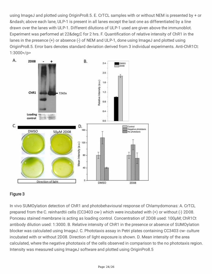
Page 24/26
using ImageJ and plotted using OriginPro8.5. E. CrTCL samples with or without NEM is presented by + or– above each lane, ULP-1 is present in all lanes except the last one as differentiated by a linedrawn over the lanes with ULP-1. Different dilutions of ULP-1 used are given above the immunoblot.Experiment was performed at 22°C for 2 hrs. F. Quanti�cation of relative intensity of ChR1 in thelanes in the presence (+) or absence (-) of NEM and ULP-1, done using ImageJ and plotted usingOriginPro8.5. Error bars denotes standard deviation derived from 3 individual experiments. Anti-ChR1Ct:1:3000</p>
Figure 3
In vivo SUMOylation detection of ChR1 and photobehavioural response of Chlamydomonas: A. CrTCLprepared from the C. reinhardtii cells (CC3403 cw-) which were incubated with (+) or without (-) 2D08.Ponceau stained membrane is acting as loading control. Concentration of 2D08 used: 100µM; ChR1Ctantibody dilution used: 1:3000. B. Relative intensity of ChR1 in the presence or absence of SUMOylationblocker was calculated using ImageJ. C. Phototaxis assay in Petri plates containing CC3403 cw- cultureincubated with or without 2D08. Direction of light exposure is shown. D. Mean intensity of the areacalculated, where the negative phototaxis of the cells observed in comparison to the no phototaxis region.Intensity was measured using ImageJ software and plotted using OriginPro8.5

Page 25/26
Figure 4
Enrichment and analysis of SUMOylated proteome of C. reinhardtii for its effect on photobehaviouralresponse: A. Immunoblotting in CrTCL with anti-SUMO2 antibody (left lane: 20µg, right lane: 40µg). FreeSUMO detected shown with an arrow. Conjugated SUMO proteins are represented with a bold linealongside of the bands. Dilution used: 1:1000. B. Pie chart presenting the protein category distributiondetected in anti-SUMO2 antibody IP Nano-LC-MS/MS. C. Interactome predicted with SUMO Nano-LC-MS/MS identi�ed proteins using STRING program and edited in Cytoscape v3.7.2. Some proteins wereselected from literature to connect the interactome as shown in olive green coloured node. Dashed edgesbetween the nodes shows connection identi�ed from other evidences as provided by STRING database(Supplementary table 6). Centriole proteins are shown in green-coloured nodes. Photoreceptors (ChR1,

Page 26/26
Phototropin, and Cop5) are shown in orange-coloured nodes. The protein-protein network is based ondegree of centrality determining tool in Cytoscape that predicts the biological importance of a protein inthe network comparative to others. The scale for degree of centrality measurement is the size of nodewhich is predicted as larger value corresponding to large sized node.
Figure 5
Proposed model for working of ChR1 mediated photomotile response of C. reinhardtii: ChR1 consist ofciliary targeting sequence (CTS) and SUMOylation motif in its C-terminus required for its functionallocalization and hence collectively involved in the directional photomotility of C. reinhardtii. Ciliarytargeting sequence, VMPS, in ChR1 is interacting with a small GTPase, CrARL11, in C. reinhardtii. ChR1 isSUMOylated in the cellular system. IFT machinery mediated tra�cking of ChR1 into the cilia shown to beinterconnected through SUMOylation. Lacking SUMOylated ChR1 or IFT or both may affect thephotomotility of C. reinhardtii. C, chloroplast; N, nucleus; V, vesicle; E, eyespot; MT, microtubule; B, basalbodies; Ci, Cilia
Supplementary Files
This is a list of supplementary �les associated with this preprint. Click to download.
SUMOCiliaIFTSupplementary.docx