Methanobactin transport machinery - PNAS
6
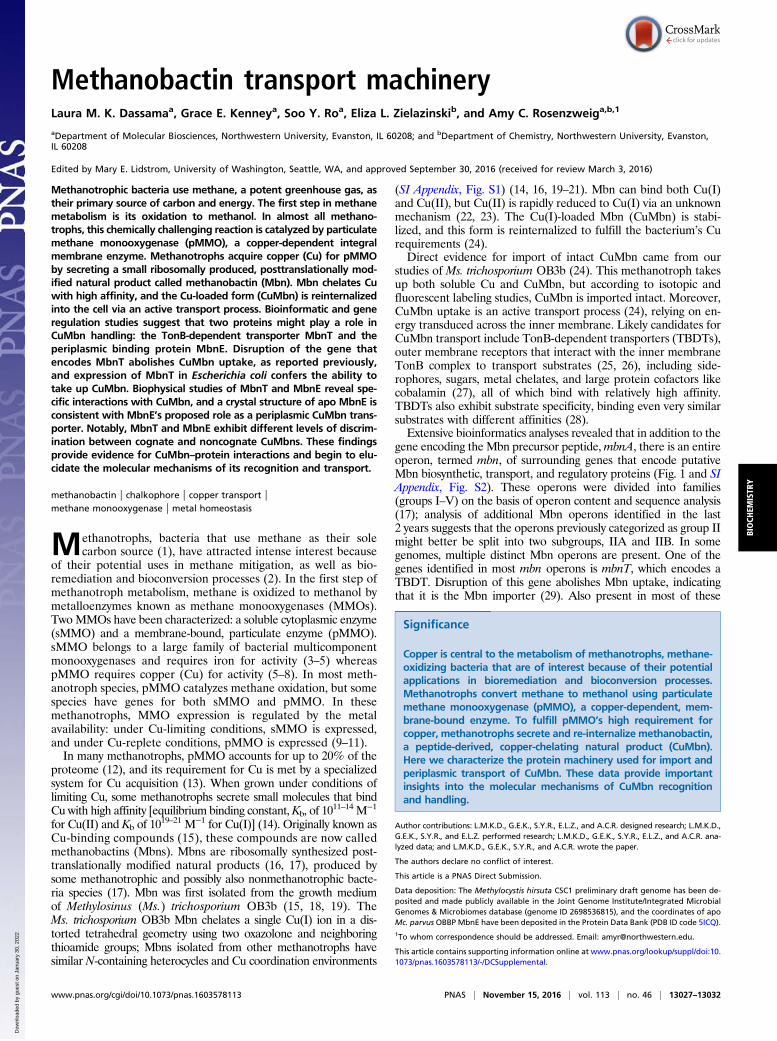
Methanobactin transport machinery Laura M. K. Dassama a , Grace E. Kenney a , Soo Y. Ro a , Eliza L. Zielazinski b , and Amy C. Rosenzweig a,b,1 a Department of Molecular Biosciences, Northwestern University, Evanston, IL 60208; and b Department of Chemistry, Northwestern University, Evanston, IL 60208 Edited by Mary E. Lidstrom, University of Washington, Seattle, WA, and approved September 30, 2016 (received for review March 3, 2016) Methanotrophic bacteria use methane, a potent greenhouse gas, as their primary source of carbon and energy. The first step in methane metabolism is its oxidation to methanol. In almost all methano- trophs, this chemically challenging reaction is catalyzed by particulate methane monooxygenase (pMMO), a copper-dependent integral membrane enzyme. Methanotrophs acquire copper (Cu) for pMMO by secreting a small ribosomally produced, posttranslationally mod- ified natural product called methanobactin (Mbn). Mbn chelates Cu with high affinity, and the Cu-loaded form (CuMbn) is reinternalized into the cell via an active transport process. Bioinformatic and gene regulation studies suggest that two proteins might play a role in CuMbn handling: the TonB-dependent transporter MbnT and the periplasmic binding protein MbnE. Disruption of the gene that encodes MbnT abolishes CuMbn uptake, as reported previously, and expression of MbnT in Escherichia coli confers the ability to take up CuMbn. Biophysical studies of MbnT and MbnE reveal spe- cific interactions with CuMbn, and a crystal structure of apo MbnE is consistent with MbnE’s proposed role as a periplasmic CuMbn trans- porter. Notably, MbnT and MbnE exhibit different levels of discrim- ination between cognate and noncognate CuMbns. These findings provide evidence for CuMbn–protein interactions and begin to elu- cidate the molecular mechanisms of its recognition and transport. methanobactin | chalkophore | copper transport | methane monooxygenase | metal homeostasis M ethanotrophs, bacteria that use methane as their sole carbon source (1), have attracted intense interest because of their potential uses in methane mitigation, as well as bio- remediation and bioconversion processes (2). In the first step of methanotroph metabolism, methane is oxidized to methanol by metalloenzymes known as methane monooxygenases (MMOs). Two MMOs have been characterized: a soluble cytoplasmic enzyme (sMMO) and a membrane-bound, particulate enzyme (pMMO). sMMO belongs to a large family of bacterial multicomponent monooxygenases and requires iron for activity (3–5) whereas pMMO requires copper (Cu) for activity (5–8). In most meth- anotroph species, pMMO catalyzes methane oxidation, but some species have genes for both sMMO and pMMO. In these methanotrophs, MMO expression is regulated by the metal availability: under Cu-limiting conditions, sMMO is expressed, and under Cu-replete conditions, pMMO is expressed (9–11). In many methanotrophs, pMMO accounts for up to 20% of the proteome (12), and its requirement for Cu is met by a specialized system for Cu acquisition (13). When grown under conditions of limiting Cu, some methanotrophs secrete small molecules that bind Cu with high affinity [equilibrium binding constant, K b , of 10 11–14 M −1 for Cu(II) and K b of 10 19–21 M −1 for Cu(I)] (14). Originally known as Cu-binding compounds (15), these compounds are now called methanobactins (Mbns). Mbns are ribosomally synthesized post- translationally modified natural products (16, 17), produced by some methanotrophic and possibly also nonmethanotrophic bacte- ria species (17). Mbn was first isolated from the growth medium of Methylosinus (Ms.) trichosporium OB3b (15, 18, 19). The Ms. trichosporium OB3b Mbn chelates a single Cu(I) ion in a dis- torted tetrahedral geometry using two oxazolone and neighboring thioamide groups; Mbns isolated from other methanotrophs have similar N-containing heterocycles and Cu coordination environments (SI Appendix, Fig. S1) (14, 16, 19–21). Mbn can bind both Cu(I) and Cu(II), but Cu(II) is rapidly reduced to Cu(I) via an unknown mechanism (22, 23). The Cu(I)-loaded Mbn (CuMbn) is stabi- lized, and this form is reinternalized to fulfill the bacterium’s Cu requirements (24). Direct evidence for import of intact CuMbn came from our studies of Ms. trichosporium OB3b (24). This methanotroph takes up both soluble Cu and CuMbn, but according to isotopic and fluorescent labeling studies, CuMbn is imported intact. Moreover, CuMbn uptake is an active transport process (24), relying on en- ergy transduced across the inner membrane. Likely candidates for CuMbn transport include TonB-dependent transporters (TBDTs), outer membrane receptors that interact with the inner membrane TonB complex to transport substrates (25, 26), including side- rophores, sugars, metal chelates, and large protein cofactors like cobalamin (27), all of which bind with relatively high affinity. TBDTs also exhibit substrate specificity, binding even very similar substrates with different affinities (28). Extensive bioinformatics analyses revealed that in addition to the gene encoding the Mbn precursor peptide, mbnA, there is an entire operon, termed mbn, of surrounding genes that encode putative Mbn biosynthetic, transport, and regulatory proteins (Fig. 1 and SI Appendix, Fig. S2). These operons were divided into families (groups I–V) on the basis of operon content and sequence analysis (17); analysis of additional Mbn operons identified in the last 2 years suggests that the operons previously categorized as group II might better be split into two subgroups, IIA and IIB. In some genomes, multiple distinct Mbn operons are present. One of the genes identified in most mbn operons is mbnT, which encodes a TBDT. Disruption of this gene abolishes Mbn uptake, indicating that it is the Mbn importer (29). Also present in most of these Significance Copper is central to the metabolism of methanotrophs, methane- oxidizing bacteria that are of interest because of their potential applications in bioremediation and bioconversion processes. Methanotrophs convert methane to methanol using particulate methane monooxygenase (pMMO), a copper-dependent, mem- brane-bound enzyme. To fulfill pMMO’s high requirement for copper, methanotrophs secrete and re-internalize methanobactin, a peptide-derived, copper-chelating natural product (CuMbn). Here we characterize the protein machinery used for import and periplasmic transport of CuMbn. These data provide important insights into the molecular mechanisms of CuMbn recognition and handling. Author contributions: L.M.K.D., G.E.K., S.Y.R., E.L.Z., and A.C.R. designed research; L.M.K.D., G.E.K., S.Y.R., and E.L.Z. performed research; L.M.K.D., G.E.K., S.Y.R., E.L.Z., and A.C.R. ana- lyzed data; and L.M.K.D., G.E.K., S.Y.R., and A.C.R. wrote the paper. The authors declare no conflict of interest. This article is a PNAS Direct Submission. Data deposition: The Methylocystis hirsuta CSC1 preliminary draft genome has been de- posited and made publicly available in the Joint Genome Institute/Integrated Microbial Genomes & Microbiomes database (genome ID 2698536815), and the coordinates of apo Mc. parvus OBBP MbnE have been deposited in the Protein Data Bank (PDB ID code 5ICQ). 1 To whom correspondence should be addressed. Email: [email protected]. This article contains supporting information online at www.pnas.org/lookup/suppl/doi:10. 1073/pnas.1603578113/-/DCSupplemental. www.pnas.org/cgi/doi/10.1073/pnas.1603578113 PNAS | November 15, 2016 | vol. 113 | no. 46 | 13027–13032 BIOCHEMISTRY Downloaded by guest on January 30, 2022
-
Upload
khangminh22 -
Category
Documents
-
view
0 -
download
0
Transcript of Methanobactin transport machinery - PNAS
Methanobactin transport machineryLaura M. K. Dassamaa, Grace E. Kenneya, Soo Y. Roa, Eliza L. Zielazinskib, and Amy C. Rosenzweiga,b,1
aDepartment of Molecular Biosciences, Northwestern University, Evanston, IL 60208; and bDepartment of Chemistry, Northwestern University, Evanston,IL 60208
Edited by Mary E. Lidstrom, University of Washington, Seattle, WA, and approved September 30, 2016 (received for review March 3, 2016)
Methanotrophic bacteria use methane, a potent greenhouse gas, astheir primary source of carbon and energy. The first step in methanemetabolism is its oxidation to methanol. In almost all methano-trophs, this chemically challenging reaction is catalyzed by particulatemethane monooxygenase (pMMO), a copper-dependent integralmembrane enzyme. Methanotrophs acquire copper (Cu) for pMMOby secreting a small ribosomally produced, posttranslationally mod-ified natural product called methanobactin (Mbn). Mbn chelates Cuwith high affinity, and the Cu-loaded form (CuMbn) is reinternalizedinto the cell via an active transport process. Bioinformatic and generegulation studies suggest that two proteins might play a role inCuMbn handling: the TonB-dependent transporter MbnT and theperiplasmic binding protein MbnE. Disruption of the gene thatencodes MbnT abolishes CuMbn uptake, as reported previously,and expression of MbnT in Escherichia coli confers the ability totake up CuMbn. Biophysical studies of MbnT and MbnE reveal spe-cific interactions with CuMbn, and a crystal structure of apo MbnE isconsistent with MbnE’s proposed role as a periplasmic CuMbn trans-porter. Notably, MbnT and MbnE exhibit different levels of discrim-ination between cognate and noncognate CuMbns. These findingsprovide evidence for CuMbn–protein interactions and begin to elu-cidate the molecular mechanisms of its recognition and transport.
methanobactin | chalkophore | copper transport |methane monooxygenase | metal homeostasis
Methanotrophs, bacteria that use methane as their solecarbon source (1), have attracted intense interest because
of their potential uses in methane mitigation, as well as bio-remediation and bioconversion processes (2). In the first step ofmethanotroph metabolism, methane is oxidized to methanol bymetalloenzymes known as methane monooxygenases (MMOs).TwoMMOs have been characterized: a soluble cytoplasmic enzyme(sMMO) and a membrane-bound, particulate enzyme (pMMO).sMMO belongs to a large family of bacterial multicomponentmonooxygenases and requires iron for activity (3–5) whereaspMMO requires copper (Cu) for activity (5–8). In most meth-anotroph species, pMMO catalyzes methane oxidation, but somespecies have genes for both sMMO and pMMO. In thesemethanotrophs, MMO expression is regulated by the metalavailability: under Cu-limiting conditions, sMMO is expressed,and under Cu-replete conditions, pMMO is expressed (9–11).In many methanotrophs, pMMO accounts for up to 20% of the
proteome (12), and its requirement for Cu is met by a specializedsystem for Cu acquisition (13). When grown under conditions oflimiting Cu, some methanotrophs secrete small molecules that bindCu with high affinity [equilibrium binding constant,Kb, of 10
11–14 M−1
for Cu(II) and Kb of 1019–21 M−1 for Cu(I)] (14). Originally known as
Cu-binding compounds (15), these compounds are now calledmethanobactins (Mbns). Mbns are ribosomally synthesized post-translationally modified natural products (16, 17), produced bysome methanotrophic and possibly also nonmethanotrophic bacte-ria species (17). Mbn was first isolated from the growth mediumof Methylosinus (Ms.) trichosporium OB3b (15, 18, 19). TheMs. trichosporium OB3b Mbn chelates a single Cu(I) ion in a dis-torted tetrahedral geometry using two oxazolone and neighboringthioamide groups; Mbns isolated from other methanotrophs havesimilar N-containing heterocycles and Cu coordination environments
(SI Appendix, Fig. S1) (14, 16, 19–21). Mbn can bind both Cu(I)and Cu(II), but Cu(II) is rapidly reduced to Cu(I) via an unknownmechanism (22, 23). The Cu(I)-loaded Mbn (CuMbn) is stabi-lized, and this form is reinternalized to fulfill the bacterium’s Curequirements (24).Direct evidence for import of intact CuMbn came from our
studies of Ms. trichosporium OB3b (24). This methanotroph takesup both soluble Cu and CuMbn, but according to isotopic andfluorescent labeling studies, CuMbn is imported intact. Moreover,CuMbn uptake is an active transport process (24), relying on en-ergy transduced across the inner membrane. Likely candidates forCuMbn transport include TonB-dependent transporters (TBDTs),outer membrane receptors that interact with the inner membraneTonB complex to transport substrates (25, 26), including side-rophores, sugars, metal chelates, and large protein cofactors likecobalamin (27), all of which bind with relatively high affinity.TBDTs also exhibit substrate specificity, binding even very similarsubstrates with different affinities (28).Extensive bioinformatics analyses revealed that in addition to the
gene encoding the Mbn precursor peptide,mbnA, there is an entireoperon, termed mbn, of surrounding genes that encode putativeMbn biosynthetic, transport, and regulatory proteins (Fig. 1 and SIAppendix, Fig. S2). These operons were divided into families(groups I–V) on the basis of operon content and sequence analysis(17); analysis of additional Mbn operons identified in the last2 years suggests that the operons previously categorized as group IImight better be split into two subgroups, IIA and IIB. In somegenomes, multiple distinct Mbn operons are present. One of thegenes identified in most mbn operons is mbnT, which encodes aTBDT. Disruption of this gene abolishes Mbn uptake, indicatingthat it is the Mbn importer (29). Also present in most of these
Significance
Copper is central to the metabolism of methanotrophs, methane-oxidizing bacteria that are of interest because of their potentialapplications in bioremediation and bioconversion processes.Methanotrophs convert methane to methanol using particulatemethane monooxygenase (pMMO), a copper-dependent, mem-brane-bound enzyme. To fulfill pMMO’s high requirement forcopper, methanotrophs secrete and re-internalize methanobactin,a peptide-derived, copper-chelating natural product (CuMbn).Here we characterize the protein machinery used for import andperiplasmic transport of CuMbn. These data provide importantinsights into the molecular mechanisms of CuMbn recognitionand handling.
Author contributions: L.M.K.D., G.E.K., S.Y.R., E.L.Z., and A.C.R. designed research; L.M.K.D.,G.E.K., S.Y.R., and E.L.Z. performed research; L.M.K.D., G.E.K., S.Y.R., E.L.Z., and A.C.R. ana-lyzed data; and L.M.K.D., G.E.K., S.Y.R., and A.C.R. wrote the paper.
The authors declare no conflict of interest.
This article is a PNAS Direct Submission.
Data deposition: The Methylocystis hirsuta CSC1 preliminary draft genome has been de-posited and made publicly available in the Joint Genome Institute/Integrated MicrobialGenomes & Microbiomes database (genome ID 2698536815), and the coordinates of apoMc. parvus OBBP MbnE have been deposited in the Protein Data Bank (PDB ID code 5ICQ).1To whom correspondence should be addressed. Email: [email protected].
This article contains supporting information online at www.pnas.org/lookup/suppl/doi:10.1073/pnas.1603578113/-/DCSupplemental.
www.pnas.org/cgi/doi/10.1073/pnas.1603578113 PNAS | November 15, 2016 | vol. 113 | no. 46 | 13027–13032
BIOCH
EMISTR
Y
Dow
nloa
ded
by g
uest
on
Janu
ary
30, 2
022

operons is mbnM, which encodes a member of the multidrug andtoxic compound extrusion protein family (16, 17, 30, 31). As such,MbnT and MbnM were proposed in 2013 to be the prime candi-dates for the CuMbn importer and exporter, respectively (17). Inaddition, 9 kb upstream of mbnT in the Ms. trichosporium OB3bgenome is the previously unidentified mbnE, which encodes aperiplasmic binding protein (PBP) that may transport periplasmicCuMbn. Recent quantitative RT-PCR (qRT-PCR) studies of Ms.trichosporium OB3b (32) revealed that these genes, includingmbnE, are coregulated with mbnA and are all down-regulatedin response to Cu addition. Taken together with the cessation ofMbn production in an mbnA deletion mutant (33) and thereported mbnT knockout strain (29), there is strong genetic ev-idence for the involvement of mbnT and mbnE in Mbn transport.However, the interaction of these proteins with Mbn has notbeen investigated biochemically. Here we have probed the rolesof MbnT and MbnE using bioinformatic, genetic, biochemical,and biophysical approaches. Taken together, our results identifyMbnT as the CuMbn importer and MbnE as a CuMbn PBP.
Results and DiscussionBioinformatics Analysis of the Transport Proteins. The Mbn operonsand their component genes were originally identified via bio-informatics (17) (Fig. 1). We have now extended and updated thisanalysis with a focus on transport-related genes, resulting in a morecomplete model for Mbn transport. TBDTs are present in operonsfor four of five Mbn groups (17) (Fig. 1), consistent with the needfor active transport of a large metal-bound natural product.However, the TBDTs present in Mbn operons are more hetero-geneous than might be expected, given the high conservation ofother Mbn-related genes. As noted previously (17), MbnT genesthat neighbor genes encoding sigma and anti-sigma factor pairsalso encode the N-terminal extensions necessary for signal trans-duction between TBDTs and anti-sigma factors (34–36). As addi-tional Mbn operons have been identified, it has becomeincreasingly clear that these fecIRA-like mbnIRT triads are pre-sent only in group I Mbn operons, suggesting that only thoseMbnTs play a role in signal transduction. A larger comparisonof putative MbnTs with extant TBDTs in the Joint Genome
Institute/Integrated Microbial Genomes & Microbiomes andUniProt databases indicates that the differences between MbnTsextend beyond the presence or absence of an N-terminal ex-tension. MbnTs from group I Mbn operons (MbnT1s) are dis-tinct in sequence and domain content from group II MbnTs(MbnT2s), and both are distinct from MbnTs in group III and IV(MbnT3s); all MbnTs appear to be distinct from previouslycategorized TBDT groups (SI Appendix, Fig. S3 A and B and SIResults and Discussion and Datasets S1–S3). Notably, manymethanotrophs contain multiple genes encoding MbnTs, some-times from multiple MbnT groups.Although not described in the initial bioinformatics-based
characterization of Mbn operons (17), there is some evidence thatPBPs may also play a role in Mbn uptake. These proteins areperiplasmic (in Gram-negative bacteria) or membrane-anchored(in Gram-positive bacteria) and are associated with ATP-bindingcassette (ABC) importers (37, 38). The only Mbn operons con-taining PBPs are group IIA operons, which include the MbnEgene, but close homologs of the Methylocystis (Mc.) and the Ms.trichosporium OB3b mbnE genes can be found elsewhere in thegenomes of other Mbn-producing methanotrophs. In some cases,multiple potential homologs are present. All these PBPs aremembers of solute binding protein (SBP) Family 5 (39), whichcomprises primarily oligopeptide-binding proteins; in a sequencesimilarity network of this family, MbnEs cluster separately from allcharacterized PBPs except a microcin C7 transporter, YejA (SIAppendix, Fig. S4A and Dataset S4). Potential homologs are ob-served in the genomes of most species producing group III–VMbns, but they are also similar to YejA (40). The similarities be-tween the two PBP subgroups (SI Appendix, Fig. S4B) complicateany definitive assignment without evidence of coregulation withMbn operons or biochemical interactions with Mbns. However, itis notable that yejA encodes a microcin C7 transporter; microcinC7 is a ribosomally produced, posttranslationally modified naturalproduct with a heptapeptide backbone and a final mass within therange observed for Mbns (40).
Gene Regulation Patterns of Transport Proteins in Ms. trichosporiumOB3b. A previous set of gene regulation studies confirmed thatthe entirety of the mbn operon in Ms. trichosporium OB3b[mbn(E)IRTABCMNPH] (SI Appendix, Fig. S2) is coregulatedin a Cu-dependent manner (32). Many of the genes in the operonlack close homologs elsewhere in the genome. However, there areseveral TBDTs that are closely related to mbnT as well as twoother PBPs classified via bioinformatics as oligopeptide-bindingproteins and possible mbnE homologs. In total, there are 45TBDTs and 3 Family 5 PBPs in the Ms. trichosporium OB3b ge-nome (41). Thus, to ensure that we were pursuing the candidatesmost likely to be related to Mbn transport, we monitored Cu-dependent gene regulation of the closestmbnE andmbnT homologs.qRT-PCR results confirm that, although mbnE is down-regu-
lated by Cu in concert with the Mbn operon, the two most closelyrelated PBPs, a hypothetical protein (MettrDRAFT_1622) andyejA (MettrDRAFT_3640) (SI Appendix, Fig. S2), are not (SIAppendix, Fig. S5 and Table S1 and Dataset S5). The TBDT resultsare more complicated. Two mbnIRTPH operons are presentelsewhere in the genome in close proximity to one another (SIAppendix, Fig. S2). Both operons (containing thembnT1 homologsMettrDRAFT_1229 and MettrDRAFT_1241) are Cu-regulated,but down-regulation levels differ (particularly notably for thenonregulatory genes mbnPH, which are 5- to 10-fold down-regulated, compared with the 25- to 50-fold down-regulationof their mbn operon counterparts) (Dataset S5). Differencesin regulation patterns after Cu addition are also observed, par-ticularly in the operon containing the mbnT1 gene Mettr-DRAFT_1241: unlike the regulatory genes in the main mbnoperon or the other mbnIRTPH operon, which respond to Cuaddition swiftly, the mbnIR homologs in this operon are not
moaA 3-HIBADHECF factor
IIIa
IIIIVV
ECF 70 factor,
FecI-like (MbnI)
MbnB (unknown Mbnbiosynthesis protein 1)
FecR-like anti-
factor(MbnR)
MbnC (unknown Mbnbiosynthesis protein 2)
TonB-dependent
transporter (MbnT)
MATE efflux
pump (MbnM)
Aminotransferase
(MbnN)
Periplasmic bindingprotein (MbnE)
Partner of MbnH (MbnP)
FAD-dependent
oxidoreductase (MbnF)
Di-heme cytochrome cperoxidase (MbnH)
Sulfotransferase
(MbnS)
MbnA (Mbn precursor)
IIb
Dioxygenase
(MbnD)
Extended MbnB-like
protein (MbnX)
5
14
6
14
4
8
51
Fig. 1. Composition of Mbn operons. Mbn operons can be subdivided into sixgroups on the basis of operon content, precursor peptide (MbnA) sequence,and phylogenetic analysis of MbnA, MbnB, and MbnC, which are present in alloperons. The number of complete operons of a given type is presented to theright of the operon. Ms. trichosporium OB3b belongs to group I, Mc. parvusOBBP has both a group I and a group IIb operon, and Mc. hirsuta CSC1 has agroup IIa operon. Although genes related to Mbn biosynthesis, regulation,and transport are present in all operons, genes for the putative Mbn exporter,MbnM, and importer, MbnT, are notably widespread.
13028 | www.pnas.org/cgi/doi/10.1073/pnas.1603578113 Dassama et al.
Dow
nloa
ded
by g
uest
on
Janu
ary
30, 2
022
down-regulated completely within the first 15 min, but continuedecreasing at the 5-h and 24-h time points. These regulatorydifferences are statistically significant. Whether analyzed viahierarchical clustering or calculation of gene-to-gene Spearmancorrelation values, expression patterns of genes within the mbnoperon correlate significantly more closely to other genes withinthat operon than they do to genes outside of that operon (SIAppendix, Fig. S5 and Table S1 and Dataset S5). These differingresponses to Cu suggest that, even if the MbnT1s encoded bythese operons are capable of binding Mbn, the systems are notfully interchangeable with the native mbn operon. Further workwill be necessary to identify the extent of regulatory crosstalkbetween the three systems and native substrate-triggered regu-lation and/or uptake. However, these gene-regulation resultsprovide support for the preliminary assignment of the originallyidentified mbnE and mbnT genes (MettrDRAFT_3413 andMettrDRAFT_3421) as the PBP and TBDT most likely tofunction in import of the native Mbn. A possible function of theclose MbnT1 homologs could be uptake of nonnative CuMbnsproduced by other methanotrophs in the environment.
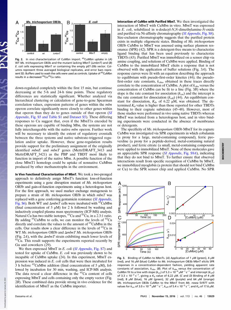
In Vivo Functional Characterization of MbnT.We took a two-prongedapproach to definitively assign MbnT’s function: loss-of-functionexperiments using a gene disruption mutant of Ms. trichosporiumOB3b and gain-of-function experiments using a heterologous host.For the first approach, we used marker exchange mutagenesis toprepare a strain of Ms. trichosporium OB3b in which mbnT wasreplaced with a gene conferring gentamicin resistance (SI Appendix,Fig. S6). Both WT and ΔmbnT cells were incubated with 65CuMbn(final concentration of 5 μM) for 2 h followed by washing andinductively coupled plasma mass spectrometry (ICP-MS) analysis.Natural Cu has two stable isotopes, 63Cu and 65Cu, in a 2.3:1 ratio.By adding 65CuMbn to cells, we can monitor the levels of 65Cudirectly and correlate the values to the amount of 65CuMbn in thecells. Our results show a clear difference in the levels of 65Cu inWTMs. trichosporium OB3b and ΔmbnT Ms. trichosporium OB3b(Fig. 2A), with the ΔmbnT strain exhibiting much lower levels of65Cu. This result supports the experiments reported recently byGu and coworkers (29).We then expressed MbnT in E. coli (SI Appendix, Fig. S7) and
tested for uptake of CuMbn. E. coli was previously shown to beincapable of CuMbn uptake (24). In this experiment, MbnT ex-pression was induced in E. coli cells that were then incubated for3 h before 65CuMbn addition (final concentration of 5 μM), fol-lowed by incubation for 30 min, washing, and ICP-MS analysis.The data reveal a clear difference in the 65Cu content of cellsexpressing MbnT and cells containing only the empty vector (Fig.2B). These combined data provide strong in vivo evidence for theidentification of MbnT as the CuMbn importer.
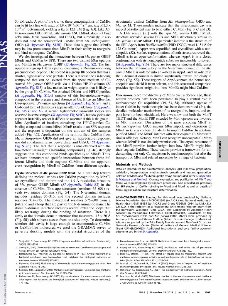
Interaction of CuMbn with Purified MbnT. We then investigated theinteraction of MbnT with CuMbn in vitro. MbnT was expressedin E. coli, solubilized in n-dodecyl-N,N-dimethylamine-N-oxideand purified via Ni-affinity chromatography (SI Appendix, Fig. S8).Size-exclusion chromatography suggests that the purified proteinexists in multiple oligomeric states. Binding of Ms. trichosporiumOB3b CuMbn to MbnT was assessed using surface plasmon res-onance (SPR) (42). SPR is a detergent-free means to characterizeligand binding that has been used previously to characterizeTBDTs (43). Purified MbnT was immobilized on a sensor chip viaamine coupling, and solutions of CuMbn were applied. Binding ofCuMbn to the immobilized MbnT elicits a response that is notobserved with the application of buffer solutions (Fig. 3A). Theresponse curves were fit with an equation describing the approachto equilibrium with pseudo-first-order kinetics (44); the pseudo-first-order rate constants, kobs, obtained in these traces directlycorrelate to the concentration of CuMbn. A plot of kobs versus theconcentration of CuMbn can be fit to a line (Fig. 3B) where theslope is the rate constant for association (kon) and the intercept isthe rate constant for dissociation (koff) (44). An equilibrium con-stant for dissociation, Kd, of 6.22 μM, was obtained. The de-termined Kd value is higher than those reported for other TBDTsbinding to their cognate substrates (45–48). However, most ofthose studies were performed in vivo using native TBDTs whereasMbnT was isolated from a heterologous host, and in vitro bind-ing experiments were conducted in the absence of membranesor detergents.The specificity ofMs. trichosporium OB3b MbnT for its cognate
CuMbn was investigated via SPR experiments in which cobalamin(a proxy for a large, metal-containing compound), ferric pyo-verdine (a proxy for a peptide-derived, metal-containing naturalproduct), and ferric citrate (a small, metal-containing compound)were applied to immobilized MbnT. None of these molecules gavean appreciable SPR response (SI Appendix, Fig. S9A), indicatingthat they do not bind to MbnT. To further ensure that observedinteractions result from specific recognition of CuMbn by MbnT,we immobilized myoglobin (a protein that should not bind CuMbnor Cu) to the SPR sensor chip and applied CuMbn. No SPR
Fig. 2. In vivo characterization of CuMbn import. 65CuMbn uptake in (A)WT Ms. trichosporium OB3b and the mutant lacking MbnT (ΔmbnT) and (B)E. coli cells expressing MbnT or containing the empty pET-20b vector. Col-umns represent three averaged biological replicates, and error bars repre-sent SD. Buffers used towash the cells were used as controls. Uptake of 65CuMbnresults in a decreased 63Cu:65Cu ratio.
Fig. 3. Binding of CuMbn to MbnTs. (A) Application of 1 μM (green), 4 μM(red), and 16 μM (blue) CuMbn to Ms. trichosporium OB3b MbnT elicits SPRresponses in a concentration-dependent fashion, yielding apparent rateconstants of association, kobs. (B). Plot of kobs versus the concentration ofCuMbn fit to a line with slope (kon) of 5.3 × 10⁻⁴ μM−1·s−1 and intercept (koff)of 3.3 × 10⁻³ s−1, giving a Kd value of 6.22 μM. (C and D) Binding of 4 μM(red), 8 μM (blue), 16 μM (green), 32 μM (purple) and 64 μM (orange)Ms. trichosporium OB3b CuMbn to the MbnT from Mc. rosea SV97T withvalues for kon of 3.0 × 10⁻⁴ μM−1·s−1, koff of 5.4 × 10⁻³ s−1, and Kd of 17.6 μM.
Dassama et al. PNAS | November 15, 2016 | vol. 113 | no. 46 | 13029
BIOCH
EMISTR
Y
Dow
nloa
ded
by g
uest
on
Janu
ary
30, 2
022

response was observed for any concentration of CuMbn (SIAppendix, Fig. S9B). To probe the specificity of a given MbnT forits cognate CuMbn, we also cloned, expressed, and purified theMbnT from Mc. rosea SV97. Although Mc. rosea SV97 CuMbnhas two nitrogen-containing heterocycles and two thioamidegroups like Ms. trichosporium OB3b CuMbn, there is no N-terminaltransamination or disulfide formation, the first oxazolone isreplaced by a pyrazinedione, and a sulfonated threonine is present(SI Appendix, Fig. S1B) (14, 16). Interestingly, the addition ofMs. trichosporium OB3b CuMbn to Mc. rosea SV97 MbnT doeselicit an SPR response (Fig. 3C), yielding a Kd value of 17.6 μM(Fig. 3D). These data suggest a complex mechanism for CuMbnrecognition and binding, wherein MbnT binds only structurallysimilar substrates, and individual MbnTs may bind cognate CuMbnswith higher affinity than nonnative CuMbns. Similar trends are ob-served for the ferric pyoverdine receptor FpvA from Pseudomonasaeruginosa ATCC 15692, which, unlike other Pseudomonas TBDTs,binds noncognate pyoverdines, albeit with different affinities (28).
Interaction of CuMbn with Purified MbnE. To investigate whetherMbnE might play a role in binding CuMbn in the periplasm,perhaps for delivery to periplasmic enzymes or, alternatively, to aninner membrane ABC transporter (37), we cloned, expressed, andpurified Ms. trichosporium OB3b MbnE. Most Ms. trichosporiumOB3b MbnE is expressed in inclusion bodies, but a small amount
remained soluble and was purified via affinity chromatography.Size-exclusion chromatography revealed the protein to be asingle oligomer (SI Appendix, Fig. S10), and circular dichroismspectroscopy suggests that it is mostly composed of α-helices andunordered loops (SI Appendix, Fig. S11).Binding of Ms. trichosporium OB3b CuMbn to its cognate
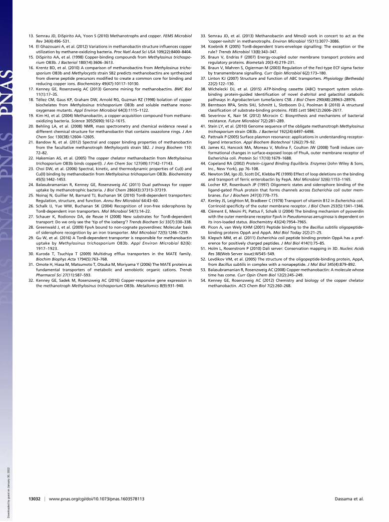
purified MbnE was then tested via SPR. The application ofCuMbn elicits an SPR response with kobs directly proportional tothe concentration of CuMbn (Fig. 4A), up to 30 μM. At 60 μMCuMbn, the kobs diverges from linearity (Fig. 4B), perhaps due toinactivation that reduces the protein’s ability to bind substrate.The plot of kobs vs. [CuMbn] fits to a line that yields kon of 1.4 ×10⁻³ μM−1·s−1, koff of 8.5 × 10⁻³ s−1, and a Kd value of 6.07 μM.This value is within the range that has been observed for bindingof oligopeptides to PBPs (Kd values of 0.3 nM–50 μM) (49, 50).The specificity of MbnE for CuMbn was investigated by addingcobalamin, ferric pyoverdine, and CuSO4 to the immobilizedprotein. In these experiments, we observed rapid associationfollowed by an even faster dissociation (SI Appendix, Fig. S12A),indicating that these molecules do not bind to an appreciableextent. Therefore, MbnE, like MbnT, is specific for CuMbn anddoes not bind soluble Cu, siderophores, or metal chelates.To investigate whether MbnE recognizes and binds non-
cognate CuMbns, we cloned, expressed, and purified MbnEsfrom Mc. hirsuta CSC1 and Mc. parvus OBBP. The Mc. hirsutaCSC1 mbnE gene is found within the Mbn operon (SI Appendix,Fig. S2 and Dataset S6); there are two close nonoperon mbnEhomologs in Mc. parvus OBBP, but the one used in this study,O5ODRAFT_02185, is more similar to group II mbnEs (SI Ap-pendix, Fig. S2). Both MbnEs are monomeric (SI Appendix, Fig.S13), and the Mc. hirsuta CSC1 MbnE comprises primarily α-heli-ces and unordered loops (SI Appendix, Fig. S14). Binding of cog-nate CuMbn to the Mc. hirsuta CSC1 MbnE was demonstrated bySPR experiments in which semipurified CuMbn (spent mediumfrom the Cu-starved cultures supplemented with CuSO4 and puri-fied on a Diaion HP-20 column) was applied to the immobilizedprotein. SPR responses were observed (Fig. 4C), and the rates andamplitudes of the response curves were directly proportional to theamount of semipurified CuMbn applied. Using the absorbance at322 nm and the reported extinction coefficient at that wavelength(14), we estimate the amounts of applied CuMbn to be 5, 15, and
Fig. 4. Binding of CuMbn to MbnEs. (A) SPR response curves (average ofthree injections) showing the binding of 0 μM (teal), 5 μM (green), 15 μM(red), 30 μM (blue), and 60 μM (purple) CuMbn to the MbnE fromMs. trichosporium OB3b. (B) Plot of kobs values versus the concentration ofCuMbn fit to a line that yields a Kd value of 6.07 μM. (C) Binding of 5 μM(red), 15 μM (blue), and 30 μM (green) CuMbn isolated from Mc. hirsuta CSC1to the MbnE from that organism. (D) A plot of kobs vs. concentration ofCuMbn fit to a line that yields a Kd value of 12.1 μM for CuMbn binding.(E) SPR response curves showing the binding of 0 μL (pink), 5 μL (red), 15 μL(blue), and 30 μL (green) of Cu-supplemented spent medium isolated fromMc. parvus OBBP to the MbnE from that organism. (F) SPR response showingthe binding a low-molecular-weight CuMbn candidate compound isolatedfrom Mc. parvus OBBP to the MbnE from that organism.
Fig. 5. The 1.9-Å resolution crystal structure of Mc. parvus OBBP apo MbnE.(A) Cartoon representation with the two domains colored in magenta andcyan. (B) Surface representations of the structure show a cavity at the do-main–domain interface that measures ∼15 Å across and 30 Å high. The cavityis open only to one side, consistent with it being the only entry point for thesubstrate. Docking models generated with the structures of CuMbns fromMs. trichosporium OB3b (C) and Mc. sp. M (D) reveal that the cavity canaccommodate both forms of CuMbns. (E) Superposition of the Mc. parvusOBBP MbnE structure with that of nonapeptide-bound AppA from B. subtilis(PDB 1XOC). Major structural rearrangements include the ordering of a loopin the N-terminal domain and the movement of a α-helix closer to the domain–domain interface (red arrows).
13030 | www.pnas.org/cgi/doi/10.1073/pnas.1603578113 Dassama et al.
Dow
nloa
ded
by g
uest
on
Janu
ary
30, 2
022
30 μM each. A plot of the kobs vs. these concentrations of CuMbncan be fit to a line with a kon of 1.9 × 10⁻³ μM−1·s−1 and koff of 2.3 ×10⁻² s−1 determined, yielding a Kd of 12.1 μM (Fig. 4D). Like Ms.trichosporium OB3b MbnE, Mc. hirsuta CSC1 MbnE does not bindcobalamin, ferric pyoverdine, and CuSO4, but surprisingly, it alsodoes not bind the semipurified CuMbn from Ms. trichosporiumOB3b (SI Appendix, Fig. S12B). These data suggest that MbnEsmay be less promiscuous than MbnTs in their ability to recognizeand bind noncognate CuMbns.We also investigated the interactions between Mc. parvus OBBP
MbnE and CuMbn by SPR. There are two distinct Mbn operonsand MbnEs in Mc. parvus OBBP (SI Appendix, Fig. S2). The firstoperon is a group I Mbn operon, containing a 16-amino-acid-longprecursor core peptide. The second is a group IIb operon that has ashorter, eight-residue core peptide. There is at least one Cu-bindingcompound that can be isolated from the spent medium of Cu-starved Mc. parvus OBBP cells via a Diaion HP-20 column (SIAppendix, Fig. S15): a low molecular weight species that is likely tobe the group IIb CuMbn. We obtained Diaion- and HPLC-purified(SI Appendix, Fig. S15A) samples of this low-molecular-weightcompound in the presence and absence of Cu. It has an Mbn-like,Cu-responsive, UV-visible spectrum (SI Appendix, Fig. S15B), and aCu-bound form of this species appears after Cu addition (SI Appendix,Fig. S15 C and D). A second higher-molecular-weight species wasobserved in some samples (SI Appendix, Fig. S15C), but low yields andapparent instability render it difficult to ascertain if this is the group IMbn. Application of fractions containing the HPLC-purified Cu-binding compound to immobilized MbnE results in an SPR response,and the response is dependent on the amount of the samplesadded (Fig. 4E). Application of the semipurified CuMbn fromMs. trichosporium OB3b did not give an appreciable response,nor did cobalamin, ferric pyoverdine, and CuSO4 (SI Appendix,Fig. S12C). The fact that a response is also observed with thelow-molecular-weight Cu-binding compound (Fig. 4F) stronglysuggests that this compound binds specifically to MbnE. Thus,we have demonstrated specific interactions between three dif-ferent MbnEs and their cognate CuMbns and no apparentcross-recognition by MbnE of CuMbns from different species.
Crystal Structure of Mc. parvus OBBP MbnE. As a first step towarddefining the molecular basis for CuMbn recognition by MbnE,we crystallized and determined the 1.9-Å resolution structureof Mc. parvus OBBP MbnE (SI Appendix, Table S2) in theabsence of CuMbn. This apo structure (residues 35–609) re-veals two major domains (Fig. 5A). The N-terminal domainincludes residues 35–313, and the second domain includesresidues 314–575. The C-terminal residues 576–609 form aβ-strand and a loop that are part of the N-terminal domain. Thedomain–domain interface includes several extended loops thatlikely rearrange during the binding of substrate. There is acavity at the domain–domain interface that measures ∼15 × 30 Å(Fig. 5B) with solvent access from one side only. To determinewhether this cavity is large enough to accommodate CuMbnor CuMbn-like molecules, we used the GRAMMX server togenerate docking models with the crystal structures of the
structurally distinct CuMbns from Ms. trichosporium OB3b andMc. sp. M. These models indicate that the interdomain cavity isindeed of sufficient size to bind either CuMbn (Fig. 5 C and D).A Dali search (51) with the apo Mc. parvus OBBP MbnE
structure revealed several PBPs and SBPs structurally similar toMc. parvus OBBP MbnE. Of particular interest is the structure ofthe SBP AppA from Bacillus subtilis (PBD 1XOC, rmsd 1.151 Å for122 Cα atoms). AppA was copurified and crystallized with a non-apeptide (52). Surface representations of both structures reveal thatMbnE is in an open conformation, whereas AppA is in a closedconformation with its nonapeptide substrate inaccessible to solvent(SI Appendix, Fig. S16). There are two major structural differencesbetween the proteins: a loop in the N-terminal domain of the Mc.parvus MbnE is ordered into an α-helix in AppA, and an α-helix inthe C-terminal domain is shifted significantly toward the cavity inAppA (Fig. 5E). These regions of AppA contact the bound non-apeptide and shield it from solvent, and this structural comparisonprovides significant insight into how MbnEs might bind CuMbn.
Conclusions. Since the discovery of Mbns over a decade ago, thesenatural products have been postulated to play a central role inmethanotroph Cu acquisition (19, 53, 54). Although uptake ofintact CuMbn by methanotrophs has been demonstrated (24), thedetailed molecular mechanisms of its recognition, import, and ex-port have not been elucidated. Here we show that both the MbnTTBDT and the MbnE PBP encoded by Mbn operons are involvedin Mbn transport. Disruption of the gene encoding MbnT inMs. trichosporium OB3b impairs Mbn import, and expression ofMbnT in E. coli confers the ability to import CuMbn. In addition,purified MbnT and MbnE interact with their cognate CuMbns withsimilar affinities. Notably, MbnT can recognize noncognate CuMbnswhereas MbnE is not similarly promiscuous. The crystal structure ofapo MbnE provides further insight into how MbnEs might bindtheir cognate CuMbns. These studies provide a framework for un-derstanding not only Cu acquistion by methanotrophs, but also thetransport of Mbn and related molecules by a range of bacteria.
Materials and MethodsDetailed procedures for bioinformatics analyses, qRT-PCR assay preparation,validation, interpretation, methanotroph growth and mutant generation,isolation of Mbns, and 65CuMbn uptake assays are included in the SI Appendix,SI Materials and Methods. Cloning, expression, and purification of MbnT andMbnE were accomplished by standard procedures. Also provided are protocolsfor SPR studies of CuMbn binding to MbnE and MbnT as well as details ofMbnE crystallization and structure determination.
ACKNOWLEDGMENTS. Funding for this work was provided by NationalScience Foundation Grant MCB0842366 (to A.C.R.) and National Institutes ofHealth Grant GM118035 (to A.C.R.) and Grant F32GM110934 (to L.M.K.D.).L.M.K.D. is the recipient of a Postdoctoral Enrichment Program grant fromthe Burroughs Wellcome Fund. G.E.K. was supported by American HeartAssociation Predoctoral Fellowship 14PRE20460104. Constructs of theMs. trichosporium OB3b and Mc. parvus OBBP MbnEs were provided byAnthony S. Gizzi and Steven C. Almo (Albert Einstein College of Medicine),who are supported by the Price Family Foundation and New York StructuralGenomics Research Center (National Institute of General Medical SciencesGrant U54-GM094663). Additional institutional and core facility acknowl-edgments are in the SI Appendix.
1. Sirajuddin S, Rosenzweig AC (2015) Enzymatic oxidation of methane. Biochemistry54(14):2283–2294.
2. Strong PJ, Xie S, Clarke WP (2015) Methane as a resource: Can the methanotrophs addvalue? Environ Sci Technol 49(7):4001–4018.
3. Rosenzweig AC, Frederick CA, Lippard SJ, Nordlund P (1993) Crystal structure of abacterial non-haem iron hydroxylase that catalyses the biological oxidation ofmethane. Nature 366(6455):537–543.
4. Lipscomb JD (1994) Biochemistry of the soluble methane monooxygenase. Annu RevMicrobiol 48(1):371–399.
5. Sazinsky MH, Lippard SJ (2015) Methane monooxygenase: Functionalizing methaneat iron and copper. Met Ions Life Sci 15:205–256.
6. Lieberman RL, Rosenzweig AC (2005) Crystal structure of a membrane-bound met-alloenzyme that catalyses the biological oxidation of methane. Nature 434(7030):177–182.
7. Balasubramanian R, et al. (2010) Oxidation of methane by a biological dicoppercentre. Nature 465(7294):115–119.
8. Culpepper MA, Rosenzweig AC (2012) Architecture and active site of particulatemethane monooxygenase. Crit Rev Biochem Mol Biol 47(6):483–492.
9. Prior SD, Dalton H (1985) The effect of copper ions on membrane content andmethane monooxygenase activity in methanol-grown cells of Methylococcus capsu-latus (Bath). J Gen Microbiol 131(1):155–163.
10. Murrell JC, McDonald IR, Gilbert B (2000) Regulation of expression of methanemonooxygenases by copper ions. Trends Microbiol 8(5):221–225.
11. Hakemian AS, Rosenzweig AC (2007) The biochemistry of methane oxidation. AnnuRev Biochem 76:223–241.
12. Martinho M, et al. (2007) Mössbauer studies of the membrane-associated methanemonooxygenase from Methylococcus capsulatus bath: Evidence for a Diiron center.J Am Chem Soc 129(51):15783–15785.
Dassama et al. PNAS | November 15, 2016 | vol. 113 | no. 46 | 13031
BIOCH
EMISTR
Y
Dow
nloa
ded
by g
uest
on
Janu
ary
30, 2
022
13. Semrau JD, DiSpirito AA, Yoon S (2010) Methanotrophs and copper. FEMS MicrobiolRev 34(4):496–531.
14. El Ghazouani A, et al. (2012) Variations in methanobactin structure influences copperutilization by methane-oxidizing bacteria. Proc Natl Acad Sci USA 109(22):8400–8404.
15. DiSpirito AA, et al. (1998) Copper-binding compounds from Methylosinus trichospo-rium OB3b. J Bacteriol 180(14):3606–3613.
16. Krentz BD, et al. (2010) A comparison of methanobactins from Methylosinus tricho-sporium OB3b and Methylocystis strain SB2 predicts methanobactins are synthesizedfrom diverse peptide precursors modified to create a common core for binding andreducing copper ions. Biochemistry 49(47):10117–10130.
17. Kenney GE, Rosenzweig AC (2013) Genome mining for methanobactins. BMC Biol11(1):17–35.
18. Téllez CM, Gaus KP, Graham DW, Arnold RG, Guzman RZ (1998) Isolation of copperbiochelates from Methylosinus trichosporium OB3b and soluble methane mono-oxygenase mutants. Appl Environ Microbiol 64(3):1115–1122.
19. Kim HJ, et al. (2004) Methanobactin, a copper-acquisition compound from methane-oxidizing bacteria. Science 305(5690):1612–1615.
20. Behling LA, et al. (2008) NMR, mass spectrometry and chemical evidence reveal adifferent chemical structure for methanobactin that contains oxazolone rings. J AmChem Soc 130(38):12604–12605.
21. Bandow N, et al. (2012) Spectral and copper binding properties of methanobactinfrom the facultative methanotroph Methylocystis strain SB2. J Inorg Biochem 110:72–82.
22. Hakemian AS, et al. (2005) The copper chelator methanobactin from Methylosinustrichosporium OB3b binds copper(I). J Am Chem Soc 127(49):17142–17143.
23. Choi DW, et al. (2006) Spectral, kinetic, and thermodynamic properties of Cu(I) andCu(II) binding by methanobactin from Methylosinus trichosporium OB3b. Biochemistry45(5):1442–1453.
24. Balasubramanian R, Kenney GE, Rosenzweig AC (2011) Dual pathways for copperuptake by methanotrophic bacteria. J Biol Chem 286(43):37313–37319.
25. Noinaj N, Guillier M, Barnard TJ, Buchanan SK (2010) TonB-dependent transporters:Regulation, structure, and function. Annu Rev Microbiol 64:43–60.
26. Schalk IJ, Yue WW, Buchanan SK (2004) Recognition of iron-free siderophores byTonB-dependent iron transporters. Mol Microbiol 54(1):14–22.
27. Schauer K, Rodionov DA, de Reuse H (2008) New substrates for TonB-dependenttransport: Do we only see the ‘tip of the iceberg’? Trends Biochem Sci 33(7):330–338.
28. Greenwald J, et al. (2009) FpvA bound to non-cognate pyoverdines: Molecular basisof siderophore recognition by an iron transporter. Mol Microbiol 72(5):1246–1259.
29. Gu W, et al. (2016) A TonB-dependent transporter is responsible for methanobactinuptake by Methylosinus trichosporium OB3b. Appl Environ Microbiol 82(6):1917–1923.
30. Kuroda T, Tsuchiya T (2009) Multidrug efflux transporters in the MATE family.Biochim Biophys Acta 1794(5):763–768.
31. Omote H, Hiasa M, Matsumoto T, Otsuka M, Moriyama Y (2006) The MATE proteins asfundamental transporters of metabolic and xenobiotic organic cations. TrendsPharmacol Sci 27(11):587–593.
32. Kenney GE, Sadek M, Rosenzweig AC (2016) Copper-responsive gene expression inthe methanotroph Methylosinus trichosporium OB3b. Metallomics 8(9):931–940.
33. Semrau JD, et al. (2013) Methanobactin and MmoD work in concert to act as the‘copper-switch’ in methanotrophs. Environ Microbiol 15(11):3077–3086.
34. Koebnik R (2005) TonB-dependent trans-envelope signalling: The exception or therule? Trends Microbiol 13(8):343–347.
35. Braun V, Endriss F (2007) Energy-coupled outer membrane transport proteins andregulatory proteins. Biometals 20(3-4):219–231.
36. Braun V, Mahren S, Ogierman M (2003) Regulation of the FecI-type ECF sigma factorby transmembrane signalling. Curr Opin Microbiol 6(2):173–180.
37. Linton KJ (2007) Structure and function of ABC transporters. Physiology (Bethesda)22(2):122–130.
38. Wichelecki DJ, et al. (2015) ATP-binding cassette (ABC) transport system solute-binding protein-guided Identification of novel d-altritol and galactitol catabolicpathways in Agrobacterium tumefaciens C58. J Biol Chem 290(48):28963–28976.
39. Berntsson RPA, Smits SHJ, Schmitt L, Slotboom D-J, Poolman B (2010) A structuralclassification of substrate-binding proteins. FEBS Lett 584(12):2606–2617.
40. Severinov K, Nair SK (2012) Microcin C: Biosynthesis and mechanisms of bacterialresistance. Future Microbiol 7(2):281–289.
41. Stein LY, et al. (2010) Genome sequence of the obligate methanotroph Methylosinustrichosporium strain OB3b. J Bacteriol 192(24):6497–6498.
42. Pattnaik P (2005) Surface plasmon resonance: applications in understanding receptor-ligand interaction. Appl Biochem Biotechnol 126(2):79–92.
43. James KJ, Hancock MA, Moreau V, Molina F, Coulton JW (2008) TonB induces con-formational changes in surface-exposed loops of FhuA, outer membrane receptor ofEscherichia coli. Protein Sci 17(10):1679–1688.
44. Copeland RA (2002) Protein–Ligand Binding Equilibria. Enzymes (John Wiley & Sons,Inc., New York), pp 76–108.
45. Newton SM, Igo JD, Scott DC, Klebba PE (1999) Effect of loop deletions on the bindingand transport of ferric enterobactin by FepA. Mol Microbiol 32(6):1153–1165.
46. Locher KP, Rosenbusch JP (1997) Oligomeric states and siderophore binding of theligand-gated FhuA protein that forms channels across Escherichia coli outer mem-branes. Eur J Biochem 247(3):770–775.
47. Kenley JS, Leighton M, Bradbeer C (1978) Transport of vitamin B12 in Escherichia coli.Corrinoid specificity of the outer membrane receptor. J Biol Chem 253(5):1341–1346.
48. Clément E, Mesini PJ, Pattus F, Schalk IJ (2004) The binding mechanism of pyoverdinwith the outer membrane receptor FpvA in Pseudomonas aeruginosa is dependent onits iron-loaded status. Biochemistry 43(24):7954–7965.
49. Picon A, van Wely KHM (2001) Peptide binding to the Bacillus subtilis oligopeptide-binding proteins OppA and AppA. Mol Biol Today 2(2):21–25.
50. Klepsch MM, et al. (2011) Escherichia coli peptide binding protein OppA has a pref-erence for positively charged peptides. J Mol Biol 414(1):75–85.
51. Holm L, Rosenstrom P (2010) Dali server: Conservation mapping in 3D. Nucleic AcidsRes 38(Web Server issue):W545–549.
52. Levdikov VM, et al. (2005) The structure of the oligopeptide-binding protein, AppA,from Bacillus subtilis in complex with a nonapeptide. J Mol Biol 345(4):879–892.
53. Balasubramanian R, Rosenzweig AC (2008) Copper methanobactin: A molecule whosetime has come. Curr Opin Chem Biol 12(2):245–249.
54. Kenney GE, Rosenzweig AC (2012) Chemistry and biology of the copper chelatormethanobactin. ACS Chem Biol 7(2):260–268.
13032 | www.pnas.org/cgi/doi/10.1073/pnas.1603578113 Dassama et al.
Dow
nloa
ded
by g
uest
on
Janu
ary
30, 2
022