Versatile in vivo regulation of tumor phenotypes by dCas9 ... - PNAS
Upload
khangminh22Category
view
0download
0
Correction
NEUROSCIENCECorrection for “Transfer of complex regional pain syndrome tomice via human autoantibodies is mediated by interleukin-1–induced mechanisms,” by Zsuzsanna Helyes, Valéria Tékus,Nikolett Szentes, Krisztina Pohóczky, Bálint Botz, Tamás Kiss,Ágnes Kemény, Zsuzsanna Környei, Krisztina Tóth, NikolettLénárt, Hajnalka Ábrahám, Emmanuel Pinteaux, Sheila Francis,Serena Sensi, Ádám Dénes, and Andreas Goebel, which was firstpublished June 10, 2019; 10.1073/pnas.1820168116 (Proc. Natl.Acad. Sci. U.S.A. 116, 13067–13076).The authors note that, due to a printer’s error, the affiliations
for Ádám Dénes appeared incorrectly. They should instead ap-pear as Momentum Laboratory of Neuroimmunology, Instituteof Experimental Medicine, H-1083, Budapest, Hungary; andDivision of Neuroscience and Experimental Psychology, Uni-versity of Manchester, Manchester M13 9PT, United Kingdom.The corrected author and affiliation lines appear below. Theonline version has been corrected.
Zsuzsanna Helyesa,b,c, Valéria Tékusa,b, NikolettSzentesa,b, Krisztina Pohóczkya,b,d, Bálint Botza,b, TamásKissa,b, Ágnes Keménya,b,e, Zsuzsanna Környeif, KrisztinaTóthf, Nikolett Lénártf, Hajnalka Ábraháme, EmmanuelPinteauxg, Sheila Francish, Serena Sensii, Ádám Dénesf,g,and Andreas Goebeli,j
aDepartment of Pharmacology and Pharmacotherapy, Medical School,University of Pécs, H-7624, Pécs, Hungary; bJános Szentágothai ResearchCentre & Centre for Neuroscience, University of Pécs, H-7624, Pécs, Hungary;cPharmInVivo Ltd., H-7629, Pécs, Hungary; dFaculty of Pharmacy, Departmentof Pharmacology, University of Pécs, H-7624, Pécs, Hungary; eDepartment ofBiology and Electron Microscopy, Medical School, University of Pécs, H-7624,Pécs, Hungary; fMomentum Laboratory of Neuroimmunology, Institute ofExperimental Medicine, H-1083, Budapest, Hungary; gDivision ofNeuroscience and Experimental Psychology, University of Manchester,Manchester M13 9PT, United Kingdom; hDepartment of Infection, Immunityand Cardiovascular Disease, University of Sheffield, Sheffield S10 2RX,United Kingdom; iDepartment of Translational Medicine, University ofLiverpool, Liverpool L9 7AL, United Kingdom; and jDepartment of PainMedicine, The Walton Centre National Health Service Foundation Trust,Liverpool L9 7LJ, United Kingdom
Published under the PNAS license.
Published online July 8, 2019.
www.pnas.org/cgi/doi/10.1073/pnas.1910290116
14782 | PNAS | July 16, 2019 | vol. 116 | no. 29 www.pnas.org
Dow
nloa
ded
by g
uest
on
Feb
ruar
y 15
, 202
2 D
ownl
oade
d by
gue
st o
n F
ebru
ary
15, 2
022
Dow
nloa
ded
by g
uest
on
Feb
ruar
y 15
, 202
2 D
ownl
oade
d by
gue
st o
n F
ebru
ary
15, 2
022
Dow
nloa
ded
by g
uest
on
Feb
ruar
y 15
, 202
2 D
ownl
oade
d by
gue
st o
n F
ebru
ary
15, 2
022
Dow
nloa
ded
by g
uest
on
Feb
ruar
y 15
, 202
2 D
ownl
oade
d by
gue
st o
n F
ebru
ary
15, 2
022
Dow
nloa
ded
by g
uest
on
Feb
ruar
y 15
, 202
2 D
ownl
oade
d by
gue
st o
n F
ebru
ary
15, 2
022
Dow
nloa
ded
by g
uest
on
Feb
ruar
y 15
, 202
2 D
ownl
oade
d by
gue
st o
n F
ebru
ary
15, 2
022
Transfer of complex regional pain syndrome to micevia human autoantibodies is mediated by interleukin-1–induced mechanismsZsuzsanna Helyesa,b,c,1,2, Valéria Tékusa,b,1, Nikolett Szentesa,b,1, Krisztina Pohóczkya,b,d, Bálint Botza,b, Tamás Kissa,b,Ágnes Keménya,b,e, Zsuzsanna Környeif, Krisztina Tóthf, Nikolett Lénártf, Hajnalka Ábraháme, Emmanuel Pinteauxg,Sheila Francish, Serena Sensii, Ádám Dénesf,g,2,3, and Andreas Goebeli,j,2,3
aDepartment of Pharmacology and Pharmacotherapy, Medical School, University of Pécs, H-7624, Pécs, Hungary; bJános Szentágothai Research Centre &Centre for Neuroscience, University of Pécs, H-7624, Pécs, Hungary; cPharmInVivo Ltd., H-7629, Pécs, Hungary; dFaculty of Pharmacy, Department ofPharmacology, University of Pécs, H-7624, Pécs, Hungary; eDepartment of Biology and Electron Microscopy, Medical School, University of Pécs, H-7624, Pécs,Hungary; fMomentum Laboratory of Neuroimmunology, Institute of Experimental Medicine, H-1083, Budapest, Hungary; gDivision of Neuroscience andExperimental Psychology, University of Manchester, Manchester M13 9PT, United Kingdom; hDepartment of Infection, Immunity and CardiovascularDisease, University of Sheffield, Sheffield S10 2RX, United Kingdom; iDepartment of Translational Medicine, University of Liverpool, Liverpool L9 7AL,United Kingdom; and jDepartment of Pain Medicine, The Walton Centre National Health Service Foundation Trust, Liverpool L9 7LJ, United Kingdom
Edited by David Julius, University of California, San Francisco, CA, and approved May 13, 2019 (received for review December 1, 2018)
Neuroimmune interactions may contribute to severe pain andregional inflammatory and autonomic signs in complex regionalpain syndrome (CRPS), a posttraumatic pain disorder. Here, weinvestigated peripheral and central immune mechanisms in a trans-lational passive transfer trauma mouse model of CRPS. Small plantarskin–muscle incision was performed in female C57BL/6 mice treateddaily with purified serum immunoglobulin G (IgG) from patientswith longstanding CRPS or healthy volunteers followed by assess-ment of paw edema, hyperalgesia, inflammation, and central glialactivation. CRPS IgG significantly increased and prolonged swellingand induced stable hyperalgesia of the incised paw compared withIgG from healthy controls. After a short-lasting paw inflammatoryresponse in all groups, CRPS IgG-injected mice displayed sustained,profoundmicroglia and astrocyte activation in the dorsal horn of thespinal cord and pain-related brain regions, indicating central sensiti-zation. Genetic deletion of interleukin-1 (IL-1) using IL-1αβ knockout(KO) mice and perioperative IL-1 receptor type 1 (IL-1R1) blockadewith the drug anakinra, but not treatment with the glucocorticoidprednisolone, prevented these changes. Anakinra treatment also re-versed the established sensitization phenotypewhen initiated 8 daysafter incision. Furthermore, with the generation of an IL-1β floxed(fl/fl)
mouse line, we demonstrated that CRPS IgG-induced changes are inpart mediated by microglia-derived IL-1β, suggesting that both pe-ripheral and central inflammatory mechanisms contribute to thetransferred disease phenotype. These results indicate that persis-tent CRPS is often contributed to by autoantibodies and highlighta potential therapeutic use for clinically licensed antagonists, suchas anakinra, to prevent or treat CRPS via blocking IL-1 actions.
CRPS | autoantibody | complex regional pain syndrome |interleukin-1 | anakinra
Complex regional pain syndrome (CRPS), with a prevalence ofabout 1:2,000, is a chronic pain condition experienced by
humans. CRPS is usually triggered by trauma to the distal re-gions of a limb and is further associated with limb-restrictededema; sensory, vasomotor, sudomotor, motor, and trophic ab-normalities; and profound sensory central nervous system (CNS)reorganization (1). In CRPS, typically no or only minimal tissuedestruction occurs (2), and although morphological change, suchas disuse atrophy, can sometimes be observed, this is not thoughtto explain the experienced intense pain (3). The underlyingpathophysiological mechanisms are poorly understood. Systemicinflammatory markers remain normal in CRPS patients, but re-gional inflammatory mediators and autoimmunity are suggested tocontribute to the manifestation of the symptoms (4). Furthermore,neuroplasticity mechanisms within the spinal cord and the brainare believed to sustain persistent pain (5).
While most patients with CRPS show an improvement withinmonths, either with or without treatment (6), 20% of patients de-velop persistent pain, often lasting for years or even through theirlifetime (7). This type of persistent pain is intrusive and results inamong the lowest quality of life scores in medical conditions (8).Among the few drug trials performed to date (9), neither conven-tional drugs used to relieve pain, (i.e., nonsteroidal antiinflamma-tory drugs, opioids, antidepressants, or anticonvulsants) nor steroidshave shown significant efficacy in persistent CRPS. Implantation ofa spinal cord stimulator, which delivers electrical impulses to thedorsal column, can override CRPS pain in about 50% of patients,but the duration of the optimum effect is limited (9). Since manypatients cannot be successfully treated, the treatment of CRPS re-mains an important unresolved problem and is still an unmetmedical need (10). Thus, to better understand the peripheral and
Significance
Complex regional pain syndrome (CRPS) is a poorly understoodpainful condition, which typically arises after distal limb trauma;20% of patients may develop lifelong severe incessant pain withfew therapeutic options. In this study, we show that immuno-globulin G autoantibodies from patients with severe, persistentCRPS, on transfer to hind paw-injured mice, elicit important fea-tures of the clinical condition and profound glial activation in pain-related brain regions. Blockade of the proinflammatory cytokineinterleukin-1 (IL-1) both prevents and reverses these changes. Ourfindings suggest that antibody-mediated autoimmunity contrib-utes to the development of severe CRPS after injury and thatblockade of IL-1 actions may be an attractive therapeutic prospect.Investigation of autoantibody contribution to other unexplainedchronic pain syndromes seems warranted.
Author contributions: Z.H., V.T., E.P., Á.D., and A.G. designed research; Z.H., V.T., N.S.,K.P., B.B., T.K., Á.K., Z.K., K.T., N.L., H.Á., S.F., S.S., Á.D., and A.G. performed research; Z.K.,K.T., N.L., E.P., S.F., and Á.D. contributed new reagents/analytic tools; Z.H., V.T., N.S., K.P.,B.B., H.Á., E.P., Á.D., and A.G. analyzed data; and Z.H., N.S., K.P., E.P., S.F., Á.D., and A.G.wrote the paper.
The authors declare no conflict of interest.
This article is a PNAS Direct Submission.
Published under the PNAS license.1Z.H., V.T., and N.S. contributed equally to this work.2To whom correspondence may be addressed. Email: [email protected],[email protected], or [email protected].
3Á.D. and A.G. contributed equally to this work.
This article contains supporting information online at www.pnas.org/lookup/suppl/doi:10.1073/pnas.1820168116/-/DCSupplemental.
Published online June 10, 2019.
www.pnas.org/cgi/doi/10.1073/pnas.1820168116 PNAS | June 25, 2019 | vol. 116 | no. 26 | 13067–13076
NEU
ROSC
IENCE
central pathophysiological mechanisms underlying CRPS, reliableand validated animal models are desperately needed (2).We have recently shown that passive transfer of serum im-
munoglobulin G (IgG) from CRPS patients to hind paw-injuredrodents elicits key features (unilateral hyperalgesia and edema)of the clinical condition (11). This suggests a “two-hit” process,where circulating IgG autoantibodies (i.e., the first hit) arerendered pathogenic in the context of paw injury (the secondhit)-related regional or central modifications (2). Although thesebehavioral results indicated that serum IgG autoantibodiescontribute to the disease pathophysiology and thus, provided firstevidence for the construct validity of the transfer model, the ob-served abnormalities were modest and short-lasting, and themechanisms mediating them have remained unknown.Using samples available from patients who consented to repeat
donation of larger blood volumes or who had received plasmaexchange treatment (12), we have now developed an enhancedpassive IgG transfer trauma model and have examined its trans-lational validity. We investigated (i) whether and how the trans-ferred behavioral signs in rodents are augmented and sustainedwith daily human IgG injections and whether there are differencesbetween preparations from different patients; (ii) the degree ofregional posttraumatic immune activation in the paw given thatmild, transient immune activation in the affected skin is sometimesdetected in patients (13) and its correlation to behavioral param-eters; (iii) the degree and mechanisms of posttraumatic glial ac-tivation in the spinal dorsal horn, since strong CNS reorganizationis recognized in the clinical cases (5); and finally, (iv) whethertargeting specific inflammatory pathways at the time of or aftertrauma can prevent or reverse transferred complex regional painsyndrome (tCRPS) to provide a translatable therapeutic approach.
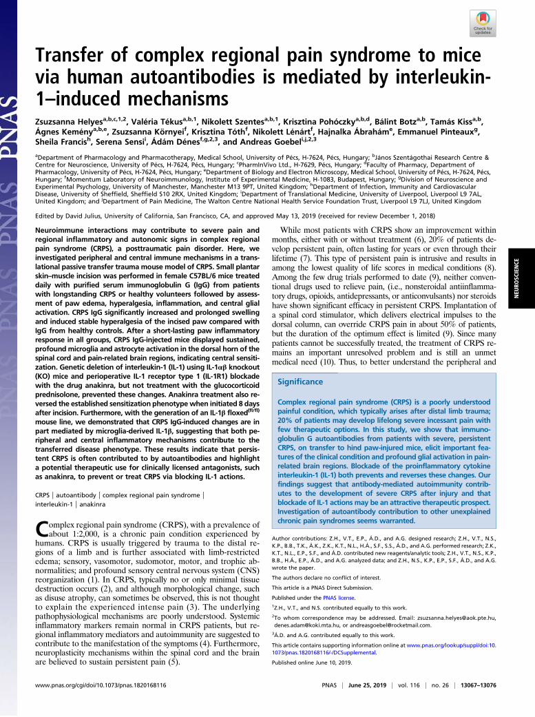
ResultsDaily Administration of Serum IgG from CRPS Patients InducesProfound and Persistent Postincisional Mechanical Hyperalgesia.The preoperation mechanonociceptive threshold values of theaffected limbs were 8.65 ± 0.08, 8.69 ± 0.09, and 8.60 ± 0.07 inthe saline, healthy IgG-, and CRPS IgG-treated mice, re-spectively (not significantly different) (Fig. 1A). Plantar skin andmuscle incision induced a 45–50% relative decrease of themechanonociceptive threshold in all groups 1 d after the surgery.On daily injections, paw sensitivity quickly recovered to mildhyperalgesia in both saline and healthy IgG-injected animals,with mild, nonsignificantly enhanced values remaining in thehealthy IgG group vs. the saline group throughout the experi-mental period. Injection of IgG from CRPS patients causedsignificantly augmented hyperalgesia compared with IgG fromhealthy volunteers, which seemed to be further enhanced towardthe end of the experimental period. This effect was evident in theIgG preparations from each individually tested patient (n = 7) aswell as a preparation pooled from seven separate patients (SI
Appendix, Fig. S1). The observed 15–32% absolute thresholdreduction was twofold compared with that seen in our previouslypublished experimental model (injections on days −1, 0, 5, and 6)(11). Contralateral paws retained normal sensitivity in all groups(SI Appendix, Fig. S2).In all groups, injured paws developed about 30% relative paw
swelling (defined as edema) on day 1, but there were no changesin contralateral paws (SI Appendix, Fig. S3). Edema resolved inhealthy IgG and saline groups, but CRPS IgG injection significantlyslowed edema resolution (Fig. 1B). While the pattern of transferredhyperalgesia was uniform, there was important variability in thedegree and pattern of transferred swelling between different patientpreparations, with no correlation between these two parameters (SIAppendix, Figs. S2 and S3). Postsurgery minimal weight loss wasobserved compared with baseline for a few days, and the weights ofthe animal then fully recovered without significant differences be-tween groups (SI Appendix, Fig. S4). We observed no spontaneousnocifensive behaviors, such as paw biting, lifting, or licking.
CRPS IgG Does Not Alter Vascular Plasma Leakage but IncreasesNeutrophil Myeloperoxidase Activity Early After Paw Incision. Indocyaningreen (ICG)-derived fluorescence detecting vascular leakageshowed a nonsignificant trend to increase in the injured paws in allgroups 2 d after paw incision; CRPS IgG did not specifically affectplasma extravasation [saline injured: 1.52 × 10⁹ ± 1.11 × 10⁸;healthy IgG injured: 1.42 × 10⁹ ± 1.54 × 10⁸; CRPS IgG injured:1.70 × 10⁹ ± 2.2 × 10⁸; fluorescence intensity: (photons per secondper 1 cm2 per steradian) per microwatt per 1 cm2].As expected, in vivo imaging of 8-amino-5-chloro-7-phenylpyrido[3,
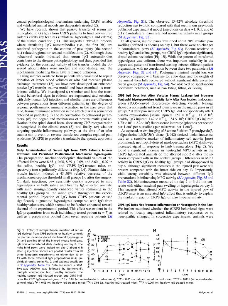
4-d]pyridazine-1,4(2H,3H) dione (L-012)–derived bioluminescence,used as a sensitive marker of inflammatory cell activity [mostprominently neutrophil-derived myeloperoxidase (MPO)], showedincreased signal in response to limb trauma alone (Fig. 2). Wefound a significant increase in neutrophil MPO activity in theCRPS IgG-treated animals on the affected side 2 d after the in-cision compared with in the control groups. Differences in MPOactivity in CRPS IgG vs. healthy IgG groups had disappeared byday 6, although significant increases in the injured paw were stillpresent compared with the intact side on day 13. Importantly,while strong variability was observed between different IgGpreparations in influencing MPO activity (SI Appendix, Fig. S5 andTable S2), bioluminescence measured on day 2 or 6 did not cor-relate with either maximal paw swelling or hyperalgesia on day 6.This suggests that altered MPO activity in the injured paw oftCRPS mice is an unrelated IgG effect that is unlikely to explainthe marked impact of CRPS IgG on paw hypersensitivity.
CRPS IgG Does Not Promote Inflammation or Neuropathy in the Paw.We further examined whether the tCRPS behavioral signs wererelated to locally augmented inflammatory responses or toneuropathic changes. In successive experiments, animals were
Fig. 1. Effect of intraperitoneal injection of serumIgG derived from CRPS patients or healthy controlson plantar incision-induced mechanical hyperalgesia(A) and swelling (B) of the injured mouse hind paw.IgG was administered daily starting on day 0. Theright hind paws were incised on day 0 about 6 hafter Ig injection. Shown are pooled results from allthree long-term experiments to either day 10 or13 with three different IgG preparations (2–4) (in-dividual results are in Fig. 2, and patients details arein SI Appendix, Table S1). Data are means ± SEM.Two-way ANOVA was followed by Bonferroni’smultiple comparison test. Healthy indicates thehealthy control IgG-injected group, and CRPS indi-cates the CRPS IgG-injected group. *P < 0.05 (vs. saline-treated control mice); **P < 0.01 (vs. saline-treated control mice); ***P < 0.001 (vs. saline-treatedcontrol mice); #P < 0.05 (vs. healthy IgG-treated mice); ##P < 0.01 (vs. healthy IgG-treated mice); ###P < 0.001 (vs. healthy IgG-treated mice).
13068 | www.pnas.org/cgi/doi/10.1073/pnas.1820168116 Helyes et al.
killed between experimental days 1 and 13, and paw tissues wereharvested to assess various inflammatory changes (animal num-bers and preparations are in SI Appendix, Table S3).Substance P (SP) levels increased in the injured paw, with
higher levels in the CRPS IgG group on day 6 (Fig. 3A), whereascalcitonin gene-related peptide (CGRP) levels were not signifi-cantly altered (Fig. 3B), consistent with earlier findings (11).Increased levels of inflammatory mediators were seen in theinjured paws, with gradual decrease over time [shown in Fig. 3C–F for interleukin-6 (IL-6), tumor necrosis factor-α (TNF-α),monocyte chemoattractant protein-1 (MCP-1), and IL-1β and in SIAppendix, Fig. S6 for additional mediators]. We detected no dif-ferences in the levels of inflammatory mediators between the CRPSand healthy IgG groups (Fig. 3 C–F and SI Appendix, Fig. S6) at any
time point, except for a mild CRPS IgG-induced MCP-1 increase atday 13 (Fig. 3E). Notably, at the time of maximum hyperalgesia(13 d postinjury), most mediators were undetectable. There wereno significant changes in plasma concentrations of any cytokinesafter correction for multiple testing (SI Appendix, Fig. S7).Histological examination revealed moderate infiltration of
inflammatory cells into areas immediately adjacent to the in-cision early after surgery, with no obvious difference betweengroups; there was no evidence of infiltration by inflammatorycells on day 13 in any experimental group. Since some patientswith persistent CRPS exhibit mild small fiber neuropathy (14),we also examined mouse paw biopsies from CRPS IgG-injectedanimals for any evidence of structural changes to small skinnerves with both light and electron microscopy. The morphologyof the axons in the right (injured) and left (intact) paws as well asthe ultrastructure of nonmyelinated and thin-myelinated axonsin the dermis appeared very similar on aspect, and there were nosignificant differences between sides on quantification of axonnumbers and diameters (SI Appendix, Table S4).
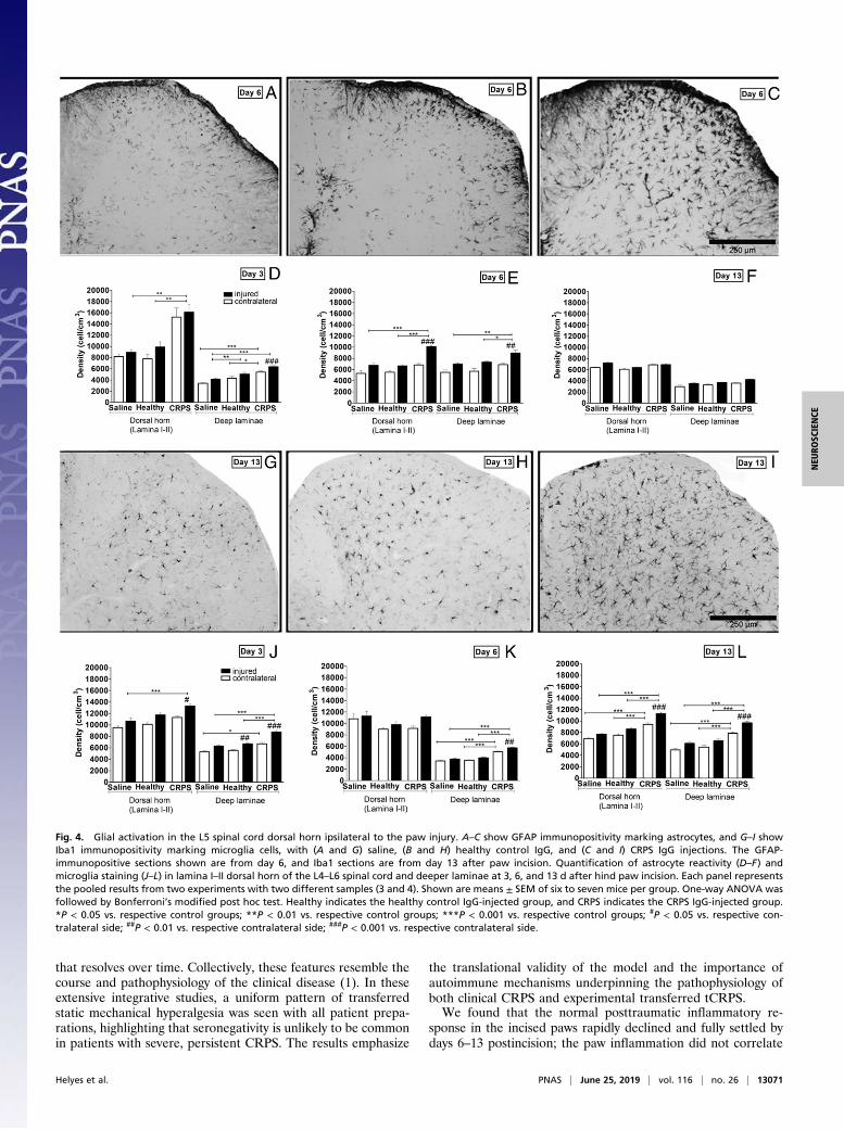
CRPS IgG Facilitates Sustained Microglia Activation in the CNS. Wenext investigated whether altered activity of microglia or astro-cytes in pain-related circuits (15) would reflect the marked effectsof CRPS IgG on pain sensitivity responses. In CNS areas receivinginput from the injured paw, CRPS IgG induced remarkable in-creases in both astrocyte and microglia cell activities comparedwith both saline and healthy IgG groups. In the dorsal horn, thisresponse was sometimes much stronger than the increases repre-senting the incision trauma (Fig. 4). Astrocyte reactivity in theCRPS IgG animals was augmented in all CNS areas at early timepoints (days 3 and 6), whereas microglia staining was enhancedthroughout the experimental period (including day 13) (Fig. 4 andSI Appendix, Fig. S9), indicating that increased mechanical hy-persensitivity in this model is associated with transient astrocyteand persistent microglial activation in the CNS.
Early IL-1 Receptor Blockade with Anakinra Prevents the Developmentof tCRPS, While Delayed Anakinra Treatment Reverses EstablishedtCRPS and Reduces Glial Activation. Since both microglia and as-trocytes are important sources of proinflammatory cytokines thatare known to contribute to pain hypersensitivity responses (16, 17)and IL-1 is a key mediator that influences neuronal activity (18,19), we investigated the effects of glucocorticoid (prednisolone)treatment or interleukin-1 receptor (IL-1R) antagonist (anakinra)treatment on CRPS IgG-induced behavioral signs and inflamma-tory changes. Prednisolone (4 mg/kg) or anakinra (10 mg/kg) wasdaily administered intraperitoneally, starting 5 h before surgery(day 0) and extending throughout the experimental period. Oneday after surgery, mechanical hyperalgesia developed equally in allgroups (Fig. 5 A and B). Glucocorticoid treatment transiently re-duced CRPS IgG-induced mechanical hyperalgesia (between days2 and 3), but this effect was lost by day 7. In contrast, anakinraprevented all CRPS IgG-induced effects throughout the experi-mental period (Fig. 5B). Anakinra, but not prednisolone treatment,almost completely reversed glia cell activation in the ipsilateraldorsal horn on day 6 (Fig. 5 D and E). Notably, anakinra treatmentalso significantly reduced paw MCP-1 levels on day 3; however,there were no other differential effects on levels of peripheralmediators between these two treatments (SI Appendix, Fig. S10).Furthermore, delayed administration of anakinra starting from day8 onward reversed the established tCRPS phenotype (Fig. 5C) andcompletely reversed the associated increased dorsal horn microgliaactivation on day 13 (Fig. 5F).
Selective Deletion of Microglial IL-1β Ameliorates While UbiquitousDeletion of IL-1αβ Completely Prevents tCRPS. Given that CRPSIgG caused significant dorsal horn glia cell activation withoutinfluencing paw IL-1 levels and that blockage of IL-1 prevented
Fig. 2. Imaging ROS demonstrates the development of inflammation in theinjured hind paws of mice. In vivo images of L-012–derived bioluminescencewere obtained during general anesthesia on days 2, 6, and 13 after pawincision. Typical images, with red color indicating strong bioluminescence,are shown in A, and quantification of the bioluminescence intensity is in B.Data at each time point represent the pooled results from experimentsconducted with separate CRPS/healthy control IgG preparations (numbers ofpreparations per time point are in brackets) (details are in SI Appendix, Fig.S4 and Table S2) and are shown as means ± SEM of n = 6–18 mice per group.One-way ANOVA was followed by Bonferroni’s multiple comparison test.Healthy indicates the healthy control IgG-injected group, and CRPS indicatesthe CRPS IgG-injected group. *P < 0.05 vs. respective control groups; **P <0.01 vs. respective control groups; #P < 0.05 vs. respective intact side; ##P <0.01 vs. respective intact side.
Helyes et al. PNAS | June 25, 2019 | vol. 116 | no. 26 | 13069
NEU
ROSC
IENCE

the tCRPS phenotype, we then investigated whether tCRPS wasassociated with enhanced glial IL-1 production in the dorsalhorn. Immunofluorescence revealed increased microglial IL-1βproduction in lumbar (L)L4/L5 dorsal horn microglia cells onlyin the tCRPS group (Fig. 6A), while IL-1α was not detected. Wethen examined whether the tCRPS phenotype would be alteredby genetic knockout (KO) of IL-1. We found that CRPS IgG-injected IL-1αβ KO mice failed to develop enhanced hyper-algesia and showed even less posttraumatic paw swelling (Fig. 6B and C) than mice treated with healthy IgG. To investigatewhether increased microglial IL-1β production is sufficient tomediate the effects of CRPS IgG on increased mechanicalhyperalgesia, we generated an IL-1β floxed(fl/fl) mouse line.Exons 4 and 5 of the IL-1B gene were flanked with loxP sites,resulting in the generation of IL-1βfl/fl allele (SI Appendix, Fig.S11). IL-1βfl/fl mice were crossed with Cx3cr1CreER mice (20),resulting in microglial deletion of IL-1β on tamoxifen adminis-tration (M-IL-1β KO), while most peripheral Cx3cr1-positivecells recovered IL-1β production due to their higher turnoveras shown by using other cre-dependent reporter lines previously(20). In fact, IL-1β protein levels were markedly reduced in IL-1β
KOmicroglia after repeated intraperitoneal injections of bacteriallipopolysaccharide compared with wild-type (WT) microglia, butno changes were seen in splenic macrophages derived fromtamoxifen-treated Cx3cr1CreER × IL-1βfl/fl mice compared withcontrols (SI Appendix, Fig. S12). Elimination of microglial IL-1βsignificantly reduced mechanical hyperalgesia and paw edema inmice treated with CRPS IgG, although this effect was smallerthan in the case of IL-1αβ KO mice (Fig. 6 B and C). Totalnumbers of microglia were reduced in IL-1αβ KO mice but werenot altered in response to microglial IL-1β deletion in the CRPSgroup (Fig. 6D), suggesting that, while microglial IL-1β is animportant driver of chronic neuroinflammation contributing topersistent pain, other IL-1β–producing cells or actions mediatedby IL-1α could also contribute to CRPS symptoms in mice.
DiscussionHere, we show in an enhanced passive transfer trauma modelthat daily administration of IgG from patients with persistentCRPS to mice elicits intense, unilateral static mechanical hyper-algesia after hind paw injury, which remains stable through theexperimental period. This is associated with increased paw edema
Fig. 3. Effects of human IgG transfer on sensoryneuropeptide and inflammatory cytokine concen-trations in the hind paws. Concentrations of (A) SPand (B) CGRP were measured by RIA in hind pawhomogenates excised after they were killed. Con-centrations of (C) IL-6, (D) TNF-α, (E) MCP-1, and (F)IL-1β were measured by cytometric bead array fromthe same samples. Data are from one to three ex-periments per time point (brackets below x axes)each with different patient preparations. Shown aremeans ± SEM. One-way ANOVA was followed byBonferroni’s multiple comparison test. Healthy indi-cates the healthy control IgG-injected group, andCRPS indicates the CRPS IgG-injected group. **P <0.01 vs. respective control groups; #P < 0.05 vs. re-spective intact side; ##P < 0.01 vs. respective intactside; ###P < 0.001 vs. respective intact side;
13070 | www.pnas.org/cgi/doi/10.1073/pnas.1820168116 Helyes et al.
that resolves over time. Collectively, these features resemble thecourse and pathophysiology of the clinical disease (1). In theseextensive integrative studies, a uniform pattern of transferredstatic mechanical hyperalgesia was seen with all patient prepa-rations, highlighting that seronegativity is unlikely to be commonin patients with severe, persistent CRPS. The results emphasize
the translational validity of the model and the importance ofautoimmune mechanisms underpinning the pathophysiology ofboth clinical CRPS and experimental transferred tCRPS.We found that the normal posttraumatic inflammatory re-
sponse in the incised paws rapidly declined and fully settled bydays 6–13 postincision; the paw inflammation did not correlate
Fig. 4. Glial activation in the L5 spinal cord dorsal horn ipsilateral to the paw injury. A–C show GFAP immunopositivity marking astrocytes, and G–I showIba1 immunopositivity marking microglia cells, with (A and G) saline, (B and H) healthy control IgG, and (C and I) CRPS IgG injections. The GFAP-immunopositive sections shown are from day 6, and Iba1 sections are from day 13 after paw incision. Quantification of astrocyte reactivity (D–F) andmicroglia staining (J–L) in lamina I–II dorsal horn of the L4–L6 spinal cord and deeper laminae at 3, 6, and 13 d after hind paw incision. Each panel representsthe pooled results from two experiments with two different samples (3 and 4). Shown are means ± SEM of six to seven mice per group. One-way ANOVA wasfollowed by Bonferroni’s modified post hoc test. Healthy indicates the healthy control IgG-injected group, and CRPS indicates the CRPS IgG-injected group.*P < 0.05 vs. respective control groups; **P < 0.01 vs. respective control groups; ***P < 0.001 vs. respective control groups; #P < 0.05 vs. respective con-tralateral side; ##P < 0.01 vs. respective contralateral side; ###P < 0.001 vs. respective contralateral side.
Helyes et al. PNAS | June 25, 2019 | vol. 116 | no. 26 | 13071
NEU
ROSC
IENCE

with the degree of CRPS IgG-induced paw hyperalgesia. Wefound little evidence for any CRPS IgG-related enhanced pawinflammation. Our results thus demonstrate that static mechan-ical hyperalgesia may not depend on persistent inflammatorymediator release, highlighting that tCRPS is not a model ofenhanced posttraumatic inflammatory pain. There was, however,some evidence for abnormal production of two specific mediatorsin the CRPS group: SP production was increased at early timepoints, in line with some clinical observations (21) and our earlierresults (11), and there was also a mild but significant bilateralincrease in MCP-1 on day 13, the role of which will require ad-ditional investigations. Although we measured common mediatorsof inflammation in the injured paws and these were normal, theinvolvement of additional mediators cannot be excluded.In mice, the development of tCRPS remained restricted to the
injured paw, consistent with the clinical situation, where about90% of the cases show symptoms in only one traumatized limb(22). The precise mechanisms through which circulating patho-genic IgG antibodies mediate both the regionally restrictedposttraumatic clinical CRPS and tCRPS are presently unclear.Early transient trauma-induced inflammatory changes or re-gional opening of blood–nerve and blood–brain barriers mayplay a role by promoting the expression of neoantigens or by
providing IgG access to privileged sites (23). The presence ofsuch facilitating mechanisms is supported by our in vivo imagingresults, which showed plasma leakage and increased MPO ac-tivity in the injured paw.Interestingly, MPO activity was variably enhanced in the ani-
mal groups injected with different CRPS IgG preparations, andthere was no correlation with mechanical hyperalgesia or swell-ing in these same animals. These findings may resemble thestrong heterogeneity seen between patients in the extent of theirlimb swellings, color changes, or temperature changes and mayalso reflect the observation that these clinical parameters do notnecessarily correlate with the patients’ perceived pain intensitiesor recorded skin sensitivities (2).Since inflammatory changes in the paw did not seem to explain
the increased mechanical hyperalgesia in the tCRPS mice, weinvestigated the potential role of glial responses in pain-relatedneuronal circuits, which have been suggested to contribute tochronification processes in posttraumatic pain models (15, 24,25). We found that tCRPS was associated with strong microgliaand astrocyte activation at all three tested levels of the noci-ceptive pathway, the ipsilateral spinal cord dorsal horn, theperiaqueductal gray, and the contralateral somatosensory cortex.In the dorsal horn, the increases in glial responses in the tCRPS
Fig. 5. Effects of prophylactic steroid or anakinra treatment (days 0–6) and delayed (therapeutic) anakinra treatment on CRPS IgG-induced mechanicalhyperalgesia and glial activation in the spinal cord. A and B show mechanical hyperalgesia in groups of animals injected intraperitoneally first with human IgGor saline and 3 h later with 4 mg/kg prednisolone, 10 mg/kg anakinra, or saline vehicle on each day between days 0 and 6. D and E show dorsal horn glia cellactivation in these mice on day 6: (D) GFAP (astrocyte) and (E) Iba-1 (microglia). Results represent the average values derived from two independent ex-periments with different preparations for each treatment, with four experiments in total; saline, healthy control IgG, and CRPS IgG outcomes are pooled fromthese experiments. (C and F) Late anakinra treatment starting on day 8: (C) behavioral outcome and (F) dorsal horn microglia cell count on day 13. Data areshown as means ± SEM. Two-way ANOVA was followed by Bonferroni’s multiple comparison test. One-way ANOVA was followed by Bonferroni’s modifiedpost hoc test. Healthy indicates the healthy control IgG-injected group, and CRPS indicates the CRPS IgG-injected group. Significance symbols for the be-havioral data as follows: *P < 0.05 (CRPS IgG vs. saline-treated control mice); **P < 0.01 (CRPS IgG vs. saline-treated control mice); ***P < 0.001 (CRPS IgG vs.saline-treated control mice); #P < 0.05 (CRPS IgG vs. healthy control IgG-treated mice); ###P < 0.001 (CRPS IgG vs. healthy control IgG-treated mice); +++P <0.001 (anakinra plus CRPS IgG vs. CRPS IgG-injected mice). Significance immunohistochemistry data: *P < 0.05 vs. respective control groups; **P < 0.01 vs.respective control groups; ***P < 0.001 vs. respective control groups; #P < 0.05 vs. respective contralateral side; ##P < 0.01 vs. respective contralateral side;###P < 0.001 vs. respective contralateral side.
13072 | www.pnas.org/cgi/doi/10.1073/pnas.1820168116 Helyes et al.

vs. control groups were sometimes severalfold larger than theextent of glia activation caused by the paw incision (Fig. 4),suggesting a powerful central effect of the transferred IgG.These results raise the question of how the observed profound
central glial cell activation is mediated. Possible mechanisms mayinclude (i) modification of nociceptor function after direct anti-body binding in the periphery as an “autoimmune channelopathy”(26), (ii) release of yet undetermined peripheral mediators (27),(iii) direct binding in the dorsal horn after temporary post-traumatic opening of the blood spinal cord barrier (28), or (iv) amissing link. Independent of the nature of these upstream mech-anisms, glial activation is likely to result in the release of mediators,such as IL-1β, TNF-α, or brain-derived neurotrophic factor, withconsequent modification of central pain processing (15, 29).We hypothesized that perioperative antiinflammatory inter-
ventions might be effective, and we initially thought that suchinterventions might act through the blockade of perioperativeregional facilitatory factors required to render circulating CRPSautoantibodies pathogenic (4). To investigate whether such inter-ventions could prevent the disease phenotype, we treated miceperitraumatically with high-dose prednisolone. Prednisolone treat-ment temporarily interrupted the process of autoantibody-dependentsensitization, but it did not stop it. Systemic glucocorticoids areconsidered potentially effective in very early CRPS based on theresults of one preliminary trial (9). These data suggest that, wherepatients produce harmful autoantibodies, the peritraumatic appli-cation of glucocorticoids is unlikely to stop disease progression. Incontrast, perioperative treatment with the IL-1R antagonist anakinrain this CRPS model consistently prevented the tCRPS phenotype.Notably, we found that there were only minor differences in theregional paw inflammatory environment based on the production ofcytokines and chemokines after treatment with (ineffective) pred-nisolone and (effective) anakinra (SI Appendix, Fig. S10); further-more, even delayed blockade of IL-1 actions with anakinra startingon day 8 after the incision trauma, when trauma-induced peripheralinflammatory responses had largely resolved, was highly effective,
suggesting that the pertinent biological effect of anakinra treatmentwas not restricted to the injured paw.IL-1 is a potent activator of astrocytes through actions via IL-
1R1, whereas both activated microglia and astrocytes can con-tribute to painful central sensitization through secreting IL-1 (29,30). These actions can be effectively blocked by anakinra (31–34). In our model, augmented dorsal horn glia cell activation inthe tCRPS group was fully reversed by anakinra. Since IL-1–mediated actions are involved in the cross-talk between neu-rons, microglia, and astrocytes in promoting neuroinflammation(35, 36), we assessed glial IL-1β and IL-1α production in thespinal cord and found that microglial IL-1β production was in-creased on day 7 in the CRPS IgG-treated group. In agreementwith these data, we found that IL-1αβ KO mice were fully pro-tected from developing the tCRPS phenotype and associatedglial activation. To specifically assess the functional role ofmicroglial IL-1β in tCRPS-associated hyperalgesia and swelling,we generated a mouse strain (IL-1βfl/fl mice), enabling the de-letion of IL-1β from microglia. Consistent with earlier datashowing prolonged cre-dependent transgene expression in long-lived microglia but not in peripheral macrophage populationswith short turnover (20), tamoxifen treatment of Cx3cr1CreER ×IL-1βfl/fl mice resulted in a marked reduction of microglial, butnot splenic, IL-1β production (SI Appendix, Fig. S12). As seen inIL-1αβ KO mice, the absence of microglial IL-1β productionfully protected from tCRPS-associated mechanical hyperalgesiaup to day 3, whereas it had a weaker effect thereafter. Thishighlights the importance of microglial IL-1β in the tCRPS dis-ease process but also, the likely involvement of other cells and/orIL-1α (35).Strengths of our study include the robust, multidimensional
evaluation of an enhanced CRPS disease transfer model withpreparations from patients whose clinical presentations differedusing outcomes designed to provide translational validity. Addi-tional strengths are the comprehensive assessment of both pe-ripheral and central markers of immune activation and of severalantiinflammatory treatments as well as gene KO strategies, which
Fig. 6. Deletion of IL-1αβ or microglia-derived IL-1βfully or partially prevents development of the CRPSIgG-induced phenotype in mice. (A) A population ofmicroglia identified by immunostaining against P2Y12,a specific microglial marker in the brain (59), displaysa morphologically activated phenotype and showsimmunopositivity for IL-1β at day 7 in the deep laminaeof the L4–L5 spinal cord near the central canal. (Scalebar: 50 μm.) (B and C) IL-1αβ KOmice are fully protectedand M-IL-1βKO mice are partially protected from thedevelopment of the CRPS IgG-induced phenotype: (B)paw hyperalgesia and (C) paw edema. Two-wayANOVA was followed by Bonferroni’s multiple com-parison test. Significance in B and C: **P < 0.01 (CRPSIgGWT vs. healthy IgGWT); ***P < 0.001 (CRPS IgGWTvs. healthy IgG WT); #P < 0.05 (CRPS IgG M-IL-1β KO vs.CRPS IgG WT); ##P < 0.01 (CRPS IgG M-IL-1β KO vs. CRPSIgG WT); ###P < 0.001 (CRPS IgG IL-1αβ KO vs. CRPS IgGWT); ++P < 0.01 (CRPS IgG IL-1αβ KO vs. CRPS IgG M-IL-1β KO); +++P < 0.001 (CRPS IgG IL-1αβ KO vs. CRPS IgGM-IL-1β KO). (D) CRPS IgG-induced microglia activationis abrogated in IL-1αβ KO but not in M-IL-1β KO mice.Data are pooled from two experiments with differentCRPS IgG preparations for each mouse type and areshown as means ± SEM. Healthy indicates the healthycontrol IgG-injected group, and CRPS indicates the CRPSIgG-injected group. One-way ANOVA was followed byBonferroni’s modified post hoc test. Significance valuesin D: **P < 0.01 vs. respective control groups; ***P <0.001 vs. respective control groups; #P < 0.05 vs. re-spective contralateral side; ###P < 0.001 vs. respectivecontralateral side.
Helyes et al. PNAS | June 25, 2019 | vol. 116 | no. 26 | 13073
NEU
ROSC
IENCE

have allowed an informed suggestion for clinical studies with alicensed drug not previously reported in this patient group.Limitations include the upper transfer limit cutoff at 13 d,
which was necessary to avoid the adverse effects of serum sick-ness (37). However, it may be argued that the “chronic phase” inthis disease model starts from the second week after incision,when peripheral inflammation and symptoms in the controlgroup resolve. As is often observed with IgG disease transfer inother disease models, tCRPS does not fully match the symp-toms of clinical CRPS (38). For example, overt motor dys-function was not detected, and commonly encountered centralfeatures, such as body perception abnormalities, were notrevealed. In line with most behavioral studies in animal modelsof chronic pain (39), we did not demonstrate the presence ofspontaneous pain but ascertained nociceptor hypersensitivityby assessing stimulus-evoked pain (mechanical hypersensitiv-ity). Independently, we only transferred serum IgG that wasderived from patients suffering from persistent (7) BudapestCRPS with high pain intensities (numeric rating scale > 7);however, these are also the patients who present the mostdifficult situation in the clinical practice. We cannot rule outthe possibility that some patients in this group do not havethese antibodies. Since each of the seven individually tested serumIgG preparations induced the abnormal phenotype (SI Appendix,Fig. S1), the likelihood that we have missed the absence of suchantibodies in more than one-half of patients of a similar pop-ulation seems very low (<1%) (Patients, Healthy Controls, andSerum Preparation); the development of serum diagnostic tests willbe required to further detail the proportion of seronegative pa-tients. We did not measure epidermal nerve fiber density or lengthin the mouse paw skin; therefore, our studies may not fully ruleout mild small fiber neuropathy.In summary, we have devised a robust translational model
reproducing pertinent aspects of an “idiopathic,” posttraumaticchronic pain condition. The consistent pathogenicity of variousserum IgG preparations indicates that, among patients who havesevere forms of this condition, autoantibody contribution isubiquitous. Since abnormal signs were entirely confined to theinjured side, we also established a general principle suggestingthat pathogenic circulating autoantibodies can cause regionallyrestricted disease when triggered by local trauma.Our results support previous clinical observations that patients
with persistent CRPS should respond to immune treatments witha reduction of at least some of their disease features (12). Theclinical use of IL-1R antagonists in CRPS has a broad thera-peutic potential. Anakinra is clinically licensed both in theUnited States and in Europe, and short-term use has an ac-ceptable side effect profile (40, 41). Since CRPS regularlydevelops in the context of elective operations, such as ar-throscopy or bunion surgery, prevention of such cases wouldhave very important implications for both patients andprocedure-related health care as well as societal costs (42). Asour results also suggest that treatment initiated after injuryresolution reverts the established transferred phenotype, clin-ical treatment of patients with persistent CRPS could now betested in a trial setting.The cellular and molecular targets of the patient autoanti-
bodies need to be clarified; recent results in other conditions thatare not associated with trauma (26, 27) have indicated that pain-sensitizing autoantibodies may directly bind to sensory neuronsor indirectly bind to juxtapositioned cells, which then releasenerve-sensitizing mediators. The duration of beneficial effectsafter the drug is withdrawn should be investigated in this ex-perimental model. In addition, identifying the exact cellulartargets for IL-1 actions in the pathophysiology of tCRPS couldfacilitate the development of alternative IL-1 targeting ap-proaches in the prevention or treatment of the clinical disease.The results of this study highlight the important role that auto-
antibodies can play in causing detrimental symptoms, absenttissue destruction and systemic inflammation.
Materials and MethodsPatients, Healthy Controls, and Serum Preparation. NorthWest Ethics HaydockUK approved serum donations. Written informed consent was obtained fromall patients. Serum or plasma samples were obtained (i) via a dedicated studydesigned to repeatedly acquire large volumes, (ii) from waste plasma afterpatients’ plasma exchange treatment, and (iii) from patients participating ina clinical trial who never had plasma exchange (43)—the latter samples werepooled to minimize animal use. Details are provided in SI Appendix. Thepatients’ baseline characteristics are provided in Table 1. We calculated theprobability that we missed an event rate of >50% of patients having no pain-sensitizing autoantibodies by [0.5(number of experiments × fraction of positive experiments)]:0.57 = 0.008125 (i.e., <1%). IgG was prepared from plasma or serum of pa-tients as previously described using Protein G columns for affinity purifi-cation followed by elution, buffering, dialysis in normal saline, andconcentration to about 8 mg/mL IgG for injection (pooled preparations12 mg/mL) (11).
Animals. The Ethics Committee on Animal Research of the University of Pecsapproved experiments involving rodents. Since CRPS affects women two tothree times more frequently than men, experiments were carried out onfemale mice on C57BL/6 background (10–12 wk old, 18–22 g). Breeding,maintenance, and ethical procedures were as previously described (11) (de-tails are in SI Appendix).
Experimental Design. After acclimatization and conditioning, three controlmeasurements of nociception and paw volume were performed on days −4,−3, and −2. Day 0 was the starting day of intraperitoneal injections (SI Ap-pendix, Fig. S13). Mice (five to seven per group) were treated daily tocompensate for rapid metabolism of human IgG in the mouse (SI Appendix,Methods) with 1 mL of IgG fractions (8 mg/mL) obtained from CRPS patientsor healthy volunteers or saline.
Six hours after the injection on day 0, a standardized incision trauma wasapplied to the right hind paw as described below. All mice removed theirstitches within 16 h postsurgery. Measurements (see below) were performedrepeatedly starting on day 1 until the respective termination day.
Animals were killed at various time points, and tissues were harvested aspreviously described (11). In some experiments, animals were perfusedtranscardially with phosphate buffered saline (PBS) followed by 4%paraformaldehyde as previously described, and the whole brains and spi-nal cords were excised and prepared for additional immunohistochemistryanalyses.
Plantar Skin and Muscle Incision. To model a CRPS-triggering injury, we usedthe hind pawplantar skinmuscle incisionmethod under general anesthesia asdescribed earlier (11, 44) and detailed in SI Appendix. This model evokes asignificant decline of the mechanonociceptive threshold, with a maximum1 d after surgery, which persists for 7–8 d; the model’s early threshold nor-malization was considered advantageous, minimizing any animal sufferingand allowing for early assessment of IgG-induced changes. All measurementswere carried out by two investigators (V.T. and N.S.) who were blinded totreatment assignment. Blinding was performed by the technicians whoperformed all injections but were otherwise not involved in the study; theydifferentially coded the animal cages and provided the decoding key aftercompletion of the last measurements.
Determination of the Mechanosensitivity of the Paw. Most patients with CRPShave, in addition to their spontaneous pain, pain with the application ofpressure to the CRPS-affected limb (“mechanical hyperalgesia”) (45, 46), andall patients included in this study experienced this feature. The corre-sponding mechanonociceptive threshold of the plantar surface of the mousehind paw was determined with a dynamic plantar aesthesiometer (UgoBasile 37400)—a modified electronic von Frey technique—as previously de-scribed (11). The blunt-end needle exerting an increasing force to the mousepaw provides a mild but basically painful stimulus activating Aδ and C fibers(47). Threshold decreases are considered as mechanical hyperalgesia and areexpressed as percentage decrease of the mechanonociceptive thresholdscompared with the baseline values (48, 49).
Paw Volume Measurement. Limb swelling is a common feature of CRPS-affected limbs, and all included patients reported intermittent limb swell-ing. Mouse paw volume was measured using plethysmometry (Ugile Basile
13074 | www.pnas.org/cgi/doi/10.1073/pnas.1820168116 Helyes et al.

Plethysmometer 7140). Edema was expressed as a percentage increase com-pared with the baseline paw volume (48).
In Vivo Optical Imaging of Plasma Leakage and Neutrophil MPO Activity. Themechanisms underpinning CRPS IgG-enhanced paw swelling are unknown,but one possibility is augmented plasma extravasation. Intravenous injectedICG, a fluorescent cyanine dye, binds to plasma proteins and remains in thehealthy vasculature. Under inflammatory conditions, it can be used to evaluatecapillary leakage. ICG (0.5 mg/kg) was dissolved freshly in 5% (wt/vol) aqueoussolution of Kolliphor HS 15 and a macrogol-based surfactant and injected in-travenously (retrobulbar sinus) under ketamine (100 mg/kg; Calypsol; GedeonRichter Plc.) and xylazine (10 mg/kg; Sedaxylan; Eurovet Animal Health B.V.)anesthesia 2 d after the paw incision. Fluorescence imaging was performed20 min postinjection using an IVIS Lumina II in vivo optical imaging system(PerkinElmer; autoacquisition time, f/stop = 1, binning = 2, excitation: 745 nm,emission filter: >800 nm) (50).
A luminol analog chemiluminescent probe, L-012 (Wako Pure ChemicalIndustries Ltd.) was used for in vivo visualization of reactive oxygen species(ROS)/reactive nitrogen species (RNS) produced by MPO in neutrophils andmacrophages; L-012 has a high sensitivity toward ROS/RNS (51). Mousepreparation, injection, imaging, and analysis procedures were conducted aspreviously described and highlighted in SI Appendix (52).
Measurement of Inflammatory Mediators. Proinflammatory neuropeptidesperipherally released from sensory nerves and inflammatory cytokines re-leased by perineuronal cells are abnormal in some patients with CRPS, andwemeasured their concentrations in the model.
The preserved frozen paws (see above) were thawed, chopped, and thenhomogenized in Triton X-100 and Calbiochem Protease Inhibitor Cocktailcontaining Tris·HCl homogenization buffer at 0 °C. Additional processingdetails are provided in SI Appendix. We measured CGRP- and SP-like im-munoreactivities in the paws by a sensitive radioimmunoassay (RIA) tech-nique developed in our laboratory as previously described (11, 53, 54).
Concentrations of IL-1α, IL-1β, IL-6, TNF-α, KC (CXCL1), MCP-1, G-CSF,RANTES (CCL5), interferon-γ, IL-4, and IL-10 in mouse plasma and the pawhomogenates were measured by cytometric bead array (BD Biosciences) aspreviously described (34, 55), and transforming growth factor-β and NGFwere measured by sandwich enzyme-linked immunosorbent assay accordingto the manufacturers’ instructions (56).
Paw Skin Light and Electron Microscopy. After biopsy, samples were immersedinto a fixative containing 2.5% glutaraldehyde buffered with phosphatebuffer (0.1 M, pH 7.4) overnight at 4 °C, fixed in 1% osmium tetroxide for35 min, and dehydrated with increasing concentration of ethanol. Aftercomplete dehydration, they were transferred to propylene oxide beforebeing placed into aluminum foil boats and then embedded into gelatincapsule containing Durcupan resin (Sigma).
Semithin and ultrathin sections were cut with Leica ultramicrotom. Semithinsections were mounted on glass slides, stained with toluidine blue, andexamined under a light microscope. Ultrathin sections were mounted oncollodion-coated (Parlodion; Electron Microscopy Sciences) single-slot coppergrids contrasted with uranyl acetate and lead citrate, and were examined in a
JEOL 1200EX-II electron microscope. Small nerve fiber quantification methodsare detailed in SI Appendix, Methods.
Immunohistochemistry. Brains and L4–L6 segments of the spinal cord wereremoved and postfixed for 4 h in 4% paraformaldehyde before being placedinto 30% sucrose (Duchefa Biochemie) in 0.1 M PBS overnight at 4 °C. Sec-tions (30 μm) were prepared using a freezing microtome (Leica BiosystemsNussloch GmbH) as free-floating sections (57, 58), and they were stained andmounted as described in SI Appendix. The sections (n = 3–4 animal pergroup) were examined by Neurolucida software (v07; MBF Bioscience) usinga Nikon Eclipse Ni-E bright-field microscope with a computer-controlledstage. A modified unbiased stereology protocol was used for quantifica-tion of glial fibrillary acidic protein (GFAP) or Iba1 immunoreactive cellsalong the nociceptive pathway as previously described (50).
Immunofluorescence. Immunofluorescence to detect microglial P2Y12 (59)and the production of IL-1α and IL-1β was performed on free-floatingbrain sections. Images were captured with a Nikon Ni-E C2+ confocalmicroscope.
Antiinflammatory Interventions. We investigated whether early immunesuppressionwould alter the disease course in themodel.We administered thesynthetic glucocorticoid prednisolone (4 mg/kg intraperitoneally) (60, 61) orthe IL-1R antagonist anakinra [10 mg/kg intraperitoneally; Swedish OrphanBiovitrum AB (publ), SE-112 76]. This prednisolone dose corresponds to thehighest dose range used in humans [pulse therapy (62)], and the anakinradose is known to be pharmacologically active in mice and und is com-parable with human therapeutic dosages (63–65). Control animals re-ceived respective vehicles. The first drug injection was 3 h before theplantar skin and muscle incision and at least 4 h after IgG or controltreatment. Drug treatment was then repeated daily through the experi-mental period. Delayed anakinra treatment was administered daily be-tween days 8 and 13.
Generation of IL-1β Floxed Mice and Microglial IL-1β KOMice. Il1btm1a(EUCOMM)Hmgu
embryonic stem cells (66) were purchased from the European Mouse MutantCell Repository. Cells from clone HEPD0840-8-E03 were prepared for mi-croinjection according to previously published protocols (67) with minormodifications (SI Appendix, Methods) and then microinjected into four- toeight-cell B6N-Tyrc-Brd/BrdCrCrl embryos. Surviving embryos were surgicallyimplanted into the oviduct of day 0.5 postcoitum pseudopregnant mice.Resulting black/white C57BL/6N chimeras were backcrossed onto C57BL/6NWT mice to assess germline penetrance. Potential founder mice werescreened by PCR for LacZ, Neo, and LoxP sites. This line was further crossedwith C57BL/6N-Tg(CAG-Flpo)1Afst/Mmucd mice. The flp recombinase ex-pression provided by this line resulted in a “conditional ready” (floxed)Il1btm1c(EUCOMM)Hmgu allele where exons 4 and 5 are flanked by loxP sites. Togenerate microglial IL-1β KO mice, IL-1β floxed mice were crossed withtamoxifen-inducible B6.129P2(C)-CX3CR1tm2.1(cre/ERT2)Jung/J mice (JAX stock020940) (20).
Statistical Analysis. Data shown are means ± SEM, and two-way repeatedmeasures ANOVA followed by Bonferroni’s post hoc test was used forcomparison of threshold values between groups at respective timepoints.One-way ANOVA followed by Bonferroni’s post hoc test was used foranalysis of the immunochemistry and cytokine results. A value of P <0.05 was considered statistically significant.
ACKNOWLEDGMENTS. This research was supported by National Brain Re-search Program 2017-1.2.1-NKP-2017-00002 (NAP-2; Chronic Pain Re-search Group), the Pain Relief Foundation Liverpool, Gazdaságfejlesztési ésInnovációs Operatív Program (Economy Development and InnovationOperative Programme) (GINOP)-2.3.2-15-2016-00050 (Peptidergic Signalingin Health and Disease; PEPSYS), Emberi Er}oforrás Operatív Program (HumanResource Operative Programme) (EFOP) 3.6.2-17-2017-00008 N (2017-2019),and Társadalmi Megújulás Operatív Program (Social Renewal Operative Pro-gramme) (TAMOP) 4.2.4. A/2-11-1-2012-0001 “National Excellence Pro-gram—Elaborating and operating an inland student and researcher personalsupport system convergence program.” Á.D. is supported by Hungarian BrainResearch Program KTIA_13_NAP-A-I/2, the “Momentum” Program of theHungarian Academy of Sciences, European Research Council (ERC)-CoG724994, and TÉT_16-1-2016-0104. The generation of IL-1bfl/fl mouse linewas funded by British Heart Foundation Grant PG/13/55/30365 (to E.P. andS.F.). We thank Dóra Ömböli and Lilla Draskóczi for their expert technicalassistance in the animal experiments and tissue preparation; Jenny Hawkesfor her expert technical assistance in the IgG preparation; and DeborahBently for proofreading the manuscript.
Table 1. Baseline characteristics of the patient serum donors
No. Sex/age Limb/DD Pain Dx Plex
1 M/62 U/9 10 +II Y2 F/37 L/15 7.5 +I N3 F/38 L/10 9.5 +I Y4 F/36 L/10 7 +I Y5 F/40 L/5 7.5 +I Y6 F/51 L/8 8 +I N7 F/49 L/7 8 +I Y
Age indicates age in years at the time of plasma/serum acquisition. Limbindicates the affected limb. DD is the disease duration in years. Pain indicates24-h average pain intensity on a 11-point numeric rating scale (0–10, with10 = pain as bad as you can imagine). Dx indicates diagnosis, with + indicatingthat the patient fulfills Budapest research diagnostic criteria (new InternationalAssociation for the Study of Pain criteria); II denotes trigger injury to a majornerve, and I denotes no trigger injury to a major nerve. Plex indicates plasmaderived from plasma exchange. N, no; Y, yes; U, upper limb; L, lower limb.
Helyes et al. PNAS | June 25, 2019 | vol. 116 | no. 26 | 13075
NEU
ROSC
IENCE

1. R. N. Harden et al., Validation of proposed diagnostic criteria (the “Budapest criteria”)for complex regional pain syndrome. Pain 150, 268–274 (2010).
2. F. Birklein, S. K. Ajit, A. Goebel, R. S. G.M. Perez, C. Sommer, Complex regional pain syndrome–Phenotypic characteristics and potential biomarkers. Nat. Rev. Neurol. 14, 272–284 (2018).
3. M. Nicholas et al.; IASP Taskforce for the Classification of Chronic Pain, The IASPclassification of chronic pain for ICD-11: Chronic primary pain. Pain 160, 28–37 (2019).
4. A. Goebel, F. Blaes, Complex regional pain syndrome, prototype of a novel kind ofautoimmune disease. Autoimmun. Rev. 12, 682–686 (2013).
5. C. Maihöfner, H. O. Handwerker, B. Neundörfer, F. Birklein, Patterns of cortical re-organization in complex regional pain syndrome. Neurology 61, 1707–1715 (2003).
6. A. Zyluk, The natural history of post-traumatic reflex sympathetic dystrophy. J. HandSurg. [Br.] 23, 20–23 (1998).
7. M. de Mos et al., Outcome of the complex regional pain syndrome. Clin. J. Pain 25,590–597 (2009).
8. M. A. Kemler, C. A. Furnée, Economic evaluation of spinal cord stimulation for chronicreflex sympathetic dystrophy. Neurology 59, 1203–1209 (2002).
9. N. E. O’Connell, B. M. Wand, J. McAuley, L. Marston, G. L. Moseley, Interventions fortreating pain and disability in adults with complex regional pain syndrome. CochraneDatabase Syst. Rev. 4, CD009416 (2013).
10. A. B. Ch. Goebel et al., Complex Regional Pain Syndrome in Adults (Royal College ofPhysicians, London, ed. 2, 2018).
11. V. Tékus et al., A CRPS-IgG-transfer-trauma model reproducing inflammatory andpositive sensory signs associated with complex regional pain syndrome. Pain 155, 299–308 (2014).
12. J. Schwartz et al., Guidelines on the use of therapeutic apheresis in clinical practice-evidence-based approach from the writing committee of the American Society forapheresis: The seventh special issue. J. Clin. Apher. 31, 149–162 (2016).
13. F. J. Huygen et al., Evidence for local inflammation in complex regional pain syn-drome type 1. Mediators Inflamm. 11, 47–51 (2002).
14. A. L. Oaklander et al., Evidence of focal small-fiber axonal degeneration in complexregional pain syndrome-I (reflex sympathetic dystrophy). Pain 120, 235–243 (2006).
15. K. Inoue, M. Tsuda, Microglia in neuropathic pain: Cellular and molecular mechanismsand therapeutic potential. Nat. Rev. Neurosci. 19, 138–152 (2018).
16. Z. J. Zhang, B. C. Jiang, Y. J. Gao, Chemokines in neuron-glial cell interaction andpathogenesis of neuropathic pain. Cell. Mol. Life Sci. 74, 3275–3291 (2017).
17. K. Popiolek-Barczyk, J. Mika, Targeting the microglial signaling pathways: New in-sights in the modulation of neuropathic pain. Curr. Med. Chem. 23, 2908–2928 (2016).
18. S. M. Allan, P. J. Tyrrell, N. J. Rothwell, Interleukin-1 and neuronal injury. Nat. Rev.Immunol. 5, 629–640 (2005).
19. A. Denes, E. Pinteaux, N. J. Rothwell, S. M. Allan, Interleukin-1 and stroke: Biomarker,harbinger of damage, and therapeutic target. Cerebrovasc. Dis. 32, 517–527 (2011).
20. S. Yona et al., Fate mapping reveals origins and dynamics of monocytes and tissuemacrophages under homeostasis. Immunity 38, 79–91 (2013).
21. M. Weber, F. Birklein, B. Neundörfer, M. Schmelz, Facilitated neurogenic in-flammation in complex regional pain syndrome. Pain 91, 251–257 (2001).
22. P. H. Veldman, H. M. Reynen, I. E. Arntz, R. J. Goris, Signs and symptoms of reflexsympathetic dystrophy: Prospective study of 829 patients. Lancet 342, 1012–1016(1993).
23. N. P. Staff et al., Post-surgical inflammatory neuropathy. Brain 133, 2866–2880 (2010).24. M. R. Suter, Microglial role in the development of chronic pain. Curr. Opin. Anaesthesiol.
29, 584–589 (2016).25. E. D. Milligan, L. R. Watkins, Pathological and protective roles of glia in chronic pain.
Nat. Rev. Neurosci. 10, 23–36 (2009).26. J. M. Dawes et al., Immune or genetic-mediated disruption of CASPR2 causes pain
hypersensitivity due to enhanced primary afferent excitability. Neuron 97, 806–822.e10 (2018).
27. G. Wigerblad et al., Autoantibodies to citrullinated proteins induce joint pain in-dependent of inflammation via a chemokine-dependent mechanism. Ann. Rheum.Dis. 75, 730–738 (2016).
28. L. S. Cahill et al., Quantifying blood-spinal cord barrier permeability after peripheralnerve injury in the living mouse. Mol. Pain 10, 60 (2014).
29. W. Guo et al., Glial-cytokine-neuronal interactions underlying the mechanisms ofpersistent pain. J. Neurosci. 27, 6006–6018 (2007).
30. C. Y. Chiang, B. J. Sessle, J. O. Dostrovsky, Role of astrocytes in pain. Neurochem. Res.37, 2419–2431 (2012).
31. J. M. Pradillo et al., Delayed administration of interleukin-1 receptor antagonist re-duces ischemic brain damage and inflammation in comorbid rats. J. Cereb. Blood FlowMetab. 32, 1810–1819 (2012).
32. A. Denes, P. Thornton, N. J. Rothwell, S. M. Allan, Inflammation and brain injury:Acute cerebral ischaemia, peripheral and central inflammation. Brain Behav. Immun.24, 708–723 (2010).
33. A. Denes et al., Interleukin-1 mediates neuroinflammatory changes associated withdiet-induced atherosclerosis. J. Am. Heart Assoc. 1, e002006 (2012).
34. Á. Dénes et al., Streptococcus pneumoniae worsens cerebral ischemia via interleukin1 and platelet glycoprotein Ibα. Ann. Neurol. 75, 670–683 (2014).
35. S. A. Liddelow et al., Neurotoxic reactive astrocytes are induced by activated microglia.Nature 541, 481–487 (2017).
36. W. Liu, Y. Tang, J. Feng, Cross talk between activation of microglia and astrocytes inpathological conditions in the central nervous system. Life Sci. 89, 141–146 (2011).
37. K. V. Toyka, D. B. Brachman, A. Pestronk, I. Kao, Myasthenia gravis: Passive transferfrom man to mouse. Science 190, 397–399 (1975).
38. P. Sillevis Smitt et al., Paraneoplastic cerebellar ataxia due to autoantibodies against aglutamate receptor. N. Engl. J. Med. 342, 21–27 (2000).
39. J. S. Mogil et al., Hypolocomotion, asymmetrically directed behaviors (licking, lifting,flinching, and shaking) and dynamic weight bearing (gait) changes are not measuresof neuropathic pain in mice. Mol. Pain 6, 34 (2010).
40. G. Lopalco et al., Safety profile of anakinra in the management of rheumatologic,metabolic and autoinflammatory disorders. Clin. Exp. Rheumatol. 34, 531–538 (2016).
41. M. T. Nurmohamed, B. A. Dijkmans, Efficacy, tolerability and cost effectiveness ofdisease-modifying antirheumatic drugs and biologic agents in rheumatoid arthritis.Drugs 65, 661–694 (2005).
42. SUVA, CRPS (Complex Regional Pain Syndrome), W. Jänig, R. Schaumann, W. Vogt,Eds. (SUVA, 2013).
43. A. Goebel et al., Mycophenolate for persistent complex regional pain syndrome, aparallel, open, randomised, proof of concept trial. Scand. J. Pain 18, 29–37 (2018).
44. E. M. Pogatzki, S. N. Raja, A mouse model of incisional pain. Anesthesiology 99, 1023–1027 (2003).
45. J. Gierthmühlen et al.; German Research Network on Neuropathic Pain (DFNS) StudyGroup Sensory signs in complex regional pain syndrome and peripheral nerve injury.Pain 153, 765–774 (2012).
46. V. Huge et al., Complex interaction of sensory and motor signs and symptoms inchronic CRPS. PLoS One 6, e18775 (2011).
47. S. Ventéo et al., Fxyd2 regulates Aδ- and C-fiber mechanosensitivity and is requiredfor the maintenance of neuropathic pain. Sci. Rep. 6, 36407 (2016).
48. A. Szabó et al., Role of transient receptor potential vanilloid 1 receptors in adjuvant-induced chronic arthritis: In vivo study using gene-deficient mice. J. Pharmacol. Exp.Ther. 314, 111–119 (2005).
49. K. Bölcskei et al., Investigation of the role of TRPV1 receptors in acute and chronicnociceptive processes using gene-deficient mice. Pain 117, 368–376 (2005).
50. Á. Horváth et al., Transient receptor potential ankyrin 1 (TRPA1) receptor is involvedin chronic arthritis: In vivo study using TRPA1-deficient mice. Arthritis Res. Ther. 18, 6(2016).
51. I. Imada et al., Analysis of reactive oxygen species generated by neutrophils using achemiluminescence probe L-012. Anal. Biochem. 271, 53–58 (1999).
52. B. Botz et al., Differential regulatory role of pituitary adenylate cyclase-activatingpolypeptide in the serum-transfer arthritis model. Arthritis Rheumatol. 66, 2739–2750 (2014).
53. J. Németh et al., Substance P radioimmunoassay for quantitative characterization ofsensory neurotransmitter release. Neurobiology (Bp.) 7, 437–444 (1999).
54. J. Németh et al., Development of a new sensitive CGRP radioimmunoassay for neu-ropharmacological research. Neurobiology (Bp.) 6, 473–475 (1998).
55. A. Denes et al., AIM2 and NLRC4 inflammasomes contribute with ASC to acute braininjury independently of NLRP3. Proc. Natl. Acad. Sci. U.S.A. 112, 4050–4055 (2015).
56. J. B. Davis, ELISA for monitoring nerve growth factor. Methods Mol. Biol. 1606, 141–147 (2017).
57. M. G. Giovannini et al., Mitogen-activated protein kinase regulates early phosphor-ylation and delayed expression of Ca2+/calmodulin-dependent protein kinase II inlong-term potentiation. J. Neurosci. 21, 7053–7062 (2001).
58. F. Cerbai et al., The neuron-astrocyte-microglia triad in normal brain ageing and in amodel of neuroinflammation in the rat hippocampus. PLoS One 7, e45250 (2012).
59. R. Fekete et al., Microglia control the spread of neurotropic virus infection viaP2Y12 signalling and recruit monocytes through P2Y12-independent mechanisms.Acta Neuropathol. 136, 461–482 (2018).
60. L. He et al., Methylprednisolone prevents nerve injury-induced hyperalgesia in ne-prilysin knockout mice. Pain 155, 574–580 (2014).
61. M. Suzuki, H. Yoshida, M. Hashizume, K. Tanaka, Y. Matsumoto, Blockade of in-terleukin-6 receptor enhances the anti-arthritic effect of glucocorticoids withoutdecreasing bone mineral density in mice with collagen-induced arthritis. Clin. Exp.Immunol. 182, 154–161 (2015).
62. F. Buttgereit et al., Standardised nomenclature for glucocorticoid dosages and glu-cocorticoid treatment regimens: Current questions and tentative answers in rheu-matology. Ann. Rheum. Dis. 61, 718–722 (2002).
63. R. G. Iannitti et al., IL-1 receptor antagonist ameliorates inflammasome-dependentinflammation in murine and human cystic fibrosis. Nat. Commun. 7, 10791 (2016).
64. A. de Luca et al., IL-1 receptor blockade restores autophagy and reduces in-flammation in chronic granulomatous disease in mice and in humans. Proc. Natl.Acad. Sci. U.S.A. 111, 3526–3531 (2014).
65. J. Petrasek et al., IL-1 receptor antagonist ameliorates inflammasome-dependent al-coholic steatohepatitis in mice. J. Clin. Invest. 122, 3476–3489 (2012).
66. M. Gertsenstein et al., Efficient generation of germ line transmitting chimeras fromC57BL/6N ES cells by aggregation with outbred host embryos. PLoS One 5, e11260(2010).
67. W. C. Skarnes et al., A conditional knockout resource for the genome-wide study ofmouse gene function. Nature 474, 337–342 (2011).
13076 | www.pnas.org/cgi/doi/10.1073/pnas.1820168116 Helyes et al.
Copyright © 2022 FDOKUMEN