e2103658118.full.pdf - PNAS
12
The membrane-linked adaptor FRS2β fashions a cytokine-rich inflammatory microenvironment that promotes breast cancer carcinogenesis Yasuto Takeuchi a,1 , Natsuko Kimura b,1 , Takahiko Murayama a,b,1 , Yukino Machida b,c , Daisuke Iejima b , Tatsunori Nishimura a,b , Minoru Terashima d , Yuming Wang a , Mengjiao Li a , Reiko Sakamoto e , Mizuki Yamamoto f , Naoki Itano g , Yusuke Inoue h , Masataka Ito i , Nobuaki Yoshida e , Jun-ichiro Inoue f , Koichi Akashi j , Hideyuki Saya k , Koji Fujita l , Masahiko Kuroda l , Issay Kitabayashi c , Dominic Voon m , Takeshi Suzuki d , Arinobu Tojo b , and Noriko Gotoh a,b,2 a Division of Cancer Cell Biology, Cancer Research Institute, Institute for Frontier Science Initiative, Kanazawa University, Kanazawa 920-1192, Japan; b Division of Molecular Therapy, Institute of Medical Science, University of Tokyo, Tokyo 108-8639, Japan; c Division of Hematological Malignancy, National Cancer Center Research Institute, Tokyo 104-0045, Japan; d Division of Functional Genomics, Cancer Research Institute, Institute for Frontier Science Initiative, Kanazawa University, Kanazawa 920-1192, Japan; e Laboratory of Developmental Genetics, Institute of Medical Science, University of Tokyo, Tokyo 108-8639, Japan; f Division of Cellular and Molecular Biology, Institute of Medical Science, University of Tokyo, Tokyo 108-8639, Japan; g Department of Molecular Biosciences, Faculty of Life Sciences, Kyoto Sangyo University, Kyoto 603-8555, Japan; h Department of Diagnostic Radiology, Kitasato University of School of Medicine, Sagamihara City 252-0373, Japan; i Department of Anatomy, National Defense Medical College, Tokorozawa 359-8513, Japan; j Department of Medicine and Biosystemic Science, Graduate School of Medicine, Kyushu University, Fukuoka 812-8582, Japan; k Division of Gene Regulation, Institute of Advanced Medical Research, Keio University, Tokyo 160-8582, Japan; l Department of Molecular Pathology, Tokyo Medical University, Tokyo 160-8402, Japan; and m Cancer Research Institute, Institute for Frontier Science Initiative, Kanazawa University, Kanazawa 920-1192, Japan Edited by Tak W. Mak, University of Toronto, Toronto, ON, Canada, and approved August 19, 2021 (received for review February 23, 2021) Although it is held that proinflammatory changes precede the onset of breast cancer, the underlying mechanisms remain obscure. Here, we demonstrate that FRS2β, an adaptor protein expressed in a small subset of epithelial cells, triggers the proin- flammatory changes that induce stroma in premalignant mam- mary tissues and is responsible for the disease onset. FRS2β deficiency in mouse mammary tumor virus (MMTV)–ErbB2 mice markedly attenuated tumorigenesis. Importantly, tumor cells derived from MMTV-ErbB2 mice failed to generate tumors when grafted in the FRS2β-deficient premalignant tissues. We found that colocalization of FRS2β and the NEMO subunit of the IκB kinase complex in early endosomes led to activation of nuclear factor–κB (NF-κB), a master regulator of inflammation. Moreover, inhibition of the activities of the NF-κB–induced cytokines, CXC chemokine ligand 12 and insulin-like growth factor 1, abrogated tumorigenesis. Human breast cancer tissues that express higher levels of FRS2β contain more stroma. The elucidation of the FRS2β–NF-κB axis uncovers a molecular link between the proin- flammatory changes and the disease onset. premalignant inflammation j breast cancer j NF-κB j cancer-associated fibroblasts j FRS3 B reast cancer is the most common cause of cancer among women (1). It is therefore imperative to prevent the onset of breast cancer; however, preventive strategies that are free from significant side effects remain to be established (2). It has been long held that inflammatory changes contribute to the onset of the disease (3). Epidemiological evidence such as the protective effects of antiinflammatory drugs, including aspirin, provides additional support to this notion (2). In addi- tion, recent studies have revealed that obesity causes inflamma- tory changes in adipose tissues, and this in turn increases the risk of breast cancer (4). Given that many nonobese women also suffer from breast cancer, the precise molecular mecha- nisms through which inflammatory changes promote mammary carcinogenesis awaits a thorough investigation. The delineation of the mechanisms is therefore a pressing medical need, as it may hold the key to the development of effective preventive strategies. Mammary tissues consist of multiple branching tubules that terminate with lobuli (5). The epithelium comprises two major cell layers: luminal cells that have a cuboidal shape and surround the inner lumen and highly elongated myoepithelia on the other side. During pregnancy, luminal cells rapidly expand and form alveoli for milk secretion. Based on the path- ological analysis, it is believed that human breast cancer ini- tiates and gradually progresses for more than 30 y (6, 7). It would be difficult to investigate the molecular mechanisms of inflammatory changes during the onset of breast cancer by using numerous human biopsy samples. It is thus useful to ana- lyze mouse mammary tumor models that recapitulate the pro- cess of tumor development. A subset of breast cancer tissues expresses ErbB2 (also known as HER2), a receptor tyrosine kinase, at a high level Significance Human breast cancer develops after a long period of latency under premalignant conditions. Strategies to target the pre- malignant conditions have yet to materialize since the molecular mechanisms remain obscure. Here, we discovered that FRS2β, expressed in a subset of mammary epithelial cells, directly activates nuclear factor–κB (NF-κB) and drives the initiation and promotion of the stroma-rich premalignant conditions. The FRS2β-triggered activation of NF-κB takes place in the early endosomes, the organelles, which have not been believed to be a major place for NF-κB signaling. The endosome signaling should be a novel focus for target- ing therapy for prevention of breast cancer. This work paves a new way to develop preventive strategies of breast tumor development. Author contributions: Y.T., N.K., T.M., Y.M., N.I., Y.I., J.-i.I., K.A., H.S., K.F., M.K., I.K., T.S., A.T., and N.G. designed research; Y.T., N.K., T.M., Y.M., D.I., T.N., M.T., Y.W., M.L., R.S., M.I., K.F., M.K., T.S., and N.G. performed research; R.S. and N.Y. contributed new reagents/analytic tools; Y.T., N.K., T.M., Y.M., D.I., T.N., M.T., M.I., K.F., M.K., T.S., and N.G. analyzed data; and Y.T., N.K., T.M., D.V., and N.G. wrote the paper. The authors declare no competing interest. This article is a PNAS Direct Submission. This open access article is distributed under Creative Commons Attribution- NonCommercial-NoDerivatives License 4.0 (CC BY-NC-ND). 1 Y.T., N.K., and T.M. contributed equally to this work. 2 To whom correspondence may be addressed. Email: [email protected]. This article contains supporting information online at http://www.pnas.org/lookup/ suppl/doi:10.1073/pnas.2103658118/-/DCSupplemental. Published October 18, 2021. PNAS 2021 Vol. 118 No. 43 e2103658118 https://doi.org/10.1073/pnas.2103658118 j 1 of 12 MEDICAL SCIENCES Downloaded by guest on January 11, 2022
-
Upload
khangminh22 -
Category
Documents
-
view
2 -
download
0
Transcript of e2103658118.full.pdf - PNAS
The membrane-linked adaptor FRS2β fashions acytokine-rich inflammatory microenvironment thatpromotes breast cancer carcinogenesisYasuto Takeuchia,1, Natsuko Kimurab,1, Takahiko Murayamaa,b,1 , Yukino Machidab,c, Daisuke Iejimab,Tatsunori Nishimuraa,b, Minoru Terashimad , Yuming Wanga, Mengjiao Lia, Reiko Sakamotoe, Mizuki Yamamotof,Naoki Itanog , Yusuke Inoueh , Masataka Itoi, Nobuaki Yoshidae, Jun-ichiro Inouef, Koichi Akashij, Hideyuki Sayak,Koji Fujital, Masahiko Kurodal, Issay Kitabayashic, Dominic Voonm, Takeshi Suzukid , Arinobu Tojob , andNoriko Gotoha,b,2
aDivision of Cancer Cell Biology, Cancer Research Institute, Institute for Frontier Science Initiative, Kanazawa University, Kanazawa 920-1192, Japan; bDivisionof Molecular Therapy, Institute of Medical Science, University of Tokyo, Tokyo 108-8639, Japan; cDivision of Hematological Malignancy, National CancerCenter Research Institute, Tokyo 104-0045, Japan; dDivision of Functional Genomics, Cancer Research Institute, Institute for Frontier Science Initiative,Kanazawa University, Kanazawa 920-1192, Japan; eLaboratory of Developmental Genetics, Institute ofMedical Science, University of Tokyo, Tokyo 108-8639,Japan; fDivision of Cellular andMolecular Biology, Institute of Medical Science, University of Tokyo, Tokyo 108-8639, Japan; gDepartment of MolecularBiosciences, Faculty of Life Sciences, Kyoto Sangyo University, Kyoto 603-8555, Japan; hDepartment of Diagnostic Radiology, Kitasato University of Schoolof Medicine, Sagamihara City 252-0373, Japan; iDepartment of Anatomy, National DefenseMedical College, Tokorozawa 359-8513, Japan; jDepartment ofMedicine and Biosystemic Science, Graduate School of Medicine, Kyushu University, Fukuoka 812-8582, Japan; kDivision of Gene Regulation, Institute ofAdvancedMedical Research, Keio University, Tokyo 160-8582, Japan; lDepartment of Molecular Pathology, TokyoMedical University, Tokyo 160-8402, Japan;and mCancer Research Institute, Institute for Frontier Science Initiative, Kanazawa University, Kanazawa 920-1192, Japan
Edited by TakW.Mak, University of Toronto, Toronto, ON, Canada, and approved August 19, 2021 (received for review February 23, 2021)
Although it is held that proinflammatory changes precede theonset of breast cancer, the underlying mechanisms remainobscure. Here, we demonstrate that FRS2β, an adaptor proteinexpressed in a small subset of epithelial cells, triggers the proin-flammatory changes that induce stroma in premalignant mam-mary tissues and is responsible for the disease onset. FRS2βdeficiency in mouse mammary tumor virus (MMTV)–ErbB2 micemarkedly attenuated tumorigenesis. Importantly, tumor cellsderived from MMTV-ErbB2 mice failed to generate tumors whengrafted in the FRS2β-deficient premalignant tissues. We foundthat colocalization of FRS2β and the NEMO subunit of the IκBkinase complex in early endosomes led to activation of nuclearfactor–κB (NF-κB), a master regulator of inflammation. Moreover,inhibition of the activities of the NF-κB–induced cytokines, CXCchemokine ligand 12 and insulin-like growth factor 1, abrogatedtumorigenesis. Human breast cancer tissues that express higherlevels of FRS2β contain more stroma. The elucidation of theFRS2β–NF-κB axis uncovers a molecular link between the proin-flammatory changes and the disease onset.
premalignant inflammation j breast cancer j NF-κB j cancer-associatedfibroblasts j FRS3
Breast cancer is the most common cause of cancer amongwomen (1). It is therefore imperative to prevent the onset
of breast cancer; however, preventive strategies that are freefrom significant side effects remain to be established (2).
It has been long held that inflammatory changes contributeto the onset of the disease (3). Epidemiological evidence suchas the protective effects of antiinflammatory drugs, includingaspirin, provides additional support to this notion (2). In addi-tion, recent studies have revealed that obesity causes inflamma-tory changes in adipose tissues, and this in turn increases therisk of breast cancer (4). Given that many nonobese womenalso suffer from breast cancer, the precise molecular mecha-nisms through which inflammatory changes promote mammarycarcinogenesis awaits a thorough investigation. The delineationof the mechanisms is therefore a pressing medical need, as itmay hold the key to the development of effective preventivestrategies.
Mammary tissues consist of multiple branching tubules thatterminate with lobuli (5). The epithelium comprises two majorcell layers: luminal cells that have a cuboidal shape and
surround the inner lumen and highly elongated myoepitheliaon the other side. During pregnancy, luminal cells rapidlyexpand and form alveoli for milk secretion. Based on the path-ological analysis, it is believed that human breast cancer ini-tiates and gradually progresses for more than 30 y (6, 7). Itwould be difficult to investigate the molecular mechanisms ofinflammatory changes during the onset of breast cancer byusing numerous human biopsy samples. It is thus useful to ana-lyze mouse mammary tumor models that recapitulate the pro-cess of tumor development.
A subset of breast cancer tissues expresses ErbB2 (alsoknown as HER2), a receptor tyrosine kinase, at a high level
Significance
Human breast cancer develops after a long period of latencyunder premalignant conditions. Strategies to target the pre-malignant conditions have yet to materialize since themolecular mechanisms remain obscure. Here, we discoveredthat FRS2β, expressed in a subset of mammary epithelialcells, directly activates nuclear factor–κB (NF-κB) and drivesthe initiation and promotion of the stroma-rich premalignantconditions. The FRS2β-triggered activation of NF-κB takesplace in the early endosomes, the organelles, which havenot been believed to be a major place for NF-κB signaling.The endosome signaling should be a novel focus for target-ing therapy for prevention of breast cancer. This work pavesa new way to develop preventive strategies of breast tumordevelopment.
Author contributions: Y.T., N.K., T.M., Y.M., N.I., Y.I., J.-i.I., K.A., H.S., K.F., M.K., I.K.,T.S., A.T., and N.G. designed research; Y.T., N.K., T.M., Y.M., D.I., T.N., M.T., Y.W.,M.L., R.S., M.I., K.F., M.K., T.S., and N.G. performed research; R.S. and N.Y. contributednew reagents/analytic tools; Y.T., N.K., T.M., Y.M., D.I., T.N., M.T., M.I., K.F., M.K., T.S.,and N.G. analyzed data; and Y.T., N.K., T.M., D.V., and N.G. wrote the paper.
The authors declare no competing interest.
This article is a PNAS Direct Submission.
This open access article is distributed under Creative Commons Attribution-NonCommercial-NoDerivatives License 4.0 (CC BY-NC-ND).1Y.T., N.K., and T.M. contributed equally to this work.2To whom correspondence may be addressed. Email: [email protected].
This article contains supporting information online at http://www.pnas.org/lookup/suppl/doi:10.1073/pnas.2103658118/-/DCSupplemental.
Published October 18, 2021.
PNAS 2021 Vol. 118 No. 43 e2103658118 https://doi.org/10.1073/pnas.2103658118 j 1 of 12
MED
ICALSC
IENCE
S
Dow
nloa
ded
by g
uest
on
Janu
ary
11, 2
022
(8, 9). The mouse mammary tumor virus (MMTV)–ErbB2transgenic mice model the stepwise initiation and developmentof human breast cancer through the overexpression of ErbB2in mammary tissues (10, 11). This follows clinicopathologicaldata that show ErbB2 overexpression in a subset of lesions ofhuman breast cancer from the initiation stage (12). ActivatedErbB2s are transported into the early endosomes, serving asplatforms for receptor signaling, from the plasma membrane(13, 14). In our earlier studies, we reported that an adaptorprotein FRS2β (also known as FRS3) is localized in the mem-brane via its N-terminal myristylation signal and constitutivelybinds to the ErbB2 receptor (15). FRS2β appears to negativelyregulate ErbB2 extracellular signal–regulated kinase (ERK)signaling. Although FRS2β is abundantly expressed in the brain(16, 17), we observed that its expression in other tissues ishighly restricted to a few epithelial cells (16).
The nuclear factor–κB (NF-κB) family of transcription fac-tors are chief orchestrators of cellular and tissue inflammation.Not surprisingly, it has been implicated in many aspects of can-cer biology (3). A variety of proinflammatory cytokines includ-ing CXC chemokine ligand (CXCL) 12 are able to activateNF-κB. In addition, ErbB2 appears to activate NF-κB inpatient-derived breast cancer cells (18). Stimulation of the cyto-kine receptors leads to activation of the IκB kinase (IKK) com-plex, which is composed of two catalytic subunits, IKK-αand IKK-β, and a scaffolding subunit, NEMO (also known asIKK-γ). The IKK complex phosphorylates NF-κB–bound IκBs,thereby targeting them for proteasomal degradation and liber-ating NF-κB dimers that are composed of RelA (also known asp65) and p50 to enter the nucleus and mediate transcription oftarget genes.
In this study, we found that FRS2β is expressed in a smallsubset of luminal cells that includes progenitor cells in mam-mary tissues. Although FRS2β-deficient mice showed no grossabnormality and only modest reduction of pregnancy-inducedexpansion of alveolar cells, mammary tumorigenesis was greatlydecreased in a background of MMTV-ErbB2 mice. We foundthat FRS2β and NEMO were frequently colocalized on theearly endosomes in macroscopically normal luminal cells. Weposit that this interaction is a pivotal node in the NF-κB activa-tion by ErbB2 at the initiation of mammary carcinogenesis.Through this pathway, numerous cytokines are induced, leadingto the chronic inflammatory changes. In particularly, weshowed NF-κB–induced insulin-like growth factor (IGF) 1 andCXCL12 play important roles for maintenance of cancer stem-like cells (CSCs) that contribute to tumor initiation in vivo andfor mobilization of cancer-associated fibroblasts (CAFs), themost frequent component of stroma (19), respectively. In theFRS2β-deficient mammary tissues, such inflammatory changesare hardly observed. Our findings have uncovered a criticalmolecular link between tissue inflammation and the neoplasticchanges that prelude the onset of breast cancer. Given thatFRS2β appears to have only a limited role in physiological con-ditions, the FRS2β pathway may be targetable without signifi-cant side effects to prevent tumorigenesis before the onset ofthe disease.
ResultsFRS2β Is Expressed in a Small Subset of Luminal Cells and IsRequired for Tumor Stroma Formation. To examine the role ofFRS2β in vivo, we mutated Frs2β in mice by gene targeting (SIAppendix, Fig. S1 A and B). FRS2β is known to be expressedabundantly in brain tissues (16, 17). Immunoblotting of brainlysate confirmed that the mutant mice were deficient for FRS2βprotein by using the anti-FRS2β antibody, which we previouslyevaluated the specificity for immunoblotting, immunofluores-cence, and immunohistochemistry (20) (SI Appendix, Fig. S1C).
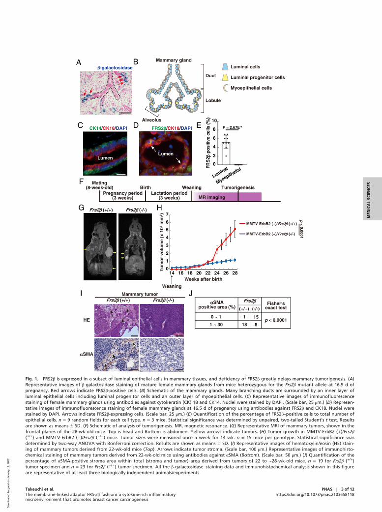
The genotype frequencies of offspring born to the heterozygousbreeding pairs were in agreement with Mendelian ratio (SIAppendix, Fig. S1D). The mutant mice grew normally and werefertile, with no gross abnormalities. Frs2β promoter activity wasdetected by β-galactosidase (LacZ) staining of the heterozygousfor the Frs2β mutant allele (SI Appendix, Fig. S1A). In the adultbrain, the staining was observed in neural cells in cerebral cortexand hippocampus and purkinje cells in cerebellum, for example(SI Appendix, Fig. S1 E and F). Similarly, immunohistochemistrywith anti-FRS2β showed cytoplasmic staining in neural cells inhippocampus, as reported previously (20) (SI Appendix, Fig.S1G). In the mature female mammary tissues, the abundance ofFrs2β transcript increased significantly during pregnancy and lac-tation and then decreased during the involution period afterweaning (SI Appendix, Fig. S1H). LacZ-stained cells wereobserved in several alveolar cells in mammary tissues during lac-tation period (Fig. 1 A and B). Immunohistochemistry revealedthat FRS2β was expressed in a small subset of luminal cells thatare positive for cytokeratin 18, a luminal cell marker (21) (Fig. 1C–E). However, FRS2β was not expressed in myoepithelial cellsthat are positive for cytokeratin 14, a myoepithelial cell marker(Fig. 1 C and E and SI Appendix, Fig. S1I). These data suggestthat a subset of luminal cells in the mammary glandexpress FRS2β.
Whole-mount staining of the mammary gland revealed nogross structural abnormality in the mutant mice (SI Appendix,Fig. S2A). There were no significant differences in number andlength of the mammary tree branches (SI Appendix, Fig. S2 Band C). Although we observed modest reduction of alveolarcells in Frs2β (�/�) mammary tissues during lactation period (SIAppendix, Fig. S2 D and E), the production of milk of Frs2β(�/�) mammary tissues should be sufficient for raising pups,since there were no differences in body weight of offspringborn to either Frs2β (+/+) or Frs2β (�/�) mother mice (SIAppendix, Fig. S2F).
Although Frs2β (�/�) mice have very minor phenotype innormal conditions, we hypothesized that FRS2β is involved inpathological conditions, such as tumorigenesis. To test thishypothesis, we used MMTV-ErbB2 (+) mammary tumor modelmice. We found that the number of FRS2β–positive luminalcells in the mammary gland during lactation period was greaterin MMTV-ErbB2 (+) mice than in MMTV-ErbB2 (�) mice (SIAppendix, Fig. S2 G and H). We crossed Frs2β mutant micewith the MMTV-ErbB2 (+) mice to generate MMTV-ErbB2(+)/Frs2β (+/+), MMTV-ErbB2 (+)/Frs2β (6), and MMTV-ErbB2 (+)/Frs2β (�/�) mice hereafter referred to as Frs2β (+/+),Frs2β (6), and Frsβ (�/�), respectively. We detected palpabletumors in Frs2β (+/+) mice at 23.4 6 1.9 wk after birth, and83% of mice had tumors (n = 8) after experiencing pregnancyand subsequent lactation after the age of 8 wk. On the otherhand, tumors became palpable in virgin Frs2β (+/+) mice at 32.66 2.6 wk, and only 23.4% of mice had tumors (n = 8) (i.e., laterand at a lower probability). Since pregnancy and subsequentlactation seem to be required for efficient tumorigenesis, weanalyzed mice after pregnancy and lactation (Fig. 1F). We usedMRI, which is sufficiently sensitive to detect very small cellmasses, even those with 1-mm diameters (22). We began toobserve small tumors (>5 mm in diameter) 5 to 8 wk after com-mencement of measurement (19 to 22-wk-old mice) (SIAppendix, Fig. S3A). Strikingly, the tumor growth rate wasmuch lower in Frs2β (�/�) mice than in Frs2β (+/+) mice (Fig. 1G and H), although tumor incidence was similar: 83.2% (n =18) in Frs2β (+/+) and 88.2% (n = 17) in Frs2β (�/�). This obser-vation indicates that FRS2β plays important roles in mammarytumorigenesis.
Histological examination revealed that Frs2β (+/+) tumorscontained ample stroma, reminiscent of human breast cancertissues (23) (Fig. 1 I, Upper, and SI Appendix, Fig. S3B). By
2 of 12 j PNAS Takeuchi et al.https://doi.org/10.1073/pnas.2103658118 The membrane-linked adaptor FRS-2β fashions a cytokine-rich inflammatory
microenvironment that promotes breast cancer carcinogenesis
Dow
nloa
ded
by g
uest
on
Janu
ary
11, 2
022
A�-galactosidase
B
Duct
Lobule
Mammary gland
Luminal progenitor cells
Luminal cells
Myoepithelial cells
D FRS2�/CK18/DAPIC
Lumen
ECK14/CK18/DAPI
0
4
6
8
10
FRS
2� p
ositi
vece
lls(%
)
Luminal
Myoepithelial
Lumen
Alveolus
2
P = 2.67E-8
F Mating(8-week-old)
Pregnancy period(3 weeks)
Lactation period(3 weeks)
Birth Weaning
MR imaging
Tumorigenesis
G
0
1
2
3
4
5
6
7
14 16 18 24Weeks after birth
Tu
mo
r vo
lum
e (x
102
mm
3 )
H
I
HE
�SMA
Frs2� (+/+) Frs2� (-/-)
MMTV-ErbB2 (+)/
MMTV-ErbB2 (+)/Frs2� (-/-)
Frs2� (+/+)
JFrs2�
(+/+) (-/-)�SMA
positive area (%)
0 ~ 1
1 ~ 30
Fisher,sexact test
1 15
18 8p < 0.0001
Weaning
20 22 26 28
p < 0.0001
Mammary tumorFrs2� (+/+) Frs2� (-/-)
Fig. 1. FRS2β is expressed in a subset of luminal epithelial cells in mammary tissues, and deficiency of FRS2β greatly delays mammary tumorigenesis. (A)Representative images of β-galactosidase staining of mature female mammary glands from mice heterozygous for the Frs2β mutant allele at 16.5 d ofpregnancy. Red arrows indicate FRS2β-positive cells. (B) Schematic of the mammary glands. Many branching ducts are surrounded by an inner layer ofluminal epithelial cells including luminal progenitor cells and an outer layer of myoepithelial cells. (C) Representative images of immunofluorescencestaining of female mammary glands using antibodies against cytokeratin (CK) 18 and CK14. Nuclei were stained by DAPI. (Scale bar, 25 μm.) (D) Represen-tative images of immunofluorescence staining of female mammary glands at 16.5 d of pregnancy using antibodies against FRS2β and CK18. Nuclei werestained by DAPI. Arrows indicate FRS2β-expressing cells. (Scale bar, 25 μm.) (E) Quantification of the percentage of FRS2β–positive cells to total number ofepithelial cells. n = 9 random fields for each cell type. n = 3 mice. Statistical significance was determined by unpaired, two-tailed Student’s t test. Resultsare shown as means 6 SD. (F) Schematic of analysis of tumorigenesis. MR, magnetic resonance. (G) Representative MRI of mammary tumors, shown in thefrontal planes of the 28-wk-old mice. Top is head and Bottom is abdomen. Yellow arrows indicate tumors. (H) Tumor growth in MMTV-ErbB2 (+)/Frs2β(+/+) and MMTV-ErbB2 (+)/Frs2β (�/�) mice. Tumor sizes were measured once a week for 14 wk. n = 15 mice per genotype. Statistical significance wasdetermined by two-way ANOVA with Bonferroni correction. Results are shown as means 6 SD. (I) Representative images of hematoxylin/eosin (HE) stain-ing of mammary tumors derived from 22-wk-old mice (Top). Arrows indicate tumor stroma. (Scale bar, 100 μm.) Representative images of immunohisto-chemical staining of mammary tumors derived from 22-wk-old mice using antibodies against αSMA (Bottom). (Scale bar, 50 μm.) (J) Quantification of thepercentage of αSMA-positive stroma area within total (stroma and tumor) area derived from tumors of 22 to ∼28-wk-old mice. n = 19 for Frs2β (+/+)tumor specimen and n = 23 for Frs2β (�/�) tumor specimen. All the β-galactosidase–staining data and immunohistochemical analysis shown in this figureare representative of at least three biologically independent animals/experiments.
MED
ICALSC
IENCE
S
Takeuchi et al.The membrane-linked adaptor FRS-2β fashions a cytokine-rich inflammatorymicroenvironment that promotes breast cancer carcinogenesis
PNAS j 3 of 12https://doi.org/10.1073/pnas.2103658118
Dow
nloa
ded
by g
uest
on
Janu
ary
11, 2
022
contrast, very little stroma was observed in Frs2β (�/�) tumors.High levels of smooth muscle actin (SMA)–positive CAFs werepresent in the stroma of Frs2β (+/+) tumors but not in Frs2β(�/�) tumors (Fig. 1 I, Lower and J). The tumor stroma is amajor component of the tumor microenvironment. We nextanalyzed the amount of blood vessels, another important com-ponent of the tumor microenvironment, by measuring theamount of CD31-positive endothelial cells. We found that theamount of CD31-positive endothelial cells was significantlysmaller in Frs2β (�/�) tumors than in Frs2β (+/+) tumors (SIAppendix, Fig. S3 C and D). These results indicate that FRS2βis required for formation of tumor stroma that includes bloodvessels.
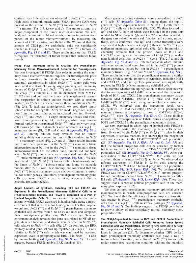
FRS2β Plays Important Roles in Creating the PremalignantMammary Tissue Microenvironment Required for Tumorigenesis.We next investigated if FRS2β plays roles in creating the mam-mary tissue microenvironment required for tumorigenesis priorto tumor formation. To test this hypothesis, we performedxenograft experiments in which Frs2β (+/+) tumor cells wereinoculated into 8-wk-old young virgin premalignant mammarytissues of Frs2β (+/+) and Frs2β (�/�) mice. We first removedthe Frs2β (+/+) tumors (∼1 cm in diameter) from MMTV-ErbB2 mice and cultured the tumor cells as spheres in serum-free suspension in sphere culture medium with a cytokinemixture, as CSCs are enriched under these conditions (24, 25)(Fig. 2A). To facilitate tumorigenesis, we used these tumorsphere cells for xenografts. After we made single-cell suspen-sion from the tumor spheres, we inoculated them into 8-wk-oldFrs2β (+/+) and Frs2β (�/�) virgin mammary tissues and moni-tored tumorigenesis (Fig. 2A). Strikingly, while large tumorsformed rapidly in transplanted Frs2β (+/+) mice, only very smallcell masses (<3 mm in diameter) were observed in Frs2β (�/�)mammary tissues (Fig. 2 B and C and SI Appendix, Fig. S4 Aand B). Limiting dilution assay revealed that no tumor-initiating ability was observed in tumor cells inoculated in Frs2β(�/�) mammary tissues (Fig. 2 D and E). These results indicatethat tumor cells grew well in the Frs2β (+/+) mammary tissuemicroenvironment but not in the Frs2β (�/�) mammary tissuemicroenvironment. On the other hand, tumors did not formwhen Frs2β (+/+) tumor cells were inoculated into the Frs2β(+/+) male mammary fat pads (SI Appendix, Fig. S4C). We alsoinoculated 10,000 Frs2β (+/+) tumor cells subcutaneously intothe flanks of Frs2β (+/+) female mice and found no palpabletumors after 35 d. From these findings, we confirmed that theFrs2β (+/+) female mammary tissue microenvironment is essen-tial for tumorigenesis. Therefore, premalignant mammary glandcells expressing FRS2β create a microenvironment that isessential for tumorigenesis.
Ample Amounts of Cytokines, Including IGF1 and CXCL12, AreExpressed in the Premalignant Mammary Epithelial Cells in anFRS2β-Dependent Manner, and FRS2β Is Expressed in a Part ofLuminal Progenitor Cells. Next, we examined the molecular mech-anisms by which FRS2β expressed in luminal cells create a micro-environment that is essential for tumorigenesis. For this purpose,we cultured Frs2β (+/+) and Frs2β (�/�) premalignant mammaryepithelial cells derived from 8- to 12-wk-old mice and comparedtheir transcriptomic profiles using DNA microarrays. Gene setenrichment analysis revealed that gene sets related to NF-κB tar-gets, stem cell function, and stroma were enriched in Frs2β (+/+)cells relative to Frs2β (�/�) cells (Fig. 2F). In contrast, the ERKpathway–related gene set was up-regulated in Frs2β (�/�) cellsrelative to Frs2β (+/+) cells, which was confirmed by increasedexpression levels of phosphorylated ERK1/2 in Frs2β (�/�) cellsby immunoblotting (SI Appendix, Fig. S4 D and E). This wasexpected because FRS2β inhibits ERK signaling (15).
Many genes encoding cytokines were up-regulated in Frs2β(+/+) cells (SI Appendix, Table S1); among them, the top 10genes at higher expression levels in Frs2β (+/+) cells than inFrs2β (�/�) cells were indicated (Fig. 2G). We then focused onIgf1 and Cxcl12, both of which were included in the gene setsrelated to NF-κB targets. Igf1 and Cxcl12 were also included inthe gene sets related to stem cell function and stroma, respec-tively. qPCR confirmed that Igf1 and Cxcl12 transcripts wereexpressed at higher levels in Frs2β (6) than in Frs2β (�/�) pre-malignant mammary epithelial cells (Fig. 2H). Immunohisto-chemistry revealed that the protein levels of IGF1 andCXCL12 were higher in the cytoplasm of Frs2β (+/+) premalig-nant luminal cells than in Frs2β (�/�) cells (Fig. 2 I–L and SIAppendix, Fig. S5 A and B). Inflamed areas in which immunecells were accumulated were observed in the Frs2β (+/+) prema-lignant mammary tissues but not in Frs2β (�/�) premalignantmammary tissues (Fig. 2 M and N and SI Appendix, Fig. S5C).These results indicate that the premalignant mammary epithe-lial cells produce ample amounts of cytokines, including IGF1and CXCL12, and that cytokine production was significantlyreduced in FRS2β-deficient mammary epithelial cells.
To examine whether the up-regulation of these cytokines wasdue to overexpression of ErbB2, we compared the expressionlevels of IGF1 and CXCL12 in premalignant mammary tissuesbetween MMTV-ErbB2(�)/Frs2β (+/+) mice and MMTV-ErbB2(+)/Frs2β (+/+) mice using immunohistochemistry andqPCR. We observed that the expression levels wereup-regulated in MMTV-ErbB2(+)/Frs2β (+/+) premalignantmammary tissues as compared to those of MMTV-ErbB2(�)/Frs2β (+/+) mice (SI Appendix, Fig. S6 A–C). These findingsindicate that overexpression of ErbB2 causes up-regulation ofIGF1 and CXCL12 in the premalignant mammary tissues.
We next examined in which type of luminal cells FRS2β isexpressed. We sorted the mammary epithelial cells derivedfrom 10-wk-old virgin Frs2β (+/+) or Frs2β (�/�) mice by theirexpression of surface markers. It is known that the progenitorcells are enriched in the CD49flow/CD24high cell population(26) (SI Appendix, Fig. S4 F, Right, P3, and G, Left, P3) andthat the luminal progenitor cells can be enriched by furtherfractionation with CD61 for the CD49flow/CD24high/CD61+
population (27) (SI Appendix, Fig. S4G, Middle, P5). After per-mealization of the CD49flow/CD24high/CD61+ cells, we nextanalyzed them by using anti–FRS2β antibody. We observed sig-nificant expression of FRS2β in 23.6% cells among theCD49flow/CD24+high/CD61+ luminal progenitor cell population(SI Appendix, Fig. S4G, Upper Right, P6). We confirmed thatFRS2β was lost in CD49flow/CD24high/CD61+ luminal progeni-tor cell population derived from Frs2β (�/�) mammary epithe-lial cells (SI Appendix, Fig. S4G, Lower Right, P6). These datasuggest that a subset of luminal progenitor cells in the mam-mary gland express FRS2β.
We then cultured premalignant mammary epithelial cells asmammospheres in which stem/progenitor cells are enriched(28). We found that the efficiency of mammosphere formationwas greater in Frs2β (+/+) premalignant mammary epithelialcells than in Frs2β (�/�) cells in several passages (SI Appendix,Fig. S4 H and I). Our findings suggest that FRS2β contributesto growth ability of macroscopically normal mammary stem/progenitor cells.
The FRS2β-Dependent Increase in IGF1 and CXCL12 Production inPremalignant Mammary Epithelial Cells Promotes Tumor SphereFormation and CAF Mobilization. Tumor sphere formation reflectsthe properties of CSCs, whose growth is dependent on cyto-kines in the culture (24). To determine whether IGF1 derivedfrom premalignant mammary epithelial cells plays a role intumor sphere formation, we cultured Frs2β (+/+) tumor cellsunder serum-free suspension condition without the standard
4 of 12 j PNAS Takeuchi et al.https://doi.org/10.1073/pnas.2103658118 The membrane-linked adaptor FRS-2β fashions a cytokine-rich inflammatory
microenvironment that promotes breast cancer carcinogenesis
Dow
nloa
ded
by g
uest
on
Janu
ary
11, 2
022
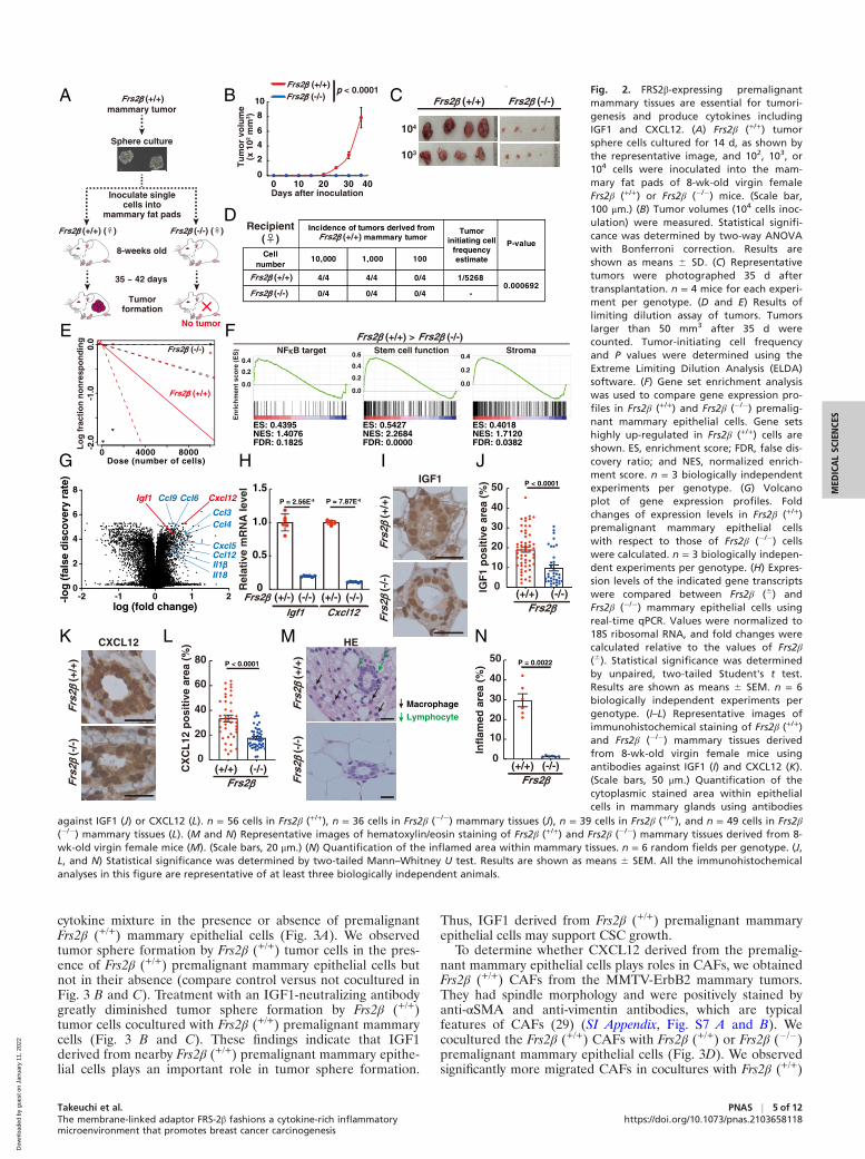
cytokine mixture in the presence or absence of premalignantFrs2β (+/+) mammary epithelial cells (Fig. 3A). We observedtumor sphere formation by Frs2β (+/+) tumor cells in the pres-ence of Frs2β (+/+) premalignant mammary epithelial cells butnot in their absence (compare control versus not cocultured inFig. 3 B and C). Treatment with an IGF1-neutralizing antibodygreatly diminished tumor sphere formation by Frs2β (+/+)tumor cells cocultured with Frs2β (+/+) premalignant mammarycells (Fig. 3 B and C). These findings indicate that IGF1derived from nearby Frs2β (+/+) premalignant mammary epithe-lial cells plays an important role in tumor sphere formation.
Thus, IGF1 derived from Frs2β (+/+) premalignant mammaryepithelial cells may support CSC growth.
To determine whether CXCL12 derived from the premalig-nant mammary epithelial cells plays roles in CAFs, we obtainedFrs2β (+/+) CAFs from the MMTV-ErbB2 mammary tumors.They had spindle morphology and were positively stained byanti-αSMA and anti-vimentin antibodies, which are typicalfeatures of CAFs (29) (SI Appendix, Fig. S7 A and B). Wecocultured the Frs2β (+/+) CAFs with Frs2β (+/+) or Frs2β (�/�)premalignant mammary epithelial cells (Fig. 3D). We observedsignificantly more migrated CAFs in cocultures with Frs2β (+/+)
A B
0
2
4
6
8
Tu
mo
r vo
lum
e(x
102
mm
3 )
10
0 10 20 30 40Days after inoculation
Frs2� (-/-)Frs2� (+/+)
D
mammary tumor
Sphere culture
Inoculate singlecells into
mammary fat pads
Frs2� (+/+)
Frs2� (+/+) (♀) Frs2� (-/-) (♀)
8-weeks old
35 ~ 42 days
Tumorformation
Lo
g f
ract
ion
no
nre
spo
nd
ing
0.0
-1.0
-2.0
0 4000 8000Dose (number of cells)
EFrs2� (-/-)
Frs2� (+/+)
FNF�B target
0.0
0.2
0.4
En
rich
men
t sc
ore
(E
S)
ES: 0.4395NES: 1.4076FDR: 0.1825
Stem cell function
ES: 0.5427NES: 2.2684FDR: 0.0000
Stroma
ES: 0.4018NES: 1.7120FDR: 0.0382
Frs2� (-/-)Frs2� (+/+) >
0.0
0.2
0.4
0.0
0.2
0.4
0.6
P = 2.56E-8
H
Frs2� (+/-) (-/-) (+/-) (-/-)Igf1 Cxcl12
Rel
ativ
e m
RN
A le
vel
0
0.5
1.0
1.5P = 7.87E-8
IGF1
CXCL12
Frs2�
(+/+
)Fr
s2�
(-/-
)
I J
K
Frs2�
(+/+
)Fr
s2�
(-/-
)
L HEM
Frs2�
(+/+
)Fr
s2�
(-/-
)
N
Frs2� (+/+) Frs2� (-/-)
104
103
Cp < 0.0001
No tumor
Cellnumber
10,000 1,000 100
4/4 4/4 0/4 1/5268
0/4 0/4 0/4 -
Tumorinitiating cell
frequencyestimate
P-value
0.000692
Recipient(♀)
Incidence of tumors derived from mammary tumorFrs2� (+/+)
IGF
1 p
osi
tive
are
a (%
)
0
10
20
30
40
50
Frs2�(+/+) (-/-)
P < 0.0001
CX
CL
12 p
osi
tive
are
a (%
)
0
20
40
60
80
Frs2�(+/+) (-/-)
P < 0.0001
Infl
amed
are
a (%
)
0
10
20
30
40
50
Frs2�(+/+) (-/-)
P = 0.0022
MacrophageLymphocyte
Frs2� (+/+)
Frs2� (-/-)
-2 -1 0 1 20
2
4
6
8
log (fold change)
-lo
g (
fals
e d
isco
very
rate
)
Cxcl12Ccl9
Ccl3Ccl4
Ccl6Igf1
Cxcl5Ccl12
Il18
G
Il1�
Fig. 2. FRS2β-expressing premalignantmammary tissues are essential for tumori-genesis and produce cytokines includingIGF1 and CXCL12. (A) Frs2β (+/+) tumorsphere cells cultured for 14 d, as shown bythe representative image, and 102, 103, or104 cells were inoculated into the mam-mary fat pads of 8-wk-old virgin femaleFrs2β (+/+) or Frs2β (�/�) mice. (Scale bar,100 μm.) (B) Tumor volumes (104 cells inoc-ulation) were measured. Statistical signifi-cance was determined by two-way ANOVAwith Bonferroni correction. Results areshown as means 6 SD. (C) Representativetumors were photographed 35 d aftertransplantation. n = 4 mice for each experi-ment per genotype. (D and E) Results oflimiting dilution assay of tumors. Tumorslarger than 50 mm3 after 35 d werecounted. Tumor-initiating cell frequencyand P values were determined using theExtreme Limiting Dilution Analysis (ELDA)software. (F) Gene set enrichment analysiswas used to compare gene expression pro-files in Frs2β (+/+) and Frs2β (�/�) premalig-nant mammary epithelial cells. Gene setshighly up-regulated in Frs2β (+/+) cells areshown. ES, enrichment score; FDR, false dis-covery ratio; and NES, normalized enrich-ment score. n = 3 biologically independentexperiments per genotype. (G) Volcanoplot of gene expression profiles. Foldchanges of expression levels in Frs2β (+/+)premalignant mammary epithelial cellswith respect to those of Frs2β (�/�) cellswere calculated. n = 3 biologically indepen-dent experiments per genotype. (H) Expres-sion levels of the indicated gene transcriptswere compared between Frs2β (6) andFrs2β (�/�) mammary epithelial cells usingreal-time qPCR. Values were normalized to18S ribosomal RNA, and fold changes werecalculated relative to the values of Frs2β(6). Statistical significance was determinedby unpaired, two-tailed Student's t test.Results are shown as means 6 SEM. n = 6biologically independent experiments pergenotype. (I–L) Representative images ofimmunohistochemical staining of Frs2β (+/+)and Frs2β (�/�) mammary tissues derivedfrom 8-wk-old virgin female mice usingantibodies against IGF1 (I) and CXCL12 (K).(Scale bars, 50 μm.) Quantification of thecytoplasmic stained area within epithelialcells in mammary glands using antibodies
against IGF1 (J) or CXCL12 (L). n = 56 cells in Frs2β (+/+), n = 36 cells in Frs2β (�/�) mammary tissues (J), n = 39 cells in Frs2β (+/+), and n = 49 cells in Frs2β(�/�) mammary tissues (L). (M and N) Representative images of hematoxylin/eosin staining of Frs2β (+/+) and Frs2β (�/�) mammary tissues derived from 8-wk-old virgin female mice (M). (Scale bars, 20 μm.) (N) Quantification of the inflamed area within mammary tissues. n = 6 random fields per genotype. (J,L, and N) Statistical significance was determined by two-tailed Mann–Whitney U test. Results are shown as means 6 SEM. All the immunohistochemicalanalyses in this figure are representative of at least three biologically independent animals.
MED
ICALSC
IENCE
S
Takeuchi et al.The membrane-linked adaptor FRS-2β fashions a cytokine-rich inflammatorymicroenvironment that promotes breast cancer carcinogenesis
PNAS j 5 of 12https://doi.org/10.1073/pnas.2103658118
Dow
nloa
ded
by g
uest
on
Janu
ary
11, 2
022
premalignant mammary epithelial cells than in those with Frs2β(�/�) cells (Fig. 3 E and F). CXCL12 binds to its receptorsCXCR4 and CXCR7 (30). Although we did not observe signifi-cant effects on the migration of CAFs upon treatment with theCXCR4 inhibitor AMD3100 (100 ng/mL) or the CXCR7 inhibi-tor CCX771 (100 ng/mL) alone (SI Appendix, Fig. S7C), a com-bination of both inhibitors greatly diminished migration of CAFsin a CCX771 dose-dependent manner in the presence of theeffective dose of AMD3100 (Fig. 3 G and H and SI Appendix,Fig. S7D). Furthermore, we observed increased migration ofCAFs by addition of recombinant CXCL12 in the culture mediain place of Frs2β (+/+) premalignant mammary epithelial cells(SI Appendix, Fig. S7 E and F). These findings indicate thatCXCL12 derived from nearby Frs2β (+/+) premalignant mam-mary epithelial cells contributes to mobilization of CAFs. Takentogether, the FRS2β–dependent increases in production of
cytokines, including IGF1 and CXCL12, enable premalignantmammary cells to maintain CSC and mobilized CAF at the onsetof tumorigenesis.
FRS2β Triggers Activation of NF-κB to Promote IGF1 and CXCL12Production in Premalignant Mammary Epithelial Cells. Next, weexamined the molecular mechanisms that induce expression ofIGF1 and CXCL2 in premalignant mammary tissues. Since Igf1and Cxcl12 were among list of the identified NF-κB target gene(Fig. 2F), we investigated if the activation of NF-κB is involvedin the production of these cytokines. To this end, we culturedFrs-2β (+/+) premalignant mammary epithelial cells and treatedthem with DHMEQ, a specific inhibitor of NF-κB (Fig. 4A).Treatment with DHMEQ inhibited the expression of Igf1,Cxcl12, and IκBα, a well-established NF-κB–inducible gene, ina dose-dependent manner (Fig. 4B). We further confirmed
A
IGF1
mammaryepithelial cells
+ IGF1 NAb
BNot co-culturedControl IGF1 NAb
Co-cultured with mammary epithelial cells
C
tumor sphere
CXCL12
CAFs
or mammary epithelial cells
CAFs co-cultured with
mammaryepithelial cells
D E
G Migrated CAFs
H
Frs2� (+/+)Frs2� (+/+)
Frs2� (-/-)
Frs2� (+/+)Frs2� (+/+)
P = 0.018
0
0.1
Sp
her
e fo
rmin
gef
fici
ency
(%
)
0.2
Notco-culturedControl IGF1
NAb
P = 0.00088
0
100
200
300
Nu
mb
er o
f m
igra
ted
CA
Fs
Mammaryepithelial cells
(-/-)Frs2� (+/+)
P = 0.00093
F
P = 0.014
0
100
200
300
Nu
mb
er o
f m
igra
ted
CA
Fs
CCX771AMD3100
0100
3010060
100 10090
(ng/mL)
P = 0.002
120 150100 100
P = 0.004
100180
P = 0.0001P = 0.0001
tumor cells
mammaryepithelial cells
CCX771AMD3100
0100
60100
180100
(ng/mL)
Frs2� (+/+)
Frs2� (+/+)
Frs2� (+/+)
Frs2� (+/+) Frs2� (-/-)
Fig. 3. IGF1 and CXCL12 produced by pre-malignant Frs2β (+/+) mammary epithelialcells induce tumor spheres and migrationof CAFs, respectively. (A–C) Schematic ofcoculture of Frs2β (+/+) tumor cells to formspheres in the lower chamber with Frs2β(+/+) premalignant mammary epithelial cellsin the upper chamber (A). (B) Representa-tive images of tumor sphere formation inthe presence of Frs2β (+/+) mammary epi-thelial cells treated with control IgG (400nM) (control) or IGF1-neutralizing antibody(NAb) (400 nM) after 7 d of coculture or inthe absence of Frs2β (+/+) mammary epithe-lial cells after 7 d of culture. (Scale bars,100 μm.) (C) Quantification of tumorsphere–forming efficiency. n = 3 mice pergroup. The results are representative of thefour independent experiments. Statisticalsignificance was determined by unpaired,two-tailed Student’s t test. Results areshown as means 6 SD. (D–F) Schematic ofcoculture of Frs2β (+/+) or Frs2β (�/�) prema-lignant mammary epithelial cells in thelower chamber with Frs2β (+/+) CAFs in theupper chamber (D). (E) Representativeimages of migrated Frs2β (+/+) CAFs cocul-tured with Frs2β (+/+) or Frs2β (�/�) mam-mary cells after 24 h of coculture. Arrowsindicate migrated CAFs. (Scale bar, 20 μm.)(F) Quantification of migrated Frs2β (+/+) orFrs2β (�/�) CAFs. n = 3 mice per genotype.The results are representative of the fourindependent experiments. Statistical signifi-cance was determined by unpaired, two-tailed Student’s t test. Results are shown asmeans 6 SD. (G and H) Representativeimages of migrated Frs2β (+/+) CAFs cocul-tured with Frs2β (+/+) mammary epithelialcells for 24 h. Cells were treated with theindicated concentration of CCX71 and 100ng/mL AMD3100 (G). (Scale bar, 100 μm.) (H)Quantification of migrated Frs2β (+/+) CAFscocultured with Frs2β (+/+) mammary epithe-lial cells for 24 h. Cells were treated withthe indicated concentration of CCX71 and100 μg/mL AMD3100. n = 3 mice per group.The results are representative of the fourindependent experiments. Statistical signifi-cance was determined by unpaired, two-tailed Student's t test. Results are shown asmeans 6 SD.
6 of 12 j PNAS Takeuchi et al.https://doi.org/10.1073/pnas.2103658118 The membrane-linked adaptor FRS-2β fashions a cytokine-rich inflammatory
microenvironment that promotes breast cancer carcinogenesis
Dow
nloa
ded
by g
uest
on
Janu
ary
11, 2
022
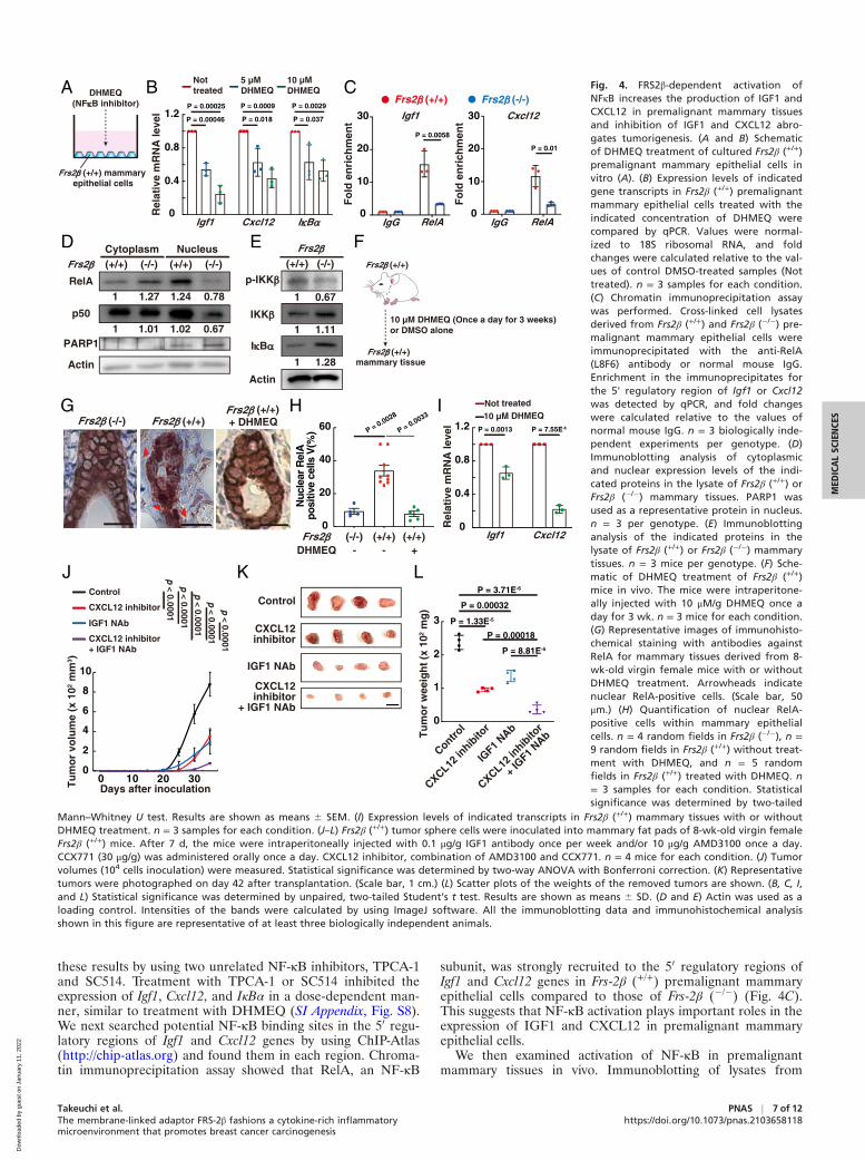
these results by using two unrelated NF-κB inhibitors, TPCA-1and SC514. Treatment with TPCA-1 or SC514 inhibited theexpression of Igf1, Cxcl12, and IκBα in a dose-dependent man-ner, similar to treatment with DHMEQ (SI Appendix, Fig. S8).We next searched potential NF-κB binding sites in the 50 regu-latory regions of Igf1 and Cxcl12 genes by using ChIP-Atlas(http://chip-atlas.org) and found them in each region. Chroma-tin immunoprecipitation assay showed that RelA, an NF-κB
subunit, was strongly recruited to the 50 regulatory regions ofIgf1 and Cxcl12 genes in Frs-2β (+/+) premalignant mammaryepithelial cells compared to those of Frs-2β (�/�) (Fig. 4C).This suggests that NF-κB activation plays important roles in theexpression of IGF1 and CXCL12 in premalignant mammaryepithelial cells.
We then examined activation of NF-κB in premalignantmammary tissues in vivo. Immunoblotting of lysates from
P = 0.0013
A DHMEQ(NF�B inhibitor)
0
0.8
1.2
0.4
I�B�Igf1 Cxcl12
Rel
ativ
e m
RN
A le
vel
B
RelA
p50
PARP1
Actin
Frs2� (+/+) (-/-) (+/+) (-/-)Cytoplasm NucleusD E
G
0
0.8
1.2
0.4
Rel
ativ
e m
RN
A le
vel
Igf1 Cxcl12
Frs2� (-/-) Frs2� (+/+)Frs2� (+/+)+ DHMEQ
epithelial cellsFrs2� (+/+) mammary
P = 0.00046
P = 0.00025
P = 0.018
P = 0.0009
P = 0.037
P = 0.0029
Frs2� (+/+)
F
10 µM DHMEQ (Once a day for 3 weeks)or DMSO alone
mammary tissue
P = 7.55E-6
0
2
4
6
8
Tu
mo
r vo
lum
e (x
102
mm
3 )
10
0 10 20 30Days after inoculation
Control
CXCL12 inhibitor
IGF1 NAb
CXCL12 inhibitor+ IGF1 NAb
Control
CXCL12inhibitor
IGF1 NAb
CXCL12inhibitor
+ IGF1 NAb
J
I
P = 1.33E-5
0
1
2
Tu
mo
r w
eeig
ht
(x 1
02 m
g)
3
Control
CXCL12 in
hibito
r
IGF1 N
Ab
CXCL12 in
hibito
r
+ IG
F1 NAb
P = 0.00032
P = 3.71E-6
K
1 1.27 1.24 0.78
1 1.01 1.02 0.67
Not treated10 µM DHMEQ
L
Nottreated
5 µMDHMEQ
10 µMDHMEQ
p-IKK�
IKK�
I�B�
Actin
Frs2�(+/+) (-/-)
1 0.67
1 1.11
1 1.28Frs2� (+/+)
H
P = 0.00018
P = 8.81E-5
p < 0.0001
p < 0.0001
p < 0.0001
p < 0.0001
p < 0.0001
40
20
Nuc
lear
Rel
Apo
sitiv
ece
llsV
(%)
0
60
Frs2� (-/-) (+/+) (+/+)DHMEQ - +-
P = 0.0028
P = 0.0033
Frs2� (-/-)Frs2� (+/+)
Fo
ld e
nri
chm
ent
0
30
20
10
RelAIgG
Fo
ld e
nri
chm
ent
0
30
20
10
RelAIgG
Igf1 Cxcl12
P = 0.0058
P = 0.01
C Fig. 4. FRS2β-dependent activation ofNFκB increases the production of IGF1 andCXCL12 in premalignant mammary tissuesand inhibition of IGF1 and CXCL12 abro-gates tumorigenesis. (A and B) Schematicof DHMEQ treatment of cultured Frs2β (+/+)premalignant mammary epithelial cells invitro (A). (B) Expression levels of indicatedgene transcripts in Frs2β (+/+) premalignantmammary epithelial cells treated with theindicated concentration of DHMEQ werecompared by qPCR. Values were normal-ized to 18S ribosomal RNA, and foldchanges were calculated relative to the val-ues of control DMSO-treated samples (Nottreated). n = 3 samples for each condition.(C) Chromatin immunoprecipitation assaywas performed. Cross-linked cell lysatesderived from Frs2β (+/+) and Frs2β (�/�) pre-malignant mammary epithelial cells wereimmunoprecipitated with the anti-RelA(L8F6) antibody or normal mouse IgG.Enrichment in the immunoprecipitates forthe 5' regulatory region of Igf1 or Cxcl12was detected by qPCR, and fold changeswere calculated relative to the values ofnormal mouse IgG. n = 3 biologically inde-pendent experiments per genotype. (D)Immunoblotting analysis of cytoplasmicand nuclear expression levels of the indi-cated proteins in the lysate of Frs2β (+/+) orFrs2β (�/�) mammary tissues. PARP1 wasused as a representative protein in nucleus.n = 3 per genotype. (E) Immunoblottinganalysis of the indicated proteins in thelysate of Frs2β (+/+) or Frs2β (�/�) mammarytissues. n = 3 mice per genotype. (F) Sche-matic of DHMEQ treatment of Frs2β (+/+)mice in vivo. The mice were intraperitone-ally injected with 10 μM/g DHMEQ once aday for 3 wk. n = 3 mice for each condition.(G) Representative images of immunohisto-chemical staining with antibodies againstRelA for mammary tissues derived from 8-wk-old virgin female mice with or withoutDHMEQ treatment. Arrowheads indicatenuclear RelA-positive cells. (Scale bar, 50μm.) (H) Quantification of nuclear RelA-positive cells within mammary epithelialcells. n = 4 random fields in Frs2β (�/�), n =9 random fields in Frs2β (+/+) without treat-ment with DHMEQ, and n = 5 randomfields in Frs2β (+/+) treated with DHMEQ. n= 3 samples for each condition. Statisticalsignificance was determined by two-tailed
Mann–Whitney U test. Results are shown as means 6 SEM. (I) Expression levels of indicated transcripts in Frs2β (+/+) mammary tissues with or withoutDHMEQ treatment. n = 3 samples for each condition. (J–L) Frs2β (+/+) tumor sphere cells were inoculated into mammary fat pads of 8-wk-old virgin femaleFrs2β (+/+) mice. After 7 d, the mice were intraperitoneally injected with 0.1 μg/g IGF1 antibody once per week and/or 10 μg/g AMD3100 once a day.CCX771 (30 μg/g) was administered orally once a day. CXCL12 inhibitor, combination of AMD3100 and CCX771. n = 4 mice for each condition. (J) Tumorvolumes (104 cells inoculation) were measured. Statistical significance was determined by two-way ANOVA with Bonferroni correction. (K) Representativetumors were photographed on day 42 after transplantation. (Scale bar, 1 cm.) (L) Scatter plots of the weights of the removed tumors are shown. (B, C, I,and L) Statistical significance was determined by unpaired, two-tailed Student’s t test. Results are shown as means 6 SD. (D and E) Actin was used as aloading control. Intensities of the bands were calculated by using ImageJ software. All the immunoblotting data and immunohistochemical analysisshown in this figure are representative of at least three biologically independent animals.
MED
ICALSC
IENCE
S
Takeuchi et al.The membrane-linked adaptor FRS-2β fashions a cytokine-rich inflammatorymicroenvironment that promotes breast cancer carcinogenesis
PNAS j 7 of 12https://doi.org/10.1073/pnas.2103658118
Dow
nloa
ded
by g
uest
on
Janu
ary
11, 2
022
premalignant mammary tissues revealed that higher amounts ofthe NFκB components RelA and p50 in the nucleus, a higherlevel of phosphorylated IKK-β, and a lower level of IκBα inFrs2β (+/+) tissues relative to Frs-2β (�/�) tissues (Fig. 4 D andE). Moreover, immunohistochemistry revealed that RelA waslocalized to the nucleus in a much greater proportion of Frs2β(+/+) than Frs2β (�/�) premalignant luminal cells (Fig. 4 G, Leftand Middle and H; compare values indicated by left bar versusthose of middle bar). Treatment with DHMEQ in vivo dramati-cally decreased the number of Frs2β (+/+) premalignant luminalcells harboring RelA in the nucleus (Fig. 4 F, G, Right, and H;compare Frs2β (+/+) and DHMEQ � versus Frs2β (+/+) andDHMEQ +) and inhibited expression of Igf1 and Cxcl12 tran-scripts in premalignant mammary tissues (Fig. 4I). Theseresults suggest that NF-κB activation in luminal cells playsimportant roles in the expression of IGF1 and CXCL12 in pre-malignant mammary epithelial cells in vivo. It thus appears thatFRS-2β triggers activation of NF-κB in the premalignant lumi-nal cells, thereby inducing the production of cytokines includingIGF1 and CXCL12 that in turn activate NF-κB in an autocrineand/or paracrine manner to spread the effects of NF-κB activa-tion to the surrounding mammary epithelial cells.
IGF1 and CXCL12 Expressed in the Premalignant MammaryEpithelial Cells Play Key Roles in Tumorigenesis. To determinewhether IGF1 and CXCL12 expressed in premalignant mam-mary epithelial cells contribute to tumorigenesis, we treatedFrs2β (+/+) xenograft mice with the IGF1-neutralizing antibodyor a combination of CXCR4 and CXCR7 inhibitors, whichblock both the receptors for CXCL12 (hereinafter called“CXCL12 inhibitor”). Treatment with either the IGF1-neutralizing antibody or the CXC12 inhibitor significantlysuppressed tumorigenesis and combined treatment with theIGF1-neutralizing antibody, and the CXC12 inhibitor yieldedthe strongest suppression of tumor volume and weight (Fig. 4I–L). We used effective doses of the inhibitors that were welltolerated without toxic effects, as previously reported (31, 32).These results indicate that the production of IGF1 andCXCL12 in premalignant mammary tissues creates a microen-vironment that is necessary for tumorigenesis.
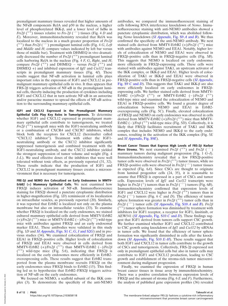
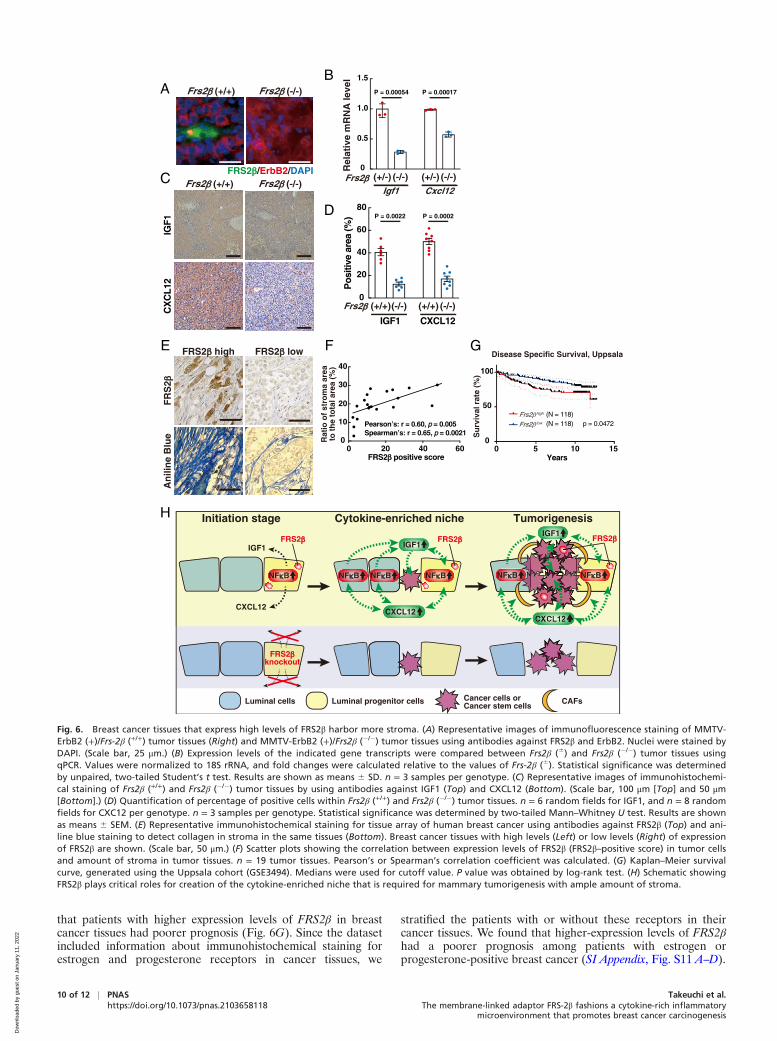
FRE-2β and NEMO Are Colocalized on Early Endosomes in MMTV-ErbB2 (+) Mammary Epithelial Cells. We next examined howFRS2β induces activation of NF-κB. Immunofluorescencestaining for FRS2β showed a punctate cytoplasmic distributionin higher-magnification (Fig. 1D) reminiscent of its localizationon intracellular vesicles, as previously reported (20). Similarly,it was reported that ErbB2 is localized not only on the plasmamembrane but also on intracellular vesicles (33). To examinewhether FRS2β is localized on the early endosomes, we stainedcultured mammary epithelial cells derived from MMTV-ErbB2(+)/Frs2β (+/+) mice or MMTV-ErbB2 (�)/Frs2β (+/+) wild-typemice with antibodies against FRS2β and an early endosomalmarker EEA1. These antibodies were validated in this study(Fig. 1D and SI Appendix, Figs. S1 C, G, I and S2G) and in pre-vious studies (34). We examined colocalization of FRS2β andEEA1 in FRS2β–positive cells. Higher levels of colocalizationof FRS2β and EEA1 were observed in cells derived fromMMTV-ErbB2 (+)/Frs2β (+/+) than MMTV-ErbB2 (�)/Frs2β(+/+) wild-type mice (Fig. 5A), indicating that FRS2β islocalized on the early endosomes more efficiently in ErbB2-overexpressing cells. These results suggest that ErbB2 trans-ported from the plasma membrane recruits FRS2β on theearly endosomes since FRS2β binds to ErbB2 (15). This find-ing led us to hypothesize that ErbB2–FRS2β triggers activa-tion of NF-κB on the early endosomes.
We focused on NEMO, a scaffold subunit of the IKK com-plex (3). To demonstrate the specificity of the anti-NEMO
antibodies, we compared the immunofluorescent staining ofcells following RNA interference knockdown of Nemo. Immu-nofluorescence staining with anti-NEMO antibodies showed apunctate cytoplasmic distribution, which was abolished follow-ing Nemo knockdown (SI Appendix, Fig. S9 A and B). We thusconfirmed the specificity of the anti-NEMO antibody. We nextstained cells derived from MMTV-ErbB2 (+)/Frs2β (+/+) micewith antibodies against NEMO and EEA1. Notably, higher lev-els of colocalization of NEMO and EEA1 were observed inFRS2β–positive cells than in FRS2β-negative cells (Fig. 5B).This suggests that NEMO is localized on early endosomesmore efficiently in FRS2β-expressing cells. These cells werestained with antibodies against TAK1, an upstream activator ofthe IKK complex, or IKK-β and EEA1. Higher levels of coloc-alization of TAK1 or IKK-β and EEA1 were observed inFRS2β-positive cells than in FRS2β-negative cells (SI Appendix,Fig. S9 C and D). This suggests that TAK1 and IKK-β are alsomore efficiently localized on early endosomes in FRS2β-expressing cells. We further stained cells derived from MMTV-ErbB2 (+)/Frs2β (+/+) or MMTV-ErbB2 (�)/Frs2β (+/+)wild-type mice and examined the colocalization of NEMO andEEA1 in FRS2β–positive cells. We found a greater degree ofcolocalization between NEMO and EEA1 in ErbB2-overexpressing cells (Fig. 5C). Finally, elevated colocalizationof FRS2β and NEMO on early endosomes was observed in cellsderived from MMTV-ErbB2 (+)/Frs2β (+/+) mice than MMTV-ErbB2 (�)/Frsβ (+/+) wild-type mice (Fig. 5D). Collectively, it islikely that FRS2β facilitates recruitment of TAK1 and IKKcomplex that includes NEMO and IKK-β to the early endo-somes, resulting in the activation of the IKK complex (Fig. 5Eand SI Appendix, Fig. S9E).
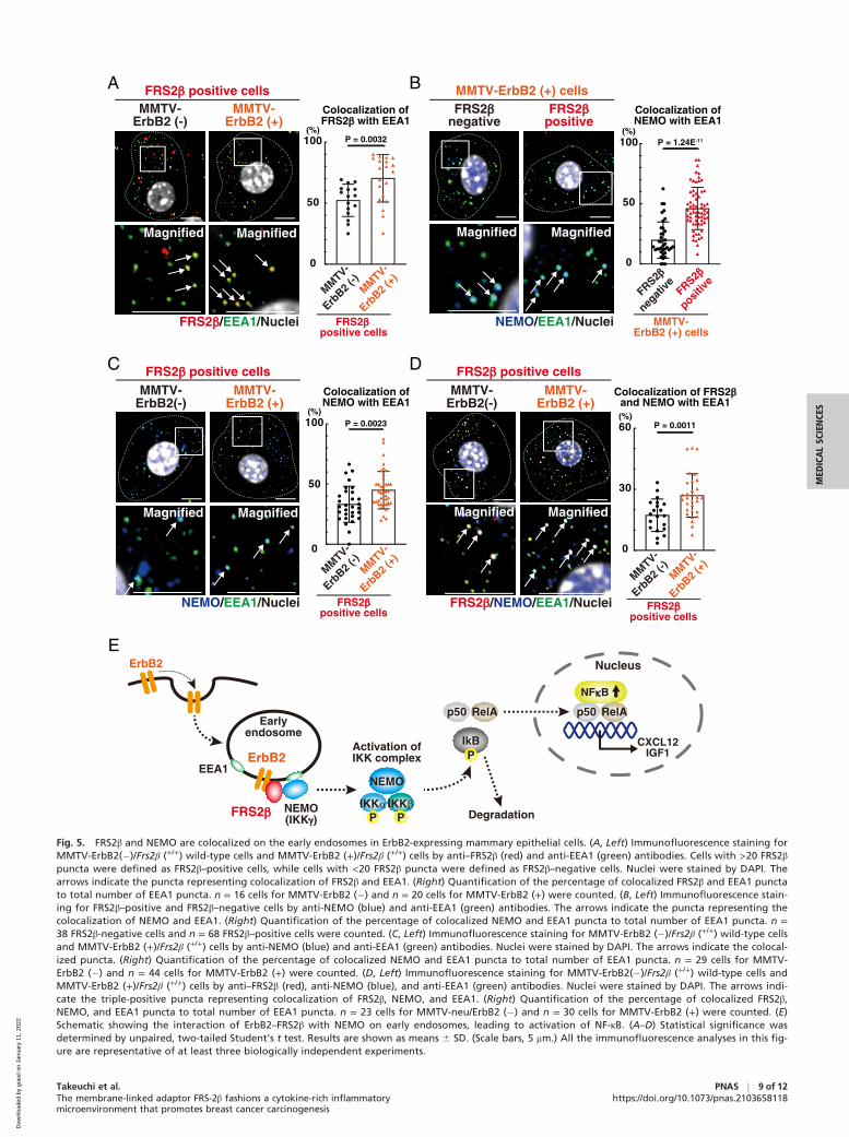
Breast Cancer Tissues that Express High Levels of FRS-2β HarborMore Stroma. We next examined Frs2β (+/+) and Frs2β (�/�)mammary tumors during malignant progression (Fig. 1 G–I).Immunohistochemistry revealed that a few FRS2β-positivetumor cells were observed in Frs2β (+/+) tumor tissues, while noFRS2β–positive cells were observed in Frs2β (�/�) tumor tissuesas expected (Fig. 6A). Since CSCs are thought to originatefrom luminal progenitor cells (24, 35), it is reasonable toassume that FRS2β is expressed in a part of CSCs and tumorcells. Expression levels of Igf1 and Cxcl12 transcripts werehigher in Frs2β (6) tumors than in Frs2β (�/�) tumors (Fig. 6B).Immunohistochemistry confirmed that expression levels ofIGF1 and CXCL12 were higher in Frs2β (+/+) tumors than inFrs2β (�/�) tumors (Fig. 6 C and D). The efficiency of tumorsphere formation was greater in Frs2β (+/+) tumor cells than inFrs2β (�/�) tumor cells (SI Appendix, Fig. S10 A and B). Frs2β(+/+) tumor sphere formation was diminished by treatment withinhibitors for IGF1 receptor, a receptor for IGF1, linsitinib, orAEW541 (SI Appendix, Fig. S10 C and D). These findings sug-gest that IGF1 derived from tumors cells support CSC growth.We further examined whether IGF1 and CXCL12 contributedto CSC growth using knockdown of Igf1 and Cxcl12 by siRNAsin tumor cells. We found that the efficiency of tumor sphereformation was significantly diminished in cells after the knock-down (SI Appendix, Fig. S10 E–H). These results suggest thatboth IGF1 and CXCL12 in tumor cells contribute to the growthof CSCs and tumorigenesis. Collectively, FRS-2β expressed notonly in premalignant epithelial cells but also in tumor cells maycontribute to IGF1 and CXCL12 production, leading to CSCgrowth and establishment of the stroma-rich tumor microenvi-ronment during malignant progression.
Finally, we examined the expression of FRS2β in humanbreast cancer tissues in tissue array by immunohistochemistry.There was a positive correlation between expression levels ofFRS2β and the amount of stroma (Fig. 6 E and F). Furthermore,the analysis of published gene expression profiles (36) revealed
8 of 12 j PNAS Takeuchi et al.https://doi.org/10.1073/pnas.2103658118 The membrane-linked adaptor FRS-2β fashions a cytokine-rich inflammatory
microenvironment that promotes breast cancer carcinogenesis
Dow
nloa
ded
by g
uest
on
Janu
ary
11, 2
022
Magnified Magnified
FRS2�/EEA1/Nuclei
MMTV-ErbB2 (-)
MMTV-ErbB2 (+)
FRS2� positive cellsA B
NEMO/EEA1/Nuclei
MMTV-ErbB2 (+) cells
Magnified Magnified
Colocalization ofNEMO with EEA1
FRS2�negative
FRS2�positive
C
Magnified Magnified
Colocalization ofNEMO with EEA1
0
50
100 P = 0.0023
MMTV-ErbB2(-)
MMTV-ErbB2 (+)
FRS2� positive cells
NEMO/EEA1/Nuclei FRS2�positive cells
FRS2�/NEMO/EEA1/Nuclei
Magnified Magnified
D
Colocalization of FRS2�and NEMO with EEA1
0
30
60 P = 0.0011
MMTV-ErbB2(-)
MMTV-ErbB2 (+)
FRS2� positive cells
ErbB2
FRS2�
Earlyendosome
EEA1
NEMO(IKK�)
NEMO
IKK� IKK�
Activation ofIKK complex
IkB
p50 RelA
P
Degradation
Nucleus
NF�B
CXCL12IGF1
P P
E
p50 RelA
Colocalization ofFRS2� with EEA1
MMTV-
ErbB2 (
+)MMTV-
ErbB2 (
-)0
50
100 P = 0.0032(%)
0
50
100 P = 1.24E-11
MMTV-ErbB2 (+) cells
FRS2�
negat
ive FRS2�
positive
(%)
(%) (%)
MMTV-
ErbB2 (
+)MMTV-
ErbB2 (
-)
FRS2��positive cells
FRS2�positive cells
MMTV-
ErbB2 (
+)MMTV-
ErbB2 (
-)
ErbB2
Fig. 5. FRS2β and NEMO are colocalized on the early endosomes in ErbB2-expressing mammary epithelial cells. (A, Left) Immunofluorescence staining forMMTV-ErbB2(�)/Frs2β (+/+) wild-type cells and MMTV-ErbB2 (+)/Frs2β (+/+) cells by anti–FRS2β (red) and anti-EEA1 (green) antibodies. Cells with >20 FRS2βpuncta were defined as FRS2β–positive cells, while cells with <20 FRS2β puncta were defined as FRS2β–negative cells. Nuclei were stained by DAPI. Thearrows indicate the puncta representing colocalization of FRS2β and EEA1. (Right) Quantification of the percentage of colocalized FRS2β and EEA1 punctato total number of EEA1 puncta. n = 16 cells for MMTV-ErbB2 (�) and n = 20 cells for MMTV-ErbB2 (+) were counted. (B, Left) Immunofluorescence stain-ing for FRS2β–positive and FRS2β–negative cells by anti-NEMO (blue) and anti-EEA1 (green) antibodies. The arrows indicate the puncta representing thecolocalization of NEMO and EEA1. (Right) Quantification of the percentage of colocalized NEMO and EEA1 puncta to total number of EEA1 puncta. n =38 FRS2β-negative cells and n = 68 FRS2β–positive cells were counted. (C, Left) Immunofluorescence staining for MMTV-ErbB2 (�)/Frs2β (+/+) wild-type cellsand MMTV-ErbB2 (+)/Frs2β (+/+) cells by anti-NEMO (blue) and anti-EEA1 (green) antibodies. Nuclei were stained by DAPI. The arrows indicate the colocal-ized puncta. (Right) Quantification of the percentage of colocalized NEMO and EEA1 puncta to total number of EEA1 puncta. n = 29 cells for MMTV-ErbB2 (�) and n = 44 cells for MMTV-ErbB2 (+) were counted. (D, Left) Immunofluorescence staining for MMTV-ErbB2(�)/Frs2β (+/+) wild-type cells andMMTV-ErbB2 (+)/Frs2β (+/+) cells by anti–FRS2β (red), anti-NEMO (blue), and anti-EEA1 (green) antibodies. Nuclei were stained by DAPI. The arrows indi-cate the triple-positive puncta representing colocalization of FRS2β, NEMO, and EEA1. (Right) Quantification of the percentage of colocalized FRS2β,NEMO, and EEA1 puncta to total number of EEA1 puncta. n = 23 cells for MMTV-neu/ErbB2 (�) and n = 30 cells for MMTV-ErbB2 (+) were counted. (E)Schematic showing the interaction of ErbB2–FRS2β with NEMO on early endosomes, leading to activation of NF-κB. (A–D) Statistical significance wasdetermined by unpaired, two-tailed Student's t test. Results are shown as means 6 SD. (Scale bars, 5 mm.) All the immunofluorescence analyses in this fig-ure are representative of at least three biologically independent experiments.
MED
ICALSC
IENCE
S
Takeuchi et al.The membrane-linked adaptor FRS-2β fashions a cytokine-rich inflammatorymicroenvironment that promotes breast cancer carcinogenesis
PNAS j 9 of 12https://doi.org/10.1073/pnas.2103658118
Dow
nloa
ded
by g
uest
on
Janu
ary
11, 2
022
that patients with higher expression levels of FRS2β in breastcancer tissues had poorer prognosis (Fig. 6G). Since the datasetincluded information about immunohistochemical staining forestrogen and progesterone receptors in cancer tissues, we
stratified the patients with or without these receptors in theircancer tissues. We found that higher-expression levels of FRS2βhad a poorer prognosis among patients with estrogen orprogesterone-positive breast cancer (SI Appendix, Fig. S11 A–D).
A
Rel
ativ
e m
RN
A le
vel
0
0.5
1.0
1.5
Frs2� (+/-) (-/-) (+/-) (-/-)Igf1 Cxcl12
P = 0.00054 P = 0.00017
B
C
D
Disease Specific Survival, UppsalaE
50
100
0
Su
rviv
al r
ate
(%)
CX
CL1
2IG
F1
Frs2� (+/+) Frs2� (-/-)
F
0 5 10 15 Years
G
Frs2� high
Frs2� low
(N = 118)(N = 118) p = 0.0472
H
FR
S2�
An
ilin
e B
lue
0 20 40 600
10
20
30
40
Rat
io o
f st
rom
a ar
eato
th
e to
tal a
rea
(%)
FRS2� positive score
Pearson’s: r = 0.60, p = 0.005Spearman’s: r = 0.65, p = 0.0021
Frs2� (+/+)(-/-) (+/+) (-/-)
CXCL12IGF1
0
20
40
60
80
Pos
itive
area
(%) P = 0.0022 P = 0.0002
Frs2� (+/+) Frs2� (-/-)
FRS2�/ErbB2/DAPI
FRS2� high FRS2� low
Initiation stage Tumorigenesis
NF�B
IGF1
CXCL12
FRS2�
FRS2�knockout
Cytokine-enriched niche
NF�BNF�B NF�B
IGF1
CXCL12
NF�BNF�B
CXCL12
FRS2� FRS2�IGF1
CAFsCancer cells orCancer stem cells
Luminal progenitor cellsLuminal cells
Fig. 6. Breast cancer tissues that express high levels of FRS2β harbor more stroma. (A) Representative images of immunofluorescence staining of MMTV-ErbB2 (+)/Frs-2β (+/+) tumor tissues (Right) and MMTV-ErbB2 (+)/Frs2β (�/�) tumor tissues using antibodies against FRS2β and ErbB2. Nuclei were stained byDAPI. (Scale bar, 25 μm.) (B) Expression levels of the indicated gene transcripts were compared between Frs2β (6) and Frs2β (�/�) tumor tissues usingqPCR. Values were normalized to 18S rRNA, and fold changes were calculated relative to the values of Frs-2β (6). Statistical significance was determinedby unpaired, two-tailed Student’s t test. Results are shown as means 6 SD. n = 3 samples per genotype. (C) Representative images of immunohistochemi-cal staining of Frs2β (+/+) and Frs2β (�/�) tumor tissues by using antibodies against IGF1 (Top) and CXCL12 (Bottom). (Scale bar, 100 μm [Top] and 50 μm[Bottom].) (D) Quantification of percentage of positive cells within Frs2β (+/+) and Frs2β (�/�) tumor tissues. n = 6 random fields for IGF1, and n = 8 randomfields for CXC12 per genotype. n = 3 samples per genotype. Statistical significance was determined by two-tailed Mann–Whitney U test. Results are shownas means 6 SEM. (E) Representative immunohistochemical staining for tissue array of human breast cancer using antibodies against FRS2β (Top) and ani-line blue staining to detect collagen in stroma in the same tissues (Bottom). Breast cancer tissues with high levels (Left) or low levels (Right) of expressionof FRS2β are shown. (Scale bar, 50 μm.) (F) Scatter plots showing the correlation between expression levels of FRS2β (FRS2β–positive score) in tumor cellsand amount of stroma in tumor tissues. n = 19 tumor tissues. Pearson’s or Spearman’s correlation coefficient was calculated. (G) Kaplan–Meier survivalcurve, generated using the Uppsala cohort (GSE3494). Medians were used for cutoff value. P value was obtained by log-rank test. (H) Schematic showingFRS2β plays critical roles for creation of the cytokine-enriched niche that is required for mammary tumorigenesis with ample amount of stroma.
10 of 12 j PNAS Takeuchi et al.https://doi.org/10.1073/pnas.2103658118 The membrane-linked adaptor FRS-2β fashions a cytokine-rich inflammatory
microenvironment that promotes breast cancer carcinogenesis
Dow
nloa
ded
by g
uest
on
Janu
ary
11, 2
022
We analyzed expression levels of FRS2β in different subtypesof breast cancer tissues using a METABLIC dataset consistingof 2,000 primary breast cancers (37). Expression levels of FRS2βwere significantly lower in the HER2-positive subtype than inother subtypes (SI Appendix, Fig. S11E). To further stratify theHER2 subtype, we used another method, expression-based clas-sification (37). We found that expression levels of FRS2β weresignificantly lower in HER2-positive and hormonereceptor–negative subtype than in other subtypes (SI Appendix,Fig. S11F). These findings raise the hypothesis that a loweramount of FRS2β might be sufficient for HER2-positive breastcancer tissues and that hormone receptor signaling might con-tribute to an increase in the expression levels of FRS2β.
DiscussionIn this study, we demonstrated that FRS2β is expressed in a smallsubset of luminal cells that include progenitor cells in the mam-mary tissues and is localized on the early endosomes togetherwith the ErbB2 transported from the plasma membrane. FRS2βmay interact with NEMO and trigger activation of IKK complex,leading to activation of NF-κB. As a result, various cytokinesincluding CXCL12 and IGF1 are produced (Figs. 5E and 6 H,Left, “Initiation stage”). CXCL12 may stimulate surroundingcells through an autocrine and paracrine loop that amplifiesintracellular NF-κB signaling. Concurrently, IGF1 would stimu-late proliferation of stem/progenitor cells in an autocrine andparacrine manner. After a long latency in association with thechronic inflammatory changes, a cytokine-rich microenvironmentis established that constitutes a preneoplastic state (Fig. 6 H, Mid-dle, “Cytokine-enriched niche”). This would promote the growthof CSCs at their appearance to generate tumor cells with thehelp of CXCL12-mobilized stromal cells that would over timedeveloped into CAFs (Fig. 6 H, Right, “Tumorigenesis”). In theabsence of FRS2β, cytokines production is suppressed and afavorable premalignant microenvironment is prevented, whichwould restrict growth of CSCs and tumor cells. In addition, weelucidated that the combinatorial targeting of IGF1 and CXCL12would effectively prevent tumorigenesis at an early stage.
Activated ErbB2s are transported into the early endosomesfrom the plasma membrane and undergo either recycling in theplasma membrane or degradation in the late endosomes andthen lysosomes. It is thought that early endosomes serve asplatforms for receptor signaling (14). We provide evidence thatErbB2–FRS2β recruits NEMO to the early endosomes, whichleads to the activation of the IKK complex. In late endosomesor lysosomes, it is likely that the ErbB2–FRS2β binds toCIN85, which binds to the E3 ubiquitin ligase Cbl for degrada-tion, as we reported previously (38). FRS2β also binds tophosphorylate ERK, sequestering ERK in the cytoplasm andpreventing its translocation to the nucleus (15).
It is known that RANK, a member of the TNF receptorsuperfamily, plays important roles for pregnancy-induced pro-liferation of ductal cells and alveolar formation (39). RANKactivates TNF receptor–associated factor (TRAF) 6, an E3ubiquitin ligase, which generates Lys63-linked ubiquitin chainson IKK complex, leading to activation of NF-κB (40). Consid-ering that expression of TRAF6 is increased in luminal epithe-lial cells during pregnancy, it is possible that TRAF6 is involvedin FRS2β-mediated NF-κB activation. To elucidate the precisemolecular mechanisms, further analysis will be required.
Based on the pathological analysis, human breast cancer beginswith the hyperplasia of epithelial cells, then ductal carcinoma in
situ (DCIS), and finally invasive carcinoma (6). It is reported thatgenes involved in inflammatory response are up-regulated in theinvasive type of DCIS (41), consistent with our finding thatchronic inflammation takes place before the onset of the disease.
Several studies have reported that FRS2β have functions act-ing as an adaptor protein to transmit signals emanating fromFGF receptors and neurotrophin receptors, which are redun-dant functions already performed by the FRS2 family memberFRS2α (42–45). Although the mutant mice in which the Frs2αgene is disrupted show embryonic lethality (46–48), in thisstudy, we showed that the mutant mice in which Frs2β is dis-rupted show minimal phenotype in physiological conditions.This is probably due to the compensation by FRS2α for the lossof FRS2β (49).
FRS2β deficiency might cause a rapid reduction in mammaryglands due to aging, since the growth ability of luminal progeni-tor cells is decreased. It is reasonable to speculate that FRS2βprevents the depletion of luminal progenitor cells by increasingthe levels of cytokines, such as IGF1, which are required fortheir maintenance under normal physiological conditions.
In conclusion, this study presents an angle to develop pre-ventive strategies for breast cancer by targeting the inflamma-tory premalignant microenvironment: the FRS2β–NF-κBpathway or its downstream cytokines, IGF1 and CXCL12.
Materials and MethodsStatistical Analyses. All images and data shown are representative of severalexperiments performed on different days. Each experiment was repeatedindependently with similar results. Statistics were computed using GraphPadPrism (version 8). For normally distributed datasets, significancewas calculatedby unpaired, two-tailed Student’s t test. Data are presented as means 6 SEMor means 6 SD. For nonparametric datasets, Mann–Whitney U tests were per-formed. Data are presented as means 6 SEM or means 6 SD. The tumorgrowth datasets were analyzed using the Bonferroni-corrected two-wayANOVA to compute statistical significance. Data are presented as means 6
SEM. Tumor-initiating cell frequency was analyzed by Extreme Limiting Dilu-tion Analysis software (bioinf.wehi.edu.au/software/elda/). The quantificationof immunoblotting results was performed using ImageJ software.Kaplan–Meier survival curves were analyzed by log-rank test. Quantificationof staining results of tissue array was performed using WinROOF software.Pearson’s and Spearman’s correlation (two sided) were performed to deter-mine correlation between expression levels of FRS-2β in tumor cells andamount of stroma in tumor tissues.
Study Approval. Mice were handled according to the guidelines of the Insti-tute of Medical Science at The University of Tokyo and Kanazawa University.The experiments were approved by the ethics committees for Animal Researchat the Institute of Medical Science, The University of Tokyo, and Kana-zawa University.
Data Availability. DNA microarray data have been deposited in Gene Expres-sion Omnibus (GSE120034).
ACKNOWLEDGMENTS. We are grateful toM.Watanabe (Institute of MedicalScience, The University of Tokyo) for technical support and to H. Nakauchi, Y.Ishii, and A. Fujita (Institute of Medical Science, The University of Tokyo) fortheir help with flow cytometry. We are thankful to ChemoCentryx, Inc.for kindly providing CCX771 and to K. Umezawa (Aichi Medical University) forkindly providing DHMEQ. This work was supported in part by an ExtramuralCollaborative Research Grant from the Cancer Research Institute at KanazawaUniversity, the Ministry of Education, Culture, Sports, Science, and TechnologyKAKENHI Grant Number. 22130009 and 20H05029; the Japan Soceity forPromotion of Science KAKENHI Grant Number. JP17K19587, JP18H02679,JP19K22557, and JP21H02761; and a research grant from AMED Project forCancer Research and Therapeutic Evolution (Grant Number. 19193063 and21446781) to N.G.
1. R. L. Siegel, K. D. Miller, A. Jemal, Cancer statistics, 2019. CA Cancer J. Clin. 69, 7–34(2019).
2. J. Cuzick, Preventive therapy for cancer. Lancet Oncol. 18, e472–e482 (2017).3. M. Karin, F. R. Greten, NF-kappaB: Linking inflammation and immunity to cancer
development and progression.Nat. Rev. Immunol. 5, 749–759 (2005).
4. H. Zahid, E. R. Simpson, K. A. Brown, Inflammation, dysregulated metabolismand aromatase in obesity and breast cancer. Curr. Opin. Pharmacol. 31, 90–96(2016).
5. J. E. Visvader, J. Stingl, Mammary stem cells and the differentiation hierarchy: Cur-rent status and perspectives.Genes Dev. 28, 1143–1158 (2014).
MED
ICALSC
IENCE
S
Takeuchi et al.The membrane-linked adaptor FRS-2β fashions a cytokine-rich inflammatorymicroenvironment that promotes breast cancer carcinogenesis
PNAS j 11 of 12https://doi.org/10.1073/pnas.2103658118
Dow
nloa
ded
by g
uest
on
Janu
ary
11, 2
022
6. C. J. Fabian, B. F. Kimler, Breast cancer chemoprevention: Current challenges and alook toward the future. Clin. Breast Cancer 3, 113–124 (2002).
7. P. S. Bhathal, R. W. Brown, G. C. Lesueur, I. S. Russell, Frequency of benign andmalig-nant breast lesions in 207 consecutive autopsies in Australian women. Br. J. Cancer51, 271–278 (1985).
8. K. Chin et al., Genomic and transcriptional aberrations linked to breast cancer patho-physiologies. Cancer Cell 10, 529–541 (2006).
9. Y. Yarden, M. X. Sliwkowski, Untangling the ErbB signalling network. Nat. Rev. Mol.Cell Biol. 2, 127–137 (2001).
10. C. T. Guy et al., Expression of the neu protooncogene in the mammary epithelium oftransgenic mice induces metastatic disease. Proc. Natl. Acad. Sci. U.S.A. 89,10578–10582 (1992).
11. A. Cicalese et al., The tumor suppressor p53 regulates polarity of self-renewing divi-sions inmammary stem cells. Cell 138, 1083–1095 (2009).
12. P. Meijnen, J. L. Peterse, N. Antonini, E. J. Rutgers, M. J. van de Vijver, Immunohisto-chemical categorisation of ductal carcinoma in situ of the breast. Br. J. Cancer 98,137–142 (2008).
13. A. Sorkin, M. von Zastrow, Endocytosis and signalling: Intertwining molecular net-works.Nat. Rev.Mol. Cell Biol. 10, 609–622 (2009).
14. H. W. Platta, H. Stenmark, Endocytosis and signaling. Curr. Opin. Cell Biol. 23,393–403 (2011).
15. D. Iejima et al., FRS2beta, a potential prognostic gene for non-small cell lung cancer,encodes a feedback inhibitor of EGF receptor family members by ERK binding.Onco-gene 29, 3087–3099 (2010).
16. N. Gotoh, S. Laks, M. Nakashima, I. Lax, J. Schlessinger, FRS2 family docking proteinswith overlapping roles in activation of MAP kinase have distinct spatial-temporalpatterns of expression of their transcripts. FEBS Lett. 564, 14–18 (2004).
17. K. McDougall, C. Kubu, J. M. Verdi, S. O. Meakin, Developmental expression patternsof the signaling adapters FRS-2 and FRS-3 during early embryogenesis. Mech. Dev.103, 145–148 (2001).
18. K. Hinohara et al., ErbB receptor tyrosine kinase/NF-κB signaling controls mammo-sphere formation in human breast cancer. Proc. Natl. Acad. Sci. U.S.A. 109,6584–6589 (2012).
19. E. Sahai et al., A framework for advancing our understanding of cancer-associatedfibroblasts.Nat. Rev. Cancer 20, 174–186 (2020).
20. Y. Minegishi et al., Prominent expression of FRS2beta protein in neural cells and itsassociation with intracellular vesicles. FEBS Lett. 583, 807–814 (2009).
21. A. Van Keymeulen et al., Distinct stem cells contribute to mammary gland develop-ment andmaintenance.Nature 479, 189–193 (2011).
22. Y. Inoue et al., Imaging living mice using a 1-T compact MRI system. J. Magn. Reson.Imaging 24, 901–907 (2006).
23. D. J. Dabbs, Breast Pathology (Elsevier, ed. 2, 2016).24. J. C. Liu, T. Deng, R. S. Lehal, J. Kim, E. Zacksenhaus, Identification of tumorsphere-
and tumor-initiating cells in HER2/Neu-induced mammary tumors. Cancer Res. 67,8671–8681 (2007).
25. C. Saygin, D. Matei, R. Majeti, O. Reizes, J. D. Lathia, Targeting cancer stemness in theclinic: From hype to hope. Cell Stem Cell 24, 25–40 (2019).
26. J. Stingl et al., Purification and unique properties of mammary epithelial stem cells.Nature 439, 993–997 (2006).
27. F. Vaillant et al., The mammary progenitor marker CD61/beta3 integrin identifiescancer stem cells in mouse models of mammary tumorigenesis. Cancer Res. 68,7711–7717 (2008).
28. G. Dontu et al., In vitro propagation and transcriptional profiling of human mam-mary stem/progenitor cells.Genes Dev. 17, 1253–1270 (2003).
29. F. Calvo et al., Mechanotransduction and YAP-dependent matrix remodelling isrequired for the generation and maintenance of cancer-associated fibroblasts. Nat.Cell Biol. 15, 637–646 (2013).
30. D. G. Duda et al., CXCL12 (SDF1alpha)-CXCR4/CXCR7 pathway inhibition: An emerg-ing sensitizer for anticancer therapies? Clin. Cancer Res. 17, 2074–2080 (2011).
31. R. Matsusue et al., Hepatic stellate cells promote liver metastasis of colon cancer cellsby the action of SDF-1/CXCR4 axis.Ann. Surg. Oncol. 16, 2645–2653 (2009).
32. K. Yamada et al., CXCL12-CXCR7 axis is important for tumor endothelial cell angio-genic property. Int. J. Cancer 137, 2825–2836 (2015).
33. R. Worthylake, L. K. Opresko, H. S. Wiley, ErbB-2 amplification inhibits down-regulation and induces constitutive activation of both ErbB-2 and epidermal growthfactor receptors. J. Biol. Chem. 274, 8865–8874 (1999).
34. H. Maib, F. Ferreira, S. Vassilopoulos, E. Smythe, Cargo regulates clathrin-coated pitinvagination via clathrin light chain phosphorylation. J. Cell Biol. 217, 4253–4266(2018).
35. E. Lim et al.; kConFab, Aberrant luminal progenitors as the candidate target popula-tion for basal tumor development in BRCA1mutation carriers.Nat. Med. 15, 907–913(2009).
36. C. Desmedt et al.; TRANSBIG Consortium, Strong time dependence of the 76-geneprognostic signature for node-negative breast cancer patients in the TRANSBIG mul-ticenter independent validation series. Clin. Cancer Res. 13, 3207–3214 (2007).
37. C. Curtis et al.; METABRIC Group, The genomic and transcriptomic architecture of2,000 breast tumours reveals novel subgroups.Nature 486, 346–352 (2012).
38. Y. Minegishi et al., Adaptor protein complex of FRS2β and CIN85/CD2AP provides anovel mechanism for ErbB2/HER2 protein downregulation. Cancer Sci. 104, 345–352(2013).
39. J. E. Fata et al., The osteoclast differentiation factor osteoprotegerin-ligand is essen-tial for mammary gland development. Cell 103, 41–50 (2000).
40. M. Yamamoto et al., TRAF6maintains mammary stem cells and promotes pregnancy-inducedmammary epithelial cell expansion. Commun. Biol. 2, 292 (2019).
41. R. Lesurf et al.; Oslo Breast Cancer Research Consortium (OSBREAC), Molecular fea-tures of subtype-specific progression from ductal carcinoma in situ to invasive breastcancer. Cell Rep. 16, 1166–1179 (2016).
42. N. Gotoh, Regulation of growth factor signaling by FRS2 family docking/scaffoldadaptor proteins. Cancer Sci. 99, 1319–1325 (2008).
43. S. Nandi, K. Alvi~na, P. J. Lituma, P. E. Castillo, J. M. H�ebert, Neurotrophin and FGF sig-naling adapter proteins, FRS2 and FRS3, regulate dentate ranule cell maturation andexcitatory synaptogenesis.Neuroscience 369, 192–201 (2018).
44. S. Wu et al., Whole-genome sequencing identifies ADGRG6 enhancer mutations andFRS2 duplications as angiogenesis-related drivers in bladder cancer. Nat. Commun.10, 720 (2019).
45. E. Manchado et al., A combinatorial strategy for treating KRAS-mutant lung cancer.Nature 534, 647–651 (2016).
46. N. Gotoh et al., The docking protein FRS2alpha is an essential component of multiplefibroblast growth factor responses during early mouse development.Mol. Cell. Biol.25, 4105–4116 (2005).
47. N. Gotoh et al., Tyrosine phosphorylation sites on FRS2alpha responsible for Shp2recruitment are critical for induction of lens and retina. Proc. Natl. Acad. Sci. U.S.A.101, 17144–17149 (2004).
48. S. Yamamoto et al., Essential role of Shp2-binding sites on FRS2alpha for corticogen-esis and for FGF2-dependent proliferation of neural progenitor cells. Proc. Natl.Acad. Sci. U.S.A. 102, 15983–15988 (2005).
49. S. Nandi et al., FGF-dependent, context-driven role for FRS adapters in the early tel-encephalon. J. Neurosci. 37, 5690–5698 (2017).
12 of 12 j PNAS Takeuchi et al.https://doi.org/10.1073/pnas.2103658118 The membrane-linked adaptor FRS-2β fashions a cytokine-rich inflammatory
microenvironment that promotes breast cancer carcinogenesis
Dow
nloa
ded
by g
uest
on
Janu
ary
11, 2
022