5970.full.pdf - The Journal of Immunology
12
of January 16, 2022. This information is current as Regulation of NO Synthesis Transcriptional and Post-Translational Oxide (NO) in Asthma: Evidence for Molecular Mechanisms of Increased Nitric Calhoun and Serpil C. Erzurum Dweik, N. Tony Eissa, Mary Jane Thomassen, William Fuhua H. Guo, Suzy A. A. Comhair, Shuo Zheng, Raed A. http://www.jimmunol.org/content/164/11/5970 doi: 10.4049/jimmunol.164.11.5970 2000; 164:5970-5980; ; J Immunol References http://www.jimmunol.org/content/164/11/5970.full#ref-list-1 , 26 of which you can access for free at: cites 56 articles This article average * 4 weeks from acceptance to publication Fast Publication! • Every submission reviewed by practicing scientists No Triage! • from submission to initial decision Rapid Reviews! 30 days* • Submit online. ? The JI Why Subscription http://jimmunol.org/subscription is online at: The Journal of Immunology Information about subscribing to Permissions http://www.aai.org/About/Publications/JI/copyright.html Submit copyright permission requests at: Email Alerts http://jimmunol.org/alerts Receive free email-alerts when new articles cite this article. Sign up at: Print ISSN: 0022-1767 Online ISSN: 1550-6606. Immunologists All rights reserved. Copyright © 2000 by The American Association of 1451 Rockville Pike, Suite 650, Rockville, MD 20852 The American Association of Immunologists, Inc., is published twice each month by The Journal of Immunology by guest on January 16, 2022 http://www.jimmunol.org/ Downloaded from by guest on January 16, 2022 http://www.jimmunol.org/ Downloaded from
-
Upload
khangminh22 -
Category
Documents
-
view
0 -
download
0
Transcript of 5970.full.pdf - The Journal of Immunology

of January 16, 2022.This information is current as
Regulation of NO SynthesisTranscriptional and Post-TranslationalOxide (NO) in Asthma: Evidence for Molecular Mechanisms of Increased Nitric
Calhoun and Serpil C. ErzurumDweik, N. Tony Eissa, Mary Jane Thomassen, William Fuhua H. Guo, Suzy A. A. Comhair, Shuo Zheng, Raed A.
http://www.jimmunol.org/content/164/11/5970doi: 10.4049/jimmunol.164.11.5970
2000; 164:5970-5980; ;J Immunol
Referenceshttp://www.jimmunol.org/content/164/11/5970.full#ref-list-1
, 26 of which you can access for free at: cites 56 articlesThis article
average*
4 weeks from acceptance to publicationFast Publication! •
Every submission reviewed by practicing scientistsNo Triage! •
from submission to initial decisionRapid Reviews! 30 days* •
Submit online. ?The JIWhy
Subscriptionhttp://jimmunol.org/subscription
is online at: The Journal of ImmunologyInformation about subscribing to
Permissionshttp://www.aai.org/About/Publications/JI/copyright.htmlSubmit copyright permission requests at:
Email Alertshttp://jimmunol.org/alertsReceive free email-alerts when new articles cite this article. Sign up at:
Print ISSN: 0022-1767 Online ISSN: 1550-6606. Immunologists All rights reserved.Copyright © 2000 by The American Association of1451 Rockville Pike, Suite 650, Rockville, MD 20852The American Association of Immunologists, Inc.,
is published twice each month byThe Journal of Immunology
by guest on January 16, 2022http://w
ww
.jimm
unol.org/D
ownloaded from
by guest on January 16, 2022
http://ww
w.jim
munol.org/
Dow
nloaded from
Molecular Mechanisms of Increased Nitric Oxide (NO) inAsthma: Evidence for Transcriptional and Post-TranslationalRegulation of NO Synthesis1
Fuhua H. Guo,*† Suzy A. A. Comhair,*† Shuo Zheng,*† Raed A. Dweik,* N. Tony Eissa,‡
Mary Jane Thomassen,* William Calhoun,§ and Serpil C. Erzurum2*†
Evidence supporting increased nitric oxide (NO) in asthma is substantial, although the cellular and molecular mechanisms leadingto increased NO are not known. Here, we provide a clear picture of the events regulating NO synthesis in the human asthmaticairway in vivo. We show that human airway epithelium has abundant expression of NO synthase II (NOSII) due to continuoustranscriptional activation of the gene in vivo. Individuals with asthma have higher than normal NO concentrations and increasedNOSII mRNA and protein due to transcriptional regulation through activation of Stat1. NOSII mRNA expression decreases inasthmatics receiving inhaled corticosteroid, treatment effective in reducing inflammation in asthmatic airways. In addition totranscriptional mechanisms, post-translational events contribute to increased NO synthesis. Specifically, high output productionof NO is fueled by a previously unsuspected increase in the NOS substrate,L-arginine, in airway epithelial cells of asthmaticindividuals. Finally, nitration of proteins in airway epithelium provide evidence of functional consequences of increased NO. Inconclusion, these studies define multiple mechanisms that function coordinately to support high level NO synthesis in the asthmaticairway. These findings represent a crucial cornerstone for future therapeutic strategies aimed at regulating NO synthesis inasthma. The Journal of Immunology,2000, 164: 5970–5980.
N itric oxide is increased in exhaled air of asthmatic indi-viduals, and its levels return toward normal after treat-ment with corticosteroids (1–3). However, the factors
regulating NO and its role in asthma are not known. Studies sug-gest that NO relaxes bronchial smooth muscles, inhibits inflam-matory cell signaling proteins, or, conversely, contributes to air-way inflammation and injury through the formation of toxicreactive nitrogen intermediates (RNI)3 (4–6). In general, the func-tional role of NO will depend on its concentration, site of produc-tion, and association with other molecules or proteins. The diffi-culty in elucidating the role(s) of NO in asthma stems from themultiple functions of NO, its production by different cell types, andits synthesis by multiple isoforms of NO synthase (NOS) (7). NOis endogenously synthesized by NOS (EC 1.14.13.39) (7). Theseenzymes convertL-arginine to NO andL-citrulline in a reactionthat requires oxygen and NADPH (7). NOSI and -III, originallyidentified in neuronal and endothelial cells, respectively, dependon increases in calcium to bind calmodulin, which result in enzyme
activation and picomolar levels of NO production (7). NOSII isinducible in diverse cell types by cytokines and contains calmod-ulin as a subunit, allowing the production of nanomolar levels ofNO at resting levels of intracellular calcium (7). Immunostainingof human bronchial biopsies suggest that increased NO in asthmamay be related to NOSII expression (8, 9). However, NO biosyn-thesis is complex with multiple checkpoints, which include tran-scriptional, translational, and post-translational regulatory mecha-nisms (7). Studies elucidating the mechanisms of increased NO inasthma are essential for understanding the role of NO in asthmaand are prerequisite for the design of future therapy targeting NO.In this context, the current studies are aimed at defining the cellularand molecular mechanisms leading to increased NO in asthmaticindividuals.
Materials and MethodsStudy population
Healthy, nonsmoking control individuals (n5 23) and asthmatic, non-smoking individuals (n5 28) were studied. To be enrolled, asthmaticindividuals must have shown a$14% increase in absolute forced expira-tory volume in 1 s (FEV1), either spontaneously or after bronchodilatorwithin the year before enrollment, and have satisfied the definition ofasthma (10). Asthma severity and temporal course in volunteers includedmild intermittent and mild persistent asthma (10). Asthmatic individualshad not received oral or i.v. corticosteroids within the previous 6 mo. Allasthmatic individuals used short-acting inhaledb2-agonists on an as-needed basis, but did not useb2-agonist medication on the day of bron-choscopic study. Seven asthmatic individuals were studied while usinginhaled corticosteroid (1000mg/day flunisolide for at least 3 wk). Healthycontrol volunteers were taking no medication. Exclusion criteria for bothasthmatic and healthy control individuals included age over 65 yr or under18 yr, pregnancy, HIV infection, history of respiratory infection in theprevious 6 wk, tobacco use within the past 5 yr, and/or.10 pack yr ofsmoking. Additional exclusion criteria for control individuals included his-tory of allergies, history of rhinitis and/or sinusitis, prolonged exposure tosecondhand smoke at home or work, exposure to dusty environments orknown pulmonary disease-producing agents, history of lung disease, orhistory of recurrent episodes of breathlessness, chest tightness, cough,
*Departments of Pulmonary and Critical Care Medicine,†Cancer Biology, LernerResearch Institute, Cleveland Clinic Foundation, Cleveland, OH 44195;‡Departmentof Medicine, Baylor College of Medicine, Houston, TX 77030; and§University ofPittsburgh, Pittsburgh, PA 15261
Received for publication November 24, 1999. Accepted for publication March22, 2000.
The costs of publication of this article were defrayed in part by the payment of pagecharges. This article must therefore be hereby markedadvertisementin accordancewith 18 U.S.C. Section 1734 solely to indicate this fact.1 This work was supported by National Institutes of Health Grant HL03117.2 Address correspondence and reprint requests to Dr. Serpil C. Erzurum, ClevelandClinic Foundation, 9500 Euclid Avenue/A90, Cleveland, OH 44195. E-mail address:[email protected] Abbreviations used in this paper: RNI, reactive nitrogen intermediates; NOS, NOsynthase; FEV1, forced expiratory volume in 1 s; FVC, forced vital capacity; BAL,bronchoalveolar lavage; WCE, whole cell extract; GAS, IFN-g activation site; IRF-1,IFN regulatory factor-1; SBE, Stat binding element; OPA,o-phthalaldehyde; ROS,reactive oxygen species; JAK, Janus kinase.
Copyright © 2000 by The American Association of Immunologists 0022-1767/00/$02.00
by guest on January 16, 2022http://w
ww
.jimm
unol.org/D
ownloaded from

and/or sputum production. Control volunteers were also excluded fromparticipating if physical examination demonstrated signs of wheezing onforced expiration. Pulmonary function testing for control and asthmaticindividuals was performed on a spirometer (Spinnaker TL, Cybermedic,Louisville, CO). The forced vital capacity (FVC), FEV1, and ratio of FEV1to FVC (FEV1/FVC) were collected for each of three efforts. The study wasapproved by the Cleveland Clinic Foundation institutional review board,and written informed consent was obtained from all individuals.
Bronchoscopic studies
Airway epithelial cells were obtained by bronchoscopic brushing of sec-ond- and third-order bronchi through a flexible fiberoptic bronchoscopy aspreviously described (11). Because many of the studies described requiredlarge numbers of cells, not all studies could be performed on all samples.The number of samples evaluated for each experiment is stated in the text.Bronchoalveolar lavage (BAL) was also performed to recover epitheliallining fluid and inflammatory cells, i.e., alveolar macrophages (12). Briefly,three 50-ml aliquots of warm (37°C) sterile saline solution were infusedinto a segmental or subsegmental bronchus and then aspirated back. NOlevels in bronchiolar gases were also measured during bronchoscopy aspreviously described (13). The bronchoscope was advanced into the lung,and real-time NO measurements were obtained at a rate of 20 samplings/susing a Teflon tube inserted through the working channel of the bronchoscopeand connected to a chemiluminescence analyzer for detection of NO (NOA280, Sievers, Boulder, CO) while the subject was holding his breath (13).
Cytokine levels in the lung were evaluated using a segmental broncho-provocation with Ag in atopic asthmatics and nonatopic healthy controls(14). Ags for bronchial challenge (ragweed, grass, cat, orDermatopha-goides farinae) had no detectable endotoxin (,0.006 ng/ml;LimulusLy-sate, BioWhittaker, Walkersville, MD). Ag sensitivity was determined byskin testing as previously described (14). Ag equal to the dose producinga 20% decrease in FEV1 with whole lung Ag challenge was inserted intothe right middle lobe subsegment during bronchoscopy. BAL (two 60-mlaliquots of warm sterile saline) was initially performed at baseline in thelingula and after Ag treatment at 8, 24, and 48 h in the right middle lobe.A quantitative ELISA for IFN-g detection was purchased from Endogen(Cambridge, MA).
Cell culture
Airway epithelial cells obtained by bronchial brushing were cultured inserum-free Lechner and LaVeck medium (LHC-8, Biofluids, Rockville,MD) on plates pretreated with coating medium containing 29mg/ml col-lagen (vitrogen from Collagen Corp., Palo Alto, CA), 10mg/ml BSA(Biofluids), and 10mg/ml fibronectin (Calbiochem, La Jolla, CA) (12, 15).BET1A, a human bronchial epithelial cell line transformed with the T-Ag-containing plasmid pRSV-T (a gift from C. Harris, National Cancer Insti-tute, Bethesda, MD) (16), was cultured in serum-free Lechner and LaVeckmedium with additives 0.33 nM retinoic acid and 2.75mM epinephrine onplates precoated with coating medium as described above. A549 cells, anepithelial cell line derived from lung adenocarcinoma (American TypeCulture Collection, Manassas, VA), were cultured in MEM (Life Technol-ogies, Grand Island, NY) with 10% heat-inactivated FBS (15). The mousemacrophage cell line RAW264.7 was cultured in DMEM (Life Technol-ogies) with 5% heated-inactivated FBS. Recombinant human IL-1b andTNF-a were obtained from Genzyme (Cambridge, MA).
RNA extraction and Northern analysis
Total RNA was extracted and evaluated by Northern analyses as previouslydescribed or by slot-blot technique by application in duplicate of 0.5mg oftotal RNA to nylon membrane (Duralon, Stratagene, La Jolla, CA) (11).The membranes were hybridized with a32P-labeled 1.9-kb NOSII cDNA(pCCF21) (12) or, as a control, withg-actin cDNA (pHFgA-1) (17). Quan-titation of NOSII mRNA relative tog-actin was accomplished using aPhosphorImager (Molecular Dynamics, Sunnyvale, CA).
NOSII mRNA transcription in airway epithelial cells
The NOSII gene transcription rate was measured by nuclear transcriptionrun-on analyses (18). Nuclei were isolated from human airway epithelialcells freshly obtained or cultured for 8 h in LHC-8. Nuclei were incubatedwith a-32P-labeled UTP (Amersham, Arlington Heights, IL; 40mCi/ml)and ATP, CTP, GTP, and RNase inhibitor (Roche, Indianapolis, IN) tolabel nascent RNA transcripts. The32P-labeled RNA was purified on anRNase-free Sephadex G-50 quick spin column (Roche). Quantitation oflabeled nascent RNA was accomplished by application of DNA targets tonylon membrane (Duralon, Stratagene) using a slot-blot technique and hy-bridization with 32P-labeled RNA. The membranes were washed with
buffer containing RNase A (5mg/ml), RNase T1 (5 U/ml), and proteinaseK (50 mg/ml), respectively. The DNA targets included plasmids containingNOSII cDNA (pCCF21), a humang-actin cDNA (17), or, as a negativecontrol, the plasmid pSK Bluescript (Stratagene) containing no humanDNA. The relative NOSII gene transcription rate in freshly obtained orcultured human airway epithelial cells was quantitated relative tog-actinusing a PhosphorImager (Molecular Dynamics).
RT-PCR and segmental analysis of the NOSII gene
cDNA was reverse transcribed from total RNA extracted from freshly ob-tained airway epithelial cells using Moloney murine leukemia virus RNaseH2 reverse transcriptase, oligo(dT)12–18 primer, and random hexamers(Life Technologies). cDNA was amplified by PCR using human NOSII-specific primers as previously described (19) for segmental analysis of theNOSII gene. PCR products were separated by gel electrophoresis and eval-uated by Southern analyses using32P-labeled full-length human NOSIIcDNA (19).
Electrophoretic mobility shift assays (EMSA)
Whole cell extract (WCE) from freshly obtained airway epithelial cells anduntreated or cytokine-treated cell lines was prepared as previously de-scribed (15). The protein concentration was measured by the Coomassieprotein assay (Pierce, Rockford, IL). The following oligonucleotides wereused in this study: the IFN-g activation site oligonucleotide (GAS) (59-GCCTGATTTCCCCGAAATGACGGC-39) corresponding to human IFNregulatory factor-1 (IRF-1) promoter from2130 to2106 bp relative to thetranscription start point (20), Stat binding element (SBE; 59-GCTCTTCTCCCAGGAACTCAATG-39) corresponding to secreted type IL-1R antag-onist gene promoter from bp2254 to 2231 relative to the transcriptionstarting point (21), andkB site(59-AACTCCGGGAATTTCCCTGGCCC-39) corresponding to human GROa gene promoter from bp282 to 260relative to the transcription start point (22). Underlined sequences representthe consensus elements for GAS, SBE, andkB, respectively. These oligo-nucleotides were synthesized by Operon (Alameda, CA) and end labeledwith [g-32P]ATP by polynucleotide kinase.
Detection of IRF-1 GAS-Stat1, and SBE-Stat6 binding complexes wasperformed as previously described (20, 21). For NFkB activation detection,32P-labeled oligonucleotide (0.2 ng) was incubated with 5mg of WCEprotein in a 25-ml final reaction volume containing 20 mM HEPES (pH7.9), 5% glycerol, 50 mM NaCl, 1 mM DTT, 0.1 mM EDTA, 200mg/mlBSA, and 4mg of polydeoxyinosinic:polydeoxcytidylic acid (Amersham).The binding reaction mixture was incubated at room temperature for 15min before electrophoresis on 4% acrylamide gels in 0.253TBE (22.3 mMTris, 22.2 mM borate, and 0.5 mM EDTA). The gels were dried and an-alyzed by autoradiography. To demonstrate specificity of binding, compe-tition was performed by adding unlabeled oligonucleotide at a 100-foldmolar excess of32P-labeled oligonucleotide probe in the binding reaction.To specifically identify DNA binding proteins, 2mg of rabbit anti-p50(NF-kB), p65 (RelA) polyclonal Abs (Santa Cruz Biotechnology, SantaCruz, CA), Stat1 monoclonal or polyclonal Abs (20, 21), or rabbit anti-Stat6 (Santa Cruz Biotechnology) were added to the binding reaction mixand incubated for 20 min at 4°C before adding the32P-labeledoligonucleotide.
Western analyses
Airway epithelial cells freshly obtained by bronchoscopic brushing fromasthmatics and healthy controls were suspended in buffer (3 mM DTT, 5mg/ml aprotinin, 1mg/ml leupeptin and pepstatin A, 0.1 mM PMSF, 1%Nonidet P-40, and 40 mM HEPES, pH 7.5), and cell lysate was preparedby three cycles of freeze/thaw. Total protein was measured using the Coo-massie protein assay (Pierce). Lysate from A549 cells stimulated with 100U/ml IFN-g, 10 ng/ml TNF-a, and 10 U/ml IL-1b for 72 h was used as apositive control (12). Whole cell lysate protein was denatured and reducedby treatment with buffer containing 0.05 M Tris (pH 6.8), 1% SDS, 10%glycerol, 0.00125% bromophenol blue, and 0.5% 2-ME for 3 min at 95°C.Total protein (50mg/lane) was separated by electrophoresis on an 8%SDS-polyacrylamide gel and then electrophoretically transferred onto ni-trocellulose (NitroBind EP4HY315F5,Micron Separations, Westboro,MA) for 2 h at 4°C. Membranes were incubated with 1% BSA in TBS (20mM Tris-HCl (pH 7.0) and 137 mM NaCl) with 0.1% Tween for 1 h atroom temperature to block nonspecific binding, then with the primary anti-nitrotyrosine polyclonal Ab (1/2500) overnight at 4°C. Following washing,a peroxidase-conjugated secondary anti-rabbit IgG (1/5000 in 1% BSA/TBS-0.1% Tween, NA934, Amersham) was incubated with the membranefor 1 h at room temperature followed by washes with TBS-0.1% Tween.
5971The Journal of Immunology
by guest on January 16, 2022http://w
ww
.jimm
unol.org/D
ownloaded from
The enhanced chemiluminescent system (Amersham) was used for detec-tion of signals. To confirm the specificity of nitrotyrosine Ab, free nitro-tyrosine (3.75 mM; Sigma, St. Louis, MO) was added to block stainingwith anti-nitrotyrosine. As a control for protein loading, Western analysesfor b-actin were performed using a primary monoclonal anti-b-actin Ab(clone AC-74 (A-5316), Sigma). Nitrotyrosine quantitation was accom-plished by the ratio of relative densitometric units of the multiple bandspositive for nitrotyrosine to theb-actin band on Western blots using aSierra Scientific resolution CCD camera (Sunnyvale, CA) and NationalInstitutes of Health Image 1.6.
Western analysis of cell lysate for NOSII was performed using a rabbitpolyclonal primary Ab against the carboxyl terminus of NOSII protein(Merck, Rahway, NJ) and a peroxidase-linked species-specific donkey anti-rabbit secondary Ab (Amersham). Quantitation of the NOSII relative tob-actin was accomplished by the ratio of relative densitometric units of thesingle NOSII band tob-actin band on Western blots using a Sierra Scien-tific resolution CCD camera and National Institutes of Health Image 1.6.Similarly, NOSI and NOSIII were evaluated by Western analyses using apolyclonal (rabbit) epitope purified anti-NOSIII Ab (PA1-037, AffinityBioReagents, Golden, CO) directed against human NOSIII peptide (aa1179–1194) at a dilution of 1/1000 and a polyclonal (rabbit) anti-NOSI Ab(PA3-032, Affinity BioReagents) directed against the calmodulin bindingdomain (aa 724–739) of rat NOSI at a dilution of 1/5000. NOS Abs weretested for cross-reactivity to 500 ng each of purified NOSI, NOSII, orNOSIII. In addition, NOSII Ab specificity was ascertained by blocking theAb using NO54 (1mg/ml), a free synthetic peptide corresponding to thecarboxyl terminus of human NOSII (YRASLEMSAL-COOH; Merck), aspreviously described (23).
Arginine and citrulline analyses by HPLC
Two separate chromatography programs were employed to detect arginineor citrulline, but the same columns and fluorescence detector were used ineach (23, 24). HPLC/fluorescence detection analysis was conducted with aPerkin-Elmer LC240 fluorescence detector (Norwalk, CT) and a BeckmanHPLC system (Palo Alto, CA) using an excitation wavelength of 340 nmand an emission wavelength of 455 nm for detection. Amino acids werepurchased from Sigma. Amino acid standards or lysate were mixed with a4-fold volume of methanol, placed on ice for 5–10 min, then centrifuged at13,000 rpm for 2 min. The supernatant (20ml) was mixed with 80ml ofo-phthalaldehyde (OPA) reagent, and 50ml was injected by an autosam-pler. Separation of amino acid derivatives was conducted on a Hypersil 5,C18 column (1253 4.0 mm; Phenomenex, Belmont, CA), using a securityguard column (C18 (ODS, Octadecyl); length, 4 mm; inside diameter, 3.0mm; Phenomenex). OPA reagent (6 mM) was prepared fresh daily in 0.1M sodium borate (Na2B4O7, Sigma) in H2O containing 1% 2-ME. Aminoacids were separated using gradients formed from two degassed solventmixtures consisting of solvent A and solvent B. For arginine detection,solvent A consisted of 5 mM ammonium acetate, pH 6.0, and methanol(4/1, v/v), and solvent B was 100% methanol. For citrulline, solvent A wascomprised of 12.5 mM sodium phosphate, pH 7.0, with 0.35% tetrahydra-furan, 10.5% methanol, and 4.5% acetonitrile, and solvent B was 100%methanol. For arginine, a flow rate of 0.5 ml/min was used with a gradientconsisting of a linear increase of solvent B from 0–50% over 13 min,followed by a linear increase to 100% over the next 2 min, then 100% Bfor 3 min followed by decrease to 0% over 1 min. Cell lysate (volumeequivalent to total protein, 2mg) was injected on the column, and peakswere compared with authentic standards of arginine (20–80 pmol; corre-lation coefficient of standard curves,$0.97). For citrulline, a volumeequivalent to total protein of 10mg was injected on the column, and peakswere compared with authentic standards of citrulline (0.31–10 pmol; cor-relation coefficient of standard curves,$0.97).
Statistical analyses
Continuous variables were summarized by group as sample size,mean, and SEM unless otherwise indicated. Statistical compari-sons were performed using ANOVA, Student’st test, or Smith-Satterthwaitet test as appropriate.
ResultsClinical characteristics
Healthy control and asthmatic individuals were similar in terms ofage, sex, and race (Table I). Healthy control volunteers had noevidence of airflow limitation and asthmatics had mild degrees of
airflow limitation as determined by ratios of FEV1 to FVC(Table I).
Increased NO in asthma
We have previously determined lung tissue levels of NO in healthycontrols by measures of NO in subsegmental airway (bronchiolar)gases by bronchoscopy during a breath-hold maneuver, i.e., head-space gas (13). Headspace NO accurately reflects the concentrationof NO in liquids/tissues, since at atmospheric pressures over 97%of NO is rapidly distributed from the liquid to the gaseous phase(13, 25). In this study NO was measured in bronchiolar gases in thelower airway during bronchoscopy, while the individuals were in-structed to breath-hold. Bronchiolar gas NO is significantly higherin asthmatics compared with controls (NO (ppb): asthma, 246 2(n 5 6); control, 6.76 0.3 (n5 5); p , 0.01).
Increased reactive nitrogen species in asthma
Reaction of NO and superoxide is rapid and produces peroxynitrite(6, 26). Peroxynitrite or other RNI can lead to nitration of tyrosine,allowing nitrotyrosine to be used as a collective marker of reac-tions between NO and reactive oxygen species (ROS) (6, 26). Wequantitated the extent of tyrosine nitration and evaluated the rangeof proteins nitrated by Western analyses using specific anti-nitrotyrosine Abs. Multiple bands representing nitrated proteins
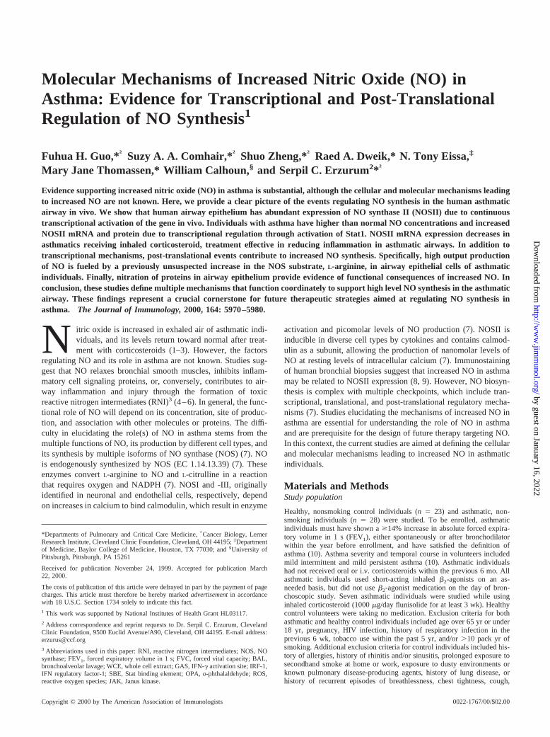
FIGURE 1. Increased nitrotyrosine in asthmatic airway epithelial cells.Western analysis of nitrotyrosine in cell lysates (50mg total protein/lane)of human airway epithelial cells obtained by bronchial brushing of healthycontrols (lanes 1and2), asthmatics not receiving corticosteroid (2CS;lane3), and asthmatics before (lane 4) and following 3 wk of inhaled cortico-steroid therapy (1CS;lane 5). A range of nitrated proteins is seen, withincreased nitration in asthmatic epithelial cell lysates. Western analysisusing anti-humanb-actin is shown as a control (lanes 6–10).
Table I. Characteristics of study populationa
Control
Asthma
–CS 1CS
Age (years) 306 3 316 2 306 2Sex (male/female) 15/8 14/7 5/2Race (Caucasion/African
American/Asian)18/4/1 16/5/0 5/2/0
FVC (% predicted)* 1036 4 896 4 826 5FEV1 (% predicted)* 996 4 806 3 646 4FEV1/FVC* 816 2 766 2 676 3
a Values are means6 SE; –CS, not on inhaled corticosteroids;1CS, on inhaledcorticosteroids.
* p , 0.02; all other comparisons are not significant.
5972 REGULATION OF NO SYNTHESIS IN ASTHMA
by guest on January 16, 2022http://w
ww
.jimm
unol.org/D
ownloaded from
are detected on Western analyses, which are blocked by free ni-trotyrosine. Increased nitrotyrosine is detected in asthmatic airwayepithelial cells compared with controls (nitrotyrosine/b-actin:asthma, 126 1 (n 5 5); controls, 56 1 (n 5 6); p 5 0.004; Fig.1). The most prominent band in both healthy control and asthmaticcell lysates is at 44 kDa and is increased in asthmatic epithelial celllysates. Interestingly, nitrotyrosine is nearly undetectable in airwayepithelial cells from asthmatics using inhaled corticosteroids (Fig.1). These studies show that NO is increasingly consumed by bio-chemical reactions in the lungs of asthmatics. In the context ofincreased consumption, increased NO in bronchiolar gasesstrongly suggests that NO synthesis is increased in asthma.
NOSII protein expression
To investigate NO synthesis, NOS protein expression was evalu-ated by Western analysis of airway epithelial cell lysates using
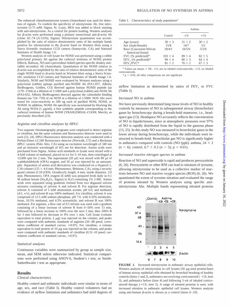
specific anti-NOS Abs. NOSI and NOSIII are not detectable inairway epithelial cells by Western analyses (Figs. 2,A and C).However, a protein (131 kDa) in the asthmatic and control airwayepithelial cell lysates is detected using anti-NOSII Ab, which issimilar in size to NOSII detected in positive control lysate fromA549 cells stimulated with IFN-g (100 U/ml), TNF-a (10 ng/ml),and IL-1b (10 U/ml) for 72 h. Asthmatic airway NOSII expressionis significantly higher than control (NOSII/b-actin: asthma, 0.6060.08 (n5 6); control, 0.336 0.06 (n5 5); p , 0.05; Fig. 2B).
Arginine levels in airway epithelial cells
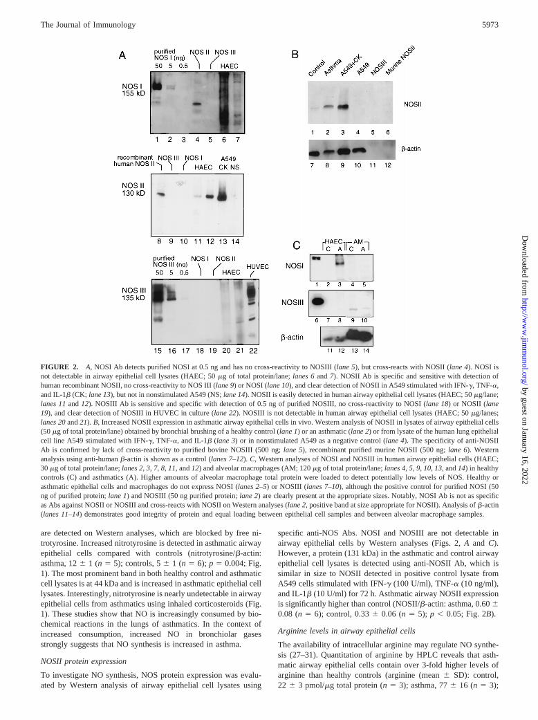
The availability of intracellular arginine may regulate NO synthe-sis (27–31). Quantitation of arginine by HPLC reveals that asth-matic airway epithelial cells contain over 3-fold higher levels ofarginine than healthy controls (arginine (mean6 SD): control,22 6 3 pmol/mg total protein (n5 3); asthma, 776 16 (n 5 3);
FIGURE 2. A, NOSI Ab detects purified NOSI at 0.5 ng and has no cross-reactivity to NOSIII (lane 5), but cross-reacts with NOSII (lane 4). NOSI isnot detectable in airway epithelial cell lysates (HAEC; 50mg of total protein/lane;lanes 6and7). NOSII Ab is specific and sensitive with detection ofhuman recombinant NOSII, no cross-reactivity to NOS III (lane 9) or NOSI (lane 10), and clear detection of NOSII in A549 stimulated with IFN-g, TNF-a,and IL-1b (CK; lane 13), but not in nonstimulated A549 (NS;lane 14). NOSII is easily detected in human airway epithelial cell lysates (HAEC; 50mg/lane;lanes 11and12). NOSIII Ab is sensitive and specific with detection of 0.5 ng of purified NOSIII, no cross-reactivity to NOSI (lane 18) or NOSII (lane19), and clear detection of NOSIII in HUVEC in culture (lane 22). NOSIII is not detectable in human airway epithelial cell lysates (HAEC; 50mg/lanes;lanes 20and21).B, Increased NOSII expression in asthmatic airway epithelial cells in vivo. Western analysis of NOSII in lysates of airway epithelial cells(50mg of total protein/lane) obtained by bronchial brushing of a healthy control (lane 1) or an asthmatic (lane 2) or from lysate of the human lung epithelialcell line A549 stimulated with IFN-g, TNF-a, and IL-1b (lane 3) or in nonstimulated A549 as a negative control (lane 4). The specificity of anti-NOSIIAb is confirmed by lack of cross-reactivity to purified bovine NOSIII (500 ng;lane 5), recombinant purified murine NOSII (500 ng;lane 6). Westernanalysis using anti-humanb-actin is shown as a control (lanes 7–12).C, Western analyses of NOSI and NOSIII in human airway epithelial cells (HAEC;30mg of total protein/lane;lanes 2,3, 7, 8, 11, and12) and alveolar macrophages (AM; 120mg of total protein/lane;lanes 4,5, 9, 10,13, and14) in healthycontrols (C) and asthmatics (A). Higher amounts of alveolar macrophage total protein were loaded to detect potentially low levels of NOS. Healthy orasthmatic epithelial cells and macrophages do not express NOSI (lanes 2–5) or NOSIII (lanes 7–10), although the positive control for purified NOSI (50ng of purified protein;lane 1) and NOSIII (50 ng purified protein;lane 2) are clearly present at the appropriate sizes. Notably, NOSI Ab is not as specificas Abs against NOSII or NOSIII and cross-reacts with NOSII on Western analyses (lane 2, positive band at size appropriate for NOSII). Analysis ofb-actin(lanes 11–14) demonstrates good integrity of protein and equal loading between epithelial cell samples and between alveolar macrophage samples.
5973The Journal of Immunology
by guest on January 16, 2022http://w
ww
.jimm
unol.org/D
ownloaded from
p 5 0.02; Fig. 3). Arginine and citrulline in BAL fluid and citrul-line in airway epithelial cell lysates are not detectable (,0.3pmol). Thus, post-translational mechanisms that support high out-put NO synthesis are induced in asthma. Increased NOSII mRNA in asthma
To evaluate whether increased NOS II protein in asthma was re-lated to increased NOSII mRNA expression, Northern analyses oftotal RNA from airway epithelial cells freshly obtained by bron-choscopic brushing and from alveolar macrophages obtained byBAL of asthmatics (n5 7) and controls (n5 9) were performed.NOSII mRNA is demonstrated in airway epithelial cells as a prom-inent signal at 4.5 kb using a32P-labeled NOSII cDNA (pCCF21),with higher levels of NOSII mRNA in asthmatics (NOSII/g-actinmRNA: asthma, 0.626 0.09 (n5 7); control, 0.276 0.08 (n59); p , 0.01; Fig. 4). In murine systems, macrophages are a majorsource of NO (32). In paired samples of airway epithelium frombronchial brushing and human lung macrophages from bronchoal-veolar lavage simultaneously obtained at bronchoscopy, abundantlevels of NOSII are present in airway epithelium, but NOSII is notdetected in macrophages by Northern analyses (Fig. 4).
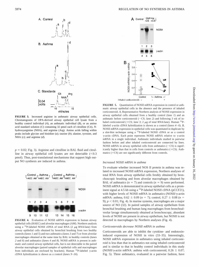
Corticosteroids decrease NOSII mRNA in asthma
Corticosteroids are able to inhibit the cytokine- and endotoxin-induced expression of NOSII in vitro (33–35). Interestingly,NOSII mRNA expression in asthmatics using inhaled corticoste-roid is less than that in asthmatics not using inhaled corticosteroidand is similar to that in healthy control individuals in this study(NOSII/g-actin mRNA: asthma with corticosteroid, 0.266 0.07;Fig. 5). Three asthmatics, evaluated in a pairwise fashion, have
FIGURE 4. Evaluation of NOSII mRNA expression in human airwayepithelial cells (HAEC) and alveolar macrophages (AM). Northern analysisusing a32P-labeled NOSII cDNA of total RNA (2mg RNA/lane) fromairway epithelial cells obtained by bronchial brushing from two healthycontrols (lanes 1and5) and two asthmatics (lanes 3and7) or from alveolarmacrophages obtained at the same time by BAL in healthy controls (lanes2 and6) and asthmatics (lanes 4and8). NOSII mRNA is present in asth-matic and control airway epithelial cells, but is not detectable in the pairedalveolar macrophages (paired samples of epithelial cells and macrophagesfrom individuals are enclosed by brackets). Human32P-labeledg-actincDNA hybridization is shown as a control (lanes 9–16).
FIGURE 5. Quantitation of NOSII mRNA expression in control or asth-matic airway epithelial cells in the absence and the presence of inhaledcorticosteroid.A, Representative Northern analysis of NOSII expression inairway epithelial cells obtained from a healthy control (lane 1) and anasthmatic before corticosteroid (2CS;lane 2) and following 3 wk of in-haled corticosteroid (1CS;lane 3; 2mg of total RNA/lane). Human32P-labeledg-actin cDNA hybridization is shown as a control (lanes 4–6).B,NOSII mRNA expression in epithelial cells was quantitated in duplicate bya slot-blot technique using a32P-labeled NOSII cDNA or as a controlg-actin cDNA. Each point represents NOSII mRNA relative tog-actinmRNA in a single individual. Asthmatic individuals studied in pairwisefashion before and after inhaled corticosteroid are connected by lines.NOSII mRNA in airway epithelial cells from asthmatics (2CS) is signif-icantly higher than that in cells from controls or asthmatics (1CS). Asth-matics (1CS) are not significantly different from controls.
FIGURE 3. Increased arginine in asthmatic airway epithelial cells.Chromatograms of OPA-derived airway epithelial cell lysate from ahealthy control individual (A), an asthmatic individual (B), or an aminoacid standard solution (C) containing 32 pmol each of citrulline (Cit),N-hydroxyarginine (NHA), and arginine (Arg). Amino acids falling withinpeaks include glycine and histidine (a); taurine (b); alanine, tyrosine, andNHA (c); and arginine (d).
5974 REGULATION OF NO SYNTHESIS IN ASTHMA
by guest on January 16, 2022http://w
ww
.jimm
unol.org/D
ownloaded from
decreased NOSII mRNA expression following 3 wk of inhaledcorticosteroid use (Fig. 5). These results suggest that the decreasedNO and nitrotyrosine in asthmatics using inhaled corticosteroidsare due to decreased NOSII gene expression.
Alternative splicing of NOSII in airway epithelial cells
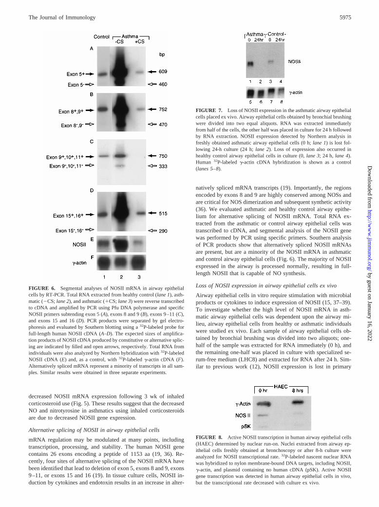
mRNA regulation may be modulated at many points, includingtranscription, processing, and stability. The human NOSII genecontains 26 exons encoding a peptide of 1153 aa (19, 36). Re-cently, four sites of alternative splicing of the NOSII mRNA havebeen identified that lead to deletion of exon 5, exons 8 and 9, exons9–11, or exons 15 and 16 (19). In tissue culture cells, NOSII in-duction by cytokines and endotoxin results in an increase in alter-
natively spliced mRNA transcripts (19). Importantly, the regionsencoded by exons 8 and 9 are highly conserved among NOSs andare critical for NOS dimerization and subsequent synthetic activity(36). We evaluated asthmatic and healthy control airway epithe-lium for alternative splicing of NOSII mRNA. Total RNA ex-tracted from the asthmatic or control airway epithelial cells wastranscribed to cDNA, and segmental analysis of the NOSII genewas performed by PCR using specific primers. Southern analysisof PCR products show that alternatively spliced NOSII mRNAsare present, but are a minority of the NOSII mRNA in asthmaticand control airway epithelial cells (Fig. 6). The majority of NOSIIexpressed in the airway is processed normally, resulting in full-length NOSII that is capable of NO synthesis.
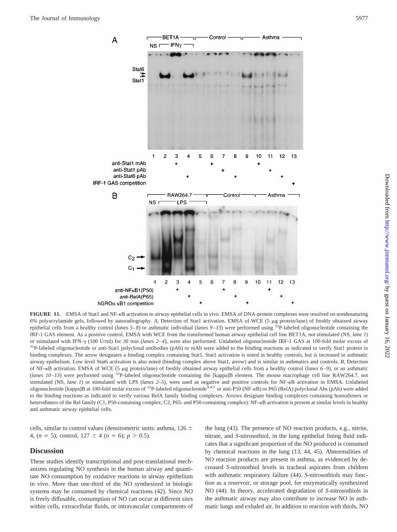
Loss of NOSII expression in airway epithelial cells ex vivo
Airway epithelial cells in vitro require stimulation with microbialproducts or cytokines to induce expression of NOSII (15, 37–39).To investigate whether the high level of NOSII mRNA in asth-matic airway epithelial cells was dependent upon the airway mi-lieu, airway epithelial cells from healthy or asthmatic individualswere studied ex vivo. Each sample of airway epithelial cells ob-tained by bronchial brushing was divided into two aliquots; one-half of the sample was extracted for RNA immediately (0 h), andthe remaining one-half was placed in culture with specialized se-rum-free medium (LHC8) and extracted for RNA after 24 h. Sim-ilar to previous work (12), NOSII expression is lost in primary
FIGURE 6. Segmental analyses of NOSII mRNA in airway epithelialcells by RT-PCR. Total RNA extracted from healthy control (lane 1), asth-matic (2CS;lane 2), and asthmatic (1CS;lane 3) were reverse transcribedto cDNA and amplified by PCR using Pfu DNA polymerase and specificNOSII primers subtending exon 5 (A), exons 8 and 9 (B), exons 9–11 (C),and exons 15 and 16 (D). PCR products were separated by gel electro-phoresis and evaluated by Southern blotting using a32P-labeled probe forfull-length human NOSII cDNA (A–D). The expected sizes of amplifica-tion products of NOSII cDNA produced by constitutive or alternative splic-ing are indicated by filled and open arrows, respectively. Total RNA fromindividuals were also analyzed by Northern hybridization with32P-labeledNOSII cDNA (E) and, as a control, with32P-labeledg-actin cDNA (F).Alternatively spliced mRNA represent a minority of transcripts in all sam-ples. Similar results were obtained in three separate experiments.
FIGURE 7. Loss of NOSII expression in the asthmatic airway epithelialcells placed ex vivo. Airway epithelial cells obtained by bronchial brushingwere divided into two equal aliquots. RNA was extracted immediatelyfrom half of the cells, the other half was placed in culture for 24 h followedby RNA extraction. NOSII expression detected by Northern analysis infreshly obtained asthmatic airway epithelial cells (0 h;lane 1) is lost fol-lowing 24-h culture (24 h;lane 2). Loss of expression also occurred inhealthy control airway epithelial cells in culture (0,lane 3; 24 h, lane 4).Human 32P-labeledg-actin cDNA hybridization is shown as a control(lanes 5–8).
FIGURE 8. Active NOSII transcription in human airway epithelial cells(HAEC) determined by nuclear run-on. Nuclei extracted from airway ep-ithelial cells freshly obtained at bronchoscopy or after 8-h culture wereanalyzed for NOSII transcriptional rate.32P-labeled nascent nuclear RNAwas hybridized to nylon membrane-bound DNA targets, including NOSII,g-actin, and plasmid containing no human cDNA (pSK). Active NOSIIgene transcription was detected in human airway epithelial cells in vivo,but the transcriptional rate decreased with culture ex vivo.
5975The Journal of Immunology
by guest on January 16, 2022http://w
ww
.jimm
unol.org/D
ownloaded from

airway epithelial cells of healthy controls in culture (n 5 4 pairedsamples; Fig. 7). Despite the high levels of NOSII mRNA in theasthmatic airway epithelial cells, NOSII mRNA is not detectableby Northern analysis following 24-h culture (n 5 3 paired sam-ples). These data provide strong support that NOSII mRNA ex-pression is dependent on factors and/or conditions related to theairway environment.
NOSII transcriptional rate in vivo
Evidence in the literature and our previous work point to transcrip-tional regulation of NOSII mRNA (15, 37–39). In this study therate of NOSII transcription relative tog-actin in vivo was com-pared with rates in vitro by run-on experiments using nuclei ex-
tracted from airway epithelial cells freshly obtained at bronchos-copy or after 8-h culture. Active transcription of NOSII mRNA ispresent in airway epithelial cells in vivo, but transcription ofNOSII in airway epithelial cells ex vivo decreases relative to invivo levels (NOSII mRNA transcription relative tog-actin mRNA(mean 6 SD): freshly obtained human airway epithelial cells,15 6 4%; airway epithelial cells after 8-h culture, 26 2%; n 5 2paired samples; Fig. 8). The rapid decrease in NOSII transcrip-tional rate ex vivo provides conclusive evidence that airway epi-thelial cells are dependent upon an in vivo factor(s) for expressionand regulation of the NOSII gene.
IFN-g in BAL fluid
IFN-g is essential for NOSII expression in human primary airwayepithelial cells in vitro (15). In this study asthmatics have a trendtoward higher IFN-g in BAL fluid compared with healthy controls( p 5 0.06; Fig. 9). IFN-g in BAL fluid of asthmatics is signifi-cantly higher than that in healthy controls using a segmental bron-choprovocation model to mimic asthma exacerbation (all timepoints, p , 0.03; Fig. 9). These results support that asthmaticshave increased levels of IFN-g, a cytokine crucial for NOSII geneexpression (15, 37–39).
We have previously shown that IFN-g and IL-4 induce expres-sion of NOSII in airway epithelial cells in culture that is dependentupon new protein synthesis and epithelial cell production of sol-uble mediators (15). IFN-g induces gene expression through theJanus kinase (JAK)-Stat1 pathway, which involves a tyrosinephosphorylation cascade (40, 41). In this context, pretreatmentwith genistein, a tyrosine kinase inhibitor, prevents IFN-g/IL-4induction of NOSII expression in airway epithelial cells (Fig. 10).Interestingly, genistein also prevents NOSII induction by the sol-uble mediators present in conditioned medium of airway epithelialcells exposed transiently to IFN-g/IL-4 (Fig. 10). These data sup-port an essential role of tyrosine phosphorylation signaling eventsin NOSII expression in human airway epithelial cells.
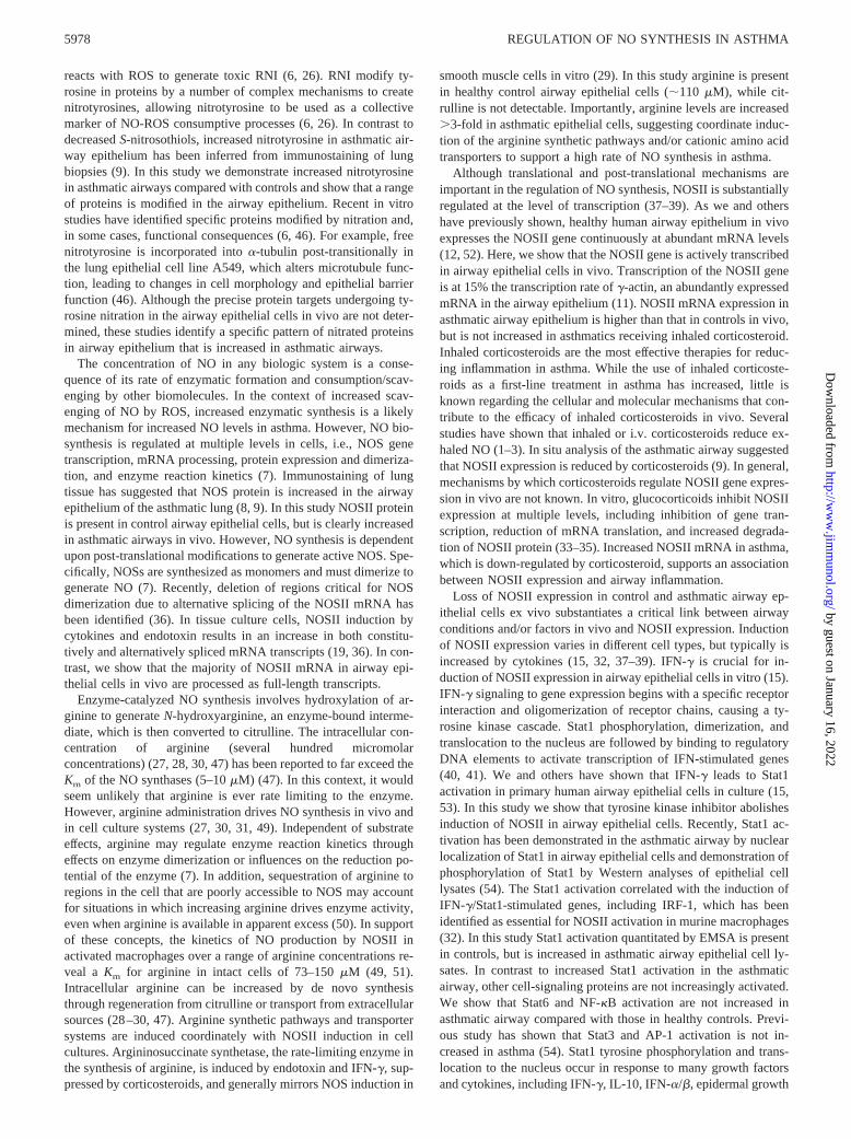
Activation of Stat in airway epithelial cells
To investigate the involvement of tyrosine kinase signaling andspecifically JAK-Stat pathway activation in NOSII expression inasthma in vivo, EMSA of WCE from freshly obtained airway ep-ithelial cells of asthmatics and controls was performed (Fig. 11).Several cytokines implicated in the airway inflammatory reactionof asthma activate the JAK-Stat1 pathway (40, 41). While IFN-guses Stat1, IL-4 activates Stat6 (40, 41). Binding complexes inairway epithelium are detected using the GAS element from thehuman IRF-1 promoter. Stat1 is confirmed in the complex by anti-human Stat1 Abs (Fig. 11A). Stat1 activation is increased in asth-matic airway epithelial cells compared with control (densitometricunits of binding complex: asthma, 466 2 (n 5 5); control, 22.660.6 (n5 6); p , 0.01). A low level of Stat6 activation is also noted(Fig. 11A). Using a Stat binding element (SBE) from the secreted-type IL-1R antagonist gene as a probe to specifically detect Stat6activation, EMSA confirms a very low level of Stat6-containingcomplex in the cell lysates of the same individuals used for de-tecting Stat1 activation (data not shown). These data are compat-ible with our previous report that WCE from airway epithelial cellsstimulated with IL-4 (10 ng/ml) in culture for 15 min induced avery faint binding complex containing Stat6 (15).
The signal transduction pathway through NF-kB was also in-vestigated by EMSA using NF-kB binding element from the hu-man GROa gene (22) (Fig. 11B). Although cytokines that signalthrough NF-kB may be increased in asthma, asthmatics in thisstudy have low levels of NF-kB activation in airway epithelial
FIGURE 10. Inhibition of NOSII induction in airway epithelial cells invitro by the TK inhibitor, genistein. NOSII mRNA is not detected byNorthern analyses in airway epithelial cells in vitro (lane 5),but is inducedin airway epithelial cells exposed to IFN-g (100 U/ml) and IL-4 (10 ng/ml;lane 1). Conditioned medium (CM) was derived from the overlying tissueculture medium of airway epithelial cells treated with IFN-g (100 U/ml)and IL-4 (10 ng/ml) for 1 h, followed by removal and discarding of thecytokine-containing medium, vigorous washing of cells, and culture infresh medium for 23 h. Transferable, soluble mediators present in CM alsoinduce NOSII mRNA (lane 3). Genistein inhibits NOSII induction by IFN-g/IL-4 (lane 2) and CM (lane 4). Genistein alone has no effect on NOSIImRNA expression (not shown). Human32P-labeledg-actin cDNA hybrid-ization is shown as a control (lanes 6–10). Similar results were obtained inthree separate experiments using airway epithelial cells from three healthycontrol individuals.
FIGURE 9. IFN-g in BAL fluid from control (n 5 8) and asthmatic(n 5 9) individuals at baseline and following segmental bronchoprovoca-tion with Ag. BAL was obtained at baseline in the lingula and after Agchallenge in the right middle lobe at 8, 24, and 48 h. IFN-g detected inBAL by quantitative ELISA are not significantly different between controlsand asthmatics at baseline (p5 0.06). However, with Ag challenge, asth-matic IFN-g is significantly higher than the control value at all other timepoints.
5976 REGULATION OF NO SYNTHESIS IN ASTHMA
by guest on January 16, 2022http://w
ww
.jimm
unol.org/D
ownloaded from
cells, similar to control values (densitometric units: asthma, 12664, (n 5 5); control, 1276 4 (n 5 6); p . 0.5).
DiscussionThese studies identify transcriptional and post-translational mech-anisms regulating NO synthesis in the human airway and quanti-tate NO consumption by oxidative reactions in airway epitheliumin vivo. More than one-third of the NO synthesized in biologicsystems may be consumed by chemical reactions (42). Since NOis freely diffusable, consumption of NO can occur at different siteswithin cells, extracellular fluids, or intravascular compartments of
the lung (43). The presence of NO reaction products, e.g., nitrite,nitrate, andS-nitrosothiol, in the lung epithelial lining fluid indi-cates that a significant proportion of the NO produced is consumedby chemical reactions in the lung (13, 44, 45). Abnormalities ofNO reaction products are present in asthma, as evidenced by de-creasedS-nitrosothiol levels in tracheal aspirates from childrenwith asthmatic respiratory failure (44).S-nitrosothiols may func-tion as a reservoir, or storage pool, for enzymatically synthesizedNO (44). In theory, accelerated degradation ofS-nitrosothiols inthe asthmatic airway may also contribute to increase NO in asth-matic lungs and exhaled air. In addition to reaction with thiols, NO
FIGURE 11. EMSA of Stat1 and NF-kB activation in airway epithelial cells in vivo. EMSA of DNA-protein complexes were resolved on nondenaturing6% polyacrylamide gels, followed by autoradiography.A, Detection of Stat1 activation. EMSA of WCE (5mg protein/lane) of freshly obtained airwayepithelial cells from a healthy control (lanes 5–8) or asthmatic individual (lanes 9–13) were performed using32P-labeled oligonucleotide containing theIRF-1 GAS element. As a positive control, EMSA with WCE from the transformed human airway epithelial cell line BET1A, not stimulated (NS,lane 1)or stimulated with IFN-g (100 U/ml) for 30 min (lanes 2–4), were also performed. Unlabeled oligonucleotide IRF-1 GAS at 100-fold molar excess of32P-labeled oligonucleotide or anti-Stat1 polyclonal antibodies (pAb) or mAb were added to the binding reactions as indicated to verify Stat1 protein inbinding complexes. The arrow designates a binding complex containing Stat1. Stat1 activation is noted in healthy controls, but is increased in asthmaticairway epithelium. Low level Stat6 activation is also noted (binding complex above Stat1, arrow) and is similar in asthmatics and controls.B, Detectionof NF-kB activation. EMSA of WCE (5mg protein/lane) of freshly obtained airway epithelial cells from a healthy control (lanes 6–9), or an asthmatic(lanes 10–13) were performed using32P-labeled oligonucleotide containing the [kappa]B element. The mouse macrophage cell line RAW264.7, notstimulated (NS,lane 1) or stimulated with LPS (lanes 2–5), were used as negative and positive controls for NF-kB activation in EMSA. Unlabeledoligonucleotide [kappa]B at 100-fold molar excess of32P-labeled oligonucleotideXXV or anti-P50 (NF-kB) or P65 (RelA) polyclonal Abs (pAb) were addedto the binding reactions as indicated to verify various RelA family binding complexes. Arrows designate binding complexes containing homodimers orheterodimers of the Rel family (C1, P50-containing complex; C2, P65- and P50-containing complex). NF-kB activation is present at similar levels in healthyand asthmatic airway epithelial cells.
5977The Journal of Immunology
by guest on January 16, 2022http://w
ww
.jimm
unol.org/D
ownloaded from
reacts with ROS to generate toxic RNI (6, 26). RNI modify ty-rosine in proteins by a number of complex mechanisms to createnitrotyrosines, allowing nitrotyrosine to be used as a collectivemarker of NO-ROS consumptive processes (6, 26). In contrast todecreasedS-nitrosothiols, increased nitrotyrosine in asthmatic air-way epithelium has been inferred from immunostaining of lungbiopsies (9). In this study we demonstrate increased nitrotyrosinein asthmatic airways compared with controls and show that a rangeof proteins is modified in the airway epithelium. Recent in vitrostudies have identified specific proteins modified by nitration and,in some cases, functional consequences (6, 46). For example, freenitrotyrosine is incorporated intoa-tubulin post-transitionally inthe lung epithelial cell line A549, which alters microtubule func-tion, leading to changes in cell morphology and epithelial barrierfunction (46). Although the precise protein targets undergoing ty-rosine nitration in the airway epithelial cells in vivo are not deter-mined, these studies identify a specific pattern of nitrated proteinsin airway epithelium that is increased in asthmatic airways.
The concentration of NO in any biologic system is a conse-quence of its rate of enzymatic formation and consumption/scav-enging by other biomolecules. In the context of increased scav-enging of NO by ROS, increased enzymatic synthesis is a likelymechanism for increased NO levels in asthma. However, NO bio-synthesis is regulated at multiple levels in cells, i.e., NOS genetranscription, mRNA processing, protein expression and dimeriza-tion, and enzyme reaction kinetics (7). Immunostaining of lungtissue has suggested that NOS protein is increased in the airwayepithelium of the asthmatic lung (8, 9). In this study NOSII proteinis present in control airway epithelial cells, but is clearly increasedin asthmatic airways in vivo. However, NO synthesis is dependentupon post-translational modifications to generate active NOS. Spe-cifically, NOSs are synthesized as monomers and must dimerize togenerate NO (7). Recently, deletion of regions critical for NOSdimerization due to alternative splicing of the NOSII mRNA hasbeen identified (36). In tissue culture cells, NOSII induction bycytokines and endotoxin results in an increase in both constitu-tively and alternatively spliced mRNA transcripts (19, 36). In con-trast, we show that the majority of NOSII mRNA in airway epi-thelial cells in vivo are processed as full-length transcripts.
Enzyme-catalyzed NO synthesis involves hydroxylation of ar-ginine to generateN-hydroxyarginine, an enzyme-bound interme-diate, which is then converted to citrulline. The intracellular con-centration of arginine (several hundred micromolarconcentrations) (27, 28, 30, 47) has been reported to far exceed theKm of the NO synthases (5–10mM) (47). In this context, it wouldseem unlikely that arginine is ever rate limiting to the enzyme.However, arginine administration drives NO synthesis in vivo andin cell culture systems (27, 30, 31, 49). Independent of substrateeffects, arginine may regulate enzyme reaction kinetics througheffects on enzyme dimerization or influences on the reduction po-tential of the enzyme (7). In addition, sequestration of arginine toregions in the cell that are poorly accessible to NOS may accountfor situations in which increasing arginine drives enzyme activity,even when arginine is available in apparent excess (50). In supportof these concepts, the kinetics of NO production by NOSII inactivated macrophages over a range of arginine concentrations re-veal a Km for arginine in intact cells of 73–150mM (49, 51).Intracellular arginine can be increased by de novo synthesisthrough regeneration from citrulline or transport from extracellularsources (28–30, 47). Arginine synthetic pathways and transportersystems are induced coordinately with NOSII induction in cellcultures. Argininosuccinate synthetase, the rate-limiting enzyme inthe synthesis of arginine, is induced by endotoxin and IFN-g, sup-pressed by corticosteroids, and generally mirrors NOS induction in
smooth muscle cells in vitro (29). In this study arginine is presentin healthy control airway epithelial cells (;110 mM), while cit-rulline is not detectable. Importantly, arginine levels are increased.3-fold in asthmatic epithelial cells, suggesting coordinate induc-tion of the arginine synthetic pathways and/or cationic amino acidtransporters to support a high rate of NO synthesis in asthma.
Although translational and post-translational mechanisms areimportant in the regulation of NO synthesis, NOSII is substantiallyregulated at the level of transcription (37–39). As we and othershave previously shown, healthy human airway epithelium in vivoexpresses the NOSII gene continuously at abundant mRNA levels(12, 52). Here, we show that the NOSII gene is actively transcribedin airway epithelial cells in vivo. Transcription of the NOSII geneis at 15% the transcription rate ofg-actin, an abundantly expressedmRNA in the airway epithelium (11). NOSII mRNA expression inasthmatic airway epithelium is higher than that in controls in vivo,but is not increased in asthmatics receiving inhaled corticosteroid.Inhaled corticosteroids are the most effective therapies for reduc-ing inflammation in asthma. While the use of inhaled corticoste-roids as a first-line treatment in asthma has increased, little isknown regarding the cellular and molecular mechanisms that con-tribute to the efficacy of inhaled corticosteroids in vivo. Severalstudies have shown that inhaled or i.v. corticosteroids reduce ex-haled NO (1–3). In situ analysis of the asthmatic airway suggestedthat NOSII expression is reduced by corticosteroids (9). In general,mechanisms by which corticosteroids regulate NOSII gene expres-sion in vivo are not known. In vitro, glucocorticoids inhibit NOSIIexpression at multiple levels, including inhibition of gene tran-scription, reduction of mRNA translation, and increased degrada-tion of NOSII protein (33–35). Increased NOSII mRNA in asthma,which is down-regulated by corticosteroid, supports an associationbetween NOSII expression and airway inflammation.
Loss of NOSII expression in control and asthmatic airway ep-ithelial cells ex vivo substantiates a critical link between airwayconditions and/or factors in vivo and NOSII expression. Inductionof NOSII expression varies in different cell types, but typically isincreased by cytokines (15, 32, 37–39). IFN-g is crucial for in-duction of NOSII expression in airway epithelial cells in vitro (15).IFN-g signaling to gene expression begins with a specific receptorinteraction and oligomerization of receptor chains, causing a ty-rosine kinase cascade. Stat1 phosphorylation, dimerization, andtranslocation to the nucleus are followed by binding to regulatoryDNA elements to activate transcription of IFN-stimulated genes(40, 41). We and others have shown that IFN-g leads to Stat1activation in primary human airway epithelial cells in culture (15,53). In this study we show that tyrosine kinase inhibitor abolishesinduction of NOSII in airway epithelial cells. Recently, Stat1 ac-tivation has been demonstrated in the asthmatic airway by nuclearlocalization of Stat1 in airway epithelial cells and demonstration ofphosphorylation of Stat1 by Western analyses of epithelial celllysates (54). The Stat1 activation correlated with the induction ofIFN-g/Stat1-stimulated genes, including IRF-1, which has beenidentified as essential for NOSII activation in murine macrophages(32). In this study Stat1 activation quantitated by EMSA is presentin controls, but is increased in asthmatic airway epithelial cell ly-sates. In contrast to increased Stat1 activation in the asthmaticairway, other cell-signaling proteins are not increasingly activated.We show that Stat6 and NF-kB activation are not increased inasthmatic airway compared with those in healthy controls. Previ-ous study has shown that Stat3 and AP-1 activation is not in-creased in asthma (54). Stat1 tyrosine phosphorylation and trans-location to the nucleus occur in response to many growth factorsand cytokines, including IFN-g, IL-10, IFN-a/b, epidermal growth
5978 REGULATION OF NO SYNTHESIS IN ASTHMA
by guest on January 16, 2022http://w
ww
.jimm
unol.org/D
ownloaded from
factor, platelet-derived growth factor, GM-CSF, IL-6, IL-11, leu-kemia inhibitory factor, ciliary neurotropic factor, oncostatin M,growth hormone, prolactin, and CSF-1 (40, 41). IFN-g has bothanti- and proinflammatory effects in the lung. In fact, IFN-g me-diates numerous anti-inflammatory effects, including inhibition ofAg and Th2-cell induced pulmonary eosinophilia and airway hy-per-reactivity (55). However, IFN-g is also implicated in thepathobiology leading to airway inflammation and hyper-reactivityin asthma (55–58). For example, OVA-sensitized mice developairway hyper-responsiveness dependent upon IFN-g (57). Further,adoptive transfer of Th1 lymphocytes, which characteristicallyproduce IFN-g, increases airway inflammation in a murine modelof allergic asthma (58). In this study IFN-g levels are higher inasthmatic epithelial lining fluid than in controls following a seg-mental bronchoprovocation with Ag. The large number of IFN-g/Stat1-stimulated genes, including IRF-1, ICAM-1, and NOSII, areprobably involved in the airway inflammatory events of asthma.Collectively, these data provide strong support for Stat1 activationmediating NOSII gene expression in human airway epithelial cellsin vivo.
NF-kB activation and binding tokB DNA elements in the 59-flanking region of the NOSII gene play a role in the cytokineinduction of NOSII in the human lung epithelial cell line A549 invitro (37–39). However, studies of NF-kB activation in asthma areconflicting, perhaps in part due to the types of samples analyzed(48, 54). Expectorated sputum from asthmatics or pooled biopsiesof asthmatic airways have shown increased NF-kB activation com-pared with controls (48), while no increase in activation was notedby nuclear localization of NF-kB in biopsies and bronchial brush-ings of asthmatic airway epithelium (54). Here, NF-kB activationin asthmatic airway epithelial cells obtained by bronchial brushingis at levels similar to those in healthy controls. Our results supportthat increased NF-kB activation is not involved in NOSII induc-tion in asthmatic airways. On the other hand, NF-kB activation ispresent in control and asthmatic epithelia and may contribute to thetonic expression of NOSII in the airway.
In conclusion, multiple mechanisms function coordinately tosupport high level NO synthesis in the asthmatic airway. Humanairway epithelium has abundant expression of NOSII due to con-tinuous transcriptional activation of the gene in vivo. We proposethat increased NOSII gene expression in asthmatic airways is re-lated to increased Stat1 activation caused by increased cytokines,e.g., IFN-g. High levels of intracellular arginine may enhance en-zyme reaction kinetics and drive NO synthesis. Thus, airway ep-ithelial cells have highly efficient NO synthetic machinery, whichis amplified in airway inflammation. These studies lay the ground-work for evaluating therapeutic strategies to decrease NO and RNIformation through inhibitors of arginine transport systems, specificinhibitors of NOSII, or antioxidant augmentation.
AcknowledgmentsWe thank H. Abu-Soud for advice on HPLC; C. Harris for BET-1A; D.Mytych for IL-4; Genentech for human IFN-g; D. J. Stuehr for recombi-nant NOSIII and NOSII; J. L. Humes for anti-NOSII Ab; Affinity Biore-agents Laboratories for anti-NOSI and anti-NOSIII Abs; O. Uttenthal foranti-nitrotyrosine Ab; L. Kedes for pHFgA-1; J. Lang for artwork; J. Ham-mel for biostatistical testing; and F. T. Kaneko and M. Numata for tech-nical support.
References1. Massaro, A. F., B. Gaston, D. Kita, C. Fanta, J. S. Stamler, and F. M. Drazen.
1995. Expired nitric oxide levels during treatment of acute asthma.Am. J. Respir.Crit. Care. Med. 152:800.
2. Kharitonov, S. A., D. H. Yates, and P. J. Barnes. 1996. Inhaled glucocorticoidsdecrease nitric oxide in exhaled air of asthmatic patients.Am. J. Respir. Crit.Care. Med. 153:454.
3. Silkoff, P. E. S., P. A. McClean, A. S. Slutsky, M. Caramori, K. R. Chapman,C. Gutierrez, and N. Zamel. 1998. Exhaled nitric oxide and bronchial reactivityduring and after inhaled beclomethasone in mild asthma.J. Asthma 35:473.
4. Sanders, S. P. 1999. Nitric oxide in asthma.Am. J. Respir. Cell Mol. Biol. 21:147.5. Raychaudhuri, B., R. Dweik, M. J. Connors, L. Buhrow, A. Malur, J. Drazba, A.
C. Arroliga, S. C. Erzurum, M. S. Kavuru, M. J. Thomassen, et al. 1999. Nitricoxide blocks nuclear factor-kB activation in alveolar macrophages.Am. J. Respir.Cell Mol. Biol. 21:311.
6. van der Vliet, A., J. P. Elserich, M. K. Shigenaga, and C. E. Cross. 1999. Reactivenitrogen species and tyrosine nitration in the respiratory tract.Am. J. Respir. Crit.Care. Med. 160:1.
7. Stuehr, D. J. 1999. Nitric oxide synthesis and breakdown.Biochim. Biophys. Acta1411:217.
8. Hamid, Q., D. R. Springall, V. Riveros-Moreno, P. Chanez, P. Howarth, A. Red-ington, J. Bousquet, P. Godard, S. Holgate, J. M. Polak, et al. 1993. Induction ofnitric oxide synthase in asthma.Lancet 342:1510.
9. Saleh, D., P. Ernst, S. Lim, P. J. Barnes, and A. Giaid. 1998. Increased formationof the potent oxidant peroxynitrite in the airways of asthmatic patients is asso-ciated with induction of nitric oxide synthase: effect of inhaled glucocorticoid.FASEB J. 12:929.
10. National Asthma Education and Prevention Program. 1997.Guidelines for theDiagnosis and the Management of Asthma, Expert Panel Report II.NationalAsthma Education and Prevention Program, National Institutes of Health, Be-thesda, Publication 97–4051.
11. Erzurum, S. C., C. Danel, A. Gillissen, C.-S. Chu, B. C. Trapnell, andR. G. Crystal. 1993. In vivo antioxidant gene expression in human airway epi-thelium of normal individuals exposed to 100% O2. J. Appl. Physiol. 75:1256.
12. Guo, F. H., H. R. De Raeve, T. W. Rice, D. J. Stuehr, F. B. J. M. Thunnissen, andS. C. Erzurum. 1995. Continuous nitric oxide synthesis by inducible nitric oxidesynthase in normal human airway epithelium in vivo.Proc. Natl. Acad. Sci. USA92:7809.
13. Dweik, R. D., D. Laskowsdi, H. M. Abu-Soud, F. T. Kaneko, R. Hutte,D. J. Stuehr, and S. C. Erzurum. 1998. Nitric oxide synthesis in the lung.J. Clin.Invest. 101:660.
14. Calhoun, W. J., E. C. Dick, L. B. Schwartz, and W. W. Busse. 1994. A commoncold virus, rhinovirus 16, potentiates airway inflammation after segmental antigenbronchoprovocation in allergic subjects.J. Clin. Invest. 94:2200.
15. Guo, F. H., K. Uetani, S. J. Haque, B. R. G. Williams, R. A. Dweik,F. B. J. M. Thunnissen, W. Calhoun, and S. C. Erzurum. 1997. Interferong andinterleukin 4 stimulate prolonged expression of inducible nitric oxide synthase inhuman airway epithelium through synthesis of soluble mediators.J. Clin. Invest.100:829.
16. Reddel, R. R., Y. Ke, B. I. Gerwin, M. G. McMenamin, J. F. Lechner, R. T. Su,D. E. Brash, J. B. Park, J. S. Rhim, C. C. Harris, et al. 1988. Transformation ofhuman bronchial epithelial cells by infection with SV40 or adenovirus-12 SV40hybrid virus, or transfection via strontium phosphate coprecipitation with a plas-mid containing SV40 early region genes.Cancer Res. 48:1904.
17. Gunning, P., P. Ponte, H. Okayama, J. Engel, H. Blau, et al. 1983. Isolation andcharacterization of full-length cDNA clones for humana-, b-, and g-actinmRNAs: skeletal but not cytoplasmic actins have an amino-terminal cysteine thatis subsequently removed.Mol. Cell. Biol. 3:787.
18. Nakamura, H., K. Yoshimura, H. A. Jaffe, and R. G. Crystal. 1991. Interleukin-8gene expression in human bronchial epithelial cells.J. Biol. Chem. 266:19611.
19. Eissa, N. T., A. J. Strauss, C. M. Haggerty, E. K. Choo, S. C. Chu, and J. Moss.1996. Alternative splicing of human inducible nitric-oxide synthase mRNA.J. Biol. Chem. 271:27184.
20. Haque, S. J., V. Flati, A. Deb, and R. G. Williams. 1995. Roles of protein-tyrosine phosphatases in Stat1 a-mediated cell signaling.J. Biol. Chem. 270:25709.
21. Haque, S. J., Q. Wu, W. Kammer, K. Friedrich, and J. M. Smith. 1997. Receptor-associated constitutive protein tyrosine phosphatase activity controls the kinasefunction of JAK1.Proc. Natl. Acad. Sci. USA 94:8563.
22. Ohmori, Y., S. Fukumoto, and T. A. Hamilton. 1995. Two structurally distinctkBsequence motifs cooperatively control LPS-induced KC gene transcription inmouse macrophages.J. Immunol. 155:3593.
23. Nicholson, S., M. G. Bonecini-Almeida, J. R. Lapa e Silva, C. Nathan, Q. W. Xie,R. Mumford, J. R. Weidner, J. Calaycay, J. Geng, N. Boechat, et al. 1996. In-ducible nitric oxide synthase in pulmonary alveolar macrophages from patientswith tuberculosis.J. Exp. Med.183:2293.
24. Meyer, J., N. Richter, and M. Hecker. 1997. High-performance liquid chromato-graphic determination of nitric oxide synthase-related arginine derivatives in vitroand in vivo.Anal. Biochem. 247:11.
25. Laurent, M., M. Lepoivre, and J. P. Tenu. 1996. Kinetic modelling of the nitricoxide gradient generated in vitro by adherent cells expressing inducible nitricoxide synthase.Biochem. J. 314:109.
26. Eiserich, J. P., M. Hristova, C. E. Cross, A. D. Jones, and B. A. Freeman. 1998.Formation of nitric oxide-derived inflammatory oxidants by myeloperoxidase inneutrophils.Nature 391:393.
27. Kurz, S., and D. G. Harrison. 1997. Insulin and the arginine paradox.J. Clin.Invest. 99:369.
28. Hecker, M., W. C. Sessa, H. J. Harris, E. E. Anggard, and J. R. Vane. 1990. Themetabolism ofL-arginine and its significance for the biosynthesis of endothelium-
5979The Journal of Immunology
by guest on January 16, 2022http://w
ww
.jimm
unol.org/D
ownloaded from

derived relaxing factor: cultured endothelial cells recycleL-citrulline to L-argi-nine.Proc. Natl. Acad. Sci. USA 87:8612.
29. Hattori, Y., E. B. Campbell, and S. S. Gross. 1994. Argininosuccinate synthetasemRNA and activity are induced by immunostimulants in vascular smooth muscle.J. Biol. Chem. 269:9405.
30. Hammermann, R., J. Hirschmann, C. Hey, J. Mossner, G. Folkerts, F. P. Ni-jkamp, I. Wessler, and K. Racke 1999. Cationic proteins inhibitL-arginine uptakein rat alveolar macrophages and tracheal epithelial cells.Am. J. Respir. Cell Mol.Biol. 21:155.
31. Lundberg, J. O. N., E. Weitzberg, J. Rinder, A. Rudehill, O. Jansson, N. P.Wiklund, J. M. Lundberg, and K. Alving. 1996. Calcium-independent and ste-roid-resistant nitric oxide synthase activity in human paranasal sinus mucosa.Eur. Respir. J. 9:1344.
32. Kamijo, R., H. Harada, T. Matsuyama, M. Bosland, J. Gerecitano, D. Shapiro,J. Le, S. I. Koh, T. Kimura, S. J. Green, et al. 1994. Requirement for transcriptionfactor IRF-1 in NO synthase induction in macrophages.Science 263:1612.
33. Geller, D. A., A. K. Nussler, M. D. Silvio, C. J. Lowenstein, R. A. Shapiro, S.Wang, R. L. Simmons, and T. R. Billiar. 1993. Cytokines, endotoxin, and glu-cocorticoids regulate the expression of inducible nitric oxide synthase in hepa-tocytes.Proc. Natl. Acad. Sci. USA 90:522.
34. Radomski, M.W., R. M. J. Palmer, and S. Moncada. 1990. Glucocorticoids in-hibit the expression of an inducible, but not the constitutive, nitric oxide synthasein vascular endothelial cells.Proc. Natl. Acad. Sci. USA 87:10043.
35. Kunz, D., G. Walker, W. Eberhardt, and J. Pfeilschifter. 1996. Molecular mech-anisms of dexamethasone inhibition of nitric oxide synthase expression in inter-leukin 1b-stimulated mesangial cells: evidence for the involvement of transcrip-tional and posttranscriptional regulation.Proc. Natl. Acad. Sci. USA 93:255.
36. Eissa, N. T., J. W. Yuan, C. M. Haggerty, E. K. Choo, C. D. Palmer, and J. Moss.1998. Cloning and characterization of human inducible nitric oxide synthasesplice variants: a domain, encoded by exons 8 and 9, is critical for dimerization.Proc. Natl. Acad. Sci. USA 95:7625.
37. Marks-Konczalik, J., S. C. Chu, and J. Moss. 1998. Cytokine-mediated transcrip-tional induction of the human inducible nitric oxide synthase gene requires bothactivator protein 1 and nuclear factorkB-binding sites.J. Biol. Chem. 273:22201.
38. Taylor, B. S., M. E. de Vera, R. W. Ganster, Q. Wang, R. A. Shapiro, S. M.Morris, T. R. Billiar, and D. A. Geller. 1998. Multiple NF-kB enhancer elementsregulate cytokine induction of the human inducible nitric oxide synthase gene.J. Biol. Chem. 273:15148.
39. de Vera, M. E., R. A. Shapiro, A. K. Nussler, J. S. Mudgett, R. L. Simmons, S. M.Morris, and T. R. Billiar. 1996. Transcriptional regulation of human induciblenitric oxide synthase (NOS2) gene by cytokines: initial analysis of the humanNOS2 promoter.Proc. Natl. Acad. Sci. USA 93:1054.
40. Haque, S. J., and B. R. G. Williams. 1998. Signal transduction in the interferonsystem.Semin. Oncol. 25:14.
41. Hill, C. S., and R. Treisman. 1995. Transcriptional regulation by extracellularsignals: mechanisms and specificity.Cell 80:199.
42. Malinski, T., Z. Taha, S. Grunfeld, S. Patton, M. Kapturczak, and P. Tomboulian.1993. Diffusion of nitric oxide in the aorta wall monitored in situ by porphyrinicmicrosensors.Biochem. Biophys. Res. Commun. 193:1076.
43. Wink, D. A., I. Hanbauer, M. B. Grisham, F. Laval, R. W. Nims, J. Laval, J. Cook,R. Pacelli, J. Liebmann, M. Krishna, et al. 1996. Chemical biology of nitric oxide:regulation and protective and toxic mechanisms.Curr. Top. Cell. Regul. 34:159.
44. Gaston, B., S. Sears, J. Woods, J. Hunt, M. Ponaman, T. McMohan, and J. S.Stamler. et al. 1998. Bronchodilator S-nitrosothiol deficiency in asthmatic respi-ratory failure.351:1317.
45. Gaston, B., J. Reilly, J. M. Drazen, J. Fackler, P. Ramdev, D. Arnelle, M. E.Mullins, D. J. Sugarbaker, C. Chee, D. J. Singel, et al. 1993. Endogenous nitrogenoxides and bronchodilator S-nitrosothiols in human airways.Proc. Natl. Acad.Sci. USA 90:10957.
46. Eiserich, J. P., A. G. Estevez, T. V. Bamberg, Y. Z. Ye, P. H. Chumley, J. S.Beckman, and B. A. Freeman. et al. 1999. Microtubule dysfunction by posttrans-lational nitrotyrosination ofa-tubulin: a nitric oxide dependent mechanism ofcellular injury.Proc. Natl. Acad. Sci. USA 96:6365.
47. Baydoun, A. R., P. W. Emery, J. D. Pearson, and G. E. Mann. 1990. Substrate-dependent regulation of intracellular amino acid concentrations in cultured bo-vine aortic endothelial cells.Biochem. Biophys. Res. Commun. 173:940.
48. Hart, L. A., V. L. Krishnan, I. M. Adcock, P. J. Barnes, and K. F. Chung. 1998.Activation and localization of transcription factor, nuclear factor-kB, in asthma.Am. J. Respir. Crit. Care Med. 158:1585.
49. Iyengar, F., D. J. Stuehr, and M. A. Marletta. 1987. Macrophage synthesis ofnitrite, nitrate, and N-nitrosamines: precursors and role of the respiratory burst.Proc. Natl. Acad. Sci. USA 84:6369.
50. McDonald, K. K., S. Zharikov, E. R. Block, and M. S. Kilberg. 1997. Acaveolar complex between the cationic amino acid transporter 1 and endo-thelial nitric-oxide synthase may explain the “arginine paradox.”J. Biol.Chem. 272:31213.
51. Granger, D. L., J. B. Hibbs, J. R. Perfect, and D. T. Durack. 1990. Metabolic fateof L-arginine in relation to microbiostatic capability of murine macrophages.J. Clin. Invest. 85:264.
52. Kobzik, L., D. S. Bredt, C. J. Lowenstein, J. Drazen, B. Gaston, D. Sugarbaker,and J. S. Stamler. 1993. Nitric oxide synthase in human and rat lung: immuno-cytochemical and histochemical localization.Am. J. Respir. Cell Mol. Biol.9:371.
53. Look, D. C., M. R. Pelletier, and M. J. Holtzman. 1994. Selective interaction ofa subset of interferon-g response element-binding proteins with the intercellularadhesion molecule-1 (ICAM-1) gene promoter controls the pattern of expressionon epithelial cells.Proc. Natl. Acad. Sci. USA 269:8952.
54. Sampath, D., M. Castro, D. C. Look, and M. J. Holtzman 1999. Constitutiveactivation of an epithelial signal transducer and activation of transcription(STAT) pathway in asthma.J. Clin. Invest. 103:1353.
55. Li, X. M., R. K. Chopra, T. Y. Chou, B. H. Schofield, M. Wills-Karp, and S. K.Huang. 1996. Mucosal IFN-g gene transfer inhibits pulmonary allergic responsesin mice.J. Immunol. 157:3216.
56. Cembrzynska-Nowak, M., E. Szklarz, A. D. Inglot, and J. A. Teodorczyk-Injeyan.1993. Elevated release of tumor necrosis factor-a and interferon-g by bronchoalveo-lar leukocytes from patients with bronchial asthma.Am. Rev. Respir. Dis. 147:291.
57. Hessel, E. M., A. J. M. Van Oosterhout, I. V. Ark, B. V. Esch, G. Hofman, H. V.Loveren, H. F. J. Savelkoul, and F. P. Nij Kamp. 1997. Development of airwayhyperresponsiveness is dependent on interferon-g and independent of eosinophilinfiltration. Am. J. Respir. Cell Mol. Biol. 16:325.
58. Hansen, G., G. Berry, R. H. DeKruyff, and D. T. Umetsu. 1998. Allergen-specificTh1 cells fail to counterbalance Th2 cell-induced airway hyperreactivity butcause severe airway inflammation.J. Clin. Invest. 103:175.
5980 REGULATION OF NO SYNTHESIS IN ASTHMA
by guest on January 16, 2022http://w
ww
.jimm
unol.org/D
ownloaded from