3713.full.pdf - Journal of Neuroscience
12
The Journal of Neuroscience, June 1994, 14(6): 37133724 Neuronal Expression of Glypican, a Cell-Surface Glycosylphosphatidylinositol-anchored Heparan Sulfate Proteoglycan, in the Adult Rat Nervous System E. David Litwack,’ Christopher S. Stipp,’ Asli Kumbasar,’ and Arthur D. Lander’** ‘Department of Biology and 2Department of Brain and Cognitive Science, Massachusetts Institute of Technology, Cambridge, Massachusetts 02139 Cell-surface proteoglycans have been implicated in cell re- sponses to growth factors, extracellular matrix, and cell ad- hesion molecules. M12, one of the most abundant mem- brane-associated proteoglycans in the adult rat brain, is a -65 kDa glycosylphosphatidylinositol-linked protein that bears heparan sulfate chains (Herndon and Lander, 1990). To assess its identity, Ml 2 was purified and internal peptide sequences obtained. Comparison of the results with pr,otein sequence predicted by a cDNA cloned from PC12 cells in- dicated that Ml2 is rat glypican, a proteoglycan first cloned from human fibroblasts. In addition, antibodies raised against a rat glypican fusion protein specifically detected the 65 kDa brain proteoglycan core protein, both by immunoprecipita- tion and by Western blotting. Northern blot analysis using a rat glypican probe also detected glypican message in the adult, as well as the developing rat brain. /n situ hybridization with glypican RNA probes showed that glypican is expressed in a subset of structures in the adult rat nervous system. These include the hippocampus, dorsal thalamus, amygdala, cerebral cortex, piriform cortex, olfac- tory tubercle, several cranial nerve nuclei, the ventral horn of the spinal cord, and the dorsal root ganglia. Several other brain regions exhibited little or no hybridization over back- ground. In most cases where glypican hybridization was ob- served, the signal could be localized specifically to the cell bodies of identifiable neurons, for example, spinal moto- neurons, hippocampal pyramidal cells. In the cerebral cor- tex, glypican hybridization was found in layers 2/3,5, and 6, but was missing from 1 and 4. The data suggest that glypican is expressed primarily by subpopulations of projection neu- rons in the adult rat nervous system. [Key words: glypican, glycosylphosphatidylinositol, he- paran sulfate proteoglycan, in situ hybridization, mRNA, neu- rons] The interaction of cellswith their environment is important for the growth and function of the nervous system.These interac- tions control processes such ascell adhesion,migration, prolif- Received June 29, 1993; revised Sept. 28, 1993; accepted Nov. 24, 1993. We thank Rosario Moratella, Sonal Jhaveri, Clive Wilson, and Jerry Yin for technical advice, Todd Grinnell for technical assistance, John Wagner for the gift of the A I26 cDNA library, and Ann Graybiel, Jonathan Ivins, and Mary Herndon for helpful comments on the manuscript. This work was supported by NIH Grant NS26862. Correspondence should be addressed to E. David Litwack, E25-417, M.I.T., Cambridge, MA 02139. Copyright 0 1994 Society for Neuroscience 0270-6474/94/143713-12$05.00/O eration, axon growth and guidance, and responsiveness to growth factors. Proteoglycans (PGs), proteins that contain one or more covalently attached glycosaminoglycan (GAG) chains, are thought to be involved in many such interactions. PGs are found both in the extracellular matrix and on cell surfaces. Through their GAG chains PGs have the ability to bind a number of important nervous systemproteins, including extracellular ma- trix proteins (such aslaminin, fibronectin, thrombospondin, and agrin), cell-surface molecules(such as NCAM, myelin-associ- ated glycoprotein, the amyloid+protein precursor), synaptic enzymes (AChE) and growth factors of the fibroblast growth factor (FGF) family (for reviews, see Jackson et al., 1991;Lander and Calof, 1993). In somecases it hasbeen demonstratedthat GAGS-in particular, GAGS of the heparansulfate (HS) class- play an essentialrole in the functions of these proteins. For example, cell-surface HS is apparently required for basic FGF (bFGF) to bind to cells and exert its effects (Rapraeger et al., 1991;Yayon et al., 1991;Kan et al., 1993) as well as for NCAM- mediated cell adhesion(Cole et al., 1986; Reyes et al., 1990), and agrin-mediatedclusteringof muscle ACh receptors (Gordon et al., 1993). Currently, it is not known which PGs in the nervous system mediatethese and other biological activities, or where such PGs are expressed. Up to 25 PG core proteins have been reported in the developing and adult rat brain (Herndon and Lander, 1990) including moleculesbearing both HS and chondroitin sulfate (CS) GAGS. Those PGs for which information exists on regional distribution in the nervous system are mainly CSPGs; theseinclude the Cat-301 antigen (Hockfield et al., 1990) neu- rocan (Rauch et al., 1992) versican (Perides et al., 1992) and the Tl antigen (Iwata and Carlson, 1993)-all of which appear to be extracellular matrix-associated-and onecell surface CSPG, NG2 (Stallcup et al., 1983; Levine and Card, 1987; Nishiyama et al., 199 1). In addition, immunological studies have identified distinct CS epitopes on subsets of CNS neurons (Fujita et al., 1989; Watanabe et al., 1989; Maeda et al., 1992). In contrast, much less is known about the expression of PGs in the brain that bear HS chains. One, known as N-syndecan or syndecan-3 (for nomenclature, see Bemfield et al., 1992) is an integral membrane PG expressed mainly during perinatal development, and appears to be associated with many cell bod- ies (Carey et al., 1992). Other brain HSPGs have been char- acterized biochemically, but their distributions have not been elucidated (Margolis et al., 1975a,b;Klinger et al., 1985; Hem- don and Lander, 1.990; Nurcombe et al., 1993). Two of the major HSPGs of the rat brain, known as M 12 and M 13(Hemdon and Lander, 1990) associate with isolatedmem-
-
Upload
khangminh22 -
Category
Documents
-
view
0 -
download
0
Transcript of 3713.full.pdf - Journal of Neuroscience

The Journal of Neuroscience, June 1994, 14(6): 37133724
Neuronal Expression of Glypican, a Cell-Surface Glycosylphosphatidylinositol-anchored Heparan Sulfate Proteoglycan, in the Adult Rat Nervous System
E. David Litwack,’ Christopher S. Stipp,’ Asli Kumbasar,’ and Arthur D. Lander’**
‘Department of Biology and 2Department of Brain and Cognitive Science, Massachusetts Institute of Technology, Cambridge, Massachusetts 02139
Cell-surface proteoglycans have been implicated in cell re- sponses to growth factors, extracellular matrix, and cell ad- hesion molecules. M12, one of the most abundant mem- brane-associated proteoglycans in the adult rat brain, is a -65 kDa glycosylphosphatidylinositol-linked protein that bears heparan sulfate chains (Herndon and Lander, 1990). To assess its identity, Ml 2 was purified and internal peptide sequences obtained. Comparison of the results with pr,otein sequence predicted by a cDNA cloned from PC12 cells in- dicated that Ml2 is rat glypican, a proteoglycan first cloned from human fibroblasts. In addition, antibodies raised against a rat glypican fusion protein specifically detected the 65 kDa brain proteoglycan core protein, both by immunoprecipita- tion and by Western blotting. Northern blot analysis using a rat glypican probe also detected glypican message in the adult, as well as the developing rat brain.
/n situ hybridization with glypican RNA probes showed that glypican is expressed in a subset of structures in the adult rat nervous system. These include the hippocampus, dorsal thalamus, amygdala, cerebral cortex, piriform cortex, olfac- tory tubercle, several cranial nerve nuclei, the ventral horn of the spinal cord, and the dorsal root ganglia. Several other brain regions exhibited little or no hybridization over back- ground. In most cases where glypican hybridization was ob- served, the signal could be localized specifically to the cell bodies of identifiable neurons, for example, spinal moto- neurons, hippocampal pyramidal cells. In the cerebral cor- tex, glypican hybridization was found in layers 2/3,5, and 6, but was missing from 1 and 4. The data suggest that glypican is expressed primarily by subpopulations of projection neu- rons in the adult rat nervous system.
[Key words: glypican, glycosylphosphatidylinositol, he- paran sulfate proteoglycan, in situ hybridization, mRNA, neu- rons]
The interaction of cells with their environment is important for the growth and function of the nervous system. These interac- tions control processes such as cell adhesion, migration, prolif-
Received June 29, 1993; revised Sept. 28, 1993; accepted Nov. 24, 1993. We thank Rosario Moratella, Sonal Jhaveri, Clive Wilson, and Jerry Yin for
technical advice, Todd Grinnell for technical assistance, John Wagner for the gift of the A I26 cDNA library, and Ann Graybiel, Jonathan Ivins, and Mary Herndon for helpful comments on the manuscript. This work was supported by NIH Grant NS26862.
Correspondence should be addressed to E. David Litwack, E25-417, M.I.T., Cambridge, MA 02139. Copyright 0 1994 Society for Neuroscience 0270-6474/94/143713-12$05.00/O
eration, axon growth and guidance, and responsiveness to growth factors. Proteoglycans (PGs), proteins that contain one or more covalently attached glycosaminoglycan (GAG) chains, are thought to be involved in many such interactions. PGs are found both in the extracellular matrix and on cell surfaces. Through their GAG chains PGs have the ability to bind a number of important nervous system proteins, including extracellular ma- trix proteins (such as laminin, fibronectin, thrombospondin, and agrin), cell-surface molecules (such as NCAM, myelin-associ- ated glycoprotein, the amyloid+protein precursor), synaptic enzymes (AChE) and growth factors of the fibroblast growth factor (FGF) family (for reviews, see Jackson et al., 199 1; Lander and Calof, 1993). In some cases it has been demonstrated that GAGS-in particular, GAGS of the heparan sulfate (HS) class- play an essential role in the functions of these proteins. For example, cell-surface HS is apparently required for basic FGF (bFGF) to bind to cells and exert its effects (Rapraeger et al., 199 1; Yayon et al., 1991; Kan et al., 1993) as well as for NCAM- mediated cell adhesion (Cole et al., 1986; Reyes et al., 1990), and agrin-mediated clustering of muscle ACh receptors (Gordon et al., 1993).
Currently, it is not known which PGs in the nervous system mediate these and other biological activities, or where such PGs are expressed. Up to 25 PG core proteins have been reported in the developing and adult rat brain (Herndon and Lander, 1990) including molecules bearing both HS and chondroitin sulfate (CS) GAGS. Those PGs for which information exists on regional distribution in the nervous system are mainly CSPGs; these include the Cat-301 antigen (Hockfield et al., 1990) neu- rocan (Rauch et al., 1992) versican (Perides et al., 1992) and the Tl antigen (Iwata and Carlson, 1993)-all of which appear to be extracellular matrix-associated-and one cell surface CSPG, NG2 (Stallcup et al., 1983; Levine and Card, 1987; Nishiyama et al., 199 1). In addition, immunological studies have identified distinct CS epitopes on subsets of CNS neurons (Fujita et al., 1989; Watanabe et al., 1989; Maeda et al., 1992).
In contrast, much less is known about the expression of PGs in the brain that bear HS chains. One, known as N-syndecan or syndecan-3 (for nomenclature, see Bemfield et al., 1992) is an integral membrane PG expressed mainly during perinatal development, and appears to be associated with many cell bod- ies (Carey et al., 1992). Other brain HSPGs have been char- acterized biochemically, but their distributions have not been elucidated (Margolis et al., 1975a,b; Klinger et al., 1985; Hem- don and Lander, 1.990; Nurcombe et al., 1993).
Two of the major HSPGs of the rat brain, known as M 12 and M 13 (Hemdon and Lander, 1990) associate with isolated mem-

3714 Litwack et al. * Glypican Expression in the Adult Rat Nervous System
branes and exhibit detergent-partitioning properties indicative of integral membrane proteins. These PGs lose their detergent- partitioning properties.when treated with phosphoinositide-spe- cific phospholipase C, indicating that they associate with mem- branes via a covalent glycosylphosphatidyl-inositol (GPI) lipid linkage (Herndon and Lander, 1990). Several other HSPGs have been reported to possess GPI anchors, including HSPGs isolated from rat ovarian granulosa cells (Yanagashita and McQuillan, 1989), mouse melanoma cells (Drake et al., 1992), rat Schwann cells (Carey and Stahl, 1990), and human lung fibroblasts (David et al., 1990). The latter molecule was cloned and given the name glypican (David et al, 1990).
In the present study we describe the purification and cDNA cloning of M 12 from rat brain and show that it is the rat homolog of glypican. In situ hybridization studies are presented that in- dicate that Ml2/glypican is regionally expressed in the adult brain. Specifically, glypican is found to be expressed at high levels by restricted populations of projection neurons.
Some of these data have been presented previously in abstract form (Litwack and Lander, 1992).
Materials and Methods Purification of HSPG M12. A membrane fraction was isolated from 46.4 gm wet weight of neonatal rat brains (roughly 165 animals), and a detergent extract of this fraction was prepared (Herndon and Lander, 1990). All further steps were done It 4°C except where noted. PGs were purified by loading the detergent extract onto a column (172 ml) of DEAE-Sephacel, washing sequentially in 50 mM Tris-HCl (pH 8.0 at 4”C), 0.1% Triton X-100, and protease inhibitors as in Hemdon and Lander (1990) containing (1) 0.15 M NaCl (starting buffer). (2) 0.25 M NaCl, (j) 0.25 M NaCl, 6 b ‘urea, and (4) 0.25 M Y?aCl, 6 & ;;ea, with 50 mM formate (pH 3.5) replacing the Tris-HCl. The pH of the column was then restored to pH 8.0 with starting buffer, and the PGs were eluted with starting buffer containing 0.75 M NaCl. This PG-enriched fraction was concentrated and exchanged into starting buffer containing 0.15 M
NaCl using a Centriprep-10 (Amicon), and was then digested with 0.09 U/ml chondroitinase ABC (Sigma) for 2 hr at 37°C. This material was loaded on a 0.2 ml DEAE-Sephacel column, washed with starting buffer (without protease inhibitors) and then 0.2 M NaCl, 6 M urea, 100 mM sodium acetate (pH 3.5), 0.1% Triton X-100. The column was washed with 25 mM ammonium acetate (pH 7.0), 0.1% Triton X-100 and eluted with a 20 ml 0.15 ~-0.75 M NaCl gradient in this buffer. The fractions from 0.3 M to 0.75 M NaCl were pooled, and this PG-enriched fraction, now depleted of CSPGs, was concentrated and exchanged into 25 mM ammonium acetate (pH 7.0), 0.1% Triton X-100, 0.15 M NaCl in a Centricon-10. This sample was made 25 mM in Tris-HCl (pH 7.1 at 37°C) and digested with 9 pg/rnl heparitinase (prepared as in Hemdon and Lander, 1990) for 3 hr at 37°C. The sample was concentrated in a Centricon- 10, subjected to electrophoresis in a 9% SDS-PAGE gel (Lae- mmli, 1970), and electroblotted to nitrocellulose (Schleicher and Schuell). HSPG core proteins were visualized by amido black staining (Schaffner and Weissman. 1973). Ml2 and Ml3 were excised. and were simul- taneously eluteh from’ the filters and digested with tjpsin as in Tempst et al. (1990). The tryptic peptides were separated by reverse-phase HPLC (Tempst et al., 1990) and microsequenced by automated Edman deg- radation (Biopolymers Lab, MIT).
Cloning and sequencing of rat glypican. To obtain a human glypican probe, cDNA was synthesized from 0.5 pg of human foreskin fibroblast total RNA (purified as described by Chomczynski and Sacchi, 1987) using MMLV-reverse transcriptase (GIBCO-Bethesda Research Labs). This cDNA was then amplified by PCR using 120 pmol each of oli- gonucleotides GLA (5’-ggtccggaaagtggctcaggtc-3’) and GLB (5’- ggttgttgatctggttggccag-3’). These primers correspond respectively to bas- es 920-941 (in the sense orientation) and 1539-l 5 18 (in the antisense orientation) of human glypican (David et al., 1990). PCR was performed for 30 cycles: 94°C for 30 set, 55°C for 1 min, and 72°C for 1.5 min. The resulting product was digested with Xhol and Kpnl (human glyp- ican contains an Xhol site at base 690 and a Kpnl site at base 1494), isolated by agarose gel electrophoresis, and cloned into pBlucscript (Stra- tagene). The clone was verified by partial sequencing.
A Lambda-Gem 2 cDNA library (gift of Dr. John Wagner, Cornell University Medical School) constructed from Al26 cells (a PC12 rat pheochromocytoma cell variant) was screened with the human glypican fragment labeled by the random primer method. Twelve positive clones were obtained from an initial screen of 400,000 plaques. The inserts from positive phage were subcloned into the EcoRl site of pBluescript and both strands of the largest, a 3 kb clone designated 41a, were sequenced by the dideoxynucleotide method using Sequenase (U.S. Bio- chemicals) or the fmol DNA Sequencing System (Promega). Sequencing primers were synthesized based on internal sequence (Biopolymers Lab, MIT), or restriction fragments were subcloned into pBluescript and sequenced using T3 and T7 primers. Clone 41a contains 3000 base pairs (bp) of the 3’ end of rat glypican.
In order to obtain the 5’ fragment of rat glypican, a primer-extended cDNA library was constructed from 5 pg of PC12 polyA+ RNA using the Time-Saver kit (Pharmacia). This polyA+ RNA was isolated over oligo-dT cellulose using the FastTrack kit (Invitrogen). The oligonucle- otide RG6 (5’-ccctgcacaaaggatcgt-3’, situated 2 13 bp downstream of the 5’ end of clone 4 1 a) was used to prime first-strand synthesis. The final cDNA product was ligated into the EcoRl site of pBluescript and trans- formed into XLl-Blue cells. A clone of about 950 bp, designated 7a3, was obtained by colony hybridization with probe 4EX (see below). Both strands of 7a3 were sequenced as above, and assembled with 4 1 a based on their overlapping sequence. The assembled cDNA contains an open reading frame that predicts the glypican amino acid sequence.
In order to obtain glypican cDNA fragments directly from rat brain, PCR was performed as above from 0.5 Kg of rat brain total RNA using the following sets of oligonucleotides: (1) RG 15 (5’-tttgttgtctccgcctcctcg- 3’) and RG12 (5’-acgtcgctcaggctaaag-3’), (2) RGl 1 (5’-cactgccgtcat- cactgg-3’) and RGlO (5’-tgctcaaaggctgccttg-3’), (3) RGl 1 and RG4 (5’- ctcatcactgacaagttc-3’), and (4) RG5 (5’-aggatgccagtgatgacg-3’) and RG7b (5’-tggccaagacagtccttt-3’). The PCR products were purified on a 2% Nu- sieve (FMC) agarose gel, and sequenced with Taq polymerase using the fmol sequencing kit (Promega), and oligonucleotides end-labeled with =P-ATP.
Rat glypican probes. Probe 4EX is a 162 bp EcoRl-Xhol fragment from clone 41a inserted into pBluescript. Antisense RNA probes were made by digesting the plasmid with EcoRl and transcribing with T7 RNA uolvmerase (Stratagene) in the oresence of Yj-UTP: sense RNA probes w&e synthesized Iby digesting-with Xhol and transcribing with T3 RNA polymerase (Stratagene). Probe 4Pl is a 567 bp Pstl fragment subcloned from 4 1 a into pBluescript. Antisense RNA probes were made by digesting with EcoRl and transcribing with T7 RNA polymerase. Probe 4P2 is a 332 bp Pst 1 fragment subcloned from 4 la into pBluescript. Antisense RNA probes were made by digesting with BamH 1 and tran- scribing with T7 RNA polymerase. A rat glyceraldehyde phosphate dehydrogenase DNA probe was a gift of Dr. Timothy Hayes (NIH). Probes for Northern blots were labeled with 32P-dCTP using the Ran- dom Primer Labeling Kit (U.S. Biochemicals), and denatured at 100°C for 7 min before use.
Northern blotting. Total RNA, purified as described by Chomczynski and Sacchi (1987), was separated by electrophoresis in a 1.2% agarose/ 6.4% formaldehyde gel and transferred to nitrocellulose by capillary blotting. The filier was air dried for at least 1 hr, and then vacuum baked for 2 hr at 80°C. Prehvbridization was carried out at 42°C over- night in 50% formamide, 5 i saline-sodium phosphate-EDTA (SSPE), 5 x Denhardt’s, 0.1% SDS, 0.2 m&ml yeast RNA, and 0.1 mg/ml salm- on snerm DNA. Hvbridization was carried out at 42°C for 2 d in 50% formamide, 5 x SSPE, 1 x Denhardt’s, 0.1% SDS, and 0.1 mg/ml yeast RNA. Filters were washed once at 42°C in 50% formamide, 3 x SSPE, 0.5% SDS for 30 min. twice at 42°C in 1 x SSPE. 0.5% SDS for 15 min each, and twice at 65°C in 0.1 x SSPE, 0.5% SDS for 30 min each. Filters were exposed to Kodak XAR-5 film at -80°C and/or imaged using a phosphorimager (Molecular Dynamics, Sunnyvale, CA).
In situ hybridization. Brains from 6-7-week-old female Sprague-Daw- ley rats were dissected and quick frozen in isopentane chilled on dry ice. Cryostat sections (10 pm) were thawed onto gelatin-coated slides or Probe-On Plus slides (Fisher). Sections were stored at -80°C until use.
In situ hybridization was performed as described by Simmons et al. (1989). Briefly, sections were pretreated by fixation with 4% parafor- maldehyde in PBS for 5 min, washed in 2 x saline-sodium citrate (SSC), and treated with acetic anhydride to block positive charges. Sections were then washed again in 2 x SSC, dehydrated stepwise in ascending alcohol concentrations, delipidated (in some instances) in chloroform for 10 min, washed again in 100% ethanol, and dried for at least 1 hr.

The Journal of Neuroscience, June 1994, 14(6) 3715
B) ENA 4
-,/ r
-28s r-288
-18s
-18s
GAPDH
Figure 1. Detection of glypican message in rat brain, PC1 2 cells, and fibroblasts. A, Northern blot of 30 pg of total RNA probed with a human glypican cDNA. Lane I, Rat fibroblast cell line F2408 RNA, lane 2, PC12 cell RNA, lane 3, neonatal rat brain RNA, lane 4, human foreskin fibroblast RNA. B, Northern blot of 20 pg of total RNA from rat brain probed with rat glypican cDNA probe 4X1. Lane E, embryonic day 18 rat brain RNA, lane N, neonatal rat brain RNA, lane A, adult rat brain RNA. The filter was stripped and reprobed for rat glyceraldehyde phosphate dehydrogenase (shown below), to demonstrate the efficiency of loading and transfer of RNA. Levels of glypican RNA relative to this control RNA are roughly equivalent at the three developmental stages. Arrows in A and B point to position of 18s and 28s ribosomal RNA bands.
Sections were incubated overnight at 55-60°C in 50% formamide, 10% dextran sulfate, 0.3 M NaCl, 1 x Denhardt’s solution, 1 mM EDTA, 10 mM Tris (pH 8.0), 0.1 mg/ml yeast tRNA, 200 mM DTT, 1% sarcosyl, and RNA probes at a concentration of 5 x lo6 to 1 x 10’ cpm/ml.
Sections were washed several times in 4 x SSC, treated with RNase A for 30-60 min, washed in descending salt concentrations, and incu- bated in 0.1 x SSC at 60°C for 30 min. Sections were then dehydrated in ethanol and dried. Sections were exposed to Hyperfilm+Max (Amer- sham) or XAR-5 film (Kodak) for 10-14 d at -80°C. Sections were dipped in NTB-2 diluted 1: 1 in water, stored at -80°C for 15-30 d, developed in D- 19 (Kodak), counterstained with either cresyl violet or bisbenzimide (Hoechst 33258; Sigma), and studied with dark-field op- tics on a Zeiss Axiophot or WILD microscope. Hybridized sections were compared in some cases with nearby sections stained with cresyl violet, and with sections illustrated by Paxinos and Watson (1986) and Swanson (1992).
Production of glypican polyclonal antibodies. The EcoR 1 fragment of glypican clone 4 la was cloned into the EcoRl site of pMalc2 (New England Biolabs), producing a construct that encoded a fusion protein of glypican with the bacterial maltose-binding protein. The construct was sequenced to verify that the junction was in-frame. The fusion protein was induced by IPTG, and isolated by affinity chromatography on an amylose column (New England Biolabs). The fusion protein was then dialyzed into 150 mM NaCl, 20 mM Tris-HCl (pH 7.5), 25 mM octyl+D-glucopyranoside. Antibodies were raised at Pine Acre Rab- bitry and Farms (Norton, MA). Rabbits were injected intradermally with 0.5 mg of fusion protein in complete Freund’s adjuvant and boosted subcutaneously with 0.5 mg of fusion protein in incomplete Freund’s adjuvant. After two boosts, sera were collected and antibodies were purified over fusion protein coupled to Affi-Gel 10 (Bio-Rad).
Immunoprecipitation. Embryonic day 18 and adult rat brain mem- brane PGs were prepared and radioiodinated as described by Hemdon and Lander (1990) and boiled in 0.1% SDS to disrupt aggregates of PGs that are present (cf. Hemdon and Lander, 1990),; Triton X- 100 was then added to a final concentration of 1%. This material was incubated overnight at 4°C with 5 pg of affinity-purified anti-glypican antibodies,
and then absorbed to protein A-Sepharose (Sigma) for 1 hr at 4°C. The protein A-Sepharose was then washed sequentially in 0.1 M Tris (pH 7.5), 2 mM EDTA, 0.5% 3-[(3-cholamidopropyl)dimethylammonio-]- I-propanesulfate (CHAPS) containing (1) 150 mM NaCl, (2) 0.5 M NaCl, and (3) 150 mM NaCl; each wash was done at 4°C for 30 min. Antibody- antigen complexes were dissociated by boiling the Sepharose in 0.5% SDS and diluting into 1% Triton X- 100,50 mM Tris, 15 mM phosphoric acid (pH 7.3), and protease inhibitors. Aliquots were digested with heparitinase at a concentration of 2 pg/ml at 43°C for 3 hr. Undigested and heparitinase digested samples were separated by electrophoresis on
Table 1. Sequence of tryptic peptides of purified Ml2 compared with human glypican
Peptide 15 AEALRPFGDAPR
I I I I I I I I I I I I Human glypican AEALRPFGDAPR
Peptide 29 BXLPEVMGDGLANQGXNPEVD
I IIIIlIIlIIlI Illl: Human glypican RYLPEVMGDGLANQINNPEVE
Peptide 15a MELETALHDSSR
: I I I I I I : I I I I Human glypican AELETALRDSSR
Peptide 26 BALQATLATQLHGIDDHFQ
I:III IIIII:::IIIII Human glypican RVLQAMLATQLRSFDDHFQ
Solid lines represent identities; dotted lines represent amino acids with similar properties. B refers to either K or R.

3716 Litwack et al. - Glypican Expression in the Adult Rat Nervous System
MELRARGWWLLCAAAALVACARGDPASKSRSCSEVRQIYGAKGFSLSDVPQAEISGEHLRI61 (rat) --------------------------------G---------------------------- (human)
CPQGYTCCTSEMEENL~HSRP/ET,ETALHDSSRALOATLATO~,HGIDDHFORLL~DSERTL122 ------------------R-HA- -----R----V---M-----RSFv----H-----m-s-
QDAFPGAFGDLYTQNTRAFRDLYAELRLYYRGANLHLEETLAEFWARLLERLFKQLHPQLL183 -AT------E-----A-------S-------------------------------------
LPDDYLDCLGKQAEAJlRPFGDAPRELRLRATRAFVAARSFVQGLGVASDVVRKVAQVPLAP2~ --------------------E--------------------------------------G-
ECSRAVMKLVYCAHCRGVPGARPCPDYCRNVLKGCLANQADLDAEWRNLLDSMVLITDKFW305 ---------------L---------------------------------------------
-T--V-S------T------------R------------------Q-p------------p
-----------------------------------------S--N----------------
v
---D--L--L---K--R- --------------------------sc-Q-pT-L-pL-LF-A
UAARPRWR558 -TV------
Figure 2. Comparison of rat and human glypican predicted protein sequences. The rat sequence is listed on top; the human, on the bottom. Amino acids in the human sequence identical to those in the rat sequence are indicated with a dash; differences are listed. Potential glycosaminoglycan attachment sites are indicated in boldface; potential N-linked glycosylation sites are indicated by asterisks; potential glypiation site is indicate by an arrow; putative N- and C-terminal transmembrane hydrophobic stretches (corresponding respectively to putative signal and glypiation sequences) are indicated in italic; regions corresponding to microsequenced peptides derived from purified Ml2 are underlined. The nucleotide sequence of the full-length cDNA extends about 70 bp past the 5’ end and 200 bases beyond the 3’ end of the cDNA reported by Karthikeyan et al. (1992). Seauence was assembled from clones 7a3 and 4 la. The 3’ end of clone 7a3 corresponds to base 883 and the 5’ end of 4 la corresponds to base 68 1 of ihe sequence in Karthikeyan et al. (1992).
a 10% SDS-PAGE gel. The gel was dried and exposed to Kodak XAR-5 film at -80°C.
Results To determine the identities of two rat brain PGs that had pre- viously been shown to contain GPI anchors, HSPGs M 12 (core M - 65 kDa) and M 13 (&I7 - 55 kDa) (Herndon and Lander, 1990) were purified from neonatal rat brain, and peptides de- rived from the two core proteins were sequenced. Four M12- derived peptides were obtained. These possessed a total of about 80% identity to regions of the human glypican protein (Table l), suggesting that Ml2 is rat glypican. Ml 3 peptides were not highly similar to human glypican, and were used to isolate a cDNA clone encoding a novel PG core protein (Stipp et al., 1994).
To obtain a rat glypican cDNA clone with which to compare the M 12 peptides, a human glypican probe was synthesized via PCR. This probe detected a single 3.7 kb message in neonatal rat brain total RNA (Fig. 1A). A message of the same size was detected in human foreskin fibroblast RNA (Fig. lA), in agree-
ment with the previously reported human glypican message size from human lung fibroblasts (David et al., 1990). A single mes- sage of this size was also detected in PC12 cells, a rat pheo- chromocytoma cell line, and was more abundant in RNA from these cells than in RNA from neonatal rat brain (Fig. 1A). This finding is consistent with previous evidence that an M12-like proteoglycan is the major, if not only, heparan sulfate proteo- glycan found in PC12 cells (Hemdon et al., 1991).
A full-length rat glypican clone was obtained from PC 12 cells as described in Materials and Methods. The cDNA sequence predicts a protein that is 89% identical to human glypican (the degree of identity increases to 9 1% if GPI-attachment sequences, which should not be present in the mature protein, are removed). The predicted protein sequence also contains sequences iden- tical to those of the peptides derived from Ml2 (Fig. 2). Anti- bodies raised and purified against a fusion protein synthesized from part of this cDNA detected Ml2 in a Western blot of rat brain membranes (data not shown) and also immunoprecipi- tated M 12 from crude mixtures of PGs obtained from both embryonic and adult rat brain (Fig. 3). Thus, we conclude that Ml2 is the rat form of glypican.

While this work was in progress, peptide sequences from a rat brain HSPG that were closely related to human glypican were reported (Karthikeyan et al., 1992). This report also con- tained a sequence for a rat glypican cDNA that contains four amino acid differences (due to single base differences) from the sequence presented here (Fig. 2): T instead of A at position 2 1, Y instead of N at position 3 12, A instead of G at position 362, and I instead of T at position 5 15. To address whether these differences represent sequence polymorphisms between rat glyp- ican from PC I2 cells and glypican from Sprague-Dawley rats, we used PCR to obtain, from Sprague-Dawley rat brain RNA, cDNA fragments spanning the regions of sequence discrepancy. Sequenceable PCR products were obtained that span three of the four regions (positions 312, 362, and 5 15), and direct se- quencing of these fragments gave results that agreed in all cases with the sequence reported here for PC 12 cells (data not shown). Consequently, the data do not support the existence of glypican sequence polymorphisms.
A Northern blot using a probe from the rat glypican cDNA confirmed that a single 3.7 kb glypican message is present in total RNA from adult rat brain, and also detected a similarly sized message in the embryonic (day 18) and neonatal rat brain (Fig. 1B). This is in agreement with the expression of Ml2 protein, which was detected at each of these stages (Herndon and Lander, 1990).
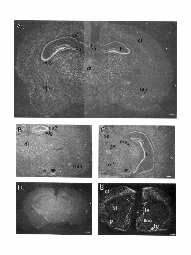
In situ hybridization experiments were undertaken to identify the cell types that express glypican in the adult rat nervous system. High levels of glypican expression were detected in sev- eral structures in the adult rat brain. In the hippocampus, glyp- ican was expressed in high levels in the pyramidal cell layer (Fig. 4A). Furthermore, the pyramidal cell layer in regions CA3 and CA4 expressed glypican mRNA at substantially higher lev- els than in regions CA1 and CA2. Some hybridization was also seen to the dentate gyrus, although at lower levels than in regions CA3 and CA4. High levels of expression were also found throughout the dorsal thalamus (Fig. 4,4-C), including the ven- trolateral, ventrobasal, mediodorsal, lateral geniculate, and me- dial geniculate nuclei, but expression was not seen in the medial and lateral habenula nor in the ventral thalamus (e.g., the re- ticular nucleus and zona incerta).
Strong hybridization was also seen to certain nuclei of the amygdala (Fig. 4A,B), notably the pyramidal layer of the nucleus of the lateral olfactory tract and the basolateral nucleus. Weaker hybridization was seen to the lateral and basomedial nuclei, but no hybridization to the anterior, medial, central, and cortical
The Journal of Neuroscience, June 1994, 14(6) 3717
Figure 3. Antibodies raised against glypican fusion protein recognize adult rat brain HSPG M 12. 1251-labeled adult rat brain PGs (Hemdon and Lander, 1990) were immunoprecipitated with 5 pg of rabbit IgG (lanes 2 and 6), 5 pg of affinity-purified anti-glypican serum (lanes 3 and 7), or 5 pg of anti-glypican serum containing 20 rg of glypican fusion protein. Lanes 1 and 5 show the starting material. Samples were either subjected to SDS-PAGE without heparitinase treatment (lanes 1-4) or treated with heparitinase before electrophoresis (lanes 5-8). Arrowhead indicates the position of the M 12 core protein. M, markers (in kDa) are shown at right.
nuclei was detected. The sense strand of glypican did not hy- bridize to any of these or other structures (Fig. 40). In addition, similar results were obtained using several antisense probes syn- thesized from different regions of glypican (see Materials and Methods) (data not shown).
Other forebrain structures tended to show somewhat less hy- bridization than that observed in the hippocampus, thalamus, and amygdala. These included the piriform cortex (Fig. 4E), olfactory tubercle (Fig. 4E), septal nuclei (data not shown), and the supraoptic (Fig. 4B) and paraventricular nuclei (data not shown) of the hypothalamus. Still weaker, but detectable hy- bridization was seen to the cerebral neocortex (Fig. 4E; see also below) and nucleus accumbens (Fig. 4E).
In the brainstem, relatively strong glypican hybridization was seen to the red nucleus (Fig. 4C), locus coeruleus (Fig. 4F,G), and several cranial nerve nuclei, including the motor nuclei of the 5th (4F,G), 7th (4H), and 12th (4Z) nerves. Hybridization was also seen to the oculomotor complex (Fig. 4C). In the spinal cord, relatively strong hybridization was seen to the ventral horns (not shown), and to the dorsal root ganglia (Fig. 4JJ9.
Many of the brain regions where glypican hybridization was seen contain principal neurons with large, easily recognizable
Figure 4. In situ hybridization of adult rat brain with rat glypican. A, Coronal forebrain section probed with 4X 1, viewed in dark field. B, Coronal forebrain section at the level of the anterior thalamus probed simultaneously with 4X1 and 4P2, viewed in dark field. C, Coronal section through the posterior thalamus and anterior midbrain, probed with 4X 1, viewed in dark field. D, Coronal section at the level of that in A probed with the sense (control) strand of 4X1, viewed in dark field. E, Coronal section through the anterior forebrain probed simultaneously with 4X1 and 4P2; hyperfilm image. F, Coronal section through the brainstem, probed with 4X1, viewed in dark field. G, A section approximately 30 pm from that in F stained with cresyl violet. H, Coronal section through the brainstem, probed with 4X1, viewed in dark field. I, Coronal section through the brainstem, probed with 4X1, viewed in dark field. J, Section through a dorsal root ganglion, probed with 4P1, viewed in dark field. Arrows point to silver grains clustered over sensory neuron cell bodies. K, Same section as in J stained with cresyl violet. L, High-magnification view of the septum and lateral ventricle from a coronal section through the forebrain. M, Same section as in L stained with cresyl violet. Asterisk in B indicates supraoptic nucleus of the hypothalamus; arrow in B indicates the pyramidal layer of the nucleus of the lateral olfactory tract. act, nucleus accumhens; bla, basolateral nucleus of the amygdala; cb, cerebellum; ct, cerebral cortex; dg, dentate gyms; dt, dorsal thalamus; e, ependyma; ha, habenula; hi, hippocampus; Zc, locus coeruleus; Iv, lateral ventricle; me5, mesencephalic nucleus of the trigeminal(5th) nerve; mg; medial geniculate nucleus of the thalamus; mo5, motor nucleus of the trigeminal(5th) nerve; oc, Edinger-Westphal nucleus of the oculomotor complex; p, piriform cortex; rn, red nucleus; s, septal area; SC, superior colliculus; sn, substantia nigra; st, striatum; ~5, sensory nucleus of the trigeminal(5th) nerve; tu, olfactory tubercle; 7, nucleus of the 7th cranial nerve; 12, nucleus of the 12th cranial nerve. In the sequence reported by Karthikeyan et al. (1992), 4X1 corresponds to bases 68 1-843,4Pl corresponds to bases 1873-2440, and 4P2 corresponds to bases 2435-2767. Scale bars: A-I, 500 pm; J and K, 200 pm; L and M, 20 pm.


3720 Litwack et al. * Glypican Expression in the Adult Rat Nervous System
Table 2. Relative levels of glypican hybridization in the adult rat nervous system
Forebrain
Cerebral neocortex
Layers 213, 5, 6
Layers 1, 4
Hippocampus
CAl, CA2
CA3, CA4
Dentate gyrus
Caudoputamen
Dorsal thalamus
Ventral anterior-lateral nucleus
Ventral posterolateral nucleus
Mediodorsal nucleus
Nucleus reuniens
Lateral geniculate nucleus
Medial geniculate nucleus
Anterodorsal
Anteromedial
Lateral dorsal
Anteroventral nucleus
Ventral thalamus
Reticular nucleus
Zona incerta
Epithalamus
Medial habenula
Lateral habenula
Amygdaloid nuclei
Basolateral
Nucleus of the lat. olfactory tract
Lateral and basomedial
Anterior, medial, central, cortical
Hypothalamus
Supraoptic nucleus
Paraventricular nucleus
Arcuate nucleus
Piriform cortex
Olfactory tubercle
Septum
Nucleus accumbens
Endopiriform nucleus
Ependyma
Large meningeal blood vessels
Brainstem
Superior colliculus
Inferior colliculus
Red nucleus
Substantia nigra
Locus coeruleus
Cerebellum
Granule cell layer
Purkinje cell layer
Cranial nerve nuclei
Edinger-Westphal
Motor V
Motor VII
Motor XII
Mesencephalic V
Sensory V
+ -
++
+++
+ -
++
++
++
++
++
++
++
++
++
+
-
-
-
-
++
++
f -
+
+ -
+
+
+
+
+/-
++
+
-
-
++ -
++
-
-
++
+++
+++
+++
+/-
+/-
Table 2. Continued
Spinal cord
Ventral horn motoneurons ++
Dorsal horn neurons +/-
Dorsal root ganglion neurons ++
Intensity of hybridization is graded as + + + (very strong), + + (strong), + (mod- erate), +/- (weak), or - (absent), based in part on autoradiographic exposure times. For example, structures such as fields CA3 and CA4 of the hippocampus were darkly exposed in < I5 d, while structures such as the cerebral cortex were usually only detected in longer exposures (225 d), or when two probes were used simultaneously during hybridization. Structures graded as - sometimes exhibited small clusters of silver grains over rare large cell bodies. Unlisted structures were not determined.
cell bodies. When sections containing these regions were ex- amined at higher magnification, it could frequently be estab- lished that hybridization was localized to such neurons. For example, Figure 54 shows glypican hybridization to cells in the nucleus of the 5th cranial nerve. Figure 5B shows that clusters of silver grains are present over large, lightly stained motoneu- ron cell bodies, but not over smaller, more darkly stained cell bodies (i.e., glia and small neurons). Similar results, that is, hybridization to large, lightly stained cells, were obtained in other regions of the brain, such as the thalamus and hippocam- pus (data not shown).
A characteristic of most of the neurons in regions that were found to express glypican mRNA is that they are projection neurons. In the cerebral neocortex, however, large numbers of local circuit neurons are also present, and in layer 4 account for a large majority ofthe neurons (Zilles, 1990). Interestingly, glyp- ican hybridization was not detected in layer 4 of the cortex, but was readily detected in other cell body layers, that is, 2/3, 5, and 6 (Fig. 6A,B). In layer 2/3, hybridization was strongest in the superficial part (Fig. 6B), and could be localized to a sub- population of large cell bodies (Fig. 6EJ). In layers 5 and 6, hybridization was present at all levels, and was associated with most, but not all, large cell bodies (Fig. 6C,D). Although cell body size and location alone are not sufficient to establish the projection patterns of cortical neurons, the data are at least consistent with the view that glypican is primarily expressed by projection neurons.
Many types of projection neurons throughout the brain ex- pressed little or no glypican mRNA, however. These included most neurons of the caudoputamen (Fig. 4E), the medial and lateral habenula (Fig. 4A), the superior and inferior colliculi (Fig. 4C and data not shown), the substantia nigra (Fig. 4C), and the cerebellum (Fig. 4H,Z). In the latter structure, individual Pur- kinje cells are readily visualized, and could be clearly shown to exhibit no glypican hybridization over background (Fig. 5C,D). Although the possibility remains that levels of glypican mRNA too low to detect are present in these cells, the results emphasize the fact that levels of glypican mRNA expression throughout the brain do not merely correlate with cell body size.
In addition to being present in subsets of neurons, glypican mRNA was also found in some supporting cell (stromal) ele- ments of the brain. For example, glypican hybridization could be seen associated with some large blood vessels, although at weaker levels than in many of the glypican-expressing neurons (data not shown). -Higher but variable levels of hybridization could also be seen associated with ependymal zones (e.g., of the lateral ventricles) (Fig. 4E,L,A4).

The Journal of Neuroscience, June 1994, M(6) 3721
Figure 5. Neuronal expression of glypican. A, Motor nucleus of the trigeminal nerve. Arrows point to clusters of silver grains. Arrowheads point to positions of small nuclei. Dark-field view. B, Same field as in A. Arrows point to large, lightly stained nuclei under the clusters of silver grains indicated in A. Arrowheads point to small nuclei at positions indicated in A. C, Cerebellar cortex. Arrows point to positions of Purkinje cells. Dark- field view. D, Same field as in C. Arrowheads indicate the Purkinje cells marked in C. Scale bars, 20 pm.
Discussion
A major PG of the adult rat brain, HSPG M 12, has been iden- tified as glypican based on the following observations: (1) M I2 is a GPI-anchored PG with a core protein size (- 65 kDa) similar to that observed for human fibroblast,glypican (David et al., 1990; Herndon and Lander, 1990), (2) peptides derived from Ml2 have amino acid sequences identical to those predicted from a rat glypican cDNA (this is in agreement with Karthikeyan et al., 1992, who also identified peptide sequences in a rat brain HSPG core protein that corresponded to sequences in a rat glypican cDNA), and (3) antibodies raised against a rat glypican fusion protein specifically immunoprecipitate M 12 from a crude mixture of brain PGs and recognize a PG core protein that comigrates with M 12 on a Western blot of heparitinase-digested brain membranes (data not shown). In addition, the expression of glypican message parallels the appearance-of M 12 core pro- tein: we have found that glypican message is expressed in the adult and developing rat brain, as is M12, and that glypican mRNA can be detected by in situ hybridization in many brain regions.
Neuronal expression of glypican
The regional distribution of glypican mRNA in the adult brain, as revealed by in situ hybridization, suggests the following gen- eral features of glypican expression in the CNS.
Glypican is primarily a product of neurons. In many of the regions where glypican mRNA is expressed, neuronal cell bodies can be recognized by morphological criteria, for example, lo- cation, nuclear size, and intensity of cresyl violet staining. In such regions (e.g., hippocampus, motor nuclei, dorsal root gan- glia), glypican hybridization is found directly over such cell bodies. This suggests that glial cells, which outnumber neurons in the brain, do not express glypican, although low level ex- pression, or expression by small numbers of glia cannot be ruled out. Supporting and connective tissue elements of the brain (e.g., linings of large blood vessels, meninges, ependyma) also exhibit some glypican hybridization, although at relatively low levels compared with neuronal expression.
Glypican-expressing neurons are predominantly projection neurons. Each of the neuronal populations that expresses glyp- ican appears to consist mainly of cells with axons that project

3722 Litwack et al. l Glypican Expression in the Adult Rat Nervous System
B 1
2/3
Z1
2/3
5
6 6
Figure 6. Glypican expression in the cerebral cortex. A: Right, Coronal section at the level of the basal ganglia stained with cresyl violet. Left, Dark-field view of coronal section 30 pm from that on left hybridized simultaneously with 4X1 and 4Pl. B, Magnified views of corresponding regions from sections in A. Lines indicate approximate divisions between cortical layers. C, High-magnification view of layer 5, dark field. D, Same field as in C, stained with Hoechst 33258. E, High-magnification view of layer 2, dark field. F, Same field as in E, stained with Hoechst 33258. So/id arrows in C-F indicate clusters of silvers grains, and the cell nuclei with which they are associated; open arrows indicate large cell bodies lacking clusters of silver grains. Scale bars: A, 500 pm; B, 200 pm; F, 20 pm.
to other brain regions, or, in the case of motoneurons, to the periphery. Even in the cerebral cortex, glypican expression is present in laminae with corticocortical or corticofugal connec- tions (e.g., layers 2/3, 5, and 6) but absent from layer 4, which consists primarily of local circuit neurons. Since projection neu- rons typically have long axons, relatively high levels of glypican mRNA expression might be required in some projection neu- rons, if it turns out that the physiological processes that involve glypican require the molecule to be present at the axonal surface. Although it remains to be determined whether glypican is, in fact, expressed on the surfaces of axons, it is known that other GPI-anchored proteins are [e.g., F3/Fll (Faivre-Sarrailh, 1992), Tag-l (Dodd et al., 1988; Furley et al., 1990), Thy-l (Xue et al., 1990)].
Levels of glypican mRNA expression vary substantially among dzfirent populations of neurons. Based on the autoradiographic exposure times required to detect glypican expression in differ- ent brain regions, we can estimate relative levels of glypican
expression. High-level expression does not bear a simple rela- tionship to neuronal cell body size or axon length. Indeed, some large and medium-sized projection neurons do not express de- tectable glypican (e.g., cerebellar Purkinje cells).
Although others have suggested that some PGs are localized to neurons that form functional or anatomical systems (Hock- field et al., 1990; Maeda et al., 1992), the types of neurons that express glypican mRNA do not correspond to any single func- tional or transmitter-specific subdivision of the brain. Never- theless, limbic structures-hippocampus, amygdala, thalamus, cingulate and piriform cortex, nucleus accumbens, and septal nuclei-are relatively prominent among glypican-expressing structures. At least one other GPI-anchored protein is known to be associated with limbic system structures [LAMP (Levitt, 1984; Zacco et al., 1990; Zhukareva and Levitt, 1993)]. Also prominent among glypican-expressing neurons are neurons whose axons contribute to peripheral nerves (spinal and cranial motoneurons; dorsal root ganglion neurons).

The Journal of Neuroscience, June 1994, 14(6) 3723
Functional roles of glypican in the adult rat nervous system
Although glypican is only one of many PGs in the mammalian brain, there are reasons to believe that whatever roles it plays there may be unique. In adult brain, glypican is a major HSPG, and it is the only HSPG that exhibits detergent partitioning properties indicative of an integral membrane protein (Hemdon and Lander, 1990). This suggests that the bulk of the HS borne by integral membrane proteins in the adult brain may be found on glypican. Since glypican is only expressed by certain popu- lations of neurons, this suggestion raises the interesting possi- bility that CNS glia and some neurons may be relatively or entirely deficient in cell-surface HS. If so, expression patterns of glypican might play an important role in restricting the sites of action of molecules that normally interact with cell-surface HS.
Among such molecules that are found in the adult brain, members of the FGF family of polypeptide growth factors are present in many locations where glypican-expressing neurons are also found. Acidic FGF, for instance, is highly expressed in spinal and cranial motoneurons, as well as neurons of the dorsal root ganglia, thalamus, septum, and red nucleus, but differs from glypican in being largely absent from the cerebral cortex and hippocampus (Elde et al., 1991; Stock et al., 1992). However, hippocampal pyramidal neurons do express basic FGF [pri- marily in subfield CA2 (Gomez-Pinilla et al., 1992; Woodward et al., 1992)], and FGF-5 [in subfields CA2 and CA3 (Gomez- Pinilla and Cotman, 1992)]. Basic FGF is also found in some cranial motor nuclei (Gomez-Pinilla et al., 1992) and FGF-5 is present in skeletal muscle (Hughes et al., 1993). Although the functions of these growth factors in the adult nervous system are not known, evidence suggests that they may be involved in facilitating neuronal repair and/or neuronal survival (Gomez- Pinilla et al., 1992; Hughes et al., 1993). Since the actions of growth factors of the FGF family apparently require the partic- ipation of the HS chains of HSPGs (Rapraeger et al., 1991; Yayon et al., 199 I), glypican may play an important role in the trophic and injury responses of neurons.
Glypican could also play a role in neuronal injury responses by virtue of interactions with HS-binding extracellular matrix molecules. For example, laminin, a strongly neurite outgrowth- promoting molecule, becomes exposed in the endoneurial sheaths of peripheral nerves during nerve regeneration (Bignami et al., 1984; Longo et al., 1984); both motoneurons and primary sen- sory neurons express glypican, which could then be involved in the recognition of and response to endoneurial laminin. Alter- natively, it is known that at least one HSPG that is released by cells strongly inhibits the neurite outgrowth effects of laminin (Muir et al., 1989); neuronal glypican might be released under some circumstances and thereby interfere with regeneration. Significantly, GPI-anchored molecules, including some GPI- anchored PGs, are known to be shed by cells, presumably through the action of endogenous surface phospholipases (Ishihara et al., 1987; Carey and Evans, 1989; Low, 1989). This could ac- count for the existence of an HSPG with biochemical properties very similar to those of glypican in the adult rat brain soluble (non-membrane-associated) fraction (“S9”; Hemdon and Land- er, 1990).
It is possible that glypican mediates different effects depending on its cellular source. Several heparin-binding proteins bind specifically to subfractions of heparin, suggesting that these pro- teins recognize aspects of GAG structure that may be found
only on some HS chains (Marcum and Rosenberg, 1989; Lee and Lander, 1991; Nurcombe et al., 1993; San Antonio et al., 1993). Purification of glypican from specific neural sources, fol- lowed by binding studies with heparin-binding proteins of the brain, should indicate whether heterogeneity in the HS borne by the glypican core protein influences the kinds of molecules for which glypican, in different types of neurons, may act as a receptor.
References Bemfield M, Kokenyesi R, Kato M, Hinkes MT, Spring J, Gallo RL,
Lose EJ (1992) Biology of the syndecans: a family of transmembrane heparan sulfate proteoglycans. Annu Rev Cell Biol 8:365-393.
Bignami A, Chi NH, Dahl D (1984) Regenerating dorsal roots and the nerve entry zone: an immunofluorescence study with neurofila- ment and laminin antisera. Exp Neurol 85:426-436.
Carey DJ, Evans DM (1989) Membrane anchoring of heparan sulfate proteoglycans by phosphatidylinsoitol and kinetics of synthesis of peripheral and detergent-solubilized proteoglycans in Schwann cells. J Cell Biol 108:1891-1897.
Carey D, Stahl R (1990) Identification of a lipid-anchored heparan sulfate proteoglycan in Schwann cells. J Cell Biol 111:2053-2062.
Carey D, Evans D, Stahl R, Asundi V, Conner K, Garbes P, Cizmeci- Smith G (1992) Molecular cloning and characterization of N-syn- decan, a novel transmembrane heparan sulfate proteoglycan. J Cell Biol 117:191-201.
Chomczynski P, Sacchi N (1987) Single-step method of RNA isolation by acid guanidinium thiocyanate phenol-chloroform extraction. Anal Biochem 162:156-159.
Cole GJ, Loewy A, Glaser L (1986) Neuronal cell-cell adhesion de- pends on interactions of N-CAM with heparin-like molecules. Nature 3201445-447.
David G, Lories V, Decock B, Marynen P, Cassiman J-J, Van den Berghe H (1990) Molecular cloning of a phosphatidylinositol-an- chored membrane heparan sulfate proteoglycan from- human lung fibroblasts. J Cell Biol 111:3 165-3 176.
Dodd J, Morton SB, Karagogeos D, Yamamoto M, Jesse11 TM (1988) Spatial regulation of axonal glycoprotein expression on subsets of embryonic spinal neurons. Neuron 1: 105-l 16.
Drake SL, Klein DJ, Mickelson DJ, Oegema TR, Furcht LT, McCarthy JB (1992) Cell surface phosphatidylinositol-anchored heparan sul- fate proteoglycan initiates mouse melanoma cell adhesion to a fibro- nectin-derived, heparin-binding synthetic peptide. J Cell Biol 117: 1331-1341.
Elde R, Cao Y, Cintra A, Brelje TC, Pelto-Huikko M, Junttila T, Fuxe K, Pettersson RF, Hokfelt T (199 1) Prominent expression of acidic fibroblast growth factor in motor and sensory neurons. Neuron 7:349- 364.
Faivre-Sarrailh C, Gennarini G, Goridis C, Rougon G (1992) F3/Fll cell surface molecule expression in the developing mouse cerebellum is polarized at synaptic sites and with granule cells. J Neurosci 12: 257-267.
Fujita SC, Tada Y, Murakami F, Hayashi M, Matsumura M (1989) Glycosaminoglycan-related epitopes surrounding different subsets of mammalian central nervous systems. Neurosci Res 7: 117-l 30.
Furley AJ, Morton SB, Manalo D, Karagogeos D, Dodd J, Jesse11 TM (1990) The axonal glycoprotein TAG-l is an immunoglobulin su- perfamily member with neurite outgrowth-promoting activity. Cell 61:157-170.
Gomez-Pinilla F, Cotman CW (1992) FGF-5 mRNA distribution in the rat brain: an in situ hybridization study. Sot Neurosci Abstr 18: 953.
Gomez-Pinilla F, Lee JW-K, Cotman CW (1992) Basic FGF in adult rat brain: cellular distribution and response to entorhinal lesion and fimbria-fomix transection. J Neurosci 12:345-355.
Gordon H, Lupa M, Bowen D, Hall Z (1993) A muscle cell variant defective in glycosaminoglycan biosynthesis forms nerve-induced but not spontaneous clusters of the acetylcholine receptor and the 43 kDa protein. J Neurosci 13:586-595.
Herndon ME, Lander AD (1990) A diverse set of developmentally regulated proteoglycans is expressed in the rat central nervous system. Neuron 41949-96 1.
Hemdon ME, Stipp CS, Lander AD (199 1) Interactions between neu-

3724 Litwack et al. * Glypican Expression in the Adult Rat Nervous System
ronal membrane heparan sulfate proteoglycans and proteins of the extracellular matrix. J Cell Biol 115: 126a.
Hockfield S, Kalb RG, Zaremba S, Fryer H (1990) Expression ofneural proteoglycans correlates with the acquisition of mature neuronal prop- erties in the mammalian brain. Cold Spring Harbor Symp Quant Biol 50:505-5 14.
Hughes RA, Sendtner M, Goldfarb M, Lindholm D, Thoenen H (1993) Evidence that fibroblast growth factor 5 is a major muscle-derived survival factor for cultured spinal motoneurons. Neuron 10:369-377.
Ishihara M, Fedarko NS, Conrad HE (1987) Involvement of phos- phatidylinositol and insulin in the coordinate regulation of proteo- heparan sulfate metabolism and hepatocyte growth. J Biol Chem 262: 4708-4716.
Iwata M, Carlson SS (1993) A large chondroitin sulfate proteoglycan has the characteristics of a general extracellular matrix component of adult brain. J Neurosci 13: 195-207.
Jackson RL, Busch SJ, Cardin AC (1991) Glycosaminoglycans: Mo- lecular properties, protein interactions, and role in physiological pro- cesses. Physiol Rev 7 1:48 l-539.
Kan M, Wang F, Xu J, Crabb JW, Hou J, McKeehan WL (1993) An essential heparin-binding domain in the fibroblast growth factor re- ceptor kinase. Science 259: 19 18-l 92 1.
Karthikeyan L, Maurel P, Rauch U, Margolis RK, Margolis RU (1992) Cloning of a major heparan sulfate proteoglycan from brain and iden- tification as the rat form of glypican. Biochem Biophys Res Commun 188:395401.
Klinger MM, Margolis RU, Margolis RK (1985) Isolation and char- acterization of the heparan sulfate proteoglycans of brain. J Biol,Chem 260:4082-4090.
Laemmli U (1970) Cleavage of structural proteins during the assembly of the head of bacteriophage T4. Nature 227:680-685.
Lander AD, Calof AL (1993) Extracellular matrix in the developing nervous system. In: Molecular genetics of nervous system tumors (Levine AJ, Schmidek HH, eds),-pp 341-355. New York: Wiley.
Lee MK, Lander AD (199 1) Analysis of affinity and structural selec- tivity in the binding of proteins to glycosaminoglycans: development of a sensitive electrophoretic approach. Proc Nat1 Acad Sci USA 88: 2168-2772.
Levine JM, Card JP (1987) Light and electron microscopic localization of a cell surface antigen (NG2) in the rat cerebellum: association with smooth protoplasmic astrocytes. J Neurosci 7:27 1 l-2720.
Levitt P (1984) A monoclonal antibody to limbic system neurons. Science 223:299-301.
Litwack ED, Stipp CS, Lander, AD (1992) Glypican, a proteoglycan, is regionally expressed in the developing rat brain. Sot Neurosci Abstr 18:1327.
Longo FM, Hayman EG, Davis GE, Ruoslahti E, Engvall E, Manthorpe M, Varon S (1984) Net&e-promoting factors and extracellular ma- trix components accumulating in vivo within nerve regenerating chambers. Brain Res 309: 105-l 17.
Low MG (1989) The glycosyl-phosphatidylinositol anchor of mem- brane proteins. Biochim Biophys Acta 988:427-454.
Maeda N, Matsui F, Oohira A (1992) A chondroitin sulfate proteo- glycan that is developmentally regulated in the cerebellar mossy fiber system. Dev Biol 15 1:564-574.
Marcum JA, Rosenberg RD (1989) Role of endothelial cell surface heparin-like polysaccharides. In: Heparin and related polysaccharides (Ofosu FA, Danishefsky I, Hirsh J, eds), pp 8 l-94. New York: New York Academy of Science.
Margolis RU, Margolis RK, Chang LB, Preti C (1975a) Glycosami- noglycans of brain during development. Biochemistry 14:85-88.
Margolis RU, Margolis RK, Preti C, Lai D (1975b) Distribution and metabolism of glycoproteins in subcellular fractions of brain. Bio- chemistry 14:47974804.
Muir D, Engvall E, Varon S, Manthorpe M (1989) Schwannoma cell- derived inhibitor of the net&e-promoting activity of laminin. J Cell Biol 109:2353-2362.
Nishiyama A, Dahlin KJ, Prince JT, Johnstone SR, Stallcup WB (199 1) The primary structure of NG2, a novel membrane-spanning proteo- glycan. J Cell Biol 114:359-37 1.
Nurcombe V, Ford MD, Wildschut JA, Bartlett PF (1993) Develop- mental regulation of neural response to FGF- 1 and FGF-2 by heparan sulfate proteoglycan. Science 260: 103-106.
Paxinos G, Watson C (1986) The rat brain in stereotaxic coordinates. Orlando, FL: Academic.
Perides G, Rahemtulla F, Lane WS, Asher, RA, Bignami A (1992) Isolation of a large aggregating proteoglycan from human brain. J Biol Chem 267:23883-23887.
Rapraeger AC, Krulka A, Olwin BB (199 1) Requirement of heparan sulfate for bFGF-mediated fibroblast growth and myoblast differen- tiation. Science 252: 1705-l 708.
Rauch U, Karthikeyan L, Maurel P, Margolis RU, Margolis RK (1992) Cloning and primary structure of neurocan, a developmentally reg- ulated, aggregating chondroitin sulfate proteoglycan of brain. J Biol Chem 267:19536-19547.
Reyes AA, Akeson R, Brezina L, Cole GJ (1990) Structural require- ments for neural cell adhesion molecule-heparin interaction. Cell Re- gul 11567-576.
San Antonio JD, Slover J, Lawler J, Kamovsky MJ, Lander AD (1993) Specificity in the interactions of extracellular matrix proteins with subpopulations of the glycosaminoglycan heparin. Biochemistry 32: 47464755.
Schaffner W, Weissman C (1973) A rapid, sensitive, and specific meth- od for the determination of protein in dilute solution. Anal Biochem 56:502-5 14.
Simmons D, Arriza J, Swanson L (1989) A complete protocol for in situ hybridization of messenger RNAs in brain and other tissues with radiolabelled single-stranded RNA probes. J Histotechnol 12: 169- 181.
Stallcup WB, Beasley L, Levine J (1983) Cell-surface molecules that characterize different stages in the development of cerebellar inter- neurons. Cold Spring Harbor Symp Quant Biol 48:761-774.
Stipp CS, Litwack ED, Lander AD (1994) Cerebroglycan: an integral membrane heparan sulfate proteoglycan that is unique to the devel- oping nervous system and expressed specifically during neuronal dif- ferentiation. J Cell Biol 124:149-160.
Stock A, Kuzis K, Woodward WR, Nishi R, Eckenstein FP (1992) Localization of acidic fibroblast growth factor in specific subcortical neuronal populations. J Neurosci 12:46884700.
Swanson LW (1992) Brain maps: structure of the rat brain. Amster- dam: Elsevier.
Tempst P, Link AJ, Riviere LR, Fleming M, Elicone C (1990) Internal sequence analysis of proteins separated on polyacrylamide gels at the submicrogram level: improved methods, applications and gene clon- ing strategies. Electrophoresis 11:537-553.
Watanabe E, Fujita SC, Murakami F, Hayashi M, Matsumura M (1989) A monoclonal antibody identifies a novel epitope surrounding a sub- population of the mammalian central nervous system. Neuroscience 29~645-657.
Woodward WR, Nishi R, Meshul CK, Williams TE, Coulombe M, Eckenstein FP (1992) Nuclear and cytoplasmic localization of basic fibroblast growth factor in astrocytes and CA2 hippocampal neurons. J Neurosci 12:142-152.
Xue GP, Calvert RA, Morris RJ (1990) Expression of the neuronal surface glycoprotein Thy- 1 is under post-transcriptional control, and is spatially regulated, in the developing olfactory nervous system. Development 109:851-864.
Yanagishita M, McQuillan DJ (1989) Two forms of plasma mem- brane-intercalated heparan sulfate proteoglycan in rat ovarian gran- ulosa cells. J Biol Chem 264:17551-17558.
Yayon A, Klagsbrun M, Esko JD, Leder P, Omitz DM (199 1) Cell surface, heparin-like molecules are required for binding of basic fi- broblast growth factor to its high affinity receptor. Cell 64:841-848.
Zacco A, Cooper V, Chantler PD, Fisher-Hyland S, Horton HL, Levitt P (1990) Isolation, biochemical characterization and ultrastructural analysis of the limbic system-associated membrane protein (LAMP), a protein expressed by neurons comprising functional neural circuits. J Neurosci 10:73-90.
Zhukareva V, Levitt P (1993) LAMP: a novel GPI-linked protein involved in cell-to-substratum adhesion by specific neuronal popu- lations. Sot Neurosci Abstr 19: 129 1.
Zilles K (1990) Anatomy of the neocortex: cytoarchitecture and mye- loarchitecture. In: The cerebral cortex of the rat (Kolb B. Tees RC. eds), pp 77-l 12. Cambridge, MA: MIT Press.