600.full.pdf - Journal of Investigative Medicine
201
Cardiovascular club I 11:00 AM Thursday, February 22, 2018 1 A POSSIBLE ROLE FOR GENETICS IN CARDIOVASCULAR DISEASE AMONG THE ACADIANS AE Tedesco*, Z Haq, KL Di Losa, MJ Ali, P Gregory. LSUHSC – New Orleans, New Orleans, LA 10.1136/jim-2017-000697.1 Purpose of study It is well documented that the Louisiana Aca- dians (‘Cajuns’) experience a disproportionate risk for some genetic diseases due to a genetic founder effect.Furthermore, cer- tain founder populations show a predisposition for developing heart disease.The current study was designed to determine both the population prevalence of cardiovascular disease in the Acadian region and to determine whether the prevalence of early cardio- vascular disease is increased among the Cajun population. Methods used We obtained descriptive information via the electronic medical record of 345 consecutive patients previ- ously diagnosed with early onset (age <50) cardiovascular dis- ease (CVD) who presented to University Hospital and Clinics Cardiology Clinic (June 2015–July 2016). The patients con- sisted of 184 African Americans (97 females and 87 males) and 161 Caucasians (74 females and 87 males).For this study, cardiovascular diseases included: coronary artery disease, HTN, myocardial infarction, and CHF. Demographics, vital signs, lipid panel results, medications, family history, date of CVD diagnosis, and past medical history were collected for all patients.The data on the (161) Caucasian patients were then stratified into either Cajun-identified or non-Cajun by compar- ing each patient’s last name to a standardised list of most popular Cajun last names. Means for each variable between the two groups were compared using independent t-tests. Summary of results The results of our analysis revealed that Cajun-identified patients were diagnosed with a CVD at a signifi- cantly younger age (40±8 years) than were non-Cajun patients (44±4 years, p=0.03).There were no significant differences between the Cajun-identified and non-Cajun groups with regard to BMI, blood pressure, HDL and LDL levels, family history of CVD, or smoking/alcohol use history. Cajun patients had lower tri- glyceride levels (184.6±140.4) than non-Cajun whites (210.3 ±153.1) although whis was not statistically significant. Conclusions Our results show that patients of Cajun ancestry were diagnosed with cardiovascular disease at a younger age than their non-Cajun counterparts.This intriguing finding sug- gests that genetic predisposition may contribute to environ- mental/behavioural factors in development of CVD. Further study is needed to determine the generalizability and the cause of this pathology. 2 STILL WITH HIGHER LDL LEVELS-HISPANICS IN PUERTO RICO SHOWS A LOWER CORONARY ARTERY DISEASE THAN THE USA-EXPLAINED BY GENETIC ADMIXTURE 1 JE Muñoz*, 1 P Elosegui, 1 C Melendez, 1,2 PI Altieri, 1,2 HL Banchs. 1 University of Puerto Rico, Medical Sciences Campus, San Juan, PR; 2 Cardiovascular Centre of Puerto Rico and the Caribbean, San Juan, PR 10.1136/jim-2017-000697.2 Purpose of study Coronary artery disease (C.A.D.) is one of the highest causes of death in the world. The purpose of this study is to compare Puerto Rico (P.R.), Hispanic, U.S.A. coun- try, with the U.S.A., in coronary artery disease. Methods used Compare a population of Hispanics with the high LDL levels with normal total cholesterol and HDL in P. R. and the U.S.A. The study population was 1000 patients. The U.S.A. health statistics and P.R. Department of Health was used for comparison. Summary of results Studying the lipid profile of Puerto Rico population, we found that the mean value of LDL lipoprotein is high (±104 mg/dl) with similar cholesterol and HDL levels in both societies; still the coronary disease (CAD) incidence is lower than the U.S.A. (20%–30%). Investigators from the U.P. R. reported the genetic admixture of this Hispanic population. They reported the admixture consisted of 3 genes called pro- tective against C.A.D. Conclusions C.A.D. is an inflammatory process involving inflammation of the endothelial cells, macrophages and other cells. Probably, this admixture protects the endothelial cells against an aggressive inflammatory process and excessive oxi- dative stress. The observation of stitziel-Washington University which described ANGPTL3 gene which produces low choles- terol levels and absent plaques in the coronary arteries sup- port our hypothesis. 3 OBESITY IS ASSOCIATED WITH AORTIC DIALATION IN MARFAN’S SYNDROME SD Grado*, C Watson, WF Campbell, S Kiparizoska, A Thibodeaux, C Richards, TA Skelton, MR McMullan, ME Hall. University of Mississippi Medical Centre, Jackson, MS 10.1136/jim-2017-000697.3 Purpose of study Marfan’s Syndrome (MS), a connective tissue disorder characterised by a slender build and long limbs, is associated with aortopathy and aortic dissection. Obesity is associated with haemodynamic and metabolic abnormalities such as hypertension, hyperglycemia, inflammation and aortic stiffness that may adversely affect the aorta. We aimed to determine if obesity is associated with aortic dilation in MS patients living in an obese environment. Methods used We retrospectively analysed anthropometric and echocardiographic data from 61 MS patients from the Univer- sity of Mississippi Medical Centre from the past 5 years. Mul- tivariable linear regression was used to assess the association of body mass index (BMI) with aortic root diameter measured on the parasternal long axis view at the sinuses of Valsalva on transthoracic echocardiogram. Summary of results The mean BMI of our MS patients was higher compared with historically published data on MS patients (24.5 kg/m 2 vs 20.1 kg/m 2 ). This corresponds with the high prevalence of overweight (BMI 25.0 to <30 kg/m 2 ) and obesity (BMI 30.0 kg/m 2 ) in the state of Mississippi and suggests that it extends to the congenital heart population. Mean aortic root dimensions were increased in overweight (40.9±5.1 mm) and obese MS patients (51.5±12.0 mm) com- pared to normal weight (37.0±6.8 mm) or underweight (38.1 ±8.6 mm) patients. Aortic root diameter increased with increasing BMI, independent of age, sex, race, systolic blood pressure and height (p=0.06). Conclusions Patients with MS tend to be tall and lean; how- ever, in Mississippi, a state with high rates of obesity, they Abstracts 354 J Investig Med 2018;66:351–640 on August 27, 2022 by guest. Protected by copyright. http://jim.bmj.com/ J Investig Med: first published as 10.1136/jim-2017-000697.609 on 1 February 2018. Downloaded from
-
Upload
khangminh22 -
Category
Documents
-
view
1 -
download
0
Transcript of 600.full.pdf - Journal of Investigative Medicine

Cardiovascular club I
11:00 AM
Thursday, February 22, 2018
1 A POSSIBLE ROLE FOR GENETICS IN CARDIOVASCULARDISEASE AMONG THE ACADIANS
AE Tedesco*, Z Haq, KL Di Losa, MJ Ali, P Gregory. LSUHSC – New Orleans, New Orleans,LA
10.1136/jim-2017-000697.1
Purpose of study It is well documented that the Louisiana Aca-dians (‘Cajuns’) experience a disproportionate risk for somegenetic diseases due to a genetic founder effect.Furthermore, cer-tain founder populations show a predisposition for developingheart disease.The current study was designed to determine boththe population prevalence of cardiovascular disease in the Acadianregion and to determine whether the prevalence of early cardio-vascular disease is increased among the Cajun population.Methods used We obtained descriptive information via theelectronic medical record of 345 consecutive patients previ-ously diagnosed with early onset (age <50) cardiovascular dis-ease (CVD) who presented to University Hospital and ClinicsCardiology Clinic (June 2015–July 2016). The patients con-sisted of 184 African Americans (97 females and 87 males)and 161 Caucasians (74 females and 87 males).For this study,cardiovascular diseases included: coronary artery disease,HTN, myocardial infarction, and CHF. Demographics, vitalsigns, lipid panel results, medications, family history, date ofCVD diagnosis, and past medical history were collected for allpatients.The data on the (161) Caucasian patients were thenstratified into either Cajun-identified or non-Cajun by compar-ing each patient’s last name to a standardised list of mostpopular Cajun last names. Means for each variable betweenthe two groups were compared using independent t-tests.Summary of results The results of our analysis revealed thatCajun-identified patients were diagnosed with a CVD at a signifi-cantly younger age (40±8 years) than were non-Cajun patients(44±4 years, p=0.03).There were no significant differencesbetween the Cajun-identified and non-Cajun groups with regardto BMI, blood pressure, HDL and LDL levels, family history ofCVD, or smoking/alcohol use history. Cajun patients had lower tri-glyceride levels (184.6±140.4) than non-Cajun whites (210.3±153.1) although whis was not statistically significant.Conclusions Our results show that patients of Cajun ancestrywere diagnosed with cardiovascular disease at a younger agethan their non-Cajun counterparts.This intriguing finding sug-gests that genetic predisposition may contribute to environ-mental/behavioural factors in development of CVD. Furtherstudy is needed to determine the generalizability and the causeof this pathology.
2 STILL WITH HIGHER LDL LEVELS-HISPANICS IN PUERTORICO SHOWS A LOWER CORONARY ARTERY DISEASETHAN THE USA-EXPLAINED BY GENETIC ADMIXTURE
1JE Muñoz*, 1P Elosegui, 1C Melendez, 1,2PI Altieri, 1,2HL Banchs. 1University of PuertoRico, Medical Sciences Campus, San Juan, PR; 2Cardiovascular Centre of Puerto Rico andthe Caribbean, San Juan, PR
10.1136/jim-2017-000697.2
Purpose of study Coronary artery disease (C.A.D.) is one ofthe highest causes of death in the world. The purpose of thisstudy is to compare Puerto Rico (P.R.), Hispanic, U.S.A. coun-try, with the U.S.A., in coronary artery disease.Methods used Compare a population of Hispanics with thehigh LDL levels with normal total cholesterol and HDL in P.R. and the U.S.A. The study population was 1000 patients.The U.S.A. health statistics and P.R. Department of Healthwas used for comparison.Summary of results Studying the lipid profile of Puerto Ricopopulation, we found that the mean value of LDL lipoproteinis high (±104 mg/dl) with similar cholesterol and HDL levelsin both societies; still the coronary disease (CAD) incidence islower than the U.S.A. (20%–30%). Investigators from the U.P.R. reported the genetic admixture of this Hispanic population.They reported the admixture consisted of 3 genes called pro-tective against C.A.D.Conclusions C.A.D. is an inflammatory process involvinginflammation of the endothelial cells, macrophages and othercells. Probably, this admixture protects the endothelial cellsagainst an aggressive inflammatory process and excessive oxi-dative stress. The observation of stitziel-Washington Universitywhich described ANGPTL3 gene which produces low choles-terol levels and absent plaques in the coronary arteries sup-port our hypothesis.
3 OBESITY IS ASSOCIATED WITH AORTIC DIALATION INMARFAN’S SYNDROME
SD Grado*, C Watson, WF Campbell, S Kiparizoska, A Thibodeaux, C Richards, TA Skelton,MR McMullan, ME Hall. University of Mississippi Medical Centre, Jackson, MS
10.1136/jim-2017-000697.3
Purpose of study Marfan’s Syndrome (MS), a connective tissuedisorder characterised by a slender build and long limbs, isassociated with aortopathy and aortic dissection. Obesity isassociated with haemodynamic and metabolic abnormalitiessuch as hypertension, hyperglycemia, inflammation and aorticstiffness that may adversely affect the aorta. We aimed todetermine if obesity is associated with aortic dilation in MSpatients living in an obese environment.Methods used We retrospectively analysed anthropometric andechocardiographic data from 61 MS patients from the Univer-sity of Mississippi Medical Centre from the past 5 years. Mul-tivariable linear regression was used to assess the associationof body mass index (BMI) with aortic root diameter measuredon the parasternal long axis view at the sinuses of Valsalva ontransthoracic echocardiogram.Summary of results The mean BMI of our MS patients washigher compared with historically published data on MSpatients (24.5 kg/m2 vs 20.1 kg/m2). This corresponds withthe high prevalence of overweight (BMI 25.0 to <30 kg/m2)and obesity (�BMI 30.0 kg/m2) in the state of Mississippi andsuggests that it extends to the congenital heart population.Mean aortic root dimensions were increased in overweight(40.9±5.1 mm) and obese MS patients (51.5±12.0 mm) com-pared to normal weight (37.0±6.8 mm) or underweight (38.1±8.6 mm) patients. Aortic root diameter increased withincreasing BMI, independent of age, sex, race, systolic bloodpressure and height (p=0.06).Conclusions Patients with MS tend to be tall and lean; how-ever, in Mississippi, a state with high rates of obesity, they
Abstracts
354 J Investig Med 2018;66:351–640
on August 27, 2022 by guest. P
rotected by copyright.http://jim
.bmj.com
/J Investig M
ed: first published as 10.1136/jim-2017-000697.609 on 1 F
ebruary 2018. Dow
nloaded from

have higher mean BMIs when compared to published data.Higher BMI was associated with larger aortic root diametersin our single-centre study. Further assessment of rates of aorticdilation in MS patients with differing BMIs is needed todetermine if overweight and obesity exacerbate aortic dilationin MS.
4 BARRIERS TO EARLY CARDIAC CATHETERIZATION INHIGH RISK NSTEMI PATIENTS
C Basman*, A Bhandary, J Daibes, P Sayegh, S Lebrun, N Coplan. Lenox Hill Hospital, NewYork, NY
10.1136/jim-2017-000697.4
Purpose of study The purpose of this study is to analyse bar-riers for patients with high risk NSTEMI to receive an earlyinvasive treatment strategy.Methods used We conducted a retrospective chart reviewdesigned to evaluate whether patients with high risk NSTEMIare receiving an early invasive (within 1 day of admission) ordelayed invasive (1–3 days after admission) strategy.Summary of results The study included 173 patients that pre-sented to the Emergency Department with high risk NSTEMI.There were 46 patients (average age 64.5 years) in thedelayed invasive arm, and 127 patients (average age 63.2years) in the early invasive arm analysed. Patients receiving adelayed invasive strategy were more likely to have a historyof atrial fibrillation (AF) and to be on anticoagulation (table1). Patients admitted on weekend/holiday were more likely tohave delayed invasive compared to patients admitted duringthe week (table 2).Conclusions It is important to recognise barriers to promptcardiac catheterization and to make hospital system adjust-ments for optimal treatment of patients with NSTEMI.
Abstract 4 Table 1 Chi square analysis; baseline features
Female Gender 19 (41%) 39 (31%) 0.2
Previous Myocardial Infarction 14 (30%) 44 (35%) 0.72
Previous Percutaneous Intervention 17 (36.5%) 40 (31.5%) 0.511
Previous CABG 7 (15%) 12 (9.5%) 0.21
History of Chronic Kidney Disease 11 (23.9%) 19 (15%) 0.13
History of Diabetes 14 (30%) 37 (29.1%) 0.86
History of Hypertension 30 (65%) 94 (74%) 0.49
Tobacco or Smoking History 18 (39%) 50 (39%) 1
History of Atrial Fibrillation (AF) 8 (17.4%) 7 (5.5%) <0.05
History of Congestive Heart Failure 10 (21.7%) 14 (11%) 0.084
History of GI Bleed 1 (2%) 2 (1.5%) 0.72
Antiplatelet Therapy 28 (60.8%) 59 (46%) 0.123
Statin Therapy 21 (45.5%) 52 (41%) 0.65
Anticoagulation Therapy 8 (17.4%) 6 (4.7%)
Beta blockade Therapy 16 (34.7%) 30 (23.6%) 0.13
Temporal change in Troponins.
(Defined in as a value above the
99th percentile of the upper
reference level. Additionally, evidence
for a serial increase or
decrease>20% is required.
45 (97.8%) 123 (97%) 1
EKG Changes (ST Depressions) 12 (26%) 32 (25%) 0.88
GRACE score>140 10 (21.7%) 16 (12.6%) 0.055
Abstract 4 Table 2 Chi square analysis; patients admitted on aweekend/holiday versus weekday
Admitted on a Weekend or
Holiday
20 24
Admitted on a Weekday 26 103
5 INTRAVENOUS IRON ADMINISTRATION REDUCESFIBROBLAST GROWTH FACTOR 23 LEVELS IN CHRONICKIDNEY DISEASE
B Panwar*, O Gutierrez. University of Alabama, Hoover, AL
10.1136/jim-2017-000697.5
Purpose of study Higher fibroblast growth factor 23 (FGF23)is associated with higher risk of heart disease and mortality inchronic kidney disease (CKD). Recently, iron deficiency hasbeen linked to elevated FGF23 levels. We examined whethertreatment with intravenous iron reduces FGF23 in individualswith CKD.Methods used 10 participants with stage 3/4 CKD (eGFR 15–59 ml/min) and scheduled to receive intravenous iron (Feru-moxytol) were enrolled in a single-arm study. The primaryoutcome variable was change in intact FGF23 (iFGF23) or c-terminal FGF23 (cFGF23) levels. Study samples were drawnat baseline and 2 weeks after iron administration. Paired t-testanalysis was used to examine change in log transformediFGF23 and cFGF23 over time.Summary of results At baseline, mean estimated glomerular fil-tration rate, mean serum haemoglobin, mean serum ferritin,mean percent transferrin saturation and mean serum phospho-rus were 29.5 (±0.5) ml/min, 8.8 (±1.5) g/dL, 54.8 (±41.6)ng/ml, 14(±11.4)%, and 3.9 (±0.5) mg/dL respectively. Therewas a significant reduction in mean serum iFGF23 concentra-tion (17.7%) at 2 weeks post iron infusion (two tailedp=0.022). (Figure) There was no significant change in meancFGF23 concentration.Conclusions Intravenous iron administration significantlyreduced serum iFGF23 levels among individuals with stage 3/4CKD. Our findings suggest that optimising iron status in indi-viduals with CKD could potentially decrease FGF23concentration.
Abstract 5 Figure 1 There was no significant change in meancFGF23 concentration
Abstracts
J Investig Med 2018;66:351–640 355
on August 27, 2022 by guest. P
rotected by copyright.http://jim
.bmj.com
/J Investig M
ed: first published as 10.1136/jim-2017-000697.609 on 1 F
ebruary 2018. Dow
nloaded from

6 HIGH GRADE HEART BLOCK FOLLOWING ACCIDENTALEXPOSURE TO ORGANOPHOSPHATE POISONING
GD Bedanie*, D Gebremariam, K Nugent. Texas Tech University Health Centre, Lubbock, TX
10.1136/jim-2017-000697.6
Introduction Organophosphates are potent cholinesterase inhib-itors capable of causing severe cholinergic toxicity followingcutaneous exposure, inhalation, or ingestion. Cardiac manifes-tations, such as sinus bradycardia, prolonged PR interval, andprolonged QTc, occasionally occur. Complete atrioventricular(AV) block has rarely been reported in this poisoning.Case presentation A 45-year-old man was found unconscious andbrought to our emergency centre as Level 1 trauma patient afterhis family found him on the floor. He was in respiratory arrestwith an unknown mechanism and duration. His blood pressureand heart rate were normal. He had abrasions and bleeding fromforehead. He was intubated and admitted to Surgical ICU underthe trauma service. On the next day, his family reported that theyare suspicious of exposure to an unknown chemical. Due to oursuspicion of organophosphate exposure, laboratory test was doneand showed a very low level of acetylcholine esterase. On thesame day, he developed severe bradycardia with recurrent 3rddegree AV block. Cardiology was consulted, and the patient wastreated with atropine.Discussion Organophosphate exposure produces clinical manifes-tations due to cholinergic excess. It inhibits the acetylcholine ester-ase enzyme. Cardiac complications develops secondary toaugmented vagal influence on the sinoatrial and AV nodes.
The presentation of our case was unusual and was mislead-ing; he had no typical manifestations of organophosphate poi-soning at presentation. Cardiac telemetry monitoring helpedus detect recurrent 3rd degree AV block and severe bradycar-dia that developed 24 hour after exposure to organophos-phate. It is rarely described in the literature and completeheart block has been reported in very small number of cases.Development of life threatening cardiac conduction abnormal-ities may not be early. Patients with suspected organophos-phate exposure should be observed closely in an acute caresetting with cardiac monitoring and access to atropine, oximes,and external pacing. Careful monitoring, early recognition ofthis complication, and appropriate management shoulddecrease the mortality rate in these patients.
7 BORTEZOMIB IN THE MANAGEMENT OF CARDIACAMYLOIDOSIS-A METAANALYSIS
JK Bissett. CAVHS/UAMS, Little Rock, AR
10.1136/jim-2017-000697.7
Purpose of study Cardiac amyloidosis is a devastating cardiomy-opathy with poor prognosis. Median survival after the diagnosis isless than 6 months after diagnosis and one year mortality is 45% ifno effective treatment can be offered. Bortezomib is a boronic acidderivative competitively bind to the proteosome S and has shownimprovement in the survival of these patients. As cardiac amyloi-dosis is a rare disease, no large randomised clinical trials has beendone to study the effectiveness of different therapies. Reportedclinical experience is limited to retrospective studies. Recentlythere are abstracts reported different therapies on Bortezomib.The purpose of this study is to review the clinical use and effec-tiveness of Bortezomib in the managment of cardiac amyloidosis.
Methods used Pubmed and Cochrane Evidence based medicinedatabase search with keywords ‘ Bortezomib’ and ‘CardiacAmyloidosis’ ‘systemic AL amyloidosis’ will be used. The listedreports/journal articles are reviewed, inclusion criteria:
1. Patient aged >18;2. Study involve systemic amyloidosis AL type and Bortezomib
used as an either a)induction agent; b) Part of thecombination chemotherapy
3. Studies/reports listed clinical response and survival.
Relevant data are extracted and listed.Summary of results There is a growing interest in the therapyof cardiac amyloidosis. There are more publications involvingcollaboration between centres and quality studies on the useof Bortezomib. Use of Bortezomib either along or more com-monly, in combination with other chemotherapeutic agentssuch as dexamethasone, melphalan and cyclophosphamideshow promise for patients with this devastatin disesse. Thereported cardiac response rate varies from 19%–30%. In addi-tion, Bortezomib used along or in combination with otherchemotherapeutic agents improves survival. The studies so farare limited in terms of number of patients and further stratifi-cation of different risk groups. However, the results so far arepromising.Conclusions Bortezomib hold promise for cardiac AL amyloi-dosis. Further quality study is needed to characterise its use.
8 EFFECTS OF GLUCAGON IN THE CONDUCTION OFSYSTEM OF THE HEART-AND POSSIBLE USES IN THECARDIOVASCULAR AND METABOLIC SYSTEM
1H Nuñez*, 1,2PI Altieri, 1,2HL Banchs, 1N Escobales. 1University of Puerto Rico, MedicalSciences Campus, San Juan, PR; 2Cardiovascular Centre of Puerto Rico and the Caribbean,San Juan, PR
10.1136/jim-2017-000697.8
Purpose of study The role of glucagon in the conduction sys-tem is not clear at the present time, but its role in glycemiccontrol is more known. We decide to clarify its electrophysio-logic effects and define its possible use in close-loop bi hor-monal systems-artificial pancreas.Methods used His bundle studies were done to find the effectin 10 patients (P.) in the sinus node, A-V node and ventricularconduction. B.S. levels were analysed prior and at the end ofthe study.Summary of results Glucagon 5 mg was injected intravenouslyin an infusion which lasted 10 min. His bundle studies weredone by standard techniques. Sinus node function was short-ened by 16.6%, increased the atrioventricular conduction12%, increased the heart rate by 17% (911 P<0.25). Noeffect in intraventricular conduction was found. The effectlasted 10 min. The baseline FBS was 90 mg/dl and at the endof the study 105 mg/dl. (N.S.).Conclusions This shows that glucagon improves the sinusnode and atrioventricular function, but not the intraventricularfunction. No other cardiac function was affected. Elevation ofB.S. was observe, but not significant. We can conclude thatthe conduction system is improved by glucagon without othercardiac abnormalities. This shows that glucagon can be usedin another compartment in the artificial pancreas to avoidhypoglycemia episodes, if any malfunction occurs in thedevice.
Abstracts
356 J Investig Med 2018;66:351–640
on August 27, 2022 by guest. P
rotected by copyright.http://jim
.bmj.com
/J Investig M
ed: first published as 10.1136/jim-2017-000697.609 on 1 F
ebruary 2018. Dow
nloaded from

Adult clinical case symposium
12:00 PM
Thursday, February 22, 2018
9 PELVIC FLOOR MUSCLE EXERCISE-BASED BEHAVIOURALTHERAPY IMPROVES URINARY SYMPTOMS INPARKINSON DISEASE
1,2CP Vaughan*, 3,4K Burgio, 3,4P Goode, 2J Juncos, 1,2L Muirhead, 3,4G McGwin,1,2T Johnson. 1Atlanta VA, Atlanta, GA; 2Emory University, Atlanta, GA; 3Birmingham VA,Birmingham, AL; 4University of Alabama at Birmingham, Birmingham, AL
10.1136/jim-2017-000697.9
Purpose of study Determine the efficacy of pelvic floor muscle-exercise based behavioural therapy (BET) for overactive blad-der (OAB) in Parkinson disease (PD).Methods used Randomised trial of BET compared to control(CON) conducted at two VA medical centres. Participantswere diagnosed with PD by a movement disorders neurologistand had �4 episodes of weekly UI. BET included pelvic floormuscle exercises with urge suppression training, fluid modifica-tion, constipation management, and self-monitoring with abladder diary. CON included a bladder diary and mirroredshape drawing. Outcomes were the International Consultationon Incontinence OAB questionnaire (range 0–16, higher=-worse) and bladder diary-based weekly UI eight weeks post-randomization. Outcomes analysed using generalised linearmodels adjusted for baseline symptoms.Summary of results 53 participants were randomised and 47reported outcome data including 26 randomised to BET and21 to CON (6 dropouts in CON). BET vs CON participantswere similar with respect to age (71.0±6.1 vs 69.7±8.2years), gender (70% vs 78% male), MDS-UPDRS part 3motor score (25.6±13.4 vs 23.8±15.7), cognition (MoCA23.5±3.1 vs 24.9±2.4), mean weekly UI episodes (13.8±9.8 vs 15.2±11.1) and OAB symptoms (8.9±2.4 vs 8.3±2.2). BET reported greater reduction in OAB symptomscompared to CON ((�3.1±2.8) vs (�1.7±2.2), p=0.04).Weekly UI reduction was similar between BET (�7.0±8.9)and CON (�4.8±12.7) (p=0.5). QOL and bother from OABwere significantly improved in BET (p<0.0001 for both) com-pared to baseline.Conclusions Behavioural therapy improved overactive bladdersymptoms in Parkinson disease. Bladder diary self-monitoringwas associated with urinary incontinence reduction in bothgroups. Providers should consider behavioural therapy as ini-tial therapy for overactive bladder symptoms in Parkinsondisease.
10 RELIABILITY OF A NOVEL INDEX OF SENESCENT CELLABUNDANCE IN HUMAN ADIPOSE TISSUE
1L Raborn*, 2J Justice, 2H Gregory, 2B Nicklas, 2J Ding, 2S Kritchevsky, 2J Williamson.1LSUHSC – NO Medical School, New Orleans, LA; 2Wake Forest School of Medicine,Winston-Salem, NC
10.1136/jim-2017-000697.10
Purpose of study We sought to evaluate reliability of scoringsenescent cell abundance by immunohistochemical [IHC] stain-ing of p16INK4a+ cells in adipose tissue.
Methods used Cells expressing p16INK4a were identified byIHC staining of adipose tissues obtained from women whowere older (69.8±7.1 years) and overweight/obese (BMI: 32.2±2.9 kg/m2). Fields were viewed at 20 × magnification usingOlyVIA pathology software; 15–20 fields per sample at werechosen randomly, and images screen-captured for offline quan-tification. Two independent examiners reviewed each visualfield and recorded total cells counted, and cells expressingStrong, Medium, or Weak intensity positive nuclear stainingfor p16INK4a. Cell counts were compared between examiners,and inter-rater reliability was assessed by partial correlationsadjusting for sample (r) and intra-class correlation coefficients(ICC) per sample.Summary of results A total of 115 visual fields were analysedfrom 6 adipose tissue samples. Overall, an average of 38.0±27 cells were counted per visual field, and 6.6%±5.1%p16INK4a+ cells scored Strong (0.58±0.96 p16INK4a+ cells perfield) or Medium (1.78±1.5 p16INK4a+ cells per field) signalintensity, and 14.7%±8.8% p16INK4a+ at any intensity. Signifi-cant differences between raters were observed in cell countswith Weak signal intensity (3.76±3.1 vs 1.83±1.2 counts perfield, p<0.05), but not total cell counts, or Medium/Strongintensity cell counts (p�0.05, all). Between-rater partial corre-lations for p16INK4a+ counts per field were: Weak r=0.61;Medium r=0.67; Strong r=0.83 (p<0.05 all). Between-raterconsistency was observed in: total cell counts per field(ICC=0.98), and p16INK4a+ cell counts of Medium(ICC=0.89) or Strong (ICC=0.99) intensity (p<0.05 all), butnot Weak (ICC=0.64, p�0.05).Conclusions Adipose tissue senescent cells that stain p16INK4a+with Medium or Strong signal intensity can be reliably identi-fied and quantified between raters. This study provides evi-dence for the utility of the adipose tissue p16INK4a + cellabundance as a biomarker of cellular senescence in olderadults.
11 INTRAEPIDERMAL BLISTERS VS RE-EPITHELIALIZATIONOF SUBEPIDERMAL BLISTERS: THE SPECTRUM OFHISTOPATHOLOGIC FINDINGS IN BULLOUS PEMPHIGOID
BD Hodge*, R Brodell, J Mitchell, AB Googe, T Patel, J Schulmeier. University of MississippiSchool of Medicine, Jackson, MS
10.1136/jim-2017-000697.11
Purpose of study To examine the incidence of re-epithelializa-tion of subepidermal blisters causing the appearance of anintraepidermal blistering process leading to diagnosticchallenges.Methods used A search of positive immunofluorescence reports(2006–2015) confirming the presence of autoimmune subepi-dermal blistering was performed and then compared to hema-toxylin and eosin (H and E) findings to identify cases ofbullous pemphigoid. The presence of subepidermal blistering,intraepidermal blistering, absence of blistering reflecting urtica-rial and eczematous pemphigoid, and evidence of partial re-epithelialization were tabulated. Cases were not counted on aper-patient basis but rather per biopsy location; therefore, twobiopsies from one patient were reported as two cases.Summary of results 77 cases were identified as having bothimmunoflouresence and histopathologic features indicative ofan autoimmune subepidermal blistering process. Of thesecases, 43 showed dermal-epidermal blisters (55.84%), 12
Abstracts
J Investig Med 2018;66:351–640 357
on August 27, 2022 by guest. P
rotected by copyright.http://jim
.bmj.com
/J Investig M
ed: first published as 10.1136/jim-2017-000697.609 on 1 F
ebruary 2018. Dow
nloaded from

showed features of urticarial or eczematous pemphigoid(15.58%), 12 showed at least focal intraepidermal blistering(15.58%), and 10 showed complete re-epithelialization produc-ing the appearance of an intraepidermal blister (12.99%). Alimitation of this study is the possibility that subepidermalautoimmune processes other than bullous pemphigoid (epider-molysis bullosa acquisita and cicatricial pemphigoid) couldhave been included in this analysis.Conclusions Re-epithelialization of subepidermal blisters canpose diagnostic challenges as it leads the clinician to suspectan intraepidermal blistering process. By starting with immuno-fluorescent findings typical of pemphigoid and then reviewingH and E findings, it was determined that 55.84% of thetime, histology demonstrated classic dermal-epidermal blister-ing. In 12.99% of specimens, however, histology showed com-plete re-epithelialization of the blister floor. Thus, thepresence of an intraepidermal blister does not preclude thediagnosis of a sub-epidermal blistering process such as pemphi-goid highlighting the importance of clinical-pathologic correla-tion. Biopsies taken of newer lesions are less likely to showthis phenomenon.
12 LANGERHANS CELL HISTIOCYTOSIS WREAKS HAVOC ONTHE HYPOTHALAMUS
C Bicknese*, M McLemore, A Ioachimescu. Emory University School of Medicine, Atlanta,GA
10.1136/jim-2017-000697.12
Case report A 23-year-old previously healthy man presentedwith polyuria, polydipsia of 10 month duration. One monthprior to the endocrinology visit, a polyp was removed fromthe right ear canal after he complained of ear pain. Pathologyrevealed Langerhans cell histiocytosis (neoplastic cells stainedfor CD45, S100, CD1a, and vimentin). Other symptomsincluded fatigue and decreased libido. On physical exam, the98 kg patient appeared euvolemic. Fluid balance was negative:intake 11.49/output 11.52 L. Laboratory studies confirmeddiabetes insipidus: serum sodium 144 (136–145), urine osmo-lality 87 mOsm/kg (300–900 mOsm/kg), and central hypogo-nadism: undetectable testosterone (280–1,1,00 ng/dL),luteinizing hormone 1.0 mlU/mL (1.2–8.6 mlU/mL), folliclestimulating hormone 2.62 mlU/mL (1.2–19.2 mlU/mL). Prolac-tin was slightly elevated (17 ng/mL; 3–13 ng/mL), cortisol, thy-roid and IGF-1 levels were normal. MRI of the brain showedmass-like enhancement of the hypothalamus and superiorinfundibulum, and enhancing lesions of right parietal calva-rium and mastoids extending to the external auditory canal.PET scan did not reveal other lesions. Ophthalmology evalua-tion was normal. The patient received monthly cytarabine,oral desmopressin and transdermal testosterone. After 3 cyclesof chemotherapy, brain MRI and PET scans showed improvedappearance of hypothalamic and bone lesions.Discussion LCH is a rare granulomatous disease characterisedby abnormal expansion of dendritic cells. The estimated inci-dence is 3–5/million in children and 1–2/million in adults.Sites involved usually in adults are bones, lungs and skin.CNS-LCH is rare and usually associated with multisystem dis-ease. Both anterior and posterior hypothalamic-pituitary axis
(HPA) can be involved and up to 30% of patients have diabe-tes insipidus. There is currently no standardised therapy forCNS-LCH. Some patients are conservatively monitored whileother receive high-dose steroids, chemotherapy (vinblastine,etoposide, cytarabine) or radiation. While prospective studiesare lacking, hypopituitarism persists in most cases aftertreatment.Conclusion LCH should be considered in the differential diag-nosis of diabetes insipidus and hypothalamic masses. Earlydiagnosis is important as LCH is a multisystem organ diseasethat may progress in absence of therapy.
13 TO DIAGNOSE IT, YOU NEED TO THINK OF IT
S Chalasani*, L Maher. University of Mississippi, Jackson, MS
10.1136/jim-2017-000697.13
Case report Immunoglobulin G4-related disease (IgG4-RD) israre immune-mediated condition affecting multiple organs suchpancreas, kidney, lung, salivary glands, lymph nodes and skinwith a characteristic lymphoplasmacytic infiltrate enriched inIgG4 positive plasma cells. It occurs mostly in older maleswith history of allergic diseases. Here we present a patientwho had multiple hospitalizations with nonspecific symptomsand extensive work up.
A 64 year old male with past medical history of hyperten-sion, dyslipidemia and diabetes comes for evaluation of 6months history of nausea, vomiting, abdominal pain andweight loss. Patient had multiple hospitalizations and under-went an extensive work-up which showed hepatomegaly witha pancreatic mass and intrahepatic lesions as well as lympha-denopathy and an abnormal SPEP without a discrete monoclo-nal spike to point to multiple myeloma. He was seen inallergy clinic 8 months prior for evaluation of hives, rhinitis,and dyspnea. He was diagnosed with asthma and was startedon Albuterol. His rheumatologic and infectious work up wasnegative. He had endoscopy and colonoscopy with biopsieswhich did not show any evidence of malignancy. He under-went right axillary lymph node biopsy that showed hyperplas-tic features with negative flow cytometry. Since he hadelevated IgG and IgG4 level of 3360, a liver biopsy was per-formed that showed dense portal lymphoplasmacytic infiltrateswith predominance of IgG4 plasma cells. A diagnose of IgG4-related disease was made and he was started on high dose ste-roids with significant clinical improvement. In addition, hereceived 2 infusions of 1 gram Rituximab 2 weeks apart withplans to repeat imaging in future.
IgG4-related diseases is heterogeneous disorder involvingmultiple organs and presents with nonspecific symptoms. Thegold standard for the diagnosis is dense lymphoplasmacyticinfiltrates enriched in IgG4-positive plasma cells organised in astoriform pattern with fibrosis and obliterative phlebitis. Serol-ogies are negative except for the elevated IgG4 (>135 mg/dL)seen in 60% to 70% of cases which can be a marker for amore aggressive disease course. Treatment includes immuno-suppressive drugs; of these, rituximab appears to be mosteffective. Due to the rarity of the disease and nonspecificsymptoms, a high index of suspicion is needed for early diag-nosis and treatment.
Abstracts
358 J Investig Med 2018;66:351–640
on August 27, 2022 by guest. P
rotected by copyright.http://jim
.bmj.com
/J Investig M
ed: first published as 10.1136/jim-2017-000697.609 on 1 F
ebruary 2018. Dow
nloaded from

14 A RARE VASCULAR CAUSE OF RECURRENT PNEUMONIA
MA Alawoki*, EK Addo-Yobo, B Boonpheng, N Kakkar, C Rosero, DM Pierce. EastTennessee State University, Johnson City, TN
10.1136/jim-2017-000697.14
Case report Pneumonia is the infection of the lung paren-chyma that can be life-threatening if not managed appropri-ately. Most community acquired pneumonias have goodprognosis, with majority recovering within a month. However,when it is recurrent in the same anatomical location, it isimportant to do further investigation to look for the cause.With this inquisitive clinical approach, an undiagnosed aber-rant right subclavian artery (ARSA) was found to be the causeof recurrent pneumonia.
A sixty five year old female with a recent history of twoepisodes of right middle lobe(RML) pneumonia, within theprevious five months, presented for a hospital follow up.Chest X-ray from both hospital visits suggested RML processeswhich resolved with levofloxacin. A detailed history duringour encounter uncovered intermittent dysphagia to solids andliquids. The recurrent RML pneumonia and the remote his-tory of dysphagia suggested significant pulmonary aspiration.A video fluoroscopy showed compression of the oesophagus.A CAT scan showed the presence of an ARSA that was com-pressing the upper oesophagus.
We discovered that an ARSA compressed the upper oeso-phagus, which caused recurrent aspiration pneumonia. ARSAis an uncommon congenital anatomical variant where the rightsubclavian artery comes directly off the aortic arch instead ofthe brachiocephalic trunk. It travels posterior to the oesopha-gus towards the right upper extremity; causing esophagealcompression. If symptomatic, it will usually present in infantswith respiratory symptoms or after decades as dysphagia alongwith chest pain, hoarseness or anorexia. Our patient had twoupper endoscopies however no imaging study was done toevaluate for extrinsic esophageal compression. Despite theappropriate workup, vascular causes can be missed if notconsidered.
This case demonstrated a presentation of an ARSA com-pressing the upper oesophagus and causing dysphagia in apatient with recurrent RML pneumonia. Patients with recur-rent pneumonia in the same anatomic location needs furtherworkup and detailed questioning about dysphagia or aspirationevents.
15 ACUTE CONFUSION AS A SEQUELA OF PEPTO-BISMOLTOXICITY
A Prystupa*, S Ahmed, RS Urban. Texas Tech Univ HSC Amarillo, Amarillo, TX
10.1136/jim-2017-000697.15
Case 61-year-old female with PMH of hypertension presentedwith acute confusion, hand tremors, and gait ataxia. Workuprevealed negative RPR, normal TSH and Vitamin B12. Urinetoxicology, blood alcohol, salicylate, and acetaminophen levelswere negative. Lab work uncovered refractory hypokalemiaand normal AG metabolic acidosis. Non-contrast head CT andbrain MRI showed age-related atrophy, subacute/chronic bilat-eral thalamic enhancement and old basal ganglia lacunarinfarctions. Family disclosed her overuse of Pepto-Bismol fordyspepsia. Bismuth toxicity was suspected due to progressiveneurological decline, new onset RTA and history of Pepto-
Bismol use. High blood and urine bismuth level confirmed thediagnosis. Patient was treated conservatively with discontinua-tion of Pepto-Bismol and serial follow-up. At 4 weeks wedetected resolution of tremor, gait ataxia, ankle clonus, andmarked improvement of memory.Impact Bismuth is a heavy metal and an active ingredient of apopular and usually safe OTC medication, Pepto-Bismol (bis-muth subsalicylate). Its chronic use can lead to bismuth intoxi-cation manifesting as memory change, confusion, depression,insomnia, ataxia, tremor, myoclonus, seizures and coma.Discussion Acute or chronic ingestion of toxic dose of bismuthcan lead to progressive confusion, myoclonus, lack of coordi-nation, and speech disturbance. Bi binds to sulfhydryl groups,leading to white matter changes in CNS and proximal tubuledamage in the kidney. Suggestive history, high blood and urinebismuth levels confirm the diagnosis of toxicity. Managementis symptomatic with a gradual but unpredictable improvementafter discontinuation of product. Extensive search for infec-tious and metabolic causes of encephalopathy was negative inour patient. The history of chronic bismuth use with highblood and urine levels and typical clinical findings with subse-quent resolution of symptoms confirmed the diagnosis of bis-muth encephalopathy. Our patient was treated conservativelyand followed the typical course of Bi encephalopathy withgradual improvement at follow-up. More detailed labellingwould increase awareness to avoid this potential toxicity. Bitoxicity should be considered in a patient presenting with sug-gestive history and symptoms.
16 RAPIDLY PROGRESSING HTLV-1 ASSOCIATEDMYELOPATHY/TROPICAL SPASTIC PARAPARESISPRESENTING AS BILATERAL LOWER EXTREMITYWEAKNESS
B Nguyen*, M Gutierrez, K Rizg, C Palacio, J Shah. University of Florida College ofMedicine, Jacksonville, FL
10.1136/jim-2017-000697.16
Case report Patient is a 60 y/o African American male with a his-tory of HTN and seizures who presented for a six-month historyof bilateral lower extremity weakness that worsened to the pointwhere he was unable to walk. Associated symptoms includedupper extremity tremors, urinary retention and repeated falls.Remarkable labs included mildly elevated CK at 329, with nega-tive RPR, Lyme, ANA, SSA/SSB, Aldolase, immunofixation, andHIV. MRI of neuro axis was done which showed no masses, noacute findings, and mild degenerative changes in C and L spinewith minor disc bulging and mild neuro foraminal stenosis in Lspine without any cord changes. Previous notes revealed an epi-sode of uveitis in March 2015, but that there were no reportedcomplaints of lower extremity weakness prior to the initial presen-tation. During outpatient Neurology follow-up, it was thoughtthat his symptoms were consistent with a neurodegenerative proc-ess. Vitamin B12, Folate, MMA, Ceruloplasmin, and HTLV-1were ordered and HTLV-1 was positive. He had multiple hospital-izations for progression of symptoms and was eventually diag-nosed with TSP based on WHO criteria as lumbar puncture wasdeferred. He was sent to rehab for physical therapy but due toprogression of disease leaving him mostly bedbound, he developedcomplications of sacral decubitus ulcers, recurrent UTI’s and diedsecondary to sepsis only 14 months after onset of symptoms.
Abstracts
J Investig Med 2018;66:351–640 359
on August 27, 2022 by guest. P
rotected by copyright.http://jim
.bmj.com
/J Investig M
ed: first published as 10.1136/jim-2017-000697.609 on 1 F
ebruary 2018. Dow
nloaded from

Human T-Lymphotropic virus 1 (HTLV-1) has been impli-cated in multiple diseases such as HTLV-1 associated myelop-athy/tropical spastic paraparesis (TSP), an insidious diseasewith progression over 12–24 years. However, we report acase of TSP with rapid progression over 14 months, whichcontributed to his demise. Possibly, due to the rapid course,this patient did not have cord atrophy that is often seen onMRI of patients with severe, chronic TSP. CSF serology stud-ies should be considered during initial admission as this mayhelp to confirm the diagnosis and to identify markers of amore rapid course of TSP in the future. We suggest that if arapid course of TSP is suspected, Psychiatry should beinvolved early on as patients can become demotivated anddecompensate rapidly.
17 LEFT VENTRICULAR NON-COMPACTIONCARDIOMYOPATHY: CASE OF A RARE CONGENITALDISORDER PRESENTING WITH MULTIPLE ORGANFAILURE
I Serrano, S Lulla, U Warden, B Al-Turk, S Maharaj. University of Florida, Jacksonville, FL
10.1136/jim-2017-000697.17
Case report A 42-year-old African American male with pastmedical history of chronic kidney disease and hypertensionpresented to the hospital with complaints of worsening dysp-nea, cough and orthopnea for one month. On presentation,he was found to be volume overloaded with significant bilat-eral lower extremity oedema, rales and a pro-BNP of 11 500.A transthoracic echocardiogram revealed severe mitral regurgi-tation, an ejection fraction of 25%–30% and left ventricularenlargement with trabeculations consistent with non-compac-tion cardiomyopathy. After diuresis and optimisation of vol-ume status, he underwent left heart catheterization withfindings of non-obstructive coronary artery disease. Patientwas initiated on goal directed medical therapy for heart failureand discharged. Months later an echocardiogram was repeatedwith no recovery in function. Medical therapy was optimisedand a dual chamber-ICD was placed.
Two years later, the patient returned with complaints ofdyspnea, palpitations, and oedema. He was found to be inatrial fibrillation with rapid ventricular response and decom-pensated heart failure. The morning after admission, thepatient had a new onset seizure and shortly thereafter wentinto PEA arrest. Return of spontaneous circulation wasachieved after 10 min and required endotracheal intubation.During hospital course the patient’s renal function worsenedwith minimal urine output, eventually requiring hemodialysis.A repeat echocardiogram revealed a worsened ejection fractionof 10%–15%.Discussion Left ventricular non-compaction (LVNC) is a rarecongenital heart disorder that occurs in-utero due to arrest inthe compaction of the developing myocardium, resulting in‘spongy’ appearance of the left ventricle and thick myocardialwall. It is a genetic cardiomyopathy that presents with heartfailure, ventricular arrhythmias, systemic embolism or suddendeath. It has a prevalence of 0.01% to 1.3% on echocardio-gram. This case presents a middle-age man who developedrapidly worsening of heart function, arrhythmias, renal failure,PEA arrest and hypoxic respiratory failure as subsequent com-plications from LVNC cardiomyopathy.
18 SERIAL DILATION FOR COMPLICATED EXTRINSICCERVICAL ESOPHAGEAL STRICTURES
CC White. Eisenhower Medical Centre, Evans, GA
10.1136/jim-2017-000697.18
Purpose of study The unconventional use of serial mechanicaldilations of extrinsic cervical esophageal strictures has beenattempted with promising results.Methods used Our patient is a 63 year old female with a his-tory of stage 3 metastatic papillary thyroid carcinoma statuspost total thyroidectomy with selective right neck dissectionwho presented to our service with progressive dysphagia andhoarseness. Two years following her initial surgical manage-ment, she had recurrence of her disease requiring right radicalneck dissection and radiation. She developed vocal cord para-lysis and became tracheostomy dependent for more than sevenyears until a cordotomy was performed. She experienced per-sistent dysphagia and hoarseness following the placement ofher tracheostomy. Despite maximal medical therapy, she oftenexperienced food regurgitation requiring additional chewing tofacilitate swallowing. She was evaluated with upper endoscopy,which revealed extrinsic esophageal strictures. ENT assessedfor the patient for symptomatic relief and was deemed unableto provide any assistance because of her pathology. Both theaetiology and location of the stricture limited intra-lesionalsteroid therapy and stent placement. An upper endoscopy wasperformed and the stricture was bypassed utilising a specialisedscope. A wire was placed and the stricture was dilated to anestimated of 10 mm. She has required 12 additional dilationsat about 4 weeks interval over a year. The most recent dila-tion was 18 mm. As expected, there has been restenosis fol-lowing each procedure and the strictures appeared more openand stable. Marked clinical improvement was noted with ourtherapy and there has been no suggestion of significantadverse effects.Summary of results Progressive serial dilation is a safe andeffective method of treatment for complex extrinsic cervicalesophageal strictures not amenable to alternative therapies.There is minimal restenosis effect after dilation as the fre-quency of the dilations increases. The appropriate frequencyof dilations is dependent on the patient’s response to therapy.To date, progressive serial dilations appear safe and effectivefor severe, complex esophageal strictures.Conclusions Progressive serial dilation is a safe and effectivemethod of treatment for complex extrinsic cervical esophagealstrictures not amenable to alternative therapies.
Neonatal case report symposium
12:00 PM
Thursday, February 22, 2018
19 ACUTE ALCOHOL INTOXICATION AND WITHDRAWAL INA PREMATURE INFANT
MG Johnson*, ER Miller, AC Farris. University of Louisville, Louisville, KY
10.1136/jim-2017-000697.19
Case report Acute alcohol intoxication in an infant has rarelybeen reported, and the majority of reported cases have
Abstracts
360 J Investig Med 2018;66:351–640
on August 27, 2022 by guest. P
rotected by copyright.http://jim
.bmj.com
/J Investig M
ed: first published as 10.1136/jim-2017-000697.609 on 1 F
ebruary 2018. Dow
nloaded from

involved exposure after delivery in term infants. Here wereport a case of a preterm infant with acute alcohol intoxica-tion at birth.
A male infant at 27 weeks gestation was admitted to theNICU for acute alcohol intoxication, metabolic acidosis andprematurity. The pregnancy was complicated by extensivematernal alcohol abuse and scant prenatal care. Maternalblood alcohol level was 382 mg/dL prior to delivery. Theinfant delivered precipitously prior to NICU team arrival andApgar scores were 1, 5, and 7 at 1, 5, and 10 min. Theinfant was quickly intubated and transferred to the NICUwhile the mother was transferred to intensive care for acutealcohol withdrawal. On admission, the infant demonstratedsevere anion gap metabolic acidosis with pH 7.00 and bicar-bonate 7 mmol/L. The infant’s blood alcohol level was132 mg/dL at nine hours of life. Despite aggressive fluid resus-citation, sodium bicarbonate administration, and appropriatemechanical ventilation, the metabolic acidosis persisted with alactic acid level of 16 mmol/L at thirteen hours of life. Liverdysfunction was noted with transaminitis and coagulopathyrequiring cryoprecipitate and platelet transfusions. Lorazepamwas initiated on day of life two for increased jitteriness andagitation presumed to be secondary to alcohol withdrawal andwas continued for several days as the anion gap acidosis grad-ually resolved.
Ethanol freely crosses the placenta and is a well-knownteratogen. Ethanol for umbilical cord care has been the mostcommon reported source of acute infant intoxication, althoughthere are also reports of ingestion related to child abuse. Onlyone case report from 1967 described an infant with acutealcohol intoxication immediately after birth, and no casereports were found describing acute intoxication in a prema-ture infant. Infants with acute alcohol intoxication can presentwith a variety of symptoms including hypothermia, tachycar-dia, poor suck, lethargy, seizures and coma. Associated labfindings include anion gap metabolic acidosis and hypoglyce-mia. Therapy includes treatment of acidosis and electrolyteabnormalities and supportive care including antiepileptic medi-cations and respiratory support as needed.
20 HEMATOCHEZIA IN A NEONATE: THINK BEYONDNECROTIZING ENTEROCOLITIS
FJ Zaidi*, P Agarwal, D Macariola, S Hollinger. East Tennessee State University, JohnsonCity, TN
10.1136/jim-2017-000697.20
Case report A 37 week late preterm female born vaginally pre-sented at 36 hours of life with neonatal abstinence syndrome.Delivery was uneventful. Maternal history was positive forhepatitis C, polysubstance abuse and poor prenatal care.Mother denied having any genital infection during her entirepregnancy. On admission, physical examination was unremark-able except for irritability and mildly increased tone. She wastreated with morphine for a total of 10 days. Withdrawalsymptoms resolved but she remained in NICU due to poorfeeding. Patient required nutritional support but was otherwisedoing well until day of life (DOL) 16, when she developedsevere rectal bleeding associated with hypothermia and respira-tory distress. She was initially placed on supplemental oxygenvia nasal cannula but with increasing oxygen requirements sheeventually needed to be on high frequency jet ventilation. The
patient was kept NPO and was started on Ampicillin andGentamicin. Sepsis work up was obtained and infectious dis-ease was consulted. Blood and urine cultures stayed negativethroughout. CRP levels raised from <0.5 on admission to45.0 mg/L. KUB, CXR and urinalysis were unremarkable. OnDOL 17, the patient developed significant apnea, bradycardia,and hepatomegaly along with thrombocytopenia. She wastreated with IV acyclovir and was transfused with platelets.Rectal bleeding worsened and was treated with fresh frozenplasma. AST and ALT were >2600 IU/L while platelet countdropped significantly from 2 53 000 to 14,000 K/uL. Haema-tology/Oncology was consulted who recommended parvovirusPCR, DIC panel and IVIG infusion. The patient progressivelydeteriorated, developed fulminant hepatitis, acute renal failureand disseminated intravascular coagulopathy. Despite assistedventilation, vasopressor support, multiple blood products, andaggressive antiviral therapy, the patient passed away on DOL19. The results of serum HSV DNA PCR came back positivepost mortem.
Disseminated cases constitute only one quarter of HSVcases but have the highest mortality of 85% in untreatedpatients. In around 30% of cases, there are no cutaneousmanifestations making an accurate diagnosis of HSV challeng-ing. We report a neonate with rectal bleeding as the initialpresenting symptom of disseminated HSV infection.
21 AN INFANT WITH CONGENITAL MYOTONIC DYSTROPHYPHENOTYPE
K Jeffries*, J Philips. University of Alabama, Birmingham, AL
10.1136/jim-2017-000697.21
Introduction Congenital myotonic dystrophy (CMD) is a rarecondition that presents with hypotonia and often respiratorydistress. We present a severe case.Case report This 2,130 g female infant was born at 34 weeksby Caesarian for NRFHT and polyhydramnios to a 28 y/omother with a history of a prior 35 week infant who diedshortly after birth from respiratory failure. The case infantwas hypotonic with no respiratory effort at birth and wasintubated and placed on a ventilator. Apgar scores were 1, 1,and 2 at 1, 5, and 10 min, respectively and were primarilydue to profound hypotonia. Admission exam showed Jeffries Ksevere hypotonia and chest X-ray showed thin ribs. Themother has a positive family history for CMD, exhibits myo-tonic facies,and has difficulty releasing her hand after a hand-shake. However, array CGH was normal and karyotyperevealed a balanced translocation between chromosomes 17qand 19q13. The infant failed numerous attempts at extubationand had severe feeding intolerance for which she received atracheostomy and feeding gastrostomy. She has developedbronchopulmonary dysplasia, persistent patent ductus arteriosuswith left-to-right shunt and right ventricular hypertrophy. At 6months of age,she is tolerating 30 min trials of continuouspositive airway pressure every 4 hours in an attempt to weanfrom the ventilator. She is on continuous gastrojejunal tubefeedings with OG tube clamping for one hour every fourhours. Profound hypotonia persists.Discussion CMD is a rare autosomal dominant genetic disor-der with an incidence of about 1 in 45 to 50 thousand livebirths. Inheritance is almost always from an affected motheralthough parental inheritance has been documented. The
Abstracts
J Investig Med 2018;66:351–640 361
on August 27, 2022 by guest. P
rotected by copyright.http://jim
.bmj.com
/J Investig M
ed: first published as 10.1136/jim-2017-000697.609 on 1 F
ebruary 2018. Dow
nloaded from

aetiology is an unstable CTG repeat in an untranslated regionof the dystrophia myotonica protein kinase (DMPK) gene.These repeats in the mRNA inhibit proper splicing and resultin disease. The more CTG repeats there are, the more severethe disease. Infants with greater than 1000 repeats manifestdisease in the newborn period while individuals with fewerrepeats have onset later in life.Conclusion All infants presenting with hypotonia in the new-born period should have a family history taken and be eval-uated for the possibility of this disease.
22 A RARE CASE OF COMBINED GENETIC SYNDROMESMOWAT WILSON AND MBD5 NEURODEVELOPMENTALDISORDER
B Gavan, T Chatmethakul, J Martinez, F Eyal. University of South Alabama, Mobile, AL
10.1136/jim-2017-000697.22
Introduction Mowat Wilson Syndrome (MWS) is the result ofpathogenic variants and deletions of the ZEB2 gene 2q22.2characterised by microcephaly, mental retardation, distinctfacial features with or without Hirschprungs disease. MBD5neurodevelopmental disorder is characterised by significantlybelow average intellectual functioning associated with impair-ments in adaptive behaviour.
We report a female newborn that presented with a pheno-typic presentation of MWS who had a large deletion at chro-mosome 2q.22.2.Case Our patient is an African American female infant bornto a 29 years old Gravida 2 Para 1 mother with a past medi-cal history of sarcoidosis. Prenatal history was significant foridiopathic polyhydramnios but otherwise unremarkable withno maternal medication exposure during prenatal period.There was no family history of mental retardation, consangui-neous marriage or congenital anomalies. Patient was born atgestational age of 37 weeks by caesarean section secondary toprevious maternal caesarean section with no complications.
At birth, patient was noted to be symmetrically SGA onexam with distinctive facial features including hypertelorism,prominent nasal bridge, tapered digits and acrodysplasia. Onday of life 2, the patient developed increased abdominal cir-cumference, delayed passage of meconium and decreased urineoutput. A contrast enema was concerning for Hirschprungsdisease, which was later confirmed with tissue pathologyshowing aganglionic cells up to the splenic flexure.
Genetics testing with chromosomal micro array showeddeletion at chromosome 2q.22.2 of 9.6 MB in size, whichincluded 25 genes. Of these 25 genes ZEB2 and MBD5 weredeleted yielding diagnosis of MWS and MBD5 neurodevelop-mental disorder.Discussion A Majority of reported cases of MWS are associ-ated with ZEB2 gene mutation. However, our patient’s pre-sentation is secondary to a unique large deletion, whichcontains deletion of 25 genes including ZEB2 and MBD5which from the literature we have been unable to findanother deletion like it. These genes play a major role in neu-rodevelopment, so monitoring of her neurodevelopmentalmilestones and seizure disorder is expected to be crucial andonly time will tell.
23 UNUSUAL BREATHING PATTERN IN A NEONATE
ES Farnsworth*, B Drabu. University of Oklahoma Health Science Centre, Edmond, OK
10.1136/jim-2017-000697.23
Introduction For most term newborns, respiratory distress istransient, with major causes being transient tachypnea of thenewborn, respiratory distress syndrome, or persistent pulmo-nary hypertension. Although rare, anatomical causes may bean aetiology of respiratory distress. In this case we describe apatient who was in apparent respiratory distress, but laterdiagnosed with a genetic condition causing anatomicaldysfunction.Case report A male infant was born to a 35 year-old G4P2 at39 week gestation via scheduled caesarean section, with nocomplications during pregnancy or delivery. Apgar scores were7 and 8 at 1 and 5 min, respectively. However, at a fewhours old, his chest appeared to move oddly whenever hetook a breath, making it seem that he was in respiratory dis-tress. On physical examination the infant had an asymmetricchest, with an area in right upper chest, approximately at thesecond rib, with paradoxical movement with inspiration. Hehad excellent oxygen saturation, normal respiratory rate, andno signs of increased work of breathing, including grunting orretractions. On further examination the patient appeared to bemissing musculature over the right chest. The remainder ofhis newborn examination, including newborn reflexes and hipexamination were normal. A chest ultrasound showed missingvs severely underdeveloped right pectoralis muscle, consistentwith the diagnosis of Poland syndrome. An echocardiogramand renal ultrasound were obtained to rule out congenitalstructural abnormalities and both were unremarkable. He wasadmitted to the newborn unit and managed routinely. Heremained stable on room air during admission. Because hismother had a history of neonatal death of a male infant fromosteogenesis imperfecta, a skeletal survey was also performedon the patient, and no fractures were observed. Genetic stud-ies had also been performed in utero with no concerns.Discussion This case is clinically significant in that apparentrespiratory distress in an infant can actually be just unusualbreathing due to an anatomical reason, like Poland syndrome.Although additional imaging was done for this particularinfant because of concerning family history, this case demon-strates that a full detailed physical exam at birth can decreasethe need for unnecessary testing in a newborn.
24 PERSISTENT TACHYPNEA IN A TERM NEONATEDIAGNOSED WITH NEUROENDOCRINE CELLHYPERPLASIA OF INFANCY
J Gallois*, C Mumphrey, J Patrick. LSUHSC School of Medicine, New Orleans, LA
10.1136/jim-2017-000697.24
Case report Neuroendocrine cell hyperplasia of infancy(NEHI), formerly known as persistent tachypnea of infancy, isa form of childhood interstitial lung disease characterised bytachypnea, retractions, crackles, and hypoxia. Symptoms canappear at birth, but can present at 3 months of life orgreater. Although the diagnostic gold standard for NEHI is
Abstracts
362 J Investig Med 2018;66:351–640
on August 27, 2022 by guest. P
rotected by copyright.http://jim
.bmj.com
/J Investig M
ed: first published as 10.1136/jim-2017-000697.609 on 1 F
ebruary 2018. Dow
nloaded from

increased neuroendocrine cells on lung biopsy, the diagnosiscan be made with clinical and radiographic findings.
We present a term male infant who required oxygen bynon- invasive positive pressure ventilation for desaturationsand severe persistent pulmonary hypertension (PPHN) at birth.The PPHN resolved within two days and the hypoxia resolvedwithin one week. The tachypnea persisted after resolution ofPPHN and hypoxia. Infectious work up was negative. Inhaledalbuterol, chest physiotherapy, flow from nasal cannula andoral diuretics failed to improve the tachypnea. Initial chestradiography showed bilateral interstitial densities that persistedon subsequent chest radiographs. Lung CT scan was notablefor diffuse ground glass opacities consistent with NEHI. Thepatient was able to safely nipple and breastfeed all feeds. Thepatient was discharged on day of life eighteen with persistenttachypnea, and close follow up for weight gain.
The incidence of NEHI is not known, but is rare, and epi-demiologic data are largely from case series reports. Increasedawareness and clinical suspicion in the neonatal population areimportant as the diagnosis may go unrecognised. Over time,most patients show clinical improvement without interventionand radiographic changes persist regardless of the initialseverity of the disease. The diagnosis of NEHI generally sup-ports a good prognosis.
25 A BUMPY ROAD: SUBCUTANEOUS FAT NECROSIS OFTHE NEWBORN ASSOCIATED WITH SYMPTOMATICHYPERCALCEMIA
DL Charles*, U Narsinghani. Navicent Health, Macon, GA
10.1136/jim-2017-000697.25
Case report We report the case of a 39 week female born viaemergency c-section for loss of fetal heart tones. Infantreceived CPR. Apgars were 0 and 2 at 1 and 5 min. Initialstudies revealed severe metabolic acidosis which improvedafter resuscitation and mechanical ventilation. Infant was sub-jected to hypothermia protocol for 72 hours. Seizures werenoted and treated with Phenobarbital.
Patient was discharged home on day of life 11 and read-mitted on day 14 with fever and lethargy. Patient underwentfull sepsis workup. Initial vital signs: Temp 37.7, RR 52, Pulse159, BP 72/54, Sp02 100%. On exam, patient had erythema-tous nodules on her back since day 11 of life. Total and Ion-ised calcium (Ca) were both elevated at 11 mg/dl and1.32 mMol/L, respectively. Additional lab work was normal.Patient was treated with empiric antibiotics and IV hydration.Patient was diagnosed with subcutaneous fat necrosis of thenewborn (SCFN) based on clinical findings. Symptomsimproved with supportive care and patient was dischargedhome at 72 hours. Skin changes resolved by 2 months of life.
SCFN is a self-limiting panniculitis affecting newborns whohave birth asphyxia, experience perinatal stress includingmaternal hypertension or are subjected to therapeutic hypo-thermia. This condition is characterised by erythematous, vio-laceous subcutaneous nodules, red to purple in colour whichdevelop on the cheeks, trunk, or extremities. Although, self-limiting, it may be associated with thrombocytopenia, hypogly-cemia, hypertriglyceridemia, and symptomatic hypercalcemia.Symptoms of hypercalcemia may include lethargy, poor feed-ing, nephrocalcinosis and fever. The specific cause isunknown, but may be related to increased levels of 1α-
hydroxylase which stimulates Ca absorption in the intestineand promotes Ca mobilisation from bone which could lead tosecondary hypercalcemia. Hypercalcemia is a life-threateningcomplication that requires close monitoring including IVhydration, use of Ca-wasting loop diuretics and restricting Caand Vitamin D intake. Diagnosis of SCFN is clinical and mostlesions resolve within weeks to months.
REFERENCE1. Del Pozzo-Magana BR, Ho N. Subcutaneous fat necrosis of the newborn: A 20-
year retrospective study. Pediatr Dermatol 2016;33:e353.
26 SUCCESSFUL ANGIOJET® AORTIC THROMBECTOMY OFECMO-RELATED THROMBUS IN A NEWBORN
ME Gutierrez*, M Law, J Alten. University of Alabama at Birmingham, Vestavia Hills, AL
10.1136/jim-2017-000697.26
Case report Thrombosis and systemic embolization are morbidcomplications of extracorporeal membrane oxygenation(ECMO). We present a 2.5 kg newborn with hypoplastic leftheart who required ECMO support after a cardiac arrest. AECMO associated thromboembolism resulting in occlusive dis-tal aortic thrombus was subsequently managed by transcatheterAngiojet (Boston Scientific, Boston, MA) thrombectomy. Theprocedure successfully restored perfusion to the lower extrem-ities, confirmed by angiography. This case is reports of theuse of Angiojet thrombectomy in a newborn on ECMO sup-port with complex congenital heart disease.
27 CENTRAL DIABETES INSIPIDUS: A RARE COMPLICATIONOF INTRAVENTRICULAR HAEMORRHAGE IN A PRETERMINFANT
P Thakore*, A Dunbar, EB Lindsay. Tulane-Ochsner Paediatric Residency Program, Metairie,LA
10.1136/jim-2017-000697.27
Case report A 710 g male infant was born at a referring hos-pital at a gestational age of 23 weeks and 2 days via vaginaldelivery and was transferred to our facility at 14 days of age.His delivery was complicated by breech presentation with dif-ficult head extraction. The infant’s initial course was signifi-cant for respiratory distress syndrome, grade III-IVintraventricular haemorrhage (IVH), acute renal failure, andlarge PDA. On day of life 29, a gradual increase in serumsodium level which was refractory to increase in total fluidvolume was noted. The combination of persistent hypernatre-mia (150–160 mmol/L), polyuria (8.4 ml/kg/hr), high plasmaosmolality (323 mosm/kg), hyposthenuria (75 mosm/kg) andan undetectable serum ADH (<0.8 pg/ml) confirmed the diag-nosis of Central Diabetes insipidus (CDI). Serum sodium andurine output decreased and urine osmolality increased aftersubcutaneous DDAVP administration and the DDAVP dose wastitrated to achieve normal values.
CDI is an uncommon cause of hypernatremia in neonatalperiod. The diagnosis can be difficult as excessive urine outputand high serum sodium can often be attributed to high insen-sible water loss in the extremely premature newborn. Persis-tent hypernatremia despite increased fluid intake combinedwith polyuria and hyposthenuria should increase suspicion forDI. The causes of neonatal CDI include asphyxia, severe
Abstracts
J Investig Med 2018;66:351–640 363
on August 27, 2022 by guest. P
rotected by copyright.http://jim
.bmj.com
/J Investig M
ed: first published as 10.1136/jim-2017-000697.609 on 1 F
ebruary 2018. Dow
nloaded from

bacterial infections such as meningitis, congenital CNS malfor-mations, and intraventricular haemorrhage. CDI in our patientwas thought to be due to grade III-IV IVH complicated bypost haemorrhagic hydrocephalus. There are few reportsdescribing CDI as a complication of IVH, and the incidenceof CDI following IVH is unclear. In conclusion, the diagnosisof central DI should be considered as a complication of severeIVH in the extremely premature neonate who demonstratespersistent hypernatremia, polyuria, decreased urine osmolality,and increased plasma osmolality. Serum ADH levels can behelpful in confirming central origin of DI and subcutaneousdesmopressin can be an effective treatment in the preterminfant.
28 IDENTIFYING BARRIERS TO DIAGNOSIS OF INFANTSWITH NEONATAL ABSTINENCE SYNDROME: A CASEREPORT AND REVIEW OF LITERATURE
1M Howell*, 2A Smith, 1,2S Drury. 1Tulane University School of Medicine, Metairie, LA;2Tulane University School of Medicine, New Orleans, LA
10.1136/jim-2017-000697.28
Background Neonatal abstinence syndrome (NAS) is a growingpublic health concern. Accurate identification of infants at riskfor NAS is crucial to quality neonatal care. Current efforts toidentify mothers at risk and their infants have poor specificityand sensitivity.Case report A 36 2/7 week infant was born to a mother withlimited prenatal care and self-reported prenatal heroin/tobaccouse, last 12 hours before delivery. APGARS were 8 and 9.Infant’s and mother’s urine toxicology were negative. NASrisk protocol was initiated. Within 24 hours, Finnegan scoringcriteria for NAS was met and medication withdrawal protocolbegan. The infant was in the NICU for 30 days, with 16 daysof withdrawal medication. Cleft palate compounded feedingdifficulties associated with NAS. Meconium resulted positive
for opiates after one week. Per state mandate, Department ofChild and Family Services was notified. Baby was dischargedhome with mother.Conclusions In this case, we highlight several factors. First,mothers often fail to disclose opioid use or to obtain adequateprenatal care. Often this is the result of shame, inadequateresources, fear of removal of infant from parents’ custody orlack of education about safe options during pregnancy,coupled with inadequate provider training/support. Second, thenegative urine toxicology highlights the rapid metabolism ofheroin and the limitations of universal and targeted drugscreening. Community level education targeting moms, soon-to-moms and providers is needed. There remains a need toexpand research related to multi-substance use exposure ininfants. Table 1 highlights the variable phamacokinetics of dif-ferent opioids and the limited data about rates of NAS follow-ing exposure.
Paediatric clinical case symposium
12:00 PM
Thursday, February 22, 2018
29 IDIOPATHIC DILATED CARDIOMYOPATHY: AN ATYPICALPRESENTATION
VC Diaz Vidal*, JP Scimeme. University of Florida College of Medicine Jacksonville,Jacksonville, FL
10.1136/jim-2017-000697.29
Case report Dilated cardiomyopathy is one of the most com-mon cardiomyopathies in the paediatric population, but com-pared to other diseases affecting children, it is quite rare.Heart disease in children presents differently compared toadults. Most symptoms are non-specific and may resemblecommon conditions or illnesses, like asthma or gastroenteritis.Some children may complain of fatigue or dyspnea as the car-diomegaly worsens, and signs of heart failure can be subtlewith only tachycardia or tachypnea as abnormal vital signs.Most children may also present with hepatomegaly and a gal-lop on auscultation suggesting signs of congestion later on.These subtle findings can make diagnosis difficult.
We present the case of a previously healthy 4-year-old malewith the sudden loss of consciousness due to a cardio-embolicstroke affecting the left middle cerebral artery and anteriorcerebral artery territories of the brain due to a left ventricularthrombus found on echocardiogram after cardiomegaly wasseen on initial x-ray. He presented to his primary care physi-cian with vague symptoms of fatigue, abdominal discomfortand decreased activity in April 2017 after a viral upper respi-ratory infection, 5 months before presentation. This case is aclassic example of the subtle and non-specific symptoms thatmake the diagnosis of idiopathic dilated cardiomyopathy diffi-cult in the paediatric population. We believe the dilated cardi-omyopathy could have been secondary to viral myocarditis,most likely parvovirus B19, based on PCR results. Having ahigh index of suspicion is crucial to identifying and treatingdilated cardiomyopathy early before irreversible complicationsarise.
Abstract 28 Table 1 Highlights the variable phamacokinetics ofdifferent opioids and the limited data about rates of NAS followingexposure
Drug Half-life
(PO)
Incidence
of NAS
with intra-
uterine
exposure
Time to
onset of
NAS
symptoms
Time to
peak
withdrawal
Duration of
withdrawal
Buprenorphine 24–
42 hour
7%–67% 36–60 hour 72 hour >/=28 days
Buprenorphine/
Naloxone
24–
42 hour/
2–
12 hour
40% 36–60 hour 72 hour >/=28 days
Methadone 9–
87 hour
13%–94% 36–72 hour 96–144 hour >/=30 days
Heroin <30 min 18%–80% 8–48 hour 36–72 hour 8–10 days
Hydrocodone 4 hour 5%–20% 8–12 hour 36–72 hour 10–30 days
Oxycodone 3–5 hour 5%–20% 8–12 hour 36–72 hour 10–30 days
Morphine 2–4 hour 5%–20% 8–12 hour 36–72 hour 10–30 days
Abstracts
364 J Investig Med 2018;66:351–640
on August 27, 2022 by guest. P
rotected by copyright.http://jim
.bmj.com
/J Investig M
ed: first published as 10.1136/jim-2017-000697.609 on 1 F
ebruary 2018. Dow
nloaded from

Abstract 29 Figure 1 Initial chest X-ray demonstrating cardiomegalyand pleural effusions
30 SEIZURES, MEDS, AND V-TACH: A JOURNEY TO ABRUGADA DIAGNOSIS
D Fitzgerald*, S Das, M Malone, S Schexnayder. University of Arkansas for MedicalSciences, Little Rock, AR
10.1136/jim-2017-000697.30
Purpose To describe a case of Brugada syndrome in a childtreated with psychotropic medications who presented withseizures and cardiac arrest.Methods Descriptive Case Report.
Summary of results A 10-year-old male presented with fever,status epilepticus, and sudden onset of pulseless ventriculartachycardia. The patient had seizure and behavioural disordersand was receiving quetiapine, doxepin, guanfacine, dexmethyl-phenidate, and lamotrigine. He received CPR, IV epinephrine,and was defibrillated once with return of spontaneous circula-tion (ROSC). IV amiodarone was given after ROSC. InitialECG showed Type 1 coved ST segment elevation >2 mm inV1 followed by a negative T wave, diagnostic of Brugada syn-drome. Elevation of transaminases developed, consistent withpost-arrest organ dysfunction. Echocardiogram demonstratednormal structure and function. Electrophysiological study withIV procainamide challenge showed increased QRS duration,prolonged QTc, J point elevation, and inverted T waves in V1suggestive of Brugada syndrome with Type I pattern. A subcu-taneous implantable cardiac defibrillator (S-ICD) was placed.Lab studies normalised, as did his neurologic status.Conclusions Brugada syndrome is an autosomal dominantgenetic disorder associated with mutations in the SCN5A genethat encodes sodium channel function, and is characterised byabnormal findings on ECG. The case illustrates complicatingfactors in the diagnosis being confounded by psychotropicmedications that affect cardiac conduction. Recognition of thisunusual ECG pattern is paramount, given the potential conse-quence of sudden death. While implantation of an ICD is anuncommon paediatric procedure, the potential benefit ofimmediate cardioversion justifies aggressive measures to reducethe risk of lethal arrhythmias.
31 CHASING FEVER OF UNKNOWN ORIGIN INTO THERABBIT HOLE
S Clark*, A Alsulami, S Boppana. University of Alabama-Birmingham, Birmingham, AL
10.1136/jim-2017-000697.31
Introduction Fever of unknown origin (FUO) is a commonclinical problem in children that carries with it a vast differen-tial, including infectious, rheumatologic, and neoplastic disor-ders. Diagnostic evaluation can become expansive andexpensive and should be guided by the history and physicalexam. Tularemia is a rare but important consideration in theevaluation of FUO.Case Patient is a 10 month old male who presented with dailyfevers>102 for two weeks. He developed a skin lesion on theright thigh 3 days following fever onset, but had no other
Abstract 30 Figure 1 Initial ECG
Abstracts
J Investig Med 2018;66:351–640 365
on August 27, 2022 by guest. P
rotected by copyright.http://jim
.bmj.com
/J Investig M
ed: first published as 10.1136/jim-2017-000697.609 on 1 F
ebruary 2018. Dow
nloaded from

symptoms. Parents denied any known insect, tick, or new ani-mal exposures including rabbits. Was found to have leukocyto-sis with elevated CRP and ESR at an outside hospital. Initialworkup including blood, urine, and wound cultures, and viralpanel, were negative. On admission to our hospital, heremained febrile with elevated inflammatory markers (CRP 16,ESR 99). Skin lesion persisted and became a central punchedout ulcer with surrounding erythema. Biopsy of lesion showedmicroabscesses but stains and cultures were negative. Patientalso had right inguinal adenopathy. Lymph node biopsyrevealed necrotizing granulomas and universal PCR was posi-tive for Francisella tularensis. Tularemia titers were also posi-tive at 1:2560. Parents later recalled their dog killing a rabbitin their yard 2 weeks prior to symptom onset. Patient wastreated with IV gentamicin for 10 days before being dis-charged home, and has remained afebrile since.Discussion Tularemia is a zoonotic infection caused by theaerobic gram-negative bacterium F. tularensis. In the US, pri-mary reservoirs are rabbits and ticks, and infections are typi-cally spread to humans via skin inoculation. The mostcommon manifestation in children is ulceroglandular disease,characterised by skin ulcers, lymphadenopathy (LAD), andfever. Diagnosis can be confirmed with serum agglutinationtiters>1:160 or other testing such as ELISA or PCR. Treat-ment of choice is gentamicin, and treatment failure rates andmortality are quite low for patients receiving recommendedtherapy.Conclusion Evaluation of FUO should be tailored based onhistory and physical exam findings. Tularemia is a rare causeof FUO, but should be considered in children with exposureto ticks or rabbits in addition to fever, skin ulcers, and LAD.
32 SCURVY, ONLY A DISEASE IN PIRATES?
SE Mayberry*. University of Alabama Birmingham, Birmingham, AL
10.1136/jim-2017-000697.32
Introduction Vitamin C deficiency is often thought to be obso-lete, however, prevalence among 12–17 year olds in theUnited States was found to be 5%–6% based on the latestNational Health and Nutrition Examination Survey. Vitamin Cdeficiency should still be considered in certain at riskpopulations.Case-report A 9 year old male with a history of epilepsy, non-verbal global developmental delay and cerebral palsy presentedto the hospital with a 3 week history of worsening petechialrash on all extremities, gum bleeding, intermittent fever, legpain and swelling. On arrival he was febrile to 101, with fuss-iness, refusal to straighten his legs, bilateral lower extremityoedema, gingival bruising and bleeding, and extensive follicularpetechiae on his extremities. His labs were remarkable fornormocytic anaemia (haemoglobin 6.5), elevated CRP (4.9 mg/dL), ESR (45 mm/h) and x-rays and venous ultrasound of thelower extremities were unremarkable. An extensive lab work-up was unrevealing of any rheumatologic, infectious, or onco-logic etiologies, including a normal skin biopsy to rule out avasculitis. Vitamin C supplements were initiated due to con-cern for possible Vitamin C deficiency in the setting of devel-opmental delay and poor oral intake. Within 24–48 hours, heshowed remarkable improvement in the rash, gingivitis, andleg swelling. His vitamin C level was found to be extremelylow (<0.1 mg/dl) supporting the diagnosis of scurvy. He was
discharged home on vitamin C supplementation, 100 mg 3times a day for 1 week then daily for 2 months.Discussion Vitamin C deficiency leads to impaired collagen syn-thesis. Manifestations include irritability, low-grade fevers, gumbleeding, petechiae (particularly perifollicular), ecchymoses, cork-screw hairs, leg swelling, arthralgia, elevated inflammatorymarkers, normocytic anaemia, and coagulopathy. Scurvy is diag-nosed clinically by dietary history and physical findings as well asrapid improvement in symptoms after starting vitamin C supple-mentation. If untreated, scurvy can be fatal from infection, cere-bral haemorrhage or hemopericardium.Conclusion Although rare in the modern era of fortifiedfoods, scurvy and other nutritional deficiencies should stillremain on the differential diagnosis in a child who is mal-nourished, developmentally delayed, has intestinal malabsorp-tion syndromes, or other restrictive diets.
33 TACHYCARDIA (IN A CHILD WITH VOMITING ANDDIARRHOEA) UNRESPONSIVE TO CRYSTALLOIDBOLUSES IN THE PAEDIATRIC EMERGENCY ROOM
R Dababneh*, E Fontane. University of Florida, Jacksonville, FL
10.1136/jim-2017-000697.33
Case report A 23 month old girl was brought to the paediatricemergency department (PED) by her grandmother for non-bloody, non-bilious vomiting and watery diarrhoea worseningover a 4 day period. In addition, the child had subjectivefever and was noted to be less active than usual. She had anunremarkable birth and medical history. On exam she was irri-table but consolable. She was ill-appearing without signs ofrespiratory distress. She was small for age, her skin appearedsallow, and she had fine scalp hair. Her rectal temperaturewas 98.7 F. Her heart rate was initially noted to be 134 beatsper minute but upon re-evaluation was noted to be 150–160beats per minute. Her blood pressure was 112/73. Her respi-ratory rate and SpO2 were normal. Her weight was at the25th percentile. Her eye exam was notable for mild proptosis.Her precordium was hyper-dynamic and her heart examrevealed a grade 3/6 harsh systolic murmur. She had normalskin turgor and moist mucous membranes. Her lungs wereclear. Her abdomen was soft and non-tender. Her bedside glu-cose was normal. Her treatment in the PED included twointravenous 20 mL/kg crystalloid boluses and intravenousondansetron. Upon reassessment, her heart rate and bloodpressure remained elevated. Her other vital signs remainedunchanged as well.
Diagnostic tests in the PED included a 12 lead EKG thatwas notable for sinus tachycardia with a pericarditis patternversus early repolarization. A chest radiograph showed a nor-mal heart size and no lung abnormalities. Echocardiographyshowed hyper-dynamic systolic function, normal valves andchamber size, and no septal defects. Her chemistry panel werenotable for a HCO3 of 12 mmol/L and anion gap of 30 mg/dL. T3 and Free T4 were elevated; 3.30 ng/dL and 5.80 ng/dL, respectively. TSH was markedly suppressedat <0.005 mIU/L. Exam and diagnostic findings were concern-ing for hyperthyroidism. She was admitted and the paediatricendocrine team was consulted. Her Thyroid stimulating immu-noglobulins (TSI) were 299% of reference control confirmingGraves’ disease as the cause of hyperthyroidism in this patient,and she was treated with atenolol and methimazole.
Abstracts
366 J Investig Med 2018;66:351–640
on August 27, 2022 by guest. P
rotected by copyright.http://jim
.bmj.com
/J Investig M
ed: first published as 10.1136/jim-2017-000697.609 on 1 F
ebruary 2018. Dow
nloaded from

34 THAT’S BELOW THE BELT: A CURIOUS CASE OFINFANTILE METHEMOGLOBINEMIA
P Redmond*, MN Frascogna, B Dillard. University of Mississippi Medical Centre, Jackson,MS
10.1136/jim-2017-000697.34
Case report Cyanosis in children can present for a variety ofreasons. When presenting outside the immediate newbornphase or when accompanied by dyspnea, respiratory compro-mise is the leading cause. However, if oxygen administrationor airway protection does not improve symptoms, othercauses should be considered. In this case we present a previ-ously healthy infant with acute onset cyanosis.
A three week old male presented in respiratory distress asan flight transfer from a tertiary care facility. He had under-gone a circumcision the day prior and had subsequently devel-oped apneic episodes and colour change in the last 12 hours.Once in our unit, the patient was noted to be hypoxemic andlethargic. He had clear breath sounds but decreased respira-tory drive. His heart rate and abdominal exam were normal.His skin was blue and mottled. He was placed on oxygenwith no change in oxygen saturation levels. Basic labs wereobtained, including an arterial blood gas with co-oximetry.Blood was visually noted to be dark brown in appearance onphlebotomy. Initial methemoglobin level was 26.7% confirm-ing diagnosis of methemoglobinemia. The patient was intu-bated for airway protection due to persistent apnea and given3.5 mg of methylene blue. Subsequent saturations returned tonormal levels and repeat methemoglobin level was 3.3%.
The patient was admitted to the paediatric intensive careunit where he was weaned to extubation and recovered com-pletely. Further history revealed that the patient’s mother hadbeen using a prescribed lidocaine/prilocaine gel on the circum-cision site. In consultation with toxicology services, furtheruse of this medication was halted and no further symptomspersisted.
35 AN UNUSUAL CASE OF CHICKENPOX IN A NEWBORN
AV Agwu*, AZ Solaiman. MGovern Medical School at UT Health, Houston, TX
10.1136/jim-2017-000697.35
Case report A 17-day-old infant was admitted for five day his-tory of worsening rash. Initially, a single pustule was noted onhis right fourth digit. It was small and round without eryth-ema. On admission, he had single pustules on erythematousbases on his head, right eyelid, hands, scrotum, back, andfeet. They were in various states of healing with occasionalcrusting.
The baby was born term via repeat caesarean section to amother with adequate prenatal care. She denied all STDs inthis pregnancy, during delivery, or prior to conception. His-tory significant for a maternal grandmother who lived withthe infant having shingles 2.5 weeks prior to the infant’sadmission. She had symptoms for 4 days before initiating andcompleting 4 days of therapy. Also, the infant’s older brotherhad a cold sore on his bottom lip noted 4 days before admis-sion. Mother had chickenpox as a child. All other contacts inthe house had been vaccinated against Varicella Zoster Virus(VZV). Review of systems was negative for fevers, cough, con-gestion, rhinorrhea, diarrhoea, emesis, decreased oral intake,jaundice, or other clinical signs of infection.
Complete blood count, comprehensive metabolic panel, andcerebrospinal fluid analysis were unremarkable. Acyclovir andMupirocin were ordered due to concerns for Cutaneous Vari-cella vs Herpes Simplex Virus (HSV). Varicella and HSV 1/2PCR on CSF, HSV eye swab, HSV genitorectal, and blood cul-tures were negative. Acyclovir was discontinued. HSV woundPCR eventually resulted negative. Varicella wound PCRresulted positive confirming the diagnosis of CutaneousVaricella.Discussion Cutaneous Varicella acquired postnatally is rarelyseen in the neonatal period due to protection from maternalantibodies. Even if a newborn does not fit the age range ofthose typically infected with chickenpox, their exposure his-tory and exam should guide clinicians to consider it in theirdifferential diagnosis. The rash of cutaneous varicella obtainedpostnatally can mimic HSV, and the consequences of missingHSV can be severe. As such, a complete HSV workup shouldbe completed if cutaneous varicella is suspected in a newborn.For term infants diagnosed with postnatally acquired cutane-ous varicella, VZV Immunoglobulin is not indicated.
36 CAN’T HEAR THE DIAGNOSIS? JUST FOLLOW YOURNOSE
AS Marshall*. University of Alabama at Birmingham, Birmingham, AL
10.1136/jim-2017-000697.36
Case report A 14-year-old male presented to the ED with a60-pound weight loss over 1 year complaining of increasedrespiratory effort and secretions. Previously healthy, he is nowtracheostomy dependent due to apparent iatrogenic subglotticstenosis, with vocal cord paralysis, progressive hearing loss,and recurrent joint swelling also noted. He is wheelchairbound due to pain and weakness. Exam revealed a mutecachectic male with poor dentition, right knee swelling, hear-ing loss, saddle nose deformity, and cauliflower ear. Initialwork-up was unremarkable on imaging, with normal electro-lytes, kidney, and liver function. ESR/CRP were 110 and CRP16.9. Depressed protein, albumin, and pre-albumin levels indi-cated poor nutrition. Patient was admitted for respiratory sup-port and further work-up.
Right knee MRI imaging showed soft tissue inflammationconsistent with synovial fluid analysis. ANA and ANCA werenot detected. ACE levels were normal. Tissue biopsies of thelung and gastrointestinal tract biopsies/brushings revealed nofungal/malignant disease. Hearing loss was sensorineural innature. Cartilage biopsies of the nose/ear revealed chronicinflammatory changes, and with the patient’s clinical picturewere consistent with Relapsing Polychondritis as a diagnosis.Collagen Type II antibody levels were elevated at 51.7 U/mL.Patient received solumedrol during admission, and Cyclophos-phamide and rituximab were started with a significant clinicalresponse after discharge.
Relapsing Polychondritis is an autoimmune disease affectingcartilage types 2, 9, and 11. Exact aetiology is unknown.Most commonly, it affects whites, both genders equally, andpresents typically at 40–60 years of age. Symptoms ofteninclude inflammation of the ears, nose, and eyes manifestingas cartilage deformities, audiovisual deficits, tinnitus, vertigo,and hypogeusia. Other symptoms such as arthralgias and jointswelling, and severe tracheobronchomalacia presenting ashoarseness, aphonia, wheezing, stridor, and obstructive apnea
Abstracts
J Investig Med 2018;66:351–640 367
on August 27, 2022 by guest. P
rotected by copyright.http://jim
.bmj.com
/J Investig M
ed: first published as 10.1136/jim-2017-000697.609 on 1 F
ebruary 2018. Dow
nloaded from

can also occur. Genetic predisposition may exist, but there isno evidence of familial inheritance.
Misdiagnosis of chronic rheumatologic diseases in childrencan lead to unnecessary work up, residual sequelae, and signif-icantly reducing quality and potential quantity of life.
37 A COMMON DENOMINATOR: ENTEROVIRUS-INDUCEDMYOCARDITIS AND ACUTE FLACCID PARALYSIS
MD Lisenby*, V Godsey, N Tofil. University of Alabama School of Medicine, Birmingham, AL
10.1136/jim-2017-000697.37
Case report MC is a previously healthy 22-month-old malewho presented to the emergency department with fever, vom-iting and lethargy for one day. On arrival, he had mild respi-ratory distress. Chest x-ray showed bilateral opacities,concerning for pneumonia. Despite escalating oxygen therapy,he became increasingly hypoxic, requiring intubation andadmission to the paediatric intensive care unit. An echocardio-gram showed dilated ventricles and mitral regurgitation con-cerning for myocarditis. He developed recurrent episodes ofnon-sustained ventricular tachycardia requiring infusions oflidocaine and calcium and required multiple vasopressors tomaintain adequate perfusion. On hospital day 4, his cardiacfunction improved enough to allow him to be weaned off vas-opressors. However, despite decreased sedation he continuedto have altered mental status and was unable to be weanedfrom the ventilator. Initial neurologic work-up with head com-puted tomography, magnetic resonance imaging and long-termelectroencephalogram was unremarkable. Spinal fluid was nota-ble for slight protein elevation (49) and 12 white blood cells.Despite treatment aimed at sedation withdrawal and possibledelirium, he failed to improve and his exam was notable forgeneralised severe weakness and horizontal nystagmus. Repeatimaging showed a large area of demyelination in the cervicalregion and lower brainstem consistent with acute flaccid para-lysis. Further testing was positive for Enterovirus non D-68.Respiratory muscle weakness persisted, requiring tracheostomyand ventilator for respiratory support.Discussion This case represents a challenging neurologic work-up in the setting of myocarditis management. Enterovirusesare a known cause of myocarditis and acute flaccid paralysisbut rarely involve cardiovascular and neurologic systems con-currently. Central nervous system involvement of enterovirusis often devastating and although polio virus has been largelyeradicated, other enteroviruses may have similar impact. Noantiviral treatments have been developed and evidence forintravenous immunoglobulin is somewhat controversial. Long-term outcomes for enterovirus-induced neurologic impairmentare variable, but unfortunately many studies indicate ongoingsymptoms at one year follow-up in most patients.
38 A CASE OF PUZZLING PARALYSIS: ACUTE FLACCIDMYELITIS
BL Clampitt*, D Hahn. University of Oklahoma, Edmond, OK
10.1136/jim-2017-000697.38
Case report Acute onset paralysis in children can represent arange of diagnoses and treatment possibilities. A more recently
recognised entity, acute flaccid myelitis (AFM), should be con-sidered in differential.
A previously healthy 12 year old girl presented with 2 daysof new onset left upper extremity and facial weakness after 9days of intermittent fevers, cough, malaise, nausea/vomiting,and headache. Exam noted significant left triceps, biceps, anddeltoid weakness, dysarthria and left facial droop. Lumbarpuncture was normal except for pleocytosis. She was initiallytreated for presumed infection with Ceftriaxone, Vancomycin,and Acyclovir, however bacterial and viral cultures and studieswere negative. MRI of the brain showed leptomeningealenhancement in the posterior fossa, with subtle T2 hyperinten-sity of the pons. Cervical spine MRI showed extensive T2 sig-nal abnormality in central grey matter, most notably theanterior horns. Anterior horn disease has been described inEnterovirus infections, and a diagnosis of AFM was madegiven her presentation with corroborating imaging findingsand prodromal viral illness, although enterovirus was not iso-lated. She received a 5 day course of intravenous immunoglo-bulin. Fluoxetine was also started, as there are reports ofimprovement of Enterovirus-related AFM with its use; it waslater discontinued due to side effects. She did note someimprovement of facial droop and triceps weakness, althoughhad persistent severe biceps/deltoid weakness and dysarthria;she was discharged with speech, occupational, and physicaltherapy follow up. At recent clinic follow up 1 year postonset, she shows continued improvement.
Although AFM is very rare, it should be included in thedifferential for paediatric patients presenting with acute onsetparalysis, even without identified Enterovirus infection. Whilethere are limited case reports of various therapies, there is noformally recognised successful treatment for AFM. Appropriatediagnosis in this setting allows for exploration of treatmentoptions, early services for physical and occupational therapies,and realistic expectations of potential outcomes for patientsand families, as well as the opportunity to obtain valuabledata and expand the literature that assists in the continuedeffort for surveillance and investigation of this condition.
Case reports in cardiovascular medicine
2:00 PM
Thursday, February 22, 2018
39 EPI-PERICARDITIS AS A MANIFESTATION OF ACUTEMYELOID LEUKAEMIA
1D Sahni*, 2ML Hess, 1S Vijayanagar. 1VCU School of Medicine, Richmond, VA; 2VirginiaCommonwealth University, Richmond, VA
10.1136/jim-2017-000697.39
Case report A 30 year old male with no significant past medi-cal history presented with tachycardia, anaemia, thrombocyto-penia and blasts consistent with acute myeloid leukaemia(AML). The patient was asymptomatic from a cardiac stand-point. Vital signs were remarkable for a temperature of38.6°C. Chemistries were significant for a troponin of 2.5 ng/ml, normal creatinine kinase and elevated brain natriureticpeptide of 426 pg/ml. ECG findings showed normal sinusrhythm with incomplete bundle branch block and lateral dif-fuse ST-T wave elevations suggestive of evolving myo-
Abstracts
368 J Investig Med 2018;66:351–640
on August 27, 2022 by guest. P
rotected by copyright.http://jim
.bmj.com
/J Investig M
ed: first published as 10.1136/jim-2017-000697.609 on 1 F
ebruary 2018. Dow
nloaded from

pericarditis. Echocardiography revealed no pericardial effusionand an ejection fraction of 55%. The patient had a pericardialfriction rub at the right lower sternal border. Cardiac MRIwas recommended, but due to tumour lysis syndrome anendomyocardial biopsy was performed. The biopsy was nega-tive for leukemic infiltration. A right heart catherization wasalso performed and intracardiac pressures were normal. Fol-lowing a short interim, the pericardial rub subsided. The tro-ponin level remained elevated. A cardiac MRI was obtainedbefore discharge and revealed mild left ventricular hypertro-phy, enhanced pericardium, and inferior basilar enhancement.The patient was confirmed to have epi-pericarditis secondaryto AML.Discussion Acute myeloid leukaemia (AML) is a cancer withpredisposition for infiltration of the blood, bone marrow andmucosal sites. Extramedullary AML (EML-AML) is a knownmanifestation of the disease. Currently, less than 1% of EML-AML is associated with invasion of the myocardium. Theknown complications of EML-AML include the formation of amyeloid sarcoma (chloroma), sudden cardiac death, restrictivecardiomyopathy, heart failure and pericardial effusion. A car-diac chloroma consists of myocardial infiltration by leukemiccells and was ruled out with biopsy and cardiac MRI. At a6 month interval, following intensive chemotherapy, the epi-pericarditis resolved. We have identified epi-pericarditis as anew complication of EML-AML.
40 STRAIGHT TO THE HEART: UNUSUAL SITE OFMETASTATIC DISEASE
1PP Kyaw*, 1S Ahmed, 2R Smalligan, 1T Vo. 1Texas Tech Univ HSC Amarillo, Amarillo, TX;2UAB-Huntsville, Huntsville, AL
10.1136/jim-2017-000697.40
Case report A 54 yo African American woman with HTN andhypothyroidism presented with progressive dizziness andfatigue for 2 months along with persistent cough with bloodstreaks. Meds: HTCZ, levothyroxine; FH neg for cancer;SocHx-few cigarettes per day, no alcohol, no drugs. PE: T36.7, SpO2 98% on RA, HR 113, RR 15, alert and oriented,no JVD, chest-clear, heart- RRR with no murmurs. Labs: CBCand CMP, TSH, T4 WNL. CT chest- right lower lobe pneu-monia with mediastinal lymphadenopathy, cardiomegaly, smallpericardial effusion. EKG – sinus tachycardia. Hospital course:patient was treated for community acquired pneumonia ini-tially. She had recurrent episodes of atrial flutter with RVR;TTE showed a 3.3×2.9 cm mass in left atrium suspicious formyxoma. She underwent excision of the tumour and pathshowed undifferentiated squamous cell carcinoma (SCC).Endobronchial ultrasound showed small masses in the tracheaand the right main stem bronchus. Primary site was confirmedby CT guided biopsy of the right infrahilar mass which
showed undifferentiated SCC. She was discharged after clini-cally justable. In consultation with oncology as an outpatient,multiple metastases were found, including brain mets andpatient chose palliative care.Discussion Cardiac tumours are uncommon, whether benignor malignant, and data on the best management and outcomeof these tumours is limited. Frequency of secondary cardiactumours of any type, as in our case, is estimated to occur inup to 1.2% of cancer pateints although it is exceedingly rareto diagnose these during life. Lung cancer accounts for 36%of cancers known to metastazie to the heart. Metastatic path-ways include direct extension, via the bloodstream, lymphaticsystem or by intracavitary diffusion from the IVC or pulmo-nary veins. Pericardial disease is most common (64%), myocar-dial and epicardial (35% combined). Endocardial mets, as inour case are rare. Histologically, adenocarcinoma accounts for26% of these mets, SCC 23%, undifferentiated 21%, andbronchoalveolar 17%. We suspect our patient‘s left atrial massoriginated hematogenously or via a small atrial septal defectthat was found at the time of surgery. Although the tumourwas successfully removed, the extent of the patient‘s diseasedictated her ultimate demise.
41 MYASTHENIA GRAVIS MASQUERADING AS A NON-ST-ELEVATION MYOCARDIAL INFARCT (NSTEMI)
1A Richardson*, 1P Staiano, 1CM Baldeo, 1C Harris, 2E Benitez, 2F Franchi. 1University ofFlorida Jacksonville, Jacksonville, FL; 2University of Florida College of Medicine Jacksonville,Jacksonville, FL
10.1136/jim-2017-000697.41
Case report A 57 year old male with a past medical history ofHypertension, Hyperlipidemia, and Myasthenia Gravis pre-sented to the emergency department for severe shortness ofbreath which occurred while climbing stairs. Denied chestpain, diaphoresis or previous history of coronary disease.Vitals upon presentation revealed blood pressure of 115/85 mmHg, heart rate of 86 beats per minute, oxygen satura-tion of 96% on room air, and respiratory rate of 18. Electro-cardiogram revealed normal sinus rhythm with no ischaemicchanges. Initial troponin T was noted to be 0.04 ng/ml, withsubsequent troponin T two hours later at 0.233 ng/ml. Crea-tine kinase-MB was 7.5 ng/ml, total creatine kinase 561 ng/ml,and pro-BNP <5 pg/ml. He was placed on a heparin infusionand admitted to the Coronary Care Unit for NSTEMI. Leftheart catheterization revealed normal coronary arteries. Echo-cardiogram showed no abnormalities, left ventricular ejectionfraction of 55%–60%, normal diastolic function. Troponin Treturned to normal limits before discharge.
Troponin T is part of a regulatory protein complex locatedin striated muscle. It is a sensitive and specific biomarker ofsuspected cardiac injury. The most common cause for elevated
Abstract 39 Figure 1 Epi-pericarditis as a new complication of EML-AML
Abstracts
J Investig Med 2018;66:351–640 369
on August 27, 2022 by guest. P
rotected by copyright.http://jim
.bmj.com
/J Investig M
ed: first published as 10.1136/jim-2017-000697.609 on 1 F
ebruary 2018. Dow
nloaded from

troponin T is myocardial ischemia. Rarely it can be elevatedin patients with neuromuscular disease. Although cardiac dis-ease can occur as a result of neuromuscular disease, up to15% of Myasthenia gravis patients, it is unusual to see eleva-tion of troponin T in the setting of no detectable disease.
Elevated troponin T in patients with neuromuscular diseaseis troublesome as it can lead to misrecognition of acute coro-nary syndromes. Further complicating diagnosis is overlappingof symptoms such as dyspnea and fatigue. Theories for ele-vated troponin T in neuromuscular disease range from upregu-lation of embryonic myogenic pathways for repair of damagedmuscle to undetectable cardiac damage. Although physiciansshould retain a high clinical suspicion for acute coronary syn-dromes, elevated troponin T must be interpreted within theentire clinical context.
42 CANNABINOID-INDUCED SYMPTOMATIC SECONDDEGREE AV BLOCK
M Heckle*, SB Latham, KT Weber, S Haji. University of Tennessee Health Science Centre,Memphis, TN
10.1136/jim-2017-000697.42
Case report Cannabinoids are the bioactive components of themarijuana plant. Over the past decade and even more sorecently, medicinal and recreational uses of marijuana haveprogressively risen. While considered a relatively safe drug,significant cardiovascular side effects can accompany its use,which we have previously reported in the American Journal ofthe Medical Sciences. We present a patient with syncope fromsecond degree AV block due to chronic daily inhaled mari-juana use.
A 30-year-old male with a history of a single chamberatrial pacemaker due to symptomatic bradycardia was sent tothe emergency room (ER) after having a syncopal event whileconducting a grocery store robbery. On initial evaluation hewas bradycardic with an average heart rate of 40 beats perminute. His electrocardiogram revealed an atrial-paced rhythmwith 4:3 s degree Mobitz type II AV block. He was admittedto the cardiology service and by the second day there was noevidence of AV block. On further questioning the patientrevealed he is a chronic daily smoker of marijuana. It was felt
that his marijuana use was the likely culprit of his transientsecond degree AV block leading to his syncopal event. Due tohaving symptomatic second degree AV block and his addictionto marijuana his pacemaker was upgraded to a dual chamberpacemaker.
Chronic inhalation of cannabinoids can be associated withbradyarrhythmias. Although bradycardia is considered a rareside effect of marijuana, it is likely an under recognised com-plication. With the recent legalisation of marijuana in 29states, physicians should be made aware of its potential sideeffects especially in daily users. Recognition of these cardiovas-cular effects can help providers avoid expensive and unneces-sary testing with potential serious side effects leading to lifethreatening situations.
43 A PECULIAR AORTIC VALVE VEGETATION
X Solis*, SK Prieto, B Rosales. TTUHSC, Lubbock, TX
10.1136/jim-2017-000697.43
Case report Our patient was a 61 year old male with a PMHof COPD, who presented to our ER after being found wan-dering the streets with confusion. He did not recognise hisdaughter who was at bedside. He was disoriented but wascomplaining of extreme generalised muscular pain and weak-ness. Vital signs were notable for a MAP of 54, T 100.6 F.He was noted to have a grade II systolic murmur over theright upper sternal border with minimal radiation to the caro-tids, it was unknown how long this murmur had been presentas the patient did not get regular medical care. He also hadextreme tenderness to palpation over all quadrants of theabdomen and large muscle groups. Pertinent labs were WBC15.42, platelets 28, lactate 2.04, CK 698. Urine drug screendid test positive for amphetamines and cannabinoids. Chest X-ray was notable for bibasilar oedema. CT abdomen/pelvis wasnotable for bibasilar pulmonary oedema. CT head was nega-tive for acute abnormalities. He was started on pressor sup-port as well as vancomycin and piperacillin-tazobactam.
Blood cultures from admission were positive for Staphylo-coccus aureus, MSSA. Antibiotic therapy was then changed tonafcillin and rifampin. Transthoracic echocardiogram was nota-ble for a large aortic valve vegetation measuring up
Abstract 42 Figure 1
Abstracts
370 J Investig Med 2018;66:351–640
on August 27, 2022 by guest. P
rotected by copyright.http://jim
.bmj.com
/J Investig M
ed: first published as 10.1136/jim-2017-000697.609 on 1 F
ebruary 2018. Dow
nloaded from

1.6×2.7×2.2 cm, highly mobile, extending into in the leftatrium. Moderate to severe aortic regurgitation was also notedAVA 1.6 cm, and transvalvular gradient measuring 6.3 mmHg.LVEF was estimated at 55%–59%. LHC was negative for anyepicardial CAD.
He underwent bioprosthetic AVR by cardiothoracic surgery.Small vegetations were seen on the mitral valve leaflets, whichwere surgically resected. He did develop post-operative pleuraleffusions and was progressing well until he suddenly devel-oped respiratory distress and metabolic acidosis. Repeat TTEwas notable for pericardial effusion and tamponade, so he wastaken to the operating room once again for chest explorationand pericardial drain placement. Aortic regurgitation didimprove was now moderate, with LVEF maintained. Thepatient then had increased ventilator requirements, aneuricrenal failure requiring CRRT, as well as hematochezia andfamily decided to withdraw care and forego any aggressivetreatment.
44 DIABETIC KETOACIDOSIS-INDUCED HYPOTHERMIACAUSING CARDIOMYOPATHY
D Gill*, M Hess. SUNY Upstate Medical University, Syracuse, NY
10.1136/jim-2017-000697.44
Case report Diabetic ketoacidosis (DKA) is often triggeredfrom underlying stressors, and can rarely lead to hypothermia.The proposed mechanism of hypothermia is secondary to theinability of glucose molecules to be transported intracellularlybecause of lack of insulin, hence leading to lack of substratefor cellular heat production. Hypothermia serves as a stressor,which can cause catecholamine surge. Catecholamine surge isthe underlying mechanism for stress induced cardiomyopathy.
25-year-old male with past medical history of diabetes mel-litus type 1, presented with 3 days of nausea, vomiting andconfusion. In the emergency department he was found to behypothermic with temperature of 31.3°C, with blood pressureof 137/94 mm Hg, and heart rate of 108 beats per minute.On physical exam he appeared to be lethargic, had a dry oralmucosa and poor skin turgor. He had a regular rate andrhythm and did not have any jugular vein distension, orinspiratory crackles. His labs showed pH of 6.77, and bicar-bonate of 5 mmol/L, anion gap of 29 mmol/L, betahydroxybutyrate of 13.3 mmol/L and glucose of 1396 mg/dL. His elec-trocardiogram showed sinus bradycardia and Osborn waves.
Echocardiogram on day 1 showed left ventricular ejectionfraction of 30% along with diffuse hypokinesis of all walls.Patient was admitted to the intensive care unit and started oninsulin drip and aggressively hydrated for DKA, along withrewarming. Repeat echocardiogram on day 4 had shown ejec-tion fraction of 40%. Cardiac enzymes remained benignthroughout the hospital stay. Patient did not have any priorcardiac history and he was not drinking, smoking or abusingillicit drugs. He also denied autoimmune disease or radiationtherapy. Hence patient was diagnosed by the cardiology serv-ice with stress-induced cardiomyopathy, a Takotsubo variantthe most likely aetiology. He was started on beta blocker andan ACE inhibitor, with full recovery of his ejection fractionand wall motion abnormalities during follow up visit at 3months.
This case is an illustration of a rare case of DKA inducedhypothermia, which then caused stress induced cardiomyop-athy. Electrocardiogram findings of Osborn waves were crucialto the diagnosis of hypothermia. Physicians should keep inmind that diabetic patients can present with new onset cardio-myopathy from DKA and hypothermia.
45 SEPTIC DIFFUSE LEFT VENTRICULAR CALCIFICATION
B Lennep*, RC Long, ME Hall, CS Guild. University of Mississippi Medical Centre, Madison,MS
10.1136/jim-2017-000697.45
Case report A 54-year-old man with no cardiovascular historyaside from hypertension presented with complaint of abdomi-nal pain and shortness of breath. Computed tomography (CT)imaging revealed right-sided pneumonia with empyema. Herapidly deteriorated into septic shock requiring intubation and3 vasopressors. He was treated with antibiotics and drainageof the empyema, with gradual recovery over 4 weeks. Duringthis time, he had several chest CT scans to evaluate the pleu-ral space. At discharge, he was referred to cardiology for leftventricular (LV) ejection fraction of 20% by echocardiogram.Review of the serial CT scans revealed development of diffusecalcification of the LV myocardium over a period of 6 daysfollowing his initial CT (figure A). Coronary angiography dem-onstrated only nonobstructive coronary disease. Cardiac mag-netic resonance imaging demonstrated diffuse subepicardiallate gadolinium enhancement (figure B). Three years later, thecalcification and diminished ejection fraction persist.
Abstract 45 Figure 1 Computed tomography image (A) demonstrating diffuse epicardial calcification of the left ventricle. Short axis cardiacmagnetic resonance image (B) demonstrating diffuse subepicardial late gadolinium enhancement
Abstracts
J Investig Med 2018;66:351–640 371
on August 27, 2022 by guest. P
rotected by copyright.http://jim
.bmj.com
/J Investig M
ed: first published as 10.1136/jim-2017-000697.609 on 1 F
ebruary 2018. Dow
nloaded from

Diffuse calcification of the LV is a rare complication ofsevere sepsis, with less than 10 published case reports. Themechanism has not been clearly established but is thought torepresent dystrophic (as opposed to metastatic) calcium deposi-tion. This phenomenon differs from the calcification seenpost-infarct in that it is diffuse and from that seen followingmyocarditis in that it spares the right ventricle. Severe LV dys-function is common and short-term mortality is very high.
46 RIGHT VENTRICULAR FAILURE WITH PULMONARYHYPERTENSION AND PROGRESSIVE RESPIRATORYFAILURE AFTER RUPTURED BREAST IMPLANTS
M Shahreyar*, S Koshy, KT Weber, N Garg. UTHSC Memphis, Collierville, TN
10.1136/jim-2017-000697.46
Introduction Several reports have described an associationbetween free silicone injections with multisystemic complica-tions, including acute and chronic pneumonitis, silicone pul-monary embolism and connective tissue disease. Rightventricular failure and pulmonary hypertension with progres-sive respiratory failure after ruptured breast implants hasrarely been reported.Case report A previously healthy 42-year-old transsexualfemale presented with progressive shortness of breath over a3 months period. For cosmetic augmentation, 10 years prior,she received silicone breast implants and free silicone injec-tions to the buttocks and calves. Prior to the onset of herpulmonary symptoms, her silicone breast implants rupturedfollowing motor vehicle accident.
At initial evaluation diffuse crackles more noticeable inbilateral upper lung fields were found. Chest radiographshowed opacities in upper lobes bilaterally, computed tomogra-phy demonstrated severe pulmonary fibrosis predominantly inupper lobe peripheral distribution. Severely dilated right ven-tricle with reduced systolic function and a normal left ventric-ular size and function were found on echocardiogram. Onright heart catheterization, mixed venous saturation was 47%mean pulmonary artery (PA) pressure 47 mmhg, capillarywedge pressure 11 mmhg, pulmonary vascular resistance (PVR)11 wood units and cardiac index 1.83 L/min/M2. Vasoreactivetesting with inhaled nitrous oxide was negative. Right upperlobe transbronchial biopsy revealed interstitial multinucleatedgiant cells and focal small multicystic area with minimal fibro-sis suggestive of silicone-induced pneumonitis.Conclusion Silicone implants and free silicon injections havebeen increasing worldwide with 2/3 breast implants ruptureover 11 years. This case highlights a devastating cardio-pulmo-nary complication such cosmetic surgery and should raisepatient and physicians awareness of these life-threateningcomplications.
47 CASTING LIGHT ON PLASTIC BRONCHITIS: AN INSIDELOOK AT COMPLICATIONS FOLLOWING FONTANPALLIATION
1EJ Schwartzenburg, 1RE Herdes*, 2SS Maqsood, 2D Edell, 2A Gutierrez, 2C Lilje. 1LouisianaState University Health Sciences Centre, New Orleans, LA; 2LSUHSC, New Orleans, LA
10.1136/jim-2017-000697.47
Case report Plastic bronchitis (PB) is a rare complication ofsingle-ventricle patients following Fontan palliation. Patientsexpectorate impressively intact ‘tree-like’ casts, which can varyin size and can cause filling defects in the lung. This canpresent as acute respiratory failure or hypoxia, and can leadto asphyxiation and death. Pathophysiology of PB remainsunknown. It is believed to be associated with central venouscongestion and subsequent lymphatic abnormalities. There areno clinical trials that demonstrate superior efficacy of any sin-gle treatment for PB beyond optimising cardiac hemodynam-ics. Case studies have suggested aerosolized mucolytics,steroids, bronchoalveolar lavage, bronchoscopic extraction, pul-monary vasodilators, and nebulized fibrinolytics.
We report the case of a 16 yo female with history of Tri-cuspid Atresia with a VSD following Fontan palliation whopresented tfor recurrent tussive episodes of white casts. Oxy-gen saturation at time of presentation was 90% on room air.Cardiac imaging and catheterization studies revealed reasonablecardiac function. Mean pressure in the fontan system was14 mmHg. A fenestration had closed spontaneously years ago.There was no evidence of intracardiac thrombosis, Fontanstenosis, or airway filling defects. CBC, CMP, and coagulationstudies were within normal limits. Patient was treated withAzithromycin, low fat diet, and nebulized heparin. The PBexacerbation resolved and patient was sent home on day 4..
This case represents a unique complication of paediatriccardiac patients with single ventricles after definite Fontan pal-liation. PB should be considered in all such patients whopresent with expectorated casts. These casts is usually an omi-nous prognostic sign. The underlying cardiac status needs tobe carefully investigated and optimised if possible. Sympto-matic treatment options to resolve the casts are evolving, asthere are very few cases documented. The successful resolu-tion of symptoms in our patient suggests support for the useof nebulized heparin in this population.
48 RESISTANT PRINZMETAL ANGINA DUE TOPHEOCHROMOCYTOMA
R Subedi*, R Dean, D Villarreal. SUNY Upstate Medical University, Syracuse, NY
10.1136/jim-2017-000697.48
Case report Prinzmetal angina is caused by the spasm of thecoronary arteries. Pheochomocytoma can cause release of cate-cholamines and vasospasm of coronary arteries precipitatingprinzmetal angina in susceptible patients. This is a case of adifficult to control prinzmetal angina from underlying meta-static pheochromocytoma.
A 36 year old male presented with intermittent chest painfor 3 days and an episode of loss of consciousness. His chestpain occurred even at rest, was worse at night and relievedby nitroglycerine. Physical exam, vital signs and electrocardio-gram (EKG) were normal. In the hospital, he had further epi-sodes of chest pain. He had a heart rate of 34 beats/min anda BP of 70/46 mmHg. An EKG showed ST elevations in theinferior leads with 3rd degree atrioventricular (AV) block.Telemetry monitoring showed intermittent 3rd degree AVblock. He had presented to an outside hospital with similarcomplaints and findings. A cardiac catheterization performedat that time revealed normal coronaries. He was started onisosorbide mononitrate and ranolazine but he continued tohave nocturnal chest pain with ST elevations and 3rd degree
Abstracts
372 J Investig Med 2018;66:351–640
on August 27, 2022 by guest. P
rotected by copyright.http://jim
.bmj.com
/J Investig M
ed: first published as 10.1136/jim-2017-000697.609 on 1 F
ebruary 2018. Dow
nloaded from

AV block. Diltiazem and cilostazol were added. A nitroglycerinpatch was applied at night. Aspirin was switched to clopidog-rel and atorvastatin was started to stabilise the endothelium.His symptoms improved with this regimen. Because of hisvery difficult to control vasospastic angina, plasma nor-meta-nephrines and 24 hour urine nor-metanephrine were sentwhich were elevated at 3694 pg/ml and 14876 ug/24 hourrespectively. Further workup revealed metastaticpheochromocytoma.
Prinzmetal angina can cause arrhythmias, including highgrade AV block and sudden cardiac death. It usually occurs atrest, mainly between midnight and early morning because ofthe natural circadian variation of hormones. Calcium channelblockers and nitrates which promote coronary vasodilatationare commonly used. Cilostazol, a vasodilator, can be added tocontrol the symptoms. Because endothelial dysfunction andoxidative stress are involved in the pathogenesis, statins andantiplatelets can be helpful to stabilise the endothelium. Inrefractory cases, endogenous production of cathecholamines bypheochromocytoma should be considered, as in our patient.
Moving your clinical case presentation intoa published manuscript
2:45 PM
Thursday, February 22, 2018
49 ADENOCARCINOMA OF THE APPENDIX PRESENTING ASRECURRENT ABSCESS FORMATION
I Serrano*, J Ruiz, A McCarthy. University of Florida, Jacksonville, FL
10.1136/jim-2017-000697.49
Case report 24-year-old African American male with a medicalhistory of appendiceal abscess and perforation presented tothe hospital with complaints of abdominal pain and an opendraining wound with purulent foul-smelling fluid. His abdomi-nal symptoms initially started eight months prior, when hewas found to have an appendix rupture with a superimposedabscess at an outside institution. At that instance, he was man-aged with intravenous antibiotics and a percutaneous Jackson-Pratt drain was placed. In the subsequent months, the patienthad 3 admissions with similar presentations requiring percuta-neous catheter to drain the abscess recollection and antibiotics.On physical exam, there was evidence of purulent dischargefrom an open abdominal wound in the right lower quadrantconcerning for recurrence of abscess with possible formationof an enterocutaneous fistula. A computed tomography (CT)-scan of the abdomen and pelvis was done with evidence of athick-walled, multilobulated collection in the anterior lowerabdomen/pelvis, which passed through the right rectus abdomi-nis muscle communicating with the skin surface. He wasstarted on antibiotic coverage and a drainage catheter wasplaced. CT-guided core biopsy of the abdominal mass con-firmed the diagnosis of adenocarcinoma with mucinous fea-tures. A PET-scan revealed bilateral metastasis to inguinal andiliac lymph nodes. He was scheduled to start neoadjuvant che-motherapy with Folinic acid, Fluorouracil and Oxaliplatin(FOLFOX) with plans of reassessment for surgical resection.
Discussion Appendix cancer is an extremely rare gastrointesti-nal malignancy found in approximately 1 percent of appen-dectomy specimens and accounting for less than 0.5 percentof all tumours of gastrointestinal origin. Mucinous adenocarci-nomas are one of the histologic types causing glandular inva-sion and mucus secretion. It commonly presents as acuteappendicitis but on rare occasions, can develop an abscess andperforation of the appendix with spreading of cancerous cellsinto the peritoneum. We report an unusual case of recurrentintra-abdominal abscess formation masquerading a diagnosis ofappendiceal mucinous adenocarcinoma. Mucinous adenocarci-nomas like the one presented, can cause build-up of mucinleading to appendiceal rupture and abscesses.
50 A RARE PRESENTATION OF ACHALASIA IN A TEENAGER
NS Jones*, W Thomas, J Bocchini. LSUHSC Shreveport, Shreveport, LA
10.1136/jim-2017-000697.50
Case report Achalasia is a rare childhood disease. Due to itsassociation with nonspecific symptoms, its diagnosis is oftendelayed. Delayed diagnoses often lead to delayed treatment.The most common symptoms in the paediatric population areas follows: weight loss, dysphagia, and regurgitation. Childrenpresenting to their primary care physician with complaints ofthe aforementioned symptoms are often diagnosed with Gas-troesophageal Reflux, Failure to Thrive, or other nonspecificeating disorders. When patients complain of progressive dys-phagia accompanied by increasing frequency in regurgitation,it is imperative to work up the patient for this diagnosis. A17 year old male with no pertinent past medical historypresents with complaints of worsening epigastric burning painoccurring in the late evening. The patient states that his symp-toms have been present for about one year. Approximatelynine months prior to presentation, he began having difficultyswallowing solids such as steak, but was able to swallowchicken, bread, and liquids. Seven months prior to presenta-tion, his dysphagia worsened, making it harder to swallowsofter textured foods. The patient developed manoeuvres toaid himself with swallowing; however, over time thesemanoeuvres failed resulting in further progression of his dys-phagia to include liquids as well. Over a two month timeperiod, the patient lost 30 pounds. His initial physical exami-nation was benign except for minimal epigastric tenderness topalpation. An upper gastrointestinal series demonstratedincomplete spontaneous contrast flow through the loweresophageal sphincter with significant fluid buildup in the prox-imal oesophagus. The study also demonstrated esophageal dila-tion proximal to the lower esophageal sphincter. Paediatricgastroenterology was consulted and an extensive work up wasperformed including esophageal manometry and esophagogas-troduodenoscopy. These diagnostic tests confirmed the diagno-sis of achalasia. Paediatric surgery was consulted, and afterdiscussing with the family the therapeutic interventions avail-able, the decision to perform a Heller myotomy with partialfundoplication was made. The patient tolerated the procedurewell and his diet was advanced without complication. At timeof discharge, he was able to tolerate a regular diet withoutany pain or discomfort.
Abstracts
J Investig Med 2018;66:351–640 373
on August 27, 2022 by guest. P
rotected by copyright.http://jim
.bmj.com
/J Investig M
ed: first published as 10.1136/jim-2017-000697.609 on 1 F
ebruary 2018. Dow
nloaded from

Adolescent medicine and paediatrics
Joint plenary poster session and reception
4:30 PM
Thursday, February 22, 2018
51 GAPS IN ATV INJURY REPORTING IN ALABAMA: A CASEFOR STATEWIDE TRAUMA DATABASES
A Burks*, K Jeffries, A Klasner, M Nichols, K Monroe. University of Alabama, Birmingham,AL
10.1136/jim-2017-000697.51
Purpose of study In 1998 Alabama passed legislation requiringhospitals to submit data regarding head and/or spinal cord injuries;collection began in 1999. That same year this was extended toinclude all traumatic injuries but only head and spinal cord report-ing was mandated. In 2007–2008 the Alabama Trauma System(ATS) was established, data is entered by EMS in the field or fromhospital entry; this is optional reporting. ATS and head/spinal inju-ries are recorded in same database. Currently there is no state planto expand or mandate recording of paediatric trauma injuries inAlabama. The purpose of this study was to compare data frommultiple reporting agencies within the state of Alabama, includingchart review from the state’s paediatric trauma centre, regardingATV injuries to determine if accurate reporting is being achieved.Methods used Data from 4 sources in the state of Alabamawas reviewed from 01/01/16 to 12/31/16 for patients aged 0–18 years old who sustained an injury from an ATV relatedaccident. Data was compared between sources to establish ifaccurate reporting of ATV injuries is being achieved. Key-words and diagnosis codes were searched through theNational Electronic Injury Surveillance System (NEISS), Child-ren’s of Alabama retrospective chart review, Alabama TraumaSystem (ATS), and Alabama pre-hospital EMS records.Summary of results National Electronic Injury Surveillance Sys-tem reported 30 injuries, 73% male, 27% female, median12.5 years (mean 11.7). Children’s of Alabama chart reviewrevealed 104 injuries, 60% male, 40% female and oneunknown gender, median 12, (mean 11.5). Alabama EMSrecorded 25 injuries 56% male, 44% female, median age 12(mean 12.2). Alabama Trauma System reported 5 injuries,80% male, 20% female, median age 14 (mean age 11).
Conclusions Injuries are the leading cause of death in paediat-ric patients. The current reporting of ATV injuries in Alabamademonstrates large gaps in accuracy. Without a uniformreporting system it is impossible to track the number of ATVaccidents, or other traumatic injuries, occurring in the state.Prevention of injuries is key to decreasing paediatric mortalityand a statewide trauma database would allow development oftargeted prevention programs, education and outcomemonitoring.
52 HPV VACCINE IS RECOMMENDED LESS OFTEN IN THESOUTH
A Caldwell*, P Darden. The University of Oklahoma, Edmond, OK
10.1136/jim-2017-000697.52
Purpose of study HPV vaccine receipt for males and femalesremains low. Provider recommendation in favour of the HPVvaccine is associated with increased vaccination. Regional var-iations in practice, including vaccine recommendations, mayexplain variations in vaccine uptake.Methods used Data were from the 2008–2013 National Immu-nisation Survey-Teen. Provider recommendation was fromparent report and vaccination status by provider report. Allanalyses accounted for the complex sampling design. Up todate (UTD) was defined as receipt of 3 doses of HPVvaccine.Summary of results UTD did not differ among males in theSouth compared to the US. UTD increased for females in theSouth from 13.9% in 2008 to 35% in 2013, however, thiswas lower than US females who increased from 17.9% to37.6%. Provider recommendation of HPV vaccine was alsolower among females in the South compared to the US,increasing from 44.6% to 66% in the South and from 49.2%to 69.5% in the US. Among males, recommendation did notdiffer except in 2013. HPV vaccine was recommended 39.4%in the South and 45.7% in the US for males in 2013. Theattached figure displays percent UTD by provider recommen-dation and sex among those who live in the South.Conclusions Females in the South receive a provider recom-mendation and the HPV vaccine less often than females inthe US as a whole. Male vaccine receipt did not differ in theSouth compared to the US and neither did recommendationexcept for 2013. There are regional variations in both HPV
Abstract 52 Figure 1 UTD by provider recommendation and sex in the south
Abstracts
374 J Investig Med 2018;66:351–640
on August 27, 2022 by guest. P
rotected by copyright.http://jim
.bmj.com
/J Investig M
ed: first published as 10.1136/jim-2017-000697.609 on 1 F
ebruary 2018. Dow
nloaded from
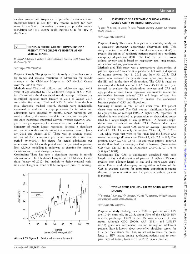
vaccine receipt and frequency of provider recommendation.Recommendation is key for HPV vaccine receipt for bothsexes in the South. Improving frequency of provider recom-mendation for HPV vaccine could improve UTD for HPV inthe South.
53 TRENDS IN SUICIDE ATTEMPT ADMISSIONS 2012-PRESENT AT THE CHILDREN’S HOSPITAL AT OUMEDICAL CENTRE
M Cooper*, S Gillaspy, R Wallace, S DeLeon. Oklahoma University Health Science Centre,Oklahoma City, OK
10.1136/jim-2017-000697.53
Purpose of study The purpose of this study is to evaluate secu-lar trends and seasonal variations in admissions for suicideattempts at the Children’s Hospital at OU Medical Centreover the last five years.Methods used Charts of children and adolescents aged 4–18years of age admitted to The Children’s Hospital at OU Med-ical Centre with the diagnosis of suicide attempt, self-harm, orintentional ingestion from January of 2012 to August 2017were identified using ICD-9 and ICD-10 codes from the hos-pital electronic medical record. Records were individuallyexamined to evaluate for appropriateness for inclusion andadmissions were grouped by month. Linear regression wasused to identify the overall trend in the data, and we plan tosue Auto Regressive Integrated Moving Average (ARIMA) anal-ysis to analyse separately for seasonal variation and trend.Summary of results Linear regression detected a significantincrease in monthly suicide attempt admissions between Janu-ary 2012 and August 2017. There was an average overallincrease of 0.21 admissions per month over the entire timeperiod (p<0.0001). See figure for actual admissions permonth over the 68 month period and the predicted regressionline. ARIMA modelling is underway to examine for seasonalvariations and recent changes in trend.Conclusions There has been a significant increase in suicideadmissions at The Children’s Hospital at OU Medical Centresince January of 2012. Full analysis to define seasonal varia-tion and changes in trend will be completed prior to meeting.
54 ASSESSMENT OF A PAEDIATRIC CLINICAL ASTHMASCORE’S ABLITY TO PREDICT DISPOSITION
1L East*, 2K Kepple, 1R Gibson, 1N Lane. 1Augusta University, Augusta, GA; 2OrlandoHealth, Orlando, FL
10.1136/jim-2017-000697.54
Purpose of study This research is part of a feasibility study fora paediatric emergency department observation unit. Thisstudy examined the ability of a clinical asthma score (CAS) topredict disposition of paediatric patients treated in a paediatricemergency department (ED). The CAS is used to assessasthma severity and is based on respiratory rate, lung sounds,retractions, and oxygen saturation.Methods used This study was a retrospective chart review ofpatients ages 2–17 treated at a paediatric ED with a diagnosisof asthma between July 1, 2012 and June 30, 2013. CASscores were obtained for patients twice: upon presentation tothe ED and at the time of disposition. The CAS is rated onan evenly distributed scale of 0–12. Student’s t-tests were per-formed to evaluate the relationships between and CAS andage, gender, or race. Linear regression was used to analyse therelationship between patients’ CAS and length of stay. Stu-dent’s t-tests were conducted to analyse the associationbetween patients’ CAS and disposition.Summary of results A total of 608 visits from 499 patientcharts were analysed. The CAS was not significantly impactedby age, gender, or race (p>0.05). A higher CAS, regardless ofwhether it was evaluated at presentation or disposition, corre-lated to a longer length of stay (p<0.0001). A patient’s dispo-sition also correlated to their CAS. Patients that weredischarged had the lowest CAS scores on average (PresentationCAS=4.1, CI: 3.8 to 4.3; Disposition CAS=1.4, CI: 1.2 to1.5), while those that went to the PICU had the highest CASscores on average (Presentation CAS=8.2, CI: 7.7 to 8.7; Dis-position CAS=6.9, CI: 6.5 to 7.3) and those patients admittedto the floor had, on average, a CAS in between (PresentationCAS=6.0, CI: 5.7 to 6.4; Disposition CAS=3.2, CI: 3.0 to3.5) (p<0.0001).Conclusions The CAS is significantly correlated with bothlength of stay and disposition of patients. A higher CAS scorepredicts both a longer length of stay and a more acute dispo-sition. Future work developing an algorithm inclusive of theCAS to evaluate patients for appropriate disposition includingthe use of an observation unit for paediatric asthma patientsis planned.
55 TESTING TEENS FOR HIV – ARE WE DOING WHAT WESHOULD?
1,2*J Fernelius, 1,2K Migacz, 1,2A Srinivasan, 1,2C Bell, 1,2L Benjamins. 1UTHealth, Houston,TX; 2McGovern Medical School, Houston, TX
10.1136/jim-2017-000697.55
Purpose of study Globally, nearly 25% of patients with HIVare 10–24 years old. In 2015, about 51% of the 61,000 HIVinfected youth ages 13–24 in the U.S. were unaware of theirstatus. Although CDC (2006), AAP (2011), and USPSTF(2013) guidelines recommend routine testing of adolescentpatients, little is known about how often physicians screen forHIV per these standards. Thus, we set out to assess the preva-lence of HIV testing among adolescent patients and to com-pare rates of testing from 2010 to 2015 in our practice.Abstract 53 Figure 1 Suicide admissinons by month
Abstracts
J Investig Med 2018;66:351–640 375
on August 27, 2022 by guest. P
rotected by copyright.http://jim
.bmj.com
/J Investig M
ed: first published as 10.1136/jim-2017-000697.609 on 1 F
ebruary 2018. Dow
nloaded from

Methods used After IRB approval, we conducted a retrospec-tive chart review of patients aged 13–18 seen in generalpaediatric and adolescent outpatient clinics in a large univer-sity-based setting. Whether or not a teen was tested for HIVat these visits was the primary outcome. Additional variablesincluded sexual history, reported condom use and HPV vac-cine status. Descriptive statistics were performed.Summary of results To date, 473 charts have been reviewed.Mean patient age was 14.9 (14.6 in paediatrics, 16 in adoles-cent), 49.9% were female, 67.2% African American and16.0% Hispanic. In 2010, 7.6% of patients were tested forHIV in the paediatric clinics, 34.3% in adolescent. In 2015,rates were 6.3% and 38.2% for paediatric and adolescent clin-ics respectively. Of the 63 patients tested for HIV, the averageage was 16. In this group, 63.5% reported sexual activity,20.6% reported unprotected sex, 44.4% had used a condomat their last sexual encounter, and 46.0% had received at leastone HPV vaccine. Among patients not tested for HIV, 15.9%reported sexual activity, however, for 28% sexual activity wasnot assessed.Conclusions Overall testing rates for HIV were similar in2010 and 2015. Teens were more likely to be tested in theadolescent clinics, however, these patients were also older.Teens who reported sexual activiity were also more likely tobe tested. This study provides important information on howoften teens were tested within this population, but may notbe generalizable to other settings. Thus, similar studies inother practice settings to determine how and when physiciansdecide to test for HIV will be crucial. Understanding this isimportant for the future design and implementation of inter-ventions directed towards routine, non-risk based screening.
56 BRUISED BUTTOCKS: IS IT ABUSE?
JP George*, R Brown, K Morgan. University of Oklahoma Health Science Centre, OklahomaCity, OK
10.1136/jim-2017-000697.56
Case report Bruising is the most common evident injury inchildren who have been physically abused1. Moreover, certainlocations of bruising are more consistent with physical abuse.For example, in children younger than 4 years, and back, but-tocks, forearm, foot and abdomen are rarely bruised from dayto day activities2. Here we discuss a 7 month old child whopresented with bruised buttocks and was diagnosed with Acutehaemorrhagic oedema of infancy (AHEI)
K.H. is a 7-month-old male who presented with burisedbuttock that mother noticed when giving the child a bath.The mother states that the child had fallen asleep on a hard-plastic swing, had a low height fall onto the floor from thefamily sofa 3 days prior, and had been increasingly fussy.
Physical exam shows a well appearing, afebrile, non-toxicchild who is noted to have a grossly benign exam except fora nontender 3 cm circular purpuric patch on the left buttockand a similar 3×2 cm patch on the right buttock with exten-sion down the right thigh laterally. Given the vague historyprovided by the mother our child abuse service was consultedand agreed a non-accidental trauma work up was warranted.The work up included labs, a computerised tomography scanof the head, a skeletal survey and ophthalmological evaluation.The work up was negative except for an elevated d-dimer,
which would be expected with the presence of bruising seen.Consequential, the diagnosis of AHEI was made. Child wasdischarge home and was followed to note complete resolutionof bruising without other complications and no other situationconcerns for abuse.
Acute haemorrhagic oedema of infancy (AHEI) is a rarecutaneous illness that affects children between the ages of 4months and 2 years of age and is classically thought of as apurely cutaneous form of Henloch-Schonlein Purpura3. AHEIhas classically been characterised with a triad fever, purpuriclesions and oedema which can yield the physician to be facedwith a wide differential4, however in the absence of a feverand often an unclear history non-accidental trauma must alsobe considered and can often pose a diagnostic challenge giventhat bruising patterns associated with AHEI are not oftenassociated childhood activities.
57 IMPROVING SCREENING AND REFERRAL FORPOSTPARTUM DEPRESSION IN A PAEDIATRIC PRIMARYCARE CLINIC
AL Hoppmann*, M Silverberg, B Ratliff, S Hall, S Walley, C Dye. University of Alabama atBirmingham, Birmingham, AL
10.1136/jim-2017-000697.57
Purpose of study Postpartum depression (PPD) has been shownto impact maternal-child bonding and infant development. AsPPD is often under-recognised and undertreated, our aimsincluded increasing paediatric resident knowledge and comfortof screening a mother for postpartum depression by 30% andreferring 50% of positively screened mothers for depressioncounselling.Methods used We implemented a resident-driven qualityimprovement project at the University of Alabama at Birming-ham (UAB) Paediatric Primary Care Clinic (Clinic) with part-nership with the UAB Community Counselling Centre.Residents had an educational curriculum which included PPDand a new screening/referral process over a 1 week period inClinic. Maternal depression was evaluated using the EdinburghPostnatal Depression Scale at the 1 month – 6 month wellchild check appointments, while resident knowledge was eval-uated via online survey collected pre and post educationalintervention. Mothers who screened positive for PPD werereferred. Strategies included process mapping and collaborativeintervention planning with clinic faculty and support staff.Data was compiled and analysed using Microsoft Excel.Summary of results Fourty-six residents completed the surveyprior to intervention while 36 residents completed the post-survey. Following the educational intervention, the proportionof residents who knew how to screen for PPD increased by30%, while the proportion of residents who felt comfortablescreening and referring mothers with PPD increasedby >50%. Of the 35 mothers who screened positive for PPD,17 (49%) were referred for counselling and 3 (15%) mothersattended an initial counselling session.Conclusions A clinic based educational curriculum improvedresident knowledge and comfort of maternal screening/referralfor PPD. While almost one-half of mothers who screened pos-itive for PPD were referred for counselling, only a small pro-portion attended. Future aims include determining alternativestrategies to assist mothers with PPD.
Abstracts
376 J Investig Med 2018;66:351–640
on August 27, 2022 by guest. P
rotected by copyright.http://jim
.bmj.com
/J Investig M
ed: first published as 10.1136/jim-2017-000697.609 on 1 F
ebruary 2018. Dow
nloaded from

58 AN UNSUSUAL CAUSE OF HIP PAIN IN A YOUNG GIRL
MN Frascogna, B Dillard, E Landry*. University of Mississippi Medical Centre, Jackson, MS
10.1136/jim-2017-000697.58
Case report Hip pain is a common complaint evaluated andtreated in the Paediatric Emergency Department. Even thoughmusculoskeletal causes are the most common, practitionersmust keep a broad differential diagnosis list in mind, includingpain referred from nearby structures. We present a case of hippain in a young girl with an unusual cause.
A 5 year old female presented to the paediatric emergencydepartment (PED) with a complaint of recurrent left hip painthat started approximately 3–4 months prior to her presenta-tion. The pain occurred infrequently and typically wouldresolve when treated at home with ibuprofen. She had beenworked up for this complaint by her primary care physicianand by a local orthopaedic surgeon with lab work and imag-ing reported by the patient’s mother to be normal. Prior toevaluation in the PED, she began to have worsening left hippain and vomiting. On exam, she was afebrile, had noabdominal tenderness, and had full range of motion of herhips with no gait abnormalities. She reported pain to palpa-tion of her left lower flank above her hip. An abdominal X-ray was performed showing moderate stool in the distal colonbut no signs of obstruction or mass effect. A complete bloodcount, metabolic panel, and urinalysis were obtained and werenoted to be unremarkable. She was given a normal salinebolus and morphine with improvement, but not resolution, ofher pain. An abdominal CT was performed and showed a pel-vic mass in the area of the left ovary. A pelvic ultrasound wasobtained and showed no definite vascular flow within the leftovary, leading to a diagnosis of left ovarian torsion with ische-mia. The paediatric surgery service was consulted and she wastaken to the operating room where an exploratory laparotomywas performed with detorsion and left oophoropexy. She wasmonitored post-procedure and was discharged home threedays after admission in good condition.
Ovarian torsion can be difficult to diagnosis, especially as itcan be intermittent. Though it is much more common inreproductive aged girls, ovarian torsion should be kept on thedifferential in all females with unilateral abdominal, back, orhip pain.
59 EXPLORATORY STUDY ON DISCUSSION OF TRANSITIONOF CARE FOR ADOLESCENTS DURING WELL CHILDCHECKS
S Leung*, S Mennito. Medical University of South Carolina, Charleston, SC
10.1136/jim-2017-000697.59
Purpose of study Despite guidelines on transitioning paediatricpatients to adult care, many young adults do not receiveappropriate preventative care resulting in emergency depart-ment overuse, high medical costs, and low adult vaccinationrates. A core factor is whether transitional care discussionsoccur between medical providers and patients. This study eval-uated whether transition discussions are held during adolescentwell child checks (WCC) and if teen health status impacts thelikelihood of those conversations occurring.Methods used We identified adolescents ages 15 and up seenin a resident continuity clinic for a well check between
January 2015 and January 2016 and categorised them asbeing ‘overall healthy’ or ‘medically complex’ based on pre-setcriteria. We reviewed charts for transitional discussion keywords or for the use of an electronic medical record (EMR)transition template developed locally for teen WCC. Two-tailed Z-test was used for two statistical analyses: at the sub-ject level, n=100, and at the visit level of each well childcheck, n=161.Summary of results A total of 432 subjects were identified ofwhom 100 were randomly selected for review. Patients were59% female and 65% overall healthy when seen between2013 and 2017, ages 15–18 years old. At the subject level,transitional discussions occurred for 60% of medically com-plex teens compared to 49% of overall healthy teens (p=0.3;Z-score of 1.02). At the visit level, a transitional discussionoccurred at 45% of WCC for medically complex teens com-pared to 33% of WCC for overall healthy teens (p=0.14; Z-score of 1.4). No documentation of transition discussionsoccurred without the use of the EMR template.Conclusions Despite transition EMR template availability, tran-sition of care topics are not consistently documented for anyadolescent, healthy or otherwise. Use of a template encour-aged documentation of transition discussions. Sports physicalsand sick visits, which utilise separate EMR templates, may bemissed opportunities. Additionally, transition of care topics areat the end of the WCC template, which may limit its use dur-ing visits with numerous topics to cover. Adjustment of theavailable template and further education to encourage earlierand more frequent talks about transition may improve rates ofthose discussions.
60 IMPACT OF MULTIDISCIPLINARY MANAGEMENT ONCOMORBIDITIES OF PAEDIATRIC OBESITY
AN Metcalf*, AE Weedn, S Gillaspy. University of Oklahoma Health Sciences Centre,Oklahoma City, OK
10.1136/jim-2017-000697.60
Purpose of study 17% of US children ages 2–19 are obese,and at risk for dyslipidemia, type 2 diabetes, and non-alco-holic fatty liver disease. The United States Preventive ServicesTask Force recommends multidisciplinary management, whichhas been shown to stabilise body mass index (BMI), but stud-ies on the impact of this treatment on obesity comorbiditiesare limited. This study examined the effect of a paediatricmultidisciplinary weight management clinic on targeted cardio-metabolic markers of health.Methods used Participants 2–18 years of age presented to theHealthy Futures Clinic from August 2012 to October 2016and had a BMI �99 th percentile or a BMI �95 th percentilewith an obesity-related comorbidity. Patients and familiesreceived behavioural counselling from a paediatrician, dietitian,physical therapist, and psychologist at an initial visit and every3 months, with individualised follow-up visits in the interim.BMI, fasting cholesterol, triglycerides, HDL, LDL, glucose,ALT, and haemoglobin A1c were obtained at baseline and at 6months. Changes in BMI and lab values were analysed contin-uously by paired t-tests and categorically (abnormal vs normalresults) by McNemar’s Test.Summary of results 138 participants presented to the clinic;63% were female and 61% were between 6 and 12 years ofage. At baseline, 43.8% had abnormal ALT levels, 43.8% had
Abstracts
J Investig Med 2018;66:351–640 377
on August 27, 2022 by guest. P
rotected by copyright.http://jim
.bmj.com
/J Investig M
ed: first published as 10.1136/jim-2017-000697.609 on 1 F
ebruary 2018. Dow
nloaded from

elevated triglyceride levels, and 34.1% had high cholesterol.Among patients still participating in the clinic at 6 months(n=78), total cholesterol improved by an average of 6 mg/dL(p<0.05), and triglycerides improved by an average of14.6 mg/dL (p<0.05). Of these patients (n=78), 32.0% hadhypercholesterolemia at baseline, which improved to 19.2%after six months of treatment. Similarly, 39.7% of returningpatients (n=78) had abnormal triglyceride levels at baseline,which was reduced to 25.6% at six months. Glucose, HDL,LDL, and ALT also improved at six months but no significantdifference was demonstrated. These improvements occurreddespite a lack of change in BMI with treatment.Conclusions Six months of multidisciplinary clinical interven-tion improved dyslipidemia in obese children, and theseimprovements occurred without change in BMI. Future studieswill examine treatment effect at 12 months.
61 ABDOMINAL MASS(ES) IN A PAEDIATRIC PATIENT
Y Nathani*, J George, C Knoles. University of Oklahoma, Oklahoma City, OK
10.1136/jim-2017-000697.61
Case report D.J. – an obese 10-year-old premenarchal femalepresented with worsening RLQ abdominal pain with nauseaand vomiting. ROS negative for fever, jaundice, weight loss,trauma. Initial VS were normal and exam was only impressivefor RLQ tenderness without peritonitis. Ultrasound showed a9×9×10 cm complex anechoic structure right pelvis with nor-mal blood flow (figure 1) and a mass in RUQ. SubsequentCT scan showed a 5 cm solid, well circumscribed mass in liver(figure 2) in addition to complex mass in pelvis. Labs werenormal and HCG was negative. Due to progressive symptoms,she underwent a laparoscopic right salpingectomy for paratu-bal cyst torsion with normal viable ovary. The liver mass wasdetermined to be focal nodular hyperplasia (FNH).
This was diagnostically challenging as the patient had twodistinct masses with progressive symptoms. Possible etiologies
included ovarian or tubal cyst, primary ovarian/hepatictumours, metastases, abscess, ovarian/tubal torsion and ectopicpregnancy.
Paratubal cysts are remnants of Wollfian and Mullerianducts and constitute 3%–7% of adenexal masses in paediatricpatients.1 Increasing size of paratubal cysts are associated withobesity, likely due to excess androgens. Most are asympto-matic; however, they can present with abdominal pain duehaemorrhage, rupture and torsion. These cysts are non-physio-logic and unlikely to spontaneously regress and definitive man-agement is operative.
FNH is generally a benign and asymptomatic lesion of theliver, usually discovered incidentally. These lesions are associ-ated with high oestrogen states, including obesity and requiresurgical intervention only if symptomatic.
REFERENCE1. Muolokwu E, Sanchez J, et al. The incidence and surgical management of paratu-
bal cysts in paediatric and adolescent population. J Pediatr Surg 2011;46:2161–2163.
Abstract 61 Figure 2 CT scan showing liver mass
62 INPATIENT BREASTFEEDING STUDY: WHAT ARE THEBARRIERS TO INPATIENT BREASTFEEDINGEXCLUSIVITY?
1E Omoruyi*, 1MS Evangelista, 2TA Sugrañes. 1University of Texas Health Science Centre atHouston, Houston, TX; 2New York-Presbyterian/Weill Cornell, New York, NY
10.1136/jim-2017-000697.62
Purpose of study Breastfeeding is widely accepted as the bestnutritional source for infants. In the United States, exclusivebreastfeeding is recommended until at least 6 months of age.Although breastfeeding has been shown to have beneficialeffects, studies have shown that problems and emotional dis-comfort experienced during early breastfeeding are risk factorsfor breastfeeding cessation before 6 months. Determining whymothers are unable to achieve breastfeeding exclusivity in theimmediate postpartum period is important to the creation oftargeted interventions.
Abstract 61 Figure 1 Ultrasound showing complex mass in rightadenexa
Abstracts
378 J Investig Med 2018;66:351–640
on August 27, 2022 by guest. P
rotected by copyright.http://jim
.bmj.com
/J Investig M
ed: first published as 10.1136/jim-2017-000697.609 on 1 F
ebruary 2018. Dow
nloaded from

Methods used This cohort study enrolled 510 women whowere each personally interviewed using a survey tool duringthe immediate post-partum period in an urban county hospi-tal. Factors assessed included breastfeeding history, perceptionsregarding breastfeeding/breastmilk, socioeconomic factors, aswell as hospital and family support. Breastfeeding exclusivityat discharge was assessed based on the mothers’ self-reportedinfant feeding behaviour during her hospital stay.Summary of results 38% of women exclusively breastfed dur-ing their inpatient stay. Several maternal, hospital, and exter-nal factors influence exclusive breastfeeding in a hospital.While the knowledge and experience of breastfeeding otherchildren is a determining factor (OR=3.33; 95% CI: 1.01 to11.04), several hospital factors such as skin to skin contact,knowledge about whom to approach and ask about breastfeed-ing help after discharge, and encountering issues in hospitalare responsible for encouraging the behaviour of exclusivebreastfeeding in a hospital (p<0.05).Conclusions This study highlights that knowledge regardingbreastfeeding as well as knowledge regarding sources of sup-port, both inpatient and outpatient, are important factors thataffect immediate breastfeeding outcome. This is one of thefew studies that looks at specific factors affecting exclusivebreastfeeding in the inpatient setting.
63 BREASTFEEDING DURATION AND BONE STRENGTH INYOUNG ADULT FEMALES
K Perofsky*, NK Pollock. Augusta University, Augusta, GA
10.1136/jim-2017-000697.63
Introduction Data on the relationship between breastfeedingand bone health are scant and equivocal. This study investi-gated the relationships between breastfeeding duration andindices of bone strength in young adult females.Methods Bone mass, density, and geometry at trabecular andcortical sites of the tibia were measured in 71 white females(aged 21±0.4 years) by using peripheral quantitative computedtomography. Breastfeeding duration was self-reported by eachparticipant’s biological mother. Fat-free soft tissue (FFST) andfat mass were measured using dual-energy X-ray absorptiome-try. Relationships between breastfeeding duration and boneparameters was determined using multiple linear regressionmodels, including height, FFST, and fat mass as covariates.Summary of results 20% of the participants reported not hav-ing been breastfeed; 32% were breastfed 1–6 months; 24%were breastfed 6–12 months; and 24% were breastfed 12months or longer. At the trabecular site of the tibia, breast-feeding duration was a positive independent predictor of totalvolumetric bone mineral density (b=0.28, p=0.045). Althoughbreastfeeding duration was positively correlated to bonestrength index (BSI; r=0.28, p=0.03), it was not an inde-pendent predictor of BSI in the regression model. At the cort-ical site, breastfeeding duration was a positive independentpredictor of bone mineral content (b=0.24, p=0.02), cross-sectional area (b=0.23, p=0.02), and cortical thickness(b=0.36, p<0.01). There were no associations between breast-feeding duration and the other bone parameters.Conclusions Our results suggest that a greater duration ofbreastfeeding may have long-term benefits on cortical and tra-becular bone. Given that our findings should be considered
hypothesis generating, further studies are needed to elucidatethe role of breastfeeding on bone development.
64 INAPPROPRIATE PRESCRIBING OF SYSTEMIC STEROIDSAND OPIOIDS TO PAEDIATRIC PATIENTS WITHPNEUMONIA OR SINUSITIS
1KG Phang*, 1J Roberts, 2S Garner, 1M Ebeling, 1W Basco. 1Medical University of SouthCarolina, Charleston, SC; 2South Carolina College of Pharmacy, Charleston, SC
10.1136/jim-2017-000697.64
Purpose of study National clinical guidelines do not recom-mend use of systemic steroids or opioids in treating paediatricpneumonia or sinusitis. The purpose of the study was to com-pare the frequency of systemic steroid or opioid prescribingfor children with pneumonia and sinusitis based on locationof care. We tested the hypothesis that inappropriate prescrib-ing of systemic steroids and opioids for paediatric pneumoniaor sinusitis was greater in the emergency department thanother clinical sites.Methods used The study evaluated paid claims for visits andmedications using 2016 South Carolina Medicaid data. Sub-jects were 5 to 18 years old with a primary diagnosis ofpneumonia or sinusitis, selected using the ICD-9 and ICD-10Clinical Classification Software Category System. The prescrip-tion of a systemic steroid or an opioid dispensed 0–7 daysfrom a visit claim was evaluated. Medicaid visits were associ-ated with one of 3 locations: emergency department (ED),urgent care (UC), or ambulatory site. Patient demographicdata avaialable included ethnicity, gender and age in months.Summary of results A total of 16 480 visits were evaluatedfrom all 3 settings. Of 2153 visits in the ED 273 (13%)included a systemic steroid and 98 (5%) included an opioid.Of 14 149 visits in the ambulatory setting 974 (7%) includeda systemic steroid and 376 (3%) included an opioid. Of 178visits in the UC 15 (8%) included a steroid. Too few patientswere prescribed an opioid in the urgent care setting to per-form statistical analysis. ED visits were associated with ahigher steroid prescription rate (chi square, p<0.0001) whencompared to ambulatory and UC visits. ED visits were associ-ated with a higher opioid prescription rate (chi square,p<0.0001) when compared to ambulatory visits.Conclusions Our results suggest that school age children andadolescents received steroid and opioid prescriptions at higherfrequency when seen in the ED versus ambulatory setting.Given safety concerns of steroids and opioids in paediatricpatients, improved prescribing practices for these medicationsare needed.
65 KNOWLEDGE OF AND ATTITUDES TOWARD HPVVACCINE IN PREADOLESCENTS AND TEENS
L Raney*, E Owers, R Pasternak, P Prasad, R Gardner. LSUHSC, New Orleans, LA
10.1136/jim-2017-000697.65
Purpose of study Human Papilloma Virus (HPV), common inboth females and males, is responsible for pathologies rangingfrom benign genital warts to cervical and penile cancer. Phar-maceutical companies have now developed a vaccine that willhelp prevent the virus-associated malignancies. The CDC rec-ommends that females ages 11–26 years and males ages 11–
Abstracts
J Investig Med 2018;66:351–640 379
on August 27, 2022 by guest. P
rotected by copyright.http://jim
.bmj.com
/J Investig M
ed: first published as 10.1136/jim-2017-000697.609 on 1 F
ebruary 2018. Dow
nloaded from

21 years receive the vaccine series. Despite being widely avail-able and highly publicised, only 40% of eligible femalesreceive the vaccine. This study aims to assess the knowledgeof HPV, the attitudes towards the HPV vaccine, and identifybarriers preventing full utilisation of the vaccine. Once identi-fied, we aim to overcome the barrier(s) and improve vaccina-tion rates in eligible adolescents.Methods used We distributed a standardised questionnaire tothe parents of eligible female and male patients in both paedi-atric hematology-oncology and general paediatric clinics. Itassessed the parents’ knowledge of HPV and the vaccine, theirviews of the vaccine, and reasons why they may oppose it.Additionally, we will compare the views of parents betweenthese two settings, in order to determine if the patient‘s per-sonal history of a hematologic or oncologic disease influencestheir decision to permit vaccination.Summary of results 75% of parents say they have been edu-cated about HPV, mostly by their primary care physician.However, only 25% knew what disorders HPV caused. Only40% felt the vaccine should be added to the typical vaccineschedule. Surprisingly, eighty percent of parents intend to oralready have vaccinated their child. In those that opposed thevaccine, one-third were concerned about potential side effectsand nearly 20% feel they do not have enough information.Conclusions The largest barrier to the utilisation of the HPVvaccine that we have identified appears to be lack of educa-tion. As a result, we have begun distributing the CDC’s HPVand vaccine patient guide to our patients’ families as an inter-vention. We will resurvey these families after implementingthe intervention to assess its success in increasing both knowl-edge and utilisation of the HPV vaccine.
66 SEPARATING FACT FROM FICTION: DIAGNOSINGFUNCTIONAL NEUROLOGICAL DISORDER
HA Rehman*, S DeLeon, D Hahn. OUHSC, Oklahoma City, OK
10.1136/jim-2017-000697.66
Case report Patient is a 13 year old girl with a history ofcomplex regional pain syndrome who presented with newonset visual changes and severe headaches. Ophthalmologicexam showed bilateral optic disc cupping, lumbar puncture(LP) revealed a mildly elevated opening pressure and magneticresonance imaging (MRI) revealed bulging optic discs; she wasadmitted for presumed idiopathic intracranial hypertension(IIH). Multiple therapeutic LPs resulted in mild headacherelief with no improvement in vision. An intracranial pressuremonitor showed normal pressure, ruling out IIH. A compre-hensive autoimmune, endocrine and infectious work-up wasnegative. She later reported back pain and spine MRI notedL1-L2 discitis osteomyelitis of unclear aetiology. She receivedextensive treatment with steroids, a selective serotonin reup-take inhibitor, a carbonic anhydrase inhibitor and several painmedications.
After 6 weeks with no definitive diagnosis, she was trans-ferred to our facility per parent request. Multiple specialistswere consulted including neurology, ophthalmology, psychol-ogy, infectious disease, physical therapy (PT), pain manage-ment and neurosurgery. She had various complaints includingpersistent headaches, changes in visual acuity and fields, andweakness and numbness of her left extremities. Her exam,however, remained inconsistent and all symptoms except for
headache and vision changes spontaneously resolved. RepeatMRIs and ophthalmology exam revealed normal orbits withresolution of disc cupping. Given persistent complaints withnormal exams and studies, she was diagnosed with functionalneurologic disorder (FND) in addition to lumbar osteomyelitisand discitis. She worked with psychology and PT and wastransferred to a rehab facility specialising in the disease proc-ess. Though potential stressors were identified, these werenever fully accepted as the aetiology by the patient or family.
FND, previously known as conversion disorder, is mani-fested by neurologic symptoms that cannot be explained by anunderlying disease pathology. Patients experience motor, visual,speech or sensory changes believed to be triggered by an emo-tional or physical stressor. Treatment involves a multidiscipli-nary approach with therapy and often medication. Earlydiagnosis is key to prevent unnecessary testing that can leadto a poorer prognosis.
67 ACUTE DISSEMINATED ENCEPHALOMYELITIS IN A SIXYEAR OLD MALE PRESENTING AS UNILATERAL OPTICNEURITIS
AR Riggs*, D Hahn. Oklahoma Health Sciences Centre, Oklahoma City, OK
10.1136/jim-2017-000697.67
Case report Our patient is a previously healthy 6 year oldmale who presented with a ten day history of intermittentnausea and vomiting, fluctuating periods of hyperactivity andfatigue, as well as acute onset loss of colour vision, decreasedvisual acuity, and appearance of scotomata in his right visionfield. The patient’s primary care physician referred him to anOphthalmologist, who noted right optic nerve swelling. Thepatient was then transferred to our Emergency Room, wherea thorough initial lab workup yielded only mild leukocytosison a blood sample. The only pertinent physical exam findingwas fatigue. A brain Magnetic Resonance Imaging (MRI) wasobtained to evaluate for optic neuritis. The MRI revealedglobal involvement of the brain with diffuse white matterlesions predominantly in bilateral cerebral hemispheres. Thesefindings steered us to a diagnosis of Acute Disseminated Ence-phalomyelitis (ADEM). We treated our patient with a five daycourse of high dose steroids. Within days, the child respondedwith symptomatic improvement. At a one month follow up,he continued to have improvement in his visual problems.
While ADEM has certainly been well-documented in chil-dren in our patient’s age group, his clinical presentation didnot initially suggest the severity of the brain involvementfound on MRI. Vacillating fatigue and hyperactivity pairedwith unilateral optic neuritis made for an unusual and uniquepresentation of ADEM.
68 HEMATEMESIS AND HEMATURIA IN A HEALTHY 3MONTH OLD MALE
AR Riggs*, D Hahn. Oklahoma Health Sciences Centre, Oklahoma City, OK
10.1136/jim-2017-000697.68
Case report Our patient is a previously healthy 3 month old malewho presented to our Emergency Department with a report ofpoor oral intake, decreased urine output, hematemesis, and hema-turia. The initial lab work-up was unremarkable with the
Abstracts
380 J Investig Med 2018;66:351–640
on August 27, 2022 by guest. P
rotected by copyright.http://jim
.bmj.com
/J Investig M
ed: first published as 10.1136/jim-2017-000697.609 on 1 F
ebruary 2018. Dow
nloaded from

exception of microscopic hematuria in a bagged urine. We admit-ted the child for further evaluation of the hematuria. Due to con-cern for formula intolerance and reflux causing his hematemesis,his formula was switched to Nutramigen and he was started on atrial of Omeprazole. The child failed to respond to our interven-tions and continued to have guaiac positive hematemesis. Anesophagogastroduodenoscopy was performed, which showed noevidence of bleeding or ulcer. While repeat bag urine specimenscontinued to show microscopic hematuria, an in-and-out catheter-ization specimen surprisingly showed no red blood cells. At thistime, multiple team members noted that patient only had positivefindings when his mother was alone with the child, but never hadpositive findings when in care of nursing alone. When we con-fronted our patient’s mother with our observations, she soonadmitted to contaminating the child’s stool, emesis, and urine withher own menstrual. The mother further confessed that she createdthe ruse in order to keep her son hospitalised so they would bothbe safe from the patient’s father. At this point, we contacted ourChild Maltreatment, and the Department of Human Services wasnotified for concern for Medical Child Abuse.
Medical Child Abuse, also known as Munchausen Syn-drome by Proxy, is an unfortunately common condition seenin the paediatric population. The simulation of symptoms canresult in procedures and hospitalizations that are not necessaryand are potentially harmful, as seen in our patient. Althoughit is difficult to recognise and confront medical child abuse, itis invaluable to do so early in order to protect patients fromunnecessary discomfort and harm.
69 ARM PAIN AMONG SOUTH CAROLINA YOUTHBASEBALL PLAYERS
J Roberts*, M Rao, E O’Brien, A Greenhouse, E Dawley, J Eichinger. Medical University ofSouth Carolina, Charleston, SC
10.1136/jim-2017-000697.69
Purpose of study Arm and shoulder injury to young athletes isincreasing in the United States, especially among baseballpitchers. Although pitch count limitations have been imple-mented in youth league baseball, the need for ulnar collateralligament reconstruction is increasing at a younger age, with56% of these procedures being performed on 15–19 yearolds. The objective of this study is to describe the injury bur-den to young athletes in South Carolina where baseball andsoftball are played year round.Methods used We asked parents who brought their 7–17 yearold child to any visit to one of 13 South Carolina PaediatricPractice Research Network (SCPPRN) practices to complete asurvey. Initial questions included the age of the child, position(s) played, injuries and past surgeries. Examples of throwing-specific questions included timing and quality of arm pain,occurrence of pain or fatigue during games or practice, limita-tions of the pain in terms of participation or performance,and pressure to play through the pain. We also inquired aboutthe number of months they play during the year, and interestin a pitch-tracking smart phone application. Questions werebased on a previous study conducted in the Northeast forcomparison. Descriptive statistics were calculated.Summary of results To date, 131 parents have completed thesurvey. 35 (26.7%) reported a previous injury, and 3 (2.3%)reported having previous surgery. Responses of ‘never’ and‘rarely’ were counted as responses in the negative, and
‘sometimes’, ‘often’, and ‘always’ were counted as responses inthe affirmative. 34 (26.0%) report pain when they throw orplay and 29 (22.1%) report that pain the day after playing. 7(5.3%) reported that previous arm pain has negative effect ontheir enjoyment of the game. 9 (6.9%) report that a coachhas encouraged him/her to keep playing despite the presenceof arm pain. 81 (61.8%) of parents would be interested inhaving access to a pitch count app.Conclusions Approximately one quarter of young athletesreport the presence of arm pain while playing. The use of asmart phone app to track pitch counts may be a helpful toolto prevent overuse injury and should be tested in a cohort ofyouth baseball players.
70 ADOLESCENT WEIGHT MANAGEMENT – IS THERE ANAPP FOR THAT?
C SanGiovanni*, M Steffen, C Pope, J Roberts. Medical University of South Carolina,Charleston, SC
10.1136/jim-2017-000697.70
Purpose of study Adolescent obesity has quadrupled in the pastthirty years from 5% to 20.5% for 12–19 year olds. Paediatri-cians report lack of resources as a barrier to management.Since adolescent smartphone use has increased significantlyand apps are useful in managing chronic diseases in adoles-cents, apps that assist with weight management could be abeneficial resource. However, little is known about featuresthat overweight/obese adolescents have considered helpful. Theobjective of this study was to explore adolescents’ preferencesabout weight management app features and willingness to usecollaboratively with their healthcare provider.Methods used Participants, recruited from an urban and ruralpractice in South Carolina, were eligible if between 13–17years old, had a Body Mass Index (BMI) at or above the85th percentile and had access to a smartphone or tablet.Semi-structured interviews were conducted from May to Octo-ber 2017. Participants were presented with 3 popular mobilehealth apps and asked to complete tasks using each app.Likes/dislikes about each app and which features (customisedworkouts, logging physical activity and food/beverage con-sumption, and social support) would encourage further usewere elicited. Content analysis was performed on interviewtranscripts.Summary of results Twelve adolescents wereinterviewed. Average participant age was 14.7 years, and aver-age BMI was 96.6 percentile. Eight (67%) were female, and 8(67%) identified as White, while the remainder identified asAfrican American. The majority had used health apps in thepast, but none were recommended by healthcare providers.All adolescents felt apps would be helpful in managing theirweight. Adolescents’ most favourable features were findinghealthy food alternatives and customising their workouts.Additional features they would include in health apps werecompeting with friends to exercise more or eat healthy, incen-tives to work toward healthy goals, sharing customised work-outs with friends, creating a music playlist for exercising, andhearing weight loss success stories from other adolescents.Conclusions Adolescents think health apps would be helpful inweight management. The use of apps should be studied todetermine their role as part of an effective weight manage-ment program.
Abstracts
J Investig Med 2018;66:351–640 381
on August 27, 2022 by guest. P
rotected by copyright.http://jim
.bmj.com
/J Investig M
ed: first published as 10.1136/jim-2017-000697.609 on 1 F
ebruary 2018. Dow
nloaded from

71 KAWASAKI DISEASE OUTBREAK IN CENTRALAPPALACHIA
VP Smith*, D Macariola, JW Schweitzer. ETSU, Johnson City, TN
10.1136/jim-2017-000697.71
Purpose of study In the US, Kawasaki Disease (KD) has aglobal hospitalisation rate of 17.1 per 1 00 000 children.Although the aetiology remains unknown, the current consen-sus is that KD is likely caused by an infectious trigger initiat-ing an abnormal immune response in genetically predisposedchildren. In March of 2017, Central Appalachia had an out-break of KD, with 4 patients concurrently hospitalised withthis disease. It is unclear whether this outbreak was due tothe genetic susceptibility of the Appalachian population, orexposure to an antigen which was particularly toxic. Thisstudy seeks to better understand the underlying genetic predis-position and pathogen antigenicity related to KD in the Appa-lachian region.Methods used All data was collected via chart review and ret-rospective study. Subjects were all admitted to the hospitalwithin 1 week of each other and presented with fever priorto admission. During their admission, each subject was diag-nosed with KD. All patients were followed for sequelae in theoutpatient setting.Summary of results Table 1 summarises the data during eachsubject’s hospitalisation. All subjects met criteria for KD whilein the hospital. All subjects were treated with IVIG and hadresolution of fever. The mortality associated with all subjectswas 0%. No subjects had long-term sequelae.Conclusions The Appalachian population may be more suscep-tible to KD, however, a larger chart review of incidence ofKD in the region for multiple years would be necessary todraw these conclusions. It is much more likely that there wasa highly antigenic pathogen present in the population in earlyspring 2017 that increased the incidence of this outbreak.Although a small sample size was used, the study’s incidencewas dramatically increased from the US incidence when con-sidered on a monthly basis. Public health officials and pro-viders should be aware that the Appalachian region may bemore genetically susceptible to outbreaks of KD, or beexposed to pathogens with increased antigenicity, and thushave a lower index of suspicion for diagnosis of KD whenmultiple cases occur in relative proximity to one another.
72 PERSISTENT HYPERGLYCEMIA SECONDARY TO SECOND-GENERATION ANTIPSYCHOTIC MEDICATIONS
N Soulages Arrese*, AB Middleman, JD Meyer. University of Oklahoma Health SciencesCentre, Oklahoma City, OK
10.1136/jim-2017-000697.72
Case report It has been shown in the literature that second-generation antipsychotics are associated with patients develop-ing hyperglycemia and diabetes mellitus along with other met-abolic changes such as weight gain and lipid profileabnormalities. This reports highlights a case of a paediatricpatient with hyperglycemia while taking second-generationantipsychotics in the setting of a complex social situation andweight loss. C.K. is a 16 year old with a history of familydysfunction, depression, suicidal behaviours, and cognitivedelay and prolonged psychiatric admissions. She presented atage thirteen to endocrine clinic with hyperglycemia, impaired
Abstract71
Table1
Summarise
sthedata
duringeach
subject’s
hospitalisation
Patie
ntAge
Gen
der
Daysof
fever
before
hospita
lisation
Oph
thalmic
chan
ges
Oral
mucosal
chan
ges
Rash
Chan
ges
of hand
s
andfeet
Cervical
lymph
aden
opathy
GI
symptom
s
Leuk
ocytosis
Neu
trop
hilia
Hypoa
lbum
inem
iaElevated
LFTs
ESR
CRP
Corona
ry
abno
rmalities
Recent
Illne
ss
A4year
M6dayhistory
++
++
++
++
+-
++
--
B2year
M11
dayhistory
++
++
+-
+-
+-
++
+-
C11
mo
F10
dayhistory
++
++
++
++
+-
NoESR
draw
n
++
+
D3year
M5dayhistory
++
++
++
++
+-
++
+-
Abstracts
382 J Investig Med 2018;66:351–640
on August 27, 2022 by guest. P
rotected by copyright.http://jim
.bmj.com
/J Investig M
ed: first published as 10.1136/jim-2017-000697.609 on 1 F
ebruary 2018. Dow
nloaded from

fasting glucose, normal HgbA1c while taking quetiapine.Approximately 1.3 years later, 1 year prior to presenting toour service, the patient developed polyuria, polydipsia, hyper-glycemia, elevated HgbA1c of 9.2%, without diabetic ketoaci-dosis, and BMI 19.7. The patient had recently been switchedfrom quetiapine to olanzapine. Antibodies associated withType 1 Diabetes Mellitus were negative. The patient wastreated with an intensive insulin regimen including long andrapid acting insulin. At presentation to our service, an inpa-tient service for malnutrition, the patient had severe malnutri-tion, BMI of 15.3 and HgbA1c of 5.7%. The mother haddiscontinued insulin when the patient had stopped eating. Thepatient indicated that she had been too tired to eat. Themother reported she was giving the patient additional olanza-pine ‘as needed’. Genetic testing for Maturity-onset diabetesof the young was completed and results were negative. Thepatient was transitioned from olanzapine to risperidone, as itis associated with less risk of hyperglycemia. During treatmentfor malnutrition, glucose levels improved, requiring rare cor-rections with regular insulin. This case serves as a reminder ofthe importance of appropriate metabolic screening for paediat-ric patients, regardless of weight, before and during treatmentwith second-generation antipsychotics; the metabolic changesmay cause significant and potentially irreversible morbidity.
73 DO CHILDREN SEEN IN THE EMERGENCY DEPARTMENTFOR SKIN AND SOFT TISSUE INFECTIONS WHO AREADMITTED TO THE INPATIENT SERVICE MEET CRITERIAFOR SEPSIS?
B Weaver*, D Nakayama, JJ Burns, B Weidner. University of Florida, Pensacola, FL
10.1136/jim-2017-000697.73
Purpose of study Admission of patients with skin and soft tis-sue infections (SSTI’s) have increased as have hospital charges.The main treatment of these is incision and drainage (I andD) for abscesses which can take place as an outpatient orinpatient.
Systemic inflammatory response syndrome (SIRS) criteriainclude temperature, heart rate, breathing rate, and whiteblood cell count. A patient with positive SIRS criteria andsource of infection qualifies as having sepsis, justifyingadmission.
The purpose of our investigation is to determine the fre-quency of positive SIRS criteria in patients admitted for SSTImanagement. We also wish to determine if SIRS criteria statuscorrelated with positive blood cultures, resolution without Iand D and if I and D was performed in the operating roomOR.Methods used A retrospective chart analysis was conducted on203 patients admitted with SSTI’s from the paediatric ED dur-ing 2011–2016. Charts were excluded if patients were repeatvisitors for the current SSTI, outside hospital transfers, immu-nocompromised state, chronic debilitating illnesses, or ifaffected area was the face, mouth or inside the scrotum. Age,heart rate, respiratory rate, temperature, blood culture results,WBC, SIRS status (positive or negative), I and D status(required or not), and I and D setting were recorded (OR ornot).Summary of results Thirty-seven met criteria for study entry;mean age was 24.7 (STD 28.8) months.
59.5% patients met SIRS criteria; 40.5% did not.
Two patients had positive blood cultures. Both werecontaminants.
72.7% of those who met SIRS criteria went to OR vs53.3% who did not meet SIRS criteria. (Chi-square;p=0.225).
22.7% of patients who met SIRS criteria had SSTIs thatresolved without I and D vs 40% with negative SIRS criteria(Fisher’s Exact Test) 0.295.Conclusions Most of the admitted patients with SSTIs metSIRS criteria, yet there were 40.5% who did not and mayhave therefore been unnecessary admissions.
This study supports previous studies that indicate thatblood cultures are not useful in SSTI management.
SIRS status did not correlate with rate of resolution with-out I and D or need for OR I and D.
Studies with higher N are needed to confirm thesefindings.
Adult case reports/ageing/geriatrics
Joint plenary poster session and reception
4:30 PM
Thursday, February 22, 2018
74 SYNCOPE EVALUATION USING POINT-OF-CAREULTRASOUND
EK Addo-Yobo*, J Treece, N Kakkar, V Pai, MA Alawoki, C Rosero. Shamas, S, EastTennessee State University, Johnson City and Mountain Home, TN
10.1136/jim-2017-000697.74
Case report While syncope represents a common presentationof cardiovascular compromise, it is rarely the first presentingsymptom of a pulmonary embolism (PE). If syncope occurs,delay in therapy can quickly become fatal. We describe a com-plex presentation of syncope in which bedside point-of-careechocardiogram would have directed appropriate managementof a PE.
Eighty seven year old male with no significant cardiac his-tory presented with three syncopal episodes. There were noprodromal symptoms or evidence of seizure activity. Bloodpressure of 76/42 mmHg and pulse of 118 bpm wererecorded by paramedics. Electrocardiogram (EKG) showedatrial fibrillation with rapid ventricular rate. He presentedhypoxemic and hypotensive. Labs showed serum troponin of3.7 ng/mL and acute renal insufficiency. EKG showed no acuteischaemic changes. Head imaging showed no acute intracranialpathology.
Acute coronary syndrome protocol with heparin drip wasinitiated. The patient opted for medical management and wastransitioned to apixaban for stroke prophylaxis. Transthoracicechocardiogram (TTE) showed characteristic signs of rightheart strain. Chest angiogram showed a large central pulmo-nary embolus in the right lung with additional multifocal seg-mental bilaterally. Subsequent lower extremity ultrasoundshowed a deep vein thrombosis in the left lower extremity.Apixaban was continued since the patient was oxygenatingwell on nasal cannula and was hemodynamically stable.
Syncope is an uncommon presentation for PE. Even whenan alternative reason for syncope exists, it is estimated that
Abstracts
J Investig Med 2018;66:351–640 383
on August 27, 2022 by guest. P
rotected by copyright.http://jim
.bmj.com
/J Investig M
ed: first published as 10.1136/jim-2017-000697.609 on 1 F
ebruary 2018. Dow
nloaded from

PE is present in 12% of patients with syncope. Our patientpresented with syncope in the setting of ACS, new onset atrialfibrillation and hypotension. The TTE showed the Mcconnelsign, a finding which has a 94% specificity for a PE. Thissign is a pattern of right ventricular dysfunction with akinesiaof the mid wall and hyper contractility of the apical wall. Ifbedside sonography was part of the initial encounter, thepatient’s presentation would have appropriately directed treat-ment towards thrombolysis or thrombectomy. It has beenshown that prognosis for PE is best if treatment within thefirst hour. Prompt initiation of treatment is even more impor-tant because syncope is an ominous sign of PE.
75 INTRAVASCULAR LYMPHOMA PRESENTING ASRESPIRATORY FAILURE
A Bokhari*, D Trofimovitch, B Boonpheng, H Zaver, T Nold, J Goldstein. ETSU, JohnsonCity, TN
10.1136/jim-2017-000697.75
Case report Lymphoma, a great mimicker, presents with anarray of signs and symptoms in almost any organ system.Here we present a case of acute respiratory failure in an oth-erwise healthy elderly man.
A 73-year-old man presented with four weeks of progres-sive dyspnea on exertion. He reported a non-productivecough, low-grade fevers, chills, and night sweats. He deniedchest pain, weight loss, or lack of appetite and was hemody-namically stable on presentation. Physical examination was sig-nificant only for diffuse crackles and 2+pitting ankle oedemabilaterally. Chest x-ray demonstrated bibasilar infiltrates, mildpleural effusion and cardiomegaly. Metabolic panel revealedhyponatremia (119 mEq/L) with high urine osmolality andNa, consistent with SIADH. Serum cortisol was normal; how-ever TSH, free T4 and rT3 were compatible with centralhypothyroidism. CBC was within normal limits. Initially hewas on antibiotic therapy for presumed Community-AcquiredPneumonia. CT chest showed bibasilar atelectasis, bilateralpleural effusions, and no evidence of embolism. Multiple inci-dental calcified mediastinal lymph nodes were aspirated, withbenign pathology. Patient underwent thoracoscopic wedge lungbiopsy; pathology indicated high-grade intravascular large Bcell lymphoma positive for CD20, CD79a, Pax-5, CD10,Mum-1, and negative for CD3, cytokeratin AE1/AE3, S100,Cam5.2, and CD34. The lymph node histology was consistentwith a remote granuloma. Gram stain, AFB, culture and fun-gal studies were negative. Bone marrow showed minimal pres-ence of lymphoma. CT abdomen showed mildhepatosplenomegaly, a mildly enlarged epigastric lymph node,and no other intra-abdominal lymphadenopathy. The patientdeveloped type 1 respiratory failure likely due to lymphangitiscarcinomatosis, which improved with chemotherapy [R-CHOP].
Intravascular lymphoma is rare and aggressive. It was anunexpected diagnosis in our patient with predominant respira-tory distress and pulmonary infiltrates. This subtype of lym-phoma can manifest in any organ system and presents adiagnostic challenge given most information is from a paucityof case reports. Clinicians should maintain a high index ofsuspicion in high-risk patients presenting with an atypical con-stellation of symptoms.
76 DOUBLING DOWN ON ANAKINRA: A CASE OFRELAPSING STILL’S DISEASE
S Chalasani*, V Majithia, S Kishore. University of Mississippi, Jackson, MS
10.1136/jim-2017-000697.76
Case report Adult onset Still’s disease (AOSD) is a rare sys-temic inflammatory disease of unknown aetiology characterisedby fever, arthralgia, salmon-pink rash and leukocytosis. Wepresent as classic case of AOSD who was refractory to treat-ment with high dose steroids and Anakinra.
A 21-year-old African American male with no past medicalhistory presents with 1 month history of worsening joint painsand fever. 2 weeks prior to presentation, he had a diffusescaly erythematous rash on his trunk and extremities. Feverswere as high as 104 °F responsive to Tylenol. Patient had sim-ilar symptoms 9 months ago that resolved with doxycyclineand prednisone. On exam, he was ill appearing with fever of100.3 °F. He had peeling macular rash involving trunk, armsand legs. He complained of pain with active and passive rangeof motion in all joints. Pertinent labs include: haemoglobin7.1 g/dL, white blood cells 17.8 TH/cmm with 73% neutro-phils, platelets 362 TH/cmm, aspartate transaminase 163 U/L,alanine transaminase 117 U/L, sedimentation rate of 104 mm/hr, C- reactive protein 21.4 mg/dL, Ferritin >100,000 ng/mland elevated IL-2 receptor at 4090 pg/mL. Other rheumatolog-ical and infectious work up was negative. Imaging relievedhepatosplenomegaly and diffuse lymphadenopathy. Bone mar-row biopsy ruled out malignancy and there was no evidenceof hemophagocytosis. Patient was started on methylpredniso-lone 60 mg thrice daily and noticed significant clinical andserological improvement. He was later discharged on 60 mgprednisone. Follow-up at 2 weeks, he had clinally worsenedrequiring admission for pulse dose steroids. He was alsostarted on Anakinra 100 mg daily. Four weeks into therapywith Anakinra, as the steroids were being tapered he hadrelapsed again. Therefore, the dose of Anakinra was doubled.Patient has been doing well on the current regimen. He isnow undergoing slow taper of his steroids with recent addi-tion of Methotrexate.
AOSD is considered an interleukin (IL)�1 or IL-6 and IL-18-driven disease. Treatment in severe disease requires high-dose steroids and IL-1 or IL-6 blockers. Anakinra is an IL-1receptor antagonist used for treatment of AOSD at recom-mended dose of 100 mg daily. However, our patient’s diseasewas not controlled on this recommended dose and requiredhigher levels of Anakinra for stabilisation.
77 LATE RENAL MANIFESTATIONS OF HENOCH-SCHOLEINPURPURA IN ADULTS AND ITS RELATION TOIMMUNOGLOBULIN A NEPHROPAHY
SH El Nawaa*, A Shredi, H Edriss, P Laoveeravat. TTUHSC, Lubbock, TX
10.1136/jim-2017-000697.77
Introduction Henoch-Schonlein purpura (HSP) is the small ves-sel vasculitis primarily seen in children with self-limiting dis-ease, however, it is rare in adults with annual incidence of0.1–1.8 per 1 00 000 individuals. HSP can lead to severecomplications with multi-organ involvement in adults in com-parison to children. Kidney is one of major organs affectedwhich can contribute to significant morbidity and mortality.
Abstracts
384 J Investig Med 2018;66:351–640
on August 27, 2022 by guest. P
rotected by copyright.http://jim
.bmj.com
/J Investig M
ed: first published as 10.1136/jim-2017-000697.609 on 1 F
ebruary 2018. Dow
nloaded from

Case presentation A 41-year-old Caucasian male from Texas,with past medical history of well controlled hypertension, type2 diabetes mellitus, gouty arthritis, alcoholism, and hyperlipi-demia, presented to his primary care physician’s office withcomplaints of severe colicky abdominal pain, nausea, vomiting,and an itchy purpuric skin lesion mainly on his lower extrem-ities and the buttock area. Patient also had diffuse arthralgia,but denied having any bloody stools or gross hematuria. Labswere significant for mildly elevated liver enzymes and normalurinalysis. Metabolic profile showed a serum creatinine(0.66 mg/dl), and BUN (15 mg/dl). Patient was diagnosed withalcoholic liver cirrhosis and the skin lesions were not thor-oughly investigated at the time. Almost six months later hepresented to our hospital with hypertensive emergency andgeneralised anasarca. Labs showed AKI with a serum creati-nine of 5.2 mg/dl and BUN of 35 mg/dl, nephrotic range pro-teinuria, and gross hematuria. There was active urinarysediment. kidney biopsy revealed mesangial expansion on LM,IgA and C3 deposits were seen on IF, no fibrin, IgG, IgMdeposition were seen. Focal effacement of foot processes andnumerous large deposits were seen on EM in the mesangium.The renal biopsy was diagnosed as IgA nephropathy. He wastreated with high dose steroids without much improvement.With worsening renal function and anasarca, patient was even-tually placed on hemodialysis with a hope of improvement indue course of time.Discussion We describe a possible correlation between HSPand IgA nephropathy. Renal involvement of HSP with an IgAnephropathy like pathology is usually seen within 3 months ofonset of rash; however, this patient’s had unusually delayedrenal involvement till 6 months after the initial rash wasnoticed.
78 FUROSEMIDE AS A TOOL FOR THERAPY ANDDIAGNOSIS
AL Heifner*, Engel LS, Lo B. LSU Health Sciences Centre, New Orleans, LA
10.1136/jim-2017-000697.78
Introduction We describe a case of cardiogenic pulmonaryoedema in a patient with negative biomarkers and a non-diag-nostic echocardiogram.Case A 68-year-old Spanish speaking woman with type 2 dia-betes and hypertension presented to the hospital with a wor-sening chronic nonproductive cough and recent onset of lowerextremity and periorbital oedema. She was seen by her pri-mary care physician who prescribed furosemide and supporthose one week prior. Her physical exam was significant forbilateral rales at the mid-lung zones and trace pedal oedema.Initial chest radiograph findings were suggestive of pulmonaryoedema. Initial labs including troponin and BNP were unre-markable. During her admission, she became acutely hypoxicwith desaturations in the 80 s requiring supplemental O2. CTangiography of her chest was negative for pulmonary embo-lism but showed bilateral pleural effusions and basilar airspacedisease with ground glass opacities. One dose of intravenousfurosemide was given in the emergency department followedby an extensive workup that included an aborted thoracente-sis, non-diagnostic echocardiogram, non-diagnostic broncho-scopy with negative AFB stains, and labs for rheumatologicand infectious processes which were all negative. Cardiology
recommended a repeat echo with straight leg raise to assessfor occult diastolic dysfunction but this was not performed.The team then decided on a trial of aggressive diuresis whichresulted in significant symptom improvement. A diagnosis ofheart failure with preserved ejection fraction was made basedsolely on the clinical improvement.Discussion The differential diagnosis for our patient was broadand included heart failure, infection, rheumatologic disorders,and malignancy. Her diagnosis was further impaired due to alanguage barrier and an inability to provide a good medicalhistory. This led to a workup that may have been overlyaggressive. A simple therapeutic trial of diuresis ultimatelyprovided the diagnosis. This case illustrates that patients pre-senting with pulmonary oedema of unclear aetiology can besafely given a trial of diuresis, a non-invasive and cost-effec-tive intervention with both diagnostic and therapeutic benefit.
79 PRIMARY CENTRAL NERVOUS SYSTEMLYMPHOPROLIFERATIVE DISORDER IN A PATIENT WITHCADAVERIC RENAL TRANSPLANT
1JM Hernandez*, 1S Maharaj, 2J Chique-Figueroa, 1C Isache. 1UF Health, Jacksonville, FL;2Mayo Clinic, Jacksonville, FL
10.1136/jim-2017-000697.79
Case presentation A 72 year-old African American female withhistory of cadaveric renal transplant in 2005 on tacrolimusand mycophenolate mofetil (MMF) presented after generalisedtonic-clonic seizures. She required intubation for status epilep-ticus. CT brain revealed multifocal hypodensities in both cere-bral hemispheres. MRI brain characterised these lesions as rimand nodular enhancing. A left cerebellar lesion was also iden-tified. Craniotomy and biopsy of a frontal lesion was per-formed, histopathology revealing post-transplantlymphoprolipherative disorder (PTLD), polymorphic type.MMF was discontinued and tacrolimus dose reduced. Rituxi-mab was commenced to be followed by whole brain irradia-tion. Serology for EBV was IgG (+) and IgM (-), with noevidence of infection prior renal transplant. Bone marrowbiopsy had no signs of malignancy.Discussion PTLD is a known complication of transplantation.Its incidence following kidney or liver transplant is 1%–5%.Risk factors include degree of immunosuppression, viral infec-tion, allograft type, and therapy with calcineurin inhibitors(CIs). Primary central nervous system post-transplant lympho-proliferative disorder (PCNS PTLD) is a rare complication ofsolid organ transplantation representing 5%–15% of PTLD.The highest incidence of systemic PTLD occurs within oneyear after transplantation, whereas the median time of trans-plantation to CNS PTLD is 54 months. CNS disease is mostoften monomorphic, associated with renal transplant, EBVinfection, and has a poor prognosis. Studies show that patientstaking CIs have a significantly lower incidence of CNS dis-ease, while the use of MMF has been linked to an increasedrisk in EBV (+) CNS PTLD. Optimal therapy is not known.Conclusion PCNS PTLD is most often associated with renaltransplant and EBV. With the upsurge in transplantations, clini-cians and pathologists must be aware of this entity. CIs havebeen linked as a potential risk factor for PTLD in multiplestudies; however recent studies postulate a protective effectfor CNS involvement.
Abstracts
J Investig Med 2018;66:351–640 385
on August 27, 2022 by guest. P
rotected by copyright.http://jim
.bmj.com
/J Investig M
ed: first published as 10.1136/jim-2017-000697.609 on 1 F
ebruary 2018. Dow
nloaded from

80 COMPLICATED LUNG ABSCESS WITH EMPYEMATHORACIS IN AN ELDERLY PATIENT FROM PRIMARYDENTAL INFECTION
MK Islam*, A Islam, T Vo. Texas Tech Univ HSC Amarillo, Amarillo, TX
10.1136/jim-2017-000697.80
Case report An 91 year old male with past medical history ofDementia, CAD, Type 2 DM, Hypertension; brought to thehospital due to recurrent non-resolving pneumonia. Patientwas treated twice for pneumonia in last 8 weeks without com-plete resolution, no fever, weight loss, and hemoptysis. Hewas living at assisted living facility for last 8 months due toadvanced dementia. Vitals: temperature 99 F, pulse 90/min,respiratory rate 18/min, BP 130/64 mmHg, SPO2%–95% onroom air. On physical exam- dehydrated, bad oral hygieneincluding multiple dental caries, confused, oriented only toperson, crackles in left lower lung, S1/S2 normal, no murmur/pericardial rub, no rash, neck stiffness, focal weakness. Initiallab: Hb 10 gm/dl, MCV 90, WBC 13 K, Neutrophil 78%,Bands 2%, Platelet 248 K, Na 138, K 4, Cl 100, HCO3 23,BUN/Cr 19/0.8, Blood sugar 81, Ca 8, Mg 2.1, albumin 2.9,AST 17, ALT 10, Alkaline Phosphatase 83, PT 13, PTT 300,d-dimer 329. CXR revealed LLL consolidation; CT chestshowed right apical infiltrate, left lingular abscess and leftloculated pleural effusion. Thoracentesis was performed,showed purulent fluid. Pleural fluid analysis suggestive ofempyema, culture grew streptococcus anginosus. Patient wastreated with ceftriaxone IV for 4 total weeks.Discussion The Streptococcus anginosus group (also known as theS. milleri group) is a subgroup of viridans streptococci that consistsof three distinct streptococcal species: S. anginosus, S. interme-dius, and S. constellatus. The organisms are normal flora of thehuman oral cavity and gastrointestinal tract and are known fortheir pathogenicity and tendency for abscess formation. Oral,head and neck, and abdominal infections caused by members ofthe S. anginosus group are often mixed; they may involve otherbacteria such as Eikenella corrodens, Fusobacterium nucleatum, orother microorganisms. Lung abscess and empyema thoracis of ourpatient is thought to be due to bad oral hygiene and dental caries;which cause bacteremia or aspiration pneumonia complicatingwith lung abscess and empyema. In resistant cases, surgical drain-age may be considered if needed. Our patient reminds physicians’about the importance to identify undiagnosed dental/oral infec-tions which might be the source of systemic infection and abscess.
81 A CASE OF METASTATIC STAGE 4 RENAL CANCER ANDTHE RELEVANCE OF HOSPICE IN END STAGES
F Mathai*, R Pattabhi, TM Reske, L Ali, S Barry, EA Aguilar. LSU Health Sciences Centre inNew Oreans, New Orleans, LA
10.1136/jim-2017-000697.81
Case report A 65-year-old female patient was admitted to thehospital for recurrent episodes of fever, chills and dysuria. Shehad no past medical history, was not on any medications, nohistory of alcohol use, cigarette smoking, no history of expo-sure to any chemicals and no family history. Evaluationrevealed that she had metastatic, Stage 4, renal cancer ulti-mately not responsive to chemotherapy. She would have bene-fited from hospice. Although this option was discussed withthe patient multiple times, she refused it.
In this case, despite the poor prognosis, the patientrequested multiple sessions of chemotherapy, which wereunsuccessful. She finally agreed to hospice the day before shepassed.
In order to receive hospice, it is important for the patientto understand the course of their illness and their outlookmust be directed towards symptom relief rather than cure ofillness. In addition to optimising medical management, hospicewould have tailored the services to help with the patientsemotional and medical needs. This case serves as a greatexample of how the available resources are underutilised inour community.Conclusion The goal of hospice is to provide a continuum ofhome, outpatient and homelike inpatient care for the termi-nally ill patient and their families. It consists of an interdisci-plinary team that meets the special needs arising out of thephysical, emotional, spiritual, social and economic stresses,which are experienced during the final stages of illness andduring dying and bereavement.
The bias against hospice is a major issue that underminesits importance and prevents it from being utilised to its fullestpotential. Patient and family education is as important asspreading an awareness amongst the physicians on its availabil-ity and indications
82 COMPLICATED RECURRENT INTRA-ABDOMINALABSCESSES IN A PATIENT WITH FABRY DISEASE
L Ngo*, RD Smalligan, A Islam. Texas Tech Univ HSC Amarillo, Amarillo, TX
10.1136/jim-2017-000697.82
Case report A 29 y/o Hispanic male with Fabry disease, HTN,and ESRD on peritoneal dialysis was admitted for C.difficilediarrhoea and peritonitis and severe sepsis. CT of the abdo-men showed multiple loculated intraabdominal abscesses. Peri-toneal dialysis catheter was removed and patient had IRguided drainage of abscesses. Family history revealed thatthree cousins had been diagnosed with Fabry disease and thatthree male members of older generation had died of renalfailure before the diagnosis of Fabry disease. Our patient wasdiagnosed with Fabry disease at age 25 when he was admittedto ICU for acute on chronic renal failure requiring dialysis.During childhood and adolescence, the patient had noticedimpaired sweating after exercise, heat and cold intolerance,periods of intense neuropathy in knee and feet, and intermit-tent purple-red skin rashes on extremities. After patient wasdiagnosed with Fabry disease, he was on peritoneal dialysisfor four years and had experienced two episodes of peritonitisand abdominal abscess. Patient was planned to initiate enzymereplacement therapy (ERT) with fabrazyme once the infectionsare treated.Discussion Fabry disease (FD) is a progressive, X-linked inher-ited disorder of glycosphingolipid metabolism due to deficientor absent lysosomal alpha-galactosidase A activity. This resultsin accumulation of globotriaosylceramide (Gb3) within lyso-somes in a wide variety of cells, thereby leading to the pro-tean manifestation of multiple organs. Renal manifestationsoccur in approximately 50 percent of affected patients whoeventually develop end stage renal disease (ESRD). Patientwith Fabry disease are also prone to life threatening infec-tions. Current treatment focuses on replacing the missing ordeficient enzyme with recombinant alpha-galactosidase A.
Abstracts
386 J Investig Med 2018;66:351–640
on August 27, 2022 by guest. P
rotected by copyright.http://jim
.bmj.com
/J Investig M
ed: first published as 10.1136/jim-2017-000697.609 on 1 F
ebruary 2018. Dow
nloaded from

Studies have shown that enzyme replacement therapy (ERT)has effectively reduced tissue deposition of Gb3 in the heart,skin, and in most cell types of the kidneys Thus, we stronglyadvise all patients with strong family history of Fabry diseaseto have screening, so that enzyme replacement and adjunctivetherapy can be initiated early, to prevent further organ dam-age and to reduce the severity of disease manifestations.
83 A COMMONLY MISSED CAUSE OF HYPONATREMIA
R Pattabhi*, F Mathai, TM Reske, L Ali, S Barry, EA Aguilar. LSUHSC-NO, New Orleans, LA
10.1136/jim-2017-000697.83
Case report A 75 year old female, with a medical history sig-nificant for hypertension presented to the ER complaining ofa three day history of bilateral lower extremity musclecramps, nausea, vomiting, decreased oral intake, loose stoolsand lethargy.
She had started hydrochlorothiazide (HCTZ) two weeksprior to admission. Physical exam showed lethargy and dryoral mucosa. Lab showed sNa 108 mmol/L, sCl 70 mmol/L,glucose 104 mg/dL, AST 49 u/L and, ALT 44 u/L. Other labswere normal.
Normal saline at 100 ml/hour was started and HCTZstopped. She was admitted to the ICU with neurochecks. sNawas 111 mmol/L after 4 hours and remained unchanged after9 hours. One hour later she developed tonic-clonic seizuresrequiring Lorazepam and intubation for airway protection.Stat labs revealed sNa 113 mmol/L, sCl 84 mmol/L, and glu-cose 119 mg/dL. Head CT showed no acute changes and EEGdid not reveal focal or epileptiform abnormalities. Post seizureher family disclosed her long history of substantial alcoholintake, with the consumption of wine nightly for many years.Unfortunately at the time of admission this information wasnot known.
Alcohol dependence is an important cause of hyponatremia,which is sometimes missed. This case iterates the fact that anaccurate history is essential for proper diagnosis and treatmentof hyponatremia. Had alcohol withdrawal been high on thedifferential, lorazepam would have been started earlier pre-venting her seizure episode and subsequent intubation. Alcoholuse is associated with hypomagnesemia, hypophosphatemia,hypocalcemia, hypokalemia and hyponatremia. 22.8% ofchronic alcohol users have been found to be hyponatremic atER presentation, half of cases were found to be hypovolemicas well.1 According to the National Institute of Alcohol Abuseand Alcoholism, alcohol is the third leading cause of death inUSA, it is estimated that 14 million Americans have an alcoholuse disorder.2
REFERENCES1. Addict J. 2015;2015:541–536.2. Clin North Am September 2013;42(3): 593–615.
84 AUTOIMMUNE HEPATITIS ASSOCIATED CHRONICMYELOMONOCYTIC LEUKAEMIA
A Shredi*, H Edriss, SH El Nawaa. Texas Tech Univ Health Science Centre, Lubbock, TX
10.1136/jim-2017-000697.84
Case report Chronic myelomonocytic leukaemia is a raremalignant hematopoietic stem cell disorder with clinical and
pathological features of both a myeloproliferative neoplasmand myelodysplastic syndrome. It occurs most commonly inolder men with a median age at diagnosis of 65 to 75 years.Cytopenia and hepatosplenomegaly are common.
As many as 10% of patients diagnosed with myelodysplasticsyndromes including CMML have autoimmune manifestationswhich range from vasculitis to glomerulonephritis.
A 62 year old man presented with an upper GI bleed.Physical examination included blood pressure 110/60 mmHg,heart rate 94 beats/minute, respiratory rate 23 breaths/minute,and temperature 98.3 F. His abdomen examination revealed adistended abdomen and a nodular liver. Computed tomogra-phy of the abdomen revealed liver cirrhosis. The patientdenied any history of liver disease, drinking alcohol, usingantiplatelet drugs, NSAIDs or anticoagulants. His initial labtests showed WBC 16,500/mL, Hb 9.6 gm/dL, platelets 92 k/mL, and INR 2. His total bilirubin was 1.4 mg/dL, albumin was2.6 mg/dL, and AST and ALT were normal. An EGD revealeda non-obstructing Schatzki ring at the gastroesophageal junc-tion and an erythematous gastric mucosa. Biopsies were nega-tive of H. pylori. Tests for Hepatitis B and C were negative.He had a positive smooth muscle antibody; other antibodies,including ANA, anticentromere, CCP, antiglomerular basementmembrane, ANCA, antimitochondrial antibodies, were nega-tive. He underwent bone marrow biopsy which showed ahypercellular marrow with monocytosis. His prognosis basedon the MD Anderson CMML prognostic calculator was verypoor with a median overall survival 10 months. The patientdied after 3 months of diagnosis.
This patient likely had autoimmune hepatitis which causedliver cirrhosis and GI bleeding. This case report represents arare association between autoimmune hepatitis and CMML.There are reported cases of association of other types of leu-kaemia with autoimmune hepatitis, however, none reportedabout autoimmune hepatitis associated with CMML. I hopereported case here could raise the awareness of the medicalpersonnel to consider this rare association.
Allergy, immunology, and rheumatology
Joint plenary poster session and reception
4:30 PM
Thursday, February 22, 2018
85 THE SARCOID CRISIS: NEUROSARCOIDOSIS CAUSINGPITUITARY-HYPOTHALAMIC DYSFUNCTION
NS Harrison*, ME Lindsey, A Subauste, V Majithia. University of Mississippi Medical Centre,Jackson, MS
10.1136/jim-2017-000697.85
Case report Sarcoidosis is a granulomatous disease of unknownaetiology that affects the lungs in 90% of cases. Thirty per-cent develop extra-pulmonary manifestations, and 5% developneurosarcoidosis (NS). NS most often presents with cranialmononeuropathy; hypothalamic-pituitary abnormalities are rareand have been identified in 2.5% of NS cases.Case This is a 31-year-old African American female with along history of systemic sarcoidosis. She initially presentedwith incontinence and lower extremity weakness, and imaging
Abstracts
J Investig Med 2018;66:351–640 387
on August 27, 2022 by guest. P
rotected by copyright.http://jim
.bmj.com
/J Investig M
ed: first published as 10.1136/jim-2017-000697.609 on 1 F
ebruary 2018. Dow
nloaded from

revealed a lumbar mass. Biopsy of this mass revealed non-caseating granulomas. Despite treatment with IV steroids fol-lowed by oral prednsione and azathioprine, the disease pro-gressed. She was noted to have poor compliance. Shedeveloped sarcoid in her lymph nodes, maxillary sinuses andGI tract. Therapy was changed to mycophenolate mofetilwithout success and her disease progressed to the central nerv-ous system. Decision was made to start IV cyclophosphamide,but patient only received one infusion. Four months later shepresented to ER with nausea, hypotension, and tachycardiawith polyuria of 7 L in 24 hours. Laboratory data revealedhypernatremia, hypokalemia, lactic acidosis, abnormal cortisol,abnormal thyroid function, and low urine osmolality thatimproved with water deprivation. She was diagnosed withpanhypopituitarism and diabetes insipidus and treated withhydrocortisone, desmopressin, and IV cyclophosphamide in thehospital. She was discharged on prednisone, cortef and desmo-pressin. She completed 6 months of cyclophosphamide. How-ever, the pituitary mass has been unchanged on repeatimaging. She remains on prednisone 40 mg with plans to startInfliximab for further treatment.Conclusion Less than 1% of intrasellar lesions are associatedwith hypothalamic pituitary dysfunction. Clinicians must con-sider neurosarcoidosis when these lesions are noted in apatient with pre-existing sarcoidosis. Glucocorticoids are theinitial treatment followed by immunomodulators. Thesepatients have poor prognosis, and most will require long termhormone replacement in addition to sarcoidosis therapy.
86 A CASE OF MAFFUCCI SYNDROME
S Kolagatla*, N Moka, S Bailey. ARH-Markey Cancer Centre, Hazard, KY
10.1136/jim-2017-000697.86
Case report Maffucci syndrome (MF) is a rare genetic disorderoccur as a result of somatic heterozygous mutation of Isoci-trate dehydrogenase 1 and 2 (IDH1/IDH2) genes that affectsskin and bone. Enchondromas, skeletal deformities and heman-giomas are characteristic of this syndrome. We report a caseof MF.
53 year old female presented to the clinic with gradualworsening of left hand swelling for the past several years. Herreview of systems is positive for fatigue, musculoskeletal painsof multiple joints and long standing skin lesions. Past historyof hemangiomas, enchondromas and chondrosarcoma, multiplesurgeries for joints. Her family history is negative for enchon-dromas. Exam is significant for multiple purplish skin lesionson the extremities and multiple healed scars from remotesurgeries.
MF is a rare genetic syndrome so far less than 200 caseshave been reported worlwide. Differential diagnosis includesOllier syndrome but can be distinguished by absence of cuta-neous lesions and age of onset. Diagnosis of MF can be madeby detailed history, examination and radiological assessment.Pathological assessment of enchondromas will support thediagnosis. Enchondromas must be distinguished from chondro-sarcomas as the later possess malignant potential. No associa-tion with mental or psychiatric disorders has been reported.Multidisciplinary team management with orthopedician isencouraged in the management of the patients
Abstract 86 Figure 1 Severe deforming joint of hand due to multipleenchondromas
87 A RING-ENHANCING LESION BY ANY OTHER NAME
ME Lindsey*, NS Harrison, J Bridges, J Blossom, V Majithia, S Kishore. University ofMississippi, Jackson, MS
10.1136/jim-2017-000697.87
Case report Systemic lupus erythematosus (SLE) affects thecentral nervous system in up to 20% of patients and is oftentermed neuropsychiatric lupus. Neuropsychiatric lupus maypresent with a wide array of clinical manifestations, andappropriate diagnostic workup is essential. A crucial step inthis process is to rule out primary etiologies that neuropsychi-atric lupus may mimic. Appropriate brain imaging can play arole in this setting, although unexpected findings may lead tomisdirection of the final diagnosis. We present a 27 year oldAfrican American male with an eight-year history of SLE whopresented to the emergency department with altered mentalstatus. His symptoms had developed insidiously over thecourse of several days. His underlying SLE had previouslybeen treated with hydroxychloroquine monotherapy. At timeof presentation, routine lab findings were fairly unremarkableand computed tomography (CT) of the brain was normal.Lumbar puncture was performed; routine cerebrospinal fluidanalysis revealed normal glucose, elevated protein, and slightlyelevated opening pressure. Magnetic resonance imaging (MRI)of the brain was obtained; this revealed unusual ring-enhanc-ing lesions reported as possible neurocysticercosis. This led tomore extensive infectious disease evaluation which ultimatelyrevealed no evidence of any bacterial, viral, fungal, or
Abstracts
388 J Investig Med 2018;66:351–640
on August 27, 2022 by guest. P
rotected by copyright.http://jim
.bmj.com
/J Investig M
ed: first published as 10.1136/jim-2017-000697.609 on 1 F
ebruary 2018. Dow
nloaded from

parasitic infectious processes. Further autoimmune serologytesting revealed a positive anti-ribosomal-P protein. Thepatient was diagnosed with neuropsychiatric lupus, and heresponded rapidly to treatment with high dose intravenousglucocorticoids. Cyclophosphamide was later added to histreatment regimen. The patient has continued to show clinicalimprovement and repeat brain MRI shows complete resolutionof the previously noted ringed lesions. This case highlights thechallenges of diagnosing neuropsychiatric lupus and illustratesthe importance of viewing all clinical information as part ofthe whole patient scenario rather than making assumptionsbased on one abnormal finding.
88 FIXED DRUG ERUPTION ASSOCIATED TO ASPIRIN
RM Medrano*, M Orellana-Barrios. TTUHSC, Lubbock, TX
10.1136/jim-2017-000697.88
Case report A previously healthy 27-year-old female presentedto Outpatient Urgent Care Clinic complaining of dark spotsthat appeared suddenly on both her feet and face. She hadbeen prescribed Aspirin (650 mg PO every 6 hours as needed)the day prior to presentation as therapy for migraine-typeheadache. She also then recalled that these spots had appearedsuddenly, in exactly the same areas approximately one yearbefore the current episode, also associated with ingestion ofan over the counter medication (Alka-Seltzer). Physical exami-nation was unremarkable with the exception of dark, erythem-atous, slightly oedematous round plaques asymmetricallydistributed over feet (figure 1) and left eyelid (figure 2). Acomplete blood count was ordered (results within normalrange) and patient advised to substitute Aspirin with Ibupro-fen, which controlled her headache. The skin lesions subsidedand disappeared without complications within 2 weeks.
Fixed drug eruption is a cutaneous drug reaction noted torecurs in the same anatomical locations upon recurrent expo-sure to the offending agent. Lesions usually resolve with nofurther treatment, but may leave post inflammatory hyperpig-mentation. Supportive treatment may include oral H1 antihist-amines and short course of steroids in more severe cases. Themost common drugs associated with fixed drug eruption areantibacterial agents, aspirin and other non-steroidal anti-inflam-matory agents, acetaminophen, and barbiturates.
Abstract 88 Figure 1 Physical examination was unremarkable withthe exception of dark, erythematous, slightly oedematous round plaquesasymmetrically distributed over feet
Abstract 88 Figure 2 Left eyelid
Cardiovascular
Joint plenary poster session and reception
4:30 PM
Thursday, February 22, 2018
89 OUTCOMES OF CORONARY ARTERY OCCLUSIONFOLLOWING TRANSCATHETER AORTIC VALVEREPLACEMENT
O Akinseye*, C Nwagbara, S Jha, UN Ibebuogu. University of Tennessee Health ScienceCentre, Memphis, TN
10.1136/jim-2017-000697.89
Purpose of study Coronary Occlusion (CO) is a rare but seri-ous complication following transcatheter aortic valve replace-ment (TAVR) with limited published data. We sought toevaluate the immediate and short-term outcomes of CO com-plicating TAVR.Methods used Studies, including case reports, case series andoriginal articles published from 2002 to 2016 describing COfollowing TAVR were identified with a systematic electronicsearch using the PRISMA Statement. Only studies reportingdata on demographic and procedural characteristics, manage-ment and follow up outcomes were analysed.Summary of results A total of 40 publications describing 96patients (86 native, 10 bioprosthetic) were identified. Meanage was 83±7 years and most (81%) were females. The meanlogistic EuroSCORE and STS score was 23.5%±14.6% and9.1%±3.2% respectively. TAVR access site was transfemoral in73% and a balloon expandable valve was used in 78%.Among those with LCA occlusion, the mean LCA ostiumheight was 10.1±1.8 mm while the mean RCA ostium heightwas 10.4±2.0 mm among those with RCA occlusion. CO fre-quently involved the left main coronary artery (80%) and themost common mechanism was displacement of native valveleaflet (60%), and most cases occurred within 1 hour post-implantation (88%). Percutaneous coronary intervention wasattempted in 82 patients and successful in 89%. Proceduraldeath was 10.4%. CO following TAVR in native aortic valvestenosis was associated with a 30 day mortality rate of 35.3%.Conclusions CO following TAVR is associated with a highprocedural and 30 day mortality rate despite aggressive resus-citative measures including percutaneous coronary intervention.
Abstracts
J Investig Med 2018;66:351–640 389
on August 27, 2022 by guest. P
rotected by copyright.http://jim
.bmj.com
/J Investig M
ed: first published as 10.1136/jim-2017-000697.609 on 1 F
ebruary 2018. Dow
nloaded from

90 ATRIAL FIBRILLATION IN HOSPITALISED, NON-CRITCALLY ILL ELDERLY PATIENTS
M Heckle*, M Nayyar, BM Ramos, KT Weber. University of Tennessee Health ScienceCentre, Memphis, TN
10.1136/jim-2017-000697.90
Purpose of study Atrial fibrillation (AF) is the most commonsustained tachyarrhythmia. It is associated with increased cardi-ovascular morbidity, mortality and preventable stroke. Itaccounts for a third of all hospitalizations for cardiac dys-rhythmias. The incidence of AF rises with advancing age.Herein, we reviewed the profiles of elderly hospitalised, non-critically ill patients diagnosed with AF on standard 12 leadelectrocardiogram to determine if certain variables were char-acteristic of these patients.Methods used A retrospective review of 413 patients with AFat an urban medical centre from November 2015 to July2017, of which 197 were >65 years of age. The followingvariables were analysed: sex, electrolytes (potassium, magne-sium, calcium), brain natriuretic peptide, serum creatinine,race, body mass index, left ventricular hypertrophy present onelectrocardiogram, corrected QT interval and presence of rapidventricular response (heart rate >100 bpm). Variables werecompared with identical features in those <65 years (n=61).Summary of results In our elderly population there were moremales (59.4%) and Caucasians (64.3%) with atrial fibrillation.When compared to the younger cohort, the elderly populationhad a higher level of brain natriuretic peptide (1171 vs736 pmol/L p=0.05) and average BMI (30.2 vs 24.9 kg/m2
p=0.02), but less incidence of rapid ventricular response(49.1% vs 61.7% p=0.04). The presence of left ventricularhypertrophy on electrocardiogram was also more common,but not statistically significant in the younger population(19.6% vs 13.3% p=0.09).Conclusions In our cohort elderly hospitalised patients withatrial fibrillation are more likely to be male Caucasians with ahigher level of serum brain natriuretic peptide and BMI whencompared to their younger counter parts. Younger hospitalisedpatients with atrial fibrillation are more likely to have electro-cardiographic evidence of left ventricular hypertrophy andhave a higher incidence of rapid ventricular response.
91 HEART FAILURE WITH RECOVERED EJECTION FRACTION.A NEW PHENOTYPE OR A RESULT OF PERCUTANEOUSCORONARY REVASCULARISATION
*I Ifedili, A Pathak, O Akinseye, KT Weber. University of Tennessee Health Science Centre,Memphis, TN
10.1136/jim-2017-000697.91
Purpose of study Recent observational studies would suggestthe presence of a new heart failure (HF) phenotype, termedheart failure recovered ejection fraction (HFrecEF). However,several reversible negative inotropic stimuli may explain therecovery in EF. We examined the characteristics of patientswith ischaemic cardiomyopathy (ICM) who had recovery inEF following percutaneous coronary revascularisation.Methods used Retrospective review of our cardiology clinicpatients with prior ICM and reduced EF (HFrEF) (<40%)who subsequently had HFrecEF (>40%) between March,2015 and April, 2017. The baseline characteristics, medicalhistory and echocardiogram of these patients were analysed
after percutaneous coronary revascularisation on average of503±320 days.Summary of results A total of 10 patients with ICM wereidentified. Mean age ±SEM was 50.2±8.7 years, 60% werefemales and all patients were African American (AA). Baselinecharacteristics reveal that 80% had hypertension, 50% haddiabetes and 70% had hyperlipidemia. Seventy percent hadacute coronary syndrome (ACS) (3 STEMI and 4 non-STEMI)at the time of their initial EF and all had percutaneous coro-nary revascularisation with most interventions done on the leftanterior descending artery (57.1%). Baseline EF was 26.6%±6.4% and which improved to 50.0%±5.9% on average of503±320 days. Seventy percent and 90% of patients were onaspirin and a statin, respectively, while 100%, 90%, and 60%were on a beta-blocker, angiotensin converting enzyme inhibi-tor and aldosterone receptor blocker, respectively.Conclusions A substantial number of our AA patients with ACSand having ICM with HFrEF had HFrecEF following percutane-ous coronary revascularisation. This would suggest hibernatingmyocardium that subsequently improved with revascularisationand optimal medical therapy and would further imply their HFwas in remission rather than a new HF phenotype.
92 ATRIAL FIBRILLATION IN PATIENTS WITH RAPIDVENTRICULAR RESPONSE
M Nayyar*, M Heckle, M Agarwal, J Tran, KT Weber. University of Tennessee HealthScience Centre, Memphis, TN
10.1136/jim-2017-000697.92
Purpose of study Atrial fibrillation (AF) is the most commonsustained tachyarrhythmia. The ventricular rate of patientswith AF is determined by the conduction properties of theatrioventricular (AV) node. Avoidance of rapid ventricularresponse (RVR) is important in preventing haemodynamicinstability and tachycardia-induced cardiomyopathy. Herein, wereviewed the profiles of atrial fibrillation in patients with andwithout rapid ventricular response.Methods used We performed a retrospective review of 239patients with AF who were seen at an urban medical centrefrom November 1, 2015 to July 1, 2017. Rapid ventricularresponse was defined as a heart rate greater than 100 beatsper minute. The following variables were analysed: age, sex,electrolytes (potassium, magnesium, calcium), brain natriureticpeptide, serum creatinine, race, body mass index (BMI), leftventricular hypertrophy present on electrocardiogram and cor-rected QT interval. Variables were compared with featuresfound in those patients whom atrial fibrillation with RVR wasnot present.Summary of results Upon review,127 patients were found tohave RVR. These patients were younger with an average ageof 60.5 (1.35) versus 66.4 (1.5) p<0.01 and had a higherbody mass index, with an average BMI of 29.2 (1.77) versus25.5 (1.29) p=0.09. Electrolytes were similar between the twogroups except that serum calcium was significantly lower inpatients with RVR, 8.42 (0.07) versus 9.03 (0.07) p<0.01. Inaddition, patients with RVR had a significantly longer QTcinterval, 468.7 m/sec (3.64) versus 453.3 m/sec (3.89)p<0.01 and were more likely to have evidence of left ventric-ular hypertrophy found in 20.5% versus 9.8% p=0.02.Conclusions In our study, patients with atrial fibrillation andevidence of rapid ventricular response were younger, had a
Abstracts
390 J Investig Med 2018;66:351–640
on August 27, 2022 by guest. P
rotected by copyright.http://jim
.bmj.com
/J Investig M
ed: first published as 10.1136/jim-2017-000697.609 on 1 F
ebruary 2018. Dow
nloaded from

higher BMI, lower serum calcium, longer QTc and were morelikely to have electrocardiographic evidence of left ventricularhypertrophy. The importance of critical illness with a hypera-drenergic state in AF with RVR was not considered herein.BMI is a clinically important and potentially modifiable riskfactor to prevent rapid ventricular rates in patients with atrialfibrillation.
93 WITHDRAWAL OF CARDIOTOXINS AND SUBSEQUENTRECOVERY IN HEART FAILURE WITH REDUCEDEJECTION FRACTION
*A Pathak, I Ifedili, O Akinseye, KT Weber. University of Tennessee Health Science Centre,Memphis, TN
10.1136/jim-2017-000697.93
Purpose of study Heart failure with recovered ejection fraction(HFrecEF) has been considered a distinct HF phenotype.However, HF in remission due to the withdrawal of a cardio-toxin and the subsequent response to optimal medical therapymay explain such a response. Herein, we monitored patientshaving HFrecEF at our urban medical centre.Methods used Retrospective review of our cardiology clinicpatients with nonischemic cardiomyopathy (<40%) with his-tory of alcohol, cocaine, marijuana and other cardiotoxin usewho subsequently had recovered EF (>40%) between March,2015 and April, 2017. The baseline characteristics, medicalhistory and echocardiogram of these patients were analysed.Summary of results There were 10 alcohol (7 former and 3current), 5 marijuana (2 former and 3 current), 4 formercocaine abusers, and a former energy drink consumer. Allstopped their abuse of the offending agent within 1 month ofHF diagnosis. Mean age ±SEM was 52.1±9.9 years, 64.3%male, 100% African American (AA). Baseline characteristicsrevealed that 85.7% had hypertension, 28.6% had diabetes,42.9% had hyperlipidemia, 28.6% had either stroke or transi-ent ischaemic attack and 28.6% had coronary artery disease.Baseline EF was 28.7%±6.8% and improved in all patients to52.5%±9.6% at 396.5±305.9 days. 100%, 92.9% and 57.1%of patients were on a beta-blocker, angiotensin convertingenzyme inhibitor and aldosterone antagonist respectively.Conclusions In our AA patient cohort with HFrecEF, theimprovement in systolic function accompanied cessation of anegative inotropic agent and optimal medical therapy to sug-gest HF was in remission rather than a new HF phenotype.
94 MECHANICAL FAILURE OF ANGIO-SEAL DEVICENEEDING SURGICAL EXTRACTION
S Pruthi*, M Abdelghany, D Chaudhuri. SUNY Upstate Medical University, Syracuse, NY
10.1136/jim-2017-000697.94
Case report Vascular closure devices have found their place toachieve homeostasis after percutaneous endovascular proce-dures, however consequences of failure are not clear. Wedescribe a case of Angio-Seal malfunction with failure oflocking system.Case 65-year-old gentleman with history significant for periph-eral vascular disease with iliofemoral-tibial-peroneal bypass andrenal artery stenosis with stenting was brought after out ofhospital cardiac arrest. Cardiac catheterization was attemptedvia right radial approach showing heavily calcified, 95% ste-nosed RCA which could not be crossed. For repeat cardiaccatheterization left femoral artery approach was used withultrasound guided micro-puncture needle and 10 cm TerumoPinnacle sheath, exchanged to 35 cm 6F sheath. Multipleattempts to cross RCA lesion were unsuccessful.
A 6F VIP Angio-Seal was used as closure device. Insertionsheath and arteriotomy locator were advanced over a guidewire into femoral artery and good pulsatile flow obtained.The locator was removed and compaction tube advancedthrough the sheath until a click was heard. The compactiontube was pulled back, but second click, indicating tube lockingwas not obtained. Device was left in place, vascular surgeryconsulted and device extracted surgically. There was no factureor missing pieces. Analysis of the device by St. Jude MedicalCompany revealed mechanical failure of the device for unclearreasons.Discussion Although Vascular closure devices are safe, severalcomplications like pseudo-aneurysm, AV fistula formation,peripheral vascular embolization with lower limb ischemiahave been reported. To treat limb ischemia authors recom-mend direct surgical cut down, retrieval of the device anddefinitive arterial repair. Though these procedures are limbsparing, they are associated with significant morbidity. Wereport a relatively rare complication of closure device withfailure of deployment of collagen plug and sandwich mecha-nism which should be kept in mind when faced with suchsituations.
95 CHEST X-RAY TO PREDICT A TORTUOUS RIGHTBRACHIOCEPHALIC ARTERY PRIOR TO TRANS-RADIALCATHETERIZATION. A RETROSPECTIVE STUDY
S Salem*, D Ardeshna, P Jagadish, S Koshy, RN Khouzam, N Garg. University of TennesseeHealth Science Centre, Memphis, TN
10.1136/jim-2017-000697.95
Purpose of study Cardiac catheterization is one of the mostwidely used diagnostic and therapeutic modalities in moderncardiology. In recent years, there has been a paradigm shifttowards percutaneous trans-radial artery catheterization as thepreferred access site. Its accessibility, lower bleeding risk,lower mortality with ST elevation myocardial infarction,
Abstract 94 Figure 1
Abstracts
J Investig Med 2018;66:351–640 391
on August 27, 2022 by guest. P
rotected by copyright.http://jim
.bmj.com
/J Investig M
ed: first published as 10.1136/jim-2017-000697.609 on 1 F
ebruary 2018. Dow
nloaded from

patient comfort, early ambulation and discharge, cost, andreduced post procedural monitoring makes it superior to fem-oral artery access.
A subset of patients have a tortuous right brachiocephalic(innominate) artery that makes catheter manipulation difficult,which can result in increased procedure time, radiation,patient discomfort, and radial artery spasm. We hypothesisedthat such patients may be identified by certain measurableparameters pertinent to the shape of their thoracic cage asmeasured by plain chest x-ray.Methods used We reviewed chest x-ray films on 56 patientsthat had undergone radial cardiac catheterization in our lab.We prospectively identified 23 patients with tortuous innomi-nate artery identified by fluoroscopy (cohort group), and 33patients with no tortuosity (Control group). Chest x-ray filmswere analysed and several measurements were obtainedbetween cohort and control groups using t-test and P-Valuemodels.Summary of results Among all measurements, we concludedthat vertebrocarinal distance (VCD), identified on chest X-rayas distance from thoracic T1 spinous process to carina, as themost statistically significant, with mean distance of 9.2 cm incohort group, compared to 11.3 cm in control group (P valueof<0.001). Other parameters derived from VCD, includingratio of body height to VCD, area of rectangle formedbetween VCD and thoracic diameter at carinal level were alsostatistically significant.Conclusions Short distance between the spinous process of T1vertebral body and the inferior edge of carinal bifurcationmeasured on chest x ray is a strong predictor of tortuousright innominate artery and may be helpful in considering analternative access site prior to trans-radial catheterization.
96 HYPOPARATHYROIDISM ASSOCIATED WITHHYPOMAGNESEMIA, HYPOCALCEMIA AND VITAMIN DDEFICIENCY
M Agarwal*, YI Harper, KT Weber. University of Tennessee Health Science Centre,Memphis, TN
10.1136/jim-2017-000697.96
Purpose Hypomagnesemia (HypoMg2+) is a frequent disorderassociated with gastrointestinal and/or renal losses whose path-ophysiologic mechanisms have included vitamin D deficiencyand impaired secretion of parathyroid hormone (PTH).Herein, we describe a case of HypoMg2+ with associatedhypoparathyroidism-related hypocalcemia and vitamin Ddeficiency.Case report This was one of several admissions for this66 year old African American male with hypertension, alcohol-ism, diabetes and coronary artery disease who was hospitalisedwith the chief complaint of weakness and fatigue, but denieddiaphoresis, seizures, tremors, vomiting, diarrhoea or urinarysymptoms. He also reported experiencing frequent palpitationsand dizziness. He was compliant with his outpatient medica-tions: aspirin, atorvastatin, lisinopril, carvedilol, magnesiumoxide (800 mg daily), pantaprazole and furosemide (80 mgBID). BP 110/64 mm Hg with regular heart rate of 94 beats/minute. Examination of all systems was noncontributory. Perti-nent laboratory findings included: serum K+ 3.1 mEq/L, Mg2+ 0.2 mg/dl, Ca2+ 7.1 mg/dl, albumin 2.8 g/dl, 25-hydroxy-vitamin D 15 ng/ml and PTH 10 pg/ml with hypokalemia,
HypoMg2+ and hypocalcemia also seen on previous admis-sions. EKG revealed sinus tachycardia without arrhythmias.Over the next 48 hours, his symptoms, serum Mg2+ and Ca2+ levels each improved, however, they remained abnormaldespite cation supplementation and discontinuation of furose-mide. Considering the clinical presentation and laboratoryworkup, we attributed his hypoparathyroidism as HypoMg2+-induced with hypocalcemia and vitamin- D deficiency. Hissupplementation was continued and he was begun on oralvitamin D (6000 units/daily) together with abstinence fromalcohol, whereby his Mg2+ (2.1 mg/dl) and Ca2+ levels (8.5 g/dl) each further improved and remained normal thereafter.Conclusion Hypoparathyroidism with hypocalcemia and vita-min D deficiency are under recognised in patients presentingwith HypoMg2+ and should be considered in its differentialdiagnosis. In our African American patient with hypovitamino-sis D, diuretic- induced and alcohol-related magnesium wastingled to HypoMg2+ with impaired release of parathyroid hor-mone and consequent hypoparathyroidism.
97 PARTIAL ANOMALOUS PULMONARY VENOUS RETURN
MA Alsharif*, C Ashangari, D Murray, R Murray. Texas Tech University Health ScienceCentre Amarillo, Amarillo, TX
10.1136/jim-2017-000697.97
Background Partial anomalous pulmonary venous return(PAPVR) is an anatomic variant in which one to three pulmo-nary vein drains into the right atrium or its tributaries ratherthan into the left atrium. We present the case of PAPVR fromthe left upper lobe with drainage to the left brachiocephalicvein.Case A 47 year old woman with past medical history of factorV Leiden mutation presented with sudden onset left side chestpain radiated to the back. Vitals were normal. Physical examwas unremarkable. EKG showed normal sinus rhythm with noST changes. TTE showed LVEF of 45%. LA, RV and RA sizewas normal. RVSP was 30–40 mmHg. CT chest angiogram
Abstract 97 Figure 1 PAPVR from the left upper lobe with drainageto the left brachiocephalic vein
Abstracts
392 J Investig Med 2018;66:351–640
on August 27, 2022 by guest. P
rotected by copyright.http://jim
.bmj.com
/J Investig M
ed: first published as 10.1136/jim-2017-000697.609 on 1 F
ebruary 2018. Dow
nloaded from

was negative for PE. PAPVR was incidentally noted involvingthe left upper lobe with drainage to the left brachiocephalicvein.Discussion PAPVR is a rare congenital condition which is usu-ally recognised in the paediatric population but may also bediagnosed during adulthood in patients who develop PAH, orin asymptomatic patients undergoing pulmonary vascular stud-ies for other indications. For the treatment, In adult patients,the criteria for surgical repair are less clear cut. Those whohave already developed symptoms due to shunting, or haveevidence of right-sided volume overload, regardless of themagnitude of the shunt, are also considered for surgery. How-ever, in asymptomatic patients with a low shunt fraction andno clinical or echocardiographic evidence of right heart over-load, pulmonary hypertension, or other symptoms, surgerymay be unnecessary.
98 WEARABLE CARDIOVERTER DEFIBRILLATOR (WCD) – ANOVEL TREATMENT OPTION IN THE PREVENTION OFSUDDEN CARDIAC DEATH
C Ashangari*, A Qasim, N Salagundla, B Khandheria. Texas Tech Univ HSC Amarillo,Amarillo, TX
10.1136/jim-2017-000697.98
Background Implantable cardioverter defibrillator (ICD) is alife saving device ensuring protection against life threateningventricular arrhythmias. There are certain situations which donot recommend the implantation of an ICD while the patientcan still be at a risk of demise due to a life threatening ven-tricular arrhythmia. The wearable cardioverter defibrillator(WCD) is a device which comes to the rescue in suchsituations.Case report 48 y old, Caucasian, male with significant PMHof HTN, hypothyroidism, COPD, DVT came with the C/Ochest pain. EKG, troponins, nuclear stress test, cardiac cathand CTA were negative. Patient’s echocardiogram showedseverely depressed left ventricular systolic function with ejec-tion fraction less than 10% and dilated left atrium and leftventricle. Patient also had grade 3 diastolic dysfunction. In thehospital stay, patient went into nonsustained VT, AVNRT, andSVT. He was stabilised and then discharged.
Patient was readmitted after few weeks due to syncopalepisodes. During this episode, there were no jerking move-ments, no urinary or faecal incontinence. BNP was in therange of 2000’s. Chest examination showed bilateral crepita-tions, there was JVD with positive hepatojugular reflux, S3.Patient was treated for CHF and discharged on WCD.
Patient was readmitted a few days later with chest pain.He noticed that the screen on the WCD showed waves of hisheart rate during that episode, but he has not feel the shock.WCD interrogation showed episode of sustained VT, success-fully terminated by the WCD. Patient denied any syncopalepisode since he started wearing his WCD.Discussion WCD may be used in patients in the early phaseafter acute myocardial infarction with poor left ventricularfunction, after acute coronary revascularisation procedures,reduced left ventricular ejection fraction (£35%) and inpatients with non-ischaemic cardiomyopathy of uncertain aeti-ology. Also, patient may not be aware of the shock deliveredby the WCD as by the time shock was delivered, patient maypass out. The WCD may also replace ICD implantation in
patients waiting for heart transplantation or who need a ven-tricular-assist device. However, patient compliance is essentialfor the effective use of this device.
99 ONE INSANE MURMUR
A Thibodeaux*, MR McMullan, WF Campbell. University of Mississippi Medical Centre,Madison, MS
10.1136/jim-2017-000697.99
Introduction Atrioventricular septal defects (AVSD) are a groupof congenital cardiac defects involving the atrial and ventricu-lar septum and the AV valves. This diagnosis is usually madeat a young age and has a strong association with Down syn-drome. Some variations may be largely asymptomatic and notdiagnosed until adulthood. Here we report a case of a non-syndromic, 20 yo female with a transitional AVSD diagnosedafter a referral for a murmur evaluation.Case description A 20 yo white female was referred to ourclinic after an ENT physician heard a murmur on her preopexam. She was sent to her PCP and then set up for an echo-cardiogram. This showed a primum atrial septal defect (ASD)with severe tricuspid regurgitation. The patient stated that sheplayed sports and did some cheerleading with no limitationsin high school. She exercises regularly and had recently com-pleted 8 weeks of ‘Insanity’ workout with no problem. Shehas noticed easy fatigability for the past 6 months. She alsohad a trip to Tennessee where she had difficulty completing a6 mile hike in the mountains. Her physical exam revealed afixed, split second heart sound, a 4/6 systolic murmur at leftlower sternal border with a right ventricular heave. Electrocar-diogram showed normal sinus rhythm with a right bundlebranch block and left axis deviation. A cardiac MRI revealedshe had a transitional AVSD with 2 separate AV valves (cleftmitral valve) and a small ventricular septal defect (VSD). Rightheart catheterization hemodynamics showed her mean pulmo-nary pressure to be 24 mmHg, with a pulmonary vascularresistance of 0.4 Wood units and QP:QS ratio of 3.7:1. Shewas referred for surgical correction. She underwent patchrepair of her ASD and VSD with cleft mitral valve repair. Shehad an uneventful recovery and reports increased exercise tol-erance on follow up.Discussion This case illustrates two key points in patients withAVSDs. First, AVSDs are not found solely in Down syndromepatients, although there is a strong association. Secondly, par-tial and transitional AVSDs can be well tolerated and notdiagnosed until adulthood. They may present with a murmur,heart failure or atrial fibrillation. Primary complete repair isthe preferred surgical approach, with overall low morbidityand mortality. It is important to remember these facts whendiagnosing and treating patients with AV septal defects.
100 DIALYSIS: TREATMENT FOR COMPLETE HEART BLOCK
K Gada*, L Perera, M Hess. SUNY Upstate Medical University, Syracuse, NY
10.1136/jim-2017-000697.100
Case report A 58-year-old female with history of HTN, ESRD,seizure disorder presented after a seizure like episode. She wasfound to have heart rate<20, was given 0.5 mg atropine withno significant response. She was started on transcutaneous
Abstracts
J Investig Med 2018;66:351–640 393
on August 27, 2022 by guest. P
rotected by copyright.http://jim
.bmj.com
/J Investig M
ed: first published as 10.1136/jim-2017-000697.609 on 1 F
ebruary 2018. Dow
nloaded from

pacing and glucagon was administered for potential betablocker toxicity with no improvement. Basic lab work showedNa 136, K 6.6, Cl 93, Cr 5.3, BUN 46. EKG showed com-plete heart block with wide QRS. She was given calcium gluc-onate and insulin drip. Thereafter urgent hemodialysis wasinitiated with return to sinus rhythm.
Hyperkalemia leads to cardiac rhythm disturbances by alter-ing the resting membrane potential of the cell which dependson the ratio of intracellular to extracellular potassium. In themyocardium, hyperkalemia depresses electrical conductionvelocity but increases the rate of repolarization.
Our case highlights the fact that clinicians should be awareof heart block as a potential manifestation of hyperkalemiaand it should be high on our list of differentials if a newonset complete heart block is seen in ESRD patients. Our casealso highlights the fact that though transcutaneous pacing isusually helpful for hemodynamically bradycardia, in hyperkale-mia given the underlying changes in cell excitability it is nothelpful and in such patients, dialysis should be undertakenwithout delay to prevent adverse outcomes.
101 DIAGNOSTIC VALUE OF SELECTIVE ANGIOGRAPHYAPPROACH IN A FEMALE PATIENT WITH CHEST PAIN
*M Khalid, G Murtaza, D Hidalgo, M Kanaa, T Paul. East Tennessee State University,Johnson City, TN
10.1136/jim-2017-000697.101
Case report 62-year-old female with past medical history ofhypertension was admitted with chest pain, nausea, epigastricpain and shortness of breath. Physical exam was significantfor epigastric tenderness. Labs were unremarkable. Troponinswere negative and EKG was normal. She had normal stress
test in 2013. Computed Tomography Angiography showedsevere atherosclerotic plaque with 70%–99% percent stenosisin a diagonal branch of Left Anterior Descending Artery (fig-ure 1) and total coronary artery calcium score was 176. shehad a Left heart catheterization that revealed Non obstructivemild proximal LAD 30%–40% disease and 20%–30% diagonalstenosis.
CCTA has high diagnostic accuracy for detection ofobstructive coronary artery disease. Recent Studies compareSelective angiography approache; CCTA followed by invasiveangiography Vs Direct invasive angiography approach showedno difference in major adverse cardiac events and has a lowercardiovascular cost with selective approach. The only disad-vantage of CCTA is higher radiation dose exposure. Factorsthat can influence the diagnostic accuracy of CCTA includegender, age, duration of symptoms, atypical presentation andcoronary artery calcification. Young female patients with atypi-cal presentations of chest pain might have less diagnostic accu-racy just like in our case leading to overestimation ofcoronary artery disease on CCTA.
Abstract 101 Figure 1 CCTA showed diagonal stenosis (blue arrow)
102 IATROGENIC CAVERNOUS SINUS AIR EMBOLISMDURING PERIPHERAL VENOUS CATHETER INSERTION
M Khalid*, ES Josan, P Sankhyan, G Hoskere. East Tennessee State University, Johnson City,TN
10.1136/jim-2017-000697.102
Case report 86 year old male with advanced dementia admit-ted with worsening confusion for 2 weeks. Physical exam wassignificant for disoriented to place and time (baseline) but noneurological deficit. Labs and CXR were normal. Computedtomography (CT) of the head without contrast showed air inleft cavernous sinus and right transverse foramen on C1 whichtracked down into venous circulation of neck. He didn’t haveany central line placement, however it was later discoveredthat he had a peripheral catheter insertion with a poorlyprimed line for saline administration and was the most likely
Abstract 100 Figure 1
Abstract 100 Figure 2
Abstracts
394 J Investig Med 2018;66:351–640
on August 27, 2022 by guest. P
rotected by copyright.http://jim
.bmj.com
/J Investig M
ed: first published as 10.1136/jim-2017-000697.609 on 1 F
ebruary 2018. Dow
nloaded from

cause of air embolization. He was kept in Trendelenburg posi-tion and hyperbaric therapy was indicated, but deferred dueto patient non-compliance in the setting of advanced demen-tia. Serial follow up neurological exam showed no deficits.Repeat CT head later showed resolution of air embolism.Echocardiography with bubble study showed normal left ven-tricular function.
Cavernous sinus air embolism is associated with infection,penetrating trauma or complication of vascular interventions.The most common cause is peripheral or central venous cath-eter lines insertion. Air embolism should be consider one ofthe differentials if the patient has acute change in neurologicalfunction especially during venous cannulation. Diagnosis usu-ally made by CT scan of the brain and treatment is 100%oxygen.
103 BRACHIAL ARTERY PSEUDOANEURYSM: A RARECOMPLICATION OF INTRAVENOUS DRUG ABUSE
V Kohli*, M Ibrahim, D Ginn. East Tennessee State University, Johnson City, TN
10.1136/jim-2017-000697.103
Introduction Trauma is the leading cause of vascular injuriesin the general population. However, this case reports a rarecomplication of intravenous drug use (IVDU) that resulted inthe development of a pseudoaneurysm (PSA) of the brachialartery (BA). Very few cases of BA PSA have been described inliterature.1 To the best of our knowledge, none of these casesoccurred in relation to to IVDU. In addition, the PSA’s inmost the of the previously cases were not infected, while ourswas.Case presentation An 18-year-old female patient with extensiveIVDU history presented to the emergency department (ED)complaining of pain, bluish discoloration, and swelling of herright (Rt.) upper extremity for 4 days. She was seen in ED 2days prior to that for the same complaint, was not septic, hada negative roentgenogram of the Rt elbow, was prescribedoral antibiotics, and sent home. However, A computerisedtomography (CT) scan of the Rt. upper extremity on herreturning to the ED showed a 1.8-centimetre (cm) PSA of the
BA with a surrounding 3.6 cm fluid collection, suspected tobe a hematoma, abscess or injected material. Despite intrave-nous antibiotics for her Methicillin-resistant Staphylococcusaureus (MRSA) bacteremia, cutaneous thromboembolic compli-cations with erosions of multiple Rt fingertips could not beavoided. Her heart valves remained intact though. The swel-ling and pain continued to worsen.Therefore, a magnetic reso-nance imaging (MRI) with contrast was ordered., It showed asignificant vascular compromise at the medial aspect of herforearm where the PSA obstructed the ulnar artery. The radialartery lumen was patent and so it maintained the circulationin her forearm and hand. Considering this finding, the vascu-lar surgery service was consulted. When her repeat blood cul-tures proved sterile, the BA PSA was resected and graftedwith a vein from her lower extremities. The patient circula-tion, swelling, pain, and discoloration improved afterwardswith continuation of the intravenous antibiotics.Summary Penetrating, blunt, and hemodialysis are the leadingcauses of arterial pseudoaneurysms and other malformations.However, given the rarity of IVDA association with this com-plication, a case of BA PSA was reported here.
REFERENCE1. Tex Heart Inst J 2003;30(4):293–297.
104 VENTRAL SEPTAL DEFECTS & VENTRICULARARRHYTHMIAS: NOT SO BENIGN
K Donovan*, M Nayyar, D Ardeshna, S Alsafwah. University of Tennessee Health ScienceCentre, Memphis, TN
10.1136/jim-2017-000697.104
Case report Ventricular septal defects (VSD) are the most com-mon congenital heart defect and is seen in approximately 38infants per 1000 live births. Although relatively uneventful inthe first 2 decades, arrhythmias become prevalent in the 3rddecade of life.
We present the case of a 52 year-old male who presentedto the clinic with a one month history of palpitations and dia-phoresis. Physical exam was significant only for a grade 2/6holosystolic murmur best heard at the right lower sternal
Abstract 102 Figure 1 Computed tomography (CT) of head without contrast revealed cerebral atrophy, air in left cavernous sinus, right transverseforamen on C1 (A) which tracked down into venous circulation of neck (B) but no traumatic injury
Abstracts
J Investig Med 2018;66:351–640 395
on August 27, 2022 by guest. P
rotected by copyright.http://jim
.bmj.com
/J Investig M
ed: first published as 10.1136/jim-2017-000697.609 on 1 F
ebruary 2018. Dow
nloaded from

border. Basic laboratory work up, including electrolytes andTSH were normal. ECG revealed sinus rhythm with incom-plete right bundle branch block. TTE showed trace mitral andtricuspid regurgitation without evidence of elevated right heartpressures.
The patient noted palpaitations and 19 beats of polymor-phic non-sustained ventricular tachycardia were captured byHolter monitor. A cardiac catheterization revealed normal cor-onary arteries and evidence of possible VSD by leftventriculogram.
A CT heart with contrast identified multiple VSDs. Therewas a 3–4 mm ventriculospetal defect at the base of the heartinferiorly, slightly anterior to the coronary sinus (figure 1A).Adjacent to this VSD, slightly more apical was a second con-trast filled channel with possible right ventricle conduit (figure1B). Also a third VSD (3.2 mm) between the left ventricleand the pulmonary outflow tract (figure 1C).
A study in UK of adults with small VSDs noted a 2% inci-dence of ventricular arrhythmias and symptomatic arrhythmiasin 8.5% of patients. Although this patient‘s VSD was classifiedas small, he is at an increased risk for malignant arrhythmias.There are limited studies investigating serious arrhythmias inpatients with VSDs and no studies including patients withVSDs. We conclude that it is prudent to exclude induciblearrhythmias in patients with small VSDs given their underlyingdiseased conduction system, increased prevalence, andincreased risk of sudden cardiac death when presenting withlifestyle limiting symptoms.
105 CARDIAC SARCOIDOSIS: A LETHAL ARRHYTMIAPRESENTATION
JR Rivas*, KR Green, J Shah, F Rollini. University of Florida, Jacksonville, FL
10.1136/jim-2017-000697.105
Case report A 50-year-old male with history of stage four sar-coidosis, non-ischaemic cardiomyopathy with non MRI com-patible implantable cardioverter-defibrillator (ICD) forsecondary prevention who presented to our institution after aspontaneous ICD discharge. Interrogation of his devicerevealed appropriate ICD discharge for a sustained monomor-phic ventricular tachycardia that failed anti-tachycardia pacing.Left heart catheterization was performed to evaluate for possi-ble ischaemic precipitant of his arrhythmia, however, revealednormal coronary arteries. He was then started on anti-arrhyth-mic control with Sotalol. A PET scan was performed whichshowed mild to moderate increased uptake within the anteriorand anteroseptal wall of the left ventricular myocardium suspi-cious for active inflammatory from cardiac sarcoidosis. Thushe was treated with prednisone. No other episodes of
monomorphic ventricular tachycardia were observed. He wasthen discharged with cardiology follow-up.Discussion This case supports that sarcoidosis with cardiacinvolvement is an important entity that leads to life-threaten-ing disorders. It is found in at least 5% of patients that couldbe asymptomatic or present with heart block, congestive heartfailure, lethal arrhythmias or sudden death. Cardiac involve-ment may occur at any point during the course of sarcoidosisand may occur in the absence of pulmonary or systemicinvolvement. Conduction disturbances and arrhythmias are themost common cardiac manifestations and reflect granuloma-tous infiltration within the conduction system or ventricularwalls. Noncaseating granulomas in the ventricular myocardiumis a potential focus for abnormal automaticity, increasing therisk for reentrant tachyarrhythmias such as ventricular tachy-cardia. Prognosis of cardiac sarcoidosis can account for upto65% deaths from it.Conclusion Cardiac sarcoidosis may occur alone or alongsidesystemic sarcoidosis but could be frequently clinically silent.Due to initial non-specific findings, the diagnosis can be chal-lenging, frequently missed or delayed. Echocardiography, MRI,PET or Nuclear scan and Endomyocardial biopsy are some ofthe available modalities for diagnosis.Early recognition anddiagnosis is imperative, especially in symptomatic patients dueto the high risk of lethal complications.
106 TETRALOGY OF FALLOT AND ANOMALOUS ORIGIN OFTHE LEFT PULMONARY ARTERY FROM THE ASCENDINGAORTA IN A NEWBORN PATIENT WITH DIGEORGESYNDROME
1C Schmitt*, 1G Pridjian, 1A Dunbar, 2J Caspi, 1S Yang. 1Tulane University School ofMedicine, New Orleans, LA; 2LSUHSC School of Medicine, New Orleans, LA
10.1136/jim-2017-000697.106
Case report Anomalous origin of the left pulmonary artery(AOPA) is a rare cardiac defect that is more commonly associatedwith conotruncal heart defect and 22q11 deletion syndrome. Thenatural history of AOPA without surgical correction is the onset ofcongestive heart failure, pulmonary hypertension and severe pul-monary vascular obstructive disease, that has a 30% 1 year sur-vival rate. This case reports a newborn with a prenatal diagnosis ofTetralogy of Fallot and DiGeorge Syndrome found to have ananomalous origin of the left pulmonary artery from the ascendingaorta. The cardiac anatomy was confirmed by 3-D CT angiogrambefore the newborn underwent successful complete surgical repair.This case highlights the importance of a genetic testing for 22q11deletions in fetuses diagnosed with conotruncal cardiac malforma-tions, and the significance on recognition of AOPA in postnatalechocardiogram for newborns diagnosed with Tetralogy of Fallot.
Abstract 104 Figure 1 (A) There was a 3–4 mm ventriculospetal defect at the base of the heart inferiorly, slightly anterior to the coronary sinus,(B) Adjacent to this VSD, slightly more apical was a second contrast filled channel with possible right ventricle conduit, and (C) Also a third VSD (3.2mm) between the left ventricle and the pulmonary outflow tract
Abstracts
396 J Investig Med 2018;66:351–640
on August 27, 2022 by guest. P
rotected by copyright.http://jim
.bmj.com
/J Investig M
ed: first published as 10.1136/jim-2017-000697.609 on 1 F
ebruary 2018. Dow
nloaded from

Abstract 106 Figure 1 Computed tomographic angiography of thechest demonstrating the left pulmonary artery [LPA] originating fromthe ascending aorta [AO]
107 COLLISION IN MY HEART
EK Swe*, MN Alexander, ME Hall. University of Mississippi Medical Centre, Jackson, MS
10.1136/jim-2017-000697.107
Introduction The differential diagnoses of right atrial (RA)masses are normal variants, primary cardiac tumours, secon-dary tumours and thrombi. Collision tumour of hepatocellularcarcinoma (HCC) and cholangiocarcinoma is very rare, and toour knowledge cardiac involvement has not been reported.Therefore, one needs to be aware of the possible direct exten-sion of hepato-biliary carcinoma while evaluating an RA mass.Case A 58-year-old white man with coronary artery diseasestatus post CABG 8 months prior, diabetes mellitus and hyper-tension presented to his cardiologist’s office with extremefatigue, dyspnea and weight loss for 6 weeks. Physical exami-nation was remarkable for only mild hypotension and pallor.In-office echocardiogram showed normal left ventricular sys-tolic function with a new right atrial mass, measuring4.2 cm ×2.1 cm extended from the inferior vena cava, parti-ally prolapsing into the right ventricle in diastole. Admissionlaboratory findings revealed acute renal failure (estimated glo-merular filtration rate was 15) and abnormal liver enzyme lev-els. Chest X-ray showed a new 1.6 cm nodular density at leftlung base. Venous Doppler showed venous thrombosis in thelower extremities. CT scan of chest, abdomen and pelvis asmalignancy workup revealed a large infiltrating right livermass. Labs were positive for markedly elevated alpha-fetopro-tein but negative for hepatitis panel. Two-phase MRI showeda large (17 cm) necrotic mass with features consistent with acollision tumour of HCC and cholangiocarcinoma, extendingvia IVC cranially into the right atrium and caudally into therenal veins. After thorough discussions regarding the extensive
and aggressive nature of the disease, treatment options werelimited. The patient and family opted for home hospice. Hewas discharged home and passed away 3 days posts discharge.Discussion A unique case of a collision tumour of HCC andcholangiocarcinoma extending into the inferior vena cava andright heart had an aggressive clinical course. Collision tumourof HCC and cholangiocarcinoma is extremely rare with fewsymptoms until alerted by metastasis. It is even more unusualfor such tumour to have cardiac involvement. The differentdiagnosis of RA mass should include not only the commonaetiology, but also the collision tumour of HCC andcholangiocarcinoma.
108 GIANT CORONARY ANEURYSMS IN A PATIENT WITHNOONAN SYNDROME
PR Thurmond*, EM Khansur, WF Campbell, ME Hall, C Richards, G Aru, MR McMullan.University of Mississippi Medical Centre, Jackson, MS
10.1136/jim-2017-000697.108
Case report Coronary artery aneurysm is defined as a localisedluminal dilation of at least 1.3 to 2 times the diameter of anormal adjacent reference segment. Giant coronary arteryaneurysms (GCAA) are generally defined as dilation of theartery greater than 4 times the reference diameter. GCAAs arevery rare, with an estimated prevalence of 0.02%–0.2%.
We present a 35-year-old black man with a history ofNoonan syndrome variant and mental retardation, who wasreferred for surgical evaluation of ascending aortic root dila-tion and left anterior descending (LAD) GCAA. During initialevaluation for Noonan syndrome in childhood, an echocardio-gram was performed revealing the presence of aortic rootdilation. Due to progression of the dilation on echocardiogramin 2010, a chest CT was obtained which noted an aneurysmof the LAD. While not formally diagnosed, review of oldrecords revealed symptoms consistent with Kawasaki disease asan infant. In 2016, CT angiography demonstrated diffuse cor-onary ectasia with a GCAA in the proximal LAD measuringover 50 mm with a dilated aortic root measuring 57 mm.There was also noted mild fusiform aneurysmal dilation ofthe left branch pulmonary artery measuring 30 mm in maxi-mal diameter. Repeat echocardiogram showed moderate con-centric left ventricular hypertrophy consistent with cardiacfindings in Noonan syndrome. Prior to surgery, a coronaryangiogram revealed stenosis distal to the aneurysm. In May2016 he underwent valve sparing root placement, aortic valvu-loplasty, septal myomectomy, and coronary artery bypass graft(CABG) with left internal mammary artery to LAD withoutligation of the GCAA. Current post-operative managementincludes beta-blocker, anticoagulation, and aspirin.
GCAAs are rare occurrences most often attributed to athe-rosclerosis, vasculitis including Kawasaki disease, and connec-tive tissue disease. Clinical sequelae include thrombusformation, distal embolization, fistula formation, and rupture.Surgical correction is generally the preferred treatment includ-ing CABG with or without resection or ligation of the aneur-ysm. In the absence of ligation of the GCAA, the decisionwas made to initiate anticoagulation to reduce the risk ofthrombus embolization.
Abstracts
J Investig Med 2018;66:351–640 397
on August 27, 2022 by guest. P
rotected by copyright.http://jim
.bmj.com
/J Investig M
ed: first published as 10.1136/jim-2017-000697.609 on 1 F
ebruary 2018. Dow
nloaded from

109 WELLEN’S SIGN IN AN ATYPICAL PRESENTATION OFSTROKE
S Werner*, LS Engel. LSU Health Sciences Centre, New Orleans, LA
10.1136/jim-2017-000697.109
Introduction Wellen’s sign is an ominous EKG finding, sugges-tive of significant stenosis of the left anterior descending(LAD) artery, with a high risk of progression to myocardialinfarction if untreated.Case A 73 year-old man without reported medical history pre-sented to the Emergency Department (ED) after a losing con-sciousness while running on his treadmill. The patient wasperforming his morning exercise, and without warning or pro-dromal symptoms, had spontaneous loss of consciousness. Hiswife found him on the floor and activated EMS. The patientwas reportedly altered until arrival to the ED, where a CodeStroke was activated. Initial computed tomographic scan ofthe head was unremarkable but MRI showed an acute rightfrontal lobe CVA. Initial troponin was 0.94 and EKG did notsuggest changes associated with ischemia. The patient wasadmitted to the ICU for stroke workup and monitoring. Onexam, he did not have any neurologic deficits or chest pain.Troponins’ trended to 9.01 and EKG showed dynamicchanges, which included deep inverted T-waves in V2-V3, andV4-V6. Echocardiogram showed apical dyskinesis, without leftventricular thrombus. Cardiology was consulted, and angiogra-phy showed a 95% stenosis in the proximal Left AnteriorDescending Artery (LAD), which was opened with a singledrug eluting stent. The patient was discharged on dual antipla-telet therapy, ACE inhibitor and a statin. Apixaban was alsoprescribed due to his apical akinesis and new onset stroke.Continuation of apixiban would be re-evaluated after a repeatechocardiogram could be performed as an outpatient.Discussion This patient had an atypical presentation of strokeand myocardial infarction. A Wellen’s pattern was identifiedand stenosis in the proximal LAD was diagnosed with angiog-raphy. The characteristic biphasic T-wave or deeply invertedT-wave seen in leads V2-V3 are a result of myocardial reper-fusion, and should alert clinicians to the likelihood of obstruc-tive coronary disease.
Endocrinology and metabolism
Joint plenary poster session and reception
4:30 PM
Thursday, February 22, 2018
110 FUNCTIONS OF LONG NON-CODING RNA INTRIMETHYLAMINE N-OXIDE PRODUCTION
MA Al-Obaide*, T Vasylyeva. Texas Tech University Health Science Centre, Amarillo, TX
10.1136/jim-2017-000697.110
Purpose of study We recently provided evidence of the ele-vated levels of trimethylamine N-Oxide (TMAO) in type 2diabetes mellitus (T2DM) and chronic kidney disease (CKD)patients, which strongly links to cardiovascular diseases [Al-Obaide et al., 2017]. Five FMO genes are differentially
expressed in liver, kidney, and other tissues and are involvedin the production of TMAO that promotes vascular inflamma-tion through NF-kB signalling. Long non-coding RNAs(lncRNAs), 200 nucleotides to many kilobases in length, arefound to have critical functions in the tissue-specific regulationof gene expression and considered therapeutic targets. Theobjective of this study was to investigate unexplored regula-tory functions of lncRNAs LOC105371611 in the expressionof FMO genes.Methods used NCBI-Gene/Nucleotide, UCSC Genome Browser,Ensembl, were used to search for the genomic setting ofFMO and lncRNAs genes. The locations of the identifiedsequences were verified and updated to hg38 version ofhuman genome sequence by the BLAT tool. RNA expressionof FMO genes and LOC105371611 (lncRNA) were extractedfrom the data in the NCBI-Gene/HPA RNA-seq normal tis-sues. Identification of mature sequences (miR) of lncRNAtranscript was performed by using the miRBase BLASTNsearch tool. The miRNA recognition element (MRE) of FMOtranscripts, were analysed and identified by the RNA22 v2tool.Summary of results To date, there are no reports on the regu-latory functions of lncRNAs in FMO expression. The FMOand lncRNA LOC105371611 mRNAs showed differentialexpression in the kidney and liver, which indicated the tissue-specific pattern. Our analysis showed the lncRNALOC105371611 locus mapped contiguous to FMO1 andFMO3 and hosted FMO2 produce miRNA mature sequences(miR) could target the MRE of FMO transcripts and conse-quently, downregulate the TMAO production.Conclusions This study provided insight into the functions oflncRNA LOC105371611 to downregulate FMO genes’ expres-sions at transcriptional and posttranscriptional levels in thekidney and liver and could be exploited in targeted therapyof T2DM-CKD.
111 SHORT-TERM EFFICACY AND SAFETY PROFILES OFINTRAVENOUS CONTINUOUS GLUCAGON INFUSION FORTHE MANAGEMENT OF REFRACTORY NEONATALHYPOGLYCEMIA
J Bhat*, A Kaulfers, M Zayek, R Bhat, F Eyal. USA, Mobile, Alabama, Mobile, AL
10.1136/jim-2017-000697.111
Purpose of study Intravenous continuous glucagon infusion isone of the treatment options for persistent neonatal hypogly-cemia despite high glucose infusion rates. However, in the lit-erature, limited published data exist on its short-term safetyand efficacy. Hence, our objective was to evaluate the efficacyand safety profiles of continuous glucagon infusion for treat-ing refractory hypoglycemia in moderately preterm and termneonates.Methods used In this retrospective, single-centre study, neo-nates born at �30 weeks of gestational age from 2012through 2014, treated with intravenous continuous glucagoninfusion for refractory hypoglycemia (blood glucose levelspersistently <47 mg/dl despite high glucose infusion rates)were included. The lowest blood glucose, serum sodium, andserum bicarbonate values twelve hours before and up to24 hours after the initiation and stoppage of glucagon infusionwere collected and compared.
Abstracts
398 J Investig Med 2018;66:351–640
on August 27, 2022 by guest. P
rotected by copyright.http://jim
.bmj.com
/J Investig M
ed: first published as 10.1136/jim-2017-000697.609 on 1 F
ebruary 2018. Dow
nloaded from

Summary of results Totally, 26 neonates with mean (±SD) ges-tational age (weeks) of 36 (±2) and birth weights (g) of 2990(±868) were included in the study. Among the includedinfants, 13 (50%) were preterm, and 18 (69%) were infantsof diabetic mothers. During the first 24 hours of glucagontreatment, lowest blood glucose levels increased from a mean(±SD) of 26 (±10) mg/dl to 61 (±13) mg/dl (p<0.01). Therates of hyponatremia, thrombocytopenia and metabolic acido-sis were comparable before and during the periods of gluca-gon infusion (table 1).Conclusions In this study, intravenous continuous glucagoninfusion was efficacious in improving blood glucose levels inneonates with refractory hypoglycemia without causing short-term adverse events.
Abstract 111 Table 1 Short-term efficacy and safety profiles ofintravenous continuous glucagon infusion in 26 neonates
Intravenous continuous glucagon
infusion
P value
Variables Before
(12 hour
period)
During
(24 hour
period)
After
(24 hour
period)
Before
vs during
Before
vs after
Lowest blood glucose (mg/
dl)
26±10 61±13 68±21 <0.001 <0.001
Maximum intravenous
fluid glucose infusion rates
(mg/kg/min)
7.7±2.3 7.5±2.6 4.9±3.2 0.73 0.001
Number of hypoglycemic
episodes, n
4±2 1±1.4 1±2 <0.001 <0.001
Intravenous fluid volume
(ml/kg/day)
85±24 77±24 50±31 0.22 <0.001
Hyponatremia (serum
sodium<135), n (%)
10(38) 7 (27) 4 (15) 0.37 0.06
Thrombocytopenia
(platelets<100, 000/ml), n
(%)
5 (19) 5 (19) 3 (11) >0.99 0.42
Metabolic acidosis (serum
bicarbonate<18 mEq/L), n
(%)
3 (11) 1 (3) 2 (8) 0.3 0.63
112 A RARE CASE OF EMPHYSEMATOUS URINARY TRACTINFECTION
JA Carlson*, J Verdecia, C Horn, J Ruiz, R Javb. University of Florida, Jacksonville, FL
10.1136/jim-2017-000697.112
Case report A 41 year old female with history of diabetes mel-litus (haemoglobin A1c 12.4%) with neuropathy, gastroparesis,and neurogenic bladder presented with complaint of abdomi-nal pain and intractable vomiting. On physical exam, thepatient was afebrile, with tenderness at the left costovertebralangle and suprapubic area. Workup showed serum white bloodcells (WBCs) of 15 125. Urinalysis had 180+WBCs, 500+glu-ucose, and nitrites. Abdominal CT showed gas in the urinarybladder and urine culture grew Klebsiella pneumonaie. A foleycatheter was placed and she was treated with 2 weeks ofantibiotics.
Emphysematous UTIs (EUTIs) are infections of the loweror upper urinary tract associated with gas formation. Air seenon imaging along the urinary tract along with nitrites on
urinalysis is essentially pathognomonic for EUTI diagnosis.Although extremely rare (less than 135 reported cases in Eng-lish literature before 2006) they are becoming more prevalentin the United States. This is due to 2/3 of cases involving dia-betics, and in the U.S. the number of diabetics increased 4fold from 1980 to 2014, totaling over 20 million. EUTIpathogenesis is poorly understood, but it is believed increasedglucose in diabetic urine provides nutrients for glucose fer-menting bacteria to thrive.
Emphysematous cystitis can usually be treated with medicaltherapy, 90% requiring IV antibiotics until susceptibilities arereturned. Bladder irrigation may be needed if blood clots arepresent, and catheter placement is often required in order torest the bladder and prevent bladder tamponade. If there isstill no improvement, surgery may be necessary.
The typical EUTI patient is a female (2:1 female to maleratio), over the age of 60, with a hgbA1c of 10% or higher.These patients can present asymptomatically or in florid sepsis,but most often with abdominal pain. In over 80% of casesEschericia coli or Klebsiella are the identified culprits, how-ever, fungal and more ominous bacterial strains (pseudomonas,staphylococcus) have been reported. It is critical hospitalistsare able to recognise EUTIs as even with proper antibiotictreatment, EUTIs have a mortality rate of 7%. If the diagnosisis missed, a patient can progress to emphysematous pyelo-nephritis where mortality approaches 40%.
113 DOES PKC MEDIATE THE HIGH RISK OF PREECLAMPSIAIN PREGNANT WOMEN WITH DIABETES?
1,2*RP Chow, 2J Zhao, 2TM Curtis, 1T Lyons, 1J Yu. 1Medical University of South Carolina,Charleston, SC; 2Queen’s University Belfast, Belfast, UK
10.1136/jim-2017-000697.113
Purpose of study Preeclampsia (PE) is a leading cause of preg-nancy-related mortality and morbidity, and its prevalence is 4-fold higher in women with diabetes vs those without, but theunderlying mechanism is unclear. The anti-angiogenic factorsoluble fms-like tyrosine kinase-1 (sFlt1) plays an importantrole in the pathogenesis of PE. Evidence suggests that PKCactivation, which is associated with the development of dia-betic vascular complications, may mediate the enhanced sFlt1release in non-trophoblast cell types; but it is unclear whetherthis mechanism occurs in the placenta and thus mediate thehigh risk of PE in diabetes. We aimed to determine:
. the role of PKC in the regulation of sFlt1 expression in ahuman placental trophoblast cell line
. whether diabetes–relevant conditions promote sFlt1expression via the PKC pathway.
Methods used Quiescent human trophoblast HTR-8/SVneo cellswere treated with the PKC activator phorbol-12-myristate-13-acetate (PMA), or the diabetes stimuli ‘heavily oxidised, gly-cated’ low-density lipoproteins (HOG-LDL) vs nativeLDL ±glucose, over 24 hours, with or without PKC inhibitorGF109203X. Both sFlt1 mRNA expression (RT-PCR) and pro-tein release (ELISA) were measured.Summary of results We found that 5 nM PMA increased sFlt1mRNA expression (p£0.001 for sFlte15a) and protein release(p£0.001) in HTR-8/SVneo cells; this effect was abrogated bythe pre-treatment of 5 uM GF109203X (p£0.001 for sFlte15aexpression, p£0.001 for protein). Similarly, 50 mg/ml HOG-
Abstracts
J Investig Med 2018;66:351–640 399
on August 27, 2022 by guest. P
rotected by copyright.http://jim
.bmj.com
/J Investig M
ed: first published as 10.1136/jim-2017-000697.609 on 1 F
ebruary 2018. Dow
nloaded from

LDL increased sFlte15a expression (p£0.001) and proteinrelease (p£0.001), which were attenuated by GF109203X.However, glucose did not appear enhance the effect of HOG-LDL.Conclusions The results are consistent with the possibility thatmodified lipoproteins may promote PE development in womenwith diabetes, likely via a PKC-mediated up-regulation andrelease of sFlt1. These findings provide new insights into thedisease mechanism, and potential targets for developing pre-ventative and interventional measures for PE.
114 A CASE OF CUSHING SYNDROME
K Ellard*, B Hartman, LS Engel, T deSilva. LSU Health Sciences Centre, New Orleans, LA
10.1136/jim-2017-000697.114
Case report Cushing’s syndrome is characterised by over-secre-tion of cortisol. Three main subtypes of Cushing’s syndromeexist:
. Cushing’s disease,
. ACTH dependent release of glucocorticoid (ectopic ACTHsyndrome),
. Adrenal overproduction of cortisol such as seen in an adrenaladenoma or carcinoma.
A 28 year old woman presented to Endocrine clinic forevaluation of secondary amenorrhea of 2 years duration. Thepatient did not bleed in response to a progesterone challengeand was subsequently referred to Endocrine clinic. She wasanxious and tearful. She denied any recent acne, excess hairgrowth, or voice deepening. She had not experienced galactor-rhea or problems with peripheral vision. She noted intermit-tent tension headaches, fatigue, polydipsia, polyuria, easybruising, and weight gain distributed primarily on her face,abdomen, and the back of her neck. At presentation, her BMIwas 21.7 kg/m2 and physical exam demonstrated a round facewith increased facial plethora, and an increased posterior fatpad. Significant labs included DHEA sulfate 15 mcg/dL (18–391 mcg/dL), FSH 6.3 mlU/mL, estradiol <15 pg/mL for anon-premenopausal woman (19–357 pg/mL), total testosterone3 ng/dL (2–45 ng/dL), prolactin 25.9 ng/dL (3.0–30 ng/dL),ACTH <5 pg/mL (6–50 pg/mL), and AM cortisol of 22.9mcg/dL (<2.0 mcg/dL), following administration of 1 mg ofdexamethasone the previous evening, Repeat low dose dexa-methasone suppression test was significant for an AM cortisol20.7 mcg/dL (4–22 mcg/dL) and dexamethasone 258 ng/dL.CT Abdomen revealed a left side adrenal mass. Left adrenalec-tomy was performed and pathological was consistent with anadrenal cortical adenoma. Her post-operative midnight salivarycortisol levels were appropriately low. Her menstrual cyclesgradually returned, facial plethora and sites of abnormal fatdeposition resolved and she reported a significant improve-ment in her psychological well-being
Menstrual irregularities are very common in women withCushing’s syndrome. Curative treatment for an adrenal sourceof Cushing’s syndrome is unilateral adrenalectomy. Post-opera-tively, patients may experience transient adrenal insufficiencydue to contralateral suppression of the other adrenal gland.Post-operatively, resolution of symptoms of Cushing’s syn-drome usually occurs over a period of several months.
115 TRIPLE THREAT AFTER CRANIOPHARYNGIOMARESECTION REQUIRES CLOSE ATTENTION
1HV Gates*, 1N Arshad, 2N Tangutur, 1R Smalligan. 1UAB, Huntsville, AL; 2HuntsvilleHospital, Huntsville, AL
10.1136/jim-2017-000697.115
Case report A 71 yo AAF with a h/o HTN and hyperlipidemiaunderwent trans-sphenoidal surgery for craniopharyngioma.On post-op day (POD) 2, she developed polydipsia and polyu-ria requiring DDAVP for early diabetes insipidus (DI). Shewent home on POD 5 with a Na of 146. POD 7 she hadnausea and vomiting that worsened and was accompanied byweakness, disorientation, and an inability to keep food down.She was brought to the ER on POD 10. Home meds: aspirin,losartan, gabapentin, baclofen and diltiazem. PE: disorientedwoman with a normal exam except for nonfocal, generalisedweakness and decreased vision in her right visual field. Labs:Na 107 and otherwise WNL. The patient was admitted to theICU and started on 3% saline with q2 hr monitoring of Na.Her Na and symptoms slowly improved.Discussion Trans-sphenoidal surgery is commonly needed forsymptomatic pituitary adenomas but is also required for cra-niopharygioma rx. Damage to the hypothalamus during sur-gery (or trauma) can result in a triphasic response in aminority of patients. First, the polyuric phase results frominhibition of ADH release due to hypothalamic dysfunction. Itstarts within 24 hour of surgery and can last 5 days. Second,the antidiuretic phase results from stored hormone beingreleased from remnants of the posterior pituitary. This phasecan last 5–7 more days. Excessive water intake during thisphase can lead to severe hyponatremia due to transient ADHsecretion. Third, the permanent DI phase occurs due to com-plete depletion of ADH from the posterior pituitary. Less than20% of patients experience all three phases.
Each phase requires a unique treatment approach. IV des-mopressin or dilute arginine ADH is used for the polyuricphase. The antidiuretic phase is treated like SIADH: stoppingIVF and restricting PO fluids. If severe hyponatremia contin-ues or the patient is symptomatic, then 3% saline may beused. Patients who enter the third phase of permanent centralDI require regular desmopressin. Treatment of these patients isdelicate and challenging because of variation in patient presen-tation and treatment responses, and few studies help guidemanagement. However, treatment should include a multidisci-plinary team approach.
116 IMPLEMENTING THE AMERICAN ACADEMY OFPAEDIATRICS RECOMMENDATIONS ON SCREENINGPRETERM INFANTS AT RISK FOR METABOLIC BONEDISEASE
1*O Ginnard, 1S Jain, 1A Nella, 2CL Blanco, 2K Bonagurio, 2A Moreira, 3AC Calabria.1University of Texas Medical Branch, Galveston, TX; 2University of Texas Health ScienceCentre of San Antonio, San Antonio, TX; 3Children’s Hospital of Philadelphia, Philadelphia,PA
10.1136/jim-2017-000697.116
Purpose of study Metabolic bone disease of prematurity (MBD)remains a common comorbidity in ELBW neonates. In 2013,AAP provided a Consensus Statement on screening and
Abstracts
400 J Investig Med 2018;66:351–640
on August 27, 2022 by guest. P
rotected by copyright.http://jim
.bmj.com
/J Investig M
ed: first published as 10.1136/jim-2017-000697.609 on 1 F
ebruary 2018. Dow
nloaded from

treating MBD in high-risk preterm infants. Our goal was to i)evaluate the feasibility of implementing a modified AAP MBDpractice guideline-based protocol, ii) describe preliminary find-ings, and iii) identify potential barriers to implementation.Methods used An 18 month retrospective study in ELBW neo-nates without major anomalies was conducted. Bone mineralsand markers obtained at 4–6 weeks of age included: serumalkaline phosphatase (APA), calcium (Ca), phosphorus (P),magnesium (Mg), parathyroid hormone (PTH), and 25-OHVi-tamin D (25-OH-D).Summary of results 46 neonates were included. APA, Ca, P,and Mg were available in 43 (93%) neonates, while PTH and25-OH-D in 76%, drawn at 36±18 days of life. Mean gesta-tional age was 26±2 weeks with a birth weight of 773±140grams. At one month of age 52% were receiving parenteralnutrition and 87% had advanced to fortified feeds. Ca, P, Mgwere all within normal limits whereas APA, 25-OH-D, andPTH values were 423±188 IU/L, 33±15 ng/mL, and 137±103 pg/mL, respectively. Of note, 3 of 43 (7%) neonateshad APA levels>800 IU/L and 4 out of 36 (11%) had 25-OH-D levels<20 ng/mL, the latter normalised with supplemen-tation. Radiographic findings were not obtained on patientswith APA >800. Barriers to implementation included: volumeof blood draw, cost, faculty preference, and collaboration withradiologists.Conclusions Implementation of a practice guideline-based pro-tocol focusing on MBD in ELBW infants is feasible. Thesepreliminary findings suggest that serum bone analytes maydemonstrate abnormalities as early as 4–6 weeks after birthand require further study. Limitations include small samplesize, single-institution, and absence of bone imaging.
117 REPEATEDLY IN RHABDO
C Gooch*, L Marzullo. UAB, Birmingham, AL
10.1136/jim-2017-000697.117
Case report Repeated presentations of an uncommon symptomin a patient should prompt a physician to evaluate for rareconditions. A teenager presented to the Children’s of AlabamaED with recurrent episodes of rhabdomyolysis and weakness.He was eventually diagnosed with McArdle’s Muscular Dystro-phy, Glycogen Storage Disease Type V. His rhabdomyolysis hasbeen severe, with a creatine kinase measurement of >320,000,myoglobinuria, transaminitis and elevated bilirubin. He has alow threshold for going into rhabdomyolysis, such as doing anhour of aerobic exercise two days in a row.
McArdle’s Disease is a Glycogen Storage Disorder in whichthe skeletal muscle cannot turn glycogen into glucose. Unlikeother glycogen storage disorders, this only affects skeletalmuscle, sparing the brain and visceral organs, and leading to avague phenotype. These patients have exercise intolerance,rhabdomyolysis, and muscle cramps. Many patients reportloading with simple carbohydrates before exercise as they havelearned this can increase their stamina. The vague symptomscan lead to decades of delayed diagnosis and significantmismanagement.
Rhabdomyolysis is the most severe finding in McArdle’sDisease and it can lead to acute kidney injury, requiring dialy-sis in the severest cases. Rhabdomyolysis has numerous causes,but recurrent episodes, especially with seemingly insignificanttriggers, should prompt a broader differential and advancedtesting. This testing can include specific exercise tests, geneticsequencing, and muscle biopsy. Our presentation will guidethe clinician through the process of evaluating recurrent rhab-domyolysis, working through a differential diagnosis and test-ing options.
118 HYPOTHYROIDISM & DVTS
M Henry*, AW Maier. UMMC, Jackson, MS
10.1136/jim-2017-000697.118
Introduction First discovered in the nineteenth century, hypo-thyroidism was diagnosed after observing the effects of thesurgical removal, which resulted in systemic tissue swelling.The pathology of thyroid disease, either through hypothalamicdysfunction or primary thyroid failure, results in a generalisedslowing of the metabolic processes leading to a plethora ofabnormalities affecting nearly all body systems. Here wereport an atypical presentation of hypothryoidism.Case report A 74 year old African American female with his-tory of hypertension, diabetes mellitus, hyperlipidemia, andprevious history of treated TB presented with a chief com-plaint of right leg pain for three weeks, most prevalent in herright hip and foot. She had been bed-bound for over oneyear after months of severe depression following the death ofher brother, resulting in severe deconditioning further worsen-ing her weakness.
On physical exam, patient was found to have significantbilateral lower extremity pitting oedema, cold feet, and decu-bitus ulcers on the right heel and midline sacrum. A CTlower extremity and abdomen/pelvis was obtained showing fill-ing defect in right common femoral vein concerning for DVT,body wall oedema, diffuse lower extremity oedema. Dopplerultrasound of bilateral lower extremities showed multiple non-occlusive DVTs in both legs and one occlusive DVT in theright femoral vein. It was felt that these clots were a result ofthe patient’s extended immobility. Thyroid function labs weretested due to her history of depression and her significantweakness and fatigue. Laboratory evaluation resulted in a highTSH level of 20.2 mcIU/mL, low T4 measurement of0.312 ng/dL and subsequently found to have thyroid peroxi-dase antibody of 443.56 IU/mL and antithyroglobulin antibody28.94 IU/mL. She was started on Synthroid 50 mcg. Duringhospitalisation, her weakness improved with physical therapyand she was discharged to an acute rehab facility with outpa-tient follow up and thyroid ultrasound scheduled in 4 weeks.Discussion 1. Hypothyroidism can manifest through a widevariety of clinical presentations and must never be overlookedas an underlying aetiology for a patient’s symptoms, evenwith an atypical picture. 2. When treating a patient withdepression, even with a clear instigating factor, thyroid dys-function should still be considered as an underlying cause.
Abstracts
J Investig Med 2018;66:351–640 401
on August 27, 2022 by guest. P
rotected by copyright.http://jim
.bmj.com
/J Investig M
ed: first published as 10.1136/jim-2017-000697.609 on 1 F
ebruary 2018. Dow
nloaded from

119 ‘BI-ECTOPICS’: THYROID CANCER AND PARATHYROIDADENOMA
N Jain*, W Bibars, H Steinberg. University of Tennessee Health Science Centre, Memphis,TN
10.1136/jim-2017-000697.119
Background Most thyroid cancers and parathyroid adenomasare eutopic. About 400 cases of ectopic thyroid cancer havebeen reported while mediastinal parathyroid adenoma (MPA)is commonly identified on autopsies than during surgery. Wereport an interesting case of both ectopic thyroid malignancyand MPA.Case A 54 year old Caucasian woman with heart failure andCOPD stage IV presented with an enlarging left neck masscausing odynophagia and shortness of breath. Vital signs werestable. On examination, normal oral cavity, without tonsillarmasses or tongue base lesions, and normal thyroid. She hadan obese neck, with a palpable, firm, non-mobile, non-tendermass in left level IB/II without other palpable masses. Thyroidfunction normal: TSH 3.24 mcIU/ml, Free T4 1.35 ng/dL, butcalcium high at 11 g/dL. Two ultrasound guided Fine NeedleAspirations were non-diagnostic and laryngoscopy normal. CTscan neck showed a 4 cm complex necrotic mass suggestive ofsquamous cell or salivary gland malignancy. A core-needlebiopsy showed a malignant lesion of unknown primary: posi-tive for TTF-1 and HBME-1, and negative for Napsin A andthyroglobulin. A diagnosis of papillary thyroid malignancyTxN2aMO was preliminarily made, and she underwent thyroi-dectomy with sentinel lymph node and neck dissection. Inci-dentally during surgery, a large anterior mediastinal mass wasfound. It was identified as thymic enlargement and wasremoved. Pathology showed normal native thyroid and leftparathyroid gland, right parathyroid showed benign thyroidtissue. 13.4×6×5.3 cm thymic mass showed enlarged andhypercellular parathyroid gland (2 cm), with small amount ofbenign thymic tissue. The 1.8 cm left submandibular mass hadmixed components of papillary thyroid cancer (TTF-1+) withde-differentiation into squamous cell components (p40, CK5/6+), both with psammomatous calcifications. A final diagnosisof papillary thyroid cancer of ectopic thyroid tissue and para-thyroid ectopic in thymus was made.Conclusion Our case highlights peculiarity of two ectopicglands in an adult female presenting as malignancy of ectopicthyroid and a ectopic parathyroid adenoma in the thymusgland, which is usually regressed in adults.
120 EXTREME PARATHYROIDEMIA
N Jain*, A Wynn, A Ogunsakin, H Steinberg. University of Tennessee, Memphis, Memphis,TN
10.1136/jim-2017-000697.120
Background Parathyroid hormone (PTH) elevation is fre-quently associated with a parathyroid adenoma. Levels>1000pg/ml are almost always due to parathyroid carcinoma, unlessproven otherwise.Case 67 year old African American female brought in forabdominal pain, confusion and lethargy for 3 days. She alsohad change in her speech and gait. Vital signs stable; examwith abdominal tenderness, disorientation, slow speech andunsteady gait. Workup for sepsis and stroke was normal. Onlabs, she had low potassium (3.1 mmol/L) and magnesium
(1.5 mg/dL) but elevated Calcium (Ca) (14.1 mg/dl) and PTH(901 pg/ml). Albumin (3.5 g/dL) and 25(OH)Vitamin D(18.4 ng/ml) were low while renal function normal. She wastreated with fluids, cinacalcet and pamidronate. A sestamibiscan showed a left lower parathyroid adenoma and nodularthyroid tissue on the right. Adenoma resection was performed.Ca and vitamin D replacement started for prevention of hypo-calcemia. On one-week follow up, pathology showed 1.15 ghyper-cellular parathyroid tissue, PTH 257 pg/ml and Ca12.9 mg/dl. The Ca-carbonate dose was reduced, cinacalcetrestarted and labs repeated in 6 weeks. She was called foradmission when PTH came at 647 pg/ml and Ca 15.1 mg/dl.Repeat tests confirmed elevated PTH 1999 pg/ml and Ca14.2 mg/dl and low normal PTH-related peptide. Ultrasoundrevealed a new parathyroid adenoma to the right of thyroidlobe and sestamibi showed persistent nodular thyroid. Con-cerned for malignancy, a CT neck/chest was performed show-ing 5.5 cm heterogeneously enhancing mass posterior totrachea, increased in size, contiguous with right thyroid andsuspicious for malignancy, but without metastasis. Subse-quently, right parathyroidectomy and hemithyroidectomy wereperformed revealing a 2.5 cm pale, firm, well circumscribed,focally haemorrhagic mass; pathology reported a parathyroidadenoma and benign nodular hyperplasia of thyroid. A workup for Multiple Endocrine Neoplasia-1 was recommended,however the patient declined and was discharged home.Conclusion Very high PTH (>1000) and Ca levels (>14 mg/dl)are associated with parathyroid malignancy, but can be reportedwith parathyroid adenomas, as highlighted by our case.
121 MATURE ONSET DIABETES OF THE YOUNG: A RAREBUT IMPORTANT ENTITY OF DIABETES
AL Marle*, RE Guinto. D Dwight. Eisenhower Army Medical Centre, Fort Gordon, GA
10.1136/jim-2017-000697.121
Case report Mature onset diabetes of the young (MODY) is agroup of genetic disorders manifested by non-ketotic diabetesmellitus, autosomal dominant mode of inheritance and age ofonset before 25 years. Though rare, accounting for only 1%to 2% of all cases of diabetes, MODY is often misdiagnosedas type 1 or type 2 diabetes. Given this disorder is autosomaldominant, diagnosing this distinct endocrine disorder from themore common forms of diabetes has treatment and prognosticimplications across generations.
Here we report a 33 year old Caucasian male with MODYsubtype 5. The patient initially presented with diabetes at theage of 24. His BMI was 22 and he participated in regularphysical activity. Routine labs obtained at the time of presenta-tion showed an elevated fasting glucose, glucosuria and a hae-moglobin A1C of 9.2%. Glutamic acid decarboxylaseautoantibodies were obtained to evaluate for type 1 diabetesbut found to be negative. He was given a formal diagnosis oftype 2 diabetes mellitus and started on appropriate oral dia-betic medications. After one year his haemoglobin A1Cimproved to 6.8%.
Over the next few years, however, the patient demonstratedworsening glycemic control and was referred to Endocrine. Inter-estingly, the patient reported no family history of diabetes. Addi-tional pancreatic autoantibodies were obtained but negative(tyrosine phosphatase-like protein IA2 and zinc transporter 8).Imaging demonstrated an atrophic pancreas and multiple renal
Abstracts
402 J Investig Med 2018;66:351–640
on August 27, 2022 by guest. P
rotected by copyright.http://jim
.bmj.com
/J Investig M
ed: first published as 10.1136/jim-2017-000697.609 on 1 F
ebruary 2018. Dow
nloaded from

cysts. Lantus was added to Metformin and Sitagliptin whichimproved the patient’s fasting blood glucose levels.
In MODY 5, a mutation occurs in the hepatocyte nuclearfactor 1b (HNF-1b) gene. Normally, this gene produces tran-scription factors that regulate insulin production. In additionto defective insulin production, the mutations in HNF-1b areassociated with pancreatic agenesis, renal abnormalities, genitaltract malformations and liver dysfunction. Our patient wasfound to have a heterozygous frameshift mutation in theHNF-1b gene confirming the diagnosis of MODY 5.
This case highlights the phenotypic manifestations and earlydisease progression seen in MODY 5. It also underscores theimportance of accurate diagnosis in patients with autosomaldominant disorders. In this gentleman’s case, he was referredfor genetic counselling.
122 SEVERE HYPOGLYCEMIA- A CASE OF VON GIERKE’SDISEASE
A Ogunsakin*, H Steinberg, E Nyenwe. University of Tennessee Health Science Centre,Memphis, TN
10.1136/jim-2017-000697.122
Background Glycogen storage disorders type 1 have an inci-dence of 1 in 1 00 000 individuals. Presentation with hypogly-cemia at initial diagnosis or periods of acute stress or illnessis common. Frequent small servings of carbohydrates must bemaintained throughout life to prevent hypoglycemia, lacticacidosis, hypertriglyceridemia, hyperuricemia and other long-term complications.Case presentation A 43-year-old male who presented to theemergency room with nausea, vomiting and severe hypoglyce-mia. Past medical history was significant for a history of type1 glycogen storage disease type 1a that had been managedmost of his life with daily corn starch to prevent the symp-toms and consequences of hypoglycemia; however, he wasunable to tolerate any oral intake on the day of presentation.Laboratory data showed severe hypoglycemia in associationwith significant metabolic acidosis and Lactic acidosis. He hada serum glucose of 29 mg/dL, anion gap of 26, carbon diox-ide level of 6 mmol/L, lactic acid level of 19.2 mmol/L andbeta-hydroxybutyrate level of 2.9 mmol.Hospital Course Hypoglycemia resolved over 24 hours inresponse to 10% intravenous dextrose for glucose infusionrate of ~2.3 mg/kg/min. Lactic acidosis resolved gradually.Discussion Von Gierke’s disease, also known as glycogen stor-age disease (GSD) type la, is a rare autosomal recessive disor-der of the metabolism in which there is an inability to breakdown glycogen into glucose due to the deficiency of enzymeglucose 6-phosphatase. Patients with GSD type 1 usuallypresent at infancy with hepatomegaly and signs and symptomsof hypoglycemia, and less commonly as adults. Severe hypo-glycemia is potentially life-threatening and individuals withglycogen storage disease such as Von Gierke’ s disease canpresent with severe hypoglycemia, when they are unable tomaintain a steady source of exogenous glucose.Conclusion Glycogen storage disorders are rare and a highindex of suspicion for such disorders is warranted whensevere hypoglycemia exists in combination with severe lacticacidosis, in the absence of sepsis. Prompt treatment of hypo-glycemia is important to prevent significant morbidity anddeath.
123 ENDOCRINE COMPLICATIONS OF LITHIUM THERAPY INA CRITICALLY-ILL WOMAN
C Ogwo*, M Haltom, E Nyenwe. University of Tennessee Health Science Centre, Memphis,TN
10.1136/jim-2017-000697.123
Case report Lithium is an effective therapy for psychiatric dis-orders but has several adverse effects including endocrine dys-function. We present a critically–ill woman with multipleendocrine abnormalities due to lithium therapy.Case presentation A 60-year-old female with a history of bipo-lar disorder treated with Lithium was brought unconscious tothe Emergency Room. She had tachycardia (heart rate-140/minute), tachypnea (respiratory rate-26/minute) and crackles inboth lung fields. Chest imaging showed bilateral lower lobeconsolidation and patchy infiltrates in the upper lobes. Shewas diagnosed with multifocal pneumonia and admitted to theintensive care unit. She was mechanically ventilated for acuterespiratory failure. Blood chemistry showed hypernatremia-147mmol/L, hypercalcemia-ionised Ca �1.41 mmol/L, TSH-0.236uIU/mL, free T4-0.69 ng/mL, and free T3 <0.50 pg/mL,PTH-53.1 pg/ml, Lithium level- 0.4 mmol/L. She was treatedwith antibiotics and corticosteroids for pneumonia, intravenoushydration for hypernatremia and hypercalcemia. Hypercalcemiaand hypernatremia improved but ionised Ca remained elevatedat 1.34 mmol/L. She also developed hyperglycemia. She madefull recovery after 31 days and was discharged.Discussion We have presented a patient with severe pneumo-nia, respiratory failure and multiple endocrinopathies attribut-able to chronic lithium therapy (hypercalcemia, hypernatremia,hyperglycemia and abnormal thyroid function). These endo-crine defects may have contributed to the severity of her ill-ness. Lithium treated patients could have endocrineabnormalities commonly diffuse goitre but a constellation ofendocrine defects as in our patient is not common. Since lith-ium is commonly used in psychiatry, physicians should beaware of the potential for endocrine abnormalities that couldcomplicate other co-morbid conditions. Hypercalcemia is dueto decreased sensitivity of the calcium sensing receptor in theparathyroid gland. Hypothyroidsm with goitre is due to inhib-ition of iodine uptake, coupling and thyroid hormone secre-tion. Lithium inhibits the expression of aquaporins in thecollecting ducts leading to concentrating defects and diabetesinsipidus. Hyperglycemia results from the inhibition of calciuminflux into the b-cell which blocks insulin secretion.
124 GRAVES THYROTOXICOSIS IN A PATIENT WITH SICKLECELL ANAEMIA
C Ogwo*, A Ogunsakin, E Nyenwe. Uthsc, Memphis, TN
10.1136/jim-2017-000697.124
Case report Graves’ disease (GD) is a common cause of hyper-thyroidism with an annual incidence of 20–50 cases/100,000persons but GD is extremely rare in patients with sickle cellanaemia (SCA). We present a patient with SCA who devel-oped GD that complicated the course of her disease and ulti-mately contributed to mortality.Case presentation A 28 year old African-American woman witha history of SCA, presented with painful vaso-occlusive crisis.She had been hospitalised several times in our facility withbone pain crises but at this admission she complained of loose
Abstracts
J Investig Med 2018;66:351–640 403
on August 27, 2022 by guest. P
rotected by copyright.http://jim
.bmj.com
/J Investig M
ed: first published as 10.1136/jim-2017-000697.609 on 1 F
ebruary 2018. Dow
nloaded from

stools, palpitation and fluttering in her chest. On examination,her heart rate was 168/minute and irregular, she had mildproptosis. ECG showed atrial fibrillation with rapid ventricularresponse. Her thyroid function tests revealed undetectableTSH, free t4-10.42 pg/mL (2.18–3.98), free t3-3.13 ng/dL(0.76–1.46). Thyroid scintigraphy showed diffuse radioiodineuptake of 69% at 24 hours (10%–30%). She was treated withmethimazole and metoprolol but treatment was complicatedby non-adherence and frequent episodes of vaso-occlusive cri-sis. Attempts to treat with radioiodine to control thyrotoxico-sis were not successful due to recurrent painful crisesrequiring hospitalisation, atrial fibrillation and difficulty keep-ing her appointments. She presented to the hospital a fewmonths ago with acute chest syndrome, thyrotoxicosis andatrial fibrillation. She developed acute respiratory failurerequiring mechanical ventilation, hypotension and cardiacarrest. Resuscitation was unsuccessful and she died. Her postmortem studies showed an enlarged thyroid of 94 grams; thecause of death was reported as cardiac arrest due to acutechest syndrome from vaso-occlusive crisis.Discussion Hypothyroidism is uncommon in patients with SCAbut GD is extremely rare. Thyrotoxicosis complicates thecourse of SCA due to added burden from thyro-cardiac dis-ease. Upregulation of adrenoceptors in hyperthyroidism resultsin a hyperdynamic circulation and arrhythmias, which contrib-uted to worsening episodes of vaso-occlusive crises and ulti-mately death in our patient. Physicians who care for patientswith SCA should keep this rare but potentially fatal conditionin mind. Thyroid function tests would be warranted inpatients with recurrent vaso-occlusive crises and tachycardia orarrhythmias.
125 CANCELLED
126 INAPPROPRIATELY HIGH PARATHYROID HORMONEWITH LOW SERUM CALCIUM IN A 60-YEAR-OLDFEMALE WITH SUSPECTED ALBRIGHT HEREDITARYOSTEODYSTROPHY
P Ratanasrimetha*, T Mingbunjerdsuk, S Thavaraputta, S Suchartlikitwong, A Rivas-Mejia.Texas Tech University Health Sciences Centre, Lubbock, TX
10.1136/jim-2017-000697.126
Background Parathyroid hormone (PTH) and vitamin D play acrucial role in serum calcium and phosphorus regulation. Pseu-dohypoparathyroidism is a rare condition that results fromparathyroid hormone resistance. As a result, patients presentwith low calcium level, high phosphorus level and inappropri-ately high PTH level as well as characteristic phenotypicappearances.Case report 60-year-old female with sustained hypocalcemiadocumented 14 years ago. She had no symptom of hypocalce-mia. She had been on calcium carbonate and vitamin Dreplacement for several years. She had history of normal men-struation periods until menopause. She denied history of necksurgery. She did not have family history of calcium disorders.
Current medications were calcium-vitamin D 1200 mg-1600IU per day, hydrochlorothiazide, metoprolol, rosuvastatin andpaclitaxel. On physical examination, her BMI was 36.9. Shewas obese and had clinical features that are compatible withAlbright hereditary osteodystrophy including round face and
short digits. She did not have knuckles abnormality. Labora-tory studies were as follow: calcium 6.4 (normal 8.8–10.5) mg/dL, phosphorus 5.7 (normal 2.7–4.5) mg/dL, PTH140 (normal 15–65) pg/mL, thyroid stimulating hormone 3.54(normal 0.27–4.20) IU/mL, and 25-hydroxy vitamin D 37(normal 30–100) ng/dL. 24 hour urine calcium after discontin-uation of calcium supplements was 17 mg. Genetic studies arepending.Conclusion Prevalence for Albright hereditary osteodystrophyis 0.79 per 1 00 000. It is characterised by end organ resist-ance to PTH action. Albright hereditary osteodystrophy hasclinical findings such as brachydactyly, round face, short stat-ure, central obesity and variable degrees of mental retardation.
Pseudohypoparathyroidism (PHP) has several variants result-ing in varying degrees of disorders. PHP type 1 is inheritedas autosomal dominant. There are differenced between mater-nal and paternal transmission. Maternal transmission manifestsin PHP type 1a expression, whereas paternal transmissionassociates with pseudo-pseudohypoparathyroidism with Albrighthereditary osteodystrophy features but without hormonalresistance.
127 A RARE CASE OF METASTASIS TO THORACIC SPINEFROM MALIGNANT PHEOCHROMOCYTOMA
A Reddy*, WS Orr, V Manucha. University of Mississippi Medical Centre, Jackson, MS
10.1136/jim-2017-000697.127
Case report Pheochromocytoma is a rare, destructive neuroen-docrine malignancy that occurs primarily in the adrenal glandsand can metastasize anywhere along the sympathetic chain.The diagnosis of malignant pheochromocytoma is often prob-lematic to analyse microscopically and thus is primarilydependent on the existence of local invasion or metastasis.The incidence of malignant pheochromocytoma with bonemetastases is approximately 10%. Vertebral involvement isexceedingly rare with only a few cases reported. We present acase of malignant pheochromocytoma with metastatic lesionsto the thoracic spine.
A 60 year-old female presented with a constant cough inthe setting of increasingly intense headaches, dizziness, nausea,and vomiting over a few weeks. A chest x-ray revealed a nod-ule. Laboratory work-up showed significantly elevated cate-cholamines in both blood and urine. ComputerisedTomography (CT) scan demonstrated a large right-sided adre-nal mass with metastatic lesions to liver, sacrum and bilateraliliac bones. A radical right adrenalectomy was performed withtumour measuring 5.9×5.6 cm extending into the peri-renalfat. Focal renal capsular invasion at superior pole and lym-pho-vascular invasion was identified. Multiple wedge resectionsof liver and right nephrectomy were performed. Immunohisto-chemistry demonstrated tumour cells positive for synaptophy-sin and chromogranin; S-100 focally highlighted sustentacularcells. Stains for cytokeratin, calretenin and MART-1 were neg-ative. Ki-67 was approximately 8%–10%. Findings consistentof pheochromocytoma. Germ-line genetic testing revealed nomutations. Patient complained of back pain at 3 month fol-low-up. CT showed T11 and T12 thoracic spine lesions. Sub-sequent bone biopsy contained bony trabeculae with tri-lineagehematopoietic elements and interspersed tumour cells positivefor synaptophysin and negative for leukocyte common antigen,consistent with a metastatic pheochromocytoma.
Abstracts
404 J Investig Med 2018;66:351–640
on August 27, 2022 by guest. P
rotected by copyright.http://jim
.bmj.com
/J Investig M
ed: first published as 10.1136/jim-2017-000697.609 on 1 F
ebruary 2018. Dow
nloaded from

She underwent radiofrequency ablation and cement aug-mentation to the thoracic spine lesions. The surgical, radiolog-ical, and pathological overlap of this case highlights theimportance of a multidisciplinary approach to these patients.
128 ADRENAL INSUFFICIENCY INDUCED BY MEGESTROLACETATE
HA Rehman*, M George, K Shah, J Krodel. OUHSC, Oklahoma City, OK
10.1136/jim-2017-000697.128
Case report Megestrol acetate (MA), or Megace, is a syntheticprogestin commonly used as an appetite stimulant in patientsat high risk for malnutrition or cachexia due to chronic ill-nesses such as cancer or HIV/AIDS. It is often used in theadult population, with limited use in the paediatric setting.Evidence has shown the benefits of MA with respect toweight gain, however, it is not without adverse effects. Dueto its affinity for the glucocorticoid receptor, MA has beenreported to cause suppression of the hypothalamic-pituitary-adrenal (HPA) axis. Thus these patients can present with clini-cal signs of Cushing’s syndrome and/or adrenal insufficiency(AI), as seen in this case.
A 4 year old Hispanic male presented to the general paedi-atrics clinic for a well child visit. Review of systems was posi-tive for weight gain and generalised weakness. His medicalhistory was significant for myopathy of unknown aetiologyand failure to thrive. He had been taking MA (300 mg/day)for over 1 year prior to presentation prescribed as an appetitestimulant due to poor weight gain. Physical exam was notablefor mild moon facies, diffuse muscle weakness and decreasedmuscle tone. He was referred to endocrinology where anACTH stimulation test was performed to assess for adrenalinsufficiency. His basal cortisol was 0.7 mcg/dL and 0.5 mcg/dL and 0.6 mcg/dL at 30 and 60 min, respectively, suggestiveof severe adrenal insufficiency. MA was slowly tapered offover a period of 2 weeks and the patient was prescribedstress-dose steroids. A repeat ACTH stimulation test 4 monthslater showed a normal response suggestive of recovery of theHPA axis with notable resolution of moon facies.
Significant complications, including death, have been associ-ated with the use of Megace in adults. MA-induced adrenalinsufficiency can be potentially life-threatening in children aswell. AI has been noted to occur either following abrupt with-drawal or during active treatment with the medication.Although the mechanism remains unclear, it is believed to bedue to suppression of the HPA axis. With the widespread useof appetite stimulants like Megace, it is important for clini-cians to be aware of the adverse effects, particularly related tocortisol axis due to its severity.
129 CLINICAL CHARACTERISTICS AND OUTCOMES OFPAEDIATRIC PATIENTS WITH SEVEREHYPERTRIGLYCERIDEMIA
1*TH Richardson, 1A Ashraf, 2S Aslibekyan. 1Children’s of Alabama, Birmingham, AL; 2UABSchool of Public Health, Birmingham, AL
10.1136/jim-2017-000697.129
Purpose of study Severe hypertriglyceridemia (HTG i.e., SerumTG >1000 mg/dl) is extremely rare in children. Little is
known about the aetiology, management and treatment out-come of this disease in children. The primary objective was toevaluate the aetiology and outcomes of severe hypertriglyceri-demia. A secondary objective was to analyse the metabolicabnormalities associated with severe HTG categorised by theaetiology.Methods used This was a retrospective Electronic MedicalRecord (EMR) chart review of paediatric patients with severehypertriglyceridemia at Children’s Hospital of Alabama, Uni-versity of Alabama at Birmingham (UAB) between 1999 to2016. Inclusion criteria were:
. serum total triglyceride concentration >1000 mg/dL
. adequate documentation of lipid panels and completemetabolic panels
. a weight recorded within 6 months of when theirtriglycerides were over 1000 mg/dl.
Patients were excluded if they had insufficient anthropomet-ric information, biochemical testing, or lacked documentationproviding a clinical picture.Summary of results 2987 patients had elevated triglyceridesbased on the ICD 9 code of 272.1 for pure hypertriglyceride-mia and 272.2 for mixed hyperlipidemia. 140 had severehypertriglyceridemia. 29 subjects were excluded. Etiologiesincluded renal disease (n=14), diabetes (n=40) TPN related(n=27), malignancy related (n=42, ALL=24, CML=3, othermalignancies=17) and miscellaneous (n=5). The average num-ber of days for serum TG to decrease to <1000 mg/dl was147.68±567.28 days. The average number of days for serumTG to decrease to <500 mg/dl was 136.84±230.9 days. 64patients had persistent dyslipidemia. The triglyceride levels fellbelow 500 mg/dl in 73 patients.Conclusions Severe HTG is rare in paediatrics and is oftendue to secondary causes rather than primary genetic abnormal-ities. More than half the patients continue to have persistentdyslipidemia at follow up indicating underlying metabolicabnormality. Severe HTG in children is a serious conditionwith serious complications that lacks specific managementguidelines. We postulate that severe hypertriglyceridemiaoccurs when patients with genetic susceptibility to hypertrigly-ceridemia are in situations where there is increased biosynthe-sis and/or of failure to clear TG-rich lipoproteins.
130 A COMPARISON OF FLUID RESUSCITATION USING 0.9%SALINE AND LACTATED RINGERS IN PAEDIATRICDIABETIC KETOACIDOSIS
A Rughani*, M Marin. University of Oklahoma Health Sciences Centre, Oklahoma City, OK
10.1136/jim-2017-000697.130
Purpose of study Patients with diabetic ketoacidosis (DKA) aredehydrated and require fluid resuscitation prior to insulintherapy. However, there are currently no clinical guidelines onthe optimal resuscitation fluid in paediatric DKA. This studydetermines the effects of 0.9% saline (NS) vs lactated ringers(LR) during initial fluid resuscitation in paediatric DKA.Methods used A retrospective cohort analysis of paediatricpatients over 5 years who presented to the ED of a tertiarycare centre and received NS, LR, or both NS and LR asresuscitation fluids. DKA was defined asbicarbonate <15 mEq/L. 3 cohorts (NS only, LR only, NS andLR) were analysed. Outcomes measured were time to
Abstracts
J Investig Med 2018;66:351–640 405
on August 27, 2022 by guest. P
rotected by copyright.http://jim
.bmj.com
/J Investig M
ed: first published as 10.1136/jim-2017-000697.609 on 1 F
ebruary 2018. Dow
nloaded from

metabolic resolution, changes in serum glucose, potassium,chloride, creatinine, and bicarbonate, and admission length.Student t statistic was used to compare the statistical signifi-cance of outcomes between NS vs LR, and NS vs NS and LRcohorts. Least squares regression analysis was used to deter-mine the correlation between fluid volume and outcomes.Summary of results 96 paediatric patients were studied. 69received NS, 19 received LR, and 8 received both NS andLR. The mean age was 12.3 years; the mean fluid bolus was23 ml/kg (95% CI: 20.8 to 25.2). At presentation, mean bicar-bonate was 7.6 mEq/L, glucose was 552 mg/dL, potassium was4.9 mEq/L, chloride was 101 mEq/L, and creatinine was1.16 mg/dL. After correction of acidosis, mean bicarbonateincreased to 17.3 mEq/L over 10.1 hours (95% CI: 9.0 to11.2); glucose corrected to 203 mg/dL at a rate of 42 mg/dL/hr (95% CI: 35.0 to 49.0), potassium declined by 1.4 mEq/L,chloride increased by 13 mEq/L, and creatinine normalised to0.53 mg/dL. Mean hospital stay was 1.9 days. Outcomesbetween cohorts (NS vs LR, NS vs NS and LR) were not stat-istically significant (a=0.05) and there was no statistically sig-nificant correlation between the amount of initial fluidreceived and metabolic outcomes.Conclusions There were no statistically significant differencesin metabolic outcomes between NS vs LR, or NS vs NS andLR as initial fluid resuscitation agents in paediatric DKA.Given cost and accessibility considerations of LR in the ED,NS is efficacious for initial fluid resuscitation in paediatricDKA.
131 IMPROVING GUIDELINE BASED DIABETESMANAGEMENT IN AN ACADEMIC OUTPATIENT CLINIC
S Sareen*, M Agarwal, C Stanton, G Hamman, J Campbell, NA Hommos. University ofTennessee Health Science Centre, Memphis, TN
10.1136/jim-2017-000697.131
Purpose of study Diabetes mellitus (DM) is a chronic illness.Haemoglobin A1c (HbA1c) is a good marker of glycemic con-trol over previous 3 months. Metformin monotherapy shouldbe started in patients at time of diagnosis of type 2 DMunless contraindications are present. Furthermore, in patientswith fasting blood glucose >300, A1c>10% or patients withhyperglycemic symptoms, insulin therapy must be considered.We had observed clinical inertia in initiating and intensifyinginsulin treatment by housestaff in our outpatient clinic. Con-sidering this, we designed a quality improvement project toeducate and update housestaff on current ADA guidelines.Methods used We evaluated consecutive patients with type2 DM and reviewed their baseline demographics, A1c, metfor-min use, and insulin dosing using our electronic record systemfrom April 1 2016 to June 30 2016 (pre-intervention period)and from Jan 1 2017 to March 31, 2017 (post-interventionperiod). We collected data on what management was offeredto these patients on their subsequent visits (2–3 months afterthe A1c was drawn in both groups). Our interventionincluded conducting small group discussions of current ADAguidelines, placing posters on appropriate use of oral anti-dia-betic agents and insulin regimens in clinic. We performedanalysis to see improvement in pre and post interventiongroups.
Summary of results Our pre-intervention group included101 DM patient], while 98 patients were included in post-intervention group. The pre-intervention group did not differfrom post intervention groups in terms of percentage ofpatients who were offered metformin (71.6% vs 75%,p=0.58), insulin initiation for non-users (41.7% vs 21.4%,p=0.27) and increase in dose of insulin (34.4% vs 34.5%,p=0.98). The post-intervention group had significant increasein rate of life style modification intervention offered (48.5%vs 32.1%, p=0.02) compared to pre-intervention group.Conclusions We observed a significant increase in rates of lifestyle modification intervention offered in the post-interventiongroup, the most important aspect of diabetes management.
132 TRANSIENT HYPERTHYROIDISM CAUSED BYPEMPROLIZUMAB: A CASE REPORT
SK Prieto, J Lado, R Shroff*. TTUHSC, Lubbock, TX
10.1136/jim-2017-000697.132
Background Immune checkpoint inhibitors are new, effectivetreatment options for some cancers. Programmed cell deathprotein 1 (PD-1) receptors are found on lymphocytes as wellas several cancers such as melanoma, lung cancer and lym-phoma. PD-1 inhibitors like pembrolizumab have been shownto prolong survival and are generally well tolerated, however,there are adverse side effects that can occur. Checkpointinhibitors are associated with autoimmune induced endocrino-pathies but the mechanism by which this occurs remainsunclear. Pembrolizumab has been associated with thyroid dys-function, more specifically hypothyroidism, that typicallydevelops weeks after treatment is initiated. It remains to beelucidated as to which patients are most susceptible to devel-oping these adverse side effects.Case report A 55 year-old male with significant smoking his-tory presented to clinic with shortness of breath, dysphagiaand weight loss. A chest computerised tomography (CT)showed multiple pulmonary nodules, a right lower lobe mass,and enlarged mediastinal and hilar lymph nodes suggestive ofmalignancy. Biopsy of the lung mass was positive for poorlydifferentiated adenocarcinoma. Further imaging revealed nod-ules on the adrenal glands bilaterally and multiple ringenhancing masses within the brain. Pembrolizumab was startedas palliative chemotherapy after whole brain radiation and thy-roid function tests drawn prior to treatment were normal.Two days later he presented to the emergency departmentwith vomiting, palpitations and constipation. Thyroid functiontests showed hyperthyroidism. Pembrolizumab was stoppedand the patient decided to return home on hospice afterrepeat CT imaging showed progression of his cancer.Discussion This unusual case reiterates the importance ofscreening patients for possible endocrine related side effectswhile on these medications. Further research is needed toinvestigate how checkpoint inhibitors cause autoimmune medi-ated endocrinopathies. Once better understood, there may bespecific factors that can be targeted to help clinicians deter-mine which patients will likely develop these adverse sideeffects.
Abstracts
406 J Investig Med 2018;66:351–640
on August 27, 2022 by guest. P
rotected by copyright.http://jim
.bmj.com
/J Investig M
ed: first published as 10.1136/jim-2017-000697.609 on 1 F
ebruary 2018. Dow
nloaded from

133 SKULL METASTASIS OF PAPILLARY THYROIDCARCINOMA: A CASE REPORT
SK Prieto, T Warmoth, J Lado, R Shroff*. TTUHSC, Lubbock, TX
10.1136/jim-2017-000697.133
Background The calvarium is a common site for metastasis,especially for breast cancer, lung cancer and prostate cancer.While 15%–25% of all cancer patients will have metastasis tothe skull, only about 2%–5% of skull metastasis are due tothyroid carcinoma. Follicular thyroid carcinoma is the mostreported type of thyroid cancer to metastasize to the skulland is typically found at the base and the calvaria. Papillarythyroid carcinoma, on the other hand, does commonly meta-stasize to the bone but rarely to the calvaria. Due to its rarity,calvaria metastasis from thyroid carcinoma pose many diagnos-tic and therapeutic challenges.Case presentation A 54-year-old man presented to his primarycare physician with swelling over the scalp and a mass that henoted to be increasing in size. Magnetic resonance imagingand computed tomography showed a large destructive massoverlying the frontal convexity bilaterally involving the extra-axial intracranial space, full-thickness of the calvarium andextending into the sagittal sinus. Frontal craniectomy was pre-formed and pathology of the mass confirmed metastatic papil-lary thyroid carcinoma involving the dura with lymphovascularinvasion. After thyroidectomy, craniectomy and resection ofmetastasis to the right humerus, he underwent radioactiveiodine ablation 6 months later. Unfortunately, post-treatment I-131 Whole Body Scan showed new activity involving the pel-vis, right femur and left thyroid lobe. He continues toundergo treatment for metastatic papillary thyroid cancer.Discussion Calvarial and dural metastasis from papillary thy-roid carcinoma is extremely rare and surgical resection is thetreatment of choice followed by postoperative radiotherapy.Due to the rarity and complexity of such widespread meta-static cancer, a multidisciplinary approach helps to provide thebest treatment outcomes.
134 CHARACTERISTICS OF PATIENTS WITH ESTABLISHEDTYPE 1 DIABETES MELLITUS ADMITTED TO THEPAEDIATRIC INTENSIVE CARE UNIT FOR DIABETICKETOACIDOSIS FROM 2012 TO 2016
S Vazquez Diaz*, N Bilbao. University of South Alabama, Mobile, AL
10.1136/jim-2017-000697.134
Purpose of study Diabetic Ketoacidosis (DKA) in establishedType 1 Diabetes (T1D) patients is an extremely common causeof hospital admission, despite being highly preventable. In thepast decade, there were remarkable improvements in insulintherapy and glucose monitoring, but the risk of recurrentDKA admission remains high. We aim to describe the charac-teristics of patients who had multiple hospital admissions forDKA in the last five years, in order to identify high riskgroups. We hope to use the findings for future developmentof targeted intervention for prevention of recurrent hospital-izations for DKA.Methods used After obtaining IRB approval, a retrospectivechart review was performed using electronic health records ofUSA Children’s and Women’s patients ages 1 to 20 years oldwith established T1D, who were admitted with a diagnosis of
DKA in the Paediatric Intensive Care Unit, from January 2012to December 2016. Age, sex, race/ethnicity, type of insulintreatment (pump, basal/bolus, conventional, mixed) and HbA1cat time of admission were collected and analysed for fre-quency distribution.Summary of results A total of 567 admissions were reviewed,of which 383 met the inclusion criteria. These admissionswere divided by year (2012–2016). 2014 was the year withmost admissions (n=84). Most admissions fall under the ages13–17 years. There were more females admitted in every year(54%), except in 2016, in which there were more male admis-sions (55%). More patients admitted were Caucasian (49%),except for 2015 in which there were more African Americanpatient admissions (58%). The most common treatmentmodality was basal/bolus regimen in all years, except 2012when patients admitted were mostly on conventional (twicedaily) insulin regimen. The HbA1c range of these patients wasmostly 10%–16%.Conclusions Patients with established T1D who were admittedfor DKA were mostly 13 to 17 years old, with a HbA1c of10% and above. Most patients admitted were receiving Multi-ple Daily Injections (basal-bolus), except in the earliest studyyear (2012), in which conventional dosing was the most prev-alent treatment modality. Development of targeted interventionfor patients with these characteristics may help decrease theirrecurrent hospitalizations for DKA.
135 EXPLORING A RARE COMPLICATION OF DIABETICKETOACIDOSIS
M Yousif*, K Shah, K Ward, S Krishnan. University of Oklahoma Health Science Centre,Oklahoma City, OK
10.1136/jim-2017-000697.135
Case report Diabetic Ketoacidosis (DKA) is typically the pre-senting sign of Diabetes Mellitus type I (DMI). Among theserious complications of DKA, cardiac issues are not appropri-ately highlighted. We report a case of a child presenting withDKA complicated by myocardial ischemia. This association hasbeen well studied in the adult population, but there is limiteddata in the paediatric population, and its possible long termconsequences.
A previously healthy 13 year old African American obesemale (BMI-36.5 kg/m2) was admitted to the Paediatric ICUwith altered sensorium. Review of symptoms revealed polyu-ria, polydipsia and extreme fatigability for 2 weeks. Admissionlabs showed bicarbonate of <10 mmol/L, pH of 7.09, glucoseof 1,300 mg/dL, anion gap of 27, and elevated creatinine of2.9 mg/dL. He was started on standard DKA protocol withinsulin infusion and fluids. After acidosis resolved, he wasnoted to have an irregular rhythm on exam, so an electrocar-diogram (EKG) was obtained and showed ST elevation in theinferolateral leads along with prolonged QT interval. Cardiol-ogy was consulted and cardiac enzymes were obtained, whichrevealed both troponin I and CK-MB to be elevated(2.215 ng/mL (normal <0.4 ng/mL) and 5.9 ng/mL (normal0.0–4.9 ng/mL) respectively). An echocardiogram was normal.The EKG changes and elevated cardiac enzymes were believedto be secondary to metabolic abnormalities resulting fromDKA rather than acute coronary syndrome and no additionalintervention was needed. Repeat chest pain profile obtained
Abstracts
J Investig Med 2018;66:351–640 407
on August 27, 2022 by guest. P
rotected by copyright.http://jim
.bmj.com
/J Investig M
ed: first published as 10.1136/jim-2017-000697.609 on 1 F
ebruary 2018. Dow
nloaded from

on outpatient follow up showed complete normalisation ofcardiac enzymes.
There are very few case reports describing cardiac dysfunc-tion as suggested by elevation of cardiac enzymes and EKGchanges in paediatric patients presenting with severe DKA.The aetiology of myocardial cell damage is not secondary toan acute coronary event. Metabolic abnormalities, fluid shifts,tachycardia, and increased sympathetic tone may lead to focalmyocardial necrosis and troponin release. Elevations in thecardiac enzymes have been associated with increased mortalityin adult population, but this is not well described in children.Paediatricians should be aware of rare complications of DKAlike myocardial strain in absence of acute coronary syndrome,for appropriate management of these children.
Gastroenterology
Joint plenary poster session and reception
4:30 PM
Thursday, February 22, 2018
136 OMELSARTAN INDUCED ENTEROPATHY
MA Alawoki*, A Gaba. East Tenn State University, Johnson City, TN
10.1136/jim-2017-000697.136
Case report Omelsartan is a relatively new angiotensin receptorblocker (ARB) but unlike most other ARBs, it has been associ-ated with drug induced enteropathy. We present the case of a75 year old female who presented to clinic with complaints ofdiarrhoea of 5 years duration. She had multiple episodes aday with no association with meals and stools were loose,fatty but not foul smelling or bloody. Abdominal pain andweight loss were present with onset of diarrhoea 5 years ago.Symptoms were first thought to be due to methotrexate usedto treat eczema (misdiagnosed as psoriasis) but they failed toresolve on discontinuation of methotrexate. Diagnosis of irrita-ble bowel syndrome was later made after workups and evalua-tion by gastroenterologist but symptoms still persisted with
trial of cholestyramine and pancrelipase. On review of medica-tions, she had been on omelsartan, omeprazole, paroxetine,and simvastatin for many years. Omelsartan was then sus-pected as the cause of her diarrhoea and she was switched tolosartan. On return to clinic 2 weeks later, patient reportedresolution of diarrhoea and she was still stable 3 monthsafter.Discussion Enteropathy was initially not associated with omel-sartan when approved in 2002 but the FDA advised thatsprue-like enteropathy be included as an adverse effect in2013. Between 2008 and 2010, Mayo clinic reported 22 casesand the ACG reported another 40 cases by mid 2012. In gen-eral these patients had moderate to severe diarrhoea, lost sig-nificant weight, had villous atrophy, negative celiac antibodiesand failed trial of gluten free diet. On stopping omelsartan,symptoms resolved and there was appropriate weight gain.Though there is a lag between initiation of therapy and onsetof symptoms, it is important for clinicians to be vigilant andrecognise that symptoms can occur at any point. Pathophysiol-ogy is unclear but likely due to suppression of intestinal acid-ity which potentiates bacterial overgrowth, effects of infectiousagents and destabilisation of gut motility. Delayed cell medi-ated immune response is also a potential cause. In the wakeof the rising awareness of omelsartan induced enteropathy, acase of valsartan induced enteropathy has also been reported.Consequently, if symptoms fail to resolve in a suspected caseafter switching omelsartan to another ARB, it is reasonable toswitch to a new class of antihypertensive.
137 PYOGENIC GRANULOMA: RARE CAUSE OFGASTROINTESTINAL BLEEDING
C Ashangari*, N Salagundla, S Siddiqui, JP Garrido. Texas Tech Univ HSC Amarillo,Amarillo, TX
10.1136/jim-2017-000697.137
Background Pyogenic granuloma is a benign, lobular capillaryhemangioma that most commonly occurs on the skin. Only ahandful of cases have been reported in the gastrointestinaltract.Case report 67-year-old, Caucasian male with significant PMHof liver cirrhosis, DM, HTN and MI presented with black
Abstract 137 Figure 1 Endoscopic appearance of multiple antral gastric polypoid nodules
Abstracts
408 J Investig Med 2018;66:351–640
on August 27, 2022 by guest. P
rotected by copyright.http://jim
.bmj.com
/J Investig M
ed: first published as 10.1136/jim-2017-000697.609 on 1 F
ebruary 2018. Dow
nloaded from

tarry stools. Hb was 9.9. EGD showed 3 columns of grade 1non-bleeding esophageal varices and moderate portal hyperten-sive gastropathy. Pathology report showed polypoid gastricmucosa with mild chronic focally active gastritis with acuteinflammation superficially within the epithelium with areas oferosion and acute inflammation within fibrinous debris. Therewas underlying lamina propria granulation tissue, foveolarhyperplasia, and elongation of the muscularis mucosa. Nointestinal metaplasia, dysplasia or malignancy was identified.Repeat upper endoscopy was done which showed multiplelarge polyps in the antrum measuring from 0.6 to 1.5 cmabout 12 to 15 of them in the antrum area, status post bandligation of 6×16 and 1 polyp identified in the distal bodygreater curvature. Diagnosis of pyogenic granuloma was madebased on the findings.Discussion Pyogenic granuloma is an uncommon lesion of theGI tract most commonly managed by excision using a poly-pectomy snare, endoscopic mucosal resection, or surgicalresection.
138 DIAGNOSTIC DILEMENA IN ADOLESCENT WITHMULTIPLE SYSTEMIC COMPLAINTS
JJ Burns*, P Tran, R Dillard. University of Florida, Pensacola, FL
10.1136/jim-2017-000697.138
Case reportHPI A 16-year-old white female presented to clinic with multi-ple complaints including fatigue, weakness, abdominal pain,anorexia, headaches, depression, weight loss and syncopal epi-sodes for 1 year. No fever, vomiting or diarrhoea.PMH Hereditary Multiple Osteochondromatosis requiring 4surgical interventions for excision of bony exostosis withmany problems including pseudoarthrosis.
Pregnancy 8 months prior; at first prenatal visit, she had alow albumin of 1.7, a total protein of 4.5 and HGB of 9.8 withmicrocytic indices. She also had post-partum endometritis andhaemorrhage requiring transfusions and ICU admission. Afterdelivery, patient had significant depression, being uninterested incaring for the baby. When evaluated 6 months post-partum, shewas experiencing persistent weakness, fatigue, weight loss and
vegetative symptoms with nearly no memory of the pregnancyor post-partum period.
Over the past 2–3 years, she had gone to the ED eighttimes and had consultations from many specialists and hadnumerous radiological and lab procedures for the myriad ofsymptoms noted above.
On PE, patient appeared chronically ill, had BMI of <1 stpercentile, T 96.8 and had pains over multiple osteochondro-mas throughout. She had otherwise an unremarkable examexcept for weakness and slight tenderness in the right upperquadrant.
Pertinent labs revealed a normal CBC, ESR, phosphorous,CMP, thyroid function tests and negative HIV. TTG IgA anti-body was 50 U/ml (normal <3) and she was referred toPaediatric Gastroenterology where a biopsy of the small bowelshowed marked villous blunting and dense lymphoplasmacyticinfiltrate to the lamina propria. A bone density DexaScanrevealed osteoporosis with Z-score of �2.7.Course Patient was placed on gluten-free diet, iron, vitamin Dand calcium supplementation and is showing dramaticimprovement with overall weight gain and resolution ofdepression and weakness.Discussion In retrospect, this patient’s celiac disease symptomsincluding unexplained malnutrition, depression, anaemia, andhypoproteinemia likely contributed significantly to her difficultpregnancy, complicated post-partum course and chronic bonedisease.
139 LIVER FAILURE CAUSED BY HEPATITIS A TREATED WITHN-ACETYLCYSTEINE
JA Carlson*, G Nelson, C Harris, J Cury. University of Florida, Jacksonville, FL
10.1136/jim-2017-000697.139
Case report An 88 year-old female with history of HTN andheart failure presented for evaluation of chest discomfort andabdominal pain for 3 days. On admission patient was ence-phalopathic with scleral icterus, RUQ abdominal tenderness,asterixis, multiple spider angiomata and 3+pitting oedema.Imaging included CXR showing pulmonary oedema and ultra-sound with hepatic steatosis. Labs were significant for ammo-nia of 127, AST 5403, ALT 2336, ALP 77, albumin 3.2, total
Abstract 137 Figure 2 Endoscopic appearance of the 12 to 15 gastric polypoid nodules in the antrum measuring from 0.6 to 1.5 cm
Abstracts
J Investig Med 2018;66:351–640 409
on August 27, 2022 by guest. P
rotected by copyright.http://jim
.bmj.com
/J Investig M
ed: first published as 10.1136/jim-2017-000697.609 on 1 F
ebruary 2018. Dow
nloaded from

bilirubin 1.8, INR 1.8, AG 18, Cr 2.99, troponin 0.39, andWBC 16.18 with 12% monocytes. Acute hepatitis panelrevealed hepatitis A (HAV) IgM positivity. After 5 days ofsupportive care and intermittent N-acetylcysteine (NAC) infu-sions, her mental status had improved as well as troponinsand LFTs returning to normal range, creatinine approachedbaseline, and she was stable on room air.
HAV can be difficult to diagnose as it can be subclinical orpresent in fulminant hepatic failure, with a worsening coursefor older adults or patients with underlying hepatic disease.Patients present with vague symptoms as 70% experienceabrupt nausea, vomiting, anorexia, fever, malaise and abdomi-nal pain. HAV is diagnosed by testing anti-HAV IgM antibod-ies which are present 2–14 weeks after initial exposure. Theoverall prognosis for acute HAV in its less severe manifesta-tions are good as long as patients are not presenting in liverfailure or with underlying liver disease. If these features areabsent, the course is generally self-limiting. Typically within 3months 85% of patients achieve clinical and biochemical reso-lution, with almost all patients resolving by 6 months. Survivalrates for Hep A patients with acute liver failure, however, areapproximately 60%, with over half of these patients requiringliver transplantation.
While NAC is well known for acetaminophen toxicity treat-ment, it may be beneficial in other forms of acute liver fail-ure. A placebo-controlled trial with 173 patients with acuteliver failure due to causes other than acetaminophen foundsignificantly higher transplant-free survival (40 vs 27%) inpatients randomised to NAC. Thus, for patients who areunlikely to qualify for liver transplant and presenting in fulmi-nant hepatic failure, as in this case, NAC may give thesepatients a better chance at survival.
140 POLYSPLENIA SYNDROME WITH PANCREASMALFORMATION; AN UNFORESEEN CLINICALPRESENTATION OF CHAUDHREY’S DISEASE
C Castillo Latorre*. San Juan City Hospital, San Juan
10.1136/jim-2017-000697.140
Case report From the zygote to the lateral plate mesodermsubsequently develops a primitive organ called the spleen. Arare clinical presentation of an even rarer pathology, wepresent the case of a 34 year old Hispanic patient, that cameto the emergency department with black stools of a few daysof evolution, also associated with a recently 10 pound weightloss history. With further interrogation an interesting surgicalhistory was evident, and an even more noteworthy past familyhistory. Upon supplementary evaluation in the case an abdomi-nal pelvic computer tomography was performed, multiplespleens were seen and surprisingly absence of the pancreasbody. We present a rare clinical presentation of polyspleniasyndrome (Incidence 1/250,000), a congenial abnormality thatis characterised of two or more spleens and various organanomalies. An extremely rare case of a 34-year-old male with-out past medical history or toxic habits, which presented withupper gastro intestinal bleeding, associated with severe throm-bocytopenia. When embryological changes occur major clinicalcomplications develop. Those alterations are commonly seenin childhood, however major catastrophic presentations could
be seen in adulthood as in our case. It is a well-documentedrelationship between malposition of visceral organs and multi-ple anomalies such as cardiovascular malformations, bowelmalrotation, pancreas agenesis, biliary atresia, and portal veinanomalies. Most of them will die during childhood, mainlydue to cardiovascular anomalies. However, less than 10% ofaffected individuals with no major cardiac anomaly reachadulthood and are nearly asymptomatic, but a even less per-centage of them will present with upper gastrointestinalbleeding.
141 GASTROINTESTINAL STROMAL TUMOUR AT AMPULLAOF VATER PRESENTED WITH UPPER GASTROINTESTINALBLEEDING
P Chariyawong*, P Laoveeravat, A Rakvit. Texas Tech University Health Sciences Centre,Lubbock, TX
10.1136/jim-2017-000697.141
Case report Gastrointestinal stromal tumours (GISTs) is a mes-enchymal tumour accounted for only one percent of primaryGI cancer. An estimated 4000 to 6000 new cases of GISTsare diagnosed annually in the United States. Common loca-tions of tumour are stomach and small intestine. GISTs atampulla of Vater is extremely rare of only 12 cases per litera-ture review. We presented the rare case of GISTs at ampullaof Vater presented with upper gastrointestinal bleeding.
Sixty five years old female with history of diverticulitis sta-tus post laparoscopic sigmoidectomy, and gastroparesis pre-sented with melena for 2 weeks. Upper endoscopy showedsubmucosal mass at the Ampulla of Vater. Biopsy showed nor-mal duodenum. CT abdomen showed common bile duct dila-tion and 1 cm soft tissue nodule with peripheral enhancementwithin the third portion of duodenum. Endoscopic ultrasoundconfirmed submucosal mass, but the fine needle biopsy wascomplicated with bleeding, and the biopsy showed normalduodenum. ERCP with stent was placed in common bile ductto relieve the obstruction. MRI abdomen showed enhancing1.7×1.5 cm mass within the periampullary region. She wasreferred to surgery. Open pancreatic sleeve duodenectomy wasperfomed. Pathology revealed 2.3 cm GISTs with free marginand positive for CD 117, CD 34 and smooth muscle actin,negative for S100. No mitosis per fifty high power fieldsreported. No adjuvant chemotherapy is indicated.
Melena is a common presentation of GISTs. However, diag-nosis can be difficult because it is a submucosal mass whichcauses obtaining biopsy problematic. This patient was provedto have GISTs by expression of CD 117 which differentiatedGISTs from leiomyoma and leiomyosarcoma. From previousreports, ulcerative lesion is concomitant findings in severalcases but not presented in our case. Although GISTs atampulla of Vater are rare, GISTs should be included in thedifferential diagnosis especially if the mass has an enhance-ment per CT scan which is characteristic of GISTs. Overall,prognosis is less favourable for small intestinal GISTs. Litera-tures reviews show that GISTs at Ampulla of Vater has fairprognosis. Most of them has no metastasis or lymph nodeinvolvement. Only one case had liver metastasis and diedfrom hepatic failure.
Abstracts
410 J Investig Med 2018;66:351–640
on August 27, 2022 by guest. P
rotected by copyright.http://jim
.bmj.com
/J Investig M
ed: first published as 10.1136/jim-2017-000697.609 on 1 F
ebruary 2018. Dow
nloaded from

142 BIRD WATCHING: A CASE OF SEVERE ACHALASIA
JA Coba*, PR Sanchez. Texas Tech University, Lubbock, TX
10.1136/jim-2017-000697.142
Case report Achalasia is chronic incurable motility disorder ofthe oesophagus characterised by loss of esophageal peristalsisand inadequate relaxation of the lower esophageal sphincter(LES).1,2 We present a case of severe achalasia and the compli-cations associated. The patient is a 67 y/o male with PMH ofachalasia s/p balloon dilation in 1995 presenting with chiefcomplain of hematemesis. Two days prior to presentation,patient had been diagnosed with community-acquired pneumo-nia and was treated as outpatient. He was admitted to medicalICU for suspected GIB and schedule for EGD. Prior to theprocedure, he experienced recurrent episodes of hematemesisthat resulted in aspiration pneumonia. He subsequentlyrequired intubation with mechanical ventilation. He underwentan EGD that revealed dilation of the entire oesophagus. Herequired a repeat EGD 2 days later for balloon dilation andendoscopy-guided NGT placement. He was successfully extu-bated, but he required antibiotic treatment for aspirationpneumonia. He demonstrated clinical improvement, thus, anesophagram was performed which showed the oesophaguswith massive dilation and tortuosity and stenosis at the gastro-esophageal junction. General surgery was consulted who pro-ceeded with outpatient manometry and planned forlaparascopic Heller myotomy with Dor fundoplication. Achala-sia, although rare, is the most common esophageal motilitydisorder. Its underlying aetiology is unknown. The proposedpathophysiological process is described as myenteric plexusand ganglion cell degeneration in the body of the oesophagusand LES, which leads to unchallenged action by cholinergicnerves and incomplete LES relaxation. This eventually leads toesophageal dilation from mechanical elongation to accommo-date food accumulation.
143 DEMOGRAPHIC PREDICTORS OF HEALTH RELATEDQUALITY OF LIFE IN CAREGIVERS AND PATIENTS WITHCHRONIC GASTROINTESTINAL CONDITIONS
M Casper*, C Pierce, M Lynch, MJ Barnes, R Dimmitt. University of Alabama, Birmingham,AL
10.1136/jim-2017-000697.143
Purpose of study This study aimed to determine what, if any,demographic variables predict health-related quality of life(HRQoL) in children with chronic gastrointestinal conditionsand their caregivers. It is understood that individuals withthese conditions, and their caregivers, may have impactedHRQoL. Literature suggests that certain demographic informa-tion can be used as predictors of HRQoL.Methods used The caregivers of 203 paediatric gastroenterol-ogy patients, age 5–17 years, were recruited for the study atthe time of their scheduled upper endoscopy. Caregivers pro-vided demographic information including age, race, employ-ment status, and annual family income. The patient’s diagnosisand current treatment were extracted from the medical record.Caregivers were asked to complete the PedsQL Family ImpactModule, providing self-reported HRQoL, as well as the care-giver-report PedsQL Generic Core Scale, providing childHRQoL.
Summary of results Analysis of caregiver self-report indicatesthat the caregiver HRQoL scores were predicted by caregiversex (b=0.20, t=2.79, p<0.01), annual family income(b=0.142, t=1.98, p=0.05), and caregiver employment status(b=-.26, t=�3.59, p<0.01). Further, the child HRQoL scoreswere also predicted by annual family income (b=0.17,t=2.34, p<0.05) and employment status (b=-.19, t=�2.66,p<0.01), as well as their diagnosis (b=0.21, t=2.98, p<0.01)and number of medications (b=-.20, t=�2.17, p<0.05).Conclusions Analysis of data indicates that certain demo-graphic factors, specifically socioeconomic factors, are predic-tive of both child and caregiver HRQoL scores. This suggeststhat beyond the child’s diagnosis and treatment, outside stres-sors impact the quality of life of both the caregiver and child.As these predictors have been identified, preemptive measures,such as involvement of social workers, could be taken. Bydoing so, the impact of outside stressors on the ability of afamily to function when a child has a chronic illness can begreatly diminished.
144 PRURITIS AS A PRESENTATION OF IGG4 RELATEDDISEASE
V Fonseca-Ferrer*, M Miranda. San Juan City Hospital, San Juan, Puerto Rico
10.1136/jim-2017-000697.144
Case report IgG4-related sclerosing cholangitis (IgG4-SC) is acharacteristic type of sclerosing cholangitis, with an unknownpathogenic mechanism. Patients with IgG4-SC displayincreased serum IgG4 levels and dense infiltration of IgG4-positive plasma cells with extensive fibrosis in the bile ductwall. IgG4-SC is frequently associated with autoimmune pan-creatitis and is commonly observed in elderly men. Obstruc-tive jaundice, pancreatitis, weight lost and salivary involvementare frequently observed. The diffuse cholangiographic abnor-malities observed in IgG4-SC may resemble those observed inprimary sclerosing cholangitis (PSC), and the presence of seg-mental stenosis suggests cholangiocarcinoma (CC). IGG4 levelsmore than 135 mg/dl are 80% sensitive and 86% specific fordiagnosis. A 71-year-old man with T2DM, chronic kidney dis-ease stage III, osteoporosis and cholelithiasis presents to hisprimary care physician due to pruritus of 2 weeks duration.Pruritus was constant and not associated with food intake. Nofever, rash, weight loss, jaundice, dizziness, abdominal pain,nausea, or bleeding. Physical examination was unremarkable.Initial laboratory work up showed elevated liver enzymes andelevated alkaline phosphate. MRCP presented intrahepatic andextrahepatic ductal dilations that suggested primary sclerosischolangitis (PSC). In the ERCP patient was found with stric-tures consistent with PSC. Cytology and biopsies from biliarystrictures were negative for malignancy or PSC. IgG4 levels,schistosomiasis and strongyloides were ordered for furtherevaluation. IgG4 levels were found to be considerably ele-vated, more than 300 mg/dl. Patient was diagnosed withIgG4-SC. Azathioprine and prednisone was started with signifi-cant improvement. Even through, he did not have pancreatitisor jaundice as seen in IgG4-SC, it is imperative to identifyIgG4-SC in patients with suspected PSC due different progno-ses between these conditions. Among patients with bile ductradiographs that look like PSC, those who have elevatedserum IgG4 levels have a much more severe course of disease;these patients are more likely to die or require transplantation
Abstracts
J Investig Med 2018;66:351–640 411
on August 27, 2022 by guest. P
rotected by copyright.http://jim
.bmj.com
/J Investig M
ed: first published as 10.1136/jim-2017-000697.609 on 1 F
ebruary 2018. Dow
nloaded from

than patients who have normal IgG4 serum levels. Fortunately,patients with IgG4-associated disease are often very responsiveto steroid therapy, which is not the case with PSC.
145 DARK STOOLS AS AN INITIAL PRESENTATION OF LUNGCANCER
D Hidalgo, S Gaddam*, M Khalid, J Phemister, M Young. East Tennessee State University,Johnson City, TN
10.1136/jim-2017-000697.145
Case report Lung cancer is the most frequently diagnosedmalignancy and the leading cause of cancer mortality world-wide. While the preferred sites of lung cancer metastasis arebrain, liver, adrenal glands and bone, the gastrointestinal (GI)tract is an unusual site of spreading.
The latest reports estimate that the incidence of gastrointes-tinal metastases from lung cancer ranges between 0.5% and10% and only 0.2% to 0.5% are gastric metastases. Aspatients are usually asymptomatic, most of this informationhas been obtained from post mortem analysis.
We report a case of an 80 years-old male with past medicalhistory remarkable for chronic obstructive pulmonary disease,essential hypertension and smoking, who presented to theemergency room after ‘passing out’. He had two episodes ofsyncope and reported he has been weaker than normal due toshortness of breath for the last couple of weeks. He also com-plained of a 6 months history of dark stools but no activebleeding.
On admission labs were remarkable for haemoglobin of8.2 g/dl. Once the patient was stabilised, he underwent anEsophagogastroduodenoscopy (EGD) that showed a 5 cmlarge, friable and ulcerated mass at the junction of the antrumand gastric body towards the lesser curvature. Biopsies weretaken and the immunohistochemistry revealed the tumour waspositive for cytokeratin AE1/AE3, cytokeratin 7, TTF-1, napsinand MOC-31. These findings showed a poorly differentiatedadenocarcinoma. The morphologic and immunostain findingswere consistent with a metastasis from a lung primary.
When a metastatic tumour is found in the gastric tract, itis most commonly metastatic melanoma, carcinoma of the cer-vix uteri, ovary, or breast. A study published in 1975reviewed 1010 autopsies of patients with cancer, 17 caseswere gastric metastases (incidence of 1.7%).
More recent studies have shown a considerable increase inthis number. A study of 470 patients with lung cancer identi-fied 11.9% of GI metastasis, 5.1% of them were gastric. Thissame study showed that the most common histological typewas adenocarcinoma, followed by squamous cell and large cellcarcinoma.
Although gastric metastasis from lung cancer is very rare,GI manifestations should be always taken seriously, with EGDand PET scan being very useful in the diagnosis of GImetastasis.
146 SERIOUS COMPLICATIONS FROM A BENIGH CONDITION:CECAL LIPOMA
D Hidalgo, S Gaddam*, M Khalid, J Phemister, M Young. East Tennessee State University,Johnson City, TN
10.1136/jim-2017-000697.146
Case report A lipoma is a non-epithelial benign tumour thatoriginates from the adipose tissue. Incidence in general popu-lation is 0.2% to 4.4% and constitutes 1.8% of all the coloniclesions. Most of these are found incidentally during surgery,colonoscopy or autopsy, as they are asymptomatic or presentwith nonspecific symptoms. Lipomas exceeding >2 cm aremore likely to be associated with symptoms that range frommild abdominal pain to gastrointestinal bleeding, intussuscep-tion or obstruction.
This is a 63 years old female with PMH remarkable forbreast cancer, hypertension and diabetes mellitus 2. She pre-sented to the emergency room(ER) due to intense abdominalpain and nausea. An abdominal CT scan incidentally showed a3.5×3.2 cm mass consistent with a lipoma. As her symptomsresolved spontaneously and this was a benign lesion, she wassent home. Three years later she presented to the ER withmelena and abdominal pain. An upper endoscopy did notreveal any acute source of bleeding and was scheduled for acolonoscopy as an outpatient. Two months later the colono-scopy revealed a 5 cm ulcerated lipoma in the cecum. A cou-ple hour after the procedure she came back to the ERcomplaining of intense mesogastric pain, nausea and vomiting.She denied fever, chills, diarrhoea or blood in stools. Anabdominal CT revealed a long colonic intussusception with thececum intussuscepting into the colon up to the splenic flexureand an increased 5.8 cm cecal lipoma as the lead point. Dueto persistent symptoms, she underwent exploratory laparotomywith right hemicolectomy and anastomosis. Post operativecourse was uneventful and symptom-free.
Although the majority of colonic lipomas are asymptomatic,treatment is warranted in symptomatic cases. Endoscopic orsurgical resection can be done based on the size and locationof the lesion. In our case, given the size of the lesion(>2 cm), ulceration causing bleeding, and intense pain, surgerywas considered as the best option. We recommend close moni-toring and timely intervention for the treatment of sympto-matic lesions. Laparoscopic tumour removal is the standard-of-care for large and symptomatic colonic lipomas. Unfortunately,our patient underwent open laparotomy with hemicolectomyand anastomosis given the large area of intussusception aswell as mass size.
147 A CASE OF PANCREATIC NEUROENDOCRINE TUMOURIN AN ADOLESCENT WITH AUTOIMMUNE HEPATITIS
A Glinky*, R Zwiener, Iglesias Escabi, B Keith. LSUHSC, New Orleans, LA
10.1136/jim-2017-000697.147
Case report Pancreatic neuroendocrine tumours (PNETs) are arare malignancy with an incidence of less than 1 case per1 00 000 persons per year. Neuroendocrine tumours can arisein any tissue of the body where neuroendocrine cells arefound but are most commonly seen in the gastrointestinaltract, pancreas and lung. They can appear in conjunction withother syndromes such as MEN1 or Von Hippel-Lindau or insolidarity. The tumours may produce and secrete functionalpeptides or may be inactive and produce nothing at all. Symp-tomatology varies based on the product secreted by thetumour.
Autoimmune hepatitis (AIH) is an inflammatory conditionof the liver marked by elevated aminotransferases. Diagnosis isoften made through the exclusion of other chronic liver
Abstracts
412 J Investig Med 2018;66:351–640
on August 27, 2022 by guest. P
rotected by copyright.http://jim
.bmj.com
/J Investig M
ed: first published as 10.1136/jim-2017-000697.609 on 1 F
ebruary 2018. Dow
nloaded from

diseases. Hyperglobulinemia, particularly of IgG, is often seenalong with IgA deficiency in children.
We present a16 year old male with history of AIH, IgAdeficiency and non-alcoholic steatohepatitis on 6-mercaptopur-ine who presented to gastroenterology clinic for routine fol-low-up. He endorsed a one month history of left upperquadrant abdominal pain and 3 kg weight loss over the lastfour months. He reported dieting as well as decreased appe-tite and energy. Abdominal ultrasound was performed showinga localised hypovascular 4.5 cm mass near the tail of the pan-creas. CT scan and MRI showed similar findings and also not-ing a concern for autoimmune pancreatitis (AIP). Furtherworkup for AIP was negative.
The patient was referred to an advanced endoscopist at anoutside hospital for endoscopic ultrasound (EUS) with fineneedle aspirate. The EUS showed a well-differentiated grade Ineuroendocrine tumour with strongly positive synaptophysinstain. The patient is currently undergoing further work-up forneuroendocrine tumour markers and will have a distalpancreatectomy.
Our team was unable to find any previous reports ofpatients with AIH later being diagnosed with PNETs. How-ever, there have been reported cases of patients with autoim-mune conditions having subsequent diagnoses ofneuroendocrine tumours. Given the relative rarity of neuroen-docrine tumours in paediatric patients, there are limitedresources to guide treatment in this case.
148 CLOSTRIDIUM DIFFICILE MASKING BILOMA
A Guzman*. University of South Florida, Tampa, FL
10.1136/jim-2017-000697.148
Case report Sub-hepatic bilomas typically manifest as persistentright upper quadrant or epigastric pain, abdominal distention,fever and leukocytosis1. Though bile duct injuries infrequent,they are potentially devastating complications of biliary tractsurgery and high index of suspicion should be maintained inpatients presenting with abdominal pain post-cholecystectomy.Here we present a case of a biloma masked by Clostridiumdifficile colitis.
A 69 year-old male with a history of hypertension, alcoholabuse and obesity presented with shortness of breath, subjec-tive fevers, and abdominal pain. He was recently treated forgangrenous cholecystitis requiring emergent cholecystectomyand 7 day course of Augmentin. He reported no change inabdominal pain since procedure, though developed 2 days ofnon-bloody, watery stools.
Physical exam showed a soft, diffusely tender abdomenwith well healing incisions at the right upper quadrant, hyper-active bowel sounds and a palpable liver. Vitals were stablewith temperature elevation of 100.3 F. On laboratory, therewas mild leukocytosis (10.6), transaminitis, and significantlyelevated inflammatory markers (CRP=27.7 mg/dL, ProCalcito-nin=1.24 ng/mL). Stool studies were positive for C diff. CXRunremarkable.
Patient was admitted and placed on oral vancomycin. Hedeveloped a supraventricular tachycardia that resolved withadenosine. Further imaging revealed a large complex fluid andgas bubble collection in the gallbladder fossa suspicious for
post cholecystectomy abscess or biloma. A biliary drain wasplaced and 250 mL bilious fluid was aspirated, cultures grow-ing E. coli. Patient was placed on IV antibiotics. HIDA scanshowed no evidence of active biliary leak. Further evaluationby ERCP demonstrated a leak from cystic duct stump and abiliary stent was placed.
A biloma is a sub-hepatic bile collection outside of the bili-ary tree caused by iatrogenic or traumatic causes. Bile ductinjuries are infrequent but potentially devastating complicationsof biliary tract surgery and have become more common sincethe introduction of laparoscopic cholecystectomy2. Evidence ofhepatomegaly on exam prompted further investigation, ulti-mately leading to detection of the biloma.
149 SUPERIOR MESENTERIC ARTERY SYNDROME: A RAREBUT REAL CAUSE OF ABDOMINAL PAIN
1J Johnson*, 2C Cantrell, 1R Smalligan, 1S Malhotra. 1UAB School of Medicine, Huntsville,AL; 2UAB School of Medicine, Birmingham, AL
10.1136/jim-2017-000697.149
Case report A 40 yo CM with a history of chronic abdominalpain (treated with narcotics) and malnutrition presented withrecurrent severe symptoms to the hospital. Chart reviewincluded multiple upper endoscopies showing only mild gastri-tis, an ultrasound showing no signs of acute or chronic chole-cystitis, a normal HIDA scan, an elective cholecystectomy, andmultiple normal colonoscopies. Urine tox screen was negativefor cannabis. Infectious causes had been excluded. He pre-sented within 24 hours of an outpatient repeat upper endos-copy with nausea and vomiting and CT revealed a smallbowel obstruction with a transition point over the 3rd portionof the duodenum. This was confirmed by upper GI series. Hewas diagnosed with superior mesenteric artery (SMA) syn-drome with an aortomesenteric angle of 18 degrees. After theobstruction resolved he was treated with a non-surgicalapproach of positioning in the left lateral position after mealsto promote weight gain. This allowed for weight gain and res-olution of symptoms including complete cessation of narcoticuse.Discussion Recurrent abdominal pain is an extremely commoncomplaint in both the inpatient and outpatient setting anddrug-seeking behaviour is always a concern. Since the pain isfrequently self-limiting, the underlying cause often goesundiagnosed. SMA syndrome has been shrouded in contro-versy in the literature due to its non-specific symptoms, itsrarity, and the fact that some patients do not improve despiteaggressive therapy. The aetiology is believed to be compressionof the 3rd portion of the duodenum due to a reduced anglebetween the SMA and the aorta leading to compression,obstruction, and other symptoms. Treatment is conservativeinitially, with a variety of surgical procedures available ifneeded including dividing the ligament of Treitz (Strong’s pro-cedure), gastrojejunostomy or duodenojejunostomy. This casereminds physicians to maintain and open mind and to thinkthrough a full differential diagnosis when evaluating chronicabdominal pain patients since making the correct diagnosiscan be life-changing. This patient has made a remarkablerecovery with conservative measures and no longer seeksnarcotics.
Abstracts
J Investig Med 2018;66:351–640 413
on August 27, 2022 by guest. P
rotected by copyright.http://jim
.bmj.com
/J Investig M
ed: first published as 10.1136/jim-2017-000697.609 on 1 F
ebruary 2018. Dow
nloaded from

150 MALIGNANT TRANSFORMATION OF TYPE ICHOLEDOCHAL CYST INTO PAPILLARY BILIARYNEOPLASM ASSOCIATED WITH A HISTORY OFANABOLIC STEROID USE
TV Joshi*, LS Engel, S Walvekar. LSU Health Sciences Centre, New Orleans, LA
10.1136/jim-2017-000697.150
Introduction Choledochal cysts are congenital malformationsand can be classified into four types. The type 1 lesion is theusual form. Type 1 is defined as an extrahepatic ductal dilata-tion. Malignant transformation can occur in 10% to 30% ofcholedochol cysts.Case A 27 year old man who had no previous past medicalhistory presented with a 2 week history of pruritus, jaundice,and a notable absence of abdominal pain. He also reporteddark urine, pale stools, indigestion as well as nausea and vom-iting. Upon further investigation he described a rigorous dietand exercise regimen which resulted in the loss of 15 pounds,extensive anabolic steroids abuse, use of testosterone boostingsupplements, and a10 pack/year smoking history. Ultrasoundand ERCP revealed a dilated proximal bile duct with an irreg-ular mass classified as Choledochal Cyst (type 1). Biopsy wasunsuccessful, however cytology brushings were obtained. Thepatient underwent cyst excision. Intraoperatively, a mass in themedial duct attached to the portal vein was noted. Completemass excision, portal node dissection, and partial portal veindissection was performed. Biliary-enteric continuity wasrestored with a Roux-en-y hepaticojejunostomy. The cytologyresults from the ERCP brushings identified the mass as amalignant adenocarcinoma with a rare papillary configuration.Surgical pathology specimens confirmed this result indicating adifferentiated adenocarcinoma with negative biopsied lymphnodes.Discussion We describe investigations leading to diagnosis ofintraductal papillary adenocarcinoma. Animal studies suggestthe possibility of expression of androgen receptors on bilecanalicular tissue and the possible carcinogenic effect oftobacco leading to the development of papillary adenocarci-noma Our patient’s history of anabolic steroids abuse, andro-genic hormone boosting supplements, smoking history mayhave played a significant part in the development of his can-cer at such an early age.
151 INTERESTING CASE OF INTRA PAPILLARY MUCINOUSNEOPLASM OF BILE DUCT
M Kanaa*, EK Addo-Yobo, M Khalid, S Arikapudi, J Yorke, M Young. ETSU, Johnson City,TN
10.1136/jim-2017-000697.151
Case report 81-year-old female with no significant past medicalhistory was found to have elevated liver enzymes on a routinefollow up clinic visit followed by abdominal ComputedTomography showed evidence of intrahepatic and extrahepaticbile duct dilation with no evidence of calcified stones. Shehad an Endoscope Retrograde Cholangio-pancreaticography(ERCP) that revealed distal common bile duct stone (CBD)and CBD dilatation. The stone was removed and a spyglassshowed a nonfriable, villas, hypertrophic appearing lesion withproximal upstream dilated left hepatic duct with normalappearing mucosa. Multiple biopsies were taken with the spy-bite biopsy forceps. The pathology analysis was suggestive of
intra-ductal papillary neoplasm of the bile duct (IPNB), withhigh-grade dysplasia. Approximately 1 month after the ERCP,the patient presented with jaundice and pruritus that wasrefractory to medications. She had another ERCP with place-ment of 2 stents. A follow up MRI showed the malignancystarted at the level of the common hepatic duct and extendedinto the proximal aspects of the right and left hepatic biliaryducts. The tumour extended into the left hepatic lobe paren-chyma, the medial and lateral segments. Referral to oncologywas made, however she was not a surgical candidate due tothe tumour burden.
Intra-ductal papillary mucinous neoplasms (IPMN) are avery rare group of machine producing tumours that originatesfrom epithelial cells. IPMNs of the pancreas are well known;however, subset of IPMNs that involves the bile ducts (IPNB)is unusual. Because IPNB is a precursor to invasive carcinoma,surgery remains the treatment of choice in both non-invasiveand invasive once the IPN-B is suspected or diagnosed. Aware-ness of this condition along with diagnostic modalities such ascholangioscopy Early recognition is essential because there isevidence that complete surgical resection has a good survivalrate even when malignant transformation has occurred.
152 REFERRAL, EVALUATION, AND MANAGEMENTPATTERNS AT THE ALABAMA AERODIGESTIVEPROGRAM
1R Kassel*, 2AC Kraus, 3B Crawford, 3A Chapman, 3L Boehm, 4N Smith, 4B Kulbersh,1G Hector, 4W Brian, 1R Dimmitt, 1WT Harris. 1University of Alabama Birmingham,Birmingham, AL; 2UT Southwestern, Dallas, TX; 3Children’s of Alabama, Birmingham, AL;4Pediatric ENT Associates, Birmingham, AL
10.1136/jim-2017-000697.152
Purpose of study The Alabama Aerodigestive Program wasfounded in 2012 to meet the multidisciplinary needs of chil-dren with complex, chronic airway and digestive concerns.Currently available literature describing referral diagnoses anddiagnostic evaluation of Aerodigestive patients is limited.
We seek to report the evaluation and management of refer-rals to a single-centre Aerodigestive Program.Methods used We performed a retrospective chart review ofthe first 200 patients referred to the Alabama AerodigestiveProgram from 2012–2014.Summary of results Mean age at referral was 41 months. Themost common indications for referral were gastroesophagealreflux (77%), chronic cough (67%), and dysphagia (56%).Most patients (83%) were evaluated by all four disciplines:otolaryngology (ENT), pulmonology, gastroenterology, andspeech pathology. Approximately 2/3 of patients underwent anendoscopic airway evaluation by flexible bronchoscopy (64%),and 70% with direct laryngoscopy and bronchoscopy (DLB).Over half had an esophagogastroduodenoscopy (EGD, 58%).Endoscopic airway examination frequently revealed bronchitis(43%), tracheomalacia (41%), laryngomalacia (34%), and bron-chomalacia (33%). A laryngeal cleft was identified in 12% ofevaluations. EGD and pH/impedance studies were typicallynormal (66% and 70% respectively), but 16% of EGD biop-sies found eosinophilic esophagitis (EoE). Reflux was onlyfound in 11% of patients. Swallow dysfunction was common,present in 58% of patients undergoing video fluoroscopicswallowing evaluation. Nearly half (49%) of polysomnogramsindicated obstructive sleep apnea (OSA).
Abstracts
414 J Investig Med 2018;66:351–640
on August 27, 2022 by guest. P
rotected by copyright.http://jim
.bmj.com
/J Investig M
ed: first published as 10.1136/jim-2017-000697.609 on 1 F
ebruary 2018. Dow
nloaded from

Conclusions Children referred to The Alabama AerodigestiveProgram represent a broad spectrum of medically complexchildren. Most have airway abnormalities and/or swallow dys-function. Findings of laryngeal cleft and EoE are increased inthis population.
153 11.28 CANCELLED
154 A RARE CASE OF ECTOPIC PANCREATIC TISSUE WITHINBARRETT’S OESOPHAGUS
P Laoveeravat*, S Suchartlikitwong, T Mingbunjerdsuk, P Chariyawong, Y Vorakunthada,A Rakvit. Texas Tech University Health Sciences Centre, Lubbock, TX
10.1136/jim-2017-000697.154
Background Ectopic pancreatic tissue can be found in stomachbut rarely in oesophagus. It is reported to be in paediatric orteenaged patients with the belief that it is congenital in origin.The patients with ectopic pancreatic tissue mostly presentedwith epigastric pain due to pancreatic enzyme secretion ormass effect.Case presentation We present a case of 69-year-old female pre-sented with heartburn for more than five years. She deniedany abdominal pain, weight loss, or loss of appetite. Sheunderwent esophagogastroduodenoscopy (EGD) in the past,and was found to have 2 cm Barrett’s oesophagus at 38–40 cm from incisors. Biopsy showed columnar mucosa andchronic inflammation without dysplasia. Her GERD symptomsare well controlled with omeprazole 40 mg daily. Follow-upEGD at 2 years showed the same endoscopic findings. Biopsyof the Barret’s oesophagus showed focal ectopic pancreas atsquamous-columnar junction with foveolar hyperplasia.Discussion Ectopic pancreatic tissue at oesophagus can befound very rare in adults with Barrett’s oesophagus. Specifi-cally, pancreatic acinar tissue, one of cell types in pancreas, isexisted 3% of adults with Barrett’s oesophagus. There aresome postulations that it is from metaplastic process of Bar-rett’s oesophagus but more likely from congenital processwithout correlation to Barrett’s oesophagus. The risk of devel-opment to malignant tissue is still unknown. We still need toobserve and monitor the tissue changes by endoscopy and tis-sue biopsy.
155 PNEUMOPERITONEUM: A CASE FOR CONSERVATIVEMANAGEMENT
A Mahajan*, P Sankhyan, K Muhammad, T Bhandari. East Tennessee State University,Johnson City, TN
10.1136/jim-2017-000697.155
Case report A 69 year old male was admitted for elective leftfoot subtalar joint fusion. His post-op course was complicatedby severe constipation. He denied any nausea, vomiting andtolerated his diet well. On post-op day 4, he had mild gener-alised abdominal discomfort with minimal tenderness withoutrigidity or rebound tenderness and stable vital signs. CT abdo-men (figure 1) showed pneumoperitneum with a large amountof faeces in colon and rectum. All labs were normal. Surgeryrecommended conservative management and he was dischargedon post-op day 5.
Pneumoperitoneum generally signifies perforation of gastro-intestinal (GI) tract and these patients present with an acuteabdomen. It is a conditioned reflex for the surgeons to pro-ceed with laparotomy in such cases. Pneumoperitoneum with-out peritonitis or non surgical pneumoperitoneum (NSP) is arare entity, where laparotomy is not a necessity. Mularski et alidentified 96 cases of NSP from MEDLINE database, ofwhich 45 had surgical exploration without any evidence ofperforated viscus. Common etiologies include tension pneumo-thorax, mechanical ventilation, peritoneal dialysis catheterplacement and post GI endoscopic procedures. Yamana et aldescribed first case of NSP due to severe constipation whereemergent laparotomy didn’t reveal any evidence of viscusperforation.
Our case highlights the importance of conservative manage-ment in carefully selected patients with NSP and developmentof a good bowel regimen early in the post-op period.
Abstract 155 Figure 1 CT abdomen showed pneumoperitneum witha large amount of faeces in colon and rectum
156 ASSOCIATIONS BETWEEN CHANGES IN FEEDINGBEHAVIOURS AND PSYCHOSOCIAL FUNCTIONING INPAEDIATRIC PATIENTS WITH FEEDING DISORDERS ANDTHEIR FAMILIES FOLLOWING AN 8-WEEK INTENSIVEFEEDING PROGRAM
1,2*ME Mastin, 1MJ Barnes, 1AJ Melton. 1University of Alabama at Birmingham,Birmingham, AL; 2Children’s of Alabama, Birmingham, AL
10.1136/jim-2017-000697.156
Purpose of study Children with complex medical and develop-mental issues treated for feeding disorders in multidisciplinaryintensive outpatient programs show success. Few studies havereported quantitative group results following treatment. Giventhe relative dearth of information regarding outcomes follow-ing intensive outpatient treatment, the present study examinedassociations between changes in body mass index, feedingbehaviours, and psychosocial functioning in paediatric patientswith feeding disorders and their families following an 8 weekintensive feeding program.Methods used Caregivers of 22 patients (mean age=5.50years) completed pre- and post-treatment measures of feeding
Abstracts
J Investig Med 2018;66:351–640 415
on August 27, 2022 by guest. P
rotected by copyright.http://jim
.bmj.com
/J Investig M
ed: first published as 10.1136/jim-2017-000697.609 on 1 F
ebruary 2018. Dow
nloaded from

behaviours and psychosocial function including the BehaviouralPaediatrics Feeding Assessment Scale (BPFAS), PaediatricAssessment Scale for Severe Feeding Problems (PASSFP), Paedi-atric Quality of Life (PedsQL) General and Family ImpactModules, and the Parenting Stress Index (PSI). Anthropometricoutcomes were abstracted from the medical record. Pairedsamples t-tests evaluated differences between pre- and post-treatment measures. Pearson correlations assessed associationsbetween change scores computed for relevant variables. Datacollection is ongoing.Summary of results Maladaptive mealtime behaviours improvedon the BPFAS [t(21)=11.80, p<0.001] and PASSFP [t(20)=�8.84, p<0.001]. Family Impact [t(21)=�2.52, p=0.020]and PedsQL [t(17)=�3.32, p=0.004] also showed improve-ments. No other variables met significance although all changescores indicated improvements post-treatment with the excep-tion of parenting stress that showed a slight increase. Correla-tions amongst change scores indicate that reductions inmaladaptive feeding behaviours are associated with increases infamily quality of life (r=�0.565).Conclusions Results support the benefits of an intensive feed-ing program for children with severe feeding disorders. Theonly variable to show no improvement following treatmentwas parenting stress which may reflect the increased responsi-bility placed on caregivers at the end of treatment to maintaintheir child’s feeding protocol outside the program.
157 LIVER IMAGING REPORTING AND DATA SYSTEM (LI-RADS) IN PATIENTS AT HIGH RISK FORHEPATOCELLULAR CARCINOMA
1,2*BP McCullar, 3A Appelbaum, 1,3JK Phillips, 1,3A Weir, 1,3B Waters. 1University ofTennessee, Memphis, TN; 2Baptist Medical Group, Memphis, TN; 3VA Medical Centre,Memphis, TN
10.1136/jim-2017-000697.157
Purpose of study Hepatocellular Carcinoma (HCC) may bediagnosed radiographically without the need for biopsy if thetypical imaging features are present. The American College ofRadiology endorsed the Liver Imaging Reporting and DataSystem (LI-RADS) algorithm with the goal of reducing varia-bility in lesion interpretation through standardisation andimproving communication with clinicians. This study utilisedLI-RADS to retrospectively analyse screening scans prior tothe diagnosis of HCC to determine if this system could pro-vide earlier detection.Methods used Following IRB approval, a retrospective chartreview was performed at the Memphis VA Medical Centre onpatients with HCC and benign liver nodules between 2009–2014. Patients with HCC who had surveillance CT imagesperformed 6 to 13 months prior to their diagnosis of HCCwere reviewed. Also identified were patients with benign livernodules undergoing surveillance CTs who did not developHCC with two year follow-up. Two Radiologists scored eachCT according to the LI-RADS diagnostic algorithm.Summary of results 70 nodules were reviewed by two Radiol-ogists. 42 nodules were in patients who developed HCC and28 nodules remained benign. The sensitivity for predictingeventual HCC was 64.3%–69% and specificity was 75%–
82.1%. LI-RADS accuracy was 71.4%. The false-negative rate
was 31.0%–35.7% and the false-positive rate was 17.9%–25%.The Radiologists agreed in scoring of 58 of the 70 nodules.The kappa statistic was 0.5992 which indicated moderateagreement.Conclusions LI-RADS was shown to have a good diagnosticaccuracy for surveillance CTs in terms of determining the riskof HCC. Utilising LI-RADS scoring would have provided anearlier suspicion of HCC in over 65% of nodules. Such infor-mation could lead to closer follow-up and additional imaging,resulting in an earlier diagnosis of malignancy.
158 RELATIONS BETWEEN HEALTH BEHAVIOURS, BODYCOMPOSITION AND DISEASE STATUS IN PAEDIATRICPATIENTS WITH INFLAMMATORY BOWEL DISEASE
AJ Melton*, M Lynch, MD Lisenby, MJ Barnes. University of Alabama at Birmingham,Birmingham, AL
10.1136/jim-2017-000697.158
Purpose of study Lean body mass (LBM) deficits in youth withInflammatory Bowel Disease (IBD) are well-established andpersist despite achievement of remission and restoration ofbody mass. LBM deficits are associated with both short andlong-term health outcomes including sarcopenia, metabolicdysregulation, and development of osteopenia/osteoporosis.The LBM deficits are multifactorial in nature though largelyexplained by inflammatory processes, inadequate dietaryintake, and increased energy needs. In addition, physical activ-ity (PA) may also play a role in LBM deficits. Unfortunately,PA and diet in youth with IBD have been understudied. Thepresent cross-sectional study describes relations between PA,diet, body composition, and disease status in a sample ofyouth with IBD.Methods used 40 patients with IBD aged 8–17 (M=14.58years; 60% female; 78% Caucasian; 60% Crohn’s) completedthe study. Measures included: moderate to vigorous PA(MVPA; Godin Leisure Time Exercise Questionnaire), Diet(4 day Food Record), Body Composition and Bone MineralDensity (BMD; Dual-energy X-ray Absorptiometry), and Dis-ease Status (Physician Global Assessment). Analyses includedindependent samples t-test for between group differences andtwo-tailed pearson correlations to evaluate relations betweenvariables across groups. Data collection is ongoing.Summary of results No differences were found on variablesbased on group. Correlational analyses (n=40) revealed thatgreater MVPA was associated with higher LBM (r=0.39) andlower fat mass (r=�0.38). Average percent protein intake wasassociated with older age (r=0.38), lower percent carbohy-drate intake (r=�0.72) and higher BMD (r=0.37). Less activedisease was associated with higher BMI (r=�0.38), higherBMD (r=�0.46), and higher LBM (r=�0.48). Interestingly,disease status was not associated with health behaviourengagement.Conclusions The present study suggests the need for furtherresearch into PA and diet to promote health and developmentoutcomes for paediatric patients with IBD. Harnessing healthbehaviours to compliment treatment modalities may lead toimprovements in body composition that in turn may mitigatelong-term health outcomes associated with IBD.
Abstracts
416 J Investig Med 2018;66:351–640
on August 27, 2022 by guest. P
rotected by copyright.http://jim
.bmj.com
/J Investig M
ed: first published as 10.1136/jim-2017-000697.609 on 1 F
ebruary 2018. Dow
nloaded from

159 AN UNUSUAL AETIOLOGY OF ACUTE RECURRENTPANCREATITIS
N Menaker*, DJ Askenazi. University of Alabama-Birmingham, Birmingham, AL
10.1136/jim-2017-000697.159
Introduction Acute recurrent pancreatitis is an uncommonpaediatric disease which can predispose patients to chronicpancreatitis and islet cell dysfunction. Thus, it is critical toevaluate anatomic and medically correctable etiologies toreduce patient morbidity. Hypercalcemia is a recognised causeof pancreatitis particularly in the setting of primary hyperpara-thyroidism, but other causes of hypercalcemia including thyro-toxicosis must be considered.Case presentation A 15-year-old Caucasian female with a his-tory of renal sarcoma status post nephrectomy with chronickidney disease IV presented as a transfer from an outside hos-pital for the second time in two months with an elevatedlipase (1100 U/L) and epigastric abdominal pain, consistentwith acute recurrent pancreatitis. The patient was hypercalce-mic (12.7 mg/dL) with normal triglycerides and ethanol levels.Abdominal ultrasound was without evidence of gallstones.Magnetic resonance cholangiopancreatography revealed pancre-atic divisum, and a genetic pancreatitis panel was collected.Medication review did not reveal any high-risk agents fordrug-induced pancreatitis.
The patient’s hypercalcemia was confirmed, and an intactparathyroid hormone (PTH) was low-normal (10 pg/mL) indi-cating a non-PTH mediated process. Vitamin D and PTHrPlevels were unremarkable, but TSH was decreased (<0.03 iU/mL) and free thyroxine (T4) was elevated (3.56 ng/dL) consis-tent with hyperthyroidism. Thyroid stimulating immunoglobu-lin was increased (12 U/L), and radioactive iodine studyshowed diffuse uptake consistent with Graves’ disease. Thepatient was initiated on atenolol and methimazole before asuccessful thyroidectomy. Outpatient follow-up revealed nor-malisation of calcium and free T4 levels, and the genetic pan-creatitis panel returned normal.Discussion This case highlights the importance of a thoroughevaluation of anatomic and medical causes of acute recurrentpancreatitis. When hypercalcemia induced pancreatitis is sus-pected, an investigation into the aetiology of hypercalcemiathat focuses on differentiating PTH-driven from non-PTHmediated processes is necessary. This report lends insight thathypercalcemia induced pancreatitis can be caused by thyrotoxi-cosis as suggested by the resolution of hypercalcemia and pan-creatitis via medical and surgical treatment of hyperthyroidism.
160 HUMAN IMMUNODEFICIENCY VIRUS ASSOCIATEDGASTROPARESIS
T Mingbunjerdsuk*, P Laoveeravat, S Suchartlikitwong, P Ratanasrimetha, A Rakvit.TTUHSC, Lubbock, TX
10.1136/jim-2017-000697.160
Case report Gastroparesis is a syndrome of delayed gastricemptying in the absence of mechanical obstruction. Multipleconditions have been described causing delayed gastric empty-ing. Among common etiologies, many viral infections havebeen associated with the occurrence of gastric stasis and severedysautonomia.
We describe a 51 year-old African American woman withHIV infection for 33 years presented with abdominal bloating
and esophageal dysphagia. She has been compliance with anti-retroviral treatment. Barium swallow study and upper endos-copy showed normal findings. Gastric emptying study revealed252 min emptying time.
Human Immunodeficiency Virus (HIV) associated gastropa-resis has not been well demonstrated. HIV infection has beenassociated with autonomic dysfunction result in delayed gastricemptying. Whether mechanism is secondary to viral infectionor immune systems remains unclear. HIV-associated gastropare-sis can become manifest at any stage of the disease. Treatmentwith dietary modification and prokinetic agents have shown toimprove quality of life.
161 A ‘SCREWED UP’ CASE OF HEMATEMESIS
A Mirza*, N Salagundla, JP Garrido. TTUHSC, Amarillo, TX
10.1136/jim-2017-000697.161
Case report 43 yo male with PMH of cervical stenosis s/panterior cervical discectomy and decompression of C3-C6. Pre-sented with fever, dysphagia and vomiting blood; a 2 cmscrew came up in his vomit. The screw was confirmed to bea part of the ACFD. X ray of the neck showed intact hard-ware, however one screw was missing. Gastrograffin swallowdid not show any leakage. EGD showed soft tissue swelling,but no perforation. The mechanism of screw dislodgementwas thought to be slow chronic extravasation. Conservativemanagement was opted. Patient improved and was discharged.Outpatient follow up showed recovery.Discussion ACDF is the standard treatment for cervicaltrauma, tumours and DJD. Complications of ACDF includedonor sight morbidity, graft dislocation, etc. Pharyngeoeopha-geal perforation is an uncommon complication, with an inci-dent rate of less than 2%. Most of these injuries heal
Abstract 161 Figure 1 X-ray of the neck showed intact hardware,however one screw was missing, screw was confirmed to be a part ofthe ACFD
Abstracts
J Investig Med 2018;66:351–640 417
on August 27, 2022 by guest. P
rotected by copyright.http://jim
.bmj.com
/J Investig M
ed: first published as 10.1136/jim-2017-000697.609 on 1 F
ebruary 2018. Dow
nloaded from

spontaneously, the risk of mediastinitis, sepsis, and death arepresent. Most perforations are identified immediately. Delayedperforations may occur due to esophageal wall compressionleading to ischemia, abscess, and ultimately perforation. Forcontained leaks and no systemic symptoms, conservative man-agement recommended. The incidence of delayed complica-tions after ACFD is low however it is important to recogniseit early due to associated morbidity and mortality. Patientswith hematemesis, dysphagia or neck pain who have a historyof cervical spine hardware should be evaluated for hardwarerelated complications.
162 COLLAGENOUS GASTRITIS A RARE ANDUNDERDIAGNOSED ENTITY
A Mirza*, S Prakash, N Salagundla, JP Garrido. TTUHSC, Amarillo, TX
10.1136/jim-2017-000697.162
Case report 64 yo female with PMH of diabetes, asthma,Hashimoto thyroiditis and sarcoidosis. Reports symptoms ofdyspepsia for long time. EGD done 8 and 3 years priorshowed chronic gastritis; biopsies were negative for malig-nancy. She presented again due to dyspepsia and 20 lb weightloss. EGD showed nodular atrophic gastritis. Biopsy resultsrevealed aggregates of giant cells in the basal lamina propria,superficial chronic gastritis, subepithilial collagen depositionand surface epithilial degeneration. Colonoscopy was normal.Discussion Collagenous gastritis is a rare clinical entity and iscommonly present in the paediatric population. In adults, isassociated with colleganous colitis. The aetiology of collage-nous gastritis is not clear and as such there is no specifictreatment. In this case, the patient has a history of hypothyr-oidism, and sarcoidosis. It has been previously suggested thatcollagenous gastritis and colitis are associated with other auto-immune diseases, but no specific association with sarcoidosishas been reported. We know from literature that collagenousgastritis may not be a primary pathology but possibly anabnormal response to an underlying trigger. Given that thispatient had underlying chronic gastritis without any pathologi-cal evidence of collagenous gastritis in the previous 2 EGDs,the question remains if her autoimmune condition contributed
towards her abnormal immune response leading to collagenousgastritis.
We recommend reporting more cases of collagenous gastri-tis so we can have a better understanding of the disease andin turn develop a successful treatment regime.
163 CASE REPORT OF HEPATIC ABSCESS IN ANADOLESCENT WITH CROHN’S DISEASE
J Nelson*, M Shaukat, El Halabi, J Grunow, F Ramji. University of Oklahoma HealthSciences Centre, Oklahoma City, OK
10.1136/jim-2017-000697.163
Case report This patient is a 14-year-old male with past medi-cal history significant for Crohn’s disease of the terminalileum who presented to the ER for acute abdominal pain andfever. Four weeks prior to presentation, he had had diffuseabdominal pain, fatigue, headache, subjective fever and chills,non-bloody and non-bilious emesis, as well as constipationand an associated 10 pound weight loss after vacationing inColorado. He was started on a prednisone taper for a pre-sumed Crohn’s disease flare. Symptoms resolved, but on thelast day of his prednisone taper he developed fever to 103Fand right upper quadrant abdominal pain. He presented tothe emergency room where labs were significant for leukocy-tosis with left shift, and an elevated ESR and CRP. Abdominalultrasound indicated hypoechoic lesion in liver that was6.5×4.2×7.6 cm. CT scan was consistent with hepatic abscessand showed multiple areas of wall thickening prominently inthe terminal ileum, with a possible mucosal ulceration in theterminal ileum. On exam, his abdomen was mildly distendedand he was tender to palpation over his RUQ with guarding.He was started on IV flagyl and IV rocephin prior to drain-age of 60ccs of purulent material the following day. Hisabscess culture grew Streptococcus intermedius. Flagyl wasstopped. He was continued on rocephin daily until repeatabdominal ultrasound showed significant improvement of hishepatic abscess, which totaled 30 days.Discussion While hepatobiliary diseases are common extraintes-tinal manifestations of IBD, the development of hepaticabscesses is uncommon. Hepatic abscesses are more commonin Crohn’s disease than in ulcerative colitis, and typicallypresent in patients with active Crohn’s but may be the initialpresenting complaint as well. This case illustrates the need tocomplete a thorough workup in a patient with Crohn’s diseasepresenting with abdominal pain and fever to rule out hepaticabscess prior to starting treatment for a presumed Crohn’sflare.
164 CELIAC ARTERY COMPRESSION SYNDROME: ANUNDER-DIAGNOSED CAUSE OF ABDOMINAL PAIN:PRESENTATIONS AND OUTCOMES
1J Nichols*, 1B Davis, 1I Al-Bayati, 2M Ghaleb, 1J Diaz, 1R McCallum. 1Texas Tech HealthScience Centre El Paso Paul L. Foster School of Medicine, El Paso, TX; 2University MedicalCentre El Paso, El Paso, TX
10.1136/jim-2017-000697.164
Purpose of study Celiac artery compression syndrome (CACS),an uncommon disorder due to compression of the celiacartery (CA) by the median arcuate ligament (MAL), presentsAbstract 162 Figure 1 Collagenous gastritis
Abstracts
418 J Investig Med 2018;66:351–640
on August 27, 2022 by guest. P
rotected by copyright.http://jim
.bmj.com
/J Investig M
ed: first published as 10.1136/jim-2017-000697.609 on 1 F
ebruary 2018. Dow
nloaded from

unexplained severe upper abdominal pain. This study presentsthree cases.Methods used Three CACS cases are summarised with chiefcomplaint, diagnostic studies, therapies, and outcomes.Summary of results Case 1: A 38-year-old female presentedwith unexplained upper abdominal pain refractory to tradi-tional therapies. Doppler ultrasound (DU) showed a peak sys-tolic velocity (PSV) of 213 cm/s and end diastolic velocity(EDV) of 57.8 cm/s on inspiration while expirational PSV andEDV were 323 cm/s and 103 cm/s, respectively. Abdominalaortogram (AA) was diagnostic for CACSat >70% compression of CA on expiration. Intra-operativeDU velocities normalised after surgical release of MAL withexpirational PSV and EDV at 178 cm/s and 52.0 cm/s, respec-tively. At 2 year follow-up, there was >80% pain relief, and arepeat AA was normal.
Case 2: A 55-year-old female presented with severe epigas-tric pain requiring narcotics and unexplained by standard diag-nostic studies. DU velocities and AA findings were typical ofCACS including >50% narrowing on expiration. Endoscopicultrasound-guided celiac plexus block (ECB) provided transientrelief, and surgical release of MAL was confirmed by intra-operative DU. Over a 3 year follow-up, a repeat AA studywas normal with 75% relief of abdominal pain beingachieved.
Case 3: A 21-year-old female presented with epigastric painof enigmatic aetiology. DU velocities suggested CACS, andthen AA revealed diagnostic CA narrowing. Surgical release ofMAL was confirmed with intra-operative DU normalisation,and pain relief was 100% at 3 month follow-up.Conclusions CACS:
. Consider when patient’s epigastric pain is disproportionate toobjective findings.
. Initial screening by DU reveals typical CA flow velocities,while diagnosis is confirmed by >50% narrowing on AAduring expiration.
. ECB provides brief symptomatic relief, possibly predictive offull resolution after surgical release of MAL.
. Celiac plexus compression and not blood flow obstructionexplains the clinical spectrum.
165 OUTCOMES OF INCIDENTALLY FOUND PANCREATICCYSTS
L Parsa*, S Sonti, CR Tombazzi, C Tombazzi. UTHSC, Germantown, TN
10.1136/jim-2017-000697.165
Purpose of study The prevalence of pancreatic cysts among U.S population varies between 3%–15% and increases to 15%–
25% after the age of 70. The risk of malignant transformationof an asymptomatic pancreatic cyst is estimated to be 0.24%/yr. The risk is even lower if no change in characteristics overfive years. The aim of our study is to find the incidence ofmalignancy in incidentally found pancreatic cysts and to evalu-ate the accuracy of endoscopic ultrasound in predictingmalignancy.Methods used We conducted a retrospective chart review ofall incidentally found pancreatic cysts who underwent endo-scopic ultrasound (EUS) between the 1/1/2011 until presentdate at our VA Medical Centre. We included 30 patients.
Data gathered included demographics, cyst features on the ini-tial diagnostic study, and changes found on surveillance.Summary of results Out of 30 patients, we lost two patients tofollow up. On initial evaluation with EUS/FNA, two cystswere found to be mucinous cystic neoplasms (MCN), one tobe a serous cystic adenoma, and one pancreatic adenocarci-noma all of which were confirmed after surgery. Out of theremaining 24 patients followed, only one patient developedpancreatic cancer. This patient’s initial evaluation didn’t showworrisome criteria, however it showed increase in size over athree year follow-up; he underwent surgery and a diagnosis ofmalignant intraductal papillary mucinous neoplasm (IPMN)was made. An additional patient that underwent surgery dueto increasing cyst size revealed serous cystadenoma. Twentytwo patients with no worrisome criteria by EUS have beenfollowed for a mean duration of 48 months. None of themhave developed cancer. Overall EUS was able to differentiatebenign from malignant cyst in all patients.Conclusions Our study showed that endoscopic ultrasound is avaluable tool in both initial diagnosis as well as follow-up ofpancreatic cysts. We should be concerned of even a singleworrisome feature as one of our patients developed malig-nancy only with one worrisome feature during follow-up.
166 SYMPTOM PRESENTATION OF PAEDIATRIC PATIENTSWITH EOSINOPHILIC ESOPHAGITIS: NEW DIAGNOSIS VSFOLLOW-UP
*C Pierce, W Casper, M Lynch, MJ Barnes, R Dimmitt. University of Alabama, Birmingham,AL
10.1136/jim-2017-000697.166
Purpose of study The study examined the symptom presenta-tion of paediatric patients newly diagnosed with EosinophilicEsophagitis (EoE) compared to patients with EoE requiring afollow-up endoscopy. Given treatment, it was hypothesisedthat the group receiving a follow-up scope would present withimproved symptom presentation as compared to patients whoare newly diagnosed with EoE.Methods used 49 paediatric patients with EoE (mean 11.33years) were recruited at the time of their upper endoscopy. 18patients were newly diagnosed, while 31 were undergoing afollow-up scope. Caregivers completed the PedsQL EoE Symp-toms Scales, recording how much of a problem 10 symptomsof EoE have been for their child over the past 7 days. Medi-cal information was gathered via medical record.Summary of results Results indicate that while histologicalresults (i.e., eos/hpf) were similar, patients with EoE requiringa follow-up scope reported an overall symptom experiencethat was significantly more severe than patients newly diag-nosed with EoE after controlling for patient age, race, andsex (F(1,43)=4.55, p<0.05). Patients with EoE undergoing afollow-up scope identified chest pain (2(2)=6.75, p<0.05) andneeding more time to eat (2(2)=10.71, p<0.01) as a signifi-cantly greater problems than patients newly diagnosed withEoE. There were no significant differences in the other EoEsymptoms (i.e., stomach ache, dysphagia, etc.) between groups.Conclusions The symptom presentation of paediatric patientswith EoE requiring a follow-up scope was significantly worsebut histological results were similar to than those newly diag-nosed. While all patients requiring a follow up scope werebeing treated for EoE, adherence was not assessed. Given the
Abstracts
J Investig Med 2018;66:351–640 419
on August 27, 2022 by guest. P
rotected by copyright.http://jim
.bmj.com
/J Investig M
ed: first published as 10.1136/jim-2017-000697.609 on 1 F
ebruary 2018. Dow
nloaded from

cross sectional design of the study, it is not possible to deter-mine the progression of symptoms. Physicians should continueto be aware of the possible change and/or increase in symp-tom presentation over time to monitor the degree to whichtreatment is successfully managing symptoms.
167 DIOS: A DIFFICULT CASE OF INTESTINAL OBSTRUCTIONIN A CYSTIC FIBROSIS PATIENT
1A Pogribny*, 1R Zwiener, 2P Arias. 1LSUHSC, New Orleans, LA; 2Children’s Hospital ofNew Orleans, New Orleans, LA
10.1136/jim-2017-000697.167
Case report Distal Intestinal Obstruction Syndrome (DIOS) is acomplication in cystic fibrosis (CF), where viscid mucofecalentmaterial becomes obstructed in the distal small bowel, typicallythe ileocecum. Pancreatic insufficiency, history of abdominalsurgery, history of meconium ileus, and severe CF genotypesplace patients at highest risk. Patients typically present withacute abdominal pain and radiographical evidence showingfaecal loading in the right lower quadrant. Aggressive medicalmanagement with laxative or mucolytic therapy will oftenrelieve the obstruction; however, the severity of obstruction,signs of perforation or intestinal ischemia, or clinical worsen-ing may require invasive methods for relief.
We present the case of a 12 year old male with a historyof cystic fibrosis with minimal lung disease, but with pancre-atic insufficiency requiring exogenous pancreatic enzymes whopresented on two occasions to the ER with abdominal painand constipation. He was initially treated with a Fleet’s enemawhich provided little relief and returned several days laterwith worsening abdominal pain, distension, and persistentemesis. Due to the location of the obstruction in the distalsmall intestine, directed therapy was needed to reach theocclusion. Radiologically-guided gastrografin enema wasattempted twice unsuccessfully. Due to the extent of obstruc-tion, progression of abdominal distension, and limitedimprovement, surgery was involved. The patient was taken tothe OR for a diverting ileostomy with a conduit for directedn-acetylcysteine lavage. His course was complicated by septicshock and aspiration of gastric contents leading to ARDSrequiring an extended antibiotic course, multiple pressors, andmechanical ventilation. Continued n-acetylcysteine directedtherapy into the ileostomy successfully relieved the obstruc-tion. After a long ICU course requiring significant supportivetherapy, the patient recovered fully.
With aggressive medical management utilising a variation oflaxatives or mucolytics, most cases of DIOS may be treatedmedically. Progression to severe obstruction, perforation, orischemia prompt surgical intervention. Surgical risks includeintra-abdominal infection, aspiration of gastric contents, andseptic shock.
168 SIMPLE GASTROINTESTINAL BLEED TURNED OUT TO BEA FISTULA FROM HEART: VERY RARE PRESENTATION
A Qasim*, D Harper, T Johnson, M Patel. TTUHSC, Amarillo, TX
10.1136/jim-2017-000697.168
Background Penetration of the heart is uncommon which usu-ally results from traumatic injuries.Even more uncommon is to
develop a fistula between myocardium and GI tract.Wedescribe a very rare presentation of Ventriculo-gastric fistula ina patient with esophageal cancer.Case report A 77 years old male patient,h/o esophageal carci-noma presented with severe UGIB.had h/o esophagectomy andjejunal esophagogastric interposition along with radiation andchemotherapy in 1982.He was unstable, BP of 60/40 and HR120. OG Tube yields 2 L of dark red coloured blood.InitialHb was 3.9. Because of instability,he was intubated and trans-ferred to ICU. Massive transfusion protocol was initiated tomaintain BP. An emergent EGD revealed jejunum (interposedbetween oesophagus and stomach) full of blood. Stomach alsofull of blood.Source of bleeding was not clear. Emergentangiogram of superior mesentric artery and celiac trunk didnot show source of bleeding.Then CTA of abdomen was donewhich showed a tracking of contrast from inferior wall of leftventricle through diaphragm into the stomach.The possibilityof ventriculo-gastric fistula was sought. After 12 hours, patientbecame hemodynamically stable and stopped bleeding.Echocar-diogram showed inferior wall akinesia.Cardiac cath showednormal coronaries.Ventriculogram showed inferior wall akinesiawith aneurysm formation but no extravasation of contrast(Most likely as patient stopped bleeding). The site of aneur-ysm formation without coronary artery disease was indirectlysuggestive of the site of ventriculogastric fistula. Patient wastransferred to higher level of care for surgical correction offistula.Discussion Our case is unique because it is extremely rare todiagnose this condition in an alive patient. We think that thispatient developed ventricular-gastric fistula as a complicationto his previous surgery. We think that extravasation of bloodhappened during the diastolic phase and fistula obliteratedduring the systolic phase resulting in relatively slower bleedinggiving time to stabilise the patient. There are only few casesreported in literature. Outcome of these cases is very poorand mostly diagnosis is made during autopsy.Conclusion Ventriculo-gastric fistula can present as a very rarecause of severe UGIB in a patient with previous upper GIsurgeries.
169 HEPATIC ENCEPHALOPATHY WITH RAPIDDEGENERATION INTO HEPATCEREBRAL DEGENERATION
SK Prieto, T Mingbunjerdsuk, M Quirch*. TTUHSC, Lubbock, TX
10.1136/jim-2017-000697.169
Background Hepatic encephalopathy develops acutely inpatients with liver dysfunction and is reversible with propertreatment. Multiple episodes can cause a degeneration into apersistent irreversible condition called acquired hepatocerebraldegeneration (AHD). Clinical manifestations can include neu-ropsychiatric symptoms, extrapyramidal symptoms or both.Magnetic resonance imaging (MRI) demonstrates cerebral,basal ganglia, and cerebellar damage.
While hepatic encephalopathy is commonly reported inpatients with advanced liver failure, AHD is rare and esti-mated to occur in about 1% of cirrhotic patients. The patho-genesis is not clear but it has been proposed that advancedliver failure causes deposits of toxic metabolites in the brain.Ammonia, gamma-aminobutyric acid receptors, altered aminoacids, neurotransmitters, short-chain fatty acids, and manganesedeposition all likely play a role.
Abstracts
420 J Investig Med 2018;66:351–640
on August 27, 2022 by guest. P
rotected by copyright.http://jim
.bmj.com
/J Investig M
ed: first published as 10.1136/jim-2017-000697.609 on 1 F
ebruary 2018. Dow
nloaded from

Case report A 52-year-old man with hepatitis C and alcoholicliver cirrhosis was admitted to the intensive care unit withgastrointestinal haemorrhage and haemodynamic instability.Upper endoscopy revealed gastroesophageal varices with stig-mata of recent bleeding and moderate portal hypertensive gas-tropathy. He was scheduled for transjugular intrahepaticportosystemic shunt placement, however, his mental statusdeclined and he developed seizure-like activity. Labs showedan elevated ammonia level. He was treated aggressively foracute hepatic encephalopathy but continued to be obtundedafter the ammonia level normalised. MRI and electroencepha-logram were consistent with severe metabolic encephalopathy.He remained in a state of persistent impaired consciousnessand acquired hepatocerebral degeneration was diagnosed.Treatments were unsuccessful and his neurological status neverrecovered.Discussion The clinical presentations of AHD vary widelymaking it difficult to diagnose. The most common symptomsare extrapyramidal such as focal dystonia, dysarthria and chor-eoathetosis. Occasionally neuropsychiatric manifestations occurlike lethargy, excessive somnolence and dementia. Impairedconsciousness and coma-like states have only been reported ina few case reports. This rare presentation of AHD shows thatthere is much more to learn about the pathogenesis of AHD.
170 A RARE CASE OF GASTRO SPLENIC FISTULA ARISINGFROM DIFFUSE LARGE B CELL LYMPHOMA
N Salagundla*, S Siddiqui, A Mirza, A Islam. Texas Tech University HSC Amarillo, Amarillo,TX
10.1136/jim-2017-000697.170
Case report A 55 year-old Caucasian male with PMH ofHTN, MDD and GERD presented with melena for 3 weekswith LUQ abdominal pain and diarrhoea, up to 10 bowelmovements/day, 13 lbs weight loss along with frequent subjec-tive fever and night sweats. Social history/habits: Brief smok-ing history of 8 pack-years. No chronic alcohol or drug use.Vitals were within normal limits. Exam revealed fullness ofLUQ without tenderness. Labs: include WBC: 17 100 with85%neutrophils, 7% lymphocytes. Hgb 6.5 g/dL, Plt 648 k,LDH 501. CT scan of chest/abdomen/pelvis has shown11×11.8 cm necrotic mass near stomach, spleen, and pancre-atic tail with large air-fluid level in spleen and gastric wall.Bulky retroperitoneal lymphadenopathy present. CT-guided ret-roperitoneal para-aortic lymph node biopsy pathology and his-tology:100% Ki-67. Negative stain for BCL2. Flow cytometryindicated CD20, CD19, and CD10 positivity, consistent withdiffuse large B cell lymphoma of monoclonal origin. Surgicalexploration with drainage of intra-abdominal abscess was per-formed, notable gastro splenic fistula and tumour involvementof left lobe of liver, posterior wall of stomach, and distal pan-creas, arising from spleen.Discussion Gastrosplenic fistulas are rarely reported, but whenoccurring, tend to be associated with malignant processes.Abdominal mass with air-fluid levels can indicate tumournecrosis but can often indicate abscess collection due to GItract communication. Here we report a middle-aged male whopresented with upper GI bleeding found to have a large upperabdominal mass involving adjacent organs. CT-guided lymph
node biopsy indicated non-Hodgkin lymphoma (NHL), diffuselarge B-cell lymphoma (DLBCL) subtype. Patient had subse-quent resection of tumour and involved organs. After recoveryfrom surgery, Initial treatment involved R-EPOCH (rituximab,etoposide, prednisone, Oncovin, cyclophosphamide, and doxor-ubicin). R-EPOCH shown to be superior to R-CHOP forhigh-intermediate IPI. Patient showed initial response withdecreasing LDH. Surgical drainage with initial IV antibioticsand correction of fistula are typically necessary. Gastro splenicfistulas, while rare, may present with symptoms of GI bleed-ing. Current literature suggest primary resection of tumourinvolvement and treatment with chemotherapy.
171 ABERRANT RIGHT SUBCLAVIAN ARTERY: A RARE CAUSEOF DYSPHAGIA
C Saraceni*, W Kwan, TV Joshi, M Spera, S Landreneau. LSUHSC, New Orleans, LA
10.1136/jim-2017-000697.171
Introduction Aberrant right subclavian artery is the abnormalinvolution of the right 4th aortic branch and proximal rightdorsal aorta. The retroesophageal course of the right subcla-vian artery behind the oesophagus, although usually asympto-matic, may cause compression and a type of dysphagia knownas dysphagia lursoria.Case description A 50 year old female with a medical historyof von Willebrand disease and hypertension presents with acomplaint of several weeks of progressive dysphagia. Her dys-phagia began with solid foods and progressed to includeliquids. Three days prior to presentation, she could not toler-ate any solids or liquids by mouth and would have immediateregurgitation. The patient admits to about 60 lbs weight lossin the past two months.Follow-up course CT neck angiography revealed a congenitallyanomalous aortic arch with a retroesophageal aberrant rightsubclavian artery reaching as far superiorly as the T1 vertebralbody. Barium esophagram revealed mild extrinsic mass effecton the posterior aspect of the upper oesophagus EGDrevealed a normal appearing oesophagus, stomach, and duode-num with no eosinophilic components on biopsy. Esophagealmanometry revealed a normal LES with normal relaxation.Discussion Most patients with aberrant right subclavian arteriesremain symptom-free throughout their lifetimes. Childhooddisease usually presents as recurrent pulmonary infections andrespiratory abnormalities. Possible mechanisms of adult onsetdisease including age related increased esophageal rigidity,right subclavian aneurysm formation, and elongation of theaorta.
Barium swallow may reveal a characteristic diagonal impres-sion in the oesophagus at the level of 3rd-4th vertebra. EGDmay reveal a pulsating mass at around the same level. Esopha-geal manometry may reveal a high-pressure zone 25–30 cmfrom the nose. CT angiography, angiography of the aorticarch, or endoscopic ultrasound may be used for definitivediagnosis.
Initial treatment should be conservative management usingprokinetic or antireflux drugs. Surgical treatment may beattempted in those who do not respond to conservative man-agement. In patients unsuitable for a surgical procedure, endo-scopic dilation may temporarily relieve symptoms.
Abstracts
J Investig Med 2018;66:351–640 421
on August 27, 2022 by guest. P
rotected by copyright.http://jim
.bmj.com
/J Investig M
ed: first published as 10.1136/jim-2017-000697.609 on 1 F
ebruary 2018. Dow
nloaded from

172 ABERRANT RIGHT SUBCLAVIAN ARTERY: A RARE CAUSEOF DYSPHAGIA
*C Saraceni, W Kwan, LS Engel, M Spera, S Landreneau. LSU Health Sciences Centre, NewOrleans, LA
10.1136/jim-2017-000697.172
Introduction Aberrant right subclavian artery (also known asArteria Lusoria) is the most common congenital anomaly ofthe aortic arch occurring in 0.5% to 1.8% of the population.The aberrant artery crosses midline behind the oesophagus(80%), between the trachea and oesophagus (15%), or ante-rior to the trachea (5%). The retroesophageal course of theright subclavian artery, although usually asymptomatic, maycause compression and a type of dysphagia known as dyspha-gia lursoria.Case A 50 year old woman with a medical history of von Wille-brand disease, hypertension, asthma, and peripheral neuropathypresented with several weeks of progressive dysphagia associatedwith heaviness in her chest and 60 lb weight loss. Her dysphagiabegan with solid foods and progressed such that three days priorto presentation, she could no longer tolerate solids or liquids. CTneck angiography revealed a congenitally anomalous aortic archwith a retroesophageal aberrant right subclavian artery. Bariumesophagram did not reveal any intrinsic mass, polyp, diverticulum,or stricture. There was mild extrinsic mass effect on the posterioraspect of the upper oesophagus however this caused no limitationof contrast passage. An EGD was unremarkable and esophagealmanometry was unremarkable with normal LES with normalrelaxation.Discussion Most patients with aberrant right subclavian arteriesremain symptom-free throughout their lifetimes.. Various pro-posed mechanisms for dysphagia include age related increasedesophageal rigidity, right subclavian aneurysm formation, andelongation of the aorta.
Barium swallow remains an effective tool for initial evalua-tion usually showing a characteristic diagonal impression inthe oesophagus at the level of 3rd-4th vertebra. EGD mayreveal a pulsating mass around the same level. Esophagealmanometry may reveal a high-pressure zone 25–30 cm fromthe nose. CT angiography of the aortic arch, or endoscopicultrasound are often used for definitive diagnosis. Initial treat-ment with a prokinetic or anti-reflux drug may be followedby surgery or endoscopic dilation if conservative therapy fails
173 LARGE MASS IN DUODENUM SECONDARY TOPANCREATIC DIVISUM
Y Vorakunthada*, A Rakvit. Texas Tech University Health Sciences Centre Lubbock,Lubbock, TX
10.1136/jim-2017-000697.173
Case report Pancreas divisum (PD) is a common anatomicalvariation when there is failure of fusion of dorsal and ventralpancreatic buds during embryogenesis. Malignancy of minorduodenal papilla (MDA) is extremely rare, but cases ofadenoma and carcinoid tumours have been reported. So, massin the duodenum warrants further investigation. Here, wepresent a case of large mass at MDA due to PD.
A 57-year-old female presented to clinic for screeningendoscopy. She had epigastric pain for many years. Laboratoryinvestigation was remarkable for elevated WBC of 16.99 K/ULand alkaline phosphatase of 200 INT units/L. Upper endos-copy revealed large mass, suspicious for adenoma, in the duo-denum. Biopsy revealed acute duodenitis. Subsequently, sheunderwent Endoscopic Retrograde Cholangiopancreatographythat revealed PD and significant bulging of the minor papilla(MP) that prevented cannulation. Biopsy from MP was nega-tive for malignancy. The Plan of our patient is to investigatefurther with Magnetic resonance cholangiopancreatographywith secretin and pancreatic enzymes.
MDP is composed of accessory pancreatic duct and rem-nant of pancreatic tissue from dorsal pancreas. In PD, thedorsal pancreatic duct becomes prominent vessel for drainagethrough the MDP, resulting in hypertrophy and recurrent pan-creatitis.1 However, our patient had no evidence of recurrentpancreatitis, possible from early diagnosis.
The correlation between PD and pancreatitis remainedunknown but studies have demonstrated increase incidence ofpost ERCP pancreatitis.2 MDP enlargement should be sus-pected in any patient with underlying PD who presents withrecurrent abdominal pain.
REFERENCES1. Terumi Kamisawa, et al. Clinical implications of accessory pancreatic duct. World
J Gastroenterol 2010;16(36):4499–4503.2. Jinru Shia, et al. Adenocarcinoma of the Minor Duodenal Papilla and its precursor
lesions a clinical and pathologic study. Am J Surg Pathol 2014;38:526–533.
Abstract 173 Figure 1 (A) Duondenal mass, EGD, (B) Mass at minor papilla, ERCP, and (C) Stent at major papilla
Abstracts
422 J Investig Med 2018;66:351–640
on August 27, 2022 by guest. P
rotected by copyright.http://jim
.bmj.com
/J Investig M
ed: first published as 10.1136/jim-2017-000697.609 on 1 F
ebruary 2018. Dow
nloaded from

174 CYTOMEGALOVIRUS INDUCED HEPATITIS IN A PATIENTWITH SYSTEMIC LUPUS ERYTHEMATOSUS
M Zhang*, S Arikapudi, K Devani, M Young. East Tennessee State University, Johnson City,TN
10.1136/jim-2017-000697.174
Case report Human Cytomegalovirus (CMV) is a member of theherpesvirus family that is ubiquitous in the environment, causingflu like symptoms in immunocompetent patients. However, inimmunocompromised patients, CMV infection may lead to thedevelopment or flare up of autoimmune diseases like SystemicLupus Erythematosus (SLE) (Hsieh AH et al. 2011, 13: R162-10).Here, we report a 25-year-old female with history of discoid lupusadmitted to hospital due to flare up. Patient presented with com-plaints of abdominal pain and rash. Exam revealed shallow oralulcers and scattered patch of macular rash on face and extremities.Lab revealed pancytopenia and elevated transaminases. Serologyrevealed high titers of anti-nuclear antibody, anti-double strandedDNA (anti-dsDNA) antibody and low complement levels, consis-tent with SLE flare up. Antibody panel also revealed positive anti-smooth muscle and anti-ribonucleoprotein antibodies. Viral panelwas negative for viral hepatitis and Human ImmunodeficiencyVirus, but revealed very high titers of CMV IgM of 236AU/ml(Normal <29.9) and positive CMV IgG. Patient was treated forSLE flare up with intravenous steroid. Patient‘s symptomsimproved significantly within few days and transaminases trendeddown to the normal limits. CMV infection is frequently seen inpatients with SLE. It may cause the onset of SLE, flare up of SLEor the virus may present as an isolated infection in patients withunderlying SLE. This is due to a C terminal peptide of CMV pro-tein pp65 that is highly immunogenic in the serum of patientswith SLE and other autoimmune conditions (Hsieh AH et al.2011, 13: R162-10). It is important for physicians to realise thatthere are many similarities of symptoms and overlaps betweenCMV infection and SLE flare (Ramos-Casals M et al. 2008Nov;87(6):311–8). Although the consensus on treatment guidelineis still not clear, it is important to identify underlying infectionssince the majority of SLE patients are immunocompromised andtakes immune suppressive therapy. With early detection of treat-able infections including herpesvirus and viral hepatitis, physicianscan better treat many detrimental opportunistic infections andmay shorten the flare up of rheumatologic diseases.
Health care research, quality improvementand patient safety
Joint plenary poster session and reception
4:30 PM
Thursday, February 22, 2018
175 UTILITY OF INDEX HOSPITALISATION CHARACTERISTICSIN PREDICTING 30-DAY HOSPITAL READMISSION INTHE UNITED STATES
1M Agarwal*, 2M Shah, 1R Bradsher. 1UTHSC, Memphis, TN; 2Montefiore Medical Centre,Bronx, NY
10.1136/jim-2017-000697.175
Purpose of study 30 day readmissions after patient’s initial hos-pitalisation are broadly accepted as a negative utilisation ofhealthcare resources. Despite this, impact of index hospitalisa-tion characteristics on readmissions is poorly understood.Hence we studied patient characteristics and readmission out-comes in 18,520,527 patients who survived the index hospi-talisation across US.Methods used Using the 2013–2014 National ReadmissionDatabase, patient demographics, hospital and admission charac-teristics, and clinical comorbidity burden (measured by theCharlson Comorbidity Index) recorded at the time of admis-sion were studied to describe the impact on 30 day readmis-sions. We then randomly selected 50% population to derive apredictive model and validated it in other 50% cohort.Summary of results There were 18,520,527 hospitalizationswith top five primary reasons for admissions as septicemia(5.1%), heart failure (5.0%), acute respiratory failure (3.1%),atrial fibrillation (2.8%) and acute myocardial infarction(2.7%). Among whole cohort, 10.5% (n=1,936,236) patientswere readmitted within 30 days of discharge. When comparedwith single-admitters, re-admitters were older (mean: 62.8years vs 55.4 years), males (46.7% vs 38.2%), public insurers(73.5% vs 59.2%) and belonged to lower socioeconomic status(29.9% vs 27.3%) (all p<0.001). Higher readmission rateswere seen in patients who were admitted on weekends(21.1% vs 19.6%), emergent basis (84.0% vs 73.0%) and tonon-teaching facilities (56.2% vs 55.5%) (all p<0.001). Highercomorbidity burden and longer length of stay were both sig-nificantly associated with higher 30 day readmission (allp<0.001). Our model had a good prediction ability (areaunder curve, c-statistics 0.70, 95% CI: 0.69 to 0.71) for30 day readmissions in both derivation and validation cohort.Conclusions Patient demographics, admission characteristics,and clinical comorbidity burden at the time of index hospital-isation significantly predicted 30 day readmissions. The pro-posed readmission predictive model can be used to guidehealthcare resources and target interventions for reducingreadmission among the highest-risk patients.
176 CHRONIC PERITONEAL IN-DWELLING CATHETERS FORTHE MANAGEMENT OF MALIGNANT AND NON-MALIGNANT ASCITES
J Caldwell*, H Edriss, K Nugent. Texas Tech University Health Sciences Centre, Lubbock, TX
10.1136/jim-2017-000697.176
Purpose of study Ascites is a debilitating condition affectingmany patients with end-stage liver disease and advancedabdominal malignancies. Serial paracentesis can reduce thesymptoms of refractory ascites, but this procedure requires fre-quent trips to a clinic. Indwelling peritoneal catheters are analternative which can allow these patients to manage theirsymptoms at home. This review aims to assess the safety andefficacy of these devices.Methods used A literature database search was conducted toidentify relevant studies which reported indwelling catheterplacement in patients with chronic ascites. Inclusion criteriawere articles which included at least 20 adult patients andhad been published within the past 15 years. Patient demo-graphics, indication, complication rates, and survival timeswere analysed.
Abstracts
J Investig Med 2018;66:351–640 423
on August 27, 2022 by guest. P
rotected by copyright.http://jim
.bmj.com
/J Investig M
ed: first published as 10.1136/jim-2017-000697.609 on 1 F
ebruary 2018. Dow
nloaded from

Summary of results Fourteen studies comprising 957 patients(687 with malignancy [71.7%], 270 without [28.3%]) werereviewed. Symptom improvement was reported in all cohorts.The most common complication in patients with malignantascites was catheter dysfunction (39/687, 5.7%). Overall infec-tion rate for patients with malignancy was 5.4% (37/687);patients with pancreatic malignancy made up at least 70.2%(26/37) of these infection cases. The infection rate for patientswith nonmalignant ascites was 12.2% (33/270), while cathetermalfunction was 1.1% (3/270). In general, infection risks sig-nificantly increased for devices in place for longer than 12weeks. Average survival time after catheter placement was 7.2weeks for patients with malignancy and 164 weeks forpatients without malignancy.Conclusions Indwelling peritoneal catheters are an effectivealternative to paracentesis for the palliation of ascites inpatients with certain malignancies. Peritonitis is a concern withthese devices in patients with ascites due to nonmalignant eti-ologies in whom prolonged use is usual, but proper implanta-tion technique and maintenance may greatly reduce infectionrisks.
177 REDUCING UNPLANNED EXTUBATIONS IN THENEONATAL INTENSIVE CARE UNIT – A QUALITYIMPROVEMENT PROJECT
1,2*SK Chilakala, 1,2AJ Talati, 1K Willis, 2K Smith, 2K Conlee. 1UTHSC, Germantown, TN;2Regional One Health, Memphis, TN
10.1136/jim-2017-000697.177
Purpose of study Unplanned extubation (UE) in the NeonatalIntensive Care Unit (NICU) is a serious safety hazard and canbe tied to increased risk of mortality and morbidity.There isno established best practice of securing an Endotracheal tube(ETT). The incidence of UEs within NICUs is not welldescribed in the literature, but the associated rates are thoughtto range between 2–4.8 per 100 ventilator days.We aime toestimate our baseline UE rate, and implement a qualityimprovement initiative in our Level IV NICU to reduce ourunplanned extubation rate by 50% within 6 months of imple-mentation. We also aim for a stretched goal of achieving anUE rate below 2 per 100 ventilator days with in 1 year.Methods used In April 2016, UE Task Force was formed inour unit and developed an interdisciplinary cause analysis toolto identify root causes and calculate incidence of UE. InNovember 2016, the QI was initiated in which we imple-mented standardised securement approach of Endotrachealtube (ETT). This included using right size Neobar, spiral tap-ing of the ETT, re securing the tape every 3 days, 2 care giv-ers for all the patient procedures, documentation of theposition of ETT and verification with airway alert card. Inaddition, increasing the awareness amongst the staff by usingdisplay board showing the number of days since last UE, extracaution in airway management for infants with higher risk ofUE, tracking all events and debriefing after every UE, multi-disciplinary weekly respiratory rounds have been implemented.Summary of results The Key measure used was median UErate per 100 ventilator days. Data was collected andabstracted for 6 months prior to intervention and 11 monthsfollowing the intervention. During the pre-intervention periodthe UE rate was 6.5. During the post intervention period, theUE rate remained 3.6 which is a 46% decrease.
Conclusions In addition to the standardised securementapproach of ETT which has been shown to be effective previ-ously, our approach of resecuring the ETT every 3 days andidentifying and implementing extra caution in infants withhigher risk of UE has been effective in reducing the UE rate.With ongoing staff training, education, detailed documentationand debriefing, we plan to achieve and sustain our stretchedgoal.
178 PILOTING UNIVERSAL HEALTH LITERACY PRECAUTIONSIN SPANISH SPEAKING PARENTS OF PAEDIATRICPATIENTS
M Cooper*, S Gillaspy, R Blucker, M Dunlap. Oklahoma University Health Science Centre,Oklahoma City, OK
10.1136/jim-2017-000697.178
Purpose of study The purpose of this study is to evaluateacceptability of institution of universal health literacy precau-tions consisting of standardised visual aids, instruction, andteachback targeting dosing of liquid medicines by Spanishspeaking caregivers of paediatric patients.Methods used We are currently in the process of recruiting aconvenience sample of twenty Spanish speaking caregivers ofpaediatric patients between one month and twelve years ofage who are receiving liquid medication at their visit. Follow-ing a visit at which liquid medication is prescribed, caregiverswill be consented and administered the Newest Vital Sign(NVS) and the Short Test of Functional Health Literacy inAdults (S-TOFHLA) by a research assistant in Spanish. Care-givers will undergo a brief tutorial using print materials basedon the HELPix program from NYU School of Medicine con-sisting of an illustration of their medication dosing in asyringe, and a log tailored to their dosing schedule. Picto-gram/teachback sessions will be timed to test for possibleclinic integration acceptability. Each caregiver will be given asurvey regarding the universal health literacy precautions pro-cedures. We will invite each participant in the interventionarm to return for a focus group, with the goal of having fourfocus groups of five participants each to review the proce-dures and suggest improvements to the process.Summary of results As of October 2017 we have recruitedfour caregivers, and we anticipated having our full comple-ment for the study within two months.Conclusions From the surveys and focus groups sessions weexpect to gauge acceptability of an intervention which hasbeen shown to be effective in other settings. With this pilotdata we plan to adjust the intervention if needed and transi-tion into a larger scale efficacy trial.
179 USING ELECTRONIC HEALTH RECORDS TO IDENTIFYADVERSE DRUG EVENTS IN AMBULATORY CARE: ASYSTEMATIC REVIEW
1C Feng*, 2AB McCoy. 1Tulane University School of Medicine, New Orleans, LA; 2TulaneUniversity School of Public Health and Tropical Medicine, New Orleans, LA
10.1136/jim-2017-000697.179
Purpose of study Previous work has established a high preva-lence of adverse drug events (ADEs) in the ambulatory setting.We aimed to identify the methods used and determine the
Abstracts
424 J Investig Med 2018;66:351–640
on August 27, 2022 by guest. P
rotected by copyright.http://jim
.bmj.com
/J Investig M
ed: first published as 10.1136/jim-2017-000697.609 on 1 F
ebruary 2018. Dow
nloaded from

roles of electronic health records (EHRs) in detecting andassessing ADEs in the ambulatory setting through systematicreview of published literature.Methods used We performed a systematic literature review bysearching PubMed and Google Scholar for studies on ADEsdetected in the ambulatory setting involving any EHR usepublished before June 2017. We extracted study characteristicsfrom included studies related to ADE detection methods foranalysis.Summary of results We identified 30 studies that evaluatedADEs in an ambulatory setting with an EHR. In 27 of thestudies, EHRs were used only as the data source for ADEidentification. In 2 studies, the EHR was used as both a datasource and to deliver decision support to providers duringorder entry. In 1 study, the EHR was a source of data andgenerated patient safety reports that researchers used in theprocess of identifying ADEs. Methods of identificationincluded manual chart review by trained nurses, pharmacists,and/or physicians; prescription review; computer monitors;electronic triggers; ICD codes; natural language processing ofclinical notes; and patient phone calls and surveys. Seven ofthe studies provided examples of search phrases, lab values,and rules used to identify ADEs.Conclusions Tools such as computer monitors and electronictriggers are replacing traditional chart review as means ofdetecting ADEs. These tools can be used with EHRs to enableresearchers to better measure, characterise, and detect ADEsin the ambulatory setting. Further research is necessary andunderway to measure ease of chart review in the EHR versuswith traditional paper charts to determine which is mosteffective.
180 A GLOBAL HEALTH TRIP EXAMINED – RURAL PERU
H French*, C Studdard, A Herren, J Cavo, T Littmann, S Ashley, D Caton, R Smalligan. UAB,Huntsville, AL
10.1136/jim-2017-000697.180
Purpose of study Designing a health care trip to a developingcountry can be difficult if patient needs and expectations arenot known. As more students and faculty take part in short-term experiences to developing countries, having an idea ofwhat to expect can improve patient care, satisfaction and out-comes. This study summarises recent experiences from such atrip to rural Peru.Methods used Prior to the trip the team reviewed a globalhealth trips ethics module. The team included 5 medical stu-dents, 1 FM resident, 1 IM resident, and 1 IM/Peds attending.Dose packs of multiple meds were prepared with instructionsin Spanish. Clinics were held in five villages within a 2 hourdrive of the small town of Curahuasi, Peru. Medical andvision stations were set up in a school, community centre orchurch as available. Patients‘ age, sex, chief complaints, BPand meds given were recorded and analysed using Excel.Summary of results A total of 541 patients of Quechua lineagewere seen during the 5 clinic days: 306 female, 186 male,(43 not recorded) with a F:M ratio of 1.7:1; average age 32,(6 mo-92 years); average adult BP 100.2/68.9 mmHg. Com-mon complaints included parasites (101), back pain (99),vision/eye problems (91), musculoskeletal pain/OA (89), abdpain (88), gastritis/GERD (79), headaches (69), and UTI (34).Most dispensed meds were albendazole, acetaminophen,
ibuprofen, omeprazole, ranitidine, multi-vitamins, iron, tinida-zole among others. Less than 10 patients were referred to alocal mission hospital for further rx or w/u (CHF, HTN, gallbladder dz, cervical CA). 90 pairs of reading glasses (+1-+4)were provided and over 100 pairs of sunglasses (to slowpterygium).Conclusions The team had prepared for infectious diseases,GERD, DM, HTN, CHF, vision problems, and pain issues.Few patients had chronic diseases, likely because of theiractive (mostly agrarian) lifestyle, no obesity, low BPs and low-fat diet. The patients truly appreciated the personal evaluationand symptomatic rx. Many were thrilled with their readingglasses which allowed them to resume sewing, tapestry makingor reading. Only a few were sick enough to be referred out.It is recommended that groups investigate and discussexpected common conditions with a local clinician if possibleprior to such a trip to improve planning.
181 ANTI-EPILEPTIC MEDICATION AVAILABILITY IN RURALOKLAHOMA
BC Gruenberg*, P Johnson, J Miller, I Kumar, M Deen, C Knoles. University of Oklahoma,Oklahoma City, OK
10.1136/jim-2017-000697.181
Purpose of study It’s imperative that rural hospitals have accessto appropriate medications to effectively abort status epilepti-cus as treating status epilepticus is time-sensitive. The objectiveof this study was to identify the availability of anti-epilepticdrugs (AEDs) on formulary, and quantify the number of StageI, II, and III AEDs available.Methods used All Oklahoma hospitals in towns of less than50 000 people were contacted and asked to participate in ananonymous phone survey about the availability of AEDs intheir hospital.Summary of results In rural Oklahoma only four hospitalshave all of the recommended medications; although 96% haveat least one preferred medication from each Stage. The mostcommon medications available were from Stage I and III with19% of responding hospitals reporting one or less preferredStage II medications.Conclusions There is a disproportionate availability of Stage Iand III AEDs compared to Stage II.
Abstract 181 Table 1 Treatment stage and name of eachmedicine in this study
Stage Subcategory Generic name
Stage I Preferred Lorazepam (85)
Diazepam (82)
Midazolam (83)
Alternate Diazepam (rectal)
(13)
Stage
II
Preferred Fosphenytoin (59)
Phenytoin (67)
Valproic Acid (36)
Levetiracetam (48)
Alternate Phenobarbital (67)
Stage
III
Preferred Propofol (68)
Ketamine (66)
Midazolam (83)
Abstracts
J Investig Med 2018;66:351–640 425
on August 27, 2022 by guest. P
rotected by copyright.http://jim
.bmj.com
/J Investig M
ed: first published as 10.1136/jim-2017-000697.609 on 1 F
ebruary 2018. Dow
nloaded from

Number of hospitals which have the medication inparentheses.
182 IDENTIFICATION OF HIGH BLOOD PRESSURE DURINGWELL-CHILD AND OBESITY ENCOUNTERS IN APAEDIATRIC TEACHING CLINIC
C Hays*, N Connolly. University of Oklahoma Health Sciences Centre, Oklahoma City, OK
10.1136/jim-2017-000697.182
Purpose of study Approximately 3.5% of children have clinicalhypertension; this prevalence increases to nearly 25% in thoseoverweight or obese. Identifying and appropriately managingelevated blood pressure in children can help reduce seriousadverse health effects related to hypertension in both child-hood and adulthood. The 2017 AAP Clinical Practice Guide-line outlines recommendations for routine blood pressurescreening during well-child visits and other healthcare encoun-ters to identify primary hypertension and asymptomatic secon-dary hypertension. Clinical practice guidelines should guideroutine clinic processes to insure quality health care and toprovide evidence-based education regarding blood pressuremanagement for paediatric residents. The purpose of thisstudy is to evaluate blood pressure measurement practices andidentification of elevated blood pressure during well-child andobesity follow-up appointments in the University of Oklahoma(OU) Sooner Paediatrics first-year resident outpatient teachingclinic.Methods used Using 2016–2017 billing records, 20 well-childappointments and 20 obesity follow-up appointments will beselected for each month. Retrospective chart review using theelectronic health record will be used to determine the follow-ing for each visit: if blood pressure was measured, if elevatedblood pressure was present, and if elevated blood pressurewas acknowledged. Standard statistical methods will be usedto analyse the data. Qualitative data involving the initial man-agement of any identified elevated blood pressure will also berecorded.
Summary of results Results from the study will identify anymissed opportunities in several areas: routine screening ofblood pressure, identification of elevated blood pressure, andmanagement/follow-up for patients with elevated blood pres-sure. Data from this study is currently being collected.Conclusions The analysis of the survey results will help theOU Sooner Paediatrics first-year resident clinic recognisemissed opportunities for identification and management of ele-vated blood pressure. This will open discussion regardingpotential quality improvement related to nursing/clinic proce-dures and resident education in order to meet clinical guide-lines, provide quality care, and help prevent co-morbiditiesassociated with childhood hypertension.
183 SUPER-UTILIZERS: CHARACTERISTICS OF PAEDIATRICPATIENTS THAT FREQUENTLY VISIT THE EMERGENCYROOM
C Jacobs*, A Tufton, B Casey, K Wojcicki, M Benak, W Morgan. LSUHSC, Metairie, LA
10.1136/jim-2017-000697.183
Purpose of study Providing healthcare to children in theUnited States amid rising costs has become a highly debatedissue. ‘Hot-spotting’ is a method of analysing patterns ofsuper-utilizers of the healthcare system consume and receiveservices in an effort to decrease costs and improve the qualityof patient care by providing super-utilizers with alternative,affordable healthcare options.Methods used We reviewed 200 patients with four or morevisits (defined as super-utilizers) to one paediatric ER over thecourse of one year. Patients seen by the hematology-oncologyservice, or who were admitted to the intensive care unit atany point over the course of the same year were excludedfrom the study. Data were collected on time of visits, diagno-sis, whether the patient had a PCP, history of chronic medicalconditions, demographic information, and other medical infor-mation, and were compared to a separate group of patientswho had only one ER visit over the course of the same year.Summary of results Super-utilizers had an average age of 5.5years, with 50% of the patients under the age of 3. 45% of
Abstract 181 Figure 1 Number of medications available per stage as reported by hospitals (responses rate=95.5%)
Abstracts
426 J Investig Med 2018;66:351–640
on August 27, 2022 by guest. P
rotected by copyright.http://jim
.bmj.com
/J Investig M
ed: first published as 10.1136/jim-2017-000697.609 on 1 F
ebruary 2018. Dow
nloaded from

the patients lived in the same 3 zip codes. The average timeof visit was 2:30 on a weekday, and less than 5% wereadmitted to the hospital for closer monitoring. Over 80% hada documented primary care physician, and 30% had at leastone chronic medical condition; 20% had a prior diagnosis ofasthma. There was no statistical significance in admissionrates, ages, or time of visit between super-utilizers and patientswith a single ER visit over this one year period.Conclusions More data and further analyses are needed todetermine the reasons these patients consistently chose an ERvisit instead of going to their PCP for minor health concerns.By understanding why these super-utilizers are not selecting tovisit primary care physicians we hope to improve physician-patient communications, and patient education by addressingthe issues that are currently influencing their decisions to godirectly to the ER. The goal is to lower healthcare costs byensuring these patients make appropriate healthcare choices toreceive affordable, quality healthcare from a primary carephysician.
184 ALL-TERRAIN VEHICLE INJURIES IN 2006 AND 2016:DETERMINING DEMOGRAPHICS TO INITIATEINTERVENTIONS
K Jeffries*, A Burks, M Nichols, W King, K Monroe. University of Alabama, Birmingham, AL
10.1136/jim-2017-000697.184
Purpose of study Over the past decade, the number of all-ter-rain vehicle (ATV) related injuries treated in United Statesemergency departments has decreased by thirty-three percentwith nearly 1 00 000 injuries in 2016. AAP guidelines statethat ATV operators should be at least 16 years of age. Yet,children under the age of 15 continue to represent nearly athird of all ATV-related injuries.Methods used In this retrospective study, medical records ofan urban paediatric children’s hospital were queried for ATV-related injuries. Relevant demographic information includingsex, age, ethnicity, type of injury, month of injury and zipcode of residency were obtained using EMR queries and chartreview from 2006 and 2016. Inclusion criteria included chil-dren less than 18 years of age and an Emergency Departmentvisit for an ATV-related injury. Data were entered into Excelspreadsheet for comparisons. Pearson correlation coefficientwas used to compare rates of ATV-related injuries and ratesof ED annual census.Summary of results A total of 47 children were seen in thestudy ED in 2006 and 105 were seen in same ED in 2016.The median age of children seen in 2006 was 9 years old(SD ±3.99) while the median age in 2016 was 12 years old(SD ±3.88). These children were primarily males (78% in2006, 60% in 2016) and nearly half of them live in the sameregion (43% in 2006, 56% in 2016). Based on the dramaticincrease from 2006 to 2016, we looked at ATV-related inju-ries from 2000 onward and found strong positive correlationbetween year and ATV injury visit rate (adjusted for numberof ED visits in that particular year) (r=0.86, r^2=0.74), witha significant increase from 2012 to 2016.Conclusions The total number of children evaluated for ATVinjuries in study ED more than doubled within 10 years. Thisdemographic information illustrates a dramatic rise in ATV-related injuries seen in our ED which interestingly appears tobe counter to the gradual decline in national treated injuries
over the same time period. We also note a higher thanexpected increase in ATV-related injuries in our ED per vol-ume. Using this information, we can help define where andwith whom specific interventions will be most effective inreducing the number of ATV-related injuries.
185 CHARACTERISATION OF YOUNG CHILDREN PRESENTINGTO THE EMERGENCY DEPARTMENT FOR MENTALHEALTH COMPLAINTS
K Mallicoat*. University of Alabama at Birmingham School of Medicine, Birmingham, AL
10.1136/jim-2017-000697.185
Purpose of study The primary objective of this study was tocharacterise children less than 10 years of age who presentedto a paediatric emergency department for mental healthcomplaints.Methods used One researcher reviewed medical records ofchildren less than 10 years old who presented to Children’sof Alabama emergency department between January 2016 andMay 2016 with a mental-health-related chief complaint. Wethen categorised patients based on demographic information,characteristics of the emergency department visit, and pastmedical and social history. Descriptive analyses were run usingSAS version 9.4.Summary of results 222 patients ages 10 years and under wereseen between January and May 2016. This age group makesup 20% of all children seen in the ED for mental-health-related complaints. In this group of patients, 73% were male(n=162) and ages ranged from 3–10 years with a mean ageof 7.8 years. Patients were 55% Caucasian (n=122), 42%African-American (n=94), 1% Hispanic (n=2) and 1% otherethnicity (n=3). Patient’s insurance coverage was 76% Medic-aid (n=168), 18% private insurance (n=39), and 6% unin-sured (n=14). Of the 219 patients treated in the ED (3 leftwithout treatment), 45% of patients were admitted (n=100).Univariate analyses showed increased odds of admission forchildren with 2 or more prior psychiatric diagnoses(OR=2.33, p=0.03), a family history of psychiatric illness(OR=2.53, p<0.01), history of any previous psychiatric care(OR=2.61, p=0.01), and a history of trauma (OR=1.84,p=0.03).Conclusions The paediatric emergency department sees a sig-nificant amount of children under age 10 for mental-health-related complaints. Nearly half of these children were admit-ted for psychiatric care. Several factors were found to predictadmission, which reflect psychosocial influences.
186 USE OF CELL PHONES WHILE DRIVING AMONGPARENTS
1K Massey*, 2G Spears, 2W King, 2K Monroe. 1Childrens Hospital of Sacred Heart,Pensacola, FL; 2University of Alabama, Birmingham, AL
10.1136/jim-2017-000697.186
Purpose of study Motor vehicle crashes (MVCs) are the num-ber one cause of death for adolescents. Distracted driving hasbeen implicated as a major contributor to MVCs. Previousstudies show that parental behaviours impact teen drivingbehaviours. This study evaluates cell phone use of parents at acommunity paediatrician office versus cell phone use of
Abstracts
J Investig Med 2018;66:351–640 427
on August 27, 2022 by guest. P
rotected by copyright.http://jim
.bmj.com
/J Investig M
ed: first published as 10.1136/jim-2017-000697.609 on 1 F
ebruary 2018. Dow
nloaded from

parents of adolescents in an urban Emergency Department(ED).Methods used After approval from the UAB InstitutionalReview Board, parents (of children birth to 18 years) at com-munity paediatrician offices (n=150) were surveyed as theybrought their child for paediatrician visit (GROUP 1) whileparents (of children 13–18 years) were surveyed during an EDvisit (GROUP 2). Survey questions included how often do youuse your cellphone while driving?Summary of results A total of 150 participants were enrolledin Group One with 76% (n=114) females, 24% (n=36 male).A total of 42 participants were enrolled in Group Two with95% Female (n=40) with 5% male (n=2). A total of 132(88%) parents in group one reported using the cell phonewhile driving versus 17 (41%) of parents in group tworeported using the cell phone while driving. More parents inGroup one reported cell phone use while driving than parentsin Group two (X2=42.6, p<0.0001).Conclusions Previous studies have shown that parental behav-iours have an impact on adolescent driving behaviours. Wealso know that distractions such as cell phone use put adoles-cent drivers at increased risk of mvc. This study shows thatwhile many parents engage in cell phone use while driving,more parents of attending community paediatrician visitreported engaging in this high risk behaviour than did parentsof adolescents in the ED. Outreach efforts to decrease highrisk driving behaviour among parents is needed.
187 DONOR-RECIPIENT MATCHING IN DECEASED DONORLIVER TRANSPLANTS: ANALYSIS OF OUTCOMES USINGUNITED NETWORK FOR ORGAN SHARING MATCHSEQUENCE DATA
MG Park*, J Seal, L De Gregorio. Ochsner Medical Centre, New Orleans, LA
10.1136/jim-2017-000697.187
Purpose of study A large proportion of the liver allografts atour centre are imported in many cases after being turneddown by other transplant centres. We hypothesise that out-comes of transplantation with expanded criteria allografts canbe optimised by selecting lower MELD low surgical risk recip-ients, by minimising blood loss, cold and warm ischaemictimes and by maintaining haemodynamic stability during reper-fusion period. To assess the effectiveness of this strategy, weanalysed outcomes at our centre based on UNOS matchsequence number to identify the cohort of expanded criteriagrafts turned down by other transplant centres.Methods used We conducted a single centre retrospectivereview of liver transplants performed at Ochsner MedicalCentre from Jan 2012-Mar 2015 (n=533). The MSN,obtained from the match run for each donor, ranged from 1–7536 and was divided into quartiles. The 4th quartile (MSN26–7536, n=133) was defined as the High MSN group andcompared with quartiles 1–3 as a control group (MSN 1–25,n=400). Primary outcomes were patient and graft survival andearly graft function as assessed by AST, ALT, total bilirubinand INR in the first week post-transplant.Summary of results Higher rate of early allograft dysfuctionwas observed in the High MSN group as defined by PeakAST or ALT>2000 or bilirubin day 7>10 or INR day 7>2.0.The cold ischemia times for the High MSN group were signif-icantly longer than the control. Nearly 70% of control group
livers were from local OPO with the majority import liversfrom Share 35. Over 90% of the High MSN livers wereimported from regional or national share. There was no sig-nificant difference in mean AST, ALT, bilirubin or INRbetween the control and High MSN groups at post-transplantday 7. There were no significant differences in patient andallograft survival between the control and High MSN groupsduring the early and follow-up periods. There was no signifi-cant difference in graft survival between local, regional andnational share livers.Conclusions Increasing marginal liver allografts utilisation inlower MELD recipients is possible without compromisingpost-transplant outcomes. Minimization of cold and warmischemia and avoiding surgically complex recipients are impor-tant factors to avoid intra-operative and post-transplantcomplications.
188 JET-INJECTION OF LIDOCAINE FOR PERIPHERAL VENOUSACCESS IN THE PAEDIATRIC EMERGENCY DEPARTMENT
P Redmond*, J Davis, MN Frascogna. University of Mississippi Medical Centre, Jackson, MS
10.1136/jim-2017-000697.188
Purpose of study Peripheral venous access is one of the mostcommon causes of pain in the paediatric emergency depart-ment (PED). Among the available techniques for local anaes-thesia prior to peripheral venous access, jet-injection of 1%buffered lidocaine (or ‘J-tip’) is a promising modality becauseit is rapid and doesn’t involve a needle. In most but not allreports in the literature, jet-injection of lidocaine has beenshown to be effective for pain associated with peripheralvenous access, however first attempt success rate has not beenstudied by means of a large prospective cohort. The primaryobjective of this study is to determine if using J-tip for localanaesthesia for peripheral venous access in the paediatricemergency department is associated with a reduction in firstattempt success rates versus peripheral access performed with-out local anaesthesia. Our hypothesis is that there will not bea difference in first attempt peripheral venous access successrates between the two groups. Secondary objectives are tomeasure pain score differences and post-procedure complica-tions between the cohorts.Methods used Parents of children 6 months to 18 years oldpresenting to the ED and requiring intravenous (IV) accesscompleted a survey. Nurses performing the IV placement wereseparated into control and exposure groups with equal rangesof experience and expertise and also completed surveys.Patients in the exposure group received 0.25 ml of 1% buf-fered lidocaine via jet injection prior to IV placement.Patient’s in the control group received no local anaesthesiaprior to placement. First attempt success rate and pain scoreswere recorded. A three day follow up survey of patients wasalso conducted with both groups and post-procedure complica-tions documented.Summary of results To date, over 70 patients and nurses havebeen enrolled in the exposure group and 20 in the controlgroup. Data collection is ongoing. Analysis is also to beundertaken in the next few months and preliminary resultsare anticipated to be ready for presentation at the SSPRconference.Conclusions No conclusions as of today.
Abstracts
428 J Investig Med 2018;66:351–640
on August 27, 2022 by guest. P
rotected by copyright.http://jim
.bmj.com
/J Investig M
ed: first published as 10.1136/jim-2017-000697.609 on 1 F
ebruary 2018. Dow
nloaded from

189 PHYSICIAN OVERPRESCRIBING: ONE DRUG FITS ALL?1*R Renacci, 2,3C Rolle, 2,4E DeJesus. 1Georgetown University School of Medicine,Washington; 2Orlando Immunology Centre, Orlando, FL; 3Emory University, Atlanta, GA;4University of Central Florida-School of Medicine, Orlando, FL
10.1136/jim-2017-000697.189
Purpose of study Many clinical practices lack oversight of thepreference and pattern of prescriptions (RXs) written by theirhealthcare providers (HCPs). Currently, there are no formalquality assessment (QA) tools for healthcare organisations toidentify overprescribing of non-opioid drugs. We describe theimpact of a single HCP-of-interest’s (HCPOI) RX behaviour ata large HIV clinic in Central Florida.Methods used Dolutegravir (DTG) was FDA-approved as a sin-gle agent to be used in combination with other antiretrovirals(ARVs) for the treatment of HIV infection in August 2013and as a single DTG plus abacavir/lamivudine tablet (ADL) inNovember 2014. A routine QA in 2014 revealed an unexpect-edly high proportion of patients were being treated with aDTG-containing regimen (DCR). We subsequently conducted aretrospective chart review of all patients seen from the dateof DTG approval to 12 weeks after ADL approval. We useddescriptive statistics to quantify the amount of DTG RXs writ-ten by the HCPOI, and compared it to 2 other clinic HCPs.Summary of results We reviewed 6168 clinic charts; 4096patients had an HIV diagnosis, and 3150 met inclusion crite-ria. The median age was 49 years, 83% were males, and themedian duration of ARV therapy was 10 years. ARVs wereinitiated or changed in 971/3150 (31%) patients, and 670/971(69%) were prescribed a DCR. Of these, 511/670 (76%) wereprescribed by the HCPOI (who was caring for 38% of allHIV patients). At week 12 following ADL approval, 43%(511/1197) of all HCPOI patients were on a DCR, and hehad an estimated 1.9% of the nationwide ADL market share.Conclusions The impact of a single HCP on new drug salescan be easily missed and underestimated within the flow ofpatients seen at a large practice. Though, the novelty of newagents could be perceived as beneficial, an appropriate risk-benefit analysis must always be conducted to protect the wel-fare of patients. This study describes the experience observedat an HIV clinic; however, similar scenarios could be encoun-tered at other specialty practices and highlights the need for apractical method to identify inappropriate prescribing of non-opioid drugs.
190 GEAUX WELL: AN INITIATIVE FOR RESIDENT WELLNESS
R Schexnayder*, A Severio, M Lemoine, C Diaz, C Sandlin. LSUHSC, New Orleans, LA
10.1136/jim-2017-000697.190
Purpose of study Burnout is the feeling of emotional and phys-ical exhaustion and depersonalization. There have been strongassociations between physician burnout and suicidal ideation.The purpose of this study was to determine if implementationof a formal wellness program would improve resident well-being and prevent burnout. We hypothesised that throughestablishment of the ‘Geaux Well’ program, residents woulddevelop an increased resilience to overcome stressors bothemotionally and physically.Methods used LSU Paediatric residents were anonymously andvoluntarily surveyed using the Physician Well-Being Index, a
questionnaire that evaluates for resident fatigue, burnout,stress, and overall quality of life. Residents were surveyedprior to program implementation and five months after imple-mentation. The ‘Geaux Well’ program consisted of monthlywellness activities and conferences. Lectures focused on edu-cating residents on stress management, coping techniques, andhealthy lifestyle habits. Wellness activities served as physicaland psychological outlets for residents to engage in and take abreak from the work routine. In addition, residents had theopportunity to participate in social activities outside of work.Index scores pre and post-implementation were analysed todetermine if overall resident wellness was improved after ini-tiation of the ‘Geaux Well’ program.Summary of results Pre-implementation survey results revealedthat majority of residents felt burnt out from work and hadbeen bothered by emotional problems. These results also dem-onstrated that most residents felt neutral to the notion thatour residency program fostered an environment of wellness.Post-implementation survey results showed an improvement inresidents’ total index scores. Most residents also felt theimplemented curriculum showed that the residency programfostered an environment of wellness.Conclusions Nearly fifty percent of physicians report symp-toms of burnout during their career. Due to the associationbetween physician burnout and suicidality, especially amongmedical trainees, many residency programs are developing cur-ricula that promote physician wellness. Our survey results sug-gest that implementation of a formal resident wellnessprogram improves resident well-being and resilience.
191 THE ROLE OF PATTERNS IN FOLLOW-UP TESTING FORLIVER FUNCTION TEST ABNORMALITIES
A Schreiner*, P Mauldin, V Durkalski-Mauldin, W Moran, J Zhang, D Rockey. MedicalUniversity of South Carolina, Charleston, SC
10.1136/jim-2017-000697.191
Purpose of study In this study, we compared liver function test(LFT) follow-up by patterns of original LFT panel abnormal-ity. We studied LFT panels with single versus multiple abnor-malities, as well as clinical patterns: cholestatic, hepatocellular,and mixed.Methods used We performed a retrospective cohort study ofpatients with abnormal liver function tests (LFTs) in a primarycare clinic. LFT abnormalities were categorised by the numberof abnormal analytes (single vs multiple) and the patterns ofthose tests. Outcomes were repeat LFT testing and the timeto repeat testing.Summary of results Of the 9545 unique patients included,6155 (64.5%) had 1 abnormal test and 3390 (35.5%) pos-sessed multiple abnormalities.
Patients with only one abnormal LFT component weremore likely to lack follow-up (12.56%) than those with multi-ple abnormalities (10.53%, p=0.003, figure 1).
Patients with only abnormal AST had the lowest rates offollow-up (10.7%, table 1). For combinations of abnormalLFTs, the AST-ALT combination of initial abnormality wasmost often lacking repeat assessment (12.6%).Conclusions LFTs are not only frequently abnormal, butabnormalities clinically notable by test (i.e. bilirubin) or degree(i.e.,>4 fold abnormal) are often not followed up.
Abstracts
J Investig Med 2018;66:351–640 429
on August 27, 2022 by guest. P
rotected by copyright.http://jim
.bmj.com
/J Investig M
ed: first published as 10.1136/jim-2017-000697.609 on 1 F
ebruary 2018. Dow
nloaded from

Abstract 191 Table 1 Proportion of patients missing follow-upLFTs, by test with initial abnormality
Test (n) Overall Degree of initial
abnormality
p-
value
1–2 X ULN (n=5874) >2–4 X ULN
(n=256)
>4 X ULN
(n=25)
Bili
(1679)
13.7% 13.7% 13.2% 22.2% 0.753
AST
(3017)
10.7% 10.9% 6.8% 20.0% 0.217
ALT
(838)
16.8% 16.9% 9.1% - 0.701
ALP
(621)
12.7% 13.0% 6.7% 33.3% 0.148
p-value <0.001 <0.001 0.410 0.854
192 IMPLEMENTATING ELECTROCARDIOGRAPHICMONITORING DURING NEONATAL RESUSCITATION INTHE DELIVERY ROOM: A NEW CHALLENGE
BA Shah*, A Wlodaver, M Blunt, A Foulks, A Makkar, M McCoy, M Escobedo, E Szyld.University of Oklahoma Health Sciences Centre, Oklahoma City, OK
10.1136/jim-2017-000697.192
Purpose of study As neonatal heart rate is a vital sign used toassess the need for and response to resuscitation, measuring itrapidly, accurately and affordably is important to cliniciansaround the world. Therefore neonatal resuscitation program(NRP) has recently revised the guidelines regarding heart ratemonitoring by electrocardiogram (ECG). Feasibility of thisimplementation in the delivery room has not been systemicallystudied. Our objective of this project was to demonstrate theprocesses related to implementation of cardiac monitoring byECG as per revised NRP guidelines in the context of a qualityimprovement (QI) project.Methods used Focus groups were conducted with responsibleparties at a tertiary regional perinatal centre and level IV neo-natal intensive care unit. An interdisciplinary needs assessment
was used to identify the barriers. Implementation approacheswere discussed.Summary of results Collaboration, sharing experience, educa-tion and a dedicated leader were key to implementing NRPchanges. Barriers were issues related to obtaining new equip-ment at the different geographic location, technological coor-dination, new or inexperienced staff and amount of timeneeded to educate staff, and the delivery room culture (table1).Conclusions Overcoming the barriers to implementing cardiacmonitoring by ECG during neonatal resuscitation in the deliv-ery room required changes in administrative, budgetary, logisti-cal, educational and personal practices, and successfullyaccomplished through a QI multidisciplinary team approach.
Abstract 192 Table 1 Barriers and multidisciplinary teamapproach used to facilitate implementation of revised NRPguidelines
Barriers Coordinating party (meeting frequency)
Issues related to obtaining new equipment Unit medical leadership (quarterly)
Challenges with technological coordination Biomedical engineering (quarterly)
New or inexperienced staff Nursing champion (monthly)
Amount of time needed to educate staff Providers and staff physicians (bi-monthly)
Delivery room culture Neonatal resuscitation team (ongoing)
193 A QUALITY IMPROVEMENT PROJECT TO REDUCEBRONCHIOLITIS READMISSIONS
1J Cleavenger, 1LL Sisterhen*, 2D Goad, 2J Holt. 1University of Arkansas for MedicalSciences, Little Rock, AR; 2Arkansas Childrens Hospital, Little Rock, AR
10.1136/jim-2017-000697.193
Purpose of study The aim of this project is to reduce bron-chiolitis readmissions by 0.7%, from 3% to 2.3% by July 1,2017 (PHIS data).Methods used An interdisciplinary team identified key driversto impact readmission rates: all patient education is deliveredon a second grade level during hospital stay and caregivers
Abstract 191 Figure 1 Proportion of patients missing follow-up LFT testing by number of abnormal LFT components
Abstracts
430 J Investig Med 2018;66:351–640
on August 27, 2022 by guest. P
rotected by copyright.http://jim
.bmj.com
/J Investig M
ed: first published as 10.1136/jim-2017-000697.609 on 1 F
ebruary 2018. Dow
nloaded from

should be comfortable with suctioning their infant’s nares.Interventions developed included:
. nursing education on teaching nasal suctioning anddocumenting teach–back
. an educational video was developed to teach caregivers aboutbronchiolitis diagnosis and management including instructionson nasal suctioning
. posters and reminders of teach–back were displayed on theunit and
. staff on the unit were asked to stock admission supplies withbulb syringe and saline for all respiratory admissions, toensure that caregivers had access.
7 day readmission rates were collected monthly using dataextracted from Solutions for Patient Safety (SPS) for DRG-138Bronchiolitis and RSV Pneumonia. The data source changedfrom PHIS to SPS due to the delay by three months of PHISdata availability. The electronic medical record was reviewedfor nursing documentation of bronchiolitis education, teach-back of nasal suctioning, and video viewed by caregiver.Summary of results In June 2017, the median 7 day readmis-sion rate was 1.65% (SPS). In addition, the median 7 dayreadmission rate over a 4 year period is 2.67% in quarter 1of 2017 (PHIS). Nursing documentation of bronchiolitis teach-ing was 100% the entirety of the project. Teach-back docu-mentation increased from 11% to 33% with a peak of 40%,and documentation of caregiver video education went from0% to 9% with a peak of 18%.Conclusions Linked improvement cycles resulted in modestimprovements in discharge instructions, with a correspondingdecrease in the median readmission rate from 3.0% to 2.67%.It is desirable that parents feel comfortable with care of aninfant with bronchiolitis upon discharge. Implementing teach-back methods improves understanding and may reduce read-missions. Lessons learned included that key driver diagramsare useful tools to guide plans for interventions, and annotat-ing run charts helps to communicate the current projectstatus.
194 TEEN DRIVING EDUCATION IN A PAEDIATRICEMERGENCY DEPARTMENT
1G Spears*, 2B Schoen, 2L Maloney, 2J Smelcer, 2T Thompson, 2E Remington, 1M Nichols,1W King, 1K Monroe. 1University of Alabama, Birmingham, AL; 2Childrens of Alabama,Birmingham, AL
10.1136/jim-2017-000697.194
Purpose of study In the US, the leading cause of death foradolescents is motor vehicle crashes.The aim of this project isto determine efficacy of education within a paediatric emer-gency department (ED) and assess current driving habits andknowledge of state law of teenagers and parents.Methods used This IRB-approved pre-post intervention studyof English-speaking, non-critically ill teenagers 13–19 years oldand their parents was conducted in an urban paediatric emer-gency department. Pre-surveys assessed current driving habitsand knowledge. Intervention was a ‘Safe Driving Toolkit’ fol-lowed by post-surveys to measure education outcomes. Preand post survey data were then analysed and compared usingEpistatSummary of results 40 parents, 2 grandparents, and 44 teen-agers were enrolled. Of the 19 participants who responded
that they knew the specifics of the GDL Law, only 1 was cor-rect on all three counts. The most common item missed wasthe curfew with n=4 believing it to be 8 p.m., n=14 believ-ing it to be 9 p.m., n=23 believing it to be 10 p.m., andn=7 believing it to be 11 p.m. Sixty-nine percent of respond-ents correctly answered that there was to be no cell phoneuse while driving for teenagers under the GDL law. Sixty-three percent of participants had never heard of the law, andof that sixty-three percent, 37% had been enrolled in a driv-er’s education course. Chi square analysis revealed no signifi-cant difference between parents and teenagers having taken adrivers’ education course (z=1.16, p=0.11), and there was nosignificant difference in parents and teens who reported learn-ing new information from this study (97.2% of surveyedreported learning new information).Conclusions The majority of participants were not aware ofGDL Law, which has been in place since 2002. Over 97% ofthose surveyed were given new information in intervention.There is a need for further public education of the law andsafe driving habits. 61% of respondents believe curfew is ear-lier than the current law’s curfew. which support strenghteningthe current law. Night-driving restrictions starting at 10 p.m.or earlier have been shown to have greater reductions inmotor vehicle crashes involving teenagers.
195 IMPROVING THE INPATIENT DISCHARGE PROCESS FORSPANISH-SPEAKING PATIENTS: A QUALITYIMPROVEMENT INITIATIVE
S Williams*, AM Wolf, M Adams, B Bawa, J Freeman, ME Gutierrez, SE Mayberry, T Moore,S Stagno. University of Alabama at Birmingham, Birmingham, AL
10.1136/jim-2017-000697.195
Purpose of study As the Spanish-speaking population in the USgrows, efforts to provide adequate translation of medicalinformation must also increase. Patients and their families,regardless of which languages they speak, should alwaysreceive informative and understandable instructions, especiallyafter an inpatient admission. In order to improve access tohealth care information for Spanish-speaking patients, ourobjective is to identify current barriers in our institution thatprevent patients from receiving discharge instructions inSpanish.Methods used This effort will be the first PDSA cycle of sev-eral which will seek to ensure that all Spanish-speaking fami-lies at our institution are provided bilingual dischargeinstructions. We retrospectively collected data over a four-month period on all inpatients whose families identified Span-ish as their preferred language. We examined this data toidentify (1) how many patients received written instructions inSpanish and (2) how many patients‘ charts had documentationof a Spanish interpreter being present during the dischargeprocess. We then worked with Language Services and NursingInformatics in focus groups to identify potential barriers inthe discharge process.Summary of results Of 171 patient visits in which familiesidentified Spanish as their primary language, written Spanishdischarge instructions were provided in 35.7%. Only 17.5%had documentation of a Spanish interpreter being present.There was no clear standardisation of the discharge process toensure that patients with Spanish as their preferred languagereceived comprehensive and intelligible instructions in Spanish.
Abstracts
J Investig Med 2018;66:351–640 431
on August 27, 2022 by guest. P
rotected by copyright.http://jim
.bmj.com
/J Investig M
ed: first published as 10.1136/jim-2017-000697.609 on 1 F
ebruary 2018. Dow
nloaded from

Conclusions Spanish-speaking patients and their families arenot currently being provided with adequate discharge instruc-tions. Our second PDSA cycle will involve working with Lan-guage Services to develop standardised Spanish dischargeinstructions for the ten most common discharge diagnosesfrom our General Inpatient Paediatric Service. We will alsodevelop a comprehensive program to increase physician andnursing awareness of the need for appropriate language inter-pretation for non-English-speaking patients. Data collectionwill begin again after implementation of the new dischargeinstructions.
196 KNOWLEDGE SUSTAINMENT AMONG PROVIDERS AFTERREGIONAL CME: AN QUALITY IMPROVEMENTINITIATIVE
C Lail, E Tomlin*, C Dunn, M Elrod, D Adams, C Rogers, SM Marchegiani. Naval MedicalCentre Portsmouth, Suffolk, VA
10.1136/jim-2017-000697.196
Purpose of study Knowledge sustainment is a challenge foractive duty, contract, and General Schedule (GS) providers car-ing for patients in military treatment facilities (MTFs) whohave variable access to paediatric continuing medical education(CME). Re-organising some MTFs into enhanced multi-servicemarkets provided uniform clinical and business operations,however quality patient care also depends on provider knowl-edge sustainment. To meet this need and CME access as aquality improvement (QI) project for the Tidewater eMSM,we created a regional one-day paediatric symposium for MTFproviders. We assessed knowledge of attendees in paediatricsub-specialty areas and measured the impact of our event onknowledge improvement and retention.Methods used Participants were assessed prior to the sympo-sium with paper versions and at one and two months afterwith provided electronic assesments. The infectious diseasequiz had 7 items, while the quiz for adolescent medicine hadfive. All corresponded with lecture objectives. Due to absentprovider names on initial assessments, differences in individualscores could not be calculated and samples were assumedindependent.Summary of results Average scores are shown in the table.One way analysis of variance yielded significant differences foreach assessment between the three time points. Post hoc com-parisons with Tukey’s paired test found significant differencesbetween the pre and first post tests for both assessments (ID:p<0.01, Adol p=0.03), but sustained improvement from pre-
test to second post-test for the adolescent medicine assessmentonly (ID p=0.12, Adol: p<0.01). Of post-test respondents,over 45% reported <10 hours of paediatric CME annually attheir MTF.Conclusions Provision of regional CME provides one methodto improve medical knowledge sustainment for providers ofmilitary beneficiaries.
Haematology and oncology
Joint plenary poster session and reception
4:30 PM
Thursday, February 22, 2018
197 AN UNUSUAL COURSE OF A RECENTLY DIAGNOSEDSYSTEMIC LUPUS ERYTHEMATOSUS (SLE) THROMBOTICMICROANGIOPATHY
*S Ahmed, PP Kyaw, L Ngo, JP Garrido, T Vo. TTUHSC, Amarillo, TX
10.1136/jim-2017-000697.197
Case report A 28 yo Hispanic female diagnosed of SLE, IVlupus nephritis 2 weeks back from admission, on prednisone,mycophenolate, and hydroxychloroquine presented with severeanaemia, fluid overload and respiratory distress requiring intu-bation. Physical exam significant for BL basilar rales and pedaloedema. Lab: Hgb 7.8, BUN/Cr 58/2.7, platelets 135 k, LDH294, haptoglobin 13, UA: 3+protein;2+hematuria, peripheralsmear showed schistocytes. Patient developed anuric renal fail-ure and fall of Hgb and platelet count shortly after admission.Emergency plasmapheresis and hemodialysis was initiated dueto concern for TTP. Further workup was consistent with lupusflare with high anti-DS DNA(284) and low complement level(C3 29.6, C4 14.2) prompting initiation of high dose IV ste-roid and mycophenolate. Following a course of prolongedplasmapheresis and immunosuppressive therapy, cell countsand inflammatory markers improved. Ultimately ADAMTS13level returned slightly low at 52%. Presence of lupus flareand normal ADAMSTS13 activity excluded diagnosis of TTPand therefore was more suggestive of SLE associated throm-botic microangiopathy.Discussion Microangipathic hemolytic anaemia with thrombo-cytopenia immediately raises concern for TTP. Other etiologiesinclude severe sepsis, DIC, Shiga Toxin associated HUS, ITP,SLE, malignancy, HELLP syndrome or adverse drug reaction.Our patient did not have bloody diarrhoea, mental status wasdifficult to assess, Beta HCG, HIV, DIC panel, HIT andCoombs tests were negative. TMA is a rare but known hema-tologic manifestation of SLE. Patients with SLE associatedTMA often have significant proteinuria, severe renal insuffi-ciency with mildly low ADAMTS13 activity; all as seen in ourpatient. TTP in contrast is characterised by milder proteinuria,less severe renal failure and very low ADAMTS13 activity.These two conditions can be difficult to distinguish. Casereports suggest survival benefit to early treatment with plasma-pheresis despite normal ADAMTS13 activity. Early recognitionand initiation of plasmapheresis along with high dose immu-nosuppressive is crucial to reduce morbidity and mortality inSLE associated TMA.
Abstract 196 Table 1 Average scores
Infectious disease lecture
Pre-test Post-test 1 Post-test 2 F-statistic (p value)
n 38 14 13 5.9 (0.004)
Mean(SD) 4.1 (1.5) 5.7 (1.5) 4.5 (1.5)
MD/Nurse 16/2 12/3 9/2
Adolescent medicine lecture
Pre-test Post-test 1 Post-test 2 F-statistic (p value)
n 30 11 10 10.5 (<0.001)
Mean(SD) 2.9 (1.0) 3.8 (0.6) 4.3 (0.7)
MD/Nurse 16/3 9/2 7/1
Abstracts
432 J Investig Med 2018;66:351–640
on August 27, 2022 by guest. P
rotected by copyright.http://jim
.bmj.com
/J Investig M
ed: first published as 10.1136/jim-2017-000697.609 on 1 F
ebruary 2018. Dow
nloaded from

198 AN UNUSUAL CASE OF NONBACTERIAL THROMBOTICENDOCARDITIS SECONDARY TO METASTATICUROTHELIAL CARCINOMA
N Amilineni*, S Bhogal, P Sankhyan, C Cook. East Tennessee State University, Johnson City,TN
10.1136/jim-2017-000697.198
Case report Nonbacterial thrombotic endocarditis (NBTE) is aconstellation of noninfectious valvular lesions, most commonlyon the aortic and mitral valves. It is most often seen in thesetting of systemic lupus erythematosus or late stage malig-nancy, with the highest prevalence among those with adeno-carcinoma of the colon, lung, ovary, prostate and biliary tract.
A 60 year old male with a past medical history of bilateralpulmonary emboli diagnosed four months earlier and subse-quent diagnosis of urothelial carcinoma with bilateral renalmasses, presented to the hospital 3 days after underdoingrenal biopsy, with complaints of progressively worsening exer-tional dyspnea since his biopsy.
On physical exam he was tachycardic at rest with heartrate in the 130 s and electrocardiogram evidence of new atrialflutter with rapid ventricular response. Transthoracic echocar-diogram showed evidence of masses on valves (tricuspid andaortic), with a confirmatory transesophageal echocardiogram(TEE) consistent with two separate 1.5 cm echodense masseson his tricuspid and aortic valves. Initial and multiple repeatblood cultures showed no evidence of pathogen growth. Ret-roperitoneal lymph node biopsy results at that time were posi-tive for metastatic urothelial carcinoma. Based on TEEfindings with negative blood cultures, in the setting ofadvanced urothelial carcinoma, the diagnosis of NBTE is con-sidered most likely. During hospitalisation, his clinical statusdeteriorated rapidly with significant hypotension and pulmo-nary oedema resulting in acute respiratory failure. The patientexpired due to hypoxemic respiratory failure.
This case illustrates an interesting presentation of a malig-nancy associated NBTE, with pulmonary emboli and multipleendocardial lesions without evidence of pathogens. Theincreased concentration of circulating cytokines seen in malig-nancy damages endothelium, ultimately leading to platelet acti-vation and deposition of inflammatory molecules. These sterileplatelet thrombi often affect previously undamaged valves, andup to half of NBTE cases will present solely with embolicphenomena. Patients with NBTE may appear otherwise asymp-tomatic without the classical symptoms of cardiac murmurs orfevers seen in infective endocarditis.
199 TYROSINE KINASE INHIBITOR ASSOCIATED DILATEDCARDIOMYOPATHY IN CHRONIC MYELOID LEUKAEMIA
N Amilineni*, S Bhogal, P Sankhyan, C Cook. East Tennessee State University, Johnson City,TN
10.1136/jim-2017-000697.199
Case report Prior to the introduction of tyrosine kinase inhibi-tors (TKIs) as a means of therapy for chronic myeloid leukae-mia (CML), outcomes were poor and the prognosis grave.While these drugs have drastically improved outcomes, theycome with significant adverse effects.
A 53 year old man with a past medical history of Philadel-phia chromosome-positive CML, diagnosed 14 years ago andpreviously managed with imatinib, presented to the hospital
with acutely worsening dyspnea and peripheral oedema. 3months earlier, he had suffered an acute blast crisis and wasswitched from imatinib to second generation dasatinib.
On physical exam he was noticeably short of breath withminimal exertion. Previous echocardiograms from the time ofhis initial CML diagnosis showed normal atrial and ventricularsize and function, with an ejection fraction (EF) ranging 50%–
60% and grade I diastolic dysfunction. Recently he was foundto have an EF 25%–30%, with significant bi-atrial and leftventricular dilation, after which he had undergone cardiaccatheterization with no evidence of coronary artery disease(CAD). This was further decreased on presentation with anEF <15% with significant grade III diastolic dysfunction. Ashe had no prior history of cardiovascular disease and currentcardiac workup did not show any evidence of CAD or ische-mia, it was determined that his cardiomyopathy was notischaemic but instead secondary to tyrosine kinase inhibitortoxicity.
He failed to improve after initial management with aggres-sive intravenous diuretics and his hospital course was compli-cated by thrombocytopenia and anaemia requiring transfusion,after which dasatinib was discontinued. Despite maximal medi-cal management to optimise cardiac function, his conditioncontinued to deteriorate and he eventually entered hospicecare.
Cardiotoxicity, characterised as dilated cardiomyopathy, is arare but potentially devastating adverse effect from TKI’s usedto treat CML. Up to 2%–4% of those taking TKI’s, specifi-cally second generation dasatinib, will develop complicationsof cardiomyopathy with diastolic dysfunction. While the hema-tologic complications of TKIs have been well studied, thispatient’s case illustrates some of the rarer cardiotoxic effectsthat may be irreversible and potentially fatal.
200 BREAST CANCER WITH METASTASIS TOGASTROINTESTINAL TRACT
MR Arevalo*, SK Prieto, S Ball, C Jones. Texas Tech University Health Sciences Centre,Lubbock, TX
10.1136/jim-2017-000697.200
Case report Breast cancer is the most common cancer inwomen. Though recent advancements have improved earlydiagnosis and therapy, many patients ultimately develop meta-stases. The most common metastases are to lymph nodes,bones, lungs, liver, and brain. Metastasis in the gastrointestinaltract has been reported as low as 1%. Despite this low overallincidence, breast cancer is the second most common cancermetastasizing to the gastrointestinal tract. Even so, the meta-static involvement of the gastrointestinal tract is not wellknown nor well described in medical literature.
Here we review the case of a 69-year-old African Americanfemale, whose case highlights gastrointestinal metastasis. Ini-tially the patient presented with severe neck and back pain.X-rays were consistent with degenerative changes only. Shortlyafterwards she developed postprandial diarrhoea and report-edly lost 80 pounds in a three month period. Computedtomography (CT) scan of the abdomen and pelvis showedextensive bony abnormalities suggestive of metastatic diseaseand magnetic resonance imaging (MRI) revealed extensivemarrow infiltration suggestive of metastatic disease. Bone mar-row biopsy was positive for metastatic carcinoma most
Abstracts
J Investig Med 2018;66:351–640 433
on August 27, 2022 by guest. P
rotected by copyright.http://jim
.bmj.com
/J Investig M
ed: first published as 10.1136/jim-2017-000697.609 on 1 F
ebruary 2018. Dow
nloaded from

consistent with breast primary (positive CK-7, GATA-3 andER, HER2 equivocal). Anastrozole was started and the patientinitially responded well to aromatase inhibitor therapy. How-ever, the patient developed recurrent profound anaemiaprompting endoscopic evaluation. Biopsies of the stomachbody, ascending colon and rectum were compatible with meta-static breast cancer. Within one month, the patient developedgastrointestinal bleeding and elevated levels of CA-15–3, indi-cating progression of her gastrointestinal metastasis, promptingthe decision to begin chemotherapy with paclitaxel.
Gastrointestinal metastasis of breast cancer poses a signifi-cant diagnostic challenge due to the nonspecific nature ofsymptoms. This case highlights the necessity of considerationof anaemia and gastrointestinal symptoms as potential diagnos-tic indicators in patients with breast cancer. Despite its rarity,additional research outlining a standard of care for patientswith gastrointestinal metastasis of breast cancer is needed toimprove clinical outcomes for this group of patients.
201 A RARE CASE OF OPTIC NEURITIS IN EARLY STAGE CLL
KC Arthur*, S Elkins, C Bigelow. University of Mississippi Medical Centre, Jackson, MS
10.1136/jim-2017-000697.201
Case report Chronic Lymphocytic Leukaemia is the most com-mon leukaemia in the Western world. CLL is considered adisease of the elderly with the average age of 72 years atdiagnosis. CNS involvement of CLL is rare, usually occurringlate in the disease course. Optic involvement is even less com-mon, and when reported, is associated with hematologicrelapse. There are few cases in the literature of optic neuritisin early CLL.
We present a 40-year-old Caucasian male with no medicalhistory who was referred to the neuro-ophthalmologist foroptic disc oedema and progressive blurry vision of severalmonths’ duration. Extensive work up included negative ANA,CRP, ESR, ACE, HIV, RPR, Bartonella, Lyme, Zoster, ANA,and HSV. MRI brain did not show an abnormality. CSF stud-ies were normal, and cytology was negative for malignancy.CBC revealed a WBC of 19.7 with lymphocytic predomi-nance. Peripheral flow was consistent with CLL, and FISHrevealed 13q14 deletion, portending a favourable prognosis;otherwise negative for high-risk markers. Optic involvement ofCLL is definitively diagnosed by optic nerve sheath biopsyshowing leukemic infiltrates. However, this procedure is notwithout significant risks. Given the extensive negative work-up, optic neuritis secondary to CLL was a diagnosis ofexclusion.
He was then referred to our Haematology clinic where hewas staged as a Rai 0 Binet A, and observation was recom-mended. He started 60 mg of prednisone daily by the neuro-ophthalmologist. However, after no response to steroids andrapidly progressive neuritis, the decision was made to initiatesystemic treatment for CLL with fludarabine and rituximab.Optical Coherence Tomography (OCT) was used to measureNerve Fibre Layer (NFL) loss. After 3 cycles of chemotherapy,OCT showed cessation of progression and stabilisation of
disease. He completed 4 cycles of fludarabine/rituximab withno significant side effects.
Case reports of optic neuritis in early stage CLL are limitedfor review. Radiation therapy and intrathecal methotrexatehave traditionally been used to treat leukemic infiltration ofthe optic nerve, but more recently, chemotherapy alone hasproduced good results in the return of visual acuity. Wepresent a rare case of optic neuritis in early stage CLL in ayoung patient with good risk cytogenetics.
202 DIFFUSE LARGE B-CELL LYMPHOMA INVOLVING LUNGPARENCHYMA
C Ashangari*, A Qasim, R Bharadwaj, P Tumula, JP Garrido. Texas Tech University HSCAmarillo, Amarillo, TX
10.1136/jim-2017-000697.202
Background Diffuse large B-cell lymphoma (DLBCL) is themost common form of NHL, accounting for 30 percent ofnewly diagnosed cases. Usual presentation of DLBC involvesnodal or extranodal mass with systemic features. Lung Paren-chymal involvement is an uncommon presentation of DLBC.Case report 56 y old, male with PMH of RA and DM type 2,presented with on and off fever and night sweats since1 month. He also had unintentional weight loss of >20 lbsfor 6 months. His home medications consisted of metforminand abatacept. He was a smoker 1.5 PPD since 40 years. Hewas exposed to copper sulphate at work. On examinationthere was generalised tenderness on palpation of abdomen andspleen palpated 8 cm below the costal margin. Lab workshowed pancytopenia with WBC-1.0, Hb-9.9, Platelets-49,ANC-0.1, AST-98, ALT-62, ALP-233. Hepatitis panel, quanti-feron test and HIV were negative. CT thorax showed bilateralupper lobe and bibasilar infiltrates, multiple small nodules allover the lung. A 1.3 cm large nodule was found in the poste-rior aspect of right lung base. Patient was initially was startedon empiric antibiotic treatment with cefepime, vancomycin,metronidazole and micafungin. Bone marrow biopsy and aspi-ration was done which was consistent with DLBCL. CTguided needle biopsy of lung nodule showed large atypicalcells positive for LCA, and CD20, BCL6 positive in >30% ofcells, Ki67 shows semi-quantative proliferative index of 98%.FISH negative for c–MYC. These findings consistent with dif-fuse B-cell lymphoma involving bone marrow, lung and liverparenchyma activated B-cell type, negative for BCL2 and nega-tive for EBER. Patient was started on rituximab followed bygemcitabine and oxaliplatin.Discussion Patients with RA are at increased risk of malignantlymphomas, most pronounced for diffuse large B cell lympho-mas (DLBCLs), characterised by relatively frequent extranodalpresentation, the most common are in stomach, CNS, bone,testis and liver. Simultaneous detection of multiple extranodalinvolvement at presentation is quite uncommon, with themajority of these cases characterised by gastric or intestinaldisease localization. Our case is unique as we report a patientwith an unusual presentation of DLBCL with significant vis-ceral involvement including lung.
Abstracts
434 J Investig Med 2018;66:351–640
on August 27, 2022 by guest. P
rotected by copyright.http://jim
.bmj.com
/J Investig M
ed: first published as 10.1136/jim-2017-000697.609 on 1 F
ebruary 2018. Dow
nloaded from

203 CONCURRENT JAK2 POSITIVE MYELOPROLIFERATIVEDISORDER AND CHRONIC MYELOGENOUS LEUKAEMIA:A NOVEL ENTITY?
1G Bader*, 1D Hansen, 2B Dreiling. 1University of Mississippi Medical Centre, Ridgeland,MS; 2GV Montgomery VA Medical Centre, Jackson, MS
10.1136/jim-2017-000697.203
Introduction JAK2 mutation and BCR-ABL translocation havebeen considered mutually exclusive. However, few cases ofcoexisting JAK2 positive myeloproliferative disorders (MPD)and chronic myelogenous leukaemia (CML) have beenreported. We describe a case of concurrent JAK2 positivemyelofibrosis and CML that we have recently diagnosed.Case Report A 75 y old male presented with weight loss,night sweats and left upper quadrant abdominal pain. Physicalexam was pertinent for hepatosplenomegaly. His WBC countwas 23.2×10³/ mcl with neutrophil count of 21.3 and noblasts. Haemoglobin was 14.3 g/dl and platelet count741×103/ mcl. Neutrophilia and thrombocytosis started in2013 and 2014 respectively. Quantitative RT-PCR was positivefor both b2a2 and b3a2 transcripts at 2.1% and 1.2% respec-tively. Imatinib was started for CML. Eight weeks later, WBCcount was 16.8×10³/ mcl and Platelet count 649×103/ mcl.RT-PCR was negative for BCR-ABL. The complete molecularresponse (CMR) without hematologic response triggered fur-ther testing. JAK2 V617F mutation was positive on peripheralblood. Bone marrow was hypercellular with proliferation ofatypical megakaryocytes and widespread grade 2 reticulin fib-rosis. No BCR-ABL translocation was detected by FISH.DIPSS PLUS score revealed intermediate 1 risk disease.Hydroxyurea was added while waiting mutational profile.Neutrophilia and thrombocytosis resolved four weeks later.Discussion The patient had both CML and myelofibrosis asfeatured above. Persistence of neutrophilia and thrombocytosisdespite CMR was due to myelofibrosis.
In most reported cases of concurrent MPD and CML,JAK2 mutation preceded BCR-ABL translocation. JAK2mutated clone appeared to expand with BCR-ABL clone sup-pression. We suggest JAK2 mutation testing in CML withatypical course where major or complete molecular responseare achieved without hematologic response. Alternatively, wesuggest checking BCR-ABL translocation in cases of JAK2 pos-itive MPD with CML-like features.Conclusion Diagnostic criteria of MPD probably need to berevised to account for the possibility of co-occurrence ofJAK2 mutation and BCR-ABL translocation which might be anovel clinical entity.
204 NASOPHARYNGEAL SQUAMOUS CELL CARCINOMAPRESENTING AS CHRONIC SINUSITIS
B Bajric*, S Maharaj, J Verdecia, C Palacio. University of Florida, Jacksonville, FL
10.1136/jim-2017-000697.204
Case report A 54 year old African-American male with no pastmedical history presented with sinusitis of three months. Hecomplained of right-sided nasal congestion, maxillary sinustenderness, and a diffuse headache. The headache was worsewhen lying down. He intermittently experienced ear fullnessover the same time period. He had completed three separatecourses of amoxicillin, amoxicillin-clavulanate, and doxycyclinewith no improvement. Fluticasone and decongestants gave
minimal symptomatic relief. Physical exam revealed swollennasal turbinates, worse on the right. A right-sided middle eareffusion was indicated by a bulging tympanic membrane. Pal-pation over the right maxillary sinus elicited subjective tender-ness. No neck mass, lymphadenopathy, or neurological deficitswere appreciated. Given the chronicity of symptoms and failedantibiotic treatment, a Computed Tomography scan of thesinuses was obtained. This showed a 5 centimetre wide by 5centimetre long mass arising from the posterior wall of theright nasopharynx, extending into the masticator space, theright cavernous and maxillary sinus, and the pterygopalatineand right middle fossa. Magnetic Resonance Imaging con-firmed the mass and the skull base invasion. A biopsy of thetumour showed poorly differentiated squamous cell carcinoma.The plan at the time of discharge was primary radiotherapy.Discussion Nasopharyngeal carcinoma is uncommon in theUnited States. Instead, it is endemic in Southeast Asia andNorth Africa. The symptoms are often difficult to recogniseand can lead to a delay in diagnosis. Risk factors includetobacco and alcohol use, exposure to Epstein-Barr Virus, andearly exposure to carcinogens. Tumours often remain asympto-matic for prolonged periods. The most common presentationfor this cancer includes headache, double vision, facial numb-ness, and a neck mass. Due to varying presentations, there isoften a delay or misdiagnosis for nasopharyngeal carcinoma.The tumours tend to metastasize quickly and typically invadethe skull base. This case demonstrates that when a patientpresents with symptoms of chronic sinusitis not responding totreatment, it is imperative to consider malignancy as apossibility.
205 HEPATOBLASTOMA IN A 9-YEAR-OLD CHILD:CONSIDERATIONS IN AN OLDER PAEDIATRIC PATIENT
L Powell*, L Raney, L Caleon, M Benak, MC Velez. Louisiana State University HealthSciences Centre, New Orleans, LA
10.1136/jim-2017-000697.205
Case report Hepatoblastoma (HB) is the most common pri-mary hepatic malignancy of early childhood, occurring mostlyin those less than 5 years of age. From 1997–2012, only 13cases were reported worldwide in children older than 5 years.While surgery remains the mainstay of therapy, less than 50%of tumours are resectable at diagnosis. Neo-adjuvant chemo-therapy has led to many non-metastatic, initially unresectabletumours becoming resectable. While liver transplant offers alast resort to unresectable cases, only 25 transplants were per-formed from 1993–2007. The 5 year event-free survival isapproximately 90% for completely resected tumours; it fallsto <70% when resection is unfeasible. A 9-year-old male pre-sented with right upper quadrant abdominal pain, anorexia,and emesis for several days. Physical exam revealed a palpableliver 10 cm below the right costal margin. A complete bloodcount and complete metabolic profile showed mildly elevatedliver transaminases, and coagulation studies assessing liverfunction were normal. Computed tomography and magneticresonance imaging demonstrated a large 9.8×10×16.2 cmhepatic mass in the right lobe of the liver, additional hepaticinvolvement in all segments, and inferior vena cava and portalvein intraluminal tumour extension, without distant metastasis.His alpha-fetoprotein (AFP), a tumour marker for HB, wasover 195,000 ng/ml. An ultrasound-guided biopsy confirmed
Abstracts
J Investig Med 2018;66:351–640 435
on August 27, 2022 by guest. P
rotected by copyright.http://jim
.bmj.com
/J Investig M
ed: first published as 10.1136/jim-2017-000697.609 on 1 F
ebruary 2018. Dow
nloaded from

the diagnosis of HB, mixed fetal and embryonal epithelialtype. The tumour was initially unresectable, and he receivedthree cycles of neo-adjuvant chemotherapy with vincristine,doxorubicin, cisplatinum, and 5-fluoruracil following theChildren’s Oncology Group protocol for non-metastatic HB,aiming to decrease tumour size. Unfortunately there was nosignificant change. He was then transferred to another institu-tion where he successfully received a liver transplant in orderto achieve remission. This case exemplifies a more extensive,aggressive disease associated with the atypical age of presenta-tion that appears resistant to standard chemotherapy. This sug-gests a potentially different tumour biology and clinicalbehaviour in older patients with HB.
206 CHORIOCARCINOMA SYNDROME
S Bhama*, JC Henegan. University of Mississippi Medical Centre, Flowood, MS
10.1136/jim-2017-000697.206
Case report Choriocarcinoma syndrome is a rare condition inwhich patients with metastatic nonseminomatous germ celltumours (NSGCT) with multiple large metastases and a highserum HCG concentration (>50,000 IU/L) often present withhaemorrhage from metastatic sites.
To promote early recognition of this rare, life threateningcondition, we report a case of a patient with choriocarcinomasyndrome who presented with symptomatic anaemia, melenaand hemoptysis.
A 29 year old Caucasian male presented with left testicularswelling in June 2016. A left orchiectomy showed a localisedmixed germ cell tumour (75% teratoma, 25% embryonal car-cinoma). He was lost to follow up.
In August 2017 he was admitted to an ICU for melenaand hemoptysis with symptomatic anaemia (Hgb 2 g/dL).Scans showed multiple tumours in his chest and abdomen. Hewas transfused blood and asked to follow-up with medicaloncology. Two weeks later, he had hemoptysis and a haemo-globin of 3.5 g/dL. Scans showed extensive liver and lungmetastases, necrotic lymphadenopathy and 7 haemorrhagicbrain metastases consistent with relapsed, Stage IIIC (pT1bcN3 cM1b S3), poor risk NSGCT. Esophagogastroduodeno-scopy and bronchoscopy did not show any areas of activebleeding or mass. Beta human chorionic gonadotropin was319,520 IU/L. He was treated with cisplatin 20 mg/m2 andetoposide 100 mg/m2 on days 1–3 with bleomycin and twomore days of cisplatin and etoposide added later in to cycle 1per the GET-UG 13 protocol.
His course was complicated by a spontaneous pneumo-thorax requiring a chest tube placed prior to cycle 2. He tol-erated the first five days of cycle 2 well but unfortunatelydied suddenly in his sleep and the cause of death is unknown.
Logothetis initially described choriocarcinoma syndrome in1984, with haemorrhage at sites of metastasis containing highvolume choriocarcinoma elements with significantly elevatedB-HCG levels. The bleeding is hypothesised to be the resultof extensive vascular invasion by syncytiotrophoblasts and
cytotrophoblasts leading to early hematogenous dissemination.Haemorrhagic complications can occur immediately after thestart of chemotherapy and/or in patients with rapid diseaseprogression. Prompt recognition and early multimodal inter-vention including chemotherapy and surgical control of bleed-ing may prevent fatalities from this rare complication ofNSGCT.
207 ARGATROBAN DOSING IN HEPARIN-INDUCEDTHROMBOCYTOPENIA IN THE SETTING OF ANELEVATED APTT DUE TO ANTIPHOSPHOLIPIDSYNDROME
Z Bhatti*, D Manta. SUNY Upstate Medical University, Massapequa, NY
10.1136/jim-2017-000697.207
Case report Heparin-induced thrombocytopenia (HIT) is a seri-ous, life-threatening complication which occurs in 1%–3% ofpatients receiving heparin. Treatment is based upon clinicalsuspicion, stopping heparin therapy and initiation of anticoa-gulation with a rapidly acting alternative non-heparin anticoa-gulant, such as argatroban. Argatroban is a synthetic directthrombin inhibitor that reversibly binds to the thrombin activesite. The recommendation for initial dosing of argatroban inHIT is 2 mg/kg/min, adjusted to achieve aPTTs>60 s. Our casehighlights the unusual circumstance of dosing argatroban inthe setting of a falsely elevated aPTT due to antiphospholipidsyndrome.
A 36-year-old male with a recent history of iliac arterystenting presented to the hospital with complaints of shortnessof breath. A computerised tomography angiography (CTA) ofthe chest showed a large acute saddle embolus. He wasstarted on intravenous heparin. On day three, his plateletcount fell over 50% of his baseline count. HIT was suspectedand he was switched to argatroban. Argatroban was monitoredusing aPTT with a goal aPTT of >60 s. Anticardiolipin anti-bodies were elevated and mixing studies suggested a factorinhibitor. Due to these findings, there was concern for anti-phospholipid syndrome. Approximately four days later thepatient abruptly went into cardiac arrest and was prononceddead.
In antiphospholipid syndrome (APS), the autoantibodies,anticardiolipin antibodies, anti-B2-glycoprotein antibodies andlupus anticoagulant bind to the phospholipid part of the PTTreagent and the patient’s specimen does not clot. The coagula-tion tests are therefore falsely prolonged. In our patient withundiagnosed APS, aPTT was falsely elevated and therefore wasan inaccurate measurement of anticoagulation. This case dem-onstrates the limitations of current treatment recommendationsof argatroban in patients with antiphospholipid syndrome.One case report recommended the use of a fixed-dose arga-troban regimen without laboratory monitoring as a manage-ment strategy.
Limited and inconsistent data exist about dosing patterns,efficacy, and safety of argatroban therapy in patients with HITand an elevated baseline aPTT due to APS.
Abstracts
436 J Investig Med 2018;66:351–640
on August 27, 2022 by guest. P
rotected by copyright.http://jim
.bmj.com
/J Investig M
ed: first published as 10.1136/jim-2017-000697.609 on 1 F
ebruary 2018. Dow
nloaded from

208 AN OBSTRUCTING DUODENAL MASS WITH PERITONEALINVOLVEMENT: NOT ALWAYS ADENOCARCINOMA!
C Busack*, M Zaarour, R Munker. Tulane University School of Medicine, New Orleans, LA
10.1136/jim-2017-000697.208
Case report Primary small bowel neoplasms are uncommontumours with overall poor prognosis. The predominant histo-logical type is adenocarcinoma followed by carcinoid, lym-phoma, and sarcoma. These tumours have often similarclinical, radiologic, and morphologic features making the dis-tinction difficult without tissue examination. Peritonealinvolvement can be a manifestation of adenocarcinoma knownas peritoneal carcinomatosis. More uncommon is the perito-neal involvement by lymphoma, known as lymphomatosis.Herein, we report a rare case of small bowel obstruction sec-ondary to a duodenal mass along with peritoneal involvement.Surprisingly, the diagnosis was not adenocarcinoma butlymphoma.
In August 2017, a 41 year-old Hispanic woman with nosignificant medical history presented with worsening nausea,abdominal pain, and 60 pound weight loss over a period of 7months. The patient was initially admitted to the ICU withsevere metabolic derangements. A contrast CT of the abdomenand pelvis showed a mass-like thickening at the level of the2nd portion of the duodenum resulting in severe luminal nar-rowing. Also, peritoneal ‘carcinomatosis’ was noted. SerumCEA and Ca 19–9 levels were normal. Upper endoscopyvisualised a friable obstructing mass in the 2nd portion of theduodenum that was biopsied. Pathology showed aCD20 +diffuse large B-cell lymphoma with germinal centreimmunophenotype. Treatment was initiated with Rituximab,Cyclophosphamide, Doxorubicin, Vincristine and Prednisone(R-CHOP). The patient tolerated cycle 1 well, and she wasdischarged home with outpatient oncology follow-up.
Although, the gastrointestinal (GI) tract is the leading extra-nodal site of non-Hodgkin lymphoma (NHL), primary GIlymphomas remain uncommon. Most cases occur in the stom-ach, followed by the colon and small intestine. Primary duo-denal lymphoma account for less than 2% of all cases of GINHL. Peritoneal involvement is an extremely rare presentationof lymphoma and is often highly indistinguishable from themore common carcinomatosis seen with adenocarcinoma.Therefore, a high index of suspicion is warranted to preventdelay in diagnosis.
209 ISCHAEMIC STROKE IN A CHILD WITH DOWNSYNDROME AND ACUTE LYMPHOBLASTIC LEUKAEMIA
L Caleon*, L Powell, M Benak, VA Cruz Flores, R Gardner. LSUHSC, New Orleans, LA
10.1136/jim-2017-000697.209
Case report Patients with Down Syndrome (DS) and leukaemia(ALL) have a higher incidence of treatment-related toxicity.ER, a sixteen-year-old girl with known DS, presented after 2weeks of low grade fever, malaise, myalgias, and pallor. Herwhite blood count was 8970, haemoglobin was 4.6 gm%, andplatelet count was 9,000, with 67% lymphoblasts. A diagnosisof pre-B ALL was made. ER was started on the COG high-risk arm for DS patients (vincristine, dexamethasone, intrathe-cal (IT) cytarabine (ARA) on day 1; pegylated-asparaginase(PEG-ASP) on day 5; day 8, IT methotrexate and leucovorin).
Two days later, ER acutely developed right lower extremityweakness. Computed tomography (CT) of the brain had nofocal findings, but magnetic resonance imaging (MRI) of thebrain revealed acute infarcts in bilateral fronto-parietal areaswith extensive vasculitis involving internal carotids, anterior,proximal, and posterior cerebral arteries without haemorrhage.The patient then developed dysarthria, dysphagia and right-sided paresis. Repeat brain CT showed right frontal lobeoedema. After intensive rehabilitation and high dose steroids,ER progressively improved, regaining speech and use of herextremities. She completed induction therapy without furtheruse of PEG-ASP, and consolidation with systemic ARA andcytoxan.
Patients with DS have an incidence of ALL 10- to 20-foldgreater than the general population in conjunction with worseoutcomes. Treatment of ALL in DS can be problematic sincethere is increased sensitivity to chemotherapy. It is uncertainto what extent methotrexate played a role in causation ofER’s neurologic complications. Although PEG-ASP has alsobeen proposed as a possible cause, there was no hypofibrino-genemia, d-dimer elevation, or other indicators of thromboticrisk. Our patient had no evidence of pre-existing neurologicproblems, but it is entirely possible that preexisting conditionssuch as MoyaMoya, seen in DS, could have predisposed herto vasculitis once treatment started. The case highlights diffi-culties of treating those with DS and ALL, and underscores aneed for thorough neurologic exam, and poses the questionof need for brain imaging before starting therapy.
210 GRAFT VERSUS HOST DISEASE AFTER SYNGENEICTRANSPLANT – A CASE REPORT AND REVIEW OF THELITERATURE
C Capra*, N Patel, J King, R Brodell, C Milner. UMMC, Jackson, MS
10.1136/jim-2017-000697.210
Introduction Syngeneic bone marrow transplant for multiplemyeloma is known to decrease treatment-related mortalitywhile increasing progression free survival (PFS) and overallsurvival (OS) as compared to autologous transplant.1–3, 7 Pre-vious studies cite the incidence of syngeneic graft-versus-hostdisease (sGVHD) from 0% to 20%, yet prophylaxis is notroutinely used in syngeneic transplants.1–5 Herein, we reportsGVHD affecting the gut and skin.Case presentation A 63 year-old Caucasian male with an IgGKappa Multiple Myeloma previously treated with Lenalido-mide with Bortezomib and Dexamethasone and subsequentCarfilzomib with Dexamethasone treated to a completeresponse was referred for transplant evaluation. He had amonozygotic identical twin brother and the decision was madeto proceed with syngeneic bone marrow transplant. Humanleukocyte antigen (HLA)-typing confirmed a 10/10 matchbetween the patient and donor. The patient underwent a mye-loablative regimen with Melphalan followed by a transplant of3.35×10e6 CD34 cells/kg taken directly from the donor’sunstimulated marrow. GVHD prophylaxis was deemedunnecessary. By day +10, the patient was having more thantwo litres of stool per day and developed a diffuse, erythema-tous macular rash without bullous formation. Biopsies of theduodenum, colon, and skin were all consistent with acuteGVHD. With appropriate immunosuppression, the rash and
Abstracts
J Investig Med 2018;66:351–640 437
on August 27, 2022 by guest. P
rotected by copyright.http://jim
.bmj.com
/J Investig M
ed: first published as 10.1136/jim-2017-000697.609 on 1 F
ebruary 2018. Dow
nloaded from

diarrhoea resolved by day +19 and patient was suitable fordischarge from the hospital on day +23.Discussion Death attributed to GVHD after HLA-matched sib-ling and matched unrelated donor stem cell transplants are ashigh as 9% and 10%, respectively.8 We routinely administerGVHD prophylaxis for every allogeneic transplant, yet we donot when performing a syngeneic transplant. There are identi-fiable risk factors for sGVHD4 as well as a previously utilisedmodel to predict GVHD and individualise prophylaxis.9–11 Wepropose the need for further investigation into which patientswould benefit from GVHD prophylaxis to preserve the benefitof syngeneic transplant.
211 TESTICULAR SEMINOMA METASTASIS PRESENTING ASCARDIAC ARRHYTHMIA
JA Carlson*, I Serrano, K Seegobin, P Reddy. University of Florida, Jacksonville, FL
10.1136/jim-2017-000697.211
Case report Testicular cancer is the most common canceramong males aged 15–35 years, with seminomas accountingfor approximately 45% of all primary testicular tumours.Diagnosis is established with radical orchiectomy, which alsoserves as the initial treatment. In patients with seminoma lim-ited to the testes (Stage I), orchiectomy provides a cure rateof greater than 85% if conducted with active surveillance aftersurgery.
A 59-year-old male smoker with history of right testicularpure seminoma, status post-radical orchiectomy one year prior,presented to the hospital with complaints of mild chest pain,palpitations, and shortness of breath. Patient stated he hadexperienced these symptoms intermittently for 5 months, andwas unable to correlate clear precipitant factors and noted tohave spontaneous resolution of these symptoms.
On exam, he was found to be tachycardic in the 130 s andan electrocardiogram showed atrial fibrillation with rapid ven-tricular rate. Chest x-ray revealed a new rounded, soft tissuedensity superior to the left hilum. Follow-up with CT-chestconfirmed the 6.2 cm mass located in the left anterior/middlemediastinum which was concerning for malignancy. CT-guidedbiopsy was performed and cytology was sent, affirming thediagnosis of recurrent metastatic seminoma.
Oncology diagnosed this as stage III mediastinal seminoma,and the patient was started on chemotherapy with VIP (Vin-blastine, Ifosfamide and Cisplatin) for 4 cycles. On day 5 ofVIP treatment, he spontaneously converted to normal sinusrhythm. He was discharged on day 7 of chemotherapy withclose outpatient follow-up with hematology-oncology.
This case illustrates the uncommon presentation of meta-static seminoma after treatment with radical orchiectomy. Thisprocedure can help avoid adjuvant medications and therapiesdue to its excellent cure rate, as long as there is close follow-up. After careful chart review of this patient, oncology hadordered a CT abdomen/pelvis and repeat tumour markers 4months after surgery to ensure full resolution, however, thispatient did not follow up. As a result, 1 year later he wasfound to have stage III metastatic seminoma. This case empha-sises that even though seminoma prognosis is excellent, activesurveillance is required to monitor for long-term remission.
212 CASCADES IN COAGULATION AND COMPLIMENT
D Chander*, P Rigby. LSU-HSC, Metairie, LA
10.1136/jim-2017-000697.212
Introduction Both the complement system and coagulationpathway proceed in a stepwise fashion via the activation ofvarious soluble factors. The interaction between the comple-ment and coagulation cascades is increasingly being seen as animportant mediator in the pathophysiology of diffuse intravas-cular coagulopathy.Case report A 56 year old male with a history of colon cancerpresented to the Emergency Department with 2 months ofabdominal pain and progressive loss of appetite. Prothrombintime (PT) was 14.6 and partial thromboplastin time (PTT)was 56.8. His white blood cell count (WBC) was elevated at27 thousand. The day after hospital admission a CT scan ofthe abdomen was ordered which demonstrated a containedperforation and intraabdominal abscess. He was begun onantibiotics and his intraabdominal abscess was drained. HisWBC and coagulation parameters normalised over the follow-ing hospital days.Discussion Ratnoff and colleagues first proposed a ‘waterfall’sequence of coagulation in 1964. The elucidation of the com-plement system dates even further to the end of the 19th cen-tury. Traditionally these pathways have been thought of asseparate, however increasing research demonstrates key inter-actions between the two pathways play a role in the patho-genesis of dysfunctional coagulation. In particular, theanaphylatoxin C5a has been shown to be an important media-tor of coagulation via increased tissue factor expression. Weaim to describe the characteristics of Cascades and update thewell-described waterfall sequence to reflect our modern under-standing of the coagulation and complement pathways.
213 WHEN THINGS AREN’T AS THEY SEEM: A CASE OF ARARE LUNG CANCER
E Dauchy*, W Baumgartner, M Modica, LS Engel. LSU Health Sciences Centre, NewOrleans, LA
10.1136/jim-2017-000697.213
Case report A 67 year old woman with chronic neck pain sec-ondary to known cervical stenosis presented with acutelyworsened neck pain and headache. She reported painful swal-lowing and palpitations, but denied chest pain or shortness ofbreath. While in the Emergency Department, she was notedto be in atrial fibrillation and was admitted for furtherworkup. Physical examination was notable for a frail womanwho appeared older than her stated age, an irregularly irregu-lar pulse, and unremarkable musculoskeletal and neurologicfindings. With the exception of an alkaline phosphatase of163 U/L, labs were normal. EKG demonstrated an irregularlyirregular rhythm consistent with atrial fibrillation and a chestx-ray revealed mediastinal lymphadenopathy that was newwhen compared to previous imaging. This was explored fur-ther with a CT of the chest and demonstrated a 2×3 cm massin the right upper lobe of the lung with innumerable pulmo-nary nodules as well as a 7.5×6.2 cm mass in the rightmediastinum with multiple enlarged lymph nodes. A CT of
Abstracts
438 J Investig Med 2018;66:351–640
on August 27, 2022 by guest. P
rotected by copyright.http://jim
.bmj.com
/J Investig M
ed: first published as 10.1136/jim-2017-000697.609 on 1 F
ebruary 2018. Dow
nloaded from

the abdomen and pelvis revealed likely metastatic disease inthe liver, adrenals, kidneys and ovaries. MRI of the brainnoted a mottled appearance of the cervical spine concerningfor metastasis to the bone. Interventional Radiology performeda biopsy of a kidney lesion and pathological findings suggesteda neuroendocrine growth pattern with high mitotic growthrate, high proliferation index (50%–60%) and positive neuro-endocrine markers (CD56 and synaptophysin); consistent withmetastatic large cell neuroendocrine carcinoma.Discussion Large cell neuroendocrine carcinoma is a rare pul-monary malignancy, representing approximately 3% of all lungcancers. Diagnosis relies on careful attention to neuroendo-crine features on light microscopy and immunohistochemicalstaining. LCNEC, like small cell lung cancer, carries a verypoor prognosis. Unfortunately, there is little data available todefine a standard treatment due to lack of clinical trials.
214 CATCH-22: ANTICOAGULATION IN A PATIENT WITH ITPAND VENOUS THROMBOEMBOLISM
E Dauchy*, J Decuir, B Mathew, LS Engel. LSU Health Sciences Centre, New Orleans, LA
10.1136/jim-2017-000697.214
Case report A 62 year old man with ITP presented with threeweeks of right leg swelling and pain. He denied chest pain orany recent travel. He reported chronic dyspnea on exertion,without change from baseline. He also denied any bruising orbleeding. Physical examination demonstrated a swollen rightlower extremity with pitting oedema from the ankle to knee.Admission labs demonstrated a normal white blood cell countand haemoglobin and hematocrit. Platelet count was 36 000.Venous ultrasonography of the right LE was notable for deepvenous thrombosis involving the popliteal, femoral and deepcalf veins. CT angiogram with PE protocol demonstrated mul-tiple bilateral pulmonary emboli. Haematology was consultedto assist with the discussion of anticoagulation in this patient.Using recommendations from the 4th Intercontinental Cooper-ative ITP Study Group that follow VTE treatment recommen-dations in cancer patients with high bleeding risk, the patientwas started on prednisone 1 mg/kg and half of the therapeuticdose of low molecular weight heparin. Within three days, thepatient’s platelet count increased to 1 01 000 and he sufferedno bleeding events. He was eventually tapered off of steroidsand was increased to full therapeutic dosing of low molecularweight heparin with no adverse events.Discussion ITP is a rare hematologic diagnosis that affectsboth young and elderly patients. Some studies have suggestedthat the risk of VTE in ITP patient is just as high as in thosewith cancer, which poses treatment issues as anticoagulation isroutinely contraindicated when the platelet count is less than50 000. Furthermore, ITP patients with thrombocytopeniahave been excluded from pivotal studies investigating neweranticoagulation agents and clinical data is lacking. Althoughmore clinical investigation is needed, this case demonstratesthat patients with ITP can be safely treated withanticoagulation.
215 AN UNCOMMONLY CONSIDERED CAUSE OF HEMOLYTICANAEMIA
E Dauchy*, A Muhs, LS Engel, C Hebert. LSU Health Sciences Centre, New Orleans, LA
10.1136/jim-2017-000697.215
Case report A 46 year old woman with previously treatedGrave’s Disease presented to the Emergency Department withgingival bleeding when brushing her teeth, dark colouredurine, easy bruising and an unintentional 10 pound weightloss in the last six months. She also admitted to drinking 48ounces of beer three times weekly. Admission labs were nota-ble for a haemoglobin and hematocrit of 6.2 g/dL and 17.6 g/dL, respectively, platelet count of 163, MCV of 104 andRDW of 44%. The differential was notable for schistocytes,tear drop cells, and many hypersegmented neutrophils. HerAST was 84, ALT was 24, alkaline phosphatase was 60 andtotal bilirubin was 1.4. Faecal occult blood was negative. Herserum iron was 56, TIBC was 287, ferritin was 18, vitaminB12 level was 55 and folate level was 12.2 with a normalthyroid profile. Reticulocyte percent was 1.3. Haptoglobinwas <30 and LDH was markedly elevated, consistent withhemolytic anaemia; direct antiglobulin test was negative. Shewas started on parenteral vitamin B12. EGD was notable fordiffuse atrophic gastric mucosa. Intrinsic factor antibody levelwas equivocal, but gastrin level was elevated at 366 and parie-tal cell antibody level was elevated at 57.9 making this consis-tent with a pernicious anaemia. Biopsy samples taken duringEGD demonstrated intestinal metaplasia and the presence ofECL cell hyperplasia in segments negative for gastrin staining,suggestive of autoimmune gastritis.Discussion Pernicious anaemia is a common cause of vitaminB12 deficiency and is often associated with autoimmune gastri-tis. Although the precise cause of autoimmune gastritis isunknown, individuals with this condition are more likely tohave a pre-existing autoimmune condition. What makes ourcase interesting is that our patient presented with a hemolyticanaemia picture, which was thought to be due to ineffectiveerythropoiesis. Our case highlights the importance of consider-ing vitamin B12 deficiency in a patient presenting with severeanaemia and hemolysis.
216 LONG TERM NEED OF IV IRON IN PATIENTS WITHHEREDITARY HAEMORRHAGIC TELANGIECTASIA
E Ellent*, P Rigby. LSUHSC, New Orleans, LA
10.1136/jim-2017-000697.216
Case report A 45-year-old woman with HHT presented to theER with recurrent epistaxis, which had been worsening overthe past 6 years despite outpatient standard of care treatment.Over this course in time, this patient had 16 ER visits with10 admissions secondary to epistaxis directly or IDA inducedchest pain. The patient’s baseline haemoglobin (Hg) level isapproximately 10 grams/DL, however, an average of her Hgupon presentation to the ER is 7.48 grams/DL. Ferritin rangedfrom 1.3 ng/mL to 75.6 ng/mL, with an average of 16.24 ng/
Abstracts
J Investig Med 2018;66:351–640 439
on August 27, 2022 by guest. P
rotected by copyright.http://jim
.bmj.com
/J Investig M
ed: first published as 10.1136/jim-2017-000697.609 on 1 F
ebruary 2018. Dow
nloaded from

mL. During this time, she has also had 15 units of pRBCtransfused. Oral iron failed and the patient was started onintravenous (IV) iron infusions. Initially, the patient receivedIV iron infusions sporadically, which did not decrease her vis-its to ED for HHT complications. More recently, the patientwas placed on scheduled bimonthly IV iron infusions. Sinceestablishing care, this patient has received 5100 mg of IV fer-umoxytol and 7300 mg of Iron Dextran in 40 months’ time.Discussion There are 6 main forms of IV iron and pharmaco-kinetics vary based on preparation. Regardless of the prepara-tion, IV iron has significant potential adverse effects such asanaphylactic reactions and infections. Clinicians must weighthe risks versus benefit, over time. Patients with HHT dohave increased morbidity, however, mortality is generally notaffected. In 2015, women had an average life expectancy of81.2 years. If this patient lives to her anticipated life expect-ancy and continues IV iron infusions then she will need 869more IV infusions which would total 443,088 mg of elementaliron. If her care is transitioned to monthly infusion then shewill require 435 IV infusions and 221,544 mg of elementaliron.Conclusion Patients with HHT may be destined to a lifetimeof IV iron infusions. This can have an adverse quality of lifeas well as economic implications.
217 ISOLATED MYELOSARCOMA TREATED AS AML, WITHPROGRESSION TO MDS/MPN
1*A Fuentes, 2G LaBier, 1B Boulmay. 1LSU Health Sciences Centre, Metairie, LA; 2LSU, NewOrleans, LA
10.1136/jim-2017-000697.217
Case report A 67-year-old woman with stage IIB lung adeno-carcinoma in January 2014, in remission after cisplatin, etopo-side, and radiation, presented January 2017 with lowerextremity skin lesions, dismissed as insect bites. One monthlater, she developed persistent fevers, splenomegaly, andCoomb’s-negative hemolytic anaemia requiring recurrent trans-fusions. Labs included WBC 3,300/uL (3% metamyelocytes,no blasts), Hgb 6.6 gm/dL, Plt 456,000/uL, CRP 3.4 mg/dL.Rheumatologic and infectious workup was non-revealing. Bonemarrow biopsy showed mild non-specific megakaryocytic and
myeloid dysplasia, normal FISH/cytogenetics, negative formutations in JAK2, CALR, BCR-ABL.
In June, the lower extremity skin lesions enlarged; biopsyrevealed myelosarcoma with monocytic differentiation. Labsshowed WBC 3,600/uL (3% blasts), Hgb 6.5 gm/dL, Plt555,000/uL. Repeat bone marrow biopsy showed 16% blasts,2.5% monocytes, background dysplasia, 7q31 deletion in 19%of the nuclei by FISH. She was refractory to induction che-motherapy with idarubicin and cytarabine. After re-inductionwith mitoxantrone, etoposide, and cytarabine, her bloodcounts did not fully recover, and bone marrow biopsy showedblasts<5%, worsened dysplasia including abnormal monocytes(37.5% of differential), marked reticulin fibrosis, and moreprominent 7q31 deletion in 61% of the nuclei. Cytopeniasworsened with WBC 500/uL with monocytic predominance(28%), Hgb 5.3 gm/dL, Plt 29,000/uL. She was started on aza-citidine, to which she is thus far clinically responding withimproving blood counts.
Myelosarcoma is rare and most often an extramedullarymanifestation of acute myeloid leukaemia (AML). Isolatedmyelosarcoma without bone marrow involvement is exceed-ingly rare, but in the scarce literature it is thought to seed thebone marrow, which is why treatment is the same as that forAML – with induction chemotherapy. This case highlightshow myelosarcoma can start as a primary extramedullarylesion, with bone marrow dysplasia occurring thereafter. Theevolution of her bone marrow findings resembles that of mye-lodysplastic/myeloproliferative neoplasm, and she is currentlyresponding to treatment with azacitidine. Her prior historyindicates that the myelodysplasia is likely secondary toetoposide.
218 SYMPTOMATIC PERICARDIAL EFFUSION ANDCYTOLOGIC DIAGNOSIS WITH HODGKIN’S DISEASE
Z Ghandour*. Tulane Medical Centre, Metairie, LA
10.1136/jim-2017-000697.218
Purpose of study Hodgkin lymphoma (HL) accounts forapproximately 10% of all lymphomas and is diagnosed pre-dominantly during two age peaks, 20 and 65 years. Approxi-mately 65% of HL patients present with mediastinalinvolvement. Pericardial effusion is seen in only approximately
Abstract 218 Figure 1 The pericardial fluid cytologic examination showed large binucleated cells (A) in a background of a mixed inflammatory cellinfiltrate. Immunohistochemical stains showed CD30 positivity (B)
Abstracts
440 J Investig Med 2018;66:351–640
on August 27, 2022 by guest. P
rotected by copyright.http://jim
.bmj.com
/J Investig M
ed: first published as 10.1136/jim-2017-000697.609 on 1 F
ebruary 2018. Dow
nloaded from

5% of HL patients and is rarely symptomatic. Histologically,HL has Reed-Sternberg (RS) cells in a background of inflam-matory cells. Cytologic examination of pericardial fluid usuallyonly show inflammatory cells.Methods used A 72-year-old female patient, recently diagnosedwith mixed cellularity HL on an excisional axillary lymphnode biopsy, presented to the emergency room with shortnessof breath and bilateral lower extremity oedema. Her echocar-diogram revealed a decreased ejection fraction of 45% and alarge pericardial effusion. Diagnostic pericardiocentesis wasperformed. The pericardial fluid cytologic examination showedlarge binucleated cells (figure A) in a background of a mixedinflammatory cell infiltrate. Immunohistochemical stainsshowed CD30 positivity (figure B) and CD45 negativity in thebinucleated cells, consistent with RS cells.Summary of results HL has four classical subtypes, nodularsclerosing being the most prevalent. HL has been geneticallyidentified as a B-cell neoplasm with RS cell expression ofCD30 and CD15 with lack of CD45 expression by immunos-tains. RS cells only compose approximately 1% of the cellularcontent of HL, so it is rarely seen in cytologic preparations.Selection of initial treatment for HL is usually based on pre-senting stage and prognostic factors, including extranodalinvolvement, such as pericardial involvementConclusions Symptomatic pericardial effusions are rare withHL patients. RS cells make up a small percentage of cellspresent in HL and may not always be present in cytologicpreparations. Accurate diagnosis in this setting is needed, asthe consequences may be dire if the patient is not treatedproperly
219 TESTICULAR MYELOID SARCOMA: A RAREMANIFESTATION OF ACUTE MYELOID LEUKAEMIA
D Hansen*, S Elkins, C Milner. University of Mississippi Medical Centre, Jackson, MS
10.1136/jim-2017-000697.219
Introduction Myeloid sarcoma of the testis is a rare entity andmay occur de novo or concomitantly with bone marrow dis-ease. Myeloid sarcoma (MS) is a neoplasm of myeloid blaststhat involves an extramedullary anatomic site. MS can arise inlymph nodes, skin, gastrointestinal tract and central nervoussystem. The incidence of MS in adults is approximately 2%and it has been linked to a poor prognosis.
Here, we report a case of a patient who developed a laterecurrence of acute myelogenous leukaemia (AML) in theform of testicular myeloid sarcoma twelve years after initialtreatment.Case report A 59-year-old white man was admitted for man-agement of relapsed AML in the form of testicular MS.Patient was initially diagnosed with AML in June of 2005with unknown diagnostic cytogenetics. He was treated withinduction chemotherapy consisting of cytarabine plus idarubi-cin and three cycles of consolidation chemotherapy with high-dose cytarabine. He remained in remission until July of 2017at which time, he began to experience weight loss, fatigueand left testicular swelling. Testicular ultrasound revealedorchitis for which he was treated with ciprofloxacin withoutimprovement. He was then evaluated by urology and had aleft orchiectomy with pathology revealing MS. Bone marrowbiopsy had no evidence of involvement by AML.
Examination of cerebrospinal fluid revealed no evidence ofleukaemia. Patient has received induction chemotherapy withcytarabine plus daunorubicin with complication of febrile neu-tropenia. He is now in remission and awaiting allogeneic hem-atopoietic stem cell transplantation (alloHSCT).Discussion Isolated extramedullary relapse in the form of tes-ticular MS is rare with only a handful of cases described. Tes-ticular MS remains a therapeutic challenge as no establishedtreatment strategy exists due to lack of prospective trials.Treatment recommendations are based on retrospective studieswith available options including surgical resection, systemicchemotherapy, local radiotherapy, alloHSCT or a combinationof these methods. The role of alloHSCT has been highlightedin several studies, in which it has been shown that alloHSCTimproves overall survival in patients with MS and may be themost beneficial element of MS therapy.
220 THALESSEMIA COMPLICATED BY MOYAMOYA CUREDWITH UNRELATED DONOR HEMATOPOIETIC STEM CELLTRANSPLANTATION
CL Harper*, A Baker, D Gunda, D Crawford. University of Oklahoma School of Medicine,Oklahoma City, OK
10.1136/jim-2017-000697.220
Case report Our patient was diagnosed in infancy with b-tha-lassemia trait due to anaemia and paternal family history.Since his haemoglobin was lower than expected, a more thor-ough evaluation was performed revealing a triplicated a-globingene from his mother. He remained asymptomatic until age 4when he required a transfusion for an aplastic crisis associatedwith a Parvovirus B19 infection. He infrequently receivedtransfusions until he presented with ataxia, headaches, andvomiting at 6 years of age. MRI revealed numerous acute andsubacute strokes in watershed areas of the anterior and middlecerebral arteries. MRA demonstrated vascular abnormalitiesconsistent with Moyamoya syndrome. He was subsequentlytreated with aspirin and chronic transfusion therapy. Severalmonths later, he underwent two synangiosis revascularisationprocedures to reduce the risk of further ischemia. ChronicPRBC transfusions, aspirin, and oral iron chelation were con-tinued for 6 more years. The patient proceeded with hemato-poietic stem cells transplant (HSCT) using a well-matchedunrelated donor. Since graft failure and graft-versus-host dis-ease (GVHD) are major complications of HSCT for thalasse-mia, he was enrolled on a protocol investigating pre-treatmentwith hydroxyurea and post-transplant Abatacept to minimisethe risk of these complications. His transplant course wascomplicated by prolonged neutropenic fever and respiratorydistress with opportunistic pneumonia leading to a 17 dayPICU stay. Another complication was stage II skin aGVHD.After discharge, he has full donor chimerism, is transfusion-independent, and doing well more than 4 months after trans-plant. Moyamoya is recognised as a complication of sickle cellanaemia including sickle/b-thalassemia. However, it has onlybeen reported in 9 thalassemia patients, the majority of whichare thalassemia intermedia patients, like ours. Another reporthas shown a high rate of silent infarcts in thalassemia interme-dia patients demonstrating that Moyamoya is not the only cer-ebrovascular manifestation of thalassemia intermedia.
Abstracts
J Investig Med 2018;66:351–640 441
on August 27, 2022 by guest. P
rotected by copyright.http://jim
.bmj.com
/J Investig M
ed: first published as 10.1136/jim-2017-000697.609 on 1 F
ebruary 2018. Dow
nloaded from

221 RACE-SPECIFIC GENETIC MUTATIONS IN PAEDIATRICPATIENTS WITH B-ACUTE LYMPHOBLASTIC LEUKAEMIA
1A Reddy*, 1I Espinoza, 1D Cole, 1J Schallheim, 1E Bhanat, 1Y Zhou, 2J Zabaleta,1G Megason, 1C Gomez. 1University of Mississippi Medical Centre, Jackson, MS; 2LSUHSC,New Orleans, LA
10.1136/jim-2017-000697.221
Purpose of study The most common form of cancer in paedi-atric patients is B-acute lymphoblastic leukaemia (B-ALL) andcomprises more than 30% of all childhood malignancies. Thesurvival of patients was found to be significantly lower inAfrican American (AA) children compared to European (EA)children in previous studies. This disparity is not related tosocioeconomic variables, suggesting a molecular basis for thelower survival rates of AA. Here we present a study showingrace-specific genetic aberrations (GA) that may play a role inhealth disparities in B-ALL in AA and EA children.Methods used Twenty newly diagnosed paediatric patients wereenrolled in our study (5 AA and 15 EA). Ages range between1 and 18 years with a median age of 4 years. None of thepatients had a relapse. Median percent of blasts was 94.8%(64.5%–99.9%). Frozen bone marrow aspirates were used toextract DNA and whole exome sequencing (WES) was per-formed, focusing on race and B-ALL specific germlinemutations.Summary of results Specific germ-line mutations were identifiedwithin the most widely accepted cancer-related genes relatedto B-ALL. Most GA (339) were shared between AA and EA,for example those present in the Anaplastic Lymphoma Recep-tor Tyrosine Kinase (ALK) gene. Some GA were specific forAA (58) such as Lipoma Preferred Partner Gene (LPP) andothers specific for EA (52) such as, Leukaemia Inhibitory Fac-tor Receptor (LIFR). The ingenuity pathway analysis revealedthese genes clustered in race-specific canonical pathways. InAA, the pathways were related to telomerase signalling andcancer signalling. While in EA, it was related to stem cell plu-ripotency and hereditary cancer. Our findings suggest thevalue of WES as a tool for development of individual genesignatures and gene scores for AA and EA children afflictedby B-ALL.Conclusions Aberrant biological networks revealed by ourstudy, provide information on GA and signalling networksthat may be involved in race-specific leukemogenesis. Ourfindings suggest that it may be possible to develop a WESgene signature in B-ALL to help define a race-specific progno-sis. These findings may ultimately impact disease managementand contribute to the elimination of disparate outcomes in B-ALL in AA children.
222 FROM METASTATIC MELANOMA TO COMPLETERESPONSE IN SEVEN MONTHS: THE POWER OFCOMBINED CHECKPOINT IMMUNOTHERAPY
SM Jeong*, N Sheehan. University of Mississippi Medical Centre, Jackson, MS
10.1136/jim-2017-000697.222
Introduction Treatment for advanced melanoma has improvedconsiderably since the introduction of checkpoint immunother-apy. Recently, combination therapy with nivolumab and ipili-mumab has demonstrated increased antitumor activitycompared with ipilimumab alone and has become a first-linetreatment for metastatic melanoma. Here we report a case of
a patient with advanced melanoma who achieved completeresponse to combined checkpoint immunotherapy.Case report A 62-year-old Caucasian male with history ofmalignant peripheral nerve sheath tumour (MPNST) of thescalp presented for evaluation of a pathologic fracture of theright humerus. Two weeks prior, he was found to have a lungmass on computed tomography of the chest; biopsy revealed ahigh grade malignant epithelioid and spindle cell neoplasmthat was diffusely S-100 positive. PET-CT revealed metastaticdisease in the brain, lungs, heart, pancreas, abdominal mesen-tery, soft tissues, and throughout the osseous structures. Bonebiopsy of his fracture site was consistent with metastatic mela-noma. He underwent whole-brain radiation as well as radia-tion therapy to his right humerus.
It was felt that the features of the patient‘s disease weremost characteristic of malignant melanoma.The biopsy wasnegative for BRAF V600 mutation. LDH was 456 U/L. In 12/2016, he was started on therapy with ipilimumab 3 mg/kgand nivolumab 1 mg/kg every 3 weeks.
After the fourth cycle, the patient experienced significantimmune-mediated side effects, including hypothyroidism, coli-tis, dermatitis, and hepatitis, requiring hospitalisation. Howeverafter recovery, a repeat PET-CT in 7/2017 showed no residual,recurrent, or metastatic disease. MRI of the brain showed asmall cavity in the right thalamus at the sight of treated meta-stasis with no new intracranial metastases. LDH returned tonormal at 183 U/L. The patient remains in complete response.Discussion The treatment and prognosis for advanced mela-noma continues to improve as further immunotherapy combi-nations are explored. Clinicians should be aware of thesignificant improvement in progression-free and overall sur-vival that combination immunotherapy offers compared tomonotherapy alone, yet be cautious in monitoring for higherrates of grade 3 or 4 immune-related adverse events.
223 PLASMACYTOMA TESTIS – RARE EXTRAMEDULLARYLESION OF PLASMA CELL MYELOMA
1ES Josan*, 1M Zayko, 1T Bhandari, 2R Vililla, 2T Tamarro. 1East Tennessee State University,Johnson City, TN; 2Mountain Home VAMC, Johnson City, TN
10.1136/jim-2017-000697.223
Case report A 45 year old male with stage III Plasma CellMyeloma (PCM) previously treated with Bortezomib, Thalido-mide, Dexamethasone and maintenance Thalidomide andZoledronate presented with back pain and left testicular swel-ling for 1 month. Imaging showed multiple skeletal lyticlesions but no abdominopelvic lesions or lymphadenopathy.AFP, bhCG and LDH were normal. A radical orchiectomyperformed after ultrasound revealed a 3 cm pale yellow, firm,homogenous and partially lobulated tumour (figure 1A). Histo-pathology showed sparing of epididymis and spermatic cord(figure 1B); partial tubular effacement, diffuse proliferation ofsmall/intermediate size plasma cells and occasional prominentbi/multinucleate cells (figure 1C). The atypical plasma cellswere strongly positive for CD138, l-light chain and negativefor CD20 by immunohistochemistry (figure 1D). A neoplasticplasma cell process was confirmed by flow cytometry with60% of abnormal cells expressing CD38, CD56 and sIg-l.
Extramedullary lesions in PCM indicate poor prognosiswith a survival rate of 1 month-2 years. Testicular lesions areseen in 0.1% cases, although the testis is a suspected
Abstracts
442 J Investig Med 2018;66:351–640
on August 27, 2022 by guest. P
rotected by copyright.http://jim
.bmj.com
/J Investig M
ed: first published as 10.1136/jim-2017-000697.609 on 1 F
ebruary 2018. Dow
nloaded from

sanctuary site. Solitary lesions warrant orchiectomy while dif-fuse disease requires chemotherapy. Palliative radiation is usedin advanced disease. Plasmacytoma is an important differentialdiagnosis of testicular neoplasms, especially in patients withPCM. A thorough physical examination is crucial for earlydetection and treatment.
224 THYROID MALT LYMPHOMA: A RARE CANCER WITH AGOOD PROGNOSIS
R Khalaf*, S Singal, E Spradling, D Jaishankar. ETSU, Johnson City, TN
10.1136/jim-2017-000697.224
Case report Marginal Zone lymphoma (MZL) accounts for 10%of all Non Hodgkins lymphomas (NHL). MALT (Mucosa associ-ated lymphoid tissue) Lymphoma is the commonest subtype ofMZL. Over half of all MALT Lymphomas are seen in the gastro-intestinal tract (80% in the gastric area). MALT Lymphomas canbe seen less commonly in other organs such as skin, lung, ocularadnexa, and rarely in small bowel, salivary glands, thyroid,breast and bladder. Lymphomas account for 1%–5% all thyroidneoplasms. A seventy three year old female with multiple medi-cal problems presented with progressive neck swelling of a fewmonths duration. No fever, sweats or weight loss reported. No
dysphagia or dysphonia noted. Imaging studies revealed a thy-roid nodule/mass. Patient underwent a thyroidectomy. Pathologyrevealed diffuse, dense atypical lympho-plasmacytic cells infiltrat-ing around residual reactive thyroid follicles. Neoplastic cellswere positive for CD20, CD79a, Pax-5, and BCL2. Extensivechronic lymphocytic thyroiditis with Hurthle cell change andadenomatous nodular hyperplasia was also noted. PET scanrevealed no other involved sites. Postoperative radiation wasdelivered for positive margins. Thyroid lymphoma is associatedwith chronic inflammation, autoimmune diseases, Hashimotos’sthyroiditis or chronic infections. It occurs in the seventh decadeequally among both sexes. Immunophenotypically, MALT cellsexpress Ig, B cell markers (CD19-CD20, CD22 and CD 79a)and are negative for CD5, CD10, CD23 and Cyclin D1. Cytoge-netic abnormality t(11,18) are commonly reported. MALT lym-phomas often present with early stage disease without Bsymptoms or bone marrow involvement. HCV, HIV, H.Pyloriand Myeloma panel testing is recommended. MALT lymphomascan be treated with resection or radiation with curative intent.Early stage gastric MALT lymphomas often regress with H.Pyloritreatment. Systemic treatment with rituximab based immunochemotherapy regimens offer reasonable control in advancedstage disease. The ten year survival with Thyroid MALT lympho-mas is 95%. There are very few cases of MALT lymphoma inthe thyroid, which account for less than 0.1% of all thyroid neo-plasms and we are reporting one such interesting case.
Abstract 223 Figure 1 (A) A radical orchiectomy performed after ultrasound revealed a 3 cm pale yellow, firm, homogenous & partially lobulatedtumour. (B) Histopathology showed sparing of epididymis & spermatic cord. (C) Partial tubular effacement, diffuse proliferation of small/intermediatesize plasma cells & occasional prominent bi/multinucleate cells. (D) The atypical plasma cells were strongly positive for CD138, l-light chain &negative for CD20 by immunohistochemistry
Abstracts
J Investig Med 2018;66:351–640 443
on August 27, 2022 by guest. P
rotected by copyright.http://jim
.bmj.com
/J Investig M
ed: first published as 10.1136/jim-2017-000697.609 on 1 F
ebruary 2018. Dow
nloaded from

225 A RARE CASE OF UROS GENE NEGATIVE CONGENITALERYTHROPOETIC PORPHYRIA
S Kolagatla*, N Moka, S Bailey. ARH-Markey Cancer Centre, Hazard, KY
10.1136/jim-2017-000697.225
Case report Congenital Erythropoietic Porphyria (CEP) alsoknown as Gunther disease is due to autosomal recessivelyinherited deficiency of uroporphyrinogen III synthase (UROS)an enzyme needed for heme synthesis that leads to accumula-tion of porphyrins in tissues along with hemolytic anaemiaand cutaneous photosensitivity. CEP is always due to muta-tions in the UROS gene. Rarely due to other genes that affectUROS gene expression. In few cases UROS coding region andthe intron-exon boundaries can be unrevealing suggesting addi-tional sites of mutations.
48 year old Caucasian male with long standing history ofblistering skin lesion on sun exposed areas presented to theclinic with worsening fatigue. His past medical history includemultiple staphylococcal infections, hemolytic anaemia. One ofhis brothers had blistering lesions. Exam showed blistering ofleft arm and healed lesion on both arms and head otherwiseno mucosal blistering appreciated. Quantitative porphobilino-gen are consistently elevated in the plasma, urine and stool.UROS gene testing was performed which was unrevealing forany mutations.He developed Normocytic anaemia with lowretic count and extreme neutrophilic leukocytosis. Bone mar-row biopsy which demonstrated evidence of myelodysplatic/myeloproliferative neoplasm. Within a few months he haddeceased with sepsis.
In CEP excess porphyrins in the urine turn red upon expo-sure to sunlight (Image 1). Mainstay of treatment is to avoidsunlight especially in patients with high levels of porphyrins,also include vitamin D replacement as this patients are usuallydeficient in Vitamin D because of sun avoidance, blood trans-fusions, iron chelation, skin and eye care. Case reportsdescribing cure with allogenic stem cell transplant with suit-able donor. CEP could potentially treated with gene therapyas over expression of UROS gene is possible in cultured hem-atopoietic stem cells but no studies no demonstrate the effi-cacy and feasibility have been reported. Our case is interestingbecause of UROS gene was unrevealing and development ofmyeloproliferative/myelodysplatic syndrome.
226 FAMILIAL POLYCYTHEMIA LIKELY DUE TO NOVELHAEMOGLOBIN VARIANT- HAEMOGLOBIN HYDEN
S Kolagatla*, N Moka, S Bailey. ARH-Markey Cancer Centre, Hazard, KY
10.1136/jim-2017-000697.226
Purpose of study In order to identify the Haemoglobin variantresulting in familial polycythemia in a family from Hyden inEastern Kentucky.Methods used p50 analysis, Haemoglobin electrophoresis, Bi-directional sequence analysis to test for mutation in all codingregions and non-coding portions of the beta haemoglobin gene.Summary of results In the course of work up of familial polycy-themia that is negative for Jak-2 with reflex Exon 12 and CALRmutation. Oxygen dissociation p50 of 19 which is low indicatingleft shifted dissociation curve. Electrophoresis cascade
demonstrate Haemoglobin A 61.2%, Haemoglobin A2 3.0% andVariant 35.8% of Beta variant.
Bi-directional sequence analysis for Molecular alterationsGene: HBB, DNA change: Codon 39, heterozygousCAG >CCG Protien change: P.G1n39Pro. [glutamine (Q) toproline (P)].
HGVS: c.119A>C, p.Q40P, Classification: Likely deleteriousvariant. (GenBank accession number NM_000518.4).Conclusions This is a previously unreported beta chain haemo-globin variant present. This particular haemoglobin variant isnamed as Haemoglobin Hyden based on the place where thisis found in Hyden, Kentucky. There have been four variantsreported at codon 40 of the beta globin gene, which is anexternal contact site between beta globin and alpha-2 globin.One variant, Hb vassa, is associated with mild hemolytic anae-mia. The three other variants (Hb Alabama, Hb Tianshui andHb San Bruno) are not asscociated with clinical or haemato-logical abnormalities.
In our opinion p.Q40P is likely a cause of erythrocytosis.In order to further establish the causality it may be beneficialto test first degree relative to in this family in order to deter-mine whether the p.Q40P alteration tracks with disease and isnot present in unaffected individuals. Haemoglobin thresholdfor phelebotomy to lower the risk of thrombosis and cardio-vascular events is yet to be defined.
227 GRAFT-VERSUS-HOST DISEASE PRESENTING WITHPANCYTOPENIA AFTER ORTHOTOPIC LIVERTRANSPLANT
E Long*. University of Mississippi Medical Centre, Jackson, MS
10.1136/jim-2017-000697.227
Case report Graft-Versus-Host Disease (GVHD) is a devastatingcomplication of bone marrow transplantation. GVHD iscaused by the activation of donor T-cells by the antigen pre-senting cells of the recipient. This case report describesGVHD presenting as pancytopenia after solid organ transplant,highlighting a rare presentation of this disease.
A 68-year-old female presented with fever to 101, shortnessof breath, fatigue, and diffuse rash; she had undergone anorthotopic liver transplant one month prior. She was found toleukopenic, with a white blood cell count of 1.2. Her TMP-SMX and mycophenolate mofetil were held, and she wasgiven a dose of filgrastim. CMV and EBV titers were negative.On follow-up a worsening leukopenia was noted, with WBCnow 0.2.
She was admitted to the hospital. Physical exam was nota-ble for the absence of rash, and the absence of erosions ofmucous membranes. Labs revealed marked pancytopenia, withWBC 0.1, Hb 22.8, and platelet count 129.
Bone marrow biopsy and aspirate revealed markedly hypo-cellular marrow with ten percent cellularity and marked hypo-plasia of granulocytes. There was no evidence of acuteleukaemia. Peripheral blood showed marked leukopenia andpancytopenia. Likely causes were considered to be medicationor infection.
Tacrolimus was stopped in favour of cyclosporine. Filgras-tim was begun. Counts began to slowly recover, with WBC2.0, Hb 9.4, and platelets 105. Patient was discharged home.
Abstracts
444 J Investig Med 2018;66:351–640
on August 27, 2022 by guest. P
rotected by copyright.http://jim
.bmj.com
/J Investig M
ed: first published as 10.1136/jim-2017-000697.609 on 1 F
ebruary 2018. Dow
nloaded from

Patient represented several days later with altered mentalstatus and seizure. CSF studies were positive for HHV-6.WBC was 0.1. Concern was raised for GVHD as a cause ofher pancytopenia. Post-transplant analysis of the peripheralblood showed chimerism, with 27% donor cells and 73%patient cells, consistent with a diagnosis of Graft-versus-Hostdisease. Unfortunately, the patient died rapidly thereafter as aresult of her disease.
Acute GHVD is rarely seen after solid organ transplant,with the number of reported cases in the hundreds. Further-more, GVHD usually affects the skin, liver, and digestivetract. GVHD presenting as pancytopenia is quite rare. Wepresent this case to raise awareness of the GVHD as a causeof profound pancytopenia in the post-transplant setting. Wehope this will allow the diagnosis to be made more expedi-tiously in future presentations.
228 IMPELLA INDUCED HEMOLYTIC ANAEMIA
S Maharaj*, K Seegobin, B Bajric, F Rana. University of Florida College of Medicine,Jacksonville, FL
10.1136/jim-2017-000697.228
Case report A 72 year old male with a history of non-ischae-mic cardiomyopathy with reduced ejection fraction (20%) wasadmitted with electrical storm from ventricular tachycardia(VT). He was intubated, sedated and managed with antiar-rhythmic therapy (amiodarone, lidocaine) but with recurrentVT required support with placement of an Impella. He didnot have any hemoglobinopathy or valvular disease and therewas no infection. After Impella placement, the haemoglobin(Hb) was 10.0 g/dL. The following day, Hb declined to 8.8 g/dL with no source of bleeding. Acute anaemia continued toworsen with a decline to 7.0 g/dL by day 5. Leukocyte andplatelet counts were normal. Hemolytic indices revealed ele-vated LDH of 671 IU/L, haptoglobin below the detectablelimit of assay and increased unconjugated bilirubin. Review ofthe peripheral blood smear confirmed the presence of helmetcells. The patient was transfused and the Impella device wasremoved shortly after with no further drop in haemoglobin.
Anaemia in patients with acute heart failure is important asit leads to worsening disease. The Impella microaxial flowdevice is placed across the aortic valve and generates bloodflow out of the left ventricle into the aorta by revolving athigh speeds. Impella is increasingly used in cardiogenic shock,VT ablation and high-risk coronary intervention. A recent casereport suggested improper placement leads to hemolysis, butin this case even with proper placement there was significanthemolysis. We postulate that turbulent flow through leads toexcessive shear stress on the erythrocytes. The interaction withforeign surfaces also lends to mechanical trauma. Large scaledata is lacking but one retrospective study found that Impellatherapy was associated with a significant decrease in haemo-globin after 24 hours, and two thirds of patients underwenttransfusion. Patients on Impella therapy should be monitoredclosely for hemolysis which can be significant. These patientsare often in cardiogenic shock and we suggest that the needfor transfusion should be anticipated. Serial monitoring ofhematologic and hemolytic indices is therefore advised toallow early detect and management to prevent haemodynamiccompromise.
229 USE OF GENE EXPRESSION PROFILING IN THEEVALUATION OF PATIENTS WITH UNCOMMON CANCERPRESENTATION: A RARE CASE OF METASTATIC BREASTCANCER TO THE UTERINE CERVIX
1J Manalac*, 2A Garcia. 1LSUHSC, Lafayette, LA; 2LSUHSC, New Orleans, LA
10.1136/jim-2017-000697.229
Purpose of study To describe an unusual presentation of meta-static breast cancer (BC) in a 63 year old woman and the clin-ical application of tumour gene expression profiling in thediagnostic evaluation.Methods used Case report and literature review.Summary of results We present a 63 year old female with anew cervical mass. In 2002 she was diagnosed with stage IIoestrogen receptor positive BC. She was treated with surgery,chemotherapy and endocrine therapy. She was in remissionuntil 2007 when she developed isolated left hip metastasistreated with radiation therapy, and a second recurrence to theright supraclavicular lymph node in 2015 with receptor posi-tive disease. She was subsequently placed on fulvestrant. Twoyears later, routine staging studies revealed interval develop-ment of a cervical mass with uterine, vaginal and bladder wallextension clinically and radiologically highly suspicious forlocally advanced cervical cancer. Biopsy of the cervical massrevealed a poorly differentiated carcinoma. Immunohistochem-istry stains were positive for CK7, GATA-3, oestrogen recep-tor, P16 (weak) and negative for Pax-8, CK5/6, p63, CK20and CD10, consistent with metastatic BC. Gene expressionprofiling using a validated 92-gene real-time PCR assay wasused to distinguish a new primary tumour of the cervix froman unusual presentation of isolated metastasis to the cervix.This gene assay revealed a 96% probability of BC and ruledout carcinoma of the cervix with 95% confidence.Conclusions Metastases to the cervix occur very rarely andrepresent a diagnostic challenge for clinicians and pathologists.It has been reported that up to 42% of metastatic lesions tothe cervix are mistaken for primary tumours. Information onthe tumour type is crucial in guiding treatment decisions..Clinicians should be aware of these new platforms which, inconjunction with clinical and pathologic evaluation, can aid inthe identification of the primary site in selected patients whopresent with carcinoma of unknown primary or unusual clini-cal presentations. These assays can help the treating physicianto estimate patient prognosis, select the most appropriate ther-apy, and determine whether tissue-dependent biomarker testingis indicated.
230 PATIENT-DERIVED TRIPLE NEGATIVE BREAST CANCERXENOGRAFTS TO DISCOVER NOVEL KINASE PATHWAYS
M Matossian*, M Burow, B Collins-Burow. Tulane University School of Medicine, NewOrleans, LA
10.1136/jim-2017-000697.230
Purpose of study Triple negative breast cancers (TNBCs) havean aggressive clinical presentation due to high rates of meta-stasis, recurrence and chemoresistance; Louisiana, specificallyNew Orleans, has among the highest incidences of TNBC inthe country. Targeted therapy remains elusive in TNBC anddiscovery of novel therapeutic targets are necessary. Currentmodels in target discovery research cannot accurately recapitu-late the complex architecture and heterogeneity of TNBC.
Abstracts
J Investig Med 2018;66:351–640 445
on August 27, 2022 by guest. P
rotected by copyright.http://jim
.bmj.com
/J Investig M
ed: first published as 10.1136/jim-2017-000697.609 on 1 F
ebruary 2018. Dow
nloaded from
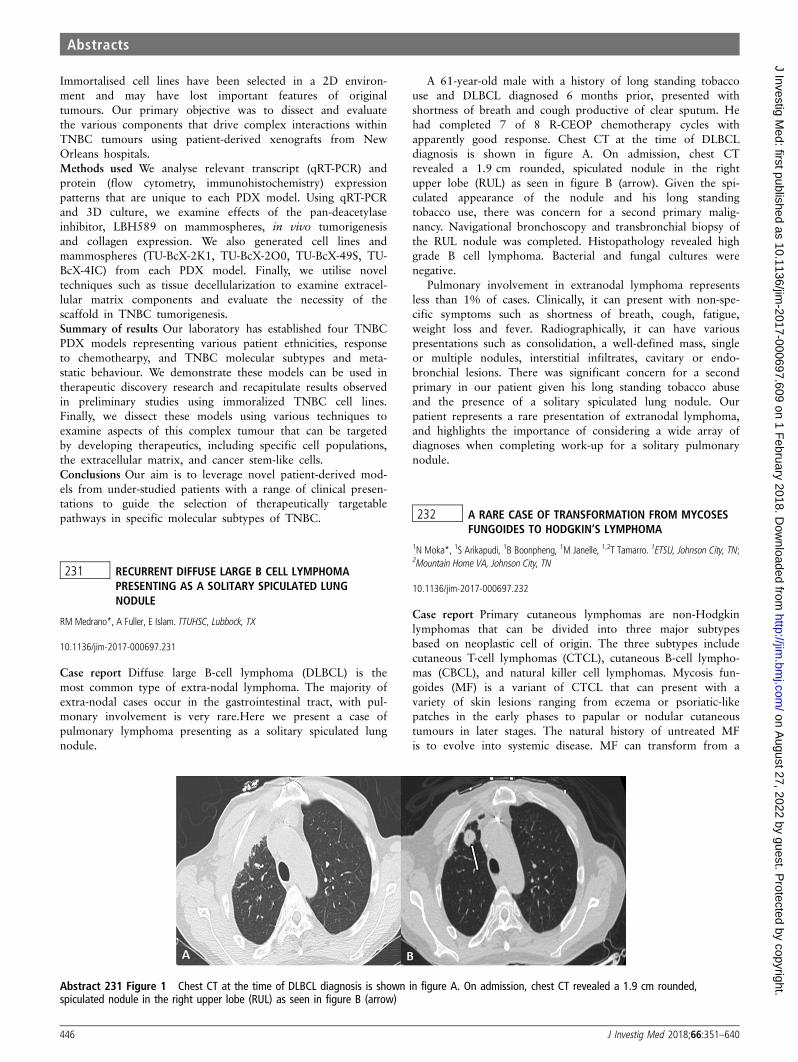
Immortalised cell lines have been selected in a 2D environ-ment and may have lost important features of originaltumours. Our primary objective was to dissect and evaluatethe various components that drive complex interactions withinTNBC tumours using patient-derived xenografts from NewOrleans hospitals.Methods used We analyse relevant transcript (qRT-PCR) andprotein (flow cytometry, immunohistochemistry) expressionpatterns that are unique to each PDX model. Using qRT-PCRand 3D culture, we examine effects of the pan-deacetylaseinhibitor, LBH589 on mammospheres, in vivo tumorigenesisand collagen expression. We also generated cell lines andmammospheres (TU-BcX-2K1, TU-BcX-2O0, TU-BcX-49S, TU-BcX-4IC) from each PDX model. Finally, we utilise noveltechniques such as tissue decellularization to examine extracel-lular matrix components and evaluate the necessity of thescaffold in TNBC tumorigenesis.Summary of results Our laboratory has established four TNBCPDX models representing various patient ethnicities, responseto chemothearpy, and TNBC molecular subtypes and meta-static behaviour. We demonstrate these models can be used intherapeutic discovery research and recapitulate results observedin preliminary studies using immoralized TNBC cell lines.Finally, we dissect these models using various techniques toexamine aspects of this complex tumour that can be targetedby developing therapeutics, including specific cell populations,the extracellular matrix, and cancer stem-like cells.Conclusions Our aim is to leverage novel patient-derived mod-els from under-studied patients with a range of clinical presen-tations to guide the selection of therapeutically targetablepathways in specific molecular subtypes of TNBC.
231 RECURRENT DIFFUSE LARGE B CELL LYMPHOMAPRESENTING AS A SOLITARY SPICULATED LUNGNODULE
RM Medrano*, A Fuller, E Islam. TTUHSC, Lubbock, TX
10.1136/jim-2017-000697.231
Case report Diffuse large B-cell lymphoma (DLBCL) is themost common type of extra-nodal lymphoma. The majority ofextra-nodal cases occur in the gastrointestinal tract, with pul-monary involvement is very rare.Here we present a case ofpulmonary lymphoma presenting as a solitary spiculated lungnodule.
A 61-year-old male with a history of long standing tobaccouse and DLBCL diagnosed 6 months prior, presented withshortness of breath and cough productive of clear sputum. Hehad completed 7 of 8 R-CEOP chemotherapy cycles withapparently good response. Chest CT at the time of DLBCLdiagnosis is shown in figure A. On admission, chest CTrevealed a 1.9 cm rounded, spiculated nodule in the rightupper lobe (RUL) as seen in figure B (arrow). Given the spi-culated appearance of the nodule and his long standingtobacco use, there was concern for a second primary malig-nancy. Navigational bronchoscopy and transbronchial biopsy ofthe RUL nodule was completed. Histopathology revealed highgrade B cell lymphoma. Bacterial and fungal cultures werenegative.
Pulmonary involvement in extranodal lymphoma representsless than 1% of cases. Clinically, it can present with non-spe-cific symptoms such as shortness of breath, cough, fatigue,weight loss and fever. Radiographically, it can have variouspresentations such as consolidation, a well-defined mass, singleor multiple nodules, interstitial infiltrates, cavitary or endo-bronchial lesions. There was significant concern for a secondprimary in our patient given his long standing tobacco abuseand the presence of a solitary spiculated lung nodule. Ourpatient represents a rare presentation of extranodal lymphoma,and highlights the importance of considering a wide array ofdiagnoses when completing work-up for a solitary pulmonarynodule.
232 A RARE CASE OF TRANSFORMATION FROM MYCOSESFUNGOIDES TO HODGKIN’S LYMPHOMA
1N Moka*, 1S Arikapudi, 1B Boonpheng, 1M Janelle, 1,2T Tamarro. 1ETSU, Johnson City, TN;2Mountain Home VA, Johnson City, TN
10.1136/jim-2017-000697.232
Case report Primary cutaneous lymphomas are non-Hodgkinlymphomas that can be divided into three major subtypesbased on neoplastic cell of origin. The three subtypes includecutaneous T-cell lymphomas (CTCL), cutaneous B-cell lympho-mas (CBCL), and natural killer cell lymphomas. Mycosis fun-goides (MF) is a variant of CTCL that can present with avariety of skin lesions ranging from eczema or psoriatic-likepatches in the early phases to papular or nodular cutaneoustumours in later stages. The natural history of untreated MFis to evolve into systemic disease. MF can transform from a
Abstract 231 Figure 1 Chest CT at the time of DLBCL diagnosis is shown in figure A. On admission, chest CT revealed a 1.9 cm rounded,spiculated nodule in the right upper lobe (RUL) as seen in figure B (arrow)
Abstracts
446 J Investig Med 2018;66:351–640
on August 27, 2022 by guest. P
rotected by copyright.http://jim
.bmj.com
/J Investig M
ed: first published as 10.1136/jim-2017-000697.609 on 1 F
ebruary 2018. Dow
nloaded from

CD4 +dominant to a more malignant CD30 +variant. How-ever, there are rare case reports of transformation betweencell lineages from a CTCL to Hodgkin lymphoma.
A 70 year old male with a 10 year history of MF presentedto the haematology/oncology clinic with worsening skin lesionsand cervical lymphadenopathy. Pertinent to his history is theclinical trajectory of his diagnosis of MF. He first presentedwith cutaneous plaques on the extensor surfaces of his legsand was diagnosed with psoriasis. Treatment with narrowband UV light and topical steroids was initiated withoutimprovement in his symptoms. He underwent treatment withetanercept and concurrent photo-chemotherapy for 3 years,after which new lesions were biopsied that were consistentwith folliculotropic MF. He was lost to follow-up for 2 afterhis diagnosis of MF and presented to the clinic to reestablishcare complaining of a new neck mass. Excisional lymph nodebiopsy was consistent with nodular sclerosing Hodgkin lym-phoma (CD15+, CD30+, CD20-, CD45-). He received 6cycles of ABVD and is currently in remission.
The clinical distinction of this case ultimately lies in theaetiology of the patient’s Hodgkin’s lymphoma. Our firsthypothesis is that the MF underwent transformation betweencell lineages and the Hodgkin’s lymphoma was an extensionof his pre-existing neoplasm. A competing theory is that twodistinct neoplastic processes occurred simultaneously. Themajor confounding variable is his 3 year treatment with eta-nercept, a TNF-alpha antagonist with a known associationwith the development of lymphoma.
233 16 YEAR OLD GIRL DIAGNOSED WITH CAPS ANDPROBABLE HIT
1M Nguy*, 2V Agarwal, 3C Gauger. 1UFCOM, Jacksonville, FL; 2Nemours, Pensacola, FL;3Nemours, Jacksonville, FL
10.1136/jim-2017-000697.233
Case report Catastrophic antiphospholipid syndrome (CAPS),also known as Asherman syndrome, is an uncommon compli-cation of antiphospholipid syndrome that occurs in <1% ofpatients that causes thrombus in multiple organs.1 In treatingCAPS, it is important to recognise heparin induced thrombo-cytopenia (HIT), as it can occur in up to 5% of patients tak-ing unfractionated heparin.2 We present a case of a 16-year-old Caucasian female with concurrent diagnosis of CAPS andprobable HIT. There are few case reports on these diagnosesin tandem in adults however none previously reported inpaediatrics.
REFERENCES1. Asherson RA, Cervera R, de Groot PG, et al. Catastrophic antiphospholipid syn-
drome registry project group. Catastrophic antiphospholipid syndrome: Interna-tional consensus statement on classification criteria and treatment guidelines.Lupus 2003;12(7):530–534.
2. Triplett DA, Asherson RA. Pathophysiology of Catastrophic AntiphosPholipid Syn-drome (CAPS). American Journal of Haematology 2000;65:154–159.
Diagnostic criteria for CAPS
1. Evidence of involvement of>3 organs systems or tissues
2. Development of symptoms in
3. Positive antiphospholipid antibodies (at least 6 weeks apart)
4. Histopathological findings of microthromboses in vasculature
Review of literature of concurrent cases of CAPS and HIT
Case Age/Sex Diagnosis/Aetiology
1 63 yo male Sepsis leading to bilateral haemorrhagic adrenal
infarction
2 28 yo
female
Post renal transplantation (end stage renal failure)
3 37 yo male DVT and Cellulitis
4 42 yo
female
Oestrogen
5 63 yo male Coronary heart disease with hematoma
234 WHAT CAME FIRST: THE OVARY OR THE LYMPHOCYTE?
H Oddo Moise*, A Coulon, J Doan, S Sanne. LSUHSC, New Orleans, LA
10.1136/jim-2017-000697.234
Case A 31 year old woman with history of daily IVDU anduntreated Hepatitis C presented to the emergency departmentwith 3 months of non-specific progressive ailments includingleft knee pain, shortness of breath with exertion and three-pil-low orthopnea, twelve pound weight loss, abdominal ‘tight-ness’ greatest in the left lower quadrant, early satiety,dysphagia to liquids and solids, left axillary node swelling andbilateral supraclavicular lymph node swelling. She denied fever,chills, night sweats, recent travel, sick contacts or family his-tory of cancer. Chest CT showed a substantial left pleuraleffusion with lobulated pleural thickening and mediastinal andhilar lymphadenopathy consistent with sarcoma versus meta-static disease. Subsequent abdominal and pelvic CT showedenlarged retroperitoneal lymph nodes of the left pelvis andgroin with a solid mass in the left deep pelvis concerning forovarian malignancy, metastatic disease, lymphoma or sarcoma.Transvaginal ultrasound revealed a solid left adnexal massmeasuring 5.6×3.4×5.2 cm. Workup was begun to ascertainthe primary site of malignancy suspicious for lymphocyte ver-sus ovarian source. A right supraclavicular lymph node biopsywas performed during which she was intubated for increasingleft pleural effusion with compression of mediastinal struc-tures. Thoracentesis was performed with removal of 1.5 L ofblood-tinged pleural fluid and a chest tube was placed. Flowcytometry of the lymph node biopsy showed 95.6% T lym-phoblasts positive for CD2, CD3, CD7, TdT and CD99 con-sistent with the diagnosis of Non-Hodgkin T-celllymphoblastic lymphoma. CA125 was mildly elevated at 47,not suggestive of ovarian malignancy. Despite numerousattempts at discussing the importance of a bone marrowbiopsy and cancer treatment options, the patient declined allmedical intervention or palliative resources.Discussion Malignant lymphoma involvement of the femalegenitourinary tract, including the ovary, is not commonly seen.While ovarian involvement is relatively rare, non-HodgkinLymphomas such as T cell lymphoblastic lymphoma (T-LBL)represent a frequency of approximately 7% to 26% of thosediagnoses and should be considered in the differential diagno-ses of young females with ovarian masses.
Abstracts
J Investig Med 2018;66:351–640 447
on August 27, 2022 by guest. P
rotected by copyright.http://jim
.bmj.com
/J Investig M
ed: first published as 10.1136/jim-2017-000697.609 on 1 F
ebruary 2018. Dow
nloaded from

235 POEMS: AN ODE TO A SYNDROME*S Ozair, B DeBoisblanc, S Subbiah, R Jetly. Louisiana State University Health SciencesCentre, New Orleans, LA
10.1136/jim-2017-000697.235
Case report A 45 year-old man with normal coronary arterieson angiography was referred for evaluation of thrombocytosis(platelet count of 812×103/UL) following two separate acutemyocardial infarctions. Testing for JAK2 and CALR mutationswere negative. He also described symptoms of worsening pol-yneuropathy and anasarca. POEMS (Polyneuropathy, Organo-megaly, Endocrinopathy, Monoclonal protein, Skin changes)syndrome was suspected. Physical exam was additionallyremarkable for splenomegaly and newly developed hyperpig-mentation of both hands with white nails. On laboratory test-ing he was found to have adrenal insufficiency andhypothyroidism. Both kappa and lambda free light chainswere elevated but there was no M-spike on serum proteinelectrophoresis. Bone marrow biopsy was consistent with anincreased population of plasma cells that formed atypicalaggregates rimming non-paratrabecular lymphoid aggregates.These plasma cells were noted to have a lambda predominantexpression pattern, suspicious for monoclonality. Imaging stud-ies confirmed osteosclerotic lesions in T10 and mediastinallymphadenopathy. VEGF (vascular endothelial growth factor)was elevated at 264 pg/ml.
The cause of POEMS syndrome is unknown, althoughchronic overproduction of pro-inflammatory cytokines (eg, IL-1, IL-6, and VEGF) appears to be a major contributor to themicroangiopathy, increased vascular permeability, thrombocyto-sis, and neovascularization that leads to thrombosis, tissueoedema, polyneuropathy, and pulmonary hypertension.
The International Myeloma Working Group (IMWG) crite-ria for the diagnosis of POEMS requires the presence of twomandatory criteria (polyneuropathy and monoclonal protein),and at least one major and one minor criterion. Major criteriainclude sclerotic bone lesions, elevated VEGF, and Castlemandisease. Minor features include organomegaly, endocrinopathy,characteristic skin changes, papilledema, extravascular volumeoverload, and thrombocytosis.
Patients with POEMS syndrome are best treated with che-motherapy with or without autologous stem cell transplanta-tion similar to that used for the treatment of multiplemyeloma.
236 FAILED SCREENING OF ADVANCED MYELOMA IN ANASIAN WOMAN
T Dickerson, S Bautista, S Palumbo*, S Hebert, LS Engel. LSU Health Sciences Centre, NewOrleans, LA
10.1136/jim-2017-000697.236
Introduction The incidence of multiple myeloma (MM) varieswith ethnicity and age; Asian populations have a lower inci-dence. The mean age of onset of MM in Asian population is62 years.Case A 51-year-old Asian woman without significant medicalhistory had multiple Emergency Department visits with pro-gressive, non-radiating low back pain of two months duration.Spinal radiographs were unrevealing and no screening labswere obtained. Eventual referral to an ambulatory clinic iden-tified substantial hypercalcemia. Upon reassessment, the patient
endorsed weakness, cramps, anorexia, fever, abdominal pain,constipation, urinary frequency, and a 15-pound weight loss.Physical exam revealed a thin female with tenderness over L4-S1 vertebrae and a 1 cm firm left axillary node. Labs showedcorrected calcium 14.52 mg/dL, haemoglobin 10.1, proteingap 8.2 g/dL, and creatinine 1.4 mg/dL. Lumbar CT revealednumerous lytic foci throughout the spine and pelvis supportedby skeletal survey revealing lytic foci of the cranium, axialand proximal appendicular skeleton. Her other labs showedelevated IgG, B2-microglobulin, Kappa light chain, and Mspike, UPEP with monoclonal band in gamma region, andbone marrow biopsy of >50% plasma cells. FISH analysisshowed translocation (11;14) with fusion of CCND1 (BCL1)at 11q13. The patient was diagnosed with multiple myeloma(MM) Stage III by ISS criteria. Hypercalcemia was treatedwith IV fluids, zoledronic acid, and calcitonin. Radiation wasutilised for spinal compression at S1. Lastly, bortezomib anddexamethasone were initiated with plans for stem celltransplant.Discussion A lower incidence of MM in the Asian populationhas been partly attributed to genetic polymorphisms withdoubled incidence in the last 10 years in South Korea andTaiwan. Analysis of genetic versus environmental risk factors islacking. MM is ‘a disease of the elderly’ but in an Asiancohort, 58.5% of those diagnosed were <65 years-old. Basedon these findings, prevalence in this cohort at earlier ages islikely to continue and research should be sustained.
237 ACUTE LIVER FAILURE CAUSED BY MULTIPLE MYELOMA
T Poosarla*, S Elkins, A Reddy. University of Mississippi Medical Centre, Jackson, MS
10.1136/jim-2017-000697.237
Case report Multiple Myeloma (MM) is a plasma cell neo-plasm that accounts for about 15 percent of all haematologicalmalignancies. It is a disorder that involves clonal proliferationof neoplastic plasma cells in the bone marrow with anincrease in monoclonal paraprotein. It is uncommonly associ-ated with extraosseous involvement such as spleen or liver,especially at initial presentation. Reports of liver involvementin MM are usually secondary to amyloidosis rather thanplasma cell involvement. Here we present a rare case of apatient with acute liver failure (ALF) who was found to haveMM.
A 56-year-old African American female with history ofhypertension and hypothyroidism who was admitted to thehospital with complaint of 2 weeks of abdominal swelling, dif-ficulty ambulating secondary to bilateral lower extremity pain,lower extremity swelling, and shortness of breath. She alsonotes a 15-pound weight loss over the last 2 months. In theemergency room, she was found to have ascites, globulin gab,elevated serum total protein, hypoalbuminemia, anaemia, andrenal failure concerning for cirrhosis. CT of abdomen and pel-vis showed a large volume of ascites, abnormal appearance ofliver parenchyma, and enlargement of the IVC and hepaticveins suggestive of cirrhosis. She underwent further workupincluding liver biopsy which showed sinusoidal involvement byplasma cells concerning for MM and no evidence of hepatitisor cirrhosis. Cytology on peritoneal fluid also showed malig-nant plasma cells. Serum protein electrophoresis consistentwith IgG lambda with M protein of 10. Bone marrow biopsyrevealed hypercellular marrow with 80% plasma cells. There
Abstracts
448 J Investig Med 2018;66:351–640
on August 27, 2022 by guest. P
rotected by copyright.http://jim
.bmj.com
/J Investig M
ed: first published as 10.1136/jim-2017-000697.609 on 1 F
ebruary 2018. Dow
nloaded from

were multiple subcentimeter lesions throughout the skeletonconsistent with MM. She was started on treatment with borte-zomib, revlimid, dexamethasone with marked improvement insymptoms and resolution of ascites.
MM presenting as ALF is extremely rare and only a fewcases have ever been reported. Liver involvement can be seenand is discovered incidentally but is clinically silent. It is asso-ciated with poorer prognosis as treatment is limited with liverfailure. It is important to consider MM in patients whopresent with ALF, especially with no risk factors for cirrhosis,since prompt diagnosis can lead to quicker treatment and the-oretically improve survival.
238 BLOOD CELLS: TOLERANCE, AUTOIMMUNITY,MODULATION
1EC Quintin*, 2KN Rezkallah, 1S Ozair, 1TM Reske, 1H Boulares, 1P Rigby. 1LSUHSC NewOrleans, New Orleans, LA; 2Presence Saint Joseph Hospital, Chicago, IL
10.1136/jim-2017-000697.238
Purpose of study All haematological cells originate from a mul-tipotent mesenchymal stem cell that are produced via myelo-and lymphopoietic pathways in the niches of the bone mar-row. The marrow structure plays a key role in the mixing ofcells and cytokines resulting in cell production, maturation,kinetics, homing and circulation in the niches and sinusoids.The purpose is to enumerate, identify, and access crosstalk ofblood cells related to Myeloid Derived Suppressor Cells(MDSC).Methods used By identifying blood cells using fluorescence-activated cell sorting (FACS), we aim to locate pairs or clus-ters utilising standard procedures and studying the functionand relationship of these cells.Summary of results The stem cells are held in place by surfacereceptors and divide under a stimulus. They mature and travelto a sinusoid where they mix with other cells before beingreleased into the venous circulation. Certain cells seek outspecific partners according to their own specific surface recep-tors to crosstalk, exchanging particles and messages causingdownhill haematological and immunological cascades.
As individuals grow and age from birth, tolerance isarranged for all self-antigens and tissues. If tolerance is lost,disease may follow. The number, types and proportions of dif-ferent cells change throughout a lifetime. The mix of MDSCwith Tregs, memory cells, NK cells, stem cells, and otherlymph cells maintain tolerance for self but changes in thenumbers of cells can affect their function, maturity or hostcharacteristics.Conclusions When autoimmune disease is found, cell crosstalkchanges depend on appropriate numbers, proportions, andcross-functions. They may be exogenously altered to regain atolerant state through modulation of the immune systems viaimmunosuppressive medications. Specific pairs are linkedthrough cell surface marker/receptor anatomy, relating cells toactivity producing certain cytokines. The relationship of cellpairs or other groupings will likely determine inhibition oractivation of certain processes that may lead to pathology.
239 A CASE OF CONCURRENT RENAL CELL CARCINOMAAND HODGKIN’S LYMPHOMA
M Quirch*, RE Gavidia Quezada, S Phisitkul. Texas Tech University Health Sciences Centre,Lubbock, TX
10.1136/jim-2017-000697.239
Background The association between developing secondarymalignancy after treatment for a primary malignancy is wellknown, particularly with leukemias and lymphomas such asHodgkin’s lymphoma. Patients treated for Hodgkin’s Lym-phoma are known to have an increased risk of developing asecond malignancy such as breast, thyroid, bone, colorectal, orstomach cancer. In a small retrospective study, multiple pri-mary malignancies have been reported, particularly in womenfrom the ages 43–68, with concurrent malignancies typicallybeing breast, uterine, or cervical.Case presentation This is a 71 year old female admitted forgeneralised weakness, hyponatremia, pancytopenia, and lacticacidosis with no verifiable signs or source of infection. Onemonth prior to admission, the patient was undergoing workup with her private urologist for a recently discovered renalmass per CT scan that was suspicious for renal cell carcinomaof 2.3×2.0 cm with multiple small lymph nodes. Decision atthe time of discovery of the mass was to proceed with sur-gery however she became acutely ill and that is what led toher admission to our hospital.
Initial blood cultures and urine culture were negative how-ever lactic acid remained persistently elevated and LDH wasalso elevated so a bone marrow biopsy was done revealingHodgkin Lymphoma. The patient rapidly deteriorated afterbone marrow biopsy. She developed acute respiratory failure,altered mental status, and hypotension which required intuba-tion, vasopressors, and admission to the medical intensive careunit. Broad spectrum antibiotics were started with culturesbeing drawn seven days after admission. Urine cultures aftertransfer were positive for pansensitive Klebsiella pneumoniaand E. coli, and respiratory culture was positive for Staphylo-coccus aureus. Blood cultures remained negative throughouther stay. Despite aggressive care, the patient did not recoverand the patient’s family pursued comfort measures.Conclusion While there is an established relationship betweendeveloping secondary malignancy after treatment with chemo-therapy, concurrent malignancies tend to be very rare. Untilnow, there has been one other reported case of Hodgkin’slymphoma occurring concurrently with renal cell carcinoma.
240 A CASE OF INCOMPLETE CARNEY TRIAD
A Ismail, M Quirch*, S Mousa, MR Arevalo. TTUHSC, Lubbock, TX
10.1136/jim-2017-000697.240
Case report Carney triad is a synchronous or metachronousassociation of gastric gastrointestinal stromal tumours (GIST),pulmonary chondroma and extra-adrenal paraganglioma. Mostpatients have only one or two components of the triad, allthree tumours being found in only about 2% of the patients.
Abstracts
J Investig Med 2018;66:351–640 449
on August 27, 2022 by guest. P
rotected by copyright.http://jim
.bmj.com
/J Investig M
ed: first published as 10.1136/jim-2017-000697.609 on 1 F
ebruary 2018. Dow
nloaded from

We present a case of a 28-year-old woman that came tothe emergency department for epigastric pain with regurgita-tion. She has a medical history of rheumatoid arthritis, Hashi-moto’s thyroiditis and Celiac disease. She reported 6 monthhistory of 20-pound weight loss and loss of appetite. Shedenied dysphagia or changes in bowel habits. She had a CTof the abdomen showing a large exophytic gastric mass, con-cerning for malignancy and incidental calcified masses wereseen in the lungs. EGD and EUS showed a subepitheliallesion, suspicious for malignant stromal cell neoplasm. Biopsyconfirmed it to be GIST, positive for CD117. Molecular diag-nosis was negative for Kit and platelet-derived growth factoralpha (PDGFRA) mutations. Lung mass biopsy was consistentwith pulmonary chondroma. Curative surgery was planned.Intraoperatively, the tumour was found invading the liver andlymph nodes. She had total gastrectomy with esophago-jejuns-tomy, celiac lymphadenectomy, and partial liver resection withnegative margins. She was discharged 2 weeks after surgeryand follows at the cancer centre.
GIST is a mesenchymal neoplasm affecting the gastrointesti-nal tract typically presenting as a subepithelial neoplasms.Although the majority of GISTs appear to be sporadic, 5% ofpatients have one of the familial autosomal dominant syn-dromes, including neurofibromatosis type 1, Carney triad, andprimary familial GIST syndrome. Most GISTs are characterisedby KIT or PDGFRA activating mutations. There are 10%–15%of GISTs lacking KIT and PDGFRA mutations, called wild-type GISTs. Among these WT GISTs, a small subset is associ-ated with succinate dehydrogenase (SDH) deficiency, known asSDH-deficient GISTs. GISTs that occur in Carney triad repre-sent specific examples of SDH-deficient GISTs. SDH-deficientGISTs locate exclusively in the stomach, showing predilectionfor children and young adults with female preponderance.The tumour generally pursues an indolent course and exhibitsprimary resistance to imatinib therapy in most cases. Surgicalresection is the preferred mode of therapy.
241 DIPLOPIA AND PROPTOSIS WITH A PITUITARY MASSEQUALS A MACROADENOMA? THINK AGAIN!
D Reddy*, M Ibrahim, K Krishnan, D Jaishankar. ETSU, Johnson City, TN
10.1136/jim-2017-000697.241
Case report Acute Myeloid Leukaemia (AML) is an aggressivehematologic malignancy. Central nervous system (CNS)involvement is rare among adult patients with AML. Wedescribe a patient with AML presenting with visual disturban-ces and a pituitary mass which resolved after systemic AMLchemotherapy.
A seventy one year old female with a history of AMLtreated successfully (without allogeneic hematopoietic stem celltransplantation {allo-HSCT}) sixteen years ago, was hospital-ised with a two week history of fatigue, weight loss and inter-mittent blurry vision. Clinical exam was notable for diplopiaand right sided proptosis without visual field defects. MRIBrain revealed a 2.4 cm sellar mass (suggestive of a macroade-noma) with cavernous sinus invasion, right carotid arteryencasement, and mass effect on right optic chiasma. Peripheralblood smear documented 28,000 WBC count with 84% blasts.Pituitary function assay was normal. Bone marrow biopsyreported hyper cellularity (60%) and 70% myeloblasts. Molec-ular studies t (8;21) established a new clonal leukaemia
distinct from her previous AML (normal cytogenetics). Lumbarpuncture revealed monocytes with rare Auer rods. She com-pleted induction chemotherapy with Idarubicin and cytarabineachieving first complete remission (CR1). She received onedose of intrathecal methotrexate and 3 cycles of cytarabineconsolidation chemotherapy course. Her diplopia and propto-sis resolved completely.
AML is characterised by a rapid clonal proliferation ofimmature hematopoietic cells in the peripheral blood andbone marrow. The overall survival of AML is dictated bycytogenetics/molecular markers and age. When CR1 isachieved, most relapses occur in the first two years. Extrame-dullary involvement with CNS leptomeningeal infiltration canbe noted in acute lymphoblastic leukaemia and in 2%–10% ofpatients after allo-HSCT. Leukemic infiltration of the pituitarygland with AML is extremely rare and documented in isolatedcase reports. Our patient represented an extremely rare andunique case of a second-primary AML with pituitary involve-ment that responded to induction and consolidationchemotherapy.
242 ‘DOSE ESCALATED’ CHEMOTHERAPY FOR AGRRESIVELYMPHOMA IN THE ELDERLY
D Reddy*, F Tawadros, D Jaishankar. ETSU, Johnson City, TN
10.1136/jim-2017-000697.242
Case report Diffuse large B-cell lymphoma (DLBCL) is anaggressive Non Hodgkins Lymphoma (NHL). The anti-CD20antibody rituximab anchors various immuno chemotherapyregimens, including R-CHOP (Rituximab,cyclophosphamide,adriamycin,vincristine,prednisone) a gold standard for DLBCL.Management of DLBCL in the elderly poses unique challengesgiven increased risk of toxicity. Anthracycline based regimensare avoided in the elderly leading to lower response rates. Wepresent a case of an elderly patient who achieved completeresponse with a unique protocol of Adriamycin based therapy.
An eighty-five year old female with multiple medical prob-lems was hospitalised with dyspnea and weakness. Non-tendercervical and inguinal adenopathy was noted. Nodal excisionbiopsy demonstrated high grade (Ki-67 >90%), B cell ‘dou-ble-hit’ lymphoma with MYC/BCL-2 rearrangement and gradeIIIB follicular lymphoma. PET/CT revealed splenomegaly anddiffuse hypermetabolic adenopathy above and below the dia-phragm. A positive bone marrow biopsy established stage IVdisease. CSF analysis was negative. The patient and familywere very keen on treatment despite her advanced age andborderline performance status (ECOG II). She received 2cycles of R-CVP (Rituximab, cytoxan, vincristine, prednisone).Adriamycin at 50% of full dose (akin to mini R-CHOP) wasintroduced at cycle 3 and continued at 75% of full dose forcycles 4–6. Post treatment PET-CT demonstrated excellentresponse.
DLBCL is the commonest type of NHL, usually presentingin the seventh decade. With improving life expectancy, its inci-dence in the elderly is predicted to rise. DLBCL is curablewith a long term survival rate of 40% with anthracyclinebased regimens. Multiple non-anthracycline based regimens areavailable for frail patients. mini-CHOP is a variant with 50%dose reduction of Cytoxan, Vincristine and Adriamycin. Ourprotocol differed in maintaining full doses of Cytoxan andVincristine while introducing Adriamycin at 50% of full dose
Abstracts
450 J Investig Med 2018;66:351–640
on August 27, 2022 by guest. P
rotected by copyright.http://jim
.bmj.com
/J Investig M
ed: first published as 10.1136/jim-2017-000697.609 on 1 F
ebruary 2018. Dow
nloaded from

and escalating doses as tolerated. Our patient completed ther-apy successfully without serious complication with clinical andradiographic improvement during therapy. Complete responsewas noted at the end of treatment. We propose ‘dose esca-lated’ mini R-CHOP as an intermediate regimen for theelderly.
243 BILATERAL OTITIS EXTERNA MASKINGEXTRAMEDULLARY RELAPSE OF ACUTEPROMYELOCYTIC LEUKAEMIA
1C Rivera-Franceschini*, 1,2V Vestal, 1A Cruz. 1San Juan City Hospital, Ponce, PR; 2VAHospital San Juan
10.1136/jim-2017-000697.243
Case report Acute promyelocytic leukaemia (APL) is a subtypeof acute myelogenous leukaemia that is characterised by thetranslocation of chromosomes 15 and 17. The introduction ofall-trans retinoic acid (ATRA) as an early therapy hasincreased the overall remission rates in these patients butrelapses still occur. Most relapses are limited to the bone mar-row and blood. However, APL can also relapse to extramedul-lary sites involving the skin, central nervous system (CNS) andother organs. A 48-year-old man with APL on remission for 2years presented to the emergency room with bilateral ear painof 3 weeks duration. Pain began after swimming in a pool. Itwas 8/10, constant and associated with tinnitus, bloody secre-tions and decreased bilateral hearing acuity. Upon evaluationpatient was found with a bilateral swollen and erythematousear canal with granulating tissue formation. He was evaluatedby an otorhinolaryngologist who recommended IV antibioticpiperacillin/tazobactam and dexamethasone/ciprofloxacin eardrops. A maxillofacial CT scan of the head showed opacifiedexternal auditory canals bilaterally, which could correlate withmucormycosis infection. Ear infection improved with antibiot-ics and biopsies of the external ear canals were taken. Theleft external ear canal biopsy was extensively involved withimmature myelocytes, compatible with involvement by acutepromyelocytic leukaemia. Bone marrow biopsy was negativefor leukaemia. Cerebral spinal fluid analysis was negative formalignancy. Patient was started on ATRA, arsenic trioxide andradiotherapy. Even though CNS involvement is the most com-mon cause of extramedullary relapse in APL, a biopsy of theear canal should be considered in a patient with suspected oti-tis externa since it could be a rare presentation of APL. It isimperative to identify this type of extramedullary relapse con-sidering that patients could benefit from inductionchemotherapy.
244 LDH AS AN EARLY MARKER FOR PRIMARYMYELOFIBROSIS?
EB Saul*, B Dreiling. University of Mississippi Medical Centre, Madison, MS
10.1136/jim-2017-000697.244
Introduction Primary myelofibrosis (PMF) is a type of chronicmyeloproliferative neoplasm with an estimated incidence of1.5 per 1 00 000 per year occurring mainly in middle agedand older adults. There are various different clinical manifesta-tions of the disease some of which include fatigue, low-gradefever or night sweats, weight loss and splenomegaly.
Case A 68 year old Caucasian male presented with a mildhypoproliferative anaemia of obscure origin that dated back to2012. He has had no fevers, night sweats or weight loss. In2012, his haemoglobin and hematocrit (H/H) were 11.6 and35.2 respectively however has gradually trended down to 9.1/29.2. WBC and platelet counts are normal. Interestingly, hisLDH was elevated in 2012 at 670 and has continued toincrease over the years to a current level of 1057. LDH frac-tionation was done revealing fraction 2>1>3>4>5 suggestinga bone marrow, cardiac or renal aetiology. CPK, renal func-tion and cardiac workup were all normal. Peripheral bloodsmear revealed nucleated red blood cells, moderate large plate-lets, elliptocytosis, and teardrop cells. Bone marrow biopsywas done given concerns for ineffective hematopoiesis. Pathol-ogy returned as a myeloproliferative neoplasm, best regardedas primary myelofibrosis. A JAK2 V617F mutation analysiswas sent and returned positive. His spleen measured 14.14 cmin length on ultrasound. He remains asymptomatic with aDIPSS-plus score of 2 and a median overall survival of 2.9years. However, given that he is asymptomatic with stableblood counts he has chosen observation at this time.Discussion There are various nonspecific abnormal laboratorytests in patients with primary myelofibrosis. Since there is no‘gold standard’ for the diagnosis of PMF, there have beendiagnostic criteria that have been proposed by the WHO.LDH is an enzyme found in the marrow and blood precur-sors. With intramedullary hemolysis associated with PMF, onecould presume elevations in LDH may actually precedechanges in the CBC assisting in the early detection of PMF.
245 CUTANEOUS LYMPHOMA: THE NON RESOLVINGNODULE
P Shelley*, KR Green. UF Health, Jacksonville, FL
10.1136/jim-2017-000697.245
Introduction Primary cutaneous anaplastic large cell lymphomais rare malignancy which characteristically exhibits CD30 posi-tivity. Usually presenting in the sixth decade with spontaneoussolitary or grouped nodules that fail to resolve. Diagnosis ismade by skin biopsy which demonstrate characteristic largecells with eosinophilic cytoplasm and occasional horseshoeshaped nuclei with evidence of epidermal hyperplasia. Treat-ment modalities include localised surgical excision and sys-temic chemotherapy. The latter isreserved for those cases withwidespread lesions.Case report A forty three year old female with a history ofnon-hodgkin’s lymphoma and sarcoidosis presented to an out-side institution with a complaint of progressive swelling in herright axilla for the past five weeks. The mass was both itchyand tender to palpation. She also complained of fevers, nightsweats, and a thirty pound unintentional weight loss. Due toconcern for potential abscess an incision and drainage wasattempted but upon incision no spontaneous drainage wasseen. She was prescribed a course of antibiotics and instructedto follow up wither her primary care physician. She then pre-sented to our institution with complaint of multiple flesh col-oured skin lesion which spontaneously erupted over the pastweek in sporadic areas from head to toe sparing the palmsand soles. Further exam revealed numerous five to ten milli-metre, firm, well circumscribed lesions on the breasts, arms,thighs, abdomen, back, neck, and face. Along with a large five
Abstracts
J Investig Med 2018;66:351–640 451
on August 27, 2022 by guest. P
rotected by copyright.http://jim
.bmj.com
/J Investig M
ed: first published as 10.1136/jim-2017-000697.609 on 1 F
ebruary 2018. Dow
nloaded from

by three tender and firm mass in the right axilla. A punchbiopsy was performed of the lesions on the left breast andright clavicle. Dermatopathology of the aforementioned sam-ples were positive for anaplastic large cell CD30 positive lym-phoma. She was then referred to an oncologist to discusstreatment options and to begin staging of her malignancy.Discussion This case highlights the necessity of prompt evalua-tion of skin lesions via biopsy, especially in those patientswith previous history of malignancy. Most cases of primarycutaneous anaplastic large cell lymphoma have a ten year sur-vival rate of ninety percent if treated appropriately. Howeverrecurrence is common. Systemic chemotherapy is typicallyreserved for those with disseminated disease or those withmultiple recurrences with surgical excision.
246 EXTENDED THIRTY MONTH PROGRESSION FREESURVIVAL WITH THIRD LINE ERIBULIN FOR METASTATICBREAST CANCER
S Singal*, R Khalaf, D Jaishankar. East Tennessee State University, Johnson City, TN
10.1136/jim-2017-000697.246
Case report Eribulin is a non-taxane microtubule inhibitorapproved for treatment of metastatic breast cancer after twoprior chemotherapeutic regimens. Eribulin as third line agentin the palliative setting has shown a median overall survival(OS) of 13 months and median progression free survival (PFS)of 3 months. We report a patient with extended (PFS) ofmore than 30 months with metastatic breast cancer treatedwith Eribulin in the third line setting. A forty-eight year oldlady was diagnosed with stage IIA (T2N0M0), high grade, tri-ple negative, invasive ductal carcinoma (IDC) of the leftbreast. She underwent neo adjuvant chemotherapy with Adria-mycin and Cytoxan followed by a negative sentinel lymphnode biopsy. At mastectomy she was noted to have a 2.5 cmtumour, high grade, triple negative IDC with three additionallymph nodes negative for metastatic carcinoma. She subse-quently pursued further chemotherapy and was treated with 6cycles of Cytoxan, Methotrexate and Fluorouracil (CMF). Shethen transferred care to our cancer centre and eight monthsinto her surveillance program developed a 2.8 cm right lowerlobe (RLL) pulmonary mass with SUV of 27 on a PET-CT. Afine needle biopsy was consistent with metastatic triple nega-tive breast cancer with sheets of poorly differentiated carci-noma similar in morphology to previous breast pathology.Imaging studies revealed oligometastatic disease. She com-menced single agent Taxol in the 1 st line metastatic settingwith dramatic decrease in RLL pulmonary mass to less than1 cm with SUV of 1.7 and resolution of other sub cm pulmo-nary nodules. The response was short lived lasting only sixmonths. She started 2nd line Gemcitabine with subsequentlargely stable disease for a period of 11 months. Progressionof RLL pulmonary nodule measuring 2.1 cm with SUV of 10noted. She then started 3rd line Eribulin with a dramaticresponse on imaging studies within three months and hasmaintained no evidence of disease (NED) on scans over thesubsequent 30 months. She is clinically stable and her tumourmarkers have plateaued. She has required Eribulin dose reduc-tions on account of neuropathy. Our patient has shown excel-lent response and tolerance to Eribulin with PFS of over 30months (ten times the norm) which is rare.
247 INTRAVASCULAR LARGE B CELL LYMPHOMAPRESENTING AS CHRONIC COUGH
S Singal*, R Khalaf, D Jaishankar, E Spradling. East Tennessee State University, JohnsonCity, TN
10.1136/jim-2017-000697.247
Case report Intravascular large B cell lymphoma (IVLBCL) is arare and aggressive subtype of large cell lymphoma that ischaracterised by proliferation of lymphoma cells within thelumina of small blood vessels, particularly capillaries and post-capillary venules most commonly reported as cutaneous andCNS lesions. We report a rare case of IVLBCL involving thelungs. A 73-year-old male, never smoked, presented with per-sistent cough and hyponatremia. Chest imaging showed calci-fied mediastinal adenopathy. Pulmonary function testingshowed no restrictive or obstructive pattern. Bronchoscopywith endobronchial ultrasound guided biopsy was non-diagnos-tic. Patient underwent video assisted thoracoscopy with rightupper middle and lower lung wedge biopsies. Pathologyshowed IVLBCL with large B cells located mainly intracapil-lary and intra-small artery and veins. Tumour cells were posi-tive for CD20, CD79a, Pax-5, CD10, Mum-1. Tumour cellswere negative for CD3, CK AE1/AE3, S100, Cam5.2, CD34.Bone marrow biopsy and aspiration showed minimal involve-ment of large B-cell lymphoma. PET scan revealed focaluptake at T4, T9, L2, L4 vertebrae concerning for lymphoma-tous involvement of the bone. Cerebrospinal fluid was nega-tive for malignancy. Brain imaging was negative. Patient wasdiagnosed with Stage IV IVLBCL. During recovery from rightlung wedge biopsies he developed acute respiratory failurerequiring oxygen at 10 L/min. Urgent chemotherapy with R-CHOP cycle 1 was initiated providing complete resolution ofall symptoms. The final treatment plan includes 6 cycles of R-CHOP with CNS prophylaxis.
Our case demonstrates extranodal disease, aggressive biol-ogy, and the requirement of urgent treatment. The rarity ofthis disease and difficulty of detecting intravascular infiltrationoften contributes to delay of diagnosis and high mortality.
248 PENILE LYMPHOMATOUS DISEASE PORTENDS POORPROGNOSIS?
F Tawadros*, D Reddy, K Krishnan, D Jaishankar. ETSU, Johnson City, TN
10.1136/jim-2017-000697.248
Case report Diffuse Large B Cell Lymphoma (DLBCL)accounts for 25% of all non-Hodgkin Lymphomas. DLBCLpresents as a rapidly enlarging mass, usually nodal enlarge-ment, or less often as a mass lesion anywhere in the body.We present a patient with DLBCL with highly atypical extranodal presentation. A sixty-two year old male presented withpersistent right upper quadrant abdominal pain and weightloss over six weeks. Imaging studies suggested gallbladderadenomyosis and patient underwent a cholecystectomy. Pathol-ogy reported DLBCL, activated B cell type, CD20 +andMUM1 +with Ki67 90%. Molecular studies ruled out doubleand triple hit lymphoma but were positive for additional cop-ies of IgH, amplification of BCL-2 gene and BCL-6 gene dele-tion. Clinical exam revealed significant penile induration. PET-CT revealed mediastinal and abdominal adenopathy with mildtonsillar, parotid, and massive penile involvement. CSF cytol-ogy, MRI Brain, Testicular Ultrasound and Bone marrow
Abstracts
452 J Investig Med 2018;66:351–640
on August 27, 2022 by guest. P
rotected by copyright.http://jim
.bmj.com
/J Investig M
ed: first published as 10.1136/jim-2017-000697.609 on 1 F
ebruary 2018. Dow
nloaded from

biopsy were negative. Chemo immunotherapy with R-CHOPand intrathecal methotrexate was initiated. Dramatic regressionof penile induration which served as an index lesion, wasnoted within days. Post treatment PET-CT documented com-plete response. Three months after therapy he developed abiopsy proven intracranial relapse in the left occipital lobe.He succumbed to his illness while receiving palliative salvagechemotherapy with high dose cytarabine and methotrexate.DLBCL is a heterogeneous entity derived from germinalcentre B cells or post-germinal centre B cells. Extra nodal dis-ease (liver, lung, skin) is a less common but well knownoccurrence. Penile involvement may present as primary penilelymphoma or secondary to systemic disease. While testicularlymphoma is well reported, secondary penile involvement israre accounting for less than 1% of extra nodal disease. Prog-nosis in DLBCL is dictated by clinicopathologic factors andextra nodal involvement portends higher relapse risk. Parame-dian lymphoma is associated with a risk of CNS relapse. Ourcase highlights a high risk variant of DLBCL with atypicalpresentation, multiple rare extra nodal sites of disease, unusualmolecular features, excellent response to therapy culminatingin CNS relapse.
249 A SUSPICIOUS CASE OF PERNICIOUS ANAEMIA ANDVITAMIN B12 DEFICIENCY
A Traina*, J Dubuc, H Oddo Moise, A Coulon, A Bourgeois, J Doan, S Guillory, S Sanne.LSUHSC, New Orleans, LA
10.1136/jim-2017-000697.249
Case report A 56 year old man with a past medical history ofhypertension was brought to the emergency department withprogressively worsening dyspnea on exertion and fatigue for 2months. He endorsed subjective fever for 2 weeks and hisfamily noted confusion over several days. He also reported 15pound weight loss for several months. He was febrile to105.5F on arrival. The physical exam was significant for asystolic flow murmur, mild confusion and slowed speech withunclear baseline neurologic exam, and normal sensation andproprioception in the distal extremities. Initial labs revealedpancytopenia and a macrocytic anaemia with WBC of 2.3103/UL, ANC of 100 cells/mL, haemoglobin 8.7 g/dL, hematoc-rit 26%, MCV 113.9 FL and platelets 41 103/UL with schisto-cytes and tear drop cells. He underwent further studies forpancytopenia and fever with negative infectious work upincluding HIV, blood and urine cultures. His vitamin B12 levelwas profoundly deficient at 64 PG/mL with normal folate andiron studies. His reticulocyte count was inappropriately normalat 0.9%. Haematology was consulted due to concern for hem-atologic malignancy. There were no blast cells identified onperipheral blood smear. Bone marrow biopsy and flow cytom-etry revealed hypersegmented neutrophils, but there was nodefinitive immunophenotypic evidence of hematopoietic neo-plasia with normal CD markers, immunophenotyping andcytogenetics. There was no identifiable source of fever ormalignancy on CT images of the chest, abdomen or pelvis.The patient was started on intramuscular vitamin B12 supple-mentation with improvement in all cell lines. Intrinsic factor
antibody test was elevated at 32.6 AU/mL and anti-parietalcell antibody was also elevated at 71.6 units, consistent withpernicious anaemia.Discussion Although pancytopenia may be caused by severevitamin B12 deficiency, it is not usually accompanied by feversand warrants and infectious and malignancy work up. Wepresent a case of profound B12 deficiency due to perniciousanaemia, an autoimmune disorder characterised by the destruc-tion of parietal cells of the gastrointestinal system resulting invitamin B12 deficiency. Lifetime intramuscular B12 supplemen-tation will be required and can prevent progression of irrever-sible neuropsychiatric changes.
250 CHEMOTHERAPY TOXICITY CONFIRMS DIAGNOSIS OFURACHAL CARCINOMA
1D Vesselinovitch*, 1,2K Thomas, 1,2M Matrana. 1University of Queensland, Ochsner ClinicalSchool, New Orleans, LA; 2Ochsner Health System, New Orleans, LA
10.1136/jim-2017-000697.250
Purpose of study The urachus is a fibrotic remnant of theallantois, a canal that collects liquid waste and exchanges gasesfrom the fetal bladder with the umbilical cord. Urachal canceris a rare and aggressive cancer that often presents as adenocar-cinoma at the dome of the bladder. If found early and con-fined to the urachus, cystectomy with en bloc re-section ofthe urachal ligament and umbilicus can be curative. For inop-erable patients, chemotherapy generally has low efficacy. Gem-FLiP (gemzar, 5-FU, leucovorin, and cisplatin) is undergoingclinical trials but its efficacy and adverse effects with urachalcarcinoma have not been confirmed.Methods used A 76-year-old male with a long history of geni-tourinary disease including renal stones, urethral stricture, andtransitional cell carcinoma in situ of the bladder presentedwith gross painless hematuria. A flexible cystoscopy showed asessile tumour at the dome of the bladder. A CT Urogram ofabdomen and pelvis confirmed an enhancing, exophytic massat the dome of the bladder. A CT guided biopsy revealedadenocarcinoma consistent with urachal cancer.Summary of results The patient began chemotherapy withGem-FLiP. He developed pedel oedema and drainage of theumbilicus. A CT of abdomen and pelvis showed an anteriorextension through the abdominal wall just below the umbilicusas well as central hypo attenuation within the mass in thebladder; this represented a fistula between the bladder andumbilicus, confirming the diagnosis of urachal cancer. Follow-ing completion of the second cycle, restaging showed partialresponse, but the patient was intolerant of the regimen. Thepatient began FOLFOX treatment (leucovorin, fluorouracil,and oxaliplatin). FOLFOX has been supported for adenocarci-noma in peer reviewed literature. He tolerated this but washospitalised shortly after for recurring anaemia.Conclusions In this case, Gem-FLiP produced an impressivepartial response. The formation of a fistula along the anatomiclocation of the urachus confirmed the diagnosis of urachalcancer that was strongly suspected. It is highly unusual that aresponse to chemotherapy confirms a diagnosis.
Abstracts
J Investig Med 2018;66:351–640 453
on August 27, 2022 by guest. P
rotected by copyright.http://jim
.bmj.com
/J Investig M
ed: first published as 10.1136/jim-2017-000697.609 on 1 F
ebruary 2018. Dow
nloaded from

251 PRIMARY LUNG DIFFUSE LARGE B-CELL LYMPHOMAWITH HEPATIC METASTASES: A RARE NEOPLASM
1Y Vorakunthada*, 2W Lilitwat. 1Texas Tech University Health Sciences Centre Lubbock,Lubbock, TX; 2University of Iowa Hospitals and Clinics, Iowa City, IA
10.1136/jim-2017-000697.251
Case report Primary pulmonary lymphoma is very rare. Here,we present a 66-year-old male with primary lung diffuse largeB cell lymphoma (DLBCL) who presented with productivecough and shortness of breath for a week. He had weightloss of 40 lbs. over 2 months. His exam showed decreasedbreath sounds with dullness on percussion on the right side ofchest. Contrast-enhanced CT of chest and abdomen revealed6.6×4.5 cm right hilar mass, 3 and 4 mm nodules withinright upper lobe, nodular thickening within the right middlelobe (figure 1), and multiple hypodensity lesions throughoutthe liver. Bone marrow biopsy was negative for malignancy.However, both transbronchial needle aspiration and liverbiopsy showed diffuse, high-grade B cell lymphoma, confirmedby positive immunohistochemistry of CD20, CD79a, CD10,BCL-2 and Ki-67. He responded well with rituximab, cyclo-phosphamide, doxorubicin, vincristine, and prednisone (R-CHOP).
Primary pulmonary DLBCL represents only 0.04% of alllymphoma cases. In our case, hilar mass, multiple pulmonaryand hepatic nodules raise a concern of lymphoma. We do notfind radiographic characteristics of intratumoral bronchialtranslucency.1 For prognosis, positive CD99 DLBCL has a bet-ter 2 year survival. However, prognostic factor for primarylung DLBCL has not been reported. R-CHOP was started dueto strong CD 20 expression for rituximab. Nonetheless, theoutcomes are contradicting between R-CHOP and CHOPregimen.
Pulmonary lung DLBCL with hepatic metastasis should besuspected in lung mass and multiple hepatic nodules. Thisrare entity needs to be studied for effective chemotherapytreatment.
REFERENCE1. Saitoh Y, et al. Unique radiological features of two cases of primary pulmonary
diffuse large B-cell lymphoma. Thorax 2017;72(9):859–60.
252 A RARE CASE OF EPIDURAL FOLLICULAR LYMPHOMA
MH Williams*, C Capra, C Milner. UMMC, Jackson, MS
10.1136/jim-2017-000697.252
Case report Back pain is one of the most common complaintsencountered in medicine, with more than 80% of adults expe-riencing back pain during their lifetime. The most frequentaetiology of back pain is lumbar strain. Malignancy accountsfor less than 1% of cases of back pain, with follicular lym-phomas comprising less than 10% of those. Here we presenta very unusual case of follicular lymphoma presenting as wor-sening low back pain.
A 46-year-old male with a previous traumatic back injurywith mild, chronic cauda equina syndrome and severe chronicback pain presented with a new and different constant dullache in his back. He denied any new weakness, difficultyambulating, bowel or bladder incontinence, weight loss, nightsweats, fever, chills, lymphadenopathy, and headaches. Neuro-logic examination was normal and there was no palpableadenopathy or splenomegaly. Magnetic resonance imaging(MRI) of the lumbar spine revealed an 8.5 cm epidural massextending out multiple neural foramen and causing severe spi-nal cord stenosis, greatest at T12-L1. Additional imaging,including MRI of the brain, computed tomography (CT) ofchest, abdomen, and pelvis, and bone scan, demonstratedextensive osseous metastases involving the left frontal skull,right distal humerus, and thoracolumbar spine. There was alsoadenopathy in the left axillary region and multiple largemasses in the mediastinum and paraspinal soft tissues. Patientthen underwent bilateral laminectomies at T12-L3 for partialresection of the lumbar spinal cord mass. Histopathologyrevealed a grade 2 follicular lymphoma with fluorescence in-situ hybridization (FISH) detecting a t(14;18). He was treatedwith four cycles of Bendamustine and Rituximab with subse-quent positron emission tomography/computed tomography(PET/CT) demonstrating a complete response.
Although it is uncommon for follicular lymphoma toinvolve organs outside of the lymphatic system and bone mar-row, cord compression may develop when epidural tumoursdo occur. Follicular lymphoma uncommonly involves the spi-nal cord, with an epidural location for lymphoma occurring in
Abstract 251 Figure 1 CT chest with mediastinal (A) and lung (B) window settings
Abstracts
454 J Investig Med 2018;66:351–640
on August 27, 2022 by guest. P
rotected by copyright.http://jim
.bmj.com
/J Investig M
ed: first published as 10.1136/jim-2017-000697.609 on 1 F
ebruary 2018. Dow
nloaded from

only 0.9%–6.5% of previously undiagnosed non-Hodgkin lym-phomas. Even though epidural follicular lymphoma is rare,and malignancy in general is a less common cause of lowback pain, clinicians should consider it in the differential diag-nosis since treatment delay can adversely affect outcomes.
253 RECURRENT CERVICAL CANCER: A UNIQUE PATH TODIAGNOSIS
MH Williams*, RA Williams, D Hansen, L Puneky, K Wilkinson. UMMC, Jackson, MS
10.1136/jim-2017-000697.253
Case report Cervical cancer is the third most common gyneco-logic cancer in the United States and human papillomavirus(HPV) is the etiologic agent of 99.7% of cases. The mostcommon histologic type of cervical cancer is squamous cellcarcinoma. Here we present a case of cervical cancer origi-nally thought to be urothelial carcinoma with squamous differ-entiation given its immunohistochemical profile.
A 53 year old black female presented with progressivelyworsening back pain, anorexia, and a 40 pound weight loss.She had a history of stage 1B squamous cell carcinoma of thecervix treated definitively with neoadjuvant chemoradiationand hysterectomy. Computed tomography (CT) of the abdo-men and pelvis revealed a large retroperitoneal mass thatextended into multiple lumbar vertebra, with extensive osseousdestruction and spinal canal involvement. The mass alsoinvaded the psoas muscle and encased the inferior vena cava,aorta, and right ureter. Pathology from biopsy of the masswas thought to be consistent with high grade urothelial carci-noma with squamous differentiation given that p40, p63, CK5/6, and GATA3 were positive, and PAX8 was negative. Shewas initially treated with 5-fluorouracil (5-FU), mitomycin-c,and radiation for stage IV urothelial carcinoma. However, dueto her history of cervical cancer, we were concerned forrecurrent disease. Subsequently, her pathologic specimen wasstained for p16 and tested for in situ HPV. Both returnedpositive, supporting our suspicion. After further review ofimaging and pathology, her tumour was felt to be consistentwith recurrent cervical cancer rather than urothelial carcinoma.She will be treated with cisplatin, paclitaxel, and bevacizumab.
GATA3 is a transcription factor that is expressed in >90%of primary urothelial carcinomas; however, it is not specific.Squamous cell carcinoma of the cervix less commonly displaysthis marker with an estimated 33% expression. PAX8 is posi-tive in 91% of cervical lesions, but was negative in thisinstance. Although the pathologic profile seen in this case isless common in cervical cancer, it highlights the importance ofconsidering the overall clinical picture when making a diagno-sis. Additionally, p16 positivity helped support the diagnosisof cervical cancer as it is an immunohistochemical markerstrongly associated with high risk HPV subtypes.
254 CHOP-R IS AN EFFICIENT TREATMENT FOR PRIMARYDURAL DIFFUSE LARGE B-CELL LYMPHOMA (PD-DLBCL):A SYSTEMATIC REVIEW OF 45 CASES
K Zakharia*, ZL Quinn, L Yin, TC Brown, JJ Schmieg, H Safah, NS Saba. Tulane University,New Orleans, LA
10.1136/jim-2017-000697.254
Purpose of study PD-DLBCL is an aggressive lymphoma thataffects the Dura mater, imitating other central nervous systemtumours, and remains with unclear optimal management.Methods used We conducted a retrospective review of the lit-erature on pathologically confirmed PD-DLBCL and analyseddata on biology, treatment outcomes, and survival.Summary of results Out of 245 screened cases, 45 cases ofPD-DLBCL were detected. 16 cases were intra-cranial and 29were intra-spinal. Median age at diagnosis was 59 years. Inci-dence was nearly equal between women (22/45) and men.When tested, CD20 was positive in each instance (21/21).Using Hans criteria when possible to determine cell of origin,3 cases were classified as ABC-DLBCL and 5 as GCB-DLBCL,confirming the representation of both subtypes in PD-DLBCL.All cases were stage IE and 6 of the 9 cases which providedKi-67 data were less than 70%, reflecting an overall lessaggressive behaviour. Survival data available from 40 casesshowed an OS of 84% at 1 year, and 81% at 5 years, whichcompares favourably to PCNSL and matches early-stageDLBCL. Tumour location (intracranial vs intra-spinal) did notimpact OS (p=0.82). Treatment was reported in 19 cases withavailable survival data. 11 patients received CHOP, 6 of whichadditionally received rituximab (CHOP-R). Eight patientsreceived high-dose methotrexate (MTX)-based therapy. Inter-estingly, no difference in OS was observed between CHOP vsMTX-based therapy (p=0.97), suggesting that PD-DLBCLshould be treated as DLBCL rather than PCNSL. Moreover,all patients who received CHOP-R remained disease free andalive. Radiation therapy was given often (25/29) in treatmentof spinal disease, but rarely (4/16) when treating cranial dis-ease, but did not impact OS.Conclusions The good outcomes associated with CHOP-Reliminate the need for applying more toxic treatment regimenssuch as High-dose MTX or radiation therapy, and are consis-tence with the PD-DLBCL location outside the blood brainbarrier.
Infectious disease, HIV, and AIDS
Joint plenary poster session and reception
4:30 PM
Thursday, February 22, 2018
255 OUTBREAK OF PENICILLIN-RESISTANTMENINGOCOCCEMIA IN RURAL APPALACHIA
S Albracht*, D Macariola, JW Schweitzer, DL Wood. East Tennessee State University,Johnson City, TN
10.1136/jim-2017-000697.255
Purpose of study In Spring 2017, multiple N. meningitidiscases erupted in rural Appalachia. N. meningitidis in theUnited States is typically penicillin-sensitive. It was hypothesiseN. meningitidis isolated among paediatric patients in theAppalachian outbreak would be penicillin-sensitive and patientswould have identical clinical presentations and CSF studies.Methods used Data was compiled via qualitative retrospectivecase series. Inclusion criteria were age <18 years plus positiveCSF studies, or presence of meningismus plus petechiae, fever,and antibiotic treatment prior to lumbar puncture. Three
Abstracts
J Investig Med 2018;66:351–640 455
on August 27, 2022 by guest. P
rotected by copyright.http://jim
.bmj.com
/J Investig M
ed: first published as 10.1136/jim-2017-000697.609 on 1 F
ebruary 2018. Dow
nloaded from

subjects met inclusion criteria. Subjects received 7 day courseof Ceftriaxone or Meropenem if Ceftriaxone-allergic.Summary of results Table 1 presents subject findings. All sub-jects improved with treatment. Mortality rate was 0% with nosequelae to date.Conclusions Although subjects had identical classic meningo-coccal symptoms (meningismus, fever, and petechiae), exactskin findings varied. CSF studies were not identical amongsubjects, even among those who did not receive pretreatmentwith antibiotics. Ceftriaxone and Meropenem adequatelytreated subjects. The penicillin-resistant N. meningitidis recov-ered in this small study and variety of subject findings did notsupport the original hypothesis. Health care providers inAppalachia should strongly consider non-penicillin therapywith future meningococcal outbreaks and recognise that notall cases may have identical findings.
256 MYCOPLASMA CEREBELLITIS WITH HYDROCEPHALUS1,2*P Ameta, 1,2VH Nayak, 1,2JJ Burns. 1Sacred Heart Children Hospital, Pensacola, FL;2University of Florida, Pensacola, FL
10.1136/jim-2017-000697.256
Introduction CNS manifestations of mycoplasma pneumoniaeoccur in approximately 0.1 percent of all patients, whichincludes aseptic meningitis, encephalitis, cerebellar ataxia andcranial nerve palsies, this can be early onset or late onset.Case 9 yo M with history of cough and fever (Tmax of101F) for 2 weeks, was initially evaluated by PCP, where hewas diagnosed with pneumonia and was prescribed 10 days ofamoxicillin. His fever and cough resolved in 3–4 days, but aweek later, he developed headache, in occipital region, moder-ate to severe in intensity associated with unsteady gait, poorappetite and non-bilious vomiting, 2–3/day. As he wasn’timproving, mom brought him to the hospital. His initial labswere: CBC showed reactive thrombocytosis (plt-575,000),BMP was normal, RPP was positive for mycoplasma
pneumoniae, CT head – normal, CSF – 55 wbc with lympho-cytic predominance, proteim and glucpse normal, enterovirusCSF PCR was negative, and chest x ray was negative.
He was admitted to floors with working diagnosis of asep-tic meningitis. He continued to worsen even after a day offluids and symptomatic management. He was requiring opioidsevery 1–2 hours for his headache. Hence, an MRI of his brainwas obtained, which showed cerebellar oedema suggestive ofcerebellitis with changes of early acute obstructive hydrocepha-lus. He was transferred to PICU and was started on highdose steroids (dexamethasone at 1 mg/kg) to decrease cerebel-lar oedema and azithromycin for 5 days. He showed dramaticimprovement with in 24 hours. His steroids were taperedgradually and stopped prior to discharge.
Additional labs were obtained – CSF encephalitis panelincluding mycoplasma pneumonia was negative, CSF cultureand blood culture were negative, serum IgM for mycoplasmawas negative but serum IgG was positive.Discussion Exact pathogenesis of neurological manifestationscaused by mycoplasma is unknown. However, it is postulatedthat early onset is due to direct invasion and late onset is dueto immune mediated process. In our patient, latency betweenrespiratory and neurological symptoms, lack of any other aeti-ology, absence of mycoplasma in CSF, and with positivemycoplasma in RPP led to the conclusion that he might havehad late onset CNS disease due to mycoplasma.
257 A DELETERIOUS COMBINATION OF INTRAVENOUSDRUG USAGE WITH A CHRONIC VIRAL INFECTION; HOWRENAL REPLACEMENT THERAPY EMANATED?
1,2*C Castillo Latorre. 1San Juan City Hospital, San Juan; 2Centro Medico De Puerto Rico,San Juan
10.1136/jim-2017-000697.257
Purpose of study Intravenous drug usage of substances likecocaine and heroin; encompasses a spectrum of generalisedstate of illness. A continuum insult which predispose patientsto chronic viral illnesses, bacterial infections and subsequentlyend organ damage due to multiple factors. The renal structureis one of the target organs involved in this process, by whicha majority of them will lately developed end stage renal dis-ease and as a result renal replacement therapy. However, thespectrum of complications of this population is enormousstarting with acquire infections like HIV, Hepatitis C, HepatitisB, severe skin infections, pneumonia, cardiovascular diseases,endovascular complications as the well known Lemierre’s syn-drome, central nervous system infections, systemic complica-tions like renal failure ending up in hemodialysis and most ofthem with a low expectancy of life. Our purpose was mainlyin finding most common conditions associated with intrave-nous drug usage and compared them.Methods used Electronic medical record was used to reachpatients with documented to be active intravenous drug users.Of the patients been study the admission diagnosis that leadto renal replacement therapy was recorded, and later in theprocess compared.Summary of results Our population of 69 patients, 16 of themended up in hemodialysis 23% (16/69), of this patients thathad renal replacement therapy 4 of them had Hepatitis C25% (4/16), Hepatitis B 19% (3/16), HIV 13% (2/16), Hyper-tension 6% (1/16) and Diabetes mellitus 6% (1/16)
Abstract 255 Table 1 Subject findings
Patient A Patient B Patient C
Age 9 years 5 yeras 8 years
Fever
(temperature�100.4 °F)
+ + +
Headache + + +
Emesis + + +
Meningismus + + +
Back tenderness no no +
Difficulty ambulating + no +
Subconjunctival
haemorrhage
no + no
Skin findings diffuse
petechiae
diffuse petechiae with
periorbital concentration
diffuse petechiae
with erythema
Pretreated CSF studies no no +
CSF Culture Negative N. meningitidis resistant to
benzylpenicillin
Negative
CSF PCR N.
meningitidis
Negative Negative
Blood Culture Negative N. meningitidis resistant to
benzylpenicillin
Negative
‘+’ indicates positive finding
Abstracts
456 J Investig Med 2018;66:351–640
on August 27, 2022 by guest. P
rotected by copyright.http://jim
.bmj.com
/J Investig M
ed: first published as 10.1136/jim-2017-000697.609 on 1 F
ebruary 2018. Dow
nloaded from

respectively. There were several precise findings that lead tohemodialysis in the population, with the majority beeninfected ulcers 25% (17/69), multilobar pneumonia 20% (14/69),upper gastrointestinal bleeding in 10%(7/69) and sympto-matic anaemia 10% (7/69) respectively. All of this patients didnot had good social support, none of them knew about thelong term consequences of renal failure and most of them didnot had positive approach of stopping drug usage.Conclusions As a whole, ilicit intravenous drug usage is associ-ated with a broad spectrum of diseases, all of them creating arapid deleterious clinical picture; mostly debuting to medicalassistance with an infectious aetiology and almost 25% willrequire lifelong hemodialysis.
258 INTRACRANIAL HYPERTENSION SECONDARY TOEOSINOPHILIC MENINGITIS BY ANGIOSTRONGYLUSCANTONENSIS
RA Cruz*, C Smith, AB Ramos, BJ Copeland, PS Seal. LSUHSC, New Orleans, LA
10.1136/jim-2017-000697.258
Purpose of study Increase awareness to practitioners about A.Cantonensis in patients with eosinophilia specially if patientshave exposure history, or have recently travel to endemicareas now including Louisiana, Texas, or Florida.Methods used Single case report.Summary of results Angiostrongylus Cantonensis, a nematode,is a well-known cause of eosinophilic meningitis in endemicareas such as Southeast Asia, the Pacific Islands, and Hawaii.Nevertheless, an increasing number of cases in the southeastof the United States have been documented recently, specifi-cally in Louisiana, Texas, and Florida. Infection is acquiredafter the undercooked fresh water snails, mollusks, or under-cooked vegetables contaminated by the slime from infectedsnails or slugs. Typical signs and symptoms include fever, gen-eral malaise, meningeal signs, headaches, photophobia, nauseaand vomiting. Here we present a 23 year-old female that pre-sented to our emergency department with signs and symptomsconsistent with intracranial hypertension, malaise, mild photo-phobia, and without fever, or meningeal signs.Conclusions Angiostrongylus Cantonensis, is the most commoncause of eosinophilic meningitis, presentation can be atypicalincluding intracranial hypertension signs and symptoms with-out significant meningeal signs. It is important for practi-tioners to consider A. Cantonensis in patients with suspectedintracranial hypertensions, or meningitis if eosinophilia ispresent. Diagnosis is made with PCR for A. Cantonensis. Thetreatment is focus on decreasing intracranial pressure, and cor-ticosteroids to reduce meningeal inflammation. Anti-hel-minthics are not recommended as they can induce a largerimmune response and therefore worsen the symptoms.
259 CHYLOUS ASCITES IN INTRA-ABDOMINALMYCOBACTERIUM AVIUM COMPLEX IMMUNERECONSTITUTION INFLAMMATORY SYNDROME
R Dean*, R Subedi, A Karkee. Upstate Medical University, Liverpool, NY
10.1136/jim-2017-000697.259
Case report Chylous ascites, a rare form of ascites, is definedas leakage of lipid-rich lymph into the peritoneal cavity. It has
been described very rarely in HIV/AIDS patients related tointra-abdominal Mycobacterium avium complex immunereconstitution inflammatory syndrome (MAC-IRIS). We presenta 51 year old male with a history of AIDS and a colonicMAC infection that presented with abdominal distension andchylous ascites.
A 51 year old male with a history of AIDS and colonicMAC infection presented for abdominal distention. One yearprior, he had been diagnosed with colonic MAC on biopsyand at the same time, with AIDS, and had an initial CD4count of 9 cells/mL. He was started on HAART therapy withritonavir, darunavir, lamivudine and efavirenz as well as levo-floxacin, ethambutol and rifampin for treatment of his MACinfection. His CD4 count increased to 150 cells/mL and unde-tectable viral load at the time of presentation. Physical exami-nation showed abdominal distention with tense ascites. Aparacentesis removed 9050 mL milky white ascitic fluid. Fluidanalysis showed a triglyceride level of 1487 mg/dL with ahigh lymphocyte count. Analysis revealed no signs of infectionor malignancy. There was no history of trauma or signs ofcardiac or autoimmune causes. The patient was continued onhis MAC and HAART medications, as well as intermittenttherapeutic paracenteses.
Chylous ascites is rare in HIV/AIDS patients and its associa-tion with MAC-IRIS has been rarely reported. The diagnosisis made by fluid analysis, which shows a milky white asciticfluid with triglyceride levels greater than 110 mL/dL and ahigh leukocyte count with a mononuclear predominance. Theetiologies include neoplastic, congenital, acquired cardiac orgastrointestinal causes, inflammatory causes, and most com-monly manipulation of lymph drainage in surgery or trauma.Infectious causes from tuberculosis and MAC in HIV patientshave been described, though rarely in MAC infected patientsas a result of IRIS. It is caused by well-formed granulomas inthe lymphatics. Because granuloma formation is dependent onCD4 cells to stimulate macrophages, it is not until the patientdemonstrates increasing CD4 counts in IRIS that we see chy-lous ascites, making it a late complication of disseminatedMAC infections.
260 CHRONIC TREATMENT OF CUTANEOUSCHROMOBLASTOMYCOSIS IN A PAEDIATRIC PATIENT
VC Diaz Vidal*, A Mirza. University of Florida College of Medicine Jacksonville, Jacksonville,FL
10.1136/jim-2017-000697.260
Case report Chromoblastomycosis is a chronic fungal infectionwhich typically affects the skin and subcutaneous tissue. It canbe found normally in the environment, in soil, wood anddecaying material. Chromoblastomycosis can be due to severalorganisms from the dematiaceous group of fungi, with Phialo-phora verrucosa, Cladosporium carrionii, and Fonsecaea spe-cies, among the most common. Infection is usually due totraumatic inoculation of the organism and generally presentsas papillomatous, verrucous or vegetative lesions. Chromoblas-tomycosis is one of the most difficult mycotic infections toeradicate, despite multiple treatment options which includeprolonged courses of oral antifungals, surgical removal, anddestructive physical therapies. It occurs more commonly intropical and sub-tropical areas, but world-wide cases havebeen reported. We present the case of a 13-year-old female
Abstracts
J Investig Med 2018;66:351–640 457
on August 27, 2022 by guest. P
rotected by copyright.http://jim
.bmj.com
/J Investig M
ed: first published as 10.1136/jim-2017-000697.609 on 1 F
ebruary 2018. Dow
nloaded from

with cutaneous chromoblastomycosis, who presented initiallywith an erythematous pinpoint lesion on her left cheek backin 2014. She was started on oral antifungals of which itraco-nazole proved to be successful. She has been on oral itracona-zole for 13 months or so, with marked improvement in sizeand contour of the lesion, proving that proper compliance,follow-up and anticipatory guidance are key in the treatmentof chronic infections.
261 HHV-6 CNS INFECTION IN A YOUNG INFANT1,2*ML Dietrich, 3A White, 1J Schieffelin, 4K Queen. 1Tulane Hospital for Children, NewOrleans, LA; 2Ochsner Hospital for Children, New Orleans, LA; 3Tulane School of Medicine,New Orleans, LA; 4Lakeview Regional Medical Centre, Covington, LA
10.1136/jim-2017-000697.261
Case report This case presentation will review the course andtreatment of an infant with the rare finding of HHV-6 CNSinfection, the current literature available on the subject, aswell as touch on the increasing challenges we may face inclinical decision making as more advanced and rapid diagnos-tics are becoming available. H is a 25 day old baby boy, pre-viously healthy, who presented with 1 day of fever and runnynose. Full sepsis work-up was initiated, and meningitis/ence-phalitis PCR panel of the CSF demonstrated Human Herpes-virus-6 (HHV-6). Initial symptoms included an episode ofapnea and bradycardia in the emergency room, and a possiblestaring spell on day two. MRI demonstrated small non-specificarea of restriction. On day two HHV-6 quantitative serumPCR was sent and resulted with 12 000 copies per mL ofvirus. He was treated with intravenous ganciclovir for 14 dayswithout incident. Repeat HHV-6 quantitative serum PCR dem-onstrated presence of virus, but too low to quantify, and con-firmed HHV-6 subtype B. HHV-6 is a common infection inyoung children presenting with rash and fever, coined roseolainfantum. While this illness is well described, the CNS mani-festations of the virus are less well understood. With the fairlyrecent advent of the widespread use of rapid, sensitive diag-nostics presumably diagnosis of this infection in the CNS will
become a more common occurrence. The literature on CNSHHV-6 is for the most part limited to severely immunosup-pressed patients, as well as those presenting with obvious signsand symptoms consistent with encephalitis. Efficacy of ganci-clovir is limited to demonstration of decrease in viral load,without paired controls. There also exists chromosomal inte-gration of HHV-6 in the gametes, which can be passed to off-spring, which occurs in an estimated 0.2% to 0.8% of births.A better understanding of the significance of the presence ofthe virus in CSF is needed in order to dictate treatment andprognosis for the febrile, immunocompetent infant in whomthis virus is identified in the CSF.
262 NOCARDIOSIS EXACERBATED BY IMMUNERECONSTITUTION INFLAMMATORY SYNDROME
J Doan*, L Damioli, R Lillis. LSUHSC, New Orleans, LA
10.1136/jim-2017-000697.262
Case report A 62 year old woman with past medical history ofHIV/AIDS with CD4 48/mm3 and noncompliance with combi-nation antiretroviral therapy (cART) was admitted to an out-side hospital 2 months prior and diagnosed with cavitaryNocardia farcinica pneumonia. She was treated with imipenemand amikacin, but developed a morbilliform drug eruptionconcerning for DRESS; dermatology determined that the rashwas more likely secondary to imipenem. Her treatment wasdiscontinued after only twelve days of imipenem and sevendays of amikacin. Repeat CT chest showed a new cavitarylesion in the right upper lobe of the lung. She was dischargedwith oral trimethoprim/sulfamethoxazole (TMP-SMX). She fol-lowed up with her HIV provider and was started on cART,which she took consistently. She felt well until the followingmonth when she suffered two tonic-clonic seizures and leftsided weakness. She denied diplopia, headache, nausea orvomiting. CT brain scan demonstrated a right frontal lobering enhancing mass consistent with a brain abscess. She wasstarted on amikacin, moxifloxacin and TMP-SMX withimprovement of weakness. She was continued on cART withsignificant increase in CD4 T-cell count from 48 to 92/mm3
and decrease of viral load from 1 95 128 to 1955 copies/mL.A MRI brain was later repeated with increased size of thebrain abscess and vasogenic oedema. She was transferred forevacuation of the abscess. Gram stain from the brain abscessgrew filamentous, branching, beaded gram positive rods thatwere modified acid fast positive that speciated as Nocardia far-cinica. She was treated with six weeks of induction withTMP-SMX and amikacin followed by oral treatment withTMP-SMX and minocycline for at least one year given herHIV/AIDS status. cART was held due to concern for immunereconstitution inflammatory syndrome (IRIS) with plans toresume based on continued radiologic improvement.Discussion Pulmonary Nocardiosis with neurological deficitsshould prompt evaluation for CNS involvement. Perhaps, earlyinitiation of cART along with inadequate treatment of pulmo-nary Nocardia resulted in ‘unmasking’ her brain abscess con-sistent with IRIS, a paradoxical inflammatory response thatmay result when a patient with HIV/AIDS has regained animmune response.
Abstract 260 Figure 1 Facial lesion after being on oral terbinafinefor 5 months
Abstracts
458 J Investig Med 2018;66:351–640
on August 27, 2022 by guest. P
rotected by copyright.http://jim
.bmj.com
/J Investig M
ed: first published as 10.1136/jim-2017-000697.609 on 1 F
ebruary 2018. Dow
nloaded from

263 ENLARGED THYROID-NEITHER A GOITRENOR A NODULEOR A TUMOUR
PK Errabelli*. UAMS, Little Rock, AR
10.1136/jim-2017-000697.263
Case report A 26 y/o male without significant PMH presentedwith nausea, vomiting, diarrhoea and decreased urine outputfor several days. He also complained of a diffuse neck swel-ling 1 day before presentation. He denied fever, chills, cough,dysuria, sick contacts, recent travel, and tick/bug bites. He wasafebrile with stable vital signs. On examination he had B/Lthyroid enlargement with tenderness (R>L) and axillary lym-phadenopathy. Labs revealed, WBC 12.5, Hb 9.4, Hct 28.2,platelets 143, Na 125, K 3.9, BUN 136, Cr 21, Phos 7.9, AG18, glucose 104, LDH 1030. The patient was admitted forwork-up. He was found to be HIV positive on a screeningtest. CT showed cardiomegaly w/small/mod pericardial effu-sion, enlarged kidneys w/perinephric stranding compatible withrenal failure, lymphadenopathy, and soft tissue masses w/inlower anterior neck, mildly dilated proximal descending aortaof 3.6 cm.
CD4 count of 113 and HIV RNA copies 4 73 536. Work-up for other sexually transmitted infections was negative. Dial-ysis was required for the poor kidney[KN1] function(AKI).USG revealed B/L enlarged kidneys with increased corticalechogenecity suggestive of medical renal disease. Kidneybiopsy was suggestive of HIVAN (HIV associated nephrop-athy). Blood culture grew E.coli. The same organism was alsoisolated from his thyroid aspirate sample which showed sup-purative thyroiditis. The thyroid pain and swelling improvedon treatment with IV antibiotics and a 2 week course wascompleted. He is being discharged on PO antibiotics in theappropriate renal dose. He remained dialysis-dependent at dis-charge. ID f/u for starting HAART as outpatient was suggestedat discharge.
We present a rare case of acute infective thyroiditis with EColi. There are only few case reports in literature.
264 UNUSUAL OPPORTUNISTIC CO-INFECTIONS IN AIDSPATIENT
LO Gerena Montano*, MA Farinacci Vilaro. San Juan City Hospital, San Juan, PR
10.1136/jim-2017-000697.264
Case report Mucormycosis is an opportunistic fungal infection,3 genera are known to be human pathogens, Rhizopus, Absi-dia and Mucor. The incidence is 1.7 per million people peryear in the USA. This infection occurs due to the inhalationof fungi spores and results in the rapid progression of pneu-monia or endo-bronchial disease and is most common onimmunocompromised patient with neutropenia andmalignancy.
Mycobacterium avium complex (MAC) is most commonlyobserved in AIDS patients with a CD4 <50. One survey esti-mated that 3000 cases of MAC pulmonary disease occurredannual in the USA in the early 1980s.
We present the case of a 53-year-old male with PMHx ofHIV not on antiretroviral therapy, crack smoker, former jailconvict who presented to ER complaining of general malaise,chills, productive cough, yellow sputum, and SOB of 3 daysduration and a previous admission with pneumonia 1 month
prior. Physical exam was relevant for: cachectic appearanceand bilateral ronchi on lung auscultation. ABGs with hypoxe-mia and metabolic acidosis +respiratory alkalosis; chest CTwith left upper lobe cavitation, nodular opacities on bothlower lobes and right upper lobe, bilateral ground glass infil-trates and left sided pleural effusion. Patient was placed onairborne respiratory isolation; started on IV antibiotic therapyfor hospital acquired pneumonia (HAP) and Pneumocystis car-inni pneumonia (PCP). In view of patient’s clinical deteriora-tion and persistent hypoxemia, sputum samples were sent forcytology and stain which results came back positive for Mucorsp. Also Acid Fast Bacilli stain and PCR samples results werenegative for TB but positive for MAC in 2 different samples.PPD was negative.
Pulmonary mucormycosis is a rapidly progressive infectionthat can spread to contiguous structures or disseminate hema-togenously with a mortality of 87%. Widely disseminatedmucormycosis have a mortality rate of 90%–100%. MAC hasa 5 year mortality of approximately 18%. There is no docu-mented pulmonary co-infection with both Mucor and MAC.Due to the aggressiveness of this conditions and high mortal-ity rate in which patient survival is dependent on an earlydiagnosis, physicians should be aware of this entity and thepossibility of a co-infection in an AIDS patient.
265 IS IT A MOLE? THINK AGAIN!1MF Habib*, 2D Zheng, 1A Islam, 1T Naguib. 1Texas Tech Univ HSC Amarillo, Amarillo, TX;2University of Texas at Houston, Houston, TX
10.1136/jim-2017-000697.265
Introduction Kaposi’s Sarcoma (KS) is an AIDS defining malig-nancy in HIV-positive patients associated with HHV-8 co-infection. The incidence of KS has declined drastically sincethe use of highly active antiretroviral therapy (HAART). Cuta-neous involvement is most common but it can also affect vis-ceral organs. In present-day, pulmonary KS is a rare finding.However, high clinical suspension with prompt diagnosis andtreatment can lead to improved prognosis and outcome.Case report 36 y.o. HIV positive male not on HAART therapypresented with a 2 months history of SOB. He was diagnosedwith HIV 8 years ago and KS of left nose 7 months ago.CD4 count was 32 and viral load was 1480. Chest X-ray andCT scan of the lung showed bilateral infiltrates, lung biopsyconfirmed Pulmonary KS. Patient was started on chemother-apy and HAART.Discussion Pulmonary KS is seen in low CD4 count patients.The clinical features are non-specific; hence, making the diag-nosis indistinguishable from other opportunistic infectionsinvolving the lungs. Diagnosis is often based on clinical pre-sentation, imaging studies, and findings on bronchoscopy; lungbiopsies are often needed. Histology shows angiogenesis andspindle cells proliferation. Immunohistology: CD31, CD34,and HHV-8 latent nuclear antigen �1. HAART is associatedwith improvement/resolution of pulmonary symptoms andincreased survival time. In more advanced cases, chemotherapyplus HAART is associated with significant reduction in diseaseprogression than HAART alone.Conclusion With the advent of HAART and decline of inci-dence over the past 2 decades, young physicians may lack thenecessary exposure to suspect pulmonary KS. We suggest,keeping a high clinical suspicion for pulmonary KS, early
Abstracts
J Investig Med 2018;66:351–640 459
on August 27, 2022 by guest. P
rotected by copyright.http://jim
.bmj.com
/J Investig M
ed: first published as 10.1136/jim-2017-000697.609 on 1 F
ebruary 2018. Dow
nloaded from

initiation of HAART with proper patient education regardingdisease progression and management would lead to betterprognosis and outcomes.
266 STREPTOCOCCUS CONSTELLATUS CAUSING SOFTTISSUE NECROSIS SURROUNDING THE TRACHEA IN ANIMMUNOCOMPROMISED PATIENT
J Norsworthy, M Hess*. SUNY Upstate, Syracuse, NY
10.1136/jim-2017-000697.266
Case report Streptococcus constellatus is a member of theStreptococcus milleri group, and this family of bacteria is par-ticularly known to cause pyogenic infections in the oral cavity,as well as the head, neck and abdomen. Complications ofpyogenic head and neck infections include mediastinitis, airwayobstruction and septic shock. We describe an uncommon caseof S. constellatus phlegmon formation resulting in soft tissuenecrosis surrounding the trachea.
A 50 year-old male with a history of a tracheostomy secon-dary to metastatic small cell thyroid cancer presented in septicshock with tenderness and erythema adjacent to his tracheos-tomy site. He also reported foul smelling drainage that hadbeen ongoing for several weeks. One week prior to presenta-tion, he was started on a new chemotherapy regimen and sub-sequently was found to have severe neutropenia. A CT scanof the neck demonstrated a defect with multiple foci of air inthe anterior soft tissues overlying the right proximal clavicleextending into the superior aspect of the mediastinum, as wellas an ill-defined hypodensity and multiple foci of air withinthe infra-glottic posterior pharyngeal space causing severe nar-rowing of the subglottic airway. The patient was taken forimmediate surgical debridement and a 7 by 2 cm area ofnecrotic tissue was debrided. The infection was found to haveformed a tract communicating with the tracheostomy stoma.His tracheostomy tube was exchanged. Intraoperative woundcultures grew Streptococcus constellatus. The patient did notrequire further debridement and after several days was dis-charged on long term antibiotics.
S. constellatus is a known pyogenic pathogen and has beenimplicated in severe head and neck soft tissue infections. It isimportant to recognise and treat these infections as they cancause rapid clinical deterioration, especially in immunocompro-mised patients. Our patient had a favourable outcome becauseof early diagnosis, surgical intervention, airway stabilisationand antibiotic therapy.
267 AN UNUSUAL CASE OF BRACHYSPIRA DIARRHOEA INAN IMMUNOCOMPETENT ADULT DURING MIDDLE EASTDEPLOYMENT
1,2*L Hoang, 1,2A Minocha, 1M Constantinescu, 1,2V Mekala, 1,2R Washburn. 1LouisianaState University Health Sciences Centre, Shreveport, LA; 2Overton Brooks VA MedicalCentre, Shreveport, LA
10.1136/jim-2017-000697.267
Background Brachyspira aalborgi and B. pilosicoli are nontre-ponemal spirochetes capable of causing persistent diarrhoea inpaediatric patients and individuals with HIV infection. Other-wise these spirochetes are usually asymptomatic and do notrequire treatment. Carriage rates are high in developing coun-tries including those in the Middle East (11%–64%) but lowerin developed countries (1%–5%). We now report an unusualcase of chronic Brachyspira diarrhoea in an immunocompetentadult.Case report A previously healthy 33 year old Caucasian malepresented with eight years of chronic epigastric and lowerabdominal discomfort that began during deployment to theMiddle East, associated with bloating, gas, diarrhoea, fatigue,and an involuntary seventy pound weight loss. Physical exami-nation was normal and noninvasive gastrointestinal evaluationwas negative, but colonoscopy revealed multiple nodules.Mucosal biopsies showed lymphoid aggregates, and spirocheteswere visualised by Steiner and periodic acid Schiff stains aswell as immunohistochemistry. Rapid plasma reagin and HIVtesting were negative.Clinical course Treatment with metronidazole 500 mg bymouth thrice daily for ten days led to complete resolution ofall previous gastrointestinal symptoms and restoration of nor-mal energy levels.Discussion Onset of symptoms during Middle East deploymentsuggested that Brachyspira exposure occurred in that region.This case highlights (1) the importance of performing mucosalbiopsies for evaluation of chronic unexplained diarrhoea fol-lowed by special stains when morphologically indicated, and(2) the efficacy of metronidazole in treating Brachyspiradiarrhoea.
268 ROTHIA MUCILAGINOSA NATIVE TRICUSPID VALVEENDOCARDITIS IN A NEUTROPENIC PATIENT
M Ibrahim*, J Yorke. Quillen College of Medicine/ETSU, Johnson City, TN
10.1136/jim-2017-000697.268
Abstract 265 Figure 1 Lesion on the nose, CT scan and lung biopsy
Abstracts
460 J Investig Med 2018;66:351–640
on August 27, 2022 by guest. P
rotected by copyright.http://jim
.bmj.com
/J Investig M
ed: first published as 10.1136/jim-2017-000697.609 on 1 F
ebruary 2018. Dow
nloaded from

Introduction Rothia mucilaginosa, while long known to be ofa low virulence,1 is being reported here as the culprit ofnative tricuspid valve endocarditis in a patient with a dentalabsces. To the best of our knowledge, this is the first casereport of right2 sided native valve endocarditis caused byRothia.Case presentation A 31-year-old female patient with history ofsarcoidosis, treatment-naïve hepatitis C, and remote intrave-nous drug abuse (IVDA) presented to the emergency depart-ment with progressive right-sided back pain of 4 days’duration. Intermittent right sided toothache, palpitations,shortness of breath, and fatigue were also reported for thelast 2–3 months. Physical exam revealed a septic, cachectic,very pale patient in moderate pain and respiratory distress.Labs revealed pancytopenia with mild neutropenia, microcyticanaemia with a haemoglobin of 4.0 g/dl, iron saturation of3%, and two out of two blood cultures positive for Rothiamucilaginosa. X-ray of the right mandible revealed an apicalabscess. Chest x-ray revealed right middle and lower lobesconsolidation with an associated effusion. Coronal tomographyexam of the chest, abdomen, and pelvis revealed massive hep-atosplenomegaly, pancreatic atrophy, diffuse soft tissue oedema,and signs of portal hypertension but no emboli to the lungs.Echocardiography revealed moderate right sided heart failurewith tricuspid valve mobile density consistent with vegetation1.4×0.9 centimetre in size.The patient continued to do betterafter the thoracentesis, starting her on iron therapy, and con-tinuing intravenous vancomycin. The haematology service wasconsulted for her pancytopenia who emphasised the likelihoodof her hypersplenism besides iron deficiency being the culpritsof her pancytopenia and that bone marrow exam seemedunnecessary at that time.Summary Rothia mucilaginosa continues to emerge as anopportunistic organism of variable virulence in neutropenicpatients. The full potential of this organism is yet to be iden-tified. However, this case adds right sided, native valve endo-carditis to its potential in the medical literature.
REFERENCES1. J Clin Microbiol May 2013;51(5):1629–1632. doi:10.1128/JCM.03173-122. J Clin Microbiol September 2014;52(9):3184–3189. doi:10.1128/JCM.01270-14
269 NOT EVERYTHING THAT VOMITS IS REFLUX, ANATYPICAL PRESENTATION OF AN ATYPICAL INFECTIONIN A NEONATE
*SR Induru, C Poole. University of Alabama, Birmingham, AL
10.1136/jim-2017-000697.269
Case report We present the case of a 1 month old female withthree weeks of worsening emesis. This patient was born full-term by vaginal delivery with an uncomplicated pregnancyand delivery. All antenatal testing including Group B strepto-coccus were negative. Baby developed emesis around a weekof life which increased in frequency and forcefulness. She hadan ultrasound at 2 weeks old which excluded pyloric stenosis.Parents were given feeding recommendations for reflux. Shehowever became more difficult to feed and her weight gainwas suboptimal, falling from the 50th percentile to the 5thpercentile while length and head circumference remained inthe 75th and 65th percentiles. On the day of presentation toour ER, parents noted some blood in the emesis togetherwith 10 min episode of rhythmic jerking movements of her
upper extremities en route to the ER. She had remained afe-brile throughout. On arrival to the ER her vital signs were T98.3 F, heart rate 159 bpm, BP 99/65, respiratory rate 23,but she was noted to have a bulging anterior fontanelle.
Clinical course and managementGiven her neurologic findings she had an emergent non-
contrasted head CT revealing holoventriculomegaly and a sub-sequent emergent MRI showing communicating hydrocephalus.The patient emergently had an EVD placed by Neurosurgerywith CSF studies revealing profound leukocytosis, elevatedprotein and low glucose consistent with bacterial meningitisand gram stain positive for gram-positive cocci CSF culturesgrew Streptococcus infantarius, a bacterium from the S. Bovisfamily.Discussion The pathways of care of infants failed to pick upthis patient’s presentation of a serious infection. Althoughbenign reflux is common in infants, failure to thrive wouldwarrant further work-up. Streptococcus infantarius has rarelybeen described as a cause of neonatal sepsis and to ourknowledge this is the first case presenting with hydrocephalus.We report this case to identify not only an atypical pathogen,S. infantarius, but also to highlight the importance of main-taining high clinical suspicion of infection in infants evenwithout presentation of fever.
270 VANCOMYCIN AND LINEAR IGA BULLOUS DERMATOSIS
A Ismail*, RE Gavidia Quezada, K Iwuji, M Tarbox. TTUHSC, Lubbock, TX
10.1136/jim-2017-000697.270
Case report A 56-year-old man presented to the hospital com-plaining of generalised skin rash with blisters. He has a medi-cal history of alcoholic cirrhosis with pancytopenia, chronicpancreatitis and chronic osteomyelitis. Prior to this presenta-tion, he was admitted for osteomyelitis of his left arm. Hehad a hardware removed from his arm and was discharged 3days prior to his presentation on intravenous vancomycin, oralciprofloxacin and metronidazole. Initially, he developed anitchy purpuric rash on his trunk. The rash progressed rapidlyto involve his extremities, back, lips, and tongue. Dermatologywas consulted and a skin biopsy was done. The biopsyshowed sub-epidermal blister with neutrophils and eosinophils.Immunofluorescence showed linear IgA and IgG deposition atthe dermal/epidermal junction. Dermatology recommendedsupportive treatment. He also had acute on chronic thrombo-cytopenia not responding to transfusion. Haematology wasconsulted and he was treated for immune thrombocytopenicpurpura with one dose of 85 g of Intravenous immunoglobu-lin and 100 mg of prednisone, followed by 40 mg of predni-sone daily over the next 3 days. His platelet count improvedto his baseline after 1 unit of platelets. The patient was hospi-talised for a week and his skin lesions continued to improvedaily. It is unclear how the IVIG and prednisone altered thecourse of his skin disease. He was re-admitted at our hospital2 months later and his skin findings had completely resolved.
LABD is a sub-epidermal bullous disease defined by thepresence of homogeneous linear deposits of IgA at the der-mal-epidermal basement membrane on direct immunofluores-cence. Histologically, it is characterised by sub-epidermalbullae with a predominantly neutrophilic infiltrate and basalcell vacuolization. Patients with drug induced LABD havespontaneous resolution of skin lesions within 1 to 3 weeks
Abstracts
J Investig Med 2018;66:351–640 461
on August 27, 2022 by guest. P
rotected by copyright.http://jim
.bmj.com
/J Investig M
ed: first published as 10.1136/jim-2017-000697.609 on 1 F
ebruary 2018. Dow
nloaded from

after removal of offending agent. They will need supportivecare including pain control, fluid and electrolyte management,and nutritional support. After the vancomycin is discontinued,the patient should have prompt improvement without residualskin lesions. If patients are re-challenged with vancomycin,they may have a more severe recurrence, including a shorterlatency and a longer course.
271 AN UNUSUAL ‘BLACK EYE’ AND SHORTNESS OFBREATH IN A 27 YEARS OLD MALE
K Iwuji*, D Payne, S Thakolwiboon. Texas Tech University Health Science Centre School ofMedicine, Lubbock, TX
10.1136/jim-2017-000697.271
Purpose of study To describe a unique presentation of KaposiSarcoma in a newly diagnosed HIV/AIDS patient.Methods used Clinical observation and physical examination.Summary of results Acute human immunodeficiency virus infec-tion may present with constellation of nonspecific symptomswhich can sometimes be missed by clinicians. An estimated10% to 60% of individuals with new HIV infection will notexperience any symptoms1. Some patients diagnosed with HIVinfection already have evidence of AIDS defining illnesses (e.g.Kaposi sarcoma, Lymphoma, mycobacterium infection, pneu-mocystis jirovecii and others) at the time of diagnosis as seenin the images of this patient with disseminated kaposi sar-coma. Despite occasional late presentation at the time of diag-nosis, patients with no other commodities who are treatedappropriately and are compliant with their antiretroviral medi-cations are expected to have same life expectancy as the gen-eral population.Case A 27-year-old male presented to a hospital with a onemonth history of black eye and shortness of breath. He hadrecently been released from jail where he had experiencedprogressive shortness of breath and fatigue. His black eye hadappeared near the same time and patient related it to recentdental work. Examination revealed an oval shaped lesion 3 cmin length below the right eye (Panel A) with multiple similarlesions on the chest back and lower extremities. Intraoralexamination revealed violaceous non-blanching plaque alonghard and soft pallet (Panel B). Biopsy of skin lesions revealedatypical vascular proliferation consistent with nodular Kaposi’ssarcoma. Computed tomography of the chest showed multifo-cal airspace disease suspicious for Pneumocystis Jirovecii pneu-monia versus Kaposi’s sarcoma. HIV screening returnedpositive with a CD4 count of 41/MCL. He was started ontreatment for Pneumocystis Jirovocii and anti-retroviral ther-apy. He tolerated therapy well and continues to follow in theoutpatient HIV clinic.Conclusions The diagnosis of acute HIV infection requires ahigh level of clinical suspicion, detail history and physicalexamination.
272 A RARE CASE OF VARICELLA ZOSTER MENINGITIS
R Kamat*, R Helm, E Machen, LS Engel, S Sanne. LSU Health Sciences Centre, NewOrleans, LA
10.1136/jim-2017-000697.272
Introduction Aseptic meningitis can have various etiologies,although viruses remain the most common cause. Historicalclues, seasonality, and regional variation can be helpful in nar-rowing down the specific viral pathogen.Case A 54-year-old man with a past medical history of hyper-tension, type 2 diabetes, cervical stenosis and hyperlipidemiapresented to the Emergency department with complaints ofheadache, nuchal rigidity, and shoulder pain for the past threedays. The patient has chronic neck pain from his stenosis, buthe felt that this nuchal rigidity differed from his previous paincrises. He also described intermittent subjective fever, chills,and nausea. One week prior to presentation he developedvesicular lesions on the right C4-C5 dermatome distributionon his shoulder, but he believed they were insect bites frombeing outside while gardening. He denied any previous historyof chicken pox, but admits he may have had it as a child.Following a lumbar puncture, he was started on broad spec-trum antibiotics and acyclovir to empirically treat bacterialmeningitis and HSV encephalitis. He had an unremarkable CTscan of the head. CSF studies showed WBC count of 121cells/mm3 with 94% lymphocytes, glucose of 90 mg/dL, andprotein of 104.2 mg/dL. Gram stain at that time was negative.MRI of brain was obtained to rule out temporal involvement,and based on CSF data antibiotics were discontinued. PCRstudied of CSF eventually returned positive for varicella zostervirus, and the patient was discharged on a two-week courseof acyclovir.Discussion In this patient, empiric therapy for both bacterialmeningitis and HSV encephalitis was initiated at the time ofinitial exam after obtaining CSF and blood cultures. AfterCSF studies showed a relatively low white count with lympho-cyte predominance, treatment was de-escalated to acycloviralone. Careful physical examination, history taking and properdiagnositic testing helped identify a clear cause and treatmentcourse for this patient’s VZV meningitis.
273 SPLENIC ABSCESS IN PAEDIATRIC PATIENT1CS Keith*, 2C Trieu, 1S Srialluri, 1S Boppana. 1Children’s of Alabama, Birmingham, AL;2Mercer School of Medicine, Macon, GA
10.1136/jim-2017-000697.273
Introduction Splenic abscesses are uncommon in children witha reported incidence of 0.03%–0.7%. The diagnosis is not fre-quently considered due to its rarity and lack of specific clini-cal findings. The most common findings include fever,splenomegaly and left upper quadrant pain.Case presentation A 14 yo previously healthy male presentedto an outside hospital (OSH) with 10 days of fever andabdominal pain. A CT scan showed a complex splenic lacera-tion with large hematoma and possible abscess. The patientdenied trauma. He was admitted and started on pipercillin-tazobactam prior to transfer to our institution.
On physical exam he had splenomegaly with tenderness inthe left upper quadrant. He had a WBC of 21.74 with 84%neutrophils and an elevated CRP (21.02 mg/dl). Paediatricradiology read his abdominal CT as a 16 cm ×14 cm ×16 cm fluid collection in the spleen. Ultrasound showed alarge complex cystic mass.
Patient underwent a laproscopic-assisted drainage of theabscess. Cultures grew methicillin susceptible staphylococcusaureus. He was treated with nafcillin but the abscess persisted
Abstracts
462 J Investig Med 2018;66:351–640
on August 27, 2022 by guest. P
rotected by copyright.http://jim
.bmj.com
/J Investig M
ed: first published as 10.1136/jim-2017-000697.609 on 1 F
ebruary 2018. Dow
nloaded from
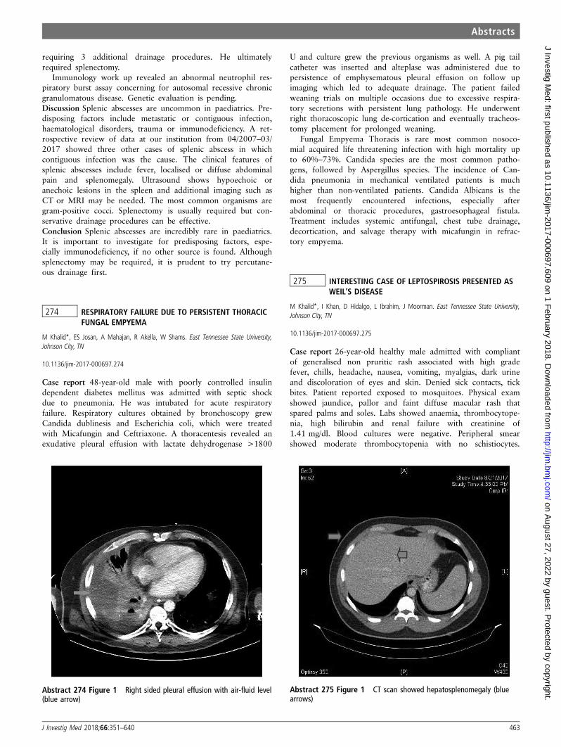
requiring 3 additional drainage procedures. He ultimatelyrequired splenectomy.
Immunology work up revealed an abnormal neutrophil res-piratory burst assay concerning for autosomal recessive chronicgranulomatous disease. Genetic evaluation is pending.Discussion Splenic abscesses are uncommon in paediatrics. Pre-disposing factors include metastatic or contiguous infection,haematological disorders, trauma or immunodeficiency. A ret-rospective review of data at our institution from 04/2007–03/2017 showed three other cases of splenic abscess in whichcontiguous infection was the cause. The clinical features ofsplenic abscesses include fever, localised or diffuse abdominalpain and splenomegaly. Ultrasound shows hypoechoic oranechoic lesions in the spleen and additional imaging such asCT or MRI may be needed. The most common organisms aregram-positive cocci. Splenectomy is usually required but con-servative drainage procedures can be effective.Conclusion Splenic abscesses are incredibly rare in paediatrics.It is important to investigate for predisposing factors, espe-cially immunodeficiency, if no other source is found. Althoughsplenectomy may be required, it is prudent to try percutane-ous drainage first.
274 RESPIRATORY FAILURE DUE TO PERSISTENT THORACICFUNGAL EMPYEMA
M Khalid*, ES Josan, A Mahajan, R Akella, W Shams. East Tennessee State University,Johnson City, TN
10.1136/jim-2017-000697.274
Case report 48-year-old male with poorly controlled insulindependent diabetes mellitus was admitted with septic shockdue to pneumonia. He was intubated for acute respiratoryfailure. Respiratory cultures obtained by bronchoscopy grewCandida dublinesis and Escherichia coli, which were treatedwith Micafungin and Ceftriaxone. A thoracentesis revealed anexudative pleural effusion with lactate dehydrogenase >1800
U and culture grew the previous organisms as well. A pig tailcatheter was inserted and alteplase was administered due topersistence of emphysematous pleural effusion on follow upimaging which led to adequate drainage. The patient failedweaning trials on multiple occasions due to excessive respira-tory secretions with persistent lung pathology. He underwentright thoracoscopic lung de-cortication and eventually tracheos-tomy placement for prolonged weaning.
Fungal Empyema Thoracis is rare most common nosoco-mial acquired life threatening infection with high mortality upto 60%–73%. Candida species are the most common patho-gens, followed by Aspergillus species. The incidence of Can-dida pneumonia in mechanical ventilated patients is muchhigher than non-ventilated patients. Candida Albicans is themost frequently encountered infections, especially afterabdominal or thoracic procedures, gastroesophageal fistula.Treatment includes systemic antifungal, chest tube drainage,decortication, and salvage therapy with micafungin in refrac-tory empyema.
275 INTERESTING CASE OF LEPTOSPIROSIS PRESENTED ASWEIL’S DISEASE
M Khalid*, I Khan, D Hidalgo, L Ibrahim, J Moorman. East Tennessee State University,Johnson City, TN
10.1136/jim-2017-000697.275
Case report 26-year-old healthy male admitted with compliantof generalised non pruritic rash associated with high gradefever, chills, headache, nausea, vomiting, myalgias, dark urineand discoloration of eyes and skin. Denied sick contacts, tickbites. Patient reported exposed to mosquitoes. Physical examshowed jaundice, pallor and faint diffuse macular rash thatspared palms and soles. Labs showed anaemia, thrombocytope-nia, high bilirubin and renal failure with creatinine of1.41 mg/dl. Blood cultures were negative. Peripheral smearshowed moderate thrombocytopenia with no schistiocytes.
Abstract 274 Figure 1 Right sided pleural effusion with air-fluid level(blue arrow)
Abstract 275 Figure 1 CT scan showed hepatosplenomegaly (bluearrows)
Abstracts
J Investig Med 2018;66:351–640 463
on August 27, 2022 by guest. P
rotected by copyright.http://jim
.bmj.com
/J Investig M
ed: first published as 10.1136/jim-2017-000697.609 on 1 F
ebruary 2018. Dow
nloaded from

Detailed work up for infectious causes include HIV, viral hep-atitis panel, EBV, CMV, monospot test, Dengue test, chikungu-nya, Ehrlichia, Anaplasmosis, RMSF which were negative.Computed Tomography abdomen and pelvis showed hepatos-plenomegaly (figure 1). Leptospirosis serology was sent to alaboratory which came back positive and patient was startedon doxycycline with improvement in the symptoms.
Leptospirosis is zonosis transmitted by domestic animals orcontact with water, soil and food contaminated with urine ofinfected animal. Weil’s disease is a severe form of presentationcharacterised by kidney, liver dysfunction and Jaundice with ahigh mortality rate. This case highlights the importance of rec-ognition of leptospirosis in patient with rash with liver andkidney dysfunction.
276 CANDIDURIA DUE TO CANDIDA KRUSEI IN A PATIENTWITH PROLONGED ICU STAY: CAN WE TREAT THISRESISTANT INFECTION WITH ECHICANDINS?
V Kohli*, P Sankhyan, L Ibrahim. East Tennessee State University, Johnson City, TN
10.1136/jim-2017-000697.276
Introduction Candiduria is common in high risk patients inthe hospital. A recent trend of non albicans candiduria isbeing reported and among those Candida krusei is a rare spe-cies to be isolated. A very small percentage of non albicanscandiduria is blamed on this species, known to be inherentlyresistant to triazoles. This report aims at finding out if mica-fungin is effective to clear UTI caused by Candida kruseiwhich does not have a good urine concentration, in a settingof azole resistant candida infection.Case report A 47 year old male who was admitted to the ICUwith multiple comorbidities and stayed for 23 days wasshifted to the ICU step down as he was improving, with afoleys catheter in place. After two days in the step down,patient started spiking fevers of 102F and with leukocytosis(WBC 17.2 K/uL) with unknown origin of foci. He was noton any antibiotics at the time. Piperacillin-tazobactam anddaptomycin were considered to treat the patient empirically.His urine and blood was sent for cultures. The blood cultureswere negative but his urine culture was positive for Candidakrusei (CFU>105/ mL). The sensitivity test showed the organ-ism to be resistant to tri azoles (eg. fluconazole and itracona-zole). The echinocandins does not have good urineconcentration but we did not have more options so we triedto treat the patient with micafungin. Repeat culture was posi-tive on the day we started the patient on micafungin againwith Candida krusei (>105 CFU/mL). After 8 days of initia-tion of the treatment, we sent in another urine sample forculture. The results came back with no growth of Candidakrusei. The patient did not have any signs of infection andthe leukocytosis resolved.Conclusion The patient required a minimum of 1 week ofmicafungin until negative cultures and was continued to com-plete the course for 14 days. We were able to treat the UTIwith micafungin completely and thus we believe echinocandinis a good option in candiduria with candida resistant to triazoles. More studies are needed to confirm this finding as thenumber of cases seen with this infections are very limited.
277 THE MITIS TOUCH
D LaChance*. Dwight David Eisenhower Army Medical Centre, Augusta, GA
10.1136/jim-2017-000697.277
Case report 58 year-old male with essential hypertension pre-sented to the ED after development of acute chest pain. Painwas sharp, constant, fluctuating in intensity, exacerbated byswallowing and deep inspiration, and radiated to his neck.Ibuprofen did not relieve his symptoms prior to presentation.Patient also noted a recent URI which was self-limited. Fur-ther ROS was also positive for odynophagia. On presentation,his vital signs were stable and he was afebrile. Cardiac bio-markers were negative; however labs were notable for a leu-kocytosis. Chest xray was unremarkable. Electrocardiogramwas normal sinus rhythm and demonstrated no changes consis-tent with ischemia. Sublingual nitroglycerin and full doseaspirin in the emergency department did not relieve his symp-toms. Given concern for acute aortic dissection, a D-Dimerwas obtained which returned positive. A CT pulmonary angio-gram was obtained which revealed a soft tissue density masswithin the superior mediastinum adjacent to the oesophaguswith adjacent free air. Concern for esophageal neoplasm ver-sus esophageal perforation arose. An esophogram with gastro-grafin ruled out esophageal perforation. Subsequent EGD withesophageal ultrasound revealed a lesion consistent with aninfected bronchogenic cyst anterior to the oesophagus wasidentified with clear delineation between the lesion and theoesophagus. Cardiothoracic surgery performed a video assistedthorocoscopy for incision and drainage of the abscess. Puru-lent fluid was aspirated and sent for gram stain and cultureand antibiotic therapy with vancomycin and piperacillin-tazo-bactam were started. Culture data revealed pan-susceptibleStreptococcus mitis. The patient was started on course ofamoxicillin clavulanate with full resolution of infection andchest pain syndrome.
Bronchogenic cysts are an uncommon congenital anomalyof the foregut which can lead to tracheobronchial compressionand infection, with a prevalence of 1/42,000–68 000. Theypredominantly develop in the lung parenchyma, howeverabout 24% develop in the mediastinum, as in the case. Theyare mostly discovered as incidental radiologic findings. Theycan become symptomatic with chest pain being the most com-mon presenting complaint. This represents an unusual aetiol-ogy of chest pain due to bronchogenic cyst infection in animmune-competent individual.
278 AN ATYPICAL PRESENTATION OF A TYPICALMYCOBACTERIUM
T Liddell*, J Lucar Lloveras, R Webb. University of Mississippi Medical Centre, Brandon, MS
10.1136/jim-2017-000697.278
Case report Mycobacterium tuberculosis is a common pathogenwhich infects one third of the world’s population. Often clini-cal suspicion is high when patients present with typical symp-toms of pulmonary tuberculosis, however, tuberculosis is oftenleft out of the differential when it presents atypically.
We present a 65 year-old Vietnamese female living in the USfor two years with no known past medical history who presented
Abstracts
464 J Investig Med 2018;66:351–640
on August 27, 2022 by guest. P
rotected by copyright.http://jim
.bmj.com
/J Investig M
ed: first published as 10.1136/jim-2017-000697.609 on 1 F
ebruary 2018. Dow
nloaded from

with left ankle pain for six months. She had been treated for goutthough foot pain continued to progress. On admission, ankle wasinflamed, and imaging revealed destruction of the talus and anenlarged left inguinal lymph node. She underwent resection of herleft talar head and neck and left inguinal lymph node. InfectiousDiseases was consulted due to osteomyelitis. Patient then reportedsymptoms of fever, night sweats, and weight loss of 20–25 lbs thatbegan around the development of her left ankle pain. Laboratorystudies revealed mild leukocytosis, thrombocytosis, anaemia, andelevated inflammatory markers. She was also found to have under-gone biopsy of her talus prior to her arrival at our hospital. Slideswere obtained and reviewed with our pathologist who reportedgranulomatous inflammation with some granulomas displayingcentral necrosis. Chest imaging was unremarkable. She was startedon active tuberculosis treatment with rifampin, isoniazid, pyrazi-namide, and ethambutol. Pathology of talus and lymph node wereconsistent with biopsy, and cultures eventually grew Mycobacte-rium tuberculosis in six of six samples.
Of the 8.7 million cases of tuberculosis reported each year,musculoskeletal tuberculosis accounts for 1%–5% or 87 000to 4 35 000 cases each year. Osteoarticular tuberculosis, par-ticularly outside the spine, requires a high level of suspicionfor prompt diagnosis. Our case has multiple characteristicsthat should cause consideration for tuberculosis including visit-ing from an endemic area, negative bacterial cultures, biopsywith caseating granulomas, and imaging with irregular cavitiesof destruction with little surrounding sclerosis. Despite a fewsignificant diagnostic clues, our patient’s symptoms werepresent for six months before she received appropriate treat-ment underlining the importance of maintaining a high suspi-cion for tuberculosis despite an atypical presentation.
279 VISUAL DEFICITS AS A LATE MANIFESTATION OFCONGENITAL TOXOPLASMOSIS: FAMILIAR SIGN IN ANUNFAMILIAR SETTING
1WJ Lindsey*, 1V Habet, 2S Lefevre. 1Children’s Hospital, New Orleans, LA; 2LSU Health,New Orleans, LA
10.1136/jim-2017-000697.279
Purpose of study While Toxoplasma gondii is generally asymp-tomatic in immunocompetent hosts, severe disease can mani-fest in congenital infection or immunocompromised hosts.70%–90% infants acquiring Toxoplasma infection transplacen-tally may be asymptomatic initially. It’s persistence can end inreactivation leading to clinical disease. The most common latemanifestation is retinochoroiditis. Approximately 90% ofuntreated children will acquire retinal lesions per year. Theprevalence of ocular toxoplasmosis in the United States (US)ranges from 0.6% to 2%. Infection rates are higher in regionsthat are at lower altitudes with a tropical climate with highprevalence in South America. We discuss a case of congenitaltoxoplasmosis in an immigrant with singular, anterior cervicallymphadenopathy and worsening vision.Methods used Case Study.Summary of results A 5 yo male with developmental delay pre-sented to general paediatric clinic for routine well-child check.The patient had recently emigrated from Brazil with his familyand had not been examined by a physician in over one year.The parents’ primary concerns were assessing a ‘lump’ on thepatient’s neck, receiving necessary immunizations to attendschool and obtaining visual examination. Physical exam
revealed a 5 cm nontender, mobile rubbery mass of the ante-rior cervical chain which had been present for over a year.Visual acuity and ocular alignment tests were attempted, butthe patient was unable to identify characters at 10 feet. Thepatient was referred to ophthalmology but was evaluated byan optometrist due to lack of insurance. Retinal imagesrevealed bilateral macular and peripapular scarring consistentwith congenital toxoplasmosis. The patient was referred to IDwho recommended close follow up without antimicrobialtreatment but has since been lost to follow-up.Conclusions In the US, prenatal testing for toxoplasmosis isnot done. This contrasts other countries with higher preva-lence of disease where prenatal testing is of significant value.Because toxoplasmosis can be asymptomatic paediatriciansmust have a high index of suspicion in children presentingwith visual disturbance. This is particularly important forimmigrants. Visual disturbances in this population should havea broader differential diagnosis including congenitaltoxoplasmosis.
280 DRESS SYNDROME IN A PATIENT WITH ARDS
R Mangat*, Z Jones, A Eranki. SUNY Upstate Medical University, Syracuse, NY
10.1136/jim-2017-000697.280
Case report Drug reaction with eosinophilia and systemicsymptoms (DRESS) syndrome is a relatively rare drug hyper-sensitivity reaction that can present with organ dysfunctionand skin symptoms two to six weeks after starting a newdrug. In this case, a middle-aged male was diagnosed withDRESS syndrome when he developed fever, elevated transami-nases and worsening eosinophilia after being restarted on anti-biotic therapy.
A 53-year-old male with a past medical history significantfor Becker’s muscular dystrophy presented to the ICU withacute respiratory failure secondary to ARDS in the setting ofcommunity acquired pneumonia and septic shock. His oxygen-ation improved after an initial 7 days treatment with vanco-mycin, zosyn and azithromycin, but he remained febrile withworsening leukocytosis. Chest x-ray showed bilateral infiltratesand he was restarted on vancomycin and zosyn for recurrentpneumonia. He developed rash and was found to have awhite count of 26 000 with 61% eosinophils, ALT of 133 andAST of 122. After the antibiotics were discontinued, thepatient’s clinical syndrome improved in 2 days.
DRESS syndrome is an important and under-recognised differ-ential diagnosis to consider in patients with fever, unexplained leu-kocytosis and rash. In this case, the patient was restarted on theoffending agents due to concern for infection several days prior toa diagnosis being made. Skin eruptions typically involve greaterthan fifty percent of the body surface area including facial oedema,infiltrative lesions, scaling and purpura. Abnormal laboratory val-ues that can help diagnose DRESS include leukocytosis with aneosinophil count >700, atypical lymphocytosis, increased transa-minases and reactivation of human herpesvirus-6. The most com-mon offending drugs include antiepileptics, sulfonamides,allopurinol with beta lactams being increasingly reported.Removal of the offending agent along with supportive care is themainstay of treatment in these patients. Prompt diagnosis ofDRESS syndrome is clinically important as patient deaths havebeen reported due to multi-organ system failure in unrecognisedcases.
Abstracts
J Investig Med 2018;66:351–640 465
on August 27, 2022 by guest. P
rotected by copyright.http://jim
.bmj.com
/J Investig M
ed: first published as 10.1136/jim-2017-000697.609 on 1 F
ebruary 2018. Dow
nloaded from

281 A CASE OF AEROCOCCUS AND GLOBICATELLABACTEREMIA
D Markabawi*, R Mangat, Z Jones, H Singh Gambhir. SUNY Upstate, Syracuse, NY
10.1136/jim-2017-000697.281
Introduction Globicatella sanguinis are catalase-negative, gram-positive cocci that were first discovered in 1992. There havebeen multiple case reports of Globicatella sanguinis isolatedfrom blood, urine and cerebrospinal fluid. The clinical signifi-cance of this bacteria is still unknown. Aerococcus viridans, isa very rare microorganism, which causes invasive infections inimmunocompromised patients, but rarely in immunocompetentpatients. It has been associated with bacteremia, septic arthri-tis, and especially infective endocarditis.Case A 72 year old male with a past medical history of CVA,hypertension, and dementia, was brought to the emergencydepartment for complaints of increased fatigue and lethargy.The patient was constipated with the last bowel movementbeing several days prior to presentation. In the ED, thepatient was noted to be afebrile and hemodynamically stable.A CT abdome/pelvis was positive for stercoral colitis. Thepatient underwent manual dis-impaction. Patient was alsonoted to be dehydrated with an elevated lactic acid, thisimproved with IV hydration. Two sets of blood cultures wereobtained in the emergency department. One out of the twosets grew Globicatella sanguinis and Aerococcus viridans.Upon consultation with the infectious disease service, it wasdecided to treat patient and not deem this a skin contaminantas this bacteremia might have been a result of bacterial intesti-nal translocation due to colitis and faecal dis-impaction. Bothorganisms were susceptible to Penicillin and third generationCephalosporins. Patient was successfully treated with Ceftriax-one 2 g IV daily for 2 weeks.Discussion While Aerococcus viridans is known to cause inva-sive infections in humans, the pathogenic role of Globicatellasanguins remains partially known. Human carriage of bothspecies has been established. There have been reports of infec-tive endocarditis due to Aerococcus viridans and meningitisdue to Globicatella sanguins. In our case, the bacteremia likelyresulted from bacterial intestinal translocation. Both specieswere susceptible to third generation Cephalosporins.Conclusion Although human carriage for both Aeroccous viri-dans and Globicatella sanguins is well known, it is importantto consider the pathogenic role of these bacteria in the rightsetting.
282 BACK PAIN1BE O’Brien*, 1M Bahn, 1,2R Steele. 1Tulane University, New Orleans, LA; 2Ochsner MedicalCentre, New Orleans, LA
10.1136/jim-2017-000697.282
Case report 11 yo caucasian male presented w/fracture of L4.He was in usual state of good health until 3 months priorwhen he developed fever, myalgia, HA, and left cervical LAD.He was dx’d w/flu, treated with Oseltamivir/Azithromycin. Hisbrother was flu positive 2 weeks prior.
2 weeks s/p tx for flu- he developed acute, intermittentleft-sided flank pain went to an ED, CTAP was normal.Patient had a kitten, Bartonella titers were drawn, and showedB. henselae IgG �1:1024 (normal <1:128) and IgM �1:20(normal <1:20).
He was dc’d home on Azithromycin/toradol. He continuedto have fatigue, decreased appetite. 10 days later- went tourgent care for flank pain, was treated empirically for UTI w/Augmentin. Labs (cbc, cmp, UA) were normal. S/P treatment,he went to ED for continued flank pain, RUS was normal. 2weeks later- he went to his paediatrician w/fatigue, decreasedappetite. Xrays of spine obtained for concern of MSK aetiol-ogy of pain. He was dx’d with scoliosis and referred toOrtho. Flank pain resolved.1 month prior to presentation, heheard a ‘popping sound’ and had sudden, severe pain. Wasseen by Ortho the next day. MRI of spine showed pathologi-cal fracture of L4. He was referred to Hem-Onc to r/omalignancy.
His CBC was normal except for elevated eos (7.4%) ondiff, CRP (7.32 mg/L). Otherwise labs (CMP, uric acid, ESR)were normal. Quant. TB Gold was negative. Repeat MRI ofCTL spine obtained showed heterogeneously enhancing lesionmeasuring 2.7 cm within left central portion of L4 vertebralbody with mild height loss and associated fracture.
PE:+small palpable lymph nodes in his anterior cervical tri-angle/inguinal regions. No bone pain, restriction in mobility orROM, or neurologic deficits.
Biopsy of lesion was sent for gram stain, aerobic/anaerobicculture/AFB/fungal culture (all negative). Path showed necrotiz-ing granulomatous inflammation in bone and bone marrow,no neoplasm. Biopsy sent to Mayo Clinic for Bartonella PCR,which was negative.
1 month later,labs were normal including crp.No hx ofrecurrent infections, but due to severity of presentation, con-cern for CGD. Neutrophil oxidative burst assay was negative.Patient has returned to baseline activity level, now has nightlylower back pain and was fitted for a brace.
283 AN UNUSUAL PRESENTATION OF DISSEMINATEDVARICELLA ZOSTER IN AN IMMUNOCOMPETENT ADULT
P Pachariyanon*, S Thakolwiboon, D Payne. Texas Tech University Health Science Centre,Lubbock, TX
10.1136/jim-2017-000697.283
Case report A 63-year-old man with a history of bladder can-cer status post transurethral resection of bladder tumour pre-sented with 5 days of fever, progressive diffuse headache, andphotophobia. Initial physical examination revealed multiplediscrete erythematous papules on his left post auricular area,trunk and both thighs. A neurological examination revealedneck stiffness without any focal neurological deficit. A compu-terised tomography scan did not demonstrate any intracranialpathology and cerebral spinal fluid analysis showed a mildlymphocyte-predominated pleocytosis. He was initially empiri-cally treated by intravenous vancomycin, ceftriaxone, ampicil-lin, and acyclovir. On the third day of admission, hedeveloped left facial droop and hearing difficulty. A repeatphysical examination revealed multiple groups of vesicles inthe left C2 and C3 dermatome and diffuse erythematous vesi-culopapular rash on his trunk and both thighs and peripheralleft facial palsy. Weber’s and Rinne’s tests suggested left sen-sorineural hearing loss (SNHL) which was confirmed byaudiogram. Magnetic resonance imaging was done and did notreveal any structural lesion. Blood and CSF cultures did notgrow any microorganism. CSF PCR for HSV-1, HSV-2, andenterovirus were negative as well as HIV and Syphilis
Abstracts
466 J Investig Med 2018;66:351–640
on August 27, 2022 by guest. P
rotected by copyright.http://jim
.bmj.com
/J Investig M
ed: first published as 10.1136/jim-2017-000697.609 on 1 F
ebruary 2018. Dow
nloaded from

screening. Subsequent immunological study revealed positiveserum IgG for HSV-1, VZV, and CMV while negative forHSV-2. However, serum IgM was positive for VZV. Addition-ally, lymphocyte subset panel did not suggest any immunodefi-ciency disorder. All anti-bacterial medications werediscontinued. Intravenous acyclovir 10 mg/kg was continuedfor total 14 days. At 1 month follow up, the skin lesionswere crusted over and had begun to recede. However, leftfacial paralysis and remained.
Herein, we described the unusual presentation of dissemi-nated varicella zoster which included both chickenpox-likerash and typical zoster skin lesion as well as neurologicalinvolvement in an immunocompetent host.
284 EATING YOUR WAY TO MENINGITIS: WHY MATERNALPLACENTOPHAGY MIGHT BE A BAD IDEA
C Parrish*, R Welliver. University of Oklahoma College of Medince, Oklahoma City, OK
10.1136/jim-2017-000697.284
Case report Our patient is a previously healthy unvaccinated15 day old female who presented with decreased oral intake(PO) and fever for 1 week. Patient was a full term infantborn at home. Maternal laboratory investigations includinggroup B streptococcus (GBS) were negative. Birth was uncom-plicated and patient was healthy until day 7 of life, when shebegan having decreased PO and fevers. On exam she was alertwith no focal neurological deficits. A septic workup showedcerebral spinal fluid (CSF) cultures positive for GBS; bloodcultures were negative. A 14 day course of IV ampicillin wasstarted. Initial MRI showed purulent material within subarach-noid spaces, and antibiotic course was extended to 21 days.MRI prior to discharge showed stable purulence. She was dis-charged home after completing full antibiotic course. At her6 week follow up, she had age-appropriate development. Ofnote, mom had sent her placenta to an outside facility to beprocessed into supplemental pills. Mom began taking thesetwo days prior to admission, and was advised to stop. Ourlaboratory recommended testing the pills for GBS be com-pleted by the Health Department, however at this time weare unsure whether the family did this.
Late onset meningitis in neonates is often associated withhorizontal transmission, one such transmission that we maynot consider is maternal ingestion of the placenta. Placentalingestion has been promoted for its potential physical andpsychological benefits, although there is no scientific evidenceto support this. In a previously reported case in Oregon themother of an infant with recurrent GBS meningitis hadingested her placenta in the form of pills that were tested andfound to be GBS-infected; the final diagnosis: ‘late onset GBSdisease attributable to high maternal colonisation secondary toconsumption of GBS-infected placental tissue’.
These cases show us the importance of expanding our clini-cal questions and differential diagnoses as the culture we prac-tice medicine in also expands. There is little research into thenumber of women who choose to ingest their placentas, andeven less scientific research on the risks of doing so. Thesecases illustrate the need for further investigation and insightinto the risks of placental ingestion as a means to better edu-cate families.
285 SEVERE AND PROGRESSIVE CELLULITIS FOLLOWING ADOG SCRATCH IN A NEUTROPENIC PATIENT
1D Pithadia*, 1E Weathers, 1R Colombo, 1,2S Baer. 1Medical College of Georgia, Augusta,GA; 2Augusta VA Medical Centre, Augusta, GA
10.1136/jim-2017-000697.285
Introduction Skin and soft tissue infections occur in over 20%of patients with chemotherapy-induced neutropenia. Grampositive bacterial infections predominate early in neutropenia,and incidence of resistant bacteria and fungi increases withprolonged neutropenia. Past infections and exposures influencethe risk of of rare pathogens.Case A 55-year-old woman, who was neutropenic from che-motherapy for primary CNS lymphoma, was scratched on herforearm by a dog. She cleaned the wound with isopropanoland was treated empirically as an outpatient with amoxicillin-clavulanate. Over the next 4 days, she developed fever pluserythema and swelling of the forearm without purulence, crep-itus, or significant pain. The wound had contacted tap water,and she denied other exposures. She was admitted and startedon intravenous vancomycin, piperacillin-tazobactam, and tobra-mycin. Cultures of blood and urine grew Pseudomonas aerugi-nosa with identical susceptibilities. Vancomycin andpiperacillin-tazobactam were continued, and all subsequent cul-tures were negative. However, daily fevers persisted and theinflammation in her arm progressed resulting in restrictedflexion of her elbow and digits. MRI of the arm showedmyositis and an elbow joint effusion. Voriconazole was addedfor empiric fungal coverage. A bulla developed at the woundsite as her neutrophils recovered, and culture of the fluidgrew Serratia marcescens. Antibiotics were switched to cefe-pime based on susceptibility. She became afebrile with substan-tial improvement of cellulitis within 48 hours and wasdischarged on oral ciprofloxacin to complete a 14 day course.Conclusion Serratia marcescens is a gram-negative bacillus thatthrives in damp environments and causes opportunistic noso-comial infections. Serratia skin infections are rare and, basedon our review, this may be the first report of Serratia cellulitisassociated with dog contact and trauma. This case highlightsthe need to consider unusual pathogens based on exposurehistory in cases of treatment-resistant soft tissue infections inimmunocompromised patients. It also emphasises the impor-tance of obtaining cultures from skin lesions to establish amicrobiologic diagnosis for targeted therapy.
286 DOUBLE TROUBLE AND A DELAY
JP Pruitt*, B Johnson, L Gilliam, R Smalligan. UAB-Huntsville, Huntsville, AL
10.1136/jim-2017-000697.286
Case report A 20 yo woman presented with 12 hours of pro-gressive abd pain, lightheadedness, dyspnea and chills 5 daysafter C-section performed at 35 weeks for preterm labourwith breech presentation. On exam vitals were T 103.3, BP87/44, HR 175, RR 18, O2 sats 100% on RA. She appearedill and had diffuse abd pain from the umbilicus to her Pfan-nenstiel incision though it was clean and dry. Labs: WBC15.4 k, Hgb 11, CRP 15, and lactic acid 1.0 (<1 nl). Abd CTshowed some subq fluid and scant air, c/w post-op changes.She received 2 L of IVF but no antibiotics initially apparentlydue to the low lactic acid level and was sent to the OB ED.Hours after presentation, antibiotics were started with further
Abstracts
J Investig Med 2018;66:351–640 467
on August 27, 2022 by guest. P
rotected by copyright.http://jim
.bmj.com
/J Investig M
ed: first published as 10.1136/jim-2017-000697.609 on 1 F
ebruary 2018. Dow
nloaded from

IV hydration. She improved initially, but by morning the painhad worsened and lactate had risen to 3.4. At this point herabd pain was out of proportion with exam and the incisionhad become erythematous. Surgery was consulted and sheunderwent emergency I and D of what was to be confirmedby pathology as necrotizing fasciitis. Wound cultures grewMRSA. She required repeated debridement and an eventualabd wall mesh but recovered and went home in 2 weeks.Discussion This patient had two related life-threatening condi-tions. First was sepsis which must be addressed aggressivelywith early IV fluids and broad spectrum antibiotics. Treatmentwas delayed initially because of a normal lactic acid. Sepsis isa clinical diagnosis based on the vitals, exam, lab, and imagingfindings as opposed to meeting a long list of diagnostic crite-ria. While an elevated lactate is associated with a poorer prog-nosis in sepsis, it is not a diagnostic marker.
Necrotizing fasciitis is also a medical emergency whichrequires a high clinical suspicion as the dx is not alwaysobvious. In our case the physical exam was initially misleadingdue to the infection being deeper in the tissues, thus theerythema, greyness and bullae often seen in the conditionwere not present. Typical CT findings of necrotizing fasciitisare subq inflammation and air which can also be seen post-op. Close f/u of this patient with early surgery when shedeteriorated were keys to prompt dx and treatment of hersecond life-threatening condition. It is important to rememberthat pain may precede skin changes and surgical debridementis the only definitive treatment.
287 TRAVEL-RELATED LEISHMANIASIS AND TUMOURNECROSIS FACTOR INHIBITORS: 2 CASES OFMISDIAGNOSIS
E Rabold*, RJ Feldman, J Lutgring, A Moanna, H Wu. Emory University, Atlanta, GA
10.1136/jim-2017-000697.287
Case report An association between leishmaniasis and the useof tumour necrosis factor (TNF) inhibitors has been suggestedby previous reports, and immunosuppression is a known riskfor severe disease. Though leishmaniasis is rarely transmittedin the U.S., travellers are at risk of infection when visitingendemic areas. We present two patients with cutaneous leish-maniasis complicated by initial misdiagnoses and TNF inhibi-tor therapy. Patient A is a 43-year-old healthy American malewho presented with a chronic, non-healing ulcer on his rightleg and a smaller lesion on his right arm after travel to Mex-ico. Histopathology of the leg ulcer was inconclusive, and apresumptive diagnosis of pyoderma gangrenosum was made.Due to non-response to topical treatments, he received variousimmunosuppressive regimens, including oral glucocorticoids,systemic cyclosporine, and adalimumab. A subsequent biopsyof the arm lesion demonstrated parasitized histiocytes, andPCR testing was consistent with Leishmania mexicana. Aftercessation of all immunosuppressive medications and initiationof oral ketoconazole, both lesions resolved. Patient B is a 59-year-old American male with psoriasis treated with adalimu-mab who presented with non-healing ulcers on his right chestand right thigh after travel to southern Spain. Squamous cellcarcinoma was diagnosed from a biopsy of the thigh lesion,and the patient received MOHS surgery. A subsequent biopsyof the chest lesion demonstrated amastigotes in the dermallayer, which was also seen on reevaluation of the first biopsy.
PCR testing was consistent with Leishmania infantum/chagasi.His chest lesion resolved with liposomal amphotericin B andcessation of the adalimumab. In these cases, the delayed diag-nosis of leishmaniasis resulted in unnecessary or prolongedexposure to immunosuppressive drugs, including TNF inhibi-tors. Physicians should consider the possibility of leishmaniasiswhen starting TNF inhibitors in patients with non-healingwounds, particularly if they have travelled to endemic areas.
288 ACQUIRED TRACHEOESOPHAGEAL FISTULA AS ACOMPLICATION OF ACQUIRED IMMUNODEFICIENCYSYNDROME (AIDS)
A Richardson*, KR Green, J Perez-Downes, CM Baldeo, SF Shah, N Najjar, V Seeram.University of Florida College of Medicine Jacksonville, Jacksonville, FL
10.1136/jim-2017-000697.288
Case report A 52 year old male with past medical history ofHIV/AIDS (CD4 count of 5) presented to the emergencydepartment with a complaint of weakness, fatigue, and dys-phagia. He had been non-compliant with antiretroviral therapyfor 3 previous years. Two days into his admission he becameseptic thus chest X-ray was ordered revealing a pneumonia.Given no clinical improvement with antibiotics, a CT chestwas ordered. The CT scan revealed a communication betweenthe oesophagus and trachea with gas in the mediastinum.Given severe cachexia, no surgical intervention was recom-mended. A gastrojejunal feeding tube was inserted, intravenousbroad spectrum antibiotics and antifungals started, and serialesophagrams were ordered to evaluate healing and closure ofthe tracheoesophageal fistula. Fistula formation was attributedto candida esophagitis given oral candida was present onadmission. Patient was discharged to follow up for initiationof antiretroviral therapy
Acquired tracheoesophageal fistulas (TEF), although uncom-mon, usually occur as a complication of mechanical ventila-tion, trauma, and malignancy. However, theimmunocompromised can acquire TEFs secondary to complica-tions from opportunistic infections. Although TEFs were morecommon early in the AIDS epidemic, acquired TEFs are raretoday given advances in antiretroviral therapy. Causative infec-tious agents of acquired TEFs include Mycobacterium, Candidaspecies, Cytomegalovirus, and herpes simplex virus. If leftuntreated, progression to perforation, necrosis, or TEF forma-tion can occur.
TEFs are a serious condition with significant morbidity andmortality. Treatment is necessary to avoid aspiration, sepsis,and pulmonary failure. Surgical repair is the preferred treat-ment and typically involves esophageal closure with resectionof the tracheal fragment. Tracheal or esophageal stents areother treatment options but considered a palliative measure.TEFs must be considered in severely immunocompromisedindividuals given their life threatening prognosis.
289 KAPOSI SARCOMA PRESENTING AS DYSPHAGIA
J Ruiz*, M Ganji, R Jacob. UF Health Jacksonville, Jacksonville, FL
10.1136/jim-2017-000697.289
Case report A 25 year old African American male with pastmedical history of HIV and neurosyphilis who presented to
Abstracts
468 J Investig Med 2018;66:351–640
on August 27, 2022 by guest. P
rotected by copyright.http://jim
.bmj.com
/J Investig M
ed: first published as 10.1136/jim-2017-000697.609 on 1 F
ebruary 2018. Dow
nloaded from

the hospital for dysphagia associated with facial and throatswelling. Physical exam was notable for significant submandib-ular and supraclavicular lymphadenopathy with concern forpossible Castleman’s syndrome. During mouth evaluation a 3centimetre hyper vascularized violaceous submucosal lesionbelow the palate was noticeable. CD4 absolute count onadmission was 607. Patient was empirically started on flucona-zole for suspected candida esophagitis and gastroenterologywas consulted for upper endoscopy which revealed multiplelarge hyper vascularized violaceous submucosal nodular lesionsseen on the soft palate and pharynx. Surgery was also con-sulted for suspicious soft palate lesion as well for excisionallymph node biopsy. Histopathology of lymph node biopsy wasnegative for Castleman’s disease but notable for spindle cellsconsistent with Kaposi’s sarcoma along with positive CD31and HHV-8 stain. Dysphagia got progressively worse in a mat-ter of hours. He eventually was transferred to the intensivecare unit due to increasing respiratory distress. During theintubation, the patient desaturated, became bradycardic,andexperienced cardiac arrest. Unfortunately, the patient expiredafter multiple rounds of resuscitative efforts with no evidenceof cardiac activity on ultrasound.Discussion Kaposi Sarcoma is angioproliferative disorder thatrequires infection with human herpes virus-8. Skin lesions arethe most common manifestations of Kaposi sarcoma. It canalso present in other organs as lymph nodes, oral mucosa andthe gastroenterology tract. Usually presented in immunocom-promised patients. Patient atypical presentation and findingson oral mucosa and upper endoscopy made this case interest-ing. Kaposi sarcoma usually is diagnosed by biopsy. Treatmentcan be either chemotherapy or radiotherapy along with antire-troviral therapy.Conclusion Kaposi sarcoma should always be kept as a possi-ble differential diagnosis on patients with past medical historyof HIV/AIDS despite decreased prevalence due to evolution inmedical therapy. This case can lead to further evaluation ofdysphagia by upper endoscopy on patients that present withdysphagia and oral lesions since only handful of cases ofKaposi sarcoma are seen now a days.
290 PARADOXICAL RESPONSE TO ANTITUBERCULOUSTHERAPY IN AN HIV NEGATIVE TB PATIENT
N Salagundla*, S Siddiqui, A Mirza, A Islam. Texas Tech University HSC Amarillo, Amarillo,TX
10.1136/jim-2017-000697.290
Case report A 50 yo F immigrant from Sudan with PMH, pre-sented with symptoms of generalised weakness, nausea, vomit-ing and chills for 2–3 months. On P/E she appeared cachecticwith bilateral diffuse crackles. CT chest: diffuse bilateral nodu-lar infiltrates with cavitary changes in left upper lobe andright pleural effusion. Further CT evaluation showed: diffusehypodensities in liver, spleen, kidney and adnexa, left parietallobe ring enhancing lesion. IGRA was positive, HIV was nega-tive. Sputum and pleural fluid AFB were negative and CTguided lung biopsy showed positive for AFB. Patient wasstarted on four drug antituberculous therapy (ATT) andstarted improving. She was discharged after 2 days and con-tinued with directly observed ATT. After one month, patientwas readmitted for worsening symptoms and WBC count of3500. CT chest: extensive bilateral, predominantly upper lobe
increased cavitary lung lesions. MRI brain: enhancing lesionsin right cerebellar and bilateral parietal regions. Drug sensitiv-ity testing of previous specimens showed pan-sensitivity tostandard ATT. Therefore, paradoxical response to treatmentwas diagnosed. ATT treatment was continued with the addi-tion of a quinolone and steroids, but eventually the patientrequired occipital craniotomy for right cerebellar mass resec-tion. Patient improved on full ATT and steroids over a oneyear period.Discussion IRIS (Immune Reconstitution Inflammatory Syn-drome) is a widely-recognised cause of paradoxical response inHIV positive patients, but PR can be seen both HIV + andHIV- patients, most commonly extrapulmonary infections asso-ciated with lower lymphocyte counts at baseline. Respiratoryand CNS TB are most commonly involved. Diagnosis is byexclusion. Exact mechanism is uncertain but immune restitu-tion may play a role even in HIV negative patients. In TB, animbalanced TH2 to TH1 ratio can impair type IV hypersensi-tivity reaction and can lead to immunosuppression and ATTcan stimulate previously suppressed immune response as aproposed mechanism. Activation and accumulation of lympho-cytes and macrophages at the site of bacterial destruction canproduce pro-inflammatory substances causing paradoxicalresponse. Steroids are added with continued ATT, with theintent of decreasing the hyper-inflammatory response.
291 BUGS IN MY HEAD: A CASE OF INTRAVENTRICULARNEUROCYSTICERCOSIS CAUSING ACUTEHYDROCEPHALUS
N Salagundla*, S Siddiqui, A Chandralekha, A Islam. Texas Tech University HSC Amarillo,Amarillo, TX
10.1136/jim-2017-000697.291
Case report A 50 YO male with no known PMH presentedwith C/C of headache which started in the morning, lastedfor more than 12 hours gradually became confused with nau-sea and vomitings. He often had headache in the past whichusually resolve with Ibuprofen. Patient came to USA 4 yearsago, grew up in Mexico, and denied smoking, alcohol ordrug history. P/E and Vitals WNL except for confusion. Initiallabs were normal. MRI has shown nonspecific foci of calcifi-cations in the parenchymal and third ventricle which consistedproteinaceous material, Cyst in 3rd ventricle obstructing drain-age of CSF causing hydrocephalus. Serological studies werepositive for T. Solium antibody. Neurocysticercosis was highlysuspicious based on symptoms, imaging and social history.Neurosurgeon immediately performed ventriculostomy withshunt to relieve increased ICP pressure. Initially patientresponded well with improvement in mentation. Infectious dis-ease specialist started antiparasitic therapy with Albendazoleand Dexamethasone. As 3 days progressed, patient had persis-tent headache and developed diplopia. Repeat CT headshowed increased dilation of 3 rd ventricle. Patient was trans-ferred to tertiary care for further management, underwentuncomplicated endoscopic removal of intraventricular cyst andsubsequently discharged home in stable condition. On followup, patient has recovered well.Discussion Cysticercosis is a neglected parasitic infectioncaused by the larval form of the pork tapeworm Taenia sol-ium. Neurocysticercosis is the most serious clinical manifesta-tion of cysticercosis, leading cause of acquired epilepsy in
Abstracts
J Investig Med 2018;66:351–640 469
on August 27, 2022 by guest. P
rotected by copyright.http://jim
.bmj.com
/J Investig M
ed: first published as 10.1136/jim-2017-000697.609 on 1 F
ebruary 2018. Dow
nloaded from

disease-endemic counties. Intraventricular neurocysticercosiscommonly presents with acute obstructive hydrocephalus sec-ondary to either cyst entrapment in foramina or ependymitis.Headache is the most common presentation along with symp-toms like nausea, vomiting, decreased visual acuity, alteredmental mental status and CN palsies. The diagnosis is basedon imaging with MRI, but serological tests are important sup-plementary diagnostic tools. Treatment is mainly surgical, pref-erably using a neuroendoscopic technique, but patients shouldreceive antihelmintic treatment with corticosteroids to reduceincidence of shunt failure and to treat undiagnosed viablelesions elsewhere.
292 SEVERE PAN-SINUSITIS IN AN APLASTIC ANAEMIAPATIENT SUCCESSFULLY TREATED WITH MULTI-AGENTTHERAPY
R Samannan*, Z Yu, R Welliver, D Crawford, C Lawrence, J Argo. University of Oklahoma,Norman, OK
10.1136/jim-2017-000697.292
Introduction Acute invasive fungal rhinosinusitis (AIFRS) is anoften fulminant disease in immunosuppressed patients withmortality rates of 50%–90%. We report a patient with severeaplastic anaemia with invasive Aspergillus rhinosinusitis whoresponded to multimodal therapy with endoscopic sinus (ES)surgery, granulocyte infusions and multi-agent antifungaltherapy.Case description A 5-year-old girl with severe idiopathic aplas-tic anaemia presented with unilateral periorbital swelling, nasalcongestion and tearing of right eye 2 months after initial diag-nosis. Hematopoietic stem cell transplant was not done due tolack of HLA matched donor. Due to recurrent infections,immunosuppressive therapy(IST) was not initiated. Upon trans-fer to our facility, a CT head was done which demonstratedsevere right sided pan-sinusitis. Sinus cultures was +ve forAspergillus flavus. Broad spectrum coverage including antifun-gal therapy was initiated and then modified to amphotericin,micafungin, and voriconazole based on sensitivity. ExtensiveES debridement was performed repeatedly over the ensuingweeks. With ongoing evidence of AIFRS, application of Ambi-some-imbedded gauze was initiated along with daily donorgranulocyte infusions with resulting trough ANC of more than500 cells/mL. After 4 weeks of combination therapy, no furtherre-accumulation of sinus debris was noted and fungal elementswere absent on histopathologic evaluation.Conclusion AIFRS is increasing due to advances in diagnosisand treatment of paediatric malignancies and immune deficien-cies. Aspergillus flavus is the most common organism. Pro-longed neutropenia and Aspergillus infection are associatedwith a high mortality, especially in patients with no prospectfor neutrophil recovery like our patient. Early institution oftherapy with aggressive sinus debridement and multi-agentantifungals are of paramount importance in reducing mortality.
293 VARICELLA ZOSTER MENINGITIS WITH SHINGLES IN AYOUNG IMMUNOCOMPETENT ADULT
P Sankhyan*, A Mahajan, M Khalid, V Kohli, D Pierce. East Tennessee State University,Johnson City, TN
10.1136/jim-2017-000697.293
Case report A 39 year old man presented with sudden onsetburning pain in the right flank, followed a few hours later bya fever (101 Fahrenheit) with headache and associated photo-phobia and phonophobia. He had no medical problems predis-posing him to an immune compromised state and had a self-limited varicella zoster infection during childhood. On physicalexamination, he had mild neck stiffness and a vesicular eryth-ematous rash in the T7-T8 dermatome (figure 1). Lumbarpuncture showed increased protein, pleocytosis with lympho-cyte predominance (94%) and normal glucose. All his othertests were unremarkable. Computed Tomography and Mag-netic Resonance Imaging of the head was normal. He wasdiagnosed with aseptic meningitis and started on Intravenous(IV) Acyclovir followed by oral valcyclovir. His cerebrospinalfluid (CSF) returned positive for Herpes zoster polymerasechain reaction (PCR) 4 days after beginning treatment. Onfollow up, he had improved significantly with minimal fatigueand no sequelae. CSF findings for varicella and herpes sim-plex are similar, but the incidence of varicella meningitis ismuch lower (range 3%–20%). It can also cause meningitispreceding the rash or without the characteristic rash in upto40% cases. Most data suggests treating immunocompromisedpatients with IV antivirals but guidelines for treating immuno-competent patients are unclear. But timely start of antiviraltreatment can help prevent complications like motor neuropa-thy, herpes encephalitis, post herpetic neuralgia and the vision-threatening complications herpes zoster ophthalmicus andacute retinal necrosis.
Abstract 293 Figure 1 Vesicular erythematous rash in the T7-T8dermatome
294 CENTRAL NERVOUS SYSTEM ASPERGILLOSIS AMEDICAL CHALLENGE
1RM Medrano*, 1D Sotello, 1K Nugent, 2S Alvarez. 1TTUHSC, Lubbock, TX; 2Mayo Clinic,Jacksonville, FL
10.1136/jim-2017-000697.294
Case report A 56 year old male with fever and headache 3days of duration. Four days later he developed generalisedtonic-clonic seizures, which improved with anti-seizure medica-tions and steroids. Had history of end-stage liver disease sec-ondary to alcoholic cirrhosis and underwent orthotopic livertransplant 2 years prior. MRI revealed a lobulated enhancing
Abstracts
470 J Investig Med 2018;66:351–640
on August 27, 2022 by guest. P
rotected by copyright.http://jim
.bmj.com
/J Investig M
ed: first published as 10.1136/jim-2017-000697.609 on 1 F
ebruary 2018. Dow
nloaded from

lesion involving the right fusiform gyrus with extension intothe ependymal surface of the right temporal horn and atrium,figure A. Cerebrospinal fluid (CSF) examination revealed clearCSF,WBC 2 cells/HPF (lymphocytes 86%) glucose 109 mg/dL,protein 82 mg/dL, negative cultures and VDRL. He had nor-mal WBC, chemistry panel, and negative serum Aspergillusantigen. MRI guided stereotactic brain biopsy showed focalnecrosis and septate fungal hyphae, culture was positive forAspergillus fumigatus. Was treated with voriconazole 400 mgevery 12 hours (indefinitely). Surgery was not recommendeddue to location of the lesion and related risk. The patient hadexcellent response after 20 months of therapy, figure B. CNSaspergillosis represents the most severe presentation of allforms of aspergillosis. Risk factors: neutropenia, hematologicmalignancies, bone marrow or solid organ transplants, andchronic use of steroids. The diagnosis requires a positive CSFculture; CSF Aspergillus antigen and/or PCR may be useful,although brain biopsy may be required. Therapy requires com-bination of surgery and antifungal therapy; mortality withoutsurgical management has been described from 60.4%–100% vs25%–28.6% with combination therapy. Voriconazole is theantifungal drug of choice.The patient favourable outcomedespite the absence of surgical intervention may be due toprompt diagnosis, aggressive antifungal therapy, and decreaseof the intensity of immunosuppression.
295 AN UNUSUAL CASE OF PERITONITIS IN A PERITONEALDIALYSIS PATIENT
1B Stadsvold*, 1,2M Varghese, 1,2S Baer. 1Medical College of Georgia at Augusta University,Augusta, GA; 2Augusta VA Medical Centre, Augusta, GA
10.1136/jim-2017-000697.295
Case report Peritoneal dialysis (PD) is a convenient alternativeto hemodialysis with a decreased risk of blood stream infec-tions for end stage renal disease patients. However, the riskof peritonitis is significant and when caused by unusual organ-isms, diagnosis and treatment may be difficult. This is thecase of a 60 year old female with a past medical history ofdiabetes mellitus, hypertension, and end stage renal disease onperitoneal dialysis for 7 years, who presented with diarrhoeaand diffuse abdominal pain not improving on amoxicillin/
clavulanate as an outpatient. Four days prior, she had receiveda blood transfusion for symptomatic anaemia and consequentlymissed a round of PD. She was admitted for sepsis, suggestedby tachycardia and leukocytosis of 30 000. Peritoneal fluidanalysis revealed 4000 leukocytes with 90% neutrophils, andgram stain of the fluid revealed no organisms but many neu-trophils. Two days after the peritoneal fluid was collected, cul-tures had growth of mould. Due to her altered mental statusand electrolyte abnormalities, anti-fungal therapy with intrave-nous isavuconazonium sulfate was initiated. The PD catheter,removed the following day, was noted to have gross mouldcontamination. Aspergillus niger was identified in culture ofthe fluid. Serum galactomannan levels were positive andrepeated to trend efficacy of treatment which showed ques-tionable improvement from 6.5 to greater than 5.48 (normalvalue:<0.5) within a week. The patient’s hospital course wascomplicated by aspiration pneumonia and septic shock requir-ing multiple vasopressors. Approximately two and a halfweeks after admission, due to the patient‘s worsening clinicalstatus, the family decided to pursue comfort care, and thepatient expired. Peritonitis is a common complication inchronic peritoneal dialysis. While the majority of cases are ofbacterial origin, fungi are responsible for up to 15% of cases,with a fraction of those caused by filamentous fungi such asAspergillus, Fusarium, Penicillium, and dematiaceous moulds.This case highlights the difficulties in diagnosis and treatmentof mould peritonitis.
296 STARI NIGHT: SOUTHERN TICK ASSOCIATED RASHILLNESS AFTER AUTOLOGOUS STEM CELL TRANSPLANT
N Summers*, Z Chai, M Shums, M Lechowicz, R Friedman-Moraco. Emory University,Atlanta, GA
10.1136/jim-2017-000697.296
Case report A 57 year-old male from Georgia with Stage IVMantle Cell Lymphoma underwent autologous hematopoieticstem cell transplant conditioned with busulfan/cyclophospha-mide/etoposide. On day 5 post-transplant, he developed feversto >40 C, headache, and multiple large erythematous skinlesions with central clearing on his right arm, right chest, leftscapula, and right ankle. He had no other systemic symptoms.
Abstract 294 Figure 1 (A) lobulated enhancing lesion involving the right fusiform gyrus with extension into the ependymal surface of the righttemporal horn and atrium; (B) after 20 months of therapy
Abstracts
J Investig Med 2018;66:351–640 471
on August 27, 2022 by guest. P
rotected by copyright.http://jim
.bmj.com
/J Investig M
ed: first published as 10.1136/jim-2017-000697.609 on 1 F
ebruary 2018. Dow
nloaded from

Fevers persisted despite >48 hours of broad spectrum antimi-crobial therapy. He endorsed multiple tick bites prior toadmission. It was felt that his clinical presentation was mostconsistent with STARI (southern tick associated rash fever).Treatment with doxycycline 100 mg twice daily was initiatedand the patient defervesced within 24 hours. His rash resolvedover the course of several days. He completed a 14 daycourse of doxycycline. Additional work-up, including acuteand convalescent antibodies for Ehrlichia and RMSF (RockyMountain Spotted Fever) were negative. He was doing well atpost-transplant follow-up with bone marrow engraftment andresolution of symptoms.Discussion STARI causes a similar clinical presentation to thatof Lyme disease (Borrelia burgdorferi) transmitted by Ixodesscapularis, however the vector is the Lonestar tick(Amblyomma americanum). Skin lesions tend to be larger andmultiple in STARI compared to Lyme disease, and patientsmay have more systemic symptoms. The causative agent is notknown, but is felt to be a spirochete. STARI in immunocom-promised patients has not been widely reported. Immunocom-promised patients may be at risk for more severemanifestations.
Abstract 296 Figure 1 Patient’s back
297 LOASIS MANIFESTED AS AN EYE WORM IN AN ACTIVEDUTY SOLDIER NATIVE OF NIGERIA
T Tobin*, K Davis. Dwight D Eisenhower Army Medical Centre, Fort Gordon, GA
10.1136/jim-2017-000697.297
Case report Ocular loaisis is a subclinical syndrome involvingthe migration of an adult L. loa worm across the sub-conjunc-tiva. Typical symptoms include eye pruritis and pain in addi-tion to transient angioedema characterised by localisedsubcutaneous swellings. While common in endemic Africancountries, it is rarely seen in the United States.
A healthy, 28 year-old male from Nigeria was seen at sickcall for left eye pruritis, erythema, and clear discharge forfour days. He had no significant medical history and was
taking no medications. He was referred to ophthalmology atBalboa Medical Centre when a worm was visualised in thesub-conjunctiva. On further history, he had returned toNigeria in 2015 but did not recall any specific insect exposureor if family members were affected. He was transferred backto his home duty station, Fort Stewart, to allow him to becloser to family while receiving treatment. Ophthalmology re-examined him on arrival, but they were unable to visualisethe worm again. Physical exam was only positive for left con-junctival injection, but no signs of anterior eye fibrosis, retinalinvolvement, or concomitant onchocerciasis were noted. Bloodtests drawn at noon local time to quantify microfilariae werenegative at the reference laboratory. Serologic testing for loia-sis was positive and negative for onchocerciasis. He was begunon diethylcarbamazine (DEC) for treatment.
L. loa is transmitted by horse flies, most commonly Chrys-ops dimidiate and C. silacea, and it can present years afterinitial inoculation. Diagnosis is made by visualisation of aworm or by serologic testing at the NIH. Treatment is withDEC. Quantification of microfilariae in the blood is importantin the management of this infection. Microfilariae are releasedby adult worms into peripheral blood and can be found circu-lating between 10 am and 4 pm. Measurement of microfilar-iae greater than 2500 per mL increases the risk of developingfatal sequela as an adverse effect of DEC treatment. Plasma-pheresis prior to DEC treatment may decrease the risk ofadverse effects when elevated microfilarial numbers arepresent. It is also important to assess for co-infection withonchocerciasis prior to initiating DEC as this can worsen withDEC treatment alone.
298 ‘EYE’ KNOW THIS IS SYPHILIS
A Traina*, J Dubuc, H Oddo Moise, A Bourgeois, A Coulon, J Doan, S Sanne. LSUHSC, NewOrleans, LA
10.1136/jim-2017-000697.298
Case report A 34 year old homosexual man with no past med-ical history presented to the emergency department with lefteye pain and redness for 7 days. Upon further questioning, hereported that his last sexual encounter was 2 years ago fol-lowed by a flu-like illness. His physical exam was remarkablefor diffuse left eye scleral injection, patchy alopecia, and thickhyperkeratotic plaques with hyperpigmented lesions on hispalms and soles. Screening tests in the emergency departmentrevealed that he was positive for Human ImmunodeficiencyVirus (HIV) which prompted further work up for other infec-tions including syphilis. RPR titer was strongly positive at1:256 with positive FTA-ABS. Although cerebral spinal studieswere negative for VDRL, he was started on intravenous peni-cillin G for treatment of tertiary syphilis given ocular involve-ment. The following day, he patient became febrile to 103F,consistent with Jarisch-Herxheimer reaction. He was alsostarted on trimethoprim/sulfamethoxazole and azithromycin asprophylaxis for opportunistic infections given his CD4 T-cellcount of 16/mm3 with 2.9% CD4 cells. Other diagnoses suchas Acute Retinal Necrosis (ARN), Progressive Outer RetinalNecrosis (PORN) and Cytomegalovirus (CMV) retinitis wereless likely given his weakly positive antibodies for Herpes Sim-plex Virus (HSV), Varicella-zoster virus (VZV) and CMV withnegative HSV DNA. Ophthalmology performed serial retinalexams without signs of retinal necrosis, but recommended
Abstracts
472 J Investig Med 2018;66:351–640
on August 27, 2022 by guest. P
rotected by copyright.http://jim
.bmj.com
/J Investig M
ed: first published as 10.1136/jim-2017-000697.609 on 1 F
ebruary 2018. Dow
nloaded from

continuing acyclovir since the entire retina was unable to bevisualised. His ocular symptoms and palmar and plantar skinchanges improved with antibiotic and antiviral treatment. Hewas discharged with 14 days of intravenous Penicillin G withoutpatient referrals to infectious disease and ophthalmology.Discussion Patients with severe immunodeficiency may notonly be unaware of their HIV status but may also presentwith complicated presentations of other sexually transmittedinfections and opportunistic diseases. This case highlights theimportance of screening for HIV infection at least once forindividuals ages 13 to 75 years or more frequently for thosewith increased risk factors to ensure early treatment and toreduce transmission to others.
299 POLYMICROBIAL ENDOCARDITIS CAUSED BYABIOTROPHIA DEFECTIVA, BACILLUS CEREUS, BACILLUSSUBTILIS AND BACILLUS MEGATERIUM IN THE SETTINGOF INJECTION DRUG USE
1TT Tran*, 1M Varghese, 1,2S Baer. 1Medical College of Georgia, Augusta, GA; 2AugustaVA Medical Centre, Augusta, GA
10.1136/jim-2017-000697.299
Case report Polymicrobial infective endocarditis (IE) is morecommonly found in the setting of injection drug use (IDU).We present a case of a 52 year-old female with ongoing IDU.She initially presented to the emergency department withlower back and right hip pain. She was discharged with oraltrimethoprim/sulfamethoxazole for a presumed urinary tractinfection. Blood cultures obtained during the visit laterreturned positive for Abiotrophia defectiva. She was lost tofollow up until two months later when she presented withdysarthria, altered mental status, and atrial fibrillation. Shewas noted to have IE on both mitral and aortic valves andwas started on intravenous vancomycin and piperacillin/tazo-bactam. Preliminary results of her blood cultures were consis-tent with Bacillus species, which were sent to reference labfor further identification. Final identification found A. defec-tiva, Bacillus cereus, Bacillus subtilis and Bacillus megaterium,so antibiotics were simplified to Vancomycin. She defervescedand her bacteremia cleared. Due to her extensive mitral andaortic valve vegetations, she underwent mitral and aortic valvereplacement. Her hospital course was further complicated bypersistent arrhythmia and critical left leg ischemia requiring apermanent pacemaker and emergent thrombectomy. She wasdischarged after 38 days to an acute long term care hospital.
This case highlights the risk of polymicrobial IE in the set-ting of IDU as many variables can introduce common organ-isms from the oral cavity and the environment into the bloodstream such as the type and purity of the drug and the useof unsterile procedures and paraphernalia. A. defectiva, part ofthe normal human flora, is a rare but aggressive cause of IEdue in part to its propensity to develop large vegetations andshed septic emboli. Bacillus species are common soil microor-ganisms and are uncommonly associated with bacteremia orIE. Thus isolation of rare or unexpected organisms fromendocarditis supports the importance of asking patients aboutthe practices associated with their intravenous drug use.
300 SOFT TISSUE INFECTIONS FOLLOWING WATEREXPOSURE
JP Walther*, R Welliver. University of Oklahoma College of Medicine, Oklahoma City, OK
10.1136/jim-2017-000697.300
Case report A previously healthy 12 yo female presented toED after a labial laceration. She feel off a raft and wasinjured by the rope attached to the boat. Patient was seen bythe gynaecological team who repaired the laceration whichmeasured 8 cm ×4 cm on R labia majora. Twenty hours afterpresentation, she developed a fever up to 39.2 C. The inci-sion had appropriate swelling with no induration. Patient wasstarted on ceftazidime. Patient became afebrile and was transi-tioned to oral Bactrim. Wound culture grew moderate grampositive cocci; blood cultures with no growth. Later that day,patient developed significant pain, was unable to move rightleg, and was febrile to 38.3 C. On exam, labial incision had a1 cm opening at the top with purulent fluid, and the rightvulva was erythematous and indurated. Patient was taken tothe OR for I and D which found purulent material withnecrotic debris at the base of the labial ulceration and puru-lent drainage tracking 10 cm into the right thigh. After wash-out, patient was started on Vancomycin, Zosyn, Doxycycline,and Ceftazidime covering MRSA, Gram Negative rods, Pseu-domonas, and Vibrio. Wound cultures grew Aeromonas vero-nii, Edwardsiella tarda, Streptococcus anginosus, and coagulasenegative Staphylococcus. Tissue cultures grew Edwardsiellatarda, Plesiomonas shigelloides, Streptococcus anginosus, Strep-tococcus gordonii, and Prevotella bivia. Vancomycin and doxy-cycline were discontinued after 24 hours. A PICC line wasplaced. She received 8 days of Zosyn and Ceftazidime, whichcovered all the bacteria grown in culture. Prior to discharge,patient was afebrile, tolerating regular diet, and able to ambu-late. Plan is to continue IV antibiotics for 14 more days.
Soft tissue infections caused by water organisms are uncom-mon but potentially fatal. The 5 organisms that should bethought of when a patient presents with a wound exposed towater are Aeromonas spp, Edwardsiella tarda, Erysipelothrixrhusiopathiao, Vibrio vulnificus, and Mycobacterium marinum.Empiric treatment includes Ceftazidime or clindamycin, Levo-floxacin, and Metronidazole or Doxycycline depending onexposure. This case illustrates the importance of recognising apotential infection from a soft tissue wound exposed to waterwith the potential for infection and the importance of empiricantibiotic treatment.
301 INVASIVE DISEASE CAUSED BY HAEMOPHILUSINFLUENZA TYPE A
AM Wolf*, S Boppana. University of Alabama Birmingham, Birmingham, AL
10.1136/jim-2017-000697.301
Case report Historically, paediatricians have feared the haemo-philus influenza type B bacteria due to its ability to cause life-threatening disease in our patients. However since the wide-spread use of the H. influenza type B vaccine, invasive infec-tions caused by H. influenza type B have been almostcompletely eradicated. Yet invasive infections caused by other
Abstracts
J Investig Med 2018;66:351–640 473
on August 27, 2022 by guest. P
rotected by copyright.http://jim
.bmj.com
/J Investig M
ed: first published as 10.1136/jim-2017-000697.609 on 1 F
ebruary 2018. Dow
nloaded from

strains of the haemophilus influenza bacterial class havebecome more common in its absence. Here, we present twochildren with meningitis due to H. influenza type A at Child-ren’s of Alabama in the last few months.
The case of an 8 mo F and a 2 yo M with h. influenzatype A meningitis were examined. The first child an 8 mofemale, initially presented to an outside hospital, was diag-nosed with hand, food, and mouth disease, and discharged tohome. She returned the following day with fever and lethargyat which time CSF studies showed meningitis. She was startedon treatment with ceftriaxone, then transferred to COA PICUafter seizure activity. CSF cultures grew H.influenza type Aand the patient quickly recovered, was extubated and trans-ferred out of the ICU. Her course was complicated by persis-tent fevers and further seizure activity prompting an MRIbrain, which revealed a right frontal empyema. The infantwas treated with ceftriaxone monotherapy with eventual fullrecovery. The second case involves a 2 yo M with no pastmedical history who presented to the ED with multiple daysof fever, vomiting, and lethargy and was found to be in septicshock. CSF studies were concerning for bacterial meningitisand the patient was admitted to the ICU, although neverrequired intubation. His CSF cultures also grew H. influenzatype A and he was treated with ceftriaxone monotherapy. Hewas placed on seizure prophylaxis due to possible seizureactivity. He also had prolonged fevers, but repeat lumbarpuncture was negative and MRI brain was consistent onlywith meningitis. He recovered and was discharged to home.
These two cases of meningitis in addition to another childwith unrecognised splenic heterotaxy who had severe sepsiscaused by H. influenza type A at our institution in the past18 months highlights the need for awareness of the impor-tance of this organism causing CNS and systemic infections. Aretrospective review of charts at COA over the past 10 yearsis in progress.
Neurology and neurobiology
Joint plenary poster session and reception
4:30 PM
Thursday, February 22, 2018
302 CHIARI-I MALFORMATION: CHALLENGE IN DIAGNOSTICAND THERAPEUTIC DECISIONS
D Gebremariam*, K Nugent, G Bedanie. TTUHSC, Lubbock, TX
10.1136/jim-2017-000697.302
Introduction Chiari malformations refer to a spectrum of con-genital hindbrain abnormalities and listed as a rare disease bythe National Institute of Health. Diagnostic differentiationfrom other causes of headache like migraine used to be achallenge. However, with routine use of magnetic resonanceimaging, it is discovered with increasing frequency. Chiaritype-I is the most common and least severe form. Its hallmarkis caudal displacement of cerebellar tonsils below the foramenmagnum referred to as congenital tonsillar herniation.
Case presentation A 31 year old man with chronic headachestreated as migraine presented with acute occipital headache of3 days duration. Headache was exacerbated when bendingdown with blurring of vision in left eye. He had no motor orcranial nerve deficit. His CSF and opening pressures were nor-mal. MRI showed low lying cerebellar tonsills and syrinx atC6–7; phase contrast cine MRI showed absence of CSF flowposterior to the tonsils. He was readmitted two times withina month due to recurring headaches, loss of sensation, andtingling in his upper extremities. Chiari-I as cause of headacheand not migraine was strongly considered; he had a decom-pression craniectomy leading to resolution of his headaches.Discussion Chiari-I malformation can present in adulthood andmay be misdiagnosed as in this patient. Most common symp-tom is headache exacerbated by cough and Valsalva manoeu-vre. Syringomyelia can present together like in this patient.MRI is the most useful imaging study. CSF flow analysisthrough foramen magnum with phase contrast cine MRI pro-vides further supportive evidence. However the real diagnosticand therapeutic challenge is encountered in patients like ourswho was thought to have migraine for years. He was initiallyassumed to have his symptoms from his migraine when hewas admitted for the first time. He was not responding tomedical management. He finally had a decompressive craniec-tomy with resolution of headache. Our case demonstratesdiagnostic and therapeutic challenges when a rare cause ofheadache like chiari-I malformation which may need a moreinvasive and aggressive treatment presents like migraine.
303 HYDRANENCEPHALY: A RARE CASE OF CORTICALABSENCE IN A NEONATAL PATIENT
HH Patrick, *RE Herdes, LM Lasseigne, E Smith, JM Volk. Louisiana State University HealthSciences Centre, New Orleans, LA
10.1136/jim-2017-000697.303
Case report Hydranencephaly is a rare disorder characterisedby the near-total absence of cerebral hemispheres. Caudalbrain structures are usually present. Aetiology is unknown,hemispheric absence is believed to be caused by a prenatalvascular insult. It is suspected that damage to vasculature atthe base of the cranium leads to the destruction of the cere-bral hemispheres. Frequently diagnosed in-utero during prena-tal screening with ultrasound, MRI after birth is the bestmodality for diagnosis. Patients can develop irritability, hyper-tonia, and seizures. Absence of sellar structures leads to pan-hypopituitarism requiring endocrine workup and treatment.They generally have a poor outcome and survivability, andmany children with this disorder expire before one year.Treatment is supportive and palliative.
We report the case of a 39 1/7 WGA term infant who pre-sented to NICU shortly after birth secondary to prenatal con-cerns for alobar holoprosencephaly on maternal ultrasound.Physical exam revealed macrocephaly with a full anterior fon-tanelle and splayed sutures. HUS at time of delivery was con-sistent with hydranencephaly. And the diagnosis was confirmedwith MRI. A palliative shunt was placed on DOL 5 due tofamily’s desire to proceed with maximum medical and surgicaltreatment. Endocrine work-up was within normal limits. Notsurprising, the patient initially required respiratory support
Abstracts
474 J Investig Med 2018;66:351–640
on August 27, 2022 by guest. P
rotected by copyright.http://jim
.bmj.com
/J Investig M
ed: first published as 10.1136/jim-2017-000697.609 on 1 F
ebruary 2018. Dow
nloaded from

and had inconsistent nippling skills requiring gavage feedingsfor adequate nutrition. Developmental evaluation showed anoverall poor prognosis for gross motor development secondaryto minimal cortical tissue.
This case represents an unusual case of cortical absence ina neonatal patient. While suspected diagnosis is usually madeprenatally with ultrasound, clinicians should be suspicious ofneonates who present with macrocephaly, respiratory distress,widened cranial sutures, irritability, seizures, hypertonia, orneurological deficits. Head US should be utilised as an initialimaging modality for diagnosis but should be confirmed withMRI. These patients represent a unique population of paediat-ric patients who receive palliative care with poor prognosticoutlooks.
304 LATERAL MEDULLARY SYNDROME; RESULTING FROMUNEXPLAINED THROMBOCYTOSIS
MK Islam*, A Islam. Texas Tech Univ HSC Amarillo, Amarillo, TX
10.1136/jim-2017-000697.304
Case An 53 year old gentleman came to the hospital becauseof dysphagia, vertigo and facial numbness that has been get-ting worse over 1 week. Denies any focal weakness, seizure,vision problem. He is a previous cocaine and methamphet-amine abuser; quit 10 years ago, but continues to take mari-juana in a daily basis. Vitals: temperature 36.7 C, pulse 65/min, respiratory rate 24/min, BP 143/91, SPO2%–100% onroom air. On physical exam: CTA B/L, RRR, S1/S2 normal,alert and oriented ×3, no motor weakness, left sided facialnumbness was present and unable to differentiate hot andcold sensations over right upper and lower extremity. Left cra-nial nerves V, IX, X were involved. Babinski and PronatorDrift were negative, gait couldn’t appreciate due to vertigoand unsteadiness. Initial lab: Hb 13.9, MCV 88, WBC 17.2,Platelet 901, Na 139, K 3.5, Cl 109, HCO3 23, BUN 11, Cr0.70, LFT normal, urine toxicology positive for amphetamineand marijuana. Initial CT scan head was negative for stroke,subsequent MRI of brain showed multiple infarct involvingleft side of medulla, posterior portion of lateral ventricle andright temporal-occipital region. CTA of head and neck showedleft intracranial vertebral artery thrombosis. TTE didn’t showany vegetation or structural heart disease. Extensive evaluationfor thrombocytosis including BCR-ABL, JAK-2, PBF, thrombo-philia screen; which came back negative.Discussion Wallenberg’s syndrome (WS) is well defined clini-cally, and the lateral medullary infarction (LMI) is the mostfrequent cause. Although the combinations of the various signsand symptoms are helpful for the clinical diagnosis of WS,the presence of the different signs and symptoms may varyfrom patient to patient. Dysphagia has been reported in 51%to 94% of the patients with WS. It has been widely acceptedthat, the dysphagia in WS is initially severe enough to requirenothing per oral but often improves rapidly. Unexplainedthrombocytosis and drug abuse could play a pivotal role fordevelopment of LMI of this patient. Our case reminds thephysician to consider unexplained thrombocytosis as a poten-tial factor for developing stroke. It is very important for acareful neurological examination which can recognise atypicalpresentation of stroke very early, which could be lifesaving.
305 NON-SSPE RELATED PERIODIC LONG-INTERVAL DIFFUSEDISCHARGES AND WIDESPREAD DIFFUSIONRESTRICTION IN THE GREY MATTER
1JK Jones*, 1AB Ramos, 2J Newsom, 1EC Mader, 1C Barton. 1LSUHSC-New OrleansNeurology Department, New Orleans, LA; 2Ochsner, Kenner, LA
10.1136/jim-2017-000697.305
Background The electroencephalogram (EEG) of patients withdiffuse encephalopathy may show generalised periodic epilepti-form discharges (GPEDs). GPEDs repeating every 0.5–4 s areperiodic short-interval diffuse discharges (PSIDDs) and GPEDsrepeating every 4–30 s are periodic long-interval diffuse dis-charges (PLIDDs). PLIDDs have been reported mostly inpatients with subacute sclerosing panencephalitis (SSPE) and afew patients with acute encephalopathy. By contrast, PSIDDsare not an uncommon finding in critically ill patients withacute encephalopathy. We report a case of non-SSPE relatedPLIDDs in a patient with acute encephalopathy and wide-spread MRI diffusion restriction in the grey matter. Within aweek, the PLIDDs were superseded by PSIDDs.Case report A 55-year-old man with chronic liver disease andportal hypertension presented with bleeding esophageal varicesand depressed sensorium requiring transfusion and intubation.On day 4, he had a tonic-clonic seizure prompting treatmentwith lorazepam and levetiracetam. EEG showed PLIDDs withno clinical correlate. Brain MRI showed bisymmetric diffusionrestriction in the insula, temporal neocortex, thalami, andother grey matter structures. After treatment of metabolic dis-turbances, he started responding to simple commands and wasextubated. On day 11, he became stuporous and EEG showedPSIDDs. Lacosamide was added to levetiracetam. EEG thenext day showed PSIDDs with shorter interval. On day 15,he developed respiratory failure and passed away.Conclusion It is not clear why PLIDDs is rare in encephalo-pathic non-SSPE patients. A possible explanation, suggested bythis case, is that PLIDDs represent an unstable state in brainneurodynamics that inevitably transitions to a more stablestate, such as PSIDDs. It is also unclear what brain structuresmust be compromised to produce PLIDDs. In SSPE, MRIlesions usually develop in the cortex and subcortical regionsand spread to the periventricular white matter. In the casepresented, the MRI obtained on the same day the PLIDDswere recorded showed bihemispheric grey matter diffusionrestriction with prominent involvement of the insula, temporalneocortex, and thalami.
306 IDIOPATHIC INTRACRANIAL HYPERTENSION ANDLEUKOENCEPHALOPATHY AS AN INITIALPRESENTATION OF SYSTEMIC LUPUS ERYTHEMATOSUS
DH McVadon*, P Craddock, J Ingram. University of Mississippi Medical Centre, Ridgeland,MS
10.1136/jim-2017-000697.306
Case report Idiopathic intracranial hypertension (IIH) is a rareneuro-ophthalmic manifestation of Systemic Lupus Erythemato-sus (SLE). Diagnosis of IIH in SLE patients can be challengingoften with delayed treatment; however, prompt diagnosis iscritical to early and appropriate treatment. Reported cases ofIIH with diffuse white matter changes on imaging are sparse,
Abstracts
J Investig Med 2018;66:351–640 475
on August 27, 2022 by guest. P
rotected by copyright.http://jim
.bmj.com
/J Investig M
ed: first published as 10.1136/jim-2017-000697.609 on 1 F
ebruary 2018. Dow
nloaded from

especially in the paediatric population. SLE should be consid-ered in patients with unexplained neurologic symptoms espe-cially females of reproductive age to ensure prompt andoptimal treatment. We present a case of a 13-year-old femalewho presented with symptoms of IIH and significant whitematter changes, subsequently found to have SLE with lupuscerebritis.
307 TRIPLE WHAMMY – ISCHAEMIC STROKE DUE MOYAMOYA DISEASE IN A PATIENT WITH GRAVES ANDSICKLE CELL DISEASE
1R Samannan*, 2M Allee. 1University of Oklahoma Health Sciences Centre, Oklahoma City,OK; 2University of Oklahoma, OKC, OK
10.1136/jim-2017-000697.307
Introduction Moya moya disease (MMD) is a chronic occlusivecerebrovascular disorder characterised by bilateral stenosis ofthe supra-clinoid portion of the internal carotid arteries withcollaterals. The disease is rare among black population. Herewe describe a young African American patient with sickle celltrait who presented with right middle cerebral artery (MCA)territory infarct and in thyrotoxic crises.Case description 34-year-old African American male with a his-tory of poorly controlled Grave’s disease presented with his-tory of acute onset of left sided weakness and chest pain.Magnetic resonance angiogram (MRA) of brain showed nearocclusion of both distal supra-clinoid internal carotid artery(ICA) with patent bilateral proximal MCA and right ischaemicinfarct diagnostic of ischaemic type of MMD. Investigationsrevealed patient to be in thyrotoxic crises with a low thyroidstimulating hormone, elevated free Thyroxine and free tri-iodo-thyronine. Sickle cell screen was positive. Due to intoler-ance of antithyroid medications due to agranuloctyosis, heunderwent thyroidectomy. Right superior temporal artery(STA) to middle cerebral artery (MCA) bypass was done laterwith some improvement of symptoms. At discharge continuedto have residual left side weakness.Discussion Although prevalent in all races, MMD is extremelyrare in black population. Various etiologies including hemolyticanemias, genetic and graves disease are for attributed to caus-ing MMD. Cellular proliferation, vascular dysregulation,immunologic stimulation, enhanced sympathetic activity arepostulated causes of MMD in graves while and subedothelialanoxia is postulated cause in hemoytic anemias
Cerebral angiography is the gold standard both for diagnos-ing MMD. Surgical bypass is recommended with superficialtemporal artery to MCA (STA-MCA) anastomosis.Conclusion This presentation of MMD with both Graves dis-ease and sickle cell trait in an African American patient isnew and has not been reported in literature.
308 TRANSIENT GLOBAL AMNESIA AND B12 DEFICIENCY; ISTHERE A RELATIONSHIP?
S Siddiqui*, N Salagundla, S Wan, A Islam. Texas Tech University HSC, Amarillo, TX
10.1136/jim-2017-000697.308
Case A 64 year-old Caucasian farmer with no past medicalhistory presented to ER with acute confusional state. He wasin good health and went out for buying parts for his tractor.
On his way back he forgot what he was doing. EMS wascalled in and they found patient to be confused and wasbrought to ER for evaluation. On arrival he was evaluated forpossible stroke or TIA. On examination patient was vitally sta-ble, neurological examination disclosed loss of memory, orien-tation, swallowing function and repeated questioning with nofocal neurological deficits. He underwent further workup withMRI brain, spinal tap and EEG; all of which did not showany pathology. He was started on artificial tube feeding fornutritional purposes for acute dysphagia. He was also startedon B12 replacement therapy as his levels were low. Patientwas diagnosed with Transient Global Amnesia and over daysof supportive treatment patient clinically improved andreturned back to his baseline functional status.Discussion Transient global amnesia (TGA) is a sudden andtemporary loss of memory. It consists of anterograde amnesiaas well as some retrograde amnesia. However, the patientretains executive functions and procedural memory. TGA canbe preceded by trauma and stress but those factors are notnecessary. The event normally resolves within 24 hours andthe patient returns to baseline memory function. There are noclear tests to confirm diagnosis of TGA. This case presents anatypical look at TGA; while it normally resolves within24 hours, our patient took more than 48 hours to recoverand he had swallowing difficulty merely because he was notsure what to do with his food. Patient received B12 supple-mentation during his hospital stay. There has been no provenlink between B12 deficiency and TGA, but there has beencases reported in which association of TGA with hyperhomo-cysteinemia is seen. To date TGA is thought to be primarily adiagnosis of exclusion but we may need to explore more inrelation to B12 deficiency and hyperhomocysteinemia. Wedescribe a rare case of TGA which required more than24 hours to resolve and patient had B12 deficiency.
Paediatric clinical case
Joint plenary poster session and reception
4:30 PM
Thursday, February 22, 2018
309 THAT’S NOT OSTEOMYELITIS
M Adams*, S Narayanan, M Orr. UAB, Birmingham, AL
10.1136/jim-2017-000697.309
Case report An 11 year old male was seen in our ED with 3 weeksof daily fevers, migratory joint pains, hematuria, and a 10 lbweight loss. Two months prior, he was admitted to an outside hos-pital with migratory joint pain and diagnosed with septic arthritisof the left hip, treated with surgical washout and 2 weeks of clin-damycin. After completion, he had continued left leg pain and wastreated with Trimethoprim-Sulfamethoxazole for 3 days, afterwhich he developed a generalised petechial rash with rosettes.Labs on admission revealed elevated CRP/ESR, gross hematuria,and an elevated protein/creatinine ratio. A bone scan revealeduptake in the left proximal femur.
Initial differential included drug reaction, vasculitis, osteo-myelitis with systemic inflammatory process, and chronic
Abstracts
476 J Investig Med 2018;66:351–640
on August 27, 2022 by guest. P
rotected by copyright.http://jim
.bmj.com
/J Investig M
ed: first published as 10.1136/jim-2017-000697.609 on 1 F
ebruary 2018. Dow
nloaded from

recurrent multifocal osteomyelitis. A CBC, BMP, LDH anduric acid were normal. MRI showed unclear left proximalfemur enhancement that could not rule out osteomyelitis; anorthopaedic surgery consult felt that the findings could berelated to post-surgical heterotopic ossification. On hospitalday 3, he became hypertensive and hypoxic; a chest X-rayrevealed small bilateral pleural effusions. At this point labsreturned with a positive cANCA concerning for a systemicvasculitis with glomerulonephritis. Non-contrast sinus andchest CT revealed minor sinus disease without bony destruc-tion and diffuse pulmonary opacities. Renal biopsy revealednecrotizing glomerulonephritis with interstitial neutrophils andeosinophils confirming the diagnosis of Granulomatosis withPolyangiitis (GPA). Femoral biopsy revealed no signs of acuteosteomyelitis. Following the biopsy, he developed hypoxemiaand pulmonary haemorrhages. He was treated with pulse dosesteroids, Rituximab and Cyclophosphamide infusions withsymptomatic improvement.
GPA, formerly known as Wegener’s Granulomatosis, is arare progressive necrotizing granulomatous vasculitis that com-monly affects the respiratory tract, kidneys, and systemic smallvessels. It is important to keep in the differential as its presen-tation may be non-specific but may rapidly progress to overtrenal failure or diffuse pulmonary haemorrhage and can befatal without prompt treatment. Unique to this case is theconcern for joint infection and its demonstration of theimportance of quickly ruling out infection so that immunosup-pressant medicines may be started early in the course.
310 COMPLEX REGIONAL PAIN SYNDROME OF THEABDOMINAL WALL IN AN 8-YEAR-OLD FEMALE
1O Altun*, 2B Faynberg. 1Karolinska Institutet, Stockholm, Sweden; 2Florida StateUniversity, Pensacola, FL
10.1136/jim-2017-000697.310
Case report Complex regional pain syndrome (CRPS) is a raredisease characterised by pain out of proportion to stimulus.Generally associated with trauma or surgery, a small percent-age of CRPS cases present spontaneously. With no gold stand-ard test for diagnosis, is a difficult condition to diagnose andmanage in the paediatric population.
An 8-year-old female presents with diffuse, progressivelyworsening abdominal pain and distention that developed overthe past 10 days. Mild and periumbilical at first, the painbecame severe, diffuse and extremely sensitive to touch, easilytriggered by clothing or a blanket. Parents report poor appe-tite, but no other symptoms. Other than some recent constipa-tion, there is no significant medical history. The patientseemed distressed, with a diffusely tender abdomen andslightly mottled abdominal skin. Vitals were normal and therest of the examination was unremarkable. Initial labs wereonly significant for HCO3 of 12, glucose of 59, and WBCcount of 14.2 with a slight elevation in neutrophils and nobands. Contrast CT only showed possible mesenteric lympha-denitis and a distended urinary bladder. Persistent symptomsprompted extensive laboratory workup and ultimately anexploratory laparoscopy, but a definitive cause was not identi-fied. Pain control, meanwhile, was very challenging. Medica-tions were largely ineffective and an epidural finally providedsome relief. In coordination with Neurology she was thenstarted on gabapentin, which helped to some degree,
suggesting possible neuropathic origin. With no better explana-tion for her symptoms, findings were compared to the Buda-pest criteria, which consider sensory, vasomotor, sudomotor,and motor/trophic characteristics. This patient had two signsand three symptoms among four different categories, whichcan diagnose CRPS with a sensitivity of 0.99 and specificityof 0.68. Following diagnosis, this patient may require multi-specialty care at a paediatric inpatient pain centre for desensi-tisation and rehabilitation.
CRPS remains a challenging diagnosis and extensive workupis required to exclude other causes. Though it most commonlyaffects extremities, it can present on anywhere and is a validconsideration in patients with abdominal pain, but no identifi-able origin.
311 AN UNREPORTED CLINICAL FEATURE OF CLASSICMENKES DISEASE: NATUREL KILLER CELL DYSFUNCTION
J Bhat*, P Maertens. USA, Mobile, Alabama, Mobile, AL
10.1136/jim-2017-000697.311
Case report Menkes disease (MD) is an X-linked, multisystemlethal disorder of copper metabolism resulting from mutationsin the ATP7A gene. Features such as Ehler Danlos syndrome,trichopoliodystrophy, urologic and skeletal changes have beenreported. We present a case of classic MD treated with copperinfusions who suffered from naturel killer (NK) celldysfunction.Case description A 2-year-old, Caucasian male child presentedat 8-month-old of age with persistent hypotonia, kinky hairand developmental regression. Diagnosis of MD was based onlow serum levels of copper {5 mg/dL (18–37)} and ceruloplas-min {18 ug/dL (75–153)} and gene sequencing studies reveal-ing exon 12 deletion in ATP7A gene. Brain MRI showed mildhypoplasia of the cerebellar vermis and vascular tortuosity typ-ical of MD. Copper chloride treatment was immediately initi-ated. Child became more alert with excellent eye contact andpurposeful movements. The child was hospitalised for recur-rent respiratory infections, each time caused by enterovirus asconfirmed by multiplex PCR. In addition, he was also admit-ted for multiple episodes of fever of unknown origin. Exten-sive immunologic studies were negative, except for a severeNK cell dysfunction (0.6 NK lytic Units; n>2.6). We considerthat NK cell dysfunction in classic MD can be explained bythe deficient incorporation of copper in endoplasmic reticulumleading to an abnormal Fenton chemistry within phagosomes.Conclusion NK cell dysfunction in classic MD has never beenreported. It is known to result in recurrent viral infections,thus increasing morbidity in MD. Therefore, evaluation of NKcell function should be considered in patients with classicMD.
312 A CASE OF RECURRENT CONTISPATION
J Blanding*, N Zeky, E Mcdonough, E Klepper. Our Lady of the Lake Regional MedicalCentre, Baton Rouge, LA
10.1136/jim-2017-000697.312
Case report Choledochal cysts are dilations that form alongthe biliary tree. The most classical presentation is the triad ofabdominal pain, jaundice, and abdominal mass. However, only
Abstracts
J Investig Med 2018;66:351–640 477
on August 27, 2022 by guest. P
rotected by copyright.http://jim
.bmj.com
/J Investig M
ed: first published as 10.1136/jim-2017-000697.609 on 1 F
ebruary 2018. Dow
nloaded from

20% of patients will present in this manner. Most will presentin the first decade of life.
A three year old female with chronic constipation presentedto the Emergency room for abdominal pain and nonbloody,nonbilious emesis that did not resolve with enema or laxa-tives. On initial lab testing, she was found to have elevatedtransaminases, gamma glutamyl transpepitase, and lipase. Totalbilirubin was within normal limits. Because of the abnormallabs, computerised tomography of the abdomen was obtainedthat showed intra and extrahepatic dilation of the bile ductsconcerning for choledochal cyst. A magnetic resonance cholan-giopancreatography was obtained and confirmed a type IVAcholedochal cyst. Surgery was consulted. Additional laboratorytesting showed an elevated Ca 19–9 but normal alpha fetopro-tein levels. Ultimately, she was taken to surgery for excisionand roux-en-y with hepaticojejunostomy. During surgery, thecholedochal cyst was found to encompass the entire length ofthe extrahepatic biliary tree and bilateral intrahepatic systems.At follow up, transaminases normalised. Pathology was nega-tive for malignancy, though the possiblity for malignancyremains if residual cyst tissue was left. She is currently beingmonitored for post-surgical complications including stenosis ofthe anastomosis site with annual labs.
While constipation is a common diagnosis in young chil-dren with abdominal pain, this case shows that with clinicalsuspicion and abnormal labs, paediatricians should think aboutother etiologies for abdominal pain in a young child.
313 POLYMICROBIAL POTT’S PUFFY TUMOUR: A PAEDIATRICPREDICAMENT
KS Booker*, S Boppana. Children’s of Alabama, Birmingham, AL
10.1136/jim-2017-000697.313
Case An eleven-year-old African-American male with haemo-globin SC disease presented with facial swelling and feverwith recent positive rapid influenza test. On exam, he hadprofound swelling over his left forehead. The left eye wasswollen shut, with yellow exudate. Initial computed tomogra-phy (CT) showed preseptal cellulitis and sinus disease. Inflam-matory markers were markedly elevated with ESR of 125.Despite intravenous antibiotics, swelling progressed. RepeatCT demonstrated abscess formation in the preseptal and fron-tal tissue with an intraorbital component. Patient underwentsurgical drainage and was found to have disease extension tothe subdural and epidural space. This required intraoperative
consult and drainage by neurosurgery. Cultures from the fore-head abscess grew Gemella morbillatum, Bacteroides uniformis,and Streptococcus constellatus. Orbital cultures grew S. con-stellatus, and intracranial abscesses grew B. uniformis and S.constellatus. The presentation is consistent with Pott’s puffytumour.Discussion Pott’s puffy tumour was originally described by SirPercival Pott in the 18th century. In the era of antibiotics, itis now a rare complication of frontal sinusitis. Clinical fea-tures include headache, fever, and frontal swelling. The condi-tion is defined by forehead oedema, thus ‘tumour’, due tosubperiosteal abscess accompanied by frontal bone osteomyeli-tis. It can be complicated by life-threatening intracranialinfection.
This is a case of a rapidly progressive invasive polymicro-bial infection of the sinuses, with intracranial extension andorbital involvement. The causative organisms are unusual. G.morbillatum is a microaerophilic gram positive coccus. S. con-stellatus is a species of viridans strep of the anginosus group,which has microaerophilic or anaerobic growth. This group ofstreptococci has a propensity to form abscesses. Finally, B.uniformis is a gram negative anaerobe. Although these bacteriahave not been previously described in the literature as occur-ring together, it is not surprising that these anaerobic andmicroaerophilic organisms thrived in the patient’s sinuses,which were inflamed due to the influenza virus, creating theenvironment suitable for a secondary bacterial infection.
314 RASH AND RENAL FAILURE IN A TEENAGE MALE
NA Booth*, N Chiriboga, T Chadha. University of Florida, Jacksonville, FL
10.1136/jim-2017-000697.314
Case report A 15-year-old boy with morbid obesity presentedto his paediatrician for a rash on his thighs and was pre-scribed oral and topical steroids. Two days later, he developedswelling in his extremities, arthralgias, fever to 103F and noimprovement of his rash. He returned to his paediatricianwho prescribed amoxicillin-clavulanate for a left acute otitismedia. As his rash and arthralgias continued to worsen overthe next 4 days (bilateral lower extremities down to his feet),he presented to the emergency room and was admitted. Whileinpatient, his rash was noted to be purpuric and bullous. Hisinitial lab values revealed a normal BMP, elevated WBC of22.5 K/mcL, elevated ESR and CRP of 95 mm/hr and24.3 mg/L, respectively and a urinalysis revealed microhematu-ria. He was pan-cultured and started on vancomycin andpiperacillin-tazobactam. Over his 6 day admission, his skinlesions continued to spread over his bilateral upper extrem-ities, trunk and lower back. A culture of the lesions was posi-tive for coagulase negative staphylococcus and he developedprogressive renal failure with oliguria, a potassium of 6.5mEq/L and BUN/Cr of 73/5.5 mg/dL. He was then transferredto our paediatric intensive care unit for further management(figure 1). He received emergent hemodialysis and underwenta complete rheumatological, infectious, and renal work-upincluding skin and kidney biopsies. While his skin biopsyrevealed a leukocytoclastic vasculitis, his kidney biopsy showedacute interstitial nephritis without vasculitis. After a multidisci-plinary discussion, he was started on high dose steroids forvasculitis of unknown aetiology. He eventually had resolutionof his rash and recovery of renal function. While rashes are
Abstract 312 Figure 1
Abstracts
478 J Investig Med 2018;66:351–640
on August 27, 2022 by guest. P
rotected by copyright.http://jim
.bmj.com
/J Investig M
ed: first published as 10.1136/jim-2017-000697.609 on 1 F
ebruary 2018. Dow
nloaded from

commonplace in paediatrics, this case stresses the importanceof early recognition of a vasculitic rash as it should promptserial monitoring of kidney function and potential evaluationby specialists.
315 SARCOIDOSIS IN AN ADOLESCENT MALE PRESENTINGWITH HYPERCALCEMIA AND WEIGHT LOSS
1J Bridges*, 1C Glendye, 2N Washington. 1University of Mississippi Medical Centre, Jackson,MS; 2Mississippi Centre for Advanced Medicine, Madison, MS
10.1136/jim-2017-000697.315
Introduction Sarcoidosis in older children usually presents withlymphadenopathy, pulmonary involvement, fever, weight loss,and malaise; it rarely presents with hypercalcemia. Diagnosisis made by histopathologic findings of non-caseating and giantcell granulomas with exclusion of other causes of granuloma-tosis. In this case, an adolescent male with hypercalcemia wasdiagnosed with sarcoidosis.Case description A 14 year old male presented with weaknessand 70 lb weight loss over one year, elevated transaminases,thrombocytosis, and severe hypercalcemia. Thyroid stimulatinghormone and C-reactive protein were within normal limits;erythrocyte sedimentation rate was elevated and parathyroidhormone and magnesium were low. Chest X-ray and com-puted tomography lacked hilar lymphadenopathy or visiblelung pathology. Aggressive hydration improved the patient’shypercalcemia and acute kidney injury. Angiotensin convertingenzyme, lysozyme, and vitamin D-1,25-OH levels were ele-vated. Concern for sarcoidosis was confirmed via lymph nodebiopsy revealing granulomatous inflammation without caseatingnecrosis. Dilated eye exam revealed anterior uveitis and pul-monary function tests showed a restrictive pattern. Symptomsimproved with pulse-dosed steroids and methotrexate. He wasdischarged with rheumatology follow up.Discussion Only 5% of paediatric patients with sarcoidosishave hypercalcemia at presentation. Hypercalcemia in sarcoido-sis is thought to be secondary to autonomous 1-alpha-hydrox-ylase activity within granulomas. In addition to hypercalcemia,the absence of pulmonary findings at time of diagnosis makesthis presentation of sarcoidosis unique. Sarcoidosis is classicallyassociated with bilateral hilar lymphadenopathy, with only10% of children that present with sarcoidosis having a normalchest X-ray. This patient did not have any abnormal
pulmonary imaging findings at time of presentation, thoughpulmonary disease was evident on pulmonary function testing.Sarcoidosis, while rare, can have serious complications andshould be considered in the differential diagnosis of hypercal-cemia, even in the setting of normal pulmonary imaging.
316 OSSEOUS METAPLASIA IN A PAEDIATRIC KIDNEYALLOGRAFT BIOPSY
1K Butler*, 1A Nayak, 2M Turman, 3A Snead, 1N Mathews. 1OU Children’s Hospital,Oklahoma City, OK; 2Phoenix Children’s Hospital, Phoenix, AZ; 3AmeriPath, Oklahoma City,OK
10.1136/jim-2017-000697.316
Introduction Osseous metaplasia (OM) is the presence of het-erotopic bone in any soft tissue. To our knowledge, only onecase of renal osseous metaplasia has been reported in a paedi-atric patient, which was discovered after allograft nephrec-tomy. We present the first case of OM in a biopsy of afunctioning paediatric kidney allograft.Case report Our patient is an 11 year-old female who under-went deceased donor kidney transplant at age 8 for end-stagerenal disease secondary to autosomal recessive polycystic kid-ney disease. Her course was complicated by multiple episodesof acute cellular rejection and chronic antibody mediatedrejection (AMR) treated with various immunosuppressantsincluding steroids, rituximab, IVIG, thymoglobulin, plasmaphe-resis and bortezomib. On presentation eGFR was decreased to21 ml/min/1.73 M2 from a baseline of 36. A repeat biopsy ofthe kidney allograft demonstrated ongoing acute and chronicAMR and focal mature bone formation consistent with OM.CT showed pericapsular bony changes with extension into therenal parenchyma and radionuclear bone scan confirmed mildactivity in those areas. After treatment, eGFR increased to31 ml/min/1.73 M2 and has been stable.Discussion The pathophysiology of OM is not well known,but is hypothesised to be induced by chronic ischaemic condi-tions in the setting of vascular and parenchymal scarring. Inthis case, the OM is likely secondary to the chronic inflamma-tion from AMR, which created a suitable environment for themetaplasia to occur.
Bone metaplasia associated with chronic allograft nephrop-athy. Tousignant, K. et al. Kidney International, Volume 70,Issue 3, 407.
Abstract 314 Figure 1 Sample of widespread rash, on admission to our institution’s PICU
Abstracts
J Investig Med 2018;66:351–640 479
on August 27, 2022 by guest. P
rotected by copyright.http://jim
.bmj.com
/J Investig M
ed: first published as 10.1136/jim-2017-000697.609 on 1 F
ebruary 2018. Dow
nloaded from

317 FOOD PROTEIN INDUCED ENTEROCOLITIS SYNDROME(FPIES) INDUCED METHEMOGLOBINEMIA
1,2*RC Calderone, 1VA Harrison, 1,2JL Stewart. 1The University of Mississippi Medical Centre,Pearl, MS; 2The University of Mississippi Medical Centre, Jackson, MS
10.1136/jim-2017-000697.317
Case report A 4 week-old female with sickle cell trait wasbrought to the Paediatric Emergency Department for a 1 weekhistory of vomiting and diarrhoea.
Emesis reportedly occurred within 1 hour of feeds andstools were 6–7 episodes of diarrhoea daily. Mom transitionedfrom breastfeeding 2 weeks prior to presentation. In the ED,a venous blood gas revealed a methemoglobin level of 14.3%.Patient admitted for evaluation.Clinical course The patient was noted to have decreased ongrowth curve form 24th to 3rd percentile. A failure to thrivework-up was benign. The patient started on a hypoallergenicinfant formula, with improved diarrhoea, increased weight,decrease in methemoglobin level. She was discharged homewith 1 week follow-up.
At follow-up, the patient was pale with mild weight loss. Arepeat methemoglobin level was 40.6%. Paitent was sent toED where methylene blue was given and the patient admitted.Her diarrhoea continued, and stool culture was positive forcampylobacter. Cow’s Milk IgE returned elevated and patientdiagnosed with Food Protein-Induced Enterocolitis Syndrome.Total peripheral nutrition was given until stool losses werecontrolled and weight gain sustained. Methemoglobin leveltrended to normal without intervention. The patient dis-charged home on elemental formula.Discussion In methemoglobinemia, nitrates are converted tonitrites in the GI tract. Nitrites oxidise iron is oxidised to theferric state, forming methemoglobin, that binds oxygen poorly.Methemoglobinemia typically presents with cyanosis,
irritability, tachypnea, and altered mental status can developwith higher levels. Methylene blue is the treatment.
318 POTT’S PUFFY TUMOUR & EPIDURAL EMPYEMAS ASCOMPLICATIONS OF SINUSITIS IN A TEENAGER WITHJIA
V Chandwani*. Emory, Atlanta, GA
10.1136/jim-2017-000697.318
Background Acute bacterial sinusitis often follows a benigncourse with outpatient antibiotic treatment but in some cases,has the potential to progress to serious life-threatening compli-cations including epidural abscesses and the rarely seen Pott’sPuffy Tumour.Case A 14-year-old female with Juvenile Idiopathic Arthritis(on an immunosupressive study drug) and history of cranialreconstructive surgery for multisuture synostosis nine yearsprior to presentation who presented with two weeks of rhi-norrhea and nasal congestion, three days of headache, feverand forehead swelling associated with vomiting. Although shewas neurologic\ally intact on presentation to the paediatricemergency room, there was a high suspicion for facial abscess.Therefore, a CT scan of her head and sinuses with contrastwas performed and revealed pansinusitis, bifrontal epiduralempyemas, with subperiosteal abscesss concerning for Pott’sPuffy Tumour. She was admitted for IV antibiotics and takento the OR for endoscopic sinus surgery with incision/drainageof her subperiosteal abscess. Her cultures grew out Streptococ-cus anginosis and she was later discharged home with a PICCline for parenteral systemic antibiotics of six weeks.Conclusion A high clinical suspicion given this patient‘s exam,history and mutliple risk factors allowed for early recognition,prompt intervention and successful treatment of this patient‘scomplicated sinusitis.
Abstract 317 Figure 1 Methemoglobinemia: methylene blue is the treatment
Abstracts
480 J Investig Med 2018;66:351–640
on August 27, 2022 by guest. P
rotected by copyright.http://jim
.bmj.com
/J Investig M
ed: first published as 10.1136/jim-2017-000697.609 on 1 F
ebruary 2018. Dow
nloaded from
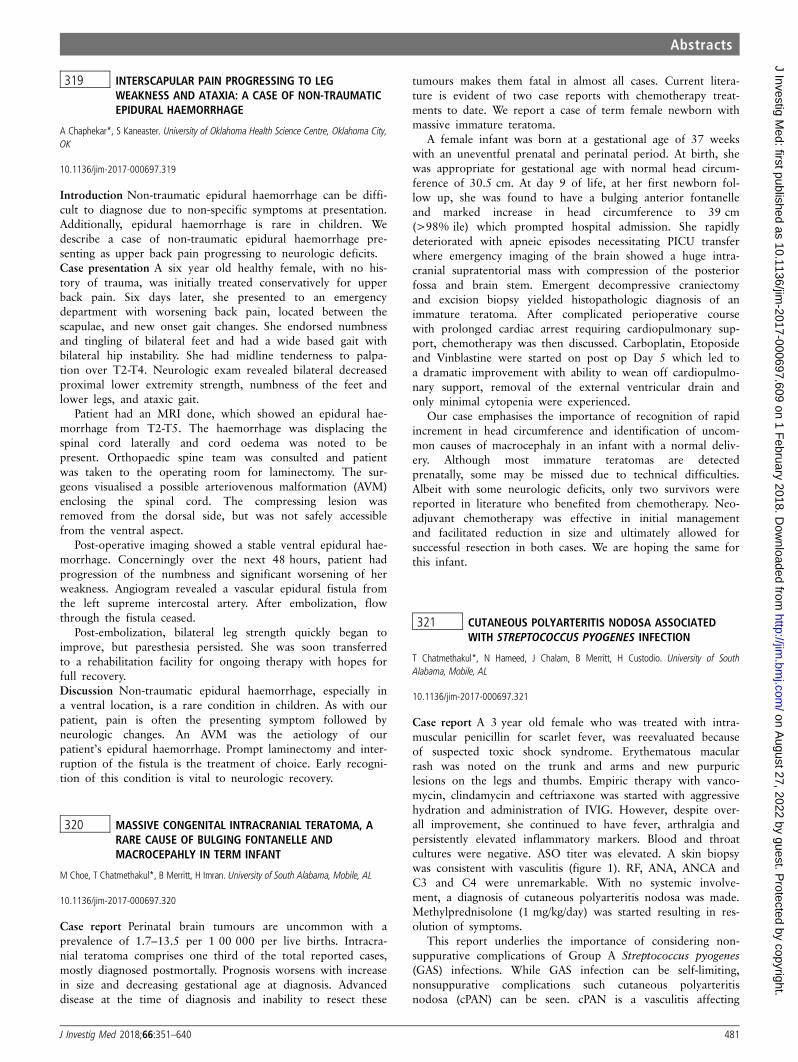
319 INTERSCAPULAR PAIN PROGRESSING TO LEGWEAKNESS AND ATAXIA: A CASE OF NON-TRAUMATICEPIDURAL HAEMORRHAGE
A Chaphekar*, S Kaneaster. University of Oklahoma Health Science Centre, Oklahoma City,OK
10.1136/jim-2017-000697.319
Introduction Non-traumatic epidural haemorrhage can be diffi-cult to diagnose due to non-specific symptoms at presentation.Additionally, epidural haemorrhage is rare in children. Wedescribe a case of non-traumatic epidural haemorrhage pre-senting as upper back pain progressing to neurologic deficits.Case presentation A six year old healthy female, with no his-tory of trauma, was initially treated conservatively for upperback pain. Six days later, she presented to an emergencydepartment with worsening back pain, located between thescapulae, and new onset gait changes. She endorsed numbnessand tingling of bilateral feet and had a wide based gait withbilateral hip instability. She had midline tenderness to palpa-tion over T2-T4. Neurologic exam revealed bilateral decreasedproximal lower extremity strength, numbness of the feet andlower legs, and ataxic gait.
Patient had an MRI done, which showed an epidural hae-morrhage from T2-T5. The haemorrhage was displacing thespinal cord laterally and cord oedema was noted to bepresent. Orthopaedic spine team was consulted and patientwas taken to the operating room for laminectomy. The sur-geons visualised a possible arteriovenous malformation (AVM)enclosing the spinal cord. The compressing lesion wasremoved from the dorsal side, but was not safely accessiblefrom the ventral aspect.
Post-operative imaging showed a stable ventral epidural hae-morrhage. Concerningly over the next 48 hours, patient hadprogression of the numbness and significant worsening of herweakness. Angiogram revealed a vascular epidural fistula fromthe left supreme intercostal artery. After embolization, flowthrough the fistula ceased.
Post-embolization, bilateral leg strength quickly began toimprove, but paresthesia persisted. She was soon transferredto a rehabilitation facility for ongoing therapy with hopes forfull recovery.Discussion Non-traumatic epidural haemorrhage, especially ina ventral location, is a rare condition in children. As with ourpatient, pain is often the presenting symptom followed byneurologic changes. An AVM was the aetiology of ourpatient’s epidural haemorrhage. Prompt laminectomy and inter-ruption of the fistula is the treatment of choice. Early recogni-tion of this condition is vital to neurologic recovery.
320 MASSIVE CONGENITAL INTRACRANIAL TERATOMA, ARARE CAUSE OF BULGING FONTANELLE ANDMACROCEPAHLY IN TERM INFANT
M Choe, T Chatmethakul*, B Merritt, H Imran. University of South Alabama, Mobile, AL
10.1136/jim-2017-000697.320
Case report Perinatal brain tumours are uncommon with aprevalence of 1.7–13.5 per 1 00 000 per live births. Intracra-nial teratoma comprises one third of the total reported cases,mostly diagnosed postmortally. Prognosis worsens with increasein size and decreasing gestational age at diagnosis. Advanceddisease at the time of diagnosis and inability to resect these
tumours makes them fatal in almost all cases. Current litera-ture is evident of two case reports with chemotherapy treat-ments to date. We report a case of term female newborn withmassive immature teratoma.
A female infant was born at a gestational age of 37 weekswith an uneventful prenatal and perinatal period. At birth, shewas appropriate for gestational age with normal head circum-ference of 30.5 cm. At day 9 of life, at her first newborn fol-low up, she was found to have a bulging anterior fontanelleand marked increase in head circumference to 39 cm(>98% ile) which prompted hospital admission. She rapidlydeteriorated with apneic episodes necessitating PICU transferwhere emergency imaging of the brain showed a huge intra-cranial supratentorial mass with compression of the posteriorfossa and brain stem. Emergent decompressive craniectomyand excision biopsy yielded histopathologic diagnosis of animmature teratoma. After complicated perioperative coursewith prolonged cardiac arrest requiring cardiopulmonary sup-port, chemotherapy was then discussed. Carboplatin, Etoposideand Vinblastine were started on post op Day 5 which led toa dramatic improvement with ability to wean off cardiopulmo-nary support, removal of the external ventricular drain andonly minimal cytopenia were experienced.
Our case emphasises the importance of recognition of rapidincrement in head circumference and identification of uncom-mon causes of macrocephaly in an infant with a normal deliv-ery. Although most immature teratomas are detectedprenatally, some may be missed due to technical difficulties.Albeit with some neurologic deficits, only two survivors werereported in literature who benefited from chemotherapy. Neo-adjuvant chemotherapy was effective in initial managementand facilitated reduction in size and ultimately allowed forsuccessful resection in both cases. We are hoping the same forthis infant.
321 CUTANEOUS POLYARTERITIS NODOSA ASSOCIATEDWITH STREPTOCOCCUS PYOGENES INFECTION
T Chatmethakul*, N Hameed, J Chalam, B Merritt, H Custodio. University of SouthAlabama, Mobile, AL
10.1136/jim-2017-000697.321
Case report A 3 year old female who was treated with intra-muscular penicillin for scarlet fever, was reevaluated becauseof suspected toxic shock syndrome. Erythematous macularrash was noted on the trunk and arms and new purpuriclesions on the legs and thumbs. Empiric therapy with vanco-mycin, clindamycin and ceftriaxone was started with aggressivehydration and administration of IVIG. However, despite over-all improvement, she continued to have fever, arthralgia andpersistently elevated inflammatory markers. Blood and throatcultures were negative. ASO titer was elevated. A skin biopsywas consistent with vasculitis (figure 1). RF, ANA, ANCA andC3 and C4 were unremarkable. With no systemic involve-ment, a diagnosis of cutaneous polyarteritis nodosa was made.Methylprednisolone (1 mg/kg/day) was started resulting in res-olution of symptoms.
This report underlies the importance of considering non-suppurative complications of Group A Streptococcus pyogenes(GAS) infections. While GAS infection can be self-limiting,nonsuppurative complications such cutaneous polyarteritisnodosa (cPAN) can be seen. cPAN is a vasculitis affecting
Abstracts
J Investig Med 2018;66:351–640 481
on August 27, 2022 by guest. P
rotected by copyright.http://jim
.bmj.com
/J Investig M
ed: first published as 10.1136/jim-2017-000697.609 on 1 F
ebruary 2018. Dow
nloaded from

medium sized vessels. In contrast to systemic PAN, cPAN islimited to the skin. However, extracutaneous symptoms mayalso be seen. Severe cPAN can lead to digital infarction andautoamputation. The aetiology remains unclear. Aside fromGAS infections, tuberculosis, hepatitis B and noninfectious ill-nesses i.e. Crohn’s disease have been associated with cPAN.Establishing the diagnosis of cPAN can be difficult given itsrarity, similarity of presentation, and association withinfection.
322 GANGLIONEUROMATOUS POLYPOSIS IN A PATIENTWITH IRRITABLE BOWEL SYNDROME
AW Cohen*, JR Mestre. University of Alabama, Birmingham, AL
10.1136/jim-2017-000697.322
Introduction Colonic polyps and associated syndromes in thepaediatric population serve as a clinically significant source ofmorbidity and potential mortality. Often associated with fami-lial origins, sporadic cases can be more challenging to diag-nose or determine clinical significance.Case A 14 year old female sought care from her physicianwith history of chronic periumbilical abdominal pain, diar-rhoea, and episodes of sweating, palpitations, and nausea. Shewas referred to a paediatric gastroenterologist where a clinicaldiagnosis of irritable bowel syndrome was made after basiclaboratory testing was not suggestive of an organic cause ofher symptoms. Due to length of symptoms and findings ofiron deficiency anaemia, patient was scheduled for upper andlower endoscopy. Upper endoscopy was without abnormality.Colonoscopy revealed innumerable firm polyps throughout theentire colon and rectum without involvement of terminalileum. Pathologic examination of polyps was consistent withganglioneuromatous polyps. Genetic evaluation was negativefor common mutations associated with polyposis syndromes(BMPR1A, NF1, PTEN, RET, and SMAD4). Patient continued
with similar intermittent symptoms. Follow up endoscopy per-formed one year after was unchanged with ganglioneuroma-tous polyps without signs of dysplasia of sampled polyps.Continued follow up planned as well as additional evaluationby a specialist in paediatric polyposis.Discussion This case demonstrates an abnormal and relativelyrare endoscopic diagnosis in a patient who meets clinical crite-ria for irritable bowel syndrome. The finding of iron defi-ciency anaemia is a significant finding as it represents themost common presentation for polyps in paediatric patients,painless rectal bleeding. Ganglioneuromatous polyposis can beseen in association with neurofibromatosis type 1, Cowdendisease, or juvenile polyposis or as an isolated finding. In theevaluation of juvenile polyps, a detailed family history is ofparticular importance as guidance for additional testing, inter-vention, or follow up. Detailed physical exam and review ofsystems should be performed to evaluate for coexisting symp-toms and physical exam findings that could point to one poly-posis syndrome or another and modify future screening andevaluation.
323 PARXOXYSMAL COMPLETE HEART BLOCK IN A CHILDWITH A STRUCTURALLY NORMAL HEART
JE Cooper*, ST McClanahan, P Glaeser. University of Alabama, Birmingham, AL
10.1136/jim-2017-000697.323
Case report A previously healthy 11 year-old African-Americanmale presented with concern for seizure and arrhythmia. Onthe morning of presentation he had a syncopal episode atschool. While his mother was driving him home he had twoepisodes of generalised body shaking, chest pain and emesis.EMS was called and noted seizure activity and periods ofabsent pulse and asystole on 3-lead electrocardiogram (ECG).
The patient denied illicit drugs, caffeine or energy drinks.He also denied history of travel outside the state of Alabama,
Abstract 321 Figure 1 Skin biopsy was consistent with vasculitis
Abstracts
482 J Investig Med 2018;66:351–640
on August 27, 2022 by guest. P
rotected by copyright.http://jim
.bmj.com
/J Investig M
ed: first published as 10.1136/jim-2017-000697.609 on 1 F
ebruary 2018. Dow
nloaded from

sick contacts, bug bites, tick exposure, rashes, fever or illsymptoms other than headache and nausea. The motherdenied family history of sudden cardiac death, drowning, sin-gle car accidents, early myocardial infarction or seizures.
Initial vitals were heart rate of 88, blood pressure 135/78,oxygen saturation of 98% on room-air and afebrile. On examthe he was ill appearing but alert with no apparent distresswhen not having an episode. He had a regular heart rate with2+femoral pulses and no murmur noted. The remainder ofhis exam was unrevealing. His baseline ECG showed normalsinus rhythm with a heart rate in the 80–90 s (figure 1). Epi-sodes began with vibration in his head, loss of consciousness,loss of pulses, flexion of the arms and legs, generalised bodyshaking for 3–4 s with subsequent return of normal vitals andconsciousness. During the episodes his ECG showed completeheart block without ventricular escape rhythm (figure 1). Hewas found to have normal cardiac anatomy, no signs of infec-tion or electrolyte abnormalities. He underwent emergentplacement of a dual chamber pacemaker for paroxysmal com-plete heart block. Subsequent work-up did not reveal a patho-logic aetiology.
324 INFECTIOUS FASCIITIS OF THE CHEST WALL AFTERSTRENUOUS ACTIVITY
1JE Cooper*, 2P Tomeny, 1CM Pruitt. 1University of Alabama, Hoover, AL; 2University ofAlabama, Birmingham, AL
10.1136/jim-2017-000697.324
Case report A 15 year-old white female presented to the emer-gency department (ED) after 48 hours of right shoulder pain.The pain started after a week of colour guard tryouts. Shedescribed sharp, severe, constant pain located mainly in heraxilla. Intramuscular ketorolac and an intramuscular steroidreceived the day before prior provided temporary pain relief,but her discomfort worsened by the day of presentation.
On physical examination, the patient was febrile to 38.5degrees Celsius, with a heart rate of 95 and blood pressure of115/58. She kept her right arm propped on a pillow, inter-nally rotated with 30 degrees of abduction. She had exquisitetenderness to palpation in the right axilla. There was no crep-itus. She refused active range of motion of the shoulder. Pas-sive range of motion was limited to less than 90 degrees. A
macular, erythematous, blanching rash along her chest, back,and groin developed during her ED course.
In the ED, the patient was administered two boluses ofnormal saline. Laboratory testing was significant for a whiteblood cell count of 15.44 103/uL, with 86% neutrophils; pla-telets 118 103/uL; and C-reactive protein 5.37 mg/dL. Thetotal bilirubin was 1.1 mg/dL, alanine aminotransferase 143 U/L, and aspartate aminotransferase 142 U/L. International nor-malised ratio was 1.5, fibrinogen 390 mg/dL, and lactate dehy-drogenase 850 U/L. Intravenous vancomycin and piperacillin-tazobactamwere initiated after obtaining blood cultures. Shewas subsequently hospitalised. Magnetic resonance imagingrevealed extensive T2 hyperintensity in the fascial planes ofthe right chest wall along the axillary region, extending fromthe infraclavicular location and along the lateral aspect of thechest wall. The patient was diagnosed with acute bacterial fas-ciitis with secondary toxin-mediated compensated shock. Shewas successfully treated with IV antibiotics prior to transition-ing to an oral regimen and did not require surgical debride-ment. This case highlights the need for early recognition ofnecrotizing infections to prevent morbidity and mortality.
325 SCLEROSING CHOLANGITIS IN KABUKI SYNDROME: ANUNUSUAL COMPLICATION OF A RARE DISORDER
1MA Corbera-Hincapie*, 2S Safder, 3R Gonzalez-Peralta. 1University of Florida, Gainesville,FL; 2Arnold Palmer Hospital, Orlando, FL; 3Florida Hospital, Orlando, FL
10.1136/jim-2017-000697.325
Background Kabuki Syndrome (KS) is a rare disorder charac-terised by distinct facial and skeletal anomalies, cardiacdefects, short stature and developmental delay. Hepaticinvolvement occurs only rarely. Herein, we describe a childwith KS and liver dysfunction that was eventually attributedto sclerosing cholangitis.Case report A 3-year-old female with KS, repaired VSD andhernia, was evaluated for persistent transaminitis over the pre-ceding 1 year (discovered incidentally). Findings on physicalexam were normal except facial dysmorphia consistent withKS. She had no jaundice, hepatosplenomegaly or stigmata ofchronic liver disease. Initial laboratory tests showed elevatedaspartate transaminase (440 U/L), alanine transaminase (281 U/L), and gamma-glutamyltransferase (408 U/L). Results of
Abstract 323 Figure 1 Electrocardiogram of normal sinus rhythm and complete atrioventricular block
Abstracts
J Investig Med 2018;66:351–640 483
on August 27, 2022 by guest. P
rotected by copyright.http://jim
.bmj.com
/J Investig M
ed: first published as 10.1136/jim-2017-000697.609 on 1 F
ebruary 2018. Dow
nloaded from

hepatitis A, B and C serology, alpha 1 antitrypsin level and Piphenotype, antinuclear, liver-kidney-microsomal, antineutrophilcytoplasmic and tissue transglutaminase antibodies were nega-tive; she had detectable smooth muscle antibody (titer 1:20).Abdominal sonogram did not visualise gallbladder but other-wise normal. Liver biopsy showed bile ductular proliferationwith pericholangitis and minimal lobular inflammation consis-tent with sclerosing cholangitis. We initiated ursodeoxycholicacid (URSO; 20 mg/kg/d) with gradual improvement of transa-minase levels that have remained normal for 5 years.Discussion In a series of KS, 2%–21% of patients developedliver disease, ranging from neonatal hyperbilirubinemia, biliaryatresia, hepatic fibrosis and sclerosing cholangitis. Clinical datais limited but severe hepatic disease requiring liver transplanta-tion occurs. Our patient’s course suggests that URSO can besuccessfully used to treat sclerosing cholangitis in KS. Ourcase also highlights the importance of monitoring for liverdysfunction in patients with KS and proceeding with thoroughevaluation to arrive at a specific diagnosis and institute appro-priate therapy.
326 A RARE CASE OF EPSTEIN BARR VIRUS-ASSOCIATEDHEMOPHAGOCYTIC LYMPHOHISTIOCYTOSIS IN ANIMMUNOCOMPROMISED PAEDIATRIC PATIENT
CM Cox*, R Herdes, M Shapiro, B Desselle, C Sandlin. LSUHSC, New Orleans, LA
10.1136/jim-2017-000697.326
Case report Kaposiform Lymphangiomatosis is a rare disorderof lymphatic vessels and is characterised by abnormal lym-phatic clusters in various regions of the body. Prognosis canbe poor, especially when there is pulmonary involvement.Complications can include pleural effusions, pericardial effu-sions, and haemorrhage. Current treatment includes chemo-therapy and/or immune modulators, specifically vincristine andsirolimus. Patients taking sirolimus have increased risk ofinfections, including Epstein-Barr Virus (EBV) infection. EBVis a leading cause of hemophagocytic lymphohistiocytosis(HLH), which is a life-threatening disorder of the immunesystem.
We report a case of a 6-year-old male with a severe formof Kaposiform lymphangiomatosis who developed EBV associ-ated HLH. The patient presented with persistent fever, inter-mittent rash, and worsening respiratory distress. He wasstarted on broad spectrum antibiotic therapy for pneumonia,and his Sirolimus was continued upon admission. Due to hisworsening clinical status, continued fevers, and progressivepancytopenia, haematology and oncology specialists recom-mended the initiation of vincristine.
The patient’s fevers continued despite these therapies, andhis hospital course was complicated by an acute gastrointesti-nal haemorrhage. His clinical status and pancytopenia contin-ued to deteriorate, prompting consideration of alternativediagnoses. After several days of hospitalisation, viral studieswere positive for EBV. A significantly elevated ferritin level(33,000 ng/ml), in conjunction with his other clinical and lab-oratory findings, led to a diagnosis of EBV associated HLH.Etoposide was given on hospital day 15, but the patient fur-ther declined and died the following day.
This case highlights the invasive nature of EBV, especiallyin immunocompromised individuals. It is important for clini-cians to recognise the risk factors associated with immune-modulating therapies and consider infectious etiologies early inthe patient’s clinical course. Though rare, HLH should beconsidered in patients with persistent fever, pancytopenia, andworsening clinical status in the setting of certain conditionsthat place patients at increased risk for the disease.
327 TETRAPHOCOMELIA: A CASE REPORT AND LITERATUREREVIEW
J Deitrick*, D Nguyen, W Sessions, M Naqvi, H Heather. Texas Tech Health Sciences CentreSchool of Medicine, Amarillo, TX
10.1136/jim-2017-000697.327
Case report Tetraphocomelia is a condition characterised bysevere symmetrical limb reduction in utero. Several syndromesare associated with this finding: Robert’s syndrome, GrebeSyndrome, Waardenber syndrome, Holt-Oram syndrome, andThrombocytopenia with Absent Radius syndrome.
Abstract 327 Figure 1 Unique presentation of a rare congenital anomaly was possibly caused by amniotic bands or a vascular accident in utero
Abstracts
484 J Investig Med 2018;66:351–640
on August 27, 2022 by guest. P
rotected by copyright.http://jim
.bmj.com
/J Investig M
ed: first published as 10.1136/jim-2017-000697.609 on 1 F
ebruary 2018. Dow
nloaded from

Furthermore, certain in utero exposures, such as thalidomide,alcohol, and cocaine, are also associated with this and similarmusculoskeletal deformations. A 24 year old female presentedfor her 24 week prenatal ultrasound, which revealed incom-plete limb development in the fetus. Subsequent to a caesar-ean section at 38 weeks and 6 days gestation, the newbornfemale presented with gross musculoskeletal deformities of allfour limbs. She was diagnosed with tetraphocomelia butlacked features of a concomitant syndrome and relevant familyor birth history associated with this anomaly. Due to the lackof ancillary symptoms, this case did not fit into any specificsyndrome and was thought to be the result of a sporadic,non-hereditary limb deficiency involving all four limb buds.While the cause of tetraphocomelia is not fully understood,current literature provides insight into possible genetic andenvironmental factors contributing to the pathophysiology ofthis condition. This unique presentation of a rare congenitalanomaly was possibly caused by amniotic bands or a vascularaccident in utero, resulting in an isolated erroneous occurrenceduring the critical period of limb development.
328 A CASE OF TOXIC EPIDERMAL NECROLYSIS CAUSED BYCEFADROXIL
TT Doan*. LSU Health Sciences, Shreveport, LA
10.1136/jim-2017-000697.328
Case report Toxic epidermal necrolysis (TEN) is a rare andsevere adverse mucocutaneous drug reaction characterised byhaemorrhagic erosions, erythema, and epidermal detachmentdue to immune mediated destruction of the epidermis. Drugssuch as allopurinol, Trimethoprim-Sulfamethoxazole, cephalo-sporins, quinolones, and anticonvulsants are assumed to be themain cause of TEN in most case. Early diagnosis and aggres-sive treatment of TEN is important for the reduction of mor-bidity and mortality associated with his condition. We presenta rare case of successfully recovered TEN caused by cefa-droxil, a first-generation cephalosporin. A 4-year-old girl pre-sented to the emergency room with progressively worsenedblisters on trunk after the second dose of oral cefadroxil pre-scribed for otitis media. The lesion started as a diffuse eryth-ematous, maculopapular rash over body, progressed intopainful blisters, followed by diffuse exfoliation of the skininvolving face, back, abdomen, and bilateral upper and lowerextremities over the course of 3 days. Past medical historywas significant for chronic otitis media. She has no knownallergies and immunisation was up to date. The initial physicalexam revealed a listless child with fever of 102.2F. She wastachycardic with heart rate of 160. Ophthalmologic examshowed bilateral periorbital oedema, skin blisters with haemor-rhagic crusts involving both upper eyelid, but no injection ofconjunctiva and no corneal involvement. She had denudedoral lesions and cracked lips with haemorrhagic crusts. Therewere multiple, thick bullous lesions filled with turbid fluidsand epidermal detachments on face, oral mucosa, neck, abdo-men, genital, arms, legs, and feet covering approximately 60%of body surface area. Nikolsky sign is positive. Initial labora-tory investigations were unremarkable including negative Her-pes simplex virus and Mycoplasma pneumoniae culture. Shewas admitted to the PICU with burn surgery assistance. Cefa-droxil was discontinued immediately. She was managed withintravenous fluids, prophylactic antibiotics, IVIG, hydrotherapy
and proper wound care. Her haemodynamic status was moni-tored continuously in the unit. She successfully recovered after2 weeks of hospitalisation and was discharged to home ingreat condition.
329 PAIN IN THE NECK: A CASE OF INFANTILE TUMORALCALCINOSIS
B Ducote*, C Jacobs, SN Epps, R Rao, M Murphy. LSUHSC, New Orleans, LA
10.1136/jim-2017-000697.329
Case report A four month old full term female presented toher paediatrician after her mother noticed that over the past3 weeks she lost the ability to roll over and hold her chestup 45 degrees while prone. She had recently been diagnosedwith torticollis and plagiocephaly and was seen by occupa-tional therapy, but the onset of this regression prompted herpaediatrician to refer her to neurology, who then admitted herfor a full workup.
On exam she was difficult to console, head lag was noted,she was unable to actively turn her head, and she cried withpassive movement. Initial labs of CBC, CMP, TSH, and FT3were normal, while ESR and CRP were elevated. MRI of thebrain and CT of the neck showed abnormal calcifications atthe articulation of the C1/C2 vertebrae likely secondary totumoral calcinosis. Skeletal survey showed only abnormal calci-fications around the cervical vertebrae. Endocrine was con-sulted and completed a workup for secondary causes oftumoral calcinosis which was negative, with normal values ofPTH, calcium, vitamin D, and phosphorus. Hematology-oncol-ogy was consulted to rule out neoplasm and suggested abiopsy. ENT performed a biopsy of the lesion which showedcalcified material without inflammatory changes confirmingour presumed diagnosis of tumoral calcinosis.
The patient was started on gabapentin for pain manage-ment per neurology recommendations. She was given NGfeeds until her pain improved and she could tolerate full feedsby mouth. She was also given 5 days of steroids to decreaseinflammation in the area, and the range of motion of herneck improved throughout her hospital stay. MRI after biopsyshowed decreased residual calcifications around the C1/C2 ver-tebrae without evidence of spinal cord impingement. Over thenext two months, she regained head control and better rangeof motion of her neck.
Tumoral calcinosis consists of calcium crystal deposits inperiarticular soft tissues. It can be hereditary with hyperphos-phatemia, a complication of dialysis, or a rare isolated findingwhich was likely what our patient had. In one previouslyreported case study in an infant, biopsy was found to be cura-tive. Tumoral calcinosis is a rare diagnosis and cause of torti-collis and loss of developmental milestones especially ininfants.
330 UNUSUAL OSTEOMYELITIS IN AN INFANT
M Duplantier*, SN Epps, M Heffernan, A Bauchat, R Gardner, A Messer. LSUHSC, NewOrleans, LA
10.1136/jim-2017-000697.330
Case report Leg pain in an infant can be difficult to detectand diagnose. Osteomyelitis is rare in a clinically well-
Abstracts
J Investig Med 2018;66:351–640 485
on August 27, 2022 by guest. P
rotected by copyright.http://jim
.bmj.com
/J Investig M
ed: first published as 10.1136/jim-2017-000697.609 on 1 F
ebruary 2018. Dow
nloaded from

appearing infant, though more common when Pseudomonas isthe underlying aetiology. In this case, initial concern for non-accidental trauma (NAT) in an otherwise healthy infant led toa diagnosis of osteomyelitis secondary to an unusual organism.Based on benign initial evaluation, there was potential for thispatient’s infection to go undiagnosed and untreated. However,our Orthopaedic Surgery team reviewed her case and uncov-ered her diagnosis.
Here we describe a 10-month-old female initially diagnosedwith a proximal femur buckle fracture and found to haveosteomyelitis and Pseudomonas stutzeri bacteremia. She pre-sented with refusal to move left leg as well as upper respira-tory symptoms. There was no history of trauma and, onexam, she was febrile with pain on left hip manipulation.
Evaluation was significant for a subacute proximal femurbuckle fracture seen on x-ray. Subsequent MRI findings wereconsistent with surrounding osteomyelitis. On presentation,white blood cell count was 12.32 × 10³/UL, C-reactive pro-tein was <0.5 mg/dL, which later rose to 1.4 mg/dL, anderythrocyte sedimentation rate was 37 mm/hr. Clinically, thepatient was well-appearing with no external abnormalities.Blood culture later grew Pseudomonas stutzeri at 1.05 days. Arespiratory viral panel revealed influenza A and adenovirus.
The Infectious Disease team recommended IV clindamycinand ceftazidime for 7 days to treat susceptible Pseudomonas aswell as typical infectious etiologies. She was then transitionedto oral clindamycin and ciprofloxacin to complete a 4 weekcourse. A Spica cast was placed; no surgical intervention wasrequired.
The differential diagnosis for leg pain in an infant is broad,which may delay prompt diagnosis. Trauma and osteomyelitisare occasionally competing diagnoses due to similarities on ini-tial evaluation and imaging, particularly in an otherwise well-appearing, non-ambulatory infant. This case serves to increaseinitial indices of suspicion for both diagnoses and demon-strates that these diagnoses are not mutually exclusive andrequire an interdisciplinary team of experts for best care.
331 NEONATAL CONJUNCTIVITIS: AN UNUSUAL CAUSE OF ACOMMON COMPLAINT
MN Frascogna*, L Moore. University of Mississippi Medical Centre, Jackson, MS
10.1136/jim-2017-000697.331
Case report Infectious conjunctivitis is a common presentationin neonates and young children. The disease course is oftenbenign and can be easily treated in most cases. However, it isimportant to be aware of the more serious etiologies that canbecome lethal if not identified early and promptly treated. Inthis report, we present a rare case of Neisseria meningitidis asthe primary cause of conjunctivitis in an otherwise healthyinfant.
A 20 day old African American female born at 39 weeksvia normal spontaneous vaginal delivery presented to thepaediatric emergency department with a two day history ofright eye swelling and discharge. The patient’s mother deniedany fever, irritability, rash, or upper respiratory symptoms.Pregnancy and delivery were largely uncomplicated andscreening labs were reportedly negative for any sexually trans-mitted infections.
In the emergency department, patient was noted to havesignificant mucopurulent discharge from the right eye with
associated eyelid swelling and erythema. Slit Lamp and fundo-scopic exam showed conjunctival injection and chemosis, butwas otherwise normal. The remaining exam was unremarkable.The patient underwent a full septic workup including ocular,CSF, blood, and urine cultures and was admitted for empiricantibiotic treatment of the presumed causative organisms,Niesseria gonorrheae and Chlamydia trachomatis.
The patient was started on intravenous Ampicillin andCefotaxime and Erythromycin ophthalmic solution. Symptomsimproved after twenty-four hours of treatment and infectiousworkup remained negative; however, on day two of hospital-isation, ocular cultures returned positive for Neisseria meningi-tidis. Ampicillin was stopped and intravenous Cefotaxime wascontinued. The Health Department was notified per protocol,and chemoprophylaxis for contacts was not recommended dueto strict confinement of the infection to the eye. The patientcompleted five days of intravenous antibiotics with markedimprovement. She was discharged home to complete a 14 daycourse of oral and ophthalmic Erythromycin. At a 2 week fol-low up appointment with ophthalmology, symptoms had com-pletely resolved and no ocular abnormalities were noted.
332 SALMONELLA IN THE SCROTUM: NOT YOUR TYPICALNEONATAL INFECTION
E Friedrich*, R Welliver, S DeLeon. University of Oklahoma Health Sciences Centre,Oklahoma City, OK
10.1136/jim-2017-000697.332
Case report A 17 day old male, born at 38 weeks withoutcomplications, presented with a 7 day history of irritability,and fever on day 2 of illness. On the day of fever, his paedia-trician prescribed amoxicillin/clavulanic acid for unknown rea-sons, and he then remained afebrile until admission. Two daysprior to presentation, his scrotum became swollen and tender,which prompted the family to seek further care. In the emer-gency room, a sepsis work up and testicular ultrasound (US)with doppler was performed. The work up yielded no evi-dence of systemic infection. His only abnormal lab was a C-reactive protein of 32 mg/dL. The US showed a right scrotalabscess with blood flow to both testes. Urology recommendedadmission for incision and drainage (I and D) of the abscess.A renal US and voiding cystourethrogram (VCUG) wereobtained to evaluate for vesiculo-ureteral reflux or abnormalfistula connexion and were negative. He was started on intra-venous vancomycin, ampicillin, and gentamicin. The I and Dwas performed with removal of 50 cc of purulent material,along with a right orchiectomy due to necrosis. The tissueculture grew a Salmonella species susceptible to ampicillin,which coverage was then narrowed to. He received 14 daysof antibiotics. His mother denied exposure to raw chicken,reptiles, or sick contacts. A granulocyte dihydrorhodamine flu-orescence test showed normal NADPH oxidase activity,decreasing the likelihood of chronic granulomatous disease.
Salmonella typically affects the gastrointestinal tract and ismost often associated with exposure to raw chicken or rep-tiles. Most cases are seen in children under the age of 5. Allother reported cases of Salmonella orchitis are in immuno-compromised patients or occur in areas where Salmonella isan endemic infection. Extra-intestinal focal infections typicallyresult from hematogenous spread, though anatomic urologicabnormalities can predispose to isolated genitourinary (GU)
Abstracts
486 J Investig Med 2018;66:351–640
on August 27, 2022 by guest. P
rotected by copyright.http://jim
.bmj.com
/J Investig M
ed: first published as 10.1136/jim-2017-000697.609 on 1 F
ebruary 2018. Dow
nloaded from

infections. Management of extra-intestinal infections includes Iand D and antibiotics. This case is unique because ourpatient’s GU imaging was normal, blood culture was negative(although he had received antiobiotics prior to admission),had no history of exposure, and had no risk factors forimmunodeficiency other than age.
333 CASE REPORT CYCLIC NEUTROPENIA AND ASSOCIATEDAMYLOIDOSIS
KM Galipp*, C Ilonze, K Shah, A Rughani, S Travis, A Nayak, WF Kern, K Wierenga,WH Meyer. University of Oklahoma Health Sciences Centre, Oklahoma City, OK
10.1136/jim-2017-000697.333
Study purpose Cyclic neutropenia is a rare hereditary disorder,characterised by recurrent neutropenia, cycling at about3 week intervals, with variable associated symptoms includingoral ulcers and fever. There are 4 reported cases of cyclicneutropenia associated with chronic inflammation leading todevelopment of reactive AA amyloidosis. One patient also pre-sented with amyloid goitre. We report a new case of cyclicneutropenia with associated renal and thyroid amyloid.Method (case report) A 12-year-old female presented with a1 month history of thyromegaly, and recurrent aphthous ulcersassociated with fevers. Laboratory workup showed severe neu-tropenia, anaemia, azotemia, and abnormal thyroid function,with an absolute neutrophil count – 0/mL, haemoglobin –
9.0 g/dL, serum creatinine – 1.89 mg/dL, and uric acid –
9.0 mg/dL. Thyroid stimulating hormone was elevated – 12.5mIU/mL, and normal free T4. Urinalysis showed 2+protein, 2+blood, and 5–10 urine red blood cells/hpf. Chest radiographshowed mild narrowing of the trachea from thyroid compres-sion. Bone marrow biopsy showed a hypocellular marrow,with tri-lineage hematopoiesis, left shifted myeloid maturationwith very rare mature neutrophils. Both renal biopsy and thy-roid fine needle aspiration revealed abundant amyloid. Ofnote, her father had AA amyloidosis, resulting in end-stagerenal disease (ESRD) requiring hemodialysis, and recurrentaphthous ulcers. The family history suggested a familial predis-position. Genetic testing revealed a pathogenic ELANE c.358A>T gene mutation with autosomal dominant inheritance con-firming the diagnosis of cyclic neutropenia. We treated ourpatient with daily granulocyte colony stimulating factor toreduce the burden of chronic inflammation induced by cyclicneutropenia, and to preserve renal and other end organ func-tion affected by further amyloid deposition.Summary of results Proband with ELANE gene mutation posi-tive cyclic neutropenia, amyloidosis of thyroid and kidney,with a positive paternal history of AA amyloidosis resulting inESRD.Conclusions Cyclic neutropenia may result in chronic inflam-matory states leading to secondary amyloidosis.
334 ALVEOLAR CAPILLARY DYSPLASIA PRESENTING ASREFRACTORY PULMONARY HYPERTENSION
J Gallois*, J Patrick, J Surcouf. LSUHSC School of Medicine, New Orleans, LA
10.1136/jim-2017-000697.334
Case report Alveolar capillary dysplasia with misalignment ofthe pulmonary veins (ACDMPV) is a developmental anomaly
of the pulmonary vasculature that takes place in the latecanalicular to early saccular phase of lung development. Thismaldevelopment results in the failure of the formation of thenormal air-blood diffusion barrier. It is almost universallyfatal. 90% of those effected are term gestation, 60% presentwith cyanosis within the first 48 hours of birth, and 80% ofcases are associated with other congenital malformations.Ninety percent of cases are associated with a genetic mutationon the FOXF1 gene on chromosome 16, and 90% of casesare de novo mutations. Diagnosis is confirmed by lung biopsyor autopsy, where pathologic features include a paucity ofalveolar capillaries, widened alveolar septae, muscularization ofpulmonary arterioles, and usually misalignment of pulmonaryveins in the bronchovascular bundle. No current supportivetherapies have been shown to change mortality.
We present a term female infant born to a 33 year oldmother with an unremarkable prenatal course. The infant wasinitially stable, then at six hours of life she was transferred tothe neonatal intensive care unit for cyanosis and hypoxia withoxygen saturations in the 50’s. She was ultimately transferredto a tertiary care facility for concerns of a congenital heartdefect versus persistent pulmonary hypertension (PPHN). Theechocardiogram at admit revealed a structurally normal heartand evidence of PPHN. Other congenital malformations werenot found. Although she initially stabilised with medical man-agement including inhaled nitric oxide, her PPHN progresseddespite maximal support. She significantly decompensated witha prolonged code while being placed on extracorporeal mem-branous oxygenation (ECMO) on day of life 2, requiringsimultaneous initiation of therapeutic hypothermia. After twofailed weans off ECMO, a lung biopsy was performed on dayof life 25 which revealed ACDMPV. With the family at thebedside, support was withdrawn.
The incidence of ACDMPV is rare, but is increasingly rec-ognised. Early diagnosis may prevent unnecessary therapiesand prolongation of treatment. Neonates with refractoryPPHN, especially if associated with anomalies, should promptevaluation for ACDMPV.
335 ACTINOMYCES EPIDURAL ABSCESS: A VIRTUALLYUNHEARD OF PROCESS IN THE VIRTUAL AGE
AL Gibson*, S Liu, M Naifeh. OU Children’s, Edmond, OK
10.1136/jim-2017-000697.335
Case report Actinomyces has been termed the ‘great pretender’in many case reports due to its ability to invade various partsof the body causing abscesses. Actinomyces epidural abscessesare virtually unheard of in the paediatric population, and onlysparse literature exists in the adult population, with often onlya few spinal segments involved. As a slow growing anaerobicgram positive bacteria, it typically spreads through the bodyvia sinus tract formation.
Here we report a 3 year old female with multiple congeni-tal anomalies including scoliosis, repaired tethered cord, his-tory of syrinx and hydrocephalus at birth with aventriculoperitoneal (VP) shunt, who on MRI was found tohave a highly invasive epidural abscess extending from thecervical spine to the sacrum and into the surrounding scalenesand psoas muscles. The patient initially presented with clearsigns of meningitis including fever, lethargy and decreasedactivity and was too ill to undergo imaging on admission.
Abstracts
J Investig Med 2018;66:351–640 487
on August 27, 2022 by guest. P
rotected by copyright.http://jim
.bmj.com
/J Investig M
ed: first published as 10.1136/jim-2017-000697.609 on 1 F
ebruary 2018. Dow
nloaded from

Cerebrospinal fluid from her VP shunt did not reveal aninfectious cause for her symptoms, so a lumbar puncture wasperformed which collected purulent fluid from her epiduralabscess. This fluid grew actinomyces turicensis and actinomy-ces europaeus. After clinical improvement, spinal imagingshowed a dorsal to dural sinus tract at L4 which is likely thepoint of initial bacterial invasion.
After thorough literature review, this is the largest epiduralabscess that has been reported. While the liklelihood of treat-ing another patient with the same complex medical history islow, this case underscores the importance of prompt andrepeat imaging in any patient with symptoms of meningitiswho does not show clinical evidence of improvement onbroad spectrum antibiotics as Actinomyces can present thisway.
336 A NOVEL DE NOVO HETEROZYGOUS MUTATION IN THEPCDH17 GENE WITH MICROCEPHALY, DEVELOPMENTALDELAY, AND ACUTE LYMPHOBLASTIC LEUKAEMIA
*KM Gleditsch, R Gardner, Y Lacassie. Louisiana State University Health Science Centre,New Orleans, LA
10.1136/jim-2017-000697.336
Case report We present an 11 year old girl evaluated by genet-ics at birth due to microcephaly. Chromosomes and a microar-ray were normal. At the age of 3 she developed pre-B-cellacute lymphoblastic leukaemia (ALL). She completed treatmentin 2012 and has been doing well in the interim. During andafter treatment she exhibited significant developmental delayand neurocognitive deficits. At age 11 her height and weightwere at or below the 5th centile and head circumference waswell below the 2nd centile (approximately 6 standard devia-tions below the mean and corresponding to the 50th centilefor a 9-month-old girl). Bone age was appropriate. She had adistinctive triangular face with micrognathia and a pointednose resembling a Seckel-like syndrome. The patient also hadclinodactyly of the fourth toes, zygodactylous triradius involv-ing the 2nd and 3rd left toes, tendency to Sydney line in theright palm and a radial loop in the left middle finger. WholeExome Sequencing (WES) was performed on the patient aswell as her biological parents (trio). A de novo heterozygousmutation in the gene PCDH17 with potential relation to thephenotype was discovered. This c.716dupA variant causes aframeshift starting with codon Asparagine 239, changing thisamino acid to a Lysine residue and creating a premature stopcodon at position 34 of the new reading frame denoted p.Asn239LysfsX34. This variant is predicted to cause loss ofnormal protein function via protein truncation or nonsense-mediated mRNA decay. To the best of our knowledge thereare no reports of pathogenic variants of this gene in the liter-ature. PCDH17 is a member of the protocadherins familywhich is important in synaptic function in the central nervoussystem. This gene is highly expressed in areas of the braininvolved in higher cortical function and speech. Research thathas been done with respect to methylation of this gene andcorrelation with poor prognosis in patients with acute lympho-blastic leukaemia is of great interest in this case. We feel thatthis is a clinically significant finding that may shed light onthe role of this gene in neural and hematologic development.
337 FRACTURED CORPUS CAVERNOSA IN A PAEDIATRICPATIENT
BC Gruenberg*, D Mortel, A Bogie. University of Oklahoma, OKC, OK
10.1136/jim-2017-000697.337
Case report Our patient is a 7 year old male who presentswith complaints of penile pain. The patient was riding ‘piggyback’ on his grandmothers back on the morning of the injurywhen she tripped and fell. The patient fell off of her backand into the staircase, hitting his penis directly on the staircase railing. He reports sudden onset penile pain after thefall. On arrival he denied any difficulty or pain with urina-tion, and reported pain only with movement.
His physical exam was significant only for ecchymosis andswelling of his penile shaft. Urinalysis showed no hematuria.
Penile ultrasound noted an area of discontinuity (6 mm)along the right corpora cavernosa with some soft tissue extru-sion, consistent with a fracture of his right corpuscaversonum.
Paediatric Urology was consulted and he was taken to theOR.
In the OR a flexible cystoscopy showed an uninjured ure-thra and bladder. The penis was then degloved circumferen-tially all the way down to the base of the penis. An artificialerection was then performed, and extravasation of fluid wasnoted posterior to the urethra. However, no hole was readilyapparent. The urethra was mobilised off the right corpus cav-ernosum and the corporeal tear was noted in this area, wasapproximately 7 mm. The corporeal injury was then closedand the shaft skin reapproximated to the preputial collar. Ourpatient had no postsurgical complications, his pain was wellcontrolled with NSAIDs, and he was discharded home onpost-op day one.
Abstract 337 Figure 1 Corporeal injury
Abstracts
488 J Investig Med 2018;66:351–640
on August 27, 2022 by guest. P
rotected by copyright.http://jim
.bmj.com
/J Investig M
ed: first published as 10.1136/jim-2017-000697.609 on 1 F
ebruary 2018. Dow
nloaded from

338 THE TROUBLE WITH LETTING GO: NEONATE WITHHYPOTONIA
S Gu*, L Barbiero, SL Nelson. Tulane University, New Orleans, LA
10.1136/jim-2017-000697.338
Case report A preterm male neonate born to a G2P2 motherpresented to the NICU with severe hypotonia from birth.Maternal history included negative maternal labs, adequateprenatal care, unremarkable prenatal ultrasounds throughoutpregnancy, and administration of magnesium. Family historywas negative for previously diagnosed neurologic, autoimmune,or genetic conditions. Physical exam was remarkable for gen-eralised hypotonia, tenting of upper lip, bilateral talipes equi-novarus, arthrogryposis, intact reflexes, and lack of tonguefasciculation. Genetics and Neurology were consulted givenexam findings. Work up included initial evaluation for sepsis,measurement of serum electrolytes and ammonia, and meco-nium drug screen, which were all unremarkable. Head ultra-sounds and brain MRI were normal. Initial magnesium levelelevated, but despite normalisation, patient remained hypo-tonic. Creatine kinase, direct bilirubin, and GGT were notedto be elevated. Subsequently, it was noted that the patient’smother had difficulty with release of grasp during a hand-shake, and further elucidation revealed a family history ofmultiple maternal family members with varying degrees ofweakness. The diagnosis was confirmed when DNA testingrevealed one expanded DMPK allele of ~1300 CTG repeats.
Congenital Myotonic Dystrophy (CMD) type 1 is an auto-somal dominant, multisystem disorder that occurs as a resultof CTG repeat expansion in the non-coding region of theDMPK gene on 19q13 that can expand in successive genera-tions, resulting in earlier presentation of the disorder. Patientswith greater than 50 CTG repeats have a>99% probability ofmanifesting signs and symptoms of CMD. The differential forhypotonia in neonates is broad and should include: central orperipheral nervous system abnormalities, myopathies, geneticdisorders, endocrinopathies, metabolic diseases, drug-induced,and electrolyte derangements. Our patient was preterm andhis mother was given magnesium perinatally, which were ini-tial considerations as etiologies. However, suggestive physicalexam findings and a detailed family history were essential todiagnosis and subsequent confirmatory genetic testing. Asrecent studies have discussed, targeted genomics in critically illnewborns is helpful in diagnosis, anticipatory guidance, anddecision-making for parents.
339 20 YEAR OLD WITH ABDOMINAL & FLANK PAIN
K Gutermuth*, J Haupt, A Sorrentino. Univeristy of Alabama, Birmingham, AL
10.1136/jim-2017-000697.339
Case A 20-year-old male with history of autism and obesitywas seen in the ED a week prior for acute right flank painthat improved with ibuprofen. Exam was unremarkable, andhe was discharged home. He was seen by his PCP and had aurinalysis significant for hematuria, proteinuria, and hyperten-sion. Later, he developed fever, abdominal pain and diarrhoea,and returned to the ED. Upon arrival, he was well-appearing,and his fever and abdominal pain had resolved. Lab work wasnotable for elevated leukocytes and inflammatory markers. CTabdomen was obtained, initially non-contrast for possibility of
nephrolithiasis, followed by contrast CT which showed appen-dicitis in the right upper quadrant adhered to the liver edge,with a large liver fluid collection consistent with an abscess.Discussion The case stresses the importance of consistent fol-low-up, return precautions, and clinical acumen. The symp-toms of flank pain, progressive hematuria/proteinuria,abdominal pain, nausea, vomiting, diarrhoea, and fevers pointto a more insidious pathology. Initial history and resolution offever and discomfort, combined with outpatient urinary find-ings suggested possible renal calculi, which were not seen onCT. Surgery evaluated the pateint due to concern for perfo-rated appendix and hepatic abscess. IR drained 280 cc darkbrown purulent material from the liver, and drain was left inplace. Interval appendectomy was performed, and he had anuncomplicated recovery.
Abstract 339 Figure 1 Appendicitis in the right upper quadrantadhered to the liver edge, with a large liver fluid collection consistentwith an abscess
340 EARLY CONGENITAL SYPHILIS: COMMON BUT NOT SOMUCH
V Habet*, B Casey, L Powell, C Saylor, KM Gleditsch, C Morrison, G Hescock, RE Begue,C Sandlin. Louisiana State University Health Sciences Centre, New Orleans, LA
10.1136/jim-2017-000697.340
Purpose of study Syphilis, the ‘great imitator’, can have a widespectrum of clinical manifestations, ranging from asymptomaticto early infant death. In the United States, the number ofcongenital syphilis cases is rising with 16 per 1 00 000 livebirths reported in 2016, a 27.6% increase since 2015. We dis-cuss the case of an infant with congenital syphilis who pre-sented with classical but nonspecific symptoms.Methods used Retrospective chart review.Summary of results A 3-month-old full term female was bornvia C-section to a mother with active herpes and gonorrhoeatreated in the third trimester. The patient presented with fever
Abstracts
J Investig Med 2018;66:351–640 489
on August 27, 2022 by guest. P
rotected by copyright.http://jim
.bmj.com
/J Investig M
ed: first published as 10.1136/jim-2017-000697.609 on 1 F
ebruary 2018. Dow
nloaded from

and discrete, annular, scaly plaques on her trunk and extrem-ities that progressed to desquamation of her hands and feet.She was initially admitted to the Haematology service forsevere anaemia. A skeletal survey revealed lytic bone lesionsconcerning for Langerhans cell histiocytosis. Bone scan, PETscan, and MRI brain/spine confirmed lesions of the axial andappendicular skeleton without involvement of other organs.CT showed inguinal and iliac lymphadenopathy, and repeatskeletal survey 18 days later showed progression of permeatedlytic lesions to transverse fractures and periostitis of the bilat-eral proximal tibias and left radius. Sawtooth appearance ofthe radial metaphyses and Wimberger’s sign of the proximaltibia were highly suggestive of congenital syphilis. RPR,VDRL, and FTA-ABS were all reactive, and the CSF VDRLwas nonreactive. Skin biopsy showed spongiform and lichen-oid dermatitis, and immunohistochemistry revealed numerousspirochetes in the epidermis. The patient received standardtherapy of intravenous Penicillin G for 10 days, and a repeatRPR 3 months after diagnosis had declined eightfold.Conclusions The mother’s RPR was nonreactive early in gesta-tion, but it was reactive when drawn following the patient’sdiagnosis. This suggests maternal seroconversion and theinfant’s contraction of syphilis later in gestation. Our caseemphasises the importance of having a high index of suspicionfor congenital syphilis in infants presenting with a combina-tion of clinical manifestations including fever, rash, rhinitis,jaundice, hepatomegaly, lymphadenopathy, and hematologic orskeletal abnormalities.
341 NEUROBLASTOMA WITH AN INTRIGUING CLINICALPRESENTATION
RG Hazboun*. University of Florida, Jacksonville, FL
10.1136/jim-2017-000697.341
Case report Neuroblastoma is the most common extra- cranialmalignant solid tumor in children. Neuroblastoma signs andsymptoms vary with the site of the primary tumor presenta-tion and the extent of metastasis. Generally the signs andsymptoms include abdominal pain, abdominal mass, anorexia,weight loss and limping. We present a case of a 20- monthold boy who presented to the primary care clinic with asym-metry in the back muscles which was noted by the parents 2months ago and is not resolving. The child also was noted tohave a new onset abnormal gait, specifically described as
unsteadiness and difficulty to rise up from sitting, although hehad achieved this developmental milestone at the expected agepreviously. On examination the patient had a positive Gower'ssign. Examination of the back showed a para spinal mass welldefined not tender with no overlying skin changes. He wasreferred to orthopedics where a spine MRI was ordered andshowed a spine mass extending from T12 to L2 with thecharacteristic dumbbells sign. Since that mass was suspiciousfor malignancy and more specifically a neuroblastoma, thepatient was referred to the Oncology department at our insti-tution. An ultrasound guided biopsy of the para spinal masswas obtained and sent to the pathology lab which showedfindings consistent with a ganglioneuroblastoma (intermixedtype) with favorable histology.
342 A CURIOUS CASE OF A FOREIGN BODY IN THEAPPENDIX OF A PEDIATRIC PATIENT
1,2MR Hincapie*, 1,2K Chen. 1University of Florida, Jacksonville, FL; 2Wolfson Children'sHospital, Jacksonville, FL
10.1136/jim-2017-000697.342
Case report A foreign body within the appendix is an entitythat has been rarely documented over the years and must beconsidered by the pediatrician. Foreign bodies ranging fromteeth, drill bits, needles, bullets, seeds, and others have beendocumented. Management is not standardized to date; how-ever, elective appendectomy has proven to be effective astreatment as possible consequences involve secondary infectionand appendicitis. Challenges arise as the time course forappendiceal signs may differ from patient to patient. Thisunique case highlights the importance of the physician recog-nizing the possibility of a foreign body within the appendix asit may require timely surgical intervention with appendectomy.A 2 year old healthy female presented to an outside ED withtwo days of worsening oral intolerance, worsening nonspecificabdominal pain, and nonbilious nonbloody vomiting. She oth-erwise had no complaints of diarrhea, blood in stool, respira-tory distress, or chest pain. Her family history and travelhistory was noncontributory. Forty-eight to 72 hours afteradmission, she began having worsening right lower quadrantpain and was febrile with a temperature of 38.3 degrees Cel-sius, possibly indicating appendicitis. There continued to be alack of foreign body progression on serial plain radiographs.Several confirmatory preoperative ultrasounds confirmed a
Abstract 341 Figure 1 Spine mass extending from T12 to L2 with the characteristic dumbbells sign
Abstracts
490 J Investig Med 2018;66:351–640
on August 27, 2022 by guest. P
rotected by copyright.http://jim
.bmj.com
/J Investig M
ed: first published as 10.1136/jim-2017-000697.609 on 1 F
ebruary 2018. Dow
nloaded from

foreign body within the appendix. Ultimately, pediatric surgeryconducted a laparoscopic appendectomy which successfullyconfirmed the foreign body within the appendix. She was ulti-mately discharged in stable condition.
343 AMELOBLASTOMA IN AN ADOLESCENT
BD Hodge*, B Googe, B McIntyre. University of Mississippi School of Medicine, Jackson, MS
10.1136/jim-2017-000697.343
Case report Ameloblastoma is a generally benign neoplasm thatarises from the odontogenic epithelium and accounts for 1%of all tumors and cysts of the jaw. It presents as a slow-grow-ing, painless mass and appears radiographically as a radiolu-cent lesion with or without cortical expansion. Although itmost commonly presents in the posterior mandible of middle-aged adults, we present a case of anterior mandibular amelo-blastoma in an adolescent.
A 14-year-old African-American male with a history ofheart murmur, ADHD, and psychosis presented to clinic witha two-month history of facial swelling of the lower left ante-rior jaw. The patient reported loosening of teeth in the ante-rior mandible but denied fever, facial pain, trismus, ordysphagia. Physical exam revealed moderate left-sided facialfullness of the anterior mandibular with no associated eryth-ema or warmth. Moderate buccal and lingual involvement wasnoted from the left midbody to the symphysis with gross dis-placement of the teeth in the region of expansion. Mucosaremained intact and there was no tenderness to palpation.Radiography revealed a radiolucent expansile mass extendingfrom left distal body to the right of the mandibular midline.Biopsy of the mass confirmed a diagnosis of invasive amelo-blastoma with cystic change. The patient was referred to plas-tic surgery for jaw resection and fibula free flapreconstruction in concordance with oral maxillofacial surgery.
Ameloblastoma is a benign odontogenic neoplasm that canerode bone and adjacent structures with the potential to causefacial deformity, malocclusion, and displacement of teeth.Ameloblastoma has a predilection for the mandible, and inAfrican-Americans, occurs most frequently in the anteriorregion of the jaw. Incomplete growth of the jaw in a child oradolescent is an important consideration in surgical manage-ment as jaw resection can cause future deformity. However, in
cases where the mass causes facial distortion and dysfunction,resection with flap reconstruction will restore aesthetics andfunction of the jaw. Post-operative follow-up is an importantaspect as 50% of ameloblastoma reoccurrences happen within5 years. Early diagnosis and referral for wide resection andreconstruction are vital to minimize its destructive effects onlocal tissue and sequelae from aggressive expansion.
344 A RARE CASE OF DEFECTIVE THYMUS EMBRYOGENESISIN UNCONTROLLED MATERNAL DIABETES LEADING TOCOMPLETE DIGEORGE ANOMALY
AR Jain*, V Linga, R Mattamal, S Parat. Texas Tech University Health Science Center,Amarillo, TX
10.1136/jim-2017-000697.344
Case report Complete DiGeorge anomaly (DGA) is character-ized by the absence of the thymus, T- B+ NK+ SCID, facialdysmorphia, congenital heart disease and neonatal hypocalce-mia. Common etiologies include Chromosome 22 deletion -DiGeorge Syndrome, CHARGE syndrome, and maternal dia-betic embryogenesis.
A 35 week large for gestational age preterm male born viac-section to a 33 yo G5P6 woman with poorly controlledtype II Diabetes Mellitus presented to the NICU for prematur-ity. Fetal US findings were positive for septal hypertrophy.Dysmorphic features like abdominal outpouching, rib, and ver-tebral anomalies were identified. At DOL 10, he had lateonset hypocalcemia seizures. Further evaluation showed lowparathyroid hormone levels, an absent thymus shadow onchest X-ray and a mild aortic coarctation on TTE. These find-ings were strongly suggestive of DiGeorge syndrome.
Both newborn screens (NBS) reported low T-cell receptorexcision circle (TREC). Additional workup demonstrated lowlevels of Absolute Lymphocyte Count and low IgG, IgA, IgM,and IgE levels. Lymphocyte Mitogen Panel (LPMGF) wasabnormal and lymphocyte subsets showed low CD3, CD4,CD8 NK cells and normal B cells. The comprehensive SCIDPanel was grossly abnormal. These findings were consistentwith T negative, B positive, NK cell-positive SCID (T-,B+,NK+). Whole exome sequencing, FISH, and chromosomal micro-array were negative for DiGeorge Syndrome.
Abstract 342 Figure 1 Foreign body within the appendix
Abstracts
J Investig Med 2018;66:351–640 491
on August 27, 2022 by guest. P
rotected by copyright.http://jim
.bmj.com
/J Investig M
ed: first published as 10.1136/jim-2017-000697.609 on 1 F
ebruary 2018. Dow
nloaded from

He was discharged on NG tube feeds, a prophylactic anti-biotic regimen with oral Fluconazole, trimethoprim-sulfame-thoxazole and Azithromycin, and education regarding strictisolation. Currently, he gets subcutaneous immunoglobulininfusions weekly for his hypogammaglobulinemia is closely fol-lowed up in the clinic and is waiting to be accepted for thy-mus transplantation.
Recent studies have shown that the most common etiologyfor the ‘deletion-negative’ subset of DGA is diabetic motherembryogenesis.
Although this is a rare disorder, we as pediatricians needto have a high index of suspicion in infants of diabetic moth-ers and detect this disorder pre-symptomatically, such thateffective treatments can be applied. Prophylactic antibiotic use,timely transplant, and close clinical follow are crucial in themanagement of this condition.
345 INVASIVE HAEMOPHILUS INFLUENZAE TYPE A DISEASEIN A CHILD WITH A COMPLEMENT DEFICIENCY
JD Jones*, B Merritt, M Rendon Bernot, EM Perez Garcia. University of South Alabama,Mobile, AL
10.1136/jim-2017-000697.345
Purpose of study 3-year-old African-American male presentedwith a temperature of 106.1oF, abdominal pain, myalgia, andneck stiffness. His past medical history was non-contributory.Initial laboratory studies revealed an elevated CRP of 18.4mg/dl. His CSF analysis revealed 118 cells/mL with 80%PMNs, protein of 86 mg/dL, glucose of 11 mg/dL and a posi-tive PCR for Haemophilus influenzae (Hi) which was laterfound to be serotype A (HiA). Treatment with ceftriaxone wasinitiated in association with a short course of therapy withdexamethasone for HiA meningitis. On hospital day 12, andafter discontinuation of his steroid therapy, he developed leftelbow septic arthritis which prompted an arthrocentesis. Thesynovial fluid Gram stain revealed Gram negative rods with anegative culture. Given the development of septic arthritiswhich was thought to be associated with disseminated Hiinfection, he received an additional 14-day course of antibiotictherapy with Ceftriaxone. He underwent an immunologic eval-uation which revealed a complement deficiency. Subsequentlyhe was immunized against Neisseriae meningitides and Strepto-coccus pneumoniae prior to discharge.Conclusions The clinical presentation of invasive diseasecaused by HiA and Heamophilus influenza type B (HiB) isindistinguishable. Given the significant population uptake ofHib vaccine, the possibility of non-type B types of Hib shouldbe considered in children with initial diagnosis of Hi infectionby PCR performed in samples from sterile sites. Children withinvasive infections caused by HiA should be evaluated for thepossibility of associated terminal complement deficiencies. Asillustrated by this case, the antibiotic and anti-inflammatorymanagement of patients with HiA invasive disease mirrors thetherapeutic approach utilized for patients with HiB infections.
346 WHY SO YELLOW? A CASE OF VANISHING BILE DUCTS
M Kaufman*, E Hauck, P Tyson. Our Lady of the Lake Regional Medical Center, BatonRouge, LA
10.1136/jim-2017-000697.346
Introduction Vanishing bile duct syndrome (VBDS) is a rare causeof cholestatic liver injury resulting in the loss of intrahepatic bileducts. Typical symptoms include pruritus, fatigue, jaundice, transa-minitis, and hyperbilirubinemia. There are several known associa-tions including drug-induced liver injury, infectious causes,Hodgkin’s lymphoma, and autoimmune disorders.Case presentation A 14 y/o M with a history of Hodgkin’slymphoma in ninth year of remission was admitted for jaun-dice, fatigue, and weight loss associated with transaminitis anddirect hyperbilirubinemia. He had recently been prescribedminocycline for acne two weeks prior to the onset of symp-toms. Physical exam was most notable for systemic jaundicewith scleral icterus. A clinical diagnosis of cholestatic liver dys-function was made and ursodiol and ADEK were started whilefurther diagnostic workup was in process. Investigationsincluded MRCP, liver biopsy, CMV/EBV antibodies, and auto-immune work up with ANA, anti-smooth muscle antibody,anti-liver kidney microsomal antibody, and anti-mitochondrialantibody. Liver biopsy was consistent with cholestasis withinflammatory damage to bile ducts which were minimallydecreased in number. Given the clinical scenario consistentwith bile duct damage and loss likely secondary to minocy-cline use, diagnostic challenges were the non-specific nature ofsymptoms and unavailability of specific tests to determine etio-logic trigger.Discussion VBDS is an acquired condition with a poorlyunderstood pathogenesis and has varying prognosis dependingon the etiologic trigger. It does have the chance of progres-sion with risk of liver failure and death making it an impor-tant diagnosis to consider in a patient with cholestatic liverdysfunction. In our patient, additional clinical considerationsinclude his prior history of Hodgkin’s lymphoma which has adocumented association to VBDS along with his recent use ofminocycline which is associated with drug-induced liver injury.Prompt recognition and liver biopsy is necessary in order toestablish a preliminary diagnosis and begin attempts to miti-gate further bile duct damage.
347 CONGENITAL EXTRA-RENAL NON-CENTRAL NERVOUSSYSTEM METASTATIC RHABDOID TUMOR
1MA Haynes*, 1C Rao, 1,2Klawinski D, 2L Mobley, 1,2M Joyce. 1University of FloridaJacksonville, Jacksonville, FL; 2Nemours, Jacksonville, FL
10.1136/jim-2017-000697.347
Case report Rhabdoid tumors are uncommon and aggressivemalignancies that have been documented most commonly inchildren. These tumors commonly arise from the kidney orcentral nervous system. When the tumor presents outside thekidney or brain, it is referred to as an extra-renal non-CNSrhabdoid tumor and carries a very poor prognosis. The
Abstracts
492 J Investig Med 2018;66:351–640
on August 27, 2022 by guest. P
rotected by copyright.http://jim
.bmj.com
/J Investig M
ed: first published as 10.1136/jim-2017-000697.609 on 1 F
ebruary 2018. Dow
nloaded from
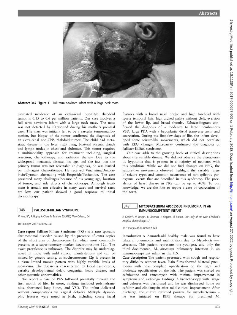
estimated incidence of an extra-renal non-CNS rhabdoidtumor is 0.15 to 0.6 per million patients. Our case involves afull term newborn infant with a large neck mass. The masswas not detected by ultrasound during his mother's prenatalcare. The mass was initially felt to be a vascular tumor/malfor-mation, but biopsy of the tumor confirmed the diagnosis ofan extra-renal non-CNS rhabdoid tumor. The child had meta-static disease in the liver, right lung, bilateral adrenal glandsand lymph nodes in chest and abdomen. This tumor requiresa multimodality approach for treatment including, surgicalresection, chemotherapy and radiation therapy. Due to thewidespread metastatic disease, his age, and the fact that theprimary tumor was not resectable at diagnosis, he was startedon multiagent chemotherapy. He received Vincristine/Doxoru-bicin/Cytoxan alternating with Etoposide/Ifosfamide. The casepresented many challenges because of his young age, locationof tumor, and side effects of chemotherapy. Although treat-ment is usually not effective in many cases and survival ratesare low, our patient showed a good response to initialchemotherapy.
348 PALLISTER-KILLIAN SYNDROME
M Knecht*, R Gupta, A Chau, M Marble. LSUHSC, New Orleans, LA
10.1136/jim-2017-000697.348
Case report Pallister-Killian Syndrome (PKS) is a rare sporadicchromosomal disorder caused by the presence of extra copiesof the short arm of chromosome 12, which most commonlypresents as a supernumerary marker isochromosome 12p. Theexact prevalence is unknown. The disorder may be underdiag-nosed in those with mild clinical manifestations and can bemissed by genetic testing, as isochromosome 12p is present ina tissue-limited mosaic pattern with highly variable levels ofmosaicism. The disease is characterized by facial dysmorphia,variable developmental delay, congenital heart disease, andother systemic abnormalities.
We report a case of PKS followed prenatally through thefirst month of life. In utero, findings included polyhydram-nios, shortened long bones, and VSD. The infant deliveredwithout complications via vaginal delivery. Multiple dysmor-phic features were noted at birth, including coarse facial
features with a broad nasal bridge and high forehead withsparse temporal hair, high arched palate without cleft, eversionof the lower lip, and broad thumbs. Echocardiogram con-firmed the diagnosis of a moderate to large membranousVSD, large PDA with a hypoplastic distal transverse arch, andcoarctation. During the first few days of life, the infant devel-oped some seizure-like movements, which did not correlatewith EEG changes. Microarray confirmed the diagnosis ofPallister-Killian syndrome.
Our case adds to the growing body of clinical descriptionsabout this variable disease. We did not observe the characteris-tic hypotonia that is present in a majority of neonates withthis condition. While we did not find changes on EEG, theseizure-like movements observed highlight the variable rangeof seizure types and common occurrence of non-epileptic par-oxysmal events that are described in this syndrome. The prev-alence of heart disease in PKS can be up to 40%. To ourknowledge, we are the first to report a case of coarctation ofthe aorta.
349 MYCOBACTERIUM ABSCESSUS PNEUMONIA IN ANIMMUNOCOMPETENT INFANT
A Kosier*, M Joseph, R Dickman, E Klepper, M Bolton. Our Lady of the Lake Children'sHospital, Baton Rouge, LA
10.1136/jim-2017-000697.349
Introduction A 2-month-old healthy male was found to havebilateral pneumonia and malnutrition due to Mycobacteriumabscessus. This patient represents the youngest, and only thethird documented, M. abscessus pulmonary infection in animmunocompetent infant in the U.S.Case description The patient presented with cough and respira-tory difficulty without fever. Plain films showed bilateral pneu-monia with near complete opacification on the right andmoderate opacification on the left. The patient was started onceftriaxone and vancomycin with minimal improvement insymptoms and radiologic findings. A bronchoscopy with lavageand cultures was performed and he was discharged home oncefdinir and clindamycin after mild clinical improvement. Afterdischarge, the culture returned positive for mycobacterium –
he was initiated on RIPE therapy for presumed M.
Abstract 347 Figure 1 Full term newborn infant with a large neck mass
Abstracts
J Investig Med 2018;66:351–640 493
on August 27, 2022 by guest. P
rotected by copyright.http://jim
.bmj.com
/J Investig M
ed: first published as 10.1136/jim-2017-000697.609 on 1 F
ebruary 2018. Dow
nloaded from

tuberculosis (MTB). Additional evaluation for MTB was unre-markable and culture growth obtained from both broncho-scopy samples and gastric aspirates was eventually identified asM. abscessus. Therapy was changed to amikacin, clarithromy-cin, and cefoxitin. A sweat chloride test was negative forcystic fibrosis and all further immunology workup to date hasbeen unremarkable. Since identification of M. abscessus andreceiving now seven months of various therapy, he has dem-onstrated slow improvement in respiratory symptoms andradiographic images though he continues to exhibitmalnutrition.Discussion This case represents an unusual presentation of afairly ubiquitous organism. Previous M. abscessus pulmonaryinfections were noted in immunocompromised children withcystic fibrosis or recipients of organ transplants. Multiple casesof M. abscessus causing osteomastoiditis and cutaneous infec-tions have been found in otherwise healthy children and typi-cally resolve without intervention. The diagnostic criteria ofnontuberculous mycobacterial (NTM) lung disease includeradiographs, at least three culture-positive specimens, andexclusion of other disorders. The two aforementioned M.abscessus cases in the literature presented similarly with a pul-monary infection found on imaging and with positive cultures.Our patient is unique given the young age of presentationand no diagnosed immunodeficiency or underlying disease.
350 ALL HYPOXEMIA IS NOT CREATED EQUAL
EA Kubota-Mishra*, AV Agwu, MS Evangelista. McGovern Medical School, Houston, TX
10.1136/jim-2017-000697.350
Case report A 3 year old female with cerebral palsy and seiz-ure disorder was admitted to general pediatrics for post-opera-tive management after correction of bilateral hip dysplasia.Her immediate post-op course was complicated by respiratorydepression secondary to narcotic use for pain control. Onpost-operative day 2 she had poor oral intake. On exam,white plaques were seen on the posterior pharyngeal mucosa.To improve oral intake from presumed pain from the lesions,throat sprays and magic mouthwash were ordered for sympto-matic relief.
A few hours later, she developed acute desaturation to themid-80s. On examination, she looked uncomfortable, moaningand crying. She had good air entry on lung auscultation. Totreat the hypoxemia, respiratory support was initiated. Addi-tional work-up performed included an unremarkable chest x-ray and a venous blood gas with a pH of 7.48, pCO2 of 39mmHg and PaO2of 98 mmHg. Despite escalation of supportfrom nasal cannula to high flow to BIPAP with 100% FiO2,her oxygen saturation did not improve. Due to refractoryhypoxemia, she was transferred to the pediatric intensive careunit and an arterial blood gas obtained with a PaO2 of 372mmHg and pulse oximetry reading of 75%. Because of thediscordance in her condition, cooximetry testing was per-formed. Her methemoglobin level was immeasurably high.Due to the rapidity of symptom development, methemoglobi-nemia was likely iatrogenic. Review of medications adminis-tered prior to the event showed recent use of a benzocaine-based throat spray. Methylene blue was administered andimproved oxygen saturation.Discussion Acute development of hypoxemia requires a clini-cian’s high index of suspicion to determine its cause.
Iatrogenic causes should be investigated thoroughly, especiallyif cardiac or respiratory causes are unlikely. The absence ofcyanosis and continued desaturation that is discordant with ahigh PaO2 on arterial blood gas warrants cooximetry testing.Benzocaine, a common anesthetic, is available in different for-mulations to treat a variety of ailments. Development ofmethemoglobinemia after use of benzocaine containing prod-ucts has been described in the literature. In recent years, theFood and Drug Administration has released a warning againstuse in pediatric population particularly those who are <2years of age.
351 A SEVERE CASE OF NEONATAL COW'S MILK PROTEINALLERGY MIMICKING SEPSIS
KM Kuehn*, TO Findley. McGovern Medical School at UT Health, Houston, TX
10.1136/jim-2017-000697.351
Case report Cow’s milk protein allergy is a clinical diagnosiswith variable manifestations. In infants, this allergy typicallypresents through a non-IgE mediated mechanism. One of themost severe forms of this is Food Protein Induced Enterocoli-tis (FPIES) which can cause vomiting and diarrhea resulting insignificant dehydration. Abnormal laboratory findings mayinclude metabolic acidosis, leukocytosis, and neutrophilia, aswell as elevation in the inflammatory marker, C-reactive pro-tein (CRP). Due to the clinical and laboratory manifestationsof this disease, it may be initially misdiagnosed as sepsisresulting in delayed treatment and further exacerbation ofsymptoms. Furthermore, there is no diagnostic test for FPIESso diagnosis is based on improvement of symptoms afterremoval of the offending agent. In this report, we describethe case of a 25 day old Chinese male infant who was trans-ferred from an outside facility for persistent leukocytosis, ban-demia, metabolic acidosis, and a marked rise in CRP despitetreatment with broad spectrum antibiotics. He also was notedto have copious watery stools and failure to thrive with aweight 150 grams (4.7%) below his birth weight. The patientshowed dramatic improvement following cessation of cow’smilk formula and transition to an elemental formula. Whilethere have been reported cases describing an elevated CRP ininfants with this disease, our case is unique in the degree ofCRP elevation (130 mg/L) in an afebrile neonate. Moreover,no cases have described bandemia from FPIES. These twomarkers are often used as indicators of underlying infectionand can delay diagnosis and treatment of FPIES. This casehighlights the importance of including FPIES in the differentialwhen presented with a critically ill infant with clinical featuresof sepsis, diarrhea, and failure to thrive.
352 AUTOIMMUNE ENCEPHALITIS: WHERE CLINICALPRESENTATION AND LABORATORY RESULTS MAYDISAGREE
S Liu*, D Chrusciel, D Hahn. University of Oklahoma, Oklahoma City, OK
10.1136/jim-2017-000697.352
Case report In patients presenting to the general pediatricianwith new onset neuropsychiatric symptoms, autoimmune ence-phalitis or viral encephalitis is commonly on the diagnosticdifferential. Although both can have corresponding laboratory
Abstracts
494 J Investig Med 2018;66:351–640
on August 27, 2022 by guest. P
rotected by copyright.http://jim
.bmj.com
/J Investig M
ed: first published as 10.1136/jim-2017-000697.609 on 1 F
ebruary 2018. Dow
nloaded from

results that support the diagnosis, negative lab results do notrule out either condition. We present a previously healthy 5-year-old male with new onset seizures and no prodromal viralsymptoms. A few days prior to presentation, he developedneuropsychiatric symptoms. On the day of admission, hedeveloped fatigue, drooling, gasping, and flexion of his neckto the right. He was not responsive to stimulation. He wastaken to an emergency room where he developed arm and legshaking. Head computerized tomography was unremarkableand he was transferred to our hospital for further care. Whilehere, he was transferred between the pediatric intensive careunit and general pediatric ward multiple times. Eventually, hisseizures were well-controlled with anti-epileptics, but signifi-cant clinical improvement occurred only after starting steroids.He continued to have erratic behaviors, including enuresis,hyperactivity, inability to follow directions, confusion, andemotional lability. Testing for multiple viruses, anti-N-methyl-D-aspartate receptor, and other antibodies were negative. Con-tinuous video electroencephalogram captured complex partialseizures with secondary generalization. Magentic resonanceimaging of the brain was normal. Due to clinical course, lackof infection, and rapid response to steroids, he was diagnosedwith autoimmune encephalitis.
Autoimmune encephalitis is an uncommon, but significantdiagnosis. Although many pediatric cases of autoimmune ence-phalitis are seropositive, autoimmune encephalitis is an ever-evolving field, and negative laboratory studies are not unex-pected. Although initial lab results and symptoms may be con-fused with viral encephalitis, with autoimmune encephalitis,delayed therapy may lead to a decreased response. This casehighlights the importance of clinically diagnosing autoimmuneencephalitis and administering prompt and appropriate therapyfor best outcomes.
353 INSPISSATED BILE SYNDROME CAUSING SEVERECHOLESTASIS IN A NEWBORN
1RM Turcu, 2AN Lyle*. 1University of Louisville, Louisville, KY; 2Indiana University,Indianapolis, IN
10.1136/jim-2017-000697.353
Case report 2-day old AGA term male infant presented withlethargy, poor feeding, and generalized jaundice since birth.He had passed one meconium stool. Infant was born at homevia SVD to a healthy Amish mother, after a normal pregnancy.Maternal blood type AB neg, prenatal labs not available.Mother is G4P3 (1 miscarriage, 1 stillbirth, 2 healthy termpregnancies. Rhogam was delayed after the miscarriage, butgiven appropriately after the other pregnancies. Father and 2yo brother are healthy. Initial workup (table): severe conju-gated hyperbilirubinemia, mild liver dysfunction, anemia withreticulocytosis (Rh incompatibility – infant’s blood type B+, 4+ DAT). Blood and CSF culture negative. Urine culture: 10–100 k E. coli CFUs. Test for genes ABCB11, ABCB4, ATP8B1,JAG1, and TJP2 neg. TSH, FT4, metabolic newborn screennormal, bile acids in normal ranges, TORCH neg. Abdominal
ultrasound: normal-appearing liver and gallbladder, no biliaryductal dilation, patent vessels. CXR, Echo unremarkable.Treatment: phototherapy, double volume exchange transfusion,IVIG, PRBC, phenobarbital, antibiotics for UTI.
Graph: bilirubin decline over time. At discharge all lab val-ues improved. Family declined HIDA scan (normal US,decreasing bilirubin) and brain MRI (normalized neuro exam).Evaluated in the developmental follow up clinic at 2.5months: appropriate growth, normal PE (incl neurological anddevelopment eval) and bilirubin levels.Discussion This is a full term neonate with hemolytic anemia(Rh incompatibility) and severe conjugated hyperbilirubinemia.Most common cholestasis causes were excluded. E. coli UTIcould have been a contributor, but unlikely the main cause.Exclusion diagnosis: Rh incompatibility with severe chronichemolysis, complicated by inspissated bile syndrome. Familydeclined liver biopsy (rapid resolution of the hyperbilirubine-mia and normal hepatic ultrasound).
354 A RARE CASE OF HEMPHAGOCYTICLYMPHOHISTIOCYTOSIS: OVERCOMING FATALOUTCOMES
A Madani*. Louisiana State University, Shreveport, Bossier City, LA
10.1136/jim-2017-000697.354
Case report Patient was a 3 month male infant with a previ-ously unremarkable past medical and family history whodeveloped a fever associated with unilateral cervical lympha-denopathy treated with Clindamycin by his primary care pro-vider. When symptoms persisted, he was referred for surgicalevaluation of neck mass and later admitted to the hospitalwhen routine labs revealed pancytopenia and elevated liverenzymes of unkown etiology. Over the next several days, hequickly deteriorated, requiring mechanical ventilation, pressorsand blood products before he was transferred to the PediaticIntensive Care Unit at our hospital on Febuary 4, 2017. Onarrival, there was high suspicion for Hemophagocytic Lympho-histiocytosis (HLH), a rare but fatal disease characterized byuncontrolled hemophagocytosis and activation of inflammatorycytokines that lead to infitration of multiple organs. The dis-ease is divided into primary (hereditary) and secondary(acquired) with the latter being triggered by infection or non-infectious causes (such as drugs, autoimmune disease or immu-nodeficiency states). Diagnosis of HLH is determined byeither having a molecular diagnosis consistent with knownmutations or second, fulfilling 5 of the 8 signs or symptoms:
. fever,
. splenomegaly,
. cytopenia,
. hypertriglyceridemia and/or hyofibrinogenemia (<150 mg/dl),
. hemophagocytosis in a lymphatic tissue excluding malignancy,Abstract 353 Table 1
Abstract 353 Figure 1 Bilirubin decline over time
Abstracts
J Investig Med 2018;66:351–640 495
on August 27, 2022 by guest. P
rotected by copyright.http://jim
.bmj.com
/J Investig M
ed: first published as 10.1136/jim-2017-000697.609 on 1 F
ebruary 2018. Dow
nloaded from

. low or absent natural killer cell cytotoxicity,
. hyperferritinemia, and
. Elevated soluble CD25 levels.
On admission, patient met criteria having 5 of the 8 find-ings but with a lack of family history or preceding trigger, itwas unclear if it was primary or secondary which has implica-tions in treatment. Nevertheless, prognosis is poor. and oftenfatal. Chemotherapy with Etoposide with Decadon was startedon admission along with broad spectrum antibiotics and sev-eral days of plasmapheresis and IVIG. Patient was in remissionby week 2 of treatment and discharged one month later sta-ble. Bone marrow aspirate and genetic testing later proved tobe primary HLH with no identifiable agent identified as atrigger as EBV, CMV and Herpes PCR was negative. He iscurrently awaiting bone marrow transplantation.
355 CONSUMED BY FEVER: A CASE OF PHAGOCYTOSISGONE WRONG
MJ Magee*, K Schneider. University of Mississippi Medical Center, Jackson, MS
10.1136/jim-2017-000697.355
Case report Hemophagocytic lymphohistiocytosis (HLH) is lifethreatening disease caused by excessive immune activation.Diagnosis can be difficult and failure to diagnose early in thedisease process can impede therapies and increase overall mor-tality. Here we present an interesting case of HLH in a 3-year-old presenting with fever of unknown origin.
A previously healthy 3-year-old white male presented toour Pediatric Emergency Department with a chief complaintof 4 weeks of daily spiking fevers (up to 104°). The feverswere accompanied by an erythematous maculopapular rash,daily vomiting, diarrhea, and occasional complaints of jointpain. On presentation, exam was notable for cervical andinguinal lymphadenopathy, hepatomegaly, and scattered macu-lopapular rash. Significant labs included anemia, leukocytosis,elevated acute phase reactants, and elevated LDH. Initial dif-ferential of infectious, neoplastic, rheumatologic, and inflam-matory processes required multiple subspecialty consultation.Ferritin levels were markedly elevated (10420) pointing to apossible diagnosis of HLH with concern for underlying malig-nancy. CT imaging showed hepatosplenomegaly and diffuselymphadenopathy throughout the chest, abdomen, and pelvisconcerning for lymphoma. Lymph node and bone marrowbiopsies were both consistent with hemophagocytosis with noevidence of malignancy. These findings along with fever,hyperferritinemia, splenomegaly, and hypertriglyceridemia satis-fied the diagnostic criteria for HLH. Treatment with steroidsand Etoposide led to resolution in fevers. He was assumed tohave secondary HLH as genetic sequencing for familial HLHwas negative though underlying cause is still unknown.
HLH is a reactive process characterized by systemic inflam-mation due to dysregulation of normal immunological path-ways. The predominant clinical features of fever, cytopenias,hepatitis, and splenomegaly reflect the immune disruption, butmany conditions can lead to this same clinical picture. Feverof unknown origin carries a broad and non-specific differentialthat encompasses numerous possible etiologies making it diffi-cult to isolate a specific diagnosis. While HLH may not behigh on the differential for fever of unknown origin, it shouldbe considered a possibility due to its mortality risk and needfor immediate therapy.
356 A NOT SO SIMPLE CASE OF DEHYDRATION
C Marbrey*, MN Frascogna, T Walker. University of Mississippi Medical Center, Jackson, MS
10.1136/jim-2017-000697.356
Case report As the only Children’s Hospital in the state, ourinstitution routinely receives transfers from both local and dis-tant locations within the region. History and clinical contextare often limited by the absence of caregivers or the emergentneed for care. We present a case involving the transport of achild to the pediatric emergency room for presumeddehydration.
An ambulance arrived to the Pediatric ER following theuneventful transport of a 14 year old male after football prac-tice for dehydration. He was unloaded from the ambulanceand on transfer to a treatment room, he suddenly becameunresponsive and, after a few agonal breaths, became apneicand pulseless. CPR was initiated immediately. Defibrillatorpads were placed and showed asystole. He received 3 roundsof epinephrine with return of spontaneous circulation after 6minutes. The patient was intubated and he was started on acontinuous epinephrine infusion. Initial venous blood gasrevealed a pH 6.94, ionized calcium (iCa) 0.46, potassium (K)6.6, and lactate 12.1. He received boluses of calcium gluco-nate, sodium bicarbonate, and a normal saline bolus. Foleycatheter placement produced a large volume of red coloredurine. An insulin/dextrose infusion was initiated for hyperkale-mia and he was admitted to the PICU for furthermanagement.
On arrival to the PICU, notably, his creatine kinase (CK)was >100,000, iCa 0.32, K 7.3, creatinine 3.58, lactate 7.7,and phosphorus 22.2. Dialysis was considered but not initi-ated. His CK remained >100,000 for 5 days and his electro-lytes gradually corrected. His PICU course was complicated byrecurrent episodes of ventricular tachycardia which eventuallyresolved. He was extubated, his CK was down trending andhe was ultimately discharged home with a diagnosis ofrhabdomyolysis.
Further history revealed that the patient was at football try-outs and had not yet participated in any football or warm-upactivity that day, making the diagnosis of rhabdomyolysis inthe setting of strenuous activity much less likely. Cardiacarrest in the setting of profound non-traumatic rhabdomyolysiswarranted additional genetic and rheumatologic work upwhich is currently pending.
357 BRONCHIAL CAST WITH BRONCHIECTASIS
L McKinney*, S Gonzalez, E Klepper, A Carrion. Our Lady of the Lake, Baton Rouge, LA
10.1136/jim-2017-000697.357
Introduction Bronchiectasis is defined as a permanent dilationof bronchi secondary to structural damage to the bronchialwall. Severe bacterial or viral pneumonia accounts for themajority of bronchiectasis in the post-infectious period of thepediatric population. We present a case of a 4-year-old femalewho developed bronchiectasis secondary to an endobronchialcast formed by a pneumonic process.Case description A previously healthy 4-year-old female pre-sented to the pediatrician’s office with 3 days of fever, cough,and tachypnea. Chest x-ray (CXR) showed left lower lobe(LLL) pneumonia. The patient was given ceftriaxone and
Abstracts
496 J Investig Med 2018;66:351–640
on August 27, 2022 by guest. P
rotected by copyright.http://jim
.bmj.com
/J Investig M
ed: first published as 10.1136/jim-2017-000697.609 on 1 F
ebruary 2018. Dow
nloaded from

azithromycin. Two days after, the patient’s repeat CXRshowed persistent LLL infiltrate, and she was given anotherdose of ceftriaxone and placed on high-dose amoxicillin afterconsultation with pulmonology. In pulmonary clinic, clinicalimprovement was noticed, but the six-week follow-up CXRshowed persistent infiltrate with improvement. Spirometryshowed possible small airway obstruction, and an inhaled cor-ticosteroid was started. One month later CXR still showedpersistent infiltrate. A chest computed tomography showed amass effect to segmental bronchi on LLL with tubular bron-chiectasis and atelectasis. Flexible bronchoscopy revealed a fri-able endobronchial tissue in the LLL resembling a cast thatwas removed with rigid bronchoscopy. Pathology showedCharcot-Leiden crystals and mixed inflammatory debris includ-ing eosinophils. The patient was continued on inhaled cortico-steroids and started on amoxicillin-clavulanate, systemic steroidcourse, and chest percussion therapy. A repeat CXR noted res-olution of the LLL infiltrate.Discussion This case demonstrates the development of post-infectious bronchiectasis in association with a bronchial cast.Most patients with bronchiectasis have chronic conditions suchas cystic fibrosis, recurrent pneumonias, or chronic rhinosinusi-tis that can lead to lung damage which then leads to bron-chiectasis. However, bronchiectasis can be seen after a singlecase of severe pneumonia. Bronchial casts typically occur inpost-Fontan patients; however, our patient did not have anypredisposing factors for the development of bronchial casts.Nevertheless, post-obstructive pneumonia could have causedbronchiectasis and promoted the formation of a bronchialcast.
358 MOLLARET'S MENINGITIS DUE TO HERPES SIMPLEX 2STARTING IN THE NEONATAL AGE GROUP
M Munir*, M Rathore. University of Florida, Jacksonville, FL
10.1136/jim-2017-000697.358
Introduction Mollaret’s meningitis is a rare condition that ischaracterized by multiple recurrent but benign episodes ofmeningitis presenting with or without fever. It is usually con-sidered a disease of adults however, it has also been reportedin children.
We report the first case of Mollaret's meningitis withoutany encephalitis, caused by HSV-2 which started in the neona-tal period.Case report An 11 year-old previously healthy male presentedwith 2 days history of fever and signs of meningism.
He had a significant past medical history of HSV disease.He was first admitted at the age of 9 days with fussiness andfever and was diagnosed with HSV-2 meningitis based onHSV-2 PCR on the CSF. An MRI of the brain with contrastwas normal. He was treated with IV acyclovir and then dis-charged on oral acyclovir prophylaxis. An audiology screenwas normal.
Nine days after discharge, patient was again admitted withone day history of fever, fussiness, vomiting and diarrhea.HSV-2 PCR was again positive in the CSF. Brain MRI withand without contrast was normal. He was again treated with21 days of IV acyclovir and HSV-2 PCR was negative in thenear treatment LP. Patient was discharged on oral acyclovir.
At the time of the current admission patient was ill appear-ing with meningismus. A CT scan without contrast was
normal. CSF PCR was positive for HSV-2. An MRI with con-trast was also normal.Discussion Mollaret’s meningitis is different than the usualrecurrent HSV meningoencephalitis in any age group, which isserious and often associated with neurological complications.These episodes can occur with weeks to years asymptomaticintervals between episodes. Our patient had recurrent HSVmeningitis which started as a neonate, his presentations wereconsistent with Mollaret’s meningitis and outcomes werebenign.
Differentiation between the classic neurological neonatalHSV and first episode of Mollaret’s meningitis is impossible.A benign course of neurological HSV infection should nottemper with aggressiveness of treatment, however a possibilityof Mollaret’s meningitis should be entertained.Future questions Recurrent HSV-1 meningitis because of immu-nological deficiencies have been reported but not for HSV-2.
359 INTESTINAL MALROTATION IN A 4 WEEK OLD MALE: ACASE REPORT AND REVIEW OF THE LITERATURE
DK Nguyen*, B Sessions, J Deitrick , A Olanrewaju , J Meller . Texas Tech Health ScienceCenter Medical School, Amarillo, TX
10.1136/jim-2017-000697.359
Case report Intestinal malrotation is a congenital abnormalityof the gut which can present as an asymptomatic incidentalfinding or a life-threatening emergency. Most cases present ininfancy once the malrotation has resulted in volvulus (twistingof the small intestine on the superior mesenteric artery). Thisleads to intestinal obstruction and requires emergency surgicalintervention. However, some cases of malrotation are mostlyasymptomatic and only detected incidentally following gastro-intestinal imaging for other reasons. We present a case of a 4week old male who presented with intermittent nonbiliousvomiting along with failure to thrive and no stools for 1week. While this patient was diagnosed with gastroesophagealreflux (GER), an upper GI series revealed incidental malrota-tion. After adequate treatment for GER and subsequentimprovement of symptoms, the surgeon still proceeded to cor-rect the malrotation with the Ladd procedure. We use thiscase to discuss the best treatment for asymptomatic malrota-tion, which is currently still widely debated among physicians.
Abstract 359 Figure 1 Upper GI series shows abnormal verticalduodenojejunal portion
Abstracts
J Investig Med 2018;66:351–640 497
on August 27, 2022 by guest. P
rotected by copyright.http://jim
.bmj.com
/J Investig M
ed: first published as 10.1136/jim-2017-000697.609 on 1 F
ebruary 2018. Dow
nloaded from

360 CROUZON SYNDROME: A CLASSIC CASE AND A LOOKINTO WHAT IS AHEAD
A Nuttli *, J Gallois , C Mumphrey , J Surcouf . LSUHSC, New Orleans, LA
10.1136/jim-2017-000697.360
Case report Crouzon Syndrome is an autosomal dominant dis-order that results from a mutation of the fibroblast growthfactor receptor-2 (FGFR-2) gene causing the classic features ofthe disease.
Our patient was born to a 19 yo G1 mother at 34 4/7WGA and was noted to have dysmorphic features at birthincluding, turricephaly, proptosis, elevated palatal arch, andlow set ears consistent with a craniosynostosis syndrome.Maternal and pregnancy history including prenatal ultrasoundscreening were unremarkable, and the mother receivedadequate prenatal care. The patient was transferred to ourcenter for multidisciplinary care. 3-D CT imaging revealed clo-sure of coronal, sagittal, and lambdoid sutures as well as max-illary and ethmoid sinus hypoplasia. These findings along withthe absence of long bone abnormalities in our patient aremost consistent with Crouzon Syndrome. Without diseasereported in either the mother or father of this infant, a denovo mutation was presumed. Other significant findingsincluded PDA, ASD, VSD, and bilateral conductive hearingloss. Our patient required tracheostomy placement due to theseverity of her midface hypoplasia. Additionally, due to therestrictive shape of the skull, the patient was monitored forhydrocephalus prior to discharge. She is currently scheduledfor shunt placement due to progressive hydrocephalus on fol-low up. Management of her Crouzon Syndrome involved avariety of subspecialists including genetics, plastic surgery, neu-rosurgery, ENT, cardiology, and audiology. A team basedapproach with numerous multidisciplinary family meetings wasnecessary to coordinate care. Long term treatment currentlyinvolves staged craniofacial surgeries. However, new researchis looking into attenuating the FGFR cell signaling pathwaysat the level of coronal sutures to alter osteoblast activity andmaintain appropriate suture patency either prenatally or inconjunction with corrective surgeries.
This case highlights the importance of a multidisciplinaryapproach to the management and treatment of Crouzon Syn-drome and explores the opportunity for future research intotargeting cell signaling pathways as a component of treatment.
361 FOURTEEN YEAR OLD TRACHEOSTOMY DEPENDENTSECONARY TO SUBGLOTTIC STENOSIS
J Oakley*, D Lozano. University of Alabama at Birmingham, Homewood, AL
10.1136/jim-2017-000697.361
Introduction Respiratory failure associated with stridor suggestsobstruction or narrowed airway and differential for underlyingcause of stridor can be broad.Case presentation The patient is a fourteen year old Caucasianmale with hearing loss, vocal cord dysfunction and tracheos-tomy dependent due to severe glottic subglottic stenosis (SGS)who presents for evaluation for weight loss, dyspnea and jointswelling. Emergent tracheostomy was performed one yearprior for respiratory failure due to SGS just below the vocalcords preventing intubation. Patient had multiple hospital staysprior for stridor and respiratory distress. Initial direct
laryngoscopy with bronchoscopy (DLB) during episode of stri-dor showed only a hypoplastic right true vocal cord that didhave normal movement followed by repeat DLB a month laterfor recurrent stridor without concern for worsening subglotticstenosis, diagnosed with vocal cord dysfunction. Admissionlabs notable for CRP 16 mg/dL, ESR 110 mm/hr and micro-cytic anemia; diagnosed with relapsing polychondritis as evi-denced by nasal and auricular chondritis (resulting in saddle-nose and cauliflower ear), subglottic stenosis, arthralgia, andextreme weight loss secondary to chronic, high-levels ofinflammation. On DLB, our patient was diagnosed with grade3 subglottic stenosis as well as malformed glottic anatomywith no discernible vocal cords.Discussion Relapsing polychondritis is a rare autoimmune dis-order of unknown etiology characterized by recurrent inflam-mation and destruction of cartilaginous structures, mostcommonly the upper respiratory tract, ears, nose, and joints.Proposed pathogenesis includes antibodies to type II, IX andXI collagen and matrillin-1. Pulmonary symptoms are presentin 20–50% of all patients and can include inflammatorychanges anywhere from the subglottis, trachea, including theposterior membranous wall, and bronchi; lung interstitium andpulmonary vasculature are not affected. Presenting symptomsof airway involvement can include dyspnea, cough, hoarsenessand stridor. Prior studies have also shown saddle nose deform-ity to correlate with pulmonary symptoms. Differential forglottic subglottic stenosis in children is broad but not limitedto traumatic, infectious, congenital or autoimmune.
362 EXTRAMEDULLARY RELAPSE OF CD19+ ALL DURINGBLINATUMOMAB THERAPY
J Onarecker*, D Crawford, Z Yu. University of Oklahoma Health Sciences Center, OklahomaCity, OK
10.1136/jim-2017-000697.362
Introduction In pediatric patients with ALL, about 90% willachieve remission. However, those that relapse often have adifficult course and many do not survive. Blinatumomab isone of a new category of immune-based therapies developedto treat relapsed or refractory pre-B cell ALL. It is a bispecificantibody that acts as a T-cell engager binding the CD3 antigenof T-cells with the CD19 antigen of leukemic cells. Whilemany patients are able to achieve a second remission with Bli-natumomab, many of those have a subsequent relapse unlessthey undergo allogeneic stem cell transplant. Most relapsesoccur as hematologic relapses in the bone marrow or lesscommonly as extramedullary relapses.Case presentation Our patient is a 14 year old boy who com-pleted therapy on a high risk COG protocol and achievedremission. He had a CNS and bone marrow relapse at 5months off therapy. A second remission was quickly achieved,but he relapsed again within a few months. He was treatedwith Blinatumomab with rapid clearance of blasts from hismarrow. However, within 3 weeks of initiation of Blinatumo-mab, he developed gingival and scalp lesions. Biopsy con-firmed the lesions to be CD19+ pre-B ALL with the sameimmunophenotype as the original leukemia. He was thentreated unsuccessfully with an additional salvage regimen. Hisdisease progressed and his family requested hospice care.Conclusions Blinatumomab is a promising option for salvageof relapsed ALL patients, but its efficacy is limited by a high
Abstracts
498 J Investig Med 2018;66:351–640
on August 27, 2022 by guest. P
rotected by copyright.http://jim
.bmj.com
/J Investig M
ed: first published as 10.1136/jim-2017-000697.609 on 1 F
ebruary 2018. Dow
nloaded from

rate of relapses. Many of these occur because of loss ofexpression of CD19, allowing the leukemia cells to evadetherapy. In other cases, extramedullary relapses occur, oftenwith cells that still express CD19. This suggests that someextramedullary sites are immunologically privileged and do notreceive the optimal benefit from anti-CD19 immunotherapy.This finding reinforces that Blinatumomab should generally beused as a bridge to further therapy, preferably to allogeneicstem cell transplant, rather than as definitive treatment.
363 RHEUMATIC HEART DISEASE IN A 6 YEAR OLD
V Pillay*. Louisana State University Health Science Center Shreveport, Shreveport, LA
10.1136/jim-2017-000697.363
Case report 6 yo AAF with no PMH was seen in the ED forfever. Associated symptoms included headache and refusal toswallow. Physical exam only revealed mild pharyngeal eryth-ema. A grade 3/6 systolic murmur was auscultated that hadnot previously been documented. A rapid strep test was -ve.Reflex throat culture was also -ve. Patient was diagnosed withviral pharyngitis and discharged home. Clinic follow up wasscheduled to address her murmur however she was lost to fol-low up for 2 months. When she returned to clinic she hadincreased fatigue, decreased activity, and intermittent fever.Heart murmur was still present. Echo was obtained and wassignificant for severe mitral valve regurgitation, moderate tosevere aortic insufficiency with nodular appearance of bothvalves, moderate dilation of the left ventricle with normal sys-tolic function. She was admitted to hospital. Physical examshowed a well appearing child in no distress. Mucus mem-branes were moist, with normal tonsils and posterior pharynx.No lymphadenopathy. No arthropathy. Her skin was negativefor rashes, nodules, splinter hemorrhages, janeway lesions orosler nodes. Cardiac exam was significant for 2/4 diastolicmurmur loudest at the mid sternal border, 3/6 holosystolicmurmur at the apex, a slightly displaced PMI to the left, and3+ bounding pulses in all extremities. Patient was started onprednisone and high dose aspirin to resolve any potentialinflammation from a possible rheumatic fever. Lisinopril wasadded to reduce afterload. Blood cultures were drawn at 0, 6,and 12 hrs. Vancomycin and Gentamicin were started afterthe 3rd blood culture. Labs were significant for a -ve ANAand elevated DNase B ab and ASO Titer (548 & 401 respec-tively). The presumed diagnosis was rheumatic heart disease.
Patient remained afebrile with no symptoms of cardiac dys-function and no change in clinical status. Vancomycin andGentamicin were discontinued 72 hours after -ve cultures.Cultures were grown for 21 days to rule out HACEK organ-isms as a cause of subacute endocarditis. She was started onPenicillin therapy until age 40, and likely to be due toincreased risk of recurrent attacks of rheumatic fever. RepeatECHO at discharge showed trivial improvement in mitralregurgitation and minimal improvement in aortic regurgitation.She will likely need valve replacement in the future.
364 STOP THE CLOT: PREVENTING POSTOPERATIVE VENOUSTHROMBOEMBOLISM IN HOSPITALIZED CHILDREN
1M Rabalais*, 2A Byrd, 2MH Roy, 2E Klepper. 1Louisiana State University Health SciencesCenter, Baton Rouge, LA; 2Our Lady of the Lake Children's Hospital, Baton Rouge, LA
10.1136/jim-2017-000697.364
Case report Hospital-associated venous thromboembolism(VTE) is now the second most common serious hospital-acquired condition in children’s hospitals. Although the inci-dence of VTE in hospitalized adults is considerably higherthan in children (0.2% to 0.6% of hospitalizations), the inci-dence in children has significantly increased recently. Adoles-cents are at higher risk of VTE due to higher rates of obesity,oral contraceptive use and long bone fractures. We present anadolescent with no previous risk factors who developed VTEfollowing a long bone fracture.
A 14 year old male presented to ED with a traumatic spi-ral fracture of the left femoral shaft. Post-operatively, thepatient was placed on enoxaparin, diazepam, and pain medica-tion. Physical therapy initiated early ambulation, and heachieved adequate pain control. Enoxaparin was discontinuedprior to discharge, and he was sent home with aspirin, oxyco-done and diazepam. His pain was initially well-controlledallowing continuation of physical therapy. However, on POD6he grew increasingly weak, pale and refused to ambulate. Hepresented to the ED febrile, tachycardic and severely anemic.CT showed a large hematoma encircling the femoral shaft,and US showed a DVT in the left distal femoral vein. Enoxa-parin was restarted, and the patient was admitted for VTEmanagement.
As the incidence of pediatric hospital-associated VTE con-tinues to rise, further effort is needed to determine the bestway to prevent and treat VTE. There is no current consensuson evidence based guidelines for VTE prophylaxis in children.Using a recently published risk assessment algorithm, ourpatient did not meet criteria for pharmacologic prophylaxis,which likely would have prevented his VTE. Our case high-lights the opportunity for the pediatric community to developmore effective risk stratification and prophylaxis algorithmsfor hospitalized children. Pediatricians need to understand therole of mechanical and pharmacologic prophylaxis and addressthe importance of early ambulation and pain control. Withmore research we can improve the prevention of this increas-ing hospital acquired condition that results in significant harmand expense.
365 THE DIFFICULT AIRWAY: A CASE REPORT IN ANEONATE
P Redmond*, C Faulk, MN Frascogna. University of Mississippi Medical Center, Jackson, MS
10.1136/jim-2017-000697.365
Case report The difficult airway is a discovery no providerwishes to make in the emergency department (ED). Here wepresent a case of a patient who presented with a markedlydifficult airway due to an unlikely cause requiring intubationattempts by multiple providers.
Abstracts
J Investig Med 2018;66:351–640 499
on August 27, 2022 by guest. P
rotected by copyright.http://jim
.bmj.com
/J Investig M
ed: first published as 10.1136/jim-2017-000697.609 on 1 F
ebruary 2018. Dow
nloaded from

A three-week-old presented as a transfer from a tertiarycare facility with respiratory distress. She had presented toour ED four days earlier at 15 days of age with chief com-plaint of wheezing and poor feeding. Radiographs and cultureswere obtained, but mother refused additional labwork. Shewas well appearing at the time and was discharged home. Thechild subsequently developed neck swelling at home and fourdays later was brought by EMS to an outside facility for stri-dor and respiratory distress. Attempts at intubation failed andthe patient was transferred by Pediatric Air Transport to ourED. In our unit the patient was lethargic and in respiratoryfailure. With ED staff, anesthesia, and otolaryngology at thebedside, repeat intubation attempts were made by employingthe use of a video laryngoscope without success. Eventuallydirect laryngoscopy was undertaken resulting in rupture of alarge submental mass and copious purulent drainage. Follow-ing suctioning and placement of a laryngeal mask airway, anendotracheal tube was successfully placed, the patient stabi-lized, and was admitted to the ICU.
Further work-up was notable for a significant leukocytosisand bandemia, while imaging revealed a pocket of fluid andair in the posterior floor of the mouth and widespread softtissue edema. These findings were consistent with a rupturedabscess due to an infected thyroglossal duct cyst. The patientwas placed on antibiotics and recovered completely withoutcomplications to date. Though rare in newborns, thyroglossalduct cysts are epithelial remnants of embryonic development,and should be considered when evaluating a newborn withstridor and respiratory distress.
366 SEVERE METABOLIC ACIDOSIS IN THE SETTING OFHETEROZYGOUS 3-OXOACID COA-TRANSFERASE 1GENE MUTATION
PA Ruhlmann*, D Hahn. University of Oklahoma Health Sciences Center, OKC, OK
10.1136/jim-2017-000697.366
Case report A previously healthy 4 year old male with milddevelopmental delay presented to The Children’s Hospital ofOklahoma (TCH) as a transfer from a small rural hospital forsevere metabolic acidosis and lethargy. He was born at term,fully immunized, with no previous hospitalizations, allergies,or significant family history. After two days of diarrhea,decreased appetite, and emesis, he developed labored tachyp-nea and was taken to his local emergency room. Labs thererevealed only a bicarbonate level <5. After transfer to TCHand receiving a total of 100 cc/kg normal saline, the patientremained acidotic with pH of 7.13 and serum bicarbonate of6. However, on exam the patient was well-appearing, sleepingbut easily awakened, and breathing comfortably 20 times perminute, despite the marked acidosis. Extensive workup ensued.Serum ketones (beta-hydroxybutyrate) were elevated to 8.46(normal 0.02–0.27) and urine analysis had 2+ ketones. Theacidosis ultimately corrected after 24 hours of bicarbonatecontaining fluids, with no further episodes. He was dischargedto follow-up with genetics, and after more specific genetictesting, he was found to have a previously unreported altera-tion in the 3-oxoacid CoA-transferase 1 gene (OXCT1), con-sistent with carrier status of succinyl-CoA-3-oxaloacid CoAtransferase (SCOT) deficiency.
OXCT1 is located on chromosome 5p13.1 and encodes themitochondrial SCOT enzyme, which catalyzes the first step of
ketolysis for utilization of ketone bodies as an energy sourcewhen blood glucose is low. OXCT1 mutations result in SCOTdeficiency, an autosomal recessive disorder that results in theaccumulation of unused ketone bodies. SCOT deficiency oftenpresents in the neonatal period with recurrent episodes ofketoacidosis (vomiting, lethargy, tachypnea, and unconscious-ness). Patients typically are asymptomatic between episodes,but often have permanent ketosis and ketonuria.
SCOT deficiency has only been documented in a very smallnumber of patients. While SCOT deficiency is generallyregarded as a mostly autosomal recessive inheritance pattern, a2017 study revealed profound metabolic acidosis in a verysmall subset of heterozygote patients with OXCT1 mutations,similar to our patient.
367 PATIENT WITH PROTEUS SYNDROME AND PARA-TESTICULAR OVARIAN-TYPE PAPILLARY SEROUSCARCINOMA
1,2A Sadaf*, 3R Berkow, 4A Nguyen, 5A Meyers, 4CK Nguyen, 3M Whery. 1University ofFlorida, Pensacola, FL; 2Studer Family Sacred Heart Children's Hospital, Pensacola, FL;3Nemours Children's Specialty Care, Pensacola, FL; 4Sacred Heart Hospital, Pensacola, FLand 5Nemours Children's Specialty Care, Orlando, FL
10.1136/jim-2017-000697.367
Introduction Proteus syndrome is a rare genetic disorder char-acterized by overgrowth and tumor susceptibility. Althoughthere are reports of genitourinary tumors in PS, we describethe youngest case of para-testicular ovarian-type papillaryserous carcinoma associated with PS.Case description A four-year-old male with hemi-hypertrophy,and other features of PS, presented with a painless left scrotalswelling. Ultrasound showed a normal testis, extra-testicularsoft tissue thickening, an enlarged epididymis and a complexhydrocele. Left inguinal radical orchiectomy revealed a thick-ened hydrocele sac and grossly abnormal para-testicular tissuewithout any obvious lymph nodes. A 1.5 × 1.5 × 1.0 cmnodule in the distal spermatic cord was biopsied for frozensection. Histologically, an unremarkable testicle was sur-rounded by an intact, thickened tunica albuginea. The tumorhad a papillary microarchitecture lined by single cuboidal epi-thelium with mild to moderate cytological atypia. There wasno involvement of the epididymis or rete testis. Frequentpsammoma bodies and tumor invasion were appreciated.Tumor cells had strong, diffuse nuclear staining for WT1 andPax 8 but were focally positive for calretinin and cytokeratin5/6. A metastatic work up was negative. A diagnosis of stage Ipara-testicular ovarian-type papillary serous carcinoma wasmade. Postoperative recovery was unremarkable and chemo-therapy was reserved for recurrent disease.Discussion OTETs are homologous to their ovarian counter-parts. They can resemble carcinoma of the rete testis (pre-served in this case) and papillary mesothelioma (does notexpress Pax 8). In the absence of management guidelines, therole of adjuvant therapy is unclear. Fortunately, recurrence israre in localized disease.
Implications for patients with PS: There are no specifictumor surveillance guidelines for patients with PS. This casehighlights the importance of maintaining a high index of sus-picion in these patients.
Abstracts
500 J Investig Med 2018;66:351–640
on August 27, 2022 by guest. P
rotected by copyright.http://jim
.bmj.com
/J Investig M
ed: first published as 10.1136/jim-2017-000697.609 on 1 F
ebruary 2018. Dow
nloaded from

368 AN IMMIGRANT CHILD PRESENTS WITH MULTIPLEAIDS-DEFINING ILLNESSES
A Severio*, A Pogribny, L Raney, C Sandlin, M Murphy, RE Begue. LSUHSC, New Orleans,LA
10.1136/jim-2017-000697.368
Case report Human Immunodeficiency Virus (HIV) affects38.6 million people worldwide. Approximately ten percent ofthese individuals are younger than 15. If not controlled, infec-tion with HIV may lead to acquired immunodeficiency syn-drome (AIDS) where devastating immunosuppression targetingCD4 positive T-cells increases host susceptibility to opportunis-tic infections and malignancy.
We present the case of an 11-year-old immigrant fromCentral Asia with a history of failure to thrive, febrile seiz-ures, and a vesicular rash following the varicella vaccination.The patient presented with one week of occipital headachesand hypertensive urgency. She developed fever and vision lossin the left eye two days after admission, with new reports ofa partial right visual field deficit for one month. MRI findingswere consistent with meningoencephalitis. The cerebrospinalfluid revealed 34 WBC and a positive vaccine strain varicella(VZV) PCR. Serum cytomegalovirus (CMV) IgG was greaterthan 10,000 u/mL. Ultimately, the patient was diagnosed withmeningoencephalitis and left optic neuritis secondary to VZVand right eye CMV retinitis.
Due to the patient’s multiple opportunistic infections, animmune work up was initiated. Her HIV antigen and antibodytests were positive, and her CD4 count was 0 cells/mm3 lead-ing to the diagnosis of AIDS. In addition to her presentationwith multiple AIDS-defining illnesses, she later developed per-forated appendicitis secondary to CMV and PlasmoblasticNon-Hodgkin Lymphoma, an HIV associated malignancy.
Although HIV infrequently progresses to AIDS in theUnited States due to accessible screening with more rapiddiagnosis and treatment, our case highlights the importance ofbeing vigilant in the screening of immigrant populations. Theearly diagnosis and initiation of treatment for HIV could pre-vent the progression to AIDS, therefore diminishing the riskof severe opportunistic infections and HIV-relatedmalignancies.
369 MULTPLE SCLEROSIS
A Shipp*. LSU, Shreveport, LA
10.1136/jim-2017-000697.369
Case report 17 y.o. obese AAF s/p MVC 2 months ago, nosignificant PMH, presented to ED on 9/26/17 for light-head-edness, and ongoing, worsening RLE weakness.
MCV July 2017, patient was restrained driver who was hiton the driver's side with +airbag deployment, was ambulatoryat scene, seen at OSH for neck and low back pain. Informedit was muscular pain and discharged on Flexeril and NSAIDs.Two weeks after MCV, patient began to feel ‘off balance’ and‘stiff ’ throughout her lower body. Since approximately 8/11/17, she experienced difficulty walking: started with R-sidedlimp and progressed to severe RLE weakness and tinglingfrom the toes to the thigh. Required assistance to ambulate.
Patient denied recent fever, URI sx, incontinence, seizures,LOC, current pain or prior episodes of similar sx.
Neurology Mental status: alert, attentive, oriented x3, intactspontaneous speech.
Cranial Nerves II-XII Intact.StrengthUpper Motor Strength Exam: unremarkable 5/5IliopsoasR/2L/4QuadricepR/2L/4HamstringR/2L/4Tibialis antR/2L/4GastrocneR/2L/4Sensory:Reduced sensation to LT, PP on the RLEReflex: Babinski upgoing on rightTricepsR/2L/2KneeR/2L/2BicepsR/2L/2AnkleR/2L/2BRR/2L/2Coordination: normal finger to nose, finger tapStation/Gait: deferredCRP of 2.14 and ESR of 24.CBC, CMP, RPR, ANA, UDS, urine pregnancy test, and
RLE ultrasound: unremarkableCSF: nucleated cells 68, protein 65, and mono/macro 3%.
Opening pressure: 24. Culture, Gram stain, glucose, RBC,color, lymph, HSV PCR unremarkable.
Ophthalmology: Mild retinal nerve fiber layer thinning ofright eye; superior changes on Humphrey visual field testingBL with low reliability
Brain/Cervical spine MRI: Inflammatory changes repre-sented by multiple enhancing lesions with many of them fol-lowing the periependymal veins and showing open ring sign.Cervical spinal cord lesions also noted. Findings consistentwith MS in a stage of relapse.
Diagnosed with MS, patient began PT/OT, and 1 gram ofSolu-Medrol daily for 5 days. Mild RLE strength improve-ments have been noted so far. Blurred vision has been absentsince admission. Plasma exchange scheduled.
Differential Diagnosis: Autoimmune (MS, Sarcoidosis, Sjog-ren's, Behcet, SLE, APS, MG, Vasculitis, NMO), Vascular (Leu-koencephalopathy, Stroke, AVM), Infectious (Encephalitis,Syphilis, Lyme), Metabolic (Vit B Deficiency, MitochondriaDiseases), Neoplasm.
Abstracts
J Investig Med 2018;66:351–640 501
on August 27, 2022 by guest. P
rotected by copyright.http://jim
.bmj.com
/J Investig M
ed: first published as 10.1136/jim-2017-000697.609 on 1 F
ebruary 2018. Dow
nloaded from

370 ARM WEAKNESS IN AN INFANT
C Smith*, E Sales, C Seaton, N Evans. Our Lady of the Lake, Baton Rouge, LA
10.1136/jim-2017-000697.370
Case report A previously healthy 4-month-old female with a 2-day history of fever presented to the emergency room withnew onset right-sided weakness. Initial work-up was remark-able for a peripheral white blood cell count of 25.9 (1000/ul).A comprehensive metabolic panel and cerebrospinal fluid stud-ies were normal. Head CT showed no acute intracranialabnormalities.. MRI/MRA revealed a middle cerebral arteryinfarction involving the left basal ganglia. Pediatric hematologyrecommended further coagulation studies including Factor VLeiden, antiphospholipid antibody panel, homocysteine andprotein C and S. No abnormalities were detected. The patientwas discharged after 3 days of hospitalization at which timefocal weakness was improving.
Stroke within the pediatric population was once believedrare but recent studies now estimate the annual incidence ofarterial ischemic stroke to range from 1 to 8 per 100,000children. Risk factors differ somewhat in this population ascompared to adults and include cardiac abnormalities, vascularlesions, hematologic abnormalities, viral/bacterial infections,trauma and genetic conditions. Although we did not identifyan etiology for our patient, we suspect a viral cause is possi-ble, given her recent fever and leukocytosis. This is supportedby previous literature that has established an associationbetween infantile strokes and viral illnesses. At time of presen-tation, infants often lack appreciable focal weakness. Instead,seizures and altered mental status are common presentingsymptoms. CT can be used as an initial screening study butcranial MRI is the preferred imaging study if readily available.After stabilization, all patients should undergo a full diagnosticevaluation. Prognosis is thought to be good with most studiesshowing greater than ninety percent survival rate. Despite neu-ral plasticity, long-term disability is common and early inter-vention with specialized therapists is recommended.
Despite its rarity, hospitalists should include ischemicstrokes in their working differential when evaluating pediatricpatients with new onset focal weakness, seizures or alteredmental status. In this case, the hospitalist team included strokeon their differential which lead to the proper evaluation anddiagnosis. No underlying cause was identified in this case,which can occur in the pediatric population.
371 WHEN ALL ELSE FAILS: A CASE FOR DOXYCYCLINE INPERTUSSIS
M1 Smithson*, 1R Smalligan, 2D Kimberlin, 3L Eiland, 1P Abston. 1UAB Huntsville,Huntsville, AL; 2UAB, Birmingham, AL; 3Auburn Harrison School of Pharmacy, Huntsville, AL
10.1136/jim-2017-000697.371
Case report A 13 yo male with moderate persistent asthmaand depression presented with congestion and cough. Hiscough was spasmodic with post-tussive gagging. No feverswere reported. Physical exam: clear oropharynx, no wheezingand no other abnormalities. Patient’s prior physician hadadvised avoiding pertussis vaccination due to a seizure afterDTaP at 7 months. Nasopharyngeal swab was positive for Bor-detella pertussis by PCR. The patient was on paliperidone,imipramine, and hydroxyzine prescribed by his psychiatrist.There is a black box warning for interaction between
paliperidone and macrolides due to risk of QT prolongation,and imipramine and hydroxyzine may increase this risk. Hehad a history of severe rash with TMP-SMX and a history of‘allergic reaction’ to amoxicillin. His pediatrician consideredreferral for penicillin skin-testing and rapid desensitization;however the need for timely treatment and concern for expos-ing others made that option nonviable. Also, amoxicillin hasreported in vitro activity against pertussis but in vivo activityis questionable. Both pediatric infectious disease and aPharmD were consulted. Doxycycline 100 mg BID for 10days was recommended based on historical use and docu-mented in vivo evidence of efficacy. An ECG was obtained toevaluate QT status, considering his multiple medications, andthe QT was normal. A follow up nasopharyngeal cultureobtained after treatment, 17 days after initial presentation and24 days after onset of symptoms, was negative. The patientreported improvement in symptoms. A Tdap was stronglyadvised.Discussion Pertussis has increased from 4,570 cases in 1990 to32,971 in 2014. With the increase of pertussis, treatment toshorten duration of symptoms and decrease transmission isimportant. Due to the rise in polypharmacy, pediatricians mayencounter patients unable to use standard recommended treat-ments. Macrolides have been effective in the treatment of per-tussis, but in patients unable to take a macrolide, TMP-SMXis recommended. This case illustrates another alternative whenthese first two lines cannot be used: doxycycline. This casealso reminds physicians of recognizing potential drug-druginteractions such as prolonged QT syndrome.
372 RITSCHER-SCHINZEL SYNDROME
S Staples*, J Philips. University of Alabama, Birmingham, AL
10.1136/jim-2017-000697.372
Introduction Congenital heart disease and Dandy-Walker mal-formations are common isolated findings, however, whenpresent concurrently and in combination with other dysmor-phisms, further evaluation is necessary for a possible unifyingdiagnosis.Case presentation This 38 WGA male infant was delivered viaCesarean for breech presentation and new onset oligohydram-nios. Multiple congenital anomalies were noted on prenatalultrasound including Dandy-Walker variant malformation withmild ventriculomegaly and complex cardiac defect. Physicalexam noted micrognathia, occipital and left frontal promi-nence, high, narrow-arched palate, bilateral clubbed feet andIII/VI systolic murmur at the left lower sternal border. Headultrasound was normal, but MRI showed pontine and cerebel-lar hypoplasia with small fourth ventricle. Echo showed tetral-ogy of Fallot with double outlet right ventricle, mildpulmonary valve stenosis and PDA. Genetics was consultedand Ritscher-Schinzel (3C – craniocerebello-cardiac) syndromewas suspected. Array CGH, karyotype and FISH for DiGeorgewere normal. Specific gene mutation testing is pending.Discussion Ritscher-Schinzel Syndrome was first diagnosed in1987 with very few documented cases and unknown preva-lence, but it is suspected to be underdiagnosed given overlap-ping features with other syndromes – Joubert Syndrome aswell as Dandy-Walker Syndrome. Diagnosis requires cardiacand cerebellar involvement, generally a Dandy-Walker variant,and craniofacial abnormalities including clefts, colobomas, or
Abstracts
502 J Investig Med 2018;66:351–640
on August 27, 2022 by guest. P
rotected by copyright.http://jim
.bmj.com
/J Investig M
ed: first published as 10.1136/jim-2017-000697.609 on 1 F
ebruary 2018. Dow
nloaded from

four of the following: prominent occiput, prominent forehead,hypertelorism, micrognathia, depressed nasal bridge, down-slanting palpebral fissures and low set ears. Two gene muta-tions have been identified including KIAA0196 gene onchromosome 8q24 that has autosomal recessive inheritanceand CCDC22, which has also been implicated in x-linkedrecessive intellectual disability.Conclusion Cardiac and cerebellar anomalies are non-specificfindings and it requires a high index of suspicion to identifysome of the subtle craniofacial features and pursue specificgene testing for Ritscher-Schinzel Syndrome. The significantintellectual disability in children with Ritscher-Schinzel Syn-drome as well as the autosomal recessive inheritance patternare important for early intervention and management andhave implications for long-term family planning.
373 CONGENITAL DIAPHRAGMATIC HERNIA IN A 16-MONTH-OLD PRESENTING WITH SEVERE IRONDEFICIENCY ANEMIA
S Suhag*J Putman, Ali Z Mohamad, L Campion. University of Oklahoma School ofCommunity Medicine, Tulsa, OK
10.1136/jim-2017-000697.373
Case report A 16-month-old female presented to an outsidehospital (OSH) with chief complaint of cough, fever, and‘earthy smelling’ brownish sputum consistent with a viral res-piratory infection. A complete blood count (CBC) four monthsprior showed a mild normocytic anemia during influenza ill-ness [Hemoglobin (Hgb) 9.7 g/dL; Mean Cell Volume (MCV)71.5 fL] that resolved 1 week later without intervention [Hgb11.1 g/dL; MCV 72.3 fL]. On presentation to the OSH, shehad severe microcytic anemia [Hgb 4.2 g/dL; MCV 47 fL]. Alarge hiatal hernia was incidentally found on the patient’schest X-ray (CXR) at this time, however it was not com-mented on as a source for her anemia. She was transferred toour pediatric intensive care unit (PICU) for stabilization andfurther management of her anemia. In the PICU, she wasRSV positive. At this time, her CXR was re-evaluated and theCDH was noted. Iron studies showed severe iron deficiencywith a low ferritin level of 4 ng/mL, low iron saturation of2.7%, and an elevated total iron binding capacity (TIBC) of589 ug/dL. She received two units of PRBCs. Upper GI con-firmed a large hiatal hernia with associated gastric organoaxialvolvulus. She underwent repair of congenital hiatal herniawith no complications. IV Venofer was given to help repleteiron stores and she was started on oral iron supplementationupon discharge. Prior to discharge, her Hgb level was 10.5 g/dL. At 2 month follow up, her Hgb was stable at 10.6 g/dL.This study provides support for imaging and other diagnosticstudies when the source of refractory IDA is uncertain. CDHis rarely seen as a cause of IDA in pediatric patients. Althoughthe etiology is unclear in children, the theory in adults is theformation of Cameron lesions, linear erosions of stomachmucosa secondary to constriction by the diaphragm, lead tochronic blood loss. Treatment of IDA in the setting of CDHinvolves surgical correction. This case demonstrates a rarecause of microcytic anemia and the importance of consideringGI origins of IDA when the etiology is unclear.
374 A CASE OF JAMESTOWN CANYON ENCEPHALITIS INLOUISIANA
K1 Tierling*, 2M Therwhanger, 1F Laique, 1A Uzodi. 1Our Lady of the Lake Children'sHospital, Baton Rouge, LA; 2Our Lady of the Lake Hospital, Baton Rouge, LA
10.1136/jim-2017-000697.374
Case report Jamestown Canyon virus is a mosquito-vector zoo-notic pathogen belonging to the California subgroup of ortho-bunyavirus. It was first isolated in a pool of mosquitos inJamestown, Colorado in 1961. Typical vertebrate amplifierhosts include white-tailed deer, but is also significantly associ-ated with large mammals. It has since been determined thatalthough rare, humans can also serve as hosts to this viruscausing diseases such as encephalitis or meningitis. In theUnited States between 2000–2013, there were 31 identifiedcases of JCV disease in residents of 13 states, with the major-ity of cases in Wisconsin. In that time, the only state in theSouth with identified cases was Mississippi. We report the firstconfirmed case of Jamestown Canyon Encephalitis inLouisiana.
A 13 year old girl living in rural Louisiana experiencedsudden onset of symptoms beginning with headache, vomitingand fever in early summer which rapidly progressed to centralnervous system involvement with seizures and left thalamicedema vs stroke. The patient was subsequently diagnosed withmeningoencephalitis. Serology studies initially resulted positivefor California La Crosse Encephalitis. Repeat samples of bloodand cerebrospinal fluid were sent to the CDC and the patientwas later confirmed to have acute infection of JamestownCanyon virus rather than California La Crosse virus. Follow-ing 8 days of intensive care treatment she was discharged.Over the next 4 months she required subsequent hospitaliza-tion due to complications consisting of recurrent seizures,short-term memory loss and headaches.
We believe this to be the first reported case of encephalitisassociated with Jamestown Canyon virus infection to be docu-mented in Louisiana. This case supports a prior analysis pub-lished in American Journal of Tropical Medicine that thisvirus may be an under-recognized mosquito-borne viral diseasethat may be endemic throughout the United States. We recom-mend that physicians consider JCV disease in patients whodevelop acute fever, meningitis, or meningoencephalitis in latespring to early fall in the United States when more commonetiologies such as enterovirus, herpes simplex virus and WestNile virus are negative.
375 SUBCUTANEOUS FAT NECROSIS FOLLOWINGTHERAPEUTIC HYPOTHERMIA
E Tucker*, D Lafitte. LSU Health Science Center, Shreveport, LA
10.1136/jim-2017-000697.375
Case report Subcutaneous fat necrosis is a rare but potentiallyserious complication seen in newborns who have undergonetherapeutic moderate hypothermia after a hypoxic ischemicevent around the time of delivery. Due to the pathophysiologyof fat necrosis, patients are at risk of hypercalcemia whichcould potentially be life threatening. Clinicians need to beaware of this risk factor so they can identify and monitor
Abstracts
J Investig Med 2018;66:351–640 503
on August 27, 2022 by guest. P
rotected by copyright.http://jim
.bmj.com
/J Investig M
ed: first published as 10.1136/jim-2017-000697.609 on 1 F
ebruary 2018. Dow
nloaded from

patients for this potential complication.The patient is a term male newborn who was transferred
to our NICU from an outside hospital for a higher level ofcare. He was born at 37 w 4 d to a 37 year old G1P0mother. Mother received routine prenatal care and labor wascomplicated by eclampsia. Mother was offered a cesarian sec-tion but opted to continue with a vaginal delivery. At birth,patient had APGAR scores of 2 and 3 at 1 min and 5 minrespectively. He required extensive resuscitation at the time ofdelivery including chest compressions, intubation and 2 roundsof epinephrine. His blood gas showed significant acidosis andpatient had 2 seizures within 30 min of delivery. Transfer to ahigher level of care was arranged and patient was cooledprior to transfer.
Patient was transferred without complication. His tempera-ture at admit to our NICU was 31.1 C. He remained cooledfor 72 hours at a temp of 33.5 C and was rewarmed over 6hours. The patient's neurologic status slowly improved. Onday 10 of life, it was noted that patient had some discolora-tion on his back. Initially the areas were faintly red, diffuseand appeared tender but darkened as time progressed. Firmnodules developed on his buttocks, sacral area, back, armsand the occipital region of his skull. Patients labs at that timerevealed thrombocytopenia at 79 K/uL, elevated calcium at10.5 mg/dl and a CRP of 10.5 mg/dl, all indicative of subcu-taneous fat necrosis. Patients calcium was monitored closelyfor significant hypercalcemia, which he did not develop. Hishighest calcium was 11.2 mg/dl at the time of discharge. Hisneurologic status improved and he was discharged home toparents in stable condition with close follow up. At the timeof discharge, the patient’s fat necrosis was stable and no newnodules had presented.
376 FOOD PROTIEN INDUCED ENTEROCOLITIS SYNDROME(FPIES)
E Tucker*. LSU Health Science Center, Shreveport, LA
10.1136/jim-2017-000697.376
Case report Food Protein Induced Enterocolitis Syndrome(FPIES) is a non-IgE mediated type of food allergy that resultsin significant gastrointestinal symptoms including severe vomit-ing and diarrhea. Symptoms can lead to dehydration, changein body temperature and shock. Because it is not an IgEmediated response, symptoms may not show up immediatelyfollowing ingestion of the causative agent which can compli-cate diagnosis.
The patient is a 7 month old male with no past medicalhistory. He presented to the ED with mother due to severevomiting and lethargy. Mother stated that patient was in hisnormal state of health until she picked him up from his aunt’shouse around 2300. Mother says shortly after he started vom-iting, became cool to the touch, clammy, pale and haddecreased responsiveness to stimulation. In the ED, patientstarted to have multiple episodes of watery diarrhea and con-tinued vomiting. He required 2 fluid boluses with improve-ment in his physical exam and was admitted to the pediatricfloor for further treatment and hydration with a suspecteddiagnosis of FPIES.
He was exclusively breast fed until the age of 6 monthswhen mother started introducing solid foods. On the day ofadmission, patient ate pureed Banana and applesauce. He had
been introduced to both foods previously. He did not haveany reaction to apples when introduced individually. With hisfirst introduction to banana, patient took a single bite andrefused further bites. His second exposure about 1 monthprior to presentation, patient ate a small amount of banana.Shortly after, he developed coughing, gagging and started towheeze. His third exposure was a few hours prior to his pro-fuse vomiting and diarrhea. Labs at the time of admissionwere normal other than a slightly elevated WBC count at 19K/uL. Patient was evaluated by Pediatric Allergy and Immunol-ogy who agreed with the diagnosis of FPIES due to Banana.Patient was discharged in stable condition and advised toavoid bananas and other fruits with similar proteins. Anappointment with AI 4 months later measured his IgE levelsto selected foods including banana and found that his levelswere not elevated which is consistent with FPIES. Patient hascontinued to do well and is tolerating a variety of tablefoods.
377 PAINFUL RIGHT INGUINAL LYMPHADENOPATHY – ACASE REPORT OF CAT SCRATCH DISEASE
N Tutak*, R Welliver. University of Oklahoma, Oklahoma City, OK
10.1136/jim-2017-000697.377
Case report Our patient is a 9 year old girl with a PMH ofrepaired bilateral inguinal hernias that presented with fever,RLQ abdominal pain, and pain and bulging in the right ingui-nal region. Pediatric surgery was originally consulted in theED for concern for incarcerated hernia which was ruled out.Patient lives on a farm and plays outside barefoot with hergoats and farm cats. On physical exam the right inguinal areahad a 3 × 4 cm palpable swelling with overlying erythemaand was tender. The right lateral lower leg had a small heal-ing red sore, not infected appearing. No other palpable lym-phadenopathy was noted. Abdominal and pelvic US showedmultiple enlarged lymph nodes and was concerning for celluli-tis. Bartonella titers were sent due to exposure and were neg-ative. Unasyn was started for likely cellulitis. Patient showedclinical improvement and was sent home on Augmentin.
Patient returned on two more occasions due to return offever and increased bulging and tenderness in the right groin.Repeat US consistently showed numerous enlarged lymphnodes with the final US showing a small pocket of fluid. Mul-tiple antibiotic courses (Vancomycin, Bactrim, and Clindamy-cin) were attempted with little improvement.
During the third and final visit Bartonella titers wereresent. Pediatric surgery was again consulted and the patientwas taken to the OR for biopsy of the right inguinal lymphnode and I&D of the right inguinal abscess. Dermatology wasconsulted the day following surgery regarding the lesion onright lateral lower leg for concern that it could be the sourceof infection.
Surgery thought the lesion could be a staph infection. Der-matology had a wide differential. The epidemiology suggestedit could be nontuberculosis mycobacterium due to cyclicresponse to antibiotics and subsequent worsening. Cat ScratchDisease is unusual on the lower extremity, however, the gran-ulomatous lesion below the regional lymph node and the per-sistent swelling were suggestive of this diagnosis.
Patient significantly improved on day of discharge and wassent home. Bartonella titers came back showing 1:1280 IgG
Abstracts
504 J Investig Med 2018;66:351–640
on August 27, 2022 by guest. P
rotected by copyright.http://jim
.bmj.com
/J Investig M
ed: first published as 10.1136/jim-2017-000697.609 on 1 F
ebruary 2018. Dow
nloaded from
for Bartonella henslea and negative IgM. Biopsy of the lymphnodes showed granulomatous inflammation consistent with catscratch disease. Punch biopsy of the right lower leg showedgranulomatous inflammation as well.
378 CONCOMITANT ACUTE TUBULAR INTERSTITIALNEPHRITIS AND ACUTE TUBULAR NECROSIS IN A CHILDTREATED WITH VANCOMYCIN AND PIPERACILLIN/TAZOBACTAM
1I Varier*, 1C Mertens, 2R Steele, 1Dyke R Van. 1Tulane University School of Medicine,Metairie, LA; 2Ochsner Medical Center, New Orleans, LA
10.1136/jim-2017-000697.378
Background Recent studies in adults have shown that combi-nation of vancomycin and piperacillin/tazobactam (Vanc-Pip/Tazo) is associated with increased risk of acute kidney injury.There is a paucity of data on the nephrotoxicity of this com-bination in pediatric patients.Clinical Case A 2 y/o previously healthy boy was admittedwith pneumonia and initially treated with ceftriaxone and azi-thromycin. Worsening respiratory status on day 4 warranted arepeat CXR that showed moderate sized pleural effusion.Chest tube was placed, and a broncho-alveolar lavage (BAL)was performed. A chest CT showed poorly enhancing lungparenchyma suggesting lung abscess or lung necrosis. On day5, antibiotics were changed to Pip/Tazo (100 mg/kg every 8hours) and vancomycin at 15 mg/kg every 8 hours with doseadjustments to maintain a trough concentration of 10–20 mcg/ml, maximum dose administered: 26 mg/kg every 6 hours.BAL samples revealed methicillin sensitive S. aureus. Vanc-Pip/Tazo combination was continued for 14 days due to worsen-ing on previous ceftriaxone regimen. On day 14 of the combi-nation antibiotic therapy he developed new onset fevers,decreasing urine output and pedal edema. A complete meta-bolic panel was performed which showed a creatinine of 6.7mg/dl. Baseline creatinine was 0.3 mg/dl. Immediately, all anti-biotics were stopped and he was aggressively hydrated in thesetting of his acute kidney injury (AKI). He met ‘failure’ crite-ria on p-RIFLE (urine output <0.3 ml/kg/hr for 24 hours)and was started on hemodialysis. Renal biopsy showed a con-comitant acute tubular interstitial nephritis and acute tubularnecrosis. He was treated with methylprednisone 10 mg/kg for3 days followed by oral prednisone 2 mg/kg for 2 weeks. Heimproved significantly with a downtrending creatinine (1.3mg/dl) prior to discharge. 10 day post-hospitalization follow-up was unremarkable with a normal creatinine level of 0.5mg/dl.Conclusion Vancomycin and Pip/Tazo combination should bejudiciously used in the pediatric population with close moni-toring of the renal function. Further prospective studies forsafety of this combination in the pediatric population areneeded.
379 WEAK FOOT ON THE GAS (GROUP A STREPTOCOCCUS)
HV Vashi*, A Miller, A Tanios. Saint Louis University, Saint Louis, MO
10.1136/jim-2017-000697.379
Case report A healthy immunized 13-year-old Caucasian malepresents with 3-day history of epigastric pain and fever of
106.1F. Abdominal CT scan was negative. Despite empiricantibiotic therapy, he went into septic shock requiring vaso-pressors and was intubated for altered mental status. Bloodculture grew streptococcus pyogenes, and he was transitionedto IV Clindamycin and Penicillin G. On day 5 of hospitaliza-tion, he became agitated and confused and developed poorstrength in the left upper and lower extremities with leftfacial droop. Differential diagnosis included brain abscess,brain empyema, stroke, dural sinus thrombosis, and cerebritis.Head CT showed paranasal sinusitis. Lumbar puncture wasnegative, and brain MRI revealed right frontoparietal cerebritiswith meningitis. ECHO showed normal anatomy with no veg-etations. After paranasal surgery, he had prolonged seizuresand was started on antiepileptic therapy. EEG was abnormalover right frontal hemisphere indicating lateralized encephalop-athy and cerebritis. He completed his 3 weeks of IV PenicillinG at home with physical therapy.Discussion Streptococcus pyogenes (GAS) is a beta-hemolyticgram-positive bacteria that can cause a wide range of infec-tions leading to more than 500,000 deaths per year. GAS canrange from pharyngitis and impetigo to more invasive infec-tions such as toxic shock syndrome, necrotizing fasciitis, bac-teremia, acute rheumatic fever, poststreptococcalglomerulonephritis, sinusitis, neuropsychiatric disorders, andcentral nervous system infections. Cerebritis is an infection ofthe brain, specifically inflammation of the cerebrum whichcontrols memory and speech. Symptoms range from headache,anxiety, and memory loss to seizures, vision disturbances, diz-ziness, behavior changes, and stroke. Common causes includean infection (bacterial or viral), lupus systemic erythematous,or when infectious agents enter the brain through the sinus orvia trauma. Treatment includes antibiotics if caused by aninfection and steroids to reduce brain swelling.Conclusion GAS has an extensive plethora of organ systemsinfections ranging from non-invasive skin infections to moreserious heart and kidney complications. Of this variety, thereare several rare debilitating neurological complications thatmust be kept in mind when a GAS infection is identified.
380 IT'S JUST A PET. OH RATS!
HV Vashi*, A Tanios. Saint Louis University, Saint Louis, MO
10.1136/jim-2017-000697.380
Case report A previously healthy, immunized 15-year-old Cau-casian male presents with 1-week history of fever and rashafter his father’s pet rat bit him on his 4th left phalanx 3weeks prior. A small reddish-purple blister initially formedthat resolved 2 days later. He then developed back pain,headache, vomiting, bilateral diffuse arthralgias, and a darkpurple macular rash on extremities sparing his trunk. His labsrevealed: elevated ESR (89) and procalcitonin (0.89). Rabiesvaccine and immune globulin were administered, and he wasstarted on IV Penicillin G 3,000,000 units q4 h. ECHO wasnormal. Blood culture grew Streptobacillus moniliformis. Onhospital day 5, patient was discharged on 5 more days ofAmoxicillin. Follow-up 1 week later revealed that arthralgiasand feeding continuing to improve.Discussion A patient with the constellation of symptomsincluding fever, headache, vomiting, arthralgias, and rash inthe setting of a rat bite has a wide differential diagnosisincluding rabies, Rocky Mountain Spotted Fever, Ehrlichiosis,
Abstracts
J Investig Med 2018;66:351–640 505
on August 27, 2022 by guest. P
rotected by copyright.http://jim
.bmj.com
/J Investig M
ed: first published as 10.1136/jim-2017-000697.609 on 1 F
ebruary 2018. Dow
nloaded from

Tularemia, and rat bite fever. More than 2 million animalbites occur each year in the United States with 1% from rats.Rat bite fever is caused by Streptobacillus moniliformis or Spi-rillum minus. S moniliformis infection includes fever, head-ache, migratory polyarthritis, rash, and no lymphadenopathy.It is transmitted by bites or scratches from infected rats, ger-bils, or squirrels and accounts for most cases of rat bite feverin the United States. SPS-free media should be used for diag-nosis. There are numerous complications of rat bite feverincluding endocarditis, vasculitis, soft tissue/solid organabscesses, osteomyelitis, and septic arthritis. Endocarditis willneed to be ruled out via ECHO. Vasculitis requires biopsywith histopathology verification for diagnosis. Soft tissue andsolid organ abscesses, osteomyelitis, and septic arthritis allmust be verified via MRI. Treatment of choice is 7–10 daysof penicillin/amoxicillin with the addition of streptomycin/gen-tamicin in cases complicated with endocarditis.Conclusion There is a large infectious differential diagnosisconcerning the symptoms of fever, headache, vomiting, arthral-gias, and rash in the setting of a rat bite. Rat bite fever has awide variety of complications, and with this comes diverseworkup strategies and different treatment modality.
381 CONRADI-HÜNERMANN-HAPPLE SYNDROME, X-LINKEDDOMINANT CHONDRODYSPLASIA PUNCTATA 2
S Walter*, K Wierenga, J Collinge, H Chaaban. University of Oklahoma Health SciencesCenter, Oklahoma City, OK
10.1136/jim-2017-000697.381
Case report A term female born at 37 weeks gestation to a 47year old G1P0 female with prenatal ultrasound findings ofmultiple anomalies and biometric disproportion, includingsmall for gestational age with severe micromelia of all extrem-ities, bell shaped chest, kyphoscoliosis, and arthrogryposis.Birth weight 2042 grams. Exam was significant for scatteredalopecia on scalp, absent red reflex, diffuse skin thickeningconsistent with ichthyosis, small low-set ears, and shortenedneck. Additionally, asymmetric shortening of extremities rightversus left with limited spontaneous movement observed. Skel-etal survey revealed stippled calcifications at growth plates ofextremities and costal cartilaginous junctions of sternum andspine. Ophthalmologic exam revealed bilateral cataracts, micro-phthalmia, and optic nerve hypoplasia. Exam and skeletal sur-vey indicated X-linked dominant chondrodysplasia punctata 2(CDPX2). Biochemical sterol quantification showed increasedlevel of 8(9)-cholestenol (71ug/mL, range 0–0.1), consistentwith deficiency of (8)- (7) sterol isomerase emopamil-bindingprotein (EBP), an integral membrane protein located mainly inthe endoplasmic reticulum functioning as a key enzyme in thefinal steps of the sterol biosynthesis pathway. EBP sequencingrevealed a c.169_173delCCATT 5 base pair deletion in exon2. Biochemistry and sequencing findings are consistent withthe clinical diagnosis.
CDPX2, also known as Conradi-Hünermann-Happle Syn-drome, is a heterogeneous disorder characterized by puncti-form calcification of the bones, congenital ichthyosiformerythroderma, cataracts, and optic nerve atrophy. It occurs in1 of 400,000 infants and predominately affects females.
Diagnosis is clinical and confirmed biochemically withincreased concentration of 8(9)-cholestenol in plasma, skinlesions, or cultured lymphoblasts. Molecular genetic testing isused typically when biochemical results are equivocal. Treat-ment is supportive management of orthopedic and ophthalmo-logic abnormalities.
In summary, diagnosis of Conradi-Hünermann-Happle syn-drome should be suspected when bone stippling on skeletalsurvey and skin lesions are present in neonates with skeletalanomalies.
382 EARLY INITIATION OF HORMONE THERAPY: LIFESAVING TREATMENT FOR A TRANSGENDER TEEN WITHANOREXIA
A Webb*, M Ladinsky. UAB, Birmingham, AL
10.1136/jim-2017-000697.382
Case report Transgender teens have an increased risk of disor-dered eating behaviors compared to traditionally high-riskpopulations. Conventional thinking has prioritized treating theeating disorder prior to addressing gender dysphoria. Thiscase highlights the interplay between the two entities and theimportance of affirming and concurrent therapies for optimaloutcome.Case The patient is a 13-year-old female-to-male transgenderteen who was referred to Eating Disorders clinic after hispediatrician noted massive weight loss. On initial presentation,BMI was at the 10th percentile. Patient endorsed over-exercis-ing and caloric restriction to less than 500 calories per daybeginning at the onset of puberty. Despite close follow up inEating Disorders clinic, he continued to have difficulty gainingweight. He endorsed significant anxiety surrounding his bodyimage and the potential for return of feminine ‘curves.’ Puber-tal suppression with Lupron was explored but was cost-pro-hibitive, so Depo-Provera was begun to induce cessation ofmenses. While he gained incremental weight, the patient’s dis-ordered eating behaviors and depression worsened dramaticallywith return of female body characteristics. After multiple dis-cussions between the Pediatric Gender Team, patient, and fam-ily, a decision was made to start testosterone therapy. Uponinitiating testosterone, patient had complete resolution of hisdisordered eating with marked improvement in depression.Discussion Gender dysphoria and eating disorders are intri-cately linked. The suppression of secondary sexual characteris-tics attained from malnourishment and severe caloricrestriction serves to reinforce disordered eating. Fear ofregaining body shape that does not match gender identity is asignificant barrier to recovery.Conclusion Heightened public awareness of gender diversity inchildhood and adolescence is prompting more youth and fami-lies to seek related medical and mental health services. Co-occurring gender dysphoria and disordered eating may begrossly underestimated. Understanding and recognizing thecomplex interplay between transgender identity, gender dys-phoria, and disordered eating patterns allows providers todeliver comprehensive and affirming medical and mentalhealth care for one of our most vulnerable and criticallyunderserved populations.
Abstracts
506 J Investig Med 2018;66:351–640
on August 27, 2022 by guest. P
rotected by copyright.http://jim
.bmj.com
/J Investig M
ed: first published as 10.1136/jim-2017-000697.609 on 1 F
ebruary 2018. Dow
nloaded from

383 A RARE CAUSE OF LEFT VENTRICULAR DYSFUNCTIONIN PATIENT WITH BICUSPID AORTIC VALVE ANDMODERATE AORTIC VALVE STENOSIS- LEFT MAINCORONARY STENOSIS
EA Wiewiorowski*, A Mir. OU Children's Hospital, Oklahoma City, OK
10.1136/jim-2017-000697.383
Case report Patient is a 2 yo with a history of circumflex rightaortic arch s/p successful arch repair in infancy. He was alsonoted to have a bicuspid valve and had stable moderate aorticvalve stenosis with normal LV ejection fraction. On his routinefollow up cardiology visit, he presented with a 3 day historyof fever, cough and other symptoms of upper respiratoryinfection. His echo at that time showed new finding of severeleft ventricular systolic dysfunction with ejection fractionbelow 30%. The patient was found to be Rhino/Entero viruspositive with BNP of 4730 concerning for possible new onsetviral myocarditis or congestive heart failure due to secondaryleft ventricular dysfunction secondary to long standing aorticstenosis although his last echo done 6 months ago had shownpreserved ventricular systolic function.
The patient was admitted for medical treatment with diure-sis and inotropic support, which did not improve his left ven-tricular systolic function. A follow up echocardiogram showedincreasing flow velocity across proximal left main coronaryartery compatible with coronary artery ostial stenosis. A car-diac catheterization was done which confirmed long segmentleft main coronary artery stenosis however a balloon aorticangioplasty was not successful. The patient was then taken tothe operating room where a surgical valvotomy was done andostial stenosis was confirmed. An ostioplasty was done success-fully at that time.
The patient's left ventricular function improved after sur-gery. At his last follow up 6 months later, he has low normalleft ventricular systolic dysfunction with EF in the low 50swith mild to moderate residual aortic valve stenosis with mildaortic valve regurgitation.
Coronary artery ostial stenosis has not been previouslyreported in patients with bicuspid aortic valve disease andaortic stenosis. This case highlights the importance of a thor-ough review of all imaging studies and maintaining a broaddifferential in order to provide successful patient managementin patients with a rare combination of defects.
384 NEUROFIBROMATOSIS TYPE 1 WITH VANISHING WHITEMATTER
TJ Wilaisakditipakorn*P Maertens. University of South Alabama, Mobile, AL
10.1136/jim-2017-000697.384
Case report Neurofibromatosis type 1 (NF1) is an autosomaldominant neurocutaneous disorder caused by mutation in theNeurofibromin gene (NF1 gene). The oligodendrocyte myelinglycoprotein (OGMP) gene is also embedded within the NF1gene. OGMP is localized on the surface of myelin and oligo-dendrocyte processes. Brain MRI of patients with NF1 com-monly shows white matter (WM) changes that increase in sizein childhood and resolve in adolescence. We report an infantwith NF1 with unusual presentation of vanishing WM withsparing of the gray matter. A male infant of a 25 year-oldfemale with known NF1 suffering from hypertension,
preeclampsia and intrauterine growth restriction was born bya cesarean section at 22 weeks gestational age with birth-weight of 329 grams with Apgars score of 6 and 9 at 1 and5 minutes. Multiple café au lait spots were noted and headcircumference was at the 41 percentile. During the first monthof his life he required ventilator support then was extubatedon day 39 and placed on CPAP. Cranial ultrasounds were nor-mal initially including the one done on day 28. He deterio-rated on day 44 which he required reintubation and achylothorax drainage. Seizures were suspected on day 47.Repeated cranial ultrasound on day 53 showed early subcorti-cal WM changes with small cyst formation. Over time cystsbecame coalescent involving the entire WM with corpus cal-losum atrophy consistent with vanishing WM (figure 1). Wepostulate that under stress at an early phase of the braindevelopment, the dysfunction of the OGMP gene embeddedin a mutant NF1 gene may provokes a break-down of the oli-godendrocytes leading to vanishing WM in patient with NF1.
Abstract 384 Figure 1 Over time cysts became coalescent involvingthe entire WM with corpus callosum atrophy consistent with vanishingWM
385 LARGE ABDOMINAL MASS IN AN ADOLESCENT MALE1JA Wilkes*, 1EM Oudin, 2M Huckabee. 1University of Arkansas for Medical Sciences, LittleRock, AR; 2Arkansas Children's Hospital, Little Rock, AR
10.1136/jim-2017-000697.385
Case report We present the case of a 14-year-old male withcomplaints of vague abdominal pain and abdominal mass. Onemonth before presentation he noticed decreased appetite, con-stipation, abdominal pain, and left flank pain when lying flat.During this time he had subjective fevers, night sweats, andan 11 lb weight loss. Two weeks before referral to our hospi-tal his primary care provider gave a diagnosis of constipationand prescribed laxatives. This resolved his constipation, butdid not improve the other symptoms. Soon after this, ourpatient noticed a bulge in his abdomen. His PCP ordered anabdominal non-contrast CT, which revealed a large retroperi-toneal mass, presumed to be arising from the kidney. Thepatient was referred to us for follow-up. On the admissionphysical exam, he appeared well and in no distress. Therewas visible protrusion of the left upper abdomen, which wassolid, non-mobile, and tender when palpated. A genitourinaryexam was not performed initially. The remaining exam was
Abstracts
J Investig Med 2018;66:351–640 507
on August 27, 2022 by guest. P
rotected by copyright.http://jim
.bmj.com
/J Investig M
ed: first published as 10.1136/jim-2017-000697.609 on 1 F
ebruary 2018. Dow
nloaded from

unremarkable. Labs were normal, except an elevated LDH.Radiologist review of the previously obtained images reportedthat the mass appeared to be a retroperitoneal lymphadenop-athy compressing the left ureter concerning for lymphoma.CT of the head, neck, chest, abdomen, and pelvis was per-formed. In addition to the abdominal mass, a large testicularmass was seen. Findings were consistent with a primary testic-ular tumor with metastasis to the para-aortic lymph nodes.AFP and b-HCG levels were elevated. Our patient underwenta left orchiectomy, and tissue biopsy revealed a mixed germcell tumor comprised of 50% yolk-sac tumor, 40% embryonaltumor, and 10% teratoma. Our patient was transferred to theHematology-Oncology service for further management.
Testicular cancer is the most common malignancy affectingmales age 15–35. Metastasis is responsible for symptoms atpresentation in 10% of cases. While treatable with a 5-yearsurvival of 95%, early detection yields better outcomes. Inour patient, the tumor was discovered at a later stage viaimaging, when a thorough physical exam and broader differ-ential could have allowed earlier discovery. This case is signifi-cant because it highlights the importance of a broaddifferential and thorough physical exam.
386 PROLONGED FEVER WITH HIP ARTHRITIS: INFECTIOUSOR AUTOIMMUNE?
BN Williams*, S Field. UAB Huntsville, Huntsville, AL
10.1136/jim-2017-000697.386
Case report A previously healthy 5-year-old boy presentedwith fever up to 102.5 F and refusing to bend his right kneefor 4 days. He had no other symptoms other than vomiting4–5 times the first night. Exam was unremarkable other thana slightly red throat. His throat culture was negative andblood count normal, but C-reactive protein (CRP) was 2.2and sed rate (ESR) was 52. In the next 4 days, he developedright hip pain that coincided with temperature spikes and wasadmitted for 3 days during which his symptoms continued.With his CRP 0.9 and ESR 44, antibiotics were withheld.ASO, RF, ANA and serologies for CMV, EBV, and parvoviruswere negative. Fluid from his right hip had 10,590 nucleatedand 5,000 rbc’s / uL with 92% neutrophils. Fungal, bacterial,and mycobacterial cultures, as well as amplified nucleic acidtests for 15 bacteria, were negative. He improved with lessthan a week of anti-inflammatories and was afebrile in lessthan 2 weeks. Arthritis symptoms also improved, and neverinvolved other joints. Past history was remarkable for over 2weeks of unexplained fever at 20 months age, and family his-tory included multiple relatives with autoimmune diseases.Discussion Fever lasting over a week along with a monarticu-lar arthritis in a child could be caused by an infected joint, orcould represent a reactive arthritis to a remote (e.g. gastroin-testinal or streptococcal) infection, or less likely a post infec-tious arthritis. Clinical resolution without antimicrobialtreatment along with extensive negative cultures and serologiesargue against infectious or septic arthritis. Acute onset ofmonarticular arthritis associated with fever could go with sys-temic onset juvenile idiopathic arthritis (SOJIA). His previousprolonged unexplained fever and positive family history ofautoimmune diseases might support SOJIA. Elevated inflamma-tory markers are seen with both infections and SOJIA, but hisrelatively rapid recovery without more than a week of anti-
inflammatories would make a non-rheumatic fever reactivearthritis diagnosis more likely.
387 A DOG SCRATCH MIGHT NOT ABSCESS YOUR LIVER –
BUT A CAT SCAN: AN IMAGE-GUIDED DIAGNOSIS OFDISSEMINATED BARTONELLA
KE Wimberly*, AG Lile. University of Kentucky, Lexington, KY
10.1136/jim-2017-000697.387
Case description A previously healthy 11 yo male was admit-ted to the hospital for evaluation of one month of fever andepisodic abdominal pain after multiple visits to his primarypediatrician did not reveal an etiology. Mother reported thathe had daily fevers for four weeks with temperatures to 104Fand ongoing left-sided abdominal pain that had worsened inthe last three days. He endorsed fatigue, night sweats, ano-rexia, and a 9-lb unintentional weight loss in the last month.He lives on a farm in a rural area, and his family has severaloutdoor dogs and cats, including a litter of kittens. He hadno recent sick contacts, travel history, or tick exposure.
On exam, he was afebrile; vitals were normal except mildhypertension for age. He had nontender lymphadenopathy inanterior and posterior cervical chains, left axilla, and bilateralinguinal regions. His abdomen was soft and nondistended buttender to palpation in the left and right upper quadrants.
Basic labs were unremarkable and blood and urine cultureswere negative. CT abdomen and pelvis with contrast revealedmultiple low-attenuation lesions within the liver and spleen,the largest measuring 1.9 cm, concerning for infarction, septicemboli, or abscesses. Bartonella henselae titers were stronglypositive confirming disseminated cat scratch disease.Discussion Cat scratch disease (CSD) is an infectious diseaseusually caused by Bartonella henselae. CSD is most commonin the southern United States, with peak incidence in childrenages 5 to 9. CSD is typically self-limited and involves locallymphadenopathy near the site of a cat scratch or bite. CSDcan rarely present as disseminated disease.
Disseminated disease is most common in those who areimmunocompromised, but has been reported in immunocom-petent patients. CSD can rarely cause liver or spleen granulo-mas or microabscesses in immunocompetent children.Hepatosplenic disease has been reported in 0.3–0.7% ofpatients with CSD. This patient had a classic presentation ofthis unusual manifestation of CSD. He was treated with 14days of rifampin and doxycycline and had complete resolutionof symptoms by the end of the antibiotic course.Conclusion Hepatosplenic abscesses are an unusual, but impor-tant, manifestation of cat scratch disease in immunocompetentchildren.
388 AN INFANT WITH ALPHA THALASSEMIA X-LINKEDINTELLECTUAL DISABILITY SYNDROME
H Wongprasert*N Sanderson, J Philips. UAB, Birmingham, AL
10.1136/jim-2017-000697.388
Case A former 29 wk male was transferred for managementof complex medical problems. He was intubated for respira-tory distress and required prolonged mechanical ventilation.He had clinical seizures treated with phenobarbital. EEG
Abstracts
508 J Investig Med 2018;66:351–640
on August 27, 2022 by guest. P
rotected by copyright.http://jim
.bmj.com
/J Investig M
ed: first published as 10.1136/jim-2017-000697.609 on 1 F
ebruary 2018. Dow
nloaded from

showed diffuse cerebral dysfunction. CBC showed normocyticanemia. PE showed a small, upturned nose, low-set ears withposterior rotation and frontal bossing, post-axial polydactylyof all extremities, and a small area of cutis aplasia over thelumbosacral area. Heart, head, renal and lumbosacral ultra-sounds were normal. Maternal history revealed a history ofAlpha thalassemia X-linked intellectual disability (ATRX) syn-drome. A maternal uncle has a confirmed diagnosis (SC withA to G mutation which creates novel splice in exon7, result-ing in a portion of XNP being removed). Maternal GM is acarrier. Given the infant’s characteristic dysmorphic features,positive family history and laboratory results consistent withalpha thalassemia, a clinical diagnosis of ATRX syndrome wasmade. CGH microarray was normal.Discussion ATRX syndrome is a rare X-linked recessive disor-der resulting from mutations of the ATRX gene located on theX chromosome. Over 200 cases have been reported but prev-alence is unknown. Carrier females almost never have signs ofthis syndrome. Affected males have distinctive craniofacial fea-tures (small head circumference, widely spaced eyes, and shortupturned nose), severe developmental delay, hypotonia, intel-lectual disability, and mild-to-moderate anemia. The diagnosisis made by clinical findings and positive family history.Genetic testing is not indicated unless the patient has anunclear family history or phenotypic findings overlapping withother syndromes. Differential includes alpha-thalassemia mentalretardation chromosome16 (ATR-16) and alpha-thalassemia forwhich CGH array and alpha-globin genotyping can help indifferentiating as they have similar clinical presentations. Man-agement requires regular assessment of growth and develop-ment, early intervention programs and special education.Anemia is usually mild and rarely requires treatment.Conclusion Early recognition and diagnosis of this syndromecould help with early developmental intervention and have apositive impact on the affected infants’ development.
Perinatal medicine
Joint plenary poster session and reception
4:30 PM
Thursday, February 22, 2018
389 A UNIQUE PRESENTATION OF ACUTE LYMPHOBLASTICLEUKEMIA WITH AN ELBOW FRACTURE AND LEFT FOOTSWELLING
1JJ Zorrilla*, 1A Athanasatos, 1D Klawinski, 1,2M Joyce. 1University of Florida College ofMedicine, Jacksonville, FL; 2Nemours Children's Clinic, Jacksonville, FL
10.1136/jim-2017-000697.389
Case report The early diagnosis of childhood leukemia isimportant for prompt initiation of therapy and avoidance ofcomplications such as cytopenias, electrolyte imbalances, infec-tions, and airway obstruction secondary to a mediastinal mass.Although the clinical features are well known, the broad signsand symptoms of leukemia can continue to confound thediagnosis. Particularly, delays in diagnosis may occur when the
main symptom is musculoskeletal in nature as this shares fea-tures with many orthopedic pathologies. We introduce thecase of a 4 year old boy, who presented following an elbowfracture. Despite treatment, he continued to endorse elbowpain in addition to left foot swelling. Further evaluation ofthe swelling with MRI imaging revealed a left foot mass,which was biopsied and suggested the presence of leukemiccells. Subsequent bone marrow biopsy confirmed the diagnosisof B-cell acute lymphoblastic leukemia (ALL). This unique casedemonstrates the importance for general pediatricians to besuspicious of acute leukemia in the setting of prolonged, mul-tifocal bone pain.
390 RELATIONSHIP BETWEEN INTERMITTENT HYPOXEMIAAND INFLAMMATION IN PRETERM INFANTS: VICIOUSCYCLE
EG Abu Jawdeh*, P Westgate, A Pant, A Stacy, A Patwardhan, H Bada, P Giannone.University of Kentucky, Lexington, KY
10.1136/jim-2017-000697.390
Purpose of study Intermittent Hypoxemia (IH) is defined asepisodic drops in blood oxygen saturation (SpO2). IH have acumulative effect on impairment in preterm infants. Inflamma-tion worsens apnea and subsequently may increase IH. Thereis rising evidence from the laboratory suggesting IH is pro-inflammatory itself. Hence, interestingly, the relationshipbetween inflammation and IH may be bidirectional. Wewanted to test the hypotheses
1) infants born with perinatal inflammation due to maternalchorioamnionitis (MC) have increased IH and
2) increased cumulative IH is associated with increasedinflammation.Methods used 30 infants <30 wks GA were enrolled in thispilot trial and monitored until 4 wks of life with pulse oxi-meters (2s averaging time). Primary IH measure was percenttime spent/week with SpO2 below 80% (%time-SpO2<80).MC was collected from medical charts. Blood samples werecollected on day of life (DOL) 1 and 30 to measure high-sen-sitivity C-Reactive Protein (hsCRP). We adjusted for GA, gen-der, ethnicity, SNAP-PE scores. Infants with sepsis or NECwere excluded.Summary of results Data were available for 26 patients (20 noMC, 6 MC). After adjusting for factors described in Methods
. Patients with MC had significantly increased IH during thestudy period (figure 1); p<0.05.
. There was a positive correlation between cumulative IH andhsCRP at DOL 30 (r=0.6, p<0.05) (figure 2).
Conclusions Our results show
. the effect of MC on IH persisted beyond the perinatal period
. a positive association between increased IH and hsCRP at endof study period.
Both findings are documented for the first time in preterminfants. We speculate that perinatal inflammation starts orexacerbates a vicious cycle early leading to a snowball effectwith persistent IH and increased inflammation.
Abstracts
J Investig Med 2018;66:351–640 509
on August 27, 2022 by guest. P
rotected by copyright.http://jim
.bmj.com
/J Investig M
ed: first published as 10.1136/jim-2017-000697.609 on 1 F
ebruary 2018. Dow
nloaded from

Abstract 390 Figure 1
Abstract 390 Figure 2
391 RENAL CONSEQUENCES OF LOW-DOSE INDOMETHACINFOR PREVENTION OF INTRAVENTRICULARHEMMORHAGE IN EXTREMELY PREMATURE INFANTS
B Adcock*, M Hanna, P Giannone, J Bauer, S Carpenter. University of Kentucky, Lexington,KY
10.1136/jim-2017-000697.391
Purpose of study Indomethacin is commonly used in the firstdays of life in preterm infants to reduce the risk of intraven-tricular hemorrhage (IVH), but many clinicians avoid thisagent due to concerns of renal injury. Our goal was to con-duct a retrospective review of patient data at our institutionto investigate the renal consequences of the use of indometha-cin in the first three days of life in infants born <30 wks forIVH prevention.Methods used Retrospective chart review was conducted of102 premature infants <30 weeks (51 males and 51 females)admitted to the University of Kentucky Neonatal IntensiveCare Unit from November 2014 to January 2017 with anaverage gestational age of 26.8 weeks (23.1–29.9 weeks) andaverage birth weight 913 g (480–1510 g). Daily urine output(ml/kg/hr) and serum creatinine during the first 7 days werereviewed to determine the incidence of AKI as defined by theneonatal modified Kidney Diseases: Improving Global Out-comes (KDIGO) in the first week of life. The incidence of
AKI was compared in those infants who received low-doseindomethacin for IVH prevention and those who did not. Chisquared statistical analysis was used for treatment comparisons.Summary of results Overall 52 of the 102 neonates (51%)received low-dose indomethacin for IVH prevention. Of theneonates that received indomethacin, 37% developed AKI and24% who did not receive indomethacin developed AKI(P=0.2, NS). The incidence of severe AKI, stage 2 or 3, was9.6% in those that received indomethacin and 10% in thosethat did not (P=1.00, NS).Conclusions Low-dose indomethacin for IVH prevention inpremature infants <30 weeks did not increase the incidenceof severe AKI as defined by the neonatal modified KDIGOcriteria.
392 RISKS OF ACUTE KIDNEY INJURY IN PRETERMNEONATES: IMPACT OF GENTAMICIN ANDPROPHYLACTIC INDOMETHACIN
B Adcock, M Hanna, P Giannone, S Carpenter. University of Kentucky, Lexington, KY
10.1136/jim-2017-000697.392
Purpose of study Acute kidney injury (AKI) is a common mor-bidity in preterm infants and can have short term and long-term consequences. Immature nephrogenesis, disrupted renaldevelopment and toxic drug exposures all play a role in thedevelopment of AKI in the preterm neonate. Recentlyimproved definitions of AKI in neonates have improvedopportunities for mechanistic insights. We investigated theoccurrence of AKI in preterm infants treated with gentamicin(GENT), in the absence or presence of prophylactic indome-thacin (INDO) in the first week. Both of these agents are rou-tinely used in most neonatal intensive care units and bothhave been separately documented to cause AKI. Our goal wasto evaluate evidence of an interaction with these two agentswith respect to AKI in preterm infants.Methods used Retrospective chart review was conducted of102 premature neonates <30 weeks (51 males and 51females) admitted to the University of Kentucky NeonatalIntensive Care Unit from November 2014 to January 2017with an average gestational age of 26.8 weeks (23.1–29.9weeks) and average birth weight 913 g (480–1510 g). Dailyurine output (ml/kg/hr) and serum creatinine during the first 7days were reviewed to determine the incidence of AKI asdefined by the neonatal modified Kidney Diseases: ImprovingGlobal Outcomes (KDIGO). The incidence of AKI within thefirst week of life were compared between GENT and GENT+INDO groups. Chi squared statistical analysis was used fortreatment comparisons.Summary of results Overall 34 of the 102 neonates (33%) hadAKI in the first 7 days of life. Of the patients that receivedGENT alone, 22% developed AKI, and 10% were stage 2 or3. Of the patients that received GENT+INDO treatment inthe first week, 36% developed AKI, and 11% were stage 2 or
Abstracts
510 J Investig Med 2018;66:351–640
on August 27, 2022 by guest. P
rotected by copyright.http://jim
.bmj.com
/J Investig M
ed: first published as 10.1136/jim-2017-000697.609 on 1 F
ebruary 2018. Dow
nloaded from

3. There were no differences in AKI risks between these twogroups (P=0.139, NS).Conclusions AKI in the first week of life is a common occur-rence in preterm infants. The frequent use of GENT maycontribute to these risks in our routine patient care. The com-bined use of prophylactic INDO in the presence of GENTdoes not apparently affect AKI risks in the first week of lifein this high-risk patient group.
393 SYNDROMIC 46XY DISORDER OF SEX DEVELOPMENT;WORK UP AND GENETIC CONSIDERATIONS
TT Alexander*, CB Atchley. University of Oklahoma, Oklahoma City, OK
10.1136/jim-2017-000697.393
Introduction Key features to determine during the work upinclude of ambiguous genitalia are genotype, external andinternal phenotype, and any other associated anomalies. Wepresent a Syndromic 46XY DSD (Disorder of Sex Develop-ment) currently without determined genetic etiology.Case A 40+3/7 weeks infant was admitted to the NICU withprenatal diagnosis of ambiguous genitalia in conjunction withmultiple congenital anomalies. Genitalia appeared female how-ever maternal serum fetal DNA test was significant for 46XYkaryotype. Exam of infant was significant for micrognathia,low set ears, epicanthal folds, small chest with wide spacednipples, large labia majora otherwise appears normal female,severe cervical kyphosis requiring intubation, talipes equinova-rus, brachydactyly, sandle toe, and bowing of legs. The cervi-cal spine had significant angulation approaching 80 degreeswith resulting cord compression and canal stenosis seen oncervical MRI. Abdominal ultrasound displayed grossly normalappearance of ovaries and intact uterus. FISH and chromoso-mal analyasis revealed 46XY genotype. Due to the associatedskeletal anomalies SOX9 mutation was at the top of the dif-ferential diagnosis however SOX9 was without mutation.Discussion Based on the clinical presentation and associatedabnormalities the most likely causal genes and syndromes forthis patient included SOX9, ATRX syndrome, Optiz syn-drome, and SRY mutation. With normal 46XY chromosomalanalysis, FISH testing, and no abnormalities with SOX9 test-ing; whole exome sequencing may be useful. Gonadal tissuetesting would also be of benefit. If testicular tissue is presentthen surgical removal of dysgenetic gonads would be suggestedto prevent risk of gonadoblastoma.
REFERENCES1. Kwok C, et al. Mutations in SOX9, the gene responsible for campomelic dysplasia
and autosomal sex reversal. Am J Hum Genet Nov 1995;57(5):1028–36.2. Mohnach L, Fechner PY, Keegan CE. Nonsyndromic disorders of testicular devel-
opment. 2008 May 21 [Updated 2016 Jun 2]. In Adam MP, Ardinger HH, PagonRA, et al. (Eds.), GeneReviews® [Internet]. Seattle (WA): University of Washing-ton, Seattle; 1993–2017.
394 USE OF GENETIC SEQUENCING IN PREMATURE INFANTWITH PERSISTENT RESPIRATORY FAILURE
ME Barbian*, A Piazza, T Gauthier, H Williams. Emory University, Decatur, GA
10.1136/jim-2017-000697.394
Background Heterozygous mutations and deletions in theNKX2-1 gene are associated with Brain-Lung-Thyroid Syn-drome (BLTS). The NKX2-1 gene encodes thyroid-
transcription factor-1 (TITF1), a nuclear protein expressed inthe lung, thyroid, ventral forebrain and pituitary gland duringembryonic development. BLTS can cause primary hypothyroid-ism, respiratory distress and neurological disorders with varia-ble phenotypic severity. NKX2-1 deletion may lead to lethalrespiratory failure due to disruption of pulmonary surfactanthomeostasis. We report the case of a pre-term infant with per-sistent respiratory failure, severe persistent pulmonary hyper-tension of the newborn (PPHN) as well as hypothyroidismwho was found to have NKX2-1 gene deletion.Case presentation Our patient is a female infant born at 30weeks gestation to a 32 year old G5P0311 woman whosepregnancy was complicated by insulin dependent gestationaldiabetes and maternal congestive heart failure. She was deliv-ered prematurely for maternal indications. Upon delivery, shewas intubated for respiratory failure. She developed severePPHN, requiring inhaled nitric oxide, inotropic medicationsand high frequency oscillator ventilation. She was subsequentlydiagnosed with hypothyroidism. The infant was transferred toour tertiary care facility for worsening PPHN. She was diag-nosed with BLTS after a microarray revealed a 3.6 Mb dele-tion of chromosome 14q13.Discussion Most documented 14q13 deletions are de novomutations. The clinical features of 14q13 deletions vary frommild to severe symptoms with clinical features of developmen-tal delay, hypothyroidism, choreoathetosis and respiratoryproblems. Our patient has required mechanical ventilationsince birth, initially presumed to be prematurity related RDSand PPHN; however, throughout her hospitalization she hascontinued to require mechanical ventilation, pressors and seda-tion. The patient was too unstable to undergo a lung biopsy,thus she underwent genetic sequencing to determine the causefor her respiratory failure. For infants with hypothyroidismand severe RDS, BLTS should be considered. Additionally, forinfants who are unable to undergo invasive diagnostic testing,genetic sequencing may provide valuable information to deter-mine the diagnosis and navigate future care.
395 STREPTOCOCCUS BOVIS IN EARLY ONSET NEONATALSEPSIS: A NEW PATHOGEN WITH FAMILIARPHYSIOLOGY
1A Bradley*, 2SM Marchegiani. 1Naval Medical Center Portsmouth, Norfolk, VA; 2NavalMedical Center Portsmouth, Portsmouth, VA
10.1136/jim-2017-000697.395
Case report Streptococcus bovis is a rare cause of early-onsetsepsis (EOS) in neonates. Typically, S. bovis manifests withgastrointestinal symptoms including diarrhea, feeding difficul-ties, and abdominal distention. For EOS, S. bovis presentswithout these features, mimicking the respiratory courseobserved in group B streptococcal (GBS) sepsis. This overlapincludes respiratory presentation, radiographic findings, andevidence of persistent fetal circulation, making them clinicallyindistinguishable. The severity, course similarity, and impact onmaternal health make awareness of this pathogen clinicallyrelevant.
We present a term male infant delivered vaginally followingnon-reassuring fetal tracings with grunting, retractions, andhypoxia requiring rapid escalation of support with intubationand NICU admission. Chest radiographs demonstrated diffuseopacities and interstitial markings concerning for retained fetal
Abstracts
J Investig Med 2018;66:351–640 511
on August 27, 2022 by guest. P
rotected by copyright.http://jim
.bmj.com
/J Investig M
ed: first published as 10.1136/jim-2017-000697.609 on 1 F
ebruary 2018. Dow
nloaded from

lung fluid and congenital pneumonia. A blood culture atadmission grew S. bovis by twelve hours with speciation con-firmed by polymerase chain reaction. Of note, the infant hadno gastrointestinal symptoms, presenting only with respiratorydistress and hypotension requiring pressor support. On day oflife three, he developed worsening pulmonary hypertension.
In adults, S. bovis is a commensal gut bacterium associatedwith endocarditis and colorectal cancer (CRC), with gut walltranslocation suspected as mode of infection. This etiology isplausible for only neonatal late onset sepsis, as commensal gutcolonization is not expected at birth. As with GBS, S. bovis inEOS likely represents an ascending infection or intrapartumtransmission, which is supported by rapid positive bloodculture.
This case highlights the overlap between S. bovis and GBSin EOS, which includes clinical pathophysiology and mode oftransmission. Further research is needed to understand implica-tions of S. bovis in neonatal and maternal health. Research ingenetic subtyping of S. bovis to predict outcomes and guidetherapy will influence recommendations, as there is an associa-tion between biotype I S. bovis and CRC. Rates of maternalcolonization with S. bovis are unknown, as are the implica-tions for future pregnancies and CRC screening.
396 INCREASED MORTALITY AND INTRAVENTRICULARHEMORRHAGE IN EXTREMELY LOW BIRTH WEIGHTOUTBORN NEONATES AT TEXAS CHILDREN’S HOSPITALNEWBORN CENTER
MI Brasher*, M Gadhia. Baylor College of Medicine, Houston, TX
10.1136/jim-2017-000697.396
Purpose of study Healthcare regionalization improves mortalityand morbidity of preterm infants born at level III or IV neo-natal intensive care units (NICUs). However, in the last dec-ade, Texas has become more de-regionalized due to hospital-based interests. Impacts of de-regionalized care in Texas onextremely low birth weight (ELBW) infants, birthweight (BW)less than 1000 g, are not known. This study determines ifthere is a significant difference in mortality and morbiditiesbetween inborn and outborn ELBW infants recently admittedto Texas Children’s Hospital’s (TCH) NICU.Methods used This is a retrospective cross-sectional study. 302ELBW neonates admitted in the first week of life to the TCH
NICU from January 2015 to December 2016 were categorizedas inborn (born at TCH) and outborn (born outside of TCH).Two BW subsets were analyzed (</=750 g and 751–1000 g).Primary outcome was mortality at 28 days; secondary out-comes included bronchopulmonary dysplasia, respiratory dis-tress syndrome, early onset sepsis, necrotizing enterocolitis,intraventricular hemorrhage (IVH), retinopathy of prematurity,surgical intervention, and length of hospitalization. De-identi-fied data was obtained from the Vermont Oxford NetworkDatabase.Summary of results 255 inborn and 77 outborn infants werestudied. More inborn ELBW infants were born to motherswho received prenatal care (99.6% vs 93.4%, p=0.004),received antenatal steroids (95.1% vs 50.6%, p£0.001), andwere delivered via C-section (78.7% vs 63.6%, p=0.014).Mortality before 28 days of life was significantly higher inoutborn infants 751–1000 g BW (12.8% vs 2.8%, p=0.032)and trended higher in outborn infants </=750 g BW (44.1%vs 27.1%, p=0.088). Incidence of severe IVH (grade III/IV)was significantly higher in outborn ELBW infants (for BW </=750 g, 50% vs 16.4%, p£0.001; for BW 751–1000 g, 31%vs 12%, p=0.009). There were no significant differencesbetween groups in the other secondary outcomes.Conclusions Mortality in the first 28 days was significantlyhigher in outborn ELBW infants with BW of 751–1000 g.The incidence of IVH was also significantly higher in outborncompared to inborn ELBW infants. These findings occurreddespite TCH being a quaternary referral center for high-riskpregnancies.
397 MISSISSIPPI’S GASTROSCHISIS BURDEN: NEC/ATRESIAIN INFANTS WITH GASTROSCHISIS
1I Byrd*, 3E Turbeville, 2J Patel, 2M Famuyide. 1University of Mississippi School of Medicine,Jackson, MS; 2University of Mississippi Medical Center, Jackson, MS; 3University ofMississippi School of Dentistry, Jackson, MS
10.1136/jim-2017-000697.397
Purpose of study Gastroschisis is a common birth defect withan increasing incidence worldwide. Risk factors such as youngmaternal age and exposure to external agents such as herbi-cides/pesticides have been associated with gastroschisis. Missis-sippi, a rural state, has a relatively high and increasingincidence and complexity of gastroschisis. We hypothesized
Abstract 397 Table 1 Shows characteristics of the cohort with gastroschisis with NEC/atresia
Abstracts
512 J Investig Med 2018;66:351–640
on August 27, 2022 by guest. P
rotected by copyright.http://jim
.bmj.com
/J Investig M
ed: first published as 10.1136/jim-2017-000697.609 on 1 F
ebruary 2018. Dow
nloaded from

that the incidence of sepsis would be higher and time to fullfeeds will be longer for infants with gastroschisis associatedwith NEC/atresia.Methods used A retrospective cohort study was conducted.Using data from our academic 102 bed, level IV NICU data-base (NeoData®) we identified patients with gastroschisisadmitted to the NICU from January 1, 2000 to June 30,2012. Patients with incomplete data and lethal congenital mal-formations were excluded in the analysis.Summary of results All 158 infants born with gastroschisisadmitted to UMMC’s level IV NICU were included in theanalysis. 31 of those had complex gastroschisis with NEC/atre-sia. The incidence of complex gastroschisis in this cohort ishigher than previously reported in the literature (17% vs20%). Table 1 shows characteristics of the cohort with gastro-schisis with NEC/atresia. There was a significant difference inthe incidence of sepsis between the complex gastroschisis/NEC/atresia cohort vs simple gastroschisis cohort (48.39% vs16.54%, p-value=0.0002). The number of days to first feedsand the time to full feeds was significantly longer in theNEC/atresia cohort (p-values £0.0001).Conclusions Our cohort is novel focusing on cases of NEC/Atresia. The high incidence in our resource limited, ruralstate, begets further epidemiological evaluation to determinethe etiology/associated factors. The economic implications arealso significant and will be further explored.
398 CAN WE PREDICT WHICH PREMATURE NICU INFANTSWILL NEED A FEEDING GASTROSTOMY TUBE?
A Chapman*K George, K Morella, A Lesher, A Selassie, RM Ryan. Medical University ofSouth Carolina, Charleston, SC
10.1136/jim-2017-000697.398
Purpose of study Some premature infants are unable to takeall of their feedings by mouth prior to discharge from theneonatal intensive care unit (NICU) and therefore need afeeding gastrostomy tube (G-tube). Both the process of work-ing with an infant to reach full oral feeds and implementing aG-tube can delay discharge for several weeks. Our goal is todevelop a model that can predict which infants will require aG-tube with the ultimate goal of earlier discharge. To do this,baseline data to determine the characteristics of infants requir-ing G-tubes at our institution were collected and are reportedhere.Methods used Infants in the NICU at the Medical Universityof South Carolina (MUSC) who had G-tubes placed for feed-ing were identified using the local NICU data base comprisedof prospectively collected data by research personnel. AllInfants who were born in 2015 or 2016 and had a G-tubeplaced were identified. Ultimately, a retrospective detailedchart review was performed on 35 NICU patients who wereborn before 30 weeks (w) gestation and received a G-tube.Comparison data was collected on infants <30 w gestationwho did not require a G-tube prior to discharge.Summary of results After assessment of the medical indicationfor a G-tube for each infant, it was determined that infantsborn �30 w gestation required G-tubes secondary to congeni-tal anomalies or chromosomal abnormalities. For those <30 wgestation at birth, the average weight at the time of G-tubewas 4.6 kg, the average corrected gestational age (CGA) was
47.2 w and the average CGA at discharge was 52.0 w. Incomparison, the infants who were �30 w at birth andreceived a G-tube were 43 w CGA at procedure and 47 w atdischarge. Infants who were <30 w (n=282) and did notreceive a G-tube were discharged much earlier at 35 w CGAand 2.4 kg.Conclusions In our unit, the average weight at the time of G-tube placement was 4.6 kg, which is well above the minimum2.8 kg necessary for the procedure. Therefore, a predictivemodel may be advantageous in identifying infants who willmost likely need this procedure, allowing for earlier G-tubeplacement and possibly expedited discharge home.
399 NEONATAL ABSTINENCE DISCHARGE PLAN OF SAFECARE: ACCESSING PEDIATRIC PROVIDER PERCEPTIONS
CS Crabtree*LA Devlin. University of Louisville, Louisville, KY
10.1136/jim-2017-000697.399
Purpose of study Neonatal abstinence syndrome (NAS) occurswhen passive opiate transfer ends at birth. Recently, NAS hasincreased substantially across the US. Infants with NAS are athigh risk for poor outcomes. To ensure the best transition tohome, a comprehensive plan of safe care should be a routinepart of discharge practices. To understand physicians’ perspec-tives of current practice and to define steps that are neededto improve the process, a survey was developed and sent toprimary care physicians (PCPs) in KY.Methods used Ten-question surveys were distributed via SurveyMonkey to pediatricians, family practice and medicine/pediatri-cians who see NAS patients in follow-up. The survey con-tained demographic data, assessed their satisfaction with NICUcommunication, and looked at barriers to effective transitionand coordination of care.Summary of results 53 surveys were returned; 90.5% ofrespondents were general pediatricians. Eighty-five percentwere female. Over half of respondents were within the first 5yrs of practice. Others were in practice 6–10 yrs (17%) andmore than 15 yrs (17%). Practices were equally distributedbetween urban, suburban, and rural settings.
Responding PCPs preferred written communication (49%)or verbal and written communication (43%) at the time ofdischarge. They indicated that appointment dates/times wereoften/always shared. They were generally satisfied (72%) withwritten discharge summaries but dissatisfied with the qualityand timeliness of verbal communication (55%). PCPs felt thatmaternal involvement with substance abuse treatment programsand CPS were not routinely communicated. PCPs reportedthat families often/always experience barriers in contactingthem, finding/contacting specialty clinics and transportation toappointments. PCPs perceived that families often/always haveknowledge deficits in the areas of infant feeding patterns,understanding behavioral cues, infant bonding and parentalstress coping mechanisms. PCPs also responded that familieswere not aware of support services.Conclusions A safe transition from hospital to home is essen-tial to ensure medical and social support for infants and fami-lies affected by NAS. Input from primary care physicians hasbeen utilized to develop a standardized discharge summaryand warm hand-off process to provide a more seamlesstransition.
Abstracts
J Investig Med 2018;66:351–640 513
on August 27, 2022 by guest. P
rotected by copyright.http://jim
.bmj.com
/J Investig M
ed: first published as 10.1136/jim-2017-000697.609 on 1 F
ebruary 2018. Dow
nloaded from

400 EFFECT OF PHENOBARBITAL ON CONJUGATEDBILIRUBIN LEVELS IN INFANTS WITH PARENTERALNUTRITION ASSOCIATED LIVER DISEASE
1,2B Davenport*1,2S Kona, 1,2S Bai, 1,2T Nienaber. 1University of Arkansas for MedicalSciences, Little Rock, AR; 2Arkansas Children's Hospital, Little Rock, AR
10.1136/jim-2017-000697.400
Purpose of study Parenteral nutrition associated liver disease(PNALD) is seen in neonatal intensive care units. PNALD isdefined as cholestasis occurring in the setting of PN if noother cause of cholestasis is identified. Cholestasis is definedas a serum conjugated bilirubin >2 mg/dL, and is typicallyseen after prolonged PN administration. Common practice isto use phenobarbital to treat cholestasis as a result of PNALD.In literature there are a few case reports showing improve-ment in conjugated bilirubin levels after treatment with pheno-barbital, but there is limited evidence that supports thisfinding in other studies. In this study we aimed to assess theefficacy of phenobarbital treatment in PNALD. The primaryobjective is to compare the time to conjugated bilirubin levelnormalization, defined as levels <2 mg/dL, in patients treatedwith phenobarbital vs patients not treated with Phenobarbital.Methods used This is a retrospective cohort study in whichneonates born between 2010 to 2016, with a diagnosis ofPNALD were identified using Children’s Hospital NetworkDatabase along with electronic medical records. Exclusion cri-teria included a diagnosis of seizure disorder or receiving PN<10 days. A sample size of 114 in each group was requiredto achieve 80% power to detect the desired effect size with atwo-sided test at a significance level of 0.05, and a correlationcoefficient of 0.6 between time points.Summary of results 350 neonates were identified to havePNALD. After exclusion criteria were applied 272 patientsremained. 163 received phenobarbital and 109 did not. Onpreliminary univariate analysis, 56.4% of the phenobarbitalgroup vs 46.5% in the no phenobarbital group achieved con-jugated bilirubin level normalization, although this differencewas not a statistically significant (P=0.23). Time to reach nor-malization was not significantly different between the twogroups (P=0.19), with 42.86±34.26 (mean±SD) days in thePhenobarbital group and 36.4±27.91 (mean±SD) days in theNo Phenobarbital group.Conclusions Based on preliminary results, we speculate thatthe use of phenobarbital may not be effective for the treat-ment of cholestasis in PNALD.
401 HOLOPROSENCEPHALY IN A PATIENT WITH 18PDELETION SYNDROME
KA Degatur*. LSUHSC-Shreveport, Shreveport, LA
10.1136/jim-2017-000697.401
Case report The deletion of the 18p chromosome is a chro-mosomal anomaly first described by de Grouchy et al. Theclinical features of those with this gene deletion syndromeinclude predominance in female gender, muscular hypotonia,holoprosencephaly, ptosis, hypertelorism, micrognathia, andpes planus. Seizures and endocrine abnormalities such as pan-hypopituitarism and growth deficiency have been reported.Little has been reported on 18p deletion syndrome with holo-prosencephaly which seems to have the worst prognosis. Inthe present case, we report on the diagnosis of 18p deletion
in a neonate with holoprosencephaly complicated by diabetesinsipidus and seizures. Our patient is a female born at 30 w1 d to a 24 year old G1P0 mother via spontaneous vaginaldelivery. Pregnancy was complicated by poor prenatal careand an ultrasound revealing holoprosencephaly. The motherdeclined amniocentesis. The delivery was complicated byPPROM and chorioamnionitis. APGARS at birth were 1, 1,and 3 at 1, 5, and 10 minutes respectively. She was bradycar-dic and was intubated and placed on mechanical ventilation inthe NICU. On exam, she had a midline cleft lip extending tothe nares, but no cleft palate. Eyes are symmetrical and unaf-fected. On day one of life, she exhibited seizure like activityand was started on phenobarbital and levetiracetam. An ultra-sound of her head showed a large mono ventricle with fusedthalami, partial development of a falx, and marked thinningor absence of the occipital cortex, left greater than right,which is consistent with semilobar holoprosencephaly. An EEGon day of life two showed seizure-like activity. Her serumsodium slowly began to rise, peaking at 165 mmol/L on dayof life four despite minimal sodium intake. Patient was startedon 0.1 mcg DDAVP. Due to increasing serum sodium levelsand increased urine output, she was placed on 1 mg/kg hydro-chlorothiazide and 10ug/kg/day synthroid due to low TSHand free T4. Serum cortisol was within normal limits. Subse-quently, her serum sodium level decreased to 158 mmol/L andthe urine volume normalized. Cytogenetic analysis of meta-phase cells revealed a deletion of the short p arm of chromo-some 18. Parents have been counseled on prognosis and arecurrently awaiting genetic counseling for subsequent pregnan-cies. Patient continues to receive treatment in the NICU andis currently 8 weeks old.
402 POSTNATAL GROWTH IN INFANTS WITH NEONATALOPIATE WITHDRAWAL (NOW) SYNDROME BEFORE ANDAFTER A CHANGE IN TREATMENT PROTOCOLS
1L Devlin*, 2C Alexander, 1P Radmacher. 1University of Louisville, Louisville, KY; 2Baptist-East Memphis Neonatology Associates, Memphis, TN
10.1136/jim-2017-000697.402
Purpose of study To compare postnatal growth rates of infantswith NOW syndrome before and after a change in treatmentprotocols.Methods used Demographic, anthropometric and feeding datawere collected for infants hospitalized for NOW between2011–2015. Growth rates were calculated from return to birthweight to hospital discharge.Summary of results Charts of 138 infants were reviewed.Infants small for gestational age (BW<10th percentile, n=35)and/or microcephalic (HC <10th percentile, n=32) wereexcluded from the growth analysis (final group n=89). Admis-sion BW, GA and HC were not different between groups.Infants treated prior to the protocol change returned to BWsooner but had similar rates of growth. Length of treatmentand length of stay were shorter in infants treated after theprotocol change; weight and HC at discharge were signifi-cantly less. The proportion of infants fed hydrolyzed milk wassignificantly higher after the protocol change as was the pro-portion of infants receiving calorically enhanced milk products(table 1).Conclusions This study suggests that coincidentally with a newNOW treatment protocol and a change in commercial milk
Abstracts
514 J Investig Med 2018;66:351–640
on August 27, 2022 by guest. P
rotected by copyright.http://jim
.bmj.com
/J Investig M
ed: first published as 10.1136/jim-2017-000697.609 on 1 F
ebruary 2018. Dow
nloaded from

products infants demonstrated a slower return to birth weightand experienced more formula changes. Infants also requiredless pharmacologic therapy and had a shorter length of stay.Differences in weight and HC at discharge may be attributableto the shorter length of stay. The effect of a lower calorieterm formula on subsequent growth is not clear but may haveimpacted outcomes. The developmental impact of early sub-optimal growth in NOW infants is unknown and warrants fur-ther study.
Abstract 402 Table 1 Results
Old
protocol
N=60
New
protocol
N=29
p
RTBW (d) 10.2±3.8 14.9±3.9 <0.001
Weight gain (g/d) 38.7±9.5 36.9±15.6 0.098
HC gain (cm/wk) 0.6±0.2 0.5±0.3 0.153
Length of treatment (d) 35.8±14.5 26.7±10.9 0.001
Length of stay (d) 40.6±14.3 30.6±10.9 0.001
Weight @ discharge (g) 4269±613 3636±611 <0.001
HC @ discharge (cm) 37.1±1.1 36.0±1.8 0.004
Fed hydrolyzed milk products
(%)
6.7% 37.9% <0.001
Fed milk >20 kcal/oz 1.7% 13.8% 0.020
403 INCIDENCE OF HYPERINSULINISM IN A NICUPOPULATION – A RETROSPECTIVE CHART REVIEW
1,2D Dinu*3J Kaiser. 1Baylor College of Medicine, Houston, TX; 2Texas Childrens Hospital,Houston, TX; 3Milton S. Hershey Medical Center, Hershey, PA
10.1136/jim-2017-000697.403
Purpose of study Hyperinsulinemic hypoglycemia, the mostcommon cause of severe and persistent hypoglycemia in new-borns is characterized by inappropriate secretion of insulinduring hypoglycemic episodes. Hyperinsulinemia can be con-genital or secondary to risk factors including being late pre-term (34 0/7–36 6/7 weeks’ gestation), an infant of diabeticmother (IDM), small (SGA) or large for gestational age(LGA), and/or perinatal asphyxia.
The goals of this study were to evaluate the incidence ofhyperinulinemic hypoglycemia in a term and late pretermnewborn population admitted to Texas Children’s Hospital,and to describe risk factors and co-morbidities associated withit.Methods used Retrospective chart review of inborn and out-born newborns discharged with the diagnosis of hyperinsulin-ism between January 2011 and December 2016.Summary of results From 2,737 newborns discharged with adiagnosis of hypoglycemia, 35 (1.3%) were diagnosed withhyperinsulinism. Preliminary analysis of 20 of the 35 casesshowed that 81% had a risk factor, with SGA the most com-mon (50%), followed by late preterm (25%), IDM (12.5%),LGA (6.3%), and birth asphyxia (6.3%). Mean gestational agewas 36.9±3.2 weeks and most cases were male (75%).Therewere two cases of congenital hyperinsulinism (10%), whichwere referred to another hospital and underwent pancreatec-tomy. Three patients (15%) presented with seizures. Highestglucose infusion rate needed to achieve glucose >60 mg/dLwas 15.5±4.9 mg/kg/min and 60% of the babies required a
central venous catheter for 15±9.6 days. 55% of patients hadtargeted congenital hyperinsulinemia genetic testing. Mostwere begun on medication (90%); mostly Diazoxide (85%) at14.2±10.8 days of life. One patient had Diazoxide discontin-ued for lack of response and was changed to Octreotide. Nocase had medication discontinued due to adverse reactions. 65% of patients were discharged home on medication, halfunderwent a glucagon challenge test before discharge, andonly 25% had a safety fasting test done.Conclusions Persistent hypoglycemia beyond 48–72 h of lifemay suggest an underlying genetic or metabolic cause andrequires a comprehensive work-up. Increased awareness amongneonatologists of this condition is necessary to expedite thediagnosis and prevent injury.
404 USE OF DIAZOXIDE FOR HYPERINSULINEMIA IN A NICUPOPULATION
1D Dinu*, 2J Placecncis, 3J Kaiser. 1Baylor College of Medicine, Houston, TX; 2TexasChildren's Hospital, Houston, TX; 3Milton S. Hershey Medical Center, Hershey, PA
10.1136/jim-2017-000697.404
Purpose of study Diazoxide, an agonist of the pancreatic beta-cell KATP channel that inhibits insulin secretion is the onlyFDA-approved drug for the treatment of hyperinsulinemichypoglycemia. The most common adverse reactions are hyper-trichosis, sodium and water retention, but there have beenincreasing and concerning reports of infants developing severepulmonary hypertension.
The goal of the study was to evaluate the use of Diazoxidein a specific newborn population with hyperinsulinemic hypo-glycemia and evaluate the incidence of pulmonary hyperten-sion associated with its use.Methods used Retrospective chart review of newborns dis-charged form Texas Children’s Hospital with a diagnosis ofhyperinsulinemic hypoglycemia between January 2011 andDecember 2016.Summary of results 35 newborns were diagnosed with hyperin-sulinemia. Preliminary analysis of 20 cases showed that 90%of the patients were started on a medication, with 85% ofthem receiving Diazoxide. The highest dose of Diazoxide wason average 10.7±3.4 mg/kg/day and was started on day oflife 14.2±10.8. 60% of babies required central venous accessfor dextrose infusion with a duration of 15±9.6 days. Gluco-ses >60 mg/dL were achieved on day of life 18.8±10.9.89.5% of the patients had an echocardiogram performed dur-ing hospital stay, and only two babies had evidence of mildpulmonary hypertension. Only one patient had Diazoxide dis-continued, due to lack of response. Length of therapy rangedfrom 14 to 804 days (mean 174±211). The dose was notadjusted during follow-up appointments, allowing a self-wean-ing of the drug. There were no documented cases of therapybeing discontinued due to adverse reactions, especially pulmo-nary hypertension.Conclusions Diazoxide is still the most commonly used drugin hyperinsulinemic hypoglycemia in our NICU. Our patientsdid not develop pulmonary hypertension, but in almost 30%of cases a smaller dose (<10 mg/kg/day) than the one gener-ally recommended was used, making the interpretation of datadifficult. True incidence of pulmonary hypertension in thisgroup of patients is not known and close monitoring ofpatients is needed. Prolonging central venous access in order
Abstracts
J Investig Med 2018;66:351–640 515
on August 27, 2022 by guest. P
rotected by copyright.http://jim
.bmj.com
/J Investig M
ed: first published as 10.1136/jim-2017-000697.609 on 1 F
ebruary 2018. Dow
nloaded from

to deliver high-concentration dextrose has its own associatedrisks of infection and/or thrombosis.
405 CONGENITAL BILATERAL EYELID EVERSION ANDCHEMOSIS: A CASE STUDY
EE Dixon*, AL Kirkpatrick, D Ledlow, J Philips. University of Alabama, Birmingham, AL
10.1136/jim-2017-000697.405
Purpose of study Congenital eyelid eversion is a rare conditionwith approximately 50 cases documented in the medical litera-ture. Here we describe the presentation and medical manage-ment of an infant with congenital eyelid eversion to augmentknowledge of the condition.Methods used We conducted a literature review of the inci-dence of congenital eyelid eversion and the current methodsof medical versus surgical management.Summary of results Conservative medical managementdescribed in the literature included antibiotic ointment, lubri-cating ointment, and patching/padding with or without 5%hypertonic saline gauze. Other interventions reported werecombination antibiotic/steroid ointment and eyelid massage.Infants in only two of the reviewed articles required invasivesurgical management.
Resolution of eyelid eversion occurred in the left eye ofour patient following management with combination antibiotic/steroid ointment and application of regular ocular lubricant,however the right eye failed to respond to the same therapy.We intensified management of the right eye by applying 5%hypertonic saline ointment and administering systemic steroids.We saw marked improvement with these changes and wereable to avoid surgical intervention. Photos will be displayed.Conclusions The prevalence of eyelid eversion is increased inmales and black newborns and is almost always bilateralthough unilateral cases have been reported. Congenital eyelideversion may be seen in infants with Down syndrome, orlamellar ichthyosis. It has also been hypothesized that congeni-tal eyelid eversion occurs in multiparous mothers followingprolonged labor and/or traumatic delivery. Our specific caserefuted this hypothesis as our patient was born to a primipar-ous mother without any birth trauma noted.
Conservative medical management is the standard of carefor congenital eyelid eversion. Our case showed good responseto medical management with combination antibiotic/steroidointment and ocular lubricant with the addition of systemicsteroid therapy and 5% hypertonic saline gauze applied to theeverted eyelid initially intractable to the aforementionedtherapy.
406 EARLY ONSET NEONATAL SEPSIS WITH EXTENDEDSPECTRUM BETA-LACTAMASE PRODUCINGESCHERICHIA COLI IN INFANTS BORN TO SOUTH ANDSOUTH EAST ASIAN IMMIGRANTS: A CASE SERIES
K Dolma*TL Summerlin, H Wongprasert, J Philips, L Winter. University of Alabama,Birmingham, AL
10.1136/jim-2017-000697.406
Introduction ESBL producing Enterobacteriaceae represent amajor worldwide threat among drug-resistant bacteria. Wepresent 3 cases of early-onset ESBL E. coli sepsis in infants
born to families from South and Southeast Asia to inform theneonatology community about this emerging threat.Case presentation Patient 1 was born at 34 wks to a 32 y/oPakistani mother with gestational diabetes. Apgar scores were6 and 8 and the infant was transferred to a step-down unit.On day 3 the baby developed abdominal distention and wasevaluated for sepsis and placed on ampicillin, cefepime andacyclovir. The baby rapidly deteriorated and died despite vigo-rous resuscitative efforts. Blood and CSF cultures grew ESBLE. coli.
Patient 2 was born at 35 wks to a 26 y/o Vietnamesemother after an uncomplicated pregnancy. The infant was sta-ble in a step-down unit for two days, then developed hypo-thermia and new onset apnea. The baby was evaluated forsepsis and placed on ampicillin and gentamicin but deterio-rated rapidly and died 4 hours after onset of symptoms.Blood culture grew ESBL E. coli.
Patient 3 was born at 30 wks to a 36 y/o Indian motherwith gestational diabetes. The infant had progressive respira-tory distress and was intubated and placed on a conventionalventilator and was later placed on a jet ventilator. A sepsisevaluation was performed and ampicillin gentamicin and cefta-zidime were started. The baby continued to deteriorate andmeropenem was added with a suspicion of ESBL infection.Blood culture grew ESBL E. coli. The infant has graduallyimproved as of this submission on a 21 days course of mero-penem with pending LP.Discussion These 3 cases occurred in a 6 months period in2017. Infants with suspected sepsis whose mother is fromAsia should be suspected of having an infection with an ESBLorganism and practitioners should strongly consider addingmeropenem to the usual initial antibiotic regimen.Conclusion Although EOS with ESBL organisms is rare in theUnited States, it can account for a substantial problem in neo-nates born to South or South East Asian immigrant mothers.Clinical suspicion with judicious use of antibiotics is required.
407 QI PROJECT: A CHECKLIST TO ASSESS FEEDINGINTOLERANCE AND REDUCE THE NEED FORABDOMINAL RADIOGRAPHS IN EXTREMELY PRETERMINFANTS AT RISK FOR NECROTIZING ENTEROCOLITIS
A Freeman*, AA Salas. University of Alabama, Birmingham, AL
10.1136/jim-2017-000697.407
Purpose of study The fear of necrotizing enterocolitis (NEC)and its association with feeding intolerance often result infeeding interruptions. Improving consistency in documentationof feeding intolerance could reduce the need for abdominalradiographs in extremely preterm infants at risk for NEC.Within 60 days with 80% compliance, this study aimed todecrease by 50% the amount of abdominal radiographsordered to rule out NEC in extremely preterm infants withgestational age £28 weeks.Methods used Over a period of 8 weeks, we conducted PDSAcycles to improve the process of assessment of feeding intoler-ance. A paper checklist that included fifteen signs and symp-toms considered to be indicative of NEC was implemented inour neonatal unit. The outcome measure was the number ofabdominal radiographs performed after implementation of thechecklist for risk assessment of NEC. The process measurewas compliance measured by comparing the number of
Abstracts
516 J Investig Med 2018;66:351–640
on August 27, 2022 by guest. P
rotected by copyright.http://jim
.bmj.com
/J Investig M
ed: first published as 10.1136/jim-2017-000697.609 on 1 F
ebruary 2018. Dow
nloaded from

completed paper checklists with the number of abdominalradiographs ordered during the study period. The balancingmeasure was the incidence of NEC or spontaneous intestinalperforation (SIP) within the first month after birth.Summary of results Data on 66 extremely preterm infantswere analyzed. GA ranged from 22 to 28 weeks gestation(median: 26 weeks). Median BW was 818 g. We documentedlow compliance with the intervention (30%), but the numberof days without orders of abdominal radiographs for suspectedNEC increased from 17% (10 of 60 days) to 28% (17 of 60days) after implementing a paper checklist (figure 1).Conclusions Despite reduced compliance, the practice of usinga standardized checklist to assess feeding intolerance inextremely preterm infants reduced the number of abdominalradiographs ordered without increasing the incidence of NECor SIP. Compliance rates could increase if the checklist is inco-porated into the electronic medical record system.
408 PERSISTENT RESPIRATORY FAILURE IN THE TERMNEWBORN: A CASE FOR EARLY WHOLE GENOMESEQUENCING
SR Glenn*W Marvin, HJ Murphy, K Vincent, RM Ryan. Medical University of South Carolina,Charleston, SC
10.1136/jim-2017-000697.408
Case report A term, male newborn delivered via repeat C-sec-tion developed respiratory failure requiring intubation at birth.During subsequent hypothermia treatment for HIE, the patientwas extubated but quickly developed respiratory distress char-acterized by significant retractions, severe bradycardia, andoxygen desaturations. Minimal breath sounds were appreciateddespite symmetric chest wall movement, necessitating reintuba-tion. With every subsequent extubation trial similar episodesoccurred, often preceded by patient irritability; time to reintu-bation varied from minutes to weeks. Between events and fol-lowing re-intubations the patient had no signs of respiratoryinsufficiency or other abnormalities. The patient needed eightintubations over the course of two months, ultimately requir-ing tracheostomy. Multiple consultation services were involved.An extensive workup including bronchoscopies, upper GIstudy, chest CT angiogram, brain MRI/MRA, dynamic airwayimaging, VEEG, and impedance probe study, was unable toidentify an etiology. Testing for Congenital Central Hypoventi-lation Syndrome and various metabolic syndromes was nega-tive. The patient was transferred from the NICU to the PICUat four months of age for continued care. A geneticist special-izing in brain injury from metabolic disorders was consulted
and recommended whole genome sequencing. At eight monthsof age, the patient was diagnosed with Congenital MyasthenicSyndrome due to a CHAT gene mutation. Nine days later thepatient was discharged home on pyridostigmine treatment.Discussion Congenital Myasthenic Syndrome refers to a groupof disorders characterized by neuromuscular junction dysfunc-tion. Disorders are classified by the site of action: presynaptic,synaptic, or postsynaptic. Typical presentation includes rapidfatigue of muscle groups with predominantly craniofacialmuscle involvement in early stages. These findings may be dif-ficult to ascertain in neonates, often leading to extensive test-ing before the correct etiology is identified. This caseillustrates a patient that may have benefited from earlierwhole genome sequencing, thus avoiding costly and painfulmedical procedures.
409 EFFECT OF CHONDROITIN SULFATE ON INTESTINALBACTERIAL INVASION
L Hannah*K Burge, J Eckert, H Chaaban. OUHSC, Oklahoma City, OK
10.1136/jim-2017-000697.409
Purpose of study Necrotizing enterocolitis (NEC) is character-ized by intestinal inflammation and necrosis that can lead toperforation, sepsis, and potentially death among prematureinfants. Intestinal integrity dysfunction and bacterial transloca-tion have been suggested to play important roles in the patho-genesis of NEC. Studies show that breast milk (BM) reducesthe incidence of NEC, but the factor(s) responsible for theprotective effect are not fully identified. Recent literaturehighlights the presence of glycosaminoglycans (GAGs) in BM.Levels of GAGs in BM are 7 times higher than in formula,with chondroitin sulfate (CS) having the highest concentration.This study seeks to determine the effects of CS on bacterialinvasion, translocation, and proinflammatory cytokine releasein intestinal epithelium in vitro.Methods used T84 cells were treated with antibiotic-free mediacontaining increasing concentrations of CS. After incubation,cells were challenged with 5 × 10 8 CFU/ml E coli. Each setof triplicate wells were harvested; collected cells were dilutedserially and plated on LB agar plates. % bacterial invasion wascalculated based on cell density, inoculum size, and bacterialplate count. Permeability was assessed through in vitro FITC-dextran flux. IL-8 levels were measured in supernatant byELISA. % cell viability was determined and compared betweengroups.Summary of results CS (750 mg/ml) was associated with 4 folddecrease in bacterial invasion compared to control
Abstract 407 Figure 1
Abstracts
J Investig Med 2018;66:351–640 517
on August 27, 2022 by guest. P
rotected by copyright.http://jim
.bmj.com
/J Investig M
ed: first published as 10.1136/jim-2017-000697.609 on 1 F
ebruary 2018. Dow
nloaded from

(p=0.0014). CS treated T84 cells had lower levels of IL-8compared to control. There was no difference in cell viabilitybetween groups. In vitro FITC-dextran flux, together withTER measurements, indicates improved maintenance of epithe-lial integrity during bacterial translocation experiments by CScompared to control.Conclusions Our results show that CS was associated withlower bacterial invasion, translocation, and IL-8 levels, withoutaffecting cell viability. These results suggest that CS could beone of the protective factors in BM. Further studies areneeded to determine its effects in vivo in NEC models.
410 MULTI-MORBIDITY: MADD AND PYLORIC STENOSIS INA NEONATE
M Heath*E Smith, D Rivera, S Olister. LSU Health Science Center, New Orleans, LA
10.1136/jim-2017-000697.410
Case report Multi-morbidity is the presence of a number ofdisorders in a patient not having any connection to each otherthrough any known pathogenetic mechanisms. We present aneonate diagnosed with multiple acyl-CoA dehydrogenase defi-ciency (MADD) and pyloric stenosis.
A nondysmorphic female delivered at 36 weeks gestationhad an abnormal newborn screen reported on day of life 5concerning for a fatty acid oxidation defect. Follow-up studies,including urine organic acids and plasma acylcarnitine levels,obtained after initial screening were consistent with suspectedMADD.
MADD, also known as glutaric aciduria Type 2, is an auto-somal recessive metabolic disorder of fatty acid, amino acid,and choline metabolism. The clinical presentation is variablebased on age of presentation and concurrent congenitalanomalies; features may include nonketotic hypoglycemia, met-abolic acidosis, recurrent vomiting, cardiomyopathy, renal andliver anomalies, and early death in the most severe form.
The patient was transitioned to a low fat, low protein,high carbohydrate formula with oral riboflavin and carnitinesupplementation. Serial echocardiograms were notable for mildhypertrophic cardiomyopathy. Initial genetic testing was nega-tive for the mutations most often implicated in MADD; anexpanded gene panel, including those associated with defectsof riboflavin transport, is pending. Progressively worseningnonbilious emesis at one month of age was initially thoughtto be secondary to the metabolic disorder and/or dietarychanges. Subsequent upper gastrointestinal study and abdomi-nal ultrasound showed hypertrophic pyloric stenosis, whichwas surgically repaired. There are no known reports of anassociation between MADD and pyloric stenosis. The patientwas successfully discharged home on day of life 41 toleratingfull feeds with resolution of recurrent emesis.
This case highlights the importance of the newborn screen-ing system as a vital tool to identify rare disorders, such asmultiple acyl-CoA dehydrogenase deficiency. It is also impor-tant to consider multi-morbidity when presented with symp-toms in a patient with a known diagnosis.
411 NOVEL APPROACH TO ADHESIVE-FREE ENDOTRACHEALTUBE SECUREMENT IN A NEONATE WITHEPIDERMOLYSIS BULLOSA
1,2J Hendricks*1,2E Sewell. 1Emory University, Atlanta, GA; 2Children's Healthcare ofAtlanta, Atlanta, GA
10.1136/jim-2017-000697.411
Introduction Epidermolysis Bullosa (EB) is a skin disorder withvariable morbidity and mortality but is often lethal in severeforms. Blisters in the oropharynx may cause upper airwayobstruction and respiratory failure. Confirmatory tests maytake weeks to result, and endotracheal tube (ETT) securementcan prove difficult due to skin desquamation. In this case, wepresent a patient who required intubation for which we
Abstract 411 Figure 1 (A) a stockinet cap with two 1 cm flaps cut on each side cap is placed on the baby who is then intubated. (B) the secondETT is cut to from a which fits from ear to ear across the baby’s lips. Slits are placed on opposite each other at the center and 1 cm from each andof the cross bar. (C) the ETT is pulled through the center holes of the cross bar. (D) the Posterior flap of the cap is pulled through the side holes ofthe cross bar with forceps, then tied in place to the anterior flap on each side. (E) the ETT is pulled through the center holes of the cross bar andsutured on either side with 4.0 silk. (F) the patient is then replaced on the ventilator
Abstracts
518 J Investig Med 2018;66:351–640
on August 27, 2022 by guest. P
rotected by copyright.http://jim
.bmj.com
/J Investig M
ed: first published as 10.1136/jim-2017-000697.609 on 1 F
ebruary 2018. Dow
nloaded from

developed a method of securing the ETT without traditionaladhesives.Case presentation A term infant born with cutis aplasia andblistering on the fingers was transferred to our quaternaryNICU where testing confirmed Herlitz type Junctional EB. Herequired endotracheal intubation secondary to upper airwayobstruction from oropharyngeal blistering. He subsequentlydeveloped facial desquamation from conventional ETT adhe-sives leading to several unintended extubations. Maintaining asecure airway was critical due to the high risk of significantbleeding. Therefore, a novel method of ETT securement with-out adhesives was utilized by suturing the patient’s ETT to acrossbar secured to the patient’s cap (figure 1). Suctioning andventilation remained effective despite ETT suturing, and nofurther unintended extubation occurred. Furthermore, therewas improvement in the epithelization of the cheeks.Conclusion While several non-adhesive ETT securement devicesexist for adults, none are available for neonates. In caseswhere skin adhesives are unavailable, undesirable, or ineffec-tive, our method of ETT securement may prove beneficial indecreasing unintended extubations.
412 DOES GLUCOSE GEL REDUCE NICU ADMISSIONS FORNEONATAL HYPOGLYCEMIA?
T Huy*, A Miceli, D Halloran. Saint Louis University School of Medicine, St. Louis, MO
10.1136/jim-2017-000697.412
Purpose of study Recent studies suggest that glucose gel iseffective for treating neonatal hypoglycemia thus may reduceneonatal intensive care unit (NICU) admissions. The purposeof this study is to compare NICU admissions for hypoglyce-mia pre- and post-implementation of a glucose gel protocolfor the management of neonatal hypoglycemia.Methods used Subjects include neonates who were at risk forhypoglycemia born between January 2017 and August 2017 ata Midwest, high-risk delivery center and admitted to the new-born nursery. Pre-implementation hypoglycemic neonates(blood glucose £40 mg/dL at <4 hours of life and £45 mg/dLat 4–24 hours of life) were breast and/or bottle fed. Post-implementation hypoglycemic neonates were treated with aglucose gel dose proportional to their birth weight and breastand/or bottle fed. Transfers were indicated for persistent hypo-glycemia. The primary outcome was percent of NICU admis-sions for neonatal hypoglycemia management pre- and post-implementation. Predictors included risk factors for hypoglyce-mia including late preterm, infant of diabetic mother, andsmall- or large-for-gestational-age.Summary of results A total of 132 infants were screened forhypoglycemia (78 pre- and 54 post-intervention). The studysample demographics were 3% Hispanic, 50% Black, and23% White with 26% unknown. Half of infants were female.The mean birthweight was 2912 grams (±620) and the meangestational age was 38 weeks (±1.6). 21% of subjects' mothersplanned to bottle feed, 69% planned to breastfeed, and theremaining 10% planned to do both. There was no statisticallysignificant difference in these characteristics in the pre- versuspost-intervention period.
The percent of NICU admissions dropped from 20.5% to18.5% in the post-intervention period (p-value NS). Afteradjusting for risk factors, intervention status remained non-sig-nificant as a predictor of NICU admissions. However, infant
of diabetic mothers were 1.4 (95% CI: 1.2 to 13.8; p=0.02)times more likely and late preterm infants were 1.7 (95% CI:1.7 to 17.1; p=0.005) times more likely to be admitted tothe NICU.Conclusions Glucose gel did not reduce the percent of NICUadmissions. Additional outcomes including changes in rates ofexclusive breastfeeding will be explored.
413 NEONATAL OUTCOME OF VERY LOW BIRTH WEIGHTINFANTS WITH INTRAUTERINE GROWTH RESTRICTION
1C Johnson*, 2S Arya, 1S Joshi, 1D Burkholder, 1A Uzoma-Uzo, 1B Niebuhr, 1S Jain.1University of Texas Medical Branch, Galveston, TX; 2Cincinnati Children's Hospital MedicalCenter, Cincinnati, OH
10.1136/jim-2017-000697.413
Purpose of study Intrauterine growth restriction (IUGR) isdefined as estimated fetal weight <10th percentile for gesta-tion and failure to reach the growth potential on serial ante-natal ultrasounds with or without abnormal Doppler indexesin the umbilical artery (fetal placental insufficiency – FPI).Very low birth weight (VLBW) infants with IUGR are at thehighest risk for poor growth, morbidity, and mortality.
We aim to assess if IUGR with or without FPI, in a high-risk setting of VLBW infants had any effect on feeding out-comes, growth, major neonatal morbidities, and mortality.Methods used Retrospective case-control study evaluatingIUGR VLBW infants born from January 2010 to January2015 (5 years) in a level IV NICU at UTMB, Galveston.IUGR infants were matched to control infants (1:1) by gesta-tional age, sex and, date of birth (within 6 months). Outcomemeasures included growth, TPN days, enteral calories, andshort and long-term neonatal morbidities. Data analyzed usingSPSS v24. IUGR vs control compared using independent sam-ple t-test, stratified groups compared using one-way ANOVA,and Chi square used for categorical variables.Summary of results Sixty-four infants (32 IUGR and 32 con-trols) were included in the study. IUGR infants were morelikely to be born to mothers with pregnancy induced hyper-tension or pre-eclampsia (78% versus 50%, p<0.02) and hadincreased incidence of FPI (28% versus 9%, p<0.05). IUGRinfants reached full feeds (120 kcals/kg/day) later (33±3 versus31±3 weeks, p<0.05). Among all infants with IUGR, infantswith FPI compared to infants without FPI had increased mor-tality (33% compared to 4%, p<0.03). There was no signifi-cant difference in the incidence of IVH, NEC, ROP, andsepsis between the groups.Conclusions VLBW IUGR infants versus AGA infants are morefrequently born to mothers with PIH/PE and develop FPI.IUGR infants take longer to achieve full feeds. VLBW IUGRinfants are not at significantly higher risk for major neonatalmorbidities.
414 AN INFANT WITH A ‘GREEK WARRIOR HELMET’APPEARANCE
N Kabani*, J Philips. University of Alabama, Birmingham, AL
10.1136/jim-2017-000697.414
Case report 9 d/o ex 33.4 wk male was transferred for evalua-tion of congenital glaucoma. Baby had known IUGR and
Abstracts
J Investig Med 2018;66:351–640 519
on August 27, 2022 by guest. P
rotected by copyright.http://jim
.bmj.com
/J Investig M
ed: first published as 10.1136/jim-2017-000697.609 on 1 F
ebruary 2018. Dow
nloaded from

twinning. APGARs were 7 and 9. At OSH, infant receivednasal CPAP and transitioned to HFNC. Electrolytes and CBCwere normal. Baby was found to have hypospadias, hyperte-lorism, tags anterior to ears, and down-slanting palpebral fis-sures. Ocular exam showed enlarged cup to disk ratio,increased IOP, and tearing consistent with glaucoma. Baby wasstarted on 5% NaCl eyedrops and transferred. Due to multi-ple congenital anomalies, chromosomes and microarray weresent. Due to symmetric SGA, CMV, Toxo, HSV, and CMVwere sent which were all negative. Head US, renal US andecho were all normal. Genetics results showed terminal 4pdeletion, consistent with Wolf-Hirchhorn syndrome. Ophthal-mology and genetics were both consulted. He also was foundto have congenital anal strictures on contrast enema andunderwent rectal dilations.
Wolf-Hirschhorn syndrome is caused by deletion in the shortarm of chromosome 4. Frequency is estimated to be about 1 in50,000, and it is more prevalent in females then males. The signsand symptoms of Wolf-Hirschhorn are related to the loss of multi-ple genes on the short arm of chromosome 4: NSD2, LETM1, andMSX1. Most cases result from a random deletion and are notinherited. It is characterized by typical craniofacial features (Greekwarrior helmet) such as wide nasal bridge, microcephaly, highanterior hairline with prominent glabella, widely spaced eyes, epi-canthus, short filtrum, micrognathia, downturned corners of themouth, and poorly formed ears with pits and tags. Affected infantshave prenatal onset growth deficiency and poor growth afterbirth. Most have some degree of developmental delay and intellec-tual disability. Seizures are present in 90–100% of individuals, andmany have other congenital anomalies including eye or optic nerveanomalies, skeletal anomalies, hearing loss, congenital heartdefects, and structural brain abnormalities. Diagnosis is madebased on clinical suspicion and confirmed by genomic tests such asmicroarray or FISH. Management involves thorough work up ofall systems including multiple biochemical studies, head imaging,EEG, echo and renal US. Patients require multi-disciplinary man-agement for their various symptoms. Prognosis depends on thevariety of anomalies and their management.
415 A UNIQUE PRESENTATION OF ACROCALLOSALSYNDROME
MF Khan*, A Pramanik. Louisiana State University Health Sciences Center, Shreveport, LA
10.1136/jim-2017-000697.415
Case report We report the first association of an FGF8 muta-tion in a patient with clinical features of Acrocallosal syn-drome (ACS). Since first described by Schinzel in 1979,approximately 26 cases of ACS have been reported. The fea-tures comprise hallux duplications, craniofacial anomalies, mac-rocephaly, enlarged fontanelle, and mainly hypoplasia oragenesis of the corpus callosum. The majority of ACS patientsalso have intellectual and psychomotor retardation. Per theOrphanet Journal of Rare Diseases, the diagnosis of ACS isbased on the presence of at least three of four physical exami-nation criteria which include: total or partial absence of thecorpus callosum, craniofacial anomalies, moderate to severepsychomotor retardation with hypotonia and polydactyly/syn-dactyly. A term male infant was admitted to the NICU due torespiratory distress and prenatal diagnosis of agenesis of cor-pus callosum. Family history was significant only for a firsttrimester spontaneous abortion. On initial evaluation the
patient did not look obviously dysmorphic. However, oncloser examination, distinct features of ACS were noted. Theseincluded macrocephaly, large anterior fontanelle, hypertelorism,bilateral down slanting palpebral fissures, micropenis, left cryp-torchidism, syndactyly of second and third toes bilaterally,wide space between fourth and fifth toes and hypotonia. MRIof the brain confirmed agenesis of corpus callosum and alsoshowed absent olfactory bulbs and tracts. Karyotyping showeda normal male genomic profile. However, further microarraygenetic testing showed a heterozygous FGF8 mutation.Although individuals with ACS have a variable range andseverity of associated findings, our patient met all of the clini-cal diagnostic criteria for ACS. In addition, our patient hasunique features of hypogonadotrophic hypogonadism, absentolfactory tracts/bulbs and FGF8 mutation which have not beenpreviously described. The diagnosis of ACS could have beeneasily missed. In this case, subtle clues to the diagnosis,including syndactyly, craniofacial features, and hypotonia,prompted further genetic workup. Management of ACSincludes genetic counseling and supportive treatment by amultidisciplinary team of medical professionals.
416 SURFACTANT PROTEIN A IN RETINALVASCULARIZATION IN SYSTEMIC INFLAMMATION
1,2J Kung*1NG Kassa, 1,2F Bhatti. 1University of Oklahoma – Children's Hospital, OklahomaCity, OK; 2Dean McGee Eye Institute, Oklahoma City, OK
10.1136/jim-2017-000697.416
Purpose of study Retinopathy of prematurity (ROP) is theleading cause of acquired visual impairment in children. Vascu-lar regression is followed by neovascularization (NV) and bothstages may be impacted by inflammation. Lipopolysaccharide(LPS) induced systemic inflammation (SI) in mouse pupsreduces retinal vascular density but has no impact on overallvascularization. Published data from our lab shows that NV isdecreased in Surfactant protein A (SP-A) deficient mice in aROP model. We hypothesize that SP-A decreases Vascular Area(VA) in a neonatal mouse SI model as well.Methods used We induced SI in wild type (C57BL6/J, WT)and SPA-/- mice at P4, by intraperitoneal injection of LPS (1mg/kg) and compared to mice injected with saline solution(NS). Retinas were dissected for flat mounting (FM) at P6, P8and P10. Retinal blood vessels were visualized via immunohis-tochemistry (IHC) on FM’s with fluorescent antibodies againstendothelial CD31. VA was quantified using Adobe PhotoshopSoftware. VEGF mRNA levels from SI mice were comparedby RT-PCR.Summary of results At P6, there was a significant decrease inVA in SP-A-/–LPS mice vs WT-NS (58 % vs 74 %, p<0.001).There was a decrease in SP-A-/–LPS compared to SP-A-/–NS(58 % vs 65 %) and a decrease in VA in SP-A-/–NS vs WT-NS (65 % vs 74 %), but both were not statistically significant.At P8, this pattern continued with a significant decrease inVA in SP-A-/–LPS vs WT-NS (82 % vs 89 %, p<0.001) aswell as in SP-A-/–LPS vs SP-A-/–NS (82 % vs 92 %, p<0.001).No differences were seen at P10. In the retinas from SPA-/-
LPS mice, VEGF levels were decreased to 30 % of WT levelsat P6 and 50 % at P10.Conclusions Both, SI and the absence of SP-A are necessary todecrease VA at P6 and P8. Furthermore, VEGF expression isdecreased in LPS injected SP-A-/- mice. We conclude that the
Abstracts
520 J Investig Med 2018;66:351–640
on August 27, 2022 by guest. P
rotected by copyright.http://jim
.bmj.com
/J Investig M
ed: first published as 10.1136/jim-2017-000697.609 on 1 F
ebruary 2018. Dow
nloaded from

negative effect of SI on vessel growth is counteracted by thepresence of SP-A. SP-A is also associated with greater expres-sion of VEGF in the retina. This counter measure may not beadequate in preterm infants because of low levels of SP-A,thereby, allowing inflammation to progress. Studying the roleof SP-A in the regulation of VEGF or other angiogenic factorsis crucial in identifying pathways to target to fight ROP inthe future.
417 EVALUATION OF NEONATAL SERVICES PROVIDED IN ALEVEL II NICU UTILIZING HYBRID TELEMEDICINE
A Makkar*, A Foulks, M McCoy, G Hallford, E Szyld. OUHSC, Lawton, OK
10.1136/jim-2017-000697.417
Purpose of study Telemedicine use as a primary means ofpatient contact and management is understudied among neo-nates. Our recent study showed infants cared for throughhybrid telemedicine at Comanche County Memorial Hospital(CCMH) Level II NICU is not inferior to conventional careof similar infants at our Level IV NICU at OU Medical Cen-ter (OUMC). Our previous study was retrospective and didn'tassess care satisfaction.Objectives
. Evaluate safety and efficacy of treatment of premature infantsmanaged by hybrid telemedicine vs conventional care.
. Compare the parental satisfaction of infants treated withhybrid telemedicine vs infants treated with conventional care.
Methods used Prospective non-inferiority study compared out-comes of premature infants admitted either to the CCMH orOUMC. All 32–35 weeks GA infants admitted between May2015 and Sept, 2017 were included. OUMC infants were alltransported from areas geographically comparable to CCMH.Infants requiring mechanical ventilation >24 hours oradvanced subspeciality care were excluded. Outcome variables:length of stay (LOS), respiratory support and time to fullenteral feed. Parents at both centers were surveyed about theirsatisfaction with the care provided. Between-group compari-sons appropriate for the type of data analyzed were donewith SAS Software (V9.3).Summary of results Seventy four neonates at CCMH and 69at OUMC were analyzed. Compared to OUMC, CCMH neo-nates had significantly shorter LOS, reached full enteral feedssooner, had fewer supplemental oxygen days and fewer nonin-vasive ventilation support days. LOS was not normally distrib-uted so a multivariable regression model, using robustregression was done. Location had a significant independenteffect (p=0.003) on LOS while controlling for GA, gender,RDS and APGAR. CCMH patients had reduced LOS of 2.8days (95% CI: 0.91 to 3.6) than OUMC patients. 49 surveysat CCMH and 28 at OUMC were analyzed. Compared toCCMH, OUMC parents reported travel distance difficulties.90 % respondents rated telemedicine image quality, and 83%rated sound quality as good. 92% reported telemedicine expe-rience as good or excellent, while 3% reported it as poor.Conclusions Hybrid Telemedicine is an effective way to extendintensive care to neonates in medically underserved areas.Parents at CCMH reported high satisfaction with telemedicineuse.
418 USE OF EARLY ONSET SEPSIS CALCULATOR ANDINTERVENTION BUNDLE TO REDUCE ANTIBIOTIC USE INEARLY ONSET SEPSIS
A Makkar*, H Chaaban, E Szyld, J Milam, K Sekar. OUHSC, Lawton, OK
10.1136/jim-2017-000697.418
Purpose of study The emergence of antimicrobial resistance inneonatal intensive care units (NICUs) has been linked to theinappropriate use of antibiotics. We initiated the use of anearly onset neonatal sepsis (EOS) calculator for choosing anti-biotics wisely as part of VON 2017 iNICQ antibiotics stew-ardship initiative.
Primary objective: To assess the effect of EOS calculator inreducing initiation of antibiotics for suspected EOS in first 48hours in our level II NICU for infants �35 weeks GA.
Secondary objective: To evaluate the impact of interventionbundle in collection of optimal blood volume (>1 ml) for cul-ture and continuing antibiotics beyond 48 hours.Methods used Pre-intervention data was collected retrospec-tively for all NICU admissions between January and December2016. Intervention bundle consisted of training on how to useEOS calculator, educating NICU staff on optimal blood vol-ume for culture, and standardizing criterion for antibioticsusage beyond 48 hours, over 2 months (Mar 2017–Apr2017). Starting May 2017, use of defined interventions wereimplemented. Inborn infants born >35 weeks GA admitted toNICU between May, 2017 to Sept, 2017 (post-intervention)were included. Data collected were: antibiotics usage in first48 hours, blood culture rate and volume, and antibiotics usagebeyond 48 hours. Pre and post intervention outcomes werecompared using Chi square test, with the SAS Statistical Soft-ware (V9.3).Summary of results Pre intervention (n=118) data was com-pared to post intervention (n=37) (table). EOS calculator wasused in 97% of eligible infants. Implementation of EOS calcu-lator resulted in reduction of obtaining blood culture from59.3 to 35.1% (p=0.01), antibiotics usage in first 48 hoursfrom 33 to 21.6% (p=0.15). Blood culture volume was opti-mal in 87% of blood cultures drawn.Conclusions EOS calculator and intervention bundle resultedin significant reduction in blood cultures obtained, antibioticsinitiation rate and continuation beyond 48 hours.
Abstract 418 Table 1 Pre intervention (n=118) data wascompared to post intervention (n=37)
Outcome measures Pre intervention
(n=118)
Post intervention
(n=37)
p
Value
Blood culture rate 59.3% 35.1% 0.01
Antibiotics initiation rate 33% 21.6% 0.15
Antibiotics beyond 48
hours
41% 25% 0.407
Optimal blood culture
volume
N/A 86.4%
EOS calculator use N/A 97.2%
Abstracts
J Investig Med 2018;66:351–640 521
on August 27, 2022 by guest. P
rotected by copyright.http://jim
.bmj.com
/J Investig M
ed: first published as 10.1136/jim-2017-000697.609 on 1 F
ebruary 2018. Dow
nloaded from

419 OUTCOME DIFFERENCES BASED ON MANAGEMENT OFPATENT DUCTUS ARTERIOSUS IN VERY LOW BIRTHWEIGHT INFANTS IN A LEVEL IV NEONATAL INTENSIVECARE UNIT
MM Makoni*, J MILAM, A Manfredo, A Chaphekar. OUHSC, Oklahoma City, OK
10.1136/jim-2017-000697.419
Purpose of study To compare outcomes between pharmacologi-cally treated versus non-treated Very Low Birth Weight(VLBW) infants with echocardiographic (echo) diagnosis ofPatent Ductus Arteriosus (PDA).Methods used Retrospective analysis of inborn premature neo-nates weighing <1500 g between 23 and 33 weeks gestationfrom January 2014 – December 2016. Variables from ourelectronic medical records (EMR) included PDA diagnosis, useof indomethacin, ibuprofen, acetaminophen and PDA ligation.Antenatal steroids, maternal demographics and the followingoutcomes were recorded: mortality, severity of intraventricularhemorrhage (IVH)(grade 3 & 4) and retinopathy of prematur-ity (ROP)(stage 3 & 4), necrotizing enterocolitis (NEC) (medi-cal and surgical), spontaneous intestinal perforation (SIP),broncho pulmonary dysplasia (BPD) and Bayley 1 & 2 scores.We also collected age at initial PDA diagnosis, age at firstecho, vasopressors in first 14 days, ventilation in first 3 days,surfactant, and total ventilator days. Data were analyzed inSAS (V 9.3) using ANOVA, non-parametric one way analysisof variance and Chi Square tests as appropriate.Summary of results Initial analysis of the 2014 data identified48 babies which met inclusion criteria (treated group n=27and non-treated group n=21), out of 129 babies. The treatedgroup had lower gestational age (p=0.015) 95% CI: 1.4001to 1.9599, lower birth weight (p=0.038) 95% CI: 129.9541to 194.3059 and more males (p=0.048). There was no statis-tically significant difference for antenatal steroids. Analysis ofoutcomes revealed significantly higher use of vasopressors inthe first 14 days (p=0.002), higher total ventilator days(p=0.0163) 95% CI: 40.5253 to 55.6147, higher SIP(p=0.029), BPD (p=0.0003) and more discharges with homeoxygen (p=0.0001). No statistically significant differences inmortality prior to discharge, severity of IVH, ROP, NEC, Bay-ley 1 and 2 scores, surfactant, age at first echo and age at ini-tial diagnosis of PDA. The remaining data (2015 & 2016) areundergoing statistical analysis.Conclusions VLBW infants with a treated PDA are of lowergestational age, smaller, sicker and have a tendency developBPD and require oxygen at discharge. However, there was nodifference in mortality prior to discharge and neurodevelop-mental outcomes.
420 NEONATAL NON-INVASIVE RESPIRATORY WEANINGPROTOCOL IMPLEMENTATION
DN Matlock*, AS Ross. University of Arkansas for Medical Sciences, Little Rock, AR
10.1136/jim-2017-000697.420
Purpose of study At the University of Arkansas for MedicalSciences during 2015, the length of neonatal intensive careunit stay was longer than the 75th percentile compared tosimilar centers in the Vermont Oxford Network. Respiratoryweaning protocols have proven to decrease days of respiratorysupport, oxygen exposure, hospital stay, and rates of
bronchopulmonary dysplasia. We hypothesized that implement-ing a weaning protocol for non-invasive respiratory supportwould decrease the time to wean from nasal continuous posi-tive airway pressure (NCPAP) to room air (RA) by 24 hours(30% reduction) over a period of 3 months in infants 30–34weeks gestation.Methods used We examined the following outcomes:
Primary outcomeTime to wean from NCPAP to RA.Secondary outcomesDuration of non-invasive respiratory support, length of stay,
and adjusted age at full enteral feeds (120 mL/kg), initiationof oral feeds, and attainment of full oral feeds.
Balance measuresDuration of oxygen exposure and growth velocity.
Summary of results Time to wean decreased from 75.7±39.3hours (mean±1SD) to 44.9±39.6 hours (n=32), a reductionby 38% or 30.8 hours (p=0.0001). Length of stay decreasedfrom 32.3±13.7 days to 25.5±14.2 days (p=0.02). Growthvelocity decreased from 19.1±8.4 grams per day to 15.1±8.0grams per day (p=0.02) after implementing the protocol. Noother outcome variability reached statistical significance.Patient characteristics were similar in both groups.Conclusions This project illustrates the effectiveness of estab-lishing and implementing a protocol to wean non-invasive res-piratory support in neonates at decreasing duration of supportand length of stay. This result raises concerns that weaningrespiratory support more quickly may impact growth velocity,perhaps by impacting work of breathing and calorie expendi-ture. This finding warrants further study.
Abstract 420 Figure 1 Average time to wean
421 PULMONARY HEMORRHAGE IN EARLY-ONSETNEONATAL KLEBSIELLA PNEUMONIAE SEPSIS
J McAteer*, J Miller, S Buchter. Emory University, Atlanta, GA
10.1136/jim-2017-000697.421
Background Early onset sepsis (EOS) is a serious complicationin neonates commonly caused by Group B Streptococcus andEscherichia coli. The early recognition of other gram-negativeorganisms such as Klebsiella pneumoniae is becoming increas-ingly important amidst declining GBS rates and the emergenceof multidrug resistant organisms. We report that pulmonaryhemorrhage may be a distinct clinical symptom of Klebsiellapneumoniae distinguishing it from other causes of EOS.Case presentation A term male was born to a 25 year old pri-migravida by cesarean section for failure to progress andmaternal chorioamnionitis. The mother received ampicillin andclindamycin during labor, and the infant was started on
Abstracts
522 J Investig Med 2018;66:351–640
on August 27, 2022 by guest. P
rotected by copyright.http://jim
.bmj.com
/J Investig M
ed: first published as 10.1136/jim-2017-000697.609 on 1 F
ebruary 2018. Dow
nloaded from

ampicillin and gentamicin after a sepsis screen was performed.At 24 hours of life, the infant became hypoglycemic requiringintravenous dextrose in the Neonatal Intensive Care Unit. At52 hours of life, he developed hemoptysis requiring intuba-tion. A PEEP of 10 cm of water was required to tamponadehis pulmonary hemorrhage and chest radiograph revealed dif-fuse patchy opacities bilaterally. He developed profound ane-mia, thrombocytopenia, and coagulopathy requiring bloodproduct resuscitation. Antibiotics were broadened to ceftazi-dime and gentamicin. At the same time, the patient’s motherdeveloped fever, tachycardia, and abdominal pain. She wasdiagnosed with endometritis after her culture grew Klebsiellapneumoniae. The infant’s antibiotic regimen was broadened tomeropenem. By day 4 of life, the pulmonary hemorrhage andcoagulopathy resolved. His blood culture grew Klebsiella pneu-moniae that was resistant to ampicillin. He received merope-nem for 21 days before being discharged home on oralfeedings.Discussion This is a rare case of Klebsiella pneumoniae EOSpresenting as diffuse pulmonary hemorrhage in a term infant.The presentation seen in our infant has not been welldescribed previously in the literature but is consistent withKlebsiella pneumoniae given the organism’s propensity toinfect and damage lung tissue. Clinicians should be cognizantof this unusual presentation of EOS as it will help with rapididentification and management of a potentially lethal Klebsiellapneumonia infection. In addition, maternal blood and endome-trial cultures are helpful in timely management of a deteriorat-ing neonate.
422 WIDELY METASTATIC CONGENITAL SMALL BLUE CELLMALIGNANCY IN A NEONATE
1W Mena*, 2D Simmons, 3DR Kelly, 4JB Philips. 1Brookwood Medical Center, Birmingham,AL; 2Cunningham Pathology, Birmingham, AL; 3Children's of Alabama, Birmingham, AL;4Univ of AL at Birmingham, Birmingham, AL
10.1136/jim-2017-000697.422
Introduction Malignancies are rare in neonates. We present aterm infant who died shortly after birth with masses in themouth and neck and widespread malignant lesions in multipleorgans.Case report This term 4 kg female had respiratory failure atdelivery and died 1.5 hours after birth. Exam revealed a 2.5× 2.5 cm mass on the right tongue and a 10 × 10 cm massin the right submandibular neck. The abdomen was tense andgrossly distended. There was generalized edema and a dissemi-nated ‘blueberry muffin’ rash, with numerous purple-grey skinand soft tissue nodules involving the chest, trunk, back, abdo-men, thighs, and lower legs. At autopsy there were bilateralserous pleural effusions (20 mL each), a serous pericardialeffusion (10 mL) and a serosanguineous peritoneal effusion(50 mL). Multiple purple-grey tumor nodules were noted inthe heart, lungs, liver, gallbladder, spleen, pancreas, kidneysand adrenals. Microscopy showed irregular sheets and nests ofsmall to intermediate round cells exhibiting variable cytologicatypia and mitotic activity (‘malignant small blue cells’) in allof the above organs. Tumor thromboemboli with hemorrhageand necrosis were noted in many of the organs and tissues.The most common congenital malignancies (neuroblastoma,leukemia, rhabdomyosarcoma) could be ruled out by location,morphologic appearance and lack of typical immunohistochem-ical staining features. While not definitive, loss of INI-1
nuclear staining suggested a diagnosis of widely metastaticsmall cell hepatoblastoma.Discussion Congenital malignancies are rare and can presentwith aggressive clinical behavior. Extracranial teratomas, neuro-blastoma, some soft tissue tumors, CNS tumors, leukemia,renal, hepatic, and cardiopulmonary tumors, and malignantmelanoma are most common. Some malignancies may beinherited while others are associated with malformation syn-dromes or prenatal exposure to environmental agents, mater-nal medical therapies or tumors. When tumor location,appearance, immunohistochemical staining methods, and labo-ratory tests can not define the origin, molecular and/or genetictesting may provide crucial diagnostic information.
423 INHIBITION OF EXTRACELLULAR SIGNAL-REGULATEDKINASES 1/2 DISRUPTS ANGIOGENESIS IN THEDEVELOPING LUNGS
R Menon*, A Shrestha, B Shivanna. Baylor College of Medicine, Houston, TX
10.1136/jim-2017-000697.423
Purpose of study Bronchopulmonary dysplasia (BPD) is themost common chronic lung disease of human preterm infants.Several insults interrupt angiogenesis and contribute to BPDpathogenesis; however, how these insults dysregulate lungangiogenesis is poorly understood. To fill this knowledge gap,we used a well established hyperoxia-induced lung injurymodel. Hyperoxia exposure affects the expression of severalmitogens, including extracellular signal-regulated kinases (ERK)1/2. Whether it affects the lung endothelial ERK1/2 signalingin neonatal mice and whether ERK1/2 promotes lung angio-genesis in human infants is unknown. Therefore, we testedthe hypothesis that ERK1/2 signaling is necessary to promotelung angiogenesis and to protect against hyperoxic lung injury.Methods used In vivo experiments: One-day-old WT micewere exposed to 21% O2 (normoxia) or 70% O2 (hyperoxia)for up to 14 d, after which lung tissues were harvested todetermine vascularization and ERK1/2 activation.
In vitro experiments: Fetal human pulmonary artery endo-thelial cells (HPAECs) were exposed to air and hyperoxia forup to 48 h, after which the cells were harvested to quantifyERK1/2 activation. Further, to determine if ERK1/2 signalingaffects lung angiogenesis, HPAECs treated with varying con-centrations of the ERK1/2 inhibitor, PD 98059. Finally, wedetermined if ERK1/2 inhibition affects hyperoxia-inducedcytotoxicity.Summary of results Hyperoxia exposure increased lung angio-genesis at 7 d, before decreasing it at 14 d, when comparedto normoxia exposed animals. Interestingly, the effects ofhyperoxia on ERK1/2 activation followed an identical patternboth in neonatal mice and HPAECs, wherein hyperoxia ini-tially increased, but later decreased ERK1/2 activation. Atbasal conditions, ERK1/2 inhibition significantly decreasedHPAEC proliferation, migration, and tubule formation, indicat-ing that ERK1/2 signaling is necessary for lung angiogenesis.Additionally, ERK1/2 inhibition augmented hyperoxia-inducedcytotoxicity.Conclusions We conclude that ERK1/2 signaling is necessaryto promote lung angiogenesis and to protect against hyperoxiclung injury. Our results indicate that ERK1/2 signaling path-way is a potential therapeutic target for the management ofhuman BPD infants with decreased lung vascularization.
Abstracts
J Investig Med 2018;66:351–640 523
on August 27, 2022 by guest. P
rotected by copyright.http://jim
.bmj.com
/J Investig M
ed: first published as 10.1136/jim-2017-000697.609 on 1 F
ebruary 2018. Dow
nloaded from

424 NORMAL TROPONIN T AND NTPROBNP LEVELS INPRETERM INFANTS WITHIN FIRST 5 DAYS OF LIFE
1VD Modi*, 1A Qasim, 1,2P Asrani, 3AM Aly, 1S Jain. 1UTMB, Dickinson, TX; 2Utah HSC,Salt Lake City, UT; 3UTMB, Galveston, TX
10.1136/jim-2017-000697.424
Purpose of study Highly sensitive Troponin T (hsTnT) andNTproBNP are biomarkers of myocardial ischemia and stretch,respectively. The purpose of this study was to determine thenormal range of hsTnT and NTproBNP levels in preterminfants (PI) in the first 5 days of life.Methods used PI (<34 weeks and <1500 gm) were prospec-tively enrolled in the observational study. ECHO was per-formed on day 3–5 and blood sample collected (centrifugedand stored at -80°C) within 30 minutes of ECHO. PI weregrouped based on PDA diameter. Data was analyzed on SPSS24.Summary of results We recruited 76 PI (see table 1 for base-line characteristics). Figure 1 shows the median and interquar-tile range (IQR) of hsTnT and NTproBNP levels in the 3groups.
Abstract 424 Table 1 Baseline characteristics
Characteristics No PDA
(44)
PDA PDA >1.5
(20)
p
value
Gestational Age (Mean±SD in
weeks)
28.3±2.08 27.6±2.5 26.8±2.0 0.024
Sex, Male n(%) 29 (66) 4 (33) 6 (30) 0.011
Birth weight (Mean±SD in grams) 1056.3
±226.4
1025.4
±297.4
884.5±283. 3 0.047
Mode of Delivery (C-section) n(%) 33 (75) 10 (83) 20 (100) 0.048
Steroid use n(%) 42 (95) 11 (92) 18 (90) NS
Respiratory support n(%) NS
- Nasal cannula 3 (7) 0 (0) 0 (0)
- CPAP/BiPAP 31 (72) 7 (58) 14 (70)
- Cont. Mechanical Ventilation 7 (16) 1 (8) 4 (20)
- HFOV 2 (5) 3 (34) 2 (10)
Pre-eclampsia n(%) 13 (30) 7 (58) 6 (30) NS
Diabetes n(%) 12 (27) 2 (17) 4 (20) NS
1. Continuous/bilevel positive airway pressure, 2. High frequency oscillation ventilationNS=not significant
Conclusions In PI without PDA, the median±IQR levels ofhsTNT and NTprBNP are 166±(90–200) pg/mL and 2045±(1045–3753) pg/mL [p1] respectively. HsTnT and
NTproBNP levels were significantly higher in infants withhemodynamically significant PDA (hsPDA).
425 THE EFFECT OF POSTNATAL ADAPTATION ANDMECHANICAL VENTILATION ON THE GENE EXPRESSIONPROFILE OF ENDOGENOUS LUNG MESENCHYMALSTROMAL CELLS
R Macias*, SB Mustafa, A Moreira, S Sidiqqui, T Johnson-Pais, J Gelfond, D Wilson,SR Seidner. UTHSCSA, San Antonio, TX
10.1136/jim-2017-000697.425
Purpose of study In preclinical studies, administration of exoge-nous mesenchymal stromal cells (MSCs) attenuate lung injury.Since engraftment is minimal and transient, the beneficialeffects, which are mimicked by conditioned medium, appearto be mediated by a paracrine mechanism to repair/regenerateendogenous lung cells, possibly including endogenous lungMSCs. However, the molecular properties of injured endoge-nous lung MSCs have yet to be clarified. The objective of thisstudy was to identify genes and/or molecular pathways inendogenous lung MSCs that are altered following MV andpostnatal adaptation in a preterm animal model.Methods used Preterm rabbit pups were delivered at 29 d ges-tational age (term=31 d). Following delivery, pups wererandomized into three groups: sacrificed at birth (fetal), spon-taneously breathing with supplemental oxygen (50%) for 4 h(SB), or mechanical ventilated (MV) with supplemental oxygen(50%) for 4 h. Following necropsy, endogenous lung MSCswere isolated by enzymatic digestion followed by Ficoll-purifi-cation and cultured using standard cell conditions. Upon con-fluency, RNA was isolated. Genome-wide transcriptomeprofiling was performed using the Agilent 44 K rabbit geneexpression microarray (n=4 per group). Array data was ana-lyzed with GeneSpring, R software, and Ingenuity pathwayanalysis (IPA) with significance denoted as a two-fold changein expression and a p value <0.05.Summary of results Of 493(total number) genes, 458 geneswere found to be differentially expressed in the MV vs fetalgroups, and 98 genes were differentially expressed betweenthe SB and fetal groups. Overall, the pathways most relevantincluded cell cycle control of chromosomal replication, DNAdamage checkpoint regulation, inhibition of angiogenesis, andembryonic stem cell pluripotency.Conclusions Postnatal mechanical ventilation with supplementaloxygen alters the gene expression in endogenous preterm
Abstract 424 Figure 1 Median and IQR of NTproBNP and hsTNT in preterm infants*Significant results; NS:not significant
Abstracts
524 J Investig Med 2018;66:351–640
on August 27, 2022 by guest. P
rotected by copyright.http://jim
.bmj.com
/J Investig M
ed: first published as 10.1136/jim-2017-000697.609 on 1 F
ebruary 2018. Dow
nloaded from

animal lung MSC’s. Pathway analysis substantiated the involve-ment of known genes integral in lung damage and repair.
426 EARLY CONTINUOUS RENAL REPLACEMENT THERAPYDURING NEONATAL EXTRACORPOREAL LIFE SUPPORTDECREASES LUNG OPACIFICATION
HJ Murphy*, M Eklund, J Hill, K Morella, JB Cahill, J Kiger, K Twombley, DJ Annibale.Medical University of South Carolina, Charleston, SC
10.1136/jim-2017-000697.426
Purpose of study Continuous renal replacement therapy(CRRT) is used to optimize fluid status during neonatalextracorporeal life support (ECLS), but the effect on lungopacification has not been studied. We hypothesized that earlyCRRT use during neonatal ECLS decreases lung opacificationon chest radiography (CXR).Methods used We conducted a case-control study comparingCXRs from neonates receiving ECLS and concurrent earlyCRRT (Cases; n=7) to case-matched neonates who receivedECLS alone (Controls; n=7). The CXR obtained prior toECLS, all CXRs obtained within the first 72 hrs of ECLS,and daily CXRs for the remainder of the ECLS course wereanalyzed. The outcome measure was the degree of lung opaci-fication, determined by independent assessment of 2 pediatricradiologists using a lung opacification scoring system devel-oped by Edwards et al. (score 0: no opacification- score 5:complete opacification).Summary of results 220 CXRs were assessed (Cases: 93, Con-trols: 127). Inter-rater reliability was established with aCohen’s weighted k=0.74 (p<0.0001, good agreement). Atbaseline, the mean opacification score difference between casesand controls was 1 point (Cases: 1.8, Controls: 2.8;p=0.049). Using repeated measures analysis and mixed model-ing, accounting for differences at baseline, the average overallopacification score was 1.2 points lower in cases than controls(Cases: 2.1, Controls: 3.3; p<0.0001). The overall distributionof scores was lower in cases than controls (figure 1).Conclusions Early CRRT utilization during neonatal ECLS sig-nificantly decreases lung opacification on CXR.
427 EXPRESSION DIVERSITY OF BASIC MYELIN-RELEVANTMOLECULES IN MULTIPLE RAT BRAIN REGIONS AFTERPERINATAL METHADONE EXPOSURE
R Oberoi*, N Mellen, T Chu, R Jagadapillai, H Ouyang, L Devlin, R Wu, J Cai. University ofLouisville, Louisville, KY
10.1136/jim-2017-000697.427
Purpose of study The purpose of this study is to evaluate theeffects of pre and postnatal MTD exposure on myelin devel-opment in multiple regions of the developing in neonatal ratbrain.Methods used Nine pregnant Sprague Dawley rat dams wererandomly assigned into three experimental groups andexposed to drinking water alone (control) or drinking watercontaining MTD (0.2 ml/L) from 7 days post coitum (dpc) topostnatal day 7 or to postnatal day 19 (P7 or P19). Pups inthe treatment groups were exposed to MTD in utero and dur-ing the postnatal period via maternal milk. All neonatal ratswere terminated at P19. Brain regions including cerebral cor-tex, hippocampus, cerebellum, and brain stem were dissectedand analyzed via Western blot for three myelin specific pro-teins; CNP, PLP, and MBP.Summary of results In all perinatal MTD-exposed rat pups,expression of CNP, PLP, MBP were significantly decreased incerebral cortex and hippocampus (p<0.05). In the cerebellum,PLP expression was down-regulated (p<0.05) without apparentalteration of CNP and MBP expression. Surprisingly, CNPprotein level was boosted although PLP and MBP expressionwere significantly inhibited in brain stem (p<0.05). In addi-tion, prolonged postnatal MTD exposure (7dpc to P19) bymaternal milk did not significantly change myelin proteins inall four brain regions compared to short-time postnatal expo-sure (7dpc to P7).Conclusions Our results demonstrate decreased expression ordownregulation of most myelin specific proteins in fourregions of the brain after pre and postnatal exposure of ratpups to maternal methadone. Decreased myelination has beencorrelated with white matter deficits, which are a known riskfactor for developmental delay. As MTD is the cornerstone ofmedication assisted treatment during pregnancy, the impact ofmethadone on the developing human brain needs to be fur-ther evaluated in both animal models and in the humaninfant.
428 ANTICOAGULATION ON EXTRACORPOREAL MEMBRANEOXYGENATION IN A PATIENT WITH INTRACEREBRALHEMORRHAGE
A Pal*, E Gravari. University of Louisville, Louisville, KY
10.1136/jim-2017-000697.428
Case report Anticoagulation Strategies vary widely betweeninstitutions while on Extracorporeal Membrane oxygenation(ECMO) without current consensus. Despite newer manage-ment modalities, anticoagulation related mortality is on therise.
Case is presented of a newborn boy, born at 36 weeks ges-tation with a weight of 2100 g and meconium present atdelivery. However, on exam, appeared to be 34 weeks gesta-tion and also had hypospadias. He developed severe respira-tory distress requiring intubation, surfactant and increasingventilator support soon after birth. Infant developed severe
Abstract 426 Figure 1 The overall distribution of scores was lower incases than controls
Abstracts
J Investig Med 2018;66:351–640 525
on August 27, 2022 by guest. P
rotected by copyright.http://jim
.bmj.com
/J Investig M
ed: first published as 10.1136/jim-2017-000697.609 on 1 F
ebruary 2018. Dow
nloaded from

persistent pulmonary hypertension and by day 2 of life hadan oxygenation index of 54 despite surfactant administration,nitric oxide, aggressive ventilation, multiple pressors andinhaled prostacyclin.
Hence, decision was made to put patient on Veno-ArterialECMO after discussion with family and evaluating risk factorsgiven lower gestational age and birth weight. He had a nor-mal head ultrasound prior to ECMO initiation, but 12 hoursafter, revealed a large intra-parenchymal bleed with midlineshift. Despite this, and after parental request, ECMO was con-tinued due to continued cardiorespiratory instability.
Anticoagulation was maintained on an unusually very lowdose of heparin (10 units/kg/hr) with tighter activated coagula-tion time (ACT) goals of 160. Other parameters such as anti-factor Xa, antithrombin III (ATIII) and thromboelastogramswere monitored that essentially confirmed a minimal heparineffect. Neurological status was closely monitored along withdaily head ultrasounds. Infant was successfully decannulatedafter 3 days of ECMO with a healthy ECMO circuit and nonew brain bleed or extension of existing parenchymalhemorrhage.
This case illustrates ECMO related morbidity in infantsmeeting borderline ECMO criteria and the use of appropriateanti-coagulation management in case of a severe brain bleed, apredictable side effect of ECMO anticoagulation. Heparin dos-ing in neonates continues to be a challenge however use ofan ultra-low dose heparin drip as seen in this patient can beused successfully in infants with severe brain bleeds. Recogni-tion of bleeding while on ECMO is critical for titration ofanticoagulation strategies to prevent morbidities.
429 ACTIVE CARE OR COMFORT CARE OF PERIVIABLEINFANTS: PARENTAL PERPLEXITY AND DECISIONREVERSAL
R Pandey*. The University of Texas Health Science Center at Houston, Houston, TX
10.1136/jim-2017-000697.429
Purpose of study The goal of this study is to identify howoften the families of periviable infants, who have receivedindividualized antenatal counselling regarding the outcomes ofpreterm infants, had chosen a prenatal care plan on whetherto provide active care or comfort care to the newborn, andhow often the decision was reversed from comfort care toactive resuscitation after the window to administer antenatalsteroids had passed.Methods used In a retrospective review of all non-anomalous,inborn, single and twins infants born at 23 and 24 weeks ofgestational age from January 2012 to December 2016, wereviewed the medical records of mother-infant dyads for pre-natal counselling, shared decision on selecting comfort care oractive care, exposure to antenatal steroids, delivery roomevents including the presence or absence of neonatal resuscita-tion team, performance of neonatal resuscitation and deliveryroom outcomes including the death of an infant orhospitalization.Summary of results We found that 4.8% of the infants wereborn within six hours of maternal hospitalization, and thefamilies did not have enough time to choose a plan for activeor comfort care. Active care was chosen for 85% of the peri-viable infants, and comfort care was opted for 6.5% of theinfants. 50% of the families who had initially opted for
comfort care and did not receive antenatal steroids subse-quently asked for initiating active resuscitation. After a sub-group analysis, we found that two-thirds of the infants bornat 23 weeks who were not exposed to ANS were due to theshared decision to provide comfort care.Conclusions There is a paucity of literature on how often thedecisions are reversed from comfort care to active resuscita-tion in periviable infants. We found that a large majority offamilies had a plan for active care or comfort care. However,among those who selected comfort care, half reconsideredtheir decision after the window to administer antenatal ste-roids had passed. All cases of decision reversal from comfortto active care occurred in infants born at 23 weeks of gesta-tional age. Future studies should focus on finding factors thatlead to decision reversals.
430 ABSTRACT WITHDRAWN
431 BRONCHOPULMONARY DYSPLASIA ASSOCIATEDPULMONARY HYPERTENSION – A SURVEY OF CURRENTNATIONAL PRACTICES
A Qasim*, S Jain. University of Texas Medical Branch, Galveston, TX
10.1136/jim-2017-000697.431
Purpose of study Bronchopulmonary dysplasia associated (BPD)pulmonary hypertension (BPD_PH) occurs in >1/3rd of pre-term infants with BPD. In 2016, American Heart Association(AHA) recommended cardiac catheterization prior to initiationof long-term therapy for BPD_PH. Since significant differencesexist regarding the optimal management of BPD_PH, our aimwas to evaluate the current national practices.Methods used A prospective cross-sectional online survey ofneonatal providers was conducted. A 25-item questionnairewas developed and piloted to collect information on the avail-ability of resources, diagnosis, and management of BPD_PH.Summary of results 103 providers (55% neonatologists, 26% neo-natal fellows and 18% neonatal nurse practitioners) participatedfrom level 4 (84%) and level 3 (16%) centers. Most centers takecare of infants >23 weeks’ gestational age, have maternal-fetalmedicine division, pediatric surgeons and pediatric cardiologists(99%). Pediatric cardiac surgeons (81%); ECMO (84%); Pediatricpulmonologist (88%); Neonatal network (62%); cardiac
Abstract 431 Figure 1 Current national practices in the managementof BPD_PH
Abstracts
526 J Investig Med 2018;66:351–640
on August 27, 2022 by guest. P
rotected by copyright.http://jim
.bmj.com
/J Investig M
ed: first published as 10.1136/jim-2017-000697.609 on 1 F
ebruary 2018. Dow
nloaded from

catheterization (79%). The majority use the NICHD definition ofBPD (55%) and the most commonly used treatment is hydrochlor-othiazide and spironolactone (44%). ECHO is used to screen forBPD_PH (84%) at 36 weeks (38%) and after 36 weeks (22%). Fortreatment, centers use oxygen to maintain saturations > 95%(4%), Sildenafil (10.5%), iNO (4%), and the combination of allabove (81.5%).Conclusions Wide variation exists in the diagnosis and man-agement of BPD_PH. Despite its availability, cardiac catheter-ization is not utilized due to lack of practicality. There is anurgent need for appropriate and practical guidelines for themanagement of BPD_PH.
432 MATERNAL VS DONOR HUMAN MILK-DOES IT MAKE ADIFFERENCE?
CB Ramirez*, DC McCurnin, A Moreira, R Tragus, CL Blanco. University of Texas Health SanAntonio, San Antonio, TX
10.1136/jim-2017-000697.432
Purpose of study Extrauterine growth restriction (EUGR) affectspremature, low birth weight infants due to limitations in the deliv-ery and absorption of calories. Despite optimization of feedingpractices and the use of human milk EUGR persists. The purposeof this study was to evaluate growth in preterm infants fed pre-dominantly maternal expressed breast milk (MEBM) or donorhuman expressed breast milk (DEBM).Methods used Data was collected on all VLBW infants over a3-year period. Outborn infants and infants with intrauterinegrowth restriction, necrotizing enterocolitis, or death/dischargeprior to 36 weeks corrected gestational age (CGA) wereexcluded. Student’s t test, chi-square and logistic regressionanalyses were performed as appropriate. Z-scores were calcu-lated and small for gestational age (SGA) was defined per Fen-ton growth parameters.Summary of results Of the 437 charts reviewed, 203 VLBWinfants were included in the final analysis. Compared to theDEBM group, MEBM infants were younger (GA 28±3 weeksvs 29±2 weeks respectively, p=0.004), had a slightly lowerBW (1073±259 g vs 1140±248 g, p=0.06), but had similarSGA % at birth. Both groups initiated enteral feeds on day oflife 3, but DEBM infants reached full feeds 7 days earlier(p=0.001). This difference was likely due to GA differencessince groups had similar co-morbidities. DEBM infantsreceived higher total caloric intake by 3 weeks of life (118 vs108 kcal/kg/day, p=0.005). Surprisingly, at 36 weeks CGA,DEBM infants trended towards a greater SGA % than thosein the MEBM group (p=0.06). After adjusting for covariates,we found DEBM was no longer associated with an increasedrisk of EUGR. However, infants predominantly fed MEBMhad a more favorable change in z-score from birth to 36weeks CGA (OR 0.42, CI: 0.20 to 0.87, p=0.02). Exponen-tial growth velocity was similar between groups (OR 0.5, CI:0.2 to 1.2, p=0.1).Conclusions In this single center study of select VLBW infants,maternal versus donor human milk did not result in differen-ces in EUGR. There were no differences in exponentialgrowth velocity nor SGA status in the two groups studied;however, infants fed predominantly maternal EBM lead toimproved z-scores from birth to 36 weeks CGA. Future stud-ies are necessary to determine the body composition effects ofhuman breast milk in preterm infants.
433 SWALLOW-BREATH INTERACTION AND PHASE OFRESPIRATION DURING NUTRITIVE FEEDING IN PRETERMINFANTS
1EW Reynolds*, 2C Bell. 1University of Texas, Health Science Center at Houston, Houston,TX; 2University of Texas Health Science Center at Houston, McGovern Medical School,Houston, TX
10.1136/jim-2017-000697.433
Purpose of study We have used our multi-channel graphicalmethod to describe swallow-breath interaction (SwBr) andphase of respiration (POR) during nonnutritive suck in variousinfants. We found 3 types of SwBr [Central Apnea (CA),Obstructive Apnea (OA) and Attenuated Respiration (AR)] and5 types of POR [Beginning Expiration (BE), Mid-Expiration(ME), End-Expiration (EE), Mid-Inspiration (MI) and Apnea(AP)]. In this study, we describe SwBr and POR in a singlestudy from the first 10 low-risk preterm infants (LRP) in ourlibrary of nutritive feeding (NF) studies.Methods used LRP infants were born before 35 0/7 weekswith no congenital anomalies, no grade 3 or 4 IVH and atlow risk for BPD. Our library includes repeated studies from20+ babies. Informed consent was obtained. Infants were fit-ted with a custom assembly of instruments to measure suckleand swallow pressures, nasal airflow and chest movement dur-ing up to 15-minutes of NF. Biometric data were displayed asa multi-channel linear graph. As a swallow is identified, thecorresponding SwBr and POR are categorized as in our pre-vious work.Summary of results The median number of swallows was 162(range: 94–477). There were 4 males and 6 females. MedianGA at birth was 27 weeks (range: 24.6–28.9). Median birth-weight was 995 g (range: 520–1160). Median day-of-life atthe time of the study was 53 days (range: 40–71) and PMAwas 34.4 weeks (range: 4.3–8.6). Studies occurred at medianof 1.4 weeks post-first nipple feed (range: 0–3.2). A pluralityof swallows occurred during apnea, in particular SwBr=CA(SwBr: CA:49%, OA:25%, AR:27%; POR: ME:4%, MI:6%,BE:14%, EE:29%, AP:46%).Conclusions In this partial analysis, most swallows occurredwith apnea during NF. This is similar to our initial resultsfrom our previous study of nonnutritive suck in the samegroup. In that work, we found a progression of SwBr andPOR to a patten with less apnea, which was influenced bydevelopmental processes and learning. If findings are similarfor NF, it would support the idea that interventions designedto influence nonnutritive suck can have a meaningful impacton the development of NF and may simplify research in thisarea since nonnutritive suck can be easier to study than NF.
434 ULTRASOUND DIAGNOSIS OF NECROTIZINGENTEROCOLITIS: A CASE SERIES
C Roitsch*, S Fallon, C Fernandes. Baylor College of Medicine, Houston, TX
10.1136/jim-2017-000697.434
Purpose of study Radiography has historically been the goldstandard of diagnosing necrotizing enterocolitis (NEC) in apremature infant. The x-ray findings of NEC range frombeing largely nonspecific to pathognomonic. Unfortunately, themore specific findings are those which indicate advanced dis-ease, likely requiring surgical intervention. In recent years,abdominal ultrasound (US) has increasingly been used in the
Abstracts
J Investig Med 2018;66:351–640 527
on August 27, 2022 by guest. P
rotected by copyright.http://jim
.bmj.com
/J Investig M
ed: first published as 10.1136/jim-2017-000697.609 on 1 F
ebruary 2018. Dow
nloaded from

diagnosis of this devastating disease, and has many advantagesover radiography.Methods used With this report, we present clinical vignettes oftwo infant cases that highlight the benefits of abdominal USin the diagnosis and management of NEC.Summary of results Our first infant was an 11-week-old ex- 255/7 week preterm female who developed sudden onset leth-argy, apnea, hypothermia and mild abdominal distention. Anevaluation for sepsis was performed, and empiric antibioticswere initiated. Except for a severe lactic acidosis (>20 mmol/L), laboratory investigations were unremarkable. An abdominalultrasound captured peripherally located branching punctatelucencies consistent with portal venous gas. Our second infantwas a one-week-old ex- 27 3/7 week preterm female whodeveloped severe apneic episodes concerning for sepsis; hence,an evaluation for sepsis was performed and empiric antibioticsinitiated. Abdominal US revealed pneumatosis intestinalis andportal venous gas. Abdominal x-ray at this time, however, wasunremarkable.Conclusions In preterm infants, pathognomonic signs for NECinclude pneumatosis intestinalis and portal venous gas, whichwere both captured real-time with ultrasonography in ourpatients. To date, abdominal US has not been widelyembraced as a means to diagnose NEC. In this report, wereview the literature that supports the use of US for the diag-nosis of NEC, highlighting its sensitivity over radiography andits benefits in resource-limited regions.
435 NEONATAL HEPATITIS OF UNKNOWN ETIOLOGY
RS Roland*, DS Shah. East Tennessee State University, Johnson City, TN
10.1136/jim-2017-000697.435
Introduction We present a unique case of neonatal hepatitiswith rash.Case description Dichorionic diamniotic AGA twins were bornat 37 weeks gestation by C-section to a 27 yo G4P3 withnegative prenatal labs. She had no reported history of genitalherpes and a questionable history of an oral cold sore in thedistant past. Within the first 8 hours of life, Twin A hadpoor feeding, Twin B was noted to have a very faint vesicularerythematous rash around the left shoulder and upper chest;both twins were hypothermic to 96 degrees and requiredplacement in a radiant warmer. On CMP, Twin B had elevatedAST to 227 U/L. Twin A’s AST was within normal limits at105 U/L.
Both babies were taken to the NICU, where sepsis workup and HSV work up were done. Attempts to obtain CSFfrom Twin A were unsuccessful. Empiric IV Acyclovir therapywas promptly initiated for both twins. Twin B’s rash disap-peared within several hours, and no specimen was obtainedfrom lesions. All cultures and HSV PCRs came back negative,along with negative serology for syphilis. A repeat CMPshowed Twin B’s AST had fallen to 47 U/L. Acyclovir wasdiscontinued after 5 days for both twins, who were dischargedwith no plan for Acyclovir suppressive therapy.Discussion Prompt empiric treatment for suspected neonatalHSV improves mortality and morbidity. However, diagnosismay be challenging due to variability of factors including his-tory, presentation, & lab findings. Also, mothers of >75% ofinfected infants are asymptomatic or unaware of HSV infec-tion. Vesicular rash with an elevated transaminase makes a
strong case for HSV hepatitis; in our case, AST was elevated,whereas ALT level is included in AAP's HSV diagnosis criteria.There is not a definite etiology for hepatitis with HSV PCRnegative and syphilis serology negative in our case. There isno available literature or recommendation for diagnosing neo-natal hepatitis or for optimal treatment.Conclusion Neonatal hepatitis is uncommon but requiresextensive investigation. The etiology is usually HSV (especiallyif skin lesions are present) unless proven otherwise, like inour case. But the return of Twin B's elevated transaminase tonormal after beginning HSV treatment raises some concernfor the diagnosis, amid no clear guidelines in the perinatalliterature.
436 NECROTIZING ENTEROCOLITIS TOTALIS DIFFERS FROMSURGICAL NECROTIZING ENTEROCOLITIS
A Rose*, I Adams-Chapman, A Piazza. Emory University, Atlanta, GA
10.1136/jim-2017-000697.436
Purpose of study The study goals are to describe the demo-graphic and laboratory characteristics of necrotizing enterocoli-tis totalis (tNEC); compare tNEC to surgical NEC (sNEC);and develop a predictive model to distinguish the two.Methods used Demographics, clinical and admission laboratoryvalues were collected from the Children’s Hospital NeonatalDatabase and chart review for infants diagnosed with tNECor sNEC. Bivariate analysis was used for clinical and labora-tory characteristics. Logistic regression and backwards selection(a=0.05) were used to find the strongest tNEC predictors.Summary of results 126 patients met inclusion criteria (48tNEC, 78 sNEC). Table 1 compares the groups’ characteristics.The model developed for predicting tNEC included paralyticagent on admission, antenatal steroids, and potassium level.Our model, with a probability level cutoff of 0.3, achieved75% sensitivity and 70% specificity with an AUC of 85% (fig-ure 1).
Abstract 436 Table 1 Compares the groups’ characteristics
Characteristic
n (%) or median (25–75%)
All
(n=126)
tNEC
(n=48)
sNEC
(n=78)
p-value
Gestational age, wks 27.0 (25.0–
30.0)
27.0 (25.0–
28.0)
27.5 (26.0–
30.0)
0.027
Birth weight, g 912 (720–
1300)
827 (678–
1095)
958 (740–
1400)
0.043
Maternal steroids 90 (73) 40 (85) 50 (66) 0.019
Paralytic agent prior to
admission
34 (27) 23 (48) 11 (14) <0.0001
Potassium 4.8 (3.8–5.7) 5.6 (4.9–6.6) 4.3 (3.7–5.2) <0.0001
Creatinine 0.7 (0.5–0.9) 0.8 (0.6–1.1) 0.6 (0.4–0.8) 0.002
pH 7.21 (7.10–
7.32)
7.13 (7.02–
7.26)
7.25 (7.14–
7.34)
0.0002
Glucose 97 (63–133) 112 (67–178) 82 (63–118) 0.015
Conclusions tNEC patients present smaller, with higher acuity,and greater laboratory derangements than those with sNEC.Using factors with the highest predictive value, our model canassist in counseling families whose infants will undergo surgery.
Abstracts
528 J Investig Med 2018;66:351–640
on August 27, 2022 by guest. P
rotected by copyright.http://jim
.bmj.com
/J Investig M
ed: first published as 10.1136/jim-2017-000697.609 on 1 F
ebruary 2018. Dow
nloaded from

Abstract 436 Figure 1 Our model, with a probability level cutoff of0.3, achieved 75% sensitivity and 70% specificity with an AUC of 85%
437 MANAGEMENT OF PULMONARY HEMORRHAGE INNEWBORNS
M Shahid*, P Westgate, S Khan, E Gomez, P Giannone, P Bhandary. University of Kentucky,Lexington, KY
10.1136/jim-2017-000697.437
Purpose of study Pulmonary hemorrhage (PH) is a catastrophiccomplication seen in 3–5% of preterm infants with high mor-tality. PH has a very high mortality rate and often presentsacutely with sudden deterioration. The aim of this study is todetermine clinical characteristics and management strategiesleading to an increased survival rate during an episode of PH.Methods used This retrospective case-control study comprisedof 63 cases that met the inclusion criteria for PH admitted tothe Neonatal Intensive Care Unit at the University of Ken-tucky from 2010 – 2014. Six cases were not included in finalanalysis because they meet exclusion criteria (infants receivingcomfort care only, congenital heart disease (except patent duc-tus) and need for extracorporeal membrane oxygenation).Echocardiograms was performed for clinical indications andinterpreted by a pediatric cardiologist. Wilcoxon, Chi-square,and Fisher’s exact tests were used to compare cases to con-trols and pulmonary hemorrhage survivors to non-survivors.Summary of results Analysis of survivors and non-survivorsgroup showed that lower birth weight (920 g vs 745 g,p=0.02), hypotension (23% vs 56%, p=0.02), endotrachealtube epinephrine use during PH episode (41% vs 78%,p=0.01), and fluid resuscitation within 24 hours after PH epi-sode (8% vs 67%, p£0.0001) were associated with increasedmortality. Switching to high frequency oscillatory ventilation(HFOV) (28% vs 22%, p=0.75) was not associated withimproved survival. There was a trend towards improved sur-vival with increasing positive end expiratory pressure (PEEP)(61% vs 33%, p=0.07) after PH. Use of indomethacin forPDA treatment improved survival in cases of PH (62% vs22%, p=0.01).Conclusions Our data suggests that Switching to HFOV wasnot associated with increased survival but increased PEEP canimprove survival. Improved survival with PDA treatment inPH cases may warrant early echocardiographic surveillance
and treatment of PDA in high risk infants. Further research iswarranted to verify these findings.
438 LEFLUNOMIDE DECREASES OXIDATIVE STRESS INHYPEROXIA-EXPOSED PRIMARY HUMAN FETAL LUNGCELLS
AK Shrestha*, R Menon, B Shivanna. Baylor College of Medicine, Houston, TX
10.1136/jim-2017-000697.438
Purpose of study Hyperoxia-induced oxidative stress contributesto the development of bronchopulmonary dysplasia (BPD) inhuman preterm infants. We observed that aryl hydrocarbonreceptor (AhR) signaling is necessary to protect primary fetalhuman pulmonary microvascular endothelial cells (HPMEC)against hyperoxic injury. Whether AhR activation is sufficientto protect HPMEC against hyperoxic injury is unknown. Tothis end, we used leflunomide (LEF), which is an AhR agonistand an immunosuppressive medication used in humans to treatrheumatoid arthritis. We tested the hypothesis that LEF willdecrease hyperoxia-induced oxidative stress in HPMEC viaAhR activation.Methods used HPMEC were treated with varying concentra-tions of LEF and exposed to air or hyperoxia (95%) for upto 24 h, following which the cells were harvested to deter-mine cell viability, hydrogen peroxide (H2O2) production andexpression of several antioxidant genes, including NAD(P)Hquinone dehydrogenase 1 (NQO1), hemoxygenase 1 (HO1),catalase, glutathione transferase (GST) and superoxide dismu-tase (SOD) -1,-2, and -3 at the mRNA and protein levels.Additionally, HPMEC were transfected with siRNA to knock-down AhR, following which the cells were treated with LEFand exposed to air or hyperoxia to determine the mechanismsby which LEF modulates oxidative stress.Summary of results LEF did not affect cell viability at 24 h ofexposure. Interestingly, LEF increased the expression ofNQO1, SOD2, and HO1 and decreased H2O2 production inair-exposed cells. In hyperoxic conditions, LEF augmentedhyperoxia-induced HO1 and NQO1 expression and decreasedH2O2 production. Our AhR knockdown studies suggested thatLEF decreased H2O2 production via AhR-independent mecha-nisms both in air- and hyperoxia-exposed cells.Conclusions LEF protects fetal HPMEC against hyperoxia-induced oxidative stress via AhR-independent activation of theanti-oxidant enzymes, NQO1, SOD2, and HO1. Our resultsindicate that LEF is a potential therapeutic drug for the man-agement of human BPD infants.
439 CUT UMBILICAL CORD MILKING: AN INEFFECTIVEMETHOD OF PLACENTAL TRANSFUSION IN PRETERMINFANTS
A Simonin*, A Safarulla, Z Farmer, J Waller, J Bhatia. Augusta University, Augusta, GA
10.1136/jim-2017-000697.439
Purpose of study Benefits of umbilical cord milking (UCM)include higher hemoglobin/hematocrit (H/H), less intraventricu-lar hemorrhage (IVH) & improved perfusion. Most literatureon UCM involves the intact cord attached to placenta beingmilked two to four times toward the neonate before it isclamped. Limited literature exists on UCM through a segment
Abstracts
J Investig Med 2018;66:351–640 529
on August 27, 2022 by guest. P
rotected by copyright.http://jim
.bmj.com
/J Investig M
ed: first published as 10.1136/jim-2017-000697.609 on 1 F
ebruary 2018. Dow
nloaded from

of detached cord while concurrently performing routine resus-citation. We hypothesized that preterm infants receiving UCMby cut umbilical cord milking (C-UCM) technique would havehigher H/H and less need for transfusion compared to thosewho did not.Methods used In October 2015, C-UCM was enacted at ourNICU for in-born babies £37 wk. A 25 cm length of cord wasclamped, cut and milked toward the baby at a rate of 10 cm/sec.We collected data on all neonates receiving C-UCM from 05/16 to05/17 and used retrospective controls. Infants were divided intothree groups: 23–27 wk, 28–32 wk and 33–37 wk.Summary of results Demographic data represented in table 1reveals that the groups are comparable. There were no statisticallysignificant differences between control and C-UCM groups interms of H/H, bilirubin, tranfusions, IVH or pressor use.Conclusions C-UCM is a safe procedure but does not lead toincreased H/H or reduced incidence of IVH and transfusions,as per previous literature. This is the first study of its kind ininfants £35 weeks.
Abstract 439 Table 1 Demographic information comparing thecord milking and non-cord milking group
Abstract 439 Table 2 Comparison of primary and secondaryoutcomes between the two groups
440 NON-IMMUNE HYDROPS DUE TO CONGENITALNEPHROTIC SYNDROME
TL Summerlin*, J Philips. University of Alabama at Birmingham, Gardendale, AL
10.1136/jim-2017-000697.440
Introduction Due to widespread use of Rh (D) immune globu-lin reducing the prevalence of Rh (D) alloimmunization andassociated hydrops, non-immune hydrops fetalis (NIH) nowaccounts for 90% of reported cases. NIH is associated with abroad differential diagnosis and warrants a thorough evalua-tion to optimize care.
Case presentation A 39 wga infant was born to a 20 y/oG0P0 via vaginal delivery. Pregnancy was complicated by scalpedema, pericardial effusion without cardiac defects and asciteson ultrasound. Mother was O+ and antibody screen was neg-ative. Maternal serologies were negative, including toxoplasmo-sis, parvovirus, CMV, HSV I/II, HIV, syphilis and rubella. TheKleihauer–Betke test was negative, and cell-free DNA testingwas normal. Newborn exam was significant for mild abdomi-nal distension and ascites with no scalp edema. Further imag-ing revealed resolution of the pericardial and pleural effusions,but persistent ascites. An extensive work-up on day 1 waslargely negative, including urinalysis. At 2 days, the infantdeveloped worsening abdominal distension and was found tohave a colonic obstruction requiring surgery. At 6 days, theinfant developed severe edema, low urine output, elevated cre-atinine, hypoalbuminemia and an elevated urine protein/creati-nine ratio of 6188 mg/g, consistent with congenital nephroticsyndrome. Prior to initiation of dialysis, the baby developedE. coli sepsis from hypogammaglobulinemia. Despite aggressivevolume and antibiotic management along with pressors, septicshock ultimately led to the infant’s demise.Discussion Congenital nephrotic syndrome is a rare but impor-tant disorder. Infants are at high risk for infection from hypo-gammaglobulinemia, hypercoagulability from loss ofanticoagulation factors and functional hypothyroidism fromloss of thyroxine-binding globulin. Infants who survive past 3months may require bilateral nephrectomies to control proteinlosses.Conclusion Although congenital nephrotic syndrome causinghydrops is uncommon, NIH as a class accounts for the major-ity of hydrops fetalis cases seen in clinical practice. The diag-nosis NIH is non-specific and requires a comprehensive work-up to determine the etiology. Though many cases remain idio-pathic, the course of treatment is ultimately guided by disease-specific processes and sequelae.
441 GASTROSTOMY TUBES IN NEONATES- INDICATIONSAND 2-YEAR OUTCOMES AT A CHILDREN’S HOSPITAL
J Sun*, K Upadhyay, AJ Talati. UTHSC, Memphis, TN
10.1136/jim-2017-000697.441
Purpose of study The reasons for poor oral intake in a neo-nate can vary. Gastrostomy tubes (G-tubes) are commonlyused to aid in the feeding of infants who are unable to swal-low or do not acquire oromotor coordination. Multiple stud-ies have shown that G-tube placement in neonates can be anindicator of adverse outcomes in the infants’ development andhigher risks for neurodevelopmental delay. The objective ofour study was to identify the characteristics of infants requir-ing g-tube and review their post-discharge outcomes.Methods used We conducted a retrospective chart review ofall patients receiving G-tubes from March 2013- December2016 at our level 4 NICU at LeBonheur Children’s Hospitalusing the pediatric research database (PRD). Patients wereidentified using appropriate ICD codes and CPT codes. Demo-graphics, clinical information and outcomes data were col-lected from patient charts. Means, medians, percentages,ranges, and standard deviations were used as appropriate foreach data point.Summary of results Out of 1928 NICU admissions, total of225 (12%) patients were identified as having undergone G-
Abstracts
530 J Investig Med 2018;66:351–640
on August 27, 2022 by guest. P
rotected by copyright.http://jim
.bmj.com
/J Investig M
ed: first published as 10.1136/jim-2017-000697.609 on 1 F
ebruary 2018. Dow
nloaded from

tube placement. Half of this cohort was female gender and52% were African American. Median age at g-tube placementwas 13 (6,24) wk after birth, while 204 (91%) had lapro-scopic placement. Majority 115 (51%) needed g-tube for dys-phagia, followed by failure to thrive in 17 (8%); 63 (28%)infants had multiple indications. Term/near-term non-malforma-tion infants received G-tubes later (53 vs 47 wk, p=0.001)than infants with malformations (table).At 1 year post-surgery,70% of patients still had G-tubes in place, and at 2 yearspost-surgery, 73/177 (41%) developed normal PO feeding,while the rest still relied on g-tube.Conclusions Both term and preterm infants needed g-tubeplacement for variety of reasons, primarily dysphagia. Infantwith malformations received g-tube at an earlier age and mostinfants relied on them for at least 1 year.
Abstract 441 Table 1 Comparison between term-malformation/preterm/term infants
Clinical factor Malformation
(N=163, 63%) median
(IQR)
Preterm (
median
(IQR)
Term/near-term (>32 wk)
(N=47, 21%) median (IQR)
EGA 36 (31,38) wk 27 (25,29)
wk
38 (36,39) wk
Birth Weight 2326±1373 g 1122±68 g 2860±1062
Age @ g-tube
placement
12 (5, 21) wk 19 (13, 28)
wk
16 (6,29) wk
Corrected GA @
g-tube placement
47 (43, 52) wk 47 (42, 54)
wk
53 (45, 64) wk
G-tube @1 year
age
112 (73%) 15 (67%) 30 (64%)
442 SEPTO-OPTIC DYSPLASIA PRESENTING AS PROLONGEDTACHYPNEA IN A NEONATE
DE Thompson*, C Mumphrey, J Patrick. LSUHSC, New Orleans, LA
10.1136/jim-2017-000697.442
Case report Respiratory distress affects up to 7% of terminfants and is one of the most common problems seen in theneonatal intensive care unit. Signs of respiratory distress mayinclude grunting, nasal flaring, tachypnea, and chest retrac-tions. Causes of respiratory distress in a newborn can varyand are not always related to the lungs. Evaluation of othersystems may be necessary to determine the underlying causeof respiratory distress.
We present a term male with no prenatal care born viaCaesarean section secondary to failure to progress and fetaldecelerations. Delivery complications included meconium-stained amniotic fluid and maternal fever. In the deliveryroom, the infant required intubation due to poor respiratoryeffort. Chest xray was consistent with meconium aspirationsyndrome. He was started on a course of antibiotics, and res-piratory support was weaned. He was extubated by day oflife 2. However due to persistent tachypnea, he required res-piratory support until day of life 25. Due to the prolongedtachypnea, a head ultrasound was performed. It revealed dys-genesis of the corpus callosum and absent septum pellucidum.MRI of the brain was consistent with septo-optic dysplasia,showing agenesis of the corpus callosum, pituitary and opticnerve hypoplasia and olfactory bulb agenesis. The infant’s
tachypnea eventually resolved. He was discharged home onday of life 38 with multidisciplinary follow-up including neu-rology, ophthalmology, genetics and pulmonology.
Septo-optic dysplasia is a rare group of disorders due toabnormal early forebrain development. It consists of at leasttwo of the following: optic nerve hypoplasia, abnormal forma-tion of midbrain structures, and pituitary hypoplasia. The inci-dence is 1 in 10,000 infants, and is typically due to de novomutations. The most common features are hypopituitarism,visual impairment, and developmental delay. Presenting symp-toms can vary from asymptomatic to those that include jitteri-ness or seizures, apnea, lethargy, hemodynamic instability, orfailure to thrive. Because infants with septo-optic dysplasiamay initially be asymptomatic or present with respiratorysymptoms, medical professionals must have a high index ofsuspicion for central nervous system causes of respiratorydistress.
443 EVALUATION OF SURFACTANT PROTEIN D IN THEMOUSE RETINA
1F Vieira, 1,2F Bhatti, 1J Kung*. 1Oklahoma University, Oklahoma City, OK; 2Dean McGeeEye Institution, Oklahoma City, OK
10.1136/jim-2017-000697.443
Purpose of study Retinopathy of prematurity (ROP) and itsassociated neovascularization (NV) is the leading cause ofacquired childhood blindness worldwide. Surfactant Protein D(SP-D) plays an important role in innate immunity via regula-tion of inflammation. SP-D protein is known to up-regulatevascular endothelial growth factor in response to inflammationcausing NV in lung tissue. Absence of related Surfactant Pro-tein A (SP-A) decreases NV in C57BL6/J (WT) mouse retinas.We therefore hypothesize that SP-D is present in the mouseretina, which may also regulate NV, resulting in ROP.Methods used Immunolocalization of SP-D was done by immu-nohistochemistry (IHC), protein expression by Western-Blot(WB) and quantification by ELISA, using commercially avail-able antibodies. Mass Spectrometry (MS) was used as an alter-native method to detect SP-D. To reinforce our proteomicresults, we used mRNA PCR with primers that would onlyhybridize with WT SP-D mRNA. SP-D expression was inducedby ligand activation with toll-like receptors 2 and 4 (TLR-2and 4), and by the oxygen induced retinopathy (OIR) model,which is representative of ROP, and tested with our mRNAPCR strategy.Summary of results IHC showed SP-D in the same distributionas SP-A in the mouse retina. ELISA successfully measured SP-D in the WT lung (positive control), however it failed toidentify SP-D in the retina. WB had similar results to theELISA. MS was then used to compare peptides of WT lung(positive control), SP-D-/- lung (negative control) and WT reti-nas. Peptides homologous to SP-D were found in WT lung,but not in SP-D-/- lung or WT retina. mRNA PCR showedpositive bands in WT lung (positive control) and negativeresults in SP-D-/- lung (negative control). It failed to identifySP-D mRNA in retinas at developmental time point (P0, P2,P5, P7, P14 and adult) as well as retinas exposed to OIR andTLR-2 and 4 ligands.Conclusions SP-D was not identified in the mouse retina byproteomic methods dependent or independent of antibodies.In the same manner, the genomic evaluation of SP-D mRNA
Abstracts
J Investig Med 2018;66:351–640 531
on August 27, 2022 by guest. P
rotected by copyright.http://jim
.bmj.com
/J Investig M
ed: first published as 10.1136/jim-2017-000697.609 on 1 F
ebruary 2018. Dow
nloaded from

failed to detect transcription of SP-D in developing retinas,adults and retinas exposed to noxious stimuli known to up-regulate SP-D. Therefore, we conclude that SP-D is notpresent in the mouse retina and has no local immuno-regula-tory effect in WT mouse retina.
444 ACUTE KIDNEY INJURY GUIDELINES IMPROVERECOGNITION AND FOLLOW UP FOR NEONATALPATIENTS
K Vincent*, HJ Murphy, K Twombley, J Ross. Medical University of South Carolina,Charleston, SC
10.1136/jim-2017-000697.444
Purpose of study Studies demonstrate that neonatal acute kid-ney injury (AKI) is associated with increased morbidity andmortality. AKI survivors are at risk for renal dysfunction andchronic kidney disease and require long-term follow up. Tomaximize identification of infants with AKI and ensure appro-priate referral, we created guidelines for diagnosis, evaluationand management of neonatal AKI in a single neonatal inten-sive care unit (NICU).Methods used We conducted a retrospective cohort study, ana-lyzing data from the electronic medical records (EMR) ofinfants admitted to the NICU within 48 hours of birth andhospitalized for >14 days. Neonatal AKI Guidelines weredeveloped and implemented on 7/1/17. Comparisons weremade between two Cohorts: Cohort 1-neonates treated priorto guideline implementation (n=175; 7/1/14–12/31/14) andCohort 2- neonates treated after guideline implementation(n=51; preliminary data 7/1/17-present). Outcome measuresincluded incidence of AKI, documented diagnosis of AKI(modified KDIGO criteria), pediatric nephrology inpatient andoutpatient consultation.Summary of results Based upon chart review, 68 episodes ofAKI were found in 52 patients in Cohort 1 and 9 episodeswere found in 6 patients in Cohort 2. Of the 68 AKI epi-sodes in Cohort 1, 24 (17%) were diagnosed by the medicalteam and documented in the EMR. In Cohort 2, there was asignificant increase in recognition of AKI with 7/9 (78%) epi-sodes diagnosed and documented (p=0.026). There was a sig-nificant increase in the incidence of inpatient pediatricnephrology consultation in Cohort 2 [C1: 12/68 (18%), C2:7/9 (78%); p=0.001]. In Cohort 1, 3/68 (4%) AKI episodesresulted in outpatient referrals. In Cohort 2, data collection isongoing and patients have not yet been discharged.Conclusions Preliminary data analysis suggests neonatal AKIguideline implementation led to statistically significantimprovements in recognition and diagnosis of AKI by themedical team with associated documentation in the EMR aswell as higher rates of inpatient nephrology consultation. Earlyrecognition and diagnosis along with specialist referral mayimprove outcomes among neonatal AKI survivors, ensuringappropriate future monitoring and long term follow up.
445 KLINEFELTER SYNDROME, TETRALOGY OF FALLOT,BILATERAL ANOTIA, CLEFT LIP AND PALATE: A NOVELCONSTELLATION OF FINDINGS
T Wang*, J Philips. University of Alabama at Birmingham, Birmingham, AL
10.1136/jim-2017-000697.445
Introduction Klinefelter syndrome (KS) is one of the mostcommon sexual chromosome abnormalities leading to primaryhypogonadism. However other than secondary sexual physicalfindings, KS is rarely associated with congenital heart diseaseor other congenital anomalies. To the authors’ knowledge, thisis the first case report of major cardiac and ENT anomalies ina patient with KS.Case presentation The patient was a full-term infant born byC-section at our hospital due to prenatal findings concerningfor Tetralogy of Fallot (TOF). Prenatal workup also includedcell-free DNA concerning for KS. At delivery, the patient wasnoted to have bilateral anotia with cleft lip and palate but sta-ble on room air. Upon arrival in NICU, the patient had anechocardiogram that confirmed TOF with moderate valvularstenosis and moderate main pulmonary artery atresia. Geneticswas consulted and recommended a head CT, CGH array, andchromosome analysis. Results confirmed KS but without anyother microdeletions or duplications. Head CT showed absentexternal auditory canals but intact inner ear structures. Thepatient’s hospital course became complicated by congenitalhypothyroidism (thyroid US showing small midline thyroid tis-sue), feeding difficulties (requiring gastrostomy tube place-ment), and TOF correction surgery. The patient was laterdischarged home with stable medical management. At the timeof discharge, the patient’s exact diagnosis remains unknownand pending the results of whole exome sequencing.Discussion This is the fourth case report for a patient withKS and associated TOF. However, there are no reports ofassociated ENT anomalies in conjunction with congenital heartdisease and KS. Although the patient’s cleft lip and palatecould be secondary to hemifical microsomia, the patient’snovel constellation of findings point in the direction of a cur-rently unknown genetic disorder. Given this suspicion, a broadworkup with a multidisciplinary team is needed for properworkup and management.Conclusion A wide differential remains for this patient withthese multiple congenital anomalies. Workup remains pendingfor this likely genetic disorder.
446 DISCORDANT CONGENITAL CUTANEOUS CANDIDIASISIN PRETERM TWINS: A CASE SERIES
M Khawaja, P Ward*, R Jester, S Fowler, L Wine Lee. Medical University of South Carolina,Charleston, SC
10.1136/jim-2017-000697.446
Case report Preterm dichorionic/diamniotic twins were born at25 5/7 weeks gestation via cesarean section secondary totransverse presentation of Twin B and preterm premature rup-ture of membranes (PPROM) in Twin B, 2 days prior todelivery. On day of life 5, Twin B developed generalizederythema and desquamation which progressed by day of life 8to include the trunk, buttocks, perineum and arms, accompa-nied by acute respiratory failure. There were superficial ero-sions and maceration in the axillae and groin folds. Pediatricdermatology conducted potassium hydroxide skin scrapingswhich were positive for yeast and pseudohyphae and bloodculture was positive for Candida albicans; she was diagnosedwith invasive fungal dermatitis. Twin A was noted to have dif-fuse erythema and fine scale which was positive for yeast andpseudohyphae but blood culture was negative for yeast. Pla-cental pathology for Twin B showed severe acute necrotizing
Abstracts
532 J Investig Med 2018;66:351–640
on August 27, 2022 by guest. P
rotected by copyright.http://jim
.bmj.com
/J Investig M
ed: first published as 10.1136/jim-2017-000697.609 on 1 F
ebruary 2018. Dow
nloaded from

chorioamnionitis, panvasculitis and funisitis with a Grocott’smethenamine silver stain was positive for pseudohyphae,whereas, Twin A only had mild acute chorioamnionitis and nopseudohyphae on silver staining. They were started on IV flu-conazole, and completed 6 weeks (Twin B) and 3 weeks (TwinA) of therapy. Both twins had no recurrence of Candida, how-ever, Twin B went on to develop erosive reticulated, supplescaring, a rare complication of congenital skin lesions.Discussion Congenital cutaneous candidiasis (CCC) is anextremely rare disease. Prematurity/very low birth weight isthe most significant risk factor for developing CCC. A severesequelae, invasive fungal dermatitis, presents with widespreaddesquamating and/or erosive dermatitis leading to fungemia.This case demonstrates the clinical discordance between twins.We attributed this primarily to the PPROM of Twin B whichillustrates the importance of intact membranes (amniotic sac)in protecting fetuses from ascending infections. Finally, whenone twin is identified with CCC, it is prudent to assess theother twin, given they share the same degree of immaturityand in utero environment.
447 NEONATAL ABSTINENCE SYNDROME: A LOOK AT THEMATERNAL-INFANT DYADS AT THE MEDICALUNIVERSITY OF SOUTH CAROLINA
1P Ward*, 2S Fields, 3O Kapera, 1D Jenkins. 1Medical University of South Carolina,Charleston, SC; 2Washington and Lee University, Lexington, VA; 3College of Charleston,Charleston, SC
10.1136/jim-2017-000697.447
Purpose of study Neonatal Abstinence Syndrome (NAS) is awithdrawal syndrome experienced by infants after in uteroexposure to opioids. The NAS birth prevalence rate in SouthCarolina has increased from 0.9 to 3.9 per 1000 births from2000 to 2013, respectively. To address this increase, our aimswere to describe (i) maternal-infant dyad demographics and(ii) the associated therapeutic approaches for the managementof NAS at a Regional Perinatal Center.Methods used We performed a retrospective chart review usingour local database at the Medical University of South Carolina(MUSC) to identify infants born between 02/2012–02/2017, at35–42 weeks gestation and with a diagnosis of NAS or docu-mented maternal drug use. Of the 111 dyads identified, 32were excluded (9 with iatrogenic NAS and 23 were drugexposed but untreated).Summary of results For the 79 maternal-infant dyads we foundan average (SD) maternal age of 29 (5.6) years; 93% Cauca-sian, 4% African American, 3% Hispanic; 81% were Medicaidor self-pay and 49% of mothers had limited prenatal care(£10 prenatal visits). Birth characteristics included: 56% vagi-nal deliveries; average (SD) gestational age at birth was 37.9(1.2) weeks; average (SD) birth weight of 2930 (434) grams;62% male infants and 83% were inborn at MUSC. At MUSC,the standard therapy for NAS treatment is morphine alone,however, clonidine was used as an adjunct in 25% of infants.The median (IQR) length of treatment was 16 (9.5–26) dayswith a median (IQR) hospital length of stay of 20 (13.5–30)days.Conclusions Despite our NAS treatment protocol only recom-mending morphine therapy alone, clonidine was used as anadjunct in 25% of the infants. These data will serve as thebasis for a comparative study looking at the infants treated
for NAS with morphine alone versus morphine plus clonidine.Additionally, we will compare these NAS treated infants toinfants who were exposed to drugs in utero but did notrequire treatment for NAS to better understand why someinfants do not require treatment for NAS. Furthermore, thiswill allow us to evaluate areas for quality improvement withthe goal of refining overall care for these infants and reducinglength of treatment and hospital length of stay.
Population health & precision medicine
Joint plenary poster session and reception
4:30 PM
Thursday, February 22, 2018
448 L1 EXPRESSION ANALYSIS IN INDUCED PLURIPOTENTSTEM CELLS
T Kaul*, M Morales, P Deininger. Tulane Cancer Center, New Orleans, LA
10.1136/jim-2017-000697.448
Purpose of study Long interspersed element-1s (L1s) are auton-omous, mobile elements that are able to copy and insertthemselves throughout the genome with their own reversetranscriptase and endonuclease. These elements make up 17%of the human genome with over 500,000 copies, though thevast majority of these elements are defective and only a fewdozen are potentially responsible for L1 activity. It is reportedthat there is increased retrotransposon activity in induced plu-ripotent stem cells (hiPSCs) and human embryonic stem cells(heESCs). hiPSCs hold the promise of broad application in thebiomedical field including regenerative treatment. However,there is an increased risk of tumorigenesis when these reprog-rammed cells are implanted. As L1 has the potential to con-tribute to tumor progression through insertional mutagenesisand increased genomic instability, we investigated its expres-sion in hESCs and hiPSC.Methods used To better understand the potential of L1-medi-ated mutagenesis in stem cells, it is imperative to first identifythe few culprit L1s at specific loci that are actively transcrib-ing to RNA. Our lab has developed a novel approach indetecting full length L1 expression by PacBio sequencing5’RACE-selected full length L1 RNAs and mapping sequenceresults to the reference genome using our in-house bioinfor-matics pipeline.Summary of results Here we provide proof of concept with theapplication of this novel method in characterizing full-lengthexpressed L1s in 2 human cord blood derived endothelial cell lines(hCBEC), 1 fibroblast cell line (hFF), 3 hESC lines, and 8 iPSClines. We characterized L1 expression patterns at the specific locuslevel in the four types of cells lines and saw an increase in L1expression in hESC and iPSC cell lines.Conclusions As there is an increase in L1 expression ininduced pluripotent stem cells, there is greater potential forL1-mediated mutagenesis. This is important when consideringinduced pluripotent stem cells as potential therapy. Futuredirections will focus on identifying the functional impact ofL1 retrotransposition in induced pluripotent stem cells.
Abstracts
J Investig Med 2018;66:351–640 533
on August 27, 2022 by guest. P
rotected by copyright.http://jim
.bmj.com
/J Investig M
ed: first published as 10.1136/jim-2017-000697.609 on 1 F
ebruary 2018. Dow
nloaded from

449 OBSTETRIC OUTCOMES IN WOMEN WITH RHEUMATOIDARTHRITIS: RESULTS FROM NATIONWIDE INPATIENTSAMPLE DATABASE
S Kishore*, V Majithia. University of Mississippi, Flowood, MS
10.1136/jim-2017-000697.449
Purpose of study Fertility is reduced in patients with Rheuma-toid Arthritis (RA) due to unknown cause. Few studies mostlyhave addressed pregnancy outcomes in RA. This study wasundertaken to determine the frequency of complications occur-ring during pregnancy for women with RA and to comparethese outcomes with the general obstetric population.Methods used By using the 2003–2011 Nationwide InpatientSample database, we estimated the number of obstetric hospi-talization, deliveries and caesarean deliveries in womenbetween the age group 18–50 years. Then we comparedmaternal and pregnancy complications for all pregnancy-related admissions for women with and without RA. Multi-variate logistic regression analysis was used to obtain adjustedodds ratio (OR).Summary of results The total number of obstetric hospitaliza-tion was 42.32 million of which 31439 were women withdiagnosis of RA. The maternal age of RA population washigher (30.5 years) than that in the control group (27 years)(p<0.001). After adjusting for potential confounders, maternalRA population had a significantly higher prevalence of hyper-tensive diseases, premature rupture of membranes, antepartumhemorrhage, preterm delivery, intrauterine growth retardationand cesarean delivery. However, the prevalence of postpartumhemorrhage and the risk of inpatient mortality were not dif-ferent between two groups. The frequencies of the above out-comes along with OR are provided in table 1.
Abstract 449 Table 1 Obstetric outcomes for pregnancy relatedhospitalizations
Conclusions Women with RA have a higher risk of adverseoutcomes of pregnancy than without RA and thus close ante-natal and post delivery monitoring need to be performed inorder to reduce complications. The mean maternal age of RApopulation is higher likely secondary to infertility. Furtherstudies are needed to examine these findings in relation toseverity of disease, medication use and the presence of othercomorbidities.
450 ASSESSING CONSENTERS ATTITUDES AND OPINIONSOF GENETIC BIOREPOSITORY RESEARCH IN CHILDREN
L Robinson*, E Ellison, R Rooney, C Brown, S Arnold. University of Tennessee, Memphis, TN
10.1136/jim-2017-000697.450
Purpose of study Genetic biorepositories are an invaluableresource for investigating the causative and mitigating factorsof common and rare diseases. Maximum participation ensuresbroad applicability of study findings.We aimed to understandattitudes and opinions associated with participation in the Bio-repository and Integrative Genomics (BIG) Initiative at LeBonheur Children’s Hospital in Memphis, TN.Methods used After families of inpatients watched a briefinformational video and agreed or declined participation inthe biorepository, we surveyed them about theirbackgrounds,prior research experience, and motivations for their decisionsabout participation in BIG. The collected data was analyzed toidentify associations between family characteristics and partici-pation, as well as to identify opinions that may be barriers toparticipation.Summary of results Among the 333 (39% black, 58% white)families who completed surveys, 290 agreed and 43declinedto participate in BIG. Consenter characteristics associated withparticipation were: race (92% white vs 81% black, p=0.006),education level (94% with some college or more vs 80% withonly high school education, p<0.0001and prior participationin research (100% with prior participation vs 85% with noprior participation, p=0.001). The most common reasons forparticipation were: helping the hospital (82%), lack of risk orinconvenience (47%) and desire to help others (46%). Themost common reasons fordeclining to participate were: nopersonal benefit toparticipation (77%), concern about receivingunwanted information regarding risk for disease (42%), thechild did not have a genetic disease (40%), concern aboutgovernment or law enforcement obtaining information (40%)and concern that samples would be kept indefinitely (35%).Most (97%) survey participants were satisfied with the white,male physician presenting the study in the consent video.Conclusions Our results show decision-making about consentfor genetic research may be socially contextualized and moreoften based on preconception rather than the content of theinformation presented. This information will be used toimprove patient education and better inform approaches tomaximize participation in genetic research.
Pulmonary and critical care medicine
Joint plenary poster session and reception
4:30 PM
Thursday, February 22, 2018
451 MY BABY GAVE ME HYPOXEMIA1D Aponte*, 1M Farinacci, 1,2M Cruz, 1,2K Rivera. 1San Juan City Hospital, Caguas; 2SanJuan City Hospital, San Juan, PR
10.1136/jim-2017-000697.451
Case report Intrapulmonary shunting of any degree can occurin pregnancy. Its manifestation can be overlooked as sideeffect of gestation. Analogous to hepato-pulmonary syndrome,intrapulmonary vascular dilatations (IPVDs) can cause intravas-cular shunting resulting in hypoxemia. Progesterone and estro-gen have a direct influence on vasodilation triggered byengaging different pathways.
Abstracts
534 J Investig Med 2018;66:351–640
on August 27, 2022 by guest. P
rotected by copyright.http://jim
.bmj.com
/J Investig M
ed: first published as 10.1136/jim-2017-000697.609 on 1 F
ebruary 2018. Dow
nloaded from

This is the case of 27 year old woman, G2P1C0A0, at 35weeks of gestation with asthma, presents with shortness ofbreath and nonproductive cough without constitutional symp-tom of two months evolution, worsening. Symptom was over-looked as part of dyspnea of pregnancy since first pregnancywas uneventful. Physical exam showed cyanosis, tachypnea,tachycardia, clear to auscultation. Pulmonary function test(PFTs) and ABGs revealed hypoxemia and low DLCO. Periph-eral oxygen saturation decreased to 84% with minimal exer-tion. EKG showed First Degree AV Block, Chest X-Ray,laboratories including rheumatologic, viral, cardiac enzymescame back normal. Imaging; echocardiogram, lower extremitiesvenous doppler, Chest CT with IV contrast, pulmonaryangiography came back negative for pulmonary embolism, pul-monary hypertension or arteriovenous malformation. Work upruled out common etiologies of hypoxemia. No improvementwith respiratory therapies and steroids. ABGs at 21% and100% oxygen revealed shunt fraction of 17% suggestive ofshunt. Echocardiography with bubble contrast positive at thefifth heartbeat, showing unspecified for pulmonary vs cardio-vascular shunt. Labor was induced by 37 weeks, symptom per-sisted. Patient remained oxygen dependent. Cardiac MRInegative for cardiovascular anomalies. However, weeks passed,patient started to feel better. Hypoxemia started to improveand oxygen was no longer required. Repeated ABGS andPFTS normal, only remarkable for low but improved DLCO.
The importance is there’s only two reported cases of hypo-xemia developed during pregnancy with similar presentationsuggestive of intrapulmonary shunting, after all common etiol-ogy have been ruled out. This mismatch may occur duereduced diffusion from IPVDs. Hypoxemia severity can becorrelated to the degree of intravascular shunting, which inour case was directly related to female hormones during andafter pregnancy.
452 THE EVALUATION AND MANAGEMENT OF PLASTICBRONCHITIS
CE Atkinson*, R Katz. University of Oklahoma, Oklahoma City, OK
10.1136/jim-2017-000697.452
Introduction We present a case involving a child with a com-plex cardiac medical history requiring total anomalous pulmo-nary venous return repair, subtotal pulmonary ligation,bidirectional Glenn and Fontan procedure which was compli-cated by plastic bronchitis.Case report Our patient is a 12-year-old male with history ofheterotaxy syndrome with complete atrioventricular septaldefect, double outlet right ventricle and total anomalous pul-monary venous return requiring total pulmonary venous returnrepair, subtotal pulmonary artery ligation, bidirectional Glennprocedure, and completion of fenestrated Fontan with catheterand device closure of fenestration. Six years after his lastoperation he presented with a two month history of produc-tive cough with expectorant resembling white worms. Givenhis cardiac history and clinical presentation, plastic bronchitiswas diagnosed. He was started on inhaled fluticasone and azi-thromycin. CXR showed evidence of opacification and volumeloss within the right upper lobe. After interventional broncho-scopy, there was improvement in his symptoms and he wasdischarged home on nebulized albuterol.
In July 2017, he was admitted for nebulized albuterol andtissue plasminogen activator (tPA) every six hours along withvest therapy. Soon after starting nebulized therapy he reportedan increase in cough productiveness. He was sent home withnebulized tPA, albuterol, and budesonide along with vest ther-apy. At his follow-up visit, he reported coughing up casts dailybut coughing less frequently and had improvement in appetite.His oxygen saturations were 87–92% at home without oxygensupplementation. He was admitted to the Children’s Hospitalof Philadelphia in August 2017 for lymphatic occlusion forplastic bronchitis. The procedure involved occluding anoma-lous lymphatic channels, but the left thoracic duct was leftintact in order to drain intestinal channels. Since his surgery,all respiratory treatment has been discontinued without signifi-cant cough or cast production.Discussion This case demonstrates that nebulized tPA can havean immediate effect in patients with plastic bronchitis. In thiscase, the therapy provided a temporary bridge to definitivelymphatic surgery.
453 ACUTE RESPIRATORY FAILURE FOLLOWING SCORPIONSTINGS: ANAPHYLAXIS OR SEVERE SYSTEMICENVENOMATION?
PK Attaluri*, AC Castillo. Texas Tech University Health Sciences Center, Lubbock, TX
10.1136/jim-2017-000697.453
Introduction This is the case of a patient presenting with ana-phylaxis after scorpion stings.Case A 58-year-old woman presented to her local emergencyroom with difficulty breathing after beings stung by a scor-pion. She had labored breathing with retractions and wasmaking grunting noises. She had a swollen tongue and throat,stridor, and diminished breath sounds; she had no skin rash.Her husband reported she was allergic to bee stings. Initialvital signs included blood pressure 160/93 mmHg, heart rate95 beats per minute, respiratory rate 22 breaths per minute.Arterial blood gases included a pH 7.24, PaCO262 mmHg,and a PaO279 mmHg on a FiO2100%. Oral intubation failed,and an emergency cricothyrotomy was done. She did notreceive corticosteroids or scorpion antivenom. Vital signs aftertransfer to our hospital included blood pressure 89/63 mmHg,heart rate 90 beats per minute, and respiratory rate 16breaths per minute. Her oxygen saturation was 96% on aFiO2 of 45%. The patient received intramuscular epinephrine,norepinephrine, and methylprednisolone followed by predni-sone throughout the hospitalization. She also received famoti-dine, diphenhydramine, and albuterol-ipratropium. A surgicaltracheotomy was completed, and she required mechanical ven-tilation for 8 days. She did not develop pulmonary edema,acute kidney injury, or neurologic complications. The patientwas eventually placed on a tracheostomy collar anddischarged.Discussion Scorpion venom contains numerous toxins that tar-get ion channels found in mammals. These toxins can stimu-late both sympathetic and parasympathetic autonomic centersand can lead to severe symptoms, such as myocardial injuryand cardiogenic shock. The treatment of scorpion envenoma-tion includes symptomatic measures, vital function support,and administration of antivenom. Anaphylaxis is possible ifthe patient is allergic to scorpion venom, or if the scorpionvenom cross reacts with venoms from insects, such as bees
Abstracts
J Investig Med 2018;66:351–640 535
on August 27, 2022 by guest. P
rotected by copyright.http://jim
.bmj.com
/J Investig M
ed: first published as 10.1136/jim-2017-000697.609 on 1 F
ebruary 2018. Dow
nloaded from

and ants, to which the patient is allergic. We think thispatient had an acute anaphylactic reaction to scorpion venomresulting in upper airway obstruction. Patients who are allergicto insect venom should be aware of possible cross reactivityto venom from other species.
454 DERMATOMYOSITIS WITH A RARE LUNG DISEASE
CM Baldeo*, Hernandez M Morales, R Sharma, JM Hernandez, K Seegobin, C Monteiro. UFCollege of Medicine, Jacksonville, FL
10.1136/jim-2017-000697.454
Case A 30 y.o. female with amyopathic dermatomyositis pre-sented with dyspnea, chest tightness and dry cough for weeks.She was tachypneic,afebrile, BP121/75 mmHg, pulse130 bpm,SaO2 86% on room air and 95% with 2 L O2. Generalizednon-tender hyperpigmented skin lesions were noted. CT chestshowed diffuse ground glass opacities and bilateral hilar lym-phadenopathy.Bronchoscopy with pathology showed candidaltracheobronchitis. This was treated with fluconazole. VATSbiopsy pathology was consistent with pulmonary alveolar pro-teinosis (PAP). She was treated with Hydroxychloroquine anddischarged on prednisone, home oxygen and Atovaquone forPJP prophylaxis,with plans to start azathioprine.Thiopurinemethyltransferase (TMPT) activity was normal. Antibodies toMDA5 and TIF were positive. Antibodies against Granulocyte-macrophage colony-stimulating factor (GM-CSF) werepending.Discussion PAP is an infrequently seen diffuse lung disordercharacterized by the accumulation of amorphous, insolubleperiodic acid-Schiff (PAS)-positive lipoproteinaceous material inthe distal alveolar spaces,causing impairment of gas exchangeleading to severe hypoxemia. It is rare with prevalence of 0.1per 100,000 individuals. Whole lung lavage (WLL) has beenthe gold standard therapy in PAP until the advent of GM-CSF.Conclusion The association of dermatomyositis and PAP is notclearly understood.However, correct diagnosis to distinguishILD, which is more commonly found in dermatomyositis,from PAP is vital since management can prevent life threaten-ing complications that occurred in our case.
Abstract 454 Figure 1 Picture of patient's characteristic rash ofdermatomyositis
455 VASCULITIS PRESENTING AS DIFFUSE ALVEOLARHEMORRHAGE
CM Baldeo*, K Seegobin, A Richardson, J Ruiz. UF College of Medicine, Jacksonville, FL
10.1136/jim-2017-000697.455
Introduction Microscopic polyangiitis (MPA) is an uncommonsystemic vasculitis of varying severity.The most commonlyaffected organs are the lungs and kidneys.This is a case ofMPA presenting with diffuse alveolar hemorrhage (DAH) andacute renal failure.Case A 20 y.o female with history of vasculitis diagnosed 8years earlier,off all medications for 1 year,presented withhemoptysis, dyspnea, pleuritic chest pain for 1 week. She washypoxic with oxygen saturation (SaO2) 80% on room air.Auscultation revealed bibasilar rales.Serum creatinine was ele-vated14.45 mg/dL,BUN of 95 mg/dL.WCC was normal.Hemo-globin was low at 5.5 g/dL,MCV 88fl.Chest x-ray and CTchest showed bilateral opacities.Azithromycin and ceftriaxonewere started empirically.Bronchoscopy showed normal mucosa,pink to red secretions throughout left more than right.Bron-choalveolar lavage was negative.Autoimmune workup showedantibodies for ANA, ANCA/MPO,anti RNP, SSA.Complementlevels were normal.ESR and CRP were elevated. Methylpred-nisolone was started. She became progressively hypoxic requir-ing intubation.She received 7 PLEX (plasma exchange) sessionsand hemodialysis for oliguric AKI. Rituximab was started.Shewas successfully extubated and remained hemodynamically sta-ble off oxygen.Hemodialysis was continued.Discussion DAH is a rare but frequently life-threatening com-plication of AAV (ANCA-associated vasculitis).DAH resultsfrom injury to the alveolar capillaries, arterioles, and venulesleading to red blood cell accumulation in the distal air spaces.The incidence of DAH is between 10–30% in MPA. DAH isan important cause of morbidity and mortality in ANCA-asso-ciated vasculitis,the mortality rate may reach 66%.The corner-stone of management of AAV-related DAH consists ofremission induction with high-dose pulse methylprednisolonefollowed by daily oral glucocorticoids in combination withcyclophosphamide.However, rituximab has been recently intro-duced as an alternative to cyclophosphamide.For patients withrespiratory failure and severe renal disease,PLEX has beenadvocated as an adjunct, even though its therapeutic efficacyis not well supported by the literature.Conclusion Physicians need to be aware that microscopic poly-angitis can rarely present with diffuse alveolar hemorrhageand acute renal failure requiring dialysis.
456 A CASE OF RESPIRATORY BRONCHIOLITIS-ASSOCIATEDINTERSTITIAL LUNG DISEASE
Z Bloomer*, S Burkett. Eisenhower Army Medical Center, Fort Gordon, GA
10.1136/jim-2017-000697.456
Introduction Respiratory bronchiolitis-associated interstitial lungdisease (RB-ILD) is rare clinicopathologic syndrome foundalmost exclusively in active cigarette smokers. Unlike othersmoking-related diseases, RB-ILD carries a good prognosis
Abstracts
536 J Investig Med 2018;66:351–640
on August 27, 2022 by guest. P
rotected by copyright.http://jim
.bmj.com
/J Investig M
ed: first published as 10.1136/jim-2017-000697.609 on 1 F
ebruary 2018. Dow
nloaded from

with most patients improving with smoking cessation with orwithout corticosteroid treatment.Case presentation A 40 year-old woman with an active, 30pack-year history of cigarette smoking presented to the emer-gency department with one week of worsening cough anddyspnea. She denied fever, chills, rigors, hemoptysis, orpleurisy. She denied recent travel or unusual exposures. Vitalsigns were significant for tachypnea with pulse oximetry drop-ping to 83 percent breathing ambient air. Physical examrevealed diffuse fine inspiratory crackles without wheezing,jugular venous distention, or lower extremity edema. Com-puted tomography of the chest revealed diffuse, bilateralground glass opacities and bronchial wall thickening. Pulmo-nary function testing revealed a moderate restrictive patternwith a mild decrease in the diffusion capacity of carbon mon-oxide (DLCO). Bronchoscopy with bronchoalveolar lavagerevealed increased cellularity with pigment-laden macrophagescomprising 100 percent of white blood cells. Special stainsand cultures were negative for viral, bacterial, acid-fast, orfungal organisms. Her clinicoradiologic syndrome was mostconsistent with RB-ILD and a lung biopsy was not pursued.She received prednisone 40 mg daily for 7 days and wasextensively counseled on smoking cessation. At one month fol-low up, she remained abstinent from smoking and demon-strated complete clinical, spirometric, and radiographicimprovement.Discussion Active cigarette smoking is a well-known cause ofchronic obstructive pulmonary disease and lung carcinoma.Less commonly, tobacco smoke is associated with interstitiallung diseases, such as desquamative interstitial pneumonia(DIP), pulmonary Langerhans cell histocytosis (LCH), acuteeosinophilic pneumonia (AEP), and RB-ILD. This case high-lights the clinicoradiologic features of RB-ILD which carries agood prognosis with smoking cessation.
457 A CASE OF CONGENITAL PULMONARYLYMPHANGIECTASIA IN A NEONATE
NN Dinh*, L Nuss, S Levine, J Patrick. LSUHSC, New Orleans, LA
10.1136/jim-2017-000697.457
Case report Congenital pulmonary lymphangiectasia (CPL) is arare primary developmental defect of the lung that causesdilatation of the lymphatics and impaired drainage of lymphfluid in the lungs. It may present in utero with hydrops andpleural effusions. Neonates with CPL often develop severe res-piratory distress and failure due to pleural effusions, pulmo-nary hypoplasia, and surfactant deficiency. Recent estimatesshow that approximately 1 in 1000 stillbirths or neonataldeaths may be due to CPL. It is ordinarily terminal, especiallyneonatal CPL that is limited to the lungs.
We present a case of a late pre-term male infant born to amother with gestational hypertension. At delivery, he had norespiratory effort and required positive pressure ventilationand intubation. Empiric antibiotics and nitric oxide for persis-tent pulmonary hypertension were initiated. He failed a highfrequency oscillator trial and multiple extubation attempts.Due to progressive cystic changes on chest radiographs andworsening clinical status, high resolution chest CT showedsevere multilobar hyperinflation and emphysema with septalthickening and ground-glass opacities, large cysts and bron-chiectasis. An open-lung biopsy showed mixed emphysematous
changes, alveolar simplification, large dilated pleural vessels,and over-distention of complex acini with marked cysticchange, which were positive for D2-40, a lymphatic marker.These findings solidified the diagnosis of CPL. Due to theinability to wean off of mechanical ventilation, a tracheostomywas performed as a palliative measure to facilitate dischargehome with the family. However, the infant remains on highventilator support in the neonatal intensive care.
To evaluate for CPL, imaging modalities like CXR, chestCT or MRI, lymphoscintigraphy, and lung biopsy can be used.Treatment is supportive including tracheal intubation andassisted ventilation, maximizing nutritional support with totalparenteral nutrition and use of medium-chain triglyceride for-mulas. The prognosis of CPL is dire, with few reported casesof patient survival. In these few cases, symptoms tend toimprove with time if they can survive the neonatal stage anddo not have other significant congenital malformations.
458 THE ASSOCIATION BETWEEN BODY MASS INDEX ANDDRIVING PRESSURE IN PATIENTS WITH SEPSIS ANDACUTE RESPIRATORY FAILURE
H Edriss*, AA Sanchez, E Juarez Ramirez Tello, M Lear, S Yang, K Nugent. Texas TechUniversity Health Science Center, Lubbock, TX
10.1136/jim-2017-000697.458
Purpose of study A recent multicenter study reported that driv-ing pressure (plateau pressure minus PEEP pressure) providesa good index of respiratory system mechanics and thatincreases in driving pressure are associated with increasedmortality. This study considers the effect of obesity on drivingpressure and the association between this pressures and out-comes in sepsis patients.Methods used The medical records of patients hospitalizedbetween 2010 and 2016 with sepsis who required mechanicalventilation (MV) were reviewed to collect demographic charac-teristics, clinical information including BMI, pressures requiredfor MV, and outcomes including mortality and length of stay(LOS) in the ICU and in the hospital. Driving pressures wererecorded 24 hours after the initiation of MV. This timeframeallowed clinicians to adjust the ventilator and to stabilize thepatient.Summary of results This study included 173 adult patients.The mean age was 58.5±16.7 years; 53.2% were men. Themean BMI was 29.6±11.9. Pulmonary infections were presentin 43.9%; 34.7% had extrapulmonary infections. The overallmortality was 44.5%. The mean LOS was 12.4±11.8 days inthe ICU and 16.6±13.6 days in the hospital. The plateaupressure increased from 16.3±4.3 cm of H2O in underweightpatients (BMI <18.5) to 21.3±5.5 cm of H2O in obesepatients (BMI >30). The driving pressure increased from 10.5±3.6 cm of H2O in underweight patients (BMI <18.5) to14.8±5.9 cm of H2O in obese patients (BMI>30). IncreasingBMI was significantly associated with increased plateau anddriving pressure. Higher pressures were associated withincreased mortality in a multivariable analysis adjusted for age,gender, BMI, number of comorbid conditions, and APACHE 2scores.Conclusions These results demonstrate relationships amongBMI, plateau and driving pressures, and mortality in patientswith sepsis requiring MV. Increased driving pressures wereassociated with an increased risk for mortality. Prospective
Abstracts
J Investig Med 2018;66:351–640 537
on August 27, 2022 by guest. P
rotected by copyright.http://jim
.bmj.com
/J Investig M
ed: first published as 10.1136/jim-2017-000697.609 on 1 F
ebruary 2018. Dow
nloaded from

studies are warranted to investigate the effect of reducingdriving pressure on ICU survival, while taking into account ofpatient BMI.
459 ACUTE RESPIRATROY DISTRESS SYNDROME DUE TOCHRONIC BC POWDER USE- A RECURRENT MISS
1H Edriss*, 1A Shredi, 1Nawaa SH El, 2B Williams. 1Texas Tech University Health ScienceCenter, Lubbock, TX; 2Covenant Medical Center, Lubbock, TX
10.1136/jim-2017-000697.459
Background The diagnosis of salicylate overdose, can be chal-lenging due to the lack of an acute insulting and the lack ofclinicians’ awareness that daily use of a relatively safe medica-tion can cause acute pulmonary edema.Case Summary A 58-year-old woman with history of SLE pre-sented to the ED due worsening agitation, confusion, andshortness of breath for 2 days. Vital signs: temperature 97°F,blood pressure 108/81 mmHg, heart rate 81 beats/min, respi-ratory rate 40 breaths/min, SpO2 89% on nonrebreather.Physical exam: disorientation to time, date and person, bilat-eral diffuse crackles. She was intubated for acute respiratoryfailure. Due to her complicated clinical picture and laboratoryresults, acute drug toxicity was suspected. Lab: white bloodcount 13 k/mL, sodium152 mmol/L, potassium 4.3 mmol/L,chloride 102 mmol/L, CO2 21 mmol/L, AG 30, creatinine 1.7mg/dl, salicylate 26 mg/dl (3–20), lactic acid 2.7 mmol/L.Arterial blood gases: pH7.29, PCO2-45,PaO2 170 onFiO280%. Chest x-ray demonstrated diffuse bilateral infiltrates.The patient was managed with ventilation, a bicarbonate infu-sion, and hemodialysis. Her husband later reported that shehad a toothache and had been taking more BC powder (8packages/day) than usual (2–3 packages /day). He alsoreported that patient had similar episodes over the past yearand that the most recent one was 3 months ago when sherequired intubation.Discussion Non-cardiogenic pulmonary edema is more com-mon in patients with long-term ingestion of salicylate thanwith an acute overdose. In the absence of clear cause for pul-monary edema, salicylates should be suspected as an etiology,especially in presence of an acid-base disorder. Salicylate con-centrations should not be the primary method to determinetreatment, as concentrations in chronic users may not be con-sidered toxic. Early systemic alkalinization and hemodialysisare essential for treatment.
460 SEVERE REFRACTORY HYPOXEMIA, WHAT NOW?
Vilaro MA Farinacci*, Montano LO Gerena, K Rivera, D Aponte. San Juan City Hospital, SanJuan
10.1136/jim-2017-000697.460
Case report Acute respiratory distress syndrome (ARDS) anacute, diffuse, inflammatory lung injury that leads to increasedpulmonary capillary permeability and loss of aerated tissue.ARDS occurs within 6 to 72 hours of an inciting event andworsen rapidly. Patients presents with dyspnea, hypoxemia, dif-fuse crackles and bilateral alveolar patchy infiltrates. With anincidence of 86 per 100,000 person-years for a PaO2/FiO2 of<300 mmHg and an incidence of 64 per 100,000 person-year for a PaO2/FiO2 <200 mmHg. Treatment is most likely
with invasive mechanical ventilation, volume limited have beenmore studied and should be prompt to a lung protectiveventilation.
36 yo morbidly obese man presents with 1 day of short-ness of breath, cough, chills and green sputum production.Physical exam with decreased breath sounds, diffuse crackles,tachycardia and lower extremities edema. ABG’s show hypoxe-mia, respiratory acidosis and oxygen saturation (SO2) of77.9% with a non-rebreathing mask, A-a gradient of 587.7and PaO2/FiO2 of 49.2. CXR shows bilateral alveolar infil-trates. Chest CT showed extensive parenchymal consolidations.With severe ARDS secondary to pneumonic process patientwas started on invasive mechanical ventilation. After extensivemanagement, recruitment maneuvers with high PEEP/low FiO2table by ARDS net patient didn’t reached goal saturation of88%; patient was then started on airway pressure release ven-tilation (APRV) as a salvage therapy, with immediate improve-ment on oxygenation reaching SO2 of >90%. Patient wasextubated and discharged home.
ARDS is associated with high mortality, ranging from 26–56%, which increase with disease severity (PaO2/FiO2 <100mmHg). APRV is a relatively new mode of ventilationdescribed as CPAP with brief intermittent release in airwaypressure resulting in alveolar ventilation and removal of CO2,preventing ventilation associated lung injury decreasing shunt-ing due to alveolar collapse maintaining a lower peak pressureand allow patient to perform spontaneous breathing duringrespiratory cycle. Physician should be aware of ARDS due tohigh incidence, mortality rate and the availability of anothermode of ventilation as APRV that could improve patient oxy-genation and be used as a salvage therapy
461 HYPERALDOSTERONISM AIDING THE DIAGNOSIS OFSMALL CELL LUNG CANCER
T Gilotra*, S Arikapudi, H Storer, D Pierce. East Tennessee State University, Johnson City, TN
10.1136/jim-2017-000697.461
Introduction Ectopic adrenocorticotrophic hormone (ACTH)production from small cell lung cancer (SCLC) cells resultingin paraneoplastic Cushing syndrome (CS) is a known phenom-enon but only occurs in 1–5% of SCLC cases. Ectopic ACTHsyndrome (EAS) usually lacks typical cushingoid features, pre-senting with findings suggesting hyperaldosteronism includinghypokalemia, metabolic alkalosis, and resistant hypertension.These features result from cross-reactivity of excess cortisol onmineralocorticoid (MC) receptors with suppressed aldosteronelevels. In this case, respiratory failure with concomitant find-ings of hyperaldosteronism lead to the diagnosis of SCLC.Case presentation A 57-year-old smoker with uncontrolledhypertension, diabetes, chronic kidney disease was evaluatedfor on-going pneumonia with pleural effusion. Chest x-rayconfirmed right lung consolidation with moderate pleural effu-sion. Hypertensive urgency and anasarca were also noted. Fur-ther work-up revealed severe hypokalemia, metabolic alkalosis,significant proteinuria, and hypoalbuminemia. Bumetanide wasinitiated and losartan was increased for nephrotic syndrome,yet hypokalemia worsened despite additional supplementalpotassium. Elevated ACTH (175 pg/mL) and urinary cortisol(246 mg/dL), diminished aldosterone levels (<1 ng/dL) andrefractory hypokalemia suggested hyperaldosteronism and CSfrom EAS. Spironolactone improved blood pressure and
Abstracts
538 J Investig Med 2018;66:351–640
on August 27, 2022 by guest. P
rotected by copyright.http://jim
.bmj.com
/J Investig M
ed: first published as 10.1136/jim-2017-000697.609 on 1 F
ebruary 2018. Dow
nloaded from

electrolytes. Pleural fluid analysis revealed an exudative effu-sion with atypical cells. PET CT-chest demonstrated a largehilar mass with mediastinal lymphadenopathy, right lower lobelymphangitic carcinomatosis, and peritoneal carcinomatosis.Bronchoscopic pathology of lung mass confirmed SCLC. Aleast nephrotoxic platinum-based chemotherapy for metastaticSCLC was chosen over medical management of EAS alone.Unfortunately, the patient expired from complications oftumor lysis syndrome, neutropenic fever, and respiratoryfailure.Discussion EAS results from ectopic secretion of ACTH bySCLC neuro-endocrine cells with consequent overproductionof cortisol, resulting in hyperaldosteronism from cross-reactiv-ity on MC receptors. This case underscores the importance ofancillary findings of hyperaldosteronism from EAS, facilitatingearly diagnosis of SCLC.
462 A CASE OF RAPIDLY PROGRESSING RESPIRATORYFAILURE
C Horn*, SF Shah, P Staiano, V Seeram. UF-COM, Jacksonville, FL
10.1136/jim-2017-000697.462
Case report A 59 year old male with COPD presented to theER with acute onset dyspnea. He was hypoxic, tachpneic,hypotensive, and tachycardic. He had wheezes and rhonchiand placed on full face BiPAP. He received antibiotics and ste-roids. A chest Xray showed bilateral pulmonary infiltrates. ACT chest showed nonspecific interstitial pneumonia andground glass opacities. He was transitioned to high flow nasalcanula. Autoimmune workup was negative except a positiveANA. Blood, urine, and respiratory cultures were negative. Hebecame tachypneic with and was emergently intubated. Imme-diately after intubation became acutely hypotensive but resusci-tated with fluids. One day later he again became acutelyhypotensive again and required vasopressor support. A bron-choscopy was performed, which showed no organisms ormalignant cells. Overnight he developed acute renal failureand severe acidosis. The patient suffered a PEA arrest andCPR was initiated. The patient was made DNR by his health-care surrogate and all heroic efforts were halted.
Acute interstitial pneumonia (AIP) goes by many names andis considered a subtype of interstitial pneumonia. It is rapidlyprogressing and presents with rapid onset, most often inhealthy adults older than forty years old. Tissue microscopy isused to diagnose AIP, showing interstitial fibroblast prolifera-tion with a thickened stroma and collapse of the alveoli. Thepathological process is split into two phases: exudative andorganizing phase. The clinical manifestations of AIP mostoften presents as a nonproductive cough for one to twoweeks prior to presentation followed by rapidly progressivedyspnea leading to intubation. The diagnosis of AIP isobtained by a high resolution CT showing ground glass opac-ities, consolidation and traction bronchiectasis, bronchoscopyto exclude other diseases. Treatment for AIP is supportivecare, oxygenation and intubation. Steroids and immunosup-pressants have an unknown role in treatment, as small studieshave had mixed results. The mortality rate of AIP is quitehigh, with the vast majority of patients deceased within threemonths.
463 TRIMETHOPRIM SULFAMETHOXAZOLE INDUCED LIVERFAILURE IN AN INFANT
I Justement*, A Shakir, S Palle, HC Allen, AK Gormley. OUHSC, Oklahoma City, OK
10.1136/jim-2017-000697.463
Case report Our patient is a 4 month old female withDiGeorge syndrome and Tetralogy of Fallot with absent pul-monary valve, hospitalized with respiratory failure secondaryto viral bronchiolitis. She had a complicated course includingright ventricle to pulmonary artery conduit. After cardiac sur-gery, she developed a sternal wound infection treated withbroad spectrum antibiotics. Coverage was changed to trimetho-prim sulfamethoxazole (TMP-SMX). Three days after startingTMP-SMX, she became coagulopathic, had increased total andconjugated bilirubin, and transaminases consistent with acuteliver failure (ALF). TMP-SMX was discontinued after 4 daysof therapy and liver function improved. She underwent com-plete evaluation for etiology of liver failure including biopsy.The work up was negative except for positive serum CMVPCR; however, specific CMV immunostains on the biopsywere negative. Liver biopsy demonstrated centrolobular hepa-tocellular necrosis, cholestasis and portal inflammation, consis-tent with drug-induced liver injury. Liver function graduallyimproved with supportive care.
Drug-induced liver injury (DILI) is an under recognizedcause of liver injury and non-acetaminophen drugs have beenshown to be the cause of around 5% of the cases of ALF inchildren. In the general population, TMP-SMX is amongst thetop 5–10 causes of drug-induced, idiosyncratic fulminant liverfailure. Liver injury appears to be caused by hypersensitivityto an antigenic metabolite of TMP-SMX with toxic andimmunologic reaction to these metabolites. TMP-SMX inducedliver injury can present in various forms: hepatocellular, hepa-tocellular and cholestatic, or Vanishing Bile Duct Syndrome.Our patient had mixed presentation with hepatocellular andcholestatic involvement. In general TMP-SMX liver injuryoccurs a few days after exposure but can present after 1–2months of treatment. Management is primarily supportiveafter cessation of the offending agent. This case is unique inthat this is the youngest patient with TMP-SMX induced liverfailure reported in the literature and due to her DiGeorgesyndrome the phenotype of immune dysfunction is similar toHIV infected patients, in whom liver injury with TMP-SMX isfrequent.
464 HIV MODULATES PROSTANOID RECEPTOR EXPRESSIONIN ALVEOLAR MACROPHAGES
1EP Knott*, 1,2SK Cribbs, 1A Kukoyi, 1X Fan, 1,2D Guidot, 1BS Staitieh. 1Emory University,Atlanta, GA; 2Atlanta VAMC, Decatur, GA
10.1136/jim-2017-000697.464
Purpose of study Although the advent of anti-retroviral therapyhas dramatically altered the course and prognosis of HIVinfection, persons living with HIV (PLWH) continue to sufferfrom a wide range of complications, particularly bacterialpneumonia. Prior work from our group has demonstrated sig-nificant deficiencies in alveolar macrophage (AM) innateimmune functions and antioxidant defenses. Recently, we havebegun to focus on the prostaglandin E2 pathway, which has
Abstracts
J Investig Med 2018;66:351–640 539
on August 27, 2022 by guest. P
rotected by copyright.http://jim
.bmj.com
/J Investig M
ed: first published as 10.1136/jim-2017-000697.609 on 1 F
ebruary 2018. Dow
nloaded from

been shown to have a key role in a wide range of innateimmune functions through its action on the prostanoid recep-tors, particularly EP2 and EP4. In this study, we attempt toelucidate the effects of HIV and HIV-related viral proteins onprostanoid receptor expression in AMs.Methods used Human monocyte-derived-macrophages (hMDM)obtained from volunteer subjects were infected with/withoutHIV ex vivo and taken for EP2 and EP4 gene expressionanalysis by qRT-PCR. AMs were obtained by whole lung lav-age from an HIV transgenic rat model, which produces HIV-related viral proteins in the alveolar space but is not infec-tious. AMs were then plated and treated with gp-120 and Tat(two HIV-related viral proteins). Twenty-four hours later, sam-ples were taken for EP2 and EP4 analysis by PCR andimmunofluorescence.Summary of results Gene expression of EP2 is increased andEP4 is decreased in HIV-infected hMDMs compared to unin-fected controls from the same subject. Both gene and proteinexpression of EP4 were reduced in AMs obtained from HIVtransgenic rats compared to their littermate controls, and EP2levels were relatively unchanged.Conclusions The modulation of prostanoid receptor expressionin the setting of HIV suggests a fruitful new route of investi-gation into the innate immune defects seen in PLWH. Thedivergent expression profiles between the two models studiedhere raises the possibility that acute HIV infection of hMDMsmay result in different effects than the chronic exposure toviral proteins modeled by the HIV transgenic rat. Furtherstudies are needed to verify the consequences of these impair-ments, but these data nevertheless raise the intriguing possibil-ity of new treatment options that may improve the lunghealth of PLWH.
465 WHEN AN IMMUNOCOMPETENT PATIENT NEEDSSURGERY FOR FUNGAL LUNG DISEASE
PP Kyaw*, S Milton. Texas Tech Univ HSC Amarillo, Amarillo, TX
10.1136/jim-2017-000697.465
Case report 42-year-old Asian man, a recent immigrant, PMH ofextensive pulmonary TB 8–10 years prior to arrival in the UnitedStates. He received curative treatment prior to arrival. He pre-sented to the hospital with recurrent hemoptysis for more than 2days. He had a recent admission with similar complaints severalmonths prior. He had been followed by the public health author-ities and placed on 4 drugs therapy for presumed recurrent TB.Treatment had been stopped several weeks prior to this admissiondue to negative workup (sputum cultures).
SocHx -never smoked, no alcohol, no drugs. He works ina meat factory. Never been incarcerated. FH- insignificant. PE-Vitals were WNL, no apparent distress. Chest exam- decreasedbreath sounds on the right with scattered crackles. Theremainder of exam was unrevealing. Lab work was unreveal-ing including CBC as well as blood cultures and sputum cul-tures and fungal serologies. Radiology findings showed a largeright upper lobe cavitary mass in the posterior segment of theright lung that suggested a malignancy versus fungal infection.
Clinical course- Initially isolated with airborne precautions.He eventually underwent bronchoscopy and was found tohave an endobronchial mass in the right upper lobe. Biopsyof this showed extensive fungal elements suggesting Aspergillusspecies. Thoracic surgery evaluated him and successfully
resected the mass by performing a right upper lobe wedgeresection. He received voriconazole 200 mg twice daily andwas discharged within a short period of time. Follow-up CT2 months later showed significant improvement and no clinicalevidence of active infection.Discussion Hemoptysis is a common way in which aspergillosiscan present in an immunocompetent individual. Recurrenthemoptysis can be problematic and occasionally lead to severebleeding complications. Bronchoscopy, if feasible, is very usefulin making the diagnosis. Patients presenting in this mannershould undergo prompt surgical evaluation. Medical therapy isof uncertain value. Effective antifungal therapy such as vorico-nazole can be useful in cases where persistent infection is sus-pected. Pertinent lab work such as sed rate and CRP andchest x-ray is important in patients who receive longer coursesof antifungal therapy. Differential diagnosis should includerecurrent TB and tumor.
466 THE ASSOCIATION BETWEEN BODY MASS INDEX ANDAIRWAY PRESSURES IN PATIENTS WITH SEPSIS ANDACUTE RESPIRATORY FAILURE
M Lear*, H Edriss, AA Sanchez, Ramirez Tello E Juarez, S Yang, K Nugent. Texas TechUniversity Health Sciences Center, Lubbock, TX
10.1136/jim-2017-000697.466
Purpose of study Patients with increased body mass indices(BMI) have excessive adipose tissue in the thoracic wall andin the abdomen. This reduces chest wall compliance and cre-ates worse gas exchange secondary to abnormal ventilation/perfusion relationships in the lung bases. This study considersthe effect of obesity on the pressures required for mechanicalventilation in patients with sepsis and acute respiratory failure.Methods used The electronic medical records of patients hos-pitalized between 2010 and 2016 with sepsis who requiredmechanical ventilation were reviewed to collect demographiccharacteristics, clinical information including BMI, pressuresrequired for mechanical ventilation, management requirements,and outcomes including mortality and length of stay in theICU and in the hospital. Peak pressures and plateau pressureswere recorded 24 hours after admission to the medical inten-sive care unit and the initiation of mechanical ventilation.This timeframe allowed clinicians to adjust the ventilator andto stabilize the patient.Summary of results This study included 173 adult patients. Themean age was 58.5±16.7 years; 53.2% were men. The mean BMIwas 29.6±11.9. The mean white blood count was 14.3±8.0 k/mL,43.9 % of the patients had pulmonary infections, and 34.7% hadextrapulmonary infections. The overall mortality was 44.5%. Themean length of stay was 12.4±11.8 days in the ICU and 16.6±13.6 days in the hospital. The mean peak pressure on day one ofmechanical ventilation increased from 19.5±5.1 cm H2O inunderweight patients (BMI< 18.8) to 26.0±8.0 cm H2O inpatients in the obese category (BMI> 30). The mean plateau pres-sure on day 1 of mechanical ventilation increased from 16.3±4.3cm H2O in underweight patients to 21.3±5.5 cm H2O in obesepatients. There is a significant increase in plateau pressure acrossBMI categories.Conclusions These results indicate that patients with increasedBMI require higher average ventilator pressures to maintainadequate gas exchange. This likely reflects reduced chest wallcompliance. This result also suggests that trans-pulmonarypressures are less certain in these patients.
Abstracts
540 J Investig Med 2018;66:351–640
on August 27, 2022 by guest. P
rotected by copyright.http://jim
.bmj.com
/J Investig M
ed: first published as 10.1136/jim-2017-000697.609 on 1 F
ebruary 2018. Dow
nloaded from

467 AN UNUSUAL OCCURRENCE OF CHYLOTHORAX IN SVCSYNDROME
W Li*, B Madhira. SUNY Upstate Medical University, Syracuse, NY
10.1136/jim-2017-000697.467
Introduction Chylous pleural effusion is a collection of chylein the pleural cavity from accumulation of lymphatic vesselleakage. A condition with multiple etiologies, SVC syndromerelated chylothorax is a rare occurrence.Case Our patient was a 47 year-old male correctional facilityresident with a history of COPD and NSCLC presenting withSVC syndrome and left brachiocephalic vein and IVC throm-bosis with tumor extension to the right para-tracheal region.With management including therapeutic enoxaparin, chemo-therapy and radiation, he developed hemoptysis secondary toradiation induced esophagitis. Vital signs were significant fortachycardia 114 beats/min with blood work revealed hemoglo-bin of 9.6 g/dL and hematocrit of 28.8% though results onemonth prior were 12.2 g/dL and 36.5% respective. Because ofabdominal discomfort, a CT was performed which revealedincidental pleural effusion confirmed with CTA also to ruleout PE. Three thoracenteses were completed for effusion re-accumulation and diagnostic purposes. Lab findings were con-sistent with exudative effusion (fluid LDH 114 U/L, fluid totalprotein 2.9 g/dL, serum LDH 224 U/L and serum total pro-tein 5.7 g/dL) with similar findings subsequently. Because ofchylomicrons on lipoprotein electrophoresis, elevated triglycer-ide level 277 mg/dL, cholesterol level <50 mg/dL, negativecultures and no malignant cells on cytology, the effusion wasdeemed to be chylous secondary to SVC syndrome.Discussion Chylothorax associated with SVC syndrome is arare occurrence though it has been reported in pediatrics. Toour knowledge only one case of SVC syndrome associatedwith chylothorax has been reported. The causative mechanismwas related to compression of the thoracic duct leading to ele-vated pressure and rupture or SVC obstruction leading toback pressure on the thoracic duct causing leakage into thepleural space. Chylothorax from non-traumatic etiologies aremostly treated with conservative measures though continuousdrainage catheter placement becomes the management ofchoice in those with high-volumes or who are symptomatic.Other more invasive strategies include thoracic duct ligation,talc pleurodesis, and fluoroscopic percutaneous embolisation.
REFERENCE1. Talwar A, Lee H. A contemporary review of chylothorax. Indian J Chest Dis Allied
Sci 2008;50:343–351.
468 MOYAMOYA DISGUISED AS MULTIPLE SCLEROSISEXACERBATION
S McClelland*, D Nodurft, P Gurram, M Joshi. University of Arkansas for Medical Sciences,Little Rock, AR
10.1136/jim-2017-000697.468
Case report A 29-year-old African American Man with tume-factive multiple sclerosis (MS) and diabetes presented from anoutside hospital for new MS lesions seen on imaging. Patientwas started on solumedrol and sent to UAMS for plasma
exchange; however, patient became obtunded and febrile andwas transferred to the ICU. An MRI showed small infarcts inthe frontal lobes, basal ganglia, and internal carotid arteries(ICA). MRA showed severe reversible cerebral vasoconstrictionwith occlusion and narrowing of the supraclinoid ICA. Work-up for vasculitis and infection were negative. Repeat imagingshowed worsening MS lesions and small infarcts. Patient con-tinued to decline and nimodipine was started to avoid hypo-tension. Patient's family opted for comfort care measures giventhe overall poor prognosis. Autopsy revealed sickle cell traitand cause of death was complications from Moyamoya.
Abstract 468 Figure 1 Extensive areas of infarcts in the frontallobes, basal ganglia, and bilateral ICA
Abstract 468 Figure 2 Left image: brain at autopsy. Right image:histologic sections of the left MCA shows thrombus in the narrowlumen (A, H&E stain) and concentric thickening of the intima (B,Verhoeff-van Gieson stain). The intimal thickening is demonstrated tobe proliferation of smooth muscle cells (C&D)
Abstracts
J Investig Med 2018;66:351–640 541
on August 27, 2022 by guest. P
rotected by copyright.http://jim
.bmj.com
/J Investig M
ed: first published as 10.1136/jim-2017-000697.609 on 1 F
ebruary 2018. Dow
nloaded from

469 MALPLACEMENT OF NUSS BAR INTO RIGHT VENTRICLE
FC Moore*, D Harris, A Ramage, D Dwight. Eisenhower Army Medical Center, Evans, GA
10.1136/jim-2017-000697.469
Case report 26 year old active duty sailor presents to the ERwith three weeks of nausea, vomiting, and 10 lb weight loss.On exam he was in acute distress, had a mass in his rightmid-axillary line and a new 6/6 blowing holosystolic murmur.EKG demonstrated sinus tachycardia, right axis deviation,poor R-wave progression, T-wave inversions in the ant precor-dial leads, and RSR’ in V1. CXR demonstrated presence of anopaque object across the right lower lung field: one end nearthe right ventricle, the other end protruding between the ribsin his right chest. Further questioning indicated the patientunderwent Ravitch procedure at an outside facility 6 monthsprior for pectus excavatum, and this object was the stabilizingNuss bar. CT angiography demonstrated bilateral pulmonaryemboli and presence of the bar in the right ventricle. Emer-gent TTE was performed and the echocardiogram definitivelyproved the bar had come through the right ventricular freewall, caused a ventricular septal defect, and was sitting in theright ventricle. There was a small pericardial effusion, but noevidence of tamponade. CT surgery was consulted and thepatient was taken for emergent open-heart surgery withperipheral cardiopulmonary bypass. The bar was removed andhis VSD and RV free wall were successfully repaired. Post-opcourse was complicated by decreasing hgb, inappropriatelyresponding to blood-product transfusion. Patient required asecond open-heart surgery to repair a laceration of the inter-nal thoracic artery causing bilateral hemothorax. Small pneu-mothorax was appreciated on follow-up CXR with removal ofpleural drains, the patient remained asymptomatic. After 12days in the ICU, the patient was discharged on full anticoagu-lation for provoked pulmonary emboli and expected to makea full recovery.Conclusion Nuss bar malplacement is a known, though rare,complication following minimally invasive repair of pectusexcavatum (MIRPE). Upon review of the literature, we havefound one prior case report involving asymptomatic malplace-ment into the right ventricle. We suspect that our patientexperienced an initial right ventricular free wall injury withcreation of a VSD over time. This demonstrates an exception-ally rare complication following MIRPE.
470 ROHHAD SYNDROME1,2VH Nayak*1,2AF Bowman, 1,2JJ Burns. 1University of Florida, Pensacola, FL; 2Sacred HeartHospital, Pensacola, FL
10.1136/jim-2017-000697.470
Introduction ROHHAD syndrome is an acronym for rapid-onset obesity (RO) with hypothalamic dysregulation (H),hypoventilation (H), and autonomic dysregulation (AD). It’s arare life threatening syndrome with a mortality rate of 50 to60%, often due to cardio-resp arrest.Case 5 year old male with history of cough and congestionfor 2 days was admitted to PICU with respiratory failure.Mother noted that she woke up to gurgling sounds made bythe child and found him to be unresponsive and cyanotic.Mom denied any history of fever or headache or seizures inthe past. His review of systems was positive for throat pain,
intermittent acro-cyanosis, intermittent temperature changes ofhis extremities and 28-pound weight gain over the past 2years. His past medical history was significant for an admis-sion to PICU at 4 years of age for cardio-respiratory failuresecondary to choking on hotdog, and, unexplainedhypernatremia.
His initial labs were as follows- blood gas showed respira-tory acidosis (ph-7.255, pco2-75, po2-65), CBC was unre-markable, RPP was positive for Rhino/enterovirus, UDS -negative, CT head – normal, CSF studies were normal, chestx ray was normal, blood culture, urine culture and CSF cul-ture were negative. HSV and enterovirus CSF PCR was nega-tive. EEG was normal. Serum sodium was136 at admissionbut rest of his metabolic panel during the stay had hyperna-tremia. He was initially started on Rocephin, but was stoppedafter 2 days of negative cultures.
He was extubated to BiPAP on D4 of hospitalization, afterone failed attempt. He continued to require high BiPAP set-tings, which was gradually weaned during the course of hospi-talization. He was noted to be hypotonic throughout theadmission, had signs of autonomic lability in the form ofintermittent tachycardia with cooling of extremities and hyper-algesia of his feet. Further investigations showed growth hor-mone deficiency (IGF1 – 25 ng/ml (IGFBP- 1640 ng/ml(1843–4968)) and hyperprolactinemia (prolactin – 43.9 ng/ml(2.64–13.3)).
Based on the above findings, clinical diagnosis of ROH-HAD syndrome was made.Conclusion The diagnosis of ROHHAD syndrome can beextremely challenging as there is no single confirmatory diag-nostic test, but early recognition and intervention of this syn-drome may minimize mortality and morbidity.
471 MONITORING CENTRAL VENOUS PRESSURE AND ITSRELATIONSHIP IN ACHIEVING INITIAL THERAPEUTICEND POINTS IN PEDIATRIC PATIENTS WITH SEPTICSHOCK
S Nye*, B Palmer, P MacLennan, L Hayes. University of Alabama at Birmingham,Birmingham, AL
10.1136/jim-2017-000697.471
Purpose of study Pediatric septic shock is a major cause ofmorbidity and mortality worldwide. Early goal-directed ther-apy (EGDT) has been the mainstay for sepsis management formany years, but recent studies suggest EGDT may not lead toimproved outcomes. One component of EGDT includes theuse of central venous pressure (CVP) monitoring to guidefluid resuscitation in patients with septic shock. Adult recom-mendations target a goal CVP of 8–12 mmHg achieved withinthe first six hours of management, but this recommendation isnot provided for children. Due to the invasive nature ofobtaining a CVP in children, we sought to better understandthe need for CVP in the management in pediatric septicshock.Methods used We performed a retrospective, single-centercohort study to evaluate the monitoring of CVP in pediatricseptic shock and its association with meeting other therapeuticend points, including normal blood pressure and heart ratefor age, urine output >1 ml/kg/hr, and mixed venous oxygensaturation >70%. We hypothesize that patients with a CVPobtained within the first six hours after admission to the
Abstracts
542 J Investig Med 2018;66:351–640
on August 27, 2022 by guest. P
rotected by copyright.http://jim
.bmj.com
/J Investig M
ed: first published as 10.1136/jim-2017-000697.609 on 1 F
ebruary 2018. Dow
nloaded from

intensive care unit (ICU) have a decrease in time to achievethe outlined therapeutic end points, compared to patients witha CVP obtained >6 hours after ICU admission and patientswith no CVP obtained.Summary of results A total of 213 patients admitted with thediagnosis of sepsis were reviewed, and 117 patients met inclu-sion criteria (need for more than 60 ml/kg of fluid resuscita-tion or pressors within the first six hours of ICU admission).A CVP was obtained in 42 (36%) patients within six hours ofadmission, while a CVP was obtained in 45 (38%) patientsmore than 6 hours after admission, and no CVP was obtainedin 30 (26%) patients. Complete analysis relating to CVP andtherapeutic end points is currently in process and results willbe available at time of the conference.Conclusions The majority of studies regarding the use of CVPcome from adult studies. This is the first study that we knowof evaluating the relationship of obtaining a CVP and time toachieve other therapeutic targets in pediatric patients with sep-tic shock.
472 MONITORING CENTRAL VENOUS PRESSURE AND ITSRELATIONSHIP TO THE DEVELOPMENT OF FLUIDOVERLOAD IN PEDIATRIC PATIENTS WITH SEPTICSHOCK
S Nye*, B Palmer, P MacLennan, L Hayes. University of Alabama, Birmingham, AL
10.1136/jim-2017-000697.472
Purpose of study Pediatric septic shock continues to be a majorcause of morbidity and mortality worldwide. Early goal-directed therapy (EGDT) has been the mainstay for sepsismanagement for many years. However, recent studies suggestEGDT may not lead to improved outcomes. One componentof EGDT includes the use of central venous pressure (CVP)monitoring to guide fluid resuscitation in patients with septicshock. Adult recommendations target a goal CVP of 8–12mmHg achieved within the first six hours of management, butthis recommendation is not provided for children. Due to theinvasive nature of obtaining a CVP in children, we sought tobetter understand the need for CVP in the management inpediatric septic shock.Methods used We performed a retrospective, single-centercohort study to evaluate the monitoring of CVP in pediatricseptic shock and its association with the development of fluidoverload. We hypothesize that patients with a CVP obtainedwithin the first six hours after admission to the intensive careunit (ICU) will have less fluid overload at 24, 48, and 72hours after admission compared to patients with a CVPobtained >6 hours after ICU admission and patients with noCVP obtained.Summary of results A total of 213 patients admitted with thediagnosis of sepsis were reviewed, and 117 patients met inclu-sion criteria (need for more than 60 ml/kg of fluid resuscita-tion or pressors within the first six hours of ICU admission).A CVP was obtained in 42 (36%) patients within six hours ofadmission, while a CVP was obtained in 45 (38%) patientsmore than 6 hours after admission, and no CVP was obtainedin 30 (26%) patients. Complete analysis relating to CVP andfluid overload is currently in process and results will be
available at time of the conference. In addition, groups willbe compared in relation to mortality, PICU length of stay(LOS), hospital LOS, duration of mechanical ventilation, lac-tate level, and need of pressors.Conclusions The majority of studies regarding the use of CVPcome from adult studies. This is the first study that we knowof evaluating the relationship of obtaining a CVP and fluidoverload in pediatric patients with septic shock.
473 TNFa EFFECTS ON MIR-181A AND MIR-1 IN HUMANALVEOLAR EPITHELIAL CELLS
M Pacurari*. Jackson State University, Jackson, MS
10.1136/jim-2017-000697.473
Purpose of study Inflammation is the underlying mechanism ofmany lung pathologies including acute lung injury (ALI).TNFa, a pro-inflammatory cytokine released during ALI, ini-tiates a cascade of signaling pathways. ALI has a high morbid-ity and mortality rates. MicroRNAs (miRs) are short strandsof RNAs that regulate gene expression thus mediating signal-ing in disease development. Studies have shown that miR-181a regulates inflammatory responses whereas miR-1 acts asa tumor suppressor. To further identify miRs mediated signal-ing in ALI, we used bioinformatics tools and identified Notch2 a potential target for miR-181a and miR-1. Notch 2 is amember of the evolutionary conserved Notch family of recep-tors that regulate cell fate determination and differentiationduring lung development and injury. In the present study, weinvestigated whether TNFa regulates miR-181a and miR-1and identified target Notch 2 in human alveolar epithelialcells.Methods used Using A549, the regulation of miR-181a andmiR-1 by TNFa and Notch 2 were analyzed using qPCR,western blot, and IHC. A549 cells were exposed to TNFa (1or 10 ng/ml) for 6 or 24 h. Total RNA was extracted usingTRIzol. miR cDNA and cDNA were generated and analyzedby qPCR. Western blot and IHC were performed using spe-cific antibodies.Summary of results Low concentration of TNFa and shortexposure (6 h) slightly decreased miR-181a (0.86- vs 1.0-foldchange control). After 24 h, TNFa at low concentration inhib-ited miR-181a (0.27- vs 1.0-fold change). High concentrationof TNFa and short exposure, increased miR-181a (1.86- vs1.0-fold change). After 24 h, high dose of TNFa had noeffect on miR-181a.After 24 h, TNFa potently increasedNotch 2, regardless of dose (4.75 and 35.15-fold change vscontrol). Notch 2 localized at cell periphery following stimula-tion with TNFa 24 h. Transfection assays showed decreasedNotch 2 level by miR-181a mimic. Ectopic miR-1 181a hadno effect on cell morphology whereas inhibition of miR-181ainduced cell morphology changes including cell rounding andless cell-cell contact compared to control.Conclusions These results suggest that TNFa temporally anddifferentially regulates miR-181a and miR-1 and Notch 2,thus influencing inflammation-mediated signaling in lunginjury. These data also suggest that miR-181a may represent apharmacological target in inflammation mediated-lung injury.
Abstracts
J Investig Med 2018;66:351–640 543
on August 27, 2022 by guest. P
rotected by copyright.http://jim
.bmj.com
/J Investig M
ed: first published as 10.1136/jim-2017-000697.609 on 1 F
ebruary 2018. Dow
nloaded from

474 ABSTRACT WITHDRAWN
475 OBSTRUCTIVE SLEEP APNEA AND UNCORRECTEDATRIAL SEPTAL DEFECT PROGRESSING TOEISENMENGER SYNDROME
J Ruiz*, JA Carlson, C Horn. UF Health Jacksonville, Jacksonville, FL
10.1136/jim-2017-000697.475
Case report A 22 y/o female patient with significant medicalhistory of Down Syndrome,Obstructive Sleep Apnea(OSA) andnon-compliance with CPAP, Hypothyroidism and history ofPDA closure at young age who was hospitalized due to wor-sening dyspnea two days’ post-mastoidectomy.Associated symp-toms severe edema of the lower extremities and weight gain.Chest imaging pertinent for cardiomegaly and pulmonaryedema.Pro-BNP was elevated at 4225.Patient was placed onnon-invasive ventilation and diuresis with good response to inlower extremity edema, but continuous hypoxia.Physical examsignificant for systolic murmur around the third intercostalspace and severe lower extremity edema.Transthoracic echocar-diogram(TTE)with bubble study demonstrated ejection fractionof 50–55% with right ventricle systolic pressure of 57 mmHgwith severe right ventricular enlargement and interatrial shuntconsistent with newly developed atrial septal defect(ASD).Patient underwent right and left heart catheterization whichshowed moderate pulmonary arterial hypertension(PAH)andright to left interatrial shunt leading to Eisenmenger phenom-enon.Surgical intervention and closure of ASD was not per-formed because of increased risk of mortality due to rightheart failure.Patient was subsequently treated in the intensivecare unit with Treprostinil,an analog of prostacyclin for PAHwith good clinical response and improvement of hypoxia.Counseling made for strict adherence to medication,weightloss,CPAP and close follow up as an outpatient to monitorsymptoms.Discussion Eisenmenger syndrome is a reversal of pressure gra-dients from the normally elevated left side to right to leftside across a shunt. Uncorrected ASD may lead to pulmonaryhypertension and right sided heart failure.As in this case,thecompounding effect was the group I PAH secondary toobstructive sleep apnea and non-compliance with the CPAPmachine,leading to accelerated progression to Eisenmengersyndrome. Individuals should be treated with diuretics, vasodi-lators, and supplemental oxygenation with concentration onmodifiable risk factors for worsening heart failure and hypo-xia.A right heart catheterization must be completed to evaluatefor reversibility of pulmonary pressures with vasodilator chal-lenge prior to surgical intervention.
476 NEUTROPHIL ELASTASE BIND TO NEUTROPHIL DERIVEDEXOSOMES IN A PASSIVE, CHARGE-MEDIATED FASHION
DW Russell*, K Genschmer, J Blalock. University of Alabama at Birmingham, Hoover, AL
10.1136/jim-2017-000697.476
Purpose of study After activation with the neutrophil (PMN)stimulator formyl- methionine-leucine-phenylalanine (fMLP),PMNs are known to secrete exosomes with enzymaticallyactive neutrophil elastase (NE), whereas quiescent PMNssecrete exosomes of similar quantity and size but with
markedly reduced NE activity. Exosomal NE is resistant to theantiprotease A1AT. However, the mechanism by which acti-vated PMNs load NE onto secreted exosomes is unknown.
We hypothesize that NE is not secreted with exosomes butrather binds PMN exosomes passively upon activation andconcurrent degranulation.Methods used PMNs were exposed to either quiescent condi-tions or stimulation with fMLP; exosomes were then purifiedby ultracentrifugation and counted by nanotracking analysis.NE activity of these exosomes was measured colorimetricallyusing the substrate MeOSucc AAPV-Pna. Exosomes from acti-vated PMNs were incubated with positively charged com-pounds protamine and lysine, the neutral amino acid proline,or PBS conftrol in the presence or absence of A1AT, reiso-lated by ultracentrifugation, and NE activity was measured.The supernatant of each condition was also measured for lev-els of the A1AT/NE complex by ELISA.Summary of results NE activity of activated PMN derived exo-somes was diminished by coincubation with A1AT in combina-tion with protamine and lysine, but not proline. A1ATresistant NE activity of quiescent PMN derived exosomes wasproduced after coincubation with nascent recombinant humanNE. This NE activity could then be reduced again by incuba-tion with A1AT in combination with protamine and lysine,but not proline.Conclusions Activated PMN-derived exosomes are similar innumber and size to quiescent PMN-derived exosomes buthave increased NE activity. Activated PMN-derived exosomeslose their affiliation with NE in the presence of highly posi-tive charge. Quiescent PMNs secrete NE-poor exosomes butthese exosomes are capable of passively binding NE via anapparently similar mechanism. Collectively these results suggestthat NE loading onto exosomes is passive and charge-medi-ated. Further research to identify the putative receptor is mer-ited to explore this novel mechanism of protease secretion.
477 THE ASSOCIATION BETWEEN BODY MAS INDEX ANDGAS EXCHANGE IN PATIENTS WITH SEPSIS AND ACUTERESPIRATORY FAILURE
AA Sanchez*, H Edriss, Ramirez Tello E Juarez, M Lear, S Yang, K Nugent. TTUHSC,Lubbock, TX
10.1136/jim-2017-000697.477
Purpose of study Obese patients with reduced chest wall com-pliance usually have reduced trans-pulmonary pressures, espe-cially at the lung bases, during mechanical ventilation. Thislikely reduces regional lung volumes during mechanical ventila-tion and creates more abnormal ventilation/perfusion relation-ships. This study considers the effect of body mass index(BMI) on gas exchange measured by PaO2/FiO2 ratios andrequired PEEP levels during sepsis.Methods used The electronic medical records of patients hos-pitalized between 2010 and 2016 with sepsis who requiredmechanical ventilation were reviewed to collect demographiccharacteristics, clinical information including BMI, pressuresrequired for mechanical ventilation, management requirements,and outcomes including mortality and length of stay in theICU and in the hospital. PEEP pressures and PaO2/FiO2 wererecorded 24 hours after admission to the medical intensivecare unit and the initiation of mechanical ventilation. This
Abstracts
544 J Investig Med 2018;66:351–640
on August 27, 2022 by guest. P
rotected by copyright.http://jim
.bmj.com
/J Investig M
ed: first published as 10.1136/jim-2017-000697.609 on 1 F
ebruary 2018. Dow
nloaded from

timeframe allowed clinicians to adjust the ventilator and tostabilize the patient.Summary of results This study included 173 adult patients.The mean age was 58.5±16.7 years; 53.2% were men. Themean BMI was 29.6±11.9. The mean white blood count was14.3±8.0 k/mL, 43.9 % of the patients had pulmonary infec-tions, and 34.7% had extrapulmonary infections. The overallmortality was 44.5%. The mean length of stay was 12.4±11.8days in the ICU and 16.6±13.6 days in the hospital. Themean PaO2/FiO2 ratio decreased from 251±14 in the under-weight patients (BMI <18.5) to 185±11 in the obese patients(BMI<18.5). The mean PEEP level increased from 5.6±1.3cm H2O in the underweight patients (BMI< 18.5) to 6.4±2.6cm H2O in the obese patients (BMI >30). These trends inPaO2/FiO2 ratios PEEP levels across BMI categories were notstatistically significant.Conclusions These results suggest that gas exchange based onPaO2/FiO2 ratios is worse in obese patients with acute respira-tory failure associated with sepsis, but these differences didnot reach statistical significance. On average, obese patients donot require higher FiO2s or PEEP levels to maintain adequateoxygenation.
478 ACUTE RESPIRATORY FAILURE AS AN INITIALPRESENTATION OF SYSTEMIC LUPUS ERYTHEMATOSUS
1P Sankhyan*, 2M Shukla, 1A Mahajan, 1C Cook. 1East Tennessee State University, JohnsonCity, TN; 2Cooper University Hospital, Camden, NJ
10.1136/jim-2017-000697.478
Case report A 24 year old Asian male with no significant pastmedical history presented with a diffuse maculopapular rash,epistaxis and diffuse arthralgias. The hospital course was com-plicated by worsening hypoxia requiring intubation andmechanical ventilation, hypotensive shock requiring multipleblood transfusions and prolonged intensive care unit stay. Theadmission laboratory (lab) tests were notable for pancytopeniaand transaminitis. An extensive infectious disease workup wasnegative. He was initially started on broad spectrum antimi-crobials for neutropenic fever and atypical infections but hefailed to respond. He was then started on empiric glucocorti-coids based on the clinical presentation and low complements.The results of his auto-immune workup was strongly positivefor Lupus. Bone marrow biopsy showed hypocellular marrowwith no evidence of dysplasia or malignancy and unremarkableflow cytometry and cytogenetics. Skin biopsy results were con-sistent with the cutaneous involvement of lupus with necrotiz-ing vasculitis. The patient was treated with mycophenolate &hydroxychloroquine in addition to prednisone, resulting inimprovement of symptoms and resolution of pancytopenia.Discussion Systemic Lupus Erythematosus (SLE or Lupus) pri-marily affects middle aged and young women with a slightlyhigher incidence in the Asian population. However, it hasbeen observed that male gender is associated with higher dis-ease activity at the time of diagnosis. A cohort study in 2016found that there was a much higher chance of acute respira-tory failure in men and women with SLE than their agematched non SLE cohort. SLE is known to increase the riskof respiratory disease including obstructive airway disease,pneumonia, pulmonary embolism, pleural effusion and diffusealveolar hemorrhage. Indicators of poor prognosis include
male sex, younger age, renal disease, hypertension, antiphos-pholipid syndrome.Conclusion In cases with fever or rash with pancytopenia withor without acute respiratory failure, undiagnosed autoimmunediseases like SLE must be suspected, even in individuals withno prior history or risk factors as in our patient.
479 METASTATIC LEIOMYOSARCOMA TO THE LUNGS
AJ Shah*, K Durwas, D Manta. State University of New York Upstate Medical Center,Syracuse, NY
10.1136/jim-2017-000697.479
Introduction Leiomyoscarcoma is a soft tissue sarcoma derivedfrom smooth muscles cells typically of the uterus. It is typi-cally aggressive with 5-year survival of metastatic disease being30%. In this case, we present an elderly female with leiomyo-sarcoma who developed worsening respiratory failure due toAV malformations (AVMs).Case presentation We present a 71 year old female with pastmedical history significant for hypertension, hypothyroidism,hyperlipidemia, leiomyosarcoma involving lung for 18 years,and chronic hypoxic respiratory failure requiring 5 L O2,who presented with worsening shortness of breath.Chest x-ray showed extensive, multi-focal opacities consistentwith widespread metastasis of leiomyosarcoma.CTA thorax showed extensive tumor in her chest. MultipleAV malformations were noted within these tumors.A shunt trial was performed with 100% FiO2 on high flownasal canula. PaO2 was 51.Patient was subsequently evaluated by interventional radiologywho performed a pulmonary angiogram and coiled the largestright sided AVM.Post embolization, repeat shunt trial was performed and herPaO2 only increased to 55.Patient was discharged with her saturating in the mid-80s,which was her baseline, with 5 L nasal canula.Conclusion This is an extremely unusual case of a patientwith widespread leiomyosarcoma who lived well past theanticipated life expectancy of 5 years and had an unusualnumber of AVMs within her tumors. AVMs within metastaticleiomyosarcoma are sparsely documented in case reports. Thisis an extremely rare case in which therapy was attempted tohelp with oxygenation. Despite trial of embolization, herPaO2 did not increase significantly due to the large numberof AVMs.
Although we were unsuccessful in elevating the patient’sPaO2 with embolization of AVMs, this is a novel therapy fortumor related AVMs that could be studied further to helpoxygenation in patients.
480 FATAL PULMONARY HEMORRHAGE FROM S.MALTOPHILIA IN A CHILD
FD Sifers*, R Gallant, HC Allen. University of Oklahoma, Oklahoma City, OK
10.1136/jim-2017-000697.480
Case report Stenotrophomonas maltophilia is a multidrug-resist-ant gram-negative bacillus that causes opportunistic infection.This pathogen is an important cause of morbidity and mortal-ity in immunocompromised patients. There are reports of
Abstracts
J Investig Med 2018;66:351–640 545
on August 27, 2022 by guest. P
rotected by copyright.http://jim
.bmj.com
/J Investig M
ed: first published as 10.1136/jim-2017-000697.609 on 1 F
ebruary 2018. Dow
nloaded from

adults with neutropenia that develop pulmonary hemorrhagefrom S. maltophilia, no pediatric cases have been reported.We present a pediatric patient with relapsed acute lymphoblas-tic leukemia (ALL) and fatal pulmonary hemorrhage secondaryto S. maltophilia infection.
A two-year-old female undergoing induction therapy wasadmitted for neutropenic fever. Initial lab work revealed pan-cytopenia with severe neutropenia, WBC 0.03 K/mm3, abso-lute neutrophil count (ANC) 30. Standard empiric antibiotic,cefepime, was started. Initial blood culture from her infusaporton hospital day (HD) 1 was positive for P. aeruginosa, sotobramycin was added. Subsequent cultures remained negativeuntil HD 10, then her infusaport culture was positive forMSSA. On HD 11 the culture was positive for S. maltophilia.Her antibiotics were tailored to ceftazidime, nafcillin, andtobramycin. Daily cultures remained positive for only S. mal-tophilia leading to infusaport removal on HD 17. Sheremained profoundly neutropenic with maximum WBC 0.04K/mm(ANC 0) and thrombocytopenic.
Shortly after infusaport removal she acutely decompensateddeveloping tachycardia, hypotension, and increased work ofbreathing. She was fluid resuscitated, given stress dose hydro-cortisone, placed on high flow nasal cannula and transferredto the PICU. On the morning of HD 18 she developed wor-sening respiratory distress, desaturations and frank hemoptysis,requiring intubation. Her chest x-ray revealed a right upperlobe infiltrate representing pulmonary hemorrhage. She wasthrombocytopenic (platelets 13 K) and coagulopathic (INR3.4) which were corrected with transfusions of platelets, FFP,and factor VII. She continued to copiously hemorrhage fromthe ETT despite correction and aggressive ventilator supportwith high PEEP and mean airway pressure. She was given epi-nephrine and factor VII through the ETT in an effort to tam-ponade her hemorrhage. Despite these interventions sheultimately died on HD 18. To our knowledge this is the firstcase report of a pediatric patient with fatal pulmonary hemor-rhage secondary to S. maltophilia infection.
481 HIGH-FLOW NASAL CANNULA USE IN BRONCHIOLITISAT A TERTIARY CHILDRENS HOSPITAL
FD Sifers*, H Patel, HC Allen. University of Oklahoma, Oklahoma City, OK
10.1136/jim-2017-000697.481
Purpose of study High-flow nasal cannula (HFNC) is a nonin-vasive method of respiratory support that is frequently used inchildren and infants with acute respiratory distress for viralbronchiolitis despite a lack of high-grade evidence. The loca-tion of initiation of HFNC, PICU vs general ward, and themaximal liter/min flow or liter/kg/min flow varies among insti-tutions with no evidence to guide clinical decisions.
The objective of this study was to evaluate the use ofHFNC in infants and children with viral bronchiolitis at a ter-tiary children’s hospital in both the general wards and PICU.Methods used Retrospective chart review was conducted ofinfants and children less than 24 months of age admitted withviral bronchiolitis that required HFNC during their staybetween January and March 2017. Patients were excluded forprematurity <34 weeks gestation and underlying congenitalheart disease. HFNC failure was defined as the clinical deci-sion to escalate to non-invasive ventilation (NIV) or need formechanical ventilation.
Summary of results A total of 90 patients met inclusion crite-ria: 53 had PICU admission, 37 remained on the generalwards for the entirety of their hospital stay. Median age of allpatients was 2.3 months and 63% were males. Comparing thetwo groups, patients who were RSV positive were more likelyto be admitted to the PICU (47 vs 26, p=0.03). The maxi-mum HFNC (L/min) mean for the general ward was 8.9 L/min, median 10 L/min and the maximum HFNC mean forthe PICU was 12.6 L/min, median 12 L/min (p<0.001). Themean HFNC per weight (kg) for ward patients was 1.5 L/min/kg and the mean HFNC per weight (kg) for PICUpatients was 2.5 L/min/kg (p<0.001). Of the 90 patients, 9failed HFNC therapy (10%). Seven required NIV and 2required mechanical ventilation. All of the patients that failedHFNC were admitted to the PICU prior to failure of HFNC.No patients in the study died.Conclusions The mean HFNC flow rate per kilogram washigher at our hospital compared to previously reported valueswith a failure rate comparable to published literature. The useof HFNC for viral bronchiolitis in infants and children lessthan 24 months without significant comorbid conditions wassafe on the general wards although at lower flow rates com-pared to the HFNC flow use in the PICU.
482 ASSESSING THE IMPACT OF NEOINTIMAL LESIONS ONELASTICITY AND BLOOD FLOW THROUGH PULMONARYARTERIES
JE Smith*, C Zhou, T Stevens. University of South Alabama, Mobile, AL
10.1136/jim-2017-000697.482
Purpose of study Pulmonary arterial hypertension (PAH) is adefined as a chronic increase in blood pressure of the pulmo-nary artery (>25 mmHg). Medial hypertrophy and hyperpla-sia, and adventitial thickening occur in early stages of thedisease process. With progression, neointimal lesions developin pulmonary arterioles, leading to narrowed vessel lumensand vascular occlusion. It was originally thought that thelesions are uncommon, and contribute little to increases inpulmonary artery pressure (Ppa). However, recent 3-D recon-struction of hypertensive arterioles suggests that lesion densityis more widespread than previously thought. Presently, physio-logical approaches do not discriminate whether or not neointi-mal lesions impact Ppa. Here, we test the hypothesis thatretrograde perfusion will enable quantitative assessment of theimpact of neointimal lesions on pulmonary arterial pressure.Methods used PAH was induced in male Fischer rats via a sin-gle injection of SUGEN 5416 (20 mg/kg) followed by hypoxiaexposure (10% O2). The animals were studied after 1 and 3weeks in hypoxia, and after 3 weeks of hypoxia plus 2 addi-tional weeks in normoxia. To examine pulmonary vascularresistance and elastic recoil, the heart and lungs were isolated,ventilated, and perfused with 6% whole blood in forward andretrograde orientations at 8 mL/min. Thereafter, flow rateswere increased by 8 mL/min every 5 minutes until trachealedema occurred. Pulmonary artery and venous pressures weremeasured continuously via physiograph recorder, and doubleocclusion pressures were measured following each 5-minuteinterval. Note that double occlusion pressures were used as aserogate for pulmonary capillary wedge pressure.Summary of results In experimental PAH, Ppa, Fulton Index,and elastic recoil of pulmonary arteries increase with disease
Abstracts
546 J Investig Med 2018;66:351–640
on August 27, 2022 by guest. P
rotected by copyright.http://jim
.bmj.com
/J Investig M
ed: first published as 10.1136/jim-2017-000697.609 on 1 F
ebruary 2018. Dow
nloaded from

progression. PAH lungs accommodated forward perfusion overthe entire 5-week time course. However, only 1 and 3 weeklungs accommodated retrograde flow. Retrograde flow wasunsuccessful in 5 week animal, indicating that neointimallesions had become prominent.Conclusions Experimental PAH is a progressive disease due tomedial and adventitial remodeling and neointimal lesion forma-tion. These occlusive lesions appear to prevent retrograde perfu-sion, suggesting they are more prominent than previously thought.
483 BOERHAAVE SYNDROME: A RARE COMPLICATION OFINTRACTABLE VOMITING
K Iwuji, X Solis*, A Ismail, E Islam. TTUHSC, Lubbock, TX
10.1136/jim-2017-000697.483
Case report An 85-year-old male was brought to the emer-gency center after his daughter found him slumped over andcovered in coffee ground emesis. The patient complained oftwo weeks of generalized weakness, fatigue, and nausea. Aftermultiple episodes of emesis, with very little oral intake, hewas noted to have dry heaves the morning of admission. Onpresentation, he had tachypnea, tachycardia, lethargy, andaltered mental status.
Relevant laboratory revealed severe lactic acidosis with pH7.0, lactate 19.5 mmol/L, leukocytosis of 15 K/uL, acute renalfailure with BUN/Cr of 54/4.1, and negative gastro-occultblood. Mean arterial pressure dropped to 40s despite properintravenous fluid resuscitation, requiring two vasopressors tokeep his mean arterial pressure greater than 65 mmHg. A fewhours after arrival to the hospital, he developed acute hypoxe-mic respiratory failure requiring intubation. Empiric antibioticswith piperacillin-tazobactam and metronidazole were initiated.
A computerized tomographic scan of the chest and abdo-men without contrast revealed pneumomediastinum withesophageal perforation and bilateral pleural effusions, consis-tent with Boerhaave syndrome due to repeated vomiting.
The patient was evaluated by cardiothoracic surgery anddeemed a poor surgical candidate due to age, comorbidities,and hemodynamic instability. After extensive discussion withhis family regarding his diagnosis, treatment options and prog-nosis, they decided on comfort measures and the patient wasterminally extubated, dying the same day.
484 SELENIUM ENHANCES AURANOFIN-MEDIATED NRF2ACTIVATION IN LUNG EPITHELIAL CELLS
RL Tindell*, S Wall, Q Li, R Li, K Dunigan, TE Tipple. University of Alabama, Birmingham, AL
10.1136/jim-2017-000697.484
Purpose of study Bronchopulmonary dysplasia (BPD) is com-mon in preterm infants and acute lung injury (ALI) is associ-ated with significant mortality in critically ill patients.Thioredoxin reductase-1 (TrxR1) inhibition by auranofin(AFN) activates Nrf2-dependent responses in murine trans-formed club cells (mtCC), decreases lung damage, andimproves survival in murine models of BPD and ALI. TrxR1activity is selenium (Se) dependent and Se deficiency is com-mon in preterm infants and critically ill patients. We testedthe hypothesis that Se supplementation would enhance Nrf2induction and transcriptional activation by AFN.
Methods used MtCCs, supplemented with 0, 25, or 100 nMSe, were treated with 0.5 mM AFN or vehicle for 1 h. TrxR1activity was assessed and nuclear Nrf2 protein amounts deter-mined. Data (mean±SEM) were analyzed by ANOVA or t-testas indicated.Summary of results We detected a concentration-dependenteffect of Se supplementation on TrxR1 activity in control-treated mtCCs (R2=0.97, p<0.0001). AFN inhibited TrxR1activity in control and Se-treated groups (p<0.0001 vsvehicle). Nuclear Nrf2 protein was increased in all AFN-treated groups compared to respective vehicle-treated controls(0 nM: 2.7±0.1 vs 1.0±0.2; 25 nM: 5.6±0.6 vs 1.5±0.4;100 nM: 4.6±0.4 vs 0.6±0.1; all p<0.05). The magnitude ofAFN-induced increases in nuclear Nrf2 was greatest in Se-sup-plemented mtCCs (25 nM: 13.9±2.3 vs 5.8±0.2; and, 100nM: 13.5±1.5 vs 5.8±0.2, p=0.02). To evaluate transcrip-tional activation, antioxidant response element (ARE)-luciferaseactivity was measured in Se-supplemented mtCCs. ARE-lucifer-ase activity was not different between vehicle and AFN-treated0 nM and 10 nM supplemented mtCCs. In contrast, AFNincreased ARE-luciferase activity by 2.3 times in 25 nM and5.4 times in 100 nM supplemented mtCCs.Conclusions Collectively, our findings support the hypothesisthat Se supplementation enhances Nrf2 activation by TrxR1inhibition. We speculate that Se status may modulate the effi-cacy of TrxR1 inhibitors as therapeutic agents to prevent ortreat BPD and/or ALI. Optimization of Se status couldenhance the therapeutic efficacy of TrxR inhibition.
485 VEIN OF GALEN MALFORMATION MASQUERADING ASPULMONARY HYPERTENSION
1C Tran*, 2A Nagpal. 1OUHSC, Edmond, OK; 2OUHSC, Oklahoma City, OK
10.1136/jim-2017-000697.485
Case report We describe a 6 day old neonate, born full-termand with uncomplicated pregnancy, who presented to ouremergency department (ED) with increase work of breathingand decrease oral intake. Physical exam was significant for a3/6 holosystolic murmur. Cardiomegaly was seen on initialroentogram in the ED concerning for congenital heart disease.Initial echocardiogram (ECHO) revealed evidence of pulmo-nary hypertension (PHTN) and a patent ductus arteriosus.Serial measurements of brain naturetic peptide (BNP) weregreater than 5000. Cardiology was consulted and the neonatewas started on Sildenafil to treat PHTN. In addition, he wasfound to be rhino/enterovirus positive on respiratory panel bypolymerase chain reaction from a nasopharyngeal swab. Thepatient was stable on nasal cannula and subsequently trans-ferred to the regular ward. He developed a single episode ofsupraventricular tachycardia (SVT) that resolved after adminis-tration of adenosine. He was then transferred to the pediatricintensive care unit due to respiratory insufficiency necessitatingescalation to high flow nasal cannula. Differential diagnosisincluded myocarditis from rhino/enterovirus infection resultingin heart failure. Follow up ECHO revealed dilated head andneck vessels, flow reversal of proximal descending aorta, anddilated LV and RV (normal function) suggestive of atriovenousmalformation (AVM). Head ultrasound revealed a vein ofGalen aneurysmal malformation (VGAM) confirmed by MRI/MRA with mass effect on cerebral aqueduct resulting in mildobstructive hydrocephalus. The patient was taken for
Abstracts
J Investig Med 2018;66:351–640 547
on August 27, 2022 by guest. P
rotected by copyright.http://jim
.bmj.com
/J Investig M
ed: first published as 10.1136/jim-2017-000697.609 on 1 F
ebruary 2018. Dow
nloaded from

diagnostic and therapeutic angiogram and underwent successfulcoil embolization. Despite coiling, BNP continued to begreater than 5000, and he was started on inhaled nitric oxide(iNO) in addition to sildenafil for PHTN. Serial ECHOsrevealed improvement of PHTN and iNO was weaned off.The patient was initiated on lasix and digoxin and subse-quently transferred to the floor.
This case emphasizes that alternative diagnoses should besought in a previously healthy full-term neonate presentingwith signs of PHTN. VGAMs are formed by arteriovenousshunts and constitute less than 1% of intracranial vascularmalformations. If left untreated, VGAMs have been reportedto have a morality rate of greater than 50% in neonates.
486 POST-INTUBATION TRACHEOBRONCHOMALACIA IN AYOUNG ADULT: A RARE CASE REPORT
1Y Vorakunthada*, 2W Lilitwat. 1Texas Tech University Health Sciences Center Lubbock,Lubbock, TX; 2University of Iowa Hospitals and Clinics, Iowa City, IA
10.1136/jim-2017-000697.486
Case report Tracheobronchomalacia (TBM) is characterized byweakness of cartilaginous supporting structures of tracheal andbronchial walls, resulting in central airway obstruction. It is arare condition after prolonged intubation.
Here, we report a 26-year-old male who had TBM, mildsubglottic, and severe tracheal stenosis. He had a history ofintubation for 2 weeks from ARDS as well as septic shock,and was extubated successfully without upper airway obstruc-tion. Two weeks later, he developed 90% occlusion of trachealstenosis and TBM (figure). Balloon dilation was performedwith 50% dilation of lumen size by CRE balloon. Argonplasma coagulation cautery, cryotherapy, and mitomycin Cinjections were also used. Oral steroids and amoxicillin/clavu-lanic acid were completed for two weeks.
Subglottic and tracheal stenosis can occur after extubationbut TBM is uncommon. We have ruled out other causes ofTBM such as vascular ring, goiter and esophageal disorders.In our case, prolonged intubation and gastroesophageal refluxare risk factors.1 Prolonged internal compression of tracheacan predispose degeneration of normal cartilage as well asacid reflux. However, the exact mechanism is still unknown.
Post-intubation TBM is a life-threatening condition if leftuntreated. Early detection and timely management canimprove the outcome of patients.
REFERENCE1. Kandaswamy C, et al. Severe tracheomalacia in the ICU: Identification of diagnos-
tic criteria and risk factor analysis from a case control study. Respir Care 2013;58(2):340–7.
487 PARAVALVULAR HEMOLYSIS MASKING A PLEURALTRANSUDATE
RA Williams*, B Malhotra, C Milner, G Abraham. University of Mississippi Medical Center,Ridgeland, MS
10.1136/jim-2017-000697.487
Introduction In patients with pleural effusions, Light’s criteriais used to classify the fluid as transudative or exudative. Anelevated pleural fluid lactate dehydrogenase (LDH) alone issufficient to classify the fluid as exudative. Serum LDH, usedby Light’s criteria in the fluid to serum ratio, can be elevatedby the rare phenomenon of paravalvular hemolysis, whichcauses significant anemia in <1% of valve replacements. Wepresent a case of an apparent exudative effusion by LDH inthe setting of significant hemolysis without any identified exu-dative pathology.Case description A 73 year old woman with aortic stenosis and amechanical mitral valve presented with progressive dyspnea. Herexam revealed decreased breath sounds at the right base and a sys-tolic murmur at the right upper sternal border. Imaging revealed a
Abstract 486 Figure 1 (A) and (B) chest CT showing severe tracheal stenosis. (C) and (D) bronchoscopic view of tracheomalacia
Abstracts
548 J Investig Med 2018;66:351–640
on August 27, 2022 by guest. P
rotected by copyright.http://jim
.bmj.com
/J Investig M
ed: first published as 10.1136/jim-2017-000697.609 on 1 F
ebruary 2018. Dow
nloaded from
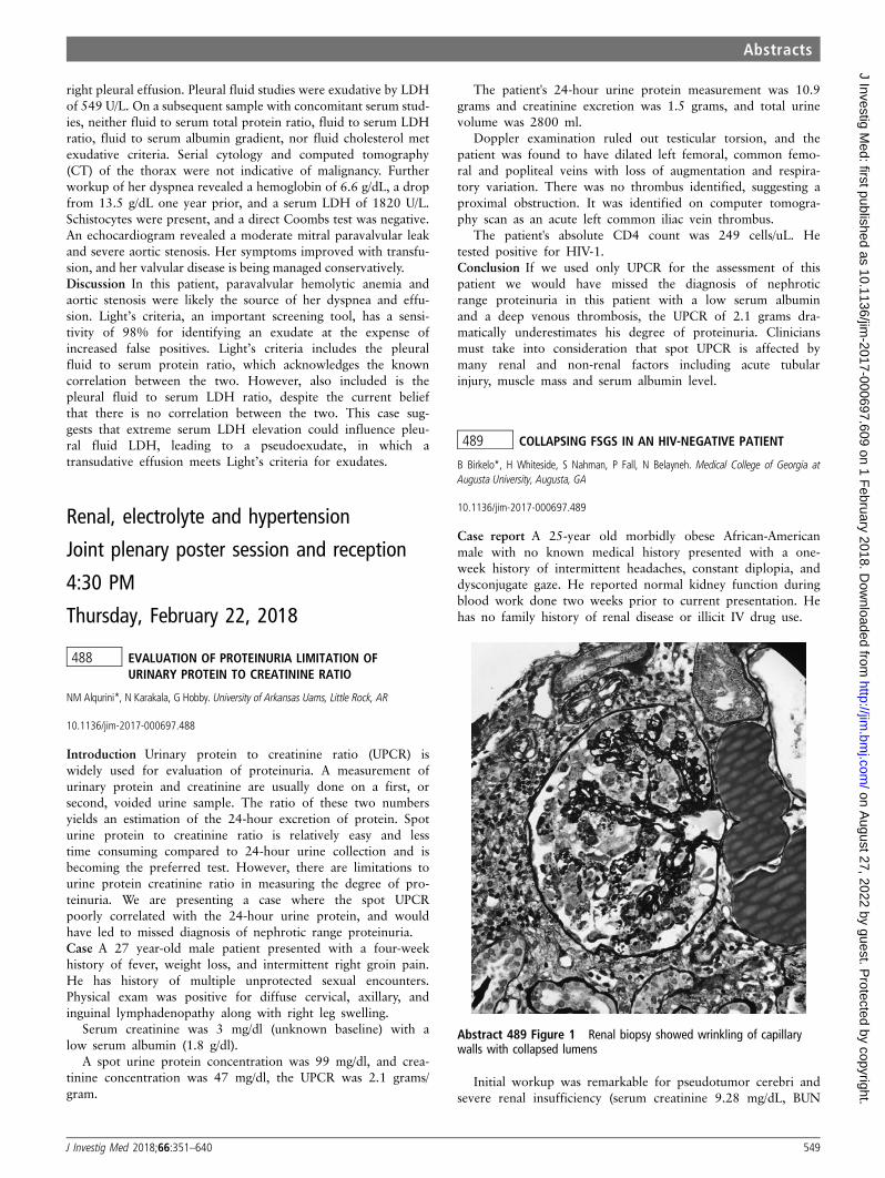
right pleural effusion. Pleural fluid studies were exudative by LDHof 549 U/L. On a subsequent sample with concomitant serum stud-ies, neither fluid to serum total protein ratio, fluid to serum LDHratio, fluid to serum albumin gradient, nor fluid cholesterol metexudative criteria. Serial cytology and computed tomography(CT) of the thorax were not indicative of malignancy. Furtherworkup of her dyspnea revealed a hemoglobin of 6.6 g/dL, a dropfrom 13.5 g/dL one year prior, and a serum LDH of 1820 U/L.Schistocytes were present, and a direct Coombs test was negative.An echocardiogram revealed a moderate mitral paravalvular leakand severe aortic stenosis. Her symptoms improved with transfu-sion, and her valvular disease is being managed conservatively.Discussion In this patient, paravalvular hemolytic anemia andaortic stenosis were likely the source of her dyspnea and effu-sion. Light’s criteria, an important screening tool, has a sensi-tivity of 98% for identifying an exudate at the expense ofincreased false positives. Light’s criteria includes the pleuralfluid to serum protein ratio, which acknowledges the knowncorrelation between the two. However, also included is thepleural fluid to serum LDH ratio, despite the current beliefthat there is no correlation between the two. This case sug-gests that extreme serum LDH elevation could influence pleu-ral fluid LDH, leading to a pseudoexudate, in which atransudative effusion meets Light’s criteria for exudates.
Renal, electrolyte and hypertension
Joint plenary poster session and reception
4:30 PM
Thursday, February 22, 2018
488 EVALUATION OF PROTEINURIA LIMITATION OFURINARY PROTEIN TO CREATININE RATIO
NM Alqurini*, N Karakala, G Hobby. University of Arkansas Uams, Little Rock, AR
10.1136/jim-2017-000697.488
Introduction Urinary protein to creatinine ratio (UPCR) iswidely used for evaluation of proteinuria. A measurement ofurinary protein and creatinine are usually done on a first, orsecond, voided urine sample. The ratio of these two numbersyields an estimation of the 24-hour excretion of protein. Spoturine protein to creatinine ratio is relatively easy and lesstime consuming compared to 24-hour urine collection and isbecoming the preferred test. However, there are limitations tourine protein creatinine ratio in measuring the degree of pro-teinuria. We are presenting a case where the spot UPCRpoorly correlated with the 24-hour urine protein, and wouldhave led to missed diagnosis of nephrotic range proteinuria.Case A 27 year-old male patient presented with a four-weekhistory of fever, weight loss, and intermittent right groin pain.He has history of multiple unprotected sexual encounters.Physical exam was positive for diffuse cervical, axillary, andinguinal lymphadenopathy along with right leg swelling.
Serum creatinine was 3 mg/dl (unknown baseline) with alow serum albumin (1.8 g/dl).
A spot urine protein concentration was 99 mg/dl, and crea-tinine concentration was 47 mg/dl, the UPCR was 2.1 grams/gram.
The patient's 24-hour urine protein measurement was 10.9grams and creatinine excretion was 1.5 grams, and total urinevolume was 2800 ml.
Doppler examination ruled out testicular torsion, and thepatient was found to have dilated left femoral, common femo-ral and popliteal veins with loss of augmentation and respira-tory variation. There was no thrombus identified, suggesting aproximal obstruction. It was identified on computer tomogra-phy scan as an acute left common iliac vein thrombus.
The patient's absolute CD4 count was 249 cells/uL. Hetested positive for HIV-1.Conclusion If we used only UPCR for the assessment of thispatient we would have missed the diagnosis of nephroticrange proteinuria in this patient with a low serum albuminand a deep venous thrombosis, the UPCR of 2.1 grams dra-matically underestimates his degree of proteinuria. Cliniciansmust take into consideration that spot UPCR is affected bymany renal and non-renal factors including acute tubularinjury, muscle mass and serum albumin level.
489 COLLAPSING FSGS IN AN HIV-NEGATIVE PATIENT
B Birkelo*, H Whiteside, S Nahman, P Fall, N Belayneh. Medical College of Georgia atAugusta University, Augusta, GA
10.1136/jim-2017-000697.489
Case report A 25-year old morbidly obese African-Americanmale with no known medical history presented with a one-week history of intermittent headaches, constant diplopia, anddysconjugate gaze. He reported normal kidney function duringblood work done two weeks prior to current presentation. Hehas no family history of renal disease or illicit IV drug use.
Abstract 489 Figure 1 Renal biopsy showed wrinkling of capillarywalls with collapsed lumens
Initial workup was remarkable for pseudotumor cerebri andsevere renal insufficiency (serum creatinine 9.28 mg/dL, BUN
Abstracts
J Investig Med 2018;66:351–640 549
on August 27, 2022 by guest. P
rotected by copyright.http://jim
.bmj.com
/J Investig M
ed: first published as 10.1136/jim-2017-000697.609 on 1 F
ebruary 2018. Dow
nloaded from

58 mg/dL). Urinalysis demonstrated 4+ proteinuria (4 gm/dayon spot urine protein-creatinine ratio) with bland sedimentand no hematuria. Serologic work up was negative for infec-tion including HIV, parvovirus, CMV, and EBV. Renal biopsyshowed wrinkling of capillary walls with collapsed lumens inevery glomerulus, without endothelial tubuloreticularinclusions.
The patient was subsequently diagnosed with idiopathic col-lapsing FSGS. Therapy was initiated with prednisone 120 mgpo q48 hr and the patient discharged without need to initiatedialysis. Three months following discharge, renal function hadimproved further with labs showing BUN 17 and Cr 1.67.
490 PATIENT CHARACTERISTICS FOR BK POLYOMAVIRUS INRENAL TRANSPLANT RECIPIENTS AT AUGUSTAUNIVERSITY (AU) TRANSPLANT CENTER
S Dhanani*, F Yang, L Mulloy, A Lee. Medical College of Georgia, Augusta, GA
10.1136/jim-2017-000697.490
Purpose of study BK Virus (BKV) is a renal transplant compli-cation which can threaten transplant graft survival and is aconsequence of required immunosuppression post-transplant.Data on risk factors for BKV varies between studies and pop-ulations. Our aim was to characterize the BKV patients at AUtransplant center and review the degree of heterogeneitybetween donors and recipients.Methods used AU renal transplant recipients from January2006 to December 2016 were reviewed for evidence of serumBKV copies >500 and urine BKV copies >10,000. Includedpatients (n=183) were divided into four groups:
. serum negative, urine positive (reference),
. serum positive, urine negative,
. serum positive, urine positive, and
. biopsy-proven BKV.
A multinomial logistic regression model was used to calcu-late the relative risk of being in groups 2–4 compared toreference group 1 for each patient characteristic.Summary of results There are significant characteristics, com-pared to the reference group 1, for groups 2 and 4. Thosewho were older (RR=1.05, 95% CI: 1.01 to 1.09), White(RR=10.22, 95% CI: 1.02 to 102.12) or Black (compared toother groups, RR=15.82, 95% CI: 1.76 to 141), received thy-moglobulin (RR=6.09, 95% CI: 1.63 to 22.69), and hadincreased most-recent creatinine levels (RR=1.25, 95% CI:0.998 to 1.58) had a higher relative risk of being in group 2than group 1. However, for every increase in one unit changeof creatinine levels from baseline there was a 41% lower rela-tive risk (95% CI: 0.37 to 0.95), of being in group 2 com-pared to group 1. Increased change in creatinine levels frombaseline (RR=7.00, 95% CI: 1.72 to 28.56) and increasedrecent creatinine levels (RR=1.56, 95% CI: 1.12 to 2.17)show a higher risk of being in group 4 compared to group 1.All p’s are £0.05.Conclusions We were able to identify patient profiles for theBKV outcome groups compared to the serum negative, urinepositive group. The serum positive, urine negative group wassignificantly associated with age, race, thymoglobulin regimen,most recent creatinine levels, and creatinine change from base-line. Though there were no significant differences with theserum positive, urine positive group, the patient characteristics
for the biopsy proven group were associated with bothincreased creatinine change and most recent creatinine level.
491 AN UNEXPECTED CAUSE OF ACUTE RENAL FAILURE
C Gregg*, J Bruno, M Laskey, J Gaines, LS Engel, J Amoss. LSU Health Sciences Center,New Orleans, LA
10.1136/jim-2017-000697.491
Introduction Acute Renal Failure is a common cause of hospi-tal admissions and a frequent diagnosis in hospitalizedpatients. Pre-renal Azotemia is caused by states of low arterialperfusion to the kidneys, often due to hypovolemia, heart fail-ure with reduced ejection fraction, or acutely decompensatedliver disease. Frequently, renal failure that requires dialysis iscaused by irreversible insults to the renal parenchyma.Case A 54-year-old woman with a history of hepatic cirrhosissecondary to right heart failure, atrial fibrillation, hyperten-sion, and diabetes mellitus presented to our hospital with acomplaint of bilateral leg swelling and pain. She was found tohave an elevated creatinine of 11 mg/dl from a baseline of 1,elevated potassium of 7.7 mEq/L, and she was admitted tothe internal medicine team with an initial diagnosis of acuterenal failure and emergency dialysis was initiated. Of note,she was seen in the emergency department 10 days prior forlower extremity cellulitis and she had refused admission forelevated creatinine at that time. She was discharged with sevendays of clindamycin. She was also found to be in rate-con-trolled atrial fibrillation, although she was not anticoagulatedat home due to non-compliance with anticoagulation. On day6 of hospitalization, she began to complain of coldness in herright foot without pain. Bedside Doppler ultrasound wasunable to demonstrate arterial blood flow to the right lowerextremity. Angiography revealed a large thrombus at the bifur-cation of the aorta, with complete occlusion of the right iliacartery, and near-complete occlusion of the left iliac artery. Tis-sue plasminogen activator was directed via catheter directlyinto the clot, and she regained lower extremity pulses severalhours later. She remained in renal failure and was dischargedhome on anticoagulation and regular dialysis.Discussion Embolic disease is a rare cause of acute renal fail-ure and can result from atrial fibrillation. In a patient withnormal renal function, an embolus must occlude both renalarteries or separate emboli must occlude each renal artery.Acute thrombotic or embolic disease should be considered asa cause of acute renal failure, particularly in those patientsthat are at high risk of thrombosis.
492 DOXYCYCLINE ASSOCIATED OXALATE NEPHROPATHY1M Malik*, 1,2T Naguib. 1Texas Tech Univ HSC Amarillo, Amarillo, TX; 2VA Medical Center,Amarillo, TX
10.1136/jim-2017-000697.492
Case report Doxycycline is a safe antibiotic in patients withimpaired renal function. Unlike earlier tetracyclines, tetracy-cline and chlortetracycline, doxycycline has not been impli-cated in causing acute kidney injury (AKI). Likewise, there isno reported association between doxycycline and oxalatenephropathy. We report a case of biopsy proven oxalatenephropathy precipitating severe AKI in a previously healthy
Abstracts
550 J Investig Med 2018;66:351–640
on August 27, 2022 by guest. P
rotected by copyright.http://jim
.bmj.com
/J Investig M
ed: first published as 10.1136/jim-2017-000697.609 on 1 F
ebruary 2018. Dow
nloaded from

person who received oral doxycycline and propose a mecha-nism for this renal insult.
58 year old man presented with progressive anorexia andnausea for few days. Vital signs were stable. Exam revealedmild symmetrical pitting edema of bilateral lower extremitiesextending up to mid thighs. He had normal CBC, sodium135, potassium 5.1, chloride 102, bicarbonate 20, BUN 42,creatinine 4.7 and negative urinalysis with pH of 5.5. Bilateralrenal ultrasound was inconclusive. He was given intravenousfluids and dismissed at that point.
He was later admitted to the hospital for further evaluationof his worsening symptoms and elevation of creatinine to 5.2mg/dl. Due to lack of initial improvement, CT guided renalbiopsy was performed. However, his renal function slowlyimproved on its own and he never required any form of dial-ysis. His functional status improved and his creatininedropped to 1.3 mg/dl over the next 12 months. Renal biopsyshowed acute tubular injury with accumulation of calciumoxalate crystals in the tubules and his final diagnosis was oxa-late nephropathy subsequent to doxycycline use.
As seen above, doxycycline may lead to AKI from oxalatenephropathy. The association itself, let alone the mechanism,is nowhere to be found in literature. We propose that doxycy-cline may lead to oxalate nephropathy from suppressing Oxa-lobacter formigenes in human gut. By doing so, it leads toenhanced oxalate absorption from the intestine resulting inhyperoxaluria and subsequently predisposing an individual tooxalate nephropathy, even without nephrolithiasis as seen inour case. Incidence of this event would be higher in individu-als who have other risk factors for hyperoxaluria such as vol-ume depletion, malabsorption and hypocitraturia.
In summary, doxycycline may cause oxalate nephropathy bycausing enhanced oxalate absorption from the gut likely dueto its' antimicrobial effect on Oxalobacter formigenes.
493 ACUTE RENAL INFARCTION; A DIAGNOSTIC CHALLENGE
D Markabawi*, H Singh Gambhir. SUNY Upstate, Syracuse, NY
10.1136/jim-2017-000697.493
Introduction Acute renal infarction is a scarcely reported con-dition. Clinical suspicion for this condition is usually lowgiven its rarity and how its presentation can mimic othermore common pathologies. Contrast enhanced CT scan isessential for diagnosis. The most common etiology of thiscondition is cardio-embolic, however at least one study foundthat up to 59% of cases had what was classified as idiopathicacute renal infarction.Case A 41-year-old male with history of alpha-1 antitrypsindeficiency, was transferred to our hospital from an outsidehospital, where he was admitted for acute onset right flankpain and fever. A CT Abdomen with contrast done thereshowed findings concerning for right pyelonephritis or renalinfarction. Urinalysis was performed twice and was negative.Patient was treated with antibiotics for 2 days withoutimprovement; he was transferred to our hospital given con-cern for renal infarction.
At our hospital, he complained of right flank and rightupper quadrant abdominal pain. Physical exam was remarkablefor a temperature of 380 C and right upper quadrant tender-ness. Laboratory workup was remarkable for leukocytosis anda serum creatinine of 1.5 mg/dl (baseline of 1.2). A CTA
abdomen showed infarction of the right kidney’s upper andlower poles. Vascular surgery did not recommend any inter-vention and the patient was started on Enoxaparin. Work-upwas negative for all autoimmune markers of vasculitis. it wasalso negative for Factor V Leiden and Prothrombin mutations.A TEE was negative for cardiac emboli. Telemetry did notdetect any disrhythmia. Visceral angiogram was incompatiblewith PAN. Patient was thus diagnosed with idiopathic acuterenal infarction. He was discharged on Enoxaparin.Discussion Diagnosing acute renal infarction can be challeng-ing. Our patient was diagnosed with pyelonephritis initiallybefore a CTA of the abdomen confirmed the diagnosis ofrenal infarction. Testing for the most common etiologies (car-dio-embolic, local renal artery involvement and hypercoagu-lable states) in this patient was negative. There is little dataguiding treatment, we elected to treat with Enoxaparin.Conclusion In conclusion, acute renal infarction, atlhough rare,should be suspected in patients presenting with acute flank/abdominal pain in whom the more common etiologies havebeen ruled out.
494 IS DIETARY PROTEIN INTAKE PREDICTIVE OF ONE-YEARMORTALITY IN DIALYSIS PATIENTS?
1DP Murray*, 1L Young, 1J Waller, 1S Wright, 1R Colombo, 1,2S Baer, 1V Spearman,1R Garcia-Torres, 1K Williams, 1M Kheda, 1,2S Nahman. 1Augusta University, Augusta, GA;2VA Medical Center, Augusta, GA
10.1136/jim-2017-000697.494
Purpose of study Mortality is high in dialysis patients and maybe associated with protein-energy wasting (PEW) syndromecharacterized by progressively depleted protein and energystores. While early diagnosis and treatment of PEW canreduce mortality, clinically practical measures for its detectionare lacking. Poor dietary protein intake (DPI) is associatedwith risk of malnutrition and PEW. However, the impact ofDPI on mortality is unclear. The purpose of this study is toexamine the ability of DPI to predict one year mortality indialysis patients.Methods used This retrospective, secondary study using datafrom the Comprehensive Dialysis Study (CDS) and UnitedStates Renal Data System examined risk factors associatedwith one year mortality in new dialysis patients. The CDSdata used for this study included sociodemographic, clinical,dialysis-related and dietary variables.Summary of results Seventeen (7.5%) of the 227 subjects diedwithin one year following baseline data collection. One yearsurvivors were significantly younger (60±13.6 vs 71±12.8;p=0.0043), had a lower Charlson Comorbidity Index (CCI)score (1.6±2.3 vs 4.0±3.6; p=0.0157), higher serum albuminlevel (3.5±0.5 vs 3.3±0.4; p=0.0173), and had higher DPI(63±33.7 vs 49.5±21.5 g/day; p=0.0386) than those whodied. In multivariable Cox proportional hazards model analy-ses, the CCI adjusted hazard ratio for death (1.24) was signifi-cantly associated with an increased risk of one-year all-causemortality. The CDS data showed no association between DPIand one year mortality in new dialysis patients.Conclusions Future studies using more precise measures areneeded to examine the predictability of DPI on mortalitygiven the definitive link between DPI and PEW syndrome andsurvival in dialysis patients.
Abstracts
J Investig Med 2018;66:351–640 551
on August 27, 2022 by guest. P
rotected by copyright.http://jim
.bmj.com
/J Investig M
ed: first published as 10.1136/jim-2017-000697.609 on 1 F
ebruary 2018. Dow
nloaded from

495 ANTI-GLOMERULAR BASEMENT MEMBRANEGLOMERULONEPHRITIS WITH CHRONIC HEPATITIS C —
A RARE COMBINATION
S Patil*, N Karakala, G Hobby. University of Arkansas for Medical Sciences, Little Rock, AR
10.1136/jim-2017-000697.495
Purpose of study Anti-GBM disease is a rare autoimmune dis-ease caused by the deposition of the circulating antibodiesagainst noncollagenous C terminal (NC1) domain of the a3chain of collagen type IV of the GBM. It is a rapidly pro-gressing condition with very high mortality if not timely iden-tified and treated. Viral infections such as influenza A2 havebeen implicated as triggers, other infections like HIV and EBVhave been reported. we could not find any reports of HeptitisC infection associated with anti-GBM disease.Methods used We present a 49 year old white male withPMH of HCV and HTN with nausea, emesis and AKI. Hehad history of aortic and mitral valve regurgitation due toinfective endocarditis 1 year ago. He was a former smokerand intravenous drug abuser. Labs on admission were signifi-cant for Hb of 9.7 g/dL, serum creatinine of 3.4 mg/dL (nor-mal baseline creatinine). UA showed moderate blood, 10–25RBCs, 0–2 WBCs. No dysmorphic RBCs on microscopy.Serum anti-GBM antibodies were present. Serologic workupwas negative. Patient was on treatment for HCV infectionwith recent negative HCV viral loads. A kidney biopsyobtained showed diffuse crescentic GN (95%) with linear IgGstaining along the GBM, consistent with anti-GBM GN.Summary of results Our patient had renal limited anti-GBMdisease with no pulmonary symptoms and normal CT Chest.He received 7 plasmapheresis sessions with 3 pulse doses ofsolumedrol. Immunosuppression with cytoxan and prednisonewas started. however renal function worsened to the point ofdialysis dependence. Severe pancytopenia, GI tract ulcers andbleeding occurred. As biopsy showed 95% cresentic glomeruliand no renal recovery signs, we chose to stop immunosup-pression in a risk-benefit analysis.Conclusions Anti-GBM GN incidence is less than 1 case per 1million so few studies present to draw associations. On litera-ture review, there is 1 case report each in a patient with acuteHepatitis A and hepatitis B infection. An explanation for thisviral hepatitis association is due to immune complexes produc-tion inciting glomerular injury and exposing sequestered GBMantigens. Our patient lacked pulmonary symptoms. Smoking isa risk factor, but there is also a possibility that his risk wasincreased in the setting of chronic hepatitis C infection, mak-ing him more susceptible.
496 RARE CASE OF RENAL TUBULAR ACIDOSIS TYPE IV ASA COMPLICATION OF LUPUS NEPHRITIS
S Ravula*. UAMS, Little Rock, AR
10.1136/jim-2017-000697.496
Case report Renal Tubular Acidosis ofetn poses diagnostic chal-lenge more so in setting of renal insufficiency,hence promptrecognition and treatment of condition is neccessary.On review of literature,it is understood that renal tubularacidosis occurs as a complication of lupus nephritis in settingof high SLE disease activity index score.
We hereby present an interesting case of lupus nephritis withhigh SLE DAI index score associated with type 4 RTA.This is a 21 year old african american female with recentdiagnosis of renal biopsy proven class 4 and 5 lupus nephritis.Renal biopsy showed diffuse proliferative lupus nephritis withcrescents and membranous features as well. As defined by theNIH criteria, the findings in biopsy corresponded to an activ-ity index of 18 -scale 0–24 and a chronicity index of 6 -scale0–12.Patient recieved three days of solumedrol as pulse steroids fol-lowed by oral prednsione along with mycophenolate as partof induction regimen. Renal function had improved from pre-senting GFR of around 30 ml/min/m2 to 40 ml/min/m2 attime of hospital discharge.However, one week later on routine lab work,it was notedthat pateint had severe hyperkalemia with no worsening ofrenal function. Hyperkalemia persisted despite patient beingon adequate dose of lasix and laxatives for regular bowelmovements.Pseudohyperkalemia was ruled out with concurrent elevationof plasma potassium as well.Other causes like ongoing hemolysis was ruled out too withstable hematocrit, normal haptoglobin,LDH and peripheralsmear. After negative workup for hyperkalemia thus far inclinical course, hyporeninemic hypoaldosteronism associatedtype IV RTA was suspected with normal anion gap metabolicacidosis, severe hyperkalemia with stable renal function andurinary pH of 5.1. Trans Tubular Potassium Gradient whencalculated was 1.9 in setting of high serum potassium thatindicated hypoaldosternic state with urine osmolaity being 357mosm/kg and urine sodium was 87 mmol/L at time ofcaluculation.Subsequently pateint was started on fludrocortisone 0.1 mgsOD along with sodium bicarbonate supplemnets for acidosisand this led to normalisation of serum potassium levels.
497 A UNIQUE CASE OF RENAL ONCOCYTOSIS IN AVETERAN EXPOSED TO AGENT ORANGE
A Reddy*, M Sessums, JC Henegan, V Manucha. University of Mississippi Medical Center,Jackson, MS
10.1136/jim-2017-000697.497
Case report More than 3 million veterans during the VietnamWar had exposure to Agent Orange, an herbicide containingdioxin that has been linked to increased cancer risk. There issufficient evidence for an association between Agent Orangeand hematological disorders, but only a few cases have beenreported in relation to renal neoplasms. Here we present acase of right-sided renal oncocytosis (RO) with Agent Orangeexposure in a patient with chronic kidney disease (CKD).
A 68-year-old Caucasian male with hypertension, CKD, andprevious Agent Orange exposure was noted to have elevatedserum creatinine 1.9 mg/dL and blood urea nitrogen 36 mg/dL which triggered further evaluation. He had no recent hem-aturia, flank pain, or weight loss. Renal ultrasound showedmultiple renal masses, and MRI confirmed these to be con-cerning for renal cell carcinoma with one mass in the upperpole of right kidney measuring up to 3.3 cm and a secondmass in the interpolar region of left kidney measuring 2.3 cm.All nodules were completely confined to the kidney.
Abstracts
552 J Investig Med 2018;66:351–640
on August 27, 2022 by guest. P
rotected by copyright.http://jim
.bmj.com
/J Investig M
ed: first published as 10.1136/jim-2017-000697.609 on 1 F
ebruary 2018. Dow
nloaded from

Preoperative germline testing of a panel of genes in whichvariants are associated with hereditary renal carcinoma syn-dromes revealed no pathogenic mutations. Right partial neph-rectomy was performed which noted multiple oncocyticnodules ranging in size from microscopic collection of a fewcells to large, grossly visible nodules. Immunostains were nega-tive for AMACR and CK7 and positive for CAM5.2 andCD117, consistent with a diagnosis of RO.Discussion RO – multiple oncocytic nodules of renal paren-chyma – is an extremely rare disorder with an incidence ofabout 4.3% of all solid renal masses. It is associated withCKD and Birt-Hogg-Dube Syndrome. The diagnosis of ROremains a challenge due to the difficulty in distinguishingbetween benign and malignant lesions with imaging. As withthis case, partial nephrectomy allowed for definitive diagnosis.RO, in this case, could be related to CKD and any link toprevious exposure to Agent Orange will require further fol-low-up of patients with this history to determine if it is a riskfactor for RO. According to literature, there have been only 4similar cases reported of oncocytosis with Agent Orange expo-sure but there has been no definitive link.
498 NOVEL PHARMACOMECHANICAL TREATMENT OF ALARGE ARTERIAL ANASTOMOSIS THROMBUS OF ANARTERIOVENOUS GRAFT
DS Rees*, H Ho-Pham, RB Vareldzis, MV Naljayan, EA Aguilar, S Barry, F Yazdi, SA Morse,E Reisin. LSU Health Sciences Center, New Orleans, LA
10.1136/jim-2017-000697.498
Case report A 70-year-old male with medical history of endstage renal disease, coronary artery disease, peripheral arterydisease, recurrent bilateral foot ulcers, and atrial fibrilationwas referred for percutaneous thrombectomy of a clotted rightfemoral arteriovenous graft (AVG). He was on no chronicanticoagulation except aspirin 81 mg daily. He had multiplethrombosed accesses previously. Current right femoral graftwas placed two years prior and had required thrombectomy,and revision due to pseudoaneurysm.
Percutaneous thrombolysis was attempted with tissue plas-minogen activator (tPa) and balloon angioplasty; then throm-bectomy was attempted with a Fogarty catheter. A largethrombus remained at the arterial anastomosis extending intothe juxta-anastomosis segment of the graft and into the femo-ral artery Typically, Fogarty aspiration, direct aspiration, orinjection of tPa would be used to resolve the thrombus. Dueto the extent of the thrombus, we felt that it would not easilybe removed with those techniques; instead an infusion catheterwas placed with its tip in the femoral artery proximal to theanastomosis and tPa was infused overnight.
The patient was monitored in the ICU overnight with nocomplications. Arteriogram showed patent femoral artery butresidual clot in the graft. Mechanical thrombectomy was per-formed restoring flow through the graft allowing for dialysisuse the same day. Two months post procedure, the graftremained usable and had not re-thrombosed.
AVG failure is a cause of morbidity and mortality in ESRDpatients and is commonly caused by thrombosis. AVG throm-bosis can be treated surgically or percutaneously with no dif-ference in success rate, complications, or patency. Percutaneousmethods of resolving AVG thrombus include mechanicalthrombectomy and pharmacomechanical thrombolysis. There is
risk of creating emboli with these procedures that can resultin pulmonary embolism or arterial embolism. The majority ofemboli are retrievable by percutaneous techniques and somemay be observed without intervention if they areasymptomatic.
499 A COMPLICATED ILLNESS SCRIPT
M Touchy*, MV Naljayan, EA Aguilar, S Barry, F Yazdi, SA Morse, E Reisin. LSU HealthSciences Center, New Orleans, LA
10.1136/jim-2017-000697.499
Case report A 55 year old African American woman with pastmedical history of coronary artery disease with multiple myo-cardial infarcts with seven stents, heart failure, peripheralartery disease, type 2 diabetes mellitus (on metformin), hyper-tension, hyperlipidemia, and tobacco abuse was brought toemergency department by family members secondary toaltered mental status for approximately one day. One weekprior, she was treated for flash pulmonary edema secondaryto NSTEMI and underwent cardiac catheterization, whichrevealed 50–70% in-stent re-stenosis, and a sub-total occlusionof the circumflex artery, successfully intervened with balloonangioplasty. On admission, the patient was unable to commu-nicate secondary to intubation. However, her family divulgedthat two days prior to admission, the patient began to experi-ence worsening hallucinations and vomiting. She was hypoten-sive with systolic pressures ranging from 60–70; temperatureof 93 °F; BUN/Cr 77/10.1 mg/dL; potassium 8.2 mEq/L;bicarbonate of <5 mEq/L; anion gap 41; lactic acid 15 mmol/L; negative toxicology screen; BNP 4042 pg/ml; arterial bloodgas with pH 6.8 and pCO2 23. She coded a total of fourtimes, with each episode lasting no longer than five minutesbefore a pulse returned. She was transferred to ICU in criticalcondition on two vasoactive agents and a bicarb infusion;emergent CRRT was initiated for severe acidosis in setting ofacute kidney injury. Over the next twelve hours, she was ableto move all four extremities and follow simple commands;over the next two days, her hemodynamic status, electrolytes,and acidosis and renal function improved. By hospital dayfour, she was extubated.Discussion This is a great example of a complicated illnessscript with multiple organs involved, including lungs, kidneys,heart, and brain. Our working diagnosis was that the patienthad incurred a kidney injury, possibly form recent coronaryangiogram, which was further exacerbated by the patient’scontinued use of metformin, resulting in a severe lactic acido-sis. This contributed to patient’s hypotension and hemody-namic collapse.
500 AN INTERESTING CASE OF HYPONATREMIA IN HEADAND NECK CANCER CAUSED BY PEMBROLIZUMAB
RB Vareldzis*, T deSilva, MV Naljayan, EA Aguilar, S Barry, F Yazdi, SA Morse, E Reisin.LSU Health Sciences Center in New Oreans, New Orleans, LA
10.1136/jim-2017-000697.500
Case report A 62 old male with stage IV Squamous cell carci-noma (SqCC) of the proximal esophagus diagnosed in 2010received cisplatin base chemotherapy, radiation and resectionin 2011. He was then diagnosed with SqCC of the tongue in
Abstracts
J Investig Med 2018;66:351–640 553
on August 27, 2022 by guest. P
rotected by copyright.http://jim
.bmj.com
/J Investig M
ed: first published as 10.1136/jim-2017-000697.609 on 1 F
ebruary 2018. Dow
nloaded from

2015, which he refused resection. He elected to initiate radia-tion & chemotherapy instead. He was then found to have aright upper lung and left lower lung masses concerning formetastatic disease.
A diagnostic biopsy was performed and revealed SqCC. Hewas started on pembrolizumab (keytruda tm) 200 mg IV every3 weeks in 10/2016 in outpatient setting. On routine outpa-tient blood work, he was found to have severe hyponatremia.Patient was hospitalized, his serum sodium was 117 mmol/L,urine osmolality 158 mosm/kg, urine analysis specific gravity<1.005, serum osmolality 241 mosm/kg, urine sodium 62mEq/L, Thyroid stimulating hormone 80.6 mlU/L. After initiat-ing Levothyroxine and Tolvaptan his urine osmolality 383mosm/kg and serum sodium improved to130 mmol/Lby hospi-tal day five. Hyponatremia is the most common electrolyteabnormality in cancer patients. In the US the direct costs oftreatment hyponatermia annually are estimated to be $1.6-$3.6 billion.
Hyponatremia is an independent predictor of poor outcomein cancer patients. Pembrolizumab could play a role in causingimmune mediated hypothyroidism especially in patients whosuffer from head and neck SqCC. Hypothyroidism leads todecrease cardiac output, increase ADH release, decrease freewater excretion by up regulating V2 receptors expression inprinciple cells similar to Syndrome of inappropriate ADH.Treatment with Levothyroxine and Tolvaptan improved freewater excretion, allowed a safe hyponatremia correction,decreased hospital stay and permitted chemotherapy futureadministration.
Southern Society for Clinical Investigationand Southern American Federation forClinical Research
Plenary session
SSCI young investigator award finalists
SSCI poster award finalists
SAFMR/SSCI/ young faculty award
SAFMR/SSCI/ trainee research award
8:00 AM
Friday, February 23, 2018
501 IMPAIRED ARTERIAL BAROREFLEX SENSITIVITY INPREHYPERTENSION
IT Fonkoue*, ML Kankam, J Park. Emory University, Decatur, GA
10.1136/jim-2017-000697.501
Purpose of study Prehypertension is associated with increasedrisk of hypertension and cardiovascular disease (CVD), themechanisms of which remain unclear. Prior studies haveshown increased resting sympathetic nerve activity (SNA), andaugmented blood pressure (BP) responses during mental stress,suggesting autonomic dysregulation at rest and during stress.
We hypothesized that compared to normotensives (£120/80mmHg), prehypertensives (120/80–139/89 mmHg) haveimpaired arterial baroreflex sensitivity (BRS) leading to auto-nomic dysregulation, and increased neurocardiovascular reactiv-ity to mental stress.Methods used 22 participants were studied: 12 otherwisehealthy prehypertensives (35±6 years) and 10 matched nor-motensive controls (32±6 years). We recorded muscle SNA(MSNA) using microneurography, beat-to-beat BP, and continu-ous EKG during 5 minutes of supine rest and 3 minutes ofstress via mental arithmetic. Arterial baroreflex sensitivity(BRS) was measured via modified oxford technique using IVboluses of nitroprusside and phenylephrine to manipulate arte-rial BP. The slope of the linear relationship between diastolicBP and MSNA (sympathetic BRS), and systolic BP and R-Rinterval (cardiovagal BRS) were assessed.Summary of results As expected, baseline systolic BP (130±7vs 117±8 mmHg) and diastolic BP (87±7 vs 74±8 mmHg)were significantly higher in prehypertensives (p<0.001). Rest-ing MSNA (25±12 vs 18±10 bursts/min) tended to be higherin prehypertensives (p=0.08). Sympathetic BRS was compara-ble between the groups, but cardiovagal BRS (13±10 vs 22±10 ms/mmHg) was significantly lower in prehypertensives(p=0.03). During mental arithmetic, minute by minuteincreases in BP and MSNA did not differ between the groups.However, there was a significant correlation between diastolicBP reactivity to mental stress and resting cardiovagal BRS(r=0.703, p=0.016), as well as with resting sympathetic BRS(r=0.795, p=0.010) in the prehypertensive group. In contrast,in normotensive controls, there was no correlation betweenBP responses to stress and cardiovagal (r=0.126, p=0.766) orsympathetic BRS (r=0.287, p=0.581).Conclusions These findings suggest that early impairment ofarterial BRS may be present in prehypertension and may mod-ulate BP responses to stress, contributing to increased hyper-tension and CVD risk.
502 NATIONAL TRENDS OF SICKLE CELL DISEASE RELATEDADULT HOSPITALIZATIONS
1N Jain*, 2M Agarwal, 1M Kalliny, 1F Syed. 1Meharry Medical College, Nashville, TN;2University of Tennessee Health Sciences, Memphis, TN
10.1136/jim-2017-000697.502
Purpose of study Sickle cell disease (SCD) is an increasingglobal health problem. Data pertaining to contemporary trendsof patient characteristics with SCD is limited. Hence, westudied national trends of patient characteristics admitted witha primary diagnosis of SCD from 2003 to 2014 across UnitedStates.Methods used Using 2003 to 2014 Nationwide Inpatient Sam-ple (NIS) database of the Healthcare Cost Utilization Project(HCUP), we examined the contemporary trends for patientdemographics, hospitalization characteristics, in-hospital com-plications and healthcare resource utilization of SCD relatedhospitalizations in the US.Summary of results Overall there were 770,000 SCD hospital-izations between 2003 and 2014 with an increase in annualfrequency from 52,741 in 2003 to 72,930 in 2014, ptrend<0.001. There was a significant decrease in mean age (31.94to 31.53, years) and proportion of females (55.0% to 53.8%)(all ptrend<0.001). While the frequency of hospitalizations for
Abstracts
554 J Investig Med 2018;66:351–640
on August 27, 2022 by guest. P
rotected by copyright.http://jim
.bmj.com
/J Investig M
ed: first published as 10.1136/jim-2017-000697.609 on 1 F
ebruary 2018. Dow
nloaded from