The Ugly Duckling Turned to Swan - The Journal of Immunology
Upload
khangminh22Category
view
2download
0
of July 6, 2022.This information is current as
ColitisParticipate in the Perpetuation of ChronicCells Residing Outside the Intestine
Memory T+Long-Lived Colitogenic CD4
Tetsuo Sudo, Satoshi Matsumoto and Mamoru WatanabeRyuichi Okamoto, Kiichiro Tsuchiya, Tetsuya Nakamura,Tamako Shinohara, Naoya Sakamoto, Teruji Totsuka, Yasuhiro Nemoto, Takanori Kanai, Kaori Kameyama,
http://www.jimmunol.org/content/183/8/5059doi: 10.4049/jimmunol.0803684September 2009;
2009; 183:5059-5068; Prepublished online 28J Immunol
Referenceshttp://www.jimmunol.org/content/183/8/5059.full#ref-list-1
, 6 of which you can access for free at: cites 27 articlesThis article
average*
4 weeks from acceptance to publicationFast Publication! •
Every submission reviewed by practicing scientistsNo Triage! •
from submission to initial decisionRapid Reviews! 30 days* •
Submit online. ?The JIWhy
Subscriptionhttp://jimmunol.org/subscription
is online at: The Journal of ImmunologyInformation about subscribing to
Permissionshttp://www.aai.org/About/Publications/JI/copyright.htmlSubmit copyright permission requests at:
Email Alertshttp://jimmunol.org/alertsReceive free email-alerts when new articles cite this article. Sign up at:
Print ISSN: 0022-1767 Online ISSN: 1550-6606. Immunologists, Inc. All rights reserved.Copyright © 2009 by The American Association of1451 Rockville Pike, Suite 650, Rockville, MD 20852The American Association of Immunologists, Inc.,
is published twice each month byThe Journal of Immunology
by guest on July 6, 2022http://w
ww
.jimm
unol.org/D
ownloaded from
by guest on July 6, 2022
http://ww
w.jim
munol.org/
Dow
nloaded from

Long-Lived Colitogenic CD4� Memory T Cells ResidingOutside the Intestine Participate in the Perpetuation ofChronic Colitis1
Yasuhiro Nemoto,* Takanori Kanai,2* Kaori Kameyama,* Tamako Shinohara,*Naoya Sakamoto,* Teruji Totsuka,* Ryuichi Okamoto,* Kiichiro Tsuchiya,*Tetsuya Nakamura,* Tetsuo Sudo,† Satoshi Matsumoto,‡ and Mamoru Watanabe*
To understand the perpetuation of inflammatory bowel disease (IBD), it is important to clarify whether the colitogenic CD4� T cellsare self-limited effector or long-lived memory T cells. We here investigate the latency of colitogenic CD4� T cells in the remission stageof colitis under germfree (GF) conditions. We isolated splenic (SP) CD4� T cells from colitic CD4�CD45RBhigh T cell-injected SCIDmice maintained under specific pathogen-free (SPF) conditions and transferred them into SPF or GF SCID mice. Donor colitic SP CD4�
T cells have a characteristic CD44highCD62L�IL-7R�high effector-memory T-type phenotype. Six weeks after transfer of cells to GFSCID mice, one group of mice was continued in GF conditions (GF3GF), and the other was transferred into SPF conditions(GF3SPF). GF3SPF but not GF3GF SCID mice developed colitis with elevated production of Th1 and Th17 cytokines at 4 wk aftertransfer. Surprisingly, a large number of CD4� effector-memory T cells and a small but substantial number of central-memory T cellsremained resident in SP and bone marrow, but not in lamina propria, of the GF3GF SCID recipients. Consistent with this, GF3SPFbut not GF3GF SCID mice rapidly developed colitis. Taken together, these findings suggest that long-lived colitogenic memory CD4�
cells can be established even in the presence of commensal Ags, reside outside the intestine in the absence of commensal bacteria, andparticipate in the perpetuation of colitis. Thus, blocking a stimulus of colitogenic memory CD4� cells such as IL-7 may have therapeuticbenefit for treatment of inflammatory bowel disease. The Journal of Immunology, 2009, 183: 5059–5068.
S tudies of Ag exposure in murine models of acute virus in-fection have provided much information about the dynamicsof naive, effector, and memory CD8� T cell responses (1–3).
Acute virus infection elicits massive proliferation of viral Ag-specificCD8� T cells, which acquire effector functions (effector phase). Afterthe peak of T cell proliferation, most of the effector CD8� T cells areeliminated (contraction phase). Following virus (Ag) clearance, asmall proportion of the remaining cells differentiate into memoryCD8� T cells that are maintained by cytokine (IL-7 and/or IL-15)-dependent homeostatic proliferation and survive in the absence oftheir corresponding Ags (memory phase). Thus, Ag clearance is es-sential for the emergence of memory CD8� T cells.
In contrast to the generation of CD8� memory T cells, it is con-troversial whether Ag clearance is needed for the generation of CD4�
memory T cells. Indeed, there is evidence that persistence of the Ag
itself is essential for the maintenance of CD4� memory T cells (4–6).Given that Ag-specific effector T cells are thought to be terminallydifferentiated and short-lived cells, there must be cellular mechanismsby which memory T cells specific for persistent Ags are maintained inthe host as “memory-stem cells.” In support of the idea that persistentAg helps maintain CD4� memory T cells, rats immunized with irra-diated sporozoites are protected from malaria infection, but this pro-tection is lost after drug treatment to remove the remaining parasites(5). A similar phenomenon is found in Leishmania major infection inmice where CD4�CD25� regulatory T cells and IL-10 are thought toprevent a Th1 immune response from clearing the infection, thus al-lowing the host to retain CD4� memory T cells specific to the or-ganism (6). Furthermore, more recent data have shown that in theabsence of MHC class II signals that are essential for Ag presentationto CD4� T cells, surviving memory T cells are functionally impaired,suggesting that TCR signaling is involved in the maintenance ofCD4� memory T cells (7).
All complex metazoans, including humans and mice, are colo-nized with microbial organisms that comprise an indigenous mi-croflora (8). Although the host evidently benefits from the residentmicroflora in the gut (9), the presence of commensal bacteria ap-pears to be of crucial importance in the development of humaninflammatory bowel disease (IBD)3 and in almost all animal mod-els of IBD (10, 11). For instance, results from most IBD modelsshowing that in germfree (GF) rodents intestinal inflammation is
*Department of Gastroenterology and Hepatology, Graduate School of Medicine,Tokyo Medical and Dental University, Tokyo, Japan; †New Frontiers Research Lab-oratories, Toray Industries, Kanagawa, Japan; and ‡Yakult Central Institute for Mi-crobiological Research, Tokyo, Japan
Received for publication November 12, 2008. Accepted for publication August12, 2009.
The costs of publication of this article were defrayed in part by the payment of pagecharges. This article must therefore be hereby marked advertisement in accordancewith 18 U.S.C. Section 1734 solely to indicate this fact.1 This study was supported in part by grants-in-aid for Scientific Research, ScientificResearch on Priority Areas, Exploratory Research and Creative Scientific Research fromthe Japanese Ministry of Education, Culture, Sports, Science, and Technology; the Jap-anese Ministry of Health, Labor, and Welfare; the Japan Medical Association; the Foun-dation for Advancement of International Science; the Terumo Life Science Foundation;the Ohyama Health Foundation; the Yakult Bio-Science Foundation; and the ResearchFund of Mitsukoshi Health and Welfare Foundation.2 Address correspondence and reprint requests to Dr. Takanori Kanai, Department ofGastroenterology and Hematology, Tokyo Medical and Dental University, 1-5-45Yeshiva, Bunkyo-ku, Tokyo 113-8519, Japan. E-mail address: [email protected]
3 Abbreviations used in this paper: IBD, inflammatory bowel disease; BM, bone marrow;CD62L, L-selectin; LP, lamina propria; MLN, mesenteric lymph node; SP, spleen orsplenic; TEM, effector-memory T; TSLP, thymic stromal lymphopoietin; WT, wild type.
Copyright © 2009 by The American Association of Immunologists, Inc. 0022-1767/09/$2.00
The Journal of Immunology
www.jimmunol.org/cgi/doi/10.4049/jimmunol.0803684
by guest on July 6, 2022http://w
ww
.jimm
unol.org/D
ownloaded from
absent indicate that commensal bacteria are indispensable contrib-utors to the pathogenesis of chronic immune-mediated intestinalinflammation.
IBD is caused by excessive and tissue-damaging chronic inflam-matory responses, which are thought to be due to inappropriateactivation of the immune system in many cases and which com-monly take a persistent, disabling course (10, 11). In some pa-tients, disease progresses steadily, whereas in others, it follows arelapsing-remitting course. According to present understanding,the disease is caused and controlled by colitogenic effector andmemory CD4� T cells presumably reacting to commensal bacte-rial Ags. Importantly, however, it is not known whether differenteffector CD4� T cells are recruited at each relapse or whethersequential memory CD4� T cells are derived from members of theinitial attack cohort throughout the entire course of disease. Inother words, the nature and regulation of colitogenic effector andmemory CD4� T cells in the host-commensal interaction overtime and the correlation between colitogenic effector and memoryCD4� T cells in chronic colitis remain largely unknown.
Given the possible importance of the microflora in intestinalinflammation, we conducted a series of experiments to test the invivo effect of commensal bacteria using a GF system for the es-tablishment and maintenance of colitogenic CD4� T cells. We alsoattempted to assess whether colitogenic CD4� T cells are self-limited effector cells or long-lived memory cells in a host-com-mensal bacteria mutualism to understand the perpetuation of IBDand to develop a strategy for IBD treatment.
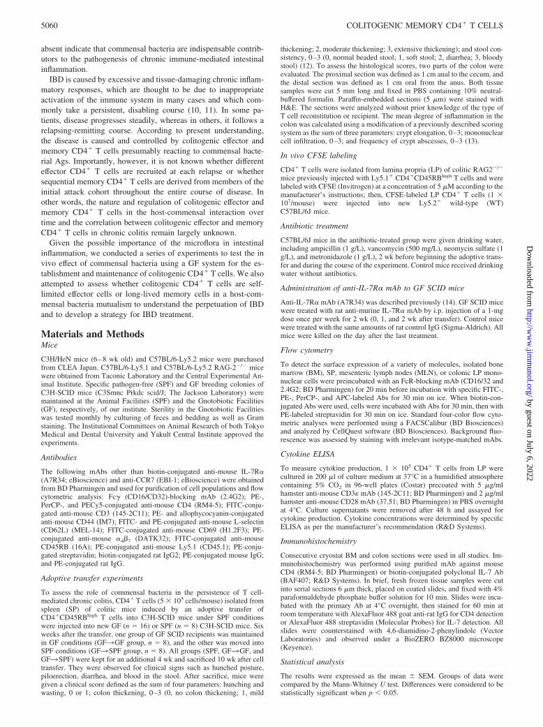
Materials and MethodsMice
C3H/HeN mice (6–8 wk old) and C57BL/6-Ly5.2 mice were purchasedfrom CLEA Japan. C57BL/6-Ly5.1 and C57BL/6-Ly5.2 RAG-2�/� micewere obtained from Taconic Laboratory and the Central Experimental An-imal Institute. Specific pathogen-free (SPF) and GF breeding colonies ofC3H-SCID mice (C3Smnc Prkdc scid/J; The Jackson Laboratory) weremaintained at the Animal Facilities (SPF) and the Gnotobiotic Facilities(GF), respectively, of our institute. Sterility in the Gnotobiotic Facilitieswas tested monthly by culturing of feces and bedding as well as Gramstaining. The Institutional Committees on Animal Research of both TokyoMedical and Dental University and Yakult Central Institute approved theexperiments.
Antibodies
The following mAbs other than biotin-conjugated anti-mouse IL-7R�(A7R34; eBioscience) and anti-CCR7 (EBI-1; eBioscience) were obtainedfrom BD Pharmingen and used for purification of cell populations and flowcytometric analysis: Fc� (CD16/CD32)-blocking mAb (2.4G2); PE-,PerCP-, and PECy5-conjugated anti-mouse CD4 (RM4-5); FITC-conju-gated anti-mouse CD3 (145-2C11); PE- and allophycocyanin-conjugatedanti-mouse CD44 (IM7); FITC- and PE-conjugated anti-mouse L-selectin(CD62L) (MEL-14); FITC-conjugated anti-mouse CD69 (H1.2F3); PE-conjugated anti-mouse �4�7 (DATK32); FITC-conjugated anti-mouseCD45RB (16A); PE-conjugated anti-mouse Ly5.1 (CD45.1); PE-conju-gated streptavidin; biotin-conjugated rat IgG2; PE-conjugated mouse IgG;and PE-conjugated rat IgG.
Adoptive transfer experiments
To assess the role of commensal bacteria in the persistence of T cell-mediated chronic colitis, CD4� T cells (5 � 105 cells/mouse) isolated fromspleen (SP) of colitic mice induced by an adoptive transfer ofCD4�CD45RBhigh T cells into C3H-SCID mice under SPF conditionswere injected into new GF (n � 16) or SPF (n � 8) C3H-SCID mice. Sixweeks after the transfer, one group of GF SCID recipients was maintainedin GF conditions (GF3GF group, n � 8), and the other was moved intoSPF conditions (GF3SPF group, n � 8). All groups (SPF, GF3GF, andGF3SPF) were kept for an additional 4 wk and sacrificed 10 wk after celltransfer. They were observed for clinical signs such as hunched posture,piloerection, diarrhea, and blood in the stool. After sacrifice, mice weregiven a clinical score defined as the sum of four parameters: hunching andwasting, 0 or 1; colon thickening, 0–3 (0, no colon thickening; 1, mild
thickening; 2, moderate thickening; 3, extensive thickening); and stool con-sistency, 0–3 (0, normal beaded stool; 1, soft stool; 2, diarrhea; 3, bloodystool) (12). To assess the histological scores, two parts of the colon wereevaluated. The proximal section was defined as 1 cm anal to the cecum, andthe distal section was defined as 1 cm oral from the anus. Both tissuesamples were cut 5 mm long and fixed in PBS containing 10% neutral-buffered formalin. Paraffin-embedded sections (5 �m) were stained withH&E. The sections were analyzed without prior knowledge of the type ofT cell reconstitution or recipient. The mean degree of inflammation in thecolon was calculated using a modification of a previously described scoringsystem as the sum of three parameters: crypt elongation, 0–3; mononuclearcell infiltration, 0–3; and frequency of crypt abscesses, 0–3 (13).
In vivo CFSE labeling
CD4� T cells were isolated from lamina propria (LP) of colitic RAG2�/�
mice previously injected with Ly5.1� CD4�CD45RBhigh T cells and werelabeled with CFSE (Invitrogen) at a concentration of 5 �M according to themanufacturer’s instructions; then, CFSE-labeled LP CD4� T cells (1 �107/mouse) were injected into new Ly5.2� wild-type (WT)C57BL/6J mice.
Antibiotic treatment
C57BL/6J mice in the antibiotic-treated group were given drinking water,including ampicillin (1 g/L), vancomycin (500 mg/L), neomycin sulfate (1g/L), and metronidazole (1 g/L), 2 wk before beginning the adoptive trans-fer and during the course of the experiment. Control mice received drinkingwater without antibiotics.
Administration of anti-IL-7R� mAb to GF SCID mice
Anti-IL-7R� mAb (A7R34) was described previously (14). GF SCID micewere treated with rat anti-murine IL-7R� mAb by i.p. injection of a 1-mgdose once per week for 2 wk (0, 1, and 2 wk after transfer). Control micewere treated with the same amounts of rat control IgG (Sigma-Aldrich). Allmice were killed on the day after the last treatment.
Flow cytometry
To detect the surface expression of a variety of molecules, isolated bonemarrow (BM), SP, mesenteric lymph nodes (MLN), or colonic LP mono-nuclear cells were preincubated with an FcR-blocking mAb (CD16/32 and2.4G2; BD Pharmingen) for 20 min before incubation with specific FITC-,PE-, PerCP-, and APC-labeled Abs for 30 min on ice. When biotin-con-jugated Abs were used, cells were incubated with Abs for 30 min, then withPE-labeled streptavidin for 30 min on ice. Standard four-color flow cyto-metric analyses were performed using a FACSCalibur (BD Biosciences)and analyzed by CellQuest software (BD Biosciences). Background fluo-rescence was assessed by staining with irrelevant isotype-matched mAbs.
Cytokine ELISA
To measure cytokine production, 1 � 105 CD4� T cells from LP werecultured in 200 �l of culture medium at 37°C in a humidified atmospherecontaining 5% CO2 in 96-well plates (Costar) precoated with 5 �g/mlhamster anti-mouse CD3� mAb (145-2C11; BD Pharmingen) and 2 �g/mlhamster anti-mouse CD28 mAb (37.51; BD Pharmingen) in PBS overnightat 4°C. Culture supernatants were removed after 48 h and assayed forcytokine production. Cytokine concentrations were determined by specificELISA as per the manufacturer’s recommendation (R&D Systems).
Immunohistochemistry
Consecutive cryostat BM and colon sections were used in all studies. Im-munohistochemistry was performed using purified mAb against mouseCD4 (RM4-5; BD Pharmingen) or biotin-conjugated polyclonal IL-7 Ab(BAF407; R&D Systems). In brief, fresh frozen tissue samples were cutinto serial sections 6 �m thick, placed on coated slides, and fixed with 4%paraformaldehyde phosphate buffer solution for 10 min. Slides were incu-bated with the primary Ab at 4°C overnight, then stained for 60 min atroom temperature with AlexaFluor 488 goat anti-rat IgG for CD4 detectionor AlexaFluor 488 streptavidin (Molecular Probes) for IL-7 detection. Allslides were counterstained with 4,6-diamidino-2-phenylindole (VectorLaboratories) and observed under a BioZERO BZ8000 microscope(Keyence).
Statistical analysis
The results were expressed as the mean � SEM. Groups of data werecompared by the Mann-Whitney U test. Differences were considered to bestatistically significant when p � 0.05.
5060 COLITOGENIC MEMORY CD4� T CELLS
by guest on July 6, 2022http://w
ww
.jimm
unol.org/D
ownloaded from
ResultsEffector-memory T (TEM) type of CD4� T cells reside in varioussites in colitic mice
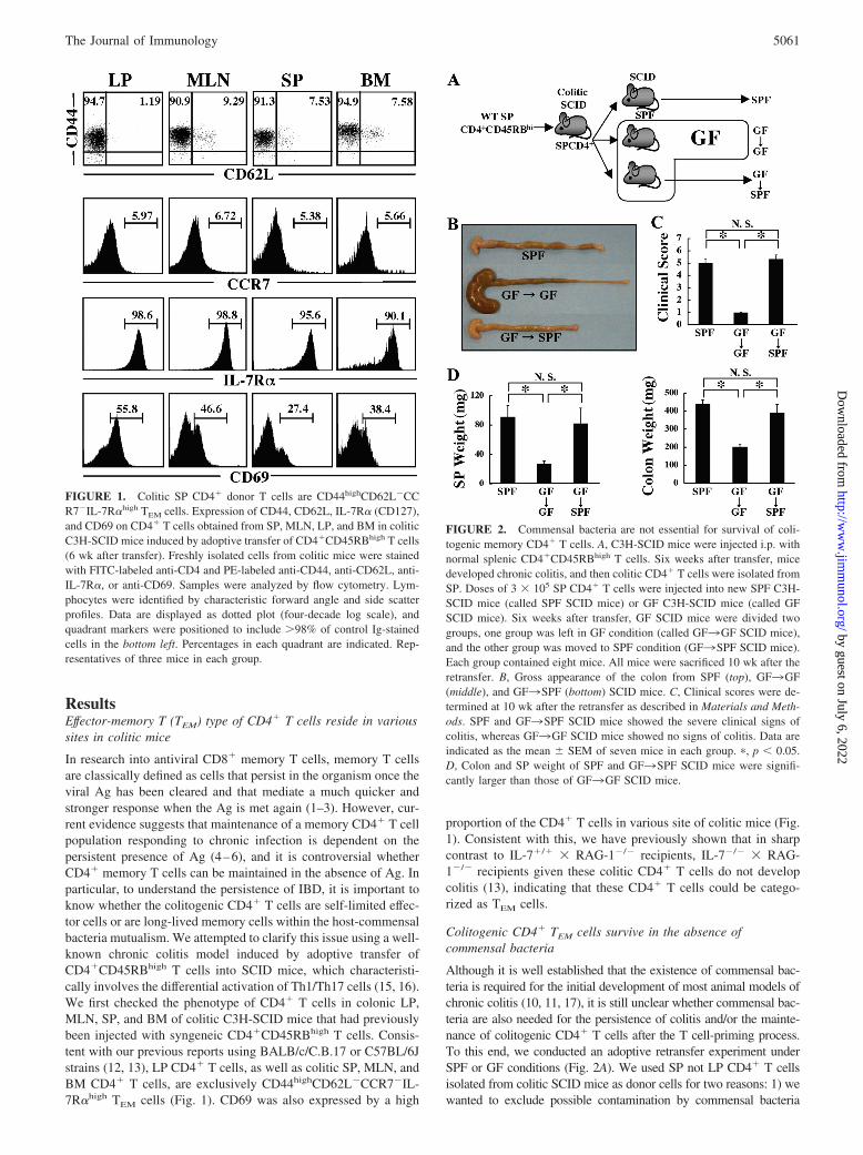
In research into antiviral CD8� memory T cells, memory T cellsare classically defined as cells that persist in the organism once theviral Ag has been cleared and that mediate a much quicker andstronger response when the Ag is met again (1–3). However, cur-rent evidence suggests that maintenance of a memory CD4� T cellpopulation responding to chronic infection is dependent on thepersistent presence of Ag (4–6), and it is controversial whetherCD4� memory T cells can be maintained in the absence of Ag. Inparticular, to understand the persistence of IBD, it is important toknow whether the colitogenic CD4� T cells are self-limited effec-tor cells or are long-lived memory cells within the host-commensalbacteria mutualism. We attempted to clarify this issue using a well-known chronic colitis model induced by adoptive transfer ofCD4�CD45RBhigh T cells into SCID mice, which characteristi-cally involves the differential activation of Th1/Th17 cells (15, 16).We first checked the phenotype of CD4� T cells in colonic LP,MLN, SP, and BM of colitic C3H-SCID mice that had previouslybeen injected with syngeneic CD4�CD45RBhigh T cells. Consis-tent with our previous reports using BALB/c/C.B.17 or C57BL/6Jstrains (12, 13), LP CD4� T cells, as well as colitic SP, MLN, andBM CD4� T cells, are exclusively CD44highCD62L�CCR7�IL-7R�high TEM cells (Fig. 1). CD69 was also expressed by a high
proportion of the CD4� T cells in various site of colitic mice (Fig.1). Consistent with this, we have previously shown that in sharpcontrast to IL-7�/� � RAG-1�/� recipients, IL-7�/� � RAG-1�/� recipients given these colitic CD4� T cells do not developcolitis (13), indicating that these CD4� T cells could be catego-rized as TEM cells.
Colitogenic CD4� TEM cells survive in the absence ofcommensal bacteria
Although it is well established that the existence of commensal bac-teria is required for the initial development of most animal models ofchronic colitis (10, 11, 17), it is still unclear whether commensal bac-teria are also needed for the persistence of colitis and/or the mainte-nance of colitogenic CD4� T cells after the T cell-priming process.To this end, we conducted an adoptive retransfer experiment underSPF or GF conditions (Fig. 2A). We used SP not LP CD4� T cellsisolated from colitic SCID mice as donor cells for two reasons: 1) wewanted to exclude possible contamination by commensal bacteria
FIGURE 1. Colitic SP CD4� donor T cells are CD44highCD62L�CCR7�IL-7R�high TEM cells. Expression of CD44, CD62L, IL-7R� (CD127),and CD69 on CD4� T cells obtained from SP, MLN, LP, and BM in coliticC3H-SCID mice induced by adoptive transfer of CD4�CD45RBhigh T cells(6 wk after transfer). Freshly isolated cells from colitic mice were stainedwith FITC-labeled anti-CD4 and PE-labeled anti-CD44, anti-CD62L, anti-IL-7R�, or anti-CD69. Samples were analyzed by flow cytometry. Lym-phocytes were identified by characteristic forward angle and side scatterprofiles. Data are displayed as dotted plot (four-decade log scale), andquadrant markers were positioned to include �98% of control Ig-stainedcells in the bottom left. Percentages in each quadrant are indicated. Rep-resentatives of three mice in each group.
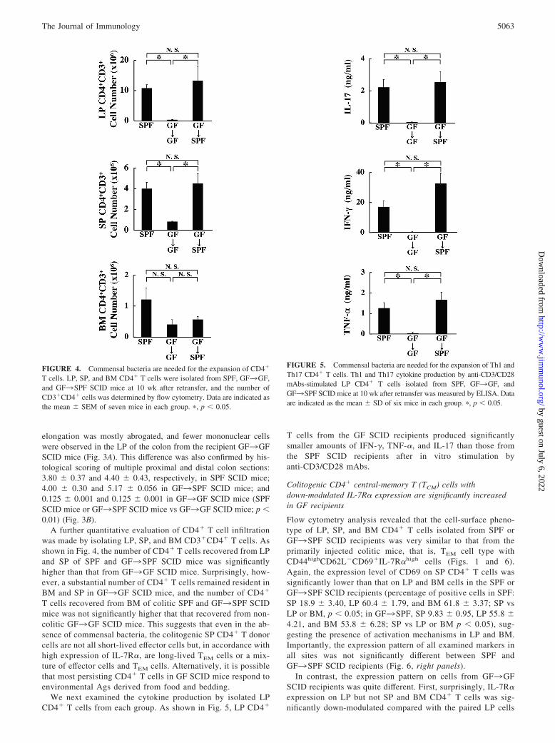
FIGURE 2. Commensal bacteria are not essential for survival of coli-togenic memory CD4� T cells. A, C3H-SCID mice were injected i.p. withnormal splenic CD4�CD45RBhigh T cells. Six weeks after transfer, micedeveloped chronic colitis, and then colitic CD4� T cells were isolated fromSP. Doses of 3 � 105 SP CD4� T cells were injected into new SPF C3H-SCID mice (called SPF SCID mice) or GF C3H-SCID mice (called GFSCID mice). Six weeks after transfer, GF SCID mice were divided twogroups, one group was left in GF condition (called GF3GF SCID mice),and the other group was moved to SPF condition (GF3SPF SCID mice).Each group contained eight mice. All mice were sacrificed 10 wk after theretransfer. B, Gross appearance of the colon from SPF (top), GF3GF(middle), and GF3SPF (bottom) SCID mice. C, Clinical scores were de-termined at 10 wk after the retransfer as described in Materials and Meth-ods. SPF and GF3SPF SCID mice showed the severe clinical signs ofcolitis, whereas GF3GF SCID mice showed no signs of colitis. Data areindicated as the mean � SEM of seven mice in each group. �, p � 0.05.D, Colon and SP weight of SPF and GF3SPF SCID mice were signifi-cantly larger than those of GF3GF SCID mice.
5061The Journal of Immunology
by guest on July 6, 2022http://w
ww
.jimm
unol.org/D
ownloaded from

from colitic LP or MLN samples, and 2) we have previously shownthat most of the SP CD4� T cells in this mouse colitis model expressthe TEM cell phenotype with a capacity to induce colitis similar to thatof LP CD4� T cells (12). Thus, we injected these SP cells into newSPF or GF C3H-SCID mice (hereafter called SPF or GF SCID mice,respectively) in a retransfer. Furthermore, to assess whether GF SCIDrecipients develop colitis when exposed to resident commensal bac-teria, one group of the GF SCID recipients was moved into the SPFenvironment 6 wk after the retransfer and kept for an additional 4 wk(GF3SPF SCID mice), and the other group was kept in GF condi-tions for 10 wk (GF3GF SCID mice) (Fig. 2A). Consistent withprevious reports (12, 13) and in sharp contrast to GF SCID mice, SPFSCID mice showed an enlarged colon with a greatly thickened wall(Fig. 2B) and manifested progressive weight loss from 3 wk afterretransfer (data not shown). Consistent with this, the clinical score ofSPF SCID mice at 10 wk after retransfer was significantly increased
compared with that of GF SCID mice, which showed no clinical signsof colitis or weight loss throughout the observation period (Fig. 2C).It was notable that GF3SPF SCID recipients also showed an en-larged and thickened colon, and their clinical scores were significantlyincreased compared with those of GF3GF SCID recipients, beingcomparable to those of SPF SCID recipients. As previously reported(18), a markedly enlarged cecum was also reproducibly observed inthe GF3GF SCID-recipient mice (Fig. 2B). The integrity of the GFconditions was confirmed by repeated stool culture tests in the GFgroups (data not shown). SP and colon weights of SPF SCID miceand GF3SPF SCID mice were significantly greater than those ofGF3GF SCID mice (data not shown).
Histological examination showed a prominent epithelial hyperpla-sia with glandular elongation and massive infiltration of mononuclearcells in the LP of proximal and distal colon from SPF SCID andGF3SPF SCID mice (Fig. 3A). In contrast, the glandular
FIGURE 3. Commensal bacteriaare needed to sustain the colitis. A,Proximal and distal colon from SPF,GF3GF, and GF3SPF SCID micewere stained with H&E. Originalmagnification: �40 (top) and �100(bottom). B, Histological scores weredetermined at 10 wk after transfer asdescribed in Materials and Methods.Data are indicated as the mean �SEM of five mice in each group. �,p � 0.05.
5062 COLITOGENIC MEMORY CD4� T CELLS
by guest on July 6, 2022http://w
ww
.jimm
unol.org/D
ownloaded from
elongation was mostly abrogated, and fewer mononuclear cellswere observed in the LP of the colon from the recipient GF3GFSCID mice (Fig. 3A). This difference was also confirmed by his-tological scoring of multiple proximal and distal colon sections:3.80 � 0.37 and 4.40 � 0.43, respectively, in SPF SCID mice;4.00 � 0.30 and 5.17 � 0.056 in GF3SPF SCID mice; and0.125 � 0.001 and 0.125 � 0.001 in GF3GF SCID mice (SPFSCID mice or GF3SPF SCID mice vs GF3GF SCID mice; p �0.01) (Fig. 3B).
A further quantitative evaluation of CD4� T cell infiltrationwas made by isolating LP, SP, and BM CD3�CD4� T cells. Asshown in Fig. 4, the number of CD4� T cells recovered from LPand SP of SPF and GF3SPF SCID mice was significantlyhigher than that from GF3GF SCID mice. Surprisingly, how-ever, a substantial number of CD4� T cells remained resident inBM and SP in GF3GF SCID mice, and the number of CD4�
T cells recovered from BM of colitic SPF and GF3SPF SCIDmice was not significantly higher that that recovered from non-colitic GF3GF SCID mice. This suggests that even in the ab-sence of commensal bacteria, the colitogenic SP CD4� T donorcells are not all short-lived effector cells but, in accordance withhigh expression of IL-7R�, are long-lived TEM cells or a mix-ture of effector cells and TEM cells. Alternatively, it is possiblethat most persisting CD4� T cells in GF SCID mice respond toenvironmental Ags derived from food and bedding.
We next examined the cytokine production by isolated LPCD4� T cells from each group. As shown in Fig. 5, LP CD4�
T cells from the GF SCID recipients produced significantlysmaller amounts of IFN-�, TNF-�, and IL-17 than those fromthe SPF SCID recipients after in vitro stimulation byanti-CD3/CD28 mAbs.
Colitogenic CD4� central-memory T (TCM) cells withdown-modulated IL-7R� expression are significantly increasedin GF recipients
Flow cytometry analysis revealed that the cell-surface pheno-type of LP, SP, and BM CD4� T cells isolated from SPF orGF3SPF SCID recipients was very similar to that from theprimarily injected colitic mice, that is, TEM cell type withCD44highCD62L�CD69�IL-7R�high cells (Figs. 1 and 6).Again, the expression level of CD69 on SP CD4� T cells wassignificantly lower than that on LP and BM cells in the SPF orGF3SPF SCID recipients (percentage of positive cells in SPF:SP 18.9 � 3.40, LP 60.4 � 1.79, and BM 61.8 � 3.37; SP vsLP or BM, p � 0.05; in GF3SPF, SP 9.83 � 0.95, LP 55.8 �4.21, and BM 53.8 � 6.28; SP vs LP or BM p � 0.05), sug-gesting the presence of activation mechanisms in LP and BM.Importantly, the expression pattern of all examined markers inall sites was not significantly different between SPF andGF3SPF SCID recipients (Fig. 6, right panels).
In contrast, the expression pattern on cells from GF3GFSCID recipients was quite different. First, surprisingly, IL-7R�expression on LP but not SP and BM CD4� T cells was sig-nificantly down-modulated compared with the paired LP cells
FIGURE 4. Commensal bacteria are needed for the expansion of CD4�
T cells. LP, SP, and BM CD4� T cells were isolated from SPF, GF3GF,and GF3SPF SCID mice at 10 wk after retransfer, and the number ofCD3�CD4� cells was determined by flow cytometry. Data are indicated asthe mean � SEM of seven mice in each group. �, p � 0.05.
FIGURE 5. Commensal bacteria are needed for the expansion of Th1 andTh17 CD4� T cells. Th1 and Th17 cytokine production by anti-CD3/CD28mAbs-stimulated LP CD4� T cells isolated from SPF, GF3GF, andGF3SPF SCID mice at 10 wk after retransfer was measured by ELISA. Dataare indicated as the mean � SD of six mice in each group. �, p � 0.05.
5063The Journal of Immunology
by guest on July 6, 2022http://w
ww
.jimm
unol.org/D
ownloaded from

of SPF or GF3SPF SCID recipients (Fig. 6A). Second, and asexpected, CD69 expression on cells from all sites was also re-duced compared with the matching cell source from SPF orGF3SPF SCID recipients (Fig. 6B). Third, and quite interest-ingly, the proportion of CD3�CD4�CD44highCD62L� TCM
cells was significantly increased in SP and BM but not in LP ofGF3GF SCID recipients (SP 2.41 � 0.85%; BM 5.21 �0.95%; LP 3.09 � 0.95%) compared with SPF SCID recipients(SP 1.14 � 0.1%, p � 0.05; BM 2.80 � 0.48%, p � 0.05; LP0.35 � 0.059%, p � 0.10) and GF3SPF SCID recipients (SP1.31 � 0.17%, p � 0.05; BM 2.23 � 0.33%, p � 0.05; LP0.18 � 0.040%, p � 0.28), suggesting the conversion of cellsfrom TCM to TEM after movement of mice from GF3SPF(Fig. 6C).
TCR V� repertoires of CD4� T cells do not change in thepresence or absence of commensal bacteria
The TCR V� repertoires of SP CD4� T cells from SPF,GF3GF, and GF3SPF SCID mice differed only slightly andonly in such V� families as V�7, V�9, and V�11, which are notmajor groups in this model (Fig. 7). This suggested that long-lived colitogenic memory CD4� T cells are broadly polyclonalrather than specifically oligoclonal. Furthermore, this findingsuggests that in GF conditions, colitogenic memory CD4� Tcells may be maintained by homeostatic cytokines that wouldlead to polyclonal proliferation, rather than by response to aspecific Ag such as food or bedding that may lead to a specificoligoclonal expansion.
BM IL-7 may support colitogenic memory CD4� T cells in theabsence of commensal bacteria
We have previously reported that colitogenic memory CD4� Tcells are retained in an IL-7-dependent manner in BM of SPF-conditioned CD4�CD45RBhigh T cell-injected colitic mice
FIGURE 6. Colitogenic CD4� TCM cells with the down-modulated IL-7R� expression were significantly increased in GF recipients. Expression ofCD44 and CD62L (C), IL-7R� (CD127) (A), and CD69 (B) on CD3�CD4�
T cells obtained from LP, SP, and BM from SPF, GF3GF, and GF3SPFSCID mice at 10 wk after retransfer. Freshly isolated cells were stained withallophycocyanin-labeled anti-CD4, PerCP-labeled anti-CD3, PE-labeled anti-CD44, FITC-labeled anti-CD62L, biotin-labeled anti-IL-7R�, FITC-labeledanti-CD69 mAb, or streptavidin PE. Samples were analyzed by flow cytom-etry. Lymphocytes were identified by characteristic forward angle and sidescatter profiles. Data are displayed as dotted plot (four-decade log scale), andquadrant markers were positioned to include �98% of control Ig-stained cellsin the bottom left. Percentages in each quadrant are indicated. Representativesof five mice in each group.
FIGURE 7. TCR V� repertoires show little difference in the presence orabsence of commensal bacteria. Flow cytometric analysis of V� familieson the surface of the splenic CD4� T cells in SPF, GF3GF, and GF3SPFmice as described in Fig. 2. To analyze the TCR V� family repertoire,splenic cells were four-color-stained with PerCP-conjugated anti-CD3mAb, allophycocyanin-conjugated anti-CD4 mAb, PE-conjugated anti-Ly5.1 or Ly5.2 mAb, and a panel of 15 FITC-conjugated V� mAbs. Eachpercentage value indicates the frequency of each V� (n � 6). Data areindicated as the mean � SEM of five mice in each group. �, p � 0.05.
5064 COLITOGENIC MEMORY CD4� T CELLS
by guest on July 6, 2022http://w
ww
.jimm
unol.org/D
ownloaded from

(19). Thus, we hypothesized that BM IL-7 may maintain coli-togenic memory CD4� T cells in noncolitic GF3GF SCIDmice even in the absence of commensal bacteria. To test thishypothesis, frozen sections of colon and BM from each groupwere stained with polyclonal anti-IL-7 Ab (green) and anti-CD4mAb (red). First, Fig. 8 clearly demonstrates marked infiltrationof CD4� T cells in the colon of colitic SPF and GF3SPF SCIDrecipients, although the expression of IL-7 in epithelial cellswas markedly decreased in these two groups (Fig. 8, left top andbottom). In sharp contrast, only a scattering of CD4� T cellswere found in the LP of noncolitic GF3GF SCID recipientsdespite the presence of epithelial IL-7 (Fig. 8, left middle).However, consistent with the constant expression of IL-7 inBM, substantial numbers of CD4� T cells were resident in BMof all three groups, regardless of the presence or absence ofcommensal bacteria (noncolitic or colitic) (Fig. 8, right panel).
Neutralization of IL-7 reduces the number of CD4� T cellsrecovered in various sites of GF SCID mice injected with coliticCD4� T cells
To confirm the importance of IL-7 for the maintenance of co-litogenic memory CD4� T cells even in GF conditions, we nextblockaded IL-7. GF SCID mice injected with colitogenic SPCD4� T cells were separated in two groups: one group wastreated with anti-IL-7R� mAb, and the other group was treatedwith control rat IgG (Fig. 9A). Two weeks after transfer, allmice were sacrificed, and LP, MLN, SP, and BM cells wereanalyzed by flow cytometry to determine the number ofCD3�CD4� T cells recovered (Fig. 9B). As expected, the num-bers of CD4� T cells recovered from sites other than LP of GFSCID mice treated with anti-IL-7R� mAb were significantlyreduced compared with the control IgG-treated group (Fig. 9B).
However, although the number of CD4� T cells recovered fromLP of mice treated with anti-IL-7R� mAb tended to be lowerthan that from LP of mice treated with control IgG, the differ-ence was not significant, suggesting the possibility of additionalstimuli, such as food and/or bedding Ags and pathogen-associ-ated molecular patterns included in sterile foods, maintainingCD4� T cells in LP. Collectively, this result supports our hy-pothesis that IL-7 may maintain colitogenic memory CD4� Tcells outside the intestine of the injected GF SCID mice even inthe absence of commensal bacteria.
Colitogenic memory CD4� T cells are retained outside theintestine even in lymphocyte-sufficient conditions
Finally, we asked whether colitogenic memory CD4� T cellscan be retained in lymphocyte-sufficient normal mice, becauseit is very important to know whether our findings are applicableto the pathogenesis of human IBD where the patients are alwaysimmunosufficient. To this end, we injected CFSE-labeled coli-togenic LP Ly5.1�CD4� T cells into Ly5.2� WT mice treatedwith either a mixture of antibiotics in distilled water or distilledwater alone (Fig. 10A). One week and 4 wk after transfer, cellnumbers were recovered from LP, SP, and BM from injectedWT mice, and their numbers of cell divisions were assessed bya flow cytometry (Fig. 10, B and C). As shown in Fig. 10B, asubstantial number of Ly5.1�CD4� T cells were recoveredfrom LP, SP, and BM of lymphocyte-replete mice at 1 and 4 wkafter cell transfer, regardless of antibiotic treatment. Notably,there is almost no difference between antibiotic-treated and con-trol mice in the number of CD4� T cells in various sites. Con-sistent with this, a CFSE dilution assay revealed thatLy5.1�CD4� T cells divided well in LP, SP, and BM at the
FIGURE 8. Substantial number of CD4� T cells was resident in BM ofnoncolitic SCID recipients in the absence of commensal bacteria. Frozensections of colon from each group (SPF, GF3GF, and GF3SPF) werestained with polyclonal anti-IL-7 Abs (green) and anti-CD4 Abs (red).Representative of five separate samples in each group. Original magnifi-cation: �100.
FIGURE 9. Neutralization of IL-7 reduced the recovered cell number ofCD4� T cells in various site of GF SCID mice transferred with coliticCD4� T cells. A, GF SCID mice transferred with colitogenic SP CD4� Tcells previously transferred with CD4�CD45RBhigh T cells were treatedwith anti-IL-7R� mAb or control rat IgG at 0, 1, and 2 wk after transfer.B, Recovered CD3�CD4� T cell number from LP, SP, MLN, and BMwere analyzed by a flow cytometry. Data are indicated as the mean � SEMof five mice in each group. �, p � 0.05.
5065The Journal of Immunology
by guest on July 6, 2022http://w
ww
.jimm
unol.org/D
ownloaded from

indicated time points regardless of antibiotic treatment. Collec-tively, these results suggest that in the noncolitic immunosuf-ficient condition, the maintenance of the colitogenic memoryCD4� T cells is entirely independent of the presence of com-mensal bacterial Ag but is presumably dependent on homeo-static cytokines such as IL-7.
DiscussionThe present study has demonstrated that latent long-lived colito-genic memory CD4� T cells not only reside outside the intestine(for example in the IL-7-sufficient BM environment of noncoliticmice that had been previously injected with colitogenic CD4� Tcells under GF conditions) but also participate in the recurrence ofcolitis in the presence of commensal bacteria. In other words, thesedata clearly showed that colitogenic CD4� memory T cells are es-tablished during the process of development of colitis even in thepresence of commensal bacteria and can be maintained outside theintestine, possibly in an IL-7-dependent manner, in the remissionstage of chronic colitis. Thus, our present results may explain whyIBD is an intractable lifelong disease if it depends on the persis-tence of latent colitogenic CD4� memory T cells.
Immunologically, “memory” is now generally believed to be aphenomenon that occurs after Ags have been eliminated (1–3).More specifically, Ags must be eliminated from the body in orderfor CD8� memory T cells to form. For example, in the lympho-cytic choriomeningitis virus model of acute viral infection, lym-phocytes differentiate into CD8� memory T cells for the first timeafter the virus has been eliminated (20, 21). In contrast, in a
chronic viral infection model using a mutant lymphocytic chorio-meningitis virus, it is thought that no CD8� memory T cells areproduced and that the CD8� effector T cells are ultimately de-stroyed as a result of “exhaustion” (22, 23). The fact that the Agsresponsible for IBD are derived from commensal bacteria that cannever be eliminated suggests the possibility that effector T cellscontinue to emerge and become exhausted without memory T cellsever being produced. However, a requirement for constant produc-tion of colitogenic CD4� effector cells from naive CD4� T cellsthat are continuously supplied by the thymus may be difficult toexplain in view of the involution of the thymus in both mice andhumans. We therefore hypothesized that even though commensalbacteria may permanently reside in the intestine, colitogenic CD4�
T cells could be retained as memory cells. Consistent with thishypothesis, we previously demonstrated that IL-7�/� � RAG-1�/� mice injected with colitogenic CD4� T cells never developcolitis, whereas evidence of disease was seen in IL-7�/� � RAG-1�/� recipients (13). These findings demonstrated that IL-7 is es-sential for the development and maintenance of chronic colitis.Accordingly, it is now assumed that IL-7-dependent memory Tcells make up at least part of the population of colitogenic CD4�
T cells and contribute to the overall maintenance of colitis even inthe presence of enteric bacteria.
In contrast, our group has previously reported that IL-7 is pro-duced by epithelial cells in the intestine, especially by intestinalgoblet cells (24), and it is known that the pathology of IBD ischaracterized by a decrease in goblet cells when it becomes
FIGURE 10. Colitogenic memoryCD4� T cells were retained outside theintestine even in the normal immuno-sufficient condition. A, CFSE-labeledLP CD4� T cells obtained from coliticRAG2�/� mice previously transferredwith Ly5.1�CD4�CD45RBhigh T cellswere transferred into Ly5.2� WT micetreated with antibiotics or DW. B, Oneweek and 4 wk after transfer, LP, SP,and BM cells from transferred WTmice were analyzed by a flow cytom-etry, and the recovered cell number ofCD3�CD4� T cells in LP, SP andBM was calculated. Data are indi-cated as the mean � SEM of fivemice in each group. �, p � 0.05. C,Cell division assay. At the indicatedtime points after transfer (1 and 4 wkafter transfer), CFSE incorporationwas determined by flow cytometry.Histograms are gated on CD4� Tcells. These data were representativefrom six experiments.
5066 COLITOGENIC MEMORY CD4� T CELLS
by guest on July 6, 2022http://w
ww
.jimm
unol.org/D
ownloaded from

chronic (25). There was also a marked decrease in strongly stain-ing Alcian blue-positive goblet cells at the sites of the chroniccolitis lesions in the CD4�CD45RBhigh T cell transfer model thatwe used, and we discovered that there was a concomitant decreasein IL-7 production by the epithelial cells (Ref. 26; Fig. 8). Whythen does IL-7 derived from inflamed intestine decrease when IL-7is essential to the development of chronic colitis? We hypothesizedthat intestinal IL-7 is not required to maintain chronic colitis andthat colitogenic CD4� memory T cells are maintained by IL-7outside the intestine. We previously performed parabiosis surgerythat connected the flanks of colitic RAG-2�/� mice into whichCD4�CD45RBhigh T cells had been injected with the flanks ofuntreated IL-7�/� � RAG-1�/� mice or IL-7�/� � RAG-1�/�
mice. We allowed the hemodynamics of the two mice to be sharedfor several days after the parabiosis, and even though the intestinalepithelial cells of the IL-7�/� � RAG-1�/� host mice do notproduce IL-7, they can be viewed as mice that produce IL-7 out-side the intestine because of the shared hemodynamics. As ex-pected, �4 wk after the parabiosis, the transfer of colitogenicCD4� T cells from the colitic RAG-2�/� donor mice was asso-ciated with the development of chronic colitis in the IL-7�/� �RAG-1�/� host mice, which was accompanied by marked CD4�
T cell infiltration of the large intestine. Surprisingly, despite thedeficiency of intestinal IL-7, frank colitis also developed in theIL-7�/� � RAG-1�/� host mice in the same way as in the controlIL-7�/� � RAG-1�/� host mice (26). These previous findingsdemonstrated that intestinal IL-7 is not essential for the mainte-nance of colitogenic CD4 memory T cells in mice with chroniccolitis.
In this study, we further attempted to resolve the following twoissues. 1) How are colitogenic CD4� T cells maintained in in-flamed mucosa despite the lack of epithelium-derived IL-7? 2) Cancolitogenic CD4� T cells be maintained for a long period in theabsence of commensal bacteria in IL-7-sufficient conditions, as isthe case for memory CD8� T cells? If so, are latent colitogenicCD4� memory T cells involved in the recurrence of colitis aftermoving from a GF to an SPF environment?
First, we showed that almost all CD4� T cells in LP of nonco-litic GF3GF SCID mice disappeared despite the presence of ep-ithelium-derived IL-7 (Fig. 8). In contrast, a substantial number ofCD4� T cells still resided outside the intestine of GF3GF SCIDmice, showing that the number of CD4� T cells recovered fromthese noncolitic mice was comparable with that from colitic SPF orGF3SPF SCID mice, especially in BM (Fig. 4). This result sug-gests that colitogenic CD4� T cells may be reciprocally regulatedin intestine and in the BM so that colitogenic LP and BM CD4�
T cells are stimulated via commensal bacterial Ags and via IL-7signaling, respectively. Consistent with this idea, we previouslyshowed that: 1) synchronous stimulation of human LP CD4� Tcells by anti-CD3 mAb and IL-7 induces apoptosis of those cells(16), and 2) colitogenic CD4� T cells actively circulate in coliticmice and the blockade of the circulation by FYT720 treatmentameliorates the colitis in this model (27). This suggests that epi-thelial IL-7 plays a physiologically important role in intestinal im-mune homeostasis that prevents the triggering of IBD, whereas theabsence of epithelium-derived IL-7 itself may be needed for thepersistence of chronic colitis. Further study using IL-7-deficientrecipient mice under GF conditions will be needed to test thishypothesis.
Second, and surprisingly, we showed that colitogenic SP CD4�
T cells survived for 6 wk in GF conditions and are involved in therecurrence of colitis after their movement to SPF conditions. Be-cause effector T cells are believed to be short lived in contrast tolong-lived memory T cells, this result indicates that the colitogenic
SP CD4� T cells contained at least some fraction of colitogenicmemory CD4� T cells. As an additional explanation for the per-petuation of IBD, the ratio of TCM/TEM cells in GF3GF SCIDmice may support the idea that colitogenic “memory stem”-likeTCM cells are generated in the process of development and/or per-sistence of chronic colitis.
The results of our current project show some differences fromthe findings published by Veltkamp et al. (17). Using their originalmodel of chronic colitis induced by transplantation of BM fromnormal mice into CD3� transgenic (BM3Tg�26) mice, theyshowed that BM3Tg�26 mice do not develop colitis in GF con-ditions whereas control BM3Tg�26 in SPF conditions do. How-ever, they also showed that viable intestinal bacteria are not nec-essary for the survival of CD4� T cells that retain their functionalintegrity in noncolitic BM3Tg�26 mice in GF conditions andinduce colitis when mice are moved to SPF conditions and thatTg�26 mice develop colitis in SPF conditions when MLN cellsfrom BM3Tg�26 mice maintained in GF conditions are injected.Thus, they concluded that continuous stimulation by commensalbacteria is essential for the development of colitis. Although theresults of their study seem to be similar to ours, the interpretationof each study is distinct. First, it is very likely that peripheral MLNcells isolated from noncolitic BM3Tg�26 mice in GF conditionscontain a substantial number of naive CD4� T cells, and thus it isvery possible that lymphopenic Tg�26 mice injected with thesecells develop colitis when transferred to SPF conditions. In con-trast, we first isolated colitogenic SP CD4� memory T cells fromcolitic mice, and as shown in Fig. 1, these cells all have the phe-notype of memory CD4�CD44high T cells. Also, it is very impor-tant that naive CD4� T cells are continuously generated in theirBM transplantation system but not in our adoptive transfer systemusing T cell-deficient SCID mice that lack the ability to generatenaive CD4� T cells continuously from the thymus.
Finally, it should be noted that the expression of IL-7R� wassignificantly reduced on CD4� T cells from all sites of noncoliticGF3GF SCID mice compared with the paired sites in colitic SPFor GF3SPF SCID mice. This was very surprising, being incon-sistent with the recent immunological dogma that memory CD4�
T cells express high levels of IL-7R�. Although it makes sense thatthe high expression of IL-7R� on CD4� T cells of noncoliticGF3GF SCID mice would be maintained because the significantdecrease in expression of the activation marker CD69 on thosememory T cells suggests that they are in the resting state, we alsoobtained the same result using antibiotic-treated mice that had pre-viously been injected with CD4�CD45RBhigh T cells. Althoughfurther study will be needed to address this issue, colitogenic mem-ory CD4� T cells may be quite different from conventional mem-ory CD4� T cells.
Taken together, these findings suggest that long-lived colito-genic memory CD4� cells can be established as CD4� TEM orTCM cells during the process of development of colitis, even in thepresence of commensal Ags, and that these cells participate in theperpetuation of colitis. Thus, blocking a stimulus of colitogenicmemory CD4� cells such as IL-7, or alternatively an immunolog-ical reset by BM transplantation to remove these colitogenic mem-ory CD4� T cells (28), may have therapeutic benefit for treatmentof IBD.
DisclosuresThe authors have no financial conflict of interest.
References1. Sprent, J., and C. D. Surh. 2002. T cell memory. Annu. Rev. Immunol. 20:
551–579.
5067The Journal of Immunology
by guest on July 6, 2022http://w
ww
.jimm
unol.org/D
ownloaded from
2. Sallusto, F., J. Geginat, and A. Lanzavecchia. 2004. Central memory and effectormemory T cell subsets: function, generation and maintenance. Annu. Rev. Im-munol. 22: 745–763.
3. Seder, R. A., and R. Ahmed. 2003. Similarities and differences in CD4� andCD8� effector and memory T cell generation. Nat. Immunol. 9: 835–842.
4. Robertson, J. M., M. MacLeod, V. S. Marsden, J. W. Kappler, and P. Marrack.2006. Not all CD4� memory T cells are long lived. Immunol. Rev. 211: 49–57.
5. Scheller, L. F., and A. F. Azad. 1995. Maintenance of protective immunityagainst malaria by persistent hepatic parasites derived from irradiated sporozo-ites. Proc. Natl. Acad. Sci. USA 92: 4066–4068.
6. Belkaid, Y., C. A. Piccirillo, S. Mendez, E. M. Shevach, and D. L. Sacks. 2002.CD4�CD25� regulatory T cells control Leishmania major persistence and im-munity. Nature 420: 502–507.
7. Kassiotis, G., S. Garcia, E. Simpson, and B. Stockinger. 2002. Impairment ofimmunological memory in the absence of MHC despite survival of memory Tcells. Nat. Immunol. 3: 244–250.
8. Hooper, L. V., and J. I. Gordon. 2001. Commensal host-bacterial relationships inthe gut. Science 292: 1115–1118.
9. Berg, R. D. 1996. The indigenous gastrointestinal microflora. Trends Microbiol.4: 430–435.
10. Podolsky, D. K. 2002. Inflammatory bowel disease. N. Engl. J. Med. 347:417–429.
11. Sartor, R. B. 2006. Mechanisms of disease: pathogenesis of Crohn’s disease andulcerative colitis. Nat. Clin. Pract. Gastroenterol. Hepatol. 3: 390–407.
12. Totsuka, T., T. Kanai, R. Iiyama, K. Uraushihara, M. Yamazaki, R. Okamoto,T. Hibi, K. Tezuka, M. Azuma, H. Akiba, et al. 2003. Ameliorating effect ofanti-ICOS monoclonal antibody in a murine model of chronic colitis. Gastroen-terology 124: 410–421.
13. Totsuka, T., T. Kanai, Y. Nemoto, S. Makita, and M. Watanabe. 2007. IL-7 isessential for the development and the persistence of chronic colitis. J. Immunol.178; 4737–4748.
14. Yamazaki, M., T. Yajima, M. Tanabe, K. Fukui, E. Okada, R. Okamoto,S. Oshima, T. Nakamura, T. Kanai, M. Uehira, et al. 2003. Mucosal T cellsexpressing high levels of IL-7 receptor are potential targets for treatment ofchronic colitis. J. Immunol. 171: 1556–1563.
15. Powrie, F., M. W. Leach, S. Mauze, L. B. Caddle, and R. L. Coffman. 1993.Phenotypically distinct subsets of CD4� T cells induce or protect from chronicintestinal inflammation in C.B-17 scid mice. Int. Immunol. 5: 1461–1471.
16. Yen, D., J. Cheun, H. Scheerens, F. Poulet, T. McClanahan, B. McKenzie,M. A. Kleinschek, A. Owyang, J. Mattson, W. Blumenschein, et al. 2006. IL-23
is essential for T cell-mediated colitis and promotes inflammation via IL-17 andIL-6. J. Clin. Invest. 116: 1310–1316.
17. Veltkamp, C., S. L. Tonkonogy, Y. P. De Jong, C. Albright, W. B. Grenther,E. Balish, C. Terhorst, and R. B. Sartor. 2001. Continuous stimulation by normalluminal bacteria is essential for the development and perpetuation of colitis in Tg(�26) mice. Gastroenterology 120: 900–913.
18. Thompson, G. R., and P. C. Trexler. 1971. Gastrointestinal structure and functionin germ-free or gnotobiotic animals. Gut 12: 230–235.
19. Nemoto, Y., T. Kanai, S. Makita, R. Okamoto, T. Totsuka, K. Takeda, andM. Watanabe. 2007. Bone marrow retaining colitogenic CD4� T cells may be apathogenic reservoir for chronic colitis. Gastroenterology 132: 176–189.
20. Wherry, E. J., V. Teichgraber, T. C. Becker, D. Masopust, S. M. Kaech, R. Antia,U. H. von Andrian, and R. Ahmed. 2003. Lineage relationship and protectiveimmunity of memory CD8 T cell subsets. Nat. Immunol. 4: 225–234.
21. Kaech, S. M., E. J. Wherry, and R. Ahmed. 2002. Effector and memory T celldifferentiation: implications for vaccine development. Nat. Rev. Immunol. 2:251–262.
22. Barber, D. L., E. J. Wherry, D. Masopust, B. Zhu, J. P. Allison, A. H. Sharpe,G. J. Freeman, and R. Ahmed. 2006. Restoring function in exhausted CD8 T cellsduring chronic viral infection. Nature 439: 682–687.
23. Blackburn, S. D., and E. J. Wherry. 2007. IL-10, T cell exhaustion and viralpersistence. Trends Microbiol. 15: 143–146.
24. Watanabe, M., Y. Ueno, T. Yajima, Y. Iwao, M. Tsuchiya, H. Ishikawa, S. Aiso,T. Hibi, and H. Ishii. 1995. Interleukin 7 is produced by human intestinal epi-thelial cells and regulates the proliferation of intestinal mucosal lymphocytes.J. Clin. Invest. 95: 2945–2953.
25. Riddell, R. H. 2004. Pathology of idiopathic inflammatory bowel diseases. InKirsner’s Inflammatory Bowel Diseases, 6th Ed. S. B. Sartor and W. J. Sandborn,eds. Saunders, London, pp. 399–424.
26. Tomita, T., T. Kanai, Y. Nemoto, T. Totsuka, R. Okamoto, K. Tsuchiya,N. Sakamoto, and M. Watanabe. 2008. Systemic, but not intestinal, IL-7 is es-sential for the persistence of chronic colitis. J. Immunol. 180: 383–390.
27. Fujii, R., T. Kanai, Y. Nemoto, S. Makita, S. Oshima, R. Okamoto, K. Tsuchiya,T. Totsuka, and M. Watanabe. 2006. FTY720 suppressesCD4�CD44highCD62L� effector memory T cell-mediated colitis. Am. J. Physiol.2006. 291: G267–G274.
28. Oyama, Y., R. M. Craig, A. E. Traynor, K. Quigley, L. Statkute, A. Halverson,M. Brush, L. Verda, B. Kowalska, N. Krosnjar, et al. 2005. Autologous hema-topoietic stem cell transplantation in patients with refractory Crohn’s disease.Gastroenterology 128: 552–563.
5068 COLITOGENIC MEMORY CD4� T CELLS
by guest on July 6, 2022http://w
ww
.jimm
unol.org/D
ownloaded from
Copyright © 2022 FDOKUMEN