784.full.pdf - Journal of Investigative Medicine
99
Midwestern Regional Meeting Thursday, April 26–27, 2018 Chicago, IL Cardiology/Cardiovascular Disease B01 ABSTRACT WITHDRAWN B02 INTERVENTIONS TO REDUCE SEDENTARY BEHAVIOR AT WORK 1 Jacquelyn Kulinski, 1 Ariel Bodker, 2 Scott Strath, 1 Michael Widlansky, 1 David Gutterman. 1 Medical College of Wisconsin, WI; 2 University of Wisconsin-Milwaukee, WI 10.1136/jim-2018-000745.1 Objective Sedentary behavior is associated with all-cause and cardiovascular disease (CVD) morbidity and mortality, inde- pendent of physical activity. However, the biological mecha- nisms underlying the deleterious consequences of sedentary behavior are largely unknown. We hypothesized that obese subjects with sedentary desk jobs, when assigned use of a sit- stand desk, will reduce their daily sedentary time and demon- strate improvement in arterial flow-mediated dilation (FMD), an early indicator of CVD. Methods Obese subjects without known CVD were recruited at our institution via electronic flyers and received an adjust- able sit-stand desk at work. Activities were quantified objec- tively with an accelerometer for 7 days at baseline and during the intervention. Subjects were incentivized for accelerometer compliance. FMD of the brachial and femoral arteries and nitroglycerin-mediated dilation of the brachial artery, fasting lipids, HbA1c, CRP, and anthropometrics were measured at baseline and 12 weeks. Paired t-tests were used to compare measurements over time. Results Ten participants were enrolled (90% female, mean age 40±5, mean BMI 34±4). Mean sedentary time at work (Monday-Friday 8 AM-5 PM) decreased over the intervention period by an average of 114±33 minutes per day (p=0.003, figure 1). This correlated with an increase in standing time at work (111±39 to 205±77 minutes per day, p<0.005). Femo- ral FMD improved an average of 3.6% (4.7±1.8 to 7.1±1.6, p=0.019, figure 2) while there was a non-significant trend toward improvement in brachial FMD (7.7±3.1 to 9.9±1.9, p=0.07). There was no change in (endothelial-independent) nitroglycerin-mediated FMD. A significant reduction in fasting triglyceride level was noted (mean reduction 35±13 mg/dL, p=0.005). There was no significant change in body weight or other anthropometrics, HbA1c, CRP, work-day or 24-hour step counts or moderate and vigorous physical activity. Conclusions A significant reduction in sedentary time during working hours was identified with utilization of a sit-stand desk for 12 weeks. Improvement in FMD provides insight into mechanisms of adverse health risk associated with seden- tary behavior. Ongoing enrollment in this pilot study, in addition to 24- week follow-up, may strengthen these findings. Abstract B02 Figure 1 Comparison of M-F 8am–5pm mean sedentary time: baseline to 12-week Abstract B02 Figure 2 Comparison of femoral FMD %: baseline to 12-week B03 ACUTE DECOMPENSATED HEART FAILURE SECONDARY TO METASTATIC OROPHARYNGEAL SQUAMOUS CELL CARCINOMA Zachary Oman, Adam Fritz, Lucas Gu, Rishi Menon, Max Bourdillon. St. Louis University Hospital, MO 10.1136/jim-2018-000745.2 Background Unexplained cardiomyopathy is rarely found to be caused by infiltrative malignancy, occurring in only 0.49% of cases. Most cases are identified on post-mortem autopsy as only 10% of such patients present with symptoms. Any malig- nancy is capable of metastasizing to the heart, however, only 0.6% of cases are from oral cavity carcinoma. Case presentation A 60-year-old male with a history of sub- mandibular squamous cell carcinoma (SCC) status post resec- tion and chemoradiation therapy presented after 23-months of remission with symmetric lower extremity edema and worsen- ing dyspnea of five days duration. He was found to have bibasilar diminished breath sounds and jugular venous disten- tion. Initial echocardiogram revealed reduced left ventricular ejection fraction with massive asymmetric posteroseptal hyper- trophy with left ventricular mass. Cardiac MRI revealed a dif- fuse mass extending into interventricular septum involving both ventricles with encasement of the coronary arteries (fig- ure 1). Whole body PET CT scan failed to show evidence of malignancy outside of the heart. Biopsy of cardiac mass revealed poorly differentiated SCC similar in morphology to prior oropharyngeal SCC. Our patient was treated with *AFMR Midwestern Regional Scholars Abstracts 784 J Investig Med 2018;66:784–886 on March 18, 2022 by guest. Protected by copyright. http://jim.bmj.com/ J Investig Med: first published as 10.1136/jim-2018-000745.1 on 5 April 2018. Downloaded from
-
Upload
khangminh22 -
Category
Documents
-
view
3 -
download
0
Transcript of 784.full.pdf - Journal of Investigative Medicine

Midwestern Regional Meeting
Thursday, April 26–27, 2018
Chicago, IL
Cardiology/Cardiovascular Disease
B01 ABSTRACT WITHDRAWN
B02 INTERVENTIONS TO REDUCE SEDENTARY BEHAVIOR ATWORK
1Jacquelyn Kulinski, 1Ariel Bodker, 2Scott Strath, 1Michael Widlansky, 1David Gutterman.1Medical College of Wisconsin, WI; 2University of Wisconsin-Milwaukee, WI
10.1136/jim-2018-000745.1
Objective Sedentary behavior is associated with all-cause andcardiovascular disease (CVD) morbidity and mortality, inde-pendent of physical activity. However, the biological mecha-nisms underlying the deleterious consequences of sedentarybehavior are largely unknown. We hypothesized that obesesubjects with sedentary desk jobs, when assigned use of a sit-stand desk, will reduce their daily sedentary time and demon-strate improvement in arterial flow-mediated dilation (FMD),an early indicator of CVD.Methods Obese subjects without known CVD were recruitedat our institution via electronic flyers and received an adjust-able sit-stand desk at work. Activities were quantified objec-tively with an accelerometer for 7 days at baseline and duringthe intervention. Subjects were incentivized for accelerometercompliance. FMD of the brachial and femoral arteries andnitroglycerin-mediated dilation of the brachial artery, fastinglipids, HbA1c, CRP, and anthropometrics were measured atbaseline and 12 weeks. Paired t-tests were used to comparemeasurements over time.Results Ten participants were enrolled (90% female, mean age40±5, mean BMI 34±4). Mean sedentary time at work(Monday-Friday 8 AM-5 PM) decreased over the interventionperiod by an average of 114±33 minutes per day (p=0.003,figure 1). This correlated with an increase in standing time atwork (111±39 to 205±77 minutes per day, p<0.005). Femo-ral FMD improved an average of 3.6% (4.7±1.8 to 7.1±1.6,p=0.019, figure 2) while there was a non-significant trendtoward improvement in brachial FMD (7.7±3.1 to 9.9±1.9,p=0.07). There was no change in (endothelial-independent)nitroglycerin-mediated FMD. A significant reduction in fastingtriglyceride level was noted (mean reduction 35±13 mg/dL,p=0.005). There was no significant change in body weight orother anthropometrics, HbA1c, CRP, work-day or 24-hourstep counts or moderate and vigorous physical activity.Conclusions A significant reduction in sedentary time duringworking hours was identified with utilization of a sit-standdesk for 12 weeks. Improvement in FMD provides insightinto mechanisms of adverse health risk associated with seden-tary behavior.
Ongoing enrollment in this pilot study, in addition to 24-week follow-up, may strengthen these findings.
Abstract B02 Figure 1 Comparison of M-F 8am–5pm meansedentary time: baseline to 12-week
Abstract B02 Figure 2 Comparison of femoral FMD %: baseline to12-week
B03 ACUTE DECOMPENSATED HEART FAILURE SECONDARYTO METASTATIC OROPHARYNGEAL SQUAMOUS CELLCARCINOMA
Zachary Oman, Adam Fritz, Lucas Gu, Rishi Menon, Max Bourdillon. St. Louis UniversityHospital, MO
10.1136/jim-2018-000745.2
Background Unexplained cardiomyopathy is rarely found to becaused by infiltrative malignancy, occurring in only 0.49% ofcases. Most cases are identified on post-mortem autopsy asonly 10% of such patients present with symptoms. Any malig-nancy is capable of metastasizing to the heart, however, only0.6% of cases are from oral cavity carcinoma.Case presentation A 60-year-old male with a history of sub-mandibular squamous cell carcinoma (SCC) status post resec-tion and chemoradiation therapy presented after 23-months ofremission with symmetric lower extremity edema and worsen-ing dyspnea of five days duration. He was found to havebibasilar diminished breath sounds and jugular venous disten-tion. Initial echocardiogram revealed reduced left ventricularejection fraction with massive asymmetric posteroseptal hyper-trophy with left ventricular mass. Cardiac MRI revealed a dif-fuse mass extending into interventricular septum involvingboth ventricles with encasement of the coronary arteries (fig-ure 1). Whole body PET CT scan failed to show evidence ofmalignancy outside of the heart. Biopsy of cardiac massrevealed poorly differentiated SCC similar in morphology toprior oropharyngeal SCC. Our patient was treated with*AFMR Midwestern Regional Scholars
Abstracts
784 J Investig Med 2018;66:784–886
on March 18, 2022 by guest. P
rotected by copyright.http://jim
.bmj.com
/J Investig M
ed: first published as 10.1136/jim-2018-000745.1 on 5 A
pril 2018. Dow
nloaded from

diuresis and beta blocker therapy, with plans for palliative che-motherapy. Unfortunately, he died shortly after discharge.Decision-making Our patient presented with New York HeartAssociation class IV heart failure secondary to metastatic infil-tration of SCC. Cardiac metastasis is more likely to occurwith disruption of lymphatic drainage from the heart, whichcreates favorable conditions for tumor growth. Neoplasticinvasion leads to a loss of normal myocardium which alterscontractility and compliance leading to restrictive physiology.Additionally, tumor invasion of electrical conduction pathwayscan lead to the development of arrhythmias. Treatmentoptions are limited.Conclusion We present a case of an extraordinarily rare causeof heart failure in a patient with a history of oropharyngealSCC thought to be in remission. While it is an uncommondiagnosis with poor prognosis, it should be considered inpatients presenting with new symptoms of heart failure andhistory of neoplasm.
Abstract B03 Figure 1 Cardiac MR with diffuse mass extending intointerventricular septum involving both ventricles with encasement ofcoronary arteries
B04 THE EFFECT OF E-CIGARETTE VAPING ON CARDIACFUNCTION IN MICE
Huilin Shi, Xiaoming Fan, Jiang Tian. University of Toledo College of Medicine, OH
10.1136/jim-2018-000745.3
Objective The rapid increase in use of electronic-cigarettes (e-cigarettes), especially among youth, raises the urgency for reg-ulating bodies to make informed decisions, guidance, and pol-icy on these products. This study will evaluate the toxicity ofe-cigarettes in animals, especially on the cardiac function.
Method We exposed C57BL/6 mice to e-cigarette vapor usingan InExpose smoking system from SCIREQ. Bodyweight wasmeasured every two days. The cardiac function was assessedusing echocardiography. Cardiac tissues were collected at theend of e-cigarette exposure for pathological analysis.Results Our experimental data showed that chronic exposureto e-cigarette vapor (1 h/day for 3 months) induced significantcardiac and renal fibrosis in mice. Exposure to e-cigarettesalso decreased the tissue expression of microRNA-29 b-3 p(miR-29b), an anti-fibrotic microRNA. Interestingly, short-termexposure of e-cigarette vapor (3 h/day for 2 weeks) had nosignificant effect on tissue fibrosis but significantly increasedangiogenesis in mouse heart tissue. We found that short-terme-cigarette exposure increased the angiogenesis marker CD31by about 2 fold in female heart tissue and about 50% increasein male heart tissue. The e-cigarette exposure also significantlyincreased the expression of Flk-1, the receptor of VEGF.However, it did not change the plasma level of VEGF. Inaddition, e-cigarette exposure caused inhibition of weightincrease in experimental mice, but had no significant effect oncardiac function as measured by ejection fraction.Conclusion These results suggest that e-cigarette vaping couldcause cardiac tissue remodeling and affect cardiac tissueangiogenesis.
A03 THE PLATELET PHENOTYPE IN PATIENTS WITH ST-SEGMENT ELEVATION MYOCARDIAL INFARCTION ISDIFFERENT FROM NON-ST-SEGMENT ELEVATIONMYOCARDIAL INFARCTION
Scott J Cameron, Rachel A Schmidt, Preya Simlote, Fred Ling, Genaro Fernandez,Joe Gervase, David Adler, Craig Morrell. University of Rochester, NY
10.1136/jim-2018-000745.4
Objective We recently identified ERK5 in normal human plate-lets and in a murine myocardial infarction (MI) model as amediator of dysregulated platelet activity and infarct expansionvia matrix metalloproteinase-9 (MMP9). We investigatedwhether MI changes the human platelet phenotype, perhapspromoting unpredictable and off-target responses to antiplate-let medications which has been observed.Methods Blood was obtained from consenting patients withnon-ST-segment elevation MI (NSTEMI) and ST-segment ele-vation MI (STEMI) following aspirin therapy but prior to aP2Y12 receptor antagonist at the time of emergency depart-ment (ED) arrival. Platelet activation was assessed by FACS(surface P-selectin) following agonist stimulation for the majorplatelet receptors. Platelet ERK5 and MMP9 activity wereexamined by Western blot and gel zymography, respectively,
Abstract A03 Figure 1 Platelet reactivity in patients with stemi is different from nstemi: all patients were given 325 mg aspirin at least 30 minutesprior to blood draw. Platelets were isolated and examined basally (0) or after stimulation with agonists for (A) the P2Y12 receptor (ADP), (B) thethromboxane receptor (U46619), and (C) Protease-Activated Receptor 1 (PAR1) for 15 mins and activation assessed by FACS by P-selectin expression,Mean Fluorescence Intensity (MFI) ±SEM, all performed in quadruplicate in each group, n=12–17. *p<0.05 and **P<0.01 between STEMI endNSTEMI
Abstracts
J Investig Med 2018;66:784–886 785
on March 18, 2022 by guest. P
rotected by copyright.http://jim
.bmj.com
/J Investig M
ed: first published as 10.1136/jim-2018-000745.1 on 5 A
pril 2018. Dow
nloaded from

and plasma MMP9 by ELISA. Receiver Operator Characteristicanalyses evaluated biomarker performance.Results Platelet activation through the thromboxane receptorwas quite different between groups (3.4-fold NSTEMI vs. 6-fold STEMI over baseline, p=0.007) and the protease-acti-vated receptor 1 (PAR1) signaling pathway (7.1 fold NSTEMIvs. 4.6-fold STEMI over baseline, p=0.001). P2Y12 receptoractivation curves were almost identical for NSTEMI andSTEMI platelets (figure 1). Aspirin therapy by measuredplasma thromboxane confirmed equal efficacy in STEMI andNSTEMI. Platelet ERK5 activation was greater in NSTEMIand STEMI compared to control subjects: 3.5-fold higher inNSTEMI (P=0.0009) and 1.9-fold higher in STEMI(P=0.049). ERK5 protein expression was decreased by 2-foldin platelets at the time of STEMI compared to control andNSTEMI platelets (P<0.05). ERK5 inhibition dose-dependentlydecreased PAR1-mediated platelet activation in patients withNTEMI. MMP9 protein expression was 3-fold greater inNSTEMI and STEMI platelets vs. control. Plasma MMP9 incontrol subjects was 284 ng/ml, 395 in NSTEMI patients(p=0.002 vs. control), 638 ng/mL in STEMI patients(p=7x10⁻⁶vs. control). Plasma MMP9 concentration in thefirst blood sample in the ED predicted STEMI at a cut-off of357 ng/mL (AUC 0.80, p<0.001; sens. 80%, spec. 90%) inwhich some samples were troponin negative.Conclusion NSTEMI and STEMI platelets have different phe-notypes and function suggesting a personalized anti-plateletregimen may be considered. Platelet-derived and plasma bio-markers such as ERK5 and MMP9 appear to rapidly distin-guish between NSTEMI and STEMI patients in the EDpopulation. ERK5 appears to be a viable target for anti-plate-let medication, especially for patients with NSTEMI.
A04 HDAC INHIBITION-ENHANCED AUTOPHAGY RESCUESMITOCHONDRIAL FUNCTION DURING CARDIACISCHEMIA/REPERFUSION (I/R) INJURY
Jing Yang, Jin He, Mahmoud Ismail, Sonja Tweeten, Scott Ballinger, Martin Young,Sumanth Prabhu, Jiangyi Zhang, MIN XIE. University of Alabama at Birmingham, AL
10.1136/jim-2018-000745.5
Objective Reperfusion injury contributes significantly to myo-cardial infarct size and mortality. Currently, there are no clini-cal therapies targeting reperfusion injury. Elucidation ofmechanisms of reperfusion injury is urgently needed. Our labhas shown that cardiomyocyte autophagy induced by an FDA-approved anti-cancer Histone DeACetylase (HDAC) inhibitor,SAHA, blunts I/R injury when give at the time of reperfusion.However, the mechanism underlying cardioprotection ofSAHA is not clear. We hypothesize that SAHA protects myo-cardium through maintaining mitochondrial function andreduces Reactive Oxygen Species (ROS) production duringreperfusion injury.Methods Cultured neonatal rat ventricular myocytes (NRVMs)and human embryonic-stem-cell-derived cardiomyocytes (hESC-CMs) were treated with SAHA 16 hours before subjecting tosimulated I/R. Total mitochondrial DNA (mtDNA) was meas-ured by qPCR (COX2, D-Loop and ATP6). Intact mtDNAwas detected by semi-quantitative long PCR. Mitochondrialmass (mitoTracker), mitochondrial membrane potential (MMP,TMRM dye) and cellular ROS level (H2DCFDA dye) weremeasured by fluorescent microscopy and flow cytometry.
Additionally, 9 mice were randomized into 3 groups: vehiclecontrol, SAHA pretreatment (one day prior and at surgery),and SAHA treatment at the time of reperfusion only. Eachgroup was subjected to I/R surgery (45min coronary ligation,24 h reperfusion). MtDNA (by qPCR) and mitochondrial mass(by electron microscopy, EM) were measured in infarct, bor-der and remote zones. ATG 7 knockdown by siRNA inNRVM and hESC-CM were used to test the dependency ofautophagy.Results. SAHA pre-treatment in hESC-CMs increased total
mitochondrial DNA (mtDNA) by ~50% and increased intactmtDNA by ~20% (n=3, p<0.05). I/R in mice reduced totalmtDNA ~50% in the border zone. SAHA pre andreperfusion only treatment preserved total mtDNA at thenormal level in the border zone after I/R injury (n=3,p<0.05).
. In hESC-CMs and NRVMs, the total mitochondrial massdetected by mitoTraker increased ~40% (n=3, p<0.05). Byelectron microscopy, the mitochondrial mass was significantlyincreased in the border zone after SAHA treatments.
. In both NRVMs and hESC-CMs, SAHA pre and reperfusiononly treatment preserved I/R induced mitochondrialmembrane potential (MMP) loss by ~50% and reduced ROSproduction by ~30% (n=3, p<0.05).
. SAHA pretreatment in NRVMs and hESC-CMs significantlyincreased the translocation of LC3II and Drp1 by ~50%indicating increased mitophagy. Knocking down ATG7abolished SAHA’s protective effects on both MMP and ROSproduction.
Conclusions The FDA-approved anti-cancer HDAC inhibitor,SAHA, preserves I/R induced mitochondrial dysfunction andreduces myocardial ROS production when given before theischemia or at the time of reperfusion. The protective effectsare dependent on autophagy/mitophagy. These results demon-strated that I/R-induced mitochondrial dysfunction is onemajor contributor to reperfusion injury and will enable us todesign novel therapies to treat reperfusion injury.
A05 CYANOSIS AS A LONG TERM COMPLICATION OFPULMONARY VALVE SURGICAL VALVOTOMY
Usman A Bhatti, Maham Hayat. University of Oklahoma Health Sciences Center, OK
10.1136/jim-2018-000745.6
Background Severe pulmonary valve (PV) regurgitation canresult from surgical or balloon valvotomy. If left untreated PVregurgitation in the setting of right ventricular non-compliancemay lead to progressive cyanosis and increased morbidity andmortality.Case A 60-year-old white male with history of hypoxia requir-ing supplemental O2 presented with acute dyspnea. He wasdiagnosed with chronic obstructive pulmonary disease(COPD). His medical history was notable for valvular PStreated with surgical valvotomy at age 27 for symptomaticvalvular (PS) after which he was lost to follow-up. Initially,patient was treated for COPD exacerbation. His exam wasnotable for pulmonary wheezing, elevated JVP, hepatomegaly,and peripheral cyanosis with clubbing. Despite COPD treat-ment and improvement of wheezing, he continued to requirehigh levels of oxygen supplementation. TTE revealed normalleft ventricle function, severe pulmonary and tricuspid valve
Abstracts
786 J Investig Med 2018;66:784–886
on March 18, 2022 by guest. P
rotected by copyright.http://jim
.bmj.com
/J Investig M
ed: first published as 10.1136/jim-2018-000745.1 on 5 A
pril 2018. Dow
nloaded from

regurgitation, dilated right ventricle, normal estimated rightventricular systolic pressure and stretched PFO with largeright-to-left shunting.Decision making Given longstanding COPD history, initialworking diagnosis was end stage COPD resulting in severepulmonary hypertension with right-to-left shunting leading tocyanosis. However, due to history of PV surgical valvotomy,invasive hemodynamics were obtained and showed normalpulmonary artery pressure and pulmonary wedge pressure con-firming elevated right atrial pressures secondary to RV non-compliance, tricuspid regurgitation and PV regurgitation. Hewas taken to surgery for a redo sternotomy with pulmonicand tricuspid valve replacements with closure of PFO. He wasdischarged a week later with oxygen saturation above 95% onroom air.Conclusion Elevated right atrial pressure secondary to RVnon-compliance and severe pulmonary regurgitation may prog-ress to right-to-left atrial shunting if a PFO is present, leadingto cyanosis. It is critical to realize that right-to-left shunting atthe atrial level is a function of RV filling pressure, and notalways secondary to elevated right heart systolic pressure.Right heart catheterization plays a pivotal role in diagnosisespecially in patients with coexisting pulmonary disease.
A31 MYXOID DEGENERATION OF ASYMPTOMATICACCESSORY MITRAL VALVE TISSUE WITHOUT LEFTVENTRICULAR OUTFLOW TRACT OBSTRUCTION IN ANADULT: A CASE REPORT
Muhammad T Bajwa, Sepideh N Asadbeigi, Aneesh Pakala, Lacy Harville, Kofi Vandyck.University of Oklahoma Health Sciences Center, OK
10.1136/jim-2018-000745.7
Objective Accessory mitral valve tissue (AMVT) is a rare con-genital heart disease with an echocardiographic incidence of 1per 26000. To date, only about 106 cases of AMVT havebeen reported in literature with less than 10 cases reported ofpatients being diagnosed in 7th decade. Myxoid degenerationof AMVT has not been reported in the literature and wepresent a first case of AMVT showing myxoid degeneration.Case presentation A 68-year old male with no significant pastcardiac history was found to be a candidate of coronaryartery bypass graft (CABG) after a diagnostic angiogramshowed multi vessel coronary artery disease. Intraoperativetransesophageal echocardiogram (TEE) demonstrated a sub-aortic valve abnormality appeared to be attached on the ante-rior leaflet of the mitral valve on the ventricular side beneaththe left coronary cusp with an accessory cord causing attenua-tion/wind socking of that portion of anterior leaflet. Theabnormality was resected and sent for pathological analysis.We observed a unique histological characteristic that is tradi-tionally been associated with Mitral valve prolapse (MVP).The resected specimen had myxoid degeneration on pathologi-cal analysis. The characteristic myxoid lesion is the prolifera-tion of the spongiosa of MV leaflets, with mucopolysaccharidedeposits and excessive water content causing leaflet thickeningand redundancy. There is marked increase in collagen type III.So the accessory tissue can undergo same disease process thata normal mitral valve does in cases of MVP.Conclusion AMVT can have a lifelong asymptomatic courseand can be found incidentally in a patient being managed forsome other cardiac disease. This idea also gives birth to the
possibility of multiple cases in population dying undiagnosedso the actual number of cases of AMVT in world can behigher than what have been reported in literature. If ourpatient was not scheduled for a CABG, then just based ontransthoracic echocardiogrm he would have remained undiag-nosed. We also agree that in the absence of other indications,a patient with asymptomatic isolated AMVT not causing leftventricular outflow tract obstruction can be safely followed upat an outpatient basis and no surgical intervention is necessary.
Abstract A31 Figure 1
A32 ABSTRACT WITHDRAWN
A33 ACUTE MYOCARDIAL INFARCTION IN PATIENT WITHINFECTIVE ENDOCARDITIS OF HYPERTROPHICOBSTRUCTIVE CARDIOMYOPATHY
Zachary Oman, Lucas Gu, Max Bourdillon. St. Louis University Hospital, MO
10.1136/jim-2018-000745.8
Introduction Coronary artery embolism (CE) is an uncommoncause of acute myocardial infarction (AMI). We present a caseof infective endocarditis (IE) of the left ventricular outflowtract (LVOT) in a patient with hypertrophic obstructive cardi-omyopathy (HCM) complicated by subsequent CE.Patient presentation A 28-year-old man with history of HCMwas initially admitted to an outside hospital (OSH) for Hae-mophilus parainfluenza bacteremia. He denied intravenousdrug use. Transesophageal echocardiogram (TEE) was negativefor intracardiac vegetations as well as interventricular pressuregradient and patient was discharged with intravenous antibiotictherapy for presumed IE. Two days later, he subsequentlydeveloped left-sided body weakness, left facial droop, slurredspeech and returned to same OSH were immediate mechanicalthrombectomy resolved his neurologic symptoms. On repeatTEE, a mobile vegetation was adherent to the septal portionof the LVOT and interventricular gradient was present.
Upon immediate transfer to our center, he developed severechest pain with shortness of breath. Electrocardiogram wassignificant for lateral Non-ST Segment Elevation MyocardialInfarction with corresponding Troponin-I elevation of 120 ng/mL. Coronary angiography revealed septic emboli of proximalleft circumflex and first obtuse marginal branch with Throm-bolysis in Myocardial Infarction flow grade 3 present.
He was urgently taken to the operating room for saphenousvein bypass graft to the obtuse marginal artery, embolectomy ofleft circumflex artery, and myomectomy of the interventricularseptum to remove mobile mass. He was discharged 14-days laterand remained symptom free after one month.
Abstracts
J Investig Med 2018;66:784–886 787
on March 18, 2022 by guest. P
rotected by copyright.http://jim
.bmj.com
/J Investig M
ed: first published as 10.1136/jim-2018-000745.1 on 5 A
pril 2018. Dow
nloaded from

Discussion AMI is typically caused by thrombotic occlusion ofa coronary artery due to underlying atherosclerotic plaquerupture or erosion. The prevalence of non-atherosclerotic AMIis difficult to quantify in the acute clinical setting, althoughprevious studies based on autopsy and coronary angiographyfindings have characterized the rate of non-atheroscleroticAMI to be 4–7%.1 Few studies have characterized the preva-lence of CE, however, in a 1978 autopsy study, 13% had CEInfarcts. 2 The prevalence of CE-related de novo AMI hasbeen reported at 2.9%, and about 15% of patients had multi-vessel CE. The most common reported cause of CE is atrialfibrillation (73%) whereas septic emboli due to IE accountsfor 4% of cases.3
IE is a rare complication of HCM. IE antibiotic prophy-laxis (IEAP) had been previously recommended for HCM until2007 when it was controversially retired citing significantmorbidity associated with IEAP and a lack of evidence sup-porting IEAP efficacy.4,5 The estimated prevalence IE in HCMis 3.7 per 1000 patient-years with an incidence 18 to 28times higher than the general population, respectively.6
LVOT obstruction (LVOTO), left atrial enlargement (>5cm), and dental procedures have been associated with IE inHCM.6,7 The high velocity and turbulence of blood flow dur-ing systole and resulting mitral-septal contact causes micro-trauma to the mitral valve (MV), aortic valve (AV), and septalendocardium seeding microorganisms.6,7 Dilation of the leftatrium may reflect severe hemodynamic impairment withincreasing LVOTO, mitral regurgitation, and greater endocar-dium damage.6
Of patients with IE in HCM, the MV is the most commonsite of infection while streptococcus spp. are the most frequentcausative agents.6,7 Septal IE occurred in only 5 of the 84reported cases of IE in HCM, highlighting the unique natureof our case. Furthermore, dentistry evaluation revealed no evi-dence of dental infection in our patient.Conclusion We present a rare case of septal IE in a patientwith HCM complicated by CE-related AMI.
A34 PEA ARREST IN PATIENT WITH AL CARDIACAMYLOIDOSIS
Zachary Oman, Lucas Gu, Max Bourdillon. St. Louis University Hospital, MO
10.1136/jim-2018-000745.9
Introduction Amyloidosis is the deposition of insoluble proteinaggregates in extracellular tissue. Clinical manifestation ofamyloidosis is dependent on the type of amyloid and the tis-sue affected. Here we describe a case of cardiac amyloidosiscausing PEA arrest.Case report A 66-years-old male presented with non-specificsymptoms of fatigue, decreased appetite, weakness, orthostasis,and syncope. Initial laboratory evaluation showed elevated cal-cium, abnormal SPEP with faint monoclonal protein, and elevatedliver enzymes. Bone marrow biopsy with flow cytometry showedabnormal plasma cell population and Fluorescence In Situ Hybrid-ization (FISH) was negative for myeloma. Tissue samples from sig-moid mucosa and liver biopsy were positive for Congo redstaining. A diagnosis of plasma cell dyscrasia with immunoglobulinlight chain (AL) amyloidosis was made. Cardiac MRI showed dif-fuse subendocardial delayed enhancement and thickening of theleft ventricular myocardium (figure 1) consistent with cardiacamyloidosis. Our patient underwent autologous peripheral blood
stem cell transplantation for AL amyloidosis treatment. Duringtransfusion, he developed three episodes of Pulseless ElectricalActivity (PEA) arrest with Return of Spontaneous Circulation(ROSC) after two minutes of CPR following each arrest. Afterhospital discharge, he continued to experience syncope fromorthostatic hypotension and was started on Midodrine therapywith resolution of symptoms.Discussion Cardiac amyloidosis is exceptionally rare diseaseand typically presents with symptoms of heart failure. Thedeposition of amyloid within the myocardium causes ventricu-lar wall hypertrophy leading to restrictive cardiomyopathywith diastolic dysfunction. Advanced myocardial involvementis associated with conduction abnormalities resulting in suddencardiac death. Exertional syncope in this patient population isassociated with poor prognosis and has been shown toincrease mortality at two months.1 The most common arrhyth-mias in cardiac amyloidosis are PEA and ventricular arrhyth-mias.2 Small clinical trials of prophylactic intracardiacdefibrillator (ICD) implantation have not shown to signifi-cantly reduce morality as the majority of deaths are secondaryto electromechanical dissociation.3 However, in selected cases,ICD’s have shown to prevent sudden cardiac death related toventricular arrhythmias.4 Managing patients with cardiac amy-loidosis continues to be challenging as there is limited evi-dence and consensus for treatment therapy.Conclusion Sudden cardiac death is a major cause of mortalityin AL cardiac amyloidosis and is often preceded by PEA arrestand ventricular arrhythmias. Patient who presents with exer-tional syncope should be evaluated on a case by case basis forthe potential benefit of ICD insertion.
Abstract A34 Figure 1
A35 MYOSIN BINDING PROTEIN-H LIKE IS A NOVELMYOFILAMENT COMPONENT IMPLICATED INARRHYTHMIA AND DILATED CARDIOMYOPATHY
1David Y Barefield, 1Megan J Puckelwartz, 2Ellis Y Kim, 1Lisa D Wilsbacher, 1Lisa Dellefave-Castillo, 2Lorenzo L Pesce, 1Elizabeth M McNally. 1Northwestern University, IL; 2University ofChicago, IL
10.1136/jim-2018-000745.10
Objective Dilated cardiomyopathy (DCM) is a heritable diseaseand is a major cause of heart failure. Approximately 100
Abstracts
788 J Investig Med 2018;66:784–886
on March 18, 2022 by guest. P
rotected by copyright.http://jim
.bmj.com
/J Investig M
ed: first published as 10.1136/jim-2018-000745.1 on 5 A
pril 2018. Dow
nloaded from

genes have been linked to DCM, nearly all of which exhibitautosomal dominant inheritance with variable expressivity andpenetrance. Using whole-genome sequencing, we identified apremature stop variant (R255X) in the MYBPHL gene in afamily with DCM and congenital conduction system disease.MYBPHL encodes a previously unstudied protein, myosinbinding protein-H like (MyBP-HL), which is structurally simi-lar to myosin binding protein H (MyBP-H), a skeletal muscleprotein, and the cardiomyopathy-associated myosin bindingprotein C (MyBP-C). Using human induced pluripotent stemcells differentiated to cardiomyocytes from the affected family,we identified that the R255X variant results in a null allele.We characterized heterozygous and homozygous Mybphlmutant mice and found systolic dysfunction and atrial andventricular conduction system abnormalities. We showed thatMyBP-HL is highly expressed in human and mouse atria, andis expressed in a small percentage of ventricular cardiomyo-cytes (VCMs). Based on these findings, we hypothesized thatloss of MyBP-HL alters the function of specific VCMs in amanner that allows the development of ventricular arrhythmiasand reduced cardiac function.Methods Adult mouse cardiomyocytes were isolated using col-lagenase digestion on a retrograde perfusion Langendorffapparatus. Frozen whole hearts were sectioned 5 mm thick.Cardiomyocytes and sectioned tissue were stained using MyBP-HL, cMyBP-C, and Cntn2 antibodies. Immunofluorescencemicroscopy was performed with an epifluorescent or a confo-cal structured illumination microscope.Results Loss of MyBP-HL results in arrhythmia; therefore, wehypothesized that MyBP-HL may be expressed in the ventricu-lar conduction system (VCS). We evaluated MyBP-HL proteinexpression in hearts and isolated VCMs from Mybphl WT,Het, and Null mice. In WT hearts, ~1 in 200,000 VCMsexpressed MyBP-HL while the frequency of MyBP-HL+VCMs in Het samples was only 10% of WT levels. MyBP-HL+ cardiomyocytes were enriched in the right ventricular freewall of WT hearts, with a significant reduction of MyBP-HL+cells in the whole heart and RV free wall in Het hearts. TheVCS was evaluated with the marker contactin-2 (Cntn2) andco-stained with MyBP-HL. MyBP-HL was observed to be co-expressed in Cntn2+ VCMs in the transition zone betweenatrial and ventricular tissue around the AV node. MyBP-HL+cells also were observed in a subset of Cntn2+ Purkinjefibers. Interestingly, many MyBP-HL+ VCMs were not in theVCS, suggesting an additional role for those cells in regulatingventricular contraction. As little is known about normalMyBP-HL incorporation in the myofilament, we used struc-tured illumination microscopy to generate super-resolutionimages of myofilaments from atrial cardiomyocytes. MyBP-HLco-localized with cMyBP-C in the C-zone of these cardiomyo-cytes, but also occupied additional space towards the M-lineof the sarcomere. This localization was not altered in atrialcardiomyocytes from Het hearts. This localization pattern sug-gests a distinct interaction of MyBP-HL with the coiled-coiltails of myosin thick filaments originating from the M-line.Conclusions Following the discovery that loss of MyBP-HL isassociated with cardiac dysfunction and conduction systemabnormalities, we have now identified that MyBP-HL associ-ates with the VCS and heterozygous loss of MyBP-HL dramat-ically reduces the total number of MyBP-HL+ ventricularcardiomyocytes. These data suggest that loss of MyBP-HL may
deregulate critical regions of the VCS through cardiomyocytedisruption, leading to arrhythmias and ventricular dysfunction.
A36 HIF-1a IS REQUIRED FOR FLOW-MEDIATEDREGULATION OF ENDOTHELIAL CELL METABOLISM
David Wu, Ru-Ting Huang, Matthew Krause, Robert Hamanaka, Angelo Meliton, Yun Fang,Gokhan Mutlu. University of Chicago, IL
10.1136/jim-2018-000745.11
Objective Endothelial cell (EC) dysfunction and activation is acharacteristic feature of sepsis, a condition marked by cytokinestorm and disturbed blood flow, which by itself can lead to furtheractivation of ECs and increased pro-inflammatory changes. ECdysfunction caused by disturbed blood flow is also a hallmark ofchronic conditions such as atherosclerosis. In contrast, laminarblood flow promotes endothelial health and maintains the vascularintegrity. Thus, flow-mediated mechanical regulation of endothe-lial health plays a critical role in health and disease. Increasing evi-dence suggests that metabolic reprogramming may play a role inEC activation. However, how flow induces changes in EC metabo-lism has not been fully explored.Method We subjected human aortic ECs (HAECs) to eitherdisturbed or laminar flow via a cone-plate viscometer systemand performed RNA-seq. Data were analyzed with gene-setenrichment analysis, Ingenuity Pathway analysis, and geneontological methods. We also measured gene (qRT-PCR) andprotein (Western blotting) expression of glycolytic enzymesand pro-inflammatory cytokines. Glycolytic and mitochondrialfunctions were assessed by bioenergetics measurements (Sea-horse), and microscopy. ROS generation was measured usingCellROX. We also obtained mouse and porcine aortas for fur-ther analysis.Results We found that disturbed flow significantly inducestranscriptomic pathways involved in glycolysis. Bioinformaticanalysis was confirmed via Seahorse assays, which showedincreased glycolytic and reduced mitochondrial capacity inHAECs subjected to disturbed flow compared to laminar flow.Importantly, these metabolic alterations were required forHAEC activation and upregulation of inflammatory genes.HIF-1a knockdown reversed the effect of disturbed flow onmetabolism and inhibited the upregulation of glycolysis andreduction in mitochondrial capacity, as well as disturbed flow-induced inflammation. By sequentially inhibiting metabolicentry points into the mitochondria, we found that the mito-chondria capacity is limited by substrate availability via pyru-vate dehydrogenase activity kinase-1, which controls pyruvatelevels in mitochondria, and is induced by HIF-1a. Mechanisti-cally, we demonstrate that HIF-1a is stabilized by NOX4-dependent ROS generation due to an increased availability ofNADH, itself present at a higher level due to decreased mito-chondrial utilization under disturbed flow. Lastly, we foundthat NOX4, ROS, and HIF-1a levels are higher in disturbedflow regions of pig aortas, compared to laminar flow regions.Conclusion These results demonstrate that flow mediatedmechanical forces drive metabolic changes in ECs, increasingglycolysis and reducing mitochondrial function by a HIF-1adependent mechanism. These metabolic changes are requiredfor EC activation.
Abstracts
J Investig Med 2018;66:784–886 789
on March 18, 2022 by guest. P
rotected by copyright.http://jim
.bmj.com
/J Investig M
ed: first published as 10.1136/jim-2018-000745.1 on 5 A
pril 2018. Dow
nloaded from

A55 ACUTE ANGIOTENSIN-(1–7) ADMINISTRATION DOESNOT LOWER BLOOD PRESSURE IN ESSENTIALHYPERTENSION
1Amy C Arnold, 2Jorge E Celedonio, 2Italo Biaggioni, 2Alfredo Gamboa. 1Penn State Collegeof Medicine, PA; 2Vanderbilt University Medical Center, TN
10.1136/jim-2018-000745.12
Objective The renin-angiotensin system (RAS) is a critical hor-monal regulator of blood pressure. Over-activation of theRAS, and in particular the hormone angiotensin II, contributesto hypertension and cardiovascular diseases via numerousmechanisms including vasoconstriction, sympathetic activation,oxidative stress, and inflammation. Emerging evidence suggeststhat the deleterious cardiovascular actions of angiotensin II areopposed by the counter-regulatory hormone angiotensin-(1–7).Deficiency of circulating angiotensin-(1–7) is found in animalmodels of hypertension and chronic restoration of this hor-mone produces vasodilation to lower blood pressure in thesemodels. There are limited and conflicting clinical studies withangiotensin-(1–7), however, and it is unclear if this hormonecontributes to blood pressure regulation in humans. We pro-pose that difficulties in showing cardiovascular effects ofangiotensin-(1–7) in previous clinical studies relates to arterialbaroreflex buffering to prevent changes in blood pressure. Theobjective of this study was to test the hypothesis that angio-tensin-(1–7) would produce negligible effects on blood pres-sure with intact baroreceptors, and that its cardiovasculareffects would be unmasked following elimination ofbaroreflexes.Method To test this, we examined the effects of acute intra-venous angiotensin-(1–7) infusion (ascending doses from 0.5to 20 ng/kg/min) on supine blood pressure in subjects withessential hypertension under intact conditions and followingacute autonomic withdrawal with the ganglionic blocker tri-methaphan in a randomized, open-label, crossover study.Blood pressure was restored to baseline levels following auto-nomic blockade with individually titrated phenylephrinedoses.Results Seven subjects with essential hypertension completedthis study (6 male; 48±4 years of age; 29±2 body massindex). All subjects were withdrawn from antihypertensivemedications for at least two weeks, and were placed on afixed sodium diet for three days, before each study day.When comparing change from baseline to maximum dose,angiotensin-(1–7) did not alter systemic hemodynamics underintact conditions (systolic blood pressure: 3±4 mmHg; dia-stolic blood pressure: 3±1 mmHg; heart rate: 0±1 bpm). Incontrast to our hypothesis, angiotensin-(1–7) did not elicit ablood pressure-lowering effect under autonomic blockade (sys-tolic blood pressure: 10±8 mmHg, p=0.299 vs. intact; dia-stolic blood pressure: 7±3 mmHg, p=0.512; heart rate: -2±1bpm, p=0.166). Plasma angiotensin-(1–7) was measured infour subjects and showed an approximate 10-fold increasewith infusions [8±4 baseline vs. 93±7 pg/mL after Ang-(1–7)], to levels within a physiologic range.Conclusion Our data suggest that angiotensin-(1–7) infusiondoes not acutely induce vasodilation to lower blood pressurein essential hypertension. While chronic studies are needed,these findings provide new insight into acute regulation of thecardiovascular system by angiotensin-(1–7) in humanhypertension.
B37 ROLE OF VITAMIN D INSUFFICIENCY AND/ORDEFICIENCY IN PATIENTS WITH HEART FAILURE ANDREDUCED EJECTION FRACTION
1Fadi Ghrair, 2Hassan Alkhawam, 1Maher Homsi, 1Priya Bansal, 1Muneer Kaba, 1Amir Sara,1Muaataz Azawi, 3Timothy Vittorio. 1Icahn School of Medicine at Mount Sinai (Elmhurst),NY; 2Saint Louis University School of Medicine, MO; 3Bronx Lebanon Hospital Center, NY
10.1136/jim-2018-000745.13
Introduction Lower vitamin D level has been linked toincreased adverse outcomes in patients with heart failure andreduced ejection fraction (HFrEF). However, its significance inpredicting these outcomes has not been well-illustrated. Ourstudy evaluates the role of vitamin D insufficiency and defi-ciency in predicting 30-day readmission rate and length ofstay (LOS) in patients with HFrEF admitted for acute heartfailure syndrome (AHFS).Methods A retrospective analysis of 2,087 patients admittedbetween January 1, 2005 and December 31, 2014 for AHFS.Patients without vitamin D level measured as 25-dihydroxyvi-tamin D (25[OH]D) were excluded. Normal 25(OH)D levels,insufficiency and deficiency were defined as �30 ng/m, 20–29ng/mL, and <20 ng/mL, respectively.Results Of 2,087 patients admitted for AHFS, 180 had HFrEFand documented vitamin D levels, of which 42 (23.3%) had nor-mal 25(OH)D levels, 83 (46.1%) had deficiency and 55 (30.6%)had insufficiency. In HFrEF-25(OH)D deficiency group, averageage upon admission for AHFS was 62.7 years versus 69.9 years inHFrEF-normal 25(OH)D (p=0.007). After standardizing medicaltherapy and comorbidities in each group, the 30-day readmissionrate among HFrEF-25(OH)D deficiency group was 40% versus16.6% in HFrEF-normal 25(OH)D (OR: 3.4, 95% CI: 1.3 to 9.3,p=0.01). Average LOS in HFrEF-25(OH)D deficiency group was8.2 days versus 4.1 days in HFrEF-normal 25(OH)D level(p=0.04). Mortality rate did not differ between the two groups(p=0.7). In HFrEF-25(OH)D insufficiency group, average ageupon admission for AHFS was 69 years versus 69.9 years inHFrEF-normal 25(OH)D (p=0.9). The 30-day readmission ratewas 29% in HFrEF-25(OH)D insufficiency group versus 16.6% inHFrEF-normal 25(OH)D group (OR: 2.1, 95% CI: 0.8 to 5.6,p=0.2). LOS was 7.2 days in HFrEF-25(OH)D insufficiencygroup vs 4.1 in HFrEF-normal 25(OH)D group (p=0.3). Mortal-ity rate did not differ between both groups (p=0.9).Conclusion Vitamin D deficiency, but not insufficiency, seemsto be a significant predictor for an early age of hospitaliza-tion, higher 30-day readmission rate and LOS among HFrEFpatients admitted for AHFS. Further studies are warranted toevaluate vitamin D supplementation on outcomes.
B38 CORONARY ARTERY DISEASE AND THE IMPACT OFAUTOIMMUNE DISEASES (RA, PSORIASIS AND SLE) ONTHE SEVERITY, TYPE OF PRESENTATION ANDPREVALENCE: A RETROSPECTIVE ANALYSIS
1Fadi Ghrair, 2Hassan Alkhawam, 1Priya Bansal, 3Amir Sara, 4Muneer Kaba, 1Maher Homsi,1Sara Shahid, 5Timothy J Vittorio. 1Icahn School of Medicine at Mount Sinai (Elmhurst), NY;2Saint Louis University, MO; 3Wayne State University-Detroit Medical Center, MI; 4NY;5Bronx Lebanon Hospital, NY
10.1136/jim-2018-000745.14
Abstracts
790 J Investig Med 2018;66:784–886
on March 18, 2022 by guest. P
rotected by copyright.http://jim
.bmj.com
/J Investig M
ed: first published as 10.1136/jim-2018-000745.1 on 5 A
pril 2018. Dow
nloaded from

Background It is well-known that Coronary artery disease(CAD) is more prevalent among patients with various types ofautoimmune diseases such as rheumatoid arthritis (RA), psoria-sis and systemic lupus erythematosus (SLE) among others.Aim Evaluating the extent and severity, type of presentationand prevalence of CAD among patients with SLE, RA andpsoriasis.Methods A retrospective analysis of 8,978 patients who hadcardiac catheterization between 01/01/2005–12/31/2016.Patients with documented history of SLE, RA or psoriasiswere compared, as one group then separately, with thosewithout history of autoimmune disease.Results Of 8,978 patients underwent cardiac catheterization,86 (1%) had history of RA, psoriasis or SLE, of them 80(87.2%) had CAD versus 7539/8892 (84.8%) without autoim-mune disease (OR: 2.4, 95% CI: 1.04 to 5.5, p=0.03). Aver-age age in CAD-autoimmune disease group was 60.2 yearsversus 61.5 years in CAD without autoimmune disease(p=0.1). Patients without autoimmune disease presented moreas acute coronary syndrome (OR: 2.4, 95% CI: 1.4 to 3.9,p<0.001), while autoimmune disease patients presented moreas stable angina (OR: 1.5, 95% CI: 1.1 to 2.3, p=0.02). InCAD-autoimmune disease, 47.7% of patients found non-obstructive CAD versus 26.6% in CAD-without autoimmunedisease (OR: 2.6, 95% CI: 1.6 to 3.9, p=0.001). Separatecomparisons between patients with no autoimmune diseaseversus those with RA, Psoriasis and SLE are illustrated intables 1, 2, 3 respectively. Patients with CAD-autoimmune dis-ease had higher ESR (50 mm/hr) and CRP (4.5 mg/dl) com-pared to <10 mm/hr and <1 mg/dl, respectively, inautoimmune disease without CAD (p=0.01, 0.03, respectively)Conclusion Prevalence of CAD in patients with psoriasis, RAor SLE is higher than those without any. Majority of themwere found to have non-obstructive disease with single vesseldisease. Elevation in CRP and ESR can explain the underlyinginflammatory process of CAD among those patients.
B39 EVALUATION OF THE EXTENT OF CORONARY ARTERYDISEASE AMONG PATIENTS WITH SOLID CANCER: ARETROSPECTIVE ANALYSIS
1Fadi Ghrair, 2Hassan Alkhawam, 3Amir Sara, 1Muaataz Azawi, 1Maher Homsi,1Priya Bansal, 1Darshan Patel, 1Sara Shahid, 4muneer Kaba, 5Timothy J Vittorio. 1IcahnSchool of Medicine at Mount Sinai (Elmhurst), NY; 2Saint Louis University, MO; 3WayneState University-Detroit Medical Center, MI; 4NY; 5Bronx-Lebanon Hospital Center, NY
10.1136/jim-2018-000745.15
Background As the number of cancer survivors continues togrow with advancement of available therapies, their risk ofhaving cardiovascular complications, especially Coronary arterydisease (CAD), needs to be further illustrated.Aim Assessing risk and extent of CAD among patients withsolid cancer, and the role played by chemotherapy and/orradiotherapy on that risk.Methods A retrospective study of 8,997 patients who pre-sented with chest pain and underwent cardiac catheterizationbetween 01/01/2005 and 12/31/2016. Patients with history ofsolid cancer were identified by chart review.Results Of 8,997 patients who had cardiac catheterization,180 had documented history of solid cancer, of them, 150(83.3%) had evidence of CAD versus 7,673/8,997 patients(85.3%) without history of solid cancer who had CAD (OR:0.8, 95% CI: 0.5 to 1.3, p=0.4). Presentation as stable angina
noted in 122 (81.3%) patients in CAD-cancer group versus5201 (67.8%) in CAD-no cancer group (OR: 2.1, 95% CI:1.4 to 3.1, p<0.001). The average period between the diagno-sis of cancer and CAD was 4.5 years (SD: ±0.9). CAD-cancergroup had higher rate of non-obstructive CAD compared toCAD-no cancer one (OR: 1.1, 95% CI: 0.7 to 1.6, p=0.6).No difference between the two groups in terms of single ormultiple vessel disease (OR: 0.9, 95% CI: 0.7 to 1.3, p=0.7).In CAD-cancer group, those who received chemotherapy and/or radiotherapy had higher risk of obstructive CAD comparedto those who did not (OR: 3.8, 95% CI: 2.1 to 7, p<0.001).In CAD-cancer group, 53 patients (35.3%) had prostate can-cer, 43 (28.7%) breast, 8 (5.3%) colon, 4 (2.7%) gastric, 5(3.3%) thyroid, 8 (5.3%) bladder, 5 (3.3%) lung, and 24(16%) had other types of cancer. Prostate cancer patients hadhigher risk of CAD compared to non-prostate cancer group(OR: 4.7, 95% CI: 2.2 to 7.2, p=0.03) without age differ-ence. Similarly, breast cancer patients had lower risk of CADcompared to non-breast cancer group (OR: 0.5, 95% CI: 0.3to 0.8, p= 0.008).Conclusion Patients with solid cancer have about the same riskand severity of CAD compared to those without cancer. However,exposure to chemotherapy and/or radiation carried a significanthigher risk of obstructive CAD. Furthermore, prostate cancer hadthe highest risk of CAD among all other solid cancers.
B40 UTILIZATION OF ROUTINE LABS TO DETERMINE LENGTHOF STAY, MORTALITY AND READMISSION RATES FORPATIENTS ADMITTED FOR ACUTE HEART FAILURESYNDROME
Darshan Patel, Priya Bansal, Fadi Ghrair, Maher Homsi, Lawrence Reich. Icahn School ofMedicine at Mount Sinai (Elmhurst) Program, NY
10.1136/jim-2018-000745.16
Background Heart failure (HF) carries a huge burden onhealthcare in The United States with a prevalence of around5.7 million patients and an annual healthcare cost of approxi-mately $30.7 billion. With a 5-year survival rate of around50%, HF is a leading cause of morbidity, mortality, and fre-quent hospital readmissions. Many strategies have been testedto subjectively monitor these patients; however, a more stand-ardized, widely-accepted strategy to risk stratify HF patientswould likely have significant beneficial effects on overallpatient outcomes and healthcare costs. With this study, weattempt to gain further insight into the role of routine labsand its association with length of stay (LOS), readmission rate,and all-cause mortality during the same hospitalization.Methods We randomly determined 2059 patients who wereadmitted to inpatient medicine between 2005 to 2014 with adiagnosis of acute heart failure. All patients irrespective oftheir ejection fraction were included in the study. A retrospec-tive analysis was done on routine admission labs, such as CBCand electrolytes, to determine whether they carry any correla-tion to readmission, length of stay, and mortality rates. Binarylogistic regression was utilized to determine the odds ratio.Results Out of 2059 patients, 1118(54.3%) were male and940 (45.7%) were female; 1912 patients (92.9%) had an EFof 50% or less, and 146 patients (7.1%) had an EF of morethan 50%.
Mean LOS for patients with sodium (Na) at levels lessthan 135, 135 to 150, and greater than 150 were 6.8385,
Abstracts
J Investig Med 2018;66:784–886 791
on March 18, 2022 by guest. P
rotected by copyright.http://jim
.bmj.com
/J Investig M
ed: first published as 10.1136/jim-2018-000745.1 on 5 A
pril 2018. Dow
nloaded from

5.5683, and 5.5000 days respectively. We subcategorized datainto Na levels less than 125 when compared with levelsgreater than 125, were found to have higher 24-month read-mission rate [OR: 1.91, 95% CI: 0.99 to 3.69, p=0.052] andno significant association for 1-month, 6-month, and 12-month readmission rates or mortality during hospitalization.
Mean LOS for patients with potassium (K) at levels lessthan 3.5, 3.5 to 5, and greater than 5 were 7.2213, 5.4812,and 7.4313 days respectively. We subcategorized data into Klevels greater than 5 and compared with less than 5, andfound lower readmission rate at 24 months [OR: 0.764, 95%CI: 0.59 to 0.99, p=0.043], at 12 months [OR: 0.71, 95%CI: 0.54 to 0.92, p=0.009] at 6 months [OR: 0.69, 95% CI:0.53 to 0.89, p=0.005] and 1-month readmission [OR: 0.53,95% CI: 0.39 to 072, p=0.000].
Mean LOS for patients with Chloride (Cl) at levels lessthan 95, 96 to 110, and greater than 110 were 7.3446,5.8048, and 4.9456 days respectively. We subcategorizing databased on Cl greater than 110 and compared with Cl less than110, and found low readmission rate at 24 months [OR:0.57, 95% CI: 0.39 to 0.82, p=0.002], at 12 months [OR:0.52, 95% CI: 0.37 to 0.74, p=0.000], at 6 months [OR:0.57, 95% CI: 0.40 to 0.80, p=0.001] and no significantassociation for 1-month readmission.
Patients with bicarbonate at levels less than 23, 24 to 32,and greater than 33 were found to have mean LOS of6.2253, 5.5466 and 7.3793 days respectively, however did notdemonstrate statistically significant correlation with readmissionrates or mortality during hospitalization
Mean LOS for Hemoglobin (Hb) at levels 9 or less, andgreater than 9 were 8.6345 and 5.5828 days respectively. Hblevels showed decreased risk of readmission at 24 months[OR: 0.95, 95% CI: 0.91 to 0.97, p=0.006], at 12 months[OR: 0.95, 95% CI: 0.91 to 0.95, p=0.007], at 6 months[OR: 0.93, 95% CI: 0.90 to 0.97, p=0.001] and at 1 months[OR: 0.94, 95% CI: 0.89 to 0.99, p=0.015]. We subcatego-rizing data based on Hb less than or equal to 9 and com-pared with Cl greater than 9, and found higher readmissionrate at 12 months [OR: 1.396, 95% CI: 1.04 to 1.87,p=0.026], at 6 months [OR: 1.494, 95% CI: 1.11 to 2.01,p=0.008] and no significant association with 1-month and 24-month readmission, and mortality during hospitalization.
WBC at levels <4000, 4000–11,000, and >11,000 had amean LOS of 5.8590, 5.6420, and 6.8757 days respectively.In the study population, platelets and WBCs were found tohave no significant association with readmission and mortality.Conclusions As routine labs like CBC and electrolytes have adirect correlation with LOS and readmission rates, validatingthese results with multi-centric studies and identifying high-risk patients to ensure closer follow up during and after hos-pitalization may have a positive impact on improving the out-comes of patients with HF.
B41 VALIDITY OF THE ALIVECOR SOFTWARE IN DETECTINGPAROXYSMAL ATRIAL FIBRILLATION: A SUB STUDY OFTHE INTERMITTENT VS CONTINUOUSANTICOAGULATION THERAPY IN PATIENTS WITHATRIAL FIBRILLATION (ICARE-AF) PILOT STUDY
1Maiwand Mirwais, 1Jared Hooker, 1Joel Kardokus, 2Sabawoon Mirwais, 1Stavros Stavrakis.1University of Oklahoma Health Sciences Center, OK; 2University of Health Sciences
10.1136/jim-2018-000745.17
Introduction The Intermittent vs. Continuous AnticoagulationtheRapy in patiEnts with Atrial Fibrillation (iCARE-AF)randomized pilot study was a recent trial conducted at Univer-sity of Oklahoma Health Sciences Center examining the feasi-bility of using the AliveCor Heart Rhythm monitoring deviceas a means of guiding anticoagulation during episodes of par-oxysmal atrial fibrillation. We conducted a sub analysis of thestudy to determine the sensitivity and specificity of the Alive-Cor software in accurately diagnosing episodes of atrialfibrillation.Method Patients with paroxysmal atrial fibrillation wereblindly randomized to receive an iPhone-based rhythm-moni-toring device and instructed to send a daily 30 second singlelead ECG for evaluation. A random month from the iCARE-AF study was selected for analysis. Two blinded physiciansthen individually interpreted the ECGs submitted from thatmonth. Any discrepancy in the interpretation of an individualECG was sent to another physician for final interpretation.The AliveCor software was then used to interpret the ECGs.The AliveCor software algorithm utilized the RR interval andthe presence or absence of P waves for detecting atrialfibrillation.Results Fifty-eight total patients were enrolled in the (iCARE-AF) randomized pilot study. Twenty-nine patients wererandomized to receive an iPhone-based rhythm-monitoringdevice. A total of 239 ECGS were analyzed. Eight total ECGshad a discrepancy between physician interpretation and weresent for further analysis before final interpretation. A total of21 episodes of AF were detected. The AliveCor software had100% sensitivity and 91% specificity for the detection ofatrial fibrillation.Conclusion The iPhone based rhythm monitoring device isvalid means of detecting atrial fibrillation in patients with par-oxysmal atrial fibrillation. Further study is needed before thedevice can be used to manage patients with atrial fibrillation.
B42 MENTAL STRESS-INDUCED MYOCARDIAL ISCHEMIA: ASYSTEMATIC REVIEW OF THE LITERATURE
Nicole M Pristera, Zainab Samad. Duke University Hospital, NC
10.1136/jim-2018-000745.18
Background There is robust evidence that psychosocial factorsplay a central role in the development of ischemic heart dis-ease, and the body of literature on mental stress-induced myo-cardial ischemia (MSIMI) is rapidly growing. With thedevelopment of mental stress testing, it is now possible todirectly measure the impact of this particular type of stress onthe heart. We conducted a systematic review on MSIMI, witha focus on the unique responses of men and women to men-tal stress.Methods A systematic search of PubMed was conducted forarticles published between 1996 and 2017. Peer-reviewedarticles in the English language involving mental stress testingin adult patients with known or suspected heart disease (IHD)were selected for examination. The bibliographies of allarticles were reviewed to ensure inclusion of the broadest pos-sible scope of data. Studies using ECG assessment, transthora-cic echocardiography, and radionuclide ventriculography fordiagnosis were included.Results 183 articles were identified, and 21 met inclusion cri-teria. Of these, 2 were randomized controlled trials, 18 were
Abstracts
792 J Investig Med 2018;66:784–886
on March 18, 2022 by guest. P
rotected by copyright.http://jim
.bmj.com
/J Investig M
ed: first published as 10.1136/jim-2018-000745.1 on 5 A
pril 2018. Dow
nloaded from

prospective observational studies, and 1 was a case controlstudy. MSIMI was defined variably as left ventricular segmen-tal wall motion abnormality (n=5), perfusion defect (n=10),or reduction in left ventricular ejection fraction by �5% fromresting measurement (n=6). A public speaking task was themost commonly used mental stressor (n=10). Other forms ofmental stress tasks included mental arithmetic in 2 studies,Stroop color test in one study, and a combination of stressorsin 5 studies. 31.6% of studies utilized radionuclide angiogra-phy, 52.6% utilized nuclear perfusion, and 21.0% utilizedechocardiography as the modality (or modalities) of choice toexamine ischemia.
Of the 4156 patients in the selected literature, 1102 werewomen (26.5%). 4.7% of studies had no female participants.96.7% of patients had stable ischemic heart disease, while theremaining 3.3% were either post-myocardial infarction patientsor healthy subjects. The overall prevalence of MSIMI was31.2%, with a prevalence of 28.9% among men and 36.6%among women. Five studies have investigated potential causesof this sex difference, with proposed mechanisms includingmicrovascular dysfunction and unique psychological risk fac-tors in women. Long term survival data was available in 865patients. MSIMI was independently associated with reducedsurvival time in all six studies that measured this outcome. Inone study, every 5% drop in left ventricular ejection fractionin response to mental stress was associated with a 5% increasein the probability of all-cause mortality and hospitalizationsfor cardiac causes over a median follow up period of 4 years.Studies on prevention and treatment of MSIMI have focusedlargely on antidepressant therapy or psychosocial intervention,but the effect of these modalities on survival has yet to beexplored.Conclusions MSIMI is common in patients with IHD, with aprevalence of over 30%. Women appear to be more suscepti-ble to MSIMI, and the mechanism underlying this observationmerits further exploration. Optimal treatment of MSIMI isnot yet defined and should be investigated in prospectivestudies.
B43 A CASE OF CLOZAPINE INDUCED MYOCARDITIS
Zachary Oman, Lucas Gu, Max Bourdillon. St. Louis University Hospital, MO
10.1136/jim-2018-000745.19
Introduction Clozapine is an effective neuroleptic in the treat-ment of schizophrenia, however it is associated with life-threatening side effects including myocarditis and agranulocy-tosis. Myocarditis affects approximately 0.7% to 1.4% ofpatient being treated with Clozapine1 and generally presentacutely during initiation of Clozapine therapy. Here wepresent a case of myocarditis two weeks after initiating Cloza-pine therapy.Case report A 33-years old male who was admitted for psy-chosis that was refractory to Valproic Acid, Risperidone andHaloperidol. Clozapine was started at 25 mg nightly andtitrated up to 250 mg over a course of 14 days. Serum Cloza-pine level was 452 ng/mL on day 15. Our patient had persis-tent tachycardia without fever after three days of Clozapinetherapy. On day 14 he developed chest pain. Electrocardio-gram (ECG) revealed sinus tachycardia with less than 1-milli-meter ST-segment elevation in leads II, V2, V3 and V5, andV6. Troponin was elevated at 3.455 ng/ml. Dual antiplatelet
therapy with Aspirin and Plavix was initiated along with ther-apeutic Lovenox anticoagulation. Echocardiogram revealed nor-mal left ventricular function with no regional wall motionabnormalities and CT coronary angiogram showed widely pat-ent coronary arteries. Prominent eosinophilia of 7.4% wasfound on laboratory data. Clozapine therapy was discontinuedleading to resolution of symptoms.Discussion Clozapine induced myocarditis is a rare event gen-erally occurring within the first 4 weeks of initiating treat-ment. Myocardial damage is thought to be from animmunoglobulin-E mediated hypersensitivity reaction1 givenpositive eosinophilic infiltrates on autopsy of fatal cases ofClozapine induced myocarditis.2 In one study of 116 patientswho developed myocarditis following Clozapine treatment,74% of cases occurred in the first 4 weeks.1 Although eosino-philia is associated with Clozapine-induced myocarditis, adefinitive diagnosis can be difficult as the eosinophilic reactioncan be delayed up to 7 days following troponin elevation.1
For patients who develop persistent tachycardia with or with-out fever and chest pain while on Clozapine therapy, druginduced myocarditis should be considered in the differentialdiagnosis. The primary treatment includes the cessation ofClozapine and hemodynamic support which has been shownto reduce mortality.3
Conclusion Clozapine is an effective neuroleptic in the treat-ment of schizophrenia but clinical vigilance of the potentiallife-threatening side effects is paramount. If tachycardia, fever,or chest pain develop while on Clozapine therapy, immediatecessation of the drug is warranted until a precise diagnosis ismade to prevent mortality.
B44 NEW ONSET CONGESTIVE HEART FAILURE WITHPOLYCYTHEMIA VERA
Laith Derbas, Laith Numan, Kavelin Rumalla, Ahmed Elkaryoni, Anweshan Samanta,Paramdeep Baweja. UMKC, MO
10.1136/jim-2018-000745.20
Polycythemia vera (PV) is a myeloproliferative neoplasmmarked by increased hematocrit resulting in increased bloodviscosity and hypercoagulability. It is traditionally associatedwith adverse cardiac events due to its propensity to triggerischemic events. However, reports of acute congestive heartfailure with dilated cardiomyopathy related to microinfarcts inPV are exceedingly rare (Konopka A, et al. 1998; Monyk BC,et al. 2016).
A 58 year-old male with no significant past medical historyand no evidence of recent infectious illness presented to anoutside hospital with symptoms and signs consistent withacute congestive heart failure and atrial fibrillation with rapidventricular response. The initial echocardiogram revealeddilated cardiomyopathy with global hypokinesis and signifi-cantly reduced ejection fraction (28%). Coronary angiographywas negative for significant coronary artery disease or signs ofischemia that would explain the patient’s presentation. Atrialfibrillation was rate controlled on high dose beta-blocker.Heart failure was managed with diuresis, beta blocker andACE inhibitor. Routine laboratory studies were positive formarked thrombocytosis, leukocytosis, and polycythemia.Peripheral smear narrowed our differential to myeloprolifera-tive or myelodysplastic process. Bone marrow biopsy findings,presence of JAK2 V617F mutation, and low erythropoietin
Abstracts
J Investig Med 2018;66:784–886 793
on March 18, 2022 by guest. P
rotected by copyright.http://jim
.bmj.com
/J Investig M
ed: first published as 10.1136/jim-2018-000745.1 on 5 A
pril 2018. Dow
nloaded from

level were suggestive of polycythemia rubra vera. The patientreceived therapeutic phlebotomy and was started on low-doseaspirin. At discharge, the patient was clinically asymptomaticand scheduled for outpatient follow-up for further workup ofheart failure etiology.
To the best of our knowledge, we report the first case ofPV-induced non-ischemic cardiomyopathy resulting in acuteheart failure in the United States. Our patient did not have apast medical history of any risk factors for heart failure. Afterexclusion of common etiologies of acute heart failure, webelieve that patient had PV-induced cardiomyopathy due tomicroinfarcts.
B59 ANTICOAGULATION FOR ATRIAL FIBRILLATIONFOLLOWING GI BLEED IN PATIENTS WITH HIGHHASBLED AND CHADS2VASC SCORES
Nasreen Shaikh, Muhammad A Sardar, Muhammmad Azharuddin, Samrah Zaigham,Sai Koyoda, Jacob Aasems, Doantrang Du. Monmouth Medical Center, NJ
10.1136/jim-2018-000745.21
Objective Physicians are often faced with the dilemma, to anti-coagulate or not, when a patient with atrial fibrillation andhigh risk for stroke as well as major bleeding is admitted forGI bleed. We aim to compare outcomes in patients with highHASBLED and CHADS2Vasc Scores who were discharged onanticoagulation with those who were not.Method A retrospective chart analysis was performed for allpatients with nonvalvular atrial fibrillation on oral anticoagula-tion who were admitted for GI bleed with a HASBLED scoremore than or equal to 3 and CHADS2Vasc score >1. A totalof 81 patients were included from January 2013 to January2017. Patients were further divided in two groups, in group 1(n=31) anticoagulation was restarted at discharge and ingroup 2 (n=50) anticoagulation was discontinued. Categoricaldata was analyzed using Chi square test or Fischer's Exact testand continuous variables were compared using Student t test.Results Mean age in group 1 was 81.18±7.29 years and ingroup 2 was 79.2±8.84 years (p=0.303). 77.4% in group 1were male vs 47% males in group 2 (p=0.008). The meanCHADS2Vasc score in group 1 was 4.52±1.18 vs 3.96±1.58in group 2 (p=0.0949). Mean HASBLED score in group 1was 3.77±0.72 vs 3.44±0.61 in group 2 (p=0.0281). Read-mission for GI bleed was seen in 25.8% in group 1 comparedto 20% in group 2, (p=0.923). Readmission within 1 year forstroke was not seen in any patient in group 1 compared to6% in group 2 (p= 0.001).Conclusion Incidence of stroke was significantly higher in thegroup that did not continue anticoagulation at discharge. Theyalso had a significantly lower HASBLED score. Our data sug-gests that when faced with a patient with high risk for strokeand bleeding based on CHADS2Vasc and HASBLED scores,patients are more likely to have worse outcomes if not dis-charged on anticoagulation.
B60 GALECTIN-3 AS A RISK PREDICTOR OF SUDDENCARDIAC ARREST IN ISCHEMIC CARDIOMYOPATHY
Umesh Sharma, Wassim Mosleh, Zaid Al-Jebaje, Kevin Frodey, Sahoor Khan, John Elibol,Tanvi Shah, Charl Khalil, Milind Chaudhari, John Canty. University at Buffalo, NY
10.1136/jim-2018-000745.22
Objective Sudden cardiac arrest (SCA) is a common and cata-strophic complication of ischemic cardiomyopathy. Currently,left ventricular (LV) function is the only parameter identifyingpatients at highest risk for SCA. However, many patients withischemic cardiomyopathy develop SCA despite having pre-served LV function. There are no known serum biomarkersthat are predictive of SCA in these patients. We tested thehypothesis that serum galectin-3, implicated in cardiac fibrosis,can predict the risk of SCA and early (30-day) mortality afterSCA in patients with ischemic cardiomyopathy.Method We studied two different patient cohorts of coronaryartery disease. The first study group included 204 patientswith ischemic cardiomyopathy and severely reduced LV func-tion (Ejection fraction <35%). These patients were followedup for 4.1 years to determine the incidence of emergent SCA.The second study group included patients with coronaryartery disease who survived the first episode of out-of-hospitalcardiac arrest. These patients were followed up for 30-days todetermine the early mortality after SCA. We measured serumgalectin-3 levels in both study cohorts comparing survivors vs.the patients who either developed SCA (first study group), ordied within 30-days of emergent SCA (second study group).Results After 4.1 years of follow up in the first study group,the incidence of SCA was 16.2%. Binary logistic regressionanalysis showed galectin-3 as a potential predictor of SCA inthis cohort (galectin-3, ng/ml: survival, 9.4±3.6, N=108;SCA, 11.1±5.6, N=28, p<0.05). Among the survivors of out-of-hospital cardiac arrest (second study group), multivariateanalysis showed galectin-3 as a strong predictor of 30-daymortality (galectin-3, ng/ml, survival, 26.7±19.4, N=23;death, 48.1±21.8, N=18, p=0.002).Conclusion Elevated serum galectin-3 can predict SCA in patientswith severe ischemic cardiomyopathy. In the survivors of out-of-hospital cardiac arrest, higher serum galectin-3 levels are associ-ated with increased chances of death within 30-days of the firstepisode of cardiac arrest. These findings have implications for theearly identification of patients at risk of SCA and consequentdeath, and need for automatic defibrillator implantation.
B61 ST-ELEVATION MYOCARDIAL INFARCTION LEADING TOEMPHYSEMATOUS CHOLECYSTITIS AND SEPTIC SHOCK
Zachary Oman, Krystyna Majkut, Tarek Ajam, Ammar Nasir, Elsayed Abo-Salem. St. LouisUniversity Hospital, MO
10.1136/jim-2018-000745.23
Introduction ST-Elevation Myocardial Infarction (STEMI) is awell-known and serious medical dilemma often seen withinthe emergency room. Depending on the amount of myocar-dium involved and impact on systemic perfusion pressure,there can be many sequelae following STEMI including anoxicbrain injury, shock liver, and acute tubular necrosis to name afew. However, to our knowledge, emphysematous cholecystitis(EC) has yet to be documented in literature as a secondaryeffect following STEMI. We present an unusual case of ECleading to septic shock following a STEMI.Case presentation A 65-year-old man with hypertension, diabetesmellitus, and tobacco dependence presented to our hospital withsudden onset chest pain, nausea, vomiting, severe hypotension,and bradycardia. He was afebrile without leukocytosis and hadunremarkable liver enzymes. ECG revealed inferior STEMI (figure1) with corresponding Troponin-I of 4.984 ng/ml. He was loaded
Abstracts
794 J Investig Med 2018;66:784–886
on March 18, 2022 by guest. P
rotected by copyright.http://jim
.bmj.com
/J Investig M
ed: first published as 10.1136/jim-2018-000745.1 on 5 A
pril 2018. Dow
nloaded from

with aspirin and ticagrelor and started on a dopamine drip beforeundergoing coronary angiography which revealed 99% acutethrombotic subtotal occlusion of a dominant mid-left circumflexartery with Thrombolysis In Myocardial Infarction (TIMI) 2 flowsecondary to plaque rupture (figure 2). A Synergy 3.0 × 20mmdrug eluting stent was placed resulting in 0% stenosis and TIMI-3flow with resolution of his chest pain. Two days later, he devel-oped fever, right upper quadrant abdominal pain, tachycardia, andrecurrent hypotension. Laboratory data was significant for whiteblood cell count of 18.0 × 10³/mL, lactic acid of 3.2 mmol/L, totalbilirubin 27.6 mg/dL predominantly unconjugated, and hepatocel-lular injury pattern on liver enzymes. CT abdominal imagingrevealed gas in the gallbladder (GB) wall with moderate pneumo-bilia (figure 3) and two intrahepatic gas containing foci (figure 4).He was urgently intubated, started on intravenous antibiotic andpressor therapies, and taken to operating room for open cholecys-tectomy. Within 48-hours of abdominal pain presentation, hedeveloped multi-organ failure secondary to worsening septicshock due to Methicillin-Resistant Staphylococcus aureus (MRSA)bacteremia and Clostridium perfringens within the biliary tree.Despite continued hemodynamic support and appropriate anti-infective therapies, he continued to clinically deteriorate andexpired soon after he was made comfort care.
Abstract B61 Figure 1 Electrocardiagram showing sinus bradycardiawith inferior ST-segment elevation in leads II, III, aVF and anterior ST-segment depression in leads V1–4
Abstract B61 Figure 2 Coronary angiogram show 99% occlusion ofdominant mid-left circumflex artery
Abstract B61 Figure 3 CT abdomen with contrast showing gas inthe gallbladder well and lumen representing emphysematouscholecystitis with moderate pneumobilia
Abstract B61 Figure 4 CT abdomen with contrast showing 1 to 2intrahepatic gas containing foci likely representing abscess formation
Discussion EC is an uncommon diagnosis caused by infectionof the gallbladder wall by gas-forming organisms whichaccounts for 1–3% of all acute cholecystitis presentations. It ismostly commonly seen in diabetic men who are greater than50-years-old with peripheral vascular disease1. GB ischemialeading to necrosis is considered the key inciting event forthis life-threatening condition which allows for secondaryinfection1. There have been cases reported in patients under-going hemodialysis given the large hemodynamic changesaffecting visceral circulation, and no cases have been reportedin current literature following a myocardial injury event. Webelieve that because of the reduced perfusion pressure to the
Abstracts
J Investig Med 2018;66:784–886 795
on March 18, 2022 by guest. P
rotected by copyright.http://jim
.bmj.com
/J Investig M
ed: first published as 10.1136/jim-2018-000745.1 on 5 A
pril 2018. Dow
nloaded from

GB via the cystic artery, our patient developed an acute ische-mic event leading to infarction and necrosis allowing spreadof gas-forming organisms with resultant spread throughout thebiliary tree and into the liver causing profound septic shock.Conclusion This case highlights the challenging management ofa common patient presentation leading to an uncommon andlife-threatening diagnosis. While our patient was successfullytreated for his myocardial infarction, all efforts at reversing hissevere septic shock were not enough to prevent his passing.
C19 IMPACT OF CREATION OF NATIONAL HEART VALVEDISEASE AWARENESS DAY ON PUBLIC ENGAGEMENTONLINE AND SUBSEQUENT CREATION OF WEBSITE/IPHONE APPLICATION FOR PUBLIC AWARENESS
1Rizwan A Khan, 2Omer Iftikhar, 1Nicole Tran. 1University of Oklahoma, OK; 2University ofChicago (Northshore campus), IL
10.1136/jim-2018-000745.24
Objective/background Internet-based tools allow individuals togather and communicate their ideas and information. Use ofinternet by the general public has increased sharply over thepast decade. Internet provides opportunity to health care pro-fessionals to create awareness, to educate people on healthcare issues & new advances in medical field. As many as fivemillion Americans are estimated to have heart valve diseases,which occurs if one or more of heart valves don't work prop-erly, but 2/3 people know nothing about these diseases at thetime of diagnosis. Even after such widespread prevalence ofthese diseases and innovations in its management, publicawareness about heart valve disease stays alarmingly low. It isfor this reason that 21 national organizations joined togetherto establish the first-ever National Heart Valve Disease Aware-ness Day on Feb. 22, 2017. We planned to analyze the impactof creation of this day on public engagement and discussionsonline and how these trends changed over a period of oneyear. Subsequent online educational tool was created by atrainee led team.Methods We analyzed and collected social media demographicswith Captiv8 software to see public engagement related toheart valves from July 2016-June 2017 by searching for tags‘aorticvalve’, ‘mitralvalve’, ‘mitralvalveprolapse’ and ‘TAVR’. Wesearched similar tags on Google trends from July 2016-June2017 to analyze the peak popularity of these searches and to
estimate if National Heart Valve Disease Awareness Day onFeb. 22, 2017 had any impact on search trends online. Onlinepublic awareness content for valvular heart diseases was ana-lyzed for their simplicity and suitability for non-medical popu-lation and their quality of engagement and subsequent plans tocreate simple online tool (website/phone application) for publicawareness of valvular heart diseases was initiated.Results We found that all four search tags related to heartvalves had peak popularity within February or just immedi-ately after in March. Demographics data from online postswithin last year also showed that the maximum engagementinvolved females, from USA of white ethnicity (figure-1).www.heartvalvediseases.com was created by a trainee led teamwith subsequent creation of iphone application (Currently inbeta version), with goal to engage internal medicine residentclinic patient population and subsequently create more aware-ness of valvular heart diseases.Conclusion Our analysis showed that the creation of NationalHeart Valve Disease Awareness Day in February this yearresulted in increase public engagement and interest online.Further efforts are needed to encourage more engagement ofpeople and more engaging content is needed to improve pub-lic awareness of these critical diseases. Trainee led publicawareness efforts, through the creation of website, led tomore patient engagement and education related to valvularheart diseases.
C20 SIGNIFICANCE OF 30-DAY READMISSION RATE INPATIENTSWITH CORONARY ARTERY DISEASE ADMITTEDFOR CARDIAC CATHETERIZATION
1Fadi Ghrair, 2Hassan Alkhawam, 1Priya Bansal, 1Maher Homsi, 3Amir Sara, 3Muneer Kaba,4Timothy J Vittorio. 1Icahn School of Medicine at Mount Sinai (Elmhurst), NY; 2Saint Louis,MO; 3NY; 4Bronx Lebanon, NY
10.1136/jim-2018-000745.25
Background Thirty day readmission rate has been a measureof quality affecting hospitals reimbursements. Reducing hospi-tal readmissions has been made a national priority.Aim Assessing significance of 30-day readmission rate amongpatients with coronary artery disease (CAD) admitted for car-diac catheterization.Methods A retrospective study of 9,023 patients who under-went cardiac catheterization between 01/01/2005 and 12/31/
Abstract C19 Figure 1
Abstracts
796 J Investig Med 2018;66:784–886
on March 18, 2022 by guest. P
rotected by copyright.http://jim
.bmj.com
/J Investig M
ed: first published as 10.1136/jim-2018-000745.1 on 5 A
pril 2018. Dow
nloaded from

2016. Thirty day readmission rate, 6-month mortality, severityof CAD and number of vessels affected were obtained.Results Of 9,023 patients, who had cardiac catheterization,1,109 patients (12.3%) had readmission in 30 days, and 31patients (0.3%) had mortality in 6 months. Of 1,109 patients30-day readmission, 1023 (92.2%) had CAD on cardiac cathe-terization. Average age of patients with 30-day readmission59.3 years versus 59.4 in those without (p=0.9). ObstructiveCAD was noted in 84.8% of patients with 30-day readmissionversus 73.1% in those without (OR: 2.03, 95% CI: 1.7 to2.4, p<0.0001). Multiple vessel disease was noted in 69.2%of patients with 30-day readmission versus 54.3% in thosewithout (OR: 1.9, 95% CI: 1.7 to 2.2, p<0.0001). Presenta-tion as acute coronary syndrome (ACS) was noted in 21.5%of patients with 30-day readmission versus 21.1% in thosewithout (OR: 0.9, 95% CI: 0.8 to 1.1, p=0.2). Patients with30-day readmission but without 6-month mortality had moreobstructive CAD (86%) compared to those with 30-day read-mission and 6-month mortality (76%) (OR: 0.6, 95% CI: 0.2to 1.5, p=0.3). Patients with 30-day readmission without 6-month mortality were more likely to have multiple vessel dis-ease (69%) compared to those with 30-day readmission and6-month mortality (46%) (OR: 2.6, 95% CI: 1.2 to 5.7,p=0.02). However, the average age in patients with 30-dayreadmission without 6-month mortality was 61 years versus68 in patients with both 30-day readmission and 6-monthmortality (p=0.001).Conclusion Obstructive CAD and multiple vessels disease weremore prevalent among patients who had a 30-day readmission.No significant difference noted in 6-month mortality in rela-tion to 30-day readmission rate. However, age was an inde-pendent risk factor for 6-month mortality among patients with30-day readmission.
C21 URINARY LEVELS OF MARINOBUFAGENIN ANDANGIOGENIC FACTORS IN PREECLAMPTIC PATIENTS
1Afroze Midori, 2John M Wages, 3Nathan Drever, 4David C Zawieja, 3Thomas J Kuehl,2James W Larrick, 2Andrew R Mendelsohn, 4Mohammad N Uddin. 1University of Texas atAustin, TX; 2Panorama Research Inc., CA; 3Baylor Scott & White Health, TX; 4Texas A&MUniversity Health Science Center College of Medicine, TX
10.1136/jim-2018-000745.26
Objective Comparison of angiogenic factors, and marinobufa-genin (MBG) in urine of pregnant patients with and withoutpreeclampsia (PreE).
Method A prospective cohort of patients with normal pregnan-cies and of those with PreE were recruited to provide urinesamples. sFlt-1, soluble endoglin (sEng), PlGF, TGF-b-1,VEGF, and MBG were assayed using ELISA and results cor-rected to units per mg creatinine. Patient characteristics werecompared using Chi-square tests for proportions, Student’s ttest for parametric data, and Mann-Whitney U test for non-parametric data. P<0.05 was taken as significant. Variationassociated with gestational age was examined using correla-tions and when appropriate linear regression equations wereobtained. Receiver operator curves (ROC) were examined toestablish criteria levels for those factors that varied with PreE.Results In total, 40 patients with normal pregnancies weresampled between 22 and 39 weeks gestation and 25 patientswith PreE were sampled between 28 and 39 weeks gestation.As expected groups differed (p<0.02) for blood pressure, ges-tational age at delivery, and age. They did not differ forweight (p=0.28), height (p=0.38), and creatinine levels(p=0.29). Groups differed in sEng (p=0.016), PlGF(p=0.018), sFlt-1 (p=0.00066), and MBG (p<0.0001). TheP1GF levels were less in PreE pregnancies while sEng, sFlt-1and MBG levels were increased. TGF-b-1 and VEGF did notdiffer between groups (p>0.14). Only urinary MBG wasfound to vary with gestational age in normal pregnancies(p=0.0009). MBG, sFlt-1, PlGF, and sFlt-1/PlGF ratio had sig-nificant ROC area under the curve measures (p<0.02) withcriteria for PreE that provided sensitivities between 65 and88% and specificities between 74 and 96%.Conclusion Urinary levels of certain angiogenic and antiangio-genic factors as measured in urine demonstrated differences inpatients with clinical diagnosis of PreE. Urine specimen pro-curement and assay are a less invasive and potentially moreacceptable for assessment of PreE.
C22 CELIAC DISEASE AND RISK OF ATRIAL FIBRILLATION: ASYSTEMATIC REVIEW AND META-ANALYSIS
Boonphiphop Boonpheng, Diego Hidalgo, David Roy Ginn. East Tennessee State University,TN
10.1136/jim-2018-000745.27
Objective Several studies have found that celiac disease maybe associated with a variety of cardiac manifestations. Atrialfibrillation is one of the most common arrhythmias that cancause significant morbidity. However, the risk of atrial fibrilla-tion in patients with celiac disease according to epidemiologic
Abstract C22 Figure 1
Abstracts
J Investig Med 2018;66:784–886 797
on March 18, 2022 by guest. P
rotected by copyright.http://jim
.bmj.com
/J Investig M
ed: first published as 10.1136/jim-2018-000745.1 on 5 A
pril 2018. Dow
nloaded from
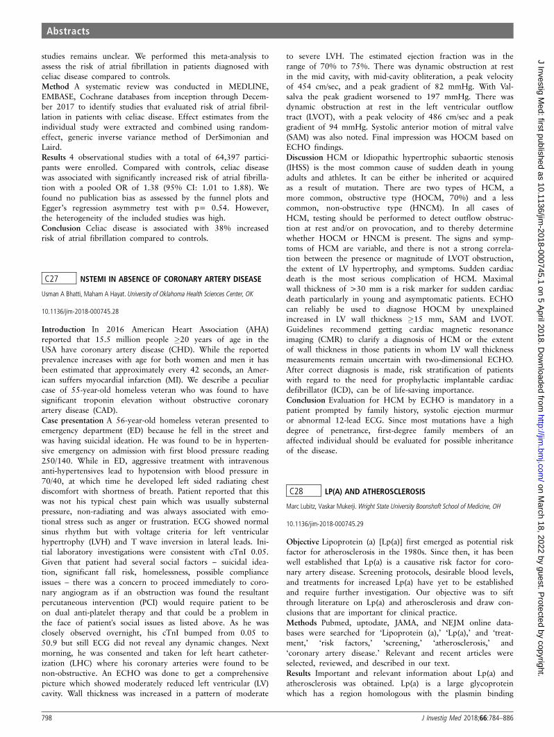
studies remains unclear. We performed this meta-analysis toassess the risk of atrial fibrillation in patients diagnosed withceliac disease compared to controls.Method A systematic review was conducted in MEDLINE,EMBASE, Cochrane databases from inception through Decem-ber 2017 to identify studies that evaluated risk of atrial fibril-lation in patients with celiac disease. Effect estimates from theindividual study were extracted and combined using random-effect, generic inverse variance method of DerSimonian andLaird.Results 4 observational studies with a total of 64,397 partici-pants were enrolled. Compared with controls, celiac diseasewas associated with significantly increased risk of atrial fibrilla-tion with a pooled OR of 1.38 (95% CI: 1.01 to 1.88). Wefound no publication bias as assessed by the funnel plots andEgger’s regression asymmetry test with p= 0.54. However,the heterogeneity of the included studies was high.Conclusion Celiac disease is associated with 38% increasedrisk of atrial fibrillation compared to controls.
C27 NSTEMI IN ABSENCE OF CORONARY ARTERY DISEASE
Usman A Bhatti, Maham A Hayat. University of Oklahoma Health Sciences Center, OK
10.1136/jim-2018-000745.28
Introduction In 2016 American Heart Association (AHA)reported that 15.5 million people �20 years of age in theUSA have coronary artery disease (CHD). While the reportedprevalence increases with age for both women and men it hasbeen estimated that approximately every 42 seconds, an Amer-ican suffers myocardial infarction (MI). We describe a peculiarcase of 55-year-old homeless veteran who was found to havesignificant troponin elevation without obstructive coronaryartery disease (CAD).Case presentation A 56-year-old homeless veteran presented toemergency department (ED) because he fell in the street andwas having suicidal ideation. He was found to be in hyperten-sive emergency on admission with first blood pressure reading250/140. While in ED, aggressive treatment with intravenousanti-hypertensives lead to hypotension with blood pressure in70/40, at which time he developed left sided radiating chestdiscomfort with shortness of breath. Patient reported that thiswas not his typical chest pain which was usually substernalpressure, non-radiating and was always associated with emo-tional stress such as anger or frustration. ECG showed normalsinus rhythm but with voltage criteria for left ventricularhypertrophy (LVH) and T wave inversion in lateral leads. Ini-tial laboratory investigations were consistent with cTnI 0.05.Given that patient had several social factors – suicidal idea-tion, significant fall risk, homelessness, possible complianceissues – there was a concern to proceed immediately to coro-nary angiogram as if an obstruction was found the resultantpercutaneous intervention (PCI) would require patient to beon dual anti-platelet therapy and that could be a problem inthe face of patient's social issues as listed above. As he wasclosely observed overnight, his cTnI bumped from 0.05 to50.9 but still ECG did not reveal any dynamic changes. Nextmorning, he was consented and taken for left heart catheter-ization (LHC) where his coronary arteries were found to benon-obstructive. An ECHO was done to get a comprehensivepicture which showed moderately reduced left ventricular (LV)cavity. Wall thickness was increased in a pattern of moderate
to severe LVH. The estimated ejection fraction was in therange of 70% to 75%. There was dynamic obstruction at restin the mid cavity, with mid-cavity obliteration, a peak velocityof 454 cm/sec, and a peak gradient of 82 mmHg. With Val-salva the peak gradient worsened to 197 mmHg. There wasdynamic obstruction at rest in the left ventricular outflowtract (LVOT), with a peak velocity of 486 cm/sec and a peakgradient of 94 mmHg. Systolic anterior motion of mitral valve(SAM) was also noted. Final impression was HOCM based onECHO findings.Discussion HCM or Idiopathic hypertrophic subaortic stenosis(IHSS) is the most common cause of sudden death in youngadults and athletes. It can be either be inherited or acquiredas a result of mutation. There are two types of HCM, amore common, obstructive type (HOCM, 70%) and a lesscommon, non-obstructive type (HNCM). In all cases ofHCM, testing should be performed to detect outflow obstruc-tion at rest and/or on provocation, and to thereby determinewhether HOCM or HNCM is present. The signs and symp-toms of HCM are variable, and there is not a strong correla-tion between the presence or magnitude of LVOT obstruction,the extent of LV hypertrophy, and symptoms. Sudden cardiacdeath is the most serious complication of HCM. Maximalwall thickness of >30 mm is a risk marker for sudden cardiacdeath particularly in young and asymptomatic patients. ECHOcan reliably be used to diagnose HOCM by unexplainedincreased in LV wall thickness �15 mm, SAM and LVOT.Guidelines recommend getting cardiac magnetic resonanceimaging (CMR) to clarify a diagnosis of HCM or the extentof wall thickness in those patients in whom LV wall thicknessmeasurements remain uncertain with two-dimensional ECHO.After correct diagnosis is made, risk stratification of patientswith regard to the need for prophylactic implantable cardiacdefibrillator (ICD), can be of life-saving importance.Conclusion Evaluation for HCM by ECHO is mandatory in apatient prompted by family history, systolic ejection murmuror abnormal 12-lead ECG. Since most mutations have a highdegree of penetrance, first-degree family members of anaffected individual should be evaluated for possible inheritanceof the disease.
C28 LP(A) AND ATHEROSCLEROSIS
Marc Lubitz, Vaskar Mukerji. Wright State University Boonshoft School of Medicine, OH
10.1136/jim-2018-000745.29
Objective Lipoprotein (a) [Lp(a)] first emerged as potential riskfactor for atherosclerosis in the 1980s. Since then, it has beenwell established that Lp(a) is a causative risk factor for coro-nary artery disease. Screening protocols, desirable blood levels,and treatments for increased Lp(a) have yet to be establishedand require further investigation. Our objective was to siftthrough literature on Lp(a) and atherosclerosis and draw con-clusions that are important for clinical practice.Methods Pubmed, uptodate, JAMA, and NEJM online data-bases were searched for ‘Lipoprotein (a),’ ‘Lp(a),’ and ‘treat-ment,’ ‘risk factors,’ ‘screening,’ ‘atherosclerosis,’ and‘coronary artery disease.’ Relevant and recent articles wereselected, reviewed, and described in our text.Results Important and relevant information about Lp(a) andatherosclerosis was obtained. Lp(a) is a large glycoproteinwhich has a region homologous with the plasmin binding
Abstracts
798 J Investig Med 2018;66:784–886
on March 18, 2022 by guest. P
rotected by copyright.http://jim
.bmj.com
/J Investig M
ed: first published as 10.1136/jim-2018-000745.1 on 5 A
pril 2018. Dow
nloaded from

domain of fibrinogen. This impairs fibrinolysis and leads toincreased clotting. It also promoted foam cell formation. Lp(a)levels are 90% genetically determined with an identified var-iant allele causing increased levels in affected patients. Manystudies have shown that higher levels of lp(a) are correlatedwith coronary artery disease with hazard ratios and relativerisk ratios nearing 2.5 when compared to normal Lp(a) levels.The studies do not use universal cutoff points to distinguishbetween ‘high’ and ‘low,’ lp(a), but show a linear correlationbetween lp(a) and disease. Studies addressing the allelic variantof lp(a) strongly suggest a causal relationship between lp(a)and coronary disease. Current screening recommendations arenot standardized, and treatment has been proven to decreasedisease risk. Niacin and PCSK-9 inhibitors are known todecrease Lp(a) levels. Low dose aspirin has been shown todecrease risk of disease in patients with high lp(a). Statins donot lower lp(a) but still decrease disease. Lipid apheresis candecrease levels greatly and RNA antisense drug treatments thattarget Lp(a) mRNA are currently in early clinical trials.Conclusion Lp(a) is a well described, casual risk factor for cor-onary artery disease. Appropriate screening, desirable bloodlevels, and effective treatments to mitigate disease burden haveyet to be described. Niacin and PCSK-9 inhibitors representreasonable adjunctive treatments for patients with high lp(a),in addition to statin and aspirin therapy. Antisense RNA drugsare exciting future treatments that will hopefully decrease Lp(a) levels and coronary artery disease.
C29 POSTERIOR REVERSIBLE ENCEPHALOPATHY SYNDROMEPRESENTING AS AN ST-ELEVATION MYOCARDIALINFARCTION
Zachary Oman, Lucas Gu, Hassan Alkhawan, Max Bourdillon, Ammar Nasir, Tarek Ajam. St.Louis University Hospital, MO
10.1136/jim-2018-000745.30
Introduction Posterior reversible encephalopathy syndrome(PRES) is an uncommon diagnosis secondary to cerebral injury.The exact pathogenesis remains unclear but it has been associ-ated with hypertension (HTN) and immunosuppressive ther-apy. As a secondary consequence, PRES can lend to thedevelopment of stress induced cardiomyopathy. We present arare case of PRES presenting as an ST-Elevation MyocardialInfarction (STEMI).Patient presentation A 62-years-old woman with past medicalhistory of hypertension, rheumatoid arthritis, and adrenalinsufficiency on immunosuppressive therapy was found unre-sponsive by her husband and taken to an emergency depart-ment where she developed tonic-clonic seizures and wassubsequently intubated for airway protection. CT head imag-ing was significant for a small area of hypoattenuation in theleft parieto-occiptal region concerning for subacute stroke. Shewas transferred to our facility for further stroke evaluation.Upon presentation, her ECG was significant for inferolateralSTEMI (figure 1) with corresponding Troponin-I of 12.44 ng/ml. Urgent coronary angiography revealed widely patent coro-nary arteries while echocardiography was significant for stressinduced cardiomyopathy with left ventricular (LV) apical aki-nesis with small apical thrombus, and preserved systolic func-tion of basal segments (figure 2). A brain MRI was performedto further workup neurologic events which revealed an injury
pattern consistent with posterior reversible encephalopathysyndrome (PRES) with areas of infarction (figure 3). She wascleared for anticoagulation therapy to treat LV apical thrombusalong with adequate blood pressure control.
Abstract C29 Figure 1 Electrocardiogram with inferolateral ST-segment elevation myocardial infarction
Abstract C29 Figure 2 Echocardiogram in end systole with leftventricular akinesis. White arrow indicating LV apical thrombus. (LV: leftventricle, RV: right ventricle)
Discussion PRES can be a consequence of HTN alone but onestudy has found that the rate for development of PRES inhospitalized patients with sudden HTN matched for non-hos-pitalized controls using age and HTN history found noincrease in incidence for the development of PRES.8Patientswho present with PRES will often have significant HTN andpossibly seizures, but other disease processes such as chronicimmune suppressive therapy can also contribute. Of signifi-cance, our patient not only had a history of HTN and pre-sented with seizure, but she was also chronicallyimmunosuppressed with Leflunomide, Methotrexate, and Pre-dnisone for her rheumatoid arthritis and adrenal insufficiency.Interestingly, our patient presented with clinical evidence ofmyocardial infarction for which urgent coronary angiographywas negative.
The proposed mechanism of cardiac injury is hyper-cate-cholamine stress resulting in cardiac injury. Studies in animalmodels have shown that intracerebral damage caused elevatedcatecholamines and was associated with coronary microvascular
Abstracts
J Investig Med 2018;66:784–886 799
on March 18, 2022 by guest. P
rotected by copyright.http://jim
.bmj.com
/J Investig M
ed: first published as 10.1136/jim-2018-000745.1 on 5 A
pril 2018. Dow
nloaded from
spasm and myocardial necrosis.2 Myocardial injury due toacute cerebral infarction produces myofibrillar degenerationcharacterized by dense eosinophilic transverse bands withintervening granularity as opposed to coagulation necrosis seenin coronary thrombosis where myofibrillar structure isintact.3,4 Elevated troponin in the setting of acute cerebralinfarction is also a poor prognostic indicator associated withhigher risk of in-hospital death and non-fatal cardiac event.7
Conclusion ST-Elevation Myocardial Infarction with elevatedtroponins can be misleading in patients presenting with recentcentral nervous system damage and seizure. While it is para-mount to rule out a significant myocardial event, one shouldalso take into consideration the neurogenic impact on cardiacfunction as seen in this case with PRES.
Abstract C29 Figure 3 Brain MR with area of T2/FLAIRhyperintensity
C42 DEMOGRAPHICS INCLUDING AGE, GENDER ANDIMMIGRATION STATUS IMPACT ONCORONARY ARTERYDISEASE: A RETROSPECTIVE ANALYSIS
1Fadi Ghrair, 2Hassan Alkhawam. 1Icahn School of Medicine at Mount Sinai (Elmhurst), NY;2Saint Louis University, MO
10.1136/jim-2018-000745.31
Background Coronary artery disease (CAD) is the leadingcause of death worldwide. Several factors play a role in therisk and severity of CAD.Aim To assess the effect of demographics including age, gen-der, and immigration status among patients with CAD.Methods A retrospective, single-center analysis was applied toall patients admitted for chest pain and underwent cardiac
catheterization between 01/01/2005 and 12/31/2016. Our hos-pital serves one the most diverse populations in New YorkCity. Information about age, gender, and immigration statuswas obtained from the chart. The term undocumented immi-grant refers to foreign nationals residing in the U.S. withoutlegal immigration status. The term legal resident refers to allother types of residence including but not limited to U.S.Citizens, permanent residents, tourist and student visaholders.Results Of 8,917 patients who underwent cardiac catheteriza-tion, 4,537 (50.9%) were <60 years old versus 4,380(49.1%) were �60 years old. CAD was more prevalent,severe, male-predominant and presented more as acute coro-nary syndrome (ACS) among patients �60 years old com-pared to those <60 years old. Table 1 of 8,917 patients,2,547 (28.6%) patients were undocumented immigrants versus6,370 (70.4%) who were legal residents. Undocumentedimmigrants displayed evidence of CAD at a younger age andhad more severe CAD than legal residents. Table 2 CAD wasmore prevalent among males but females developed CAD ata younger age. Males had more severe CAD and presented asACS more than females. Table 3.Conclusion Older patients have a higher degree of CAD.Undocumented immigrants have less prevalence of CAD butthey do have worse presentation and more server CAD find-ings at earlier ages. Males have more CAD prevalence, severeCAD and worse presentation than females.
Abstract C42 Table 1 Age
Abstracts
800 J Investig Med 2018;66:784–886
on March 18, 2022 by guest. P
rotected by copyright.http://jim
.bmj.com
/J Investig M
ed: first published as 10.1136/jim-2018-000745.1 on 5 A
pril 2018. Dow
nloaded from

Abstract C42 Table 2 Legal resident vs undocumented
Abstract C42 Table 3 Males vs females
C49 DONOR TATTOOS ARE NOT ASSOCIATED WITH POOROUTCOMES IN HEART TRANSPLANTATION
Mohamed Khayata, Sadeer Al-Kindi, Guilherme Oliveira. University Hospitals ClevelandMedical Center, OH
10.1136/jim-2018-000745.32
Objective Cardiac donor’s medical and social history is thesecond most common reason for non-recovery of hearts after
poor graft function. There is limited data that studied the sig-nificance of cardiac donor’s history of high-risk social behav-ior on recipient post transplant outcomes. We sough toinvestigate the impact of the presence of tattoos in cardiacdonors on recipient overall and graft survival.Method We identified all adults who received heart transplan-tation between 2000 and 2015 in the United States. Patientswere divided by the presence or absence of donor tattoos.Cox proportional hazard models were performed to identifyimpact on long-term outcomes.Results We identified a total of 30,261 transplantations. Ofthose, 10,796 had organs from donors with tattoos (DwT).DwT were more likely to be younger (29 vs 33 years,P<.001), male (76% vs 68%, P<.001), and to have a historyof heavy alcohol use (16% vs 9%, P<.001), and to usecocaine (21% vs 10%, P<.001). After adjusting for donor andrecipient factors, donor tattoos was not associated with overallsurvival (Hazard Ratio 0.98 [0.93–1.03], P=0.43), or graftsurvival (HR 0.98 [0.93–1.03], P=0.32).Conclusion Our study showed that presence of tattoos in car-diac donors has no impact on overall and graft survival. Tothe best of our knowledge, this is the first analysis thatstudied the impact of tattoos on heart transplantation recipi-ents. Our results may be helpful by considering donors forheart transplantation who were labeled as having a high-risksocial behavior.
Diagnosis or Treatment of a Disease Processor Clinical Syndromes
A07 EPIGASTRIC PAIN POST-PCI; A RARE ATHEROEMBOLICCOMPLICATION OF A COMMON PROCEDURE
Ranju Kunwor, AnnMarie Canelas. MacNeal hospital, IL
10.1136/jim-2018-000745.33
Objective/background The reported incidence of atheroembolicevents after Percutaneous Coronary Intervention (PCI) variesfrom 0.6 to 0.9 % in retrospective studies to 1.4 to 1.9 % inprospective series.1 The kidney is one of the principal endorgans that may be affected by atheroembolic events followedby skin with incidence of 0.9% and 0.6% respectively in aprospective study.2 Isolated splenic infarction is a rare athe-roembolic complication and only a few cases have beenreported in the literature thus far.3,4
Method We present a case of isolated splenic infarction postcardiac catheterization.Result 68 years old Caucasian Male with past medical history ofangina, hypertension, and abdominal aortic aneurysm was admit-ted for elective PCI. PCI was performed via radial access. Angiog-raphy showed dominant Right Coronary Artery (RCA) with 95%stenosis, for which RCA was stented with a drug-eluting stent. Thepatient complained of mild epigastric pain post PCI but the peri-procedural course was otherwise uneventful. The patient was dis-charged home about 6 hours after the procedure. Later that day,he presented to the Emergency Department with increasing epi-gastric pain. He had tried over the counter medications but withno relief. He denied any nausea, vomiting, or sweating. His vitalsigns were within normal limits (WNL). On physical examinationwas significant for epigastric tenderness and rest of examinationwas WNL. Laboratory finding for CBC, CMP, and lipase was
Abstracts
J Investig Med 2018;66:784–886 801
on March 18, 2022 by guest. P
rotected by copyright.http://jim
.bmj.com
/J Investig M
ed: first published as 10.1136/jim-2018-000745.1 on 5 A
pril 2018. Dow
nloaded from

WNL. EKG was unremarkable with the rate of 73/min, and no ST-Twave changes. Troponins were negative. CT abdomen with con-trast showed multiple small wedge-shaped defects in an enhance-ment of spleen suggestive of splenic infarcts; Focal aneurysmaldilatation of infra-renal abdominal aneurysm was stable at 3.5 cm.The patient was symptomatically managed for pain. Esophagogas-troduodenoscopy was performed that showed a hiatal hernia with-out gastritis or esophagitis. The patient was discharged after 24hours, continuing dual antiplatelet therapy, and close follow upwith primary care physician. One year after, he has not had anycomplications related to splenic infarction and is doing well.Conclusion This case illustrates that a symptomatic isolatedsplenic infarction can be a complication after cardiac catheter-ization. Management includes rest, analgesics, and follow upscans to confirm the resolution of infarction. Symptoms usu-ally resolve with conservative management. The true incidenceof splenic infarction after cardiac catheterization is difficult todetermine because of its rarity. Splenic hemorrhage, ruptureand abscess are the potential rare but fatal consequences ofsplenic infarction. Physicians should be aware of this poten-tially serious complication in patients developing abdominalpain post-cardiac catheterization.
REFERENCES1. Bashore TM, Gehrig T. Cholesterol emboli after invasive cardiac procedures. J.
Am. Coll. Cardiol July 2003;42(2):217–8.2. Fukumoto Y, Tsutsui H, Tsuchihashi M, Masumoto A, Takeshita A, Cholesterol
Embolism Study(CHEST) Investigators. The incidence and risk factors of cholesterolembolization syndrome, a complication of cardiac catheterization: A prospectivestudy. J. Am. Coll. Cardiol July 2003;42(2):211–6.
3. Hussein A, Kasmani R, Irani F, Mohan G. Athero-embolic isolated splenic infarc-tion following left cardiac catheterization. J. Cardiovasc. Med. March 2009;10(3):271–272.
4. Rose M, Dinour D, Chisin R. Splenic infarction: A complication of cardiac catheter-ization. Clin. Cardiol 1992;15:697–698.
A08 BRIC: A RARELY CONSIDERED DIAGNOSIS IN THEEVALUATION OF INTRAHEPATIC CHOLESTASIS
Mohamad H Khattab, Ahmed Salman, Ijlal Ali, George Salem, Javid Fazili. University ofOklahoma Health Sciences Center, OK
10.1136/jim-2018-000745.34
Intrahepatic cholestasis is a common disease entity internistssee in their daily practice with a wide differential includingDubin-Johnson syndrome, infiltrative diseases, drug inducedliver injury, and many others. Typical guideline directedworkup includes first reviewing medications and obtaining aright upper quadrant ultrasound. Based on the presence orabsence of ductal dilatation, one may proceed to ERCP vs.MRCP or obtaining anti-mitochondrial (AMA), anti-nuclear(ANA) and anti-smooth-muscle (ASMA) antibodies, respectively.One rare cause (prevalence not known exactly but fewer than1 in 100,000 people worldwide) of intrahepatic cholestasis isbenign recurrent intrahepatic cholestasis (BRIC), a disease proc-ess with a benign natural history of no risk for decompensa-tion of synthetic liver function. It is not typically considered inthe differential diagnosis of many internists and gastroenterolo-gists, leading to expensive, repetitive and unnecessary testing.
A 24-year-old male with a history of unexplained hyperbilir-ubinemia and alcohol use presented to the hospital with pro-gressive jaundice, pruritis and fatigue for the past 3 weeks.Patient reported only remote alcohol use 5–6 years ago andno history of intravenous drug use. He was not on any
medications, prescription or over the counter. He reported twoprevious week long hospitalizations in the last 18 months forsimilar issues. Labs on admission were notable for an elevatedaspartate aminotransferase (55 U/L), alanine aminotransferase(73 U/L), alkaline phosphatase (289 U/L), total bilirubin (24.8mg/dL), and direct bilirubin (16 mg/dL). He was extensivelyworked up in the past without any definitive diagnosisreached. Workup included hepatitis panel, ANA, AMA, ASMA,and ceruloplasmin levels. Liver ultrasound and CT of the abdo-men had been unremarkable. Transjugular liver biopsy showedparenchymal lobules that exhibited mild unrest with core disar-ray, slight kupffer cell hyperplasia and subtle evidence of canal-icular cholestasis. With the patient’s worsening jaundice andescalating bilirubin, the aforementioned tests were repeated andonce again were all negative (extremely low titers). Review ofprevious records revealed normalization of bilirubin 2 monthsafter his last episode of jaundice. Given the typical recurrentpattern of direct hyperbilirubinemia with interval normalization,and the findings on the pathology report, a diagnosis of BRICwas made. The patient was educated on the benign nature ofhis disease and discharged with Ursodiol and Atarax for pru-ritic control. He was followed up in liver clinic and continuesto do well with slowly improving bilirubin levels.
BRIC is a rare and subtle diagnosis, especially consideringthe prevalence of viral hepatitis in our patient population, aswell as Wilson disease in this age group. BRIC, first describedmany years ago, is diagnosed by identifying 2 of 6 specificcharacteristics (noted below) of which our patient had all 6.1
BRIC is not known to cause progressive liver injury or cirrho-sis, and the mainstay of management is supportive care andsymptom management as effective treatment modalities havenot otherwise been established.2 Familiarity with the belowcriteria and considering BRIC in patients with recurrent cho-lestasis can help prevent redundant, costly workup.
Diagnostic Criteria for BRIC:
1. At least two episodes of jaundice with asymptomatic intervalof months to years
2. Laboratory investigations suggestive of intrahepatic cholestasis3. Cholestasis induced severe pruritus4. Cholangiography showing normal intra and extrahepatic bile
ducts5. Liver histology suggesting centrilobular cholestasis6. Absence of other causes of cholestasis.
REFERENCES1. Kumar P, Charaniya R, Ahuja A, Mittal S, Sahoo R. Benign recurrent intrahepatic
cholestasis in a young adult. Journal of Clinical and Diagnostic Research: JCDR2016;10(6):OD01–OD02.
2. Summerfield J, Scott J, Berman M, Ghent C, Bloomer J, Berk D, Sherlock S.Benign recurrent intrahepatic cholestasis: studies of bilirubin kinetics, bile acids,and cholangiography. Gut 1980 February;21(2):154–160.
A16 ABDOMINAL WALL ABSCESS SECONDARY TOCHOLECYSTOCUTANEOUS FISTULA: RARECOMPLICATION OF COMMON BILE DUCT STENTOCCLUSION
Taseen A Syed, Sultan Mahmood, Salman Nusrat. University of Oklahoma Health SciencesCenter, OK
10.1136/jim-2018-000745.35
A 68-year-old female with history of chronic obstructive lungdisease, gastroesophageal reflux disease, choledocholithiasis s/p
Abstracts
802 J Investig Med 2018;66:784–886
on March 18, 2022 by guest. P
rotected by copyright.http://jim
.bmj.com
/J Investig M
ed: first published as 10.1136/jim-2018-000745.1 on 5 A
pril 2018. Dow
nloaded from

endoscopic retrograde cholangiopancreatography (ERCP) withcommon bile duct stenting presented with chronic abdominalpain and subjective fever for three weeks. Physical examina-tion was significant for blood pressure of 90/50 mmhg, heartrate of 114 beats/minute, fever of 38.6 Celsius, abdominal dis-tention and palpable abdominal wall mass in right lower quad-rant. Initial investigations showed white cell count of 29 k/mm3, Hgb of 10.7 g/dl, platelets of 540,000 mm3, INR of1.2 and creatinine of 0.52 mg/dl. Abdominal computerizedtomography showed enhancement of gallbladder wall and fis-tulous tract extending from gall bladder to right lower quad-rant abdominal wall with an abdominal wall abscess. (Figure1) A hepatobiliary (HIDA) scan showed radiotracer beingexcreted through the fistulous tract from the gallbladder intothe right abdominal wall and stasis of the radiotracer withinthe CBD stent suggesting at least partial obstruction of thestent. (Figure 2) Patient was started on broad spectrum antibi-otics, and incisional drainage of the abdominal wall abscesswas performed. Later ERCP was performed that showedoccluded biliary stent that was removed. (Figure 3) Hospitalcourse was complicated with sepsis and recollection of theabdominal wall fluid for which wound vacuum was placed.Despite appropriate management patient progressively deterio-rated and goals of care opted towards comfort measures.
A cholecystocutaneous fistula is an abnormal epithelial tractbetween the gallbladder and the skin. This is an extremelyuncommon presentation that is usually seen as a complicationof chronic biliary tract disease such as calculous cholecystitis,though cases have been reported with adenocarcinoma of thegallbladder as well.1,2 The obstruction of the cystic duct dueto a calculus or a carcinoma, causes perforation and subse-quent fistula formation.3 The most common presenting symp-toms are fevers, sweats, abdominal pain, anorexia andassociated lump under the skin indicative of an abscess.4 Theinitial imaging studies are ultrasound and computerized tomog-raphy that can show a thickened gall bladder, fistulous tractand abdominal wall abscess. In the presence of a fistula open-ing, a fistulography can be a useful diagnostic modality. ERCP
can be used as a diagnostic modality and for removal of astone blocking the cystic or common bile duct. Treatmentbegins with antibiotics with surgical intervention as the defini-tive treatment. A laparoscopic vs a single-stage surgicalapproach can be determined based on comorbidities. An opensurgical approach is preferred if there is an underling malig-nancy of the gall bladder.3,4 To our knowledge, there has notbeen any other case of cholecystocutaneous fistula formationcomplicated by intraabdominal abscess secondary to occlusionof a previously placed common bile duct stent.
Abstract A16 Figure 2
Abstract A16 Figure 3
Abstract A16 Figure 1
Abstracts
J Investig Med 2018;66:784–886 803
on March 18, 2022 by guest. P
rotected by copyright.http://jim
.bmj.com
/J Investig M
ed: first published as 10.1136/jim-2018-000745.1 on 5 A
pril 2018. Dow
nloaded from

A06 RECURRENT PANCREATITIS SECONDARY TO DIFFUSELARGE B-CELL LYMPHOMA
Taseen A Syed, Zain Asad, Maham Hayat, Molly Disbrow, William Tierney. University ofOklahoma Health Sciences Center, OK
10.1136/jim-2018-000745.36
Introduction Pancreatic lymphoma is uncommon, representingless than 0.5% of pancreatic tumors, with diffuse large B-cell lym-phoma (DLBCL) being the predominant type. Acute pancreatitissecondary to pancreatic lymphomas is a rare presentation. Wereport a case of recurrent pancreatitis secondary to DLBCL.Case presentation 57-year-old female with history of recurrentepisodes of pancreatitis presented with complaint of upperabdominal pain. Physical examination was significant for ten-derness in the epigastrium and left upper quadrant along withpoint tenderness at level of thoracic vertebra T8 – T9. Initialinvestigations showed a normal complete blood count, with anincreased lipase of 149 units/L and a creatinine of 0.68 mg/dl.Abdominal computerized tomography showed stranding sur-rounding the pancreas reflecting acute pancreatitis. Magneticresonance imaging of the back showed a large mass at T9 ver-tebral body surrounding the aorta and causing severe left neu-roforaminal narrowing. PET scan showed hypermetabolic focusin head of pancreas with maximum dimension of 2.5 cm withmultiple hypermetabolic osseous metastases largest being atlevel of T9 vertebra. An endoscopic ultrasound was performedwith fine needle biopsy of the paraspinal mass. Pathologyslides diagnosed the mass as Stage IV high Grade DiffuseLarge B-Cell Lymphoma. Patient subsequently, underwent 5cycles of R+DA-EPOCH regimen, one cycle of R-HCVADmini B cycle and 6 cycles of intrathecal methotrexate alternat-ing with cytarabine. Currently, he is being followed up inoncology clinic with significant symptom improvement.Conclusion Secondary involvement of the pancreas by diffuseLarge B-Cell lymphoma is a rare occurrence. Patients whopresent with pancreatitis specifically recurrent pancreatitisshould be evaluated for this diagnosis. Pathological diagnosisis important to distinguish pancreatic lymphoma from pancre-atic adenocarcinoma. Treatment usually consists of a combina-tion of chemotherapy and radiation therapy, or stem celltransplantation. Most cases however respond very well to che-motherapy only.
Abstract A06 Figure 1
Abstract A06 Figure 2
Abstract A06 Figure 3
Abstracts
804 J Investig Med 2018;66:784–886
on March 18, 2022 by guest. P
rotected by copyright.http://jim
.bmj.com
/J Investig M
ed: first published as 10.1136/jim-2018-000745.1 on 5 A
pril 2018. Dow
nloaded from

Abstract A06 Figure 4
Abstract A06 Figure 5
A39 PRIMARY DUODENAL ADENOCARCINOMA PRESENTINGAS A GIANT DUODENAL ULCER
Muaataz Azawi, Raghav Bansal, Maher Homsi, Soohwan Chun. Mount Sinai Elmhurst, NY
10.1136/jim-2018-000745.37
Introduction Giant duodenal ulcer refers to a duodenal ulcerthat is greater than 2 cm in diameter. In majority of thecases, the etiology is benign; however, primary duodenal carci-noma is encountered not infrequently, upto 15% of GDU.Despite this, there is no standard guideline in diagnosticapproach or in surveillance. Here we present a case of refrac-tory giant duodenal ulcer that was ultimately diagnosed as pri-mary duodenal adenocarcinoma, 2 months after the initialpresentation.Case presentation 76 year-old female presented with epigastricpain and hematemesis. Patient was taking aspirin 81mg daily,however denied taking any other non-steroidal anti-inflamma-tory medications. Initial laboratory test was notable for hemo-globin of 4.1 g/dL. Upper endoscopy showed a large ulcer induodenal bulb, approximately 20 mm in diameter with smalladherent clot. No biopsy was taken as the ulcer appearedbenign endoscopically. The etiology was attributed to aspirinand H. Pylori infection (positive stool antigen). Aspirin wassubsequently discontinued and patient was discharged with 14-day course of triple therapy. Two months later, patient
presented again with massive upper gastrointestinal bleeding.Repeat upper endoscopy showed previously seen duodenalulcer that had enlarged in size with worsened surroundingedema. Biopsies were taken from the edges of the ulcer; finalhistopathology revealed adenocarcinoma. A small hypodenseliver lesion seen on the abdominal Computed Tomographywas found to be a metastatic focus on liver biopsy. Patientwas not deemed to be a surgical candidate. She progressivelydeteriorated with development of gastric outlet obstructionand ultimately decided on palliative management.Discussion Giant duodenal ulcer accounts for 1–2% of allduodenal ulcers. As with any other peptic ulcers, the mostcommon etiologies are H. Pylori infection and use of non-steroidal anti-inflammatory medications. Primary duodenalmalignancy is rare in general. However, upto 15% of GDUwas found to be primary duodenal adenocarcinoma in oneseries. There is no standard guideline about obtaining biopsiesat index endoscopy or regarding surveillance. Newton, et al.suggested taking biopsies of all giant duodenal ulcer to ruleout malignancy. In the case of gastric ulcers, biopsies are rec-ommended for all ulcers to exclude malignancy and surveil-lance endoscopy to be performed in a selected group of highrisk patients. Similar approach may be necessary for giantduodenal ulcers, as the cost of missed diagnosis of malignancycan be paramount.
A09 RHEUMATIC HEART DISEASE IN AN ADULT PATIENTPRESENTING WITH NON-SPECIFIC SYMPTOMS
1Maiwand Mirwais, 2Sabawoon Mirwais, 1Laura Fay. 1University of Oklahoma HealthSciences Center, OK; 2University of Health Sciences
10.1136/jim-2018-000745.38
Introduction Rheumatic Heart Disease is cardiac inflammationand scarring triggered by an autoimmune reaction to infectionwith group A Streptococci. In the acute stage, this conditionconsists of pancarditis, with chronic disease manifested byvalvular fibrosis. It is a serious complication of RheumaticFever which is rare before age 5 years and after age 25 years;and is most frequently observed in children and adolescents.We discuss a case of Rheumatic Heart Disease in a relativelyhealthy 34 year old man.History and clinical findings A thirty four year old male pre-sented with complaints of severe muscle aches, arthralgia,fatigue, generalized malaise, subjective fevers and loss of appe-tite going on for a few weeks; beginning while he was onvacation in California. The symptoms began in his lowerextremities and gradually spread to the rest of his body mak-ing it relatively difficult to complete activities of daily life likechanging gears and applying brakes while driving. The patientdenied any sore throat and didn’t have any signs and symp-toms of pharyngitis.Laboratory and diagnostic data On presentation, his ESR andCRP were very elevated and an extensive autoimmune workupwas mostly inconclusive. His infectious workup was positivefor elevated Antistreptolysin O antibody titres which were atfive times the upper limit of normal (ULN). He underwent atransthoracic echocardiogram showing a small vegetation overthe pulmonary valve thus leading to a clinical diagnosis ofRheumatic Heart Disease.Management On admission, before the definitive diagnosis, thepatient was started on Tylenol, gentle fluid and dietary
Abstracts
J Investig Med 2018;66:784–886 805
on March 18, 2022 by guest. P
rotected by copyright.http://jim
.bmj.com
/J Investig M
ed: first published as 10.1136/jim-2018-000745.1 on 5 A
pril 2018. Dow
nloaded from

resuscitation. After the diagnostic workup, high dose Aspirinone gram every six hours and Amoxicillin 500 mg twice dailywere initiated. With significant symptomatic improvement, thepatient was discharged home to be started on penicillin pro-phylaxis on an outpatient basis for a total of ten years givenhe had valvular involvement.Conclusion Rheumatic heart disease is a rare condition andrequires a high degree of suspicion for diagnosis. It canpresent with non-specific symptoms like generalized malaisewithout a history of pharyngitis. The key is timely diagnosisand adequate antibiotic treatment followed by penicillinprophylaxis.
A56 UNUSUAL PRESENTATION OF SOLID PSEUDOPAPILLARYTUMOR OF THE PANCREAS WITH PYELONEPHRITIS ANDWEIGHT LOSS
Michael Kalinowski, Shana Kothari, Matthew Kobeszko, Rogelio Silva. University of Illinoisat Chicago – Advocate Christ Medical Center, IL
10.1136/jim-2018-000745.39
Introduction A solid pseudopapillary tumor (SPT) is a rareexocrine pancreatic neoplasm that accounts for 2–3% of pan-creatic neoplasms. In about 15% of patients, the neoplasmsare incidentally detected. Most patients have nonspecific clini-cal features such as abdominal pain, poor appetite, or nausea,all of which can be related to tumor compression on adjacentorgans. We present a unique case of a 20 year old femalethat initially presented with pyelonephritis and weight loss andwas found to have SPT of the pancreas.Case A 20 year old Jordanian female with history of type Idiabetes mellitus presented to the emergency department withleft sided flank pain that radiated into her groin, nausea, vom-iting, dysuria for three days, and a subjective 20 poundweight loss over the past 3 months. Upon presentation,patient was afebrile, normotensive, and tachycardic with perti-nent lab findings of leukocytosis. Urinalysis and culture werecompleted, and findings were consistent with pyelonephritis.On physical exam, left sided CVA tenderness was elicited, butno palpable abdominal masses were found. Patient underwentCT abdomen/pelvis to evaluate for possible nephrolithiasis.Imaging was negative for nephrolithiasis, but demonstrated a7.5 × 7 cm mass in the pancreatic head with mass effect;suggestive of SPT of the pancreas or GIST tumor. GI wasconsulted and recommended outpatient follow-up after fullresolution of the infection. Upon follow-up, EUS-FNA wasperformed, which revealed an epithelial neoplasm, most con-sistent with SPT of the pancreas. The tumor was compressingthe gastric antrum and duodenal bulb causing partial gastricoutlet obstruction. Surgery was consulted, and patient under-went pylorus preserving Whipple resection. Patient recoveredwell post-op and was discharged home in stable condition.Discussion SPT of the pancreas is a rare tumor that was firstdescribed by Frantz et al in 1959 and accounts for less than3% of all pancreatic tumors. Recently, there has been a 7-foldincrease in the number of SPTs diagnosed since the year2000, likely attributed to improvement of CT imaging andincreased access to EUS. SPT predominantly affects females(82–92%) with a median age of 22 and are found in thehead (34–40%) or tail (24–36%) of the pancreas in a majorityof cases. The most common presenting signs are upperabdominal pain (38–47%), followed by mass effect or palpable
abdominal mass (36%), or no symptoms at all (15%). Amongasymptomatic patients, the diagnosis is made incidentally or incases of acute unrelated injury. In our case, the only present-ing sign associated with the SPT was weight loss, which israre as it represents less than 1% of SPT cases.
Diagnosis of SPT is made through a combination of imag-ing modalities and pathologic findings. CT scan is the mostcommon initial modality of choice followed by diagnostic con-firmation with EUS guided FNA. The advent of EUS guidedFNA increased the diagnostic accuracy of SPT to 82.4% from23.5% for CT scans alone and 41.2% for EUS alone. Onaverage, SPTs are 6–8.5 cm in diameter and present withmetastasis 9–15% of the time. Overall, patients have a favor-able outcome with a 5 year survival of 94–97% following sur-gical resection. The recurrence rate is less than 5%.Conclusion This case is a unique presentation of SPT involvinga patient with pyelonephritis and weight loss. Routine CTimaging to evaluate for nephrolithiasis revealed an incidentalfinding of a 7.5 cm mass suggestive of possible SPT of thepancreas. Physicians should consider this neoplasm as a differ-ential in patients, specifically females 20–30 years old, thatpresent with unexplained weight loss and abdominal pain.
A57 ATYPICAL BRUGADA SYNDROME PRESENTING ASPERSISTENT VENTRICULAR TACHYCARDIA STORMREFRACTORY TO MAXIMAL MEDICAL ANDINTERVENTIONAL MANAGEMENT
Usman A Bhatti, Maham Hayat. University of Oklahoma Health Sciences Center, OK
10.1136/jim-2018-000745.40
Introduction Brugada Syndrome is an ECG abnormality with ahigh incidence of sudden death in patients with structurallynormal hearts. First described in 1992 by the Brugada broth-ers, the disease has since had an exponential rise in the num-bers of cases reported, to such an extent that the secondconsensus conference reported in 2005 that it was the secondleading cause of death in males <40 (after trauma). We reporta successful treatment of an unusual case of Brugada Syn-drome which was refractory to all forms of medical, surgicaland electrophysiological interventions.Case report A 48-year-old quadriplegic male presented withVF/VT arrest at home. He was successfully resuscitated withDCCV, started on amiodarone drip and transferred to hospitalwhere coronary angiogram failed to show any vessel obstruc-tion. EF was estimated to be 55–65%. Electrolytes and bloodglucose were within normal limits. His ECG findings wereconcerning for Brugada Type I pattern; hence an ICD wasplaced. Throughout his stay he continued to have recurrentPMVT/VF requiring multiple ICD shocks despite maximal dos-age of quinidine, amiodarone and isoproterenol. He was takenfor radiofrequency ablation which was unfortunately not suc-cessful and he had to be intubated and sedated to ride outthe electrical storm. He was extubated a few days later. Next,Dr. Shivkumar at UCLA, the world's leading expert on usingstellate ganglionectomy to treat electrical storm was contacted.He recommended bilateral stellate ganglionectomy all the waydown to T4-T5. Hence, thoracic surgery team was involvedand patient underwent uneventful surgery. Unfortunately,patient’s response to surgery was sub-optimal. At this point intime, patient had developed PTSD due to persistent ICDshocks. We discussed goals of care with patients family and
Abstracts
806 J Investig Med 2018;66:784–886
on March 18, 2022 by guest. P
rotected by copyright.http://jim
.bmj.com
/J Investig M
ed: first published as 10.1136/jim-2018-000745.1 on 5 A
pril 2018. Dow
nloaded from

offered our last option, renal sympathetic denervation, (RSD)as a final attempt to help patient’s VT storm. RSD is a proce-dure not yet approved in US as a therapy, however therehave been <20 case reports that it may suppress electricalstorm. Patient then underwent successful ablation of left ven-tricle purkinje system and renal sympathetic denervation afterwhich he had no further episodes of arrhythmia. His bloodpressure remained labile post ablation, due to loss of auto-nomic system as he had to rely completely on his endocrinesystem for blood pressure maintenance. He was started onmidodrine and pseudoephedrine to augment that response. Hecontinued to improve clinically and was sent to a skilled nurs-ing home on low dose amiodarone for rhythm control withclose follow-up in clinic.Discussion Brugada syndrome is due to a mutation in the car-diac sodium channel gene. There are 3 variants;
. Type 1 (Coved ST segment elevation >2mm in >1 of V1-V3followed by a negative Twave).
. Type 2 (has >2mm of saddleback shaped ST elevation).
. Type 3 (morphology of either type 1 or type 2, but with<2mm of ST segment elevation).
Diagnosis is made on ECG finding along with clinical crite-ria such as documented VT/VF, syncope and family history ofSCD among others. ECG changes can be unmasked by multi-ple factors: fever, ischemia, hypokalemia, hypothermia andmedications (antiarrhythmic drugs, cocaine, alcohol). The onlyproven therapy is an implantable cardioverter – defibrillator(ICD). Quinidine has been proposed as an alternative in set-tings where ICD’s are unavailable or where they would beinappropriate (e.g.: neonates).Conclusion Brugada Syndrome has a classic ECG patternwhich is potentially diagnostic but it might be missed on dueto transient nature of ECG changes. Hence, it merits a highindex for suspicion in patients presenting as VT/VF with fam-ily history of sudden cardiac death and normal coronary circu-lation. It is appropriate for patients with suspicious ECGchanges to undergo electrophysiological evaluation to look forinducible VT/VF as undiagnosed Brugada syndrome has beenestimated to have a mortality of 10% per year.
A58 POSTPARTUM INDUCED THROMBOTICTHROMBOCYTOPENIC PURPURA
Aneesh kuruvilla, Elizabeth Brindise, Sushma Chinni, Jeffry Ording. UIC/Christ, IL
10.1136/jim-2018-000745.41
Objective Postpartum induced Thrombotic ThrombocytopenicPurpura (TTP) is a rare but feared complication of pregnancy.We present a 28-year-old gravida 1 para 1 female whorecently had a full term normal spontaneous vaginal deliverywho presented with severe acute thrombocytopenia and ane-mia in whom ultimately etiology of thrombocytopenia wasfound to be due to TTP.Method A 28-year-old Gravida 1 Para 1 female with no signif-icant past medical history, who recently had a full term preg-nancy without any complications via normal spontaneousvaginal delivery presented with lightheadedness, nausea, andvomiting for 1 week. Upon presentation, she was hemody-namically stable. Her physical examination was unremarkable.Her blood work showed severe acute anemia with Hgb of 6.1and severe acute thrombocytopenia with a platelet count of
27. Previous lab works showed mild thrombocytopenia. Crea-tinine was elevated at 2.15 and bilirubin elevated at 1.9. HerLFTS and PT/INR were within normal limits. Her peripheralblood smear showed 6–7 schistocytes. The clinical picture wasconcerning for TTP and a ADAMS13 level was checked andwas found to be less than 5 %, this confirmed the diagnosisand she was immediately started on plasmapheresis. Patientunderwent a total of 10 session of plasmapheresis. Shereceived pre treatments with solumedrol and Benadryl beforeeach plasmaphe resis session.Her symptoms improved and heracute anemia and thrombocytopenia resolved. Her creatinineand bilirubin were also normalized and patient was dischargedhome with a short course of prednisone.Result Postpartum induced TTP is a very rare yet, feared com-plication of pregnancy. Since the differences between causes ofthrombocytopenia associated with pregnancy are subtle, it’svery important to have correct diagnosis since the manage-ment are different. It is important to note that the classicalpentad of symptoms associated with TTP may not be presentin all patients. Our patient, had no neurological symptoms orfever upon presentation. Therefore a high index of suspicionfor TTP is warranted in the setting of thrombocytopenia,microangiopathic hemolytic anemia and kidney dysfunctionalone. Another important consideration is to check forADAMS 13 level before starting Plasmapheresis since postplasmapheresis ADAMS13 values are not accurate. A value lessthan 10 % suggest TTP.Conclusion There are numerous causes of thrombocytopeniaassociated with pregnancy, yet the differences between themare very subtle.This case report further illustrates the impor-tance of diagnosing the cause of thrombocytopenia since treat-ment depends upon the diagnosis.
A38 METASTATIC GASTROINTESTINAL STROMAL TUMORDISGUISING AS PANCREATIC PSEUDOCYST
Aneesh kuruvilla, Elizabeth Brindise, Amer Al Homssi, Neesh Anand. UIC/Christ, IL
10.1136/jim-2018-000745.42
Objective Gastrointestinal Stromal Tumors (GISTs) are themost common non-epithelial benign tumor of the gastrointesti-nal tract. We present a patient with abdominal pain previouslydiagnosed with pancreatic pseudocyst in which etiology wasultimately determined to be due to metastatic GISTs.Method A 59-year-old African American female with a pastmedical history of hypertension, and diabetes mellitus pre-sented to the Emergency room with diffuse abdominal painfor a month. Patient also reported early satiety, nausea and 50lbs weight loss over the past 2–3 months. She initially pre-sented about a month ago to an out-of-state hospital withabdominal pain. Evaluation revealed a large pancreatic pseudo-cyst. She underwent, an endoscopic ultrasound-guided cysto-gastrostomy with the placement of stent. She continued tohave abdominal pain, she was currently in town visiting fam-ily, but because of worsening abdominal pain she presented tothe emergency room. Imaging of the abdomen had revealed avery large abdominal mass between the gastric body and pan-creatic body and tail. There was also a lymph node lesion.
She underwent an endoscopic ultrasound, which revealedan ulcerated mass 3.5 cm in size in the proximal stomachalong the lesser curvature. On the contralateral gastric wall, inthe mid gastric body, there was the previously placed stent,
Abstracts
J Investig Med 2018;66:784–886 807
on March 18, 2022 by guest. P
rotected by copyright.http://jim
.bmj.com
/J Investig M
ed: first published as 10.1136/jim-2018-000745.1 on 5 A
pril 2018. Dow
nloaded from

and an attempt to the scope through the stent was unsuccess-ful with marked friable tissue ingrowth. There was no cysticcavity. The endoscopic ultrasound noted a very large hypoe-chocic mass at least 8 × 9 cm in size, appearing to arisefrom either pancreatic body or gastric wall with invasion intothe gastric wall. There was also 2.3 cm hypoechoic mass inthe left lobe of the liver. All the above lesions have beenbiopsied and results showed a high-grade malignant spindlecell neoplasm consistent with GISTs. Patient was referred toOncology and Surgery for further care and management.Results GISTs are the most common non-epithelial benigntumor of the gastrointestinal tract. They are most commonlyfound in the stomach and small intestine. Our patient waspreviously diagnosed with pancreatic pseudocyst for which sheunderwent endoscopic drainage with a negative workup formalignancy. Tumor markers including CEA, CA19–9, and AFPwere normal as well.Conclusion This Case illustrates that GIST should be consid-ered in the differential diagnosis when a patient presentingwith pancreatic pseudocyst.
A60 SEVERE REFRACTORY HYPOXEMIA; A CASE OFUNTREATED EBSTEIN’S ANOMALY IN AN ADULT
Ayesha Saeed, Rizwan A Khan, C Erick Kaufman. University of Oklahoma, OK
10.1136/jim-2018-000745.43
Background Ebstein’s anomaly is characterized by deformitiesof the anterior leaflet of the tricuspid valve and atrializationof the right ventricle. Patients with severe tricuspid regurgita-tion are recommended to have tricuspid valve surgery withconcomitant atrial septal defect closure. Most of the patientsare treated through surgery in early childhood. Untreatedcases often present with hypoxemia and other complicationsand early identification and subsequent treatment should beensured. We present a case of young female who presentedwith refractory hypoxemia secondary to untreated ebstein’sanomaly.Case report A 30-year-old Spanish speaking female presentedto the ED with shortness of breath since 1 day, and worsen-ing pedal edema since 4 days, and was subsequently found tohave Cr of 13 with anion gap metabolic acidosis. Patient wasinitially started on bicarbonate drip and subsequently under-went dialysis for refractory acidosis and volume overload.Upon further investigation, she was found to have history ofebstein’s anomaly w/ ASD, was reportedly lost to follow upmany years ago and also had history of CKD stage 5. Ontransthoracic echocardiography she had torrential tricuspidregurgitation, marked right heart dilation with systolic dys-function. During dialysis patient developed hypoxia with pulseoximetry saturations in 60–70's range without any improve-ment on non-rebreather or 100% high flow oxygen. Rapidresponse sequences were called for the patient by the nursingstaff although patient was clinically and neurologically intactwithout showing any signs of respiratory distress with theselow oxygen saturations. Cardiology was consulted who recom-mended transfusing patient with blood to keep hgb in polycy-themia range and congenital cardiology recommended surgicalintervention for the patient. Detailed discussion was held withthe patient to explain need for surgical intervention, butpatient refused any surgical interventions. The history ofpatient’s CKD was determined to be FSGS in the past by
nephrology, but the worsening was attributed to patient’suntreated cardiac congenital anomaly. Patient was optimizedwith dialysis sessions and blood transfusions but stayed persis-tently hypoxic. She was eventually discharged with plannedfollow up in cardiothoracic surgical clinic. She was again lostto follow up, only to present again to the ED with cardiacarrest and very severe refractory hypoxemia and eventuallydied during that hospital admission.Discussion Severe hypoxemia can be seen in patients withuntreated congenital heart diseases and often it is refractoryto oxygen supplementation due to underlying cardiac shunting.In patients with untreated ebstein’s anomaly, severe tricuspidregurgitation and an associated ASD results in right to leftshunting of blood causing severe hypoxemia. Transfusion ofblood products can temporarily improve oxygenation but sur-gical treatment is the only permanent cure. Patients withuntreated ebstein’s anomaly with severe tricuspid regurgitationshould be recognized and prompt surgical treatment should bepursued to prevent their clinical decline.
A61 PULMONARY ARTERY ANEURYSM AND PERICARDIALEFFUSION: HEART TEAM APPROACH IN MANAGING ANUNCOMMON CASE
Mohamed Khayata, Sanjay Rajagopalan, Farshad Farouzadneh. University HospitalsCleveland Medical Center, OH
10.1136/jim-2018-000745.44
Background Pulmonary artery (PA) aneurysm is rare in clinicalsettings. Early recognition and careful treatment should beconsidered to avoid fatal complications of this condition.Case report A 66-year-old male presented to an outside hospitalwith left sided chest pain and dyspnea. Chest CT angiographyshowed small pericardial effusion and severe PA dilation (7.5 cm).He was discharged on naproxen for possible pericarditis. Nine-teen days later, he presented with worsening symptoms. C-reac-tive protein was >68.5mg/l and EKG showed atrial flutter withrapid ventricular response. Repeated CT angiography showedenlarged pericardial effusion. After stabilizing the patient, he wastransferred to our cardiac ICU. Transthoracic echocardiographyshowed ejection fraction (EF) of 45%, severely dilated PA, ele-vated right ventricular systolic pressure, moderately dilated infe-rior vena cava, along with stenosis and moderate regurgitation ofpulmonic valve. Cardiac MRI showed aneurysmal PA (8 cm).Chest MRA showed aneurysmal ascending thoracic aorta (4.1 ×4.1 cm) and confirmed the aneurysmal main PA with the left andright pulmonary arteries measuring 4.5 and 5.4 cm, respectively.Right heart catheterization showed mildly elevated PA pressureswith wide pulse pressure and pulmonary angiogram showedseverely dilated aneurysmal PA.750 cc of serosanguineous fluidwere removed by pericardiocentesis and EF improved to 60% and75% the next day and four days after pericardiocentesis, respec-tively. Fluid analysis showed exudative effusion with negative cul-ture and cytology. Patients’ symptoms improved and he wasdischarged to follow up with pulmonary and cardiothoracic sur-gery for evaluation for possible pulmonic valve replacement basedon his symptoms and progression of his pulmonary regurgitation.Conclusion This case demonstrates the vital role of early diag-nosis and a heart team approach in management of rare andchallenging cases with potential serious complications such asPA aneurysm.
Abstracts
808 J Investig Med 2018;66:784–886
on March 18, 2022 by guest. P
rotected by copyright.http://jim
.bmj.com
/J Investig M
ed: first published as 10.1136/jim-2018-000745.1 on 5 A
pril 2018. Dow
nloaded from

A62 DELAYED MANAGEMENT OF MICROSCOPICPOLYANGIITIS LEADS TO CATASTROPHIC DIFFUSEALVEOLAR HEMORRHAGE AND PROLONGED ICU STAY
Laith Numan, Laith Derbas, Mohammad Tahboub, Mohannad Abu Omar. University ofMissouri-Kansas City, MO
10.1136/jim-2018-000745.45
Introduction Microscopic polyangitis (MPA) is a necrotizingvasculitis, predominantly affecting small and medium-sized ves-sels, commonly involving the kidney and lung. We present acase of MPA that presented with subtle initial manifestationswhich rapidly progressed to diffuse alveolar hemorrhage(DAH) and generalized skin rash leading to intubation andprolonged ICU stay.Case report A 71-year-old female with a history of hyperten-sion on hydralazine presented to the ED with dry cough andmild skin rash on her right lower extremity, both startedaround 5 days. On physical exam, the patient was found tohave normal oxygen saturation but bibasilar crackles and viola-ceous purpuric rash with a bullae formation on medial rightankle. Chest X-ray showed scattered interstitial opacities. D-dimer was >1000. CT angiogram of the showed small bibasi-lar areas of pulmonary consolidation but no pulmonary embo-lism. CRP at that time was 1.5. The decision was to send herhome on Levofloxacin for five days for a presumedpneumonia.
Two weeks later, she came back with worsening dyspnea.Physical exam was notable for oxygen saturation of 89% onroom air, bilateral crackles and worsening skin rash involvingbilateral lower extremities, back and shoulders and face (figure1). Her labs showed hypoxemia with PaO2 of 55 on roomair. Chest x-ray showed progressive diffuse bilateral heteroge-neous interstitial opacities. CRP was 18.5. Patient was subse-quently admitted to the intensive care unit and intubated foracute hypoxemic respiratory failure. CT chest was concerningfor alveolar hemorrhage which was confirmed on bronchoal-veolar lavage by bronchoscopy. Extensive workup revealed P-ANCA, MPO positive antibodies. Results were confirmed byskin biopsy and findings were consistent with ANCA associ-ated Vasculitis.
Abstract A62 Figure 1
Empiric high dose Methylprednisone was initiated followedby a total of 4 doses of Rituximab and 7 sessions of plasma-pheresis once the diagnosis was confirmed. Patient respondedto therapy well and was ultimately extubated.Conclusion The etiology for MPA flare remains a medicaldilemma. Hydralazine in our case is a known culprit to causeANCA vasculitis. Close attention and review of medications isnecessary when there is a concern or uncertainty in the diag-nosis. Second, there should be a high index of suspicion toprevent such dramatic and life-threatening scenarios.
B32 A UNIQUE PRESENTATION OF ACUTE SARCOIDMONOARTHRITIS IN A PATIENT WITH SARCOIDOSIS
1Ahmed Alsaei, 2John Hilts, 2S Volkov, 2Nadera Sweiss. 1UIC/Advocate Christ MedicalCenter, IL; 2University of Illinois at Chicago, IL
10.1136/jim-2018-000745.46
Sarcoidosis is a multisystem disorder of unknown etiology thatis characterized pathologically by the presence of non-caseatinggranulomas in affected organs. It predominantly affects thelungs and intrathoracic lymph nodes, but up to 30 percent ofpatients present with extrapulmonary manifestations. Wereport an interesting case of a sarcoidosis patient presentingwith acute monoarthritis found to have sarcoid arthropathy.
A 49 year old male with past medical history of sarcoidosispresented with acute on chronic left knee pain. The pain waslocated diffusely across his anterior knee, worse towards theend of the day, limiting his ability to ambulate. He rated thepain as 10 out of 10 in severity. He was taking oral methyl-prednisolone 8 mg daily for his sarcoidosis, and hydrocodone/acetaminophen 10/650 mg four times daily over the weekbefore presentation for his increased knee pain. He denied theuse of Disease Modifying Antirheumatic Drugs (DMARDs) inthe past. The patient denied other symptoms including fevers,weight loss, vision changes, cough, dyspnea, or rashes.
On presentation, his blood pressure was 109/63, tempera-ture was 97.9 degrees Fahrenheit, pulse was 93 beats perminute, respiratory rate was 20 breaths per minute, and pulseoximetry was 98% on ambient air. Physical exam was remark-able for left knee warm effusion without erythema and fullrange of motion with moderate pain. A semi-firm, 5 cm × 1cm, mobile, mass was palpated at the left, medial, distal thigh.Lab tests showed white blood cells were 9.5 103/uL with noleft shift, hemoglobin was 13.4 g/dl, and platelets were164,000/mcL. Basic metabolic panel and liver function testswere normal. Left knee x-ray revealed tricompartmental jointspace narrowing, osteophytes, and suprapatellar effusion. Onultrasound of the lower extremities, a non-vascular structurewas found in the left, medial, distal thigh. MRI of the leftknee with and without contrast shows a large complex jointeffusion with internal enhancing synovium, numerous nodulesand debris, and mass effect on left medial musculature with-out discrete mass.
An arthrocentesis of the left knee was performed and tencc’s of clear-yellow fluid was aspirated. On synovial fluid anal-ysis, there was 4,890 /mm3 white blood cells with 49% neu-trophils. No crystals were identified and no growth onbacterial culture seen.
The patient was given a left knee intra-articular corticoste-roid injection with relief of his pain and discharged home.The patient had a whole body PET/CT scan to evaluate extent
Abstracts
J Investig Med 2018;66:784–886 809
on March 18, 2022 by guest. P
rotected by copyright.http://jim
.bmj.com
/J Investig M
ed: first published as 10.1136/jim-2018-000745.1 on 5 A
pril 2018. Dow
nloaded from

of sarcoidosis which showed focal uptake in his left knee. Hewas started on corticotropin subcutaneous injections with goodcontrol of his pain and inflammation.
Acute monoarthritis is a common initial presentation ofmany joint disorders. The most common causes include osteo-arthritis, crystalline disease, and infection. In patients with ahistory of sarcoidosis, or hilar adenopathy and erythemanodosum, sarcoid arthritis should be considered if the initialwork up is inconclusive. Sarcoidosis may mimic or occur con-comitantly with many rheumatologic diseases. About 10–15percent of sarcoidosis patients have an associated arthropathy.Due to the fact that it can mimic rheumatologic diseases,delayed diagnosis and/or misdiagnosis can occur. Therefore, inpatients presenting to a rheumatologist with musculoskeletalsystem findings, it should definitely be considered in the dif-ferential diagnosis. Acute sarcoid arthritis is treated with anti-inflammatory medications such as NSAIDs or oral corticoste-roids. Most symptoms tend to resolve spontaneously.
A40 SPONTANEOUS ATRAUMATIC SPLENIC RUPTURE IN APATIENT WITH STREPTOCOCCAL PNEUMONIA
1Maiwand Mirwais, 2Sabawoon Mirwais, 1Ahmed Awab. 1University of Oklahoma HealthSciences Center, OK; 2University of Health Sciences
10.1136/jim-2018-000745.47
Introduction Spontaneous atraumatic splenic rupture is a rarebut dramatic occurrence that is most commonly attributed toinfection or neoplasia. Deciphering the etiology can be chal-lenging with many cases remaining unclear despite full investi-gations. We describe a case of such splenic rupture secondaryto streptococcus pneumoniae infection.History and initial laboratory data A fifty nine year old malewith a past medical history of peptic ulcer disease and hyper-tension presented with nausea, vomiting and subjective feversgoing on for a week. He was admitted for pneumonia; hisnasopharyngeal swab was positive for Coronavirus and hisurine was positive for Streptococcus pneumoniae antigen. Hewas started on appropriate antibiotics and during the hospitalstay, while being evaluated for acute blood loss anemia, hewas incidentally found to have spontaneous atraumatic splenicrupture and intraperitoneal bleeding.Management He had an emergent splenectomy done, andreceived post splenectomy immunizations. His hospital coursewas complicated by pulmonary embolism post operatively andthen an incidentally found descending aortic pseudoaneurysmwith ulceration requiring thoracic endovascular aortic repair(TEVAR). It wasn’t clear if the splenic rupture was related tothe descending aortic pseudoaneurysm from a pathophysiologi-cal stand point. His further workup including testing forCytomegalovirus, Ebstein-Barr virus, Human Immunodeficiencyvirus, viral hepatitis, syphilis infection were negative. His con-nective tissue diseases’ workup including Rheumatoid factor,ANA, c-ANCA, p-ANCA, cryoglobulin level were negative aswell. His splenic histopathology was negative for any malig-nancy and showed necrotic foci with inflammation. His hospi-tal course was further complicated by deconditioning whichimproved significantly with physical therapy and he was dis-charged home with close outpatient follow up.Conclusion Spontaneous splenic rupture is a rare phenomenonwhich has been associated with malignancies and certain infec-tious etiologies. Our patient appeared to have streptococcalinfection which despite ample antibiotic treatment, lead to
splenic rupture. This case also explains how important opti-mum post-operative care is to prevent long term decondition-ing and morbidity.
B33 MULTICENTRIC MYELOID SARCOMA WITH TESTICULARINVOLVEMENT
Natthapon Angsubhakorn, Attaya Suvannasankha. Indiana University School of Medicine,IN
10.1136/jim-2018-000745.48
Objective Myeloid sarcoma is a rare presentation of AcuteMyeloid Leukemia (AML) that can present either as an iso-lated extramedullary disease or in association with AML. It ismore commonly seen in the pediatric population and caninvolve any organs of the body; specifically, skin and lymphnodes are the two most common sites. Testicular involvementis extremely rare and often poses diagnostic and managementdilemmas. Here, we report a rare case of newly-diagnosedAML with multicentric myeloid sarcoma involving testes.Method Both authors cared for the patient, reviewed and sum-marized data from previously published cases, and drafted themanuscript. Written informed consent for case publication wasalready obtained.
Abstract B33 Figure 1
Abstracts
810 J Investig Med 2018;66:784–886
on March 18, 2022 by guest. P
rotected by copyright.http://jim
.bmj.com
/J Investig M
ed: first published as 10.1136/jim-2018-000745.1 on 5 A
pril 2018. Dow
nloaded from

Results A previously healthy 29-year-old man presented with aone-month history of fatigue and epigastric discomfort. Onexamination, he had ptosis of the right eye and bilateral tes-ticular enlargement with 1-cm hard mass adjacent to the lefttestis. Complete blood count showed elevated white bloodcells of 78,000 cells/mm3 with anemia and thrombocytopenia.Radiological studies confirmed multiple masses involving theright orbit, left lower lung, abdominal viscera, and testes (fig-ures 1 and 2). Findings on peripheral blood smear and flowcytometry were consistent with AML (figure 3). Multicentricmyeloid sarcoma was presumed, and the patient subsequentlyreceived induction chemotherapy, with an improvement of cir-culating blasts, eyelid swelling, and testicular enlargement.Conclusion This case report highlights an atypical presentationof myeloid neoplasms that physicians should be aware of. Therecommended treatment for myeloid sarcoma is systemic che-motherapy with induction regimen used to treat AML. Testicu-lar myeloid sarcoma and epididymal myeloid sarcoma areextremely rare. As systemic chemotherapy may not haveadequate testicular penetration, orchiectomy for tissue diagno-sis and symptom control followed by local radiation therapyand systemic chemotherapy has been most commonly used.
Abstract B33 Figure 2
Abstract B33 Figure 3
A37 CHILAIDITI’S SYNDROME ASSOCIATED WITH ACUTECOLONIC PSEUDO-OBSTRUCTION: A RADIOLOGICALDIAGNOSIS
Taseen A Syed, Samid Farooqui, Sultan Mahmood, Donald Kastens. University of OklahomaHealth Sciences Center, OK
10.1136/jim-2018-000745.49
Chilaiditi’s syndrome is a rare radiological anomaly of hepato-diaphagramatic interposition of bowel associated with clinicalsymptoms. We report a case of Chilaiditi’s syndrome associ-ated with acute colonic pseudo-obstruction.
Abstract A37 Figure 1
Abstract A37 Figure 2
Abstract A37 Figure 3
Abstracts
J Investig Med 2018;66:784–886 811
on March 18, 2022 by guest. P
rotected by copyright.http://jim
.bmj.com
/J Investig M
ed: first published as 10.1136/jim-2018-000745.1 on 5 A
pril 2018. Dow
nloaded from

A 90-year-old male presented with hypertensive emergency.Physical examination showed distended abdomen and rightupper quadrant tenderness. Chest radiograph demonstratedmarked elevation of the right diaphragm and interposition ofthe hepatic flexure of the colon between the diaphragm andthe liver. Patient improved clinically with conservativemanagement.
Awareness of this syndrome and its association with acutecolonic pseudo-obstruction is important to opt towards amore conservative management instead of unnecessary inter-ventions including surgeries.
B63 PRIMARY GASTRIC HODGKIN’S LYMPHOMA
Maher Homsi, Soohwan Chun, Ishan Patel, Fadi Ghrair, Raghav Bansal. Ichan School ofmedicine at Elmhurst Hosptial, NY
10.1136/jim-2018-000745.50
Objective Hodgkin’s Lymphoma is a B-Cell lymphoma thatarises from germinal center or post-germinal center B cells. Itaccounts for 10% of all lymphomas and approximately 0.6%of all cancers diagnosed in the developed world annually.1
While the gastrointestinal tract is the predominant site inextra nodal Non-Hodgkin’s lymphoma, primary gastrointestinalinvolvement is rare in Hodgkin’s lymphoma. We report a caseof primary gastric Hodgkin’s lymphoma.Case presentation A 70 year-old female presents with a fewweeks of subjective fever, 40 lb weight loss and fatigue. Onexam, patient was febrile to 100.6 F and appeared cachecticwith evidence of temporal wasting; rest of the exam wasunremarkable. Laboratory test was significant for microcytichypochromic anemia with hemoglobin level of 8.2 gm/dL.Contrast-enhanced computed tomography of the abdomenshowed thickened gastric wall along the greater curvature andmultiple enlarged peri-gastric lymph nodes. An upper endos-copy revealed an ulcerative lesion in the proximal body (a).Biopsies showed clusters of lymphocytes and occasional atypi-cal cells with an ‘owl’s eye’ appearance, resembling Reed-Sternberg cells (b). Immunohistochemical stains were positivefor CD 30, weakly positive for PAX5 and negative for CD45, confirming Hodgkin’s lymphoma (c). Positron emissiontomography showed distant involvement of pulmonary lymphnodes. Patient was started on chemotherapy with ABVD regi-men (Adriamycin, Bleomycin, Vinblastine and Dacarbazine).Patient tolerated well five cycles of the chemotherapy, butrefused further therapy. At completion of the 5th cycle,patient was asymptomatic with appreciable weight gain; PETscan revealed no evidence of residual disease.Discussion Stomach is the predominant extra-nodal organinvolved in lymphoma, usually as a secondary site. Occasion-ally, non-Hodgkin’s lymphoma such as MALToma or diffuselarge B cell lymphoma can present as primary gastric lym-phoma. However, primary gastric Hodgkin’s lymphoma isextremely rare. Diffuse mucosal involvement of the stomachand absence of prominent extra-gastric disease suggest primarygastric Hodgkin’s lymphoma. Biopsies may not show the typi-cal Reed-Sternberg cells; immunohistochemical stain is neces-sary for a definitive diagnosis. Prognosis is largely unknownwith no clear guidelines for treatment, because of its rarity.Similar to prior reports, our patient responded favorably tochemotherapy and remains asymptomatic at 10 months.
REFERENCES1. Siegel RL, Miller KD, Jemal A. Cancer Statistics, 2017. CA Cancer J Clin
2017;67:7.2. Satti, et al. Ann Saudi Med. 1999 July–August;19(4):352–6.3. Ji-Han Jung, et al. J Korean Surg Soc. 2012 August;83(2):111–114.4. Dawson IM, Cornes JS, Morson BC. Primary malignant lymphoid tumours of the
intestinal tract: Report of 37 cases with a study of factors influencing prognosis.Br J Surg. 1961;49:80–89.
5. Parsonnet J, et al. Helicobacter pylori infection and gastric lymphoma. N Engl JMed 1994;330(18):1267–71.
6. Fahad S Hossain, et al. World J Surg Oncol 2007;5:119.
B56 CROSSING A BRIDGE TO HEARTBREAK
Huiwen Chen, Ayham Alagha, Parin Mehta. West Suburban Medical Center, IL
10.1136/jim-2018-000745.51
Objective To describe an atypical cause of myocardial ischemia,in a patient presenting with chest pain and elevated troponins.Introduction Myocardial bridging was first described by Rey-man in 1737, as muscle overlying the intramyocardial segmentof an epicardial coronary artery which was characterized bysystolic compression of the tunneled segment. In the vastmajority of cases, it remains clinically silent. We present aclassic case of acute coronary artery disease caused by myocar-dial bridging.Clinical presentation Patient is a 33 year old AAM with noknown medical history who presented with sudden onsetlocalized left sided chest pain for one week. Pain is intermit-tent and intense, rated as a 9/10. It lasts for an hour with noaggravating or alleviating factors. The patient denies nausea,vomiting, diarrhea, constipation, fever, vision problems, uri-nary symptoms, shortness of breath or syncope. No prior his-tory of similar presentation. No family history of heart diseaseor sudden death. He denies any use of illicit drugs or smok-ing. Vitals are stable on admission. His exam was significantfor left sided chest wall point tenderness. His electrocardio-gram showed T wave inversions in the lateral leads, in con-junction with an elevated troponin of 7.41. The subsequenttroponin was 6.35. Urine drug toxicology was unremarkable.Patient was treated as non-ST elevation myocardial infarctionwith heparin infusion, aspirin and plavix. He was taken to thecath lab the following day and was found to have myocardialbridge with compression of left anterior descending artery. Allvessels were deemed patent. Patient was discharged with medi-cal treatment.Discussion The hemodynamic changes brought upon by myo-cardial bridging are rarely clinically apparent, however as thiscase demonstrates can have potentially severe consequences.As the coronary artery courses through the myocardium, sys-tolic compression can result in angina, arrhythmias, myocardialischemia and subsequent acute coronary syndrome. Recentstudies suggests that myocardial bridges are present in approx-imately one-third of adults, with 75% of the cases beingsuperficial bridges. These instances involve the myocardiumcrossing over the LAD in a perpendicular or nearly perpendic-ular fashion. Conversely, the remaining 25% occur when themyocardium crosses the LAD in another orientation, typicallystretching from the right ventricle into the interventricularseptal fibers. Even though the anatomical defect of myocardialbridging has been long known, the advent of newer imagingtechniques such as coronary computed tomography angiogra-phy and intravascular ultrasound have allowed for betterunderstanding of the disease process itself. It has been noted
Abstracts
812 J Investig Med 2018;66:784–886
on March 18, 2022 by guest. P
rotected by copyright.http://jim
.bmj.com
/J Investig M
ed: first published as 10.1136/jim-2018-000745.1 on 5 A
pril 2018. Dow
nloaded from

that atherosclerosis frequently develops immediately proximalto the tunneled section of the vessel, likely due to a changein applied tangential forces. The compressed segment itself ismore commonly spared. First-line therapy includes beta-blockers and non-dihydropyridine calcium-channel blockers,while nitrates are contraindicated. Surgical myotomy, stentplacement, and CABG surgery exist as alternatives if apatient remains symptomatic despite optimal medicalmanagement.Clinical implication With regards to anginal chest pain, myo-cardial bridging is a rare etiology that has become betterunderstood coinciding with the improved sensitivity of cardiacimaging, CT angiography and IVUS have been vital in under-standing the pathophysiology of a tunneled coronary vessel.Other forms of imaging such as magnetic resonance tomogra-phy, doppler-flow catheterization, and transthoracic Dopplerechocardiography have been used to detect bridging, but theirdefinitive role has yet to be established. With advancements invarious imaging modalities, the bridge between an underlyingstructural anomaly and its clinical implication will only be fur-ther strengthened.
B57 LIQUID BIOPSY DISTINGUISHES RECURRENT GBM FROMRADIATION NECROSIS IN PERIPHARAL BLOOD OFPATIENTS WITH GBM
David C Soler, Anrew B Young, Kevin D Cooper, Amber Kerstetter-Fogle, Jill S Barnholtz-Sloan, Haley Gittleman, Thomas S McCormick, Andrew E Sloan. Case Western ReserveUniversity, OH
10.1136/jim-2018-000745.52
Objective Glioblastoma is the most aggressive brain cancer andis very difficult to distinguish from conditions such as radia-tion necrosis. The objective of this study is to present a reli-able test to differentiate glioblastoma from pseudoprogressionand radiation necrosis using a minimally invasive blood-basedliquid biopsy.Method A 4mL aliquot of peripheral blood from humanpatients is subjected to Ficol gradient and PBMC purificationfollowed by flow cytometry on magnetically purified CD14+
cells stained using CD14, HLA-DR and novel surface bio-marker VNN2 fluorochrome-conjugated antibodies to quantifyMonocytic Myeloid-Derived Suppressor Cells (Mo-MDSC) andCD14+ VNN2+ cells in GBM or radiation necrosis patients(RN).Results The quantification of Mo-MDSC and CD14+ VNN2+
cells on 18 GBM and 6 RN patients showed that GBMpatients have a high number of Mo-MDSCs compared to RNpatients (Median 33% vs. Median 6.5% respectively,p=0.0008) while a low proportion of CD14+ cells expressingVNN2 (Median 5.7% and 32% respectively, p=0.0003). Inboth GBM and RN patients, the quantity of Mo-MDSC andCD14+ VNN2+ cells was combined into a unified indexdesigned DR-Vnn2 Index or DVI. Values �1 are indicative ofGBM diagnosis, while values <1 are indicative of RN(p=0.0004).Conclusion This novel, fast and minimally invasive test wasable to successfully differentiate GBM from RN patients witha high degree of certainty and reliability.
B58 PAROXYSMAL AUTONOMIC INSTABILITY ANDDYSTONIA FOLLOWING A MOTOR VEHICLE ACCIDENT
Matthew V Purbaugh, Marco A Gonzalez-Castellon, Krishna M Galla. University ofNebraska Medical Center, NE
10.1136/jim-2018-000745.53
Objective We were consulted on a patient in the surgical ICU(SICU) following new onset spells concerning for seizures.Our objective was to properly diagnosis the patient and pro-vide adequate treatment for his condition. Our patient was a20-year old male who presented following a motor vehicleaccident, 7 days prior. He initially presented with a Glasgowcoma score of 5, diffuse axonal injury, intra-parenchymal hem-orrhage (left basal ganglia), increased intra-cranial pressure,right sided pneumothorax and fractures of ribs 6 and 7, rightscapula and vertebral end plates of T3-T6. In the interim thepatient had been intubated, had an intraventricular shuntplaced and removed after 5 days and finished a 7-day courseof Keppra prophylaxis. He had been stable until the morningwe were consulted.Methods On observation the patient was found to have epi-sodes extensor posturing, diffuse dystonia and high frequency,low amplitude tremors. He was also noted to have autonomicinstability limited to these spells with unstable vital signsincluding hyperthermia up to 39.3 °C, tachycardia up to 153bpm and tachypnea up to 46 breaths per min. On exam thepatient was found to have 3+ reflexes bilaterally at thepatella, biceps and brachioradialis. Brain stem reflexes includ-ing gag, cough and corneal were intact. The patient wouldrespond to painful stimuli (nail bed pressure) with increasedextensor posturing and increased rigidity but no withdrawal.We obtained a video EEG that showed generalized rhythmicdelta activity, lateralized rhythmic delta activity in the rightfrontal and temporal regions and epileptiform discharges inthe left frontal regions. Lab results were non-specific expectfor an elevated CK level, which had been previously trendingdown.
At that time our leading diagnosis was new onset seizuresfollowing traumatic brain injury (TBI) and intraparenchymalhemorrhage. Due to the concern for possible seizures and theepileptiform activity on EEG the patient was loaded with Kep-pra and continued a maintenance dose. He was further treatedwith as needed diazepam. These treatments failed to controlthe patients dystonic tremor, dystonia, posturing and onlyminimally helped his autonomic instability. Due to the inad-equacy of our treatment we continued to look for other possi-ble causes of our patient’s condition. We conducted a briefliterature search to help us arrive at our final diagnosis.Results We arrived at the diagnosis of paroxysmal autonomicinstability and dystonia (PAID) syndrome. We treated outpatient with clonazepam 1 mg twice a day, Gabapentin 300mg twice a day, baclofen 10 mg every 6 hours and 10 mg ofpropranolol three times a day. This treatment gave us greatresults with stabilization of the autonomic system and resolu-tion of the dystonia and dystonic tremor. The patient did con-tinue to have a few breaks through paroxysms but was underbetter control. His CK began to drop and the patient beganto respond to commands for hand griping and eye opening.Conclusions PAID syndrome is a rare syndrome that mostcommonly affects young persons following severe TBI. The
Abstracts
J Investig Med 2018;66:784–886 813
on March 18, 2022 by guest. P
rotected by copyright.http://jim
.bmj.com
/J Investig M
ed: first published as 10.1136/jim-2018-000745.1 on 5 A
pril 2018. Dow
nloaded from

physiology behind the condition is thought to involve disinhi-bition of the sympathoexcitatory regions of the CNS causingcortically provoked catecholamine surges leading to paroxysmsof autonomic instability. The dystonia likely arises from dis-ruption of the pontine and vestibular nuclei. PAID syndromeshould be thought of in the differential diagnosis young TBIpatients who are not responding to typical treatments for seiz-ures who have signs of autonomic instability. In our experi-ence PAID syndrome responds well to a regimen ofclonazepam, gabapentin, baclofen and propranolol.
B62 LIFE THREATENING: CARDIAC TAMPONADE FROMACUTE BLEEDING SECONDARY TO APIXABAN
Sardar H Ijaz, Taseen Syed, Zain U Asad, Aneesh V Pakala. Oklahoma University of HealthSciences Center, OK
10.1136/jim-2018-000745.54
Background Direct oral anticoagulants (DOAC) includingdirect factor X-a Inhibitor, Apixaban are indicated for the pre-vention of embolic events in non-valvular atrial fibrillationand for treatment for venous thromboembolism (VTE) includ-ing Deep Venous thrombosis (DVT) and Pulmonary Embolism(PE).1 As compared to vitamin K antagonists the bleeding riskis low and with the advent of new reversal agents, they havea promising future.2 However, hemopericardium is a life-threatening emergency rarely reported with anticoagulants.There are quite a few case reports of hemopericardiumreported with dabigatran and rivaroxaban, however, data incase of apixaban is limited.3,4 We report a case of cardiactamponade secondary to Apixaban use requiring acute surgicalintervention.Case description 62-year-old African American male with thehistory of Hypertension, Atrial fibrillation, deep venousthrombosis, metastatic renal cell carcinoma on Apixaban pre-sented with progressively worsening peripheral edema andshortness of breath. Physical examination showed blood pres-sure of 88/50 mmHg with the heart rate of 130 beats/minute,bilateral pitting edema, positive jugular venous distention, neg-ative Kussmaul sign with pulsus paradoxus difference of 8–10mmHg. Laboratory values were remarkable for a drop inhemoglobin from 10 g/dl to 8.2 g/dl, platelets of 62,000/mm3,Creatinine of 1.82 mg/dl, potassium of 4.7mEq/L and mildelevation of troponin to 1.7 with no specific electrocardio-graphic findings. Chest radiograph showed enlarged cardiacsilhouette. Echocardiogram showed a large pericardial effusionup to 3.89 cm. Subsequently, cardiothoracic surgery wasinvolved and the patient had pericardiocentesis with 800 ccfluid drainage, along-with pericardial window formation, andpericardial biopsy. The pathology of the pericardial fluid andpericardium was negative for any malignant cells. Apixabanwas held. The patient had an extensive intensive care unitstay and passed away secondary to cardiorespiratory failure.Conclusions Reports of life-threatening hemopericardiumshould warrant clinicians to use DOACs carefully assessingrisks versus benefits in the patients. This also emphasizes theneed to look for possible interventions to control acute bleed-ing secondary to DOAC use. Hemopericardium should also beon our differential when patients treated with anticoagulants,including DOACs, present with clinical and radiographic signssuggestive of cardiac tamponade.
REFERENCES1. Sigawy C, Apter S, Vine J, Grossman E. Spontaneous hemopericardium in a
patient receiving apixaban therapy: First case report. Pharmacotherapy 2015;35(7):e115–7.
2. Touma L, Filion KB, Atallah R, Eberg M, Eisenberg MJ. A meta-analysis ofrandomized controlled trials of the risk of bleeding with apixaban versus vitaminK antagonists. Am J Cardiol 2015;115(4):533–41.
3. Chai-Adisaksopha C, Hillis C, Isayama T, Lim W, Iorio A, Crowther M. Mortalityoutcomes in patients receiving direct oral anticoagulants: A systematic review andmeta-analysis of randomized controlled trials. J Thromb Haemost 2015;13(11):2012–20.
4. Stollberger C, Heger M, Finsterer J. Hemopericardium under dabigatran for strokeprevention in atrial fibrillation. Blood Coagul Fibrinolysis 2017;28(2):185–8.
Abstract B62 Figure 1
B64 RETROPERITONEAL FIBROSIS- AN UNCOMMON CAUSEOF ACUTE RENAL FAILURE
Aneesh kuruvilla, Tenisha Mckaskil, Irfan Siddiqui, Ghulam Murtaza. UIC/Christ, IL
10.1136/jim-2018-000745.55
Objectives Retroperitoneal fibrosis is an uncommon cause ofacute renal failure, which leads to obstruction of retroperito-neal structures, typically affects ureters. We present a 41-year-old male who presented with acute renal failure; ultimatelyetiology was found to be retroperitoneal fibrosis.Methods A 41-year-old African American Male with past med-ical history of dyslipidemia presented to the Emergency Roomwith Left lower quadrant aching pain not associated withfood or bowel movements that is radiating to the back forthe past three weeks.. He also endorsed fourteen pound unin-tentional weight loss in the past 2 months and night sweatsand chills. Ibuprofen made the pain better and nothing madethe pain worse. He work as correctional officer and deniedany recent travel or exposure to TB. He is current half apack day smoker and social drinker. His Physical examinationwas benign with mild generalized abdominal pain to deeppalpation.
CBC was unremarkable other than mild macrocytic anemiaand CMP showed BUN of 21 and Creatinine of 1.67. ESRwas 48 and CRP was 6.1. Thyroid Stimulating hormone andurine analysis was unremarkable ANA screen and QuantiferonTB test was negative. A CT of the abdomen was done whichshowed lower abdominal aorta surrounded by soft tissue masslike density, which suggested retroperitoneal fibrosis and lefturethral obstruction. MRI of the abdomen and pelvis furthersuggested the diagnosis. An IR guided stent was placed for his
Abstracts
814 J Investig Med 2018;66:784–886
on March 18, 2022 by guest. P
rotected by copyright.http://jim
.bmj.com
/J Investig M
ed: first published as 10.1136/jim-2018-000745.1 on 5 A
pril 2018. Dow
nloaded from

urethral obstruction. His kidney function improved and subse-quently he was discharged home and referred him to Interven-tional radiology for biopsy.Results Retroperitoneal fibrosis is an uncommon cause of acuterenal failure, which is due to the inflammation, and fibrosisof the retroperitoneal organs. More than two thirds of thecases of retroperitoneal fibrosis are idiopathic. Other one thirdof the cases are due to secondary causes such as drugs. malig-nancies infections sradiations and or smoking.
Most of the patients present with non-specific symptoms.Physical examination findings are not specific. Laboratoryexams may show an elevated BUN and Createnine due to theobstruction. Most of the patients also have elevated ESR andCRP. The diagnosis is usually made during imaging studiesdue to the non specific nature of symptoms and physicalexam findings. CT with contrast is the image of choice. A CTguided core biopsy or open biopsy confirms the diagnosis. AllPatients diagnosed with retroperitoneal fibrosis should bethourghly evaluated for malignancies, infections and previousexposure to radiation and drug history.Conclusion Retroperitoneal fibrosis is a rare cause of acutekidney injury. Although most common cause is idiopathicother secondary causes such as infectious, malignancy, expo-sure to radiation and drugs should be ruled out.
C01 A PACEMAKER THAT WAS AVOIDED
Husnain Waseem, Arsalan Hashmi, Neehal Wali, Daniel Rodriguez. Maimonides MedicalCenter, NY
10.1136/jim-2018-000745.56
Objective Thyroxine is an essential hormone in human bodyand exerts many effects on the cardiovascular system. Lowmetabolic state in hypothyroidism causes bradycardia andreduced cardiac contractility leading to reduced cardiac output,severe bradycardia and Atrio-Ventricular (AV) blocks. Wepresent a case of severe hypothyroidism causing high gradeAV block which was successfully treated with thyroxine hor-mone replacement without requiring cardiac pacemakerplacement.Method 87 years old man with past medical history of hyper-tension and hypothyroidism was told by his cardiologist topresent to Emergency Department (ED) after he was found tohave abnormal electrocardiogram (EKG) findings on Holtermonitor. Patient denied chest pain, palpitations, shortness ofbreath, headache or dizziness at the time of presentation toED. Physical examination findings were as follows: Pulse 36beats /min, respiratory rate 16/ minute, Blood Pressure 180/70mmHg, percentage oxygen saturation was 96% on room air.EKG showed New onset atrial fibrillation with slow ventricu-lar response, frequent episodes of bradycardia to less than 40bpm and Left Bundle branch block (figure 1). The patient wasnot taking any AV nodal blocking agents such as beta block-ers, calcium channel blockers or digoxin. Thyroid stimulatinghormone (TSH) was elevated to 74.03 IU/ml (reference range0.39–4.08 IU/ml), with free T4 decreased to 0.53 mg/dl(0.58–1.64 mg/dl). The patient reported that he was on levo-thyroxine 25 mcg daily for at least 3 years and he had beentaking it on an empty stomach daily. Echocardiogram showedEF 46–50%, mild diastolic dysfunction and increased pericar-dial fat. Patient was admitted and levothyroxine dosage was
increased to 50 mcg per oral daily. Telemetry review on dayone showed high degree AV block with 2.9 second sinuspause. However, subsequent telemetry review showed completeresolution of high degree AV block and pauses were no longerseen few days after increasing the dose of levothyroxine. Per-manent pacemaker insertion was avoided and patient was dis-charged on levothyroxine 50 mcg daily, apixaban 20 mg dailyfor new onset atrial fibrillation and was advised to follow upwith PCP for repeat thyroid function testing in 4–6 weeks.Results Cardiac dysrhythmias have been reported with hyperas well as hypothyroidism. Bradyarrhythmias are typically asso-ciated with hypothyroidism. Kazim et al. reported a study onAV blocks in patients with thyroid disease. A subgroup analy-sis of the study shows that 7 out of 29 patients (24%) whohad hypothyroidism and AV blocks had complete resolution ofAV blocks after treatment with levothyroxine. However, thesample size was too small to draw any definitive conclusion.AV blocks due to hypothyroidism in most texts is consideredto be reversible, however, literature is controversial and fewstudies showed that most patients with AV block need perma-nent pacemaker placement. ACC/AHA/HRS 2008 guidelinesfor device based therapy of cardiac rhythm abnormalities rec-ommend permanent pacemaker implantation in patients withadvanced second degree and third degree AV blocks who havesymptoms (Class I recommendation, level of evidence C).However, the guidelines also give Class III recommendation infavour of deferring a pacemaker placement in patients whoare asymptomatic and have a benign reversible cause of AVblocks such as Lyme disease, drug toxicity, or transientincreases in vagal tone (Level of evidence: B). There is noclear guideline regarding how to manage patients with highdegree AV blocks with severe hypothyroidism and there is alot of controversy in the literature. Our patient had a baselinefirst degree AV block and was found to be in new onsetAtrial fibrillation with slow ventricular response and highdegree AV block. AV block improved with supplementation ofthyroxine.Conclusion Advanced second degree and third degree AVblock with symptoms is considered to be an indication forpacemaker placement as per the latest guidelines by AHA/ACC. However, the decision to insert a permanent pacemakershould be individualized, especially, in patients with a reversi-ble cause of heart block such as hypothyroidism. In ourpatient, there was complete resolution of advanced seconddegree AV block within days after increasing the dose of hislevothyroxine. Further studies may show better insight forrole of permanent pacemaker in AV blocks in patients withthyroid dysfunction.
Abstract C01 Figure 1
Abstracts
J Investig Med 2018;66:784–886 815
on March 18, 2022 by guest. P
rotected by copyright.http://jim
.bmj.com
/J Investig M
ed: first published as 10.1136/jim-2018-000745.1 on 5 A
pril 2018. Dow
nloaded from

C13 EPIGLOTTITIS IN AN ADULT: A TRUE EMERGENCY FORTHE INTERNIST
Narayana Gandham. MacNeal Hospital, IL
10.1136/jim-2018-000745.57
Objective Epiglottitis in adults can be unpredictable and some-times catastrophic. It is of utmost importance to be able toquickly assess such cases for airway compromise.Method I present a case of adult epiglottitis in which thepatient’s condition deteriorated rapidly requiring emergenttracheostomy.Results A 47 year old man with a 2 week history of sorethroat presented to the ER with worsening of pain, fever andpainful swallowing for the past 1 day. At the time of presen-tation he denied any change in voice or trouble breathing.Physical exam was significant only for pharyngeal erythema.Labs were remarkable for WBC of 25,000. CT with contrastshowed epiglottitis. No critical airway stenosis was noted.Patient was started on ceftriaxone, dexamethasone and wasadmitted to a telemetry unit for observation. In the next 16hours the patient reported difficulty with swallowing secre-tions and later difficulty breathing. Stridor was noticed onphysical exam. Fiberoptic assessment of the airway revealedsignificant inflammation and airway obstruction to the extentthat intubation was deemed not possible and the patient wastaken to OR for emergent tracheostomy. Videolaryngoscopyexamination in the OR showed severe epiglottic edema withlimited visualization of the glottic opening. Awake tracheos-tomy was performed and the patient was later transferred tothe ICU. Dexamethasone was discontinued on day 4, Patientwas decannulated on day 5 and was eventually discharged tohome on day 7.Conclusion With the advent of immunizations against Haemo-philus influenzae serotype b, epiglottitis in the pediatric popu-lation has declined rapidly in the past several decades. As aresult, most cases of epiglottitis now occur in adults. Interest-ingly some studies show that the incidence of epiglottitis isincreasing in those aged 45 to 64 and in those older than age85. Patients with epiglottitis usually presents with symptomsof sore throat and painful dysphagia. Presence of stridor,dyspnea and a short duration of symptoms prior to presenta-tion (less than 24 hrs), are all described as predictors of air-way loss. Treatment is centered on airway management.Antibiotics are the mainstay with steroids as adjuncts.In con-clusion, epiglottitis in adults can be unpredictable and some-times catastrophic and any clinical suspicion of epiglottitiswarrants an aggressive approach. An Internist can sometimesbe the first responder in inpatient units and it is of utmostimportance to be able to quickly assess such cases for airwaycompromise.
A59 HYPERTRIGLYCERIDEMIA LEADING TO ACUTEPANCREATITIS IN A THIRTY ONE YEAR OLD MALE
1Maiwand Mirwais, 2Sabawoon Mirwais, 1Usman Bhutta. 1University of Oklahoma HealthSciences Center, OK; 2University of Health Sciences
10.1136/jim-2018-000745.58
Introduction Pancreatitis is an inflammatory process in whichpancreatic enzymes auto-digest the gland. The gland sometimesheals without any impairment of function or any morphologicchanges; this process is known as acute pancreatitis. Long-
standing alcohol consumption and biliary stone disease causemost cases of acute pancreatitis, but numerous other etiologiesare known. In 10–30% of cases, the cause is unknown, andhypertriglyceridemia has been shown to be a relatively rareetiology. We discuss a case of acute pancreatitis associatedwith hypertriglyceridemia.
History and clinical findingsA thirty one year old male with history of depression and
anxiety presented with sharp epigastric pain going on for fourdays. The pain was associated with cramping, diarrhea, nauseaand non-biliary vomiting. He denied alcohol abuse andreported drinking around twice a month, had no history ofgallstones or biliary colic, illicit drug abuse or use of medica-tions known to cause pancreatitis or hepatitis. He denied anyfamily history of premature sudden cardiac deaths, hyperlipi-demia or diabetes. On physical exam, he appeared to be anoverweight gentleman in mild distress. He had epigastric ten-derness and tachycardia.Laboratory and diagnostic data Initial investigations showedincreased serum lipase levels, mildly increased white blood cellcounts and a Computed Tomography scan of the abdomenshowing peri-pancreatic fluid with parenchymal stranding con-cerning for inflammation along with severe fatty liver diseasedisproportionate to the patient’s age. The patient’s fastinglipid profile showed a triglyceride level of 882. His liverenzymes were not elevated and bilirubin was mildly raised at1.8. He was also found to be hyperglycemic with a raisedhemoglobin A1C. His acute hepatitis panel, serum alcohollevel, serum calcium level and urine drug screen were nega-tive. Abdominal ultrasonography was negative for gallstones orbiliary obstruction.Management The patient was started on intravascular normalsaline, was kept nil per os (NPO) and since the patient didn’tappear to be septic, antimicrobial therapy was withheld. Hewas also started on low dose atorvastatin, multiple dose insu-lin injection therapy with outpatient follow up for newly diag-nosed diabetes mellitus. With symptomatic improvement, thepatient’s diet was restarted and he was counselled about fami-lial hyperlipidemias.Conclusion Hypertriglyceridemia is a relatively rare cause ofpancreatitis and requires a high index of suspicion for diagno-sis. The treatment of pancreatitis itself in these cases is similarto that of other etiologies with the primary management beingsupportive.
C14 TIME DOES NOT HEAL ALL WOUNDS
Maham Hayat, Bitar Hussein. University of Oklahoma Health Sciences Center, OK
10.1136/jim-2018-000745.59
Introduction This is a case of pyoderma gangrenosum present-ing 14 years after procto-colectomy for ulcerative colitis.Response to oral steroids and wound therapy was suboptimalwarranting use of anti-TNF therapy.Case presentation A 61 year old female with past medical his-tory significant for ulcerative colitis s/p procto-colectomy in2003, orthotopic liver transplant for primary sclerosing chol-angitis (maintained on sirolimus) and chronic kidney diseasepresents with a non-healing ulcerated wound on the leftmedial calf. The wound started as a blister and progressed toa 20 × 10 × 2 cm ulcer with elevated violaceous borders.Despite initial concerns for infectious cellulitis, response to
Abstracts
816 J Investig Med 2018;66:784–886
on March 18, 2022 by guest. P
rotected by copyright.http://jim
.bmj.com
/J Investig M
ed: first published as 10.1136/jim-2018-000745.1 on 5 A
pril 2018. Dow
nloaded from

multiple prolonged courses of antibiotics was marginal. Givenhistory of inflammatory bowel disease and wound characteris-tics, pyoderma gangrenosum was suspected. Vasculitis, calcificuremic arteriolopathy and brown recluse spider bite were putforward as differential diagnoses. Wound biopsy showed vascu-lar thrombosis with extensive necrobiotic changes and foci ofacute inflammation which was nonconclusive. Sirolimus washeld to allow for better wound healing. Poor wound healingpersisted despite three weeks on high dose prednisone. Patientwas then started on infliximab, and the wound showedmarked improvement after a few weeks of therapy. Howeverthereafter, family decided to proceed with hospice care,mainly because of decline in mental status of unclear etiology.Discussion Pyoderma gangrenosum is a rare cutaneous mani-festation of ulcerative colitis and Crohn’s disease. Itseems tohave a course independent of the underlying disease activity.Tradiotionally treatment consisted largely of local wound careand topical and systemic steroids. Topical tacrolimus and oralcyclosporine have also been used as single agents or morecommonly as combination therapy with steroids. Surgicaldebridement may actually worsen disease and is best avoided.Recently the role of biologics has been explored. PG is associ-ated with the upregulation of several pro-inflammatory cyto-kines including IL-8, TNF, IL-1b, IL-6, and interferon gamma,among many others, which may explain the response anti-TNF agents. Level of evidence supporting use of these variousagents is variable. Randomized controlled trials support theuse of prednisolone, cyclosporin, and infliximab. Decision onusing a particular agent will depend on wound extent, localexpertise and associated comorbidities (for patients with con-comitant active inflammatory bowel disease for example, anti-TNF agents may be an attractive option).
C15 DIABETIC KETOACIDOSIS COMPLICATED BYPSEUDOHYPONATREMIA AND SEVERE DYLIPIDEMIA
Maham Hayat, Adanma Ayanambakkam. University of Oklahoma Health Sciences Center,OK
10.1136/jim-2018-000745.60
Introduction Diabetic ketoacidosis remains a life threateningemergency with multiple complications. Here we present acase report of diabetic ketoacidosis with multiple severe meta-bolic abnormalities, and was a diagnostic challenge.Case report A 53-year-old Caucasian male presented to theemergency department with a small subcutaneous abscess in hisright thigh. His past medical history was significant for coro-nary artery disease, hypertension, dyslipidemia with hypertrigly-ceridemia and morbid obesity. He underwent an incision &drainage in the ED. Routine laboratory evaluation revealed sig-nificant hyponatremia (Sodium 106 mEq/L), hyperglycemia(Glucose 600 mg/dl) with elevated anion gap metabolic acidosis(Anion gap 20) and an elevated beta-hydroxybutyrate. He wasclinically asymptomatic without any mental status changes. Onemonth prior to admission, patient had a HbA1c of 14 withhypertriglyceridemia (TGL 1900 mg/dl). Patient denied knowl-edge of being diagnosed with diabetes but reported being com-pliant on atorvastatin 80mg and a healthy diet to lose weight.
Interestingly his serum was reported to be very lipemic,with significant hypercholesterolemia and hypertriglyceridemiawhich was reported too high to quantify. Patient’s serum wastoo lipemic to quantify serum creatinine. His serum osmolalitywas normal (304 osmol/kg) supporting a diagnosis of pseudo-
hyponatremia secondary to hyperlipidemia and hyperglycemia.He was diagnosed with diabetic ketoacidosis precipitated by asubcutaneous infection resulting in significant lipolysis andresultant metabolic abnormalities. Endocrinology was consultedand he was started on an insulin drip, fenofibrates and statins.
He remained hemodynamically stable without any clinicalsymptoms. His keto-acidosis resolved within 12 hours, and hewas tolerating an oral diet but his insulin drip was continuedin view of evident lipolysis. Upon resolution of hyperglycemiahis serum sodium increased to 116 mEq/L, and after 48 hoursof remaining on the insulin drip his serum sodium increasedto 130 mEq/L. He was subsequently transitioned to a subcuta-neous insulin regimen and was discharged home.Discussion Diabetic ketoacidosis (DKA) is an acute metaboliccomplication that occurs mainly in type I diabetes but hasbeen increasingly reported in type 2 diabetes mellitus. Insulindeficiency increases free fatty acid (FFA) and amino acidsrelease from adipose tissue and muscle, and increased counter-regulatory hormones causes increased gluconeogenesis and gly-cogenolysis in the liver. Elevated FFA taken up by liver leadsto increased production of very low density lipoprotein(VLDL) resulting in hypertriglyceridemia. Pseudo-hyponatremiais a common complication of DKA with severe hypertriglyceri-demia. Serum triglycerides (TG) >2,500 mg/dL, can decreasemeasured electrolytes by over 5% due to intracellular move-ment of serum lipid components.
Although moderate hypertriglyceridemia in DKA is com-mon, TG levels exceeding 1,000 mg/dL, increases the risk ofdevastating consequences such as acute pancreatitis or lipemicretinalis warranting early recognition and appropriatetreatment.
Hypertriglyceridemia is an uncommon cause of acute pan-creatitis accounting for 1–4% of cases, especially when theserum concentrations exceeds 1,000 mg/dL. High plasma TGs,are hydrolyzed by lipase in the pancreatic capillaries andcauses activation of trypsinogen resulting in pancreatitis. Thecommon clinical scenario of hypertriglyceridemia-induced acutepancreatitis involves poorly-controlled diabetes mellitus withtype IV hyperlipidemia, or chronic alcoholism.
First line therapy for hypertriglyceridemia comprises ofinsulin and lipid lowering agents, most importantly fibratetherapy. Plasma exchange has also been very effective in treat-ing severe hypertriglyceridemia and in preventing complica-tions. Compliance with lipid lowering agents and insulin iscritical in prevention of recurrence.Conclusion Hypertriglyceridemia and pseudo-hyponatremia arecommon complications in DKA. This case report demonstratesthe severity of these metabolic abnormalities. The prompt rec-ognition of these metabolic complications and rapid initiationof appropriate treatment can prevent multiple life threateningcomplications.
B45 ADDISON’S DISEASE IN A PATIENT WITH NECKTRAUMA
Anusha Lekshminarayanan, Naila Goldenberg. Functional Endocrinology, OH
10.1136/jim-2018-000745.61
Objective Addison’s disease is the insufficient function of adre-nal cortex due to destruction or dysfunction of 90% or moreof both adrenal cortices. A rare disease, around 40–60 casesare reported per 1 million US population. Failure or delay in
Abstracts
J Investig Med 2018;66:784–886 817
on March 18, 2022 by guest. P
rotected by copyright.http://jim
.bmj.com
/J Investig M
ed: first published as 10.1136/jim-2018-000745.1 on 5 A
pril 2018. Dow
nloaded from

diagnosis can be life-threatening. Concomitant endocrine prob-lems increase the risk for debilitating illness.Method Case report.Results A 38-year-old female presented to our office in Octo-ber 2017 with worsening of the following symptoms: light-headedness, anxiety, fatigue, cold intolerance, brain fogginess,dry skin, hair loss, depression, abnormal labs, decreased appe-tite, unintentional weight loss (since Jun 2016), amenorrheawithout pregnancy (since April 2017), depression and insom-nia. She had been diagnosed with Hashimoto’s disease (posi-tive thyroid peroxidase antibodies and anti-thyroglobulinantibodies) in May 2017 and was on levothyroxine 100 mcgevery morning. Her symptoms began in summer 2016 eversince she was accidentally hit in the right side of her neck bya baseball. She initially had a bruise and neck pain for a shortperiod of time followed by all the symptoms mentionedabove. Her mother has a history of Addison’s disease, thyroidissues, ovarian or cervical cancer. On examination, she lookedcachectic and tanned, and had a goiter. Her lab work was sig-nificant for very low cortisol (<0.8 microg/L). She was imme-diately started on Hydrocortisone 10mg oral tablet inincreasing doses. She has since reported a decrease in hersymptoms and an overall improvement.Conclusion In patients with thyroid related symptoms overlap-ping with adrenal insufficiency features, Addison’s diseaseshould be considered and treated on time.
B46 IMMUNE THROMBOCYTOPENIC PURPURA- NOTNECESSARILY A DIAGNOSIS OF EXCLUSION
Muhammad A Sardar. Monmouth Medical Center, NJ
10.1136/jim-2018-000745.62
Objective To describe a case of thrombocytopenia due to ITPthat was obscured by presence of decompensated liver diseaseand massive splenomegaly.Method We describe a case of 68 year old male with pastmedical history of alcoholic liver disease who was admittedfor worsening ascites. Laboratory findings were significant forpancytopenia with a platelet count of 47 × 109 per liter.During the hospital course patient’s platelet count dropped toas low as 9 × 10⁹ per liter. Patient received multiple units ofplatelets without any significant improvement in platelet countwhich was thought to be secondary to sequestration frommassive splenomegaly.Results Eventually a trial of intravascular immunoglobulin wasgiven and patient’s platelet count improved to 90 x109 perliter. A diagnosis of ITP was thus made.Conclusion Immune thrombocytopenic purpura is generally adiagnosis of exclusion. We should be cognisant of the factthat ITP could be simultaneously present with more obviouscauses of thrombocytopenia. A trial of IVIG may be worth-while in a patient with low platelets refractory to transfusion.
C30 SEVERE BRADYCARDIA REQUIRING TEMPORARYPACING IN THE EMERGENCY SETTING: THE VICIOUSTRIAD OF AKI, HYPERKALEMIA AND AV NODALBLOCKER TOXICITY
1Rizwan A Khan, 2Ayesha Saeed, 1Nicole Tran. 1University of Oklahoma, OK; 2
10.1136/jim-2018-000745.63
Background In the emergency setting, physicians are oftenrequired to make quick decisions with limited available infor-mation. Severe bradycardia with shock is one of the true med-ical emergencies. Hyperkalemia is a frequent cause of cardiacrhythm disturbances and severe bradycardia can be seen withhyperkalemia and acute kidney injury, but in concomitancewith AV nodal blockers, it may be even more profound anddifficult to treat. We present a case of a patient who pre-sented with refractory bradycardia and shock in unclear cir-cumstances requiring urgent temporary transcutaneous pacingen-route to the hospital, it was subsequently found that under-lying driving factor was severe hyperkalemia, acute kidneyinjury and beta blocker toxicity.Clinical presentation 62 year old male with past medical his-tory of Hepatitis C cirrhosis, end stage liver disease, alcoholabuse, CKD (stage 1), HTN and Osteoarthritis who wasbrought to the ED after he had a syncopal episode while eat-ing dinner at his skilled nursing facility. Patient was reportedlyunresponsive after that syncopal episode. On EMS arrival hewas noted to have wide complex bradycardia with criticallylow heart rate ~20s and hypotensive 64/48 which promptedtranscutaneous pacing at 70 beats/minute. In the ED patientwas intermittently interactive, disheveled and poorly kept.Patient had no previous history of cardiac problems and themost recent echo 3 months ago had normal ejection fractionand no evidence of any structural abnormalities. Turning thepacemaker off resulted in asystole on monitor (figure 1), andthus patient was completely dependant on transcutaneous pac-ing in light of his bradycardia of unclear cause. Patient wasgiven bicarbonate, calcium chloride and started on dopaminedrip. Laboratory work-up was sent and cath lab was alertedfor possible need for transvenous pacing in light of patient’sprofound bradycardia. Subsequent labs showed acute renal fail-ure with Cr of 5.38 (baseline ~1.5), K 8.3, Na 114. Cath labwas deactivated and patient was admitted to medical ICUservice. Patient was started on CRRT in the ICU and contin-ued on transcutaneous pacing for few more hours after whichhe only required backup pacing. Even though patient's hyper-kalemia resolved within few hours he continued to stay brady-cardiac with rates 40–50s, necessitating the use of epinephrineand dopamine to maintain heart rate and blood pressure ingood range. Patient was taking propranolol 40mg twice dailyat nursing facility, thus due to concern of beta-blocker toxicityand persistent bradycardia, patient was started on glucagondrip. Patient subsequently made good recovery resulting innormal heart rate as well as improvement of renal function tohis baseline and was transferred to medicine floor followed byeventual discharge to skilled nursing facility.Discussion According to the current guidelines of the Ameri-can College of Cardiology/American Heart Association forpacemaker implantation, clinically significant symptomatic bra-dycardia is one of the major indications for pacemakerimplantation. However, if symptomatic bradycardia is relatedto extrinsic causes, removal of potentially reversible causes ofthe bradycardia is always the first approach to management.Thus early identification of these reversible causes is vital toavoid any un-necessary need for cardiac pacing. Severe symp-tomatic bradycardia has been observed during therapy withbeta-blockers and other AV nodal blockers, but their deleteri-ous effects are very pronounced in patients with kidney dys-function. Hyperkalemia is a common clinical condition thatcan induce deadly cardiac arrhythmias. Renal failure causeshyperkalemia and also causes the accumulation of some AVnodal blockers. This hyperkalemia then synergizes with AV
Abstracts
818 J Investig Med 2018;66:784–886
on March 18, 2022 by guest. P
rotected by copyright.http://jim
.bmj.com
/J Investig M
ed: first published as 10.1136/jim-2018-000745.1 on 5 A
pril 2018. Dow
nloaded from

node blockers to cause more profound bradycardia and hypo-perfusion. Hypoperfusion, in turn, causes worsening of therenal failure resulting in this cycle to worsen.Conclusion In patients who develop triad of hyperkalemia,acute kidney injury and AV nodal blocker toxicity resulting inbradycardia and hypotention, the underlying pathophysiologyis for hyperkalemia to synergize with AV node blockers tocause profound bradycardia. Thus a high index of suspicionof this clinical entity should be present in patients at risk ofrenal failure and taking AV nodal blockers who present withsevere bradycardia.
C31 USE OF MECHANICAL CIRCULATORY SUPPORT DEVICESIN TAKOTSUBO CARDIOMYOPATHY: CASE SERIES
Muhammad Bajwa, Samid Farooqui, Zain Asad. University of Oklahoma Health SciencesCenter, OK
10.1136/jim-2018-000745.64
Objective Takotsubo cardiomyopathy (TC), also known asstress induced cardiomyopathy or ‘broken heart syndrome’, isa well-defined clinical entity in cardiovascular medicine. Sup-portive treatments with inotropes, vasopressors, mechanical cir-culatory support devices (MCS) and medications such asangiotensin-converting enzyme inhibitors remain the mainstaymanagement. We present two cases of TC complicated by car-diogenic shock which were supported by the use of two dif-ferent MCS devices.Method The first case was of a 64-year-old female status postresection of meningioma that was admitted with cardiogenicshock with findings of profound dyspnea and hypotension.She had a lactic acid of 11, creatinine 1.8, troponin 24.9 andECG with 1 mm ST elevations in leads V2-V3. Emergent cor-onary angiography showed normal coronaries and left ventri-culogram showed apical ballooning and ejection fraction 35–40%. The second case was of a 42- year-old female with his-tory of multiple cardiac stents in the past presented withsevere respiratory distress and cardiac arrest requiring cardio-pulmonary resuscitation. Her ECG showed diffuse anterior STelevations and emergent coronary angiography showed normalcoronaries. Left ventriculogram showed diffuse hypo kinesis ofapical segment with EF 30–35%. Both of these cases
presented with an absence of obstructive coronary artery dis-ease, leading us to diagnose both of these cases with TC.Results In the former case, we proceeded with the placementof an intra-aortic balloon pump (IABP), which was set at 1: 1to augment the cardiac output along with Norepinephrine,Vasopressin and Phenylephrine for circulatory support. Overthe next 12 hours we saw an improvement in her cardiacindex from 1.2 to 1.92. Due to an additional septic shockcomponent, the patient was tachycardic and the IABP was nottiming well with the diastole and hence we decided to removethe IABP. Repeat echocardiogram in 4 days showed improve-ment in her ejection fraction to 50–55%, cardiac indeximproved to 2.1 and cardiac output to 3.8. In the latter case,we placed an Impella CP. During our treatment, the patientsuffered a compounding anoxic brain injury and was declaredbrain dead. Per family request, patient was transferred to hos-pice and all life sustaining support, including the Impella CPdevice, was withdrawn. The patient expired in the presence ofall of her family members.Conclusion Management in this form of cardiomyopathy ismainly supportive and the cardiac functions in almost all ofthe cases recover to the baseline in a few days to a fewweeks. Use of vasoactive medications, such as catecholaminesand inotropes, and MCS devices provide hemodynamic sup-port and maintain effective circulatory blood volume. The twomajor modalities of these devices are Left Ventricular AssistDevices, such as Tandem Heart, Impella, and Extra CorpealMembrane Oxygenator, and IABP. We used IABP and ImpellaCP devices in our patients to augment the cardiac output andreduce the burden on the already strained myocardium. Whenwe compared the outcome of our case series with the casereports published in literature, we reach the followingconclusions:
. We encourage the use of MCS devices in this acute non-ischemic process and we also encourage researchers toundertake trials to assess the efficacy of various modalities.
. The presence of comorbidities in a patient with TC such asconcomitant septic shock in case 1 and anoxic brain injury incase 2, can drastically change the outcome of management ofTC.
. Early recognition and treatment of comorbidities in suchpatients is essential in ensuring complete cardiac, as wellphysical, recovery.
Abstract C30 Figure 1
Abstracts
J Investig Med 2018;66:784–886 819
on March 18, 2022 by guest. P
rotected by copyright.http://jim
.bmj.com
/J Investig M
ed: first published as 10.1136/jim-2018-000745.1 on 5 A
pril 2018. Dow
nloaded from

C32 A COMPLICATED CASE OF SIGMOID COLONCARCINOMA
Salah D Dajani, Stephen P Boghossian. Rosalind Franklin University of Medicine andScience, IL
10.1136/jim-2018-000745.65
The majority of colorectal cancers are not due to genetic pre-disposition, rather the majority are due to old age, lifestylefactors, and comorbidities. Individuals with a family history ofcolorectal cancer among first-degree relatives have a 3-foldgreater risk of disease, however, this only accounts forapproximately 20% of all cases (2). The main method ofdetection of colorectal cancer is through screening colono-scopy. The American Cancer Society recommends that screen-ing begin at the age of 50 and is performed every 10 years.If an individual has a first-degree relative that had colon can-cer, it is recommended they begin screening at age 40 or 10years earlier than the age of diagnosis of the first-degree rela-tive. The current treatment options are surgical resection, che-motherapy, and radiation therapy. There have also beensuccessful monoclonal antibodies recently developed.
Our patient was a 52 year old male with a complicatedprevious medical history involving morbid obesity, coronaryartery bypass graft x4, Type II Diabetes, HTN, HLD, CAD,GERD, OSA, arthritis, and depression. The patient’s fatherhad a history of both colorectal and prostate cancer. Thepatient had not yet received a colonoscopy. We performed ascreening colonoscopy on the patient and it revealed a sig-moid colon mass. Multiple biopsies were taken and they werepositive for carcinoma features. The patient then underwentfurther evaluation and was noted to have multiple gallstones.We began planning to perform a sigmoid resection and chole-cystectomy due to the risk of postoperative cholecystitis. Inthe operating room there was difficulty inserting a Foley cath-eter due to fibrous scar tissue surrounding the penis due to atraumatic event during the patient’s childhood. This requireddorsal reflection of the foreskin and utilization of an uretero-scope along with a guidewire that was then used to slide thecatheter over. We then began with the cholecystectomy andsigmoid resection under the ERAS protocol. Under laparo-scopy we mobilized the colon and transected the colon 10 cmproximal to the tumor and 10 cm distal to the tumor. Wethen opened the abdomen and removed the transected por-tion. After removal we performed a side-to-side colocolostomyin a stapled fashion. We then closed the patient and sent thespecimen to pathology. The pathology report revealed that alow-grade adenocarcinoma and all 13 lymph nodes testedshowed no evidence of metastatic carcinoma.
C50 A FATAL CASE OF PINEAL GLAND TUMOR
Muhammad A Sardar, Nasreen Shaikh, Chandler Patton. Monmouth Medical Center, NJ
10.1136/jim-2018-000745.66
Objective To describe a fatal case of papillary tumor of thepineal gland in a 23 year old male.Method A 23 year old male was brought to our emergencyroom in a state of unresponsiveness and intubated by theparamedics in field. The family gave history of progressivelyworsening gait instability and confusion that began threeweeks before presentation. An emergent CT head was
performed which revealed obstructive hydrocephalus with athird ventricular mass and uncal and tentorial herniation.External ventricular drainage (EVD) catheter was placed toreduce intracranial pressure. An endoscopic third ventriculos-tomy with subtotal resection of the mass was performed andthe specimen was sent for pathological evaluation. Pathologywas consistent with papillary tumor of the pineal gland.Results Despite two ventriculostomy drains and sub total resec-tion of the tumor, the hydrocephalus persisted on serial CTscan and MRI with no improvement in clinical condition.Patient was eventually terminally extubated.Conclusion Pineal gland tumors are rare accounting for lessthan 1% cases of intracranial tumors. Pineal tumors usuallypresent with signs and symptoms consistent with hydrocepha-lus. Initial approach is directed towards reducing intracranialpressure. Diagnosis is confirmed by biopsy and treatment con-sists of surgical resection which is frequently followed by adju-vant radiotherapy. Prognosis depends upon disease extent andhistological type. Prompt recognition of signs and symptomsof increased intracranial pressure is important and warrantsimmediate medical attention.
C51 COAGULOPATHY COMPLICATED BY BLEEDINGDISORDER
Anusha Lekshminarayanan, Naila Goldenberg. Functional Endocrinology, Oh
10.1136/jim-2018-000745.67
Objective Certain acquired hypercoagulable states cause anincrease in tendency to develop blood clots, venous and arte-rial thrombosis. Thrombosis in turn leads to significant mor-bidity and mortality. Common risk factors for acquiredhypercoagulability are age, immobilization, pregnancy, hormo-nal supplements and cancer. Hereditary factors leading tothrombosis usually present at a younger age. Causes includemutations of Factor V Leiden and Prothrombin 20210A, anddeficiency of Protein C, Protein S and antithrombin. Rarely,bleeding and clotting tendencies can present in the samepatient leading to a challenging situation requiring a fine bal-ance between those two conditions.Method Case report.Results A 61year-old Caucasian male presented with necktightness and burning chest pain in September 2012, found tohave pericarditis and non-obstructive CAD. He had a historyof radiation exposure in Chernobyl, long-standing well-con-trolled type 2 diabetes associated with hyperlipidemia, chronicback pain, Vitamin D deficiency and depression. He had asmoking history of 30 pack years. His medications includedliraglutide, rosuvastatin, trazodone, Vitamin D3, losartan, gaba-pentin and oxycodone. Electrocardiogram showed old anteriorwall myocardial infarction. Angiogram was normal. Hence,clotting factor work up was done. Blood work showed Homo-cysteinemia (13 U, reference: <10 U), PAI-1 genotype, hetero-zygous MTHFR C677T and heterozygous MTHFR A1298Cmutations. All these factors contributed to hypercoagulability.He was given methyl folate, Vitamin B6 and Vitamin B12supplementation for homocysteinemia. He was doing well for2 years, except occasional anginas which were treated withnitroglycerine. During a routine blood work in October 2014,he was found to have thrombocytopenia (platelet count-51,000, compared to 231,000 in April 2013). Between 2014and 2016, his platelet counts ranged between 40,000 to
Abstracts
820 J Investig Med 2018;66:784–886
on March 18, 2022 by guest. P
rotected by copyright.http://jim
.bmj.com
/J Investig M
ed: first published as 10.1136/jim-2018-000745.1 on 5 A
pril 2018. Dow
nloaded from

104,000, he was diagnosed with immune thrombocytopenicpurpura per Hematologist. In July 2016, he developed severepain in left leg and was diagnosed with deep vein thrombosisand started on rivaroxaban. In August 2016, he was testedpositive for DRVVT lupus anticoagulant, cardiolipin antibodyIgM and beta-2 glycoprotein 1 IgG antibodies. One weekbefore the DVT diagnosis his platelet count dropped from51,000 to 26,000. He was treated with prednisone and IVIG-his platelet count started climbing up but still ranged between50,000–80,000. In December 2016, his platelet count was3,000 requiring emergent transfusion, IVIG and increased doseof prednisone followed by recovery to 130,000 over the nextfew days. He was started on rituximab, but his thrombocytecount did not normalize. At the end of January 2017 patient’srivaroxaban was stopped and about 3 weeks later he devel-oped acute PE. Anticoagulation was restarted, and the patientwas started on apixaban. Since August through October 2017,his platelet count has been within normal range. On repeatinglupus anticoagulant test, the test turned out positive for plate-let neutralization and DRVVT test.Conclusion Thrombocytopenia in the presence of clotting dis-orders still can lead to development of deep vein thrombosisand severe drop of platelets should increase suspicion for that.Management of thrombocytopenia and deep vein thrombosisat the same time is very challenging and should be done byhematologist who specializes in both.
C58 ELECTROMAGNETIC FREQUENCY AND ITS HEALTHEFFECTS IN A FEMALE
Anusha Lekshminarayanan, Naila Goldenberg. Functional Endocrinology, Oh
10.1136/jim-2018-000745.68
Objective We are surrounded by electromagnetic frequenciesevery moment every day. National institute for occupationalsafety and health is actively researching the implications ofradio frequencies- usually emitted by cell phones and cell tow-ers, extremely low frequencies- usually from our air condi-tioners and static magnetic fields – DC current. We presentthe case of a female showing clinical features that remind usthe health hazards of being close to such radiation.Method Case report.Results In 2015, a 48-year-old female presented with sensitivity toher Smart Meter-which was installed in April 2013. Withinmonths of installation, she developed the following symptoms:back aches, worsening of her reflex sympathetic dystrophy (whichshe had since 1993), diaphoresis, peri-menopausal issues, hypogly-cemia, weight loss, diarrhea, coughing, insomnia (slept just 1 hourat a time), difficulty walking and stress. She had a history of leadand mercury toxicity for which she underwent chelation therapy.Treatment with hormone replacement therapy for peri-meno-pausal symptoms made her feel worse. She was a non-smoker onan organic Paleo gluten-free diet (20 years). Lab work showedVitamin D deficiency and dyslipidemia. Since the Smart Meterswere removed in July 2015, her sweating reduced, sleepimproved, pain decreased, and coughing stopped. Blood sugarswere better. She regained 4 lbs. She relocated to a quiet localitywill the least radiation. She has since been doing well. She usesSmart Shield 360 to absorb electromagnetic radiations.Conclusion We concluded that in patients with non-specificsymptoms with or without prior history of sensitivity to radia-tion, we must rule out electromagnetic frequency sensitivity.
C61 HUMAN IMMUNODEFICIENCY VIRUS INFECTION WITHMYCOBACTERIUM AVIUM COMPLEX PROHIBITINGMEDICATION ABSORPTION AND CAUSING ELEVATEDVIRAL LOAD
Fadi Ghrair, Nevin Varghese. Icahn School of Medicine at Mount Sinai (Elmhurst), NY
10.1136/jim-2018-000745.69
Introduction HIV is a viral infection causing multiple oppor-tunistic infections including mycobacterium avium complex(MAC). MAC causes diarrheal disease and subsequentlydecreased absorption of nutritional ingredients.Case presentation We present a rare case of MAC infection inan HIV infected patient causing him to have chronic elevatedviral load despite compliance with antiretroviral medications.
Endocrinology/Metabolism
B05 THE ROLE OF ADIPOSE MECHANISTIC TARGET OFRAPAMYCIN COMPLEX 2LONGEVITY AND THERESPONSE TO CALORIE RESTRICTION
1Deyang Yu, 2Jay L Tomasiewicz, 1Shany E Yang, 1Blake R Miller, 1Matthew H Wakai,1Jacqueline A Brinkman, 1Dawn S Sherman, 1Emma L Barr, 1Faizan A Syed, 1Dudley LLamming. 1UW-Madison, WI; 2William S. Middleton Memorial Veterans Hospital, WI
10.1136/jim-2018-000745.70
Objective Calorie restriction (CR) is a dietary intervention thatrobustly and reproducibly extends both healthspan and life-span in diverse species, including mice. In mammals, a broadlyconserved metabolic effect of CR is improved insulin sensitiv-ity, and it has been proposed that improved insulin sensitivitymay mediate some of the benefits of CR. The mechanistic tar-get of rapamycin (mTOR), a protein kinase which serves as acentral integrator of nutrient signaling, has been shown to bea key mediator of the effects of CR in yeast, worms, andflies, and treatment with rapamycin, a specific acute inhibitorof mTOR complex 1 (mTORC1), extends lifespan in mice.Chronic treatment with rapamycin also inhibits a secondmTOR complex, mTORC2, which functions as an importantmediator of insulin action in tissues including liver, muscleand adipose tissue. We have shown that genetic depletion ofRictor, which encodes an essential protein component ofmTORC2, in the liver or the whole body of mice, impairsglycemic control in both males and females, and specificallyinhibits male but not female lifespan. Intriguingly, while rapa-mycin disrupts mTORC2 signaling in adipose tissue, somegenetic mouse models of adipose-specific insulin resistance (e.g. the FIRKO mouse) as well as systemic insulin resistancehave actually been shown to promote health and longevity.Thus the purpose of this study is to examine the effect ofgenetically disrupting mTORC2 specifically in adipose tissueon longevity and in the metabolic response to CR.Method We generated a mouse model of adipose-specificmTORC2 depletion by crossing mice conditionally expressionRictor with mice expressing Adiponectin-Cre (AQ-RKO). Wefed AQ-RKO mice and their wild-type littermates either adlibitum or 40% CR, and longitudinally examined weight,body composition, glucose homeostasis and survival.Results We observed sex-specific effects of disruptingmTORC2 on weight and body composition, with ad libitumfed AQ-RKO female mice having increased body weight and
Abstracts
J Investig Med 2018;66:784–886 821
on March 18, 2022 by guest. P
rotected by copyright.http://jim
.bmj.com
/J Investig M
ed: first published as 10.1136/jim-2018-000745.1 on 5 A
pril 2018. Dow
nloaded from

lean mass relative to littermate controls, and AQ-RKO malemice having reduced body weight and body fat. Compared towild-type littermate controls, AQ-RKO mice of both sexes fedad libitum are insulin resistant, and unlike wild-type mice donot have improved insulin sensitivity when placed on a CRdiet. In contrast to the results of Rictor deletion specifically inthe liver or in the whole body, which impairs male lifespan,male AQ-RKO mice have a normal lifespan; however, femaleAQ-RKO mice have reduced longevity. However, both sexesof AQ-RKO mice have increased lifespan when placed on aCR diet, and live as long as wild-type mice on CR.Conclusion While adipose mTORC2 is required for normalfemale lifespan and is necessary for CR to improve insulinsensitivity, adipose mTORC2 is dispensable for the effects ofCR on longevity. The ability of CR to extend lifespan aretherefore independent of the effects of CR on insulinsensitivity.
A10 BRAIN GRAY MATTER VOLUME DIFFERENCES IN OBESEYOUTH WITH TYPE 2 DIABETES: A PILOT STUDY
Jacob Redel, Mark DiFrancesco, Jennifer Vannest, Mekibib Altaye, Dean Beebe,Jane Khoury, Lawrence Dolan, Greg Lee, Amy S Shah. Cincinnati Children's Hospital MedicalCenter, OH
10.1136/jim-2018-000745.71
Objective Adults with type 2 diabetes (T2D) have significantlylower gray matter volume (GMV) compared to healthy peers.The extent of GMV differences in youth with T2D remainsunclear. Our objective was to compare global and regionalGMV between obese youth with T2D with age, race, and sexsimilar healthy controls.Methods In a cross-sectional study, 20 obese youth with T2Dunderwent T1-weighted brain magnetic resonance imaging
(MRI). Comparisons were made to 20 age, race, and sex simi-lar controls. Differences in global and regional GMV betweengroups were identified using voxel-based morphometry.Results Youth with T2D had a significantly lower globalGMV-to-intracranial volume ratio (0.51±0.02 in T2D vs 0.53±0.02 in controls, p=0.02, Cohen’s d 0.85). There were four-teen regions where GMV was significantly lower in the T2Dgroup, and nine of these were found in either the temporalor occipital lobes (figure-blue). There were six regions withincreased GMV in T2D (figure-red). All regional differenceswere significant at p<0.05 after adjusting for multiplecomparisons.Conclusions Results from this pilot study show obese youthwith T2D have significantly lower global GMV and regionalGMV differences, when compared to their age, race, and sexsimilar peers. Future work is needed to determine whetherthese brain findings are a direct result of adolescent-onsetT2D.
A11 HYPOTHALAMIC MTORC2 REGULATES METABOLICHOMEOSTASIS
1,2Jacqueline A Brinkman, 3Sebastian I Arriola Apelo, 1,2Mark Morrison, 1,2Jay Tomasiewicz,3Amy Lin, 3Emma Meyer, 1,2Dudley W Lamming. 1Department of Medicine, University ofWisconsin-Madison, Madison, WI, USA; 2William S. Middleton Memorial Veterans Hospital,Madison, WI, USA; 3Department of Dairy Science, University of Wisconsin, Madison, WI,USA
10.1136/jim-2018-000745.72
The mechanistic Target of Rapamycin (mTOR) is a serine/threonine protein kinase that is inhibited by the FDA-approved immunosuppressant and anti-cancer agent rapamycin.Inhibition of mTOR complex 1 (mTORC1) extends lifespan
Abstract A10 Figure 1
Abstracts
822 J Investig Med 2018;66:784–886
on March 18, 2022 by guest. P
rotected by copyright.http://jim
.bmj.com
/J Investig M
ed: first published as 10.1136/jim-2018-000745.1 on 5 A
pril 2018. Dow
nloaded from

and can prevent or delay age-related diseases in mice. How-ever, chronic administration of rapamycin inhibits mTORComplex 2 (mTORC2) which causes side-effects such as dis-rupted glucose homeostasis. It has been shown that hypothala-mic activity of mTORC2 plays a critical role in the regulationof metabolic homeostasis. The central nervous system, espe-cially the hypothalamus, largely controls energy expenditureand food intake. Recently, we found that mTORC2 increaseswith age in the hypothalamus of the murine brain. Further-more, mice lacking Rictor, a key component of mTORC2, inneurons have increased fat mass, adiposity, and glucose intoler-ance (Kocalis, et al. Molecular Metabolism. 2014).Objective Here, we investigated the effects of hypothalamicmTORC2 with respect to metabolic health and lifespan.Method We used male and female mice with Rictor specificallydeleted in the hypothalamus and tracked their glucose metabo-lism, body composition, and energy expenditure with age.Results Interestingly, Rictor deletion impairs glucose and pyru-vate tolerance in males but not in females and this phenotypestrengthens with age. We also found that Rictor deletion inboth males and females results in increased body mass, fatmass and lean mass that persists with age. These phenotypesare associated with elevations in heat production.Conclusion These results suggest Rictor hypothalamic deletionhas a greater effect on metabolic homeostasis in males com-pared to females.
A17 ESTRADIOL IMPEDES FIRST TRIMESTERCYTOTROPHOBLASTS FUNCTION VIA ANTI-ANGIOGENICAND APOPTOTIC SIGNALING
1Ahmed F Pantho, 1Syeda H Afroze, 2Nathan Drever, 3David C Zawieja, 1Thomas J Kuehl,3Mohammad N Uddin. 1University of Texas at Austin, TX; 2Baylor Scott & White Health, TX;3Texas A&M University Health Science Center College of Medicine, TX
10.1136/jim-2018-000745.73
Objective Proper regulation of estradiol (E2) plays an impor-tant role for normal placental development during the first tri-mester of pregnancy. Previous studies demonstrated thatunusually high levels of E2 during early pregnancy could ham-per normal development of the placenta via increased apopto-sis of cytotrophoblast cells (CTBs). However, the mechanismof this effect in the CTBs is yet unknown. The aim of thisstudy is to determine whether E2 impedes CTBs migration,invasion, and proliferation.Method The first trimester human CTBs were treated with17beta-E2 at concentrations between 0 and 100 nM. Expres-sion of invasion related proteins: urokinase plasminogen acti-vator (uPA) and plasminogen activator inhibitor 1 (PAI-1);migration related proteins: Matrix metallopeptidase 9 (MMP-9) and MMP-2; stress and apoptotic proteins: p38 MAPKphosphorylation, Bax/Bcl-2, Caspase-9 and Caspase-3 weremeasured by western blot in E2-treated CTBs cell lysates. Lev-els of vascular endothelial growth factor (VEGF), placentalgrowth factor (PlGF), soluble fms-like tyrosine kinase-1 (sFlt-1), and soluble endoglin (sEng) were measured in culturemedia using ELISA kits. Statistical comparisons were per-formed using analysis of variance with Duncan’s post-hoc test.Results The expression of uPA was downregulated in �25 nME2-treated CTBs compared to basal (*p<0.05); however, PAI-1 was upregulated in �25 nM E2-treated CTBs. Both MMP-2and MMP-2 were downregulated in �25 nM E2-treated CTBscompared to basal (*p<0.05). The expression of proapoptotic
caspase 3, caspase 9, and BAX/Bcl-2 ratio and p38 MAPKphosphorylation were significantly increased in �25 nM E2-treated CTBs compared to basal (*p<0.05). Secretion of sFlt-1 and sEng were increased while VEGF and PIGF weredecreased in CTBs treated �25 nM of E2 (*p<0.05 foreach).Conclusion These findings suggest that E2 inhibits migrationand invasion of CTBs and induces the anti-angiogenic milieuand apoptosis in the first trimester CTBs. These factors maybe associated with pregnancy complications including infertil-ity, and uteroplacental inadequacy.
A18 ABSTRACT WITHDRAWN
A19 TCF19, A NOVEL DIABETES GENE, REGULATES STRESSRESPONSE PATHWAYS IN THE PANCREATIC b-CELL
1Joseph T Blumer, 2Grace H Yang, 1Danielle A Fontaine, 1Jee Young Han, 3Avtar Roopra,4Dawn B Davis. 1Department of Medicine, Division of Endocrinology, University of Wisconsin– Madison, WI; 2Interdepartmental Graduate Program in Nutritional Sciences, University ofWisconsin – Madison, WI; 3Department of Neuroscience, University of Wisconsin-Madison,WI; 4Middleton S Memorial VA Hospital, Madison, WI
10.1136/jim-2018-000745.74
Objective Diabetes is a disease of reduced functional pancreaticb-cell mass. b-cells sense glucose and release insulin, the hor-mone responsible for maintaining glucose homeostasis.Reduced b-cell mass is seen in both type 1 and type 2 diabe-tes resulting in the inability to maintain normal blood glucoselevels. Altered rates of b-cell proliferation and apoptosis bothimpact b-cell mass. The identification of genes that regulateproliferation and survival of the b-cell can provide new thera-peutic strategies for diabetes. Transcription Factor 19 (TCF19)is a putative transcription factor associated with both type 1and type 2 diabetes in genome wide association studies.TCF19 is expressed in pancreatic b-cells and is upregulated inproliferating islets. We have previously shown that knockdownof TCF19 in a b-cell line resulted in decreased cellular prolif-eration and increased apoptosis, both at baseline and inresponse to thapsigargin-induced endoplasmic reticulum stress.Therefore, we hypothesized that TCF19 is a key regulator ofb-cell proliferation and survival. The objective of this studywas to determine the role of TCF19 in b-cell growth and sur-vival using in vivo and in vitro models.Methods We generated a germline whole body knockout ofTcf19 in C57BL/6N mice. We obtained metabolic phenotypeson animals between 4–16 weeks of age. RNA sequencing(RNASeq) was performed on pancreatic islets from Tcf19knockouts and wild type controls. Some results were verifiedwith quantitative PCR (qPCR). To further understand the roleof Tcf19 in the b-cell, we expressed human TCF19 in theINS1 rat b-cell line. We performed 3H-thymidine incorpora-tion assays, qPCR, and RNAseq on cells with TCF19 expres-sion versus empty plasmid control. RNASeq data was analyzedwith STRING, DAVID, and a locally developed algorithm toidentify transcription factors associated with promoters of dif-ferentially regulated genes.Results In lean, young mice lacking Tcf19 we found no differ-ence in body weight, fasting glucose, or fasting insulin com-pared to controls. We also found no difference in glucosetolerance or in vivo insulin secretion. RNAseq of pancreatic
Abstracts
J Investig Med 2018;66:784–886 823
on March 18, 2022 by guest. P
rotected by copyright.http://jim
.bmj.com
/J Investig M
ed: first published as 10.1136/jim-2018-000745.1 on 5 A
pril 2018. Dow
nloaded from

islet RNA revealed upregulation of several genes involved inDNA damage repair pathways, including Ddx60 (p=3.7E-05).qPCR verification confirmed that islet mRNA levels of Ddx60and two additional genes involved in DNA damage repairpathways, Gadd45a and Dtx3l, were significantly upregulated(p=1.02E-07, 1.2E-03, 3.6E-04, respectively). In addition,Ki67, the cellular marker of proliferation, was decreased by 3-fold while expression level of Chop, a marker of cellularapoptosis, was significantly increased. Expressing humanTCF19 in the INS1 rat b-cell line resulted in a 2-fold increasein 3H-thymidine incorporation (p=0.02). Interestingly, no sig-nificant increase in cell cycle gene expression was observed.However, RNAseq identified upregulation of 160 genes, withseveral from the PARP and Oasl family, genes that are ofteninvolved in stress response pathways and DNA damage repair.STRING analysis on upregulated genes from this data setrevealed a tight connection between the genes, and furtheranalysis revealed that STAT1, a transcription factor that playsa role in mediating stress responses to various stimuli, was akey regulator associated with promoters of the upregulatedgenes (p=1E-17).Conclusion Our data suggest that under non-stressed condi-tions, Tcf19 is not necessary for normal b-cell developmentand function. However, Tcf19 may be necessary for DNAdamage repair, and the loss of Tcf19 may increase susceptibil-ity to DNA damage stressors and decrease proliferation in theb-cell. Taken together, these results suggest that TCF19 mayincrease DNA repair pathways via STAT1 signaling, therebyimproving survival of the b-cell under stress conditions.
*A20 EVIDENCE OF HEPATOTOXICITY FOLLOWING CHRONICLOW DOSE EXPOSURE TO MICROCYSTIN-LR IN AMURINE MODEL OF NON-ALCOHOLIC FATTY LIVERDISEASE
Apurva C Lad, Adam Spegele, Andrew Kleinhenz, Fatimah K Khalaf, Shungang Zhang,Robin Su, Deepak Malhotra, Steven Haller, David J Kennedy. University of Toledo, OH
10.1136/jim-2018-000745.75
Introduction Microcystins are a class of hepatotoxic cyclic hep-tapeptides produced by cyanobacteria. These toxins frequentlycontaminate freshwater sources (including municipal and agri-cultural sources) and pose a serious and growing global publichealth risk. While the Lake Erie cyanobacteria bloom in 2014gained considerable notoriety, these blooms occur frequentlynot only in every single region of the US, but have beenreported with increased frequency in almost every countryworld-wide. The health based criteria for safe exposure limitsto microcystins has been extrapolated to humans from toxicol-ogy studies performed only in healthy animal models howeverthe effect of these hepatotoxins in at-risk settings such as pre-existing liver disease is unknown.Objective We tested the hypothesis that the No ObservedAdverse Effect Level (NOAEL) of microcystin, as establishedin healthy animals, would cause significant hepatic injury in amurine model of Non-alcoholic Fatty Liver Disease (NAFLD).Methods/results We gavaged male Leprdb/J mice with 50 mg/kgor 100 mg/kg microcystin-LR (MC-LR, one of the most com-mon microcystin congeners) or vehicle every 48 hours for 4weeks (n=12–16 mice/group). This treatment strategy yieldedMC-LR exposures below the published NOAEL levels reportedby Fawell, et al. (Hum Exp Toxicol 1999 Mar;18(3):162–7.)
During the course of the treatment, we observed a non-statis-tically significant trend in decreased survival with controlgroup showing 100% survival whereas the 50 mg/kg and 100mg/kg group showed 93% and 85% survival respectively. Todetermine if MC-LR aggravated the lipid accumulation in thelivers of the NAFLD mice, we performed Oil Red O stainingon liver tissue sections. Quantitative histopathologic analysis ofthe stained sections indicated that while macrovesicular steato-sis did not increase significantly in the MC-LR treated groups,there was a significant (p<0.01) increase in microvesicularsteatosis in a dose-dependent manner. Next we investigatedthe changes in genetic markers of major drug-induced hepato-pathology using quantitative PCR (qPCR). Here we observedthat treatment with MC-LR (both 50 and 100 ug/Kg doses)yielded significant increases in genetic markers of cholestasis(>10–100 fold increases of superfamily of ATP-binding cas-sette (ABC) transporters Abcb1a, Abcb4, Abcc2, and Abcc3),steatosis (>15 fold increases in Fatty acid synthase andCD36), phospholipidosis (>10 fold increases in Serpina3n andMrps18b), non-genotoxic hepatocarcinogenicity (>20 foldincreases in Cdkn1a, and Ddx39), necrosis (>30 fold increasesin Col4a1 and Mlxipl), and generalized hepatotoxicity (>100fold increases in Aldoa, Cxcl12, and Cyp1a2). Furthermore,we observed that treatment with MC-LR significantly elevatedgene expression levels of hepatic antioxidant enzymes (>100–1000 fold increase in Peroxiredoxins such as Prdx1, Prdx2and Catalase), as well as genes involved in reactive oxygenspecies metabolism (>10–1000 fold increase in Ncf2, Nox4and Noxa1) and also oxygen transporters (>10 fold increasein Ngb).Conclusion Our results suggest that the NOAEL of MC-LR, asestablished in healthy animals, results in significant hepaticinjury in a murine model of NAFLD as assessed by increasesin microvesicular steatosis and genetic markers of hepatotoxic-ity and oxidative stress.
A41 CHOLECYSTOKININ PROTECTS INS-1 CELLS ANDHUMAN ISLETS FROM CYTOKINE-MEDIATED APOPTOSIS
1Hung Tae Kim, 1Arnaldo H De Souza, 1Carly R Kibbe, 1Jeeyoung Han, 1Amelia KLinnemann, 1,2Dawn B Davis. 1Department of Medicine, Division of Endocrinology,Diabetes, and Metabolism, University of Wisconsin-Madison, Madison, WI; 2 WilliamMiddleton VA Hospital, Madison, WI
10.1136/jim-2018-000745.76
Objective Cholecystokinin (CCK) is an incretin-like hormonethat is produced in the beta-cellunder conditions of stress andobesity. Activation of cholecystokinin receptor (CCKR) canat-tenuate the onset and progression of several modes of celldeath in various mammalian celltypes. We have previouslyshown that mice with beta-cell-specific CCK expression are-protected against oxidative stress-induced beta-cell death afterstreptozotocin injection.Conversely, CCK null obese mice haddecreased islet mass and increased beta-cell apoptosis.The nextstep is to determine which of the two CCK receptors mediateCCK’s pro-survivaleffects and to ensure that similar benefitsare seen in human islets.Methods Using the INS-1E rat insulinoma cell line, we per-formed a time course of cell viability (measured by Trypanblue exclusion) over 48 hours of exposure to cytokine cocktailwith CCK, GLP-1, CCKAR agonist A71623, CCKBR agonist(gastrin-17-II), and CCK in combination withGLP-1. Apoptosiswas measured in INS-1E cells treated with cytokines and CCK
Abstracts
824 J Investig Med 2018;66:784–886
on March 18, 2022 by guest. P
rotected by copyright.http://jim
.bmj.com
/J Investig M
ed: first published as 10.1136/jim-2018-000745.1 on 5 A
pril 2018. Dow
nloaded from

concentrationsranging from 10 pM to 100 nM for 24 hoursby staining for annexin V and propidium iodide (PI) andquantification using imaging flow cytometry. Finally, to testCCK-8’s ability to protect humanpancreatic islets exposed tocytokines ex vivo, dispersed human islets were pre-treatedwith100 nM Glu-Gln-CCK-8 or vehicle for 1 hour beforetreatment with a cytokine cocktail of 75 U/mlIL-1 beta, 750U/ml IFN-gamma, and 1,000 U/ml TNF alpha. Caspase 3/7activity wasmeasured 24 hours later using the Caspase-Glo 3/7assay system.Results CCK-8 peptide (100 nM) treatment reduced cytokine-induced cell death (n=7, p <0.05) for up to 48 hours invitro. The treatment was most effective during the first 24hours (40.8% vs. 19.3% death). Similar protection was seenwith GLP-1 alone. Combination therapywith CCK and GLP-1did not provide additive benefit. Additionally, we saw similarprotection at24 hrs when using specific CCKAR or CCKBRagonists, suggesting that both receptors may beable to mediatethe pro-survival effect in INS-1 cells. Using image flow cytom-etry, we find thatCCK has concentration-dependent bi-phasicefficacy where it’s most efficient anti-apoptotic doseis differentfor the early and late stage of apoptosis. The higher concen-tration of CCK at 10–100 nM range most efficiently sup-pressed (~2.5-fold) the onset of early-stage apoptosis.However, only the low level (10–100 pM) of CCK signifi-cantly prevented the progression of INS1E cellsinto the late-stage apoptosis by ~2.8-fold at 24 hours of cytokine exposure(n=6, p<0.05).Finally, treatment of dispersed human isletswith CCK reduces apoptosis due to cytokineexposure from2.3-fold to 1.7-fold, as measured by caspase 3/7 activity (n=6,p=0.04).Conclusion From these findings, we hypothesize that CCK hastherapeutic potential to reducethe progression of islet cellsinto late apoptosis and that selective activation of CCKR canbeexploited to protect pancreatic islet cells from cell deathunder diabetogenic stress conditions inhumans. Future studieswill continue to discern the role of the individual receptoragonists inhuman islet cells, where the expression pattern ofCCKR’s differs from the INS-1 cell line.
A42 A CURIOUS CASE OF KANSAS LADY
Usman A Bhatti, Maham A Hayat. University of Oklahoma Health Sciences Center, OK
10.1136/jim-2018-000745.77
Introduction The association of Cushing's Syndrome (CS) withneuroendocrine tumors (NET) such as carcinoid tumor (CS) iswell documented. Since first report published in 1928, at least40 other cases have appeared in the literature but all exclu-sively linked to bronchopulmonary carcinoid tumor. We definea novel and a very rare case of ACTH-independent Cushingsyndrome associated with midgut NE carcinoid tumor.Case report A middle age Caucasian female without significantpast medical or surgical history was brought to ED for suddenonset of altered mental status, right lower limb weakness andpainless vision loss in left eye. Family mentioned she had sig-nificant weight gain, marked fatigue and abdominal pain forpast one month. On arrival, she was found to be in acuterenal failure (ARF) requiring admission to ICU and continuousrenal replacement therapy (CRRT). Brain imaging revealedextensive intracranial thromboembolic disease involving middlecerebral and ophthalmic artery. A week later her mentation
improved but her clinical picture kept deteriorating, her rightlower extremity was now consistent with critical ischemia butintervention was not yet possible due to ARF. Investigatingweight gain, we found her to have increased cortisol andACTH level which were not suppressed by high dose dexame-thasone. Inferior Petrosal Sinus Sampling (IPSS) was also notpossible given cerebrovascular accident. Belly pain lead toabdominal imaging which revealed concerning lesions in pan-creas, spleen and liver. Once stabilized, she underwent biopsyof these lesions which proved to be NET (carcinoid) ofmidgut. Further complicating the picture, throughout her stayshe continued to have evolving arterial and venous clots.Extensive workup revealed increased homocysteine levels aswell. She was given a final diagnosis of Intra-Abdominal Car-cinoid tumor with paraneoplastic Cushing Syndrome. She wasstarted on Ketoconazole, Pasirotide to which she respondedappropriately and was eventually discharged home.Case discussion In general, carcinoid tumors are considered tobe low to moderate grade malignant tumors; however, whenassociated with Cushing syndrome, they are clinically aggres-sive and have poor outcome. Ectopic ACTH productionaccounts for about 15% of all cases of CS and is usually dueto small cell lung carcinomas (50%), non-small cell lung carci-nomas (5%), lung carcinoid (10%), pancreatic tumors (10%).Carcinoid tumors represent 1.2%-1.5% of all gastrointestinaltract neoplasms. The ideal treatment for abdominal carcinoidis an endoscopic or a radical surgical excision. In cases ofliver metastases, a surgical resection and/or cytoreductive tech-niques, such as radiofrequency ablation and chemoemboliza-tion, may improve carcinoid syndrome symptom that aremediated by hormones produced by the tumor, to improvethe quality of life and increase survival. Somatostatin analogsmay induce symptomatic and biochemical responses that stabi-lize the disease and reduce the growth of metastases throughcytostatic effects like in our patient.
A43 GHRELIN RECEPTOR ANTAGONIST [D-LYS-3] GHRP-6INHIBITS CHRONIC ALCOHOL INDUCED HEPATICSTEATOSIS
Carol A Casey, Kusum K Kharbanda, Frederick G Hamel, Jacy L Kubik, Karuna Rasineni.Department of Internal Medicine, University of Nebraska Medical Center, Research Service,Veterans’ Affairs Nebraska-Western Iowa Health Care System Omaha, NE
10.1136/jim-2018-000745.78
Purpose Alcoholic fatty liver (steatosis) is the earliest and mostcommon response of the liver to excessive ethanol consump-tion, and the presence of this rise in fat increases the suscepti-bility of the liver to develop advanced stages of liver disease.In our laboratory, we consistently observe that chronic alcoholadministration significantly decreases plasma insulin levelswhile significantly increasing plasma ghrelin levels. It is wellknown that insulin (produced in the pancreas) has profoundeffects on lipid metabolism in both liver and adipose tissue.Ghrelin, on the other hand, is a hormone secreted mainlyfrom the stomach, and is known to inhibit insulin secretionfrom pancreatic b-cells. During chronic alcoholic administra-tion, decreased serum insulin levels promote adipocyte lipoly-sis, resulting in release of free fatty acids into the serum.Subsequently, this increased fatty acid mobilization from adi-pose to the liver can contribute to hepatic steatosis. In ourrecent studies, we investigated a role for increased ghrelin inthis scenario.
Abstracts
J Investig Med 2018;66:784–886 825
on March 18, 2022 by guest. P
rotected by copyright.http://jim
.bmj.com
/J Investig M
ed: first published as 10.1136/jim-2018-000745.1 on 5 A
pril 2018. Dow
nloaded from

Methods For these studies, we pair-fed male Wistar rats withLieber-DeCarli liquid diet for 6 weeks. After 6 weeks of feed-ing, a subset of rats in each group were injected with eithersaline or ghrelin receptor antagonist ([D-Lys-3] GHRP-6 at adose of 9 mg/kg BW for 5 days. We then analyzed serum andliver tissue to determine if blocking ghrelin activity by theagonist would result in altered measures of lipid metabolismand storage.Results Rats injected with receptor antagonist normalizedserum insulin, serum free fatty acids and hepatic triglycerideslevels to control levels in ethanol fed rats. Receptor antago-nist-injected animals also showed decreased expression of hep-atic fatty acid transporter CD36, and enzymes related to fattyacid synthesis (FAS, DGAT1 & 2) and increased expression ofPPAR-a, a transcription factor that regulates the expression ofenzyme involved in fatty acid oxidation.Conclusions Injection of Ghrelin receptor antagonist to alco-hol-fed animals abrogated the ethanol-induced defects byimproving insulin secretion from pancreas. Consequently,improved circulating insulin levels inhibited fatty acid mobili-zation from adipose tissue to liver, thereby inhibiting hepaticsteatosis. Our studies provide new insights on the role ofghrelin in modulating the pancreas-adipose-liver axis afteralcohol exposure.
A44 MATRIX STIFFNESS PROMOTES ADIPOCYTETHERMOGENESIS
Maximilian McCann, Michael Mkrtschjan, Victoria Gil, Brenda Russell, Chong Wee Liew.University of Illinois at Chicago, IL
10.1136/jim-2018-000745.79
Objective Mechanical forces are an oft-overlooked aspect ofbiology that play an underlying role in every cell system.Pushing and pulling, the forces applied on the cell’s architec-ture can alter signaling pathways, differentiation, motility, andcell survival. Outside of their effects on adipogenesis, little isknown about how mechanical forces alter adipocyte biology.White adipose tissue is softer and less thermogenically activethan brown fat. Based on this qualitative observation, we setout to determine whether environmental stiffness couldincrease the ability of white adipocytes to performthermogenesis.Method Stromal vascular fraction isolated from murine subcu-taneous and visceral fat was differentiated on polyacrylamidegels with various stiffnesses.Results Differentiating the cells on the stiffest substrates had apro-thermogenic effect on both subcutaneous and visceral adi-pocytes, as evidenced by upregulation of thermogenic markersincluding UCP1, Cox8b, and Cyc1. Hormone sensitive lipaseand the GLUT4 transporter, genes respectively involved in tri-glyceride breakdown and glucose uptake in adipocytes, weresimilarly upregulated in the adipocytes differentiated on thestiffest substrates, suggesting that matrix stiffness can increasethe amount of metabolic substrates available for adipocytes tometabolize for heat production.Conclusion These experiments provide novel evidence thatextracellular matrix stiffness is a regulator of thermogeniccapacity in white adipose tissue.
A63 MANAGEMENT AND COMPLICATIONS OF GCK-MODY INPREGNANCY IN WOMEN ENROLLED IN THE USMONOGENIC DIABETES REGISTRY
Laura T Dickens, Lisa R Letourneau, May Sanyoura, Siri AW Greeley, Louis H Philipson,Rochelle N Naylor. University of Chicago, IL
10.1136/jim-2018-000745.80
Objective GCK-MODY is an autosomal dominant form of dia-betes due to heterozygous mutations in the GCK gene. Clini-cally, there is stable, mild hyperglycemia without typical long-term complications of diabetes and treatment is not requiredoutside of pregnancy. In pregnancy, fetal abdominal growth onsecond trimester sonograms is used as a proxy for GCK sta-tus. Current recommendations are to treat pregnant womenwith GCK-MODY with insulin only if fetal ultrasound moni-toring shows macrosomia, suggesting the fetus is wildtype forthe GCK gene. There is concern that maternal insulin use willresult in reduced fetal growth if the fetus has inherited theGCK mutation. However, data on management and outcomesof pregnancies in women with GCK-MODY is lacking andbest management is debated. The aim of this study was to ret-rospectively assess hyperglycemia management during preg-nancy, complications, and pregnancy outcomes amongstwomen with GCK-MODY in the University of Chicago Mono-genic Diabetes Registry.Method A survey was distributed electronically via Redcap towomen above the age of 18 with a known genetic diagnosisof GCK-MODY. Of the 94 patients who were invited to com-plete the survey, 55 women completed all or part of the sur-vey. These women reported a total of 151 pregnancies andsurvey data was completed for 130 of the pregnancies.Results Pregnancy outcomes included 79 term births (61%),15 pre-term births (12%), 24 miscarriages (18%), 10 abortions(8%), and 2 currently pregnant (2%). Average timing of mis-carriage was 8.7 weeks (range 4–12 weeks). Regarding treat-ment of high blood sugar during pregnancy, 50 women wereon no treatment, 39 were treated with insulin (including 3patients on insulin and oral medications), and 5 were treatedwith oral medications only. Among patients treated with insu-lin, 8 were on insulin prior to pregnancy and 31 were startedon insulin during pregnancy. Twenty-two women on insulin(56%) experienced either occasional or frequent hypoglycemiaand 9 women on insulin (23%) reported severe hypoglycemia,including need for glucagon in two respondents.
Genetic testing of offspring was done for 17 of the 39cases of insulin-treated mothers; 8 were diagnosed with GCKand 9 were wild type. Average birth weight was significantlyless for GCK offspring compared to wild type offspring at2902 grams and 3626 grams, respectively (p=0.003). SGAwas observed in one GCK offspring (1 of 8, 12.5%) com-pared to no wild type offspring.Conclusion In our study, the miscarriage rate of 18.5% wascomparable to the background population rate of 15%.Patients treated with insulin had a significant incidence ofsevere hypoglycemia (23%) and one case of SGA occurred inthe insulin-treated, GCK-affected offspring group. These dataunderscore the importance of additional studies to determineoptimal management of GCK-MODY in pregnancy to mini-mize maternal morbidity (particularly hypoglycemia) and pre-vent fetal complications.
Abstracts
826 J Investig Med 2018;66:784–886
on March 18, 2022 by guest. P
rotected by copyright.http://jim
.bmj.com
/J Investig M
ed: first published as 10.1136/jim-2018-000745.1 on 5 A
pril 2018. Dow
nloaded from

A64 REDUNDANT EFFECTS OF INSULIN-LIKE GROWTHFACTOR-2 ON HUMAN PROSTATE STEM-PROGENITORCELLS AMPLIFICATION
Danping Hu, Wen-Yang Hu, Lishi Xie, Ye Li, Gail Prins. University of Illinois at Chicago, IL
10.1136/jim-2018-000745.81
Objective Insulin-like growth factor type-1 receptor (IGF-1R)is activated by both IGF-1 and its related hormone IGF-2.Our previous study showed that prostate stem-progenitor cellsexpress IGF-1R and exhibit proliferative response to IGF-1.The goal of the current study is to investigate whether thereis a redundancy of IGF-2 and IGF-1 signaling in prostatestem-progenitor cells amplification.Method Adult prostate stem-progenitor cells were isolatedfrom disease free primary human prostate epithelial cells(PrEC) using 3D prostasphere (PS) culture. Prostate stem cellsself-renewal and progenitor cells amplification were evaluatedby PS number/size as well as PS-based BrdU retention assay.Gene expression was quantitated by RNA sequencing followedby confirmation qPCR. Protein expression was measured byimmunostaining.Results In addition to IGF-1R, PS cells also expressed high levelsof IGF-2R mRNA and protein. Further RNA-seq analysis revealedenrichment of IGF-2 (16.7 folds ") in BrdU-retaining prostatestem cells as compared to non-retaining progenitors, implicating apossible role of IGF-2 signaling in prostate stem cells. Similar toIGF-1 effects, IGF-2 (5 mM) treatment significantly increased boththe number and size of PS. Interestingly, combination treatmentsof IGF-1 and IGF-2 did not showed additive effects on PS num-ber/size, indicating a redundancy of IGF-1 and IGF-2 in prostatestem-progenitor cell amplification.Conclusion In addition to IGF-1, the present study identifiedIGF-2 as another cell growth hormone that regulates prostatestem-progenitor cells amplification, and that IGF-1 and IGF-2effects in prostate stem-progenitor cells are redundant.
This work was supported by NIH/NCI award R01CA172220.
B13 A LOOK INTO COUP-TFII ROLE IN GLUCOSEHOMOEOSTASIS AND BLOOD PRESSURE REGULATIONIN DAHL SS RATS
Usman M Ashraf, Sivarajan Kumarasamy. University of Toledo College of Medicine and LifeSciences, OH
10.1136/jim-2018-000745.82
Environmental factors such as diets rich in salt and/or fat sig-nificantly increases the risk for developing cardiovascular dis-ease including hypertension, and metabolic syndrome.Through GWAS studies, a locus with significant associationfor hypertension was found in human chromosome 15 con-taining the gene Chicken Ovalbumin Upstream Promotor Tran-scription Factors II (COUP-TFII). To validate this gene, wedeveloped a ZFN based mutant rat in Dahl SS (SS) back-ground. Previously, we have shown that under high salt dietthe Coup-TfIImutant rat had an improved cardiac and renalfunction when compared to SS rats. Being a known transcrip-tional regulator, CoupTfII also plays a role in other physiolog-ical events including glucose homeostasis and energymetabolism. However, the molecular mechanism behind glu-cose homeostasis and energy balance still under investigation.
In our study, we found that Coup-TfII expression levels aremodulated in response to glucose levels in H4IIE rat hepa-toma cells; under high glucose condition Coup-TfII expressionwas significantly decreased (p£0.05); whereas in low glucosecondition Coup-TfII expression was significantly increased(p£0.05). Similar to our in-vitro findings, Coup-TfII geneexpression was up-regulated in SS rat liver during fasting ascompared the re-fed condition (p£0.05). Coup-TfIImutant rathave also showed a significant decrease in fasting blood glu-cose levels when compared to SS rat (p£0.05). Furthermore,the Coup-TfIImutant rat demonstrated a superior glucoseresponse, along with an increase in insulin sensitivity measuredby glucose and insulin tolerance test (p£0.05). Further, mRNAexpression analysis in livers of SS and CoupTfIImutant ratsrevealed that the key enzymes involved in both beta oxidationand gluconeogenesis was down regulated in Coup-TfIImutant
rats compared to SS rats (p£0.05). Previously, we have shownthat the SS rat when maintained on a high calorie diet (60%Kcal) significantly increases the BP compared to rats fed withlow calorie (10% Kcal) diet. Overall, our preliminary findingssuggest that Coup-TfII is important for glucose homeostasisand blood pressure regulation in SS rats. Further understand-ing of Coup-TfII function will help to dissect the relationshipbetween metabolic syndrome and blood pressure regulationunder the influence of high calorie diet.
B47 ALPHAB-CRYSTALLIN EXPRESSION IN BREAST CANCERCORRELATES WITH POOR CLINICAL OUTCOMES
Gisele Toumi, Zhanhai Li, Emmanuel Sampene, Christopher Flynn, Vincent L Cryns.University of Wisconsin School of Medicine and Public Health, WI
10.1136/jim-2018-000745.83
Objective Triple Negative Breast Cancers (TNBCs) are a groupof breast cancers characterized by the absence of the estrogenreceptor (ER), human epidermal growth factor receptor-2(HER-2), and progesterone receptor (PR). TNBCs have anespecially poor prognosis, with a one-year survival rate of lessthan 20%. The presence of aB-crystallin (ABC), a small heatshock protein involved in apoptosis inhibition, has beenshown to be associated with lung and brain metastasis inmouse models and poor survival in patients. We investigatedthe link between ABC expression in breast cancer patients andclinical outcomes, including metastasis.Method We conducted a study using breast tissue samplesfrom a cohort of 372 female patients diagnosed with breastcancer. Tissue was subsequently stained using immunohisto-chemistry staining for ABC, and samples were scored basedon the amount of ABC present (0, 1+ or 2+ staining).Results ABC expression correlated with several clinical andpathological features, including tumor size (p<0.0001),advanced stage (p=0.003), proliferation (p=0.0015) andTNBC status (p<0.0001). In addition, ABC expression wasassociated with poor clinical outcomes, such as an increasedlikelihood of requiring treatment with chemotherapy(p=0.002), incidence of metastasis (p=0.02), breast cancer-spe-cific mortality (p=0.002) and overall mortality (p=0.002).Conclusion Overall, breast tumors expressing ABC exhibitedmore aggressive phenotypes, suggesting ABC plays a criticalrole in TNBC progression. Future studies will focus on exam-ining the role of ABC in cancer stem cell survival, and poten-tial therapeutic targets.
Abstracts
J Investig Med 2018;66:784–886 827
on March 18, 2022 by guest. P
rotected by copyright.http://jim
.bmj.com
/J Investig M
ed: first published as 10.1136/jim-2018-000745.1 on 5 A
pril 2018. Dow
nloaded from

B48 THE TAXONOMIC COMPOSITION OF THE GUTMICROBIOME IS ALTERED BY AND AFFECTS THEMETABOLIC RESPONSE TO DIETARY BRANCHED-CHAINAMINO ACIDS
Heidi Pak, Nicole Cummings, Colin Boyle, Deyang Yu, Shany Yang, Blake Miller,Jay Tomasiewicz, Madalyn Walter, Dudley Lamming. University of Wisconsin-Madison, WI
10.1136/jim-2018-000745.84
Background Obesity and type 2 diabetes are a major andgrowing problems both in the United States and worldwide.Obesity afflicts 39 percent of adults worldwide, and obesityand diabetes increase the risk of numerous diseases, includingcardiovascular disease and cancer. As dietary interventionsbased on caloric reduction have proven unsustainable for mostpeople, new and effective strategies to promote metabolichealth are urgently needed. An attractive and potentially moresustainable alternative to simply restricting calories is to limitonly certain macronutrients. Recently, we demonstrated that alow protein diet improves metabolic health in both rodentsand humans, and that specifically restricting dietary branched-chain amino acids (BCAAs; leucine, isoleucine, valine) canrecapitulate the beneficial effects of a low protein diet. Thegut microbiome has recently emerged as a potential mediatorof host metabolism, and has been implicated in obesity. Weanalyzed the effect of reducing dietary BCAAs on the taxo-nomic composition of the gut microbiome using 16S rRNAsequencing. Intriguingly, we observed a significant shift in thetaxonomic composition of the gut microbiome, including analtered firmicutes to bacteroidetes ratio, which is known toalso be altered in obese and insulin-resistant animals. How-ever, the physiological and molecular mechanisms which medi-ate these effects are unknown.Objective To determine if changes in the gut microbiome con-tributed to the beneficial impacts of low protein or BCAA-reduced diets.Methods We tested the effect of feeding three diets (Control,Low AA, or Low BCAA) to C57BL/6J mice treated with anti-biotics (neomycin, vancomycin, and ampicillin) in order toablate the gut microbiome.Results We observed that antibiotic treatment did not preventthe effects of a Low AA or Low BCAA diet on body compo-sition or weight. While a Low AA diet improved glucose tol-erance in both antibiotic-treated and vehicle-treated mice, aLow BCAA diet improved glucose tolerance only in vehicletreated mice.Conclusion Our results suggest that the gut microbiome playsa role in the beneficial effects of a Low BCAA diet on glyce-mic control.
B49 DECREASED CONSUMPTION OF SPECIFICMACRONUTRIENTS PROMOTES METABOLIC HEALTHAND LONGEVITY
Nicole E Cummings, Elizabeth Konon, Colin Boyle, Alexis Mitchell, Megan Finke,Abigail Radcliff, Elizabeth Williams, Allison Rodgers, Timothy Hacker, Dudley Lamming. UW-Madison, WI
10.1136/jim-2018-000745.85
Calorie restriction (CR) is the nutritional ‘gold standard’ inextending lifespan and improving metabolic health in manymodel organisms. CR has proven challenging to translate to
humans as it is extremely difficult to implement in a popula-tion that struggles with obesity and cheap, readily availablefood. CR limits the intake of all macronutrients, but itsunclear which restricted macronutrient drives many of thesebenefits. Many researchers have in turn investigated the effectsof limiting only certain macronutrients while leaving caloricintake unaltered, changing the ratio of macronutrient intakeinstead. A low protein, high carbohydrate diet increases life-span and improves metabolic health in both rodents andhumans. However, the specific amino acid composition ofprotein intake may have more of an impact on metabolismand aging than researchers and physicians previously thought.Objective Our lab has previously determined that a dietrestricted in branched chain amino acids (BCAAs; leucine, iso-leucine, and valine) improves glycemic control and metabolichealth in young, wild-type mice. In addition, we have deter-mined that reduced consumption of BCAAs can improve thehealth of a diet-induced obese mouse that is metabolically dis-advantaged, even as these mice continue to consume a high-fat, high-sugar, otherwise western diet. We have sinceexpanded our research to explore the effects of BCAA restric-tion in aged mice and in progeroid models of rapid aging.Our objective was to determine the effects of a reducedBCAA diet as an intervention in aged mice, as well as anintervention in two progeroid mouse models.Method To investigate the effects of BCAA restriction in aging, weintervened in aged male and female mice with a reduced BCAAdiet, and compared physiological effects to a Control diet longitu-dinally, as well as tracked lifespan. In separate progeroid mousemodels, we implemented a Low BCAA diet at an early age andmonitored lifespan as well as differences in metabolic health overtime, compared to Control fed mice.Results We find that when BCAAs or total amino acids arerestricted in a progeroid mouse, we promote longevity andrescue some aspects of cardiac function. BCAA restrictionimproves weight and glycemic control of aged, wild-type miceas well as progeroid mice. In addition, BCAA restricted dietsmaintain a healthier aged mouse that is less frail and performsbetter in a physical challenge.Conclusion Overall, our work demonstrates that a reductionin dietary BCAAs promotes metabolic health and may pro-mote longevity. This work may represent a highly translatableoption to treat age-related disease. Further research will focuson the molecular mechanisms that may promote longevity inamino acid restricted diets.
B50 A CASE OF DRUG INDUCED PANCREATITIS
Zachary Oman, Lucas Gu, Max Bourdillon. St. Louis University Hospital, MO
10.1136/jim-2018-000745.86
Introduction Acute pancreatitis is most commonly caused by gall-stone occlusion of the biliary tree or alcohol ingestion. Druginduced pancreatitis (DIP) is a much more infrequent cause ofacute pancreatitis with an incidence of 0.1% to 2%1. Here wedescribe a case of acute pancreatitis with no identifiable causewhich improved after withdrawal of antimicrobial therapy.Case report A 34-years-old female presented to our hospitalfor weakness, fatigue, and altered mental status. She wasfound to have newly diagnosed diabetes mellitus with diabeticketoacidosis and idiopathic hemorrhagic chemosis concerningfor mucormycosis infection for which she was empirically
Abstracts
828 J Investig Med 2018;66:784–886
on March 18, 2022 by guest. P
rotected by copyright.http://jim
.bmj.com
/J Investig M
ed: first published as 10.1136/jim-2018-000745.1 on 5 A
pril 2018. Dow
nloaded from

started on Amphotericin B on the first day of hospital admis-sion. On day eight, she was started on Trimethoprim-Sulfame-thoxazole (TMP-SMX) for Stenotrophomonas and Klebsiellapneumonia. CT scan on day 13 was performed for evaluationof pneumonia, which incidentally showed peripancreatic fatstranding and fluid collection. Subsequent serum lipase wasfound to be elevated at 282 U/L. A right upper quadrantabdominal ultrasound was negative for cholelithiasis, cholecys-titis, or biliary duct dilatation and patient denied history ofalcohol use. Amphotericin B was discontinued on day 16, andTMP-SMX was discontinued on day 18 with initiation of Lev-ofloxacin therapy. Repeat abdominal CT on day 21 showedprogression to necrotizing pancreatitis. Peripancreatic fluid waspercutaneously drained on day 23 with negative cultures. Ourpatient was discharged on day 30 after clinical and sympto-matic improvement with conservative management and contin-ued cessation of previous anti-infective therapies.Discussion Acute pancreatitis is caused by premature activationof pancreatic zymogens that results in autodigestion of thepancreatic parenchyma. Biliary ductal occlusion from gallstonepancreatitis causes bile stasis with reflux leading to activationof Zymogen. Alcohol is thought to induce overproduction ofenzymes, which are prematurely activated due to bile stasis2.Drug induced pancreatitis is difficult to diagnose due to thelow incidence and the challenge of establishing a causal rela-tionship to the offending agent. Drugs associated with acutepancreatitis are classified as class I, II, III or IV based on theweight of evidence and positive recurrence of pancreatitis afterre-challenge. Sulfamethoxazole is a class I drug as there havebeen several case reports documenting recurrent pancreatitiswith repeat exposure to TMP-SMX4. Amphotericin B hasbeen reported to cause acute pancreatitis, although evidence islimited5. The diagnosis of DIP requires ruling out other iden-tifiable causes of acute pancreatitis. The management of DIPrequires prompt cessation of the offending medication andtreatment of pancreatitis.Conclusion Drug induced pancreatitis is a rare cause of acutepancreatitis. Nevertheless, for patients presenting with pancrea-titis without the common causes, a thorough review of theirrecent and current medications should be performed with ces-sation of all offending agents to reduce morbidity and mortal-ity associated with pancreatitis.
C33 DIARRHEA DISTURBING HORMONES: A CASE REPORTOF MYXEDEMA COMA SECONDARY TO CLOSTRIDIUMDIFFICILE COLITIS.
Maria Tariq Siddiqui, Samid Farooqui, Shouvik Chakrabarty. University of Oklahoma HealthSciences Center, OK
10.1136/jim-2018-000745.87
Introduction Myxedema coma is a life-threatening form ofsevere decompensated hypothyroidism with an incidence of0.22 million per year.1 Inciting factors include metabolicderangements, hypothermia, certain medications (amiodarone,barbiturates) but overwhelming number of cases are attributedto infection usually pneumonia, influenza or urinary tractinfection/urosepsis.2 Clostridium Difficile colitis has rarelybeen cited as a precipitating factor for myxedema coma andwe describe such a case.Case report A 92 year old male with a past medical history ofischemic cardiac disease, chronic kidney disease and
hypothyroidism (on 50mcg of levothyroxine supplementation, lastTSH of 5.56, fT4 of 1.4, 9 months prior to admission) presentedto the ER with altered mental status for 1 day and diarrhea for thepast 8–9 days (suspected antibiotic use for an unknown infection afew weeks prior). On admission, he was hypothermic (29.7 C),bradycardic (HR 49), with BP 140/71 mmHg and RR 16. Onphysical examination, the patient was responsive only to noxiousstimuli and had non pitting edema bilaterally. Laboratory workuprevealed a blood glucose level of 30 mg/dl, CBC with WBC count1.82, Hgb 8.1 g/dl and platelet count of 112 (pancytopenia), ele-vated lactic acid, procalcitonin and a creatinine of 4.07 (baselineCr 1.8–2.8) suggestive of acute kidney injury. A CT of the headdemonstrated a small subdural hematoma of unclear chronicity;CT abdomen and pelvis revealed consolidations in RLL and LLL(concerning for aspiration) and evidence of colitis without perfo-ration. Further workup revealed a TSH level of 98.5 and free T3of 0.4 and free T4 of 0.73. Stool PCR tested positive for Cdifficile.
Patient was started on IV levothyroxine, hydrocortisone,piperacillin/tazobactam, IV metronidazole and PO vancomycindue to severity of C difficile. His mental status continued towax and wane over the next two weeks. Steroids weretapered off and after five days piperacillin/tazobactam was dis-continued (as management of possible community acquiredpneumonia). Metronidazole and vancomycin were continued.After 12 days of IV therapy, levothyroxine was discontinuedin favor of PO which was switched back to IV as the patient’scondition worsened. High residuals prompted an AXR forileus which was normal. Hospital course was further compli-cated by hematochezia and episodes of atrial fibrillation. Twoweeks after admission, patient developed worsening uremiawith marked metabolic acidosis, hyperkalemia and hypercap-neic respiratory failure and was transferred to the ICU. Hismedical condition continued to deteriorate and family optedfor a comfort care approach and he passed away shortly.Discussion Myxedema coma is a life threatening emergencywith mortality rates of 30–60%,2 even with prompt diagnosisand treatment. The homeostasis in a hypothyroid state can beeasily disrupted by a pathological process, primarily infection,which may precipitate myxedema coma. We postulate that thediarrhea caused by C diff lead to decreased absorption of thy-roid supplementation which lead to the patient’s condition.Thyroid hormone supplementation is the mainstay of manage-ment; however, there is no consensus on the dosing and fre-quency of administration. While administering therapy, carefuladministration is key to achieve physiologically effective hor-mone levels while preventing adverse effects on the cardiovas-cular system especially in patients with existing coronaryartery disease.Conclusion Unusual infections, like Clostridium Difficile Colitiscan precipitate myxedema coma in patients with underlyinghypothyroidism. A high index of clinical suspicion is war-ranted as early recognition is crucial for administration oftherapy and prevention of fatal outcomes.
C57 DEVELOPMENT OF CLINICAL TRIAL TO TEST THEIMPACT OF LOW BRANCHED CHAIN AMINO ACID DIETON OBESITY AND PREDIABETES
Rowan Karaman, Brittany Galusha, Dawn Belt Davis, Dudley W Lamming. University ofWisconsin – Madison, WI
10.1136/jim-2018-000745.88
Abstracts
J Investig Med 2018;66:784–886 829
on March 18, 2022 by guest. P
rotected by copyright.http://jim
.bmj.com
/J Investig M
ed: first published as 10.1136/jim-2018-000745.1 on 5 A
pril 2018. Dow
nloaded from

Objective In recent mouse studies, a diet low in the branched-chain amino acids (leucine, isoleucine, and valine) was shownto improve metabolic parameters in diet-induced obese mice.Mice consuming a high-fat, high-sugar diet that induced obe-sity and glucose intolerance were switched to diets in whichthe branched-chain amino acids were specifically reduced. Thediets were equivalent in calories and fat. After 4 weeks, micethat were fed the branched-chain amino acid reduced diet hadreturned to their normal weight as a result of decreased fatmass, and had improved glucose tolerance. We aim to deter-mine if a low branched-chain amino acid diet could be atranslatable intervention for humans suffering from obesityand pre-diabetes. Our objective is to determine the feasibilityof replacing two meals a day with beverages made from abranched-chain amino acid free medical food, and determineif reducing dietary branched-chain amino acids consumption inmales with prediabetes and obesity improves metabolicparameters.Method We are currently recruiting 12 subjects; 6 will be random-ized to a control (whey protein powder) arm, and 6 will berandomized to consume beverages made from a branched-chainamino acid free medical food. Subjects are males aged of 35–65with BMI 28–35 and fasting glucose 101–125 mg/dL who are notplanning on starting a new exercise or diet program and havemaintained a stable weight within the past 3 months. At a screen-ing visit, we will measure BMI, assess a 4-day food diary to ensureminimum required protein intake and baseline caloric intake, andperform a taste test for palatability of the low branched-chainamino acid and whey protein powders. We will also draw labs thatassess metabolic parameters, including Hgb A1c, fasting blood glu-cose, fasting insulin, ALT, and AST. If they meet screening criteria,they will start with a base visit (visit 2) that includes resting meta-bolic rate assessment with a calorimeter, an oral glucose tolerancetest, DXA for body composition, and jumping mechanography toassess muscle function. Meal replacement powder will be dis-pensed at this visit. A stool sample will be collected to analyze themicrobiome before starting the meal replacement beverages.Approximately 30 days after starting the meal replacement pow-der, subjects will have a third visit that includes BMI, review of afood diary, and drawing labs to assess metabolic status. Approxi-mately 60 days after the start of the diet, visit 4 will repeat whatwas done at the base visit 2, fully assessing the effect of the diet onbody composition and glucose tolerance. The diet will be stoppedat this time. A follow-up visit about 2 weeks after stopping the dietwill complete the study. Subjects will be called weekly to assess foradverse events or new medical issues.Results We are actively recruiting subjects for this trial.Conclusion All potential subjects so far screened report thatboth the branched-chain amino acid free beverages and wheyprotein powder beverages are palatable, and we expect to beable to present preliminary results.
Epidemiology/Health Outcomes/QualityImprovement/Bio-Informatics
A52 INHIBITION OF 14–3–3 PROTEINS PROVIDES A NOVELANTIFUNGAL STRATEGY
Jenna McGowan, Jacqueline Kratch, Saurabh Chattopadhyay, Bina Joe, Heather R Conti,Ritu Chakravarti. University of Toledo, OH
10.1136/jim-2018-000745.89
Objective Fungal infections are one of the most common com-plications in post-operative as well as immuno-compromisedpatients. Though fungal, bacterial and viral infections are sus-pected triggers of autoimmune diseases, it is unusual to findan infected patient with active autoimmune disease. To under-stand and exploit the immunological features of autoimmunitythat protect us against fungal infections, we began our investi-gation with autoantigen, the key player of autoimmunity. Werecently reported 14–3–3z as an autoantigen in human aorticaneurysms caused by autoimmune diseases. 14–3–3 proteinsare ubiquitously present in eukaryotes, and play essential rolein cell survival and proliferation. The 14–3–3 family of pro-teins is evolutionarily conserved from yeast to mammals. Sim-ulation of yeast 14–3–3 (a.k.a BMH) showed high similaritywith human 14–3–3 structures. The BMH protein plays essen-tial role in life cycle of yeast. These observations led us toinvestigate if human 14–3–3 inhibitors can be repurposed asanti-fungal agents.Method We used bioinformatic analysis including LoGo analy-sis, phylogenetic analysis and structure simulations by VMDor Raptor to analyze human and yeast 14–3–3 proteins. Wetested effect of 14–3–3 inhibitors (R18 and BV02) on thegrowth and cellular infection ability of Candida Albicans invitro or in human oral epithelial cells, respectively.Results Our preliminary results showed effective inhibition ofCandida albicans growth by human 14–3–3 inhibitors. We per-formed short-term (1 hour) contact assay and found 20–30%decrease in fungal viability. Importantly, 14–3–3 inhibitorswere equally effective on curbing the fungal growth in bothproliferation assays as well as cellular infection assays. Thiseffect was diminished in the case of non-pathogenic fungi, e.g.S. cerevisiae.Conclusion Overall, our bioinformatic-based discovery suggest-ing novel antifungal role of the human 14–3–3 inhibitors indi-cate that the sequences and structural similarities between themammalian and fungal proteins may influence the antigenicnature of these proteins, and may be responsible for molecularmimicry in autoimmune responses.
B34 ABSTRACT WITHDRAWN
A32 THE IMPACT OF MATERNAL DEPRESSION ON FUTURECHILD PROBLEM BEHAVIOR
Natalie Guerrero, Ronald Gangnon, Marah Curtis, Carmen Valdez, Deborah Ehrenthal,Elizabeth Jacobs. University of Wisconsin, WI
10.1136/jim-2018-000745.90
Objective Although children are highly responsive to changesin their environment between infancy and preschool, literaturehas focused almost exclusively on the impact of prenatal andperinatal maternal depression on child health. Our purposewas to examine the impact of exposure to maternal depres-sion when a child is age 3 on internalizing and externalizingchild problem behavior at ages 3, 5, and 9 and to investigatewhether race/ethnicity is a moderator of this relationship.Method We used data from the Fragile Families and ChildWell-Being Study. Ordinal logistic regression was used tomodel problem behavior on maternal depression status at year3, and adjusted analyses included child, maternal, and house-hold variables.
Abstracts
830 J Investig Med 2018;66:784–886
on March 18, 2022 by guest. P
rotected by copyright.http://jim
.bmj.com
/J Investig M
ed: first published as 10.1136/jim-2018-000745.1 on 5 A
pril 2018. Dow
nloaded from

Results Weighted, adjusted analyses indicated that childrenwhose mother was depressed when the child was age 3 weresignificantly more likely to have higher externalizing (adjustedodds ratio [AOR]=2.31, 95% confidence interval [CI]: 1.13to 4.72) problem behavior scores at age 3 compared to thosewhose mother was not depressed. At age 9, children whosemother was depressed when the child was age 3 were signifi-cantly more likely to have higher internalizing (AOR=1.92,95% CI: 1.42 to 2.61) and externalizing (AOR=1.65, 95%CI: 1.10 to 2.48) problem behavior scores compared to thosewhose mother was not depressed. Race/ethnicity did not havemoderating effects on this relationship.Conclusion These results indicate that exposure to maternaldepression after the prenatal and perinatal periods has a nega-tive impact on children’s behavioral development through age9. Interventions that directly target maternal depression at thiscritical time should be developed.
C16 PATIENT AND SOCIETAL FACTORS ASSOCIATED WITHACUTE DECOMPENSATED HEART FAILURE ADMISSION
1Kristina M Munoz, 2Chakradhar Velagapudi, 2Sonja Brune, 2Anand Prasad,2Shweta Bansal. 1Central Michigan University College of Medicine, MI; 2University of TexasHealth San Antonio, TX
10.1136/jim-2018-000745.91
Objective Acute decompensated heart failure (ADHF) is acommon and serious health condition responsible for about5% of all emergency hospital admissions in the United States.Patients hospitalized for ADHF have up to 30% risk of read-mission within 30–60 days post discharge. Many interventions,medications, and treatment plans have been implemented toreduce these high hospitalization rates and resulting financialburden on the healthcare system. However, for better alloca-tion of resources, much needs to be learned about the rootcauses leading to hospital admission. We aim to identify fac-tors associated with ADHF and hospital admission at thepatient and societal level.Methods We examined the medical records of 109 congestiveheart failure patients who were admitted to the UniversityHospital, San Antonio, Texas with at least one symptom(dyspnea, orthopnea, or edema) and one sign (rales, peripheraledema, ascites, or radiographic pulmonary edema) of volumeoverload. These patients were potential participants for a clini-cal study of high dose aldosterone antagonist for the treat-ment of loop diuretic resistant ADHF. Other inclusion criteriaincluded: glomerular filtration rate (eGFR) >30 ml/min, andabsence of concomitant systemic infection, liver disease, andacute pulmonary emboli. Patient charts were examined fordemographics, vitals, co-morbidities, and echocardiogram find-ings. Additionally, we examined the admission history chartfor possible reasons leading to volume overload and hospitaladmission. Those reasons were multifactorial and categorizedinto following six groups:
a. unable to afford medications;b. noncompliance with medications and/or dosage;c. no regular healthcare/insurance;d. noncompliance with food and/or diet restrictions;e. admission despite compliance with medications and dietary
restriction; andf. could not determine.
Results Lack of healthcare/insurance and medication availabilityin combination was the most common cause (34%) for ADHFadmission. Admission despite compliance with medications anddietary restriction was the next common reason (30%), sug-gestive of either disease progression or inadequate dose ofdiuretics. No information was available regarding the fre-quency of encounters with a health professional to allowadjustment in the dose of diuretics. Table 1 shows the studypopulation characteristics by the different categories for poten-tial reasons for admission. On multiple comparison analysis,group C population with no regular health care/insurance, wasyounger than the rest of the groups. However, other demo-graphics and co-morbidities did not differ between thesegroups. Moreover, the heart failure severity assessed by ejec-tion fraction and pulmonary arterial systolic pressure was alsosimilar among all the groups.
Abstract C16 Table 1 Demographics, comorbidities, and clinicaldata according to factors of admission
Overall
(n=109)
A
(n=15)
B
(n=19)
C
(n=22)
D
(n=5)
E
(n=33)
F
(n=15)
Age (years) 56±13 54±9 53±13 52±12 59±20 59±14 66±12
Sex (% Male) 68 73 79 59 80 60 73
Hispanic (%) 63 73 79 68 60 51 53
Non-Hispanic
White (%)
21 20 15.79 4.55 20 27.27 40
DM (%) 51 53.3 57.9 40.9 25 57.6 53.3
HTN (%) 70 80 84.2 59.1 25 72.7 66.7
CVA (%) 14 20 10.5 9.1 0 15.2 20
PVD (%) 9 20 15.8 4.6 0 6.1 13.1
Current Smoker
(%)
28 33.3 15.8 36.4 20 21.2 40
SBP (mmHg) 134±27 145±32 128±28 144±26 113
±10
127±23 137±19
DBP (mmHg) 82±20 91±25 81±21 91±22 76±18 76±16 77±14
EF (%) 31±18 26±17 33±21 27±15 30±19 36±17 37±22
PASP (mmHg) 45±12 40±10 43
±9.51
45±11 58±10 48±15 46±12
DM=diabetes mellitus, HTN=hypertension, CVA=cerebral vascular accident, PVD=peripheralvascular disease, SBP=systolic blood pressure, DBP=diastolic blood pressure, EF=ejectionfraction, PASP=pulmonary artery systolic pressure
Conclusions Lack of accessibility to the health care and medi-cations were the most common reasons for ADHF admissionin our study population. This group comprised of a youngerpopulation. The results of our analysis provide guidance forthe local health care policies to reduce ADHF admission andhospital costs.
C17 ASSESSING COST-EFFECTIVENESS AND SOCIAL JUSTICEIN PHARMACOGENOMIC AND PUBLIC HEALTHAPPROACHES TO THREE MAJOR CANCER GROUPS
Stephen M Modell, Sharon LR Kardia. University of Michigan, MI
10.1136/jim-2018-000745.92
Objective Define and contrast medical/pharmacogenomic andpublic health approaches to precision health, comparing thecost-effectiveness of primary and tertiary preventive
Abstracts
J Investig Med 2018;66:784–886 831
on March 18, 2022 by guest. P
rotected by copyright.http://jim
.bmj.com
/J Investig M
ed: first published as 10.1136/jim-2018-000745.1 on 5 A
pril 2018. Dow
nloaded from

approaches to cancer, and their applicability to differentracial-ethnic and socio-economic groups.Background In the emerging culture shift represented by preci-sion medicine, the U.S. Precision Medicine Initiative (PMI) hasinitially focused on cancer research. The fact that non-smallcell lung cancer comprises 85% of all lung cancers yet has a5-year survival rate of 21%, and that 40–60% of metastaticcolorectal cancer patients with wild-type KRAS status do notrespond to anti-EGFR therapy suggests the need for targetedapproaches. The development of diagnostic tests to further theidentification of pharmacogenomically responsive patients canheighten success rates, but affordability and cost-effectivenessneed consideration. Public health-oriented proposals for earlygenetic testing combined with later-stage medical-surgical inter-ventions provide a useful alternative for reaching diversegroups. The use of large collections of pooled patient data fora particular cancer type (‘big data’), drawn from multiplesources and clinical trials, bears consideration.Method Lung, breast, and colorectal cancer management repre-sent key examples within the wider body of precision oncol-ogy literature. PubMed and Google-based searches with cost-effectiveness, health policy, and disparities as key terms wereconducted, yielding 67 pieces of relevant literature. Pharmaco-genomic regimens (therapy + companion diagnostics) wereevaluated in terms of quality-adjusted life years (QALYs)gained and incremental cost-effectiveness ratio (ICER), thencross-compared with primary prevention strategies leading toearly intervention. Current information on health system- andPMI-based precision centers was examined. Articles wereassessed for potential to reduce or widen disparities fordiverse groups.Results While assay costs can be minimal (~$60 Can) for theuse of EML4-ALK fusion oncogene testing in the therapy ofnon-small cell lung cancer, therapeutic costs can run up to$250,632 per QALY gained. Research is proving useful forrecruiting African American PMI cohort participants, andengaging non-smoking Asian populations. In the determinationof HER2 status for Herceptin use in breast cancer, cost-effec-tiveness is comparable to BRCA1/2 testing with early-stagecancer patients. The cost-effectiveness of KRAS testing for col-orectal cancer management varies by country. At $29,600–63,900 per QALY gained, universal Lynch syndrome genetictesting in recently diagnosed CRC patients is as low as$29,600–63,900 per QALY gained. Recent drug pricing strat-egies, e.g., in leukemia immunotherapy, can make regimensmore affordable to different racial-ethnic and socio-economicgroups. Information on cancer risk scores for early interven-tion is becoming increasingly available, but can still be costly(> $3,000).Conclusion Distinction must be made between the cost-effec-tiveness of precision oncogenomics in individual patients, andin patient groups. Therapeutic costs may overrun savings fromthe use of combination diagnostic-therapeutic strategies.Though genetic testing as a form of primary prevention inlung cancer is premature, genetic testing in primary asopposed to later-stage prevention in breast and colorectal can-cer is cost-effective. While public health is often considered aguarantor of social justice, balanced recruitment of minoritiesin PMI research and innovative market pricing approaches canalso make precision oncogenomics sensitive to these concerns.Potential benefits of precision health to diverse groups mayalso accrue when strategies are used on a wider scale.
C18 HIGH RATES AND LOW DOCUMENTATION OF HOSPITALACQUIRED ACUTE KIDNEY INJURY
Milda R Saunders, Tanya Rudakevych, David Meltzer, Marshall H Chin. University ofChicago, IL
10.1136/jim-2018-000745.93
Objective Hospital-acquired acute kidney injury (HA-AKI) is acommon condition that occurs in 5–20% of hospitalizedpatients depending on diagnostic criteria and patient character-istics. HA-AKI is associated increased risk for developmentand progression of chronic kidney disease (CKD). HA-AKI isstill often under-recognized and under-documented which maylead to poor continuity of care. We sought to determine HA-AKI rates, documentation and follow-up in general medicalinpatients at an urban, academic medical center.Method Our sample included 1164 unique general medicineinpatients discharged between 1/2011 and 6/2012 without his-tory of transplant or ESRD. Our primary outcome was theincidence and severity (stage) of acute kidney injury (AKI)using the Kidney Disease Improving Global Outcomes(KDIGO) classification of acute kidney injury. A dynamic base-line serum creatinine was defined for each time point as theaverage of all serum creatinine levels measured within theprior 7 days. In addition, we investigated the AKI documenta-tion rates using ICD-9 codes and proportion with nephrologycontact post-AKI. We used logistic regression to analyze likeli-hood of AKI after adjusting for (1) demographic factors: age,gender, race/ethnicity, and education; (2) prior CKD; (3) dia-betes and hypertension; and (4) hospitalization within the pre-vious year and length of stay.Results Our sample of urban adult hospitalized general medi-cine patients was 67% African-American and 56% female withan average age was 56.2 years. The incidence of hospital-acquired AKI was 21% (243/1164) with 83.5% Stage I AKI,6.6% Stage II AKI, and 9.9% Stage III AKI. In multivariableanalysis, factors associated with developing HA-AKI includeprior CKD Stage 4 or 5 (OR 3.5 and 4.0, respectively),length of stay 4 days or longer (OR 4.3), diabetes (OR 1.5),CHF (OR 1.6), all p<0.05. AKI was coded in discharge ICD-9 codes in only 38% of cases, although the rates increased to71% for those with Stage III AKI. Only 35% (84/243) ofpatients with HA-AKI had any nephrology contact (referral,consult or visit within 3 months of discharge).Conclusion HA-AKI incidence in our general medicine inpa-tients is 21% which is higher than national estimates. In ourhigh risk patient population, we miss opportunities to docu-ment HA-AKI and refer for these individuals for follow-up.
C26 A SINGLE INSTITUTIONS EXPERIENCE WITHGREENLIGHT PHOTOVAPORIZATION OF THE PROSTATE:EVALUATION OF IMMEDIATE POSTOPERATIVECOMPLICATIONS
Edward Capoccia. Rush University Medical Center, IL
10.1136/jim-2018-000745.94
Introduction and objective Treatment of benign prostatic hyper-plasia (BPH) using greenlight photovaporization of the pros-tate (PVP) has emerged as a popular minimally invasivealternative to transurethral resection of the prostate (TURP).
Abstracts
832 J Investig Med 2018;66:784–886
on March 18, 2022 by guest. P
rotected by copyright.http://jim
.bmj.com
/J Investig M
ed: first published as 10.1136/jim-2018-000745.1 on 5 A
pril 2018. Dow
nloaded from

In this study, we aim to review our experience with PVP todetermine the immediate postoperative complications.Methods A retrospective chart review was performed on allconsecutive patients who underwent PVP at a single institutionfrom 2010 to 2016. Urodynamic study was only performedon patients who were in urinary retention at the discretion ofthe physician. All patients who undergo a PVP received 5days of postoperative antibiotics.Results A total of 204 consecutive PVP were performedbetween 2010 and 2016. A void trial was attempted on 171patients within two days of the procedure. A total of 125 of171 (73.1%) patients passed a void trial on POD 0/1. 16patients stayed an extra day for a second void trial, of which11 (68.7%) passed their second void trial. Of the 46 patientsthat failed a void trial, 29 (61.7%) had a foley catheter pre-operatively. A total of 12 patients (5.9%) developed a febrileepisode. Preoperative foley catheter was associated with beingdischarged home with a foley catheter, p<0.001. Having apreoperative foley catheter was also associated with a longeroperative time (68.35 vs 79.64 minutes, p=0.002) and longerlength of stay (LOS), 1.51 vs 0.91 days, p=0.026. The aver-age procedure duration in patients that developed a feverpostoperatively compared to those that did not was 86minutes vs 72 minutes, p=0.14. Preoperative foley catheterwas not associated with a febrile episode post-operatively,p=0.27.Conclusion A void trial should be encouraged on POD 0/1. Inthose that fail, it is reasonable to repeat a void trial the fol-lowing day as they have an equal chance of passing. Patientswith preoperative foley catheters have a higher likelihood offailing their void trial. It may be more cost effective to sendthese patients home on POD0 with foley catheter in placeand defer void trial for a later date.
C34 ABSTRACT WITHDRAWN
C55 BARRIERS AND FACILITATORS TO USING WELL-WOMANCARE AMONG LOW-INCOME WOMEN OF COLORPRESENTING FOR ABORTION
Julie Chor, Danielle Young, Sarah Garcia-Ricketts, Michael T Quinn, Melissa Gilliam. TheUniversity of Chicago, IL
10.1136/jim-2018-000745.95
Objective For women who do not obtain well-woman care,the abortion visit presents a potential link to future care Thisstudy focuses on the abortion visit as an opportunity to iden-tify women who do not regularly obtain well-woman care andexplores factors that influence their ability to obtain well-woman care.Methods We conducted semi-structured interviews with womenpresenting for abortion who reported not having a regularhealth care provider. Domains explored included: future goals,barriers/facilitators, and outcome expectancies of using well-woman care. Interviews were transcribed and analyzed usingAtlas.ti. Salient themes are presented.Results This study includes thirty-four participants. Despitepast challenges trying to engage in well-woman care, mostparticipants recognized its value and planned to seek well-woman care post-abortion. Barriers to care included: workschedules; family obligations; insurance disruptions; provider
accessibility; and not prioritizing their health. Facilitators tocare included: family support; integration of health care withschool/workplace; obtaining insurance; and strong patient-pro-vider relationships. While many women expected positive out-comes from obtaining care, several women feared beingdiagnosed with a new medical problem. Several women indi-cated that past barriers to obtaining care and lacking a pro-vider with which to obtain well-woman care likely contributedto their experiencing an unintended pregnancy.Conclusion Although most women in this study desired toseek well-woman care post abortion, women expressed con-cerns about known barriers to obtaining this care. Therefore,future interventions to link women to care after the abortionmust help women develop strategies to overcome potentialobstacles and leverage facilitators to care.
Gastroenterology/Clinical Nutrition
A45 EXAMINING SPATIAL ACCESSIBILITY TO SCREENINGCOLONOSCOPY AND DISPARITIES IN COLORECTALCANCER SCREENING IN CHICAGO
Keith Naylor, Karen Kim. University of Chicago, IL
10.1136/jim-2018-000745.96
In the US for the year 2017, colorectal cancer (CRC) wasprojected to cause greater than 50,000 deaths, making it thesecond leading cause of cancer related mortality. Guidelineconsistent screening is known to reduce death related to CRC.However, black Americans are known to have lower rates ofboth screening and diagnostic colonoscopy compared towhites. In Illinois, black residents have an approximately 7%greater incidence rate and a 30% higher mortality rate whencompared to white residents.
It is not known to what degree racial disparities in CRCscreening are related to differences in spatial accessibility toCRC screening related healthcare infrastructure. The Universityof Chicago Comprehensive Cancer Center supported ChicagOMultiethnic Prevention And Surveillance Study (COMPASS)examines factors that impact the risk of developing cancerand why some racial or population sub-groups are at greaterrisk than others. COMPASS participants completed an inter-view questionnaire regarding their health history and health-care behaviors including receipt of CRC screening bycolonoscopy, sigmoidoscopy, or barium enema.Objective Use geographic information systems to explore theassociation between CRC screening utilization amongst COM-PASS participants and spatial accessibility to outpatient screen-ing colonoscopy facilities.Method 1,212 COMPASS participants age 50 years or older,with public and/or private health insurance, were groupedaccording to their residential census tract. Seventy healthcarefacilities that offer screening colonoscopy procedures wereidentified through review of Illinois hospital and ambulatorysurgery center accreditation listings. Confirmation of on-sitescreening colonoscopy services and the number of endoscopyprocedure rooms per facility were obtained by telephone inter-view. Spatial accessibility to screening colonoscopy infrastruc-ture was estimated through the enhanced two-step floatingcatchment area (E2SFCA) method using 6, 12, and 18 milecatchment areas with distance impedance weights of 1, 0.28,
Abstracts
J Investig Med 2018;66:784–886 833
on March 18, 2022 by guest. P
rotected by copyright.http://jim
.bmj.com
/J Investig M
ed: first published as 10.1136/jim-2018-000745.1 on 5 A
pril 2018. Dow
nloaded from

and 0.03, respectively. Mapping and geospatial analysis wereperformed using Esri ArcMap version 10.4.Results The total prevalence of CRC screening among COM-PASS participants was 59%. The CRC screening rate in blackparticipants was 56%, compared to 65% among non-blacks[p=0.005]. On univariate analysis, black race was associatedwith decreased spatial access to screening colonoscopy [oddsratio (OR)=0.97; confidence interval (CI): 0.97 to 0.98]. Onmultivariate analysis, COMPASS participants who resided inareas with high spatial accessibility were more likely to reporta history of prior CRC screening after controlling for age,sex, insurance type, and black race [OR=1.63; 95% CI: 1.07–2.48].Conclusion In Chicago, there are significant racial disparitiesin CRC screening utilization and spatial access to facilities thatprovide screening services. Among COMPASS participants,black race was associated with a lower odds of CRC screeningand decreased spatial accessibility to screening colonoscopy.Small-area variation in screening infrastructure may be animportant determinant of CRC related healthcare disparities.Equalizing access to healthcare for all is a key factor in elimi-nating cancer-related disparities.
B14 SITE-2 PROTEASE AND NON-ALCOHOLIC FATTY LIVERDISEASE
Yanchao Xu, Angelica Calderon-Rodriguez, Luke J Engelking. UT Southwestern, TX
10.1136/jim-2018-000745.97
Objective Mechanisms of liver injury in non-alcoholic fattyliver disease (NAFLD) are incompletely understood. Recentstudies have confirmed that that excessive fatty acid (FA) syn-thesis contributes to hepatic triglyceride (TG) accumulation.Hepatic cholesterol, FA, and TG synthesis is controlled bysterol regulatory element-binding proteins (SREBPs), a familyof membrane-bound transcription factors. In cultured cells,SREBPs are synthesized as inactive precursors in the endoplas-mic reticulum (ER) that are transported by Scap, a regulatoryprotein, to the Golgi apparatus. In Golgi, SREBPs are cleavedby two proteases, Site-1 and Site-2 protease (S1P and S2P),which liberate transcriptionally-active nuclear SREBPs. Obesityand insulin resistance stimulate the major hepatic SREBP iso-form, SREBP-1c, to drive excessive hepatic lipogenesis. Elimi-nation of SREBP-1c by knockout of SREBP-1c itself or ofScap reduces hepatic lipogenesis and ameliorates hepatic stea-tosis in obese, insulin resistant mice, providing direct evidencethat the SREBP pathway may be a therapeutic target forNAFLD. However, SREBP-1c and Scap are poor targets forsmall-molecule inhibitors, and so are not ideal candidates asdrug targets. Whether S2P is needed for SREBP activation inthe liver is unknown. Also, whether antagonism of S2P, a pro-tease that may be amenable to pharmacological inhibition, hasameliorative effects on hepatic steatosis is unknown. In thisstudy, we assess whether ablation of S2P in vivo blocksSREBP activation and reduces hepatic triglycerides to thereforebe a potential therapeutic strategy in NAFLD.Method Mice with liver-specific deletion of S2P (L-S2P- mice)were generated by inserting loxP sites flanking exon 4 ofMbtps2 and resultant floxed mice were injected via the tailvein with adeno-associated viruses (AAV) expressing Crerecombinase. Control littermates received AAV-GFP. Groups of
L-S2P- and control mice were subjected to fasting and refeed-ing with high-sucrose diet, a condition of maximal SREBP-1cstimulation and insulin-stimulated lipogenesis. Other groups ofmice were treated with ezetimibe and lovastatin, a conditionof maximal SREBP-2 stimulation. Protein and mRNA levelswere measured by immunoblot analysis and qPCR or RNA-Seq, respectively. Plasma and hepatic lipid levels were meas-ured by biochemical assay. Statistical significance betweengroups was assessed by the student's t test.Results Hepatic S2P deficiency prevented formation of nuclearSREBP-1 and SREBP-2, which reduced mRNA levels of SREBPtarget genes in sterol biosynthetic (#78% Acss2, #80%Hmgcs1, #69% Hmgcr, #86% Fdps, p<0.001) and lipogenicpathways (#74% Acaca, #75% Acly, #90% Fasn, #96% Scd1,p<0.001). Importantly, mRNA levels of Pnpla3, a phospholi-pase for which a common sequence variant (I148M) inhumans is associated with NAFLD and its progression to cir-rhosis and hepatocellular cancer, was reduced by 95% in S2P-deficient mouse livers. Serum and liver TG levels in L-S2P-
mice were reduced by 47% and 57%, respectively (p<0.001).The insulin-induced overactivation of SREBP-1c and its lipo-genic target genes provoked by fasting and refeeding wascompletely abolished in L-S2P- mice, as was the overactivationof nuclear SREBP-2 and its cholesterologenic target genes pro-voked by cholesterol deprivation with lovastatin and ezetimibe.Hepatic S2P ablation was overall well-tolerated by mice, whichdeveloped no overt signs of toxicity.Conclusion These results demonstrate an essential role for S2Pin regulating SREBP proteolysis and cholesterol, FA, and TGsynthesis in the liver. Ablation of S2P resulted in reducedlipogenesis and hepatic TG contents. Furthermore, S2P isrequired for the hepatic expression of Pnpla3, and hepaticS2P ablation was well-tolerated in mice. Therefore, S2P inhibi-tion may potentially be of therapeutic benefit for NAFLD inhumans, particularly in individuals bearing PNPLA3 I148Malleles. Further studies will be needed to determine if S2Pablation can prevent or reverse NAFLD in dietary and geneticmouse models of obesity. Lastly, further studies will also berequired to determine if S2P, which also impacts the unfoldedprotein response (UPR) through its proteolytic activation ofactivating transcription factor 6 (ATF6), could have otherroles in NAFLD pathogenesis by modulation of the hepaticUPR.
B15 MYC ONCOGENE AMPLIFICATION AND COMMONLYMUTATED GENES IN ADENOSQUAMOUS CARCINOMAOF PANCREAS
Belinda Sun, Erik Knudsen, Agnieszka Witkiewicz. Banner-University Medical Center Tucson,AZ
10.1136/jim-2018-000745.98
Objective Adenosquamous carcinoma of the pancreas (ASCP) isa rare variant of pancreatic ductal adenocarcinoma (PDA) andhas a worse prognosis. Up to date, little has been known inthe molecular carcinogenesis of ASCP and no findings havepromoted a successful targeted therapy. Our present study hasinvestigated the genetic sequences of ASCP in comparison toconventional PDA and identified the specifically mutated genesin ASCP in order to develop targeted therapy for this aggres-sive malignancy.
Abstracts
834 J Investig Med 2018;66:784–886
on March 18, 2022 by guest. P
rotected by copyright.http://jim
.bmj.com
/J Investig M
ed: first published as 10.1136/jim-2018-000745.1 on 5 A
pril 2018. Dow
nloaded from

Method A total of 112 surgically resected PDA cases including14 ASCP cases were analyzed by whole-exome sequencing.Somatic point mutations and insertion/deletion of DNA in thetumor tissues compared with their corresponding normal tis-sues were identified using the MuTect and VarScan2 algo-rithms. The significantly mutated genes were defined by theMutSigCV algorithm. A total of 26 ASCP cases were furtheranalyzed for MYC amplification by Fluorescence in situhybridization (FISH) at the MYC locus using Vysis LSI MYCdual color ‘break apart' rearrangement probe.Results Detected by whole-exome sequencing, MYC oncogenemutation is found in 14% of 112 PDA and 57% of 14 ASCPin our study cohort. FLG1 mutation is also frequent (10%) inthe PDA and more frequent (29%) in ASCP in this cohort. Inaddition, other genes including KRAS, TP53, CDKN2A, andSMAD4 are commonly mutated in both ASCP (figure 1A) andconventional PDA. The amplification of MYC oncogene isuniquely associated with poor outcome (figure 1B). In therecent TCGA data, amplification of MYC is also identified fre-quently in PDA (14%) and defined poor-outcome (figure 2).MYC application is associated with increased expression ofKRT5 and CDKN2A/p63 which are markers of ASCP. FISHstudy for MYC amplification on 26 ASCP cases, in which 9cases with precursor lesions pancreatic intraepithelial neoplasia(PanIN), show that 77% (7 of 9 cases) of PanIN and its asso-ciated invasive ASCP have copy increase or amplification ofMYC. These findings suggest that MYC amplification is adriver in the oncogenesis of ASCP.Conclusion The identification of MYC amplification and othercommonly mutated genes in ASCP provides potential targetsfor developing new therapeutic approaches for the treatmentof ASCP.
B16 RISK OF URINARY STONES IN PATIENTS WITH CELIACDISEASE: A SYSTEMATIC REVIEW AND META-ANALYSIS
Boonphiphop Boonpheng, Diego Hidalgo, David Ginn. East Tennessee State University, TN
10.1136/jim-2018-000745.99
Objective Patients with celiac disease are thought to havehigher risk of urinary stones due to malabsorption or someother mechanisms. However, the risk of urinary stones inpatients with celiac disease according to epidemiologic studiesremains unclear. We performed this meta-analysis to assess therisk of urinary stones in patients diagnosed with celiac diseasecompared to controls.
Method A systematic review was conducted in MEDLINE,EMBASE, Cochrane databases from inception through Decem-ber 2017 to identify studies that evaluated risk of any typesof urinary stones in patients with celiac disease. Effect esti-mates from the individual study were extracted and combinedusing random-effect, generic inverse variance method of DerSi-monian and Laird.Results Three observational studies with a total of 43,598 par-ticipants were enrolled. Compared with controls, celiac diseasewas associated with significantly increased risk of urinarystones with a pooled OR of 1.90 (95% CI, 1.14 to 3.17). Wefound no publication bias as assessed by the funnel plots andEgger’s regression asymmetry test with p= 0.23. However,the heterogeneity of the included studies was high.Conclusion Celiac disease is associated with 90% increasedrisk of urinary stones compared to controls.
B51 EXPRESSION OF THE HEPATOCYTEASIALOGLYCOPROTEIN RECEPTOR IS DECREASED INHUMAN CIRRHOTIC LIVERS OF DIFFERENT ETIOLOGIES
1Daniel D Penrice, 1Rinjal Brahmbhatt, 2Benjamin Roberts, 2Steven Weinman,1Benita McVicker, 1Paul Thomes, 1Carol Casey. 1University of Nebraska Medical Center, NE;2University of Kansas Medical Center Liver Center, KS
10.1136/jim-2018-000745.100
Purpose Liver cirrhosis is a common cause of mortality andthe second most frequent reason for liver transplantation. Avariety of diseases can progress to liver cirrhosis, includingprimary biliary disease, auto-immune hepatitis, non-alcoholicfatty liver disease, cholangitis, hepatitis B and C-related inju-ries, and alcoholic liver disease (ALD). Our laboratory hasextensively examined ALD in animal models, and identifiedabnormal function of the hepatic asialoglycoprotein receptor(ASGPR) in rats and mice fed alcohol. Since expression of theASGPR has been implicated in maintaining a healthy liver inhumans, and its activity is decreased by up to 50% in somediseased livers, we sought to characterize and quantify ASGPRcontent in livers of human cirrhotics of various etiologies,namely non-alcoholic liver disease (NAFLD), hepatitis C(HPC), and alcoholic liver disease (ALD), and compare theresults to normal liver tissue.Methods Normal human liver tissue and liver tissue fromtransplant patients with a previous history of ALD, NAFLD,and HPC, and diagnosed with cirrhosis was provided by ourcollaborators at the University of Kansas Medical Center. All
Abstract B16 Figure 1
Abstracts
J Investig Med 2018;66:784–886 835
on March 18, 2022 by guest. P
rotected by copyright.http://jim
.bmj.com
/J Investig M
ed: first published as 10.1136/jim-2018-000745.1 on 5 A
pril 2018. Dow
nloaded from

tissue was stained for ASGPR using standard immunohisto-chemistry (IHC) and then analyzed using Definiens ImageAnalysis Software for quantification of IHC intensity. Guide-lines for selection of these areas was based on previous studiesconducted using Definiens Software. Entire tissue sectionswere then analyzed and the amount of each intensity of stainwas quantified as a percentage of the overall section. H scoreswere then calculated using a previously published method: (%low × 1)+(% medium × 2)+(% high ×3).Results Using Definiens Image Analysis Software, the averageASGPR IHC intensity in human cirrhotic liver tissue waslower compared with normal controls (control H score=266).The average H scores for ALD was 177 (a significant 30%decrease compared to controls), while H scores for the HPC(211) and NAFLD (214) samples were similar to each other,but also significantly less than controls. Additionally, intensityin the ALD sections was significantly less than either theNAFLD or the HPC. There was an N of 5 for each of theseunique etiologies.Conclusions These data suggest that expression of the ASGPR isdecreased in the injured, cirrhotic liver of multiple different etiolo-gies in humans, similar to what is identified in the alcohol-injuredlivers of rats and mice. The data also suggest that the expression ofASGPR in alcoholic liver disease is even lower than that in Hepati-tis C or non-alcoholic fatty liver disease.
B52 SELECTED HYDROXYEICOSATETRAENOIC ACIDS INPATIENTS WITH PANCREATIC CANCER: A PRELIMINARYREPORT
1Wojciech Blogowski, 2Tomasz Bodnarczuk, 2Anna Deskur, 2Katarzyna Dolegowska,2Barbara Dolegowska, 2Teresa Starzynska. 1University of Zielona Gora; 2PomeranianMedical University
10.1136/jim-2018-000745.101
Objective Previous experimental reports have proved that lip-oxygenase (LOX) derivatives of arachidonic acid (AA), such ashydroxyeicosatetraenoic acids (HETEs), may significantly par-ticipate in the pathogenesis of pancreatic cancer. However,these observations have not been confirmed in human studies.Method In this study, we comprehensively evaluated theperipheral concentrations of selected LOX-derived HETEs (5-,12-, and 15-HETE) in patients with pancreatic adenocarci-noma (n=36), other pancreatic diseases (n=39), as well as incontrol individuals (n=35).Results In comparison to the control individuals, patients withpancreatic adenocarcinoma had significantly higher concentra-tions of 5-, 12-, and 15-HETE (at least p<0.05 in all cases).Similar results were observed in patients with other than can-cer types of pancreatic diseases, who had elevated levels of allexamined HETE acids compared to healthy individuals (atleast p<0.003 for all). However, the concentrations of theexamined HETEs were not significantly associated with theTNM stage of pancreatic cancer in our patients. Finally, analy-ses of receiver operating characteristic – ROC curves demon-strated that all HETEs examined here had relatively low areaunder the curve (AUC) values for discriminating pancreaticadenocarcinoma from non-cancerous conditions (p>0.05 ineach case).Conclusion This study demonstrates preliminary translational evi-dence for the significance of the examined HETEs in the clinicalpathogenesis of pancreatic cancer and other pancreatic diseases inhumans. Moreover, our data show that HETEs examined here do
not present sufficient clinical potential to be used as independentbiomarkers for differentiating pancreatic adenocarcinoma fromother non-cancerous conditions in humans. Supported by theTANITA Healthy Weight Community Trust.
B53 ALTERATIONS IN THE GASTRIC LANDSCAPE AND THEIREFFECTS ON HELICOBACTER PYLORI PATHOGENESIS
Jose B Saenz, Nancy Vargas, Jason C Mills. Washington University School of Medicine, MO
10.1136/jim-2018-000745.102
Objective Infection with Helicobacter pylori (Hp) remains themost significant risk factor for the development of gastricadenocarcinoma, a leading cause of cancer-related deathsworldwide. The sequence of events leading to gastric neoplasiabegins with the gradual loss of acid-secreting parietal cells, fol-lowed by the expansion of pre-neoplastic changes in the settingof chronic inflammation. This early mucosal response is charac-terized by an increased proliferation of gastric progenitor cellsand the reprogramming of post-mitotic chief cells at the gastricgland base into a proliferating population of metaplastic cells.While the metaplastic response is normally a transient altera-tion in the gastric landscape to facilitate subsequent restorationof normal architecture, epithelial repair can be blocked, leadingto a progression to gastric dysplasia in the setting of chronicinflammation. It remains unclear how Hp adapts to changes inthe gastric landscape that accompany metaplasia. Hp adhesionto gastric epithelium is mediated by the binding of two of itsadhesins, BabA and SabA, to host Lewis B and sialylated LewisX (sLex), respectively, antigens terminally expressed on gastricmucins. We hypothesized that Hp interacts differently withmetaplastic gastric epithelium and that this could explain Hp’sability to expand its niche within the stomach.Methods We explored the differential binding of Hp to meta-plastic tissue both in vivo and in situ. Mice were orally gav-aged with Hp and chronically infected for 6–8 weeks. Wethen injected mice intra-peritoneally with three daily doses ofhigh-dose tamoxifen (HD-Tam; 5 mg/20 g body weight),which we have previously shown induces an acute and reversi-ble gastric metaplastic response. Bacteria were harvested fromdistinct regions of the murine stomach (corpus vs antrum),and the topographic and glandular distribution of Hp wasevaluated by bacterial harvesting and immunofluorescent stain-ing of histological sections, respectively. To directly probebinding of Hp to metaplastic gastric epithelium, a novel insitu Hp adherence assay was developed, wherein Hp bindingto free-floating, thick (100 mm), fixed tissue sections of unin-jured and metaplastic gastric epithelium could be qualitativelyand quantitatively assessed.Results Induction of metaplasia following treatment with HD-Tam results in an expansion of sLex expression along thelength of the gastric unit axis. Consistent with this change inexpression pattern, Hp is able to penetrate deep within meta-plastic glands in vivo, while it is largely restricted to the sur-face epithelium of uninjured glands. This phenotype could bereproduced in situ, demonstrating that Hp binds metaplasticepithelium independent of active acid secretion. In addition,the ability of Hp to penetrate deep within gastric glands insitu could be inhibited by cleaving terminal sialic acid residuesor by pre-treating sections with glycoconjugates that competefor binding to sLex. Following recovery from injury, the sLex
expression pattern reverts to that of uninjured tissue.
Abstracts
836 J Investig Med 2018;66:784–886
on March 18, 2022 by guest. P
rotected by copyright.http://jim
.bmj.com
/J Investig M
ed: first published as 10.1136/jim-2018-000745.1 on 5 A
pril 2018. Dow
nloaded from

Accordingly, Hp is unable to penetrate deep within recoveredglands, suggesting that Hp’s binding is mediated in part byreversible changes in sLex expression. Finally, consistent within situ findings, Hp is able to expand its topographic distribu-tion in vivo by more effectively colonizing the gastric corpusof mouse stomachs undergoing metaplasia compared to unin-jured stomachs, implying that Hp has a tropism for metaplas-tic epithelium.Conclusion We present a potential mechanism by which Hpcan expand its niche and interacts with metaplastic gastric epi-thelium through its binding to sLex, an early host-microbialinteraction that likely shapes the pre-neoplastic gastric milieu.
B54 THE EFFECTIVENESS OF NANOFORMULATED COPPER/ZINC SUPEROXIDE DISMUTASE IN AMELIORATINGETHANOL AND/OR FREE FATTY ACID-INDUCED LIVERINJURY
Saraswathi Viswanathan, Gopalakrishnan Natarajan, Curtis Perriotte-Olson, Carol Casey,Terrence Donohue, Alexander Kabanov, Cyrus Desouza. University of Nebraska MedicalCenter, NE
10.1136/jim-2018-000745.103
Objective Obesity and excess body weight is strongly associ-ated with the severity of alcoholic liver disease (ALD).Although oxidative stress appears to be a common mechanismlinking obesity with ALD, the role of oxidative stress initiatedby superoxide in modulating the pathogenesis of obesity-linkedALD is unclear. Moreover, the potential role of antioxidantsin modulating the progression of ALD in obesity remainslargely unknown. Obesity and ethanol feeding is associatedwith increased free fatty acid (FFA) release from adipose tis-sue. The objective of this study is to determine the effective-ness of nanoformulated copper/zinc superoxide dismutase(nanoSOD) in attenuating free fatty acid (FFA) and ethanol-induced hepatocyte injury.Method We used primary hepatocytes and genetically-engi-neered and ethanol-metabolizing HepG2 hepatoma cells over-expressing either cytochrome P450 2E1 alone (e47 cells) orboth cytochrome P450 2E1 and alcohol dehydrogenase(VL17A cells). Cells were treated with linoleic acid (LA,120mM), a FFA, and/or ethanol (50 mM). In separate experi-ment, we delivered nanoSOD to ethanol-fed mice.Results We show that combined treatment with LA and ethanolbut not the individual treatments led to a significant increasein DCF fluorescence (P<0.05), a marker of oxidative stress inthese cells. Next, we show that nanoSOD was more effectivein delivering SOD1 protein to primary hepatocytes and e47hepatoma cells (P<0.001) compared to native SOD. Along thelines, pre-treatment with nanoSOD was more effective thannative SOD in reducing LA+ethanol-induced DCF fluorescencein all three types of cells tested. Further mechanistic studiesshowed that primary hepatocytes pre-treated with nanoSODshowed a significant increase in AMPK phosphorylation uponLA+ethanol treatment. Of note, AMPK signaling plays a crit-ical role in attenuating ethanol-induced hepatocyte injury. Wenext conducted an in vivo study delivering nanoSOD to etha-nol-fed mice. NanoSOD improved hepatic AMPK signaling andreduced ethanol-induced liver injury in mice.Conclusion Together, our data suggest that delivery of SOD1to hepatocytes is effective in ameliorating ethanol-induced oxi-dative stress and liver injury and this effect is mediated atleast in part, via improved AMPK signaling. The findings will
be relevant to determining the therapeutic potential of nano-formulated antioxidant enzymes in treating ALD in the pres-ence or absence of obesity.
C03 CULTURE NEGATIVE NEUTROCYTIC ASCITESFOLLOWING A URINARY TRACT INFECTION: A CASEREPORT
Onix C Garib, Marcial Torres. Hospital Universitarion Ramon Ruiz Arnau, PR
10.1136/jim-2018-000745.104
Bacterial infections are very common, and account for majormorbidity and mortality in cirrhosis. These infections accountfor about 30% to 50% of deaths in patients with cirrhosis.Common types of infections include spontaneous bacterial peri-tonitis (SBP) (25% to 31%), and its variants, urinary tractinfection (UTI) (20% to 25%), pneumonia (15% to 21%), bac-teremia (12%), and soft tissue infection (11%). Bacterial infec-tions are common in patients with liver cirrhosis, including theassociation between the two most common ones, SBP andUTI, as explored in detail in the following case report. A 50year old, unemployed man. Independent in his daily livingactivities with history of chronic alcoholism and IV drug use.Medical history of HIV, Hepatitis C, Chronic Kidney Diseaseand Chronic Liver Disease. He has been not compliant withmedical treatment. The patient presented at the emergencyroom for the first time on February 9, 2017, complaining ofabdominal distention and shortness of breath. Abdominal sono-gram showed hepatomegaly, severe ascites, no liver massesidentified. Urinalysis was positive for Nitrites. Admitted withcomplicated liver cirrhosis, ascites de novo, and complicatedurinary tract infection as principal diagnosis. Child -Turcotte-Pugh was found in 10 points class C. Paracentesis was per-formed, showing a SAAG of 3.0, WBC 81, RBC 66, total pro-tein 1.0, Albumin 0.2, Cytology: unsatisfactory due to scantcellularity. Blood and peritoneal fluid cultures showed nogrowth in 5 days. Urine culture showed colony count less than10,000 cols/ml. He was treated with Rocephin IV, which waschanged to Levaquin IV for 5 days. Initial chief complaintresolved and patient was discharged back home with outpatientfollow up with Internal Medicine and Gastroenterology. Hereturned 5 months later, on July 24, 2017, complaining ofbloating sensation, diffuse abdominal pain 8 out of 10, pres-sure like. Also refers subjective fever. At this time he wasunder medical treatment for HIV with Isentres (Raltegravir)and Descovy (Emtricitabine/Tenefovir), however still not com-pliant with medical treatment or diet. Jaundice was present onphysical exam, along with a fever of 38.1, tachycardia of 112;diffuse abdominal distention and tenderness with present fluidwave. Abdominal sonogram shows severe ascites with no newfindings in comparison with prior sonogram. CT ABD & Pelvisshows large amount of ascites, no masses identified. Child -Tur-cotte- Pugh 13 points class C, alpha-fetoprotein 46.3 ng/ml. 7liters of yellowish fluid were removed during paracentesis. Peri-toneal fluids analysis shows: WBC 960, PMN 78% (748.8cell), RBC 132, Albumin 0.3, Total Protein 1.0, Albumin blood1.4, SAAG 1.1. Peritoneal fluid culture shows no growth in 5days, peritoneal fluid cytology revealed no malignant cell. Adiagnosis of culture negative neutrocytic ascites was made andtreatment with Rocephin and Albumin were administered withadequate clinical response. After 6 days the patient was dis-charged home with Norfloxacin oral for secondary prevention
Abstracts
J Investig Med 2018;66:784–886 837
on March 18, 2022 by guest. P
rotected by copyright.http://jim
.bmj.com
/J Investig M
ed: first published as 10.1136/jim-2018-000745.1 on 5 A
pril 2018. Dow
nloaded from

of SBP and follow up with Gastroenterology in an outpatientsetting. He returned on 08/08/2017 complaining of intermit-tent, dull, 4 out of 5 diffuse abdominal pain, associated withchills, subjective fever, and anorexia. At this time, physicalexam showed presence of fever of 38.3, tachycardia of 110,fluid wave on abdominal palpation and bilateral lower extrem-ities pitting edema +2. Child -Turcotte- Pugh 13 points, ClassC, HIV RNA copies 295/ml, Absolute CD4 388 cells/ul.Abdominal sonogram shows severe ascites, no masses. Paracent-esis was performed on 8/29/2017, 6 liters were removed with,WBC 455, PMN 96% (431.5), RBC 1320, Albumin (blood)2.1, Albumin 0.4, TP 1.3. SAAG 1.7. Blood Culture & Perito-neal Fluid shows no growth in 5 days × 2. Diagnosis of Cul-ture negative neutrocytic ascites was made. Rocephin andAlbumin were administered, successfully. Patient was discharged7 days after admission on Norfloxacin, with follow up as out-patient with gastroenterology. In the present case, a clear asso-ciation was established between Urinary Tract Infection andCulture Negative Neutrocytic Ascites on a patient with livercirrhosis. Preventive measures, extended patient education, earlyrecognition of risk factors, prompt and proper diagnosis andmanagement are necessary to minimize morbidity and mortalityin patients with liver cirrhosis and associated infections.
C04 SERUM METABOLIC PROFILES PROVIDE NEW INSIGHTON THE EFFECT OF ALCOHOL AND METABOLICPATHWAYS IN HUMANS
1AdePeju Oshodi, 1Kristina Chandler, 2Praveen Kusumanchi, 2Ruth Anne Ross,3Suthat Liangpunsakul. 1Indiana University School of Medicine and Roudebush VenteransAdministration Medical Center, IN; 2Indiana University School of Medicine, IN; 3IndianaUniversity School of Medicine and Roudebush Veterans Administration Medical Center, IN
10.1136/jim-2018-000745.105
Objective Excessive alcohol consumption can lead to the disrup-tion of several metabolic pathways which may perpetuate thedevelopment of alcoholic liver disease (ALD). Several studies havemainly focused on analyzing the differential expression of geneslinking to ALD. However, many processes are regulated beyondthe gene/transcriptional level as such studies only provide a partialillustration into the specific pathways involved. The present studytook a novel and high-throughput approach to assess for the alter-ations of metabolites in the following well characterized cohorts:controls, heavy drinkers without liver disease (HD), and thosewith ALD using metabolomics analysis.Method Comprehensive serum metabolomics analyses usingUltrahigh Performance Liquid Chromatography-Tandem Mass
Spectroscopy (UPLC-MS/MS) were performed on non-drinkercontrols (CTRL, n=22), heavy drinkers without significantdrinking within 10 days of enrollment (HD1, n=57), heavydrinkers with significant drinking within 10 days (HD2,n=90), and subjects with alcoholic liver disease (ALD, n=33).Results Using p£0.05 as a cutoff, 142, 268, and 420 metabo-lites were altered in HD1, HD2, and ALD when compared toCTRL, respectively. Alterations in the levels of these metabo-lites were found in pathways related to carbohydrate, aminoacid, fatty acid, and bile acid metabolism. Principal componentanalyses significantly differentiated the ALD sera from CTRLand HD. Pyruvate levels were slightly increased in ALD seraand lactate levels were significantly elevated in ALD and HD2sera, relative to CTRL (p<0.05). Serum levels of several longchain fatty acids (i.e palmitate) were significantly elevated inthe serum of patients with ALD. Branched-chain amino acidswere reduced in the serum of patients with ALD, along withthe a-ketoacids produced by branched-chain aminotransferase(BCAT). Many bile acids were also elevated in the HD1 andHD2 sera, relative to CTRL. The bile acid synthesis inter-mediate, 7 alpha-hydroxy-3-oxo-4-cholestenoic acid (7-HOCA),was sharply higher in the ALD sera, relative to all othergroups. Further analyses also revealed levels of fibroblastgrowth factor (FGF) family; such as FGF21 were elevated inHD (442±82 pg/ml) and ALD (632±112 pg/ml) compared toCTRL (222±24 pg/ml)(p=0.001).Conclusion Metabolomic analyses provide a useful means to iden-tify unique metabolites altered by alcohol which has future impli-cations for clinical practice in terms of identifying and quantifyingheavy alcohol use. Further studies are needed to explore the mech-anism related to such changes and ALD pathogenesis.
C23 RECURRENCE OF RENAL CELL CARCINOMA PRESENTINGAS HEMATEMESIS FROM ESOPHAGEAL METASTASIS
Taseen A Syed, Sanober Nusrat, George Salem, Ralph Guild. University of Oklahoma HealthSciences Center, OK
10.1136/jim-2018-000745.106
Renal cell carcinoma (RCC) is a relatively uncommon malig-nancy and accounts for approximately 3% of all adult malig-nancies. Esophageal metastasis with recurrent RCC isexceedingly unusual. To the best of our knowledge, only sixcases of esophageal metastases have been reported in litera-ture. Our patient’s upper gastrointestinal (GI) bleeding wassuccessfully controlled with low doses of hemostatic radiation
Abstract C23 Figure 1
Abstracts
838 J Investig Med 2018;66:784–886
on March 18, 2022 by guest. P
rotected by copyright.http://jim
.bmj.com
/J Investig M
ed: first published as 10.1136/jim-2018-000745.1 on 5 A
pril 2018. Dow
nloaded from

therapy (RT) to the gastroesophageal lesion. A 70-year-oldCaucasian male with a history of hypertension and renal cellcarcinoma status post radical nephrectomy five years agodeemed to be in remission, was admitted with complaint ofhematemesis. Esophagogastroduodenoscopy (EGD) showed a 7cm non-obstructing, fungating mass involving the gastroeso-phageal junction concerning for malignancy. Biopsies from themass were consistent with metastatic renal cell carcinoma.Patient’s upper GI bleeding was successfully treated with sixsessions of directed radiotherapy to the gastroesophageal. Inconclusion, RCC is an indolent malignancy that can metasta-size to the GI tract after many years of definitive treatment atthe time of diagnosis. Involvement of the esophagus and stom-ach is extremely rare in this metastatic disease process.Patients can present with overt signs of GI bleeding secondaryto the hyper-vascular characteristic of the metastatic lesion. Ifresection is possible, endoscopic or surgical resection is thetreatment of choice. Otherwise, multidisciplinary treatment,including directed radiotherapy and molecular-targeting ther-apy is needed for more palliative targets.
C35 SPLENIC RUPTURE AFTER DIAGNOSTIC UPPERENDOSCOPY
1Shana Kothari, 1Sybila Martinez, 2Tarek Almouradi. 1University of Illinois at Chicago/ACMC,IL; 2Advocate Christ Medical Center, IL
10.1136/jim-2018-000745.107
Introduction Upper GI endoscopy is considered a safe proce-dure that has low risk of complications. The adverse eventrate has been reported as 1 of every 2,500 to 11,000 diagnos-tic upper endoscopies and mortality rates range from none to1 in 2000. These complications typically include bleeding, per-foration of upper GI tract, and cardiopulmonary reaction tosedation. We present a unique case of a splenic lacerationafter an outpatient routine diagnostic EGD.Case report A 55 year old female with history of pyloricstenosis, peptic ulcer disease, hypothyroidism, and
Abstract C23 Figure 2
Abstract C23 Figure 3
Abstract C23 Figure 4
Abstracts
J Investig Med 2018;66:784–886 839
on March 18, 2022 by guest. P
rotected by copyright.http://jim
.bmj.com
/J Investig M
ed: first published as 10.1136/jim-2018-000745.1 on 5 A
pril 2018. Dow
nloaded from
hyperlipidemia presented to the emergency department withabdominal pain hours after an outpatient routine EGD withpyloric biopsies for evaluation of pyloric stenosis. Patientexperienced acute onset, worsening diffuse abdominal painalong with multiple syncopal episodes. Upon presentation,patient was hypotensive (71/41 mmHg) and abdominal examrevealed tenderness to palpation and guarding. Bedside ultra-sound in the emergency department revealed large amounts offree fluid in abdomen. Blood work showed a hemoglobin of7.8, down from normal level few weeks earlier. After resusci-tating the patient, she underwent emergent exploratory lapa-rotomy. Intraoperatively, patient was found to have a spleniclaceration with 3L of hemoperitoneum along with findings ofpyloric stenosis and gastric outlet obstruction. No bowel per-foration was found. Splenectomy and retrocolic gastrojejunos-tomy were performed. Her postoperative course wasuncomplicated. Patient recovered well and was discharged onpostop day 6.Conclusion Splenic injury most commonly occurs from blunttrauma, however it can also be caused from hematological,metabolic, infectious, and iatrogenic causes. Iatrogenic trau-matic injury to the spleen related to endoscopy is anextremely rare complication with a small number of casesreported in medical literature, mostly following colonoscopyand ERCP. The mechanism of the injury is thought to be dueto a capsular tear or laceration from retraction devices or ten-sion of the gastrosplenic ligament and short gastric vesselsthat result from surgical or endoscopic maneuvers of thecolon, stomach, pancreas, kidney, or abdominal aorta. To ourknowledge, this is the first reported case of splenic rupturefollowing routine diagnostic EGD without balloon dilatation.It is important to include splenic rupture as an extremelyrare, but potential complication with patients undergoingupper endoscopy.
C36 CLOSTRIDIUM PERFRINGES CAUSING SEPTICARTHRITISSECONDARY TO ACUTE CHOLECYSTITIS: A CASEREPORT
1Yousaf Zafar, 2Adnan Zafar. 1University of Missouri at Kansas City, KS; 2CMH LahoreMedical College, KS
10.1136/jim-2018-000745.108
Introduction Both native and prosthetic joints are susceptibleto bacterial infections resulting in septic arthritis. Although thespread in native joints is usually from hematogenous spread,prosthetic joint infections from bacteremia are less frequent.The causatic agents of joint infections are usually gram-posi-tive aerobes with anaerobes attributing to 1% of the disease.We report a case of prosthetic arthritis by Clostridium per-fringes secondary to Acute Cholecystitis.Case report A 77-year-old Caucasian male with a PMH signifi-cant for diverticulosis and B cell lymphoma stage IIIa (inremission for 5 years), osteoarthritis with prosthetic kneereplacement 2 years ago, chronic systolic heart failure presentsto the ED with complaints left knee pain and swelling thatstarted a day back. The patient had presented 3 days prior tothe Emergency Department for abdominal pain that radiatedinto his chest. His symptoms improved and he was dischargedhome. With the Knee pain the patient reported fever at homeas high 102F recorded at home. The left knee is painful with
weight bearing and range of motion. No other joint wasinvolved and there was no previous history of sexually trans-mitted disease or recent trauma. On evaluation in ED, patienthas arthrocentesis with large numbers of WBC of 141 350.No Organism was seen on stain nor was any crystalsobserved. There was high concern for infection and was eval-uated by orthopedic surgery. The patient was started on Van-comycin and Metronidazole. Synovial fluid cultures grewClostridium perfringes. Gastroenterology was consulted givensuspicion for Intestinal source but the Colonoscopy performedwas negative. The patient underwent Irrigation of the jointand Debridement by surgery. CT abdomen was obtained, giventhat it is not a typical organism for joint infection. Patientwas transitioned to Ertapenam and Moxifloxacin after sensitiv-ities returned. CT abdomen showed abnormality in mesentery,concern for infection vs malignancy. A PET scanned showeddistended gallbladder and fat attenuation at mesentery that didNOT have uptake. Surgery consulted who recommended aHIDA scan given gallbladder. Surgery, recommended a chole-cystectomy tube and laparoscopic Cholecystectomy. Pathologyof the gallbladder grew Clostrdium perfingens. The patientwas discharged on IV Antibiotics that he would have to con-tinue to 6 weeks.Discussion With increasing prevalence of an aging populationand their associated comorbidities there has a been a synchro-nous risk of anaerobic infections. Clostridium soft tissues andjoint infections although result from exogenous inoculationlike trauma and or surgery, can spread hematogeneouslymostly from a GI source as a part of the flora of the lowerintestinal tract. Treating these infections aggressively with con-comitant irrigation of the joint would result in treating theinfection based on reports that are present in literature.
C43 PREVALENCE OF GASTRIC INTESTINAL METAPLASIA INA DIVERSE COMMUNITY
Maher Homsi, Muaataz Azawi, Fadi Ghrair, Darshan Patel, Rafael Ching Companioni.Mount sinai, NY
10.1136/jim-2018-000745.109
Objective Gastric Intestinal metaplasia is an intermediate pre-cancerous gastric lesion where normal gastric mucosa isreplaced by intestinal epithelium. The goal of this study is todetermine the prevalence and demographic of this lesion in adiverse community in NYCMethod A single center case control study, at a tertiary carecenter, performed on all patients who were diagnosed withintestinal metaplasia between January 2015 and October 2015and compared to a matched control group without intestinalmetaplasia.Results Among 535 patients who underwent esophagogastro-duodenoscopy (EGD), 99 patients had intestinal metaplasia(18.5%). There was no significant difference in h pylori infec-tion between patients with intestinal metaplasia and controlgroup (n=38 vs 36, p>0.05). Male/Female ratio was 1.0 inpatients with intestinal metaplasia. Intestinal metaplasia wasmore common in patients with Hispanic heritage.Conclusion Gastric intestinal metaplasia is a common precan-cerous gastric lesion that is only diagnosed by biopsy. No cur-rent guidelines are established for surveillance.
Abstracts
840 J Investig Med 2018;66:784–886
on March 18, 2022 by guest. P
rotected by copyright.http://jim
.bmj.com
/J Investig M
ed: first published as 10.1136/jim-2018-000745.1 on 5 A
pril 2018. Dow
nloaded from

Genetic and Molecular Medicine
B17 ABSTRACT WITHDRAWN
Hematology and Oncology
B06 ONCOGENIC FACTORS REGULATE NAMPT GENE(NICOTINAMIDE PHOSPHORIBOSYLTRANSFERASE)PROMOTING LUNG NON-SMALL CELL CARCINOMAPROGRESSION
Angela Sun, Radu Oita, Joe Garcia. University of Arizona, AZ
10.1136/jim-2018-000745.110
Objective Nicotinamide phosphoribosyltransferase (NAMPT) isa pleiotropic protein, functioning as an enzyme, cytokine,growth factor and adipocytokine. NAMPT is involved ininflammation, cell growth, survival, DNA replication andrepair, and energy metabolism and has been reported toincrease in more than ten different cancers (carcinogenesis,progression, invasion and metastasis). Meta-analysis ofgenome-wide expression data identified NAMPT-influencedgenes implicated in cancer pathobiology with NAMPT a noveloncogene with a central role in carcinogenesis. We furtherinvestigated molecular mechanisms of NAMPT transcriptionalregulation by oncogenic factors in lung non-small cell carci-noma (NSCLC).Method NAMPT CRISPR/Cas9 KO or scramble control plas-mids were transfected into the NSCLC A549 cell line. Theproliferation and migration of cells were measured by BrdUcell proliferation and migration assay, respectively. The secre-tion of NAMPT (eNAMPT) from cells were measured byELISA. The proximal promoter region of NAMPT was clonedfrom human genomic DNA and fused to luciferase reportervector, then transfected into lung NSCLC and non-cancer celllines. The promoter containing genetic variants were generatedby site-directed mutagenesis. The promoter activities weremeasured by dual-luciferase reporter assays following challengewith the oncogenic growth factor, EGF.Results Cell migration and proliferation of NSCLC A549 weresignificantly attenuated in cells transfected by NAMPTCRISPR/Cas9 KO, compared to scramble controls (p<0.01).NSCLC cells A549 and H1993 secreted greater amounts ofeNAMPT than the non-cancer cell lines HBE and BEAS-2b.Transfection of the NAMPT promoter into lung cancer cellsA549 and H1993 revealed significantly greater promoter activ-ity compared to non-cancer cells HBE and BEAS-1b (p<0.05).
EGF 100 ng/ml robustly increased NAMPT promoter activityin A549 cells (p<0.01). Genetic variant rs61330082 (-1535C/T), associated with decreased risks of several different carcino-mas and decreased oncogenic transcription factor binding toNAMPT in silico, significantly attenuated EGF-inducedincrease in NAMPT promoter activities (p<0.01).Conclusion Our studies demonstrated that NAMPT is tran-scriptionally regulated by oncogenic factors, influenced by acancer-related genetic variant, and promotes progress of lungNSCLC. These findings indicate that NAMPT is a potent ther-apeutic target in the treatment of lung NSCLC.
A01 REDUCED HEMOLYSIS AND RELATED COMPLICATIONSIN FEMALES WITH SICKLE CELL DISEASE
Santosh Saraf, Rasha Raslan, Xu Zhang, Binal Shah, Victor Gordeuk. University of Illinois atChicago, IL
10.1136/jim-2018-000745.111
Objective Sickle cell disease (SCD) is an inherited erythrocytedisorder characterized by chronic hemolysis and vasculopathy.Females have reduced levels of hemolysis compared to malesin both the general population (Jordan A, et al. Vox Sang2016) as well as in SCD (Kanias T, et al. Transfusion 2016).This phenomenon may be due to the effects of gender onhemoglobin (Hb) F levels (Dover GJ, et al. Blood 1992), redblood cell membrane stability (Jordan A, et al. Vox Sang2016), and nitric oxide (NO) bioavailability (Gladwin MT, etal. Circulation 2003). We wished to confirm reduced hemoly-sis in SCD females and whether this would be associated withreduced hemolytic complications, some of which are associatedwith mortality. Finally, we wished to see if there were sugges-tions of improved survival in females compared to males withSCD.Method We investigated differences in laboratory and clinicalfeatures by gender in 395 (155 male, 240 female) SCDpatients treated at the University of Illinois at Chicago (UIC)and in 668 (304 male, 364 female) SCD patients from themulticenter Walk-PHaSST cohort. Comparisons of linear andcategorical variables were performed using ANOVA and logis-tic regression analysis, respectively adjusting for age, SCD gen-otype, and Hb F levels. Median values and interquartile range(IQR) are provided.Results In SCD patients either not on hydroxyurea or withHb F levels available prior to initiating hydroxyurea, weobserved higher Hb F values in females vs. males from boththe UIC (n=329, P=0.004) and Walk-PhaSST (n=368,P=0.002) cohorts (table 1). Hemolytic markers were lower infemales vs. males and these relationships persisted after adjust-ing for HbF levels, including LDH (UIC: P=0.0004, Walk-PHaSST: P=0.01); total bilirubin (UIC: P=0.008; Walk-
Abstract A01 Table 1 Hemolytic markers by gender in sickle cell disease patients from the university of Illinois at Chicago (UIC) (n=395) andwalk-PHaSST (n=668) cohorts
Abstracts
J Investig Med 2018;66:784–886 841
on March 18, 2022 by guest. P
rotected by copyright.http://jim
.bmj.com
/J Investig M
ed: first published as 10.1136/jim-2018-000745.1 on 5 A
pril 2018. Dow
nloaded from

PHaSST: P=0.002), AST (UIC: P=0.08; Walk-PHaSST:P=0.0002), and absolute reticulocyte count (UIC: P=0.04,Walk-PHaSST: P=0.01) (table 1). Less hemolysis would beexpected to decrease the occurrence or severity of a hemolyticvasculopathy and its manifestations. Pulse pressures were lowerin females than males in both cohorts (UIC: P<0.0001; Walk-PHaSST: P=0.0002). Additional manifestations less frequentlyobserved in females than males included leg ulcer history(UIC: P<0.0001; Walk-PHaSST: P=0.09) and tricuspid regur-gitant velocity >3.0m/s (UIC: P=0.2; Walk-PHaSST: P=0.01)(table 1). The degree of hemolysis is an independent predictorfor early mortality in SCD (Kato GJ, et al. Blood 2006,Nouriae M, et al. Haematologica 2013). There were progres-sively higher ratios of females: males with increasing age inSCD patients (Cochran’s linear trend; UIC: P=0.03, Walk-PHaSST: P=0.005) (figure 1). We compared gene expressionin peripheral blood mononuclear cells by gender in 180 SCDpatients from UIC using the Affymetrix Human Exon 2.0 STarray. Restricting analyses to autosomes with an FDR <0.05,no statistically significant differences in gene expression wereobserved. We also determined the X-linked G6PD A- status byPCR in 259 SCD patients from the UIC cohort. G6PD A-deficiency was observed in 4% (6/155) of females and 10%(10/104) of males and was not associated with hemolyticmarkers.Conclusion In summary, females had a lower degree of hemol-ysis, decreased vasculopathy, and represented a progressivelyhigher proportion of SCD patients with increasing age in boththe UIC and Walk-PHaSST cohorts. Future investigation ofloci on the sex chromosomes for effects on Hb F levels andthe degree of hemolysis are warranted. Understanding themechanisms for reduced hemolysis in women with SCD, suchas the potential effects of testosterone and estrogen on eryth-rocyte membrane stabilization, may also help guide futuretherapies.
B08 ARSENIC DISTURBS PROSTATE STEM-PROGENITORHOMEOSTASIS BY ACTIVATION OF NRF2 PATHWAY
Lishi Xie, Wen-Yang Hu, Dan-Ping Hu, Ye Li, Gail S Prins. University of Illinois at Chicago, IL
10.1136/jim-2018-000745.112
Objective Inorganic arsenic (iAs) is a ubiquitously distributedenvironmental toxicant which increases cancer risk, includingprostate cancer. However, the underlying biological mecha-nisms for iAs-induced prostate carcinogenesis are poorlyunderstood. Our previous study suggested that arsenic blocksautophagic flux in human prostate stem-progenitor cells anddrives transformation through sustained NRF2 pathway
activation. The present study sought to determine the impactof the NRF2 pathway on prostate stem-progenitor cellshomeostasis.Method Primary human prostate epithelial cells (PrEC) fromdisease-free donors were used. Stem-progenitor cells wereenriched by 3D prostasphere (PS) culture and their differentia-tion capability was evaluated by prostate organoid (PO) cul-ture ±1 mM iAs. Illumina HumanHT12 Gene ExpressionMicroarray analysis was applied to identify iAs-dysregulatedgenes. Stable gene overexpression and knockdown in PS wereachieved by lentiviral infection.Results Gene Set Enrichment Analysis (GSEA) analysis of iAs-dysregulated genes revealed that iAs activated the NRF2 path-way in both PS and PO groups. Interestingly, we found thatthe NRF2 pathway was enriched in control stem-progenitorcells (PS) and decreased during differentiation (PO). This sug-gests that NRF2 activation may contribute to iAs modulationof prostate stem-progenitor homeostasis. Indeed, we foundthat PS formation was increased when NRF2 pathway wasactivated by Oltipraz (an NRF2 inducer) or NRF2 gene over-expression whereas NRF2 knockdown by shRNA markedlyinhibited PS formation suggesting that NRF2 activationincreases prostate stem-progenitor cell self-renewal capacity.Furthermore qRT-PCR following NRF2 knockdown in 2D-cul-tured PrECs induced expression of epithelial differentiationgenes (CEACAM6, LCN2, S100P) and led to prostate stem-progenitor depletion.Conclusion NRF2 activation increased prostate stem-progenitorcell self-renewal while NRF2 inhibition drove cell differentia-tion and stem-progenitor cell depletion, together indicates thatNRF2 plays an essential role in maintaining homeostasis ofnormal prostate stem-progenitor cells. Since iAs chronicallyelevates NRF2 expression, we proposed that iAs may trans-form normal prostate stem-progenitor cells by disturbing theirnormal homeostatasis via NRF2 pathway activation.
A21 STEM CELL LINEAGE HIERARCHY BY KERATINPROFILING IN NORMAL HUMAN PROSTATE EPITHELIALCELLS AND PROSTATE CANCER
Wenyang Hu, Danping Hu, Lishi Xie, Ye Li, Gail Prins. University of Illinois at Chicago, IL
10.1136/jim-2018-000745.113
Objective Keratins are intermediate filament proteins that per-form both mechanical and non-mechanical functions. Severalspecific keratins have been implicated in stem cells and can-cers. Using a sphere-based label retention assay, we recentlyisolated and characterized prostate stem cells and cancer stem-like cells from mixed progenitor populations (PMID28651114), identifying keratin 13 (KRT13) as a specific
Abstract A01 Figure 1
Abstracts
842 J Investig Med 2018;66:784–886
on March 18, 2022 by guest. P
rotected by copyright.http://jim
.bmj.com
/J Investig M
ed: first published as 10.1136/jim-2018-000745.1 on 5 A
pril 2018. Dow
nloaded from

prostate stem cell marker regulating self-renewal. Herein, weutilize detailed keratin profiles to further clarify the humanprostate epithelial lineage hierarchy and identify prostate can-cer stem-like cells.Methods and results Primary prostate epithelial cells were 3Dcultured (5 days) to form prostaspheres (PS) with mixed stemand daughter progenitor cells. The stem cells were separatedfrom progenitors using long-term label retention and FACS. Innormal prostate tissues from 3 healthy donors, RNA-seqrevealed enrichment of KRT13, 23, 80, 78, 86 and 4 inlabel-retaining prostate stem cells while KRT6, 17, 14, 5, 8,18 and P63 were enriched in non-retaining progenitors. Wenext used Fluidigm C1 captured single cell RNA-seq and iden-tified three major clusters in the label-retaining stem cell pop-ulation; Cluster I represents quiescent stem cells (KRT13, 23,80, 78, 4 enriched), while Clusters II & III contain activestem cells and bipotent progenitors, respectively (KRT16, 17,6 enriched). GSEA analysis found stem cell and cancer relatedpathways enrichment in Cluster I. Three additional clusterswere identified in non-retaining progenitor cells, with ClusterIV representing unipotent basal progenitor cells (KRT5, 14, 6,16 enriched) and Clusters V & VI as early & late stage unipo-tent luminal progenitors (KRT8, 18, 10 enriched). Cancerstem-like cells were similarly isolated from three prostate can-cer specimens and RNA-seq with MetaCore pathway analysisfound enrichment of cytoskeleton remodeling_Keratin fila-ments. Interestingly, in addition to normal stem cell keratins(KRT13, 23, 80, 78, 4), other keratins (KRT10, 19, 6, 75,16, 79, 3, 82) were enriched in cancer stem-like cells. Surpris-ingly, stem-like cells from patient-matched benign regionsrevealed a similar keratin profile, suggesting a cancer fieldeffect for stem-like cell populations.Conclusion Taken together, using gene profiling with anemphasis on keratin patterns, we have delineated the lineagehierarchy of human prostate stem cells originating from theactivation of quiescent stem cells to bipotent progenitors thatgive rise to unipotent basal and luminal progenitor cells. Wehave identified common keratins enriched in stem cells fromnormal prostate and cancer/benign tissues, as well as keratinsunique in stem-like cells from prostate cancer. This clarifica-tion of the stem cell lineage hierarchy and keratin profiling ofhuman prostate stem cells and cancer stem-like cells may pro-vide enhanced opportunities for translational studies that tar-get therapeutic-resistant cancer stem-like cells. (CA-172220)
A22 AUTOMATED MAPPING AND ANALYSIS OF STROMALCELLS IN TUMOR MICROENVIRONMENT IN PANCREATICADENOCARCINOMA AND CHOLANGIOCARCINOMAUSING DEEP LEARNING
1Sunyoung S Lee, 2Jin Cheon Kim, 3Seongwon Lee, 4Jillian Dolan, 4Andrew Baird. 1RoswellPark Cancer Institute, NY; 2ImmunoAI; 3National Institute for Mathematical Sciences;4University at Buffalo, School of Medicine, NY
10.1136/jim-2018-000745.114
Objective A characteristic histologic feature of pancreaticadenocarcinoma (PAD) and cholangiocarcinoma (COL) isextensive desmoplasia alongside leukocytes and stromal cells.Advances in mRNA-sequencing have enhanced our understand-ing of cancer biology in relation to selective changes inexpressivity amongst stromal cells. Desmoplastic changes sec-ondary to aberrant expression in stromal cells creates a barrierto absorption and penetration of therapeutic drugs, but fewmodels exist to analyze the spatial and architectural elementscomposing the complex tumor microenvironment (TME) inassociation with mRNA levels.Methods The histopathology images (H&E stain) and mRNA-seqof 178 PAD and 36 COL patients were obtained from the CancerGenome Atlas (TCGA) of National Cancer Institute and analyzedwith the deep learning algorithm, which characterizes histologicalfeatures in comparison to the corresponding mRNA-seq, allowingfor rapid automated analysis of large quantities of data. Ninetygenes enriched in leukocytes (CD8+ Tcells, B cells, CD4+ regula-tory Tcells, macrophages, neutrophils, NK cells, and plasmacytoiddendritic cells), 7 genes for cytolytic activities (GZMA, PRF1,GZMH, GZMK, NKG7, CD3E, and CD247), and 5 genesinvolved with desmoplastic changes (PDFGRA, ACTA2, COL1A1,COL1A2, and PDPN) were analyzed. For each patient, mRNA lev-els of select genes were analyzed against histologic features, includ-ing degree of desmoplastic reaction, number of leukocytes, anddegree of leukocyte clustering and isolation from tumor cells.Results Deep learning analysis demonstrates that the numberof fibroblasts and degree of desmoplasia correlates with andpredicts the mRNA expression of genes associated with fibro-blastic and demosplastic changes. The mRNA level ofCXCL12 correlates with the degree of leukocyte clusteringand spatial isolation in PAD and COL. As shown in figure 1,a histology image from a patient having an elevated mRNAlevel of CXCL12 reveals a higher degree of leukocyte
Abstract A22 Figure 1 Shows histopathology images of patients with pancreatic adenocarcinoma obtained from TCGA. (A) a patient with CXCL 12mRNA level of 241.26 and (B) that of 5,283.512 gene counts, respectively. Scale bar=300 mm in both (A) and (B)
Abstracts
J Investig Med 2018;66:784–886 843
on March 18, 2022 by guest. P
rotected by copyright.http://jim
.bmj.com
/J Investig M
ed: first published as 10.1136/jim-2018-000745.1 on 5 A
pril 2018. Dow
nloaded from

clustering (figure 1B), compared to that with a low expressionof CXCL12 mRNA (figure 1A). The 5 genes associated withdesmoplasia and fibrosis do not have a linear relationshipwith CXCL12 mRNA levels (R2<0.1) in COL and(R2=0.2196 to 0.6279) in PAD. Cytolytic activity, measuredby the mRNA levels from 7 genes, does not correlate withCXCL12 expression (R2<0.1) in COL, and (R2=0.3530 to0.6060) in PAD (figure 2).Conclusion A deep learning model enables automated analysisand mapping of demoplastic changes within stromal andmalignant cells, revealing the spatial and architectural relation-ship in the TME with varying gene expression. This demon-strates that the degree of leukocyte clustering and isolationfrom tumor cells correlates with CXCL12 mRNA levels inPAD and COL. CXCL12 expressivity appears to be a contri-buting factor, limiting access of leukocytes to tumor cells anddiminishing an important mechanism combating tumor pro-gression. Varying degrees of desmoplastic reaction and cyto-lytic activities of immune cells within the TME were alsoobserved in association with CXCL12 expression in PAD andCOL. Further biomarker-driven prospective studies in the con-text of immunotherapy and anti-fibrosis are warranted.
Abstract A22 Figure 2 Is the gene correlation map of 171 genesenriched in the stroma of pancreatic adenocarcinoma. These genesindude those associated with cytolytic activities, desmoplastic reaction,stromal and immune cells, and genes associated with simulatory orinhibitory signals of antigen presenting cells and Tcells. Each boxdemonstrates a squared correlation coefficient (R2) of two respectivegenes
A53 ASCORBIC ACID INDUCES EPIGENETIC MODULATION OFLYMPHOMA CELLS VIA TET (TEN-ELEVENTRANSLOCATION) ACTIVITY ENHANCEMENT
1Niraj Shenoy, 2Tushar Bhagat, 2Amit Verma, 1Thomas Witzig. 1Mayo Clinic Rochester, MN;2Albert Einstein College of Medicine, NY
10.1136/jim-2018-000745.115
Objective The Ten-Eleven Translocation (TET) enzymes havebeen found to be mutated in both diffuse large B-cell(DLBCL) and peripheral T-cell (PTCL) lymphomas resulting in
DNA hypermethylation. Various important tumor suppressorgenes, such as SMAD1 (part of TGF-b signaling), are under-expressed due to aberrantly methylated promoters orenhancers. Recent studies in embryonal stem cells showed thatAscorbic Acid (AA) is a cofactor for TET with a binding siteat the catalytic domain, and enhances TET activity. Wehypothesized that AA could potentially enhance TET activityin lymphoma cells to cause DNA demethylation, reactivateexpression of tumor suppressor genes, and enhance chemosen-sitivity. We also aimed to determine the frequency of AA defi-ciency in lymphoma patients.Method In vitro TET activity was performed on DLBCL cellline LY-1 and PTCL cell line Karpas-299 with pH neutralizdAA (L-AA). Genome wide quantitative 5-mc and 5-hmc wasevaluated using Liquid Chromatography Electrospray Ioniza-tion Tandem Mass Spectrometry (LC-ESI-MS/MS). SMAD1expression was determined using qRT-PCR. Proliferation assaywas performed on LY-1 and Karpas-299 using the Cell TiterBlue assay. Plasma AA levels were measured using fresh sam-ples and standard assay procedures.Results AA, at doses achieved by the intravenous route,increased TET activity in LY-1 and Karpas 299. Genome wide5-mc was significantly reduced and 5-hmc was increased, cor-relating with increase in TET activity. SMAD1 expression wasincreased with AA treatment. Pre-treatment with AA enhancedchemosensitivity of LY-1 and Karpas-299 to cisplatin and dox-orubicin respectively. Since AA leads to the generation ofH2O2 in-vitro, catalase was used as control. This did notreverse the effect of AA on epigenetic changes. Proliferationinhibition and enhanced chemosensitivity was seen despite cat-alase control. 29% (10/34) of unselected lymphoma patientshad plasma AA levels that were deficient. Patients with highbulk disease were significantly more prone to AA deficiency.Conclusion AA has the potential to modify TET function inlymphoma and enhance chemosensitivity. AA deficiency seenin some lymphoma patients may further impair TET functionand contribute to resistance.
A54 VINCRISTINE INDUCED UNILATERAL VOCAL CORDPARALYSIS
Zachary Oman, Julie Friend, Lucas Gu, Max Bourdillon. St. Louis University Hospital, MO
10.1136/jim-2018-000745.116
Introduction Vincristine is a vinca-alkaloid chemotherapeuticagent that is used in treatment of neoplastic disease. While ithas many possible adverse reactions, vocal cord paralysis hasrarely been documented. Of the published cases, bilateralvocal cord paralysis of children is most common. We presenta case of unilateral vocal cord paralysis of an adult patientleading to stridor, dysphagia, and vocal hoarseness one monthafter receiving first dose of Vincristine. To our knowledge,this is the first case of an adult experiencing unilateral vocalcord paralysis after administration of single dose of Vincristinetherapy.Patient presentation A 60-year-old woman was diagnosed withB-cell acute lymphoblastic leukemia (ALL), positive for Phila-delphia 1 chromosome and p190 mutation. She presented forinduction chemotherapy with R-Hyper CVAD (Rituximab,Cyclophosphamide, Vincristine, and Doxorubicin) plus Metho-trexate and Cytarabine. One month after receiving her firstdose of Vincristine 2mg on day 4 of cycle 1A, she presented
Abstracts
844 J Investig Med 2018;66:784–886
on March 18, 2022 by guest. P
rotected by copyright.http://jim
.bmj.com
/J Investig M
ed: first published as 10.1136/jim-2018-000745.1 on 5 A
pril 2018. Dow
nloaded from

with stridor, hoarseness, dysphagia, and shortness of breath.Urgent laryngoscopy revealed left vocal cord paralysis (figure1). No other focal neurological deficits were found on physi-cal examination. A Gel-Foam injection of left vocal cord wasperformed for appropriate medialization and temporary reliefof symptoms and Vincristine was held from her chemotherapyregimen. Her presenting symptoms completely resolved 5-months later.Discussion Vincristine has been shown to cause neuropathiesin 19.6% of patients6. This is thought to be secondary tobinding of alpha-tubulin dimers which disrupts the formationof microtubules causing alteration in size and orientation lead-ing to impaired axonal transport with subsequent axonaldegeneration2. Sensory fibers are usually affected earlier andmore severely than motor fibers5. Previous studies have shownthat 60% of the patients experiencing neuropathy begin tomanifest with a clinically significant sensory or sensorimotorneuropathy at cumulative doses ranging from 30–50 mg6.Vocal cord paralysis caused by Vincristine use can be unilateralor bilateral, however bilateral involvement has been morecommonly reported in the literature at this time. Bilaterallyaffected vocal cords can be life-threatening requiring intuba-tion and mechanical ventilation4.
Vincristine-induced vocal cord paralysis has no specifictreatment, however the use of pyridoxine and pyridostigminehas been associated with a more rapid recovery time in a ser-ies of patients who experienced peripheral neuropathy secon-dary to Vincristine. Most reported cases of Vincristine-inducedvocal cord paralysis have shown that it is usually reversibleand complete resolution can occur within 6–9 months5.Conclusion We present a rare case of Vincristine induced uni-lateral vocal cord paralysis successfully managed with conser-vative therapies. We recommend that this novel approach beconsidered in future Vincristine induced unilateral vocal cordparalysis without respiratory distress.
Abstract A54 Figure 1 Laryndoscopy showing paralysis of left vocalcord
B18 PRE-CLINICAL EVALUATION OF CINOBUFOTALIN AS APOTENTIAL ANTI-OVARIAN CANCER AGENT
1Syeda H Afroze, 2Anthony B McDowell, 2VO Speights, 2Timothy C McCormick, 2Thomas JKuehl, 1David C Zawieja, 1Mohammad N Uddin. 1Texas A&M Health Science Center Collegeof Medicine, TX; 2Baylor Scott & White Health, TX
10.1136/jim-2018-000745.117
Objective Cinobufotalin (CINO), a cardiotonic steroid (CTS)or bufadienolide, is extracted from the skin secretions of thetraditional Chinese medicine giant toads (Chan su). Recently ithas been demonstrated that CINO inhibits lung and ovariancancer cell function. In this study, we evaluated the molecularmechanism of CINO by which it inhibits ovarian cancer cellfunction by utilizing three ovarian cancer cells; SK-OV-3,CRL-1978 and CRL-11731. We also performed CRL1978tumor xenograft model in nude mice and evaluated whetherCINO inhibits the tumor growth.Method Each Cell lines were treated with different concentra-tions of CINO (0.1, 1, 5 and 10 mM). For each cell line cellproliferation, migration and invasion were measured by usinga CellTiter Assay (Promega), Cytoselect Assay (Cell Biolabs)and by using a FluoroBlock Assay (BD) respectively. Proliferat-ing Cell Nuclear Antigen (PCNA) was also evaluated in celllysates of CINO treated these 3 ovarian cancer cells by west-ern blot analysis. Cell Cycle arrest and Cell viability weredetermined by fluorescence-activated cell sorting (FACS) analy-sis. We also performed Annexin V staining on CINO treatedthese 3 ovarian cancer cell lines by immunofluorescence toevaluate the pro-apoptotic protein expression and mitochon-drial membrane potential (MMP) has also been measuredusing MMP kit utilizing FACS analysis. Male nu/nu mice wereinjected with CRL-1978 cells. When tumor volumes are meas-ured at approximately 200–300 mm3, treatment with CINOwas initiated. Upon completion of treatment mice were moni-tored for up to a week before euthanasia, xenografts wereexcised, then measured, weighed, and preserved. The sectionswere observed by microscopic examination.Results Concentration of CINO at 0.5 mM inhibit SK-OV-3,CRL-1978, and CRL-11731 cells proliferation, migration andinvasion without cell death and loss of cell viability. Each celllines differ in response to CINO doses for PCNA expressionas well as Annexin V pro-apoptotic protein expression. CINOdecreases mitochondrial membrane potential for SK-OV-3 butnot for CRL-1978 and CRL-11731. A statistically significantdecrease (p<0.05) in tumor size was observed after treatmentwith both 1 and 5 mg/kg concentrations of CINO when com-pared to vehicle.Conclusion CINO is cell specific, as each cancer cell lineresponds differently. These data demonstrate that the mode ofaction of CINO is different on these 3 types of ovarian can-cer cells. Treatment with Cinobufotalin inhibits the growth ofClear cell ovarian cancer cell line CRL-1978. This model is avalid testing platform for additional tumor cell cultures.
B35 WHEN YOUR GUARDIAN ANGEL TURNS AGAINST YOU(EXTRANODAL NATURAL KILLER CELL/T-CELLLYMPHOMA)
Isma N Javed, Zain Asad, Sarbajit Mukherjee, Terence Herman. University of OklahomaHealth Sciences Center, OK
10.1136/jim-2018-000745.118
Objective Extranodal natural killer/T-cell lymphoma, nasal type(ENKTCL-N) is an extremely rare form of Non-HodgkinLymphoma. It occurs in strong association with Epstein BarrVirus infection. It is most prevalent in Asian and NativeAmerican population with a male predominance. Our aim isto increase awareness about this rare condition and to encour-age clinicians to broaden their differential diagnosis in cases
Abstracts
J Investig Med 2018;66:784–886 845
on March 18, 2022 by guest. P
rotected by copyright.http://jim
.bmj.com
/J Investig M
ed: first published as 10.1136/jim-2018-000745.1 on 5 A
pril 2018. Dow
nloaded from

of non-resolving pneumonia. We present a case of ENKTCL-N in a young male masquerading as non-resolving pneumonia.Method N/AResults A 37-year-old Native American man presented withshortness of breath, fever, productive cough and weight lossof one-month duration. He had treatment failure with oralantibiotics for suspected community-acquired pneumonia. Hehad stable vital signs upon admission. Physical exam wasmostly unremarkable except for a small ulcer on right anteriorshin. Laboratory findings were within normal limits. Chest X-ray showed diffuse patchy consolidation. Chest CT Angiogramruled out pulmonary embolism. He failed to improve onbroad-spectrum intravenous antibiotics initiated for possibledrug-resistant pneumonia. Eventually, biopsy of the right shinlesion was performed which came back positive for ENKTCL-N. PET scan revealed hypermetabolic activity involving lungs,pancreas, bilateral lower extremities, pelvis and sternum.Biopsy of the right upper lobe of lung revealed tumor cellspositive for CD3 and CD56. In situ hybridization was positivefor Epstein Barr virus-encoded ribonucleic acid. Bone marrowbiopsy was negative for malignancy. A final diagnosis of extra-nodal natural killer cell/T-cell lymphoma, nasal type wasmade. He had a prolonged hospital course during which hereceived multiple rounds of chemotherapy and radiation treat-ment, complicated by neutropenic fever and liver failure. Hepassed away 12 months after initial diagnosis.Conclusion ENKTCL-N is a rare, aggressive lymphoma withpoor prognosis. Non-resolving pulmonary infections despiteappropriate medical management should prompt a clinician toconsider alternative differential diagnoses. Median survivaltime is usually less than 12 months but this patient lived for1 year. All reported cases had 100% mortality. This holdstrue for our patient as well.
B36 PROTEIN KINASE D1-CD36 SIGNALING IN ARTERIOLARDIFFERENTIATION OF MICROVASCULAR ENDOTHELIALCELLS: A LINK BETWEEN ENDOTHELIAL CELLS ANDSKIN CANCER STEM CELLS FOR TUMOR PROGRESSION?
1Nicholas Barnekow, 1Rong Yuan, 2Patrick Moran, 1,2Bin Ren. 1BloodCenter of Wisconsin,WI; 2Medical College of Wisconsin, WI
10.1136/jim-2018-000745.119
Cancer stem cell (CSC) subpopulation in tumors plays a pivo-tal role in cancer development, growth and metastasis, anddrug resistance. CSCs promote tumor angiogenesis by genera-tion of higher levels of proangiogenic factors, such as VEGFand SDF-1, and recruitment of more endothelial cells (ECs).In turn, perivascular ECs activated Notch signaling in tumormicroenvironment (TME) influences dynamic changes in CSCpopulations and may contribute to phenotypic and functionalplasticity and diversity of CSCs.Objective To determine whether PKD-1-CD36 signaling axismediates crosstalk between endothelial cells and skin cancerstem cells for tumor progression.Methods To determine the impact of arteriolar differentiationof microvascular ECs in TME as the CSC niche for skin can-cer progression, we have developed a human microvascularendothelial cell line (HMVECi-D). To further study interac-tions of CSCs with differentiated arteriolar EC from MVECsin tumor progression, we have identified a subpopulation ofskin CSCs (SCSCs) that expressed specific surface markers
CD44 and ALDH1A and established a skin cancer model inthe control and transgenic mice.Results HMVECi-D formed tube-like structure in two dimen-sional Matrigel assay. When exposed to VEGF or transducedwith constitutively active PKD-1, HMVECi-D increasedexpression of delta-like ligand 4 and neuropilin 1. Weobserved that these cells formed spheres in three dimensionalagarose gel assays. We then implanted SCSCs (500 to 20000cells/per injection) into control and transgenic mice: Tie2-CreER; pkd-1loxP/loxP that allow inducible endothelial lineage-specific deletion of PKD-1 in response to tamoxifen. Wheninoculated into mice, we observed that primary tumors grewsignificantly after 13–15 days, and that tumor not onlyinvaded locally but also generated liver and lung metastasis.We also observed that a subset of metastatic CSCs expressedboth CD44 and CD36 in the skin cancer tissues. Intriguingly,injection of tamoxifen to induce PKD-1 deletion in the tumorendothelium did not inhibit the malignant progression of skincancer. Some of the mice started to die at day 27 and later.Conclusion Our studies suggest that the PKD-1-CD36 signalingaxis plays a pivotal role in arteriolar differentiation of TAECsand tumor angiogenesis and may regulate interactions betweenTAECs and SCSCs. Targeting the PKD-1-CD36 signaling axismight act synergistically on tumor associated arterioles andCD36+ metastatic CSCs. It could be a promising therapeuticstrategy when combined with conventional anticancertreatment.Acknowledgement This study is supported by the Ann’s HopeFoundation (FP00011709, B. Ren), an Institutional ResearchGrant (# 86–004–26) from American Cancer Society (B.Ren), and the National Institute of Health (NHLBI R01HL136423, B. Ren).
C05 METASTATIC SARCOMATOID CARCINOMA TO THEHEART, PANCREAS, AND UPPER EXTREMITY
Zachary Oman, Lucas Gu, Max Bourdillon, Tarek Ajam. St. Louis University Hospital, MO
10.1136/jim-2018-000745.120
Introduction Sarcomatoid carcinomas (SC) are uncommon,poorly differentiated, and aggressive lung malignancies. Wepresent a case of metastatic SC with cardiac involvement, ini-tially presenting as pancreatitis in a 58 year-old man.Patient presentation A 58- year-old man with hypertension and 30pack-year smoking history was admitted to an outside hospital(OSH) with acute pancreatitis. Abdominal OSH CT imagingScanrevealed a 3x3x3-cm pancreatic mass. An incidental left ventricu-lar (LV) mass was also found oOn transthoracic echocardiogram(TTE), an incidental left ventricular (LV) mass was found. Thera-peutic lovenox was initiated for presumed LV thrombus.
He was transferred to our center for further workup andmanagement of LV mass and possible malignancy. Repeat TTErevealed a 1.3 × 2 × 2-cm fixed homogenous mass attachedto LV apical septum with irregular borders. Cardiac MRI dis-played 3 masses suggestive of metastatic disease in the LV api-cal septum, left lateral wall, and inferolateral LV wall (figure1). CT scan revealed a large necrotic mass in the right lungapex with enlarged necrotic mediastinal, right supraclavicular,and right hilar lymph nodes as well as numerous subcentime-ter pulmonary nodules (figure 2). A gluteal muscle lesion wasalso noted. A tissue sample of a right upper extremity massdemonstrated sarcomatoid carcinoma.
Abstracts
846 J Investig Med 2018;66:784–886
on March 18, 2022 by guest. P
rotected by copyright.http://jim
.bmj.com
/J Investig M
ed: first published as 10.1136/jim-2018-000745.1 on 5 A
pril 2018. Dow
nloaded from

He was discharged with planned oncology follow-up but wasre-admitted 3 days later he for acute hypoxic respiratory failure inthe setting of post-obstructive necrotizing pneumonia. In light ofhis poor prognosis, he elected to transition to hospice care.Discussion SCs are a heterogeneous group of non-small celllung carcinomas (NSCLC) that contain sarcoma-like elements(malignant spindle or giant cells) in addition to malignant epi-thelial components.1 This rare disease comprises less than 1%of all lung cancers.2 The prognosis is dismal and overall 5-year PSC survival of ranges from 16 to 33%.3
SC exhibits a 4: 1 preference for males and prevalence insmokers, with an average age of presentation of 603. SCs typi-cally metastasize via lymph nodes and blood vessel routes typi-cally to the brain, bone, adrenal gland and liver. The heartand pancreas, while unusual, have been previously reportedsites of metastasis.3
While surgery remains a mainstay of treatment for localizedSC, effective systemic chemotherapy regimens have not been welldefined for metastatic SCs. Treatment for SC typically followsfirst-line chemotherapy regimens commonly used for NSCLC.However, both platinum-containing and platinum-free regimensexhibited high rate of resistance and minimal efficacy for diseasestabilization.4Epidermal growth factor receptor (EGFR) overex-pression and K-ras mutations has been reported in SC, offeringpossibilities for targeted therapy.3,5,6 A recent case of 7-month dis-ease stability with geftinib, an EGFR tyrosine kinase inhibitor,highlights the potential benefit of targeted immunotherapy withaccurate characterization of this disease.3
In contrast to primary cardiac tumors, cardiac metastasesare relatively common. The reported incidences range from2.3–18.3%.3 Endocardial metastases are typically due to hema-tologic invasion through the heart chambers with intracavitarylodging. Myocardial or epicardial involvement almost exclu-sively involves retrograde lymphatic spread7. The most com-mon primary tumors with cardiac metastasis are mesothelioma(48.4%), melanoma (27.8%) and lung adenocarcinoma (21%).Of lung carcinomas, undifferentiated carcinoma has a 21.2%rate of cardiac metastasis. Across all lung cancer histiotypes,the pericardium is most typically involved.7
Conclusion SC is a rare disease with dismal prognosis and lit-tle data exists for successful treatment options. We present acase of metastatic SC characterized by pulmonary, cardiac,pancreatic, and muscular metastases.
Abstract C05 Figure 1 Cardiac MRI displaying masses within LVapical septum (1), left lateral wall (2), and inferolateral wall (3). LV:Left Ventricle
Abstract C05 Figure 2 CT scan of chest with large necrotic mass inright lung apex
C47 COMPARISON OF 3,4’,7-O-TRIMETHYLQUERCETININDUCED APOPTOTIC SIGNALING IN THREE OVARIANCANCER CELL LINES
1AHMZ Ashraf, 2Ahmed F Pantho, 3Syeda H Afroze, 3David C Zawieja, 4Thomas J Kuehl,3Mohammad N Uddin. 1Texas A&M University Central Texas, TX; 2University of Texas atAustin, TX; 3Texas A&M University Health Science Center College of Medicine, TX; 4BaylorScott & White Health, TX
10.1136/jim-2018-000745.121
Objective Ovarian cancer is the fourth leading cause of cancermortalities. The survival rate is approximately 90% if thetumor is detected earlier stages. Significant progress in thefield of translational research has been geared towards deter-mining if a therapeutic natural compound can be formulatedto induce apoptotic signals in the cancer cells. Quercetin is aclass of flavonoids which is a component of several differentplants. 3,4’,7-O-trimethylquercetin (34’7TMQ), a derivative ofquercetin, has shown to inhibit melanoma B16 cell metastaticactivity in vitro. Our study evaluated the effect of 34’7TMQon three types of ovarian cancer cells (SK-OV-3, CRL11731and CRL1978) and we previously found that it inhibited ovar-ian cancer cell migration and invasion without effecting prolif-eration. This phase of the study was directed to elucidate if34’7TMQ induced apoptotic signaling in the cancer cells. Theexpression of pro-apoptotic proteins such as BAX/Bcl-2, p38MAPK, and Caspase-9 were measured by Western Blot analy-sis. The induction of apoptotic signaling was detected byimmunofluorescence using Annexin V and DAPI stains.Method Western Blot Assay Cells were cultured in 6-wellplates and treated with 0, 3.125, 6.25 and 12.5 mM34’7TMQ for 24 hours. Cells were scraped and lysed using aprotein inhibitor cocktail followed by protein determinationby bicinchoninic acid (BCA) protein assay kit (#23227;Thermo). Protein samples were separated by sodium dodecylsulfate-polyacrylamide gel electrophoresis (SDS-PAGE) on Invi-trogen™ Novex™ NuPAGE™ 4–12% Bis-Tris gel (Fisher Scien-tific, Waltham, MA). Proteins were subsequently transferredonto a nitrocellulose membrane (Bio Rad, Hercules, CA,USA). Protein density was visualized using enhanced chemilu-minescence detection system (LAS-4000; Fujifilm, Tokyo,Japan) and quantified by Image J Software® (https: //imagj.nih.gov/ij/). Immunofluorescence CRL-1978 cells were seededon coverslips in a 6-well plate and allowed to adhere
Abstracts
J Investig Med 2018;66:784–886 847
on March 18, 2022 by guest. P
rotected by copyright.http://jim
.bmj.com
/J Investig M
ed: first published as 10.1136/jim-2018-000745.1 on 5 A
pril 2018. Dow
nloaded from

overnight. The cells were then stimulated with 0, 3.125, 6.25and 12.5 mM 34’7TMQ for 48 hours. Afterwards the cellswere incubated with biotinylated Annexin-V (Roche AppliedScience) and then Cy3-labeled streptavidin (GE Healthcare),the coverslips were mounted on microscope slides with thenuclei marker 4’, 6’ diamidino-2-phenylindole (DAPI,Invitrogen).Results In CRL-1978 cell line, the expression of BAX/Bcl-2and Caspase-9 increases whereas the expression of p38 MAPKis decreased. Expression of all three pro-apoptotic proteins areupregulated in the SK-OV-3 cell line. Lastly, in the CRL-11731 cell line, it appears that p38 MAPK and Caspase-9 areupregulated whereas BAX/Bcl-2 is downregulated. This showsthat 34’7TMQ has a differential mechanism of action in eachof the cancer cells. The Annexin V staining displayed theexpression of Pro-Apoptotic signaling in CRL-1978 cell line.In the control sample (0 mM 34’7TMQ), nuclei that arestained DAPI and Annexin V staining are observed to beintact. On the contrary, in 6.25 mM 34’7TMQ, the nuclei aredistorted and there is significant overlapping of DAPI andAnnexin V and more green patches (referring to apoptotic sig-naling). This demonstrates that 3,4’,7-O-trimethylquercetininduces apoptotic signaling in CRL-1978 ovarian cancer cellline.Conclusion Our data from the western blot analysis demon-strates that 34’7TMQ is capable of inducing apoptosis inCRL-1978, SK-OV-3, and CRL-11731 cell lines but it affectseach cell line differentially. The data from immunofluorescenceconfirms that 34’7TMQ is inducing apoptosis on CRL-1978cell lines.
C48 MULTIPLE CANCERS AFTER CHEMOTHERAPY,RADIOTHERAPY AND HORMONAL THERAPY ATUNIVERSITY OF FLORIDA: A PILOT STUDY
Karan Seegobin, Estela Staggs, Robina Khawaja, Satish Maharaj, Shiva Gautam,Carmen Smotherman, Fauzia Rana. University of Florida College of Medicine, FL
10.1136/jim-2018-000745.122
Objective We aim to characterize the occurrence of multipleprimary cancers in patients diagnosed with one cancer andexplore risk factors for this. Individuals with cancer are atincreased risk for subsequent tumours. This risk is multi-facto-rial and include genetic predisposition, environmental expo-sure, exposure to carcinogenic cancer therapies, and life-stylefactors. Use of chemotherapy and radiotherapy has improvedsurvival. Unfortunately, it comes at a price due to therapy-related malignancies. In some reports occurrence of a secondcancer is highest 5–10 years after diagnosis of the first. Weaim to further understand whether prior exposure to chemo-therapy, radiotherapy and hormonal therapy has a role in thedevelopment of another different primary cancer.Method The University of Florida, Jacksonville cancer databasewas used to identify patients with a diagnosis of cancer in2011. A total of 1487 patients were included. We observedthe number cancers diagnosed in 2011. Additionally, the num-ber of different primary cancers occurring before and after2011 were also recorded for this study population. Exposureto chemotherapy, radiotherapy and hormonal therapy werealso recorded.Results A total of (74 patients, 5. 10%) had more than onecancer, with lung cancer being the most common second
cancer, followed by renal and prostate cancer. One patienthad a third cancer. Most second cancers occurred within 3years after diagnosis of the first. 29 (9%) of the 341 whohad chemotherapy for the first cancer compared to 46 (4%)of the 1146 with no chemotherapy had a second cancer(p=0. 0009). There was no significant difference in the rateof a 2nd cancer between patients with and without radiother-apy (6% versus 4%, p=0. 151), nor between patients on bothchemo/radiotherapy and without the combo (13% versus 5%,p=0. 191). Patient on hormonal therapy had higher rate of a2nd cancer (9%) compared to patients without hormonal ther-apy (p=0. 017).Conclusion Second cancers can occur after treatment of onecancer. Exposure to chemotherapy and hormonal therapy mayfurther increase this risk. There should be heightened aware-ness among physicians in the detection of new cancers, espe-cially within five years after diagnosis and treatment of theinitial one. Further research is needed in developing systemsfor screening patients for another cancer after having one.This may be critical in reducing the national burden ofcancer.
C56 PILOT STUDY: HIGH DOSE FISH OIL IN COLORECTALCANCER (CRC) PREVENTION IN PATIENTS WITH LYNCHSYNDROME
Anwaar Saeed. Kansas University Medical Center, KS
10.1136/jim-2018-000745.123
Lynch syndrome is the most common inherited CRC. It is anautosomal dominant disorder responsible for about 3% ofnewly diagnosed CRC. It is caused by germline mutation inone of DNA mismatch repair (MMR) genes (MLH1, MSH2,MHS6, PMS2) or loss of expression of MSH2 due to deletionin the EPCAM gene. These derangements result in a Micro-Satellite Instability (MSI) which is associated with a 70% life-time risk of CRC in these patients.
Effective interventions to reduce the risk of development ofcolorectal cancer are limited to surveillance and surgical pro-phylaxis. While patients with hereditary colorectal cancer syn-dromes have the highest risk of developing colorectal cancer,majority of chemoprevention trials are focused on sporadicCRC.
Prostaglandin E2 (PGE2) is associated with a higher risk ofcolon cancer. Eicosapentaenoic Acid (EPA) is an omega-3 fattyacid linked to reduction of PGE2 in colon tissue. EPA at 2gram daily for 6 months given to patients with FamilialAdenomatous Polyposis (FAP) resulted in 22.4% reduction inadenoma number and 29.8% reduction in adenoma size. Theefficacy of EPA as a chemopreventive agent in FAP hasprompted the currently ongoing seAFOod Polyp PreventionTrial, a large placebo-controlled, randomized trial of EPA inpatients with a history of colorectal adenoma within the Eng-lish Bowel Cancer Screening Program.
Experimental studies have demonstrated that inflammationinactivates MMR function and increases mutation rates. PGE2has been shown to silence DNA repair genes by enhancingDNA methylation to promote colonic tumor growth. A largeretrospective analysis by Song et al showed that high marinew-3 PUFA intake is associated with lower risk of MSI-highCRC, suggesting a potential role of w-3 PUFAs in protectionagainst CRC through DNA MMR. Based on these findings
Abstracts
848 J Investig Med 2018;66:784–886
on March 18, 2022 by guest. P
rotected by copyright.http://jim
.bmj.com
/J Investig M
ed: first published as 10.1136/jim-2018-000745.1 on 5 A
pril 2018. Dow
nloaded from

and prior data showing a stronger inverse association of w-3PUFA with proximal colon cancer, we hypothesize that marinew-3 PUFAs will more likely to inhibit inflammatory pathwaysassociated with the development of tumors that arise fromdefective MMR.
To the best of our knowledge, there is no published pro-spective trials that looked into the potential chemopreventiverole of w-3 PUFA in lynch or MSI high colorectal cancer sur-vivors. Finding an effective way to reduce the burden of CRCin this high risk population beyond screening colonoscopiesand prophylactic surgery is an area of great unmet need.
Supported by published EPA chemo-preventive properties,we are proposing a prospective randomized placebo controlledpilot trial evaluating safety and feasibility of high dose fish oilin patients with lynch syndrome.
We will investigate whether 12 months of EPA 2G po dailywould result in bio-molecular, metabolomics and intestinalmicrobiota changes consistent with chemopreventive efficacy.This will include biomarkers of carcinogenesis, proliferation,apoptosis, and angiogenesis in blood, stool, and colonic tissuein this patient population. This trial is supported by a KansasUniversity Cancer Center grant and is planned to open forenrollment around April 2018.
C59 RARE MYXOID LIPOSARCOMA WITH BRAINMETASTASIS
Zachary Oman, Krista Dollar, Lucas Gu, Max Bourdillon. St. Louis University Hospital, MO
10.1136/jim-2018-000745.124
Introduction Of the various soft tissue sarcoma (STS) subtypes,leiomyosarcoma, liposarcoma, and malignant fibrous histiocy-toma are most likely to metastasize to the brain.1 Only 1–3%of patients with STS have subsequent brain metastases (BM).2
Liposarcoma is the second most common STS which com-prises 10–20% of all soft tissue malignancies in adults.3 Basedon literature review, the mean interval for detection of brainmetastasis of STS is between 20–30 months, however, BM ofliposarcoma has been detected up to 36-months after initialtreatment with overall survival ranging from 7–16 months.4
This case reviews a 65-year-old male with liposarcoma in theleft lower extremity, who presented six months later with BMto the left frontal lobe.Patient presentation A 65-year-old male with past medical his-tory of myxoid liposarcoma of left lower extremity statuspost-surgical resection and radiation therapy presented to ourhospital for increasing numbness and weakness of bilaterallower extremities, urinary retention and saddle anesthesia six-months after initial diagnosis. Magnetic Resonance Imaging(MRI) revealed retroperitoneal metastasis along with multiplespinal lesions from the 8th–10th thoracic vertebrae and 1st–2nd lumbar vertebrae. Laminectomy with excisional biopsieswere found to be metastatic myxoid liposarcoma. His spinewas stabilized and he was discharged to rehabilitation withpalliative chemotherapy. Three weeks later, he again presentedto our hospital with new onset altered mental status. BrainMRI revealed a 3 × 3.5 centimeter heterogeneously enhancinghemorrhagic mass within the left frontal lobe causing an 11-millimeter left-to-right mid-line shift. Due to the location andrapid growth of metastatic lesion, surgical intervention wasnot a treatment option. Our patient declined palliative
radiation and returned home with hospice care where he soonpassed away.Discussion Myxoid Liposarcoma (MLS) is composed of uni-form round shaped primitive non-lipogenic mesenchymal cellswith small signet ring lipoblasts in a prominent myxoidstroma. MLS accounts for about 10% of all adult soft tissuesarcomas, comprising more than one-third of all liposarcomas.MLS occurs mostly in the deep soft tissues of the extremitiesas a large painless mass. The karyotypic hallmark of myxoidliposarcoma is (t12: 16) (q13: P11) which leads to the fusionof the DDIT3 and FUS genes at 12q13 and 16p11 and ispresent in 90% of cases.5
The origin of the liposarcoma helps to dictate the extentof the relapse.1,3 In STS of the extremity or trunk, the firstsight of distant metastasis is usually the lung with rare localrecurrence. In contrast, patients with retroperitoneal and vis-ceral STS tend to have local recurrence of the tumor ratherthan metastasize to different sites.1 MLS, specifically, metasta-sizes to the bone, like the spine, and other soft tissues.
Despite available treatment options, prognosis is grim. Theextent of the systemic disease along with the Karnofsky Per-formance Scale (KPS) are important prognostic variables inpatient outcomes.3 Current literature offers very few survivalmarkers. In one cohort study of 40 patients presenting withSTS BM, the overall 1-year and 2-year disease specific survivalwas 55% and 25%, respectively, with a median survival of15-months. The 1-year and 2-year post-metastasis survival was34% and 20%, respectively, with a median survival of 7-months.1
Treatment for BM consist of resection of the tumor, wholebrain radiation therapy, or stereotactic radiosurgery (SRS). Thestandard treatment consists of early surgical intervention ofisolated intracranial metastases to prolong disease free andoverall survival.8 In recent years, more patients have beentreated with SRS, which is delivered as a single dose of radia-tion. SRS is only considered for tumors less than 3-cm indiameter, without mass effect,7 and does not provide increasedsurvival.8
Conclusion We present a case of a rapidly progressive MLSwhich progressed to BM and death after approximately 6months. We recommend that patients with MLS receive earlyand regular brain MRI to diagnose early brain metastasis.
C62 CHRONIC MYELOID LEUKEMIA WITH EOSINOPHILIALEADING TO STROKE, NSTEMI AND PNEUMONIA
Usman A Bhatti, Sardar Ijaz, Maham Hayat. University of Oklahoma Health Sciences Center,OK
10.1136/jim-2018-000745.125
Introduction Chronic myeloid leukemia(CML) with eosino-philic differentiation is not just an uncommon disease buteven a rare cause of hyper-eosinophilia syndrome. It can havemultifarious presentation ranging from asymptomatic eosino-philia to multiple end organ damage. We report a case ofnewly diagnosed CML with significant hypereosinophiliawhich evoked not just hyper-viscosity syndrome but leukostasislead to ischemic stroke, myocarditis and eosinophilicpneumonitis.Case report A 57 year old male with history of newly diag-nosed chronic myeloproliferative disorder with eosinophilia,gout, mitral valve leaflet perforation, severe mitral
Abstracts
J Investig Med 2018;66:784–886 849
on March 18, 2022 by guest. P
rotected by copyright.http://jim
.bmj.com
/J Investig M
ed: first published as 10.1136/jim-2018-000745.1 on 5 A
pril 2018. Dow
nloaded from

regurgitation presented to emergency with one day history ofright sided hemiplegia, expressive aphasia and gait instability.On arrival patient was disoriented, lethargic, afebrile andtachypnea. Laboratory results were concerning with leukocyto-sis – white blood cells 658 k/dl with eosinophilia 400 k/dlcells. Troponin-I levels were elevated but without dynamicelectrocardiographic changes, later coronary angiographyshowed non-obstructive coronary arteries as well. MRI braindid show multiple acute infarcts in left cerebral hemisphere.Patient underwent leukapheresis and later on started on highdose corticosteroids and hydroxyurea as a treatment of eosino-philic CML. Late on, there was concern of health careacquired pneumonia as well since patient spiked fever duringhis clinical course. Bronchoscopy with lavage was consistentwith eosinophilic pneumonitis. As the counts returned withinnormal limits and results of his bone marrow biopsy and flowcytometry were back, which confirmed the diagnosis of CML,he was started on imatinib.Discussion Hypereosinophilia syndrome secondary to myelo-proliferative disorder requires prompt diagnosis, especiallywhen it presents with end organ damage especially to vitalorgans such as brain and heart. Early institution of steroidtherapy has both morbidity and mortality benefit.
Infectious Disease
B09 BLOOD GROUP A PREDICTS ENTEROTOXIGENICESCHERICHIA COLI SEVERITY
Frederick M Kuhlmann, Pardeep Kumar, Brunda Tumala, Tim J Vickers, Matthew A Ciorba,Srikanth Santhanam, Alexander Polino, Alaullah Sheikh, James M Fleckenstein. WashingtonUniversity, MO
10.1136/jim-2018-000745.126
Objectives Enterotoxigenic Escherichia coli (ETEC) are a pre-dominant diarrheagenic E. coli pathovar responsible a substan-tial burden of acute diarrheal illness, deaths, and post-diarrheal sequelae including malnutrition, growth delays, andimpaired cognitive development. Clinically, ETEC diarrheal ill-ness ranges from mild to severe cholera-like disease. ETECexpress heat-stabile toxin (ST) or heat-labile toxin (LT), orboth. LT is an AB5 toxin closely related to cholera toxin.While epidemiological surveys have highlighted an associationbetween individuals with blood group O and severe cholera,there is no apparent increase in ETEC disease severity linkedto blood group O. Interestingly, our recent studies of ETECpathogenesis led to the finding that a secreted adhesin specificto ETEC, EtpA, binds preferentially to blood group A (BgA)glycans. These interactions govern the effective delivery ofboth LT and ST to target epithelial receptors thereby contribu-ting to disease severity. The study goal is to advance ourunderstanding of the relationship between EtpA, diseaseseverity, and blood type.Methods A variety of molecular methods were using to con-firm an interaction between EtpA and BgA sugars. Wescreened against an array of more than 400 glycans, and con-firmed the interactions with biolayer interferometry andhemagglutination. Next we generated a CRISPR-derivedmutant of BgA expressing HT-29 carcinoma cells to produceonly blood group O sugars which were again used to confirmthe BgA-EtpA interaction. To advance our findings, werepropagated intestinal stem cell derived enteroids as polarized
monolayers from individuals with Blood Group A, B, or O.Finally, we obtained a convenience sample of 106 volunteersin controlled human infection model (CHIM) studies of ETECdiarrhea who served as vaccine placebo controls in prior stud-ies. Blood type was determined in a blinded fashion and ameta-analysis was performed to determine the associationbetween blood type and diarrheal severity.Results Our glycan array containing more than 400 humanglycans demonstrated that EtpA binds specifically to BgA sug-ars which were confirmed using biolayer interferometrywhereby EtpA bound to BgA but not group B or O sugars.Only BgA red blood cells agglutinated in the presence ofEtpA. Using the CRISPR-derived mutant of HT29 cells, EtpAbinding was disrupted in the mutant line (blood group O) aswas adhesion of the EtpA-expressing H10407 ETEC strain.Moreover these in vitro studies demonstrated that both EtpAand blood group A expression were essential for effectivedelivery of both toxins. We next confirmed these findings inpolarized enteroid monolayers derived from human intestinalstem cells. Using enteroid lines from individuals with bloodgroups A, B, or O we demonstrated preferential binding ofEtpA, as well as enhanced EtpA dependent bacterial adhesionand toxin delivery to blood group A cells. These findingsprompted an examination of the relationship of blood groupand disease severity using samples obtained from four priorETEC CHIM studies in which 106 volunteers were challengedwith the EtpA-expressing H10407 strain. A meta-analysis dem-onstrated that blood group A or AB volunteers were morelikely to develop moderate or severe diarrhea (MSD) relativeto blood group O and B volunteers (odds ratio=1.44, 95%CI: 1.10 to 1.90). Overall, 81% (95% CI: 66 to 97%, N=24) of BgA patients developed MSD while for group O, 56%(44–69%, n=64) and B, 53% (25–82%, n=15) developedMSD. Age and race were not associated with MSD.Conclusions Collectively our results provide additional insightinto the molecular pathogenesis of these common pathogens.EtpA appears to be the sole BgA binding lectin expressed byETEC and is likely to be responsible for the increased diseaseseverity in BgA volunteers. These findings have significantimplications for design and interpretation of future ETECCHIM studies and can inform the rational design of vaccinesto protect individuals at risk for the most severe cholera-likepresentation of these infections.
B19 PRIMARY HUMAN TYPE I ALVEOLAR EPITHELIAL CELLSPRODUCE BIOLOGICALLY-ACTIVE NEUTROPHIL ANDMONOCYTE CHEMOKINES IN RESPONSE TO BACILLUSANTHRACIS SPORE EXPOSURE
1J Leland Booth, 1Elizabeth S Duggan, 1Vineet I Patel, 1Wenxin Wu, 2Dennis M Burian,3David C Hutchings, 2Vicky L White, 4K Mark Coggeshall, 5Mikhail G Dozmorov, 1Jordan PMetcalf. 1University of Oklahoma Health Sciences Center, OK; 2Federal AviationAdministration, OK; 3Venesco LLC, VA; 4Oklahoma Medical Research Foundation, OK;5Virginia Commonwealth University, VA
10.1136/jim-2018-000745.127
Objective The lung is the entry site for Bacillus anthracis ininhalation anthrax, the deadliest form of the disease. Sporesescape through the alveolar epithelial cell (AEC) barrier andmigrate to regional lymph nodes, germinate and disseminateto cause disease. Although several proposed mechanismsinvolve a carrier cell in alveolar escape, all require that theAEC barrier is overcome.
Abstracts
850 J Investig Med 2018;66:784–886
on March 18, 2022 by guest. P
rotected by copyright.http://jim
.bmj.com
/J Investig M
ed: first published as 10.1136/jim-2018-000745.1 on 5 A
pril 2018. Dow
nloaded from

Method We incorporated our primary human type I AECmodel, microarrays, qRT-PCR, multiplex ELISA, and neutro-phil and monocyte chemotaxis assays to study the response ofAEC I to 1 MOI of B. anthracis, (Sterne) spores.Results Spore exposure altered gene expression in AEC after 4and 24 hours and differentially expressed genes (±1.3 fold,p£0.05) included CCL4/MIP-1b (4 hours), CXCL8/IL-8 (4 and24 hours) and CXCL5/ENA-78 (24 hours). Gene enrichmentanalysis revealed that pathways involving cytokine or chemo-kine activity, receptor binding, and innate immune responsesto infection were prominent. qRT-PCR and multiplex ELISAassays confirmed that the AEC I response to spores includedinduction of neutrophil and monocyte chemokines. Specifically,at 24 hours of spore exposure, there was significant inductionof the neutrophil chemokines CXCL8/IL-8 (35 fold,P<0.0001) and CXCL5/ENA-78 (9.5 fold, P<0.01). Therewas also induction of the neutrophil chemokines CXCL3/GROb (2 Fold) and CCL20/MIP-3a (12 fold) though induc-tion did not reach statistical significance. The monocyte che-mokines CXCL10/IP-10, CCL3/MIP-1a, and CCL4/MIP-1bwere only produced by spore-exposed cells but induction didnot reach statistical significance. Chemotaxis assays demon-strated spore induction of biologically active neutrophil andmonocyte chemotactic activity. CXCL8/IL-8 was the majorneutrophil chemokine induced, and CXCL5/ENA-78 alsodirectly contributed to the neutrophil chemotactic activitypresent. As naïve media spiked with equivalent amounts ofCXCL3/GROb and CCL20/MIP-3a did not induce neutrophilchemotaxis, but addition of corresponding blocking antibodiesto spore-exposed supernatants inhibited chemotaxis, these che-mokines may contribute to neutrophil chemotaxis by priming.None of the monocyte chemokines directly induced chemo-taxis at the levels present in spore-exposed AEC Isupernatants.Conclusion These data provide the first whole transcriptomicdescription of the human AEC I initial response to B. anthra-cis spores. A major part of that response is induction of neu-trophil and monocyte chemotactic activity release from AEC I.This role of this response in the pathogenesis of inhalationalanthrax is likely to attract inflammatory cells that either aidin destruction of B. anthracis, or that may facilitate escape ofthe pathogen from the alveolar space.
B20 ABSTRACT WITHDRAWN
B21 UPREGULATION OF INTERFERON-REGULATEDSIGNATURES IS ASSOCIATED WITH ERYTHROPOIETICSUPPRESSION IN UGANDAN CHILDREN WITH CEREBRALMALARIA AS COMPARED TO SEVERE MALARIALANEMIA
1Srinivas Nallandhighal, 2Gregory S Park, 2Yen-Yi Ho, 3Robert O Opoka, 1Chandy C John,1Tuan M Tran. 1Indiana University School of Medicine, IN; 2University of Minnesota MedicalSchool, MN; 3Makerere University, Kampala, Uganda
10.1136/jim-2018-000745.128
Objective Among the severe malaria syndromes, severe malarialanemia (SMA) is the most common, whereas cerebral malaria(CM) is the most lethal. However, the mechanisms that differ-entiate these syndromes are unclear. We combined whole-blood transcriptomics with analysis of hematological indicesand plasma cytokine profiles to gain a better understanding ofthe molecular and cellular mechanisms that differentiate CMand SMA.Method We performed genome-wide transcription profiling byIllumina BeadChip microarray using peripheral whole bloodobtained from Ugandan children with acute CM (n=17) orSMA (n=17) on hospital admission and healthy communitychildren without P. falciparum infection (controls, n=12). Wedetermined differential expression of genes and functionaltranscription modules between groups using linear models.Plasma erythropoietin levels were tested by high-sensitivityradioimmunoassay. We measured plasma cytokines by Bio-Plexmultiplex immunoassays. Multiple linear regression modelswere used to determine the relationships between the expres-sion of specific transcription modules, clinical indices, andplasma cytokines.Results Principal components analysis and unsupervised hier-archical clustering analysis of whole-blood expression datarevealed clustering by disease state with sub-clustering of SMAchildren with sickle-cell disease (SMA HbSS, n=6). Thus, allsubsequent analyses included adjustment for the HbSS geno-type. Modest increases in the expression of neurological-related gene modules were observed in children with CMwhen compared with controls or with SMA. Compared tocontrols, TLR and inflammatory signaling, neutrophil, andmonocyte modules were differentially upregulated in both CMand SMA. Differential expression of Th1 CD4 T cell moduleswere observed in CM, but not in SMA, relative to controls.Unexpectedly, erythrocyte differentiation and heme biosynthe-sis modules were markedly decreased in CM relative to SMAdespite significantly higher hemoglobin levels in CM andappropriate increases in plasma erythropoietin in response toanemia. Consistent with this finding, both red blood cell dis-tribution width and expression of reticulocyte-specific geneswere significantly lower in CM than SMA even after control-ling for erythropoietin levels. In univariate analyses, transcrip-tion modules related to viral sensing and immunity/interferonregulatory factor 2 (IRF2) targets network (M111.0 andM111.1), T-cell activation, and cytokine/chemokine receptorsignaling were found to negatively predict reticulocyte signa-tures. Multiplex analysis of plasma cytokines and chemokinesrevealed interferon gamma-induced protein 10 (IP-10 orCXCL10) to correlate positively with M111.0 and negativelywith reticulocyte signatures. M111.0 module expressionremained a significant negative predictor of reticulocyte-spe-cific gene expression after adjusting for hemoglobin, parasitedensity, IP-10 levels, and malaria syndrome. Notably, bothexpression of M111.0 and IP-10 levels were significantlyincreased in CM relative to SMA irrespective of HbSS status,suggesting a potential pathway relevant to erythropoietic sup-pression and/or syndrome-specific pathogenesis during CM.Conclusion During severe malaria syndromes, upregulation ofinterferon-regulated pathways is associated with increased IP-10levels and T-cell activation signatures, which may contribute toineffective erythropoiesis and/or CM-specific pathogenesis. Fur-ther studies are needed to confirm these findings and examine therole of components within this pathway during CM.
Abstracts
J Investig Med 2018;66:784–886 851
on March 18, 2022 by guest. P
rotected by copyright.http://jim
.bmj.com
/J Investig M
ed: first published as 10.1136/jim-2018-000745.1 on 5 A
pril 2018. Dow
nloaded from

B22 LIPOXINS AND THEIR ROLE IN KAPOSI'S SARCOMA-ASSOCIATED HERPESVIRUS (KSHV) INFECTION ANDPATHOGENESIS
Neeam Sharma-Walia. rfums, IL
10.1136/jim-2018-000745.129
Objective Lipoxins are host anti-inflammatory molecules, whichplay a vital role in restoring tissue homeostasis. The efficacy of lip-oxins, their analog epilipoxins, and aspirin triggered analogs intreating inflammation and related diseases have been well docu-mented. Kaposi’s Sarcoma (KS) and Primary Effusion Lymphoma(PEL) are two well-known inflammation related diseases caused byKaposi’s Sarcoma-Associated Herpesvirus (KSHV). Controllinginflammation is one of the strategies adopted to treat KS and PEL,a primary motivation for exploring and evaluating the therapeuticpotential of using lipoxins.Method Most of the experiments were performed in humanmicrovascular dermal endothelial cells (HMVEC-d) and PELcells (KSHV positive [KSHV+]/EBV negative [EBV�]; BCBL-1,BC-3 cells). Various tissue sections from diverse populations,including both healthy subjects and subjects with Kaposi’s sar-coma, were obtained from the AIDS and Cancer SpecimenResource (ACSR, San Francisco, CA), and used for lipoxinreceptor ALX staining by immunohistochemistry. Lipoxin [5(S), 6(R), 15(R)- trihydroxy-7E, 9E, 11Z, 13E- eicosatetraenoicacid], Lipoxin A4 methyl ester [5(S), 6(R), 15(R)-TriHETEmethyl ester] were purchased from Cayman Chemical (AnnArbor, MI), and ethanol was used as solvent control for allexperiments involving treatments with inhibitors. Lipoxin A4in the conditioned medium was determined by ELISA. Taq-Man gene expression analysis was done for host 15- lipoxyge-nase and 12- lipoxygenase genes. Activation of cellular signaltransduction molecules was determined by checking their totaland phosphorylated forms by Western blotting. CRISPR/CAS9technology mediated ALX/FPR gene editing was done to studythe role of lipoxin receptor.Results Our study demonstrates how KSHV manipulates anddownregulates the secretion of the anti-inflammatory lipoxinA4 in host cells and the viral factors involved in this processusing in vitro KS and PEL cells as models. Presence of lipoxinreceptor [ALX/Formyl peptidyl receptor (FPR)] in human KSpatient tissue sections and in vitro KS and PEL cell modelsoffers a novel possibility for treating KS and PEL with lipox-ins. Treating de novo KSHV infected endothelial cells with lip-oxin and epilipoxin creates an anti-inflammatory environmentby decreasing levels of NFkB, AKT, ERK1/2, COX-2, and 5-lipoxygenase. Lipoxin treatment on CRISPR/CAS9 technologymediated ALX/FPR gene deleted Human Osteosarcoma cells(U2OS) cells revealed the importance of the lipoxin receptorALX for effective lipoxin signaling. A viral miRNA cluster wasidentified as the primary factor contributing to the down reg-ulation of lipoxin A4 secretion in host cells. The KSHVmiRNA cluster probably targets enzyme 15-lipoxygenase,which is involved in lipoxin A4 synthesis.Conclusion Lipoxin treatment in KSHV infected cells haveshown to reduce cell proliferation and cell survival. Latencyfactor LANA-1 levels were found to decrease on lipoxin andepilipoxin treatment. Several Pro-apoptotic gene levels wereup regulated on lipoxin treatment. Several host transcriptionfactors were found to play an important role in regulatingKSHV life cycle. This study provides a new insight into thetreatment of KS and PEL using nature’s own anti-inflamma-tory molecule, lipoxin.
C06 RISK FACTORS FOR CLOSTRIDIUM DIFFICILE INFECTIONAMONG PEDIATRIC INPATIENTS: A META-ANALYSIS
1Scott Anjewierden, 1Zheyi Han, 2Abhishek Deshpande. 1Cleveland Clinic Lerner College ofMedicine at Case Western Reserve University, OH; 2Medicine Institute Center for ValueBased Care Research, Cleveland Clinic, OH
10.1136/jim-2018-000745.130
Objective Clostridium difficile infection (CDI) is one of themost frequent causes of healthcare associated infections inboth adult and pediatric patients. Studies in adult populationshave identified several important risk factors, but it remainsunclear whether these risk factors apply to the pediatric popu-lations. The objective of this study was to determine the riskfactors for CDI in hospitalized pediatric patients in order toinform strategies for recognition and prevention.Method MEDLINE and three other databases were systemati-cally searched for subject headings and text words related toCDI and pediatrics from 1975 to 2017. All studies evaluatingrisk factors for primary CDI in pediatric inpatients in multi-variate and univariate analyses were eligible. Data fromincluded studies were combined using a random effects modeland summary odds ratio (OR) estimates with 95% confidenceinterval (CIs) were calculated. The Newcastle-Ottawa scale wasused to assess the methodologic quality of included studies.Any risk factors that were reported by at least 3 studies werethen meta-analyzed to evaluate the association by calculatingthe summary estimate.Results Of the 2032 studies identified in the search, 12 studies,together reporting 10,531,861 children met our inclusion criteria.Among these, 22,353 patients developed CDI. Antibiotic exposureincreased the risk for CDI (OR, 2.09, 95% CI: 1.28 to 3.43).There was significant heterogeneity among the studies, with an I2
value of 63%. Also, there were insufficient studies to determinethe association between most antibiotic classes and the risk ofCDI. We noted no effect of carbapenem antibiotics (OR, 1.68,95% CI: 0.5 to 5.6) or acid suppressive therapy (OR, 1.13, 95%CI: 0.81 to 1.57) on CDI risk.Conclusion Antibiotic exposure continues to be the importantrisk factor for CDI in pediatric inpatients. However the riskwas not greater with carbapenem antibiotics. Gastric acid sup-pression was not significantly associated with an increased riskfor CDI. Judicious use of antibiotics may help reduce the bur-den and incidence of CDI in this population.
C37 THE TRANSCRIPTION FACTOR IRF-7 DIRECTLYREGULATES THE FIRST PHASE OF INTERFERONINDUCTION TO MODULATE RIG-I EXPRESSION DURINGINFLUENZA A INFECTION OF HUMAN AIRWAYEPITHELIA
Wenxin Wu, Wei Zhang, Lili Tian, Jordan P Metcalf. University of Oklahoma Health SciencesCenter, OK
10.1136/jim-2018-000745.131
Objective Influenza A (IAV) infection is a major cause of mor-bidity and mortality. In 2009, a pandemic caused by the novelH1N1 IAV infected over 300,000 individuals with at least16,000 confirmed deaths worldwide. To combat a viral infec-tion, the host’s immune system must recognize the invader’sforeign material, and develop an effective antiviral response.Retinoic acid-inducible protein I (RIG-I) plays an importantrole in the recognition of IAV in most cell types, and leads to
Abstracts
852 J Investig Med 2018;66:784–886
on March 18, 2022 by guest. P
rotected by copyright.http://jim
.bmj.com
/J Investig M
ed: first published as 10.1136/jim-2018-000745.1 on 5 A
pril 2018. Dow
nloaded from

induction of interferon (IFN), a critical component of theresponse to influenza infection. In turn, type I IFN inductionby RIG-I depends on the transcription factor interferon regu-latory factor (IRF-7). There is also positive feedback by IFNon RIG-I mediated IFN induction, though this process ispoorly understood.Method We investigated RIG-I and IFN induction by IAV inBci-NS1, an immortalized human airway basal cell line. Bci-NS1 cells were infected with A/Puerto Rico/8/1934 (PR8)H1N1 IAV at an MOI =1. Virus diluents (mock) were usedas negative controls. After 24 h of infection, cells and super-natants were collected for qRT-PCR and western blot to deter-mine RIG-I and IFN mRNA and protein expression levels.Next, we used siRNA against IRF-7 to specifically block IRF-7expression in these cells in order to determine the role ofIRF-7 in the induction of IFN.Results As expected, we found that the basal expression levelof RIG-I was very low in BCi-NS1 cells, and that IAV infec-tion induced robust RIG-I and IFN expression. siRNA againstIRF-7 inhibited RIG-I mRNA expression and IFN inductionby IAV infection. Most importantly, even without virus infec-tion, IFN-beta alone induced RIG-I, and siRNA against IRF-7failed to inhibit RIG-I mRNA induction by IFN-beta.Conclusion Our results suggest that RIG-I expression is highlyinducible and greatly amplified by the first phase of IFN pro-duction, and that IRF-7 controls RIG-I expression by directlyregulating the first phase of IFN induction during influenzainfection in human lung airway epithelial cells. IRF-7 is not,however, involved in the portion of the IFN feedback loopwhereby IFN stimulates RIG-I. This discovery will be impor-tant in designing strategies for the development of novel treat-ments for influenza infections.
C38 ABSTRACT WITHDRAWN
C52 ‘I CANNOT WALK BECAUSE OF THE FLU’: A CASE OFCEREBELLITIS POST VIRAL UPPER RESPIRATORY TRACTINFECTION
Anam Siddiqui, Samid M Farooqui, Shouvik Chakrabarty. Department of Internal Medicine,OUHSC, OK
10.1136/jim-2018-000745.132
Introduction Cerebellitis also knows as acute cerebellar ataxia,is an inflammatory process that involves cerebellar dysfunc-tion, caused by infectious, post infectious, or post vaccinationdisorders. Among the infectious causes, viruses have been themost common reported cause, with Ebstein Bar virus(EBV)and Varicella Zoster Virus(VZV) being most common. Parain-fluenza type 4 is a rare cause of post viral cerebellitis after arespiratory infection.Case We present the case of a 40-years-old woman fromCameroon with past medical history of Type I Diabetes Melli-tus and HIV. Patient presented with complaints of nausea,weakness, anorexia, and moderate dizziness noted only withsudden head movement and standing. She also reported diffi-culty walking for 3 weeks and a flu-like illness with lethargyfor the past three weeks. She came to the ED after an epi-sode of dizziness resulting in a fall. On physical exam, perti-nent findings included intact finger to nose exam and
alternating hand movements. However, patient had ataxia, anabnormal Romberg’s test, and a positive heel-to-shin test. Ini-tial laboratory workup was within normal limits except for alow sodium of 132. Absolute CD4 count was 154 on admis-sion. LP was suspicious for viral etiology with CSF analysisshowing elevated WBC with lymphocytic predominance, ele-vated protein, and normal glucose. Respiratory viral PCR 20(via a nasopharyngeal swab) was positive for parainfluenzatype 4 and CSF PCR was negative for HSV1/2, VZV, CMV,JC virus, VDRL, and Toxoplasmosis. MRI brain showed bilat-eral cerebellar enhancement concerning for cerebellitis. Differ-ential diagnosis included possible infectious cause or ImmuneReconstitution Inflammatory Syndrome (IRIS) (figures 1 and
Abstract C52 Figure 1
Abstract C52 Figure 2
Abstracts
J Investig Med 2018;66:784–886 853
on March 18, 2022 by guest. P
rotected by copyright.http://jim
.bmj.com
/J Investig M
ed: first published as 10.1136/jim-2018-000745.1 on 5 A
pril 2018. Dow
nloaded from

2). She was diagnosed with acute cerebellar dysfunction con-sistent with post- infectious cerebellitis secondary to parain-fluenza 4 infection. Her symptoms improved with supportivemanagement and she was discharged home on anti-retroviraltherapy and Acyclovir, as well as prophylaxis for HSV/VZV.Discussion Cerebellitis is an uncommon disease in the generalpopulation. All though common viral infections can result incerebellitis as part of the post-infectious sequelae, Parain-fluenza 4 virus is a rare cause of post-viral cerebellitis andneeds to be considered in patients presenting with signs andsymptoms of cerebellar disease with acute onset.
C53 A CASE OF SEVERE MALARIA
Zachary Oman, Lucas Gu, Ariana Mooradian, Max Bourdillon. St. Louis University Hospital,MO
10.1136/jim-2018-000745.133
Introduction Malaria is a mosquito-borne parasitic infectioncaused by Plasmodium protozoans. Typically endemic to sub-Saharan Africa, Southeast Asia and parts of South America,malaria infections may be complicated by both severity andlevel of resistance of the particular species as well as potentialside effects of therapy. Severe malaria is determined by a setof criteria developed by the World Health Organization1 andis associated with increased overall mortality.2 Here wedescribe a case of severe malaria originating from an area ofhigh resistance.Case presentation A 56-year-old male patient who returnedfrom Liberia 18 days prior to admission presented to our hos-pital with cyclic fevers, headache, vomiting, and diarrhea. Heendorsed non-compliance with prophylactic Doxycycline dueto side effects experienced during previous trips to the coun-try. Peripheral blood smear showed P. falciparum malaria withassociated parasitemia load of 18.4% (figure 1). Laboratorywork up revealed hyperbilirubinemia of 5.5 mg/mL thus meet-ing criteria for severe malaria (table 1). Our patient wasadmitted to the medical intensive care unit for treatment withintravenous (IV) Quinidine and oral Clindamycin. On electro-cardiogram, his baseline QTc-interval was 422 millisecondsprior to treatment with maximum QTc-interval of 530 milli-seconds during treatment. Malarial smear on day 2 of treat-ment showed a parasitemia load of 0.07% and the patientwas transitioned to oral Quinine and Clindamycin. He com-pleted one week of anti-malarial treatment and made a fullrecovery.Discussion Severe malaria generally occurs in children inendemic areas as adolescents and adults develop immunityafter repeated exposures. However, non-immunized adults whovisit endemic areas are at high risk. Age is both a risk andprognostic factor in non-immunized adults. There is a signifi-cantly increased probability of developing severe malaria andthus higher mortality in patients greater than 40 years ofage.3IV therapy with Artemisinin derivatives or Cinchona alka-loids is recommended for severe malaria as they are moreeffective at preventing mortality. When compared to IV Quini-dine, IV Artesuntae has been shown to significantly reducemortality by 34.7%,4 however, IV Artesunate is not currentlyavailable in the United States as it has not been approved bythe CDC. Quinidine, a class Ia antiarrhythmic agent, is aderivative of Quinine with anti-malarial activity. Quinidineblocks potassium channels and functions as an insulin
secretagogue, which can cause life threatening QTc prolonga-tion and severe hypoglycemia. Close cardiac monitoring isrequired to prevent potentially fatal ventricular arrhythmiasand hypoglycemia.Conclusion Severe malaria is associated with a high risk ofmortality. Early recognition and treatment is critical both forappropriate therapeutical intervention as well as preventingend-organ damage. Currently, readily available therapy in theUnited States is intravenous Quinidine in conjunction witheither Doxycycline, Tetracycline, or Clindamycin. Routine elec-trocardiograms with daily blood glucose monitoring is para-mount to monitor for QTc prolongation and hypoglycemia.
Abstract C53 Table 1 WHO criteria for severe malaria5
Abstract C53 Figure 1 Peripheral blood smear with multiply infectedRBC’s with plasmodium falciparum
C60 STENOTROPHOMONAS MALTOPHILIA INFECTIONMASQUERADING AS SEEMINGLY BENIGN NASAL ULCER
Isma N Javed, Blake Jacobs, Nazir Ahmad, Shari Jones, Mohamad Cherry. University ofOklahoma Health Sciences Center, OK
10.1136/jim-2018-000745.134
Objective Stenotrophomonas maltophilia (S. maltophilia) is one ofthe opportunistic, multidrug-resistant gram-negative bacteria.Common presentations include bacteremia, pneumonia, endocar-ditis and meningitis with rare skin involvement. Important riskfactors include health care settings, immunocompromised status,and presence of infected indwelling intravascular devices. Wepresent a case of S. maltophilia infection in a neutropenic patient.Methods N/AResults A 52-year-old man with newly diagnosed acute mye-loid leukemia (AML) was admitted to the hospital for induc-tion chemotherapy. He developed pancytopenia after thetreatment. The absolute neutrophil count (ANC) dropped
Abstracts
854 J Investig Med 2018;66:784–886
on March 18, 2022 by guest. P
rotected by copyright.http://jim
.bmj.com
/J Investig M
ed: first published as 10.1136/jim-2018-000745.1 on 5 A
pril 2018. Dow
nloaded from

below 500 and he remained neutropenic for several weeks.He was placed on appropriate antimicrobial prophylaxis. Hedeveloped intermittent febrile episodes. Therefore, pan-cultureswere obtained and anti-microbial coverage was broadened.Multiple sets of cultures and rest of the infectious workupremained unrevealing. Meanwhile, a small superficial ulcer wasnoted on the left nasal wall that had failed to heal with topi-cal bacitracin and neomycin. Punch biopsy of the nasal lesionwas performed in the setting of non-resolving fevers. The tis-sue culture came back positive for S. maltophilia with con-comitant positive blood cultures as well. He was started onTrimethoprim-Sulfamethoxazole. Repeat blood cultures werenegative. The fevers resolved and he was discharged home.The nasal ulcer had almost resolved upon discharge. He alsounderwent clinical remission of AML based on repeat bonemarrow biopsy results.Conclusion S.maltophilia infections are rare but prevalence hasincreased over last few decades. It causes significant mortalityand morbidity, particularly in immunocompromised hosts. Evi-dence from retrospective studies prompts a clinician to con-sider S.maltophila infection in the differential diagnosis inseverely neutropenic, febrile cancer patients. S.maltophiliararely infects intact skin but a high index of suspicion in therelevant patient population with appropriate diagnosticapproach can facilitate timely management. It is important toknow that these bacteria are resistant to Carbapenems. Trime-thoprim-Sulfamethoxazole is the drug of choice.
Nephrology
B07 RADIATION IMPAIRS AFFERENT ARTERIOLARENDOTHELIAL FUNCTIONS IN MALE AND FEMALE RATS
1Abdul H Khan, 1Melissa Skibba, 2Brian Fish, 1John D Imig, 3Eric P Cohen. 1Pharmacology &Toxicology, Medical College of Wisconsin, Milwaukee, WI; 2Radiation Oncology, MedicalCollege of Wisconsin, Milwaukee, WI; 3Baltimore Veterans Affairs Medical Center,Baltimore, MD
10.1136/jim-2018-000745.135
Chronic kidney disease occurs in 15% of hematopoietic stemcell transplant patients, and also in patients who receive radio-nuclide therapy for cancer. There is a 6–8 week latent periodafter irradiation that leads to the development of proteinuria,azotemia, and hypertension in rats. The current study testedthe hypothesis that impaired endothelial-signaling contributesto reduced afferent arteriolar acetylcholine responses in maleand female rats exposed to total body irradiation (TBI). Maleand female WAG/RijCmcr rats were subjected to TBI (6 Gy)and afferent arteriolar responses to acetylcholine using the jux-tamedullary nephron technique were determined at 6 weekspost irradiation. Renal functional parameters (blood urea nitro-gen and urinary protein excretion) were similar between con-trol and TBI groups in both male and female rats. Afferentarteriolar diameters averaged 23.2±0.9 and 21.7±1.5 mm inmale and female control groups (n=11/group), respectively. InTBI groups, the afferent arteriolar diameters averaged 23.1±1.4 and 22.0±1.0 mm in male and female groups (n=5–6/group), respectively. In male rats the afferent arteriolarresponse to acetylcholine (0.01. 0.1, 1, and 10 mmol/L) wasimpaired in TBI compared to non-irradiated control rats.Afferent arteriolar responses to 10 mmol/L acetylcholine were
reduced by 50% in the male TBI rats compared to controls(P<0.05, n=5–6). Similar to the male rats, the TBI femalerats demonstrated impaired afferent arteriolar response to ace-tylcholine compared to controls (P<0.05, n=5–6). TBI malerats demonstrated lower CYP2C11 and higher CYP4A1 renalmicrovascular protein expression compared to controls(P<0.05). In male rats, the renal microvascular expression ofCYP2C23, COX1, COX2 and eNOS were similar betweenTBI and control groups. In female rats, the CYP2C11 expres-sion in renal microvessel was lower in TBI group (P<0.05)and CYP2C23 expression was not different between groups.Unlike male rats, the female TBI group demonstrated lowereNOS (P<0.05) and higher COX enzymes (P<0.05), andunchanged CYP4A1 expression in the renal microvessels com-pared to non-irradiated controls. Overall, we demonstrate thatradiation impaired afferent arteriolar endothelial-dependentacetylcholine responses in male and female rats. We furtherdemonstrate that the contribution of endothelial signalingpathways to the impaired afferent arteriolar response to ace-tylcholine caused by radiation is different between male andfemale rats.
B10 OBESITY AND HYPERTENSION MODULATEMITOCHONDRIAL DYNAMICS, CONTRIBUTING TORENAL INJURY IN SWINE
Arash Aghajani Nargesi, Ishran M Saadiq, Hui Tang, Amir Lerman, Lilach O Lerman,Alfonso Eirin. Mayo Clinic, MN
10.1136/jim-2018-000745.136
Objective The kidney possesses high numbers of mitochondria,which constantly undergo fusion and fission. Under physiologi-cal conditions, the rate of fusion and fission in the kidney isfinely tuned to maintain mitochondrial homeostasis, butwhether cardiovascular risk factors alter this balance remainsunknown. We hypothesized that coexisting obesity and hyper-tension interfere with renal mitochondrial dynamics, contribu-ting to mitochondrial damage and consequent renal injury inswine.Method Twenty-eight domestic pigs were studied after 16weeks of a high-cholesterol/carbohydrate diet or standardchow with or without hypertension (HTN) induced by renalartery stenosis (n=7 each). Renal expression of the mitochon-drial fusion marker mitofusin (MFN)-2 and the fission markerphosphorylated dynamin-related protein (pDRP)-1 was meas-ured by Western blot. Mitochondrial ATP generation wasmeasured by colorimetric methods and renal fibrosis by tri-chrome staining.Results Blood pressure was similarly higher in Obese, HTN,and Obese+HTN compared to Lean (table). Both HTN andObese+HTN pigs achieved similar hemodynamically significantstenoses. Renal expression of MFN-2 was higher in Obesecompared to Lean, but lower in HTN, whereas expression ofpDRP-1 decreased only in Obese+HTN pigs (figure 1). ATP/ADP ratio similarly decreased in Obese and HTN comparedto Lean, and further decreased in Obese+HTN, whereas renalfibrosis that increased in HTN, further increased in Obese+HTN. Furthermore, changes in DRP-1 expression, ATP/ADPratio, and renal fibrosis were attributed to the effects of Obe-sity, HTN, and their interaction (p=0.02, p=0.03, andp<0.001, respectively; 2-way ANOVA).
Abstracts
J Investig Med 2018;66:784–886 855
on March 18, 2022 by guest. P
rotected by copyright.http://jim
.bmj.com
/J Investig M
ed: first published as 10.1136/jim-2018-000745.1 on 5 A
pril 2018. Dow
nloaded from

Conclusion Obesity, HTN, and their coexistence modulaterenal mitochondrial fusion and fission, associated withimpaired mitochondrial energy production and renal fibrosis.Obesity and HTN-induced changes in renal mitochondrialdynamics may constitute important mechanisms and potentialtherapeutic targets to ameliorate renal structural damage.
A12 POLYCYSTIC KIDNEY DISEASE (PKD) IS ASSOCIATEDWITH INCREASED NOX4 ACTIVITY AND FUMARATELEVELS
Ivan Vuckovic, Song Zhang, Jennifer Arroyo, Petras Dzeja, Peter Harris, Slobodan Macura,Vicente Torres, Maria V Irazabal. Mayo Clinic, MN
10.1136/jim-2018-000745.137
Objective Autosomal Dominant Polycystic Kidney Disease(PKD) is the most frequent hereditary renal disease and thefourth leading cause of end-stage renal disease. However, theexact mechanisms of cystogenesis and disease progressionremain to be elucidated. Deficiency in fumarate hydratase(FH) is known to be accompanied by increased levels offumarate and is associated with the development of kidney
cysts, hereditary leiomyomatosis and renal cell cancer. Previousstudies in diabetic nephropathy (DN) have shown thatNADPH oxidase (NOX)-4 (Nox4) can inhibit FH leading toaccumulation of fumarate. No studies to date have exploredthe role of Nox4 in PKD.Method 1H-NMR-based metabolomic analysis of cell extracts(3 PKD lines), urine, plasma and kidney of PCK (n=32) andwildtype (WT; n=24) rats, and human samples (ADPKDn=10; ctrl n=10) was performed on a Bruker 600 MHz spec-trometer and the results were confirmed by MS. AbdominalMR imaging was performed on an Avance DRX 700WB atdays 10, 21 and 35±1 in PCK and WT rats, and total kidneyvolumes (TKVs) were measured using Analyze software.Immunoreactivity and protein expression of FH and Nox4were assessed by staining and western blotting, and FH activ-ity by a colorimetric method (biovision). Mitochondrial struc-ture was assessed by electron microscopy in proximal tubules(PT), medullary thick ascending limbs (mTAL), distal tubules(DT), and collecting ducts (CD) of PCK and WT rats.Results Metabolomic analysis revealed that fumarate was con-sistently increased in PKD-deficient cells, PCK rats and humansamples (figure 1 A-D). Mitochondrial FH activity was consis-tently and significantly lower in PCK compared to WT rats
Abstract B10 Figure 1 A: body weight, degree of stenosis, and Mean Arterial Pressure (MAP) of study group. B: renal protein expression of MFN-2and DRP1. C: Mitochondrial ATP prpdution. D: renal fibrosis assessed by trichrome staining. MAP, mean arterial pressure. *p<0.05 vs lean, † p<0.05vs obese, ‡p<0.05 vs HTN
Abstracts
856 J Investig Med 2018;66:784–886
on March 18, 2022 by guest. P
rotected by copyright.http://jim
.bmj.com
/J Investig M
ed: first published as 10.1136/jim-2018-000745.1 on 5 A
pril 2018. Dow
nloaded from

(figure 2A), although protein expression and immunoreactivitydid not differ (figure 2B, C). FH activity highly correlatedwith tissue fumarate levels in PCK and WT rats (figure 2D).Consistent with previous work in DN, decreased FH activitywas associated with significantly increased protein expressionand immunoreactivity of Nox4 in kidneys from PCK com-pared to WT rats (figure 3). Furthermore, these findings wereassociated with disruption of mitochondria cristae, swelling,and decreased matrix density exclusively in tubular cells fromCD lining microcysts in PCK rats (figure 4). Mitochondrialarea and matrix density were preserved in PT, mTAL, and DTof PCK rats (not shown).Conclusion Comprehensive multilevel metabolomic analysisidentified fumarate as a potential mediator of cystogenesis inPKD. Accumulation of fumarate in PKD may be due to FHinhibition through upregulation of Nox4. Mechanistic and lon-gitudinal studies are ongoing to investigate the role of Nox4,FH and fumarate in PKD.
Abstract A12 Figure 1 (A) miMCD cell extracts; (B) HRCTE cellextracts; (C) rat urine; (D) patient urine
Abstract A12 Figure 2
Abstract A12 Figure 3
Abstract A12 Figure 4
A23 EFFECTS OF IRON SUCROSE ON FGF23 LEVELS IN IRON-DEFICIENT PATIENTS WITH CHRONIC KIDNEY DISEASE
1Rupal Mehta, 1Alexander Hodakowski, 1Xuan Cai, 1Sanjiv Shah, 1James Paparello,2Myles Wolf, 1Tamara Isakova. 1Northwestern University, IL; 2Duke University, NC
10.1136/jim-2018-000745.138
Objective Elevated levels of fibroblast growth factor 23(FGF23), an osteocyte-derived phosphate-regulating hormone,are associated with increased risks of adverse clinical out-comes. Fgf23 transcription and FGF23 cleavage determine cir-culating FGF23 levels. Iron deficiency is a recently identifiedstimulus of Fgf23 transcription. In health, iron deficiencyinduced increased Fgf23 transcription is accompanied by paral-lel increases in FGF23 cleavage. This results in normal levelsof the bioactive intact FGF23 (iFGF23) and increased totalFGF23 (tFGF23) levels that measure both the intact hormoneand C-terminal fragments. Because FGF23 cleavage is impairedin chronic kidney disease (CKD), unlike in health where onlytFGF23 is increased, iron deficiency will increase bothtFGF23 and iFGF23 levels. Effects of intravenous iron therapyon tFGF23 and iFGF23 levels in iron-deficient CKD patientsare not well-known. We investigated the effects of intravenousiron repletion with iron sucrose on both tFGF23 and iFGF23levels in iron-deficient patients with CKD.Methods We recruited 22 individuals with iron deficiency ane-mia and CKD into a detailed physiologic study to study theeffects of 5 weekly doses of iron sucrose on tFGF23 andiFGF23 levels over time. Iron deficiency anemia was definedas a hemoglobin <12 g/dl, ferritin <300 mg/dl and transferrinsaturation <20%, or a hemoglobin <12 g/dl with a ferritin<100 mg/dl and transferrin saturation <30%. Laboratorymeasurements were performed at screening, prior to eachdose of iron sucrose, one week after completion of the lastdose, and at a 3 month follow-up visit.
Abstracts
J Investig Med 2018;66:784–886 857
on March 18, 2022 by guest. P
rotected by copyright.http://jim
.bmj.com
/J Investig M
ed: first published as 10.1136/jim-2018-000745.1 on 5 A
pril 2018. Dow
nloaded from

Abstract A23 Table 1 Characteristics before and after ironsucrose infusion
N=22 Pre-iron sucrose Post-iron sucrose
Age, yrs 56±13.4 -
Female, % 73 -
Ferritin (ng/mL) 47 (30, 74) 252 (205, 386)
Transferrin Saturation (%) 19±6 25±6
Hemoglobin (g/dL) 10.5±1.0 11.0±1.2
eGFR ml/min/1.73m2 32.8±18.4 31.9±17.8
Phosphorus (mg/dL) 3.7±0.7 3.6±0.9
tFGF23 (Ru/ml) 367 (164, 535) 176 (92, 566)
iFGF23 (pg/ml) 162 (81, 473) 134 (97, 536)
Abstract A23 Figure 1 tFGF23 levels with iron sucrose treatment
Abstract A23 Figure 2 iFGF23 levels with iron sucrose treatment
Results Baseline characteristics before and after iron sucrosetherapy are detailed in table 1. Mean estimated glomerular fil-tration rate (eGFR) was 32.8±18.4 ml/min/1.73m2, medianferritin was 47 mg/dl (interquartile range [IQR] 30–74), meantransferrin saturation 19±6%, median tFGF23 367 RU/ml(IQR 164–535), and median iFGF23 162 pg/dl (IQR 81–473).As iron deficiency was treated and ferritin rose to >100 ng/ml, tFGF23 and iFGF23 levels fell (figures 1 and 2). Mean %
change from baseline to tFGF23 nadir was 36%, SD of 27%.Mean % change from baseline to iFGF23 nadir was 21%, SDof 27%. Serum phosphate did not change with iron sucrosetherapy.Conclusion Iron deficiency treatment with iron sucrose low-ered tFGF23 values without significant changes in serum phos-phate. Iron sucrose therapy lowered iFGF23 values, howeverthere was some variability in the response. Some patients hadlarge falls in iFGF23 (maximum reduction: 349 pg/ml, eGFR17), and in others we saw no effect. Given that proportionallymore intact hormone exists as eGFR declines due to progres-sive impairment of FGF23 cleavage, we propose that the mag-nitude of iFGF23 reduction with iron sucrose may bedependent on factors such as eGFR. We hypothesize that themagnitude of iFGF23 reduction may be the greatest in indi-viduals with lowest eGFR because the effects of iron defi-ciency-induced Fgf23 transcription are magnified when FGF23cleavage is most impaired. Further research is needed to iden-tify if iron repletion is a therapeutic approach to loweriFGF23 levels in subpopulations of patients with varyingstages of CKD.
A24 EFFICIENT CLEARANCE OF FREE LIGHT CHAINS USINGHEMOFILTRATION
Ashraf El-Meanawy, Kayla Schwartz. Medical College of Wisconsin, WI
10.1136/jim-2018-000745.139
Objective Monoclonal gammopathy is relatively common andprevalence is as high as 9% in African American men. Highfree light chains (FLCs) burden can be toxic to multipleorgans. Heart or Kidney involvement manifesting as heart fail-ure or acute kidney injury is associated with significant mor-bidity and mortality. Very high levels of FLCs usually occur inmultiple myeloma and specific chemo or biological therapycan takes long time to reduce levels of FLCs. During thistime, organs are exposed to light chain toxicity which some-times can be irreversible. FLCs can cause significant renal fail-ure requiring dialysis or heart failure which can preclude thepatient’s suitability for stem cell transplant. Removal of FLCsusing plasma exchange sometimes not very successful due tohigh volume of distribution of FLCs. Moreover plasmaexchange has to be done in hospital setting, costly, and carriesserious complications. We describe a method of clearing freelight chains using hemofiltration and we show successful clear-ance of both kappa and to a lesser extent Lambda free lightchains using FDA approved and commercially available hemo-filtration system.Method We used NxStage system-one cycler in combinationwith matching dialysis filters to measure clearance kinetics offree light chains in an invitro closed system. We also meas-ured Pre and Post hemofiltration free light chains levels inpatients with multiple myeloma and are anuric.
Human free light chains were purified in the lab andexamined by protein assay, gel electrophoresis, and westernblot analysis. These FLCs were used to spike human plasmaobtained from blood center of Wisconsin. It was crucial touse plasma as it provides similar concentration polarization ofthe dialysis membrane that mimics blood and thus provide rel-evant data. Also, we adjusted the replacement fluid rate to cir-cuit volume to accommodate the difference between circuitand extracellular fluid volume.
Abstracts
858 J Investig Med 2018;66:784–886
on March 18, 2022 by guest. P
rotected by copyright.http://jim
.bmj.com
/J Investig M
ed: first published as 10.1136/jim-2018-000745.1 on 5 A
pril 2018. Dow
nloaded from

Interval aliquots from dialysis circuit were removed duringthe hemofiltration and analyzed for free light chains levelsusing immune-assay.Results. Western blot analysis of non-reducing gels agreed with
previously published data that Majority of the Kappa lightchains exist as a monomer while the majority of Lambdaexists as a dimmer.
. Sieving coefficient for FLCs was 0.7 when mixed in plasmaand higher for buffer suspended FLCs.
. After 18 h of hemofiltration approximately 90% of kappalight chains were removed from the closed circuit.
. In an anuric patient, after overnight hemofiltration, the serumconcentration of free Kappa light chains was reduced by77%. The difference in clearance rate between patients andclosed system is accounted for by the rate of ongoingaddition of free light chains (production minus metabolism).
Conclusion The convective property of hemofiltration com-bined with sieving coefficient of NxStage filters makes it aneffective modality for clearing FLCs in patient with multiplemyeloma. This can be important in patients who are sufferingfrom FLCs-related end organ damage. Rapid reduction ofFLCs burden can provide organ protection until chemothera-peutic response is achieved. Clinical trials are needed to evalu-ate the impact of this therapy. Successful use of this techniquewill be invaluable for patients who have significant AKI dueto light chain damage and this might reduce their time ondialysis or improve recovery. Also multiple myeloma patientswho are not eligible for bone marrow transplant because heartfailure might benefit by improving cardiac function withreduction of FLCs burden. Although dialysis trials have beendone in multiple myeloma patients without significant success,all these trials were conducted using diffusive modality andused filters with different characteristics. The availability ofthe system for home users will make it convenient and costeffective.
*A25 CARDIOTONIC STEROID SIGNALING THROUGH NA/K-ATPASE-A-1 AND SRC KINASE ENHANCE FUNCTIONALINTERACTIONS BETWEEN IMMUNE CELLS AND ENDO/EPITHELIAL CELLS
1Fatimah K Khalaf, 1Amal Mohamed, 1Andrew Kleinhenz, 1Erin Crawford, 1Jiang Tian,2Zijian Xie, 1Deepak Malhotra, 1Steven Haller, 1David Kennedy. 1University of Toledo, OH;2Marshall University, WV
10.1136/jim-2018-000745.140
Introduction Cardiotonic steroids (CTS) are Na/K-ATPasealpha-1 isoform (NKA a-1) ligands that are increased in vol-ume expanded states associated with renal diseases, such aschronic kidney disease. We have found that CTS mediate pro-inflammatory responses in both renal proximal tubular cellsand macrophages upon binding and signaling through theNKA a-1. Inflammation and oxidative stress play a centralrole in the onset and progression of renal injury associatedwith CKD. Immune cell adhesion is a critical step in theinflammatory response, however it is unknown whether CTSplay a role in driving this important process.Objective We tested the hypothesis that CTS signaling throughNKA a-1 and Src kinase enhances immune cell recruitmentand adhesion to endo/epithelium that ultimately advanceinflammation.
Methods/results First, we examined the effect of CTS on theexpression of the biological markers that are associated withadhesion in both immune and endo/epithelial cells. We foundthat in THP-1 monocytes the CTS telocinobufagin (TCB, 10nM, 24 hours) enhanced the expression of the b2 integrinfamily members CD11b/CD18 (p<0.05) which are importantin cellular adhesion and cell-cell interactions. Additionally,TCB (10 nM, 24 hours) induced the expression of intercellu-lar adhesion molecules I-CAM and V-CAM (both p<0.05) ina human endothelial cell line. Next, we used a functionalmonocyte adhesion assay to investigate the effect of CTS onimmune cell adhesion to endothelial and epithelial cells underphysiologically relevant conditions. We found that TCB (10nM, 24 hrs) induced increases in the adhesion of monocytesto endothelial cells compared to vehicle control (p<0.05).Next, we tested the effect of TCB on macrophage adhesion in2 stable cell lines derived from LLC-PK1 renal proximal tubu-lar cells which had either normal levels of NKA a-1 (wildtype) or 90% NKA a-1 knock-down. In these experimentsTCB induced macrophage adhesion was diminished >80% inNKA a-1 knock-down cells (p<0.01). Further, pretreatment ofwild type cells with a specific peptide inhibitor of the NKAa-1-Src kinase pathway (pNaKtide, 1 uM) yielded a 75%reduction in macrophage adhesion (p<0.01). Finally, we useda series of in vivo models to study the effect of CTS oninflammatory cells. Here we found that rats injected withTCB (100 ug/Kg/day i.p. for 4 weeks) showed a significantincrease in the accumulation of immune cells in the peritonealcavity compared to vehicle treated animals (p<0.05). Finally,we infused TCB (100 ug/Kg/day i.p.) or vehicle into miceexpressing wild type NKA-a-1 (WT), as well as mice witheither knock-down of NKA-a-1 (NKA-Het) or those express-ing a transgenic human NKA-a-1 (NKA-Tg), which rendersthem more sensitive to CTS. Peritoneal macrophages collectedfrom TCB infused mice expressed increased adhesion markerssuch as CD11b/CD18 compared to the control group(p<0.05). We also found that these adhesion markers inducedby TCB were decreased in NKA-Het mice while they wereincreased in NKA-Tg vs WT (p<0.05).Conclusion These findings suggest that CTS potentiate immunecell activation and adhesion to endo/epithelium through anNa/K-ATPase-a-1/Src dependent mechanism.
A26 EARLY AUTOSOMAL DOMINANT POLYCYSTIC KIDNEYDISEASE (ADPKD) PRESENTS WITH ENDOTHELIALDYSFUNCTION THAT PRECEDES HEMODYNAMICCHANGES AND CORRELATE WITH DISEASE SEVERITY
Ali Kahveci, Alfonso Eirin, Lilach O Lerman, Vicente Torres, Maria V Irazabal. Mayo Clinic,MN
10.1136/jim-2018-000745.141
Objective Vascular manifestations are the most important non-cystic complications and the main cause of death in patients withautosomal dominant polycystic kidney disease (ADPKD). Endo-thelial dysfunction and vascular remodeling are detectable early inADPKD, and a decrease in magnetic resonance imaging (MRI)-derive renal blood flow (RBF) has been proposed as a marker ofdisease severity. Homocysteine (Hcy), a precursor of hydrogen sul-fide, is an established biomarker for endothelial dysfunction andvascular disease and linked to increased oxidative stress. However,whether increased circulating Hcy levels correlate with disease
Abstracts
J Investig Med 2018;66:784–886 859
on March 18, 2022 by guest. P
rotected by copyright.http://jim
.bmj.com
/J Investig M
ed: first published as 10.1136/jim-2018-000745.1 on 5 A
pril 2018. Dow
nloaded from

severity and precede hemodynamic changes in early ADPKD hasnot been reported.Method We prospectively measured circulating levels of Hcy(LC/MS/MS) and 8-isoprostane (ELISA) in early (18–30 yearsold, eGFR>90 mL/min/1.73 m²) normotensive (<140/90mmHg without BP medication) ADPKD patients, and in age-matched healthy volunteers (HV) (n=10, 6F/4M each). Totalkidney volume (TKV) and RBF were evaluated with MRI.Results Mean age was 23 years old, but TKV twofold higherin ADPKD vs. HV (table 1). BP tended to be elevated inADPKD, but eGFR, RBF, and height adjusted RBF (HtRBF)were similar between the groups (table 1). Circulating levelsof Hcy and 8-isoprostanes were elevated in ADPKD vs. HV(table 1, figure 1, p<0.05). Furthermore, Hcy and isoprostane(but not RBF) levels directly correlated with TKV and HtTKV(figure 1).Conclusion Early ADPKD is associated with elevation in circu-lating Hcy and isoprostane levels, which correlate with diseaseseverity. These findings imply that oxidative stress and endo-thelial dysfunction might be present before overt hemody-namic changes, and possibly contribute to disease progression.Further experiments are needed to investigate the sources ofoxidative stress in patients with ADPKD.
A46 DETERMINANTS OF OUTCOMES AFTER PERCUTANEOUSANGIOPLASTY OF HEMODIALYSIS FISTULAS ANDGRAFTS
Malgorzata A Kochanek, Rita McGill, Rakesh Navuluri, Mary Hammes. University ofChicago, IL
10.1136/jim-2018-000745.142
Objective Hemodialysis patients depend upon arteriovenous fis-tulas (AVF) and grafts (AVG), which often require percutane-ous transluminal angioplasty (PTA) to maintain patency. Wechose to prospectively evaluate several clinical characteristicsof patients having PTA and obtained formal anatomic meas-urements before and after procedures. We correlated these fac-tors with one month outcomes after PTA to evaluate potentialpredictors of post-procedure patency.Method All persons referred for PTA of a patent AVF or AVGfrom 11/2016–10/2017 who consented were included in thestudy. Demographic and clinical data were obtained fromrecords; indication for PTA and the type and location of eachlesion were collected. Each stenosis was evaluated in twoorthogonal planes so percentage of stenosis could be calcu-lated, compared to a reference vessel, before and after PTA.Clinical outcomes were ascertained directly from dialysis unitstaff, one month after PTA. Success was defined as being ableto maintain dialyzer blood flows of 450 mL/min during dialy-sis, without other complications, such as: prolonged bleeding,cannulation pain, high venous pressure, low arterial pressure,pulling clots, infiltrations, poor clearance, infections, or swel-ling of the arm, neck, or head.Results We observed 115 stenoses in 80 participants, of whom52.5% were female, 98.8% were African American, reflectingour core hemodialysis population. Mean age was 58.4 yearsand mean BMI 28.8 kg/m2. Success at one month after inter-vention was seen in 56 patients who had 80 stenoses. Thestrongest association with success was due to use of aspirin(P=0.007). In addition, referral for high venous pressures asopposed to any other indication, was also associated with pro-cedural success (P=0.01). There was no association with his-tory of hypertension (P=0.9), history of tobacco use (P=0.9),use of renin-angiotensin aldosterone inhibitors (P=0.8), or
Abstract A26 Table 1
Abstract A26 Figure 1
Abstracts
860 J Investig Med 2018;66:784–886
on March 18, 2022 by guest. P
rotected by copyright.http://jim
.bmj.com
/J Investig M
ed: first published as 10.1136/jim-2018-000745.1 on 5 A
pril 2018. Dow
nloaded from

statins (P=0.6). Interestingly, there were also no anatomicassociations with procedural success, although these have beensuggested in small studies in the past. Clinical characteristicsare presented according to outcome in the table 1 below.
Abstract A46 Table 1 Clinical characteristics by outcome
All (n=80) Failure (n=24) Success (n=56) p-value
Diabetes, % 61.2 50.0 66.1 0.2
Aspirin, % 56.2 33.3 66.1 0.007
Indication for procedure, % 55.0 33.3 64.3 0.01
Multiple stenoses, % 42.5 41.7 42.9 0.4
Cephalic arch stenosis, % 17.5 12.5 19.6 0.4
Pre-PTA stenosis 59.6 (17.1) 60.8 (18.9) 59.1 (16.3) 0.6
POst-PTA stenosis 19.8 (14.2) 16.9 (12.3) 21.0 (14.8) 0.15
Change in stenosis after PTA 39.8 (19.1) 43.9 (20.5) 38.1 (18.3) 0.13
Conclusion Success after PTA of a hemodialysis AVF or AVG wasstrongly associated with aspirin use, and also with referral for highvenous pressures, as opposed to any other indication. We couldnot demonstrate any significant associations between proceduralsuccess and any anatomic features or measurements.
The usefulness of a platelet inhibitor such as aspirin in thissetting is intuitively plausible, and is also an intervention thatcould be feasibly applied in the outpatient dialysis unit setting.Future work is needed to determine if there are beneficialeffects of platelet inhibition or other mechanisms of aspirinon longer term outcomes, validate the current findings inother populations, and investigate the potential biologic mech-anisms by which aspirin use may be beneficial for patientswith hemodialysis vascular access.
B23 METABOLIC SYNDROME INDUCES POST-STENOTICKIDNEY MITOCHONDRIAL DAMAGE, MAGNIFYINGRENAL INJURY AND DYSFUNCTION IN PIGS WITHRENOVASCULAR DISEASE
Arash Aghajani Nargesi, Kyra L Jordan, Hui Tang, Amir Lerman, Lilach O Lerman,Alfonso Eirin. Mayo Clinic, MN
10.1136/jim-2018-000745.143
Objective Subjects with renovascular disease (RVD) often mani-fest the metabolic syndrome (MetS) as well. MetS is associatedwith nutrient surplus and kidney hyperfiltration, which mightexacerbate mitochondrial injury. We hypothesized that
Abstract B23 Figure 1 A. single kidney RBF and GFR were higher in MetS compared to lean, but decreased in MetS+RVD. B. electron microscopicimages showing mitochondria with loss of cristae membrane and decreased matrix density in MetS+RVD. C. renal mitochondrial COX-IV activity thatdecreased in MetS versus lean, further decreased in Mets+RVD. Tubular injury score was higher in MetS vs lean, and further increased in MetS+RVD.*p<0.05 vs lean; †p<0.05 vs MetS
Abstracts
J Investig Med 2018;66:784–886 861
on March 18, 2022 by guest. P
rotected by copyright.http://jim
.bmj.com
/J Investig M
ed: first published as 10.1136/jim-2018-000745.1 on 5 A
pril 2018. Dow
nloaded from

coexistence of MetS and RVD induces in the post-stenotickidney mitochondrial damage, aggravating tubular injury anddysfunction in swine RVD.Method Domestic pigs were studied after 16 weeks of a high-fat/high-fructose diet with or without renal artery stenosis,and Lean animals served as controls (n=7 each). Single-kidneyhemodynamics and function were measured in-vivo withmulti-detector-CT. Renal mitochondrial morphology wasassessed by transmission electron microscopy. Renal mitochon-dria were isolated (MITO-ISO kit) and their cytochrome-c oxi-dase (COX)-IV activity measured by colorimetric/fluorimetricmethods. Tubular injury was scored in renal sections stainedwith Periodic acid-Schiff.Results MetS+RVD pigs achieved significant stenosis (77.5±4.9%). Blood pressure was elevated in MetS and MetS+RVD compared to Lean. Renal blood flow (RBF) and glo-merular filtration rate (GFR) were elevated in MetS (figure 1),but decreased in MetS+RVD. Renal mitochondrial density waspreserved in MetS, but decreased in MetS+RVD, associatedwith significant mitochondrial structural abnormalities (e.g.loss of cristae membranes and decreased mitochondrial matrixdensity). Renal mitochondrial COX-IV activity that decreasedin MetS versus Lean, further decreased in MetS+RVD, associ-ated with tubular injury (figure 1).Conclusion Coexistence of MetS and RVD induces mitochon-drial structural abnormalities and decreases energy production,associated with post-stenotic kidney injury and dysfunction.MetS+RVD-induced mitochondrial damage may thus contrib-ute to structural injury and dysfunction in the post-stenotickidney.
B24 VANCOMYCIN VS CEFAZOLIN AS AN INTITIALTREATMENT OF DIALYSISASSOCIATED PERITONITIS
Ziad AlKhouri. University of Chicago, IL
10.1136/jim-2018-000745.144
Background Peritonitis is a common and serious complicationof peritoneal dialysis (PD). Although less than 5% of peritoni-tis episodes result in death, peritonitis is the direct or majorcontributing cause of death in around 16% of PD patients. Inaddition, severe or prolonged peritonitis leads to structuraland functional alterations of the peritoneal membrane, eventu-ally leading to membrane failure. Peritonitis is a major causeof PD technique failure and conversion to long-termhemodialysis
ISPD recommends initial empiric treatment for gram posi-tive organisms may include either vancomycin or a first gener-ation cephalosporin, such as cefazolin.
No significant difference in clinical response rates between van-comycin and cefazolin have been found in single center trials.
ISPD recommends center-specific empiric therapy based onlocal sensitivities of PD-causing organisms.Objective To determine whether initial, empiric treatment withvancomycin or cefazolin in PD patients with peritonitis has aneffect on the rate of repeat, recurrent, relapsing, and refrac-tory peritonitis caused by gram positive organisms.Secondary objective To assess whether patients who weretreated in the hospital had less rates of repeat, recurrent,relapsing, and refractory peritonitis. To assess rate of Culturenegative peritonitis
Hypothesis Vancomycin is superior to Cefazolin as an initialempiric therapy for catheter related peritonitis caused by grampositive organismsMethod Retrospective chart review of all patients on perito-neal dialysis who were treated for peritonitis between January1, 2010 and July 30, 2016
This evaluation has been exempt from review by the Insti-tutional Review Board due to its scope as a quality assuranceassessment.Inclusion criteria Age >18 years, Patients who were diagnosedwith catheter related peritonitis. Received at least one dose ofeither vancomycin or cefazolin. Peritonitis as defined by hav-ing 2 or more of the following: abdominal pain or cloudyperitoneal dialysis effluent, white blood cell count >100/mLin peritoneal dialysis effluent, and positive gram stain or cul-ture from peritoneal dialysis effluent
Recurrent: An episode that occurs within 4 weeks of com-pletion of therapy of a prior episode but with a differentorganism. Relapsing: An episode that occurs within 4 weeksof completion of therapy of a prior episode with the sameorganism or one sterile episode.
Repeat: An episode that occurs more than 4 weeks aftercompletion of therapy of a prior episode with the sameorganism.
Abstract B24 Table 1 Demographics
Age range 30–96
Mean Age 67
Gender Distribution Male – 21
Female – 17
Abstract B24 Table 2 Comparison of drug treatment
Vancomycin group
(n=18)
Cefazolin group
(n=20)
Repeat 3 3
Recurrent - 1
Relapse 2 -
Abstract B24 Table 3 In vs out patient
In
(n=22)
Out
(n=16)
Repeat 4 2
Recurrent 1 -
Relapse 1 1
Abstract B24 Table 4 Vancomycin treatment in vs out patient
Vancomycin in (n=15) Vancomycin out (n=3)
Repeat 2 1
Recurrent - -
Relapse 1 1
Abstracts
862 J Investig Med 2018;66:784–886
on March 18, 2022 by guest. P
rotected by copyright.http://jim
.bmj.com
/J Investig M
ed: first published as 10.1136/jim-2018-000745.1 on 5 A
pril 2018. Dow
nloaded from

Abstract B24 Table 5 Cefazolin treatment in vs out patient
Cefazolin in
(n=7)
Cefazolin out
(n=13)
Repeat 2 1
Recurrent 1 -
Relapse - -
Results Vancomycin was associated with the same number ofrepeat but higher number of relapse episodes of peritonitis. Thisdifference was not statistically significant. About 68% of the peri-tonitis episodes that were treated in the hospital received vanco-mycin as an initial therapy. About 62% of all peritonitis episodeswere caused by Gram positive organisms. Culture negative perito-nitis accounts for about 18% of all peritonitis episodesConclusion Vvancomyicn is not superior to cefazolin as an ini-tial therapy for gram positive peritonitis. Patient who weretreated in the hospital were more likely to receive vancomycinas an initial therapy. The results of this review will be sharedwith the nephrology department. Limitations: Limited popula-tion size
B25 AGING MAY ALTER URINE PH BUT NOT GI ANIONABSORPTION IN WOMEN VS MEN
Cameron J Menezes, Elaine Worcester, Fredric Coe. University of Chicago, IL
10.1136/jim-2018-000745.145
Objective When eating identical diets in a CRC setting normalwomen produce a more alkaline urine pH than correspondingmale subjects, due to an increased GI absorption of foodanions. This increased alkaline load in women may have bene-ficial effects to protect bone mineral, particularly in peakreproductive years. We set out to determine whether the pHdifference between the sexes was present in 24 hour urinedata from an outpatient cohort, if the relationship betweensex and pH was influenced by age, and if the the relationshipbetween GI anion and urine pH was replicated in this largercohort.Method We analyzed pretreatment 24 hour urine data, 3 col-lections per subject, from 439 male and 284 female calciumstone formers aged 18 to 72 years old and eating free choicediets. A simple plot of urine pH and urine GI anion vs. agerevealed higher values in women (F) vs. men (M), up to age45 – 50. We therefore dichotomized the data into younger(<50, Y) and older (�50, O) and performed simple ANOVAfor each (urine pH and GI anion excretion) by younger vs.older and sex. We also modeled Urine pH using a general lin-ear model (GLM) with age, sex, GI anion excretion, urine sul-fate, and urine volume as covariates. All statistical analyseswere performed in SYSTAT 13.Results Among Y women pH exceeded that of Y men (6.18±0.02 vs. 6.03±0.02, F vs. M, p<0.001) while amongst olderpeople the sex differences were not present (pH, 5.95±0.03vs. 5.90±0.02, female vs. male, p, NS). In both cases, theolder subjects’ urine pH are significantly lower than that oftheir younger counterparts (p<.001). In a GLM modelingurine pH the most significant covariate was GI anion (f=978).The other named factors were significant, but had less effectin the model. However when returning to ANOVA analysisfor GI Anion, we did not find a marked sex difference in
young patients (31±1 vs. 31±1, F vs. M, mEq/D, p, NS)while in older patients there was a significant sex difference,but the opposite of the expected trend (GI Anion, 27±1 vs.36±1, female vs. male, p<.001).Conclusion Our prior finding of more alkaline urine pH inwomen as compared to men was replicated in youngerwomen, but not in subjects aged over 50. This suggests thatsex hormones may play a regulatory role in urine pH. How-ever hormonal control of the role of gut alkali absorptionmay not be the primary mechanism by which this urine pHeffect is mediated. Our primary hypothesis that young womenhave significantly higher urine pH than all other groups issupported, and the role of GI anion absorption will be inves-tigated further.
C07 CHARACTERIZATION OF A LONG NON-CODING RNA,THE ANTISENSE RNA OF NA/K-ATPASE ALPHA 1
Xiaoming Fan, Huilin Shi, Jiang Tian. University of Toledo College of Medicine, OH
10.1136/jim-2018-000745.146
Objective While nearly 85% of the human genome is tran-scribed, only about 2% is translated into proteins. Non-codingRNAs have been suggested to be important regulators of pro-tein-coding genes. The objective of study is to investigate theexpression pattern and function of a non-coding antisenseRNA, ATP1A1-AS in human kidney cells.Method Human kidney cell lines (HK2 and HEK cells) wereused for ATP1A1-AS analysis. Overexpression and siRNAknockdown of ATP1A1-AS were performed in these cells totest the physiological role of this antisense gene.Results We have characterized an antisense long non-codingRNA (ATP1A1-AS) that is located on the opposite strand ofthe sense gene of the Na/K-ATPase alpha1, which is an impor-tant membrane transporter for renal salt handling. Our resultsshow that at least 4 splicing variants of ATP1A1-AS gene aretranscribed in human embryonic kidney cells (HEK cells) andin adult kidney cells (HK2 cells). Overexpression of theATP1A1-AS transcript reduced the expression of Na/K-ATPasealpha1 (ATP1A1) by about 20% as assessed by RT-qPCR andWestern blot. However, siRNA against the ATP1A1-AS genehad very limited effect on ATP1A1-AS expression as well asNa/K-ATPase gene expression. In addition, overexpression ofthe ATP1A1-AS transcript causes slower cell growth and moresensitivity to ouabain-induced cell toxicity.Conclusion These results demonstrate that the antisenseATP1A1-AS gene might serve as a moderate negative regulatorof the Na/K-ATPase and suggest that the physiological role ofATP1A1-AS in human cells is in need of further investigation.
C08 INTERACTIONS OF CALCIUM, VITAMIN D, AND KIDNEYFUNCTION WITH PARATHYROID HORMONE LEVELS
1Rita McGill, 1Elaine Worcester, 1Fredric Coe, 1Sangeet Dhillon-Jhattu, 2Jennifer Ennis.1University of Chicago, IL; 2IL
10.1136/jim-2018-000745.147
Objective To characterize the in vivo interactions between cal-cium, eGFR, and vitamin D, and their effects on PTH.Method Laboratory results performed at Laboratory Corpora-tion of America Holdings (LabCorp) between November 2011
Abstracts
J Investig Med 2018;66:784–886 863
on March 18, 2022 by guest. P
rotected by copyright.http://jim
.bmj.com
/J Investig M
ed: first published as 10.1136/jim-2018-000745.1 on 5 A
pril 2018. Dow
nloaded from

and February 2014 were assessed, if simultaneous PTH, cal-cium, vitamin D and eGFR were available. Calcium and vita-min D were categorized, and analyses were stratified forNational Kidney Foundation stage of chronic kidney disease(CKD). The functional forms of the relationships of PTHwith calcium, GFR, and vitamin D were assessed, and eachwere plotted as mean PTH with 95% confidence intervals.Based on the functional forms of these variables, estimatedGFR was log-transformed, and a quadratic term was added toaccount for the U-shaped relationship between calcium andPTH, with calcium centered at the mean value (9.6 mg/dL).Logistic models were constructed for the outcome ofPTH>65. Models were adjusted for age and sex, and theimpacts of two-way and three-way interactions between varia-bles were assessed for each model. Percentages of tests inwhich a PTH>65 was observed were calculated and plottedfor each combination of calcium, vitamin D, and eGFR.Results Among 126,615 patients, 38% were male and meanage was 65.6 years. Compared to those with eGFR>90, PTHlevels were more likely to be abnormal in CKD stages 2 and3A. Higher vitamin D levels were associated with lower PTHin all patients, and this effect became more prominent withdecreasing eGFR. The normal U-shaped relationships betweencalcium and PTH were distorted in CKD stages 4 and 5.
Abstract C08 Table 1
Abstract C08 Table 2 Distribution of cGFR and Vit D Categories(N=123,615))
Abstract C08 Table 3 Logistic model for Probability of PTH>65†
Conclusion 1. Elevated PTH is common in all levels of CKD,even patients with eGFR 60–90 ml/min/1.73m2.
2. The normal U-shaped relationship between calcium andPTH becomes distorted at eGFR<30, and chaotic ateGFR<15 ml/min/1.73m2.
Abstract C08 Figure 1
Abstracts
864 J Investig Med 2018;66:784–886
on March 18, 2022 by guest. P
rotected by copyright.http://jim
.bmj.com
/J Investig M
ed: first published as 10.1136/jim-2018-000745.1 on 5 A
pril 2018. Dow
nloaded from

3. eGFR and Vitamin D have strong inverse associationswith abnormal elevation of PTH, and interact with the rela-tionship of calcium and PTH.
4. Compared to any degree of Vit D deficiency, Vit D>40 ng/mL was associated with significantly lower PTH levels in patientswith CKD stages 1–4. Further work is needed to determine ifinterventions to replete Vit D>40 might reduce hyperparathyroid-ism and its vascular complications in early CKD.
C39 DANGEROUSLY LOW SODIUM LEVEL: A CASE REPORTON HYPONATREMIA
Samid M Farooqui, Taseen Syed, Usman A Bhatti, Usman Z Bhutta. Department of InternalMedicine, OUHSC, OK
10.1136/jim-2018-000745.148
Hyponatremia is a common presentation in an inpatient set-ting. It has many inciting causes and treatment focuses on vol-ume restriction. However, abnormally low sodium values(<100 mEq/L) are rarely seen and need an extensive treat-ment plan for cautious correction of sodium levels. Wepresent the case of a 58 years old Caucasian gentleman witha past medical history significant for hypertension, COPD andalcoholism (without cirrhosis) who came to the EmergencyDepartment with altered mental status reported for less than1 day resulting in fall. A CT scan of the head was negativefor any bleeding and laboratory values were significant for asodium level of 97 mEq/L. Patient was found to have hypo-osmolar hyponatremia and treatment was initiated with normalsaline and DDAVP to achieve the goal of 6–8 mEq/L correc-tion per 24 hours with normalization of sodium level achievedin 9 days to a final level of 130 mEq/L. Patient recoveredcompletely without evidence of neurologic dysfunction.Patient’s euvolemic hyponatremia was postulated to be secon-dary to the use of HCTZ and herbal supplements. This casehighlights the importance of controlled correction of severehyponatremia as sudden electrolyte changes can be harmful,even fatal, in such cases.
Abstract C39 Figure 1
C40 PARAOXONASE REGULATION OF CARDIOTONICSTEROIDS IN CHRONIC KIDNEY DISEASE
1Chrysan J Mohammed, 2Bruce S Levison, 1Yanmei Xi, 1Pamela S Brewster, 1Andrew LKleinhenz, 1Deepak Malhotra, 3Richard W James, 4Philip A Kalra, 1Steven T Haller, 1David JKennedy. 1Department of Medicine, University of Toledo College of Medicine and LifeSciences, OH; 2Department of Physiology and Pharmacology, University of Toledo College ofMedicine and Life Sciences, OH; 3Department of Internal Medicine, Geneva UniversityHospital, Geneva, Switzerland; 4Salford Royal Hospital, Salford, UK
10.1136/jim-2018-000745.149
Introduction Cardiotonic steroids (CTS) are steroid hormoneswhich are elevated in volume expanded states such as chronickidney disease (CKD). The 2-pyrone ring structure of CTS iscritical for their binding to the Na+/K+-ATPase and subse-quent initiation of pro-inflammatory and pro-fibrotic signalingwhich can promote cardiac and renal disease. Paraoxonases(PONs) are a family of hydrolytic enzymes which are capableof hydrolyzing chemical structures similar to the 2-pyronerings found in CTS, however the native physiologic substrate(s) of PON’s are unknown.Objective We hypothesized that 2-pyrone containing CTS are sub-strates for PON hydrolytic activity (2-pyronase-like activity) andthat this specific activity is decreased in the setting of CKD.Methods/results We first examined the ability of the CTS tocompete with a chemically similar specific fluorogenic sub-strate of PON’s (7-hydroxycoumarin). PON-1 purified fromhuman plasma (both RR and QQ genotype of the PON-1Q192R polymorphism) was reacted with 7-hydroxycoumarinin the presence and absence of equimolar amounts of theCTS telocinobufagin (TCB). PON-1 hydrolytic activity toward7-hydroxycoumarin was reduced 90% in the presence of TCB(p<0.01 for both PON-1 QQ and RR variants). In order toconfirm that this reduction was related to hydrolysis of theTCB, we developed a specific LC-MS assay to measure the 2-pyrone active form of TCB. Incubation of TCB with PON-1overexpressing HEPG2 cells led to a >65% decrease in the 2-pyrone form of TCB at 24 hours (p=0.0054). Next, we meas-ured circulating PON-1 protein (ELISA) and 2-pyronase-likeactivity in diabetic nephropathy CKD patients (n= 39; consist-ing of n=14 Stage 3 CKD, n=15 Stage 4 CKD, n= 10 Stage5 CKD) vs non-CKD controls (n=15). Interestingly, we foundthat while circulating PON-1 protein levels were increasedslightly, but not significantly, across CKD stages, circulatingPON-1 2-pyronase-like activity was decreased significantly(p<0.001) across all CKD stages vs non-CKD controls (32%decrease in Stage 3 CKD, 34% decrease in Stage 4 CKD, and41% decrease in Stage 5 CKD).Conclusion These findings suggest that CTS may be physio-logic substrates for PON’s and participate in a novel regula-tory mechanism via hydrolysis of the CTS 2-pyrone ring.Furthermore, circulating PON-1 appears to have diminished 2-pyronase-like activity in the setting of CKD.
Neurology/Neurodegeneration
B12 MYELIN DENSITY IN THE PRIMARY MOTOR CORTEX ISRELATED TO THE DENSITY OF ASSOCIATED LOWERMOTOR NEURONS IN PATIENTS WITH AMYOTROPHICLATERAL SCLEROSIS
Jacqueline Chen, Volodymyr Kostenko, Erik Pioro, Bruce Trapp. Lerner Research Institute,Cleveland Clinic, OH
10.1136/jim-2018-000745.150
Objective Amyotrophic lateral sclerosis (ALS/Lou Gehrig’s Dis-ease) is a neurodegenerative disease that causes progressiveloss of voluntary motor function and death within 2–5 yearsin most patients. Loss of motor neurons is the hallmark path-ology and its mechanism is incompletely understood; however,results from a mouse model of ALS have suggested that dys-function of oligodendrocytes (cells that make the myelin thatis essential for efficient nerve conduction) may be an initiating
Abstracts
J Investig Med 2018;66:784–886 865
on March 18, 2022 by guest. P
rotected by copyright.http://jim
.bmj.com
/J Investig M
ed: first published as 10.1136/jim-2018-000745.1 on 5 A
pril 2018. Dow
nloaded from
factor. In tissues from ALS patients, myelin density has beenshown to be decreased, which could support primary dysfunc-tion of oligodendrocytes, but could also be a secondary effectof loss of motor neurons. There have been no studies to datethat have examined ALS tissues for evidence of primary oligo-dendrocyte dysfunction by measuring both myelin density andmotor neuron density. The objective of this study is to deter-mine if there is evidence of primary oligodendrocyte dysfunc-tion in the primary motor cortex (PMC) of ALS patientsMethod We removed the brain, brainstem and spinal cord fromconsented ALS patients soon after death. The PMC was excisedfrom the right hemisphere and cut into 5–6 sections coveringsuperior, middle, and inferior motor cortex (IMC); alternatingsections were either short-fixed in 4% paraformaldehyde for 2.5days or snap-frozen. Short-fixed PMC sections and brainstemwere sliced 30-mm-thick and immunostained for neurons (usingthe HuR antibody in PMC and SMI-32 in brainstem) and myelin(using the proteolipid protein, PLP, antibody). Immunoblottingwas performed on cortex dissected from frozen PMC to quantifymyelin-associated proteins (PLP and cyclic-nucleotide 3'-phospho-diesterase, CNP). In PMC, upper motor neuron and myelin den-sity were measured from immunostained tissues using automatedalgorithms. In the brainstem, the density of lower motor neuronsin the hypoglossal nucleus, which receives input from projectingupper motor neurons in the IMC, was quantified on immunos-tained tissues by manually rating by an observer blinded to otherimmunostaining data.Results Immunostaining for PLP in PMC from 11 ALSpatients and immunoblotting for PLP (in N=7) and CNP (inN=10) did not reveal demyelination (figure 1A) or decreasedmyelin proteins, providing no evidence for primary oligoden-drocyte dysfunction in ALS PMC. The IMC was found tohave significantly lower myelin density detected by immunos-taining compared to the other PMC regions (-4% � -19%,p=0.010�0.046, N=10). In the IMC, myelin density was notassociated with upper motor neuron density, but was signifi-cantly linearly correlated with the density of associated lowermotor neurons in the hypoglossal nucleus (r=0.7, p=0.02,N=9, figure 1C).Conclusion The absence of demyelination in ALS PMC sug-gests that the oligodendrocytes are not dysfunctional. Furtherinvestigation of the PMC region with the lowest myelin den-sity revealed that myelin density in IMC layer 5 (containingthe upper motor neurons) and layer 6 (containing their axonsprojecting to the lower motor neurons in the brainstem) wasnot correlated with the density of upper motor neurons, but
was correlated with the density of associated lower motorneurons (in the hypoglossal nucleus). Our results do not sup-port primary oligodendrocyte dysfunction initiating uppermotor neuron death in ALS. Decreases in myelin density inALS PMC may be secondary to loss of connection with asso-ciated lower motor neurons.
C09 GREY MATTER INJURY AFTER MODERATE CLOSED-SKULL TRAUMATIC BRAIN INJURY
Andrew D Sauerbeck, Grant Baxter, Adam Q Bauer, Terrance T Kummer, David L Brody.Washington University, MO
10.1136/jim-2018-000745.151
Objective Traumatic brain injury (TBI) is a highly-prevalentcause of morbidity and mortality worldwide with no effectivetherapy. The majority of TBI cases are mild or ‘concussive.’These patients overwhelmingly recover well. Patients withcomplicated-mild or moderate TBI, however, suffer a dramati-cally higher burden of long-lasting deficits. These patients aretherefore an attractive target for investigation. Though signifi-cant work has been made to understand white matter injuryafter TBI, efforts to understand grey matter injury have notbeen as numerous. Moreover, techniques and tools to easilyunderstand diffuse grey matter injury are not readily availablewhich makes developing new techniques ever more important.Method We developed a non-surgical, tunable, monitoredmodel of mouse TBI based off of the closed-head impactmodel of engineered rotational acceleration (CHIMERA) plat-form, called modCHIMERA, that targets this TBI severitygroup. modCHIMERA is characterized by both impact andinertial loading (linear and rotational), with accelerationexceeding scaled human injury thresholds, and righting timesconsistent with a moderate injury.Results modCHIMERA induces diffuse damage characterized bymultifocal white matter injury, cell death, neuroinflammation,and multidomain neurobehavioral deficits. Importantly, mod-CHIMERA does not generate a dominant focal, cavitary lesionas results from most moderate-severe TBI models. This approxi-mates the diffuse injuries commonly observed in patients withcomplicated-mild and moderate TBI, and facilitates investigationof grey matter cellular and subcellular damage. Such grey matterinjury pathways may drive lasting neurological impairment sepa-rate from the white matter pathology that has been the primaryfocus of most studies on post-TBI neuronal damage. Consistent
Abstract B12 Figure 1 PMC myelin density is related to the density of associated lower motor neurons in patients with AIS. PLP immunostainingof an ALS and age-matched subject with multiple sclerosis (MS). (B) shows cortical demyelination that is typical in MS. The age-matched ALS patientin (A) does not exhibit demyelination. (C) a positive linear correlation between the myelin density in the IMC layers 5 and 6 (containing the motorneurons and their projecting axons) and the neuronal density of the associated lower motor neurons in the hypoglossal nucleus
Abstracts
866 J Investig Med 2018;66:784–886
on March 18, 2022 by guest. P
rotected by copyright.http://jim
.bmj.com
/J Investig M
ed: first published as 10.1136/jim-2018-000745.1 on 5 A
pril 2018. Dow
nloaded from

with this, modifications to modCHIMERA to limit axonal injurydid not eliminate long-term neurobehavioral deficits. To charac-terize pathways leading to grey matter injury after TBI, corticalregions were assessed after modCHIMERA for neuronal celldeath, dendrite length and arbor complexity, dendritic spine den-sity, and synaptic density. Functional connectivity optic intrinsicsignal imaging was used to evaluate local circuits vs. thosedependent on projection axons, and demonstrates greater impacton ipsilateral node degree consistent with functionally-significantsynaptic or dendritic injury.Conclusion These studies will lead to a greater understandingof traumatic circuit disruption and form the basis for thera-peutic studies aimed at preventing circuit injury after TBI.
C10 VARIATIONS IN MICROVASCULAR INSULIN-DEGRADINGENZYME LEVELS IN BV-2 MICROGLIAL CELLS INRESPONSE TO CYTOKINE AND INSULIN TREATMENT
Matthew V Purbaugh, Fredrick G Hamel, Robert G Bennett, Rhonda White. University ofNebraska Medical Center andVA Nebrasaka-Western Iowa Health Care System, NE
10.1136/jim-2018-000745.152
Objective Insulin-degrading Enzyme (IDE) is a zinc metallopro-tease that degrades amyloid beta, insulin and other amyloido-genic peptides. Low levels of IDE have been linked to thedevelopment of both Alzheimer’s disease (AD) and diabetes.This common mechanism may explain the increased risk ofdeveloping AD that is seen in type 2 diabetic patients. IDEhas been identified in microvesicles secreted from microglialcells but the factors controlling their release are not welldefined. Our objective was to determine if microvesicular IDElevels could be altered following treatment with different cyto-kines and insulin. This will give us a greater understanding ofthe underlying mechanisms controlling IDE secretion and couldgive us a better understanding of the development of AD.Methods BV-2 microglial cells were cultured in DMEM-F12with 10% FBS, 1% Penn/Strep, and 1% Glutamax. The cellswere divided into the following groups. 1) Control; 2) Con-trol + insulin (10 nM/mL); 3) LPS (100 ng/mL); 4) LPS (100ng/mL) + insulin (10 nM/mL); 5) TNF-a (10 ng/mL); 6)TNF-a (10ng/mL) + insulin (10 nM/mL); 7) TGFb-1 (10 ng/mL); 8) TGFb-1 (10 ng/mL) + insulin (10 nM/mL); 9) IL-4(10 ng/mL); 10) IL-4 (10 ng/mL) + insulin (10 nM/mL). Insu-lin treatment was concurrent with cytokine treatment. Condi-tioned medium was collected at 8, 24 and 48 hours fromindividual flasks. Microvesicles were isolated by ultracentrifu-gation at 100,000xg and frozen at -80° C. IDE activity wasdetermined by trichloroacetic acid precipitation assay with 125Iinsulin with 2-hours of incubation. A one-way ANOVA wasused to analyze the data with a P<0.05 being significant.Results All IDE activity at 8 and 24-hours was confined tothe microvesicular fraction of the cultured medium P<0.001.At 48-hours there was a small amount of free IDE present inthe cultured medium but was significantly less than theamount in the 48-hour microvesicular fraction P<0.001. Themaximal effect of the cytokine treatment was observed at 24-hours P<0.0444. At 8-hours the level of IDE activity waslow, and the different treatments had no effect. By 48 hoursthe differences between the treatment groups was no longerpresent. TGFb1 and IL-4 treatment showed a strong trend ofdecreased IDE activity. TGFb1 showed this at both 24 and
48-hours, while IL-4 only showed this trend only at 24-hours.LPS had no effect on IDE levels at 8, 24 or 48 hours. TNF-a showed a strong trend towards increased IDE activity com-pared to control. When the 24-hour TNF-a group was co-treated with insulin, the insulin caused a significant decreasein IDE activity P<0.01.Conclusion IDE is confined to the microvesicular fraction ofcultured medium. This may have important implications onthe kinetics of in vivo extracellular Ab clearance. TNF-a treat-ment appeared to increase IDE activity, while the anti-inflam-matory cytokines IL-4 and TGFb1 appeared to decrease IDElevels. Co-treatment of TNF- a and insulin reversed theeffects of TNF-a treatment. This effect might be explained bythe anti-inflammatory properties of insulin causing suppressionof the activated microglial cells. A low level of immune sys-tem activation may be beneficial in increasing levels of IDEand possibly preventing the deposition of Ab. Immunosuppres-sion is therefore a possible risk factor for the developmentAD. This has important implications as many therapies forauto-immune diseases rely on iatrogenic immunosuppression.Immune system activation plays an important role in regulat-ing microvesicular levels of IDE and immunosuppressionmaybe a risk factor in the development of AD.
C41 COGNITIVE ERRORS IN THE DIAGNOSIS OFMYASTHENIA GRAVIS
Shalvinder K Seehra. University of Missouri School of Medicine, MO
10.1136/jim-2018-000745.153
Background Myasthenia gravis (MG) is a neuromuscular disor-der requiring chronic immunosuppression. We defined misdiag-nosis of MG as either diagnosing patients with MG (andtreating them) when they didn’t have it or when diagnosingpatients with a different condition when they had MG. Theobjective was to identify the types of cognitive error thatresulted in misdiagnosis and clinical factors which led to thecognitive error.Method Retrospective study of 269 patient charts (from2011–2017) with ICD-9 code of 358.00/358.01. Thosepatients who met our definition of ‘misdiagnosis’ wereincluded and chart reviewed for the cause of misdiagnosis.Results 20 patients, mean age 60 (SD=±17), 13 women and7 men were included. 17 were misdiagnosed as MG (differenteventual diagnosis). 20% of those had no clear diagnosis.25% neuropathy and rest were somatoform disorder, stroke,ALS, nutritional deficiencies. 9 were due to framing effect(relying solely on subtle exam clues despite extensive negativetesting); 5 blind obedience (relying on incorrectly reportedrepetitive nerve stimulation from an expert); 4 were anchoringheuristic (relying only on patient reported response to pyridos-tigmine) and 2 availability heuristics. 3 cases were diagnosedwith MG after a different initial diagnosis (2 stroke and 1GBS) and were due to availability heuristic (stroke and GBScommonly seen as acute presentation).Conclusion Relying on subtle exam findings, incorrectly inter-preted repetitive nerve stimulation and patient reported benefitwith pyridostigmine are causes of MG misdiagnosis. Failure toconsider MG as a differential in the acute setting can alsolead to misdiagnosis.
Abstracts
J Investig Med 2018;66:784–886 867
on March 18, 2022 by guest. P
rotected by copyright.http://jim
.bmj.com
/J Investig M
ed: first published as 10.1136/jim-2018-000745.1 on 5 A
pril 2018. Dow
nloaded from

Pathophysiology
C44 RAPIDLY PROGRESSIVE GLOMERULONEPHRITIS INSYSTEMIC LUPUS ERYTHEMATOSUS, A CASEPRESENTATION
Sara Shahid, Fadi Ghrair, Ismail Omran. Icahn School of Medicine at Mount Sinai (Elmhurst),NY
10.1136/jim-2018-000745.154
Introduction Systemic lupus erythematosus (SLE) is an autoim-mune disease known to produce antibodies against several cel-lular components including the glomerular capillary wallcausing renal function compromise. As recent guidelines forlupus nephritis screening and treatment, up to half of SLEpatients exhibit renal pathology most commonly diffuse prolif-erative nephritis(DPGN). On the contrary, rapid progressiveglomerulonephritis (RPGN) specifically Pauci Immune p-ANCA/MPO positive nephritis consists of no specific Ig or C3deposition. These negative immunofluorescence findings helpdistinguish Pauci immune RPGN from Lupus nephritis wheresubendothelial and intramembranous IgG-based immuno-complements are abundant. Although SLE usually presents asDPGN, MPGN, or even focal segmental glomerulonephritis(FSGN), there seems to be correlation between SLE andRPGN. Here we present a case of RPGN that shows an over-lapping etiology with the patient’s SLE diagnosis.Case presentation 47 year old female with no PMH presentedwith fatigue, decreased appetite and occasional nausea for 4weeks. She noticed that her urine had a pink tinge for the 3weeks. She had lost about 9 pounds over the course of amonth and had a significantly decreased appetite. Physicalexam was benign and patient’s labs showed elevated WBC,increased BUN, creatinine, and ALP with RBCs and protein inurine. Upon further investigation, patient’s Hepatitis and HIVserology were negative, urine culture was negative, anti GBMnegative, ANA and AntiDNA antibody positive, pANCA posi-tive, Myeloperoxidase antibody positive, Proteinase 2 AB nega-tive and cANCA negative. C3 and C4 levels were normal.Renal biopsy results revealed Pauci immune necrotizing andcrescentic glomerulonephritis (ANCA associated) with corre-lated mesangial proliferative lupus nephritis ISN/RPS class II.The biopsy also showed moderate interstitial fibrosis and tubu-lar atrophy and secondary acute tubule interstitial nephritis.Light microscopy showed crescentic deposits in 29/31 glomer-uli, immunofluorescence microscopy stained mesangium forIgG (1–2+), IgM (2+), C3(2+) and trace C1q with no signif-icant IgA staining and Electron microscopy showed no suben-dothelial or subepithelial deposits, and severe podocyteeffacement. Here we described an interesting case of RPGNsuperimposed on LN like morphology. Steroid and Rituximabtherapy led to a good initial clinical response and patient willnow be continually followed up outpatient.Discussion Although 20% of patients with SLE have ANCApositivity by immunofluorescence, there are conflicting reportson the significance of these findings. A study looking at 10patients with Lupus nephritis showed renal biopsy findings ofcellular crescents and basement membrane scarring andnecrosis. The biopsy findings in these patients showed verytrace subendothelial deposits instead mainly crescentic mor-phology concluding overlapping LN and ANCA associatedGN. Nine out of ten patients were pANCA positive andMPO. Our patient as well as the study presented support the
pathogenic role of ANCA positivity in LN patients with super-imposed crescentic findings. Our case of RPGN with LN pre-sentation suggests more than just coincidental findings forcorrelation between these two diseases. Further studies onpathogenesis of ANCA positivity and its role in SLE can bene-fit understanding the wide array of nephritis and nephroticsyndromes in SLE patients. It is possible that the widespreadinflammatory response seen in SLE patients increases expres-sion of surface antigens such as Myeloperoxidase, PR3, lacto-ferrin, cathepsin G, lysozyme, and elastase yielding ANCApositivity. Immunotherapy with steroids and Cyclophosphamideor Rituximab show a beneficial clinical outcome in ANCApositive RPGN superimposed on LN.
C45 SIMPLE THROMBUS OR SOMETHING MORE SINISTER:INTRACARDIAC FUNGAL BALL SECONDARY TO TOTALPARENTERAL NUTRITION; THE ONLY REPORTED CASE
Sardar H Ijaz, Murtaza Mazhar, Usman A Bhatti, Nicole T Tran. Oklahoma University ofHealth Sciences Center, OK
10.1136/jim-2018-000745.155
Background It is extremely rare for Candida to cause intra-cardiac masses or fungus balls with very rare cases reported inneonates. To our knowledge, only one case has been reportedso far in the literature of Candida Albicans causing Left atrialmass. We report the first case of massive intracardiac candidafungal mass in an adult with native valve endocarditis in apatient on long-term total parenteral nutrition (TPN).Case A 51-year-old man with acquired short bowel syndromeafter colectomy on chronic total parenteral nutrition (TPN)was admitted with sepsis. At presentation he was alert andoriented, afebrile, hemodynamically stable with Mean ArterialPressure (MAP) of 65., saturating well at room air with thebenign physical examination. Admission labs revealed a whitecount of 12000/mm3 (range 4–11 k/mm3), ESR was 99 andCRP was 139 mg/L. EKG showed normal sinus rhythm with-out any conduction abnormalities. Patient was started onbroad-spectrum antibiotics as well as micafungin and flucona-zole. Blood cultures done at the time of admission grew Can-dida dubliniensis and Candida glabrata. On day 3 ofhospitalization, the patient went into undifferentiated shock.Urgent Transthoracic Echocardiogram (TTE) showed echo-genic large right atrial (RA) mass (4x6x2.5cm) extendingfrom the superior vena cava into the right atrium and pro-lapsing into the right ventricle and right ventricle outflowtract without evidence of outflow obstruction, findings wereworrisome for fungal ball versus Intracardiac thrombus.
Abstract C45 Figure 1 Intracardiac fungal mass on echocardiography
Abstracts
868 J Investig Med 2018;66:784–886
on March 18, 2022 by guest. P
rotected by copyright.http://jim
.bmj.com
/J Investig M
ed: first published as 10.1136/jim-2018-000745.1 on 5 A
pril 2018. Dow
nloaded from

Transesophageal echocardiogram (TEE) revealed aortic valvevegetation (1x0.5 cm) in addition to confirming the massseen on TTE (figure 1). CT chest showed diffuse nodularopacities bilaterally concerning for multiple septic emboli.Cardiothoracic surgery was consulted for surgical interventionof the intracardiac fungal mass, however, the patient wasdeemed high-risk surgical candidate given his comorbiditiesand size as well as the extent of the mass. The patient wastreated with amphotericin B for 2 weeks followed by mica-fungin for 8 weeks and fluconazole for the lifetime. TEE wasdone 3 months after the initial echocardiography showedcomplete resolution of intracardiac mass without any evidenceof persistent vegetations confirming the mass to be fungal inorigin as opposed to a thrombus.Conclusion This case highlights the importance of keeping inmind the rare but dreaded complication of invasive fungalinfection in patients on long-term TPN. The case also empha-sizes that fungal masses should be considered in the differen-tial whenever atrial or ventricular masses are seen onechocardiogram, especially in septic patients.
C54 PERICARDITIS AS THE PRESENTING SYMPTOM OFGRAVE’S DISEASE, A RARE CASE REPORT
1Fadi Ghrair, 1Sara Shahid, 2Hassan Alkhawam, 1Priya Bansal, 3Amir Sara, 4Muneer Kaba,1Maher Homsi, 1Muaataz Azawi, 5Timothy J Vittorio. 1Icahn School of Medicine at MountSinai (Elmhurst), NY; 2Saint Louis University, MO; 3Detroid Medical Center, Wayne StateUniversity, MI; 4NY; 5Bronx Lebanon, NY
10.1136/jim-2018-000745.156
Introduction Acute pericarditis is the most common pericardialdisease and can be caused by viruses, bacteria, tuberculosis,myocardial infarction, malignancy, trauma and autoimmunediseases. Graves’ disease is an autoimmune disorder that leadsto over activity of the thyroid gland (hyperthyroidism). Well-known cardiovascular complications of Grave’s disease areatrial fibrillation, atrial enlargement and congestive heart fail-ure. Moreover, it is linked with autoimmune complications,such as cardiac valve involvement and pulmonary arterialhypertension. However, acute pericarditis has been infre-quently reported as a complication of Grave’s disease and theexact pathophysiological mechanisms and associations betweenthem still need further investigation.Case presentation We report the case of a 29 year-old malepatient with no past medical history who presented to ourhospital with chest pain. Patient reported that his pain startedsuddenly and described it as sharp, high in intensity, radiatingup to bilateral shoulders and back aggravated by deep breathsand movement. His pain was also associated with nausea,shortness of breath, and palpitations. On review of systems,patient admitted to having diarrhea, 50 lbs weight loss andbilateral hand tremor for more than a year. He admitted alsoto smoking marijuana daily for the past 15 years. On physicalexamination, the patient was febrile to 101 F, tachycardic to135, tachypneic to 25, and hypertensive to 150/65 (equalblood pressure measured bilaterally) with no other positivefindings. Lab findings included a benign blood count, elevatedC-reactive protein to 49 mg/dl, negative D-dimer, normal par-tial thromboplastin time, negative troponin, negative drugscreen, negative acetaminophen, salicylate, and alcohol screen,negative HIV serology, stable electrolytes and benign renalfindings. EKG showed sinus tachycardia with diffuse ST eleva-tion in leads II, III, AVF, v2-v6. Chest x-ray is unremarkable.
Transthoracic echocardiogram revealed normal left and rightsystolic function with an ejection fraction of 65%. Upon fur-ther investigation, Thyroid stimulating hormone (TSH) wasundetectable, free T4 elevated to 5 ng/dl and TSH stimulatingantibodies positive. The patient was diagnosed with acute peri-carditis and thyrotoxicosis associated with Grave’s disease. Hewas treated with Ibuprofen and colchicine for his acute peri-carditis, and with Propranolol and Methimazole for histhyrotoxicosis.Discussion The etiology of acute pericarditis is often thoughtto be autoimmune. Graves' disease, a well-known multisystemautoimmune disease, has been associated with acute pericardi-tis in a few case reports. It is difficult to tell whether themutual presence of these two entities represents coincidentexistence or there is an underlying etiologic pathophysiologylinking them. However, our case report, among other pub-lished reports, serves as growing evidence that an etiologicassociation is likely be present, and it may very well beexplained by the underlying autoimmune process accountingfor both of them.
Pulmonary/Critical Care
OYI FIBROBLAST GROWTH FACTOR 2 DECREASESBLEOMYCIN-INDUCED PULMONARY FIBROSIS THROUGHINHIBITION OF FIBROBLAST COLLAGEN PRODUCTION
1Robert Guzy, 1Hyun Young Koo, 1Lamis El-Baz, 2Stacey House, 2David Ornitz. 1TheUniversity of Chicago, IL; 2Washington University School of Medicine, MO
10.1136/jim-2018-000745.157
Objective Recently approved treatments for IPF target Fibro-blast Growth Factor (FGF) signaling; however the mechanismthrough which FGFs contribute to pulmonary fibrosis remainsunclear. FGF2 promotes recovery from injury in a variety oftissues, including heart, skin, bone, retina, and intestine. Wepreviously reported that mice lacking FGF2 have increasedmortality and impaired epithelial recovery following bleomycintreatment, suggesting that FGF2 may be a protective or repar-ative growth factor following bleomycin-induced lung injury.We therefore hypothesized that overexpression of FGF2 woulddecrease bleomycin-induced lung injury and subsequent pulmo-nary fibrosis.Method We developed an inducible genetic system to expressFGF2 in type II pneumocytes. Double transgenic (DTG) micewith doxycycline-inducible overexpression of FGF2 (SPC-rtTA;TRE-hFGF2) or single-transgenic controls were administeredintratracheal bleomycin and fed doxycycline chow starting ateither day zero or seven. Additionally, wild type mice wereadministered intratracheal recombinant FGF2 (5 mg in 20 mLPBS) at the time of bleomycin treatment. To test the effect ofFGF2 in vitro, primary mouse and human lung fibroblasts(HLFs) were treated with recombinant FGF2 (2 nM) with orwithout TGFb1 (2 ng/ml).Results Overexpression of FGF2 in mouse lungs for up to 5months did not result in histologic abnormalities. Comparedto controls, doxycycline-induced DTG mice had decreasedCollagen 1 (Col1a1), Collagen 3 (Col3a1), and connective tis-sue growth factor (Ctgf) expression, decreased number ofalpha smooth muscle actin (aSMA) positive fibroblasts, anddecreased pulmonary fibrosis 21 days post-bleomycin. Thereduction in pulmonary fibrosis was seen when FGF2
Abstracts
J Investig Med 2018;66:784–886 869
on March 18, 2022 by guest. P
rotected by copyright.http://jim
.bmj.com
/J Investig M
ed: first published as 10.1136/jim-2018-000745.1 on 5 A
pril 2018. Dow
nloaded from

overexpression was started both at day zero and seven post-bleomycin. FGF2 overexpression did not result in alteredwhole lung epithelial gene expression, broncho-alveolar lavage(BAL) cellularity, or BAL total protein. Treatment with intra-tracheal recombinant FGF2 significantly reduced bleomycin-induced mortality and the number of aSMA+ fibroblasts. Invitro, FGF2 strongly inhibited baseline expression of aSMA(Acta2), Col1a1, and Ctgf in primary mouse lung fibroblasts,and TGFb1 induced expression of COL1A1, LOX, and ACTA2in primary mouse and human lung fibroblasts. The effect ofFGF2 was abolished by PD173074, a FGFR-specific tyrosinekinase inhibitor. While FGF2 did not suppress phosphorylationof Smad2 or Smad-dependent gene expression, FGF2 inhibitedTGFb1-induced stress fiber formation and serum response fac-tor (SRF) luciferase reporter activity.Conclusion In summary, overexpression of FGF2 is well toler-ated and reduces bleomycin-induced pulmonary fibrosis invivo when given as both a preventative and treatment proto-col. FGF2 does not alter either inflammation or whole lungepithelial gene expression post-bleomycin. FGF2 reversesTGFb1-induced stress fiber formation as well as collagen andaSMA expression in vitro, in part through inhibition of SRF-dependent gene expression. These results suggest that FGF2 isantifibrotic through a direct action on lung fibroblasts, leadingto decreased collagen expression and differentiation of fibro-blasts to myofibroblasts.
B11 ILC2S AND EOSINOPHILS PROTECT AGAINSTSTAPHYLOCOCCUS AUREUS-INDUCED SEPSIS
Philip A Verhoef, Paulette Krishack, Tyler J Louviere, Anne I Sperling. University of Chicago,IL
10.1136/jim-2018-000745.158
Objective Sepsis is defined as life-threatening organ dysfunc-tion caused by an inappropriate inflammatory response toinfection. While this detrimental response remains poorlyunderstood, we and others recently demonstrated that patientswith diseases associated with the type 2 immune response(such as asthma and allergies) are protected from becomingseptic. We confirmed this observation in a mouse model ofStaphylococcus aureus (S. aureus) bacteremia, wherein micewith allergic airways inflammation were protected from lethalsepsis. The objective of this study was to determine the cellu-lar mechanisms by which type 2 immune responses protectagainst sepsis.Methods C57BL/6 mice were treated intratracheally with 100ng IL-33 for 3 days (or PBS control) to induce innate type 2allergic pulmonary inflammation, followed by intravenousinfection with a lethal dose of USA300 S. aureus on day 4.The following day flow cytometry was used to quantify lungtype 2 innate lymphoid cells (ILC2s), neutrophils and eosino-phils. For survival analysis, mice were monitored for 8 daysafter infection. To investigate the role of specific cell types insurvival, PLZF-/- mice (lacking functional ILC2s, and lackingfunctional natural killer T cells, or NKT), CD1d-/- mice (lack-ing functional NKT) and Rag1-/- mice (lacking all T cells)were used. Inducible eosinophil depletion was achieved bytreating iPHIL mice, which express the diphtheria toxin recep-tor exclusively on eosinophils, with diphtheria toxin prior toinfection. To determine the importance of specific type 2
cytokines in protection, mice were injected intraperitoneallywith IL-5 and IL-13 prior to infection.Results Mice pretreated with IL-33 prior to S. aureus infectiondemonstrated an expanded number of ILC2s and type 2 cyto-kines compared with infected control mice. Infection ofPLZF-/- mice resulted in accelerated mortality compared towild type littermate controls. Moreover, IL-33 did not rescuePLZF-/- mice from death. However, infection of mice deficientin only NKT cells (CD1d-/-) or all T cells (Rag1-/-) demon-strated no difference in mortality compared to controls, likelybecause both strains retained functional ILC2s. Further sup-porting this conclusion, injection of ILC2-derived cytokinesinto PLZF-/- mice prior to infection protected mice againstlethality. Because ILC2 activation and cytokine secretion leadsto eosinophil recruitment, we evaluated granulocyte subsets inthe lungs and noted an increase in eosinophils and a decreasein neutrophils in IL-33 treated, S. aureus infected mice, com-pared to mice infected with S. aureus alone. Remarkably,inducible depletion of eosinophils after infection abrogated IL-33 mediated protection, revealing a novel role for eosinophilsin protecting against sepsis.Conclusion Type 2 innate immune activation induced by IL-33to promote secretion of ILC2-derived cytokines induces pul-monary eosinophilia that provides critical protection against S.aureus-mediated sepsis. Modulation of the type 2 immuneresponse represents a novel therapeutic target for improvingthe care of patients with sepsis.
A13 DNA METHYLATION AS A REGULATOR OF THENEONATAL CD4+ T CELL RESPONSE TO PNEUMONIA
1Benjamin Singer, 1Kathryn Helmin, 1Shang-Yang Chen, 1Kishore Anekalla, 1Hiam Abdala-Valencia, 2Roland Ndeh, 2J Michael Collaco, 2Franco D'Alessio, 2Sharon McGrath-Morrow.1Northwestern University Feinberg School of Medicine, IL; 2Johns Hopkins University, MD
10.1136/jim-2018-000745.159
Objective Neonates with pneumonia have poorer outcomesthan older children. CD4+ T cells, including T helper andregulatory T (Treg) cells, play a critical role in coordinatingthe immune response to pediatric pneumonia in murine mod-els. We hypothesized that DNA methylation—a developmen-tally dynamic regulator of T cell identity and function—drivesa unique CD4+ T cell program in neonates compared withjuveniles exposed to lower respiratory tract E. coli.Methods Neonatal (3–4 days) and juvenile (11–14 days)C57BL/6NJ mice received 2.8 million colony forming units ofE. coli or PBS via aspiration. Lung tissue was harvested 48hours later for CD4+ T cell isolation by magnetic bead pre-enrichment and fluorescence-activated cell sorting. RNA andgenomic DNA were purified and subjected to transcriptionalprofiling (RNA-sequencing) and genome-wide DNA methyla-tion profiling (modified reduced representation bisulfitesequencing). Differential gene expression and methylation anal-ysis were performed using the R/Bioconductor packages edgeRand DSS as well as the SeqMonk platform with downstreamanalysis performed in R.Results Neonates experienced higher mortality than juveniles.We identified 3,932 differentially expressed genes across thefour groups using a q-value cutoff of 0.05 in an ANOVA-liketest. Unbiased techniques revealed an attenuated lung CD4+T cell transcriptional response to pneumonia among neonatescompared with juveniles. In contrast to neonates, juveniles up-regulated a robust set of immune response genes, including
Abstracts
870 J Investig Med 2018;66:784–886
on March 18, 2022 by guest. P
rotected by copyright.http://jim
.bmj.com
/J Investig M
ed: first published as 10.1136/jim-2018-000745.1 on 5 A
pril 2018. Dow
nloaded from
important pathway components of Th1, Th17, and Treg cells.DNA methylation profiling demonstrated 28,623 differentiallymethylated CpGs, which preferentially clustered around tran-scriptional start sites and CpG islands. Association analysisrevealed that gene promoter methylation and gene expressioncarried an inverse correlation. Based on these data, we per-formed methylation difference filtering for promoter CpGswith a methylation directionality (hyper- or hypo-methylatedby 25%) opposite to the corresponding gene expression direc-tionality (down- or up-regulated) between groups. This processrevealed 643 genes whose promoter methylation status passedthe filter. Of these 643 genes, we identified important Th1,Th17, and Treg cell pathway components including Tbx21,Ahr, Ikzf3, and Ikzf4.Conclusion Compared to juveniles, neonatal mice displaypoorer outcomes and a limited lung CD4+ T cell transcrip-tional response to pneumonia. DNA methylation within thepromoters of a core set of T cell response genes is statisticallylikely to regulate the maladaptive gene expression pattern seenin neonates. Future work with pharmacologic and epigeneticediting tools may help uncover novel therapeutic pathways forpediatric pneumonia.
*A14 TIMING DIFFERENCES IN DIAGNOSIS BETWEEN SOFA,QSOFA, AND SIRS CRITERIA IN A LARGE ICU COHORT
John Willoughby, Dominic Gentile, Prarthana Pradeep, Patrick Belvitch. UIC, IL
10.1136/jim-2018-000745.160
Objective The introduction of the Sepsis-3 Criteria for thediagnosis of sepsis and septic shock in 2016 caused significantchanges to the accepted definition of sepsis, severe sepsis, andseptic shock. Previous guidelines in 2001 defined sepsis as thepresence of the systemic inflammatory response syndrome(SIRS) in addition to the suspected presence of an infection.The 2016 guidelines introduced the Sequential Organ FailureAssessment (SOFA) and quick SOFA (qSOFA) scores as part ofthe diagnostic criteria of sepsis. Given the complexity of theSOFA score and the lack of specificity of the qSOFA socre,there has been concern that adherence to these modalitiescould result in a delay in recognition of sepsis. Our researchcontrasts the use of these three scores to diagnosed sepsis inan ICU population.Method We performed a retrospective chart review of 577patients from 2012–2016 at the University of Illinois at Chi-cago Hospital with an admitting diagnosis of sepsis, severesepsis, septicemia, or septic shock. We obtained baseline data,including patient demographics and laboratory values Vitalsigns and laboratory data over the first twenty-four hours ofadmission were assessed. The time at which each patient metthe definitions of sepsis, severe sepsis or septic shock wasrecorded based on SIRS criteria, qSOFA score and SOFAscore. Additional data points included length of stay, length ofICU stay, and time to first antibiotic.Results Our data set included 577 patients with sepsis, severesepsis, and septic shock. The average age was 55 years.Greater than fifty percent of patients were African-Americanor Hispanic. The most common source of infection was pneu-monia. On average, it took 58.3 minutes to meet sepsis bySIRS, 273.7 minutes by qSOFA, and 145.4 minutes by SOFA.The average SOFA score was 7.2. The average length of staywas 12.1 days, with an average of 4.7 days in the ICU.
Conclusion In this large, ethnically diverse cohort of septicpatients admitted to the ICU, Sepsis-2 criteria for diagnosingsepsis outperformed both the qSOFA and SOFA scores. Giventhe association between early antibiotic administration andmortality, early recognition of sepsis with Sepsis-2 criteria maystill be useful in some settings. This timing difference is animportant consideration in future research.
A15 GROUP V SECRETORY PHOSPHOLIPASE A2 MEDIATESMRSA-INDUCED ENDOTHELIAL PERMEABILITY ANDACUTE LUNG INJURY
Lucille Meliton, Huashan Wang, Yulia Epshtein, Eleftheria Letsiou, Steve Dudek. Universityof Illinois at Chicago, IL
10.1136/jim-2018-000745.161
Objective Group V phospholipase A2 (gVPLA2) is a secretoryenzyme that hydrolyzes membrane phospholipids and initiateseicosanoid biosynthesis. Prior work indicates that it is a crit-ical messenger involved in induction of airway hyperrespon-siveness, airway inflammation, and acute lung injury (ALI)caused by LPS or mechanical ventilation. In this study weexplored the role of gVPLA2 in lung endothelial (EC) dysfunc-tion and ALI induced by the clinically relevant stimulus,methicillin-resistant Staph aureus bacteria (MRSA).Method Human pulmonary artery lung microvascular endothe-lial cells (HPAEC or HLMVEC) were cultured and challengedwith heat-killed MRSA (1–2 × 10⁸ CFU). Barrier functionwas determined by transendothelial resistance (TER).LY311727 sPLA2 inhibitor or monoclonal Ab against gVPLA2
was used to determine the effects of gVPLA2 activity. Co-cul-ture studies employing HLMVEC and HCI H441 human lungepithelial cell line were used to assess the permeability effectsof MRSA and gVPLA2. In vivo, gVPLA2 knockout (KO) micewere used to assess effects on live MRSA-induced ALI (0.5–2× 10⁸ CFU MRSA, IT). ACE antibody-linked liposomes(ACE-NC) were used to overexpress gVPLA2 in lung EC ingVPLA2 KO mice. Multiple indices of ALI (BAL protein, etc)were collected 18 hours after MRSA.Results Heat-killed MRSA induces lung EC permeability in adose-dependent fashion that is partially reversed by gVPLA2
inhibition. Similar effects of gVPLA2 inhibition were observedin human lung EC-epithelial co-culture studies of permeabilityinduced by MRSA. In vivo, gVPLA2 KO mice were signifi-cantly protected against ALI induced by live MRSA. Restora-tion of gVPLA2 expression in the lung EC of gVPLA2 KOmice by ACE-NC reverses the protective effects observed inthese mice after MRSA.Conclusion These results indicate that gVPLA2 plays a criticalfunctional role in mediating lung EC permeability and ALIinduced by MRSA. This work supports a potential therapeuticbenefit of inhibiting gVPLA2 in ALI/ARDS caused by MRSA.
A27 MRSA INDUCES EPIGENETIC CHANGES ANDPERMEABILITY IN LUNG ENDOTHELIUM THAT AREATTENUATED BY FTY720 S-PHOSPHONATE
Lichun Wang, Lucille Meliton, Eleftheria Letsiou, Huashan Wang, Steven M Dudek. Univ ofIllinois at Chicago, IL
10.1136/jim-2018-000745.162
Abstracts
J Investig Med 2018;66:784–886 871
on March 18, 2022 by guest. P
rotected by copyright.http://jim
.bmj.com
/J Investig M
ed: first published as 10.1136/jim-2018-000745.1 on 5 A
pril 2018. Dow
nloaded from

Objective Effective therapies are needed to preserve the criticallung vascular barrier that is disrupted during acute inflammatorydiseases such as ARDS. Prior work has demonstrated that FTY720S-phosphonate (Tysiponate/Tys), an analog of sphingosine 1-phos-phate (S1P) and FTY720, is protective against barrier disruption invitro and in the LPS- and bleomycin-induced models of mouseALI. An important mechanism by which Tys decreases permeabil-ity is by preserving expression of the barrier promoting S1P recep-tor 1 (S1PR1). In this report, we characterize the epigenetic andinflammatory effects on endothelium caused by methicillin-resist-ant Staph aureus bacteria (MRSA), an important and clinically rel-evant stimulus, and Tys.Method Human pulmonary artery endothelial cells (EC) wereused for experiments. Chromatin immunoprecipitation (ChIP)assay, ELISA, immunofluorescence microscopy and westernblotting were performed per standard protocols. Heat-killedMRSA (HK-MRSA) were used for EC barrier disruption/injury.Results Tys demonstrated significant barrier protective effectsin EC after HK-MRSA injury, or after exposure to alphahemolysin toxin, a potent staph aureus product. HK-MRSAinduced significant disappearance of VE-Cadherin and ZO-1proteins from junctional locations within EC, which werereversed by Tys. In addition, HK-MRSA significantly inducedrelease of IL-6, IL-8 and TNF-a, which was inhibited by theS1PR2 inhibitor JTE-013. HK-MRSA induced epigeneticchanges in lung EC, including methylation of histone H3lysine 4, which was reversed by Tys. Exposure to HK-MRSAand Tys significantly increased acetylation of histone H3 lysine27. Tys and S1P also significantly increased acetylation of theactin-binding protein and important EC barrier regulator cor-tactin. HK-MRSA and p-FTY markedly inhibited HDAC activ-ity, while Tys treatment had no significant effect. By CHIPanalysis, HK-MRSA significantly enriched H3K9Ac in theNFAT binding region of the S1PR1 promoter, which was sig-nificantly inhibited by Tys treatment. Furthermore, HK-MRSAincreased the expression of S1PR1 mRNA and protein, whichwas partially inhibited by p-FTY but not Tys.Conclusion MRSA induces significant barrier disruption, cyto-kine release, and epigenetic changes in cultured lung EC. Tyspotently reverses many of these injurious effects. These resultsprovide additional mechanistic insights into the protectiveeffects of Tys on lung EC function during inflammatorystimuli.
A28 SYNDECAN-1 MEDIATES LUNG ENDOTHELIAL CELLINFLAMMATORY SIGNALING BY INTEGRIN b4.
Yulia Epshtein, Mariam Anis, Weiguo Chen, Ting Wang, Anne E Cress, Jeffrey R Jacobson.UIC, IL
10.1136/jim-2018-000745.163
Rationale We previously reported lung endothelial cell (EC)inflammatory responses are mediated by integrin b4 (ITGB4).In addition, we identified decreased ITGB4 phosphorylation asa key vascular-protective effect of simvastatin, an HMG CoA-reductase inhibitor. To explore the mechanisms involved inregulating ITGB4 phosphorylation we investigated the role ofsyndecan-1 (SDC1), a cell-surface proteoglycan that binds theITGB4 cytoplasmic domain, is expressed by EC, and has beenimplicated as a mediator of ALI. Accordingly, we hypothesizedthat syndecan-1 mediates ITGB4 signaling associated with vas-cular inflammation.
Methods Human pulmonary artery EC were treated with sim-vastatin (5 mM, 16 h) and lysates were used for Western blot-ting for ITGB4 and SDC1 or for immunoprecipitation withan ITGB4 antibody followed by Western blotting for SDC1.To investigate a functional link between SDC1 and inflamma-tory responses regulated by ITGB4 EC were transfected withsiRNA specific for SDC1 (siSDC1, 100 mM, 3 d) or non-spe-cific siRNA prior to excessive cyclic stretch (18% elongation,2 h) followed by immunoprecipitation of ITGB4 and Westernblotting with a phospho-tyrosine antibody as well as measure-ments of inflammatory cytokines, IL-6 and IL-8, in the media.Finally, we utilized SDC1 C-terminal peptide, a competitiveinhibitor of ITGB4-SDC1 interaction to assess effects of SDC1inhibition on LPS-induced ITGB4 phosphorylation and ECbarrier function. EC were pretreated with the competitivepeptide (30 mM) or control peptide 30 min prior to LPS (1mg/ml, 1 h) followed by Western blotting for total and p-ITGB4. Transendothelial electrical resistance (TER) measure-ments of EC under similar conditions were performed toassess barrier effects.Results Simvastatin treatment of EC was associated with dra-matically increased ITGB4 and markedly decreased SDC1expression levels. Simvastatin treatment also abrogated thestrong association of SDC-1 with ITGB4 that was presentunder basal conditions. Moreover, SDC1 silencing significantlydecreased ITGB4 phosphorylation after cyclic stretch and atte-nuated CS-induced expression of IL-6 and IL-8. Finally, LPS-induced ITGB4 phosphorylation and EC barrier disruptionmeasured by TER were both significantly attenuated by inhibi-tion of the ITGB4-SDC1 interaction with the SDC-1 competi-tive inhibitor.Conclusions These studies implicate SDC1 as a critical media-tor of ITGB4 signaling in lung EC inflammatory responses.Inhibition or knockdown of SDC1 is associated with decreasedITGB4 phosphorylation and an attenuation of agonist-inducedinflammatory cytokine expression levels and EC barrier disrup-tion. Our findings suggest SDC1 may serve as a novel targetto mitigate lung vascular inflammatory responses in a varietyof clinical contexts.
*A29 ABSTRACT WITHDRAWN
*A30 ELUCIDATING THE ROLE OF PHOSPHOLIPASE D INIDIOPATHIC PULMONARY FIBROSIS AND HOW ITMEDIATES EPITHELIAL INJURY AND APOPTOSIS
Vidyani Suryadevara, Viswanathan Natarajan. University of Illinois, Chicago, IL
10.1136/jim-2018-000745.164
Objective Idiopathic pulmonary fibrosis (IPF) is a chronic andultimately fatal pulmonary disease characterized by a progres-sive decline in lung function, leading to respiratory failure andultimately death. This is due to dysregulated repair of the epi-thelial injury in the lung, which involves epithelial apoptosisand epithelial to mesenchymal transition (EMT), thereby lead-ing to the deposition of the extracellular matrix in the lung.This study would lead to delineation of the phospholipase Dsignaling mechanism in IPF to identify new therapeutictargets.Method To define the role of PLD2 in murine model of pul-monary fibrosis, PLD2 was down-regulated in mice by genetic
Abstracts
872 J Investig Med 2018;66:784–886
on March 18, 2022 by guest. P
rotected by copyright.http://jim
.bmj.com
/J Investig M
ed: first published as 10.1136/jim-2018-000745.1 on 5 A
pril 2018. Dow
nloaded from

manipulation or activity was inhibited by specific validatedinhibitors or by over-expression of dominant negative PLD2mutant. To elucidate the role of PLD signaling in epithelialinjury, Beas2B cells were pre-incubated with either PLD1(VU0155069- 250nM), PLD2 (VU0364739- 500nM) or PLD1+PLD2 inhibitors prior to bleomycin challenge.Results Genetic deletion of PLD2 in mice conferred protectionagainst bleomycin induced PF. PLD2-/- mice exhibited lessinflammation on day 7 and less pulmonary fibrosis on day 21post-bleomycin challenge compared to wild type mice. GSK3band Akt are known to be involved in crucial signaling path-ways that regulate cell death and survival. Bleomycin challengeenhanced phosphorylation of GSK3b at Ser9 leading to itsinactivation, which was reversed by inhibition of PLD1 andPLD2. Also, activated Akt as shown by enhanced phosphoryla-tion at Ser473 was reversed upon PLD1 and PLD2 inhibition.Inhibition of PLD1 and PLD2 was necessary to attenuate bleo-mycin mediated apoptosis of Beas2B cells as evidenced bycleavage of Caspase-3 and PARP.Conclusion The results presented here suggest that PLD regu-lates bleomycin-induced pulmonary fibrosis by modulatingapoptosis and EMT via Akt/GSK3b signaling in the epithelialcells, and targeting PLD in epithelial cells may be beneficialagainst IPF. This work is supported by National Institutes ofHealth grant P01 HL 98050 to VN.
A47 MECHANOTRANSDUCTION IN ALVEOLARMACROPHAGES CONTRIBUTES TO LUNG INJURY BYMODULATING PRO-INFLAMMATORY CYTOKINESECRETION AND MIR-146A EXPRESSION
Christopher Bobba, Megan Ballinger, Joshua Englert, Samir Ghadiali. The Ohio StateUniversity Wexner Medical Center, OH
10.1136/jim-2018-000745.165
Rationale The acute respiratory distress syndrome is a life-threatening condition that involves disruption of the alveolar-capillary barrier, severe hypoxia and often requires mechanicalventilation (MV). Unfortunately, the mechanical forces gener-ated during MV can exacerbate lung injury in a phenomenoncalled ventilator induced lung injury (VILI). VILI is character-ized by the release of pro-inflammatory cytokines and blood-air barrier disruption. Alveolar macrophages (AMs) are criticalregulators of the innate immune response and may thereforecontribute to the pro-inflammatory response during MV.Although alveolar macrophages (AMs) are critical regulators ofthe innate immune response, the mechanotransduction
mechanisms by which AMs contribute to VILI are not wellunderstood. In this study we characterize the response of pri-mary human AMs to mechanical forces and identify a novelmechanosensitive microRNA. When overexpressed, this micro-RNA mitigates the force-induced inflammatory response inAMs. We also observed similar changes in miRNA expressionusing an in vivo model of VILI.Methods Primary human AMs were isolated via bronchoalveo-lar lavage (BAL) from lungs deemed unsuitable for transplanta-tion from the Lifeline of Ohio procurement agency. Forpressure experiments, AMs were seeded on 0.4 mm poretranswell permeable supports within 6-well culture plates andsubjected to oscillatory pressure for 16 h at 0.2 Hz and 20cm-H2O. Following the experiment, cells were harvested andRNA extracted, and media cytokine levels were determinedvia ELISA. RNA was extracted and analyzed with RT-PCR,cytokine levels were determined via ELISA. C57Bl6/J wild-type mice were ventilated for 4 h with 12mL/kg TV andPEEP=0 cm H2O to induce VILI. AM depletion was achievedvia delivery of liposomal clodronate 24–48 h prior to ventila-tion. Murine AMs were isolated from BAL followingventilation.Results In vitro, AMs exposed to oscillatory pressure exhibitedincreased pro-inflammatory cytokine (IL-8) secretion (figure1B) and increased expression of anti-inflammatory miR-146a(figure 1A). Although pressure caused an increased in bothcytokine and miR-146a expression, pressure-induced expres-sion of miR-146a was modest and likely an insufficient com-pensatory response by cells to reduce force-inducedinflammation. To test this hypothesis, we overexpressed miR-146a by ~100-fold and exposed these cells to transmuralpressure. As shown in figure 1B, overexpressing miR-146a inAMs completely mitigated the force induced response (miR+pressure). In an in vivo model of VILI, miR-146a expressionwas increased in AMs isolated from the BAL of injuriouslyventilated mice and expression decreased when AMs weredepleted prior to ventilation (figure 1C). AM depletion invivo also decreased pro-inflammatory cytokine secretion fol-lowing ventilation.Conclusion These results indicate that AMs play a role in themechanotransduction processes responsible for VILI and thatmiR-146a can regulate these mechanotransduction events. ThismiRNA may provide a point of therapeutic intervention inmitigating the pro-inflammatory cytokine secretion that occursduring VILI. Ongoing in vitro studies are investigating themolecular mechanisms of miR-146a regulation in AMs and invivo studies are using miR-146a knock out mice to furtherdemonstrate the role miR-146a plays in VILI.
Abstract A47 Figure 1 A. MiR-146a expression in human AMs following 16 h oscillatory pressure. B. ILB secretion after application of pressureand overexpression of MiR-146a. C. MiR-146a expression in vivo following ventilation with and without AM depletion
Abstracts
J Investig Med 2018;66:784–886 873
on March 18, 2022 by guest. P
rotected by copyright.http://jim
.bmj.com
/J Investig M
ed: first published as 10.1136/jim-2018-000745.1 on 5 A
pril 2018. Dow
nloaded from

REFERENCE1. Huang, et al. FASEB J 2012;26:3351–3364.
A48 GROUP V PHOSPHOLIPASE A2 (GVPLA2) MEDIATESRESPONSES TO METHICILLIN-RESISTANT STAPH AUREUS(MRSA) IN LUNG ENDOTHELIUM
Yu M Htwe, Lucille Meliton, Kawasaki Takeshi, Steven M Dudek. University of Illinois atChicago, IL
10.1136/jim-2018-000745.166
Rationale Infections are the most common cause of the acuterespiratory distress syndrome (ARDS), which is characterizedby inflammatory disruption of the alveolar-vascular barrier sec-ondary to activation of the lung endothelium cells (EC). Pre-vious work has demonstrated that the 14-kDa mammalianenzyme group V phospholipase A2 (gVPLA2) is activated afterLPS and increases pulmonary EC permeability directly throughaction as a membrane hydrolytic agent. In the current studywe characterized the functional role of gVPLA2 expression inmediating lung endothelial responses to methicillin-resistantStaph aureus (MRSA), an important and clinically relevantstimulus.Methods Human pulmonary artery or microvascular lung EC(HPAEC or HLMVEC) were cultured and exposed to heat-killed MRSA (1–3 × 10*8 CFU). In some experiments, ECalso were treated with LY 311727 (50mM), a specific groupII sPLA2 small molecule inhibitor, either pre or post MRSAexposure. Barrier function was determined by transendothelialresistance (TER) using the ECIS assay, as well as labeled dex-tran transwell permeability. Cytokine release was determinedby ELISA. Expression of gVPLA2 was determined by RT-PCR.Mouse pulmonary vascular EC (mPVEC) were isolated fromgVPLA2 KO and C57/BL6 WT mice, sorted by flow cytome-try, and then passaged several times until sufficient numberswere obtained for ECIS studies.Results MRSA significantly increases gVPLA2 expression inHPAEC within one hour as measured by RT-PCR. MRSAinduces IL-8 secretion from HLMVEC, which is attenuated byLY 311727. Heat-killed MRSA significantly increases perme-ability in human lung EC in a dose-dependent fashion.LY311727 attenuates these effects after either pre or posttreatment. In general, isolated mPVEC required substantiallyhigher concentrations of thrombin, LPS, or HK-MRSA toincrease permeability than needed for cultured human lungEC. mPVEC isolated from gVPLA2 KO mice exhibited lesspermeability after these barrier-disrupting agents than mPVECfrom WT mice. CONCLUSION: These results demonstratethat gVPLA2 is upregulated in lung EC by MRSA and partici-pates in mediating inflammatory and permeability effectscaused by this clinically relevant stimulus. Strategies targetingthis enzyme may have therapeutic potential in ARDS causedby MRSA infection.
A49 SIMVASTATIN UPREGULATES ENDOTHELIAL CELLINTEGRIN B4 EXPRESSION BY PROMOTER ACTIVATIONAND EPIGENETIC MODIFICATION
Weiguo Chen, Venkateswan Elagovan, Jeffrey R Jacobson. UIC, IL
10.1136/jim-2018-000745.167
Objective We previously reported that protection conferred bysimvastatin in murine acute lung injury (ALI) is mediated bythe upregulation of endothelial cell integrin b4 (ITGB4). How-ever, the mechanisms underlying these effects are unknown.To explore this further, we investigated ITGB4 promoter regu-lation and epigenetic modifications by simvastatin.Method Human pulmonary artery EC were transfected withluciferase reporter promoter deletion constructs prior to treat-ment with simvastatin (5 mM) to identify specific 5’ regionswithin the ITGB4 gene responsible for statin-mediated ITGB4expression. Regions of interest were then subjected to chroma-tin immuoprecipitation (ChIP) to identify candidate transcrip-tion factors (TFs). Subsequently, EC were transfected withsiRNA specific for TFs of interest prior to simvastatin treat-ment and Western blotting for ITGB4. In separate experi-ments, EC were pretreated with either a histone deacteylationinhibitor, trichostain (TSA, 5 mM, 2 h), or a histone methyla-tion inhibitor, 5-aza-2’-deoxycytidine (5-Aza, 5 mM, 2 h) priorto treatment with simvastatin (5 mM, 16 h) followed by RT-PCR and Western blotting for ITGB4 mRNA and protein lev-els, respectively. Finally, ChIP-qPCR assays were conducted toassess simvastatin effects on ITGB4 promoter methylation aswell as specific histone modifications including H3K9 acetyla-tion and H3K4 trimethylation associated with transcriptionactivation.Results ChIP assays identified simvastatin-induced NF-kB bind-ing to the ITGB4 promoter. Subsequently, we confirmed thatsilencing of NF-kB2 specifically inhibits ITGB4 upregulationby simvastatin. Using CHIP-qPCR, we identified H3K9 acety-lation and H3K4 trimethylation of ITGB4 promoter targetregions by simvastatin. In addition, Inhibition of histonedeacetylation (TSA) resulted in increased simvastatin-inducedITGB4 expression while inhibition of histone methylation (5-Aza) was associated with increased ITGB4 expression inresponse to simvastatin.Conclusion These data confirm ITGB4 upregulation by simvas-tatin is mediated by both NF-kB activation of the ITGB4 pro-moter as well as specific epigenetic changes including DNAmethylation and histone modifications. Our findings identifymechanisms of ITGB4 expression regulation which may ulti-mately lead to novel therapeutic strategies in patients withALI.
A50 CORTACTIN DEFICIENCY ALTERS LUNG MECHANICSDURING INFLAMMATORY LUNG INJURY
1Patrick Belvitch, 1Huashan Wang, 1Nirmal Desai, 1Regaina Demeritte, 2Joe GN Garcia,1Steven M Dudek. 1University of Illinois at Chicago, IL; 2University of Arizona, AZ
10.1136/jim-2018-000745.168
Objective The pulmonary endothelial barrier is disrupted dur-ing inflammatory lung injury and results accumulation of pro-tein rich edema fluid in the alveolar and interstitial spaces.This leads to alterations in gas exchange and lung mechanicscharacteristic the acute respiratory distress syndrome (ARDS).Peripheral cytoskeletal structures and rearrangements leadingto cell membrane lamellipodia are crucial to the maintenanceand recovery of endothelial barrier integrity. We have previ-ously identified a critical role for the actin-binding proteincortactin in regulating peripheral cytoskeletal dynamics. In thecurrent study we characterize the effect of cortactin deficiencyon lung mechanics in a murine model of lung injury.
Abstracts
874 J Investig Med 2018;66:784–886
on March 18, 2022 by guest. P
rotected by copyright.http://jim
.bmj.com
/J Investig M
ed: first published as 10.1136/jim-2018-000745.1 on 5 A
pril 2018. Dow
nloaded from

Additionally, we investigate the subcellular localization of cor-tactin under barrier enhancing and disruptive conditions invitro.Method Cortactin heterozygote knockout mice (±) or theirwild type (WT) C57/BL6 littermates were subjected tomechanical ventilation 18 hours following administration ofintratracheal LPS (5mg/kg). Lung mechanics were measuredusing the flexiVentTM (Scireq) small animal ventilator. In vitrostudies were performed using cultured human pulmonaryartery endothelial cells. Subcellular fractionations were takenunder control, barrier enhancing or barrier disruptive condi-tions and relative protein localization determined by westernblot.Results Baseline lung compliance was similar between WT andcortactin (±) animals (0.036±0.002 mL/cmh20 vs 0.033±0.001 ml/cmh20). Following intratracheal LPS (5mg/kg × 18hrs) compliance was significantly reduced in cortactin (±) mice(0.0239±0.001 ml/cmh20) compared to WT (0.0296±0.001ml/cmh20; p=0.02) consistent with previous studies demon-strating increased vascular leak after inflammatory injury incortactin (±) animals. Analysis of pressure-volume (PV) loopsrevealed an increased PV loop area in WT mice compared tocortactin heterozygotes (5.36±0.78 vs 3.93±0.56) suggestive ofa decrease in recruitable lung among cortactin deficient ani-mals. In vitro treatment of lung endothelial cells with sphingo-sine-1-phosphate (1 mM × 15 min) resulted in a 37%±7%reduction and a 17%±4% increase of cortactin in cell mem-brane and cytoskeletal fractions respectively compared to con-trol (p=0.04). No significant change in cortactin membranelocalization was found after thrombin (1 U/ml × 15 min).Conclusion These data support a protective role for cortactinin the pathophysiology of inflammatory lung injury throughregulation of endothelial cell cytoskeletal and membranedynamics.
A51 DYSREGULATED METABOLISM IN INTERSTITIALMACROPHAGES MAY CONTRIBUTE TOTHROMBOSPONDIN-1 MEDIATED HYPOXIC PULMONARYHYPERTENSION
Rahul Kumar, Claudia Mickael, Biruk Kassa, Linda Sanders, Daniel Hernandez-Saavedra,Dan Koyanagi, Rubin Tuder, Brian Graham. Program in Translational Lung Research,Department of Medicine, University of Colorado, Denver, USA
10.1136/jim-2018-000745.169
Background Hypoxia-induced pulmonary hypertension (PH) isone of the most common causes of cardiovascular disease.The pathobiology of hypoxic PH is complex. Increased densityof monocytes and macrophages within the perivascular/adventi-tial space and metabolic reprograming due to altered substrateuptake has been implicated in the activation of macrophages.In addition, TGF-b signaling has been identified as necessaryand sufficient for many forms of pulmonary vascular disease,including hypoxia-PH. Hif2a stabilization regulates thrombo-spondin-1 (TSP-1), and TSP-1 is a known activator of TGF-b.However, it has not been determined exactly which cell popu-lations produce TSP-1, or the mechanisms that result in stabi-lization of Hif2a in this compartment specifically whichpromotes TSP-1-mediated hypoxic PH. Therefore, we hypothe-sized that under hypoxic conditions a metabolic dysfunction inrecruited interstitial macrophages may results in TSP-1 expres-sion and PH.
Methods and results Our preliminary data using flow cytome-try on whole lung digest and single cell dispersion in hypoxiaexposed 6–8 weeks old female C57Bl6/J mice showed a newpopulation of cells which express intracellular TSP-1+ com-pared to control (normoxic) mice. These TSP-1+ cells basedon cell surface markers were consistent with interstitial macro-phages (CD64+ CD11b+ CD11c- Ly6int MHCIIlo). Interest-ingly, these interstitial macrophages were circulating prior tohypoxia exposure as intraperitoneal clodronate liposomesadministered prior to hypoxia resulted in depletion of thesecells. TSP-1-/- bone marrow protects against hypoxia-PH andthus suggests that these cells are bone marrow derived. Inter-estingly, flow sorting of these cells followed by RNA extrac-tion and RT-PCR showed suppression of multiple glycolysisenzymes.Conclusions Recruited interstitial macrophages could be themajor source of TSP-1 and decreased glucose uptake and oxi-dation may contribute to the phenotype of these pathologiccells that drives hypoxia-induced vascular diseases.
B26 MYD88 REGULATES AIRWAY MUCINS MUC5AC ANDMUC5B IN A MODEL OF AGRICULTURAL ORGANIC DUSTEXPOSURE RELATED CHRONIC BRONCHITIS
1John Dickinson, 1Jenea Sweeter, 2Burton Dickey, 1Jill Poole. 1University of NebraskaMedical Center, NE; 2MD Anderson Cancer Center, TX
10.1136/jim-2018-000745.170
Objective Organic dust exposure (ODE) contains bacterial cellwall ligands that activate the innate immune system. It leadsto chronic bronchitis and asthma symptoms in agriculturalworkers. MyD88 is central to agriculture ODE exposure-induced lung inflammation, however its role in mucin regula-tion is unknown. We hypothesized that ODE increases airwaymucins and that this response is MyD88-dependent.Methods Using an established murine model, C57BL/6 wildtype (WT) and MyD88 knock-out (KO) mice were treatedwith intranasal inhalation of saline or ODE daily for 1 week.Muc5ac was marked by lectin UEA-1 which is up-regulatedduring inflammation and Muc5b was localized by a mousemonoclonal antibody.Results By immunostaining of lung tissues, ODE exposureincreased Muc5ac, and to a lesser extent, Muc5b levels. Incomparison to WT, ODE treated MyD88 KO mice demon-strated increased Muc5ac and Muc5b staining area per secre-tory cell and number of positive secretory cells per airway.Muc5ac and Muc5b airway staining were also increased in sal-ine treated and untreated MyD88 KO mice as compared toWT (figure 1 A-D). Immunoblots for lectin UEA-1 andMuc5b confirmed the enhanced Muc5ac and Muc5b levelsfrom MyD88KO mouse lung homogenates (figure 1 E-F). TheWT and MyD88 KO mice treated with ODE and subsequentlychallenged with nebulized ATP demonstrated a normativedecrease in intracellular airway mucins. ODE-induced inflam-mation increased Muc5ac expression by qPCR in WT andMyD88 KO mouse lung homogenates. The increase in mucinstaining and expression in MyD88 KO mice was notexplained by neutrophil-mediated inflammation as ODE-induced neutrophil influx, neutrophil elastase levels, and IL-17A were reduced in MyD88 KO mice as compared to WT.Lastly, the ODE mediated in UEA-1 staining was enhancedalong with reduced apical mucin secretion in MyD88 KO
Abstracts
J Investig Med 2018;66:784–886 875
on March 18, 2022 by guest. P
rotected by copyright.http://jim
.bmj.com
/J Investig M
ed: first published as 10.1136/jim-2018-000745.1 on 5 A
pril 2018. Dow
nloaded from

derived mouse tracheal epithelial cells (mTEC) suggestingreduced baseline mucin secretion.Conclusion Bacterial wall components from ODE trigger a neu-trophil-mediated airway inflammation and mucous cell metaplasia.MYD88 signaling is required for this neutrophil-mediatedresponse. However, deletion of MyD88 results in enhanced pro-duction and secretion of Muc5ac and Muc5b. This suggests thatMYD88 is required to properly regulate the mucin responses fol-lowing ODE mediated-airway inflammation.
B27 ENDOTHELIAL GROUP V PHOSPHOLIPASE A2 (GVPLA2)MEDIATES ACUTE LUNG INJURY CAUSED BYMETHYCILLIN-RESISTANT STAPHYLOCOCCUS AUREUS(MRSA) IN MICE
Huashan Wang, Lucille Meliton, Yulia Epshtein, Jiwang Chen, Lichun Wang,Eleftheria Letsiou, Steven Dudek. University of Illinois at Chicago, IL
10.1136/jim-2018-000745.171
Introduction Prior work indicates that the14-kDa inflammatorysecretory phospholipase gVPLA2 is involved in acute lunginjury (ALI) pathogenesis induced by lipopolysaccharide (LPS)in mice. In the current study we characterized the role ofgVPLA2 in ALI caused by MRSA, an important and moreclinically relevant stimulus.Methods 8–12 week old, male homozygous gVPLA2 knockout(gVPLA2 KO) mice and C57B6 (B6) as wild type (WT) con-trol were used. Liposomes containing gVPLA2 expression or
control plasmid were made under sterile conditions andadministrated (10mg/kg) to mice by intravenous (IV) retro-orbital injection. The angiotensin converting enzyme (ACE)antibody was conjugated to the liposomes to target the pulmo-nary vasculature selectively. After 24 hours, mice were chal-lenged with live MRSA (Wild -type USA300 strain)intratracheally (IT) 0.75 x10^8 CFU and then allowed torecover for 18 hours before multiple lung injury indices weredetermined. Bronchoalveolar lavage (BAL) protein and totalwhite blood cells (WBC) were determined.Results IT MRSA in mice causes a significant increase in BALgVPLA2 levels, total BAL protein, and total WBC and neutro-phil levels compared to PBS control (18 hours). Mice geneti-cally deficient in gVPLA2 (KO) receiving control liposomes aresignificantly protected. In gVPLA2 KO mice in which lipo-somes specifically overexpress gVPLA2 in lung endothelium,BAL protein levels are significantly increased (from 765±267mg/ml in control liposome to 1290±278 mg /ml in PLA2 lipo-some) by 69% (p<0.05), BAL total WBC recruitment (from1.1925 x106 cell/ml in control liposome to 1.815X10⁶ cells/ml in PLA2 liposome) by 52% (p<0.05). No difference wasobserved between gVPLA2 KO with gVPLA2 liposome and theWT MRSA control.Conclusion These results demonstrate that gVPLA2 KO areprotected from MRSA-induced ALI, but ‘add back’ experi-ments in which gVPLA2 expression is reconstituted in lungendothelium by liposomes eliminates this protection. Thusendothelial gVPLA2 regulates ALI caused by MRSA model inmice.
Abstract B26 Figure 1
Abstracts
876 J Investig Med 2018;66:784–886
on March 18, 2022 by guest. P
rotected by copyright.http://jim
.bmj.com
/J Investig M
ed: first published as 10.1136/jim-2018-000745.1 on 5 A
pril 2018. Dow
nloaded from

B28 PULMONARY FUNCTION TESTING AS A SCREENINGTOOL TO PREDICT INTERSTITIAL LUNG DISEASE
Shehabaldin Alqalyoobi, Aleksandar Kitich, Elyse LaFond, Saad A Choudhry, Cesar Mendez,Michael Kadoch, Justin Oldham. University of California at Davis Medical Center, CA
10.1136/jim-2018-000745.172
Objective Interstitial lung disease (ILD) is a category of diffuseparenchymal lung disorders that develops through inflamma-tory, fibrotic and immunologic pathways and often results inirreversible pulmonary fibrosis. Symptoms are usually non-spe-cific and many times will result in misdiagnosis and delays inappropriate care. Pulmonary function test (PFT) and Chestcomputed tomography (CT) are two essential tests that areused for diagnosing and monitoring ILD. Since they arewidely available and easily accessible, each can be optimal forearly ILD detection. The objective of this study is to createand validate an ILD prediction tool, using PFT and Chest CT,that allows early detection of ILD and avoids diagnostic andtherapeutic delays in patient care.Methods Clinical and pulmonary function testing data wereextracted from the electronic medical record and chest CTswere reviewed by two chest radiologists. ILD was consideredpresent when non-dependent sub-pleural reticular opacitieswere observed. Exploratory analysis using logistic regressionwas conducted to identify clinical and PFT predictors of ILD.A final, parsimonious multivariable model was chosen andincluded age, total lung capacity, forced expiratory volume in1 second and diffusion capacity. Selected variables were cate-gorized into ordinal strata with each increasing strata beingassigned an increasing number of points. An aggregated ILDPFT score was developed and exploratory analysis conductedto identify optimal strata for test performance characteristicestimation.
Abstract B28 Figure 1
Results Of 668 patients who underwent PFT and CT chestwithin a six-month timeframe, 650 had complete PFT dataand were included in the final analysis. ILD was identified in130 (20%) patients. The ILD prediction score ranged from 0–9, with each increase in score corresponding to a nearly 2-fold increase in odds of having ILD (OR 1.94; 95% CI: 1.71to 2.21; p<0.0001). The receiver operator characteristic ofthe ILD prediction score was 0.8 (figure 1). A score of 3 orhigher had a sensitivity and specificity of 0.87 and 0.48,respectively.
Conclusions This ILD prediction tool can effectively rule outILD in patients presenting with classic ILD symptoms, butresults in a high number of false positives. Further research isneeded to improve upon these test performance characteristics.
B29 IMPAIRED HYPOXIC PULMONARY VASOCONSTRICTIONCAUSE BY ACUTE LUNG INJURY IS MEDIATEDTHROUGH NUCLEAR FACTOR OF ACTIVATED T-CELLS INPULMONARY ARTERY SMOOTH MUSCLE CELLS
Dustin R Fraidenburg, Jason X-J Yuan, Roberto F Machado, Steven M Dudek. University ofIllinois at Chicago, IL
10.1136/jim-2018-000745.173
Objectives Hypoxic pulmonary vasoconstriction (HPV) is animportant physiologic mechanism directing blood flow awayfrom poorly ventilated regions of the lung in order to supportadequate gas exchange and oxygenation. Impaired HPV hasbeen implicated in multiple disease processes associated withhypoxemia and inflammation. Canonical transient receptorpotential (TRPC) channels have been shown to play an impor-tant role in Ca2+ entry in pulmonary artery smooth musclecells, a process central to modulating vascular tone. Nuclearfactor of activated T-cells (NFAT) is a calcium-dependent tran-scription factor with emerging evidence to suggest an impor-tant role in inflammatory as well as respiratory disorders. Wehypothesize that lipopolysaccharide (LPS)-induced lung inflam-mation activates NFATc3 leading to impaired HPV throughdownstream mechanisms which includes downregulated TRPCexpression.Methods Murine lung inflammation is induced by intratrachealinstillation of LPS (1–2 mcg/gm body weight) and acutehypoxic pulmonary vasoconstriction is elicited by five minutechallenges of 1% inspired oxygen tension using an isolatedperfused mouse lung (IPL) preparation. Human pulmonaryartery smooth muscle cells were used for in vitro experimentsand lung injury was modeled using LPS treatment (10 ug/mL).Western blotting and real-time polymerase chain reaction wascompleted using whole murine lung lysates and human pulmo-nary artery smooth muscle cells.Results LPS-induced lung inflammation significantly impairsHPV at 24 hours compared to sham (saline) treatment. Inprotein isolates from whole mouse lung lysates, LPS-inducedlung inflammation is associated with significant increasesNFATc3 protein expression at 24 hours which is prevented bypretreatment with a calcineurin inhibitor, cyclosporin A, atboth 30 and 100 mg/kg. LPS-induced lung inflammation isassociated with decreased mRNA expression of TRPC7, whileother subtypes remain unchanged and this effect is preventedwith cyclosporine A treatment. Nonspecific Inhibition ofTRPC channels using either 2-Aminoethoxydiphenyl borate (2-APB) and pyrazole (Pyr3) significantly diminished hypoxic pul-monary vasoconstriction in an isolated perfused mouse lungmodel. Treatment of pulmonary artery smooth muscle cellswith LPS increases expression of NFATc3 and is associatedwith decreased expression of TRPC7 confirming the impor-tance of pulmonary artery smooth muscle in these findings.Conclusion We conclude that LPS-induced lung inflammationimpairs hypoxic pulmonary vasoconstriction in mice mediatedthrough NFATc3. Inhibiting NFAT activation using cyclospor-ine A protected against impaired HPV and was associated withrestored mRNA expression of TRPC7. The effects of LPS on
Abstracts
J Investig Med 2018;66:784–886 877
on March 18, 2022 by guest. P
rotected by copyright.http://jim
.bmj.com
/J Investig M
ed: first published as 10.1136/jim-2018-000745.1 on 5 A
pril 2018. Dow
nloaded from

NFATc3 and TRPC7 were confirmed in human pulmonaryartery smooth muscle cells as the likely predominant cell typeaffecting HPV. Ca2+ influx in pulmonary artery smoothmuscle cells is essential to pulmonary vasoreactivity, thereforedecreased TRPC expression seen after lung inflammation maybe a major mechanism for impaired HPV during lung injuryand targeting this signaling cascade could conceivably identifynovel therapeutics.
B30 IDENTIFYING BARRIERS TO PHYSICAL ACTIVITY AMONGSEDENTARY AFRICAN AMERICAN WOMEN WITHASTHMA
Sharmilee Nyenhuis, Nida Shah, Lisa Sharp. University of Illinois at Chicago, IL
10.1136/jim-2018-000745.174
Objective To identify key barriers to physical activity in Afri-can American women with asthma.Methods Patients that had been seen at least once in thehealth system over the past 2 years and met the first 3 eligi-bility criteria (female sex, African American race and age 18–69) were randomly selected to be recruited through mailings.Women were invited to participate in a focus group sessionafter successfully meeting the other 2 eligibility criteria: self-report of <150 min/week of moderate-vigorous physical activ-ity and sub-optimally controlled persistent asthma based onAsthma Control Questionnaire (ACQ>/=1.5). Ninety-minutefocus groups or interviews were conducted to assess barriersto PA, specifically walking. The focus group/interview guidewas developed using the COM-B (Capability, Opportunity,Motivation-Behavior) model, designed to incorporate existingtheories of behavior change and link a full range of interven-tion functions that can be effective in eliciting change in aspecific target behavior. Potential barriers to PA were identi-fied from the literature and surveys we conducted previously,and were mapped to the following COM-B components: psy-chological and physical capability, physical and social opportu-nity and reflective motivation. The questions in the focusgroup guide targeted these COM-B components and the spe-cific constructs within each component. Audio transcripts ofthe sessions were examined for pre-conceived themes andthen analyzed independently by two coders. Inter-coder reli-ability was assessed by a third qualitative expert. Descriptivestatistics of demographic data was analyzed using R.Results Eleven focus groups/interviews were held with a totalof 20 sedentary African American women with sub-optimallycontrolled asthma. The focus group participants representedan obese (BMI: 37 kg/m2 ±11), middle-aged (age: 46 years±15), low-income population with varying education levels,marital status and dependent-care responsibilities. MedianACQ score were found to be 2.5±1.2. All 20 women indi-cated that asthma was a barrier to engaging in PA. Many ofthe women felt that they did not have sufficient skills orknowledge on how to prevent and treat asthma symptomsduring physical activity. Less than half of the women wereaware of the benefits that physical activity had on asthma.Personal negative experiences with the effects of asthma thatmore frequently affect African American women strongly influ-enced many women’s attitudes towards the danger of beingphysically active with asthma. Specifically, one-third of thewomen had someone close to them die from asthma, or hadbeen intubated in the past for their asthma. A lack of
properly maintained sidewalks and walking paths were alsoidentified as barriers to walking safety. Most women expressedthe need for a support system for women living with asthma,and felt that this was a barrier to engaging in physicalactivity.Conclusion We found that African American women weremore likely to experience unique barriers to engaging in phys-ical activity. These included a lack of knowledge of the bene-fits of physical activity for asthma, lack of areas to walk, lackof social support and negative beliefs of capability and conse-quences of engaging in physical activity with asthma. Obtain-ing and incorporating the input of a high-risk population,such as African American women with asthma, into a physicalactivity intervention designed to address their specific barriersto behavior change may ultimately have important implicationsfor improving the standard of asthma care.
B55 NOVEL DRUG BASED THERAPY FOR TREATMENT OFVASCULAR LEAKAGE
Xinyan Qu, Ben Hitchinson, Avik Banerjee, Chongxu Zhang, Vadim Gaponenko,Yulia Komarova. University of Illinois at Chicago, IL
10.1136/jim-2018-000745.175
Objective Acute Respiratory Distress Syndrome (ARDS) is adevastating lung disease characterized by severe leakage ofplasma proteins in the interstitium and migration of inflamma-tory cells into the alveolar space. This causes pulmonary insuf-ficiency and eventually multisystem organ failure, morbidityand mortality. Despite recent advances in understanding theepidemiology and pathogenesis of ARDS, mortality due toARDS remains unacceptably high. Therefore, there is a press-ing need for novel strategies that will improve the clinicaloutcomes of this disease. We have demonstrated that knockoutof Microtubule End Binding (EB) protein 3 in endothelial cellsor inhibiting EB3 function with a 7-mer allosteric EB Inhibi-tory (EBIN) peptide significantly attenuated the effect of cyto-kine storm on lung microvasculature by reducing edema andlung injury in the experimental model of endotoxemia. Tooptimize physicochemical and biochemical properties of EBINfor future clinical studies we have employed rational design.Method The binding affinity of EBIN analogs was assessedusing Saturation Transfer Difference (STD) by Nuclear Mag-netic Resonance (NMR) approach. The binding interfaces ofthe compounds to target protein were mapped by heteronu-clear single quantum correlation (HSQC) NMR. The in vitroefficacy of the compounds in inhibiting specific protein-proteininteraction was determined by competitive binding assay.Results We have designed and tested sixty-one compounds,twenty-six of which demonstrating an affinity at least two-times higher than EBIN. Furthermore, the 15N, 13C, and 2Hstable isotope enriched sample of C-terminus of EB3 was pre-pared and utilized for sequential assignment of backbone reso-nances using a suite of standard NMR experiments. Theobtained signal assignments are sufficient to define the bindinginterfaces between EB3 and the tested analogs. The HSQC-NMR experiments with selected analogs further indicated theirbinding to the sites that were remarkably similar to EBIN.Conclusion These interactions have exerted a long-range effecton the EB3 structure. Our studies establish the workflow forrational drug design to target and allosterically modulate theprotein structure and function.
Abstracts
878 J Investig Med 2018;66:784–886
on March 18, 2022 by guest. P
rotected by copyright.http://jim
.bmj.com
/J Investig M
ed: first published as 10.1136/jim-2018-000745.1 on 5 A
pril 2018. Dow
nloaded from

C11 VENTILATOR MANAGEMENT IN ARDS: A SINGLEACADEMIC CENTER EXPERIENCE
1Ashvini Damodaran, 1Jessica Kuppy, 1Natalia Levytska, 1Shuvani Sanyal, 2Joe GN Garcia,1Steven M Dudek, 1Patrick G Belvitch. 1University of Illinois at Chicago, IL; 2University ofArizona, AZ
10.1136/jim-2018-000745.176
Objective Acute respiratory distress syndrome (ARDS) is asevere consequence of infection or other inflammatory stimuliand is associated with significant morbidity and mortality.However, the underlying pathophysiology remains incom-pletely elucidated, which has hindered accurate diagnosis andeffective treatment. As part of an ongoing study at our institu-tion, we are collecting clinical information and blood samplesfrom DNA, RNA and protein analysis in order to create arepository of ARDS and control patient samples that can aidin identifying potential ARDS biomarkers. The current studyis an initial description of the clinical characteristics of thepatients enrolled over a three-year period from 2010 to 2013.Method Our patient sample is a single center cohort com-prised of 53 patients admitted to our medical intensive careunit between August 2010 and June 2013. Data obtainedfrom a retrospective chart review of the subjects include age,reason for ICU admission, length of ICU stay, mortality, venti-lator settings and other parameters.Results The average age of the subjects in this cohort was 52years. The average length of ICU stay was 10.7 days, and 17of the subjects died during their ICU stay. Twenty seven ofthese subjects had ARDS, and the mortality among theseARDS subjects was 59%. The majority of these subjects withARDS was concurrently diagnosed with either septic shock,acute pancreatitis, and/or decompensated liver cirrhosis. Of the41 subjects in the total cohort requiring mechanical ventila-tion, the average duration of ventilation was 12.5 days. All ofthese subjects were maintained on volume control ventilationfor at least part of their time spent intubated, and three ofthese subjects were also ventilated with airway pressure releaseventilation. Among ARDS patients, 14 of 27 patients (52%)received a tidal volume of 6 mL/kg ideal body weight or lessfor some duration during their time intubated. The mortalityrate among the ARDS subjects receiving low tidal volume ven-tilation was higher compared those not receiving low tidalvolume ventilation (64% versus 46%).Conclusion In this retrospective analysis of a medical ICUcohort from our institution, adherence to lung-protective ven-tilation with low tidal volumes was poor, consistent withrecent observations made in the multicenter, multinationalLUNG-SAFE study. These data will help identify areas ofpotential improvement in ARDS care and the barriers prevent-ing optimal treatment of this devastating condition.
C12 REGULATION OF IL33 NUCLEOKINE FUNCTION IN COPD
Jennifer Alexander-Brett, Ella Katz, Deborah Steinberg. Washington University, MO
10.1136/jim-2018-000745.177
Objective IL-33 is an airway epithelial progenitor-derived fac-tor that is central to the pathogenesis of chronic airway dis-eases asthma and COPD. This project aims to elucidate thecellular mechanism by which the IL-33 nuclear factor (nucleo-kine) is processed and released from airway progenitor cells to
signal as a cytokine and drive inflammation and mucusproduction.Method Toward this goal, we have identified multiple IL-33splice variants in COPD airway cells that exhibit alteredexpression, distinct cellular localization, and release behavior.These variants lack one or more exons within the N-terminalpart of IL-33, which is hypothesized to include the nuclearlocalization sequence (NLS), chromatin interacting domain andputative cleavage site. To further investigate these transcriptswe have developed airway cell lines stably expressing taggedversions of these variants that can be tracked by live-cellmicroscopy (mCherry/GFP) or immunoprecipitated (IP, FLAG/His).Results Through these studies, we have found that IL-33 splicevariants lacking combinations of exons 3, 4, and 5 exhibit dis-tinct patterns of nucleo-cytoplasmic shuttling and cellularrelease from intact airway cells. Proteomic studies are in prog-ress to determine the functional implications, including differ-ences in protein and nucleic acid interactions for thesevariants that lead to altered cellular localization, processingand release from airway cells.Conclusion Results obtained from this work will provide newinsight to the function of this enigmatic cytokine and revealnovel approaches to therapeutically target this highly relevantsystem in chronic airway disease.
C24 ARTERIOVENOUS METABOLOMICS REVEALS INCREASEDCEREBRAL METABOLITE CONSUMPTION IN A PORCINEMODEL OF HEMORRHAGIC SHOCK
1Benjamin H Singer, 1Brandon Cummings, 1Brendan McCracken, 2Yihan Sun, 2Zyad Smiley,1Hakam Tiba, 2Kathleen A Stringer. 1University of Michigan Medical School, MI; 2Universityof Michigan College of Pharmacy, MI
10.1136/jim-2018-000745.178
Objective Survivors of critical illness commonly experienceacute brain dysfunction in the form of delirium, and chronicbrain injury resulting in cognitive and affective disorders. Thepathogenesis of these disorders is likely multifactorial, butpoorly understood. Invasive brain monitoring has the potentialto reveal both metabolic derangements and biomarkers of cel-lular breakdown to guide therapy, but cerebral oximetry andmicrodialysis are clinically reserved for patients with directbrain injury, usually from trauma or cardiac arrest. We there-fore hypothesized that less invasive measurement of cerebralmetabolism using metabolomic analysis of paired arterial andinternal jugular blood samples in a porcine model of hemor-rhagic shock would reveal limitations of metabolite delivery tothe brain and spillage of brain injury markers into the venouscerebral circulation. We also sought to determine if centralvenous blood, which is readily accessible in critically illpatients, could serve as a surrogate readout of brainmetabolism.Method Swine were fasted overnight with free access to water.General anesthesia was induced, once animals were endotra-cheally intubated and instrumented, maintained with propofol.Hemorrhagic shock was induced by a combination of hindlimbsoft tissue injury and computer controlled hemorrhage to amean arterial pressure of 30–40 mmHg and oxygen debt of80 mL/min. Swine were then resuscitated with auto-transfusedcitrated whole blood and normal saline. Paired internal jugu-lar, mixed venous, and carotid artery blood samples weredrawn at baseline, maximum oxygen debt, early resuscitation,
Abstracts
J Investig Med 2018;66:784–886 879
on March 18, 2022 by guest. P
rotected by copyright.http://jim
.bmj.com
/J Investig M
ed: first published as 10.1136/jim-2018-000745.1 on 5 A
pril 2018. Dow
nloaded from

and 120 minutes post resuscitation. Whole heparinized bloodwas immediately frozen and later analyzed by quantitative 1H-NMR spectroscopy.Results Paired samples were collected from 3 swine. Asexpected, arterial lactate (p<0.01) and lactate/pyruvate ratio(p<0.01) increased from baseline to peak shock. Surprisingly,we did not observe release of products of anaerobic metabo-lism into the cerebral venous circulation. Rather, for multiplemetabolites, arterial concentration rose in excess of venousconcentration, indicating increased extraction during shock(multivariate PCA and Hotelling’s T2 p=0.03 with post-hocp<0.05, FDR<0.1). While cerebral blood flow was not meas-ured directly, under the conservative assumption that cerebralautoregulation was completely compromised and blood flowwas proportional to cardiac output, we found that arteriove-nous consumption of 3-hydroxybutyrate, hypoxanthine, lactate,and multiple amino acids including glutamate increased frombaseline to peak shock. There was no correlation betweenmetabolite levels in internal jugular and mixed venous blood.Conclusion Hemorrhagic shock results in significant changes tothe whole blood metabolome. Contrary to our hypothesis thatshock would be associated with increased products of anaero-bic glucose metabolism in the venous cerebral circulation dueto ischemia, we found that shock was associated withincreased consumption of amino acids, ketones, and lactate.Furthermore, these metabolomic changes were not evident byexamining mixed venous blood, likely due to admixture fromthe hepatic circulation. Ongoing studies in a porcine model ofsepsis and in critically ill patients will determine if arteriove-nous differences in cerebral metabolites are associated withcritical illness in the absence of severe hemodynamic compro-mise. Measuring arteriovenous metabolite differences may be afeasible approach for physiologic brain monitoring in criticalillness.
C25 VIRTUAL TEACH-TO-GOALTM IS EFFECTIVE FORTEACHING RESPIRATORY INHALER SKILLS BUT MAY BELESS EFFECTIVE AMONG OLDER PATIENTS WITH COPD
Valerie G Press, Vineet M Arora. University of Chicago, IL
10.1136/jim-2018-000745.179
Objective While respiratory inhaler technique is a critical self-management skill, many patients fail to use inhalers correctly.Previous studies have shown that in-person Teach-To-Goal(TTG) improves inhaler skills for patients across health liter-acy levels. However, because inhaler technique skills requiresubsequent educational reinforcement, we developed a virtualTTGTM (V-TTGTM) intervention to be used across care andhome settings. V-TTG was shown to be overall efficacious inreducing inhaler misuse among hospitalized patients. However,concerns about the influence of technology literacy bypatients’ age may impact the generalizability of these findingsafter discharge home. Patients with asthma tend to be, onaverage, younger than patients with COPD. This abstract eval-uates whether V-TTG’s effectiveness differed among patientswith asthma versus COPD who are respectively, on averageyounger versus older patient populations.Method This was a secondary analysis of a pre-post study ofV-TTG. Patients were assessed for whether they had physiciandiagnosed asthma versus COPD. Means (paired T-tests) and
dichotomized misuse (yes vs. no; McNemars) analyses wereperformed.Results Among 83 participants, the mean age was 48 years,60% were female, 94% were black, and 51% completed atleast high school. About a third had COPD (31%). Patientswith COPD were significantly older in mean age (59 vs. 43years, p<0.001). Overall, older patients were not more likelyto misuse inhalers prior to education (p=0.6), but were morelikely to misuse after education (p=0.03). Pre-V-TTG, mostpatients with COPD or asthma misused inhalers (79% and87%, respectively; p=0.4). After V-TTGTM, there were no dif-ferences by diagnosis in proportions with misuse (27 vs 23%,p=0.8), however, when testing within diagnosis, older patientswith COPD were more likely to misuse after education(p=0.008), though older age was not associated with asthmapatients (p=0.7).Conclusion This study demonstrates that a technology-basedlearning tool was as effective for inhaler skill instructionamong patients of all ages, but that older patients with COPDhad a significantly increased likelihood of ongoing inhaler mis-use post V-TTG education. Future work is needed to under-stand if patient repeated use of V-TTG versus in-personeducation improves inhaler technique among patients with per-sistent inhaler misuse after one V-TTG educational session.
C46 PULMONARY ASPERGILLOMA DISGUISED ASACTINOMYCES INFECTION IN A HEALED TUBERCULOUSCAVITARY LESION
Usman A Bhatti, Maham Hayat. University of Oklahoma Health Sciences Center, OK
10.1136/jim-2018-000745.180
Introduction Aspergilloma or Pulmonary Mycetoma is a sapro-phytic infection of a diseased lung. It most commonly occurswithin a previously healed lung lesion such as tuberculosis,lung abscess, bronchiectasis, lymphoma etc. The incidence ofcavitary TB being affected by aspergilloma formation is 11–17%. The most common presenting symptoms are cough andhemoptysis. Actinomycosis on the other hand is a rare infec-tion in immunocompetent individuals. Its association as super-infection in aspergilloma is not well documented.Case report An immunocompetent HIV negative 30-year-oldman originally from Cameroon presented with frank hemopty-sis for the past one year. He was diagnosed with primary pul-monary tuberculosis (TB) back home in 2011 but he soughtnot treatment. He then moved to Korea in 2013 for school-ing where he received full course of treatment. Later, heimmigrated to USA in 2016 when he first developed off andon episodes of hemoptysis which resolved on their own. TBwas ruled out by 3 negative AFB sputum analysis as well asnegative TB PCR. Again, in May '17 he started having frankhemoptysis and cough. He was seen at clinic where CT scanof the chest was concerning for LUL cavitation. Additionally,patient had some diffuse areas of tree-in-bud appearance alongwith a small nodule in left lower lobe. Bronchoscopy wasarranged which showed erythematous mucosa. BAL was donewhich was negative for malignant cells but grew Actinomycesfor which he was started on Augmentin but his symptomsfailed to resolve even after 3-month course of antibiotics.Next, fungal serology were ordered which confirmed Aspergil-losis Fumigatus and finally he was started on Posaconazole.
Abstracts
880 J Investig Med 2018;66:784–886
on March 18, 2022 by guest. P
rotected by copyright.http://jim
.bmj.com
/J Investig M
ed: first published as 10.1136/jim-2018-000745.1 on 5 A
pril 2018. Dow
nloaded from

His symptoms resolved for the next 6 months after which heunderwent successful elective left upper lobectomy.Conclusion Pulmonary mycetoma is a characteristic clinical-radiological lesion due to colonization of aspergillus or can-dida species in pre-existing pulmonary cavities following anumber of diseases. Immunocompromised and high-risk indi-viduals who present with hemoptysis, cough and a history ofcavitary lung lesion warrant a diagnostic bronchoscopic evalua-tion to rule out grave infections prior to undergoing treatmentof choice i.e. surgical excision. Serology and sputum analysisis critical for diagnosis as well.
Rheumatology/Immunology/Allergy
A02 PSEUDOVASCULITIS SECONDARY TO SCURVY
Omar A Tolaymat, Florentina Berianu, Benjamin Wang. Mayo Clinic Jacksonville, FL
10.1136/jim-2018-000745.181
Objective Recognize scurvy as a vasculitis imitator.Method 56 year old Caucasian female without significant pastmedical history was admitted for fatigue, shortness of breath andhematochezia for one week duration. She had no known bleedingdisorders, was not on any medications and denied taking any overthe counter drugs. In hospital she developed a petechial rash toinvolve the lower extremities (Panel A). It extended over the nextfew days involving the abdomen and upper extremities. Shereceived a total of five packed red blood cells (PRBC). She under-went colonoscopy and esophagogastroduodenoscopy that showedsimilar petechial lesions in the entire GI tract but no active site ofGI bleeding was found (Panel B). Rheumatology was consulted forsuspected systemic vasculitis with cutaneous and GI involvement.Skin biopsy was obtained and prednisone 60 mg was initiatedempirically. Two days later the cutaneous petechial lesions wors-ened. She developed a large hematoma in her right thigh with min-imal trauma and splinter hemorrhages under the fingernails (PanelC). Prednisone, which had provided no benefit, was discontinued.Further questioning revealed that she had been eating boiled andpureed food for years without fresh fruits or vegetables due to asubjective throat irritation from these foods.Results Blood work found a hemoglobin of 5 g/dL and ele-vated CRP at 37.1 (normal: 0.0–8.0). Coagulation studies andplatelet counts were normal. Evaluation for hereditary andacquired bleeding disorders were negative. Autoimmune workup returned with a negative antinuclear antibodies, extractable
nuclear antibodies, ANCA (including: myeloperoxidase, anti-proteinase 3 antibodies) and complement levels were normal.Her ascorbic acid (Vitamin C) serum level was severely low:less than 0.1 mg/dl (normal: 0.6–2).
Pathology results from skin, stomach and colonic mucosawere consistent with non-inflammatory purpura.Conclusion She was started on vitamin C supplementationswith significant improvement. The cutaneous lesions clearedand no recurrent episodes of GI bleed were reported at 3month follow up. Scurvy can mimic vasculitis.
REFERENCES1. Reuler JB, Broudy VC, Cooney TG. Adult scurvy. JAMA 1985;253:805–7.2. Pimentel L. Scurvy: historical review and current diagnostic approach. Am J Emerg
Med 2003;21:328–32.3. Levine M. New concepts in the biology and biochemistry of ascorbic acid. N Engl
J Med 1986;314:892–902.
B31 A RARE COMPLICATION OF LUPUS
Omar A Tolaymat, Florentina Berianu. Mayo Clinic Jacksonville, FL
10.1136/jim-2018-000745.182
Purpose Diffuse alveolar hemorrhage (DAH) is a rare anddeadly complication of Systemic Lupus Erythematosus (SLE).Methods 41-year-old male with history of SLE and diabetesmellitus presenting with dyspnea for five months and hemopt-ysis for one month. He was treated as an outpatient forrecurrent episodes of pneumonia with oral antibiotics. Thetreatment provided no relief and he slowly declined. Becauseof the lack of improvement, he came to the emergencydepartment. Physical exam was without synovitis, but withoral ulcers (figure 1) and bilateral pulmonary rales. On admis-sion to the hospital, he was initially treated with IV antibioticsfor presumed pneumonia. Patient had progressive hypoxic res-piratory failure and required orotracheal intubation andmechanical ventilation.Results Laboratory studies showed mild leukocytosis, plateletsof 77, hemoglobin of 10.3 g/dL, INR 1.2 (0.8–1.1), partialthromboplastin time (PTT) 46 (33.7–36.1 seconds).
ANA was 6.3U (<1.0 U), C3 70 mg/dL (75–175 mg/dL),C4 9 mg/dL (14–40 mg/dl), anti-cardiolipin antibody IgG 41.5(<15 GPL), anti-B2GP IgG 35.7 u/mL (<9.5 u/mL). Urinalysis(UA) with microscopy showed 30 protein, 6 white blood cells,74 red blood cells, three granular casts.
Negative anti-glomerular basement membrane antibodies, anti-smith, anti-dsDNA, anti-U1RNP, p-ANCA, c-ANCA, and MPO.
Abstract A02 Figure 1
Abstracts
J Investig Med 2018;66:784–886 881
on March 18, 2022 by guest. P
rotected by copyright.http://jim
.bmj.com
/J Investig M
ed: first published as 10.1136/jim-2018-000745.1 on 5 A
pril 2018. Dow
nloaded from

Chest radiograph showed bilateral diffuse interstitial promi-nence (figure 2). CT chest showed diffuse ground glass andconsolidative airspace opacities (figure 3).
Bronchoalveolar lavage (BAL) was with progressively hem-orrhagic aliquots.
He was treated with pulse methylprednisolone, plasmaphe-resis, and cyclophosphamide with improvement of oxygenationand eventual extubation. He was discharged in stablecondition.Conclusion Classic presentation of diffuse alveolar hemorrhage(DAH) includes dyspnea, cough and hemoptysis. Althoughhemoptysis is present in only 44% of cases at the time ofpresentation.1 Chest radiographs typically show bilateral cen-tral opacities with peripheral sparing. BAL shows progressivelyhemorrhagic aliquots, which is diagnostic of alveolarhemorrhage.
DAH is a rare complication of SLE. In a 10 year retrospec-tive chart review of 510 patients admitted for SLE there were19 episodes of DAH in 15 patients. Etiology of DAH in SLEis unclear. In 8/10 patients, pulmonary capillaritis was presenton biopsy.2
DAH in SLE is considered life threatening. Mortality rateshave been found to be about 50%.2 Despite high mortalityrates there are no randomized clinical trials for appropriatetreatment. In
a systematic review of 174 episodes of DAH in SLE corti-costeroids were almost universally used. Cyclophosphamidewas used in 54% of cases, and plasmapheresis was used in31% of cases.3
Our patient meets American College of Rheumatology clas-sification criteria for SLE with the following 5/11 criteria: Pos-itive ANA, granular casts on UA, thrombocytopenia, oralulcers and abnormal serum levels of IgG anticardiolipinantibodies.
Abstract B31 Figure 1
Abstract B31 Figure 2
Abstract B31 Figure 3
DAH is a rare and deadly complication of SLE. It is impor-tant to keep DAH in the differential of SLE patients present-ing with pulmonary symptoms because of its high mortality.Early and aggressive therapy should be implemented toimprove outcome. To date, no randomized trials have beenpublished on treatment. Pulse dose methylprednisolone, cyclo-phosphamide and plasmapheresis resulted in improvement inour patient.
REFERENCES1. Martínez-Martínez MU, Abud-Mendoza C. Recurrent diffuse alveolar haemorrhage
in a patient with systemic lupus erythematosus: Long-term benefit of rituximab.Lupus 2012;21(10). doi:10.1177/0961203312444171
2. Zamora M, Warner M, Ruder R, Schwarz M. Diffuse alveolar hemorrhage and sys-temic lupus erythematosus: Clinical presentation, histology, survival and outcome.Med (Baltimore) 1997;76(3):192–202.
3. Ednalino C, Yip J, Carsons SE. Systematic review of diffuse alveolar hemorrhagein systemic lupus erythematosus focus on outcome and therapy. doi:10.1097/RHU.0000000000000291
Abstracts
882 J Investig Med 2018;66:784–886
on March 18, 2022 by guest. P
rotected by copyright.http://jim
.bmj.com
/J Investig M
ed: first published as 10.1136/jim-2018-000745.1 on 5 A
pril 2018. Dow
nloaded from