2471.full.pdf - Journal of Neuroscience
14
Cellular/Molecular The Coupling between Ca 2 Channels and the Exocytotic Ca 2 Sensor at Hair Cell Ribbon Synapses Varies Tonotopically along the Mature Cochlea X Stuart L. Johnson, 1 * X Jennifer Olt, 1 * Soyoun Cho, 2,3 * X Henrique von Gersdorff, 2 and X Walter Marcotti 1 1 Department of Biomedical Science, University of Sheffield, Sheffield S10 2TN, United Kingdom, 2 Vollum Institute, Oregon Health & Science University, Portland, Oregon 97239, and 3 Center for Sensory Neuroscience, Boys Town National Research Hospital, Omaha, Nebraska 68131 The cochlea processes auditory signals over a wide range of frequencies and intensities. However, the transfer characteristics at hair cell ribbon synapses are still poorly understood at different frequency locations along the cochlea. Using recordings from mature gerbils, we report here a surprisingly strong block of exocytosis by the slow Ca 2 buffer EGTA (10 mM) in basal hair cells tuned to high frequencies (30 kHz). In addition, using recordings from gerbil, mouse, and bullfrog auditory organs, we find that the spatial coupling between Ca 2 influx and exocytosis changes from nanodomain in low-frequency tuned hair cells (2 kHz) to progressively more microdomain in high-frequency cells (2 kHz). Hair cell synapses have thus developed remarkable frequency-dependent tuning of exocytosis: accurate low-latency encoding of onset and offset of sound intensity in the cochlea’s base and submillisecond encoding of membrane receptor potential fluctuations in the apex for precise phase-locking to sound signals. We also found that synaptic vesicle pool recovery from depletion was sensitive to high concentrations of EGTA, suggesting that intracellular Ca 2 buffers play an important role in vesicle recruitment in both low- and high-frequency hair cells. In conclusion, our results indicate that microdomain coupling is important for exocytosis in high-frequency hair cells, suggesting a novel hypothesis for why these cells are more susceptible to sound-induced damage than low-frequency cells; high-frequency inner hair cells must have a low Ca 2 buffer capacity to sustain exocytosis, thus making them more prone to Ca 2 -induced cytotoxicity. Key words: calcium channels; cochlea; exocytosis; hair cells; ribbon synapse Introduction The sensory neuroepithelium of vertebrate auditory organs is tonotopically organized such that the characteristic frequency of the hair cells (the sound frequency at which they respond best) gradually changes with their position along the sensory organ. Hair cells have developed unique morphological, molecular, and biophysical features that allow them to distinguish a wide range of sound frequencies and intensities (Fettiplace and Fuchs, 1999) while maintaining submillisecond temporal precision (Matthews and Fuchs, 2010; Heil and Peterson, 2017). However, the mech- anisms by which hair cell ribbon synapses ensure accurate Received Sept. 3, 2016; revised Jan. 6, 2017; accepted Jan. 10, 2017. Author contributions: H.v.G. and W.M. designed research; S.L.J., J.O., S.C., H.v.G., and W.M. performed research; S.L.J., J.O., S.C., H.v.G., and W.M. analyzed data; S.L.J., J.O., S.C., H.v.G., and W.M. wrote the paper. This work was supported by Wellcome Trust 102892 to W.M. and National Institute of Deafness and Other Communication Disorders DC004274 to H.v.G. S.L.J. is a Royal Society University Research Fellow. The authors declare no competing financial interests. *S.L.J., J.O., and S.C. contributed equally to this work. Correspondence should be addressed to either of the following: Dr. Walter Marcotti, Department of Biomedical Science, University of Sheffield, Sheffield S10 2TN, United Kingdom, E-mail: [email protected]; or Dr. Henrique von Gersdorff, Vollum Institute, Oregon Health and Science University, Portland, OR 97239. E-mail: [email protected]. DOI:10.1523/JNEUROSCI.2867-16.2017 Copyright © 2017 Johnson et al. This is an open-access article distributed under the terms of the Creative Commons Attribution License Creative Commons Attribution 4.0 International, which permits unrestricted use, distribution and reproduction in any medium provided that the original work is properly attributed. Significance Statement In the inner ear, sensory hair cells signal reception of sound. They do this by converting the sound-induced movement of their hair bundles present at the top of these cells, into an electrical current. This current depolarizes the hair cell and triggers the calcium- induced release of the neurotransmitter glutamate that activates the postsynaptic auditory fibers. The speed and precision of this process enables the brain to perceive the vital components of sound, such as frequency and intensity. We show that the coupling strength between calcium channels and the exocytosis calcium sensor at inner hair cell synapses changes along the mammalian cochlea such that the timing and/or intensity of sound is encoded with high precision. The Journal of Neuroscience, March 1, 2017 • 37(9):2471–2484 • 2471
-
Upload
khangminh22 -
Category
Documents
-
view
3 -
download
0
Transcript of 2471.full.pdf - Journal of Neuroscience
Cellular/Molecular
The Coupling between Ca2� Channels and the ExocytoticCa2� Sensor at Hair Cell Ribbon Synapses VariesTonotopically along the Mature Cochlea
X Stuart L. Johnson,1* X Jennifer Olt,1* Soyoun Cho,2,3* X Henrique von Gersdorff,2 and X Walter Marcotti1
1Department of Biomedical Science, University of Sheffield, Sheffield S10 2TN, United Kingdom, 2Vollum Institute, Oregon Health & Science University,Portland, Oregon 97239, and 3Center for Sensory Neuroscience, Boys Town National Research Hospital, Omaha, Nebraska 68131
The cochlea processes auditory signals over a wide range of frequencies and intensities. However, the transfer characteristics at hair cellribbon synapses are still poorly understood at different frequency locations along the cochlea. Using recordings from mature gerbils, wereport here a surprisingly strong block of exocytosis by the slow Ca 2� buffer EGTA (10 mM) in basal hair cells tuned to high frequencies(�30 kHz). In addition, using recordings from gerbil, mouse, and bullfrog auditory organs, we find that the spatial coupling betweenCa 2� influx and exocytosis changes from nanodomain in low-frequency tuned hair cells (��2 kHz) to progressively more microdomainin high-frequency cells (��2 kHz). Hair cell synapses have thus developed remarkable frequency-dependent tuning of exocytosis:accurate low-latency encoding of onset and offset of sound intensity in the cochlea’s base and submillisecond encoding of membranereceptor potential fluctuations in the apex for precise phase-locking to sound signals. We also found that synaptic vesicle pool recoveryfrom depletion was sensitive to high concentrations of EGTA, suggesting that intracellular Ca 2� buffers play an important role in vesiclerecruitment in both low- and high-frequency hair cells. In conclusion, our results indicate that microdomain coupling is important forexocytosis in high-frequency hair cells, suggesting a novel hypothesis for why these cells are more susceptible to sound-induced damagethan low-frequency cells; high-frequency inner hair cells must have a low Ca 2� buffer capacity to sustain exocytosis, thus making themmore prone to Ca 2�-induced cytotoxicity.
Key words: calcium channels; cochlea; exocytosis; hair cells; ribbon synapse
IntroductionThe sensory neuroepithelium of vertebrate auditory organs istonotopically organized such that the characteristic frequency of
the hair cells (the sound frequency at which they respond best)gradually changes with their position along the sensory organ.Hair cells have developed unique morphological, molecular, andbiophysical features that allow them to distinguish a wide rangeof sound frequencies and intensities (Fettiplace and Fuchs, 1999)while maintaining submillisecond temporal precision (Matthewsand Fuchs, 2010; Heil and Peterson, 2017). However, the mech-anisms by which hair cell ribbon synapses ensure accurate
Received Sept. 3, 2016; revised Jan. 6, 2017; accepted Jan. 10, 2017.Author contributions: H.v.G. and W.M. designed research; S.L.J., J.O., S.C., H.v.G., and W.M. performed research;
S.L.J., J.O., S.C., H.v.G., and W.M. analyzed data; S.L.J., J.O., S.C., H.v.G., and W.M. wrote the paper.This work was supported by Wellcome Trust 102892 to W.M. and National Institute of Deafness and Other
Communication Disorders DC004274 to H.v.G. S.L.J. is a Royal Society University Research Fellow.The authors declare no competing financial interests.*S.L.J., J.O., and S.C. contributed equally to this work.Correspondence should be addressed to either of the following: Dr. Walter Marcotti, Department of Biomedical
Science, University of Sheffield, Sheffield S10 2TN, United Kingdom, E-mail: [email protected]; or Dr.Henrique von Gersdorff, Vollum Institute, Oregon Health and Science University, Portland, OR 97239. E-mail:[email protected].
DOI:10.1523/JNEUROSCI.2867-16.2017Copyright © 2017 Johnson et al.
This is an open-access article distributed under the terms of the Creative Commons Attribution LicenseCreative Commons Attribution 4.0 International, which permits unrestricted use, distribution and reproduction inany medium provided that the original work is properly attributed.
Significance Statement
In the inner ear, sensory hair cells signal reception of sound. They do this by converting the sound-induced movement of their hairbundles present at the top of these cells, into an electrical current. This current depolarizes the hair cell and triggers the calcium-induced release of the neurotransmitter glutamate that activates the postsynaptic auditory fibers. The speed and precision of thisprocess enables the brain to perceive the vital components of sound, such as frequency and intensity. We show that the couplingstrength between calcium channels and the exocytosis calcium sensor at inner hair cell synapses changes along the mammaliancochlea such that the timing and/or intensity of sound is encoded with high precision.
The Journal of Neuroscience, March 1, 2017 • 37(9):2471–2484 • 2471
sound encoding are still largely unknown (Fuchs, 2005;Safieddine et al., 2012). Ribbons are able to tether a largenumber of vesicles at the cell’s presynaptic active zones, allow-ing them to facilitate high rates of sustained neurotransmis-sion (Glowatzki and Fuchs, 2002; Keen and Hudspeth, 2006).Vesicle fusion at hair cell synapses is triggered by Ca 2� entrythrough CaV1.3 Ca 2� channels (Platzer et al., 2000), clusteredat the cell’s presynaptic active zones (Roberts et al., 1990;Coggins and Zenisek, 2009; Frank et al., 2010), and usesotoferlin as the major Ca 2� sensor for exocytosis (Roux et al.,2006). However, how Ca 2� is able to regulate exocytosis atmature ribbon synapses is still mostly undetermined.
Spatial tightening between Ca 2� channels and docked vesiclesimproves release efficiency and is important for fast and high-fidelity neurotransmission, not only at functionally mature sen-sory synapses (e.g., calyx of Held) (Fedchyshyn and Wang, 2005;Leao and von Gersdorff, 2009; Chen et al., 2015), but also in thesquid giant synapse (Augustine et al., 1991) and mammalian con-ventional synapses (Bucurenciu et al., 2008; Schmidt et al., 2013).Calcium nanodomain coupling between a few Ca 2� channelsand the exocytotic Ca 2� sensor (Neher, 1998; Stanley, 2016) hasalso been proposed to control vesicle fusion in inner hair cells(IHCs) (Brandt et al., 2005; Wong et al., 2014). This tight cou-pling has the advantage of providing accurate temporal encodingfor phase-locking to low-frequency tones (Rose et al., 1967; Li etal., 2014) and also allows for the synchronized release of multiplevesicles (Graydon et al., 2011), which produces large AMPA-receptor mediated EPSCs (Glowatzki and Fuchs, 2002). How-ever, another hypothesis is that the coupling of Ca 2� influx andexocytosis is controlled by the cooperativity of many channels(Ca 2� microdomain), and it is the Ca 2� sensor that generates theefficient exocytosis in mature IHC ribbon synapses (Johnson etal., 2010).
We have previously investigated the effect of the fast Ca 2�
chelator BATPA on exocytosis and found it to be comparable inapical and basal gerbil IHCs (Johnson et al., 2008). However,BAPTA chelates Ca 2� independently from the tightness of thecoupling between the Ca 2� source and the sensor for vesiclefusion (Wang and Augustine, 2015). Here we used instead the“slow” Ca 2� chelator EGTA, which has similar affinities for Ca 2�
as BAPTA, but a 140-fold slower ON-binding rate (Naraghi andNeher, 1997), which allows it to bind Ca 2� slower than the sensorfor exocytosis and, as such, act as a high-pass temporal filter forCa 2� (Wang and Augustine, 2015). Therefore, we investigatedthe effect of varying the intracellular EGTA concentration in haircells positioned at different locations along the mammalian co-chlea (mouse and gerbil) and the amphibian papilla (bullfrog),thus covering cells tuned to sound frequencies from �300 Hz to�30 kHz. We found that the coupling between the Ca 2� chan-nels and the exocytotic Ca 2� sensor at hair cell ribbon synapseschanges with high-frequency cells being more microdomain, al-lowing them to better encode a large dynamic range of soundintensities, whereas low-frequency cells operate via Ca 2� nano-domains for precise time encoding. We also found that recoveryfrom vesicle pool depletion was slowed by high EGTA concentra-tions. We propose that exocytosis at mature hair cell ribbon syn-apses can operate via either Ca 2� nanodomain or microdomaindepending on their location along the sensory epithelium, thefunction of which could be to optimize the responses of theseprimary auditory receptors.
Materials and MethodsElectrophysiology from mammalian hair cells. IHCs from young adult ger-bils of either sex (Muller, 1996) were studied in acutely dissected organsof Corti from postnatal day 18 (P18) to P60, where the day of birth is P0.Recordings were performed from IHCs positioned in the apical, middle,and basal gerbil cochlea of overlapping age range, which correspond tothe in vivo mean characteristic frequency (CF) of �0.35, �2.5, and �30kHz, respectively. Experiments were also performed on P19 to P26mouse (of either sex) IHCs positioned in the apical coil of the cochleawith a mean CF of �3.0 kHz. All experiments in mice and gerbils wereperformed in accordance with Home Office regulations under the Ani-mals (Scientific Procedures Act) 1986 and following approval by theUniversity of Sheffield Ethical Review Committee.
Cochleae were dissected from gerbils and mice in normal extracellularsolution (in mM) as follows: 135 NaCl, 5.8 KCl, 1.3 CaCl2, 0.9 MgCl2, 0.7NaH2PO4, 5.6 D-glucose, 10 HEPES-NaOH. Sodium pyruvate (2 mM),amino acids, and vitamins were added from concentrates (Fisher Scien-tific). The pH was adjusted to 7.5 (osmolality �308 mmol/kg). The dis-sected cochlear coils were transferred to a microscope chambercontaining extracellular solution and viewed using an upright micro-scope (Olympus BX51WI or Nikon FN1) with Nomarski DIC optics anda long working distance 60� water-immersion objective.
Gerbil and mouse recordings were performed at body temperature(34°C–37°C) using an Optopatch amplifier (Cairn Research). Patch pi-pettes (2–3 M�) were coated with surf-wax (Mr Zoggs SexWax) andcontained the following (in mM): 106 Cs-glutamate, 20 CsCl, 3 MgCl2, 1EGTA-CsOH, 5 Na2ATP, 0.3 Na2GTP, 5 HEPES-CsOH, 10 Na2-phosphocreatine, pH 7.3 (294 mmol/kg). In the experiments in which 1mM EGTA was replaced by different EGTA concentrations (0.1, 5, and10 mM), Cs-glutamate was adjusted to keep the osmolality constant. In afew experiments, perforated patch was used, and the pipette-filling solu-tion contained the following (in mM): 110 Cs-aspartate, 21 CsCl, 3MgCl2, 5 Na2ATP, 1 BAPTA, 5 HEPES-CsOH, 10 Na2-phosphocreatine,pH 7.3 (295 mmol/kg). The antibiotic amphotericin B (Merck Millipore)was dissolved in dry DMSO before dilution in the above intracellularsolution to 120 or 240 �g/ml (Johnson et al., 2007).
Real-time changes in membrane capacitance (�Cm) were measured aspreviously described (Johnson et al., 2008, 2010). Briefly, a 4 kHz sinewave of 13 mV RMS was applied to IHCs from 81 mV and was inter-rupted for the duration of the voltage step. The sine wave was smallenough not to activate any significant membrane current because �Cm
requires a high and constant membrane resistance (Rm), which was738 61 M� (n � 87). In the experiments performed at the physiologicalmembrane potentials (see Fig. 8), our single sine wave was sufficientlyrapid to activate only a small amount of tonic ICa, evident by the com-paratively large Rm in these recordings (639 77 M�, n � 20), whichcould possibly lead to some facilitation of vesicle release (Cho et al.,2011). The capacitance signal from the Optopatch was filtered at 250 Hzand sampled at 5 kHz. �Cm was measured by averaging the Cm trace overa 200 ms period following the voltage step and subtracting the pre-pulse baseline. Data were acquired using pClamp software (RRID:SCR_011323) and a Digidata 1440A (Molecular Devices) and analyzedwith Origin 2016 (OriginLab, RRID:SCR_002815). Membrane poten-tials were corrected for the voltage drop across the series resistance(whole-cell recordings: apical coil IHCs, 4.8 0.1 M�, n � 60; middle,4.9 0.2 M�, n � 12; basal, 5.5 0.2 M�, n � 45; perforated patchrecordings: apical coil IHCs, 5.2 0.2 M�, n � 4; basal, 4.8 0.1 M�,n � 5) and a liquid junction potential of 11 mV, measured betweenelectrode and bath solutions. The cell membrane capacitance (Cm) inwhole cell was as follows: apical coil IHCs, 11.6 0.2 pF, n � 60; middle,11.3 0.5 pF, n � 12; basal, 11.4 0.4 M�, n � 45; Cm in perforatedpatch was as follows: apical coil IHCs, 10.8 0.3 pF, n � 4; basal, 10.0 0.5 M�, n � 5. The average voltage-clamp time constant (product of Rs
and Cm) in whole cell was 56 2 �s in apical, 55 2 �s in middle, and62 4 �s in basal IHCs; in perforated patch, it was 55 2 �s in apicaland 48 2 �s in basal IHCs. Experiments were performed in the pres-ence of 30 mM TEA and 15 mM 4-AP in the extracellular solution (Fluka,Sigma-Aldrich) to block the BK current (IK,f: Kros et al., 1998) and de-
2472 • J. Neurosci., March 1, 2017 • 37(9):2471–2484 Johnson et al. • Ca2� Channel to Exocytosis Coupling

layed rectifier K � currents (IK,neo and IK,s), and linopirdine (80 �M:Tocris Bioscience) to block IK,n (Marcotti et al., 2003).
Statistical comparisons of means were made by the two-tailed t test or,for multiple comparisons, ANOVA, one-way ANOVA followed by theBonferroni test. Data are mean SEM. p � 0.05 indicates statisticalsignificance.
Electrophysiology from bullfrog auditory hair cells. Following an OregonHealth and Science University (Institutional Animal Care and Use Commit-tee) approved animal care protocol, amphibian papillae of adult female ormale bullfrogs (Rana catesbeiana) were carefully dissected as previously de-scribed (Keen and Hudspeth, 2006; Li et al., 2009). Semi-intact preparationsof hair cells and their connecting afferent fibers were placed in a recordingchamber with artificial perilymph containing the following (in mM): 95NaCl, 2 KCl, 2 CaCl2, 1 MgCl2, 25 NaHCO3, 3 glucose, 1 creatine, 1 Na-pyruvate, pH adjusted to 7.3 with NaOH, and continuously bubbled with95% O2 and 5% CO2 (osmolality 230 mmol/kg). Oxygenated artificial per-ilymph was perfused continuously (2–3 ml/min) during the recordings,which were performed at room temperature.
An Olympus BX51WI microscope equipped with a 60� water-immersion objective lens (Olympus) and digital CCD camera (QImagingScientific) were used to view the preparation, and electrophysiologicalrecordings were performed in the middle area of amphibian papillae atan average CF of �0.4 kHz (Li et al., 2014). All recordings were per-formed at room temperature using an EPC-10/2 patch-clamp amplifierand Patchmaster software (HEKA, RRID:SCR_000034). The control in-tracellular pipette solution contained the following (in mM): 77 Cs-gluconate, 20 CsCl, 1 MgCl2, 10 TEA-Cl, 10 HEPES, 2 EGTA, 3 Mg-ATP,1 Na-GTP, and 5 Na2-phosphocreatine (adjusted to pH 7.3 with CsOH).The amount of Cs-gluconate was adjusted to match osmolarity of 230mmol/kg for pipette solution containing 0.1 and 10 mM EGTA instead of2 mM EGTA. For whole-cell recordings, patch pipettes of borosilicateglass were pulled to resistances of 6 –7 M� for hair cells and 8 –9 M� forafferent fibers. Hair cells were voltage-clamped with a resting membranepotential of either 60 mV or 90 mV, and afferent fibers were held at90 mV (Cho and von Gersdorff, 2014). Membrane potentials werecorrected for a liquid junction potential of 10 mV. The current signal waslow-pass filtered at 5.0 kHz and sampled at 100 kHz. The averaged un-compensated series resistances in whole-cell recordings were 12.1 0.2M� for hair cells (n � 93) and 26.5 1.7 M� for afferent fibers (n � 17).�Cm measurements were performed under voltage clamp with the“Sine � DC” method (Lindau and Neher, 1988; Gillis, 2000) using anEPC-10/2 (HEKA) patch-clamp amplifier and Patchmaster software(HEKA). Under voltage-clamp conditions, 2 kHz sine waves were super-posed on the holding potential and the resulting current response wasused to calculate Cm via a Patchmaster software emulator of a lock-inamplifier (Gillis, 2000).
Data analysis was performed with Igor Pro software (WaveMetrics, RRID:SCR_000325) and Prism (GraphPad Software, RRID:SCR_002798). Statistical significance was assessed with unpaired t test andone-way ANOVA followed by the Bonferroni test. Data are expressed asmean SEM.
ResultsWhole-cell patch-clamp recordings were used to investigateCa 2�-dependent exocytosis in hair cells at specific CFs of themature gerbil, mouse, and bullfrog auditory organs. Althoughthe mouse and the frog are the most common animal modelsused for hearing research, they are mainly tuned to high- (mousehearing frequency range: �2–100 kHz) (Ehret, 1975; Green-wood, 1990) and low- (bullfrog amphibian papilla: �0.15–1.2kHz) (Lewis et al., 1982; Li et al., 2014) frequency, respectively.The advantage of the gerbil is that it has an extended low-frequency hearing range (�0.1– 60 kHz) (Muller, 1996), moresimilar to human hearing (�0.02–20 kHz) (Greenwood,1990), which should demarcate better any tonotopic differ-ences along the spiral extension of the cochlea in a singlemammalian species.
To obtain physiologically relevant data, measurements wereperformed at body temperature (Johnson et al., 2005, 2010; Nou-vian, 2007) and using the extracellular Ca 2� concentrationpresent in the perilymph surrounding the IHCs (1.3 mM)(Wangemann and Schacht, 1996). The physiological couplingbetween Ca 2� influx and the synaptic machinery was investi-gated from experiments in which exocytosis was recorded in thepresence of different intracellular concentrations of EGTA. Thisenables increases in intracellular Ca 2� to be buffered only rela-tively far away from its source and thus intercept Ca 2� travelingwithin a microdomain to the Ca 2� sensor for exocytosis (Neher,1998; Stanley, 2016). This is different from the action of the Ca 2�
chelator BAPTA, which binds Ca 2� more rapidly than the Ca 2�
sensor for exocytosis and as such is able to chelate Ca 2� indepen-dently of the tightness of the coupling between the Ca 2� sourceand the exocytotic Ca 2� sensor (Wang and Augustine, 2015). Assuch, synaptic coupling can be inferred by the different effective-ness of EGTA and BAPTA in decoupling Ca 2� channels from theCa 2� sensor for exocytosis. Physiological processes that are pre-vented by BAPTA but not by EGTA are mediated by a local ornanodomain coupling, whereas those that are blocked by bothimply the presence of a longer distance between the Ca 2� sourceand its sensor (microdomain) (e.g., Adler et al., 1991, Borst andSakmann, 1996; Meinrenken et al., 2002; Fedchyshyn and Wang,2005; Wang and Augustine, 2015).
Frequency-dependent variation in the coupling of Ca 2�
influx and exocytosisCalcium-dependent exocytosis was measured from IHCs (P20-P27) positioned in the apical (low-frequency: CF �0.35 kHz),middle (CF �2.5 kHz), and basal (high-frequency: CF �30 kHz)regions of the gerbil cochlea. Calcium currents (ICa) and corre-sponding �Cm recordings from IHCs positioned along the gerbilcochlea are shown in Figure 1. Recordings were obtained in re-sponse to 50 ms depolarizing voltage steps (holding potential of81 mV), which allows the release of only vesicles docked at theactive zones, resembling the readily releasable pool (RRP), whenperforming experiments using physiological 1.3 mM extracellu-lar Ca 2� at body temperature (Fig. 2) (Johnson et al., 2005, 2010).The size of ICa was not significantly affected by the different con-centrations of EGTA or by the position of the IHC along thecochlea (apical IHCs: 0.1 mM EGTA, 141 9 pA, n � 6; 10mM EGTA, 176 18 pA, n � 8; middle IHCs: 0.1 mM EGTA,122 21 pA, n � 6; 10 mM EGTA, 129 9 pA, n � 7; basalIHCs: 0.1 mM EGTA, 136 10 pA, n � 13; 10 mM EGTA,139 13 pA, n � 10). This is consistent with previous findingsshowing that the size of ICa in apical and basal gerbil IHCs wasunaffected by different concentrations of the intracellular Ca 2�
buffer BAPTA (Johnson et al., 2008, their Fig. 5). In 0.1 mMEGTA, the peak �Cm was found to be not significantly differentin IHCs along the cochlea (p � 0.09, overall one-way ANOVA).Although in apical IHCs 10 mM EGTA did not significantly affect�Cm (9.6 1.0 fF, n � 8) compared with 0.1 mM EGTA (10.5 0.8 fF, n � 6, p � 0.1) (Fig. 1A,D), the ability of the Ca 2� chelatorEGTA to uncouple Ca 2� influx and exocytosis greatly increasedtoward the high-frequency region of the gerbil cochlea. In thepresence of 0.1 mM intracellular EGTA, the size of the induced�Cm in IHCs from the middle (19.1 2.1 fF, n � 6; Fig. 1B,E)and basal (22.7 3.9 fF, n � 13; Fig. 1C,F) cochlear regions weresignificantly larger (p � 0.005 and p � 0.0001, respectively) thanthe values obtained when EGTA was increased to 10 mM (mid-dle: 8.5 1.5 fF, n � 7, Fig. 1B,E; basal: 1.3 0.9 fF, n � 10, Fig.1C,F). In 10 mM EGTA, �Cm was significantly (p � 0.001)
Johnson et al. • Ca2� Channel to Exocytosis Coupling J. Neurosci., March 1, 2017 • 37(9):2471–2484 • 2473
smaller in basal and middle IHCs compared with apical cells.With 10 mM intracellular EGTA, the largely reduced or absent�Cm in middle and basal IHCs, respectively, suggests the pres-ence of a microdomain coupling between the Ca 2� channels andthe Ca 2� sensor for vesicle fusion. This finding is also supportedby the fact that, although the size of �Cm in apical IHCs (0.1 mMEGTA: Fig. 1D) is comparable to that previously reported using 1mM intracellular EGTA (50 ms voltage step) (Johnson et al., 2008),that measured in basal IHCs (Fig. 1F) was in most cells larger despitethe similar number of synaptic ribbons per cell in the two regions(Johnson et al., 2009; Meyer et al., 2009). Because low-frequencyIHCs seem to experience a nanodomain scenario, decreasing theconcentration of EGTA from 1 mM (Johnson et al., 2008) to 0.1 mM(Fig. 1D) is unlikely to result in a different �Cm. However, the mi-crodomain scenario in high-frequency IHCs would allow Ca2� totravel further when reducing the concentration of EGTA from 1 to
0.1 mM, and most likely able to recruit a small part of the secondaryreleasable pool in some IHCs (see below).
To investigate whether the vesicle pool dynamics in highEGTA also varied as a function of frequency position, we mea-sured the rate of neurotransmitter release in gerbil IHCs(P18-P31) by measuring �Cm in response to depolarizing voltagesteps to 11 mV of varying duration (2 ms to 1.0 s: Fig. 2; inter-step interval was at least 11 s), which allowed us to investigate theemptying of different synaptic vesicle pool populations. Whenusing 1 mM intracellular EGTA and 1.3 mM extracellular Ca 2�,stimuli �50 ms reveal the RRP (see also Johnson et al., 2005,2010). Longer steps induce the release of vesicles from a second-arily releasable pool (SRP) that is located further away from theCa 2� channels (frog: Rutherford and Roberts, 2006; mouse:Moser and Beutner, 2000; gerbil: Johnson et al, 2008). In 10 mMEGTA, the release from the SRP was almost completely abolished
Figure 1. Ca 2� dependence of exocytosis in gerbil IHCs. A–C, ICa and �Cm from apical (A: �0.35 kHz), middle (B: �2.5 kHz), and basal (C: �30 kHz) IHCs in the presence of 0.1 mM EGTA (left)and 10 mM EGTA (right). Recordings were obtained in response to 50 ms voltage steps from the holding potential of 81 mV to 11 mV. For clarity, only responses at 81 mV and 11 mV areshown. D–F, Average peak I–V and �Cm–V curves in apical (D: 0.1 mM EGTA, P20 –P21, n � 6; 10 mM EGTA, P21–P27, n � 8), middle (E: 0.1 mM EGTA, P23–P24, n � 6; 10 mM EGTA, P23–P24,n � 7), and basal (F: 0.1 mM EGTA, P18 –P27, n � 13; 10 mM EGTA, P21–P27, n � 10) IHCs. In this and the following figures, Ac, apical coil; Mc, middle coil; Bc, basal coil.
2474 • J. Neurosci., March 1, 2017 • 37(9):2471–2484 Johnson et al. • Ca2� Channel to Exocytosis Coupling
in all IHCs investigated, regardless of their cochlear location (api-cal: Fig. 2A,D; middle: Fig. 2B,E; basal: Fig. 2C,F), which is alsoin agreement with previous reports in mice (Moser and Beutner,2000) and lower vertebrates (Graydon et al., 2011). However, therelease from the RRP was differentially affected along the ger-bil cochlea. In apical low-frequency IHCs (�0.35 kHz; Fig.2G), the size of the isolated RRP in 10 mM EGTA (11.7 1.2fF, n � 5) was not significantly different from that obtained in0.1 mM EGTA (18.0 2.3 fF, n � 8, p � 0.07, from fits toindividual IHCs), as also shown in Figure 1D. The initial re-lease rate was also similar between the two recording condi-tions (0.1 mM EGTA: 817 115 fF/s or 22,074 3109vesicles/s, n � 8; 10 mM EGTA: 596 129 fF/s or 16,115
3493 vesicles/s, n � 5, p � 0.2, from fits to individual IHCs:Fig. 2G). However, compared with 0.1 mM EGTA, 10 mMEGTA largely reduced the release from the RRP in middle-coilIHCs (middle �2.5 kHz: 0.1 mM EGTA, 20.2 4.6 fF, n � 5;10 mM EGTA, 7.1 1.1 fF, n � 6, p � 0.02; Fig. 2H ) andalmost completely abolished it in basal cells (basal �30 kHz:0.1 mM EGTA 22.1 1.4 fF, n � 6; in 10 mM EGTA, the RRPcould only be measured in 2 of 11 IHCs and was 2.4 0.1 fF;Fig. 2I ). As for IHCs in the apical coil, the initial release rate inmiddle IHCs was also similar between the two recording con-ditions (0.1 mM EGTA: 1000 101 fF/s or 27076 2754vesicles/s, n � 5; 10 mM EGTA: 723 141 fF/s or 19558 3708 vesicles/s, n � 6, p � 0.2, from fits to individual IHCs).
Figure 2. Kinetics of vesicle release in gerbil IHCs. A–C, �Cm from apical (A), middle (B), and basal (C) IHCs in the presence of 0.1 mM EGTA (black traces) and 10 mM EGTA (gray traces).Recordings were obtained in response to voltage steps from 2 ms to 1.0 s (to �11 mV) that elicit both the RRP and SRP. For clarity, only a few responses are shown. D–F, Average �Cm
obtained using the above protocol from apical (D: 0.1 mM EGTA, n � 8; 10 mM EGTA, n � 5), middle (E: 0.1 mM EGTA, n � 5; 10 mM EGTA, n � 6), and basal (F: 0.1 mM EGTA, P18 –P20,n � 6; 10 mM EGTA, P21–P31, n � 11) IHCs revealing the SRP. G–I, Isolated RRP (first 50 ms expanded from D–F ) approximated with single exponential functions from the average data:apical (G: 0.1 mM EGTA, maximum �Cm � 19.2 5.0 fF, � � 31 12 ms; 10 mM EGTA, �Cm � 11.1 1.0 fF, � � 18 3 ms), middle (H: 0.1 mM EGTA, �Cm � 19.9 5.8 fF, � �25 13 ms; 10 mM EGTA, �Cm � 7.9 1.3 fF, � � 21 8 ms), and basal (I: 0.1 mM EGTA, �Cm � 23.1 5.7 fF, � � 28 12 ms; 10 mM EGTA data could not be fitted because�Cm was almost absent). Note that the time on the x-axis in D–H indicates the voltage step duration.
Johnson et al. • Ca2� Channel to Exocytosis Coupling J. Neurosci., March 1, 2017 • 37(9):2471–2484 • 2475
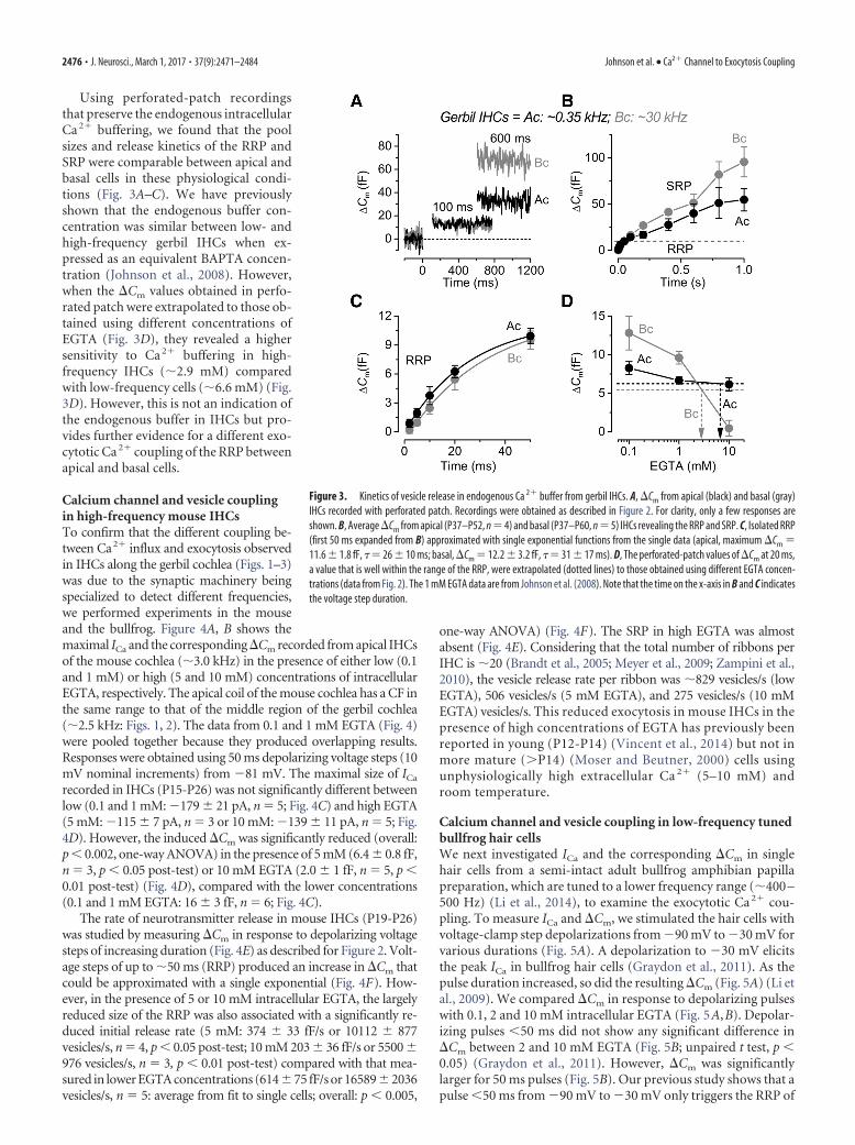
Using perforated-patch recordingsthat preserve the endogenous intracellularCa 2� buffering, we found that the poolsizes and release kinetics of the RRP andSRP were comparable between apical andbasal cells in these physiological condi-tions (Fig. 3A–C). We have previouslyshown that the endogenous buffer con-centration was similar between low- andhigh-frequency gerbil IHCs when ex-pressed as an equivalent BAPTA concen-tration (Johnson et al., 2008). However,when the �Cm values obtained in perfo-rated patch were extrapolated to those ob-tained using different concentrations ofEGTA (Fig. 3D), they revealed a highersensitivity to Ca 2� buffering in high-frequency IHCs (�2.9 mM) comparedwith low-frequency cells (�6.6 mM) (Fig.3D). However, this is not an indication ofthe endogenous buffer in IHCs but pro-vides further evidence for a different exo-cytotic Ca 2� coupling of the RRP betweenapical and basal cells.
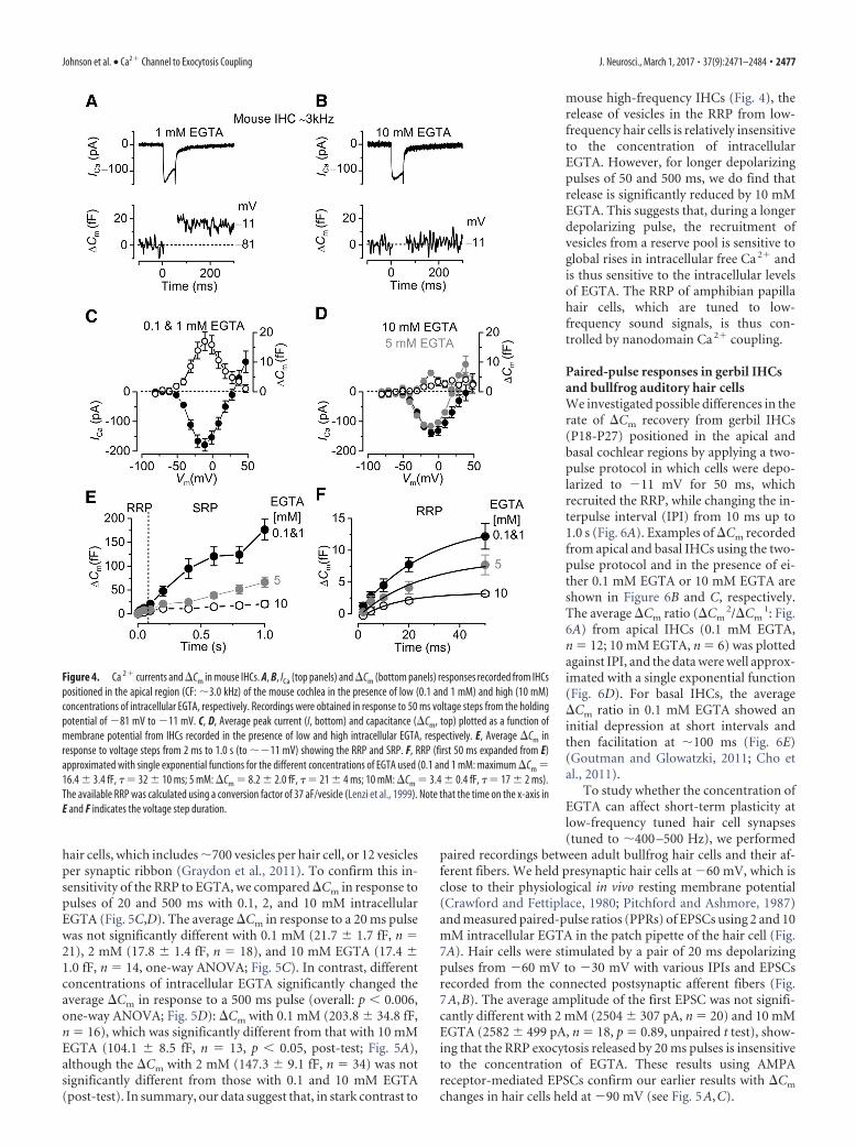
Calcium channel and vesicle couplingin high-frequency mouse IHCsTo confirm that the different coupling be-tween Ca2� influx and exocytosis observedin IHCs along the gerbil cochlea (Figs. 1–3)was due to the synaptic machinery beingspecialized to detect different frequencies,we performed experiments in the mouseand the bullfrog. Figure 4A, B shows themaximal ICa and the corresponding �Cm recorded from apical IHCsof the mouse cochlea (�3.0 kHz) in the presence of either low (0.1and 1 mM) or high (5 and 10 mM) concentrations of intracellularEGTA, respectively. The apical coil of the mouse cochlea has a CF inthe same range to that of the middle region of the gerbil cochlea(�2.5 kHz: Figs. 1, 2). The data from 0.1 and 1 mM EGTA (Fig. 4)were pooled together because they produced overlapping results.Responses were obtained using 50 ms depolarizing voltage steps (10mV nominal increments) from 81 mV. The maximal size of ICa
recorded in IHCs (P15-P26) was not significantly different betweenlow (0.1 and 1 mM: 179 21 pA, n � 5; Fig. 4C) and high EGTA(5 mM: 115 7 pA, n � 3 or 10 mM: 139 11 pA, n � 5; Fig.4D). However, the induced �Cm was significantly reduced (overall:p � 0.002, one-way ANOVA) in the presence of 5 mM (6.4 0.8 fF,n � 3, p � 0.05 post-test) or 10 mM EGTA (2.0 1 fF, n � 5, p �0.01 post-test) (Fig. 4D), compared with the lower concentrations(0.1 and 1 mM EGTA: 16 3 fF, n � 6; Fig. 4C).
The rate of neurotransmitter release in mouse IHCs (P19-P26)was studied by measuring �Cm in response to depolarizing voltagesteps of increasing duration (Fig. 4E) as described for Figure 2. Volt-age steps of up to �50 ms (RRP) produced an increase in �Cm thatcould be approximated with a single exponential (Fig. 4F). How-ever, in the presence of 5 or 10 mM intracellular EGTA, the largelyreduced size of the RRP was also associated with a significantly re-duced initial release rate (5 mM: 374 33 fF/s or 10112 877vesicles/s, n � 4, p � 0.05 post-test; 10 mM 203 36 fF/s or 5500 976 vesicles/s, n � 3, p � 0.01 post-test) compared with that mea-sured in lower EGTA concentrations (61475 fF/s or 165892036vesicles/s, n � 5: average from fit to single cells; overall: p � 0.005,
one-way ANOVA) (Fig. 4F). The SRP in high EGTA was almostabsent (Fig. 4E). Considering that the total number of ribbons perIHC is �20 (Brandt et al., 2005; Meyer et al., 2009; Zampini et al.,2010), the vesicle release rate per ribbon was �829 vesicles/s (lowEGTA), 506 vesicles/s (5 mM EGTA), and 275 vesicles/s (10 mMEGTA) vesicles/s. This reduced exocytosis in mouse IHCs in thepresence of high concentrations of EGTA has previously beenreported in young (P12-P14) (Vincent et al., 2014) but not inmore mature (�P14) (Moser and Beutner, 2000) cells usingunphysiologically high extracellular Ca 2� (5–10 mM) androom temperature.
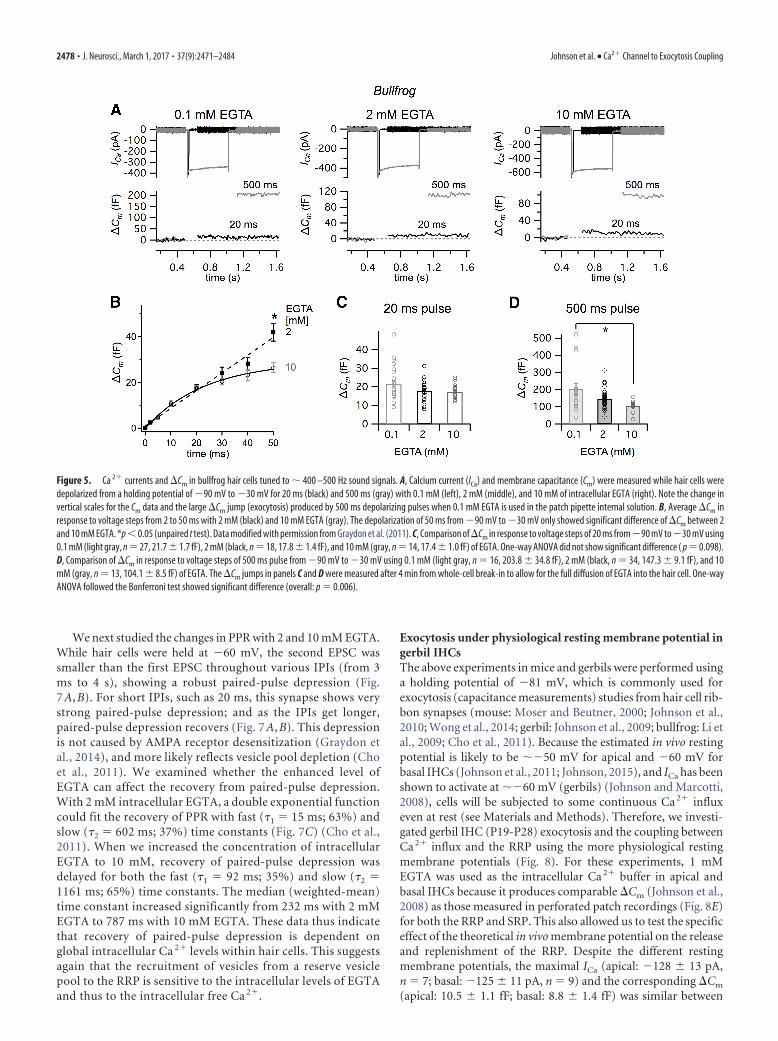
Calcium channel and vesicle coupling in low-frequency tunedbullfrog hair cellsWe next investigated ICa and the corresponding �Cm in singlehair cells from a semi-intact adult bullfrog amphibian papillapreparation, which are tuned to a lower frequency range (�400 –500 Hz) (Li et al., 2014), to examine the exocytotic Ca 2� cou-pling. To measure ICa and �Cm, we stimulated the hair cells withvoltage-clamp step depolarizations from 90 mV to 30 mV forvarious durations (Fig. 5A). A depolarization to 30 mV elicitsthe peak ICa in bullfrog hair cells (Graydon et al., 2011). As thepulse duration increased, so did the resulting �Cm (Fig. 5A) (Li etal., 2009). We compared �Cm in response to depolarizing pulseswith 0.1, 2 and 10 mM intracellular EGTA (Fig. 5A,B). Depolar-izing pulses �50 ms did not show any significant difference in�Cm between 2 and 10 mM EGTA (Fig. 5B; unpaired t test, p �0.05) (Graydon et al., 2011). However, �Cm was significantlylarger for 50 ms pulses (Fig. 5B). Our previous study shows that apulse �50 ms from 90 mV to 30 mV only triggers the RRP of
Figure 3. Kinetics of vesicle release in endogenous Ca 2� buffer from gerbil IHCs. A, �Cm from apical (black) and basal (gray)IHCs recorded with perforated patch. Recordings were obtained as described in Figure 2. For clarity, only a few responses areshown. B, Average �Cm from apical (P37–P52, n � 4) and basal (P37–P60, n � 5) IHCs revealing the RRP and SRP. C, Isolated RRP(first 50 ms expanded from B) approximated with single exponential functions from the single data (apical, maximum �Cm �11.6 1.8 fF, �� 26 10 ms; basal, �Cm � 12.2 3.2 fF, �� 31 17 ms). D, The perforated-patch values of �Cm at 20 ms,a value that is well within the range of the RRP, were extrapolated (dotted lines) to those obtained using different EGTA concen-trations (data from Fig. 2). The 1 mM EGTA data are from Johnson et al. (2008). Note that the time on the x-axis in B and C indicatesthe voltage step duration.
2476 • J. Neurosci., March 1, 2017 • 37(9):2471–2484 Johnson et al. • Ca2� Channel to Exocytosis Coupling
hair cells, which includes �700 vesicles per hair cell, or 12 vesiclesper synaptic ribbon (Graydon et al., 2011). To confirm this in-sensitivity of the RRP to EGTA, we compared �Cm in response topulses of 20 and 500 ms with 0.1, 2, and 10 mM intracellularEGTA (Fig. 5C,D). The average �Cm in response to a 20 ms pulsewas not significantly different with 0.1 mM (21.7 1.7 fF, n �21), 2 mM (17.8 1.4 fF, n � 18), and 10 mM EGTA (17.4 1.0 fF, n � 14, one-way ANOVA; Fig. 5C). In contrast, differentconcentrations of intracellular EGTA significantly changed theaverage �Cm in response to a 500 ms pulse (overall: p � 0.006,one-way ANOVA; Fig. 5D): �Cm with 0.1 mM (203.8 34.8 fF,n � 16), which was significantly different from that with 10 mMEGTA (104.1 8.5 fF, n � 13, p � 0.05, post-test; Fig. 5A),although the �Cm with 2 mM (147.3 9.1 fF, n � 34) was notsignificantly different from those with 0.1 and 10 mM EGTA(post-test). In summary, our data suggest that, in stark contrast to
mouse high-frequency IHCs (Fig. 4), therelease of vesicles in the RRP from low-frequency hair cells is relatively insensitiveto the concentration of intracellularEGTA. However, for longer depolarizingpulses of 50 and 500 ms, we do find thatrelease is significantly reduced by 10 mMEGTA. This suggests that, during a longerdepolarizing pulse, the recruitment ofvesicles from a reserve pool is sensitive toglobal rises in intracellular free Ca 2� andis thus sensitive to the intracellular levelsof EGTA. The RRP of amphibian papillahair cells, which are tuned to low-frequency sound signals, is thus con-trolled by nanodomain Ca 2� coupling.
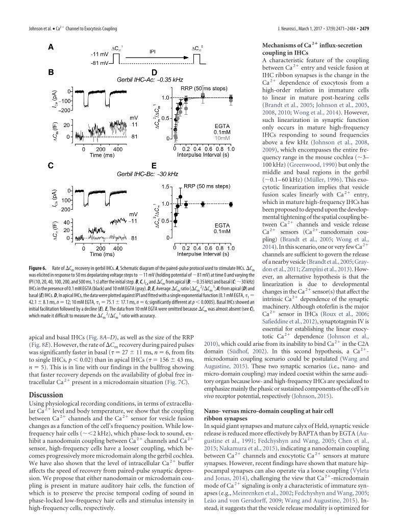
Paired-pulse responses in gerbil IHCsand bullfrog auditory hair cellsWe investigated possible differences in therate of �Cm recovery from gerbil IHCs(P18-P27) positioned in the apical andbasal cochlear regions by applying a two-pulse protocol in which cells were depo-larized to 11 mV for 50 ms, whichrecruited the RRP, while changing the in-terpulse interval (IPI) from 10 ms up to1.0 s (Fig. 6A). Examples of �Cm recordedfrom apical and basal IHCs using the two-pulse protocol and in the presence of ei-ther 0.1 mM EGTA or 10 mM EGTA areshown in Figure 6B and C, respectively.The average �Cm ratio (�Cm
2/�Cm1: Fig.
6A) from apical IHCs (0.1 mM EGTA,n � 12; 10 mM EGTA, n � 6) was plottedagainst IPI, and the data were well approx-imated with a single exponential function(Fig. 6D). For basal IHCs, the average�Cm ratio in 0.1 mM EGTA showed aninitial depression at short intervals andthen facilitation at �100 ms (Fig. 6E)(Goutman and Glowatzki, 2011; Cho etal., 2011).
To study whether the concentration ofEGTA can affect short-term plasticity atlow-frequency tuned hair cell synapses(tuned to �400 –500 Hz), we performed
paired recordings between adult bullfrog hair cells and their af-ferent fibers. We held presynaptic hair cells at 60 mV, which isclose to their physiological in vivo resting membrane potential(Crawford and Fettiplace, 1980; Pitchford and Ashmore, 1987)and measured paired-pulse ratios (PPRs) of EPSCs using 2 and 10mM intracellular EGTA in the patch pipette of the hair cell (Fig.7A). Hair cells were stimulated by a pair of 20 ms depolarizingpulses from 60 mV to 30 mV with various IPIs and EPSCsrecorded from the connected postsynaptic afferent fibers (Fig.7A,B). The average amplitude of the first EPSC was not signifi-cantly different with 2 mM (2504 307 pA, n � 20) and 10 mMEGTA (2582 499 pA, n � 18, p � 0.89, unpaired t test), show-ing that the RRP exocytosis released by 20 ms pulses is insensitiveto the concentration of EGTA. These results using AMPAreceptor-mediated EPSCs confirm our earlier results with �Cm
changes in hair cells held at 90 mV (see Fig. 5A,C).
Figure 4. Ca 2� currents and �Cm in mouse IHCs. A, B, ICa (top panels) and �Cm (bottom panels) responses recorded from IHCspositioned in the apical region (CF: �3.0 kHz) of the mouse cochlea in the presence of low (0.1 and 1 mM) and high (10 mM)concentrations of intracellular EGTA, respectively. Recordings were obtained in response to 50 ms voltage steps from the holdingpotential of 81 mV to 11 mV. C, D, Average peak current (I, bottom) and capacitance (�Cm, top) plotted as a function ofmembrane potential from IHCs recorded in the presence of low and high intracellular EGTA, respectively. E, Average �Cm inresponse to voltage steps from 2 ms to 1.0 s (to �11 mV) showing the RRP and SRP. F, RRP (first 50 ms expanded from E)approximated with single exponential functions for the different concentrations of EGTA used (0.1 and 1 mM: maximum �Cm �16.4 3.4 fF, � � 32 10 ms; 5 mM: �Cm � 8.2 2.0 fF, � � 21 4 ms; 10 mM: �Cm � 3.4 0.4 fF, � � 17 2 ms).The available RRP was calculated using a conversion factor of 37 aF/vesicle (Lenzi et al., 1999). Note that the time on the x-axis inE and F indicates the voltage step duration.
Johnson et al. • Ca2� Channel to Exocytosis Coupling J. Neurosci., March 1, 2017 • 37(9):2471–2484 • 2477
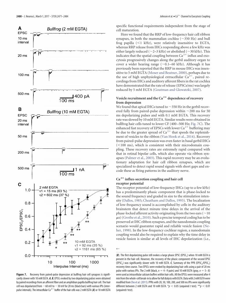
We next studied the changes in PPR with 2 and 10 mM EGTA.While hair cells were held at 60 mV, the second EPSC wassmaller than the first EPSC throughout various IPIs (from 3ms to 4 s), showing a robust paired-pulse depression (Fig.7 A, B). For short IPIs, such as 20 ms, this synapse shows verystrong paired-pulse depression; and as the IPIs get longer,paired-pulse depression recovers (Fig. 7 A, B). This depressionis not caused by AMPA receptor desensitization (Graydon etal., 2014), and more likely reflects vesicle pool depletion (Choet al., 2011). We examined whether the enhanced level ofEGTA can affect the recovery from paired-pulse depression.With 2 mM intracellular EGTA, a double exponential functioncould fit the recovery of PPR with fast (�1 � 15 ms; 63%) andslow (�2 � 602 ms; 37%) time constants (Fig. 7C) (Cho et al.,2011). When we increased the concentration of intracellularEGTA to 10 mM, recovery of paired-pulse depression wasdelayed for both the fast (�1 � 92 ms; 35%) and slow (�2 �1161 ms; 65%) time constants. The median (weighted-mean)time constant increased significantly from 232 ms with 2 mMEGTA to 787 ms with 10 mM EGTA. These data thus indicatethat recovery of paired-pulse depression is dependent onglobal intracellular Ca 2� levels within hair cells. This suggestsagain that the recruitment of vesicles from a reserve vesiclepool to the RRP is sensitive to the intracellular levels of EGTAand thus to the intracellular free Ca 2�.
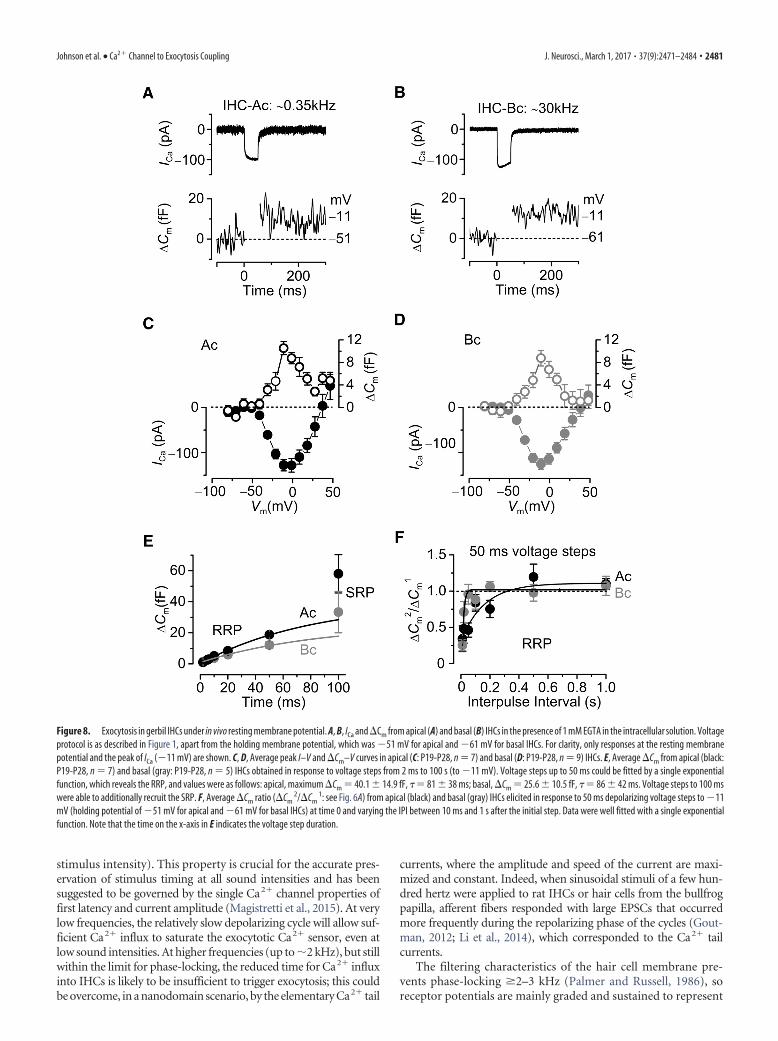
Exocytosis under physiological resting membrane potential ingerbil IHCsThe above experiments in mice and gerbils were performed usinga holding potential of 81 mV, which is commonly used forexocytosis (capacitance measurements) studies from hair cell rib-bon synapses (mouse: Moser and Beutner, 2000; Johnson et al.,2010; Wong et al., 2014; gerbil: Johnson et al., 2009; bullfrog: Li etal., 2009; Cho et al., 2011). Because the estimated in vivo restingpotential is likely to be �50 mV for apical and 60 mV forbasal IHCs (Johnson et al., 2011; Johnson, 2015), and ICa has beenshown to activate at �60 mV (gerbils) (Johnson and Marcotti,2008), cells will be subjected to some continuous Ca 2� influxeven at rest (see Materials and Methods). Therefore, we investi-gated gerbil IHC (P19-P28) exocytosis and the coupling betweenCa 2� influx and the RRP using the more physiological restingmembrane potentials (Fig. 8). For these experiments, 1 mMEGTA was used as the intracellular Ca 2� buffer in apical andbasal IHCs because it produces comparable �Cm (Johnson et al.,2008) as those measured in perforated patch recordings (Fig. 8E)for both the RRP and SRP. This also allowed us to test the specificeffect of the theoretical in vivo membrane potential on the releaseand replenishment of the RRP. Despite the different restingmembrane potentials, the maximal ICa (apical: 128 13 pA,n � 7; basal: 125 11 pA, n � 9) and the corresponding �Cm
(apical: 10.5 1.1 fF; basal: 8.8 1.4 fF) was similar between
Figure 5. Ca 2� currents and �Cm in bullfrog hair cells tuned to � 400 –500 Hz sound signals. A, Calcium current (ICa) and membrane capacitance (Cm) were measured while hair cells weredepolarized from a holding potential of 90 mV to 30 mV for 20 ms (black) and 500 ms (gray) with 0.1 mM (left), 2 mM (middle), and 10 mM of intracellular EGTA (right). Note the change invertical scales for the Cm data and the large �Cm jump (exocytosis) produced by 500 ms depolarizing pulses when 0.1 mM EGTA is used in the patch pipette internal solution. B, Average �Cm inresponse to voltage steps from 2 to 50 ms with 2 mM (black) and 10 mM EGTA (gray). The depolarization of 50 ms from 90 mV to 30 mV only showed significant difference of �Cm between 2and 10 mM EGTA. *p � 0.05 (unpaired t test). Data modified with permission from Graydon et al. (2011). C, Comparison of �Cm in response to voltage steps of 20 ms from 90 mV to 30 mV using0.1 mM (light gray, n � 27, 21.7 1.7 fF), 2 mM (black, n � 18, 17.8 1.4 fF), and 10 mM (gray, n � 14, 17.4 1.0 fF) of EGTA. One-way ANOVA did not show significant difference ( p � 0.098).D, Comparison of �Cm in response to voltage steps of 500 ms pulse from 90 mV to 30 mV using 0.1 mM (light gray, n � 16, 203.8 34.8 fF), 2 mM (black, n � 34, 147.3 9.1 fF), and 10mM (gray, n � 13, 104.1 8.5 fF) of EGTA. The �Cm jumps in panels C and D were measured after 4 min from whole-cell break-in to allow for the full diffusion of EGTA into the hair cell. One-wayANOVA followed the Bonferroni test showed significant difference (overall: p � 0.006).
2478 • J. Neurosci., March 1, 2017 • 37(9):2471–2484 Johnson et al. • Ca2� Channel to Exocytosis Coupling
apical and basal IHCs (Fig. 8A–D), as well as the size of the RRP(Fig. 8E). However, the rate of �Cm recovery during paired pulseswas significantly faster in basal (� � 27 11 ms, n � 6, from fitsto single IHCs, p � 0.02) than in apical IHCs (� � 156 43 ms,n � 5). This is in line with our findings in the bullfrog showingthat faster recovery depends on the availability of global free in-tracellular Ca 2� present in a microdomain situation (Fig. 7C).
DiscussionUsing physiological recording conditions, in terms of extracellu-lar Ca 2� level and body temperature, we show that the couplingbetween Ca 2� channels and the Ca 2� sensor for vesicle fusionchanges as a function of the cell’s frequency position. While low-frequency hair cells (��2 kHz), which phase-lock to sound, ex-hibit a nanodomain coupling between Ca 2� channels and Ca 2�
sensor, high-frequency cells have a looser coupling, which be-comes progressively more microdomain along the gerbil cochlea.We have also shown that the level of intracellular Ca 2� bufferaffects the speed of recovery from paired-pulse synaptic depres-sion. We propose that either nanodomain or microdomain cou-pling is present in mature auditory hair cells, the function ofwhich is to preserve the precise temporal coding of sound inphase-locked low-frequency hair cells and stimulus intensity inhigh-frequency cells, respectively.
Mechanisms of Ca 2� influx-secretioncoupling in IHCsA characteristic feature of the couplingbetween Ca 2� entry and vesicle fusion atIHC ribbon synapses is the change in theCa 2� dependence of exocytosis from ahigh-order relation in immature cellsto linear in mature post-hearing cells(Brandt et al., 2005; Johnson et al., 2005,2008, 2010; Wong et al., 2014). However,such linearization in synaptic functiononly occurs in mature high-frequencyIHCs responding to sound frequenciesabove a few kHz (Johnson et al., 2008,2009), which encompasses the entire fre-quency range in the mouse cochlea (�3–100 kHz) (Greenwood, 1990) but only themiddle and basal regions in the gerbil(�0.1– 60 kHz) (Muller, 1996). This exo-cytotic linearization implies that vesiclefusion scales linearly with Ca 2� entry,which in mature high-frequency IHCs hasbeen proposed to depend upon the develop-mental tightening of the spatial coupling be-tween Ca2� channels and vesicle releaseCa2� sensors (Ca2�-nanodomain cou-pling) (Brandt et al., 2005; Wong et al.,2014). In this scenario, one or very few Ca2�
channels are sufficient to govern the releaseof a nearby vesicle (Brandt et al., 2005; Gray-don et al., 2011; Zampini et al., 2013). How-ever, an alternative hypothesis is that thelinearization is due to developmentalchanges in the Ca2� sensor(s) that affect theintrinsic Ca2� dependence of the synapticmachinery. Although otoferlin is the majorCa2� sensor in IHCs (Roux et al., 2006;Safieddine et al., 2012), synaptotagmin IV isessential for establishing the linear exocy-totic Ca2� dependence (Johnson et al.,
2010), which could arise from its inability to bind Ca2� in the C2Adomain (Sudhof, 2002). In this second hypothesis, a Ca2�-microdomain coupling scenario could be postulated (Wang andAugustine, 2015). These two synaptic scenarios (i.e., nano- andmicro-domain coupling) may indeed coexist within the same audi-tory organ because low- and high-frequency IHCs are specialized toemphasize mainly the phasic or sustained components of the cell’s invivo receptor potential, respectively (Johnson, 2015).
Nano- versus micro-domain coupling at hair cellribbon synapsesIn squid giant synapses and mature calyx of Held, synaptic vesiclerelease is reduced more effectively by BAPTA than by EGTA (Au-gustine et al., 1991; Fedchyshyn and Wang, 2005; Chen et al.,2015; Nakamura et al., 2015), indicating a nanodomain couplingbetween Ca 2� channels and exocytotic Ca 2� sensors at maturesynapses. However, recent findings have shown that mature hip-pocampal synapses can also operate via a loose coupling (Vyletaand Jonas, 2014), challenging the view that Ca 2�-microdomainmode of Ca 2� signaling is only a characteristic of immature syn-apses (e.g., Meinrenken et al., 2002; Fedchyshyn and Wang, 2005;Leao and von Gersdorff, 2009; Wang and Augustine, 2015). In-stead, it suggests that the vesicle release modality is optimized for
Figure 6. Rate of �Cm recovery in gerbil IHCs. A, Schematic diagram of the paired-pulse protocol used to stimulate IHCs. �Cm
was elicited in response to 50 ms depolarizing voltage steps to 11 mV (holding potential of 81 mV) at time 0 and varying theIPI (10, 20, 40, 100, 200, and 500 ms, 1 s) after the initial step. B, C, ICa and �Cm from apical (B: �0.35 kHz) and basal (C: �30 kHz)IHCs in the presence of 0.1 mM EGTA (black) and 10 mM EGTA (gray). D, E, Average �Cm ratio (�Cm
2/�Cm1; A) from apical (D) and
basal (E) IHCs. D, In apical IHCs, the data were plotted against IPI and fitted with a single exponential function (0.1 mM EGTA, �1 �42.1 8.1 ms, n � 12; 10 mM EGTA, �1 � 75.1 17.1 ms, n � 6; significantly different at p � 0.0005). Basal IHCs showed aninitial facilitation followed by a decline (E). E, The data from 10 mM EGTA were omitted because �Cm was almost absent (see C),which made it difficult to measure the �Cm
2/�Cm1 ratio with accuracy.
Johnson et al. • Ca2� Channel to Exocytosis Coupling J. Neurosci., March 1, 2017 • 37(9):2471–2484 • 2479
specific functional requirements independent from the stage ofcell maturation.
Here we found that the RRP of low-frequency hair cell ribbonsynapses, in both the mammalian cochlea (�350 Hz) and bullfrog papilla (�1 kHz), were relatively insensitive to EGTA,whereas RRP release from IHCs responding above a few kHz waseither largely reduced (�2–3 kHz) or abolished (�30 kHz). Thisindicates that the spatial coupling between Ca 2� influx and exo-cytosis progressively changes along the gerbil auditory organ tocover a wider hearing range (�0.1– 60 kHz). Although it haspreviously been reported that the RRP in mouse IHCs was insen-sitive to 5 mM EGTA (Moser and Beutner, 2000), perhaps due tothe use of high unphysiological extracellular Ca 2�, paired re-cordings from IHCs and auditory afferent fibers in the rat cochleahave demonstrated that the rate of release (EPSCs/ms) was largelyreduced by 5 mM EGTA (Goutman and Glowatzki, 2007).
Vesicle recruitment and the Ca 2� dependence of recoveryfrom depressionWe found that apical IHCs tuned to �350 Hz in the gerbil recov-ered fully from paired-pulse depression within �200 ms for 50ms depolarizing pulses and with 0.1 mM EGTA. This recoveryrate was slowed by 10 mM EGTA. Similar results were obtained inbullfrog hair cells tuned to lower CF (400 –500 Hz; Fig. 7C). Theenhanced fast recovery of EPSCs with lower Ca 2� buffering maybe due to the greater spread of Ca 2� that speeds the replenish-ment of vesicles to the ribbon (Van Hook et al., 2014). Recoveryfrom paired-pulse depression was even faster in basal gerbil IHCs(�100 ms), which is consistent with their microdomain cou-pling. These recovery rates are extremely rapid compared withthat in retinal bipolar cells, which also operate via ribbon syn-apses (Palmer et al., 2003). This rapid recovery may be an evolu-tionary adaptation for hair cell ribbon synapses, which arespecialized to detect rapid sound signals with short gaps and en-code these as firing patterns in the auditory nerve.
Ca 2� influx-secretion coupling and hair cellreceptor potentialThe receptor potential of low-frequency IHCs (up to a few kHz)has a predominantly phasic component that is phase-locked tothe sound frequency and graded in size to the stimulation inten-sity (Dallos, 1985; Cheatham and Dallos, 1993). The localizationof low-frequency sound is accomplished by cells in the auditorybrainstem that detect minute time delays in the arrival of thephase-locked afferent activity originating from the two ears (�10�s) (Grothe et al., 2010). Such a precise temporal coding has to bepreserved at IHC ribbon synapses, and the nanodomain couplingscenario would guarantee rapid and reliable vesicle fusion (Ne-her, 1998). In the low-frequency cochlear region, a nanodomaincoupling would also be required to explain why the time delay invesicle fusion is similar at all levels of IHC depolarization (i.e.,
Figure 7. Recovery from paired-pulse depression at bullfrog hair cell synapses is signifi-cantly slower with 10 mM EGTA. A, B, EPSCs evoked by two depolarizing pulses were obtainedby paired recordings from an afferent fiber and an amphibian papilla bullfrog hair cell. The haircell was depolarized from 60 mV to 30 mV for 20 ms (black bars) with various IPIs (inter-pulse intervals). The intracellular Ca 2� buffer of the hair cells was 2 mM EGTA (A) or 10 mM EGTA
4
(B). The first depolarizing pulse still evokes a large phasic EPSC (EPSC1) when 10 mM EGTA ispresent in the hair cell. However, the recovery of the phasic component of the second EPSC(EPSC2) was significantly slower with 10 mM EGTA. C, Summary of the PPR (EPSC2/EPSC1)recovery time course. Two EPSCs were evoked by depolarizing hair cells using a pair of 20 mspulse with various IPIs. The 2 mM (black, n � 4 –9 pairs) and 10 mM EGTA (gray, n � 5– 8)were used as intracellular calcium buffers within hair cells. All the EPSCs were measured after 4min from the whole-cell break-in to allow for full dialysis with EGTA. Data with 2 mM EGTA weremodified from Cho et al. (2011). PPRs with 20, 50, 100, 200, and 500 ms IPIs were significantlydifferent between 2 mM EGTA and 10 mM EGTA. *p � 0.05 (unpaired t test). **p � 0.01(unpaired t test).
2480 • J. Neurosci., March 1, 2017 • 37(9):2471–2484 Johnson et al. • Ca2� Channel to Exocytosis Coupling
stimulus intensity). This property is crucial for the accurate pres-ervation of stimulus timing at all sound intensities and has beensuggested to be governed by the single Ca2� channel properties offirst latency and current amplitude (Magistretti et al., 2015). At verylow frequencies, the relatively slow depolarizing cycle will allow suf-ficient Ca2� influx to saturate the exocytotic Ca2� sensor, even atlow sound intensities. At higher frequencies (up to �2 kHz), but stillwithin the limit for phase-locking, the reduced time for Ca2� influxinto IHCs is likely to be insufficient to trigger exocytosis; this couldbe overcome, in a nanodomain scenario, by the elementary Ca2� tail
currents, where the amplitude and speed of the current are maxi-mized and constant. Indeed, when sinusoidal stimuli of a few hun-dred hertz were applied to rat IHCs or hair cells from the bullfrogpapilla, afferent fibers responded with large EPSCs that occurredmore frequently during the repolarizing phase of the cycles (Gout-man, 2012; Li et al., 2014), which corresponded to the Ca2� tailcurrents.
The filtering characteristics of the hair cell membrane pre-vents phase-locking �2–3 kHz (Palmer and Russell, 1986), soreceptor potentials are mainly graded and sustained to represent
Figure 8. Exocytosis in gerbil IHCs under in vivo resting membrane potential. A, B, ICa and �Cm from apical (A) and basal (B) IHCs in the presence of 1 mM EGTA in the intracellular solution. Voltageprotocol is as described in Figure 1, apart from the holding membrane potential, which was 51 mV for apical and 61 mV for basal IHCs. For clarity, only responses at the resting membranepotential and the peak of ICa (11 mV) are shown. C, D, Average peak I–V and �Cm–V curves in apical (C: P19-P28, n � 7) and basal (D: P19-P28, n � 9) IHCs. E, Average �Cm from apical (black:P19-P28, n � 7) and basal (gray: P19-P28, n � 5) IHCs obtained in response to voltage steps from 2 ms to 100 s (to 11 mV). Voltage steps up to 50 ms could be fitted by a single exponentialfunction, which reveals the RRP, and values were as follows: apical, maximum �Cm � 40.1 14.9 fF, � � 81 38 ms; basal, �Cm � 25.6 10.5 fF, � � 86 42 ms. Voltage steps to 100 mswere able to additionally recruit the SRP. F, Average �Cm ratio (�Cm
2/�Cm1: see Fig. 6A) from apical (black) and basal (gray) IHCs elicited in response to 50 ms depolarizing voltage steps to 11
mV (holding potential of 51 mV for apical and 61 mV for basal IHCs) at time 0 and varying the IPI between 10 ms and 1 s after the initial step. Data were well fitted with a single exponentialfunction. Note that the time on the x-axis in E indicates the voltage step duration.
Johnson et al. • Ca2� Channel to Exocytosis Coupling J. Neurosci., March 1, 2017 • 37(9):2471–2484 • 2481
sound intensity and stimulus envelope (Russell and Sellick,1978). High-frequency sound localization is performed by cellsthat compare interaural-level differences originating from gradedresponses in �3 kHz IHCs of each ear (Caird and Klinke, 1983).Therefore, high-frequency IHCs are not designed to follow thefrequency components of sound and, as such, do not require theprecise timing provided by nanodomain coupling (Matveev et al.,2011), which is likely to be unsuitable for accurate intensity cod-ing. Instead, the changes in the amplitude and kinetic propertiesof the macroscopic ICa with sound intensity are now more rele-vant (Magistretti et al., 2015), which is more in line with a mi-crodomain coupling reported in this study.
Damage due to loud sounds: why are basal IHCsmore susceptible?We found that high-frequency IHCs (especially those at �30kHz) exhibit a strong block of exocytosis by 10 mM EGTA, indi-cating that these cells cannot have a large endogenous Ca 2� buff-ering capacity, because it would severely impair transmitterrelease. This was confirmed by the estimated intracellular Ca 2�
buffer expressed as an equivalent of EGTA concentration (Fig. 3).Indeed, a triple knock-out mouse for different Ca 2�-bindingproteins did not reveal changes in synaptic sound encoding(Pangrsic et al., 2015), suggesting that high-frequency IHCs maythus have a relatively low concentration of Ca 2�-binding pro-teins. By contrast, low-frequency tuned bullfrog hair cells have anestimated 8 mM of high-affinity Ca 2�-binding sites on smallmobile proteins (e.g., parvalbumin and calbindin) (Heller et al.,2002), suggesting that their endogenous Ca 2� buffering capacitymay be more equivalent to 10 mM EGTA.
High-frequency hair cell synapses are also particularly vulner-able to damage during loud noises and aging, which has beenshown to lead to the loss of both IHC synaptic ribbons (Kujawaand Liberman, 2009; Kujawa and Liberman, 2015) and low-spontaneous rate afferent fibers (Furman et al., 2013). We thuspropose that low-frequency IHCs may express higher concentra-tions of Ca 2�-binding proteins, which will not block exocytosisbut may confer neuroprotection against excessive Ca 2� influxduring prolonged stimulation. By contrast, the low Ca 2� buffercapacity in high-frequency basal IHCs, which is required for theirgraded release, will make them more prone to Ca 2�-inducedcytotoxicity. A tonotopic gradient in Ca 2�-binding protein ex-pression has been reported in auditory hair cells (Hackney et al.,2003, 2005; Patel et al., 2012), which may facilitate a frequency-dependent tuning of exocytosis in some animal species (Schnee etal., 2005; Rutherford and Roberts, 2006; Patel et al., 2012).
ReferencesAdler EM, Augustine GJ, Duffy SN, Charlton MP (1991) Alien intracellular
calcium chelators attenuate neurotransmitter release at the squid giantsynapse. J Neurosci 11:1496 –1507. Medline
Augustine GJ, Adler EM, Charlton MP (1991) The calcium signal for trans-mitter secretion from presynaptic nerve terminals. Ann N Y Acad Sci635:365–381. CrossRef Medline
Borst JG, Sakmann B (1996) Calcium influx and transmitter release in a fastCNS synapse. Nature 383:431– 434. CrossRef Medline
Brandt A, Khimich D, Moser T (2005) Few CaV1.3 channels regulate theexocytosis of a synaptic vesicle at the hair cell ribbon synapse. J Neurosci25:11577–11585. CrossRef Medline
Bucurenciu I, Kulik A, Schwaller B, Frotscher M, Jonas P (2008) Nanodo-main coupling between Ca 2� channels and Ca 2� sensors promotes fastand efficient transmitter release at a cortical GABAergic synapse. Neuron57:536 –545. CrossRef Medline
Caird D, Klinke R (1983) Processing of binaural stimuli by cat superiorolivary complex neurons. Exp Brain Res 52:385–399. Medline
Cheatham MA, Dallos P (1993) Longitudinal comparisons of IHC ac and dcreceptor potentials recorded from the guinea pig cochlea. Hear Res 68:107–114. CrossRef Medline
Chen Z, Das B, Nakamura Y, DiGregorio DA, Young SM Jr (2015) Ca 2�
channel to synaptic vesicle distance accounts for the readily releasablepool kinetics at a functionally mature auditory synapse. J Neurosci 35:2083–2100. CrossRef Medline
Cho S, von Gersdorff H (2014) Proton-mediated block of Ca 2� channelsduring multivesicular release regulates short-term plasticity at an audi-tory hair cell synapse. J Neurosci 34:15877–15887. CrossRef Medline
Cho S, Li GL, von Gersdorff H (2011) Recovery from short-term depressionand facilitation is ultrafast and Ca 2� dependent at auditory hair cell syn-apses. J Neurosci 31:5682–5692. CrossRef Medline
Coggins M, Zenisek D (2009) Evidence that exocytosis is driven by calciumentry through multiple calcium channels in goldfish retinal bipolar cells.J Neurophysiol 101:2601–2619. CrossRef Medline
Crawford AC, Fettiplace R (1980) The frequency selectivity of auditorynerve fibres and hair cells in the cochlea of the turtle. J Physiol 306:79 –125. CrossRef Medline
Dallos P (1985) Response characteristics of mammalian cochlear hair cells.J Neurosci 5:1591–1608. Medline
Ehret G (1975) Masked auditory thresholds, critical ratios, and scales of thebasilar membrane of the housemouse (Mus musculus). J Comp Physiol103:329 –341. CrossRef
Fedchyshyn MJ, Wang LY (2005) Developmental transformation of the re-lease modality at the calyx of Held synapse. J Neurosci 25:4131– 4140.CrossRef Medline
Fettiplace R, Fuchs PA (1999) Mechanisms of hair cell tuning. Annu RevPhysiol 61:809 – 834. CrossRef Medline
Frank T, Rutherford MA, Strenzke N, Neef A, Pangrsic T, Khimich D, FejtovaA, Gundelfinger ED, Liberman MC, Harke B, Bryan KE, Lee A, Egner A,Riedel D, Moser T (2010) Bassoon and the synaptic ribbon organizeCa 2� channels and vesicles to add release sites and promote refillingNeuron 68:724 –738. CrossRef
Fuchs PA (2005) Time and intensity coding at the hair cell’s ribbon synapse.J Physiol 566:7–12. CrossRef Medline
Furman AC, Kujawa SG, Liberman MC (2013) Noise-induced cochlearneuropathy is selective for fibers with low spontaneous rates. J Neuro-physiol 110:577–586. CrossRef Medline
Gillis KD (2000) Admittance-based measurement of membrane capaci-tance using the EPC-9 patch-clamp amplifier. Pflugers Arch 439:655–664. CrossRef Medline
Glowatzki E, Fuchs PA (2002) Transmitter release at the hair cell ribbonsynapse. Nat Neurosci 5:147–154. CrossRef Medline
Goutman JD, Glowatzki E (2011) Short-term facilitation modulates sizeand timing of the synaptic response at the inner hair cell ribbon synapse.J Neurosci 31:7974 –7981. CrossRef Medline
Goutman JD (2012) Transmitter release from cochlear hair cells is phaselocked to cyclic stimuli of different intensities and frequencies. J Neurosci32:17025–17035a. CrossRef Medline
Goutman JD, Glowatzki E (2007) Time course and calcium dependence oftransmitter release at a single ribbon synapse. Proc Natl Acad Sci U S A104:16341–16346. CrossRef Medline
Graydon CW, Cho S, Li GL, Kachar B, von Gersdorff H (2011) Sharp Ca 2�
nanodomains beneath the ribbon promote highly synchronous multive-sicular release at hair cell synapses. J Neurosci 31:16637–16650. CrossRefMedline
Graydon CW, Cho S, Diamond JS, Kachar B, von Gersdorff H, Grimes WN(2014) Specialized postsynaptic morphology enhances neurotransmitterdilution and high-frequency signaling at an auditory synapse. J Neurosci34:8358 – 8372. CrossRef Medline
Greenwood DD (1990) A cochlear frequency-position function for severalspecies: 29 years later. J Acoust Soc Am 87:2592–2605. CrossRef Medline
Grothe B, Pecka M, McAlpine D (2010) Mechanisms of sound localizationin mammals. Physiol Rev 90:983–1012. CrossRef Medline
Hackney CM, Mahendrasingam S, Jones EM, Fettiplace R (2003) The dis-tribution of calcium buffering proteins in the turtle cochlea. J Neurosci23:4577– 4589. Medline
Hackney CM, Mahendrasingam S, Penn A, Fettiplace R (2005) The concen-trations of calcium buffering proteins mammalian cochlear hair cells.J Neurosci 25:7867–7875. CrossRef Medline
Heil P, Peterson AJ (2017) Spike timing in auditory-nerve fibers during
2482 • J. Neurosci., March 1, 2017 • 37(9):2471–2484 Johnson et al. • Ca2� Channel to Exocytosis Coupling
spontaneous activity and phase locking. Synapse 71:5–36. CrossRefMedline
Heller S, Bell AM, Denis CS, Choe Y, Hudspeth AJ (2002) Parvalbumin 3 isan abundant Ca 2� buffer in hair cells. J Assoc Res Otolaryngol 3:488 –498. CrossRef Medline
Johnson SL (2015) Membrane properties specialize mammalian inner haircells for frequency or intensity encoding. Elife 4:e08177. Medline
Johnson SL, Marcotti W (2008) Biophysical properties of CaV1.3 calciumchannels in gerbil inner hair cells. J Physiol 586:1029 –1042. CrossRefMedline
Johnson SL, Marcotti W, Kros CJ (2005) Increase in efficiency and reduc-tion in Ca 2� dependence of exocytosis during development of mouseinner hair cells. J Physiol 563:177–191. CrossRef Medline
Johnson SL, Adelman JP, Marcotti W (2007) Disruption of spontaneousaction potential activity in inner hair cells of SK2 knockout mice preventsthe normal development of exocytotic machinery. J Physiol 583:631– 646.CrossRef Medline
Johnson SL, Forge A, Knipper M, Munkner S, Marcotti W (2008) Tono-topic variation in the calcium dependence of neurotransmitter releaseand vesicle pool replenishment at mammalian auditory ribbon synapses.J Neurosci 28:7670 –7678. CrossRef Medline
Johnson SL, Franz C, Knipper M, Marcotti W (2009) Functional matura-tion of the exocytotic machinery at gerbil hair cell ribbon synapses.J Physiol 587:1715–1726. CrossRef Medline
Johnson SL, Franz C, Kuhn S, Furness DN, Ruttiger L, Munkner S, RivoltaMN, Seward EP, Herschman HR, Engel J, Knipper M, Marcotti W (2010)Synaptotagmin IV determines the linear Ca 2� dependence of vesicle fu-sion at auditory ribbon synapses. Nat Neurosci 13:45–52. CrossRefMedline
Johnson SL, Beurg M, Marcotti W, Fettiplace R (2011) Prestin-driven co-chlear amplification is not limited by the outer hair cell membrane timeconstant. Neuron 70:1143–1154. CrossRef Medline
Keen EC, Hudspeth AJ (2006) Transfer characteristics of the hair cell’s af-ferent synapse. Proc Natl Acad Sci U S A 103:5537–5542. CrossRefMedline
Kros CJ, Ruppersberg JP, Rusch A (1998) Expression of a potassium currentin inner hair cells during development of hearing in mice. Nature 394:281–284. CrossRef Medline
Kujawa SG, Liberman MC (2009) Adding insult to injury: cochlear nervedegeneration after “temporary” noise-induced hearing loss. J Neurosci29:14077–14085. CrossRef Medline
Kujawa SG, Liberman MC (2015) Synaptopathy in the noise-exposed andaging cochlea: primary neural degeneration in acquired sensorineuralhearing loss. Hear Res 330:191–199. CrossRef Medline
Leao RM, von Gersdorff H (2009) Synaptic vesicle pool size, release proba-bility and synaptic depression are sensitive to Ca 2� buffering capacity inthe developing rat calyx of Held. Braz J Med Biol Res 42:94 –104. CrossRefMedline
Lenzi D, Runyeon JW, Crum J, Ellisman MH, Roberts WM (1999) Synapticvesicle populations in saccular hair cells reconstructed by electron tomog-raphy. J Neurosci 19:119 –132. Medline
Lewis E, Leverenz E, Koyama H (1982) The tonotopic organization of thebullfrog amphibian papilla, an auditory organ lacking a basilar mem-brane. J Comp Physiol A Neuroethol Sens Neural Behav Physiol Neuro-ethol Sens Neural Behav Physiol 145:437– 445. CrossRef
Li GL, Keen E, Andor-Ardo D, Hudspeth AJ, von Gersdorff H (2009) Theunitary event underlying multiquantal EPSCs at a hair cell’s ribbon syn-apse. J Neurosci 29:7558 –7568. CrossRef Medline
Li GL, Cho S, von Gersdorff H (2014) Phase-locking precision is enhancedby multiquantal release at an auditory hair cell ribbon synapse. Neuron83:1404 –1417. CrossRef Medline
Lindau M, Neher E (1988) Patch-clamp techniques for time-resolved ca-pacitance measurements in single cells. Pflugers Arch 411:137–146.CrossRef Medline
Magistretti J, Spaiardi P, Johnson SL, Masetto S (2015) Elementary proper-ties of Ca 2� channels and their influence on multivesicular release andphase-locking at auditory hair cell ribbon synapses. Front Cell Neurosci9:123. CrossRef Medline
Marcotti W, Johnson SL, Holley MC, Kros CJ (2003) Developmentalchanges in the expression of potassium currents of embryonic, neonataland mature mouse inner hair cells. J Physiol 548:383– 400. CrossRefMedline
Matthews G, Fuchs P (2010) The diverse roles of ribbon synapses in sensoryneurotransmission. Nat Rev Neurosci 11:812– 822. CrossRef Medline
Matveev V, Bertram R, Sherman A (2011) Calcium cooperativity of exocyto-sis as a measure of Ca 2� channel domain overlap. Brain Res 1398:126–138.CrossRef Medline
Meinrenken CJ, Borst JG, Sakmann B (2002) Calcium secretion coupling atcalyx of held governed by nonuniform channel-vesicle topography.J Neurosci 22:1648 –1667. Medline
Meyer AC, Frank T, Khimich D, Hoch G, Riedel D, Chapochnikov NM, YarinYM, Harke B, Hell SW, Egner A, Moser T (2009) Tuning of synapsenumber, structure and function in the cochlea. Nat Neurosci 12:444 – 453.CrossRef Medline
Moser T, Beutner D (2000) Kinetics of exocytosis and endocytosis at thecochlear inner hair cell afferent synapse of the mouse. Proc Natl Acad SciU S A 97:883– 888. CrossRef Medline
Muller M (1996) The cochlear place-frequency map of the adult and devel-oping Mongolian gerbil. Hear Res 94:148 –156. CrossRef Medline
Nakamura Y, Harada H, Kamasawa N, Matsui K, Rothman JS, Shigemoto R,Silver RA, DiGregorio DA, Takahashi T (2015) Nanoscale distributionof presynaptic Ca 2� channels and its impact on vesicular release duringdevelopment. Neuron 85:145–158. CrossRef Medline
Naraghi M, Neher E (1997) Linearized buffered Ca 2� diffusion in microdo-mains and its implications for calculation of [Ca 2�] at the mouth of acalcium channel. J Neurosci 17:6961– 6973. Medline
Neher E (1998) Vesicle pools and Ca 2� microdomains: new tools for un-derstanding their roles in neurotransmitter release. Neuron 20:389 –399.CrossRef Medline
Nouvian R (2007) Temperature enhances exocytosis efficiency at the mouseinner hair cell ribbon synapse. J Physiol 584:535–542. CrossRef Medline
Palmer AR, Russell IJ (1986) Phase-locking in the cochlear nerve of theguinea-pig and its relation to the receptor potential of inner hair-cells.Hear Res 24:1–15. CrossRef Medline
Palmer MJ, Hull C, Vigh J, von Gersdorff H (2003) Synaptic cleft acidifica-tion and modulation of short-term depression by exocytosed protons inretinal bipolar cells. J Neurosci 23:11332–11341. Medline
Pangrsic T, Gabrielaitis M, Michanski S, Schwaller B, Wolf F, Strenzke N,Moser T (2015) EF-hand protein Ca 2� buffers regulate Ca 2� influx andexocytosis in sensory hair cells. Proc Natl Acad Sci U S A 112:E1028 –E1037. CrossRef Medline
Patel SH, Salvi JD, O Maoileidigh D, Hudspeth AJ (2012) Frequency-selective exocytosis by ribbon synapses of hair cells in the bullfrog’s am-phibian papilla. J Neurosci 32:13433–13438. CrossRef Medline
Pitchford S, Ashmore JF (1987) An electrical resonance in hair cells of theamphibian papilla of the frog Rana temporaria. Hear Res 27:75– 83.CrossRef Medline
Platzer J, Engel J, Schrott-Fischer A, Stephan K, Bova S, Chen H, Zheng H,Striessnig J (2000) Congenital deafness and sinoatrial node dysfunctionin mice lacking class D L-type Ca 2� channels. Cell 102:89 –97. CrossRefMedline
Roberts WM, Jacobs RA, Hudspeth AJ (1990) Colocalization of ion chan-nels involved in frequency selectivity and synaptic transmission at presyn-aptic active zones of hair cells. J Neurosci 10:3664 –3684. Medline
Rose JE, Brugge JF, Anderson DJ, Hind JE (1967) Phase-locked response tolow-frequency tones in single auditory nerve fibers of the squirrel mon-key. J Neurophysiol 30:769 –793. Medline
Roux I, Safieddine S, Nouvian R, Grati M, Simmler MC, Bahloul A, PerfettiniI, Le Gall M, Rostaing P, Hamard G, Triller A, Avan P, Moser T, Petit C(2006) Otoferlin, defective in a human deafness form, is essential forexocytosis at the auditory ribbon synapse. Cell 127:277–289. CrossRefMedline
Russell IJ, Sellick PM (1978) Intracellular studies of hair cells in the mam-malian cochlea. J Physiol 284:261–290. CrossRef Medline
Rutherford MA, Roberts WM (2006) Frequency selectivity of synaptic exo-cytosis in frog saccular hair cells. Proc Natl Acad Sci U S A 103:2898 –2903. CrossRef Medline
Safieddine S, El-Amraoui A, Petit C (2012) The auditory hair cell ribbonsynapse: from assembly to function. Annu Rev Neurosci 35:509 –528.CrossRef Medline
Schmidt H, Brachtendorf S, Arendt O, Hallermann S, Ishiyama S, BornscheinG, Gall D, Schiffmann SN, Heckmann M, Eilers J (2013) Nanodomaincoupling at an excitatory cortical synapse. Curr Biol 23:244 –249.CrossRef Medline
Johnson et al. • Ca2� Channel to Exocytosis Coupling J. Neurosci., March 1, 2017 • 37(9):2471–2484 • 2483
Schnee ME, Lawton DM, Furness DN, Benke TA, Ricci AJ (2005) Auditoryhair cell-afferent fiber synapses are specialized to operate at their bestfrequencies. Neuron 47:243–254. CrossRef Medline
Stanley EF (2016) The nanophysiology of fast transmitter release. TrendsNeurosci 39:183–197. CrossRef Medline
Sudhof TC (2002) Synaptotagmins: why so many? J Biol Chem 277:7629 –7632. CrossRef Medline
Van Hook MJ, Parmelee CM, Chen M, Cork KM, Curto C, Thoreson WB(2014) Calmodulin enhances ribbon replenishment and shapes filteringof synaptic transmission by cone photoreceptors. J Gen Physiol 144:357–378. CrossRef Medline
Vincent PF, Bouleau Y, Safieddine S, Petit C, Dulon D (2014) Exocytoticmachineries of vestibular type I and cochlear ribbon synapses displaysimilar intrinsic otoferlin-dependent Ca 2� sensitivity but a different cou-pling to Ca 2� channels. J Neurosci 34:10853–10869. CrossRef Medline
Vyleta NP, Jonas P (2014) Loose coupling between Ca2� channels and release sen-sors at a plastic hippocampal synapse. Science 343:665–670. CrossRef Medline
Wang LY, Augustine GJ (2015) Presynaptic nanodomains: a tale of two syn-apses. Front Cell Neurosci 8:455. CrossRef Medline
Wangemann P, Schacht J (1996) Homeostatic mechanisms in the cochlea. In: Thecochlea (Dallos P, Popper A, Fay R, eds), pp 130–185. New York: Springer.
Wong AB, Rutherford MA, Gabrielaitis M, Pangrsic T, Gottfert F, Frank T,Michanski S, Hell S, Wolf F, Wichmann C, Moser T (2014) Develop-mental refinement of hair cell synapses tightens the coupling of Ca 2�
influx to exocytosis. EMBO J 33:247–264. CrossRef MedlineZampini V, Johnson SL, Franz C, Lawrence ND, Munkner S, Engel J, Knipper
M, Magistretti J, Masetto S, Marcotti W (2010) Elementary properties ofCaV1.3 Ca 2� channels expressed in mouse cochlear inner hair cells.J Physiol 588:187–199. CrossRef Medline
Zampini V, Johnson SL, Franz C, Knipper M, Holley MC, Magistretti J,Masetto S, Marcotti W (2013) Burst activity and ultrafast activationkinetics of CaV1.3 Ca 2� channels support presynaptic activity in adultgerbil hair cell ribbon synapses. J Physiol 591:3811–3820. CrossRefMedline
2484 • J. Neurosci., March 1, 2017 • 37(9):2471–2484 Johnson et al. • Ca2� Channel to Exocytosis Coupling