8613.full.pdf - The Journal of Immunology
12
of July 9, 2022. This information is current as Nonhuman Primates Simian Immunodeficiency Virus Infections of between Pathogenic and Nonpathogenic Early Divergence in Neutrophil Apoptosis Yves Lévy, Peter D. Katsikis and Jerome Estaquier Akarid, Bruno Hurtrel, Marie-Anne Gougerot-Pocidalo, G. Lewis, Stephanie François, Ousmane Diop, Khadija Carole Elbim, Valerie Monceaux, Yvonne M. Mueller, Mark http://www.jimmunol.org/content/181/12/8613 doi: 10.4049/jimmunol.181.12.8613 2008; 181:8613-8623; ; J Immunol References http://www.jimmunol.org/content/181/12/8613.full#ref-list-1 , 44 of which you can access for free at: cites 80 articles This article average * 4 weeks from acceptance to publication Fast Publication! • Every submission reviewed by practicing scientists No Triage! • from submission to initial decision Rapid Reviews! 30 days* • Submit online. ? The JI Why Subscription http://jimmunol.org/subscription is online at: The Journal of Immunology Information about subscribing to Permissions http://www.aai.org/About/Publications/JI/copyright.html Submit copyright permission requests at: Email Alerts http://jimmunol.org/alerts Receive free email-alerts when new articles cite this article. Sign up at: Print ISSN: 0022-1767 Online ISSN: 1550-6606. Immunologists All rights reserved. Copyright © 2008 by The American Association of 1451 Rockville Pike, Suite 650, Rockville, MD 20852 The American Association of Immunologists, Inc., is published twice each month by The Journal of Immunology by guest on July 9, 2022 http://www.jimmunol.org/ Downloaded from by guest on July 9, 2022 http://www.jimmunol.org/ Downloaded from
-
Upload
khangminh22 -
Category
Documents
-
view
0 -
download
0
Transcript of 8613.full.pdf - The Journal of Immunology
of July 9, 2022.This information is current as
Nonhuman PrimatesSimian Immunodeficiency Virus Infections ofbetween Pathogenic and Nonpathogenic Early Divergence in Neutrophil Apoptosis
Yves Lévy, Peter D. Katsikis and Jerome EstaquierAkarid, Bruno Hurtrel, Marie-Anne Gougerot-Pocidalo,G. Lewis, Stephanie François, Ousmane Diop, Khadija Carole Elbim, Valerie Monceaux, Yvonne M. Mueller, Mark
http://www.jimmunol.org/content/181/12/8613doi: 10.4049/jimmunol.181.12.8613
2008; 181:8613-8623; ;J Immunol
Referenceshttp://www.jimmunol.org/content/181/12/8613.full#ref-list-1
, 44 of which you can access for free at: cites 80 articlesThis article
average*
4 weeks from acceptance to publicationFast Publication! •
Every submission reviewed by practicing scientistsNo Triage! •
from submission to initial decisionRapid Reviews! 30 days* •
Submit online. ?The JIWhy
Subscriptionhttp://jimmunol.org/subscription
is online at: The Journal of ImmunologyInformation about subscribing to
Permissionshttp://www.aai.org/About/Publications/JI/copyright.htmlSubmit copyright permission requests at:
Email Alertshttp://jimmunol.org/alertsReceive free email-alerts when new articles cite this article. Sign up at:
Print ISSN: 0022-1767 Online ISSN: 1550-6606. Immunologists All rights reserved.Copyright © 2008 by The American Association of1451 Rockville Pike, Suite 650, Rockville, MD 20852The American Association of Immunologists, Inc.,
is published twice each month byThe Journal of Immunology
by guest on July 9, 2022http://w
ww
.jimm
unol.org/D
ownloaded from
by guest on July 9, 2022
http://ww
w.jim
munol.org/
Dow
nloaded from
Early Divergence in Neutrophil Apoptosis between Pathogenicand Nonpathogenic Simian Immunodeficiency Virus Infectionsof Nonhuman Primates1,2
Carole Elbim,*‡ Valerie Monceaux,† Yvonne M. Mueller,� Mark G. Lewis,#
Stephanie Francois,* Ousmane Diop,¶ Khadija Akarid,† Bruno Hurtrel,†
Marie-Anne Gougerot-Pocidalo,* Yves Levy,‡ Peter D. Katsikis,� and Jerome Estaquier3†‡§
We used pathogenic and nonpathogenic simian models of SIV infection of Chinese and Indian rhesus macaque (RMs) and Africangreen monkeys (AGMs), respectively, to investigate the relationship between polymorphonuclear neutrophil (PMN) death and theextent of viral replication and disease outcome. In this study, we showed that PMN death increased early during the acute phaseof SIV infection in Chinese RMs and coincided with the peak of viral replication on day 14. The level of PMN death wassignificantly more severe in RMs that progressed more rapidly to AIDS and coincided with neutropenia. Neutropenia was alsoobserved in Indian RMs and was higher in non-Mamu-A*01 compared with Mamu-A*01 animals. In stark contrast, no changesin the levels of PMN death were observed in the nonpathogenic model of SIVagm-sab (sabaeus) infection of AGMs despitesimilarly high viral replication. PMN death was a Bax and Bak-independent mitochondrial insult, which is prevented by inhibitingcalpain activation but not caspases. We found that BOB/GPR15, a SIV coreceptor, is expressed on the PMN surface of RMs ata much higher levels than AGMs and its ligation induced PMN death, suggesting that SIV particle binding to the cell surface issufficient to induce PMN death. Taken together, our results suggest that species-specific differences in BOB/GPR15 receptorexpression on PMN can lead to increased acute phase PMN death. This may account for the decline in PMN numbers that occursduring primary SIV infection in pathogenic SIV infection and may have important implications for subsequent viral replicationand disease progression. The Journal of Immunology, 2008, 181: 8613–8623.
T he primary acute phase of HIV infection ends in host/virus equilibrium. An increasing amount of evidencesuggests that this acute phase dictates the rate of pro-
gression toward AIDS. In particular, the steady-state plasmaviral load that is reached at the end of this phase, between 2 and6 mo after infection, is predictive of the time to AIDS onset (1).
Polymorphonuclear neutrophils (PMN)4 are key components ofthe first line of defense against bacterial and fungal pathogens (2).
They contribute to the early innate response by rapidly migratingto inflamed tissues, where their activation triggers microbicidalmechanisms such as the release of proteolytic enzymes and anti-microbial peptides and the rapid production of reactive oxygenspecies (ROS). Several lines of evidence also point to PMN in-volvement in the pathophysiology of viral infections. Thus, PMNplay a key role, at least through defensin expression, in controllingviruses (3–5). Moreover, human neutrophil �-defensins 1–4 havebeen reported to inhibit HIV replication in vitro (6–8), and acti-vated neutrophils exert cytotoxic activity against HIV-infectedcells (9). PMN also attract and stimulate other immune cellsthrough the release of proinflammatory chemokines and cytokines(10) and through direct interactions with immune cells such asdendritic cells (11), implying that PMN have the potential to or-chestrate adaptative immune responses. PMN might thus be keycells in the early events that control HIV/SIV replication.
Apoptosis is an intrinsic cellular process that can be regulatedby external factors. In particular, PMN activation by circulatingbacterial products, endogenous cytokines, and other proinflamma-tory mediators can affect the rate of PMN apoptosis (12–14). In-appropriate PMN survival and persistence at sites of inflammationare thought to contribute to the pathology of chronic inflammatorydiseases (15). In contrast, shortened PMN survival due to apopto-sis may contribute to susceptibility to severe and recurrent infec-tions in some pathological situations (16, 17).
*Universite Paris 7-Denis Diderot, Faculte de Medecine, Assistance Publique-Hopi-taux de Paris (AP-HP), Centre Hospitalier Universitaire Xavier Bichat, Serviced’Immunologie et d’Hematologie, Paris, France; Institut National de la Sante et de laRecherche Medicale (INSERM), Unite 773, Paris, France; †Institut Pasteur, Unite dePhysiopathologie des Infections Lentivirales, Paris, France; ‡INSERM, Unite 841,Faculte de Medecine de Creteil, Creteil, France; §AP-HP, Hopital Henri Mondor,Creteil, France; ¶Institut Pasteur, Dakar, Senegal; �Department of Microbiology andImmunology, Drexel University College of Medicine, Philadelphia, PA 19129;#BIOQUAL, Rockville, MD 20850
Received for publication November 30, 2007. Accepted for publication October20, 2008.
The costs of publication of this article were defrayed in part by the payment of pagecharges. This article must therefore be hereby marked advertisement in accordancewith 18 U.S.C. Section 1734 solely to indicate this fact.1 This work was funded by grants from the Agence Nationale de Recherches sur leSida et les Hepatites Virales, Sidaction, Fondation de France, and Fondation pour laRecherche Medicale (to J.E.) and in part by National Institutes of Health Grant R01AI62437 (to P.D.K.). C.E. holds an Assistance Publique-Hopitaux de Paris mobilitypost-doctoral position. S.F. was supported by a grant from Fonds d’Etudes et deRecherche du Corps Medical des Hopitaux de Paris.2 This work is dedicated to the memory of Bruno Hurtrel.3 Address correspondence and reprint requests to Dr. Jerome Estaquier, Institut Na-tional de la Sante et de la Recherche Medicale, Unite 841, Faculte de Medecine deCreteil, 8 Rue du General Sarail, 94010 Creteil, France. E-mail address:[email protected] Abbreviations used in this paper: PMN, polymorphonuclear neutrophil; 7-AAD,7-amino-actinomycin D; AGM, African green monkey; AID50, 50% animal infectious
dose; ALLN, N-acetyl-Leu-Leu-Nle; DiOC6, 3,3�-dihexyloxacarbocyanine iodide;��m, mitochondrial transmembrane potential; NHP, nonhuman primate; RM, rhesusmacaque; ROS, reactive oxygen species; z-VAD-fmk, N-benzyloxycarbonyl-Val-Ala-DL-Asp(Ome)-fluoromethylketone.
Copyright © 2008 by The American Association of Immunologists, Inc. 0022-1767/08/$2.00
The Journal of Immunology
www.jimmunol.org
by guest on July 9, 2022http://w
ww
.jimm
unol.org/D
ownloaded from
PMN apoptosis has rarely been investigated in HIV-infectedpatients. Increased PMN apoptosis has been reported, particularlyin the later stages of HIV disease (18–20), but the mechanismshave not been identified. Likewise, PMN apoptosis during the pri-mary acute phase of viral infection and its potential relationshipwith the rate of subsequent disease progression have not been ad-dressed. SIV infection of macaques is currently the experimentalmodel of choice for studying early events of acquired immunode-ficiency virus infection. Whereas rhesus macaques (RMs) infectedwith SIV (SIVmac) usually progress to AIDS in 1–2 years, Africannonhuman primates (NHPs) infected with their species-specificSIV rarely develop disease. Previous studies of natural, nonpatho-genic primate models of SIV infection, such as SIVagm infectionof African green monkeys (AGMs), SIVsmm or SIVmac infectionof sooty mangabeys, and SIVmnd-1 and SIVmnd-2 infection ofmandrills have consistently shown that, in these animals, the levelsof plasma viral load are similar to those observed in HIV-infectedhumans and SIVmac-infected RMs (21–29). However, only HIV-infections in humans and SIVmac infections in RMs lead to pro-gressive CD4� T cell depletion and AIDS. In pathogenic modelsof HIV and SIV infections, there is a large number of reportsshowing increased apoptosis of CD4� T cells and CD8� T cells(30–39) compared with nonpathogenic models. Understanding thebasis of pathogenic and nonpathogenic host-virus relationships islikely to provide important clues regarding AIDS pathogenesis.
In this article we report a comparative study of the initial inter-actions between SIV and PMN in two different species of NHPsthat exhibit a divergent outcome of the infection, i.e., non-naturalRM hosts in which the infection leads to CD4� T cell depletionand AIDS, and natural AGMs hosts where the infection is typicallynonpathogenic. We performed a longitudinal study of experimen-tally SIV-infected NHPs (Chinese RMs infected with SIVmac251and a nef-deleted SIVmac251, Indian RMs infected withSIVmac251, and AGMs infected with SIVagm-sab, where sab issabaeus). We analyzed, during primary SIV infection, PMN kinet-ics and apoptosis with respect to subsequent disease progression.We also examined the molecular mechanisms underlying PMNdeath in this setting and the effect of blocking protease activation.Major indices of PMN functional activity (surface CD11b expres-sion, migration, and the respiratory burst) were also measured atbaseline and after ex vivo stimulation. Our data demonstrate thatthe early level of apoptosis of PMN is associated with progressionto AIDS.
Materials and MethodsAnimals and virus infection
RMs (Macaca mulatta) of Chinese origin were inoculated i.v. with eitherthe pathogenic SIVmac251 strain (ten 50% animal infectious doses(AID50)) or the live attenuated SIVmac251�nef strain. All of the animalswere challenged with the same batch of virus, titrated in vivo in rhesusmacaques, and stored in liquid nitrogen. AGMs of sabaeus species wereexperimentally infected with three hundred 50% tissue culture infectivedoses of SIVagm.sab92018 strain. As previously described (23), this virusderives from a naturally infected AGM and has never been cultured invitro. Animals were demonstrated as being seronegative for STLV-1 (sim-ian T leukemia virus type-1), SRV-1 (type D retrovirus), herpes B viruses,and SIVmac. Animals were housed and cared for in compliance with ex-isting French regulations. Blood samples were immediately cooled to 4°Cto avoid PMN activation, and the same technical procedures were used foreach experiment.
The Indian rhesus macaques (M. mulatta) were housed at the Bioqualanimal facility according to standards and guidelines as set forth in AnimalWelfare Act and “The Guide for the Care and Use of Laboratory Animals”(40) and according to animal care standards deemed acceptable by theAssociation for the Assessment and Accreditation of Laboratory AnimalCare-International (AAALAC). All experiments were performed following
institutional animal care and use committee (IACUC) approval. The studyincluded 32 animals that were infected i.v. with 100 AID50 SIVmac251.
Reagents
The reagents and sources were as follows: allophycocyanin-conjugated an-nexin V, 7-amino-actinomycin D (7-AAD), FITC-anti-CD14 mAb, PE-anti-CD11b mAb, PE-anti-CXCR4 mAb (clone 12G5), purified anti-BOB/GPR15 mAb (GPR15), and purified anti-CCR5 mAb (clone 3A9) (all fromBD Pharmingen); purified anti-Bax mAb (N-20) (Santa Cruz Biotechnol-ogy); purified anti-Bak mAb (aa 2–14) (Calbiochem); FITC-conjugatedF(ab�)2 goat anti-mouse IgG (DakoCytomation); Alexa Fluor 488-conju-gated goat anti-rabbit IgG (H�L) (Molecular Probes, Interchim); FITC-anti-gp120 (SIVmac251) Ab (Microbix Biosystems); fMLP (Sigma-Al-drich); N-benzyloxycarbonyl-Val-Ala-DL-Asp(Ome)-fluoromethylketone(z-VAD-fmk) and N-acetyl-Leu-Leu-Nle (ALLN) (Calbiochem); and 3,3�-dihexyloxacarbocyanine iodide (DiOC6) (Molecular Probes).
Determination of viral load
RNA was extracted from plasma of SIV-infected monkeys using the TRIReagent BD kit (Molecular Research Center). Real-time quantitative RT-PCR was used to determine viral loads as previously described (41).
Measurement of PMN apoptosis
PMN apoptosis was measured immediately after sampling and after 4 h ofincubation in 24-well tissue cultures plates at 37°C with 5% CO2. Apo-ptosis of whole blood PMN was quantified by annexin V and 7-AADstaining as previously described (15). Whole blood samples (100 �l) werewashed twice in PBS, incubated with FITC-anti-CD14 and PE-anti-CD11bAbs for 15 min, and then incubated with allophycocyanin-annexin V for 15min. After dilution in PBS (500 �l), the samples were incubated with7-AAD at room temperature for 15 min and analyzed immediately by flowcytometry.
Loss of mitochondrial transmembrane potential (��m) was assessedwith the DiOC6 reagent. Samples were loaded with DiOC6 (40 nM) for 15min at 37°C then stained with PE anti-CD11b and PerCP-Cy5-5 anti-HLA-DR Abs for 30 min at 4°C.
Study of CD11b and chemokine receptor expression atthe PMN surface
To study CD11b expression, whole blood samples from each RM wereeither kept on ice or incubated with fMLP (10�6 M) or PBS at 37°C for 5min, and then 100 �l of the suspension was incubated at 4°C for 30 minwith PE-anti-CD11b Ab. To study chemokine receptor expression, samples(50 �l) were incubated at 4°C for 1 h with PE-anti-CXCR4, purified anti-BOB/GPR15, or purified anti-CCR5 Abs (10 �g/ml) or isotype controls.To measure BOB/GPR15 or CCR5 expression, samples were washed withPBS then incubated at 4°C for 45 min with Alexa Fluor 488-conjugatedgoat anti-rabbit IgG (H�L) or FITC-conjugated F(ab�)2 goatanti-mouse IgG.
Intracellular Bax and Bak assay
Cells were fixed and permeabilized (Perm & Fix; BD Pharmingen) beforeadding Abs recognizing the active form of Bax and Bak proteins (confor-mational NH2-terminal epitope). These Abs have been previously reportedto cross-react with macaques cells (42, 43). After washing, a FITC-labeledgoat anti-rabbit IgG Ab was added for 30 min at 4°C.
SIV binding to PMN
PMN were pretreated with isotype controls, anti-CCR5, or anti-BOB/GPR15 Abs for 30 min and then incubated in the presence of 400AID50 of the pathogenic SIVmac251 strain for 30 min. After washing, aFITC-anti-gp120 (SIVmac251) Ab was added for 30 min and fluorescenceintensity was assessed by flow cytometry.
Flow cytometry
We used a BD FACScalibur (BD Immunocytometry Systems). PMN func-tions were analyzed using CellQuest software. To measure apoptosis inwhole blood, PMN were identified as CD11bhighCD14low cells and 2 � 105
events were counted per sample. In other experiments, forward and sidescatter were used to identify the PMN population and to gate out other cellsand debris; 10,000 events were counted per sample.
Measurement of neutrophil chemotactic activity
Chemotaxis was measured in Transwell plates (Corning Costar) containing3-�m pore-size polyvinylpyrrolidone-free polycarbonate filters (44). The
8614 PMN DEATH DURING SIV PRIMARY INFECTION
by guest on July 9, 2022http://w
ww
.jimm
unol.org/D
ownloaded from

lower well of each chamber received 600 �l of chemoattractant solution(IL-8 at 25 ng/ml; fMLP at 10�7 M) diluted in PBS plus 1% human serumalbumin. Spontaneous migration was measured with PBS plus 1% humanserum albumin instead of chemoattractant solution. The upper well re-ceived 100 �l of whole blood diluted 1/10 in PBS. The chambers wereincubated for 4 h at 37°C. After incubation, 50 �l was removed from thelower well and erythrocytes were lysed. The total number of PMN addedto the upper well and the number of PMN that migrated into the lower wellwere counted by flow cytometry using TruCount tubes.
Proviral DNA analysis
Whole blood collected on EDTA was stained with FITC-anti-CD11b andPercP-PeCy5-anti-HLA-DR Abs for 30 min at 4°C. Erythrocytes werelysed and the cells were then washed twice and fixed with 1% paraformal-dehyde. PMN were highly purified by cell sorting (FACsVantage; BD Bio-sciences) on the basis of their size, granularity, and phenotype(CD11bhighHLA-DRlow); purity exceeded 98%. DNA was quantified byreal-time quantitative PCR as previously described (41, 45).
Statistical analysis
Data are reported as means � SEM. Comparisons were based on ANOVAand Tukey’s posthoc test using Prism 3.0 software. Correlations were iden-tified by means of the Spearman rank correlation coefficient (�).
ResultsIncreased PMN apoptosis during primary SIV infection in RMs
Because apoptosis is considered to be a major regulatory mecha-nism of PMN turnover (46), we examined Chinese RMs infectedwith either the pathogenic SIVmac251 strain (SIV� group; n � 6)or the attenuated �nef isolate (SIV�nef; n � 4) and AGMs in-fected with SIVagm-sab (n � 5) to determine whether differentpatterns of disease progression were associated with distinct ratesof PMN apoptosis.
We first assessed the dynamics of viral load in these differentgroups of SIV-infected NHPs (Fig. 1). In Chinese RMs, viremiapeaked between days 11 and 14 with values ranging between 7.0 �105 and 3.2 � 107 RNA copies/ml in Chinese RMs infected withSIVmac251 and between 4.8 � 104 and 1.3 � 105 RNA copies/mlSIV�nef. In AGMs, viremia peaked between days 8 and 14 and thevalues ranged between 7.9 � 106 and 1.2 � 108 RNA copies/ml.The set point levels (4 mo) in AGMs ranged between 1.8 � 103
and 5.3 � 105 RNA copies/ml whereas in Chinese RMs infectedwith SIVmac251 it ranged between 1.4 � 103 and 5.7 � 105 RNAcopies/ml, while the viral load was extremely low in SIV�nef. Weretrospectively classified RMs as slow and moderate progressorsbased on viral load at 4 mo (104 RNA copies/ml and 104-105
RNA copies/ml, respectively) as previously described (45). Thus,these data are consistent with numerous previous observations (24,26, 27, 47–50) indicating that both peak and set point viremia aresimilar in pathogenic and nonpathogenic primate models.
One particularity of this study is that we analyzed PMN apo-ptosis by flow cytometry in whole blood Chinese RMs and AGMs(Fig. 2). As shown in Fig. 3A, a peak of spontaneous apoptosis(time 0 h) was observed in the SIV� infected Chinese RMs groupon day 14 (range 3–17%). PMN apoptosis on day 14 was signif-icantly higher in future moderate progressors than in slow progres-sors. Thereafter, the percentage of apoptotic cells declined. After4 h of incubation at 37°C, a similar but more marked pattern wasobserved in SIV� infected RMs (Fig. 3B). In contrast, no signif-icant change was observed in the SIV�nef group. This latter resultis in keeping with previous reports that the apathogenic strain isassociated with milder immune dysfunction and with a lowerplasma viral load (45). In SIVagm-infected AGMs the level ofPMN apoptosis was not significantly different as compared withbaseline throughout the acute phase of infection despite levels ofviral replication that are similar to those observed in SIVmac251-infected Chinese RMs.
A significant transient decrease in the PMN count was also ob-served during primary infection of RMs with the pathogenic SIV-mac251 strain. The fall was maximal on day 14, when it wassignificantly larger in moderate progressors (n � 8) than in slowprogressors (n � 7) ( p 0.05) (Fig. 4A). The degree of PMN losscorrelated with the viral load plateau value on day 120 (� � 0.8;p � 0.0009) (Fig. 4B), which is predictive of clinical outcome (1).In contrast, PMN counts were not significantly changed inSIV�nef-infected macaques as well as in SIVagm-infected AGMs(Fig. 4A). Apoptosis values in individual SIV� RMs on day 14correlated negatively with the corresponding PMN counts (� ��0.9; p � 0.01).
Because we and others have shown that RMs of Indian originare more susceptible and progress more rapidly toward AIDS(29, 51), we determined whether neutropenia occurs during pri-mary SIV infection in Indian monkeys. The plasma viral loadsin Indian RMs at viral set point (day 120) were 9.7 � 5.7 � 105
RNA copies/ml (4.68 � 0.21 log) (200 –1.7 � 107), and this
FIGURE 1. Viral dynamics during primary SIV infection. A, Kineticanalysis of viremia in SIV-infected RMs with the SIVmac251 strain (RMSIVmac251). Three were slow (E) and three were moderate progressors(F). B, Kinetic analysis of viremia in SIV-infected RMs (n � 4) with theSIVmac251�nef strain (RM �nef). C, Kinetic analysis of viremia in SIV-infected AGMs (n � 5) with the SIVagm-sab (AGM SIVagm strain).
8615The Journal of Immunology
by guest on July 9, 2022http://w
ww
.jimm
unol.org/D
ownloaded from

was significantly higher than Chinese RMs. As expected, on day120 plasma viral loads were higher in non-Mamu-A*01 IndianRMs compared with the Mamu-A*01� animals (5.3 � 0.25 logRNA copies/ml vs 4.0 � 0.2 log RNA copies/ml for non-Mamu-A*01 and Mamu-A*01�, respectively, p 0.003). In-dian RMs also showed a significant decrease in blood PMNnumbers following infection (Fig. 4C). The PMN numbers de-creased between day 0 and 14 � 1724 � 303 cells/�l (n � 32;p 0.001). This decrease was also found when animals wereseparated into Mamu-A*01 positive (1220 � 210 cells/�l; n � 18;p 0.001), which generally show better viral control (52, 53), andnon-Mamu-A*01 animals (2372 � 608 cells/�l, p 0.001, n �
14). The decrease was significantly higher in non-Mamu-A*01 an-imals compared with the Mamu-A*01 ( p 0.03).
However, in contrast to Chinese RMs, in Indian RMs the de-crease in PMN was not transient but sustained and never returnedto normal (Fig. 4C). On day 91 postinfection both Mamu-A*01�
and non-Mamu-A*01 animals continued to have reduced bloodPMN numbers. On day 91, Mamu-A*01� animals had a decreaseof 1280 � 385 cell/�l from day 0, whereas in non-Mamu-A*01animals this decrease was 2222 � 549 cells/�l. Thus, Indian RMsshow a sustained drop in PMN numbers, and this again correlateswith the increased pathogenicity of SIV infection in Indian RMscompared with Chinese RMs (29, 51).
FIGURE 2. Flow cytometric analysis of whole blood PMN apoptosis. A, Dot plot showing PE-anti-CD11b staining against the side-scatter parameter.A first gate (R1) was drawn around CD11b� cells. B, Dot plot showing PE-anti-CD11b against FITC-anti-CD14 staining gated on R1. A second gate (R2)was drawn on CD14low cells to eliminate monocytes (CD14high cells) from the analysis. C, The combination of allophycocyanin (APC)-annexin V and7-AAD staining distinguished early apoptotic cells (annexin V�/7-AAD�) and late apoptotic cells (annexin V�/7-AAD�) in an SIV� RM at time (T) 0 hand in an SIV� RM (day 14) at T 0 h and T 4 h, as well as in an SIV� AGM at T 0 h and in an SIV� AGM (day 14) at T 0 h and T 4 h.
8616 PMN DEATH DURING SIV PRIMARY INFECTION
by guest on July 9, 2022http://w
ww
.jimm
unol.org/D
ownloaded from

PMN dysfunction during primary SIV infection in RMs
PMN exhibit a variety of functional defects during the asymptom-atic phases of HIV infection (54) and feline immunodeficiencyvirus infection (55). We thus examined whether the increase inPMN apoptosis observed early after SIV infection was associatedwith impaired CD11b expression, PMN migration, or ROS pro-duction, which are crucial for PMN functional activities.
During the acute phase, basal CD11b expression and ROS pro-duction were similar to the values observed before infection (datanot shown), showing that PMN are not activated during primarySIV infection. Following fMLP stimulation we observed a tran-sient decrease between days 14 and 45 in CD11b expression inSIV� infected RMs, whereas no significant change in maximalCD11b expression was observed in the SIV�nef group (Fig. 5A).In SIVagm-infected AGMs the level of PMN values remained sim-ilar to baseline throughout the acute phase of infection. In parallel,PMN migration toward fMLP and IL-8 was impaired in RMs com-pared with SIV�nef animals or with AGMs (Fig. 5, B and C). Thedecrease in PMN responses in RMs was significantly larger inmoderate progressors than in slow progressors ( p 0.05) (Fig. 5).In addition, in RMs the decrease in stimulated PMN responses onday 14 (CD11b expression and migration) was associated with thelevel of PMN apoptosis (� � 0.82 and p � 0.02, � � 0.95 and p �0.01, and � � 0.91 and p � 0.02, respectively, for CD11b expres-sion, migration toward fMLP, and migration toward IL-8). Thus,the decreased PMN responses to fMLP and IL-8 in RMs in termsof CD11b expression and migration are associated with abnormalPMN sensitivity to cell death. However, we found that PMN dys-function persisted on day 45, when the level of apoptosis hadfallen.
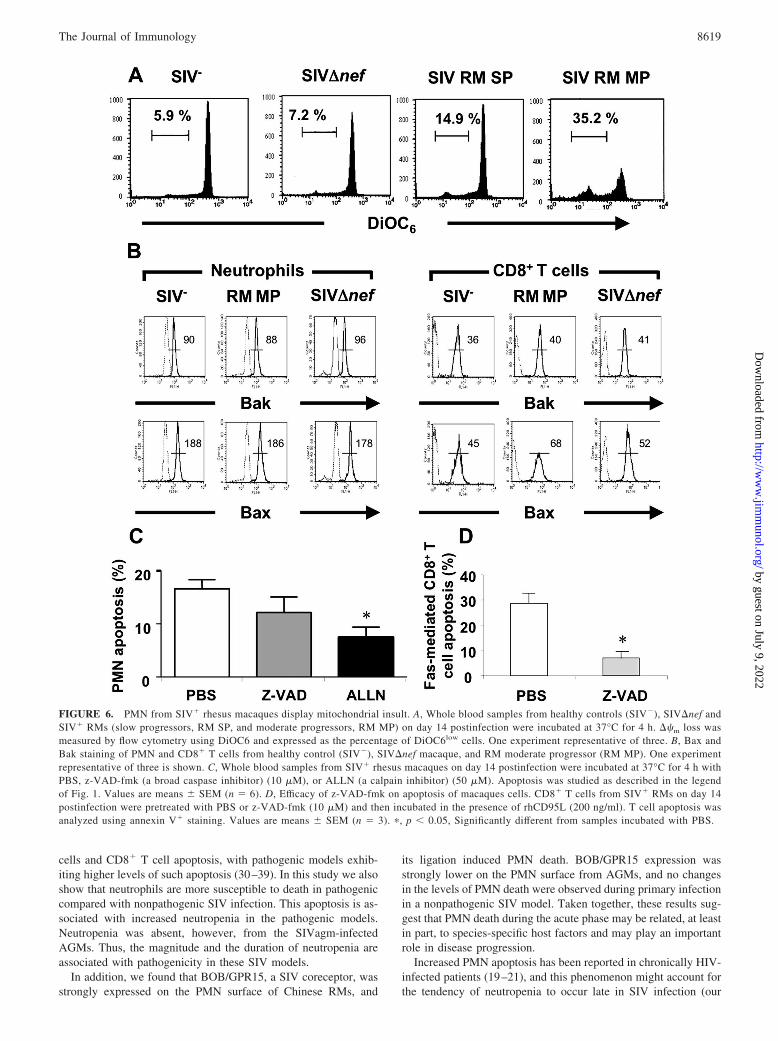
PMN death during primary SIV infection is associated withmitochondrial damage and is calpain-dependent
Apoptosis involves a cascade of signaling events in which themitochondrion is a key sensor and caspases are the cell death ef-
fectors (56). To determine the possible role of mitochondria inPMN apoptosis in this setting, we measured ��m with DiOC6. Wefound that PMN collected from SIV� RMs on day 14 displayed aloss of ��m as compared with PMN from healthy controls andfSIV�nef-infected macaques and that the percentage of PMN dis-playing ��m was more marked in future moderate progressors(32.3 � 5.1) than in slow progressors (13.8 � 3.5) (Fig. 6A). PMNfrom healthy controls and SIV�nef-infected macaques showed nodifference in ��m loss. Because Bax and Bak, proapoptotic mem-bers of the Bcl-2 family, relocate and induce ��m loss (56), weexamined whether these proteins were activated in PMN from SIV�
macaques using Abs known to cross-react with macaques cells (42,43). Our results indicated that ��m loss is not associated with changesin the NH2-terminal epitope availability of Bax or Bak expression (theactive forms of Bax and Bak, which are associated with cell death),demonstrating that mitochondrial changes in PMN in this setting areBax -and Bak-independent (Fig. 6B). In contrast, active Bax was in-creased in CD8� T cells from SIV-infected RM while Bak was notaffected in these cells (Fig. 6B).
FIGURE 3. Comparative analysis of PMN apoptosis during primary in-fection of rhesus macaques with the pathogenic SIVmac251 strain or theattenuated SIV�nef strain, and AGMs. Monkeys were infected i.v. with 10AID50 of the pathogenic SIVmac251 strain (RM, n � 6), the attenuatedSIV�nef strain (SIV�nef, n � 4), or 300 TCID50 of SIVagm strain (AGM,n � 5). Viral load measured 4 mo after infection retrospectively classifiedthe RM monkeys as slow progressors (RM SP, n � 3) or moderate pro-gressors (RM MP, n � 3). PMN apoptosis was studied immediately aftersampling (time (T) 0 h) (A) and after incubating whole blood in 24-welltissue cultures plates at 37°C with 5% CO2 for 4 h (T 4 h) (B). Results areexpressed as the percentage of annexin V�/7AAD� cells (early apoptoticcells). �, p 0.05, significantly different from SIV�nef macaques; †, p 0.05, significantly different from RM SP.
FIGURE 4. Comparative analysis of PMN counts in peripheral blood ofRMs during primary infection with the pathogenic SIVmac251 strain or theattenuated SIV�nef strain and AGMs. A, PMN count kinetics in ChineseRMs. During the first 2 mo, variations in PMN counts were monitored inperipheral blood from SIV�nef (n � 4), SIV� RMs (n � 15) (slow pro-gressors, RM SP, and moderate progressors, RM MP), and AGMs (n � 5).�, p 0.05, Significantly different from SIV�nef at the same time; †, p 0.05, significantly different from RM SP at the same time. B, Regressionanalysis comparing the loss of PMN (day 0 value minus day 14 value) andviremia on day 120 in Chinese rhesus macaques (� � 0.83; p � 0.0009).C, PMN count kinetics in Mamu-A*01� and non-Mamu-A*01 Indian RM.
8617The Journal of Immunology
by guest on July 9, 2022http://w
ww
.jimm
unol.org/D
ownloaded from

To determine the possible involvement of caspases in this PMNapoptosis, we used the broad spectrum caspase inhibitor z-VAD-fmk. This inhibitor has been previously reported to inhibit caspasesin nonhuman primates (37, 42). In agreement with previous reports(37, 42), z-VAD-fmk inhibited Fas-mediated apoptosis of CD8� Tcells from SIV-infected RMs (Fig. 6D). z-VAD-fmk (10 �M),however, failed to have any effect on PMN apoptosis (Fig. 6C). Incontrast, ALLN (50 �M), a highly specific calpain inhibitor, sig-nificantly reduced PMN apoptosis (Fig. 6C). Thus this PMN ap-optosis was calpain dependent and caspase independent.
SIV induces PMN death in the absence of productive viralreplication
Accelerated PMN death during primary SIV infection may be re-lated either to PMN infection or to external apoptogenic factors.
We used flow cytometry to sort highly purified PMN(CD11bhighHLA-DRlow) and CD4� T cells (98 and 99% pure,respectively) at the peak of the acute phase (day 14). Approxi-mately 0.1–1% of CD4� T cells were infected as previously re-ported (41, 45), whereas we detected no SIV DNA in up to 1million PMN. Moreover, no TNF-� or IL-8 (proapoptotic and anti-apoptotic cytokines, respectively) (57, 58) was detected in theplasma of either SIV� or SIV�nef macaques (data not shown)during the acute phase. As our data implied that PMN death iscaspase independent in this setting, we did not seek a role of deathligands such as FasL and Trail, which are also capable of killingPMN via caspase activation (59, 60).
We and others have shown that simple binding and/or penetra-tion of HIV (without integration) can prime death (61–66), imply-ing that collateral cell damage induced by interaction of the viralenvelope glycoprotein (gp120) with molecules expressed by PMN,and particularly chemokine receptors, may account for the ob-served increase in PMN death. We therefore incubated wholeblood samples from healthy macaques with either SIVmac251 ormock as control. We found that SIVmac251 strain (400 AID50)significantly increased apoptosis of PMN derived from RMs (Fig.7A); however, a 10-fold lower dose of SIVmac251 strain was un-able to prime PMN for death (data not shown). This apoptosis-inducing effect of SIVmac251 on PMN is unlikely due to viralinfection and replication, as the short incubation time (4 h) of theseexperiments is incompatible with viral replication in nondividingcells (64, 65). This was further supported by the finding that treat-ment of whole blood samples with didanosine (1 �M), a reversetranscriptase inhibitor, also did not prevent SIV-induced PMNdeath. Finally, aldrithiol-2-inactivated SIVmac251 could alsoprime PMN for apoptosis (Fig. 7A). Thus, viral infection and rep-lication is not required for this PMN apoptosis, and this suggestsa possible receptor-mediated effect. SIV binds to distinct chemo-kine receptors such as BOB/GPR15 and CCR5 (67–69). Flow cy-tometric analysis of chemokine receptors (CXCR4, CCR5, andBOB/GPR15) revealed that PMN from healthy RMs expressedBOB/GPR15 at higher levels than healthy AGMs (Fig. 7, B and C).In a SIV surface binding assay we demonstrated that SIV wassurface bound to PMN and that this binding was BOB/GPR15mediated, as Abs directed to BOB/GPR15 reduce SIV binding onPMN (Fig. 7D). To directly demonstrate that BOB/GPR15 bindingby SIV was mediating this PMN apoptosis, we tested whethercross-linking BOB/GPR15 could induce PMN apoptosis. Indeed,we found that incubation with a BOB/GPR15-specific Ab inducedPMN death (Fig. 7A), while CCR5-specific Ab were less potent ininducing death. Preincubation with z-VAD-fmk did not preventcell death (data not shown). Taken together, the above findingssupport the idea that SIV particle binding to the chemokine recep-tor BOB/GPR15 on the PMN surface, rather than productive viralreplication, drives PMN death. Furthermore, differences in the ex-pression level of coreceptors such as BOB/GPR15 between differ-ent nonhuman primates might be responsible for greater PMNdeath in RMs compared with AGMs.
DiscussionOur results show an increased PMN death during the acute phaseof SIV infection in Chinese RMs that coincided with a decline inPMN number. The level of PMN death was significantly moresevere in Chinese RMs that progressed more rapidly to AIDS.Indian RMs had higher and more sustained neutropenia comparedwith Chinese RMs, and this is in agreement with the overall higherpathogenicity of SIV infection in Indian compared with ChineseRMs. Pathogenic and nonpathogenic models of HIV and SIV in-fections have been shown to differ in the magnitude of CD4� T
FIGURE 5. Comparative analysis of CD11b expression and PMN mi-gration during primary infection of RMs with the pathogenic SIVmac251strain (n � 6), or the attenuated SIV�nef strain (n � 4) and AGMs (n �5). A, Whole blood samples from SIV�nef, SIV� RMs (slow progressors,RM SP, and moderate progressors, RM MP) and AGMs were incubatedwith PBS or fMLP (10�6M) at 37°C for 5 min and then stained withPE-anti-CD11b Ab at 4°C for 30 min. The results are expressed as a stim-ulation index (mean fluorescence intensity of the sample incubated withfMLP/MFI of the sample incubated with PBS). B and C, PMN migrationtoward fMLP (10�7 M) (B) or IL-8 (25 ng/ml) (C) was measured in Trans-well plates as described in Materials and Methods. The results are ex-pressed as the migration rate: (number of PMN in the lower well aftermigration/number of PMN applied to the upper well before migration) �100. �, p 0.05, Significantly different from SIV�nef macaques; †, p 0.05, significantly different from RM slow progressors.
8618 PMN DEATH DURING SIV PRIMARY INFECTION
by guest on July 9, 2022http://w
ww
.jimm
unol.org/D
ownloaded from
cells and CD8� T cell apoptosis, with pathogenic models exhib-iting higher levels of such apoptosis (30–39). In this study we alsoshow that neutrophils are more susceptible to death in pathogeniccompared with nonpathogenic SIV infection. This apoptosis is as-sociated with increased neutropenia in the pathogenic models.Neutropenia was absent, however, from the SIVagm-infectedAGMs. Thus, the magnitude and the duration of neutropenia areassociated with pathogenicity in these SIV models.
In addition, we found that BOB/GPR15, a SIV coreceptor, wasstrongly expressed on the PMN surface of Chinese RMs, and
its ligation induced PMN death. BOB/GPR15 expression wasstrongly lower on the PMN surface from AGMs, and no changesin the levels of PMN death were observed during primary infectionin a nonpathogenic SIV model. Taken together, these results sug-gest that PMN death during the acute phase may be related, at leastin part, to species-specific host factors and may play an importantrole in disease progression.
Increased PMN apoptosis has been reported in chronically HIV-infected patients (19–21), and this phenomenon might account forthe tendency of neutropenia to occur late in SIV infection (our
FIGURE 6. PMN from SIV� rhesus macaques display mitochondrial insult. A, Whole blood samples from healthy controls (SIV�), SIV�nef andSIV� RMs (slow progressors, RM SP, and moderate progressors, RM MP) on day 14 postinfection were incubated at 37°C for 4 h. ��m loss wasmeasured by flow cytometry using DiOC6 and expressed as the percentage of DiOC6low cells. One experiment representative of three. B, Bax andBak staining of PMN and CD8� T cells from healthy control (SIV�), SIV�nef macaque, and RM moderate progressor (RM MP). One experimentrepresentative of three is shown. C, Whole blood samples from SIV� rhesus macaques on day 14 postinfection were incubated at 37°C for 4 h withPBS, z-VAD-fmk (a broad caspase inhibitor) (10 �M), or ALLN (a calpain inhibitor) (50 �M). Apoptosis was studied as described in the legendof Fig. 1. Values are means � SEM (n � 6). D, Efficacy of z-VAD-fmk on apoptosis of macaques cells. CD8� T cells from SIV� RMs on day 14postinfection were pretreated with PBS or z-VAD-fmk (10 �M) and then incubated in the presence of rhCD95L (200 ng/ml). T cell apoptosis wasanalyzed using annexin V� staining. Values are means � SEM (n � 3). �, p 0.05, Significantly different from samples incubated with PBS.
8619The Journal of Immunology
by guest on July 9, 2022http://w
ww
.jimm
unol.org/D
ownloaded from

unpublished data) and human HIV infection (70, 71). In this studywe demonstrate, for the first time, that spontaneous PMN apoptosisis augmented during primary SIV infection of RMs and that thisincrease coincides with neutropenia. Moreover, the extent of PMNapoptosis and neutropenia is associated with the rate of subsequentpathogenicity and progression to AIDS. Given the role of de-fensins in the control of viral replication (3–8) and the fact thatPMN are a major source of �-defensins 1–4 (2), such PMN defects
may play a key role in early viral replication and disseminationwithin the host and might therefore be a key determinant of diseaseoutcome. The fact that SIVagm-infected AGMs do not display anymajor increase in PMN apoptosis in the context of significant lev-els of viral replication suggests that the direct effects of SIV rep-lication alone are unlikely to account for the high levels of PMNapoptosis observed in Chinese RMs at the time of peak viremia.Moreover, no SIV DNA was detected in sorted PMN from
FIGURE 7. SIV primes PMN for death. A, Whole blood samples from healthy controls (SIV�) were incubated for 4 h at 37°C in the absence(mock) or presence of 400 AID50 of the pathogenic SIVmac251 strain that has been either untreated (�) or treated with aldrithiol-2 (AT-2). Cellswere also incubated with anti-BOB/GPR15 or anti-CCR5 Abs. Values are means � SEM (n � 3). Purified sera were used as controls for chemokinereceptors Abs; no difference in the percentage of dying cells was observed relative to the mock control (data not shown). �, p 0.05, Significantlydifferent from mock-incubated samples. B, Representative CXCR4, CCR5 and BOB/GPR15 expression on PMN from a healthy macaque and ahealthy AGM, measured by flow cytometry. Dotted line histograms show isotype control stains. Filled histograms show specific Ab staining. C,Quantitative expression of CXCR4, CCR5, and BOB/GPR15 expression on PMN from healthy RMs (n � 4) and healthy AGMs (n � 4). Valuesare means � SEM. �, p 0.05, Significantly different from AGM. D, PMN from healthy controls (SIV�) were pretreated with purified serum,anti-CCR5 Ab, or anti-BOB/GPR15 Ab and then incubated in the presence of 400 AID50 of the pathogenic SIVmac251 strain. Binding at the cellsurface was revealed by FITC-anti-gp120 (SIVmac251) Ab. �, p 0.05, Significantly different from serum-incubated samples. MFI, Mean fluo-rescence intensity.
8620 PMN DEATH DURING SIV PRIMARY INFECTION
by guest on July 9, 2022http://w
ww
.jimm
unol.org/D
ownloaded from

SIV-infected Chinese RMs. Similarly, an increased lymphocytesusceptibility to apoptosis independent of infection (“bystander ef-fect”) has been proposed as one of the main mechanisms respon-sible for the CD4� T cell depletion in vivo during pathogenic HIVand SIV infections (39).
Indian RMs had higher and more sustained neutropenia comparedwith Chinese RMs, which is consistent with the overall higher patho-genicity of SIV infection in Indian compared with Chinese RMs. Incontrast to Chinese RMs, a day 14 decrease in PMN in Indian RMsdid not correlate with viral loads or CD4 counts. It is unclear why thiscorrelation is not present in the Indian RMs, but the finding that thesustained decrease in PMN in these animals may suggest that thefactors that contribute to the higher viral loads in these animals retainthe PMN decrease. Although both Mamu-A*01� and non-Mamu-A*01 Indian RM showed sustained neutropenia, Mamu-A*01� ani-mals had significantly lower decrease in PMN from baseline, and thisagain is in agreement with the better viral control that these animalshave compared with non-Mamu-A*01 Indian RMs (52, 53). Indeed,in our study non-Mamu-A*01 Indian RMs had significantly higherviral loads compared with the Mamu-A*01� Indian RMs. It shouldbe noted that although the presence of alleles such as Mamu-A*01overall provides better control, this is not in itself predictive of viralcontrol (72, 73). Another possible explanation for the difference be-tween Indian and Chinese RM neutropenia, with the former exhibitingsustained neutropenia, is that Indian RMs were challenged with ahigher dose of SIVmac251 compared with the Chinese RMs. It willbe of interest in the future to determine whether high dose viral chal-lenge determines whether sustained neutropenia is also established inChinese RMs. Overall, our studies suggest that the degree of patho-genicity of SIV infection in a particular host correlates with the mag-nitude and duration of neutropenia in nonhuman primates.
There are two main pathways of apoptosis. The extrinsic path-way is initiated upon the binding of so-called “death receptors”(belonging to the TNF receptor family) to their ligands, whereasthe intrinsic pathway is death receptor independent. These path-ways converge at a central sensor, the mitochondrion, that releasesapoptogenic factors into the cytosol leading to the activation ofeffector caspases. Growing evidence implicates caspase-indepen-dent mechanisms of cell death in most cell types, including PMN(74–77). Indeed, we found that PMN death in SIV-infected ma-caques was not prevented by a broad-spectrum caspase inhibitor,z-VAD-fmk. Our data demonstrate that PMN death in this settinginvolves a calpain-dependent pathway that has also been reportedto play a key role in the spontaneous PMN death of healthy donors(77, 78). Interestingly, a calpain-dependent pathway was recentlyreported to be involved in the death of PMN from chronicallyHIV-infected patients (79). We also found that PMN from SIV-infected Chinese RMs displayed mitochondrial injury, reflected bya loss of ��m. However, mitochondrial membrane permeabiliza-tion occurs independently of a change in the conformation of Baxand Bak, which is considered to be a major event in the fall of��m. Therefore, it remains to determined by which mechanismsthe mitochondrial outer membrane is permeabilized.
Finally, we found that SIV particles prime PMN for death in vitro.However, PMN do not need to be productively infected to undergodeath. These results for PMN death are consistent with other reportsindicating that HIV/SIV sensitize CD4� T cells to undergo apoptosisin the absence of viral replication and immune activation (62, 64, 65).Similarly, it has been shown that exposure to the HIV envelope pro-tein results in apoptosis of NK cells (66). We observed no increase inPMN death and no neutropenia in Chinese RMs infected with theattenuated strain SIV�nef. This might be related to slower acute phaseSIV�nef replication relative to the pathogenic strain (45). Indeed, wefound that in vitro a 10-fold lower dose of SIVmac251 strain did not
prime PMN death. Thus, although we cannot exclude the possibilitythat nef-deleted SIV may also induce cell death in vitro, the thresholdreached in vivo is probably too low to prime PMN for death. To-gether, our data suggest that SIV virions themselves are at least partlyresponsible for priming PMN death. CD4 is not expressed on PMN(data not shown), and HIV/SIV bind to PMN via other membranereceptors (80, 81). We observed increased BOB/GPR15 expression atthe surface of PMN from healthy Chinese RMs, whereas it was lowerexpressed on the surface of PMN from AGMs; the other coreceptors,CCR5 and CXCR4, were not different. Moreover, using specific Abs(because the natural ligand is unknown), we found that BOB/GPR15engagement primed PMN from Chinese RMs for death in a mannersimilar to that of the virus itself. These results showing that this con-stitutive receptor may induce a cell death signal suggests a distinct cellsignaling pathway following coreceptor ligation by SIV vs that in-volved in the physiological response to the unknown BOB/GPR15ligand. This would be consistent with what has previously beenshown for CCR5 and CXCR4. Whereas the natural ligands such asRANTES and SDF1 are unable to induce and prime for cell death,cross-linking of coreceptors mediates cell death (64). Thus, SIV bind-ing to the BOB/GPR15 coreceptor can prime PMN to apoptosis, andthe extent of interaction between the SIV envelope protein and thecoreceptors expressed could be the main process leading to abnormalpriming for death of PMN.
The increased PMN susceptibility to apoptosis coincided with theonset of neutropenia in SIV-infected Chinese RMs. PMN redistribu-tion from peripheral blood to sites of inflammation cannot be ruledout, but it is noteworthy that PMN isolated from SIV-infected ChineseRMs at the peak of viral replication displayed defective chemotaxisand were not activated, as reflected by basal normal CD11b expres-sion and basal normal ROS production. This indicates that PMN re-main in a resting state during acute SIV infection, in keeping with theundetectable levels of proinflammatory cytokines such as TNF-�.This differs from the chronic asymptomatic phase of HIV infection(18, 19) and of SIV infection in RMs (personal data), where PMNcounts are normal despite a greater susceptibility to die by apoptosis(although less than during the acute phase; data not shown). Consis-tent with the idea that granulopoiesis may compensate PMN lossesthrough apoptosis during the chronic phase, it has been shown thatPMN in the bone marrow of chronically SIV-infected macaques areactivated (38). Thus, our data showing neutropenia concomitant withhigher levels of apoptosis may indicate that granulopoiesis did notcompensate for PMN losses. Thus, whether granulopoiesis is defec-tive during the acute phase is, to date, unknown and merits furtherexploration.
In conclusion, the current set of data demonstrate for the firsttime increased PMN death early after infection in pathogenic SIV-infected NHP models of AIDS. This accelerated PMN death maycontribute to the neutropenia and may play a key role during pri-mary SIV infection. This PMN apoptosis is induced by SIV en-velope binding to the BOB/GPR15 chemokine receptor, and thedivergent levels of this receptor may be responsible for the differ-ences in PMN apoptosis between pathogenic and nonpathogenicSIV infection models.
AcknowledgmentsWe thank Drs. Jay Rappaport (Temple University) and David Weiner (Uni-versity of Pennsylvania) for providing blood from Indian RMs. We are alsograteful to Prof. A. G. Saimot for helpful discussions.
DisclosuresThe authors have no financial conflict of interest.
8621The Journal of Immunology
by guest on July 9, 2022http://w
ww
.jimm
unol.org/D
ownloaded from

References1. Watson, A., J. Ranchalis, B. Travis, J. McClure, W. Sutton, P. R. Johnson, S. L. Hu,
and N. L. Haigwood. 1997. Plasma viremia in macaques infected with simian im-munodeficiency virus: plasma viral load early in infection predicts survival. J. Virol.71: 284–290.
2. Babior, B. M. 1984. Oxidants from phagocytes: agents of defense and destruc-tion. Blood 64: 959–966.
3. Bastian, A., and H. Schafer. 2001. Human �-defensin 1 (HNP-1) inhibits adeno-viral infection in vitro. Regul. Pept. 101: 157–161.
4. Fujisawa, H. 2001. Inhibitory role of neutrophils on influenza virus multiplicationin the lungs of mice. Microbiol. Immunol. 45: 679–688.
5. Yasin, B., W. Wang, M. Pang, N. Cheshenko, T. Hong, A. J. Waring, B. C. Herold,E. A. Wagar, and R. I. Lehrer. 2004. � Defensins protect cells from infection byherpes simplex virus by inhibiting viral adhesion and entry. J. Virol. 78: 5147–5156.
6. Zhang, L., W. Yu, T. He, J. Yu, R. E. Caffrey, E. A. Dalmasso, S. Fu, T. Pham,J. Mei, J. J. Ho, et al. 2002. Contribution of human �-defensin 1, 2, and 3 to theanti-HIV-1 activity of CD8 antiviral factor. Science 298: 995–1000.
7. Wu, Z., F. Cicchi, D. Gentles, B. Ericksen, J. Lubkowski, A. Devico, R. I. Lehrer,and W. Lu. 2005. Human neutrophil �-defensin 4 inhibits HIV-1 infection invitro. FEBS Lett. 579: 162–166.
8. Zhang, L., P. Lopez, T. He, W. Yu, and D. D. Ho. 2004. Retraction of an inter-pretation. Science 303: 467.
9. Baldwin, G. C., D. Fuller, R. L. Roberts, D. D. Ho, and D. W. Golde. 1989.Granulocyte- and granulocyte-macrophage colony-stimulating factors enhanceneutrophil cytotoxicity toward HIV-infected cells. Blood 74: 1673–1677.
10. Scapini, P., J. A. Lapinet-Vera, S. Gasperini, F. Calzetti, F. Bazzoni, andM. A. Cassatella. 2000. The neutrophil as a cellular source of chemokines. Im-munol. Rev. 177: 195–203.
11. Gisbergen, J., M. Sanchez-Hernandez, T. B. H. Geijtenbeek, and Y. van Kooyk.2005. Neutrophils mediate immune modulation of dendritic cells through glycosy-lation-dependent interactions between Mac-1 and DC-SIGN. J. Exp. Med. 201:1281–1292.
12. Colotta, F., F. Re, N. Polentarutti, S. Sozzani, and A. Mantovani. 1992. Modu-lation of granulocyte survival and programmed cell death by cytokines and bac-terial products. Blood 80: 2012–2020.
13. Murray, J., J. A. Barbara, S. A. Dunkley, A. F. Lopez, X. Van Ostade,I. Condliffe, A. M. Dransfield, C. Haslett, and E. R. Chilvers. 1997. Regulationof neutrophil apoptosis by tumor necrosis factor-�: requirement for TNFR55 andTNFR75 for induction of apoptosis in vitro. Blood 90: 2772–2783.
14. Francois, S., J. El Benna, P. M. C. Dang, E. Pedruzzi, M. A. Gougerot-Pocidalo,and C. Elbim. 2005. Inhibition of neutrophil apoptosis by Toll-like receptor ago-nists in whole blood: involvement of the phosphoinositide 3-kinase/Akt andNF-�B signaling pathways leading to increased levels of Mcl-1, A1 and phos-phorylated Bad. J. Immunol. 174: 3633–3642.
15. Edwards, S. W., and M. B. Hallett. 1997. Seeing the wood for the trees: the forgottenrole of neutrophils in rheumatoid arthritis. Immunol. Today 18: 320–324.
16. Aleman, M., P. Schierloh, S. S. de la Barrera, R. M. Musella, M. A. Saab,M. Baldini, E. Abbate, and M. C. Sasiain. 2004. Mycobacterium tuberculosistriggers apoptosis in peripheral neutrophils involving Toll-like receptor 2 and p38mitogen protein kinase in tuberculosis patients. Infect. Immun. 72: 5150–5158.
17. Ramirez, M. J., E. Titos, J. Claria, M. Navasa, J. Fernandez, and J. Rodes. 2004.Increased apoptosis dependent on caspase-3 activity in polymorphonuclear leu-kocytes from patients with cirrhosis and ascites. J. Hepatol. 41: 44–48.
18. Pitrak, D. L., H. C. Tsai, K. M. Mullane, S. H. Sutton, and P. Stevens. 1996.Accelerated neutrophil apoptosis in the acquired immunodeficiency syndrome.J. Clin. Invest. 98: 2714–2719.
19. Baldelli, F., R. Preziosi, D. Francisci, C. Tascini, F. Bistoni, and I. Nicoletti.2000. Programmed granulocyte neutrophil death in patients at different stages ofHIV infection. AIDS 214: 1067–1069.
20. Salmen, S., G. Teran, L. Borges, L. Goncalves, B. Albarran, H. Urdaneta,H. Montes, and L. Berrueta. 2004. Increased Fas-mediated apoptosis in polymor-phonuclear cells from HIV-infected patients. Clin. Exp. Immunol. 137: 166–172.
21. Broussard, S. R., S. I. Staprans, R. White, E. M. Whitehead, M. B. Feinberg, andJ. S. Allan. 2001. Simian immunodeficiency virus replicates to high levels innaturally infected African green monkeys without inducing immunologic or neu-rologic disease. J. Virol. 75: 2262–2275.
22. Diop, O. M., A. Gueye, M. Dias-Tavares, C. Kornfeld, A. Faye, P. Ave, M. Huerre,S. Corbet, F. Barre-Sinoussi, and M. C. Muller-Trutwin. 2000. High levels of viralreplication during primary simian immunodeficiency virus SIVagm infection are rap-idly and strongly controlled in African green monkeys. J. Virol. 74: 7538–7547.
23. Estaquier, J., V. Monceaux, M. C. Cumont, A. M. Aubertin, B. Hurtrel, andJ. C. Ameisen. 2000. Early changes in peripheral blood T cells during primary in-fection of rhesus macaques with a pathogenic SIV. J. Med. Primatol. 29: 127–135.
24. Holzammer, S., E. Holznagel, A. Kaul, R. Kurth, and S. Norley. 2001. High virusloads in naturally and experimentally SIVagm-infected African green monkeys.Virology 283: 324–331.
25. Kaur, A., R. M. Grant, R. E. Means, H. McClure, M. Feinberg, and R. P. Johnson.1998. Diverse host responses and outcomes following simian immunodeficiency vi-rus SIVmac239 infection in sooty mangabeys and rhesus macaques. J. Virol. 72:9597–9611.
26. Kornfeld, C., M. J. Ploquin, I. Pandrea, A. Faye, R. Onanga, C. Apetrei,V. Poaty-Mavoungou, P. Rouquet, J. Estaquier, L. Mortara, et al. 2005. Anti-inflammatory profiles during primary SIV infection in African green monkeys areassociated with protection against AIDS. J. Clin. Invest. 115: 1082–1091.
27. Pandrea, I., C. Apetrei, J. Dufour, N. Dillon, J. Barbercheck, M. Metzger,B. Jacquelin, R. Bohm, P. A. Marx, F. Barre-Sinoussi, et al. 2006. Simian immuno-
deficiency virus SIVagm.sab infection of Caribbean African green monkeys: a newmodel for the study of SIV pathogenesis in natural hosts. J. Virol. 80: 4858–4867.
28. Silvestri, G., A. Fedanov, S. Germon, N. Kozyr, W. J. Kaiser, D. A. Garber,H. McClure, M. B. Feinberg, and S. I. Staprans. 2005. Divergent host responsesduring primary simian immunodeficiency virus SIVsm infection of natural sootymangabey and nonnatural rhesus macaque hosts. J. Virol. 79: 4043–4054.
29. Cumont, M. C., O. Diop, B. Vaslin, C. Elbim, L. Viollet, V. Monceaux, S. Lay,G. Silvestri, R. Le Grand, M. Muller-Trutwin, et al. 2008. Early divergence inlymphoid tissue apoptosis between pathogenic and nonpathogenic simian immu-nodeficiency virus infections of nonhuman primates. J. Virol. 82: 1175–1178.
30. Meyaard, L., S. A. Otto, R. R. Jonker, M. J. Mijnster, R. P. Keet, and F. Miedema.1992. Programmed death of T cells in HIV-1 infection. Science 257: 217–219.
31. Estaquier, J., T. Idziorek, F. de Bels, F. Barre-Sinoussi, B. Hurtrel, A. M. Aubertin,A. Venet, M. Mehtali, E. Muchmore, P. Michel, et al. 1994. Programmed cell deathand AIDS: significance of T-cell apoptosis in pathogenic and nonpathogenic primatelentiviral infections. Proc. Natl. Acad. Sci. USA 91: 9431–9435.
32. Estaquier, J., T. Idziorek, W. Zou, D. Emilie, C. M. Farber, J. M. Bourez, andJ. C. Ameisen. 1995. T helper type 1/T helper type 2 cytokines and T cell death:preventive effect of interleukin 12 on activation-induced and CD95 (FAS/APO-1)-mediated apoptosis of CD4� T cells from human immunodeficiency virus-infected persons. J. Exp. Med. 182: 1759–1767.
33. Estaquier, J., M. Tanaka, T. Suda, S. Nagata, P. Golstein, and J. C. Ameisen.1996. Fas-mediated apoptosis of CD4� and CD8� T cells from human immu-nodeficiency virus-infected persons: differential in vitro preventive effect of cy-tokines and protease antagonists. Blood 87: 4959–4966.
34. Katsikis, P. D., E. S. Wunderlich, C. A. Smith, and L. A. Herzenberg. 1995. Fasantigen stimulation induces marked apoptosis of T lymphocytes in human im-munodeficiency virus-infected individuals. J. Exp. Med. 181: 2029–2036.
35. Badley, A. D., K. Parato, D. W. Cameron, S. Kravcik, B. N. Phenix, D. Ashby,A. Kumar, D. H. Lynch, J. Tschopp, and J. B. Angel. 1999. Dynamic correlationof apoptosis and immune activation during treatment of HIV infection. CellDeath Differ. 6: 420–432.
36. McCloskey, T. W., S. Bakshi, S. Than, P. Arman, and S. Pahwa. 1998. Immu-nophenotypic analysis of peripheral blood mononuclear cells undergoing in vitroapoptosis after isolation from human immunodeficiency virus-infected children.Blood 92: 4230–4237.
37. Arnoult, D., F. Petit, J. D. Lelievre, D. Lecossier, A. Hance, V. Monceaux, B. Hurtrel,R. Ho Tsong Fang, J. C. Ameisen, and J. Estaquier. 2003. Caspase-dependent andindependent T-cell death pathways in pathogenic simian immunodeficiency virusinfection: relationship to disease progression. Cell Death Differ. 10: 1240–1252.
38. Silvestri, G., D. L. Sodora, R. A. Koup, M. Paiardini, S. P. O’Neil,H. M. McClure, S. I. Staprans, and M. B. Feinberg. 2003. Nonpathogenic SIVinfection of sooty mangabeys is characterized by limited bystander immunopa-thology despite chronic high-level viremia. Immunity 18: 441–452.
39. Hurtrel, B., F. Petit, D. Arnoult, M. Muller-Trutwin, G. Silvestri, and J. Estaquier.2005. Apoptosis in SIV infection. Cell Death Differ. 12 (Suppl. 1): 979–990.
40. Institute for Laboratory Animal Research, National Research Council. 1996.Guide for the Care and Use of Laboratory Animals. National Academies Press,Washington, D.C.
41. Monceaux, V., J. Estaquier, M. Fevrier, M. C. Cumont, Y. Riviere,A. M. Aubertin, J. C. Ameisen, and B. Hurtrel. 2003. Extensive apoptosis inlymphoid organs during primary SIV infection predicts rapid progression towardsAIDS. AIDS 17: 1585–1596.
42. Viollet, L., V. Monceaux, F. Petit, R. Ho Tsong Fang, M. C. Cumont, B. Hurtrel, andJ. Estaquier. 2006. Death of CD4� T cells from lymph nodes during primary SIV-mac251 infection predicts the rate of AIDS progression. J. Immunol. 177:6685–6694.
43. Cumont, M. C., V. Monceaux, L. Viollet, S. Lay, R. Parker, B. Hurtrel, andJ. Estaquier. 2007. TGF-� in intestinal lymphoid organs contributes to the deathof armed effector CD8 T cells and is associated with the absence of virus con-tainment in rhesus macaques infected with the simian immunodeficiency virus.Cell Death Differ. 14: 1747–1758.
44. Sasakawa, Y., S. Sakuma, Y. Higashi, T. Sasakawa, T. Amaya, and T. Goto.2000. FK 506 suppresses neutrophil chemoattractant production by peripheralblood mononuclear cells. Eur. J. Pharmacol. 403: 281–288.
45. Monceaux, V., R. Ho Tsong Fang, M. C. Cumont, B. Hurtrel, and J. Estaquier.2003. Distinct cycling CD4� and CD8� T cell profiles during the asymptomaticphase of simian immunodeficiency virus SIVmac251 infection in rhesus ma-caques. J. Virol. 77: 10047–10059.
46. Maianski, N. A., A. N. Maianski, T. W. Kuijpers, and D. Roos. 2004. Apoptosisof neutrophils. Acta Haematol. 111: 56–66.
47. Monceaux, V., L. Viollet, F. Petit, R. Ho Tsong Fang, M. C. Cumont, J. Zaunders,B. Hurtrel, and J. Estaquier. 2005. CD8� T cell dynamics during primary simianimmunodeficiency virus infection in macaques: relationship of effector cell differen-tiation with the extent of viral replication. J. Immunol. 174: 6898–6908.
48. Goldstein, S., C. R. Brown, I. Ourmanov, I. Pandrea, A. Buckler-White, C. Erb,J. S. Nandi, G. J. Foster, P. Autissier, J. E. Schmitz, and V. M. Hirsch. 2006. Com-parison of simian immunodeficiency virus SIVagmVer replication and CD4� T-celldynamics in vervet and sabaeus African green monkeys. J. Virol. 80: 4868–4877.
49. Goldstein, S., I. Ourmanov, C. R. Brown, B. E. Beer, W. R. Elkins, R. Plishka,A. Buckler-White, and V. M. Hirsch. 2000. Wide range of viral load in healthyAfrican green monkeys naturally infected with simian immunodeficiency virus. J. Vi-rol. 74: 11744–11753.
50. Staprans, S. I., P. J. Dailey, A. Rosenthal, C. Horton, R. M. Grant, N. Lerche, andM. B. Feinberg. 1999. Simian immunodeficiency virus disease course is predicted bythe extent of virus replication during primary infection. J. Virol. 73: 4829–4839.
8622 PMN DEATH DURING SIV PRIMARY INFECTION
by guest on July 9, 2022http://w
ww
.jimm
unol.org/D
ownloaded from

51. Ling, B., R. S. Veazey, A. Luckay, C. Penedo, K. Xu, J. D. Lifson, and P. A. Marx.2002. SIV(mac) pathogenesis in rhesus macaques of Chinese and Indian origin com-pared with primary HIV infections in humans. AIDS 16: 1489–1496.
52. Mothe, B. R., J. Weinfurter, C. Wang, W. Rehrauer, N. Wilson, T. M. Allen,D. B. Allison, and D. I. Watkins. 2003. Expression of the major histocompatibilitycomplex class I molecule Mamu-A01* is associated with control of simian immu-nodeficiency virus SIVmac239 replication. J. Virol. 77: 2736–2740.
53. Loffredo, J. T., J. Maxwell, Y. Qi, C. E. Glidden, G. J. Borchardt, T. Soma,A. T. Bean, D. R. Beal, N. A. Wilson, W. M. Rehrauer, et al. 2007. Mamu-*08-positive macaques control simian immunodeficiency virus replication. J. Virol. 81:8827–8832.
54. Elbim, C., M. H. Prevot, F. Bouscara, E. Franzini, S. Chollet-Martin, J Hakim,and M. A. Gougerot-Pocidalo. 1994. PMN from HIV-infected patients show en-hanced activation, diminished fMLP-induced L-selectin shedding and an im-paired oxidative burst after cytokine priming. Blood 84: 2759–2766.
55. Kubes, P., B. Heit, G. Van Marle, J. B. Johnston, D. Knight, A. Khan, andC. Power. 2003. In vivo impairment of neutrophil recruitment during lentivirusinfection. J. Immunol. 171: 4801–4808.
56. Hengartner, M. O. 2000. The biochemistry of apoptosis. Nature 407: 770–776.57. Salamone, G., M. Giordano, A. S. Trevani, R. Gamberale, M. Vermeulen,
J. Schettinni, and J. R. Geffner. 2001. Promotion of neutrophil apoptosis byTNF-�. J. Immunol. 166: 3476–3483.
58. Dunican, A. L., S. J. Leuenroth, P. Grutkoski, A. Ayala, and H. H. Simms. 2000.TNF�-induced suppression of PMN apoptosis is mediated through interleukin-8production. Shock 14: 284–289.
59. Liu, J. H., S. Wei, T. Lamy, P. K. Epling-Burnette, G. Starkebaum, J. Y. Djeu,and T. P. Loughran. 2000. Chronic neutropenia mediated by Fas ligand. Blood95: 3219–3222.
60. Renshauw, S. A., J. S. Parmar, V. Singleton, S. J. Rowe, D. H. Dockrell,S. K. Dower, C. D. Bingle, E. R. Chilvers, and M. K. Whyte. 2003. Accelerationof human neutrophil apoptosis by TRAIL. J. Immunol. 170: 1027–1033.
61. Esser, M. T., D. R. Graham, L. V. Coren, C. M. Trubey, J. W. Bess, L. O. Arthur,D. E. Ott, and J. D. Lifson. 2001. Differential incorporation of CD45, CD80(B7-1), CD86 (B7-2), and major histocompatibility complex class I and II mol-ecules into human immunodeficiency virus type 1 virions and microvesicles:implications for viral pathogenesis and immune regulation. J. Virol. 75:6173–6182.
62. Vlahakis, S. R., A. Algeciras-Schimnich, G. Bou, C. J. Heppelmann,A. Villasis-Keever, R. C. Collman, and C. V. Paya. 2001. Chemokine-receptoractivation by env determines the mechanism of death in HIV-infected and unin-fected T lymphocytes. J. Clin. Invest. 107: 207–215.
63. Zhang, K., F. Rana, C. Silva, J. Ethier, K. Wehrly, B. Chesebro, and C. Power.2003. Human immunodeficiency virus type 1 envelope-mediated neuronal death:uncoupling of viral replication and neurotoxicity. J. Virol. 77: 6899–6912.
64. Lelievre, J. D., F. Mammano, D. Arnoult, F. Petit, A. Grodet, and J. Estaquier.2004. A novel mechanism for HIV1-mediated bystander CD4� T-cell death:neighboring dying cells drive the capacity of HIV1 to kill noncycling primaryCD4� T cells. Cell Death Differ. 11: 1017–1027.
65. Petit, F., J. Corbeil, J. D. Lelievre, L. M. Parseval, G. Pinon, D. R. Green,J. C. Ameisen, and J. Estaquier. 2004. Role of CD95-activated caspase-1 pro-cessing of IL-1� in TCR-mediated proliferation of HIV-infected CD4� T cells.Eur. J. Immunol. 31: 3513–3524.
66. Kottilil, S., K. Shin, J. O. Jackson, K. N. Reinato, M. A. O’Shea, J. Yang,C. W. Hallahan, R. Lempicki, J. Arthos, and A. S. Fauci. 2006. Innate immune
dysfunction in HIV infection: effect of HIV envelope-NK cell interactions. J. Im-munol. 176: 1107–1114.
67. Marx, P. A., and Z. Chen. 1998. The function of simian chemokine receptors inthe replication of SIV. Semin. Immunol. 10: 215–223.
68. Deng, H. K., D. Unutmaz, V. N. Kewal-Ramani, and D. R. Littman. 1997. Ex-pression cloning of new receptors used by simian and human immunodeficiencyviruses. Nature 1388: 296–300.
69. Farzan, M., H. Choe, K. Martin, L. Marcon, W. Hofmann, G. Karlsson, Y. Sun,P. Barrett, N. Marchand, N. Sullivan, et al. 1997. Two orphan seven-transmem-brane segment receptors which are expressed in CD4-positive cells support sim-ian immunodeficiency virus infection. J. Exp. Med. 186: 405–411.
70. Levine, A. M., R. Karim, W. Mack, D. J. Gravink, K. Anastos, M. Young,M. Cohen, M. Newman, M. Augenbraun, S. Gange, and D. H. Watts. 2006.Neutropenia in human immunodeficiency virus infection: data from the women’sinteragency HIV study. Arch. Intern. Med. 166: 405–410.
71. Salmen, S., C. Guillermo, M. Colmenares, L. Barboza, L. Goncalves, G. Teran,N. Alfonso, H. Montes, and L. Berreta. 2005. Role of human immunodeficiencyvirus in leukocytes apoptosis from infected patients. Invest. Clin. 46: 289–305.
72. Yant, L. J., T. C. Friedrich, R. C. Johnson, G. E. May, N. J. Maness, A. M. Enz,J. D. Lifson, D. H. O’Connor, M. Carrington, and D. I. Watkins. 2006. Thehigh-frequency major histocompatibility complex class I allele Mamu-B*17 isassociated with control of simian immunodeficiency virus SIVmac239 replica-tion. J. Virol. 80: 5074–5077.
73. Wojcechowskyj, J. A., L. J. Yant, R. W. Wiseman, S. L. O’Connor, and D. H.O’Connor. 2007. Control of simian immunodeficiency virus SIVmac239 is notpredicted by inheritance of Mamu-B*17-containing haplotypes. J. Virol. 81:406–410.
74. Michallet, M. C., F. Saltel, M. Flacher, J. P. Revillard, and L. Genestier. 2004.Cathepsin-dependent apoptosis triggered by supraoptimal activation of T lym-phocytes: a possible mechanism of high dose tolerance. J. Immunol. 172:5405–5414.
75. Chipuk, J. E., and D. R. Green. 2005. Do inducers of apoptosis trigger caspase-independent cell death? Nat. Rev. Mol. Cell Biol. 6: 268–275.
76. Von Gunten, S., S. Yousefi, M. Seitz, S. M. Jakob, T. Schaffner, R. Seger,J. Takala, P. M. Villiger, and H. U. Simon. 2005. Siglec-9 transduces apoptoticand nonapoptotic death signals into neutrophils depending on the proinflamma-tory cytokine environment. Blood 106: 1423–1431.
77. Knepper-Nicolai, B., J. Savill, and S. B. Brown. 1998. Constitutive apoptosis inhuman neutrophils requires synergy between calpains and the proteasome down-stream of caspases. J. Biol. Chem. 46: 30530–30536.
78. Yousefi, S., R. Perozzo, I. Schmid, A. Ziemiecki, T. Schaffner, L. Scapozza,T. Brunner, and H. U. Simon. 2006. Calpain-mediated cleavage of Atg5 switchesautophagy to apoptosis. Nat. Cell Biol. 8: 1124–1132.
79. Lichtner, M., F. Mengoni, C. M. Mastroianni, I. Sauzullo, R. Rossi,M. De Nicola, V. Vullo, and L. Ghibelli. 2006. HIV protease inhibitor therapyreverses neutrophil apoptosis in AIDS patients by direct calpain inhibition. Ap-optosis 11: 781–787.
80. Olinger, G. G., M. Saifuddin, and G. T. Spear. 2000. CD4-negative cells bindhuman immunodeficiency virus type 1 and efficiently transfer virus to T cells.J. Virol. 74: 8550–8557.
81. Gabali, A. M., J. J. Anzinger, G. T. Spear, and L. L. Thomas. 2004. Activationby inflammatory stimuli increases neutrophil binding of human immunodefi-ciency virus type 1 and subsequent infection of lymphocytes. J. Virol. 78:10833–10836.
8623The Journal of Immunology
by guest on July 9, 2022http://w
ww
.jimm
unol.org/D
ownloaded from