Transplantation Immunology
20
■ Immunologic Basis of Graft Rejection ■ Clinical Manifestations of Graft Rejection ■ General Immunosuppressive Therapy ■ Specific Immunosuppressive Therapy ■ Immune Tolerance to Allografts ■ Clinical Transplantation Transplantations Routinely Used in Clinical Practice Transplantation Immunology T , immunology, refers to the act of transferring cells, tissues, or organs from one site to another. The desire to accomplish transplants stems from the realization that many diseases can be cured by implantation of a healthy organ, tissue, or cells (a graft) from one individual (the donor) to another in need of the transplant (the recipient or host). The development of surgical techniques that allow the facile reimplantation of organs has removed one barrier to successful transplantation, but others remain. One is the lack of organs for transplantation. Although a supply of organs is provided by accident victims and, in some cases, living donors, there are more patients in need of transplants than there are organs available. The seriousness of the donor- organ shortage is reflected in the fact that, as of November 2000, an estimated 73,000 patients in the United States were on the waiting list for an organ transplantation. The major- ity of those on the list (~70%) require a kidney; at present, the waiting period for this organ averages over 800 days. While the lack of organs for transplantation is a serious is- sue, the most formidable barrier to making transplantation a routine medical treatment is the immune system. The immune system has evolved elaborate and effective mecha- nisms to protect the organism from attack by foreign agents, and these same mechanisms cause rejection of grafts from anyone who is not genetically identical to the recipient. Alexis Carrel reported the first systematic study of trans- plantation in 1908; he interchanged both kidneys in a series of nine cats. Some of those receiving kidneys from other cats maintained urinary output for up to 25 days. Although all the cats eventually died, the experiment established that a transplanted organ could carry out its normal function in the recipient. The first human kidney transplant, attempted in 1935 by a Russian surgeon, failed because there was a mis- match of blood types between donor and recipient. This incompatibility caused almost immediate rejection of the kidney, and the patient died without establishing renal func- tion. The rapid immune response experienced here, termed hyperacute rejection, is mediated by antibodies and will be described in this chapter. The first successful human kidney transplant, which was between identical twins, was accom- plished in Boston in 1954. Today, kidney, pancreas, heart, lung, liver, bone-marrow, and cornea transplantations are performed among nonidentical individuals with ever- increasing frequency and success. A variety of immunosuppressive agents can aid in the survival of the transplants, including drugs and specific anti- bodies developed to diminish the immunologic attack on grafts, but the majority of these agents have an overall immunosuppressive effect, and their long-term use is delete- rious. New methods of inducing specific tolerance to the graft without suppressing other immune responses are being developed and promise longer survival of transplants with- out compromise of host immunity. This chapter describes the mechanisms underlying graft rejection, various proce- dures that are used to prolong graft survival, and a summary of the current status of transplantation as a clinical tool. A Clinical Focus section examines the use of organs from non- human species (xenotransplants) to circumvent the shortage of organs available for patients in need of them. chapter 21
Transcript of Transplantation Immunology

■ Immunologic Basis of Graft Rejection
■ Clinical Manifestations of Graft Rejection
■ General Immunosuppressive Therapy
■ Specific Immunosuppressive Therapy
■ Immune Tolerance to Allografts
■ Clinical Transplantation
Transplantations Routinely Used in Clinical Practice
TransplantationImmunology
T,
immunology, refers to the act of transferring cells,tissues, or organs from one site to another. The
desire to accomplish transplants stems from the realizationthat many diseases can be cured by implantation of a healthyorgan, tissue, or cells (a graft) from one individual (thedonor) to another in need of the transplant (the recipient orhost). The development of surgical techniques that allow thefacile reimplantation of organs has removed one barrier tosuccessful transplantation, but others remain. One is the lackof organs for transplantation. Although a supply of organs isprovided by accident victims and, in some cases, livingdonors, there are more patients in need of transplants thanthere are organs available. The seriousness of the donor-organ shortage is reflected in the fact that, as of November2000, an estimated 73,000 patients in the United States wereon the waiting list for an organ transplantation. The major-ity of those on the list (~70%) require a kidney; at present,the waiting period for this organ averages over 800 days.While the lack of organs for transplantation is a serious is-sue, the most formidable barrier to making transplantationa routine medical treatment is the immune system. Theimmune system has evolved elaborate and effective mecha-nisms to protect the organism from attack by foreign agents,and these same mechanisms cause rejection of grafts fromanyone who is not genetically identical to the recipient.
Alexis Carrel reported the first systematic study of trans-plantation in 1908; he interchanged both kidneys in a seriesof nine cats. Some of those receiving kidneys from other catsmaintained urinary output for up to 25 days. Although allthe cats eventually died, the experiment established that atransplanted organ could carry out its normal function inthe recipient. The first human kidney transplant, attemptedin 1935 by a Russian surgeon, failed because there was a mis-match of blood types between donor and recipient. Thisincompatibility caused almost immediate rejection of thekidney, and the patient died without establishing renal func-tion. The rapid immune response experienced here, termedhyperacute rejection, is mediated by antibodies and will bedescribed in this chapter. The first successful human kidneytransplant, which was between identical twins, was accom-plished in Boston in 1954. Today, kidney, pancreas, heart,lung, liver, bone-marrow, and cornea transplantations areperformed among nonidentical individuals with ever-increasing frequency and success.
A variety of immunosuppressive agents can aid in thesurvival of the transplants, including drugs and specific anti-bodies developed to diminish the immunologic attack ongrafts, but the majority of these agents have an overallimmunosuppressive effect, and their long-term use is delete-rious. New methods of inducing specific tolerance to thegraft without suppressing other immune responses are beingdeveloped and promise longer survival of transplants with-out compromise of host immunity. This chapter describesthe mechanisms underlying graft rejection, various proce-dures that are used to prolong graft survival, and a summaryof the current status of transplantation as a clinical tool. AClinical Focus section examines the use of organs from non-human species (xenotransplants) to circumvent the shortageof organs available for patients in need of them.
chapter 21

Immunologic Basis of Graft RejectionThe degree of immune response to a graft varies with thetype of graft. The following terms are used to denote differ-ent types of transplants:
■ Autograft is self-tissue transferred from one body site toanother in the same individual. Transferring healthy skinto a burned area in burn patients and use of healthyblood vessels to replace blocked coronary arteries areexamples of frequently used autografts.
■ Isograft is tissue transferred between genetically identicalindividuals. In inbred strains of mice, an isograft can beperformed from one mouse to another syngeneic mouse.In humans, an isograft can be performed betweengenetically identical (monozygotic) twins.
■ Allograft is tissue transferred between geneticallydifferent members of the same species. In mice, anallograft is performed by transferring tissue or an organfrom one strain to another. In humans, organ grafts fromone individual to another are allografts unless the donorand recipient are identical twins.
■ Xenograft is tissue transferred between different species(e.g., the graft of a baboon heart into a human). Becauseof significant shortages in donated organs, raisinganimals for the specific purpose of serving as organdonors for humans is under serious consideration.
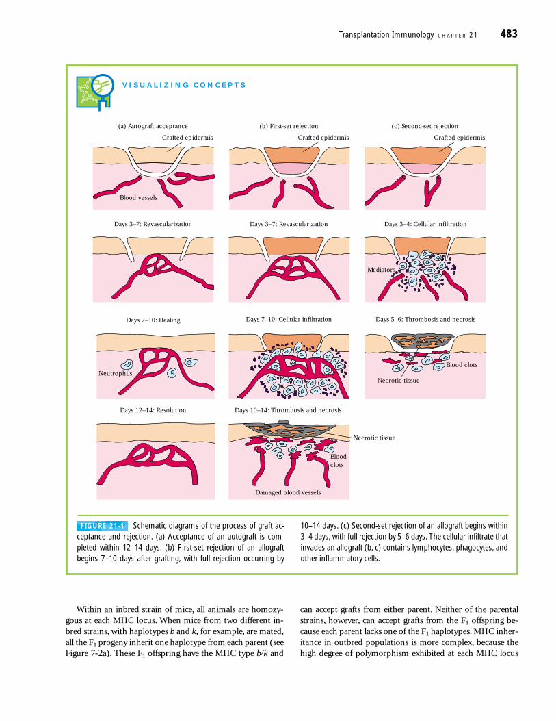
Autografts and isografts are usually accepted, owing to thegenetic identity between graft and host (Figure 21-1a). Be-cause an allograft is genetically dissimilar to the host, it is often recognized as foreign by the immune system and is re-jected. Obviously, xenografts exhibit the greatest genetic dis-parity and therefore engender a vigorous graft rejection.
Allograft Rejection Displays Specificity and MemoryThe rate of allograft rejection varies according to the tissueinvolved. In general, skin grafts are rejected faster than othertissues such as kidney or heart. Despite these time differ-ences, the immune response culminating in graft rejectionalways displays the attributes of specificity and memory. If aninbred mouse of strain A is grafted with skin from strain B,primary graft rejection, known as first-set rejection, occurs(Figure 21-1b). The skin first becomes revascularized betweendays 3 and 7; as the reaction develops, the vascularized trans-plant becomes infiltrated with lymphocytes, monocytes, neu-trophils, and other inflammatory cells. There is decreased vas-cularization of the transplanted tissue by 7–10 days, visiblenecrosis by 10 days, and complete rejection by 12–14 days.
Immunologic memory is demonstrated when a secondstrain-B graft is transferred to a previously grafted strain-A
mouse. In this case, a graft-rejection reaction develops morequickly, with complete rejection occurring within 5–6 days;this secondary response is designated second-set rejection(Figure 21-1c). The specificity of second-set rejection can bedemonstrated by grafting an unrelated strain-C graft at thesame time as the second strain-B graft. Rejection of thestrain-C graft proceeds according to first-set rejection kinet-ics, whereas the strain-B graft is rejected in an acceleratedsecond-set fashion.
T Cells Play a Key Role in Allograft RejectionIn the early 1950s, Avrion Mitchison showed in adoptive-transfer experiments that lymphocytes, but not serum anti-body, could transfer allograft immunity. Later studies im-plicated T cells in allograft rejection. For example, nude mice, which lack a thymus and consequently lack functionalT cells, were found to be incapable of allograft rejection;indeed, these mice even accept xenografts. In other studies,T cells derived from an allograft-primed mouse were shownto transfer second-set allograft rejection to an unprimed syngeneic recipient, as long as that recipient was grafted withthe same allogeneic tissue (Figure 21-2).
Analysis of the T-cell subpopulations involved in allograftrejection has implicated both CD4+ and CD8+ populations.In one study, mice were injected with monoclonal antibodiesto deplete one or both types of T cells and then the rate ofgraft rejection was measured. As shown in Figure 21-3, re-moval of the CD8+ population alone had no effect on graftsurvival, and the graft was rejected at the same rate as in con-trol mice (15 days). Removal of the CD4+ T-cell populationalone prolonged graft survival from 15 days to 30 days. How-ever, removal of both the CD4+ and the CD8+ T cells resultedin long-term survival (up to 60 days) of the allografts. Thisstudy indicated that both CD4+ and CD8+ T-cells partici-pated in rejection and that the collaboration of both subpop-ulations resulted in more pronounced graft rejection.
Similar Antigenic Profiles Foster Allograft Acceptance Tissues that are antigenically similar are said to be histocom-patible; such tissues do not induce an immunologic responsethat leads to tissue rejection. Tissues that display significantantigenic differences are histoincompatible and induce animmune response that leads to tissue rejection. The variousantigens that determine histocompatibility are encoded bymore than 40 different loci, but the loci responsible for themost vigorous allograft-rejection reactions are located with-in the major histocompatibility complex (MHC). The orga-nization of the MHC—called the H-2 complex in mice andthe HLA complex in humans—was described in Chapter 7(see Figure 7-1). Because the MHC loci are closely linked,they are usually inherited as a complete set, called a haplo-type, from each parent.
482 P A R T I V The Immune System in Health and Disease
Within an inbred strain of mice, all animals are homozy-gous at each MHC locus. When mice from two different in-bred strains, with haplotypes b and k, for example, are mated,all the F1 progeny inherit one haplotype from each parent (seeFigure 7-2a). These F1 offspring have the MHC type b/k and
can accept grafts from either parent. Neither of the parentalstrains, however, can accept grafts from the F1 offspring be-cause each parent lacks one of the F1 haplotypes. MHC inher-itance in outbred populations is more complex, because thehigh degree of polymorphism exhibited at each MHC locus
Transplantation Immunology C H A P T E R 21 483
V I S U A L I Z I N G C O N C E P T S
(a) Autograft acceptance
Grafted epidermis
Blood vessels
Days 3–7: Revascularization
(b) First-set rejection
Grafted epidermis
(c) Second-set rejection
Grafted epidermis
Days 3–7: Revascularization Days 3–4: Cellular infiltration
Mediators
Days 7–10: Healing
Neutrophils
Days 12–14: Resolution Days 10–14: Thrombosis and necrosis
Damaged blood vessels
Blood clots
Necrotic tissue
Days 7–10: Cellular infiltration Days 5–6: Thrombosis and necrosis
Necrotic tissue
Blood clots
FIGURE 21-1 Schematic diagrams of the process of graft ac-ceptance and rejection. (a) Acceptance of an autograft is com-pleted within 12–14 days. (b) First-set rejection of an allograftbegins 7–10 days after grafting, with full rejection occurring by
10–14 days. (c) Second-set rejection of an allograft begins within3–4 days, with full rejection by 5–6 days. The cellular infiltrate thatinvades an allograft (b, c) contains lymphocytes, phagocytes, andother inflammatory cells.

gives a high probability of heterozygosity at most loci. In mat-ings between members of an outbred species, there is only a25% chance that any two offspring will inherit identical MHChaplotypes (see Figure 7-2c), unless the parents share one ormore haplotypes. Therefore, for purposes of organ or bone-marrow grafts, it can be assumed that there is a 25% chance ofidentity within the MHC between siblings. With parent-to-child grafts, the donor and recipient will always have one hap-lotype in common but are nearly always mismatched for thehaplotype inherited from the other parent.
Graft Donors and Recipients Are Typed for RBC and MHC AntigensSince differences in blood group and major histocompatibilityantigens are responsible for the most intense graft-rejectionreactions, various tissue-typing procedures to identify theseantigens have been developed to screen potential donor andrecipient cells. Initially, donor and recipient are screened forABO blood-group compatibility. The blood-group antigensare expressed on RBCs, epithelial cells, and endothelial cells.Antibodies produced in the recipient to any of these antigensthat are present on transplanted tissue will induce antibody-mediated complement lysis of the incompatible donor cells.
HLA typing of potential donors and a recipient can beaccomplished with a microcytotoxicity test (Figure 21-4a, b).In this test, white blood cells from the potential donors andrecipient are distributed into a series of wells on a microtiterplate, and then antibodies specific for various class I and classII MHC alleles are added to different wells. After incubation,complement is added to the wells, and cytotoxicity is assessedby the uptake or exclusion of various dyes (e.g., trypan blueor eosin Y) by the cells. If the white blood cells express theMHC allele for which a particular monoclonal antibody isspecific, then the cells will be lysed upon addition of comple-ment, and these dead cells will take up a dye such as trypanblue. HLA typing based on antibody-mediated microcyto-toxicity can thus indicate the presence or absence of variousMHC alleles.
Even when a fully HLA-compatible donor is not available,transplantation may be successful. In this situation, a one-waymixed-lymphocyte reaction (MLR) can be used to quantifythe degree of class II MHC compatibility between potential
484 P A R T I V The Immune System in Health and Disease
First skin graft,strain A
Second skin graft,strain A
Naive strain = B mouse
First-set rejection Second-set rejection
14 days Time 6 days
Naive strain = B mouse
Necrosis
First skin graft,strain A
Necrosis
Second-set rejection
6 days
Necrosis
Spleenic T cells
FIGURE 21-2 Experimental demonstration that T cells can trans-fer allograft rejection. When T cells derived from an allograft-primedmouse are transferred to an unprimed syngeneic mouse, the recipi-
ent mounts a second-set rejection to an initial allograft from the orig-inal allogeneic strain.
Surv
ivin
g gr
afts
, %
Time after grafting, days
50
100
6030150
Anti–CD4ControlAnti–CD8
Anti–CD4and Anti–CD8
FIGURE 21-3 The role of CD4+ and CD8+ T cells in allograft rejec-tion is demonstrated by the curves showing survival times of skingrafts between mice mismatched at the MHC. Animals in which theCD8+ T cells were removed by treatment with an anti-CD8 mono-clonal antibody (red) showed little difference from untreated controlmice (black). Treatment with monoclonal anti-CD4 (blue) improvedgraft survival significantly, and treatment with both anti-CD4 andanti-CD8 antibody prolonged graft survival most dramatically(green). [Adapted from S. P. Cobbold et al., 1986, Nature 323:165.]

Transplantation Immunology C H A P T E R 21 485
Donor cell Recipient cell
HLA–A allele 2 HLA–A allele 1
Antibody toHLA–A allele 2
Complement
Dye (trypan blueor eosin Y)
Cells becomeleaky
No lysis
Dye taken up Dye excluded
(a)
1Antibody to different HLA-A antigens
Recipient
Donor 1
Donor 2
2 3 4 5 6 7 8 9(b)
(c)
Irradiation
Donor cells
Allele A
Recipient cells lacking class II MHC of donor
Recipient cells sharing class II MHC of donor
Allele B
Allele A
No reaction
Activation andproliferation ofrecipient cells [3H]thymidine
Incorporation of of radioactivity into cell nuclear DNA
FIGURE 21-4 Typing procedures for HLA antigens. (a, b) HLA typ-ing by microcytotoxicity. (a) White blood cells from potential donorsand the recipient are added to separate wells of a microtiter plate.The example depicts the reaction of donor and recipient cells with asingle antibody directed against an HLA-A antigen. The reaction se-quence shows that if the antigen is present on the lymphocytes, ad-dition of complement will cause them to become porous and unableto exclude the added dye. (b) Because cells express numerous HLAantigens, they are tested separately with a battery of antibodies spe-cific for various HLA-A antigens. Here, donor 1 shares HLA-A anti-gens recognized by antisera in wells 1 and 7 with the recipient,whereas donor 2 has none of HLA-A antigens in common with the re-cipient. (c) Mixed lymphocyte reaction to determine identity of classII HLA antigens between a potential donor and recipient. Lympho-cytes from the donor are irradiated or treated with mitomycin C toprevent cell division and then added to cells from the recipient. If theclass II antigens on the two cell populations are different, the recipi-ent cells will divide rapidly and take up large quantities of radioactivenucleotides into the newly synthesized nuclear DNA. The amount ofradioactive nucleotide uptake is roughly proportionate to the MHCclass II differences between the donor and recipient lymphocytes.

donors and a recipient (Figure 21-4c). Lymphocytes from apotential donor that have been x-irradiated or treated withmitomycin C serve as the stimulator cells, and lymphocytesfrom the recipient serve as responder cells. Proliferation ofthe recipient T cells, which indicates T-cell activation, is mea-sured by the uptake of [3H]thymidine into cell DNA. Thegreater the class II MHC differences between the donor andrecipient cells, the more [3H]thymidine uptake will be ob-served in an MLR assay. Intense proliferation of the recipientlymphocytes indicates a poor prognosis for graft survival.The advantage of the MLR over microcytotoxicity typing isthat it gives a better indication of the degree of TH-cell acti-vation generated in response to the class II MHC antigens ofthe potential graft. The disadvantage of the MLR is that ittakes several days to run the assay. If the potential donor is acadaver, for example, it is not possible to wait for the resultsof the MLR, because the organ must be used soon after re-moval from the cadaver. In that case, the microcytotoxicitytest, which can be performed within a few hours, must berelied on.
The importance of MHC matching for acceptance of allo-grafts is confirmed by data gathered from recipients of kid-ney transplants. The data in Figure 21-5 reveal that survivalof kidney grafts depends primarily on donor-recipient match-ing of the HLA class II antigens. Matching or mismatching ofthe class I antigens has a lesser effect on graft survival unlessthere also is mismatching of the class II antigens. A two-yearsurvival rate of 90% is seen for kidney transplants in whichone or two class I HLA loci are mismatched, while trans-planted kidneys with differences in the class II MHC haveonly a 70% chance of lasting for this period. Those withgreater numbers of mismatches have a very low survival rateat one year after transplant. As described below, HLA match-ing is most important for kidney and bone-marrow trans-plants; liver and heart transplants may survive with greatermismatching.
Current understanding of the killer-inhibitory receptors(KIR) on the NK cell (see Chapter 14) suggests that absenceof a class I antigen recognized by the KIR molecules couldlead to killing of the foreign cell. Rejection was observed inexperimental bone-marrow transplants where the class Imolecule recognized by the recipient NK-inhibitory receptoris absent on donor cells. The effects of such class I mismatch-ing on solid organ grafts may be less marked.
MHC identity of donor and host is not the sole factordetermining tissue acceptance. When tissue is transplantedbetween genetically different individuals, even if their MHCantigens are identical, the transplanted tissue can be rejectedbecause of differences at various minor histocompatibilityloci. As described in Chapter 10, the major histocompatibilityantigens are recognized directly by TH and TC cells, a phe-nomenon termed alloreactivity. In contrast, minor histocom-patibility antigens are recognized only when they are pre-sented in the context of self-MHC molecules. The tissuerejection induced by minor histocompatibility differences
is usually less vigorous than that induced by major histo-compatibility differences. Still, reaction to these minor tissuedifferences often results in graft rejection. For this reason,successful transplantation even between HLA-identical indi-viduals requires some degree of immune suppression.
Cell-Mediated Graft Rejection Occurs in Two Stages Graft rejection is caused principally by a cell-mediated im-mune response to alloantigens (primarily, MHC molecules)expressed on cells of the graft. Both delayed-type hypersensi-tive and cell-mediated cytotoxicity reactions have been im-plicated. The process of graft rejection can be divided into twostages: (1) a sensitization phase, in which antigen-reactivelymphocytes of the recipient proliferate in response to allo-
486 P A R T I V The Immune System in Health and Disease
Cu
mu
lati
ve g
raft
su
rviv
al, %
Time after transplantation, months
50
100
2412630
6
5
3
1
4
2
HLA mismatches (no.)
Curve no. Class I Class II
123456
01 or 23 or 4
01 or 23 or 4
000
1 or 21 or 21 or 2
FIGURE 21-5 The effect of HLA class I and class II antigen match-ing on survival of kidney grafts. Mismatching of one or two class I(HLA-A or HLA-B) antigens has little effect on graft survival. A singleclass II difference (line 4) has the same effect as 3 or 4 differences inclass I antigens (line 3). When both class I and class II antigens aremismatched, rejection is accelerated. [Adapted from T. Moen et al.,1980, N. Engl. J. Med. 303:850.]

antigens on the graft, and (2) an effector stage, in which im-mune destruction of the graft takes place.
SENSITIZATION STAGE
During the sensitization phase, CD4+ and CD8+ T cells rec-ognize alloantigens expressed on cells of the foreign graft and proliferate in response. Both major and minor histo-compatibility alloantigens can be recognized. In general, theresponse to minor histocompatibility antigens is weak, al-though the combined response to several minor differencescan sometimes be quite vigorous. The response to major histo-compatibility antigens involves recognition of both the donorMHC molecule and an associated peptide ligand in the cleft ofthe MHC molecule. The peptides present in the groove ofallogeneic class I MHC molecules are derived from proteinssynthesized within the allogeneic cell. The peptides presentin the groove of allogeneic class II MHC molecules are gener-ally proteins taken up and processed through the endocyticpathway of the allogeneic antigen-presenting cell.
A host TH cell becomes activated when it interacts with anantigen-presenting cell (APC) that both expresses an appro-priate antigenic ligand–MHC molecule complex and pro-vides the requisite co-stimulatory signal. Depending on thetissue, different populations of cells within a graft may func-tion as APCs. Because dendritic cells are found in most tis-sues and because they constitutively express high levels ofclass II MHC molecules, dendritic cells generally serve as themajor APC in grafts. APCs of host origin can also migrateinto a graft and endocytose the foreign alloantigens (bothmajor and minor histocompatibility molecules) and presentthem as processed peptides together with self-MHC mole-cules.
In some organ and tissue grafts (e.g., grafts of kidney, thy-mus, and pancreatic islets), a population of donor APCscalled passenger leukocytes has been shown to migrate fromthe graft to the regional lymph nodes. These passenger leuko-cytes are dendritic cells, which express high levels of class IIMHC molecules (together with normal levels of class I MHCmolecules) and are widespread in mammalian tissues, withthe chief exception of the brain. Because passenger leuko-cytes express the allogeneic MHC antigens of the donor graft,they are recognized as foreign and therefore can stimulateimmune activation of T lymphocytes in the lymph node. Insome experimental situations, the passenger cells have beenshown to induce tolerance to their surface antigens by dele-tion of thymic T-cell populations with receptors specific forthem. Consistent with the notion that exposure to donorcells can induce tolerance are data showing that blood tran-fusions from the donor prior to transplantation can aid ac-ceptance of the graft.
Passenger leukocytes are not the only cells involved in im-mune stimulation. For example, they do not seem to play anyrole in skin grafts. Other cell types that have been implicatedin alloantigen presentation to the immune system include
Langerhans cells and endothelial cells lining the blood ves-sels. Both of these cell types express class I and class II MHCantigens.
Recognition of the alloantigens expressed on the cells ofa graft induces vigorous T-cell proliferation in the host.This proliferation can be demonstrated in vitro in a mixed-lymphocyte reaction (see Figure 21-4c). Both dendritic cellsand vascular endothelial cells from an allogeneic graft inducehost T-cell proliferation. The major proliferating cell is theCD4+ T cell, which recognizes class II alloantigens directly oralloantigen peptides presented by host antigen-presentingcells. This amplified population of activated TH cells isthought to play a central role in inducing the various effectormechanisms of allograft rejection.
EFFECTOR STAGE
A variety of effector mechanisms participate in allograft re-jection (Figure 21-6). The most common are cell-mediatedreactions involving delayed-type hypersensitivity and CTL-mediated cytotoxicity; less common mechanisms are antibody-plus-complement lysis and destruction by antibody-dependentcell-mediated cytotoxicity (ADCC). The hallmark of graftrejection involving cell-mediated reactions is an influx ofT cells and macrophages into the graft. Histologically, the in-filtration in many cases resembles that seen during a delayed-type hypersensitive response, in which cytokines producedby TDTH cells promote macrophage infiltration (see Figure14-15). Recognition of foreign class I alloantigens on thegraft by host CD8+ cells can lead to CTL-mediated killing (seeFigure 14-4). In some cases, CD4+ T cells that function as classII MHC–restricted cytotoxic cells mediate graft rejection.
In each of these effector mechanisms, cytokines secretedby TH cells play a central role (see Figure 21-6). For example,IL-2, IFN-�, and TNF-� have each been shown to be impor-tant mediators of graft rejection. IL-2 promotes T-cell pro-liferation and generally is necessary for the generation ofeffector CTLs (see Figure 14-1). IFN-� is central to the devel-opment of a DTH response, promoting the influx of macro-phages into the graft and their subsequent activation intomore destructive cells. TNF-� has been shown to have a di-rect cytotoxic effect on the cells of a graft. A number of cyto-kines promote graft rejection by inducing expression of classI or class II MHC molecules on graft cells. The interferons (�,�, and �), TNF-�, and TNF-� all increase class I MHC ex-pression, and IFN-� increases class II MHC expression aswell. During a rejection episode, the levels of these cytokinesincrease, inducing a variety of cell types within the graft toexpress class I or class II MHC molecules. In rat cardiac allo-grafts, for example, dendritic cells are initially the only cellsthat express class II MHC molecules. However, as an allograftreaction begins, localized production of IFN-� in the graftinduces vascular endothelial cells and myocytes to expressclass II MHC molecules as well, making these cells targets forCTL attack.
Transplantation Immunology C H A P T E R 21 487

Clinical Manifestations of GraftRejection
Graft-rejection reactions have various time courses dependingupon the type of tissue or organ grafted and the immuneresponse involved. Hyperacute rejection reactions occur with-in the first 24 hours after transplantation; acute rejection reac-tions usually begin in the first few weeks after transplantation;and chronic rejection reactions can occur from months toyears after transplantation.
Pre-Existing Recipient Antibodies MediateHyperacute Rejection In rare instances, a transplant is rejected so quickly that thegrafted tissue never becomes vascularized. These hyperacutereactions are caused by preexisting host serum antibodiesspecific for antigens of the graft. The antigen-antibody com-plexes that form activate the complement system, resulting inan intense infiltration of neutrophils into the grafted tissue.The ensuing inflammatory reaction causes massive bloodclots within the capillaries, preventing vascularization of thegraft (Figure 21-7).
488 P A R T I V The Immune System in Health and Disease
TH cell
CD8+ TC CD4+ TCTDTH B cell
CD8+
CTL
NK cell ormacrophage
CD4+
CTL
Fc receptorADCC
Lysis
Complement
Class II MHCalloantigen
Class I MHCalloantigen
Activatedmacrophage
Lyticenzymes
IFN–γ TNF–β
MHCexpression
Graft
IL–2
IL–2, IL–4,IL–5, IL–6
IL–2
↓
APC
Cytotoxicity
Membranedamage
FIGURE 21-6 Effector mechanisms (purple blocks) involved in allograft rejection. The generation or activity of various effector cells
depends directly or indirectly on cytokines (blue) secreted by activatedTH cells. ADCC = antibody-dependent cell-mediated cytotoxicity.

Several mechanisms can account for the presence of pre-existing antibodies specific for allogeneic MHC antigens. Re-cipients of repeated blood transfusions sometimes developsignificant levels of antibodies to MHC antigens expressedon white blood cells present in the transfused blood. If someof these MHC antigens are the same as those on a subsequentgraft, then the antibodies can react with the graft, inducing ahyperacute rejection reaction. With repeated pregnancies, wo-men are exposed to the paternal alloantigens of the fetus andmay develop antibodies to these antigens. Finally, individualswho have had a previous graft sometimes have high levels ofantibodies to the allogeneic MHC antigens of that graft.
In some cases, the preexisting antibodies participating inhyperacute graft rejection may be specific for blood-groupantigens in the graft. If tissue typing and ABO blood-grouptyping are performed prior to transplantation, these preex-isting antibodies can be detected and grafts that would resultin hyperacute rejection can be avoided. Xenotransplants areoften rejected in a hyperacute manner because of antibodiesto cellular antigens of the donor species that are not presentin the recipient species. Such an antigen is discussed in theClinical Focus section of this chapter.
In addition to the hyperacute rejection mediated by pre-existing antibodies, there is a less frequent form of rejection
termed accelerated rejection caused by antibodies that areproduced immediately after transplantation.
Acute Rejection Is Mediated by T-Cell ResponsesCell-mediated allograft rejection manifests as an acute rejec-tion of the graft beginning about 10 days after transplanta-tion (see Figure 21-1b). Histopathologic examination revealsa massive infiltration of macrophages and lymphocytes at thesite of tissue destruction, suggestive of TH-cell activation andproliferation. Acute graft rejection is effected by the mecha-nisms described previously (see Figure 21-6).
Chronic Rejection Occurs Months or Years Post-TransplantChronic rejection reactions develop months or years after acuterejection reactions have subsided. The mechanisms of chronicrejection include both humoral and cell-mediated responses bythe recipient. While the use of immunosuppressive drugs andthe application of tissue-typing methods to obtain optimummatch of donor and recipient have dramatically increased sur-vival of allografts during the first years after engraftment, little
Transplantation Immunology C H A P T E R 21 489
Antibodies bind to antigens of renal capillariesand activate complement (C–)
2
Pre-existing host antibodies are carried tokidney graft
1
CC C
Capillaryendothelialwalls
Kidneygraft
Neutrophil lytic enzymes destroy endothelialcells; platelets adhere to injured tissue, causingvascular blockage
4
Platelets
Complement split products attract neutrophils,which release lytic enzymes
3
Enzymes
FIGURE 21-7 Steps in the hyperacute rejection of a kidney graft.

progress has been made in long-term survival. The use of im-munosuppressive drugs, which are described below, greatlyincreases the short-term survival of the transplant, but chronicrejection is not prevented in most cases. Data for rejection ofkidney transplants since 1975 indicates an increase from 40%to over 80% in one-year survival of grafts. However, in thesame period long-term survival has risen only slightly; as in1975, about 50% of transplanted kidneys are still functioningat 10 years after transplant. Chronic rejection reactions are dif-ficult to manage with immunosuppressive drugs and maynecessitate another transplantation.
General ImmunosuppressiveTherapyAllogeneic transplantation requires some degree of immu-nosuppression if the transplant is to survive. Most of theimmunosuppressive treatments that have been developedhave the disadvantage of being nonspecific; that is, theyresult in generalized immunosuppression of responses to allantigens, not just those of the allograft, which places therecipient at increased risk of infection. In addition, many
490 P A R T I V The Immune System in Health and Disease
(and those of most mammals other thanhumans and the highest nonhuman pri-mates) of a disaccharide antigen (galacto-syl-1,3-�-galactose) that is not present onhuman cells. The presence of this antigenon many microorganisms means thatnearly everyone has been exposed to itand has formed antibodies against it. Thepreexisting antibodies react with pig cells,which are then lysed rapidly by comple-ment. The absence of human regulatorsof complement activity on the pig cells, in-cluding human decay-accelerating factor(DAF) and human membrane-cofactorprotein (MCP), intensifies the comple-ment lysis cycle. (See Chapter 13 for de-scriptions of DAF and MCP.)
How can this major obstacle be cir-cumvented? Being tested are strategiesfor absorbing the antibodies from the circulation on solid supports, and usingsoluble gal-gal disaccharides to block antibody reactions. A more elegant solu-tion involves genetically engineering pigsto knock out the gene for the enzyme that synthesizes galactosyl-1,3-�-galactose.Solving the immediate rejection reactionby interfering with the specific reactionagainst this antigen may not prevent allantibody-mediated rejection. Certainly otherantigenic differences to which human re-cipients have antibodies will be present in some if not all donor/recipient pairs.However, any antibody attack on the pig
cells may be blunted if human DAF is pre-sent on the targeted cell to dampen thecomplement reaction. The lack of humanDAF is remedied by producing transgenicpigs that express this protein. Addition ofhuman complement regulators to the pigrepresents a universal solution, in that anycell that might become a target in thetransplant will resist complement lysis.
An additional concern is that pig en-dogenous retroviruses will be introducedinto humans as a result of xenotransplan-tation and cause disease. Opponents ofxenotransplantation raise the specter ofanother HIV-type epidemic resulting fromhuman infection by a new animal retro-virus. Recently, a Boston-based companyannounced development of pigs free ofendogenous pig retroviruses, reducingthe possibility of this bleak outcome.
Will we see the use of pig kidneys inhumans in the near future? The increasingdemand for organs is driving the com-mercial development of colonies of pigssuitable to become organ donors. Whilekidneys are the most sought-after organ atpresent, other organs and cells from thespecially bred and engineered animals willfind use if they are proven to be safe andeffective. A statement from the AmericanSociety of Transplantation and the Ameri-can Society of Transplant Surgeons en-dorses the use of xenotransplants if cer-tain conditions are met (Xenotransplanta-tion 7:235). These include the demonstra-tion of feasibility in a nonhuman primatemodel, proven benefit to the patient, andlack of infectious-disease risk. Barriers re-main to the clinical use of xenotrans-plants, but serious efforts are in motion toovercome them.
Unless organdonations increase drastically, most ofthe 72,000 U.S. patients on the waitinglist for a transplant will not receive one.The majority (47,000) need a kidney, butlast year only 12,500 kidneys were trans-planted. A solution to this shortfall is toutilize animal organs. Some argue thatxenografts bring the risk of introducingpathogenic retroviruses into the humanpopulation; others object based on ethi-cal grounds relating to animal rights.Nevertheless, the use of pigs to supplyorgans for humans is under serious con-sideration. Pigs breed rapidly, have largelitters, can be housed in pathogen-freeenvironments, and share considerableanatomic and physiologic similarity withhumans. In fact, pigs have served asdonors of cardiac valves for humans foryears. Primates are more closely relatedto humans than pigs are, but the avail-ability of large primates as transplantdonors is, and will continue to be, ex-tremely limited.
Balancing the advantages of pig do-nors are serious difficulites. For example,if a pig kidney were implanted into a hu-man by techniques standard for humantransplants, it would likely fail in a rapidand dramatic fashion due to hyperacuterejection. This antibody-mediated rejec-tion is due to the presence on the pig cells
C L I N I C A L F O C U S
Is There a Clinical Future for Xenotransplantation?

immunosuppressive measures are aimed at slowing the pro-liferation of activated lymphocytes. However, because anyrapidly dividing nonimmune cells (e.g., epithelial cells of thegut or bone-marrow hematopoietic stem cells) are also af-fected, serious or even life-threatening complications canoccur. Patients on long-term immunosuppressive therapyare at increased risk of cancer, hypertension, and metabolicbone disease.
Mitotic Inhibitors Thwart T-Cell Proliferation Azathioprine (Imuran), a potent mitotic inhibitor, is oftengiven just before and after transplantation to diminish T-cellproliferation in response to the alloantigens of the graft. Aza-thioprine acts on cells in the S phase of the cell cycle to blocksynthesis of inosinic acid, which is a precursor of the purinesadenylic and guanylic acid. Both B-cell and T-cell prolifera-tion is diminished in the presence of azathioprine. Func-tional immune assays such as the MLR, CML, and skin testshow a significant decline after azathioprine treatment, indi-cating an overall decrease in T-cell numbers.
Two other mitotic inhibitors that are sometimes used inconjunction with other immunosuppressive agents are cyclo-phosphamide and methotrexate. Cyclophosphamide is analkylating agent that inserts into the DNA helix and becomescross-linked, leading to disruption of the DNA chain. It isespecially effective against rapidly dividing cells and there-fore is sometimes given at the time of grafting to block T-cellproliferation. Methotrexate acts as a folic-acid antagonist toblock purine biosynthesis. The fact that the mitotic inhi-bitors act on all rapidly dividing cells and not specifically onthose involved in immune response against the allograft canlead to deleterious side reactions by thwarting division ofother functional cells in the body.
Corticosteroids Suppress Inflammation As described at the end of Chapter 15, corticosteroids, such asprednisone and dexamethasone, are potent anti-inflammatoryagents that exert their effects at many levels of the immuneresponse. These drugs are often given to transplant recipientstogether with a mitotic inhibitor such as azathioprine to pre-vent acute episodes of graft rejection.
Certain Fungal Metabolites Are ImmunosuppressantsCyclosporin A (CsA), FK506 (tacrolimus), and rapamycin(sirolimus) are fungal metabolites with immunosuppressiveproperties. Although chemically unrelated, CsA and FK506have similar actions. Both drugs block activation of resting T cells by inhibiting the transcription of genes encoding IL-2and the high-affinity IL-2 receptor (IL-2R), which are essen-tial for activation. CsA and FK506 exert this effect by bindingto cytoplasmic proteins called immunophilins, forming acomplex that blocks the phosphatase activity of calcineurin.This prevents the formation and nuclear translocation of the
cytoplasmic subunit NFATc and its subsequent assembly intoNFAT, a DNA-binding protein necessary for transcription ofthe genes encoding a number of molecules important to T-cell activation (see Figure 10-11). Rapamycin is structur-ally similar to FK506 and also binds to an immunophilin.However, the rapamycin-immunophilin complex does notinhibit calcineurin activity; instead, it blocks the prolifera-tion and differentiation of activated TH cells in the G1 phaseof the cell cycle. All three drugs, by inhibiting TH-cell prolif-eration and thus TH-cell cytokine expression, reduce the sub-sequent activation of various effector populations involvedin graft rejection, including TH cells, TC cells, NK cells,macrophages, and B cells.
The profound immunosuppressive properties of thesethree agents have made them a mainstay of heart, liver, kid-ney, and bone-marrow transplantation. Cyclosporin A hasbeen shown to prolong graft survival in kidney, liver, heart,and heart-lung transplants. In one study of 209 kidney trans-plants from cadaver donors, the 1-year survival rate was 64%among recipients receiving other immunosuppressive treat-ments and 80% among those receiving cyclosporin A. Simi-lar results have been obtained with liver transplants (Figure21-8). Despite these impressive results, CsA does have somenegative side effects, the most notable of which is toxicity tothe kidneys. Acute nephrotoxicity is quite common, in somecases progressing to chronic nephrotoxicity and drug-inducedkidney failure. FK506 and rapamycin are 10–100 times morepotent as immune suppressants than CsA, and therefore canbe administered at lower doses and with fewer side effectsthan CsA.
Transplantation Immunology C H A P T E R 21 491
0
Surv
ival
, %
36
Time after transplantation, months
60
50
40
30
10
70
20
80
90
100
1 241263
FIGURE 21-8 Comparison of the survival rate of liver transplantsin 84 patients who were immunosuppressed with azathioprine andcorticosteroids (black) with the survival rate in 55 patients who wereimmunosuppressed with cyclosporin A and corticosteroids (blue).[Adapted from S. M. Sabesin and J. W. Williams, 1987, Hosp. Pract.15( July):75.]

Total Lymphoid Irradiation EliminatesLymphocytes Because lymphocytes are extremely sensitive to x-rays,x-irradiation can be used to eliminate them in the transplantrecipient just before grafting. In total lymphoid x-irradiation,the recipient receives multiple x-ray exposures to the thymus,spleen, and lymph nodes before the transplant surgery. Thetypical protocol is daily x-irradiation treatments of about200 rads per day for several weeks until a total of 3400 radshas been administered. The recipient is grafted in this im-munosuppressed state. Because the bone marrow is not x-irradiated, lymphoid stem cells proliferate and renew thepopulation of recirculating lymphocytes. These newly formedlymphocytes appear to be more tolerant to the antigens ofthe graft.
Specific Immunosuppressive TherapyIn addition to harmful side effects peculiar to the variousimmunosuppressive treatments described above, a majorlimitation common to all is that they lack specificity, thusproducing a more-or-less generalized immunosuppressionand increasing the recipient’s risk for infection. What isneeded ideally is an antigen-specific immunosuppressantthat reduces the immune response to the alloantigens of thegraft while preserving the recipient’s ability to respond toother foreign antigens. Although this goal has not yet beenachieved in human transplants, recent successes in animalexperiments indicate that it may be possible. Specific immu-nosuppression to allografts has been achieved in animalexperiments using antibodies or soluble ligands reactive withcell-surface molecules.
Monoclonal Antibodies Can Suppress Graft-Rejection ResponsesMonoclonal antibodies directed against various surface mol-ecules on cells of the immune system have been used success-fully to suppress T-cell activity in general or to suppress theactivity of subpopulations of T cells. Results from studieswith animal models suggest further that certain monoclonalsmay be used to suppress only T cells that are activated. Suc-cesses with animal models and trials with humans give reason to believe that two types of strategies involving anti-bodies to suppress rejection will find broad clinical use.Monoclonal antibodies may be used to deplete the recipientof a certain broad or specific cell population; alternatively,they may be used to block co-stimulatory signals. In the lat-ter case, a state of anergy is induced in those T cells that reactto antigens present on the allograft.
A strategy to deplete immune cells involves use of a mon-oclonal antibody to the CD3 molecule of the TCR complex.Injection of such monoclonal antibodies results in a rapid
depletion of mature T cells from the circulation. This deple-tion appears to be caused by binding of antibody-coated T cells to Fc receptors on phagocytic cells, which then phago-cytose and clear the T cells from the circulation. In a furtherrefinement of this strategy, a cytotoxic agent such as diphthe-ria toxin is coupled with the antibody. The cell with whichthe antibody reacts internalizes the toxin, causing its death.Another depletion strategy used to increase graft survivaluses monoclonal antibodies specific for the high-affinity IL-2 receptor (anti-TAC). Since the high-affinity IL-2 recep-tor is expressed only on activated T cells, exposure to anti-TAC after the graft specifically blocks proliferation of T cellsactivated in response to the alloantigens of the graft.
Monoclonal-antibody therapy, which was initially em-ployed to deplete T cells in graft recipients, also has been usedto treat donors’ bone marrow before it is transplanted. Suchtreatment is designed to deplete the immunocompetent T cells in the bone-marrow transplant; these are the cells thatreact with the recipient tissues, causing graft-versus-host dis-ease (described below). Monoclonal antibodies with isotypesthat activate the complement system are most effective in allcell-depletion strategies.
The CD3 receptor and the high-affinity IL-2 receptor aretargets present on all activated T cells; molecules present onparticular T-cell subpopulations may also be targeted for im-munosuppressive therapy. For example, a monoclonal anti-body to CD4 has been shown to prolong graft survival. Inone study, monkeys were given a single large dose of anti-CD4 just before they received a kidney transplant. Graft sur-vival in the treated animals was markedly increased over thatin untreated control animals. Interestingly, the anti-CD4 didnot reduce the CD4+ T-cell count, but instead appeared toinduce the T cells to enter an immunosuppressed state. Thisis an example of a nondepleting antibody.
Other targets for monoclonal-antibody therapy are thecell-surface adhesion molecules. Simultaneous treatment withmonoclonal antibodies to the adhesion molecules ICAM-1and LFA-1 for 6 days after transplantation has permittedindefinite survival of cardiac grafts between allogeneic mice.However, when either monoclonal antibody was adminis-tered alone, the cardiac transplant was rejected. The require-ment that both monoclonal antibodies be given at the sametime probably reflects redundancy of the adhesion mole-cules: LFA-1 is known to bind to ICAM-2 in addition toICAM-1; and ICAM-1 is known to bind to Mac-1 and CD43in addition to LFA-1. Only when all possible pairings amongthese adhesins are blocked at the same time is adhesion andsignal transduction through this ligand pair blocked.
A practical difficulty with using monoclonal antibodies toprolong graft survival in humans is that they are generally ofmouse origin. Many recipients develop an antibody responseto the mouse monoclonal antibody, rapidly clearing it fromthe body. This limitation has been overcome by the construc-tion of human monoclonal antibodies and mouse-humanchimeric antibodies (see Figure 5-25 and Clinical Focus inChapter 5).
492 P A R T I V The Immune System in Health and Disease

Because cytokines appear to play an important role in al-lograft rejection, another strategy for prolonging graft sur-vival is to inject animals with monoclonal antibodies specificfor the implicated cytokines, particularly TNF-�, IFN-�, andIL-2. Monoclonal antibodies to TNF-� have been shown toprolong bone-marrow transplants in mice and to reduce theincidence of graft-versus-host disease. Monoclonal antibod-ies to IFN-� and to IL-2 have each been reported in somecases to prolong cardiac transplants in rats.
Blocking Co-Stimulatory Signals Can Induce Anergy As described in Chapter 10, TH-cell activation requires a co-stimulatory signal in addition to the signal mediated by theT-cell receptor. The interaction between the B7 molecule onthe membrane of antigen-presenting cells and the CD28 orCTLA-4 molecule on T cells provides one such signal (seeFigure 10-13). Lacking a co-stimulatory signal, antigen-activated T cells become anergic (see Figure 10-15). CD28 isexpressed on both resting and activated T cells and binds B7 with a moderate affinity; CTLA-4 is expressed at muchlower levels and only on activated T cells but binds B7 with a20-fold higher affinity. A second pair of co-stimulatory mol-ecules required for T-cell activation are CD40, which is pre-sent on the APC, and CD40 ligand (CD40L or CD154),which is present on the T cell.
D. J. Lenschow, J. A. Bluestone, and colleagues demon-strated that blocking the B7-mediated co-stimulatory signalwith CTLA-4 after transplantation would cause the host’s
T cells directed against the grafted tissue to become anergic,thus enabling it to survive. In their experiment, human pan-creatic islets were transplanted into mice injected withCTLA-4Ig, a soluble fusion protein consisting of the extracel-lular domains of CTLA4 and the constant region of the IgG1heavy chain (see Figure 10-14). Including the IgG1 heavy-chain constant region increases the half-life of the solublefusion protein. The xenogeneic graft exhibited long-termsurvival in treated mice but was quickly rejected in untreatedcontrols. The fact that the soluble form of the CTLA-4 re-ceptor was able to block the rejection of the human tissuetransplant in the recipient mice is evidence that blocking co-stimulatory signals in vivo is a viable strategy (Figure 21-9).
These exciting results were extended to transplantation ofkidneys mismatched for class I and class II antigens in mon-keys by Allan Kirk, David Harlan, and their colleagues. Therecipients were treated for about 4 weeks after transplanta-tion with either CTLA4-Ig or a monoclonal antibody directedagainst CD40L, or both in combination. Untreated controlanimals rejected the mismatched kidneys within 5–8 days;those treated with a single agent retained their grafts for20–98 days. However, the animals given both reagents showedno evidence of rejection at 150 days after transplantation.This suppression of allograft rejection did not lead to a stateof general immunosuppression; peripheral T-cell counts re-mained normal and other immune functions were present,including mixed lymphocyte reactivity between donor andrecipients. Human clinical trials of the procedures developedfor monkeys are planned; if successful, they could revolution-ize clinical transplantation procedures. The ability to block
Transplantation Immunology C H A P T E R 21 493
T cell
T cells that recognize graft antigens become activated
Graft rejected Graft survives
APCCD28 B7
CTLA4-Ig
T cells that recognize graft antigens lack co-stimulation and become anergic
FIGURE 21-9 Blocking co-stimulatory signals at the time of trans-plantation can cause anergy instead of activation of the T cells reactiveagainst the graft. T-cell activation requires both the interaction of theTCR with its ligand and the reaction of co-stimulatory receptors withtheir ligands (a). In (b), contact between one of the co-stimulatory re-
ceptors, CD28 on the T cell, and its ligand, B7 on the APC, is blockedby reaction of B7 with the soluble ligand CTLA-4Ig. The CTLA4 is cou-pled to an Ig H chain, which slows its clearance from the circulation.This process specifically suppresses graft rejection without inhibitingthe immune response to other antigens.

allograft rejection without general immunosuppression andwithout the deleterious side effects of suppressive drugswould enable recipients to lead normal lives.
Immune Tolerance to AllograftsThere are instances in which an allograft may be acceptedwithout the use of immunosuppressive measures. Obviously,in the case of tissues that lack alloantigens, such as cartilageor heart valves, there is no immunologic barrier to transplan-tation. However, there are also instances in which the strongpredicted response to an allograft does not occur. There aretwo general cases in which an allograft may be accepted. Oneis when cells or tissue are grafted to a so-called privileged sitethat is sequestered from immune-system surveillance. Thesecond is when a state of tolerance has been induced biologi-cally, usually by previous exposure to the antigens of thedonor in a manner that causes immune tolerance rather thansensitization in the recipient. Each of these exceptions is con-sidered below.
Privileged Sites Accept Antigenic MismatchesIn immunologically privileged sites, an allograft can beplaced without engendering a rejection reaction. These sitesinclude the anterior chamber of the eye, the cornea, theuterus, the testes, and the brain. The cheek pouch of the Syr-ian hamster is a privileged site used in experimental situa-tions. Each of these sites is characterized by an absence oflymphatic vessels and in some cases by an absence of bloodvessels as well. Consequently, the alloantigens of the graft arenot able to sensitize the recipient’s lymphocytes, and the grafthas an increased likelihood of acceptance even when HLAantigens are not matched.
The privileged location of the cornea has allowed cornealtransplants to be highly successful. The brain is an immu-nologically privileged site because the blood-brain barrierprevents the entry or exit of many molecules, including anti-bodies. The successful transplantation of allogeneic pan-creatic islet cells into the thymus in a rat model of diabetessuggests that the thymus may also be an immunologicallyprivileged site.
Immunologically privileged sites fail to induce an im-mune response because they are effectively sequestered fromthe cells of the immune system. This suggests the possibilityof physically sequestering grafted cells. In one study, pancre-atic islet cells were encapsulated in semipermeable mem-branes (fabricated from an acrylic copolymer) and thentransplanted into diabetic mice. The islet cells survived andproduced insulin. The transplanted cells were not rejected,because the recipient’s immune cells could not penetrate themembrane. This novel transplant method enabled the dia-betic mice to produce normal levels of insulin and may haveapplication for treatment of human diabetics.
Early Exposure to Alloantigens Can InduceSpecific Tolerance In 1945, Ray Owen reported that nonidentical twins in cattleretained the ability to accept cells or tissue from the genet-ically distinct sibling throughout their lives, unlike noniden-tical twins of other mammalian species. A shared placenta incattle allows free circulation of cells from one twin to theother throughout the embryonic period. Although the twinsmay have inherited distinct paternal and maternal antigens,they do not recognize those of their placental partner as for-eign and can accept grafts from them.
Experimental support for the notion that tolerance comesfrom exposure of the developing organism to alloantigenscame from mouse experiments. If neonates of mouse strainA are injected with cells from strain C they will accept graftsfrom C strain as adults. Immunocompetence of the injectedA-strain mice and specificity of the tolerance is shown by thefact that they reject grafts from other strains as rapidly astheir untreated littermates. While no human experimentaldata demonstrate such specific tolerance, anecdotal data sug-gests that it may operate in humans as well. There are exam-ples in which allografts, mismatched at a single HLA locusare accepted with little or no immune suppression. In caseswhere the mismatched antigen is expressed by the mother,but not inherited by the offspring, there is the possibility thatperinatal exposure induced subsequent tolerance to this anti-gen. Because human maternal cells do not normally cross theplacental barrier, such specific tolerance to noninheritedmaternal antigens would be an exception rather than a com-monplace event.
Clinical TransplantationFor a number of illnesses, a transplant is the only means oftherapy. Figure 21-10 summarizes the major organ and celltransplants being performed at the present time. In addition,certain combinations of organs, such as heart and lung orkidney and pancreas, are being transplanted simultaneouslywith increasing frequency. Since the first kidney transplantwas performed in the 1950s, approximately 400,000 kidneyshave been transplanted worldwide. The next most frequentlytransplanted solid organ is the liver (52,000), followed by theheart (42,000) and, more distantly, by the lung (6,000) andpancreas (2,000). Bone-marrow transplants number around80,000. Although the clinical results of transplantation ofvarious cells, tissues, and organs in humans have improvedconsiderably in the past few years, major obstacles to the useof this treatment exist. As explained above, the use of immu-nosuppressive drugs greatly increases the short-term survivalof the transplant, but medical problems arise from use ofthese drugs, and chronic rejection is not prevented in mostcases. The need for additional transplants after rejection exac-erbates the shortage of organs which is a major obstacle to the
494 P A R T I V The Immune System in Health and Disease

widespread use of transplantation. Several of the organ sys-tems for which transplantation is a common treatment areconsidered below. The frequency with which a given organ ortissue is transplanted depends on a number of factors:
■ Clinical situations in which transplantation is indicated
■ Availability of tissue or organs
■ Difficulty in performing transplantation and caring forpost-transplantation patients
■ Specific factors that aid or hinder acceptance of theparticular transplant
The urgency of the transplantation may depend on theaffected organ. In the case of the heart, lung, and liver, fewalternative procedures can keep the patient alive when theseorgans cease to function. Although dialysis may be used tomaintain a patient awaiting a kidney transplant, there are nocomparable measures for the heart or lungs if the allograftfails. Research on artificial organs is ongoing but there are noreports of long-term successes.
Transplantation Immunology C H A P T E R 21 495
V I S U A L I Z I N G C O N C E P T S
CorneaFrom cadaverImmunosuppression not required40,000 transplants per year
LungFrom brain-dead donorProcedure recently developed;little data available955 transplants in 2000Often heart/lung transplant (47 in 2000)
HeartFrom brain-dead donorHLA matching useful but often impossibleRisk of coronary artery damage, perhapsmediated by host antibody2172 transplants in 2000
KidneyFrom live donor or cadaverABO and HLA matching usefulImmunosuppression usually requiredRisk of GVHD very low13,258 transplants in 2000
PancreasFrom cadaverIslet cells from organ sufficient420 transplants in 2000Increasingly, pancreas/kidney transplantfor advanced diabetes (910 in 2000)
BloodTransfused from living donor ABO and Rh matching requiredComplications extremely rareAn estimated 14 million units used each year
SkinMostly autologous (burn victims)Temporary grafts of nonviable tissueAllogeneic grafts rare, requireimmunosuppression
LiverFrom cadaverSurgical implantation complexResistant to hyperacute rejectionRisk of GVHD4816 transplants in 2000
Bone marrowNeedle aspiration from living donorImplanted by IV injectionABO and HLA matching requiredRejection rare but GVHD a risk
FIGURE 21-10 Transplantations routinely used in clinical prac-tice. For the solid organs, the number of transplants performed in
the United States in 2000 is indicated. Estimates are included forother transplants if available.

The Most Commonly Transplanted Organ Is the KidneyAs mentioned above, the most commonly transplanted organis the kidney; in 2000, there were 13,258 kidney transplantsperformed in the United States. Major factors contributing tothis number are the numerous clinical indications for kidneytransplantation. Many common diseases, such as diabetes andvarious types of nephritis, result in kidney failure that can bealleviated by transplantation. With respect to availability, kid-neys can be obtained not only from cadavers but also from liv-ing relatives or volunteers, because it is possible to donate akidney and live a normal life with the remaining kidney. In1999, 4457 of the 12,483 kidneys transplanted in the U.S. camefrom living donors. Surgical procedures for transplantationare straightforward; technically, the kidney is simpler to reim-plant than the liver or heart. Because many kidney transplantshave been done, patient-care procedures have been workedout in detail. Matching of blood and histocompatibility groupsis advantageous in kidney transplantation because the organis heavily vascularized, but the kidney presents no specialproblems that promote rejection or graft-versus-host disease(GVHD), as the bone marrow or liver do.
Two major problems are faced by patients waiting for akidney. One is the short supply of available organs, and thesecond is the increasing number of sensitized recipients. Thelatter problem stems from rejection of a first transplant,which then sensitizes the individual and leads to the forma-tion of antibodies and activation of cellular mechanisms di-rected against kidney antigens. Any subsequent graft con-taining antigens in common with the first would be quicklyrejected. Therefore, detailed tissue typing procedures mustbe used to ascertain that the patient has no antibodies oractive cellular mechanisms directed against the potentialdonor’s kidney. In many cases, patients can never again find amatch after one or two rejection episodes. It is almost alwaysnecessary to maintain kidney-transplant patients on someform of immunosuppression, usually for their entire lives.Unfortunately, this gives rise to complications, including risksof cancer and infection as well as other side effects such ashypertension and metabolic bone disease.
Bone-Marrow Transplants Are Used forLeukemia, Anemia, and ImmunodeficiencyAfter the kidney, bone marrow is the most frequent trans-plant. Since the early 1980s, bone-marrow transplantationhas been increasingly adopted as a therapy for a number ofmalignant and nonmalignant hematologic diseases, includ-ing leukemia, lymphoma, aplastic anemia, thalassemia ma-jor, and immunodeficiency diseases, especially severe com-bined immunodeficiency, or SCID (see Chapter 19). Thebone marrow, which is obtained from a living donor by multi-ple needle aspirations, consists of erythroid, myeloid, mono-cytoid, megakaryocytic, and lymphocytic lineages. The graft,usually about 109 cells per kilogram of host body weight, is
injected intravenously into the recipient. The first successfulbone-marrow transplantations were performed between iden-tical twins. However, development of the tissue-typing pro-cedures described earlier now makes it possible to identifyallogeneic donors who have HLA antigens identical or near-identical to those of the recipients. While the supply of bonemarrow for transplantation is not a problem, finding amatched donor may be one.
In the usual procedure, the recipient of a bone-marrowtransplant is immunologically suppressed before grafting.Leukemia patients, for example, are often treated with cyclo-phosphamide and total-body irradiation to kill all cancerouscells. The immune-suppressed state of the recipient makesgraft rejection rare; however, because the donor bone mar-row contains immunocompetent cells, the graft may rejectthe host, causing graft-versus-host disease (GVHD). GVHDaffects 50%–70% of bone-marrow-transplant patients; itdevelops as donor T cells recognize alloantigens on the hostcells. The activation and proliferation of these T cells and thesubsequent production of cytokines generate inflammatoryreactions in the skin, gastrointestinal tract, and liver. Insevere cases, GVHD can result in generalized erythrodermaof the skin, gastrointestinal hemorrhage, and liver failure.
Various treatments are used to prevent GVHD in bone-marrow transplantation. The transplant recipient is usuallyplaced on a regimen of immunosuppressive drugs, oftenincluding cyclosporin A and methotrexate, in order to inhibitthe immune responses of the donor cells. In another ap-proach, the donor bone marrow is treated with anti-T-cellantisera or monoclonal antibodies specific for T cells beforetransplantation, thereby depleting the offending T cells. Com-plete T-cell depletion from donor bone marrow, however,increases the likelihood that the marrow will be rejected, andso the usual procedure now is a partial T-cell depletion.Apparently, a low level of donor T-cell activity, which resultsin a low-level GVHD, is actually beneficial because the donorcells kill any host T cells that survive the immunosuppressiontreatment. This prevents residual recipient cells from becom-ing sensitized and causing rejection of the graft. In leukemiapatients, low-level GVHD also seems to result in destructionof host leukemic cells, thus making it less likely for theleukemia to recur.
Heart Transplantation Is a ChallengingOperationPerhaps the most dramatic form of transplantation is that ofthe heart; once the damaged heart has been removed, thepatient must be kept alive by wholly artificial means until the transplanted heart is in place and beating. Heart-lungmachines are available to circulate and aerate the patient’sblood after the heart is removed. The donor’s heart must bemaintained in such a manner that it will begin beating whenit is placed in the recipient. It has been found that a humanheart can be kept viable for a limited period in ice-cold buffersolutions that effectively short circuit the electric impulses
496 P A R T I V The Immune System in Health and Disease

that control the rhythmic beating, which could damage theisolated organ. The surgical methods of implanting a hearthave been available for a number of years. The first hearttransplant was carried out in South Africa by Dr. ChristianBarnard, in 1964. Since then, the one-year survival rate fortransplantation of the heart has become greater than 80%. In2000, 2172 heart transplants were performed in the UnitedStates and about 3500 worldwide. An issue peculiar to hearttransplantation has been a new type of atherosclerotic dis-ease in the coronary arteries of the implanted organ. There issome possibility that host antibodies mediate injury to thevessels in the donated heart.
Although a heart transplant may greatly benefit patientswith various types of heart disease or damage, there is obvi-ously a strict limit on the number of available hearts. Acci-dent victims who are declared brain dead but have an intactcirculatory system and a functioning heart are the normalsource of these organs. HLA matching is desirable but notoften possible, because of the limited supply of hearts and theurgency of the procedure.
Lung Transplants Are on the IncreaseIn recent years, lung transplantation, either by itself or inconjunction with heart transplantation, has been used totreat diseases such as cystic fibrosis and emphysema or acutedamage to the lungs such as that caused by smoke inhalation.In 2000, 945 lung and 47 heart/lung transplants were per-formed. First-year survival rate for lung transplants is re-ported at about 60%.
Liver Transplants Treat Congenital Defectsand Damage from Viral or Chemical AgentsThe liver is a large organ that performs a number of func-tions related to clearance and detoxification of chemical andbiological substances. Liver malfunction can be caused bydamage to the organ from viral diseases such as hepatitis orby exposure to harmful chemicals, as in chronic alcoholism.Damage to the liver may correct itself and the damaged tissuecan regenerate after the causative injurious agent is cleared. Ifthe liver tissue does not regenerate, damage may be fatal. Themajority of liver transplants are used as a therapy for con-genital abnormalities of the liver. Because the liver is largeand has a complicated circulation, re-implantation of theliver initially posed a technical problem. Techniques have beendeveloped to overcome this major surgical challenge, and therecent one-year survival rate has risen to approximately 65%.In 2000, 4816 livers were transplanted in the United States.Increasingly, a liver from a single donor may be split andgiven to two recipients; normally, a child will receive thesmaller portion and an adult the larger.
The immunology of liver transplantation is interestingbecause the organ appears to resist rejection by hyperacuteantibody-mediated mechanisms. It has been shown that eventransplantation across blood-group barriers, which would
be expected to trigger hyperacute rejection, can be successfulin the short term. However, leukocytes within the donor or-gan together with anti–blood-group antibodies can mediateantibody-dependent hemolysis of recipient red blood cells ifthere is a mismatch of the blood groups. In addition, mani-festations of GVHD have occurred in liver transplants evenwhen donor and recipient are blood-group compatible. Thesereactions are obviously caused by donor lymphocytes carriedby the transplanted liver.
Pancreas Transplantation Offers a Cure for Diabetes MellitusOne of the more common diseases in the United States is diabetes mellitus. This disease is caused by malfunction ofinsulin-producing islet cells in the pancreas. Transplantationof a pancreas could provide the appropriately regulated levelsof insulin necessary to make the diabetic individual normal.Recently, one-year success rates for pancreas transplantationof about 55% have been reported. Transplantation of the com-plete pancreas is not necessary to restore the function neededto produce insulin in a controlled fashion; transplantation ofthe islet cells alone could restore function. Kidney failure is afrequent complication of advanced diabetes occurring inabout 30% of diabetics, therefore kidney and pancreas trans-plants are indicated. In 2000, there were 420 pancreas trans-plants and 904 simultaneous kidney/pancreas transplants. Agroup at the University of Wisconsin reports that they haveovercome surgical and medical barriers to the dual trans-plant and have achieved survival rates of 87% at one year and78% at five years for the 381 cases in their study. Whether it isbetter to carry out simultaneous kidney-pancreas transplantsor to transplant separately remains an issue to be resolved ona case-to-case basis.
Skin Grafts Are Used to Treat Burn VictimsMost skin transplantation in humans is done with autolo-gous tissue. However, in cases of severe burn, grafts of foreignskin thawed from frozen deposits in tissue banks may beused. These grafts generally act as biologic dressings, becausethe cellular elements are no longer viable and the graft doesnot grow in the new host; the grafts are left in place for sev-eral days but are regularly replaced. True allogeneic skin graft-ing using fresh viable donor skin has been undertaken insome cases, but rejection must be prevented by the use ofimmunosuppressive therapy. This is not desirable because amajor problem with burn victims is the high risk of infec-tion, and immunosuppressive therapy accentuates this risk.
The above list of common transplants is by no means all-inclusive and is expected to grow in future years. For exam-ple, intracerebral neural-cell grafts have restored functional-ity in victims of Parkinson’s disease. In studies conductedthus far, the source of neural donor cells was human em-bryos; the possibility of using those from other animal spe-cies is being tested.
Transplantation Immunology C H A P T E R 21 497

Xenotransplantation May Be the Answer to the Shortage of Donor OrgansWhile the immune system represents a formidable barrier tothe use of transplantation, there has been significant progressin overcoming this obstacle. However, there has not beencomparable progress in solving the complex problem of find-ing organs for those who need them. The insufficient supplyof available organs means that a large percentage of patientsdie while waiting for a transplant. The need for an alternativesource of donor organs has focused attention on xenotrans-plantation. The larger nonhuman primates (chimpanzees andbaboons) have served as the main transplant donors, and, asdiscussed in the Clinical Focus section, the use of the pig as asource of organs is under serious consideration.
The earliest transplants of chimpanzee kidneys into hu-mans date back to 1964. Since that time, sporadic attempts atkidney, heart, liver, and bone-marrow transplantation fromprimates into humans have been made. No attempt has metwith great success but several have received some attention. In1993, T. E. Starzl performed two liver transplants from ba-boons into patients suffering from liver failure. Both patientsdied, one after 26 days and the other after 70 days. In 1994, apig liver was transplanted into a 26-year-old suffering fromacute hepatic failure. The liver functioned only 30 hours beforeit was rejected by a hyperacute rejection reaction. In 1995,baboon bone marrow was infused into an HIV-infected manwith the aim of boosting his weakened immune system withthe baboon immune cells, which do not become infected withthe virus. Although there were no complications from thetransplant, the baboon bone marrow did not appear to estab-lish itself in the recipient.
A major problem with xenotransplants is that immunerejection is often quite vigorous, even when recipients aretreated with potent immunosuppressive drugs such as FK506or rapamycin. The major response involves the action ofhumoral antibody and complement, leading to the develop-ment of a hyperacute rejection reaction. In addition to theproblem of rejection, there is general concern that xeno-transplantation has the potential of spreading pathogensfrom the donor to the recipient. These pathogens could po-tentially cause diseases, called xenozoonoses, that are fatal forhumans. For example, certain viruses, including close rela-tives of HIV-1 found in chimpanzees and HIV-2 and herpes-virus B, which occur in several primate species, cause limitedpathogenesis in their primate hosts but can lead to deadlyinfections in humans. In addition, there is the fear that pri-mate retroviruses (see Chapter 19), such as SIV, may recom-bine with human variants to produce new agents of disease.The possibility of introducing new viruses into humans maybe greater for transplants from closely related species, such asprimates, and less in the case of more distantly related spe-cies, such as pigs, because viruses are less likely to replicate incells from unrelated species.
SUMMARY■ Graft rejection is an immunologic response displaying the
attributes of specificity, memory, and self/nonself recogni-tion. There are three major types of rejection reactions:
• Hyperacute rejection mediated by preexisting host anti-bodies to graft antigens.
• Acute graft rejection in which TH cells and/or CTLsmediate tissue damage.
• Chronic rejection, which involves both cellular and hu-moral immune components.
■ The immune response to tissue antigens encoded withinthe major histocompatibility complex is the strongest forcein rejection.
■ The match between a recipient and potential graft donors isassessed by typing MHC class I and class II tissue antigens.
■ The process of graft rejection can be divided into a sensiti-zation stage, in which T cells are stimulated, and an effectorstage, in which they attack the graft.
■ In most clinical situations, graft rejection is suppressed bynonspecific immunosuppressive agents or by total lym-phoid x-irradiation.
■ Experimental approaches using monoclonal antibodiesoffer the possibility of specific immunosuppression. Theseantibodies may act by:
• Deleting populations of reactive cells.
• Inhibiting co-stimulatory signals leading to anergy inspecifically reactive cells.
■ Certain sites in the body, including the cornea of the eye,brain, testes, and uterus, do not reject transplants despitegenetic mismatch between donor and recipient.
■ Specific tolerance to alloantigens is induced by exposureto them in utero or by injection of neonates.
■ A major complication in bone-marrow transplantation isgraft-versus-host reaction mediated by the lymphocytescontained within the donor marrow.
■ The critical shortage of organs available for transplanta-tion may be solved in the future by using organs fromnonhuman species (xenotransplants).
ReferencesAdams, D. H. 2000. Cardiac xenotransplantation: clinical expe-
rience and future direction. Ann. Thoracic Surg. 70:320.
Auchincloss, H., M. Sykes, and D. H. Sachs. 1999. Transplanta-tion immunology, in Fundamental Immunology, 4th ed. W. E.Paul, ed. Lippincott-Raven, Philadelphia. p. 1175.
Fox, A., and L. C. Harrison. 2000. Innate immunity and graftrejection. Immunol. Rev. 173:141.
498 P A R T I V The Immune System in Health and Disease
Grover, F. L., et al. 1997. The past, present, and future of lungtransplantation. Am. J. Surg. 173:523.
Harlan, D. M., and A. D. Kirk. 1999. The future of organ and tis-sue transplantation: can T-cell co-stimulatory pathway modi-fiers revolutionize the prevention of graft rejection? JAMA282:1076.
Hirose, R., and F. Vincenti. Review of transplantation—1999.Clin. Transplants 1999:295.
Hong J. C., and B. D. Kahan. 2000. Immunosuppressive agents in transplantation: past, present, and future. Sem. Nephrol. 20:108.
Kirk, A. D., et al. 1997. CTLA4-Ig and anti-CD40 ligand preventrenal allograft rejection in primates. Proc. Natl. Acad. Sci. USA94:8789.
Lenschow, D. J., et al. 1992. Long-term survival of xenogeneicpancreatic islets induced by CTLA4-Ig. Science 257:789.
Markees, T. G., et al. 1997. Prolonged survival of mouse skinallografts in recipients treated with donor splenocytes andantibody to CD40 ligand. Transplantation 64:329.
Mollnes, T. E., and A. E. Fiane. 1999. Xenotransplantation: howto overcome the complement obstacle? Mol. Immunol. 36:269.
Rayhill, S. C., et al. 1996. Simultaneous pancreas-kidney trans-plantation: recent experience at the University of Wisconsin.Exp. Clin. Endocrinol. Diabetes 104:353.
Woo, S. B., S. J. Lee, and M. M. Schubert. 1997. Graft-vs-host dis-ease. Crit. Rev. Oral Biol. Med. 8:201.
USEFUL WEB SITES
http://www.transweb.org
Links to hundreds of sites giving information on all aspects oforgan transplantation.
http//www.unos.org
United Network for Organ Sharing site has information con-cerning solid-organ transplantation for patients, families,doctors, and teachers.
http://www.marrow.org
The National Marrow Donor Program Web site contains infor-mation about all aspects of bone-marrow transplantation.
Study QuestionsCLINICAL FOCUS QUESTION What features would you include inan ideal animal donor for xenotransplantation? How would youtest your model prior to doing clinical trials in humans?
1. Indicate whether each of the following statements is true orfalse. If you think a statement is false, explain why.
a. Acute rejection is mediated by preexisting host antibod-ies specific for antigens on the grafted tissue.
b. Second-set rejection is a manifestation of immunologicmemory.
c. Passenger leukocytes are host dendritic cells that migrateinto grafted tissue and act as antigen-presenting cells.
d. All allografts between individuals with identical HLAhaplotypes will be accepted.
e. Cytokines produced by host TH cells activated in re-sponse to alloantigens play a major role in graft rejection.
2. You are a pediatrician treating a child who needs a kidneytransplant. The child does not have an identical twin, but bothparents and several siblings are willing to donate a kidney if theMHC match with the patient is good.
a. What is the best possible MHC match that could beachieved in this situation?
b. In which relative(s) might you find it? Why?c. What test(s) would you perform in order to find the best-
matched kidney?
3. Indicate in the Response column in the table on page 500whether a skin graft from each donor to each recipient listedwould result in a rejection (R) or an acceptance (A) response. Ifyou believe a rejection reaction would occur, then indicate in theright-hand column whether it would be a first-set rejection(FSR), occurring in 12–14 days, or a second-set rejection (SSR),occurring in 5–6 days. All the mouse strains listed in the tablehave different H-2 haplotypes.
4. Graft-versus-host disease (GVHD) frequently develops aftercertain types of transplantations.
a. Briefly outline the mechanisms involved in GVHD.b. Under what conditions is GVHD likely to occur?c. Some researchers have found that GVHD can be dimin-
ished by prior treatment of the graft with monoclonalantibody plus complement or with monoclonal antibodyconjugated with toxins. List at least two cell-surface anti-gens to which monoclonal antibodies could be preparedand used for this purpose, and give the rationale for yourchoices.
5. A child who requires a kidney transplant has been offered akidney from both parents and from five siblings.
a. Cells from the potential donors are screened with mono-clonal antibodies to the HLA-A, -B, and -C antigens in amicrocytotoxicity assay. In addition, ABO blood-grouptyping is performed. Based on the results in the table onpage 500, a kidney graft from which donor(s) is mostlikely to survive?
b. Now a one-way MLR is performed using various combi-nations of mitomycin-treated lymphocytes. The results,expressed as counts per minute of [3H]thymidine incor-porated, are shown in the table on page 500; the stimula-tion index (ratio of the experimental value to the controlin which identical leukocytes are mixed) is listed below inparentheses. Based on these data, a graft from whichdonor(s) is most likely to be accepted?
6. What is the biologic basis for attempting to use solubleCTL4A or anti-CD40L to block allograft rejection? Why mightthis be better than treating a graft recipient with CsA or FK506?
Transplantation Immunology C H A P T E R 21 499
Go to www.whfreeman.com/immunology Self-TestReview and quiz of key terms

500 P A R T I V The Immune System in Health and Disease
Donor Recipient Response Type of rejection
BALB/c C3H
BALB/c Rat
BALB/c Nude mouse
BALB/c C3H, had previous BALB/c graft
BALB/c C3H, had previous C57BL/6 graft
BALB/c BALB/c
BALB/c (BALB/c � C3H)F1
BALB/c (C3H � C57BL/6)F1
(BALB/c � C3H)F1 BALB/c
(BALB/c � C3H)F1 BALB/c, had previous F1 graft
For use with Question 3:
For use with Question 5a:
Respondentcells Patient Sibling A Sibling B Sibling C Sibling D Sibling E
Patient 1,672 1,800 13,479 5,210 13,927 13,808
(1.0) (1.1) (8.1) (3.1) (8.3) (8.3)
Sibling A 1,495 933 11,606 8,443 11,708 13,430
(1.6) (1.0) (12.4) (9.1) (12.6) (14.4)
Sibling B 25,418 26,209 2,570 13,170 19,722 4,510
(9.9) (10.2) (1.0) (5.1) (7.7) (1.8)
Sibling C 10,722 10,714 13,032 1,731 1,740 14,365
(6.2) (5.9) (7.5) (1.0) (1.0) (8.3)
Sibling D 15,988 13,492 18,519 3,300 3,151 18,334
(5.1) (4.2) (5.9) (1.1) (1.0) (5.9)
Sibling E 5,777 8,053 2,024 6,895 10,720 888
(6.5) (9.1) (2.3) (7.8) (12.1) (1.0)
Mytomycin C-treated stimulator cells
For use with Question 5b:
ABO type HLA-A type HLA-B type HLA-C type
Recipient O A1/A2 B8/B12 Cw3
Potential donors
Mother A A1/A2 B8/B12 Cw1/Cw3
Father O A2 B12/B15 Cw3
Sibling A O A1/A2 B8/B15 Cw3
Sibling B O A2 B12 Cw1/Cw3
Sibling C O A1/A2 B8/B12 Cw3
Sibling D A A1/A2 B8/B12 Cw3
Sibling E O A1/A2 B8/B15 Cw3
ABO type HLA-A type HLA-B type HLA-C type