pdf - The Transplantation Society
167
-
Upload
khangminh22 -
Category
Documents
-
view
0 -
download
0
Transcript of pdf - The Transplantation Society


TABLE OF CONTENTS
Siii Disclaimer
Siv Work Group Membership
Svi KDIGO Board Members
Svii Abbreviations and Acronyms
Sviii Reference Keys
S1 Abstract
S2 Foreword
S3 Guideline Scope and Intended Users
S6 Chapter 1: Induction Therapy
S10 Chapter 2: Initial Maintenance Immunosuppressive Medications
S14 Chapter 3: Long-Term Maintenance Immunosuppressive Medications
S16 Chapter 4: Strategies to Reduce Drug Costs
S19 Chapter 5: Monitoring Immunosuppressive Medications
S21 Chapter 6: Treatment of Acute Rejection
S23 Chapter 7: Treatment of Chronic Allograft Injury
S27 Chapter 8: Monitoring Kidney Allograft Function
S30 Chapter 9: Kidney Allograft Biopsy
S33 Chapter 10: Recurrent Kidney Disease
S38 Chapter 11: Preventing, Detecting, and Treating Nonadherence
S41 Chapter 12: Vaccination
S44 Chapter 13: Viral Diseases
S44 13.1: BK Polyoma Virus
S46 13.2: Cytomegalovirus
S48 13.3: Epstein-Barr Virus and Post-Transplant Lymphoproliferative Disease
S50 13.4: Herpes Simplex Virus 1, 2 and Varicella Zoster Virus
S52 13.5: Hepatitis C Virus
S53 13.6: Hepatitis B Virus
S57 13.7: Human Immunodeficiency Virus
S59 Chapter 14: Other Infections
S59 14.1: Urinary Tract Infection
S60 14.2: Pneumocystis Jirovecii Pneumonia
S61 14.3: Tuberculosis
S62 14.4: Candida Prophylaxis
S66 Chapter 15: Diabetes Mellitus
S66 15.1: Screening for New-Onset Diabetes after Transplantation
S68 15.2: Managing NODAT or Diabetes Present at Transplantation
S71 Chapter 16: Hypertension, Dyslipidemias, Tobacco Use, and Obesity
S71 16.1: Hypertension
S73 16.2: Dyslipidemias
S75 16.3: Tobacco Use
S77 16.4: Obesity
S80 Chapter 17: Cardiovascular Disease Management
S84 Chapter 18: Cancer of the Skin and Lip
S86 Chapter 19: Non–Skin Malignancies
S89 Chapter 20: Managing Cancer with Reduction of Immunosuppressive Medication
S93 Chapter 21: Transplant Bone Disease
S97 Chapter 22: Hematological Complications
S102 Chapter 23: Hyperuricemia and Gout
AJT-FM:ajt 10/8/09 4:11 PM Page i

S104 Chapter 24: Growth and Development
S106 Chapter 25: Sexual Function and Fertility
S106 25.1: Sexual Function
S107 25.2: Female Fertility
S108 25.3: Male Fertility
S110 Chapter 26: Lifestyle
S111 Chapter 27: Mental Health
S112 Appendix: Methods for Guideline Development
S125 Biographic and Disclosure Information
S129 Acknowledgments
S131 References
AJT-FM:ajt 10/8/09 4:11 PM Page ii

TABLES
S9 Table 1. Risk of acute rejection in multivariate analysesS15 Table 2. Toxicity profiles of immunosuppressive medicationsS17 Table 3. CNI cost reduction from the concomitant use of ketoconazoleS28 Table 4. Routine screening after kidney transplantationS28 Table 5. Some causes of proteinuria after kidney transplantationS29 Table 6. Definitions of proteinuria and albuminuriaS31 Table 7. Diagnostic criteria for acute kidney injuryS34 Table 8. Screening for recurrent diseasesS38 Table 9. Assessment of medication adherenceS39 Table 10. Risk factors for medication nonadherenceS39 Table 11. A summary of interventions aimed at improving medication adherenceS42 Table 12. Recommended vaccines after kidney transplantationS42 Table 13. Contraindicated vaccinations after transplantationS45 Table 14. Treatment of BKV nephropathy by modification of maintenance immunosuppressionS50 Table 15. Categories of PTLDS56 Table 16. Outcomes of clinical trials of lamivudine therapyS61 Table 17. Antimicrobial agents for the prevention of PCP in KTRsS64 Table 18. Independent predictors of CVD in KTRsS66 Table 19. Criteria for the diagnosis of diabetesS67 Table 20. Risk factors for NODATS69 Table 21. Pharmacological management of diabetes in KTRsS72 Table 22. Guideline definitions of hypertensionS72 Table 23. Adult blood pressure thresholds for defining hypertensionS73 Table 24. Advantages and disadvantages of major antihypertensive agent classes in KTRsS76 Table 25. Pharmacological therapies for cigarette smoking cessation in KTRsS77 Table 26. Definition and classification of obesity in adultsS77 Table 27. Definition and classification of obesity for children and adolescents 6 years of age and olderS79 Table 28. National Heart Lung Blood Institute weight-loss treatment guidelinesS82 Table 29. Cancers categorized by SIR for kidney transplant patients and cancer incidenceS90 Table 30. Viral-associated cancersS98 Table 31. Medications associated with hematologic abnormalitiesS114 Table 32. Systematic review topics and screening criteriaS118 Table 33. Literature search yield of RCTsS119 Table 34. Hierarchy of outcomesS119 Table 35. Classification of study qualityS120 Table 36. Example of an evidence profileS122 Table 37. GRADE system for grading quality of evidenceS122 Table 38. Final grade for overall quality of evidenceS123 Table 39. Balance of benefits and harmS123 Table 40. KDIGO nomenclature and description for grading recommendationsS124 Table 41. Determinants of strength of recommendations
American Journal of Transplantation 2009; 9 (Suppl 3): Si–Si Si
Additional information in the form of Supporting tables can be found online at http://www3.interscience.wiley.com/journal/118499698/toc
AJT-FM:ajt 10/8/09 3:16 PM Page Si
Sii American Journal of Transplantation 2009; 9 (Suppl 3): Sii–Sii
FIGURES
S56 Figure 1. HBeAg clearance vs. lamivudine duration.S118 Figure 2. Literature search diagram for systematically reviewed RCTs.
AJT-FM:ajt 10/8/09 3:16 PM Page Sii

American Journal of Transplantation 2009; 9 (Suppl 3): Siii–Siii Siii
Disclaimer
SECTION I: USE OF THE CLINICAL PRACTICE GUIDELINE
This Clinical Practice Guideline document is based uponthe best information available as of March 2009. It isdesigned to provide information and assist decision- making. It is not intended to define a standard of care, andshould not be construed as one, nor should it be interpret-ed as prescribing an exclusive course of management.
Variations in practice will inevitably and appropriatelyoccur when clinicians take into account the needs ofindividual patients, available resources, and limita-tions unique to an institution or type of practice. Everyhealth-care professional making use of these recom-mendations is responsible for evaluating the appropri-ateness of applying them in the setting of any particu-lar clinical situation. The recommendations forresearch contained within this document are generaland do not imply a specific protocol.
SECTION II: DISCLOSURE
Kidney Disease: Improving Global Outcomes (KDIGO)makes every effort to avoid any actual or reasonablyperceived conflicts of interest that may arise as aresult of an outside relationship or a personal, profes-sional, or business interest of a member of the WorkGroup.
All members of the Work Group are required to com-plete, sign, and submit a disclosure and attestationform showing all such relationships that might beperceived or actual conflicts of interest. This docu-ment is updated annually and information is adjust-ed accordingly. All reported information is publishedin its entirety at the end of this document in the WorkGroup members’ Biographic and DisclosureInformation section, and is on file at the NationalKidney Foundation (NKF), Managing Agent forKDIGO.
KDIGO gratefully acknowledges the following sponsors that make our initiatives possible: Abbott, Amgen, BeloFoundation, Coca-Cola Company, Dole Food Company, Genzyme, Hoffmann-LaRoche, JC Penney, NATCO-TheOrganization for Transplant Professionals, National Kidney Foundation-Board of Directors, Novartis, Robertand Jane Cizik Foundation, Shire, Transwestern Commercial Services, and Wyeth. KDIGO is supported by aconsortium of sponsors and no funding is accepted for the development of specific guidelines.
AJT-FM:ajt 10/8/09 3:16 PM Page Siii

Siv American Journal of Transplantation 2009; 9 (Suppl 3): Siv–Siv
Work Group Membership
Work Group Co-Chairs
Bertram L. Kasiske, MDHennepin County Medical Center
Minneapolis, MN
Martin G. Zeier, MD, FASNUniversity Hospital of Heidelberg
Heidelberg, Germany
Work Group
Jonathan C. Craig, MBChB, MM (Clin Epi), DCH, FRACP, PhDThe Children's Hospital at WestmeadWestmead, Australia
Henrik Ekberg, MD, PhDLund UniversityMalmö, Sweden
Catherine A. Garvey, RN, BA, CCTCUniversity of MinnesotaMinneapolis MN
Michael D. Green, MD, MPHChildren’s Hospital of PittsburghPittsburgh, PA
Vivekanand Jha, MD, FRCPPostgraduate Medical InstituteChandigarh, India
Michelle A. Josephson, MDUniversity of ChicagoChicago, IL
Bryce A. Kiberd, MDDalhousie UniversityHalifax, Canada
Henri A. Kreis, MDUniversité Paris Descartes & Hôpital NeckerParis, France
Ruth A. McDonald, MDUniversity of WashingtonSeattle Children’s HospitalSeattle, WA
John M. Newmann, PhD, MPHHealth Policy Research & AnalysisReston, VA
Gregorio T. Obrador, MD, MPHUniversidad Panamericana School of MedicineMexico City, Mexico
Liaison to The Transplantation Society and the GlobalAlliance of Transplantation:Jeremy R. Chapman, MD, FRACP, FRCPWestmead HospitalWestmead, Australia
Liaison to the American Society of TransplantationFlavio G. Vincenti, MDUniversity of California at San FranciscoSan Francisco, CA
Evidence Review Team
Tufts Center for Kidney Disease Guideline Development and Implementation,Tufts Medical Center, Boston, MA, USA:
Ethan M. Balk, MD, MPH, Project Director and Director, Evidence-based MedicineMartin Wagner, MD, MS, Assistant Project Director
Gowri Raman, MD, Research FellowSamuel Abariga, MD, MS, Research Associate
Amy Earley, BS, Project Coordinator
In addition, support and supervision were provided by:
Katrin Uhlig, MD, MS, Director, Guideline DevelopmentJoseph Lau, MD, Methods Consultant
AJT-FM:ajt 10/8/09 3:16 PM Page Siv

American Journal of Transplantation 2009; 9 (Suppl 3): Sv–Sv Sv
Work Group Member Area of Expertise Disclosures
Bertram L. Kasiske, MDWork Group Co-Chair; USA Transplant Nephrologist
Advisor/Consultant: Astellas; LithoLink; Novartis; Wyeth
Grant/Research Support: Bristol-Myers Squibb; Genzyme; Merck-Schering Plough
Martin G. Zeier, MD, FASNWork Group Co-Chair; Germany Transplant Nephrologist Grant/Research Support:
Astellas; Novartis; Parexel
Jeremy R. Chapman, MD, FRACP, FRCPAustralia
Transplant Nephrologist; Liaison for TheTransplantation Society and the Global Alliance for Transplantation
Advisor/Consultant: Astellas; Hoffmann-LaRoche; Novartis;Wyeth
Grant/Research Support: Bristol-Myers Squibb; Novartis; Wyeth
Jonathan C. Craig, MBChB, MM (Clin Epi), DCH, FRACP, PhD Australia
Pediatrician, Transplant Nephrologist
No relevant financial relationships reported.
Henrik Ekberg, MD, PhDSweden Transplant Surgeon
Advisor/Consultant: Astellas; Bristol-Myers Squibb; Hansa Medical; Hoffmann-LaRoche; Life Cycle Pharma; Novartis; Wyeth
Speaker: Astellas; Hoffmann-LaRoche
Catherine A. Garvey, RN, BA, CCTCUSA Transplant Coordinator No relevant financial relationships
reported.
Michael D. Green, MD, MPHUSA
Pediatrician, Transplant Infectious Disease Specialist
No relevant financial relationships reported.
Vivekanand Jha, MD, FRCPIndia Transplant Nephrologist No relevant financial relationships
reported.
Michelle A. Josephson, MDUSA Transplant Nephrologist
Advisor/Consultant: Digitas Health; MKSAP; Wyeth
Speaker: Hoffmann-LaRoche
Grant/Research Support: Amgen; Astellas; Wyeth
Bryce A. Kiberd, MDCanada Transplant Nephrologist Speaker: Hoffmann-LaRoche
Henri A. Kreis, MDFrance Transplant Nephrologist Advisor/Consultant: Novimmune
Ruth A. McDonald, MDUSA
Pediatrician, Transplant Nephrologist
No relevant financial relationships reported.
John M. Newmann, PhD, MPHUSA Transplant Recipient
Advisor/Consultant: Arbor Research Collaborative; Renaissance Health Care
Gregorio T. Obrador, MD, MPHMexico Transplant Nephrologist No relevant financial relationships
reported.
Flavio G. Vincenti, MDUSA
Transplant Nephrologist;Liaison for the American Society ofTransplantation
Grant/Research Support: Astellas; Bristol-Myers Squibb; Genentech; Hoffmann-LaRoche; Novartis; Wyeth
Work Group Member Expertise and Disclosure Information
AJT-FM:ajt 10/8/09 3:16 PM Page Sv

Svi American Journal of Transplantation 2009; 9 (Suppl 3): Svi–Svi
KDIGO Board Members
Garabed Eknoyan, MDNorbert Lameire, MD
Founding KDIGO Co-Chairs
Kai-Uwe Eckardt, MDKDIGO Co-Chair
Bertram L. Kasiske, MDKDIGO Co-Chair
Omar I. Abboud, MD, FRCPSharon Adler, MD, FASNSharon P. Andreoli, MDRobert Atkins, MDMohamed Benghanem Gharbi, MD, PhDGavin J. Becker, MD, FRACPFred Brown, MBA, FACHEJerilynn D. Burrowes, PhD, RDEvelyn Butera, MS, RN, CNNDaniel Cattran, MD, FRCPCAllan J. Collins, MD, FACPRicardo Correa-Rotter, MDWilliam G. Couser, MDOlivier CoustereAdrian Covic, MD, PhDJonathan C. Craig, MBChB, MM (Clin Epi),
DCH, FRACP, PhDAngel de Francisco, MDPaul de Jong, MDTilman B. Drüeke, MD, FRCPDenis P. Fouque, MD, PhDGordon Guyatt, MD, MSc, BSc, FRCPCPhilip Halloran, MD, PhDDavid Harris, MD
Michel Jadoul, MDVivekanand Jha, MD, FRCPMartin K. Kuhlmann, MDSuhnggwon Kim, MD, PhDAdeera Levin, MD, FRCPCNathan W. Levin, MD, FACPPhilip K.T. Li, MD, FRCP, FACPZhi-Hong Liu, MDFrancesco Locatelli, MDAlison M. MacLeod, MBChB, MD, FRCPPablo Massari, MDPeter A. McCullough, MD, MPH, FACC, FACP, FCCP,
FAHARafique Moosa, MDMiguel C. Riella, MDBernardo Rodriquez-Iturbe, MDRobert Schrier, MDTrent Tipple, MDYusuke Tsukamoto, MDRaymond Vanholder, MDGiancarlo Viberti, MD, FRCPTheodor Vogels, MSWDavid Wheeler, MD, FRCPCarmine Zoccali, MD
KDIGO Guideline Development Staff
Kerry Willis, PhD, Senior Vice-President for Scientific ActivitiesDonna Fingerhut, Managing Director of Scientific Activities
Michael Cheung, Guideline Development DirectorDekeya Slaughter-Larkem, Guideline Development Project Manager
Sean Slifer, Scientific Activities Manager
AJT-FM:ajt 10/8/09 3:16 PM Page Svi

American Journal of Transplantation 2009; 9 (Suppl 3): Svii–Svii Svii
ACCORD Action to Control Cardiovascular Risk inDiabetes
ACE-I Angiotensin-converting enzyme inhibitorADA American Diabetes AssociationADVANCE Action in Diabetes and Vascular DiseaseAGREE Appraisal of Guidelines for Research and
EvaluationALG Antilymphocyte globulinALT Alanine aminotransferaseANCA Antineutrophil cytoplasmic antibodyARB Angiotensin II receptor blockerATG Antithymocyte globulinAUC Area under concentration-time curveBCG Bacillus Calmette-GuérinBKV BK polyoma virusBMD Bone mineral densityBMI Body mass indexCAD Coronary artery diseaseCAI Chronic allograft injuryCAN Chronic allograft nephropathyCCB Calcium-channel blockerCDC US Centers for Disease Control and
PreventionCHF Congestive heart failureCI Confidence IntervalCKD Chronic kidney diseaseCKD-MBD Chronic kidney disease–mineral and
bone disorderCMV CytomegalovirusCNI Calcineurin inhibitorCOGS Conference on Guideline StandardizationCsA Cyclosporine ACsA-ME Cyclosporine A microemulsionCSF Colony-stimulating factorCVD Cardiovascular diseaseCYP3A4 Cytochrome P450 3A4D Transplant donorDGF Delayed graft functionEBV Epstein-Barr virusEC-MPS Enteric-coated mycophenolate sodiumeGFR Estimated glomerular filtration rateELISA Enzyme-linked immunosorbent assayERT Evidence Review TeamESA Erythropoiesis-stimulating agentFDA Food and Drug AdministrationFOBT Fecal occult blood testingFSGS Focal segmental glomerulosclerosisGBM Glomerular basement membraneGFR Glomerular filtration rateHbA1c Hemoglobin A1c
HBcAb Antibody to hepatitis B core antigenHBeAg Hepatitis B E antigen
HBsAb Antibody to hepatitis B surface antigenHBsAg Hepatitis B surface antigenHBV Hepatitis B VirusHCV Hepatitis C VirusHDL-C High-density lipoprotein cholesterolHIV Human immunodeficiency virusHLA Human leukocyte antigenHPV Human papillomavirusHSV Herpes simplex virusHUS Hemolytic-uremic syndromeIgA Immunoglobulin AIgG Immunoglobulin GIL2 Interleukin 2IL2-RA Interleukin-2 receptor antagonistIF/TA Interstitial fibrosis and tubular atrophyKDIGO Kidney Disease: Improving Global
OutcomesKDOQI Kidney Disease Outcomes Quality
InitiativeKTR Kidney transplant recipientLDL-C Low-density lipoprotein cholesterolMMF Mycophenolate mofetilMPA Mycophenolic acid MPGN Membranoproliferative
glomerulonephritismTORi Mammalian target of rapamycin
inhibitor(s)NAT Nucleic acid testingNIH National Institutes of HealthNKF National Kidney FoundationNODAT New-onset diabetes after transplantationOKT3 Muromonab (anti–T-cell antibody)PTH Parathyroid hormonePCP Pneumocystis jirovecii pneumoniaPPD Purified protein derivativePRA Panel-reactive antibody PSA Prostate-specific antigenPTLD Post-transplant lymphoproliferative
diseasePVD Peripheral vascular diseaseR Transplant recipientRCT Randomized controlled trialRR Relative riskrhGH Recombinant human growth hormoneSIR Standardized incidence ratioTB TuberculosisUKPDS United Kingdom Prospective Diabetes
StudyUSPSTF US Preventive Services Task ForceUTI Urinary tract infectionVZV Varicella zoster virusWHO World Health Organization
Abbreviations and Acronyms
AJT-FM:ajt 10/8/09 3:16 PM Page Svii

Sviii American Journal of Transplantation 2009; 9 (Suppl 3): Sviii–Sviii
NOMENCLATURE AND DESCRIPTION FOR RATING GUIDELINE RECOMMENDATIONS
Within each recommendation, the strength of recommendation is indicated as Level 1, Level 2, or Not Graded, and the quality of the supporting evidence is shown as A, B, C, or D.
Reference Keys
Grade* Implications
Patients Clinicians Policy
Level 1 ‘We recommend’
Most people in your situationwould want the recommended
course of action and only asmall proportion would not.
Most patients should receive therecommended course of action.
The recommendation can beadopted as a policy in most
situations.
Level 2 ‘We suggest’
The majority of people in yoursituation would want the rec-ommended course of action,
but many would not.
Different choices will be appropriatefor different patients. Each patientneeds help to arrive at a manage-ment decision consistent with her
or his values and preferences.
The recommendation is likelyto require debate and
involvement of stakeholdersbefore policy can be
determined.
* The additional category ‘Not Graded’ was used, typically, to provide guidance based on common sense or where thetopic does not allow adequate application of evidence. The most common examples include recommendations regardingmonitoring intervals, counseling, and referral to other clinical specialists. The ungraded recommendations are generallywritten as simple declarative statements, but are not meant to be interpreted as being stronger recommendations thanLevel 1 or 2 recommendations.
A: High quality of evidence. We are confident that the true effect lies close to that of the estimate of the effect.
B: Moderate quality of evidence. The true effect is likely to be close to the estimate of the effect, but there is apossibility that it is substantially different.
C: Low quality of evidence. The true effect may be substantially different from the estimate of the effect.
D: Very low quality of evidence. The estimate of effect is very uncertain, and often will be far from the truth.
AJT-FM:ajt 10/8/09 3:16 PM Page Sviii

American Journal of Transplantation 2009; 9 (Suppl 3): Six–Six Six
Conversion Factors of Metric Units to SI Units
Parameter Metric Units Conversion Factor SI Units
Albumin g/dL 10 g/L
Calcium mg/dL 0.2495 mmol/L
Cholesterol mg/dL 0.02586 mmol/L
Creatinine mg/dL 88.4 µmol/L
Creatinine clearance mL/min 0.01667 mL/s
Glucose mg/dL 0.05551 mmol/L
Hemoglobin g/dL 10 g/L
High-density lipoprotein cholesterol (HDL-C) mg/dL 0.02586 mmol/L
Insulin µU/mL 7.175 pmol/L
Low-density lipoprotein cholesterol (LDL-C) mg/dL 0.02586 mmol/L
Neutrophil count #/µL 1 x 10⁶ #/L
Parathyroid hormone pg/mL 1 ng/L
Phosphate (as inorganic phosphorus) mg/dL 0.3229 mmol/L
Platelet count #/µL 1 x 10⁶ #/L
Protein, total g/dL 10 g/L
Titers copies/mL 1000 copies/L
Triglycerides mg/dL 0.01129 mmol/L
Uric acid mg/dL 59.48 µmol/L
Urinary oxalate excretion mg/dL 11.11 µmol/d
Urinary protein excretion g/dL 1000 mg/dL
Vitamin D, 25-Hydroxyvitamin D ng/mL 2.496 nmol/L
Note: Metric unit x conversion factor = SI unit.
AJT-FM:ajt 10/8/09 7:48 PM Page Six

S1
Abstract
The 2009 Kidney Disease: Improving Global Outcomes(KDIGO) clinical practice guideline on the monitoring,management, and treatment of kidney transplant recipi-ents is intended to assist the practitioner caring foradults and children after kidney transplantation. Theguideline development process followed an evidence-based approach, and management recommendationsare based on systematic reviews of relevant treatmenttrials. Critical appraisal of the quality of the evidence andthe strength of recommendations followed the Gradesof Recommendation Assessment, Development, andEvaluation (GRADE) approach. The guideline makes rec-ommendations for immunosuppression, graft monitoring,
as well as prevention and treatment of infection, cardio-vascular disease, malignancy, and other complicationsthat are common in kidney transplant recipients, includ-ing hematological and bone disorders. Limitations of theevidence, especially on the lack of definitive clinical out-come trials, are discussed and suggestions are providedfor future research.
Keywords: Guideline; KDIGO; kidney transplant recipientcare; immunosuppression; graft monitoring; infectious dis-eases; cardiovascular disease; malignancy; mineral andbone disorder; hematological complications; hyperuricemia;gout; growth; sexual function; fertility; mental health
In citing this document, the following format should be used: Kidney Disease: Improving Global Outcomes (KDIGO)Transplant Work Group. KDIGO clinical practice guideline for the care of kidney transplant recipients. American Journalof Transplantation 2009; 9(Suppl 3): S1–S157.
American Journal of Transplantation 2009; 9 (Suppl 3): S1–S157 © 2009 The AuthorsWiley Periodicals Inc. Journal compilation © 2009 The American Society of
Transplantation and the American Society of Transplant Surgeons
doi: 10.1111/j.1600-6143.2009.02834.x

S2 American Journal of Transplantation 2009; 9 (Suppl 3): S2–S2
Since the first successful kidney transplantation in 1954,there has been an exponential growth in publications deal-ing with the care of kidney transplant recipients (KTRs). Inaddition, the science of conducting and interpreting bothclinical trials and observational studies has becomeincreasingly controversial and complex. Caring for KTRsrequires specialized knowledge in areas as varied asimmunology, pharmacology, nephrology, endocrinologyand infectious disease. The last two comprehensive clini-cal practice guidelines on the care of KTRs were publishedin 2000 by the American Society of Transplantation and theEuropean Best Practices Guidelines Expert Group. Both ofthese guidelines were based primarily on expert opinion,not rigorous evidence review. For these reasons, the inter-national consortium of kidney guideline developers, KidneyDisease: Improving Global Outcomes (KDIGO), concludedthat a new comprehensive evidence-based clinical practiceguideline for the care of KTRs was necessary.
It is our hope that this document will serve several usefulpurposes. Our primary goal is to improve patient care. Wehope to accomplish this in the short term by helping clini-cians know and better understand the evidence (or lack ofevidence) that determines current practice. By makingthis guideline broadly applicable, our purpose is to alsoencourage and enable the establishment and develop-ment of transplant programs worldwide. Finally, by pro-viding comprehensive evidence-based recommendations,this guideline will also help define areas where evidenceis lacking and research is needed. Helping to define aresearch agenda is an often neglected, but very importantfunction of clinical practice guideline development.
We used the GRADE system to rate the strength of evidence and the strength of recommendations. In all, there were only 4 (2%) recommendations in thisguideline for which the overall quality of evidence was
graded ‘A,’ whereas 27 (13.6%) were graded ‘B,’ 77(38.9%) were graded ‘C,’ and 90 (45.5%) were graded ‘D.’Although there are reasons other than quality of evidenceto make a grade 1 or 2 recommendation, in general, thereis a correlation between the quality of overall evidenceand the strength of the recommendation. Thus, therewere 50 (25.3%) recommendations graded ‘1’ and 148(74.7%) graded ‘2.’ There were 3 (1.5%) recommenda-tions graded ‘1A,’ 16 (8.1%) were ‘1B,’ 18 (9.1%) were‘1C,’ and 13 (6.6%) were ‘1D.’ There was 1 (0.5%) grad-ed ‘2A,’ 11 (5.6%) were ‘2B,’ 59 (29.8%) were ‘2C,’ and77 (38.9%) were ‘2D.’ There were 45 (18.5%) statementsthat were not graded.
Some argue that recommendations should not be madewhen evidence is weak. However, clinicians still need tomake clinical decisions in their daily practice, and theyoften ask ‘what do the experts do in this setting’? Weopted to give guidance, rather than remain silent. Theserecommendations were often rated with a low strengthof recommendation and a low strength of evidence, orwere not graded. It is important for the users of thisguideline to be cognizant of this (see Disclaimer). Inevery case these recommendations are meant to be aplace for clinicians to start, not stop, their inquiries intospecific management questions pertinent to the patientsthey see in daily practice.
We wish to thank Martin Zeier, Co-Chair, along with all ofthe Work Group members who volunteered countlesshours of their time developing this guideline. We alsothank the Evidence Review Team members and staff ofthe National Kidney Foundation who made this projectpossible. Finally, we owe a special debt of gratitude to themany KDIGO Board members and individuals who volun-teered time reviewing the guideline, and making veryhelpful suggestions.
Foreward
Kai-Uwe Eckardt, MDKDIGO Co-Chair
Bertram L. Kasiske, MDKDIGO Co-Chair
American Journal of Transplantation 2009; 9 (Suppl 3): S3–S3 S3
This guideline describes the prevention and treatment ofcomplications that occur after kidney transplantation. Wedo not include pretransplant care. Specifically, we do notaddress issues pertinent to the evaluation and manage-ment of candidates for transplantation, or the evaluationand selection of kidney donors.
Although many of the issues that are pertinent to KTRsare also pertinent to recipients of other organ trans-plants, we intend this guideline to be for KTRs only. Wecover only those aspects of care likely to be different forKTRs than for patients in the general population. Forexample, we deal with the diagnosis and treatment ofacute rejection, but not with the diagnosis and treatmentof community-acquired pneumonia. We also make recommendations pertinent to the management ofimmunosuppressive medications and their complications,including infections, malignancies, and cardiovasculardisease (CVD).
This guideline ends before the kidney fails, either by deathof the recipient with a functioning graft, return to dialysis,or retransplantation. We do not deal with the preparation ofKTRs for return to dialysis or retransplantation.
This guideline was written for doctors, nurses, coordina-tors, pharmacists, and other medical professionals whodirectly or indirectly care for KTRs. It was not developedfor administrative or regulatory personnel per se. Forexample, no attempts were made to develop clinical per-formance measures. Similarly, this guideline was not writ-ten for patients directly, although carefully crafted expla-nations of guideline recommendations could potentiallyprovide useful information for patients.
This guideline was written for transplant-care providersthroughout the world. As such, it addresses issues thatare important to the care of KTRs in both developed anddeveloping countries, but nowhere was the quality ofcare compromised for utilitarian purposes. Nevertheless,we recognize that, in many parts of the world, treatmentof end-stage kidney disease (chronic kidney disease[CKD] stage 5) with dialysis is not feasible, and trans-plantation can only be offered as a life-saving therapy ifit is practical and cost-effective. Therefore, in providing acomprehensive, evidence-based guideline for the care ofthe KTRs, we were cognizant of the fact that programsin some areas of the world may need to adopt cost-sav-ing measures in order to make transplantation possible.
Guideline Scope and Intended Users

Section I: Immunosuppression

American Journal of Transplantation 2009; 9 (Suppl 3):S5 S5
Introduction
Kidney transplantation is the treatment of choice for CKD stage 5. The risk of death for KTRs is less than half of that fordialysis patients (1). Any differences in patient survival attributable to different immunosuppressive medication regimensare substantially smaller than the survival difference between dialysis and transplantation. Specifically, marginally inferi-or immunosuppressive medication regimens will result in substantially better patient outcomes than dialysis. Thus, it isbetter to perform kidney transplantation even with an inferior immunosuppressive regimen, than to avoid transplantationaltogether.
Recommendations for immunosuppressive medications are necessarily complex, because combinations of multipleclasses of drugs are used and because the choices among different regimens are determined by the tradeoffs betweenbenefits and harm. Typically, a greater degree of immunosuppression may reduce the risk of rejection, but may alsoincrease the risk of infection and cancer. Decision analysis with patient-based utilities may be needed to correctly assessthe tradeoffs between benefits and harm, but this has not usually been done.
Rating Guideline Recommendations
Within each recommendation, the strength of recommendation is indicated as Level 1, Level 2, or Not Graded, and thequality of the supporting evidence is shown as A, B, C, or D.
Grade* Wording
Level 1 ‘We recommend’
Level 2 ‘We suggest’
Grade for quality of evidence Quality of evidence
A High
B Moderate
C Low
D Very low
*The additional category ‘Not Graded’ was used, typically, to provide guidance based on common sense orwhere the topic does not allow adequate application of evidence. The most common examples includerecommendations regarding monitoring intervals, counseling, and referral to other clinical specialists. Theungraded recommendations are generally written as simple declarative statements, but are not meant tobe interpreted as being stronger recommendations than Level 1 or 2 recommendations.

Chapter 1
Chapter 1: Induction Therapy
1.1: We recommend starting a combination of im-
munosuppressive medications before, or at the
time of, kidney transplantation. (1A)
1.2: We recommend including induction therapy with
a biologic agent as part of the initial immunosup-
pressive regimen in KTRs. (1A)
1.2.1: We recommend that an IL2-RA be the first-
line induction therapy. (1B)
1.2.2: We suggest using a lymphocyte-depleting
agent, rather than an IL2-RA, for KTRs at
high immunologic risk. (2B)
IL2-RA, interleukin 2 receptor antagonist; KTRs, kidney
transplant recipients.
Background
Except perhaps for transplantation between identicaltwins, all kidney transplant recipients (KTRs) need im-munosuppressive medications to prevent rejection. Induc-tion therapy is treatment with a biologic agent, eithera lymphocyte-depleting agent or an interleukin 2 recep-tor antagonist (IL2-RA), begun before, at the time of, orimmediately after transplantation. The purpose of induc-tion therapy is to deplete or modulate T-cell responsesat the time of antigen presentation. Induction therapy isintended to improve the efficacy of immunosuppressionby reducing acute rejection, or by allowing a reductionof other components of the regimen, such as calcineurininhibitors (CNIs) or corticosteroids. Available lymphocyte-depleting agents include antithymocyte globulin (ATG), an-tilymphocyte globulin (ALG) and monomurab-CD3. Basilix-imab and daclizumab, the two IL2-RAs that are currentlyavailable in many parts of the world, bind the CD25 anti-gen (interleukin-2 [IL2] receptor a-chain) at the surface ofactivated T-lymphocytes and thereby competitively inhibitIL2-mediated lymphocyte activation, a crucial phase in cel-lular immune response of allograft rejection.
Rationale
• There is high-quality evidence that the benefits of IL2-RA vs. no IL2-RA (or placebo) outweigh harm in abroad range of KTRs with variable immunological riskand concomitant immunosuppressive medication reg-imens.
• There is moderate-quality evidence that a lymphocyte-depleting agent vs. no lymphocyte-depleting agent (or
placebo) reduces acute rejection and graft failure inhigh-immunological-risk patients.
• There is moderate-quality evidence across a broadrange of patients with different immunological riskand concomitant immunosuppressive medication reg-imens, which shows that (compared to IL2-RA)lymphocyte-depleting agents reduce acute rejection,but increase the risk of infections and malignancies.
• Economic evaluations for IL2-RA demonstrate lowercost and improved graft survival compared withplacebo.
• Although there are sparse data in KTRs <18 years old,there is no biologically plausible reason why age is aneffect modifier of treatment, and the treatment effectof IL2-RA appears to be homogenous across a broadrange of patient groups.
• Induction therapy with a lymphocyte-depleting anti-body reduces the incidence of acute rejection com-pared with IL2-RA, but has not been shown to prolonggraft survival.
• Induction therapy with a lymphocyte-depleting anti-body increases the incidence of serious adverse ef-fects.
• For KTRs ≥18 years old, who are at high risk foracute rejection, the benefits of induction therapy witha lymphocyte-depleting antibody outweigh the harm.
In a large number of long-term, randomized controlled tri-als (RCTs) in adults, it has been consistently shown thatinduction therapy with either lymphocyte-depleting agentsor IL2-RA reduces acute rejection in patients treatedwith ‘double therapy’ (calcineurin inhibitor [CNI] and pred-nisone), or ‘triple therapy’ (CNI, an antiproliferative agent[e.g. mycophenolate or azathioprine], and prednisone).Lymphocyte-depleting antibody induction also reduces therisk of graft failure while, in more recent studies, IL2-RAreduced the risk of death-censored graft failure, but notoverall graft loss. Oral maintenance therapy may not pro-duce immediate effects on the immune response whenit is most needed, that is at the time of transplantationand antigen presentation. Pharmacokinetic and pharmaco-dynamic properties of oral maintenance agents may delaytheir full effect on immune cells.
The efficacy and safety of IL2-RA (compared to placeboor no treatment) have been confirmed in the most recentCochrane review of 30 RCTs and 4670 patients followedto 3 years (2). In this review, IL2-RA consistently reducedthe risk of acute rejection (e.g. for biopsy-proven acuterejection: 14 RCTs, 3861 patients, relative risk [RR] 0.77,
S6 American Journal of Transplantation 2009; 9 (Suppl 3): S6–S9

Chapter 1
064–0.92) and graft loss (censored for death: 16 RCTs,n = 2973 patients, RR = 0.74, 0.55–0.99). IL2-RA did not af-fect all-cause mortality (24 RCTs, n = 4468, RR 0.73, 0.50–1.07), malignancy (14 RCTs, n = 3363, RR 0.70, 0.38–1.29)or cytomegalovirus (CMV) infection (17 RCTs, n = 3767, RR0.90, 0.76–1.06), although all point estimates favor IL2-RA(all outcomes are at 1 year). The use of IL2-RA has alsobeen found to be cost-effective compared to placebo (3).
The evidence for safety and efficacy of lymphocyte-depleting antibodies is more limited than that for IL2-RA. A meta-analysis of seven RCTs (N = 794) comparinglymphocyte-depleting agents with placebo or no treatmentreported a reduction in graft failure (RR 0.66, 0.45–0.96) (4).In an individual patient meta-analysis of five of these sametrials (N = 628), the reduction in graft loss at 2 years wasgreater in patients with high panel-reactive antibody (PRA)levels (RR 0.12, 0.03–0.44), compared to the reduction inrisk for patients without high PRA (RR 0.74, 0.50–1.09) (5).
Since publication of these meta-analyses, there have beenother trials comparing lymphocyte-depleting agents withplacebo or no depleting agent. In a single-center RCT, sen-sitized patients were randomized to induction with ATG orno induction. Patients treated with ATG had a reductionin acute rejection and improvement in graft survival (6). Ina three-arm RCT, the incidence of biopsy-proven acute re-jection at 6 months was highest in deceased-donor KTRsreceiving tacrolimus, azathioprine and prednisone withoutinduction (25.4%, N = 185) compared to a group receiv-ing tacrolimus, azathioprine, prednisone and ATG (15.1%,N = 184) and a group receiving cyclosporine A (CsA),azathioprine, prednisone and ATG (21.2%, N = 186) (7).However, CMV infection occurred in 16%, 24% and 28%of the patients in these groups, respectively (p = 0.012).Similarly, leukopenia, thrombocytopenia, fever and serumsickness were all more common in the two groups re-ceiving antithymocyte induction (7). There is high-qualityevidence for a net benefit of IL2-RA compared to placebofor some patient outcomes (graft survival) but not all (all-cause mortality); and high-quality evidence of a net benefitto prevent acute rejection (see Evidence Profile and ac-companying evidence in Supporting Tables 1–4 at http://www3.interscience.wiley.com/journal/118499698/toc).
There have been a number of RCTs comparing IL2-RA withlymphocyte-depleting agents. Most of these trials havebeen small and of low quality. A meta-analysis of nineRCTs (N = 778) found no difference in clinical acute re-jection at 6 months (2). There were no differences in graftsurvival or patient survival (2). Since this meta-analysis,there have been other RCTs. The largest (N = 278),and arguably highest-quality, RCT compared ATG with da-clizumab in deceased-donor KTRs selected to be high-riskfor delayed graft function (DGF) and/or acute rejection (8).This RCT found no difference in the primary compositeend-point, but the ATG induction group had fewer biopsy-proven acute rejections and more overall infections com-
pared to the daclilzumab group (8). In an updated Cochranereview, the risk of acute rejection was higher with IL2-RAcompared with lymphocyte-depleting agents (nine RCTs,n = 1166, RR 1.27, 1.00–1.61), but the risk of graft loss (12RCTs, n = 1430, RR 1.10, 0.73–1.65), and mortality wasnot significantly different (13 RCTs, n = 1670, RR 1.28,0.74–2.20). Compared with lymphocyte-depleting agents,the risk of CMV infection (13 RCTs, n = 1480, RR 0.69,0.49–0.97), and malignancy (six RCTs, n = 840, RR 0.23,0.06–0.93) is lower with IL2-RA. Thus, there is moderate-quality evidence for trade-offs between IL2-RA and deplet-ing antibodies; depleting antibodies are superior to preventacute rejection, but there is uncertainty whether this corre-sponds to improved graft outcomes. Depleting antibodiesare associated with more infections (see Evidence Profileand accompanying evidence in Supporting Tables 5–7).
There have been few head-to-head comparisons of dif-ferent lymphocyte-depleting agents. Thus, it is unclearwhether any one of these agents is superior to any other.In meta-analyses, there do not appear to be obvious dif-ferences in the effects of different lymphocyte-depletingagents on acute rejection or graft survival.
Alemtuzumab (Campath 1H) is a humanized anti-CD52monoclonal antibody that depletes lymphocytes. In theUnited States, it has been approved by the Food andDrug Administration (FDA) for use in patients with B-celllymphomas. There have been a few small RCTs exam-ining the use of alemtuzumab as an induction agent inKTRs. All of these RCTs lack statistical power to exam-ine the effects of alemtuzumab on patient survival, graftsurvival or acute rejection. In many of the RCTs, therewere differences between the comparator groups otherthan alemtuzumab, making it difficult to discern the ef-fects of alemtuzumab alone. For example, in a single-centerRCT, 65 deceased-donor KTRs received alemtuzumab in-duction with delayed tacrolimus monotherapy and werecompared to 66 KTRs treated with no induction, mycophe-nolate mofetil (MMF) and corticosteroids. At 12 months,the rate of biopsy-proven acute rejection was 20% vs. 32%in the two groups, respectively (p = 0.09) (9). In 21 high-immunological-risk KTRs randomized to alemtuzumab plustacrolimus vs. four doses of ATG (plus tacrolimus, MMFand steroids), there were two vs. three acute rejections,respectively (10). Among 20 patients randomized to alem-tuzumab plus low-dose CsA vs. 10 patients on CsA plusazathioprine and prednisone, there were biopsy-provenacute rejections in 25% vs. 20%, respectively (11). Ninetydeceased-donor KTRs were randomly allocated to ATG,alemtuzumab or daclizumab induction, with those receiv-ing alemtuzumab also receiving a lower tacrolimus target,MMF 500 mg twice daily and no maintenance prednisone,while those in the other two groups received MMF 1000mg twice daily and prednisone. After 2 years of follow-up,acute rejections occurred in 20%, 23% and 23% in thethree groups, respectively, but there was borderline worsedeath-censored graft survival in the alemtuzumab group
American Journal of Transplantation 2009; 9 (Suppl 3): S6–S9 S7

Chapter 1
(p = 0.05), and more chronic allograft nephropathy (CAN)(p = 0.008) (12,13). Altogether, these small studies fail toclearly demonstrate that the benefits outweigh the harmof alemtuzumab induction in KTRs.
For KTRs treated with an IL2-RA, the reduction in the inci-dence of acute rejection and graft loss, without an increasein major adverse effects, makes the balance of benefitsvs. harm favorable in most patients. However, it is possi-ble that in some KTRs at low risk for acute rejection andgraft loss, the benefits of induction with IL2-RA may be toosmall to outweigh even minor adverse effects (especiallycost in developing countries) and so, in this setting, notadministering IL2A is reasonable.
In contrast to IL2-RA, induction therapy with lymphocyte-depleting antibodies increases the incidence of serious ad-verse effects. For KTRs treated with lymphocyte-depletingantibodies, a reduction in the incidence of acute rejectionsmust be balanced against an increase in major infections.This balance may favor the use of depleting agents insome, but not all, patients. Logic would suggest that thechances of a favorable balance between benefits and harmcould be maximized by limiting the use of lymphocyte-depleting agents to patients at increased risk for acuterejection.
In an individual patient, meta-analysis of five RCTs compar-ing lymphocyte-depleting antibody induction with no induc-tion (or placebo), the reduction in graft failure was greaterin patients with a high PRA (5). Unfortunately, there arefew, if any, studies comparing the relative effectivenessof lymphocyte-depleting agents vs. IL2-RA in subgroupsof patients at increased immunological risk. Nevertheless,observational data can be used to quantify the risk foracute rejection and graft failure, and thereby define pa-tients who are most likely to benefit from lymphocyte-depleting agents compared to an IL2-RA.
Risk factors for acute rejection include (Table 1):
• The number of human leukocyte antigen (HLA) mis-matches (A)
• Younger recipient age (B)
• Older donor age (B)• African-American ethnicity (in the United States) (B)• PRA >0% (B)• Presence of a donor-specific antibody (B)• Blood group incompatibility (B)• Delayed onset of graft function (B)• Cold ischemia time >24 hours (C)
where A is the universal agreement, B is the majorityagreement and C is the single study.
Retrospective observational studies have identified a num-ber of risk factors for acute rejection after kidney trans-plantation (Table 1). Younger recipients are at substantiallyhigher risk than older recipients, although there is no clearage threshold for the risk of acute rejection. Younger recipi-ents may also be better able to tolerate serious adverse ef-fects of additional immunosuppressive medication, makingit compelling to treat younger recipients with lymphocyte-depleting antibody than IL2-RA. Kidneys from older donorsmay impart increased risk for acute rejection to the re-cipient, but a distinct age threshold has not been clearlydefined.
The number of HLA mismatches between the recipientand donor is associated with the risk of acute rejection,but few studies have agreed on the number or type ofmismatches (Class 1 [AB] or Class 2 [DR]) that increasethe risk for acute rejection. In the United States, African-American ethnicity has been linked to an increased risk ofacute rejection. For deceased-donor recipients, the dura-tion of cold ischemia, for example longer than 24 hours, hasbeen associated with acute rejection. DGF has also beenassociated with acute rejection, although by the time it isapparent that graft function is delayed, it is likely too lateto decide whether or not to use a lymphocyte-depletingagent or an IL2-RA. However, induction with a lymphocyte-depleting agent could be used when there is an increasedrisk for DGF, such as in cases with expanded criteria do-nation or prolonged cold ischemia time. Finally, the pres-ence of antibodies to a broad panel of potential recipi-ents has been associated with an increased risk of acuterejection.
S8 American Journal of Transplantation 2009; 9 (Suppl 3): S6–S9

Chapter 1
Table 1: Risk of acute rejection in multivariate analyses
Patient characteristic Study characteristics
Country of study United North Portugal Netherlands Norway NorwayStates (14) Spain (15) America (16) (17) (18) (19) UK (20) (21)
Number analyzed (N) 27 377 3365 2779 children 866 790 739 518 451Percent that used living 33% 0% 100% 1.4% 0% 100% 0% 33%
donors (%)Transplant years included 97–99 90, 94, 98 87–97 85–99 83–96 94–04 91–99 94–97
Acute rejection riska
Deceased (vs. living donor) ↑ ↔ NA NA NA ↔Younger recipient age ↑ ↑↑↑ ↔ ↑↑↑ ↑↑↑ ↔ ↔
per <60 y <2 years <45 years <50 years10 years
Older donor age ↔ ↔ ↑↑↑ ↑ ↑↑≥60 years ≥65 years per
10 yearsRecipient female (vs. male) ↑ ↔ ↔ ↔ ↔Deceased donor cause of death
Cerebral vascular death ↑↑↑ NA ↔(vs. other cause)
Trauma (vs. nontrauma) ↔ NA ↔Recipient ethnicity US ↑↑ ↑↑↑
black (vs. white)Recipient Hispanic ↓↓ NA NA NA NA NA
(vs. non-Hispanic)Recipient diabetes ↑ ↔b
(vs. no diabetes)HLA mismatches
Any number of ABDR (vs. 0) ↑↑↑Any number of AB (vs. 0) ↑↑↑Any number of DR (vs. 0) ↑↑↑ ↑↑↑ ↑↑↑ ↑↑↑ ↑↑↑Per each ABDR mismatch ↔4–6 ABDR (vs. 3–1) ↔
Panel reactive antibody status NA ↔>0% (vs. 0%) ↑↑↑>15% (vs. ≤15%) ↑>50% (vs. ≤50%) ↑↑↑
Cold ischemia time>24 h (vs. <24 h) ↑↑ ↑↑↑ NAPer hour ↔ NA ↔ ↔
DGF (vs. none) ↑↑↑ ↑↑↑ ↑↑↑ ↑↑↑CMV disease (vs. none) ↑↑↑c
CMV infection (vs. none) ↑↑d ↑↑↑e
Recipient sizeBMI ≥35 kg/m2 ↑↑↑Body weight ↔
Prior transplantation ↔ ↔ ↔ ↔BMI, body mass index; CMV, cytomegalovirus; DGF, delayed graft function; HLA, human leukocyte antigen; NA, not applicable, forexample deceased-donor cause of death or cold ischemia time in studies including only living donors.aDefined in multivariate analysis by hazards ratios (Cox analysis) or odds ratios (logistic regression):↔ Indicates not significantly associated with acute rejection (p < 0.05); may have been eliminated in univariate analysis.↑ and ↓ indicate 10–20% more or less risk of acute rejection, respectively.↑↑ and ↓↓ indicate 20–30% more or less risk of acute rejection, respectively.↑↑↑ and ↓↓↓ indicate >30% more or less risk of acute rejection, respectively.bUnclear if tested in multivariable analysis.cInfection and clinical symptoms or signs of disease.dNot defined.eCMV pp65 antigen in leukocytes.
American Journal of Transplantation 2009; 9 (Suppl 3): S6–S9 S9

Chapter 2
Chapter 2: Initial Maintenance ImmunosuppressiveMedications
2.1: We recommend using a combination of immuno-
suppressive medications as maintenance therapy
including a CNI and an antiproliferative agent,
with or without corticosteroids. (1B)
2.2: We suggest that tacrolimus be the first-line CNI
used. (2A)
2.2.1: We suggest that tacrolimus or CsA be
started before or at the time of transplan-
tation, rather than delayed until the onset
of graft function. (2D tacrolimus; 2B CsA)
2.3: We suggest that mycophenolate be the first-line
antiproliferative agent. (2B)
2.4: We suggest that, in patients who are at low im-
munological risk and who receive induction ther-
apy, corticosteroids could be discontinued during
the first week after transplantation. (2B)
2.5: We recommend that if mTORi are used, they
should not be started until graft function is es-
tablished and surgical wounds are healed. (1B)
CNI, calcineurin inhibitor; CsA, cyclosporine A; mTORi,
mammalian target of rapamycin inhibitor(s).
Background
Maintenance immunosuppressive medication is a long-term treatment to prevent acute rejection and deteriora-tion of graft function. Treatment is started before or atthe time of transplantation, and the initial medication mayor may not be used with induction therapy. Agents areused in combination to achieve sufficient immunosuppres-sion, while minimizing the toxicity associated with individ-ual agents. Since the risk for acute rejection is highest inthe first 3 months after transplantation, higher doses areused during this period, and then reduced thereafter instable patients to minimize toxicity. In these guidelines,antiproliferative agents refer specifically to azathioprine ormycophenolate (either MMF or enteric-coated mycophe-nolate sodium [EC-MPS]).
Corticosteroids have traditionally been a mainstay of main-tenance immunosuppression in KTRs. However, adverseeffects of corticosteroids have led to attempts to findmaintenance immunosuppression regimens that do notinclude corticosteroids. Terminology has often been con-fusing, but ‘steroid avoidance’ is used here to refer to pro-tocols that call for the initial use of corticosteroids, whichare then withdrawn sometime during the first week after
transplantation. In contrast, ‘steroid-free’ protocols do notroutinely use corticosteroids as initial or maintenance im-munosuppression. ‘Steroid withdrawal’ refers to protocolsthat discontinue corticosteroids after the first week post-transplant. Similar definitions have been applied to the useof CNIs.
Rationale
• Used in combination and at reduced doses, drugs thathave different mechanisms of action may achieve ad-ditive efficacy with limited toxicity.
• The earlier that therapeutic blood levels of a CNI canbe attained, the more effective the CNI will be in pre-venting acute rejection.
• There is no reason to delay the initiation of a CNI, andno evidence that delaying the CNI prevents or amelio-rates DGF.
• Compared to CsA, tacrolimus reduces the risk of acuterejection and improves graft survival during the firstyear of transplantation.
• Low-dose tacrolimus minimizes the risk of new-onsetdiabetes after transplantation (NODAT) compared tohigher doses of tacrolimus.
• Compared with placebo and azathioprine, mycopheno-late reduces the risk of acute rejection; there is someevidence that mycophenolate improves long-term graftsurvival compared with azathioprine.
• Avoiding the use of maintenance corticosteroids be-yond the first week after kidney transplantation re-duces adverse effects without affecting graft survival.
• Mammalian target of rapamycin inhibitors (mTORi)have not been shown to improve patient outcomeswhen used either as replacement for antiproliferativeagents or CNIs, or as add-on therapy, and they haveimportant short- and long-term adverse effects.
Calcineurin Inhibitors
Timing of initiation
In theory, the earlier that therapeutic blood levels of a CNIcan be attained, the more effective the CNI is likely to bein preventing acute rejection. However, there are also the-oretical reasons that the early use of CNIs might increasethe incidence and severity of DGF. As a result, RCTs havecompared early vs. delayed CNI initiation after transplan-tation. In three RCTs (N = 338), there was no difference
S10 American Journal of Transplantation 2009; 9 (Suppl 3): S10–S13

Chapter 2
in the incidence of DGF with early vs. delayed CsA initi-ation. In five RCTs (N = 620), there were no differencesin acute rejection, graft failure or kidney function in earlyvs. delayed CsA initiation. Altogether, these RCTs suggestthat there is no reason to delay the initiation of CsA. Thereare no similar studies using tacrolimus, but it is suggestedthat, with a regimen including induction and reduced-dosetacrolimus, the risk for early CNI nephrotoxicity is mini-mized and optimal prevention of acute rejection can beachieved. There is moderate-quality evidence that, in CsA-containing regimens, there is no net benefit or harm ofearly vs. delayed CsA; the evidence is of low quality forCNIs in general, because of a lack of data for tacrolimus-containing regimens (see Evidence Profile and accom-panying evidence in Supporting Tables 11–13 at http://www3.interscience.wiley.com/journal/118499698/toc).
Tacrolimus vs. cyclosporine
A meta-analysis of RCTs reported reduced acute rejectionand better graft survival with tacrolimus compared to CsA(22). For every 100 patients treated for the first year withtacrolimus rather than CsA, 12 would be prevented fromhaving acute rejection, two would be prevented from hav-ing graft failure, but five would develop NODAT. The RCTsin the meta-analysis combined studies of patients receiv-ing the original CsA preparation and cyclosporine A mi-croemulsion (CsA-ME). This study also showed that lowertacrolimus were associated with higher relative risk of graftloss, while higher levels of tacrolimus were associatedwith an increased risk for NODAT.
Randomized controlled trials comparing tacrolimus withCsA-ME using concomitant azathioprine and corticos-teroids, but no induction, have shown reduced acute re-jection with tacrolimus; for example, 22% vs. 42% at12 months, respectively (p < 0.001) (23). The differencein acute rejection between the two CNIs could no longerbe observed with concomitant induction and MMF insteadof azathioprine; for example 4% vs. 6%, for tacrolimus vs.CsA-ME, respectively (24) or 7% vs. 10% at 6 months,respectively (25) when C2 monitoring of CsA was also em-ployed. Furthermore, there is evidence that subclinical re-jection (acute rejection changes in protocol biopsy not in-dicated by a change in kidney function) is more effectivelyprevented by tacrolimus and MMF compared to CsA andMMF; 15% vs. 39% (p < 0.05) (26).
A very large multicenter RCT in de novo KTRs (n = 1645;the Symphony study) showed superior graft function, bet-ter prevention of acute rejection (12.3%) and superior graftsurvival (96.4%) at 12 months with daclizumab inductionand low-dose tacrolimus (C0 3–7 ng/mL). The compara-tor groups included low-dose CsA and low-dose sirolimus,both with daclizumab induction and standard-dose CsAwithout induction. All patients received MMF (2 g/day) andcorticosteroids (27).
There is no uniform definition of NODAT used in the liter-ature. Therefore, the reported incidences of NODAT varyto a great extent. Studies reporting a difference betweentacrolimus and CsA in the incidence of NODAT, impairedglucose tolerance, or the use of antidiabetic treatment, fa-vor CsA; for example 17% vs. 9% (p < 0.01; tacrolimus vs.CsA) (25). Others have found lower incidences and no sig-nificant difference (24,28). One reason for the variation infindings may be differences in the use of corticosteroids asmaintenance medication and treatment of acute rejection.Indeed, use of a steroid-free regimen has been associatedwith a lower incidence of NODAT (29).
Overall, there is moderate-quality evidence for a net bene-fit of tacrolimus vs. CsA (see Evidence Profile and accom-panying evidence in Supporting Tables 8–10). There is noclear evidence of differences in terms of patient mortality,incidence of malignancy, infection, delayed onset of graftfunction or blood pressure. There is evidence that choles-terol, low-density lipoprotein cholesterol (LDL-C) (but nothigh-density lipoprotein cholesterol [HDL-C]), acute rejec-tion and graft loss are higher with CsA vs. tacrolimus. How-ever, there is also evidence that NODAT is more commonwith tacrolimus than CsA, so that there is clear trade-offin the different patient-relevant outcomes with these twoCNIs.
Dosing of CNI
Dosing of CNI is important, but is a relatively under-researched area. There are few trials that compare the ef-fects of different doses or target levels of the same drugsin which baseline immunosuppression is kept constantacross both arms. Indirect comparisons and case serieshave shown that high doses might increase adverse eventsand low doses might increase acute rejection. Standard-dose tacrolimus may be defined as it is recommendedby the producer (Astellas Pharma, Tokyo, Japan); the doseachieving 12-h trough levels (C0) of 10 (5–15) ng/mL. A low-dose tacrolimus has recently been introduced in the Sym-phony study and was defined as C0 of 5 (3–7) ng/mL (27).Standard-dose CsA may be defined as the dose achievingC0 of 200 (150–300) ng/mL (30) or C2 1400–1800 ng/mLearly and 800–1200 ng/mL later after transplantation (25).Low-dose CsA has been used in some recent clinical stud-ies (27,30) and was defined as achieving C0 of 75 (50–100)ng/mL.
Mycophenolate Mofetil
Randomized controlled trials have shown that MMF (2 or3 g, but not 1 g daily) is significantly better in prevent-ing acute rejection than placebo. This was seen in stud-ies using steroids as concomitant medication and eithertacrolimus or CsA (31,32). For example, acute rejection at6 months was reduced from 55% with placebo to 30%and 26% with MMF 2 and 3 g daily doses (31). Therewere 5–7% improvements of graft survival at 12 monthswith MMF, but the studies were not powered to evaluate
American Journal of Transplantation 2009; 9 (Suppl 3): S10–S13 S11

Chapter 2
this difference. There were no significant differences in pa-tient survival, graft function, malignancy, NODAT, infectionrates or gastrointestinal adverse events such as diarrhea,although there might be evidence that higher doses ofMMF cause more diarrhea than lower doses of MMF. Morebone marrow suppression was seen with MMF comparedto placebo. Overall, there is moderate-quality evidence ofa net benefit of MMF over placebo to prevent acute re-jection, but low-quality evidence for all graft and patientoutcomes overall (see Evidence Profile and accompanyingevidence in Supporting Tables 14–15).
Randomized controlled trials comparing outcomes be-tween MMF vs. azathioprine have shown some importantinconsistencies. In a recent meta-analysis of 19 trials and3143 patients, MMF was associated with less acute rejec-tion (RR 0.62, 95% confidence interval [CI] 0.55–0.87) andimproved graft survival (RR 0.76, 0.59–0.98) (33). However,there were no differences in patient survival or kidney func-tion (33). There were also no differences in major adverseeffects (e.g. infections, CMV, leucopenia, anemia and ma-lignancies) between MMF and azathioprine, but diarrheawas more common with MMF (RR 1.57; 95% CI 1.33–28.6) (33). In several RCTs, MMF reduced the incidenceof acute rejection at 6 months; for example from 36%with azathioprine (100–150 mg/day) to 20% with MMF(2 g/day) using CsA and steroids as concomitant medica-tion (34) and from 38% to 20% with the addition of con-comitant induction (35). Also, a reduction in acute rejectionfrom 29% to 7% was seen with concomitant tacrolimus,steroids and induction in using MMF 2 g, but not 1 g (36).Conversely, another study showed a smaller reduction inacute rejection at 6 months from 23% with azathioprine(100–150 mg/day) to 18% with MMF (2 g/day), a differencethat was not statistically significantly (37). These patientswere also treated with CsA-ME and steroids. However, us-ing the same concomitant medication, including CsA-ME,other investigators found a significant reduction of acuterejection at 12 months from 27% with azathioprine to 17%with MMF 2 g (38). In a third arm of this latter study, pa-tients received MMF from day 0 to day 90 and thereafterazathioprine, and the acute rejection rate was the same,17%, as for those receiving MMF for the entire study pe-riod of 12 months. Thus, high-quality evidence finds a netbenefit of MMF over azathioprine to prevent acute rejec-tion, but moderate-quality evidence exists for patient-leveloutcomes. Because of the substantially increased cost ofMMF compared with azathioprine and increased side ef-fects compared with azathioprine, there is no clear net ben-efit, but a decision based upon trade-offs is required (seeEvidence Profile and accompanying evidence in SupportingTables 16–18).
Analyses of observational registry data have shown either asmall 4% improvement in graft survival with MMF vs. aza-thioprine (39) or, more recently, no improvement in graftsurvival (40). However, for a number of reasons, the re-
sults of retrospective analyses of observational registrydata need to be interpreted cautiously (41).
MMF Compared to EC-MPS
One RCT compared MMF 2 g daily vs. EC-MPS 1.44 gdaily with CsA-ME, steroids, with or without induction(42). There were no significant differences in acute re-jection (24% vs. 23%), patient or graft survival or ratesof malignancy or infection. There was no difference inrates of gastrointestinal disorders (80% vs. 81%) despitethe fact that the potential reduction of gastrointestinaladverse events has been the incentive for the develop-ment of EC-MPS. Another study (43) tested the crossoverbetween the two formulations and also found no differ-ences in any of the outcome parameters. A summary ofthe RCTs on MMF vs. EC-MPS is available in SupportingTables 25–26.
Steroid avoidance or withdrawal
The rationale for minimizing corticosteroid exposure iscompelling and provided by well-established risks of os-teoporosis, avascular necrosis, cataracts, weight gain, dia-betes, hypertension and dyslipidemia. Such risk is not con-stant, and varies with comorbidities such as preexistingmetabolic syndrome and age. On the other hand, corticos-teroids have been the mainstay of immunosuppression forKTRs for decades, and trial data evaluating minimization ofsteroid exposure are sparse compared to the large num-ber of trials that have included steroids in the regimensbeing evaluated. In addition, many of the adverse effects at-tributed to corticosteroids were observed with high doses.Whether or not low doses (e.g. 5 mg prednisone per day)that are commonly used for long-term maintenance im-munosuppression are associated with major adverse ef-fects is less clear.
Randomized controlled trials have shown that the with-drawal of corticosteroids from maintenance immunosup-pressive medication regimens, when carried out weeksto months after transplantation, is associated with a highrisk of acute rejection (44,45). More recent studies haveexamined whether steroid avoidance (discontinuing cor-ticosteroids within the first week after transplantation)can be done safely. These studies have generally shownhigher rates of acute rejection, but lower rates of long-termadverse effects (12,29,46–48). Unfortunately, these trialshave had design limitations that make the interpretation oftheir results difficult.
Overall, there is moderate-quality evidence for trade-offsbetween steroid avoidance or withdrawal compared tosteroid maintenance, with a higher rate of steroid-sensitiveacute rejections but avoidance of steroid-related adverseeffects (see Evidence Profile and accompanying evidencein Supporting Tables 19–21).
S12 American Journal of Transplantation 2009; 9 (Suppl 3): S10–S13

Chapter 2
Mammalian target of rapamycin inhibitor(s)
Regimens using the mTORi sirolimus and everolimus havebeen compared to a number of different regimens in clin-ical trials in KTRs, for example as replacement for aza-thioprine, MMF or CNIs, and in combination with CNIs(both at high and low dose). The use of mTORi in thesetting of chronic allograft injury (CAI) is described inChapter 7. mTORi have a number of adverse effectsthat limit their use, including dyslipidemia and bone mar-row suppression (49–56). Although they have been com-pared with many other regimens in RCTs, in none ofthese RCTs was there an improvement in graft or patientsurvival.
mTORi as replacement for antiproliferative agents
In a meta-analysis of 11 RCTs with 3966 KTRs evaluat-ing mTORi as replacement for azathioprine or MMF, therewere no differences in graft or patient survival (57). mTORiappear to reduce the risk of acute rejection (RR 0.84, 95%CI 0.71–0.99; p = 0.04), but graft function and LDL-C out-comes were generally better with azathioprine or MMF(57).
mTORi as replacement for CNIs
In a meta-analysis of eight RCTs with 750 patientsevaluating mTORi as replacement for CNIs, there wereno differences in acute rejection, CAN, graft survivalor patient survival (57). mTORi were associated withhigher glomerular filtration rate (GFR), but also with in-creased risk of bone marrow suppression and dyslipidemia(49,57).
mTORi in combination with CNIs
The combined use of mTORi and CNIs should be avoided,because these agents potentiate nephrotoxicity, partic-ularly when used in the early post-transplant period(57). When used as long-term maintenance, mTORi havebeen used in two different regimens in combination withCNIs. Eight RCTs involving 1360 patients have evaluatedlow-dose mTORi and standard-dose CNI compared withstandard-dose mTORi and low-dose CNI (57). Overall, thelow-dose, CNI-standard dose mTORi regimen is associ-ated with a 30% increased risk of rejection with no differ-ence in graft survival. An additional 10 RCTs involving 3175patients have evaluated the effects of high- vs. low-dosemTORi in combination with fixed-dose CNI, showing lessrejection but lower GFR with higher-dose therapy, but noimprovement in patient outcomes.
Moderate-quality evidence for sirolimus finds net harmwithout improved graft or patient survival; CNI toxicity ispotentiated when used in combination with sirolimus (seeEvidence Profile and accompanying evidence in SupportingTables 22–24).
Research Recommendations
• A long-term RCT that has adequate statistical power todetect differences in acute rejection and major adverseevents is needed to determine whether the benefitsof steroid avoidance outweigh the harm.
American Journal of Transplantation 2009; 9 (Suppl 3): S10–S13 S13

Chapter 3
Chapter 3: Long-Term MaintenanceImmunosuppressive Medications
3.1: We suggest using the lowest planned doses of
maintenance immunosuppressive medications by
2–4 months after transplantation, if there has been
no acute rejection. (2C)
3.2: We suggest that CNIs be continued rather than
withdrawn. (2B)
3.2: If prednisone is being used beyond the first week
after transplantation, we suggest prednisone be
continued rather than withdrawn. (2C)
CNI, calcineurin inhibitor.
Background
Using high doses of immunosuppressive medications earlyafter transplantation when the risk of acute rejection ishighest, but then reducing doses later when the risk ofacute rejection is lower, has been used empirically asthe mainstay of long-term immunosuppressive medicationmanagement since the advent of kidney transplantation.However, there are no randomized trials testing this thera-peutic strategy.
Rationale
• If low-dose CNI was not implemented at the time oftransplantation, CNI dose reduction >2–4 months aftertransplantation may reduce toxicity yet prevent acuterejection.
• RCTs show that CNI withdrawal leads to increasedacute rejection, without altering graft survival.
• RCTs show that steroid withdrawal more than3 months after transplantation increases the risk ofacute rejection.
• Different immunosuppressive medications have differ-ent toxicity profiles and patients vary in their suscepti-bility to adverse effects.
CNI dose reduction
Although there are no RCTs comparing dose reduction withmaintaining initial high doses and target levels, this dosereduction strategy has been successfully adopted in mostRCTs. The assumption is that the immune system gradu-ally adapts to the foreign antigens in the graft, and that theneed for immunosuppression is thereby reduced. There isgreat individual variation, and some patients with a highrisk for immunological complications (acute and chronic
rejection) may need to continue on higher doses of im-munosuppression compared to the majority of patients.
A range of trial designs have directly and indirectly com-pared the effects of different CNI dose, usually as mea-sured by different target levels. In RCTs in which CNI hasbeen combined with mTORi (eight RCTs, 1178 patients), aseither low-dose mTORi with standard CNI or higher mTORiand lower CNI, standard-dose CNI was associated withlower rates of acute rejection (RR 0.67) but lower GFR (9mL/min/1.73 m2). Such trials are clearly confounded, butdo suggest that variable CNI exposure leads to competingbenefits and harm. Graft function may be improved by min-imizing CNI, leading to reduced CAI, but may be worsenedif acute rejection occurs.
The strongest evidence comes from RCTs that have di-rectly compared low vs. high CNI doses (four RCTs,1256 patients). In these trials, there were no differencesin outcomes (including graft survival) except for GFR,which favored low CNI in two of the four studies. Low-quality evidence suggests no net benefit or harm of low-vs. standard-dose CNI (see Evidence Profile and accom-panying evidence in Supporting Tables 27–29 at http://www3.interscience.wiley.com/journal/118499698/toc).
Using indirect comparisons of trials of different CNI doses,the risk of diabetes and graft loss was reduced with lowerdoses. However, there are sparse data on the relative ef-fects of specific CNI target values from head-to-head trials,apart from the broad category of high vs. low.
Low-dose CNI maintenance
The notion of complete CNI withdrawal, after thepeak period for immunologically mediated complications(3 months) is attractive, considering the long-term compli-cations of CNI exposure. However, RCTs of complete CNIwithdrawal show that, although some small benefit in graftfunction results, the risk of acute rejection is significantlyincreased without a clear benefit on improved graft sur-vival (eight RCTs, 1891 patients). As described above, CNItoxicity can be minimized by administering low-dose CNI,while ensuring sufficient immunosuppression is provided.Moderate-quality evidence shows a net harm to CNI with-drawal (see Evidence Profile and accompanying evidencein Supporting Tables 30–32).
Steroid withdrawal
Long-term steroid administration may lead to hyperten-sion, NODAT, osteoporosis, fractures and dyslipidemia, all
S14 American Journal of Transplantation 2009; 9 (Suppl 3): S14–S15

Chapter 3
Table 2: Toxicity profiles of immunosuppressive medications
Adverse effect Steroids CsA Tac mTORi MMF AZA
New-onset diabetes mellitus ↑ ↑ ↑↑ ↑Dyslipidemias ↑ ↑ ↑↑Hypertension ↑↑ ↑↑ ↑Osteopenia ↑↑ ↑ (↑)Anemia and leucopenia ↑ ↑ ↑Delayed wound healing ↑Diarrhea, nausea/vomiting ↑ ↑↑Proteinuria ↑↑Decreased GFR ↑ ↑AZA, azathioprine; CsA, cyclosporine A; GFR, glomerular filtration rate; MMF, mycophenolate mofetil; mTORi, mammalian target ofrapamycin inhibitor(s); Tac, tacrolimus.↑ indicates a mild-moderate adverse effect on the complication.↑↑ indicates a moderate-severe adverse effect on the complication.(↑) indicates a possible, but less certain adverse effect on the complication.
of which may affect graft survival. However, long-termsteroid administration prevents acute rejection and im-munologically mediated graft loss. In six RCTs of 1519KTRs, steroid withdrawal led to increased acute rejection,without a clear benefit for improved patient or graft out-comes, except for a reduction in total cholesterol levelsin the steroid-withdrawal group. Low-quality evidence sug-gests net harm of steroid withdrawal (see Evidence Profilein Supporting Table 33).
Individual tailoring of immunosuppressive medication
to the patient’s risk profile
Although tailoring immunosuppressive therapies to the in-dividual patient’s risk profile (both risk for acute rejectionand risk for adverse effects) is considered standard prac-tice, there are few studies that suggest how this should bedone. There are some data on the relative incidence andseverity of adverse effects, collected in clinical trials andobservational studies (Table 2). However, standard defi-nitions have not been used to define adverse effects ofimmunosuppressive medications. Data collection has gen-erally relied on spontaneous investigator reporting, whichcan lead to serious under-reporting. For these and otherreasons, the quality of data on adverse drug effects is verylow.
Withdrawal of a specific drug in an individual patient withan adverse drug effect may or may not result in clinicalimprovement. Nevertheless, drug withdrawal or substitu-tion is a logical course of action if the benefits (reducingsymptoms) appear to outweigh the harm (acute rejection).
• NODAT may be caused or exacerbated by corti-costeroids, tacrolimus, mTORi and, to a lesser extent,by CsA. In patients with impaired glucose tolerance orNODAT, steroid reduction or withdrawal may be bene-ficial. If this is not sufficient, a switch from tacrolimusto CsA-ME may be considered.
• Dyslipidemia may be caused or exacerbated by corti-costeroids, CsA and especially by mTORi. Patients withsignificant dyslipidemia before or after transplantationshould probably avoid mTORi.
• Hypertension may be caused or exacerbated by cor-ticosteroids, CsA and, to a lesser extent, tacrolimus.In patients, who are not normotensive after transplan-tation, despite adequate antihypertensive treatment,reduction or withdrawal of steroid or CNI may be ben-eficial.
• Osteopenia may be caused or exacerbated by corti-costeroids, and possibly CsA and tacrolimus. Steroidreduction or withdrawal may be helpful.
• Bone marrow suppression may be caused or exac-erbated by MMF, azathioprine and mTORi. Monitor-ing of the mycophenolic acid (MPA) area under theconcentration–time curve (AUC), and probably reduc-tion of the dose of MMF or azathioprine, are the firstsuggested actions in case of anemia or leucopenia.
• Delayed wound healing may be caused or exacerbatedby mTORi. Patients who have delayed wound healingon an mTORi may benefit from switching the mTORito a CNI.
• Diarrhea, nausea and vomiting may be caused or ex-acerbated by MMF and tacrolimus. Monitoring MPA,AUC and tacrolimus C0 levels may help to reduce thesecomplications. However, it is important to rule outtreatable, underlying causes other than the immuno-suppressive medication. In a recent study, about halfof the patients were cured by treatment of an infec-tion (58). Only after ruling out other underlying causesshould reducing the MMF, or changing MMF to aza-thioprine, be considered.
• Proteinuria may be caused or exacerbated by mTORi.Consider avoiding an mTORi in a patient with per-sistent urinary protein excretion of more than 500–1000 mg/day.
• Decreased kidney function may be caused or exacer-bated by CsA and tacrolimus. See Chapter 7 regardingtreatment of chronic CNI nephrotoxicity.
American Journal of Transplantation 2009; 9 (Suppl 3): S14–S15 S15

Chapter 4
Chapter 4: Strategies to Reduce Drug Costs
4.1: If drug costs block access to transplantation, a
strategy to minimize drug costs is appropriate,
even if use of inferior drugs is necessary to obtain
the improved survival and quality of life benefits
of transplantation compared with dialysis. (Not
Graded)
4.1.1: We suggest strategies that may reduce drug
costs include:
• limiting use of a biologic agent for in-
duction to patients who are high-risk for
acute rejection (2C);
• using ketoconazole to minimize CNI
dose (2D);
• using a nondihydropyridine CCB to min-
imize CNI dose (2C);
• using azathioprine rather than mycophe-
nolate (2B);
• using adequately tested bioequivalent
generic drugs (2C);
• using prednisone long-term. (2C)
4.2: Do not use generic compounds that have not been
certified by an independent regulatory agency to
meet each of the following criteria when com-
pared to the reference compound (Not Graded):
• contains the same active ingredient;
• is identical in strength, dosage form, and route
of administration;
• has the same use indications;
• is bioequivalent in appropriate bioavailability
studies;
• meets the same batch requirements for iden-
tity, strength, purity and quality;
• is manufactured under strict standards.
4.3: It is important that the patient, and the clinician
responsible for the patient’s care, be made aware
of any change in a prescribed immunosuppressive
drug, including a change to a generic drug. (Not
Graded)
4.4: After switching to a generic medication that is
monitored using blood levels, obtain levels and
adjust the dose as often as necessary until a sta-
ble therapeutic target is achieved. (Not Graded)
CCB, calcium-channel blocker; CNI, calcineurin in-
hibitor.
Background
A number of cost-saving strategies may offer accessto transplantation when the cost of immunosuppressive
medication is otherwise prohibitive. The use of genericmedications can substantially reduce cost. A generic im-munosuppressive medication is a medication that is man-ufactured and distributed without patent protection, but isstructurally identical to the brand-name medication. How-ever, manufacturing, distribution and quality control maydiffer among pharmaceutical companies. Regulatory au-thorities generally do not require that the efficacy andsafety of generic medications be tested in RCTs. Manufac-turers of generic drugs must only prove that their prepa-ration is bioequivalent to the existing drug in order to gainregulatory approval.
However, generic drugs approved by the US FDA havemet rigid standards. To gain FDA approval (www.fda.gov/cder/ogd; last accessed March 30, 2009), a genericdrug must:
• contain the same active ingredients as the brand drug(inactive ingredients may vary);
• be identical in strength, dosage form and route of ad-ministration;
• have the same use indications;• be bioequivalent;• meet the same batch requirements for identity,
strength, purity and quality;• be manufactured under the same strict standards of
the FDA’s good manufacturing practice regulations.
Similarly, the European Agency for the Evaluation of Medic-inal Products, also known as the European MedicinalAgency (www.emea.europa.eu/htms/human/raguidelines/datagenerics/biosimilars.htm; last accessed March 30,2009) defines a generic medicinal product as a medicinalproduct that has:
• the same qualitative and quantitative composition inactive substances as the reference product;
• the same pharmaceutical form as the reference medic-inal product;
• bioequivalence with the reference medicinal productdemonstrated by appropriate bioavailability studies.
Tacrolimus, CsA, mTORi, MMF, and azathioprine are allavailable as generics (loosely defined) in many countriesaround the world. However, the efficacy and the safety ofthese generics may not always be firmly established bylocal regulatory authorities charged with approving theseagents.
S16 American Journal of Transplantation 2009; 9 (Suppl 3): S16–S18

Chapter 4
Rationale
• Lack of dialysis facilities may make kidney transplan-tation the only life-saving therapy available for somepatients with CKD stage 5.
• Kidney transplantation is the therapy of choice to treatCKD stage 5, since overall costs are lower, and out-comes and quality of life are better compared to dialy-sis.
• Cost savings that do not compromise patient safetyare beneficial.
• Use of cytochrome P-450 inhibitors, such as ketocona-zole and diltiazem, allow therapeutic blood levels ofCsA to be achieved at a lower dose, thereby reducingcost.
• Azathioprine can be used to achieve most of the effi-cacy and safety of MMF, but at a much lower cost.
• An adequately tested bioequivalent generic formula-tion can lower cost without compromising safety andefficacy of the originally patented formulation.
Chronic maintenance dialysis is not available for many pa-tients in a number of developing countries in Asia, Africa,and South America (59). Patients living in remote areasmay not have access to dialysis. Kidney transplantation,especially preemptive transplantation (before the need forchronic dialysis), may be the only viable option for long-term renal replacement therapy in many areas of the world.Transplantation is the most cost-effective form of renal re-placement therapy, and offers a superior quality of life com-pared to dialysis (60). For all of these reasons, there is agrowing demand for kidney transplantation in the develop-ing world, and it is imperative that kidney transplantationbe affordable. Even where immunosuppressive drugs areavailable, their high cost may preclude their use if adequatehealth insurance coverage is not available (61).
Calcineurin inhibitors currently form the backbone of im-munosuppressive regimens, but their cost imposes a long-term financial burden on patients in developing countries.Forced discontinuation of CsA due to cost increases the
Table 3: CNI cost reduction from the concomitant use of ketoconazole
Mean follow-up Ketoconazole Estimated costStudy CNI Keto (N) Control (N) (months) (mg/day)a reduction (%)
First (66)b CsA 24 28 15 200 73Butman (66A) CsA 15 – 11 400 72Keogh (68)b CsA 23 20 25 200 80Sobh (69)b CsA 51 49 53 82.8 73Carbajal (71) CsA 14 17 29 54 ± 17 60El-Dahshan (73)b Tac 35 35 24 100 53Soltero (73A) Tac 11 – 15 87 78
CNI, calcineurin inhibitor; CsA, cyclosporine A; Keto, ketoconazole; Tac, tacrolimus.aFixed total once daily dose, or mean ± standard deviation.bRCT
risk of acute rejection and may result in poor long-termoutcomes (62).
Calcineurin inhibitors and mTORi (sirolimus andeverolimus) are metabolized through the hepatic cy-tochrome P-450 microsomal oxidase enzyme system.Commonly used drugs such as the antifungal ketoconazoleand the nondihydropyridine calcium-channel blocker (CCB)diltiazem are known inhibitors of this enzyme system andincrease blood levels of these immunosuppressive drugs.This, in turn, reduces the dose necessary to maintaintherapeutic blood levels (63,64).
A number of studies (Table 3) have shown that ketocona-zole, when used in a dose of 50–200 mg/day, allows sub-stantial reduction in the daily dose of CsA, tacrolimus andsirolimus, while maintaining therapeutic blood levels (65–76). In a RCT (69), 51 patients received 100 mg/day ofketoconazole along with CsA and 49 served as controls.The dose reduction was highest at 1 month (76.5%) andwas maintained at 10 years (64.6%). The cost of CsA de-creased by 73% at 1 year, 69% at 5 years and 63% at 10years in the intervention group, while the decrease in costwas 13% and 20% in the control group at 1 and 10 years,respectively.
In another study (73), 70 patients on a tacrolimus-basedimmunosuppression regimen were randomly allocated toreceive ketoconazole (n = 35) or no ketoconazole (con-trols, n = 35). The tacrolimus dose reduction was 58.7%at 6 months and 53.8% at 2 years, leading to cost reduc-tion of 56.9% and 52.2%, respectively. None of the studieshas reported any adverse effect of this approach on graftfunction.
Ketoconazole requires an acidic milieu in the stomach forits absorption; hence, concomitant use of agents that in-hibit gastric acid secretion should be avoided.
In comparison to ketoconazole, the dose reductionachieved with diltiazem is modest (67,77). Hence, somewould suggest that a nondihydropyridine CCB, such as dil-tiazem, be used only in situations where ketoconazole is
American Journal of Transplantation 2009; 9 (Suppl 3): S16–S18 S17

Chapter 4
contraindicated. On the other hand, if patients discontinueketoconazole abruptly, the levels of immunosuppressivedrugs may drop precipitously and result in acute rejection.A precipitous drop is less likely with nondihydropyridineCCBs, and the risk of acute rejection may therefore beless. In addition, most KTRs have hypertension that re-quires treatment, and nondihydropyridine CCBs may servethe dual purpose of treating hypertension and reducingcost. The choice between ketoconazole and a CCB shouldbe adapted to the patient’s situation and preference.
The use of 2-h CsA concentration (C2) monitoring foradjusting drug dose is not suitable for patients receiv-ing ketoconazole or diltiazem. Metabolic inhibitors inter-fere with the disposal—but not the absorption—of CsA ortacrolimus, and therefore flatten the AUC. In this situation,the CsA AUC correlates better with C0 than C2. Dose ad-justments based on C2 levels may lead to CsA toxicity (78).Trough concentration monitoring therefore should be usedto adjust drug dosage.
Although MMF is considered the preferred antimetabo-lite for KTRs, the Mycophenolate Steroid Sparing follow-up study showed that azathioprine-treated patients expe-rienced similar long-term outcomes compared to those
receiving MMF after a median 5.4 years (37). CsA-MEwas the CNI used in this study. The length of hospitalstay, incidence of acute rejections, and the likelihood ofreturn to dialysis were also similar in the two groups. Ina cost-minimization analysis, MMF was found to be 15times more expensive than azathioprine. This study (andthe lack of large differences in outcomes in other studiescomparing MMF with azathioprine) suggests that it maybe acceptable to use azathioprine in place of MMF whencost is an important consideration.
A number of generic formulations of CsA, tacrolimus,mTORi and MMF are now available around the world.Generic formulations vary from country to country. Mostcountries require evidence of bioequivalence in only asmall number of patients before marketing is permitted.In many countries, however, generic formulations havebeen available for over 10 years and their efficacy hasbeen established in real-life situations. Head-to-head datacomparing efficacy and toxicity are generally not avail-able for most generics (79–81). Caution should thereforebe exercised in choosing a generic formulation for usein KTRs. Ideally, a generic formulation should be usedonly after its safety and efficacy have been established inKTRs.
S18 American Journal of Transplantation 2009; 9 (Suppl 3): S16–S18

Chapter 5
Chapter 5: Monitoring ImmunosuppressiveMedications
5.1: We recommend measuring CNI blood levels (1B),
and suggest measuring at least:
• every other day during the immediate post-
operative period until target levels are reached
(2C);
• whenever there is a change in medication or
patient status that may affect blood levels (2C);
• whenever there is a decline in kidney function
that may indicate nephrotoxicity or rejection.
(2C)
5.1.1: We suggest monitoring CsA using 12-h
trough (C0), 2-h post-dose (C2) or abbrevi-
ated AUC. (2D)
5.1.2: We suggest monitoring tacrolimus using 12-
h trough (C0). (2C)
5.2: We suggest monitoring MMF levels. (2D)
5.3: We suggest monitoring mTORi levels. (2C)
AUC, area under concentration–time curve; CNI, cal-
cineurin inhibitor; CsA, cyclosporine A; MMF, mycophe-
nolate mofetil; mTORi, mammalian target of rapamycin
inhibitor(s).
Background
Cyclosporine A has a narrow therapeutic window and vari-able absorption characteristics, even with the microemul-sion formulation (CsA-ME). Therefore, the CsA dosagemust be individualized to find a balance between high lev-els that may be toxic and low levels that may be insufficientto prevent rejection. Variability in absorption is greatest dur-ing the first 4 h after dosing, and during the first few weeksafter transplantation. There are no RCTs comparing moni-toring with no monitoring; however, the fact that differenttarget levels influence efficacy and toxicity is strongly sug-gestive that monitoring is beneficial (82).
The C0 is the measured concentration after the dosing in-terval (e.g. 12 h after dosing if the dosing interval is every12 h), C2 the concentration 2 h after dosing and AUC0–4
is the AUC during the first 4 h after dosing. Fewer dataare available to guide blood-level monitoring of tacrolimuscompared to CsA. MPA is the active metabolite of MMFand the molecule generally used for monitoring of MMF.The half-lives of mTORi are greater than 48 h, making any-thing but monitoring of C0 unlikely to be useful. Thereare no clinical methods for monitoring corticosteroid bloodlevels.
There continues to be widespread interest in pharmcody-namic assays for monitoring immunosuppressive medica-tion and adjusting dosing accordingly. However, there areinsufficient data demonstrating the efficacy of pharmaco-dynamic monitoring.
Rationale
CsA monitoring
Cyclosporine A absorption may increase substantially dur-ing the first 1–2 weeks after transplantation. In KTRs,absorption stabilizes by approximately the end of thefirst month. Common factors that might change CsA bloodlevels are the use of other drugs affecting cytochromeP450 3A4 (CYP3A4) and/or P-glycoprotein, diet and intesti-nal motility. There are no studies comparing one scheduleof monitoring vs. another; however, tailoring the monitor-ing schedule to the expected absorption variability is a rea-sonable, empirical approach. There are no data to suggestwhether monitoring blood levels in stable patients beyondthe first few weeks after transplantation is beneficial.
There are few RCTs to define optimal target blood levels.Target levels should generally reflect the overall immuno-suppressive medication regimen, and therefore target lev-els may vary accordingly. For example, it may be prudent touse lower early posttransplant target blood levels when aninduction antibody is used. In any case, blood-level moni-toring with predetermined targets can be effectively usedto balance the risk for rejection with the risk for toxicity.
Cyclosporine A C0 has often been used for therapeuticdrug monitoring, but C0 does not correlate closely withAUC0–4. Blood levels at 2 h after drug administration (C2),instead of at 12 h (C0 if the dosing interval is 12 h), havebeen used to monitor CsA therapy with the CsA-ME for-mulation. Although C2 levels appear to correlate moreclosely with AUC0−4, no differences have been observedin two RCTs between the incidence of acute rejection,graft loss or adverse events whether patients were mon-itored by AUC0–4 or C2 or C0 levels (83). Overall, a verylow strength of evidence suggests uncertain trade-offs be-tween using C0 or C2 (see Evidence Profile and accompa-nying evidence in Supporting Tables 34–36 at http://www3.interscience.wiley.com/journal/118499698/toc); therefore,either C0 or C2 blood levels are acceptable.
Tacrolimus monitoring
There have been fewer studies with blood-level monitoringfor tacrolimus than for CsA. However, available evidence
American Journal of Transplantation 2009; 9 (Suppl 3): S19–S20 S19

Chapter 5
suggests that the benefits and harm of therapeutic drugmonitoring for these two CNIs are similar. Tacrolimus C0
is correlated with the AUC of tacrolimus (generally r >
0.8) (84,85). This relationship appears to be better duringthe first few months after transplant than later; however,there is high inter- and intrapatient variability. As is the casefor CsA, there are no studies comparing one schedule ofmonitoring tacrolimus vs. another; however, tailoring themonitoring schedule to the expected absorption variabil-ity is a reasonable, empirical approach. Target levels fortacrolimus should reflect the patient’s overall immunosup-pressive drug regimen and risk for rejection, with highertargets early after transplantation, and lower targets later.
MMF monitoring
The AUC is widely regarded as the best measure of over-all drug exposure of MPA. Pharmacokinetic studies havedemonstrated poor correlation of C0 with the full AUC (86).The inability of single-point sampling strategies, particularlythose in the early postdose period, to effectively predict theAUC has resulted in a number of studies investigating theuse of limited sampling strategies. These strategies use anumber of sampling points, usually between 2 and 4 h, topredict the AUC (87).
Mycophenolate mofetil has conventionally been adminis-tered at a fixed dose without routinely monitoring MPAblood levels. Therapeutic drug monitoring during MMFtherapy remains controversial. Available studies have se-rious limitations and report conflicting results. Early aftertransplantation, MPA AUC might be correlated with a lowerrisk of acute rejection than C0, but this is supported byonly a single RCT (88). There are two RCTs showing thattargeting different MPA AUC resulted in different rates ofacute rejection (89,90). Several observational studies have
also shown that MPA AUC early after transplantation cor-relates with acute rejection (91–93). Most studies showedlittle correlation between MPA pharmacokinetic parame-ters and adverse effects (89–93). In addition, there is animportant intrapatient variability of MPA pharmacokineticsand an increasing number of different drug combinations,which may affect MPA bioavailability. The proposed thera-peutic window of the MPA AUC0–12 (30–60 lg·h/mL) is re-stricted to the early posttransplant period and when MMFis used in combination with CsA. In general, MPA C0 1.0–3.5 mg/L correlates with MPA AUC0–12 (30–60 lg·h/mL) inpatients treated with CsA. A summary of the RCTs aboutMPA monitoring is provided in Supporting Table 37.
mTORi monitoring
The pharmacokinetics of mTORi sirolimus and everolimusdiffer substantially (94). Although the time to peak con-centration is similar between the two mTORi, the half-lifeof sirolimus is about 60 h in adults (10–24 in children),while that of everolimus is 28–35 h (95,96). In general,C0 correlates well with AUC0–12 (95,97). Therefore, C0 isprobably adequate for monitoring mTORi levels. There arelimited observational data suggesting that mTORi C0 cor-relate with adverse effects (98). There are no RCTs demon-strating that monitoring mTORi C0 reduces acute rejectionor adverse effects.
Research Recommendations
• RCTs with adequate statistical power are needed todetermine the cost-effectiveness of therapeutic drugmonitoring for all immunosuppressive agents withmeasurable blood levels.
S20 American Journal of Transplantation 2009; 9 (Suppl 3): S19–S20

Chapter 6
Chapter 6: Treatment of Acute Rejection
6.1: We recommend biopsy before treating acute re-
jection, unless the biopsy will substantially delay
treatment. (1C)
6.2: We suggest treating subclinical and borderline
acute rejection. (2D)
6.3: We recommend corticosteroids for the initial
treatment of acute cellular rejection. (1D)
6.3.1: We suggest adding or restoring main-
tenance prednisone in patients not on
steroids who have a rejection episode. (2D)
6.3.2: We suggest using lymphocyte-depleting an-
tibodies or OKT3 for acute cellular rejections
that do not respond to corticosteroids, and
for recurrent acute cellular rejections. (2C)
6.4: We suggest treating antibody-mediated acute re-
jection with one or more of the following alterna-
tives, with or without corticosteroids (2C):
• plasma exchange;
• intravenous immunoglobulin;
• anti-CD20 antibody;
• lymphocyte-depleting antibody.
6.5: For patients who have a rejection episode, we
suggest adding mycophenolate if the patient is
not receiving mycophenolate or azathioprine, or
switching azathioprine to mycophenolate. (2D)
OKT3, muromonab (anti–T-cell antibody).
Background
An acute rejection episode is the consequence of an im-mune response of the host to destroy the graft. It is ofcellular (lymphocyte) and/or humoral (circulating antibody)origin. An acute rejection is clinically suspected in patientsexperiencing an increase in serum creatinine, after theexclusion of other causes of graft dysfunction (generallywith biopsy). We know from the early days of transplanta-tion, before there were effective antirejection treatments,that untreated acute rejection inevitably results in graftdestruction. Therefore, it is strongly recommended thatacute rejection episodes be treated, unless the treatmentis expected to be life-threatening or to cause harm severeenough to preclude treatment.
Acute rejection is characterized by a decline in kidney func-tion accompanied by well-established diagnostic featureson kidney allograft biopsy. Subclinical acute rejection isdefined by the presence of histological changes specificfor acute rejection on screening or protocol biopsy, in theabsence of clinical symptoms or signs. Acute cellular re-jections are acute T-cell–mediated rejections and respond
to treatment with corticosteroids. Borderline acute rejec-tion is defined by histopathological changes that are only‘suspicious for acute rejection’ according to the Banff clas-sification schema (99). A rejection episode is said to beunresponsive to treatment when graft function does notreturn to baseline after the last dose of treatment.
An antibody-mediated rejection is defined by histologicalchanges caused by a circulating, anti-HLA, donor-specificantibody. The following criteria are generally used to de-termine whether an acute rejection is caused by a donor-specific antibody:
i) staining of peritubular capillaries with C4d (fourth com-plement fraction);
ii) the presence of a circulating, anti-HLA, donor-specificantibody and
iii) histological changes consistent with an antibody-mediated rejection including (but not limited to) thepresence of polymorphonuclear cells in peritubular cap-illaries.
Rationale
• Several causes of decreased kidney function can onlybe distinguished from acute rejection by biopsy.
• Treatment of decreased kidney allograft function that isnot caused by acute rejection with additional immuno-suppressive medication may be harmful.
• Treating subclinical acute rejection discovered on pro-tocol biopsy may improve graft survival.
• Most acute cellular rejection responds to treatmentwith corticosteroids.
• Treating acute cellular rejection that is unresponsive tocorticosteroids or recurs with an anti–T-cell antibodymay prolong graft survival.
• Increasing the amount of immunosuppressive medica-tion after an acute cellular rejection may help preventfurther rejection.
• Treating borderline rejection may prolong graft survival.• A number of measures may be effective in treat-
ing antibody-mediated rejections, including plasma ex-change, intravenous immunuoglobulin, anti-CD20 anti-body and anti–T-cell antibodies.
Although there are no RCTs to establish that obtaininga biopsy improves outcomes of suspected acute rejec-tion, there are alternative diagnoses that might mimic anacute rejection episode. BK polyomavirus (BKV) nephropa-thy would generally be treated differently than acute
American Journal of Transplantation 2009; 9 (Suppl 3): S21–S22 S21

Chapter 6
rejection, for example with a reduction in immunosuppres-sive medication. Therefore, logic dictates that, wheneverpossible, biopsy confirmation should be obtained to avoidinappropriate treatment.
Some centers use protocol biopsies to detect and treatsubclinical acute rejection. In a RCT, the detection and treat-ment of subclinical acute rejection in patients (N = 72) onCsA, MMF and corticosteroids resulted in better graft func-tion (100,101). However, in a larger (N = 218) multicenterRCT in patients on tacrolimus, MMF and corticosteroids,protocol biopsies and treatment of subclinical acute rejec-tion were not beneficial (102). Finally, in a single-centerRCT of 102 recipients of living-donor kidneys (treated withCsA [N = 96] or tacrolimus [N = 6], MMF [N = 55] or aza-thioprine [N = 47] and corticosteroids) protocol biopsiesand treatment of subclinical acute rejection resulted in im-proved graft function (103). Uncontrolled data suggest that,when the incidence of clinical acute rejection is low, thenumber of patients with subclinical acute rejection may betoo small to warrant the inconvenience and cost of protocolbiopsies (104).
Corticosteroid therapy is the most commonly used, first-line treatment for acute cellular rejection episodes. Al-though most patients respond to corticosteroids, the doseand duration of treatment has not been well definedby RCTs. Treatment starting with intravenous solumedrol250–500 mg daily for 3 days is a common practice.
Treatment of acute cellular rejection with an anti–T-cellantibody (muromonab [OKT3], ATG or ALG) is more ef-fective in restoring kidney function and preventing graftloss than treatment with corticosteroids (105). The sys-tematic review concluded that treatment with an antibodyis associated with more adverse effects, but whether theoverall benefits of antibody treatment vs. corticosteroidsoutweigh harm is uncertain (105). There are no RCTs ex-amining whether anti–T-cell antibodies vs. corticosteroidsshould be the initial treatment of Banff IIA or IIB (vascu-lar) rejection. A low strength of evidence suggests no netbenefits or harm between antibodies or steroids alone(see Evidence Profile in Supporting Table 39 at http://www3.interscience.wiley.com/journal/118499698/toc).
Studies suggest that steroid-resistant or recurrent T-cell–mediated rejection responds to treatment with polyclonalor monoclonal anti–T-cell antibodies (105). It is also possiblethat the addition of MMF to the postrejection maintenanceimmunosuppressive medication regimen, or replacementof azathioprine with MMF, will help to prevent subsequentacute rejection. A RCT (N = 221) compared MMF to aza-thioprine in the treatment of first acute rejection (106).Patients receiving MMF had fewer subsequent rejections,and among the 130 who completed the trial, at 3 years graftsurvival was better in the MMF group (106). A summary of
the RCTs on replacement of azathioprine by MMF in thesetting of rejection is provided in Supporting Tables 40–41.
Whether or not to treat borderline acute rejection is contro-versial. There are no RCTs addressing whether treatmentof borderline acute rejection prolongs graft survival, andwhether overall benefits outweigh harm.
If function does not return to baseline, or if there is a newdecline in function after successful treatment of an acuterejection, a biopsy should be considered to rule out addi-tional rejection, BKV nephropathy and other causes of graftdysfunction.
Anti–T-cell antibodies (OKT3, ATG, ALG) can be used whencorticosteroids have failed to reverse rejection or for treat-ment of a recurrent rejection. In such circumstances, ben-efits generally outweigh harm. However, there is inade-quate evidence from RCTs to conclusively establish thebest treatment for steroid-resistant or recurrent acute cel-lular rejection (see Evidence Profile in Supporting Table 38).Most studies comparing OKT3 to ATG or ALG did not haveadequate statistical power to show a difference in efficacy.However, in one RCT, ATG was better tolerated than OKT3(107). When a steroid-resistant rejection or a recurrent re-jection does not respond to a lymphocyte-depleting anti-body or OKT3, a new biopsy should be considered to ruleout alternative causes of graft dysfunction.
Therapeutic strategies that include combinations of plasmaexchange to remove donor-specific antibody, and/or in-travenous immunoglobulins and anti-CD20+ monoclonalantibody (rituximab) to suppress donor-specific antibodyproduction have been used to successfully treat acutehumoral rejection. However, the optimal protocol to treatacute humoral rejection remains to be determined. Indeed,there are no RCTs with adequate statistical power to com-pare the safety and efficacy of these different therapeu-tic strategies. In a RCT in 20 children, rituximab was as-sociated with better function and improved postrejectionbiopsy scores compared to treatment with anti–T-cell anti-body and/or corticosteroids (108). Clearly, additional stud-ies to define the optimal treatment of acute humoral rejec-tion are needed.
Research Recommendations
Additional RCTs are needed to determine:
• whether treating borderline acute rejection improvesoutcomes;
• when protocol biopsies and treatment of subclinicalacute rejection are cost-effective;
• the optimal treatment for antibody-mediated acute re-jection.
S22 American Journal of Transplantation 2009; 9 (Suppl 3): S21–S22

Chapter 7
Chapter 7: Treatment of Chronic Allograft Injury
7.1: We recommend kidney allograft biopsy for all pa-
tients with declining kidney function of unclear
cause, to detect potentially reversible causes. (1C)
7.2: For patients with CAI and histological evidence of
CNI toxicity, we suggest reducing, withdrawing,
or replacing the CNI. (2C)
7.2.1: For patients with CAI, eGFR >40 mL/min/
1.73 m2, and urine total protein excretion
<500 mg/g creatinine (or equivalent pro-
teinuria by other measures), we suggest re-
placing the CNI with a mTORi. (2D)
CAI, chronic allograft injury; CNI, calcineurin inhibitor;
CsA, cyclosporine A; eGFR, estimated glomerular fil-
tration rate; mTORi, mammalian target of rapamycin
inhibitor(s).
Background
Historically, KTRs with gradually declining kidney allograftfunction associated with interstitial fibrosis and tubular at-rophy (IF/TA) have been said to have ‘chronic rejection,’ or‘chronic allograft nephropathy.’ However, these diagnosesare nonspecific and the Banff 2005 workshop suggestedusing ‘chronic allograft injury’ to avoid the misconceptionthat the pathophysiology and treatment of this entity areunderstood (109). Causes of CAI include hypertension, CNItoxicity, chronic antibody-mediated rejection and others.Overall, death causes up to 50% of graft failures. How-ever, of those who return to dialysis or require retransplan-tation, the most common cause is CAI, followed by acuterejection and recurrent primary kidney disease (110,111).Moderate to severe CAI is present in about one quarter ofKTRs at 1 year after transplant, and in about 90% by 10years (112–114). CAI is a diagnosis of exclusion character-ized by the progressive reduction in graft function not dueto recurrence of disease or other recognized causes. Histo-logically, CAI is defined by IF/TA (109,114). Other featuresmay include subclinical rejection, transplant glomerulopa-thy or transplant vasculopathy.
Rationale
Graft function 6–12 months after kidney transplantation isan outcome reported in most RCTs of immunosuppressivemedications. These are described in the relevant sectionsof these guidelines. Similarly, the use of other medica-tions (antihypertensive agents, lipid-lowering agents, an-tiproteinuric agents) to prevent CAI or prevent the progres-
sion of CAI are also discussed in other sections of theseguidelines.
Some causes of CAI may be reversible. Patients found tohave acute rejection, BKV nephropathy or recurrent kidneydisease, for example, may respond to appropriate treat-ments. Therefore, it is important that patients suspectedof having CAI undergo biopsy, if possible. Most commonly,when there are no reversible causes of graft dysfunction,the biopsy will show IF/TA with or without other featuresconsistent with CAI. In other words, the diagnosis of CAI isa diagnosis of exclusion. The roles of CNI toxicity, chronicantibody-mediated rejection and other immune and non-immune mechanisms of injury are unclear. The treatmentof CAI has been controversial (115).
CNI withdrawal and/or replacement
Although there are a large number of uncontrolled stud-ies describing the effects of withdrawing CNIs in KTRswith CAI (116), there are only two RCTs. In both RCTs,the CNI was replaced with an alternative immunosuppres-sive agent. In the ‘Creeping Creatinine’ study of 143 KTRs,MMF was substituted for CsA, and outcomes were re-ported at 12 months (117). There were no differencesin mortality, graft loss, acute rejection, infection or bloodpressure between the two groups. Those randomized toMMF had a small improvement in their creatinine clear-ance (+5.0 mL/min [+0.8 mL/s] vs. –0.7 mL/min [–0.01mL/s]) at 12 months, but creatinine clearance was notmeasured in 20%, and the long-term importance of thisoutcome is uncertain. The ‘Chronic Renal Allograft Fail-ure’ study replaced CsA with tacrolimus in 186 KTRs (2:1randomization) with moderate CKD. Baseline creatininewas 220 lmol/L and outcomes were reported at 5 years(118). There was no difference in death, graft loss, acuterejection, treatment discontinuations, NODAT, hyperten-sion, infections or cancer between the two arms. How-ever, incident cardiac events favored tacrolimus. Over5 years, serum creatinine increased in the CsA groupby about 60 lmol/L compared with the tacrolimusgroup. Overall, the quality of evidence evaluating theeffects of replacing a CNI in patients with CAI islow, and there is uncertainty regarding benefit–harmtrade-offs (see Evidence Profile and accompanying ev-idence in Supporting Tables 42–44 at http://www3.interscience.wiley.com/journal/118499698/toc).
CNI replacement with mTORi
No RCTs have examined whether switching KTRs withestablished CAI from a CNI to an mTORi is benefi-cial. However, a RCT randomly allocated 830 KTRs with
American Journal of Transplantation 2009; 9 (Suppl 3): S23–S24 S23

Chapter 7
estimated glomerular filtration rate (eGFR) ≥20 mL/min/1.73 m2 to continuation of CNI (N = 275) vs. convert-ing to sirolimus (N = 555) (119). Patients were stratifiedinto two groups based on eGFR 20–40 mL/min/1.73 m2
(N = 87) and eGFR >40 mL/min/1.73 m2 (N = 743). TheData Monitoring and Safety Board stopped the trial for pa-tients with eGFR 20–40 mL/min/1.73 m2 when the primarysafety end point (acute rejection, graft failure or death at12 months) occurred in 8 of 48 of sirolimus vs. 0 of 25 CNIpatients (p = 0.045). In the stratum eGFR >40 mL/min/1.73 m2, the primary end point (change in eGFR baseline
to 12 months) was not different in the two groups, butthere was more proteinuria in the sirolimus group (119).Thus, this post hoc subgroup analysis suggested that con-verting patients with eGFR 20–40 mL/min/1.73 m2 fromCNI to sirolimus may be harmful, and that converting pa-tients with eGFR >40 mL/min/1.73 m2 may not be bene-ficial. However, the patients in this trial were not selectedto have CAI per se, and it is possible that patients withCAI, preserved kidney function and low levels of protein-uria may still benefit from conversion. Additional study isneeded.
S24 American Journal of Transplantation 2009; 9 (Suppl 3): S23–S24

Section II: Graft Monitoring and Infections
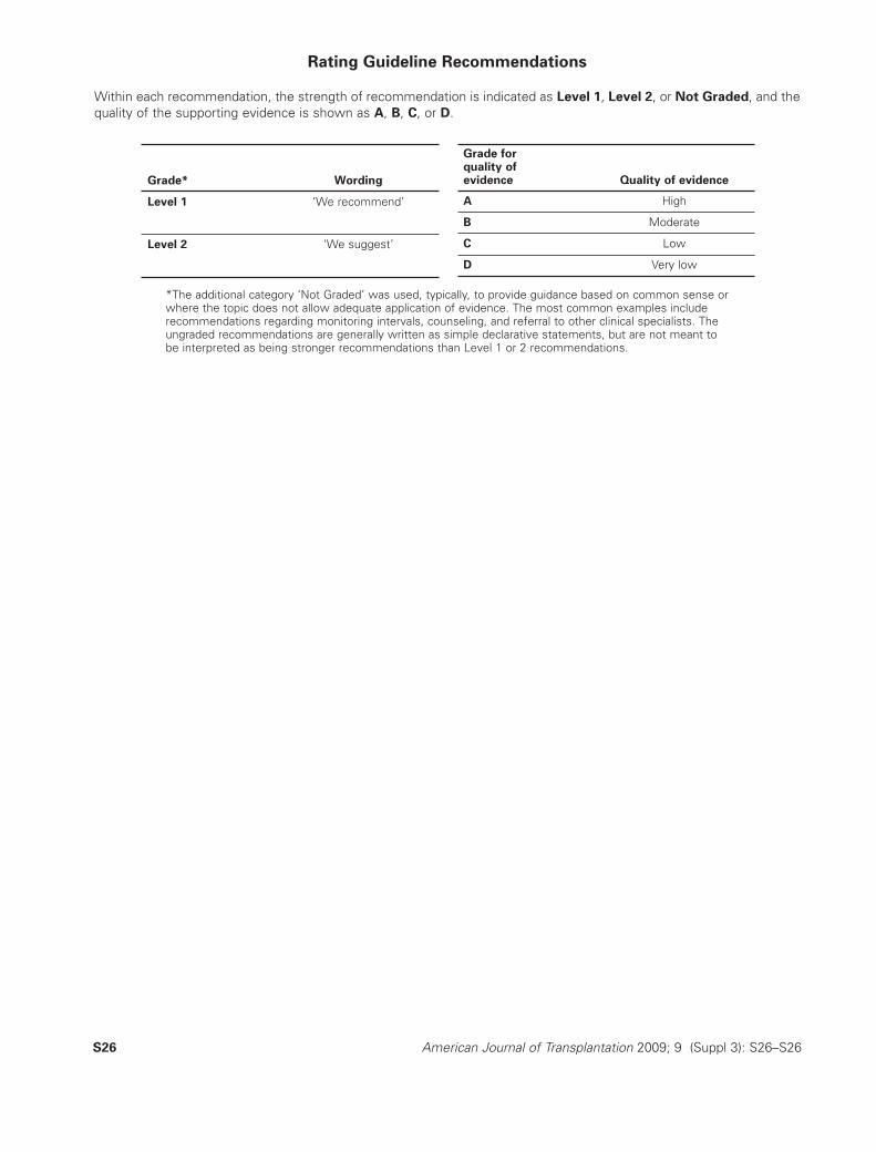
S26 American Journal of Transplantation 2009; 9 (Suppl 3): S26–S26
Rating Guideline Recommendations
Within each recommendation, the strength of recommendation is indicated as Level 1, Level 2, or Not Graded, and thequality of the supporting evidence is shown as A, B, C, or D.
Grade* Wording
Level 1 ‘We recommend’
Level 2 ‘We suggest’
Grade for quality of evidence Quality of evidence
A High
B Moderate
C Low
D Very low
*The additional category ‘Not Graded’ was used, typically, to provide guidance based on common sense orwhere the topic does not allow adequate application of evidence. The most common examples includerecommendations regarding monitoring intervals, counseling, and referral to other clinical specialists. Theungraded recommendations are generally written as simple declarative statements, but are not meant tobe interpreted as being stronger recommendations than Level 1 or 2 recommendations.

Chapter 8
Chapter 8: Monitoring Kidney Allograft Function
8.1: We suggest measuring urine volume (2C):
• every 1–2 hours for at least 24 hours after
transplantation (2D);
• daily until graft function is stable. (2D)
8.2: We suggest measuring urine protein excretion,
(2C) at least:
• once in the first month to determine a baseline
(2D);
• every 3 months during the first year (2D);
• annually, thereafter. (2D)
8.3: We recommend measuring serum creatinine, (1B)
at least:
• daily for 7 days or until hospital discharge,
whichever occurs sooner (2C);
• two to three times per week for weeks 2–4
(2C);
• weekly for months 2 and 3 (2C);
• every 2 weeks for months 4–6 (2C);
• monthly for months 7–12 (2C);
• every 2–3 months, thereafter. (2C)
8.3.1: We suggest estimating GFR whenever
serum creatinine is measured, (2D) using:
• one of several formulas validated for
adults (2C); or
• the Schwartz formula for children and
adolescents. (2C)
8.4: We suggest including a kidney allograft ultra-
sound examination as part of the assessment of
kidney allograft dysfunction. (2C)
GFR, glomerular filtration rate.
Background
Some tests need to be performed routinely to detect ab-normalities that may lead to treatment or prevention ofcomplications that are common in KTRs (Table 4). Thefrequency of screening is based on the incidence of thecomplication being screened, because there are no otherdata to determine the best interval for screening. Serumcreatinine is easily measured and readily available in mostlaboratories. Screening tests for urine protein excretion in-clude dipstick tests for total protein or albumin, as wellas randomly collected ‘spot’ urine to measure protein-to-creatinine or albumin-to-creatinine ratios.
Rationale
• Detecting kidney allograft dysfunction as soon as pos-sible will allow timely diagnosis and treatment that mayimprove outcomes.
• Urine output that is inappropriately low, or inappropri-ately high, is an indication of possible graft dysfunction.
• Serum creatinine and urine protein measurements arereadily available and are useful for detecting acute andchronic allograft dysfunction.
• Ultrasound is relatively inexpensive and reasonably ac-curate for diagnosing treatable causes of kidney allo-graft dysfunction.
Urine volume
Urine volume is an easily-measured parameter of early kid-ney allograft function (120). The recovery of kidney func-tion, measured as a decrease in serum creatinine and bloodurea nitrogen, is generally preceded by an increase in urinevolume (120). Rarely, excessive urine volume may indicatethe presence of a saline diuresis or a water diuresis causedby tubular damage. In addition to its role in assessing earlyallograft dysfunction, measuring the urine volume is an im-portant part of overall fluid and electrolyte management.
Urine protein excretion
Proteinuria is an early and sensitive marker of kidney dam-age in CKD (121). Many causes of proteinuria are poten-tially reversible with appropriate treatment (Table 5) (122),and detection of proteinuria can therefore improve graftoutcomes (113,122–132). Patients with proteinuria gen-erally have lower kidney function compared to patientswithout proteinuria (122,129). Proteinuria is also associ-ated with mortality and CVD events in KTRs (130–132).
Proteinuria includes albuminuria as well as other proteins.The urinary excretion rate for albumin and total protein canbe estimated from the ratio of albumin or total protein tocreatinine concentration in a casual urine specimen (133–136). Creatinine excretion is higher in men than in women.Therefore, the values in the general population and cut-offvalues for abnormalities in urine albumin-to-creatinine ra-tio are lower for men than women (137,138 (Table 6). Fordetails, see Kidney Disease Outcomes Quality Initiative(KDOQI) Guidelines for Chronic Kidney Disease, Part 5, As-sessment of Proteinuria (www.kidney.org/professionals/kdoqi/guidelines_ckd/p5_lab_g5.htm; last accessed March30, 2009).
Serum creatinine
Causes of kidney allograft dysfunction that require rapidintervention for treatment to be effective include acuterejection, obstruction, urine leak, vascular compromiseand some recurrent diseases, for example focal segmen-tal glomerulosclerosis (FSGS). These causes are morecommon in the first few days to weeks after kidney
American Journal of Transplantation 2009; 9 (Suppl 3): S27–S29 S27

Chapter 8
Table 4: Routine screening after kidney transplantation
Screening intervals by time after transplantation
Screening test 1 week 1 month 2–3 months 4–6 months 7–12 months >12 months
Creatininea Daily 2–3 per week Weekly Every 2 weeks Monthly Every 2–3 months
Urine proteinb ..............Once................. .....................Every 3 months...................................... AnnuallyComplete blood countc Daily 2–3 per week Weekly ...................Monthly................... AnnuallyDiabetesd ..............Weekly................ .......................Every 3 months.................................... AnnuallyLipid profilee – – Once – – AnnuallyTobacco usef Prior to discharge – – – AnnuallyBKV NATg ................................Monthly......................... .............Every 3 months.............. –EBV NAT (seronegative)h Once ....................Monthly.................. .............Every 3 months.............. –Blood pressure, pulse, ........................................................................Each clinic visit...................................................................
height, body weight
BKV, BK polyoma virus; EBV, Epstein-Barr virus; NAT, nucleic acid testing.aSerum creatinine.bUrine total protein and/or urine albumin.cComplete blood count including white blood count, hemoglobin and platelet counts.dScreen for diabetes with fasting blood glucose, glucose tolerance test, or HbA1c level.eLipid profile includes fasting cholesterol, LDL-C, HDL-C, and triglycerides.fScreen for tobacco use.gScreen for BKV using plasma NAT.hScreen for EBV using plasma NAT in patients with no antibody to EBV at transplant.
transplantation than in subsequent months to years. There-fore, it is important to closely monitor kidney function earlyafter transplantation.
Measurement of the serum creatinine concentration isa simple, inexpensive and universally available methodfor estimating GFR, and it is reliable for detecting acutechanges of kidney function (142,143). The level of serumcreatinine at year 1 after transplantation is a risk factor
Table 5: Some causes of proteinuria after kidney transplantation
Persistent disease in the native kidneys
Allograft rejection and drug toxicity
Acute rejectionThrombotic microangiopathyCAITransplant glomerulopathy
De novo and recurrent glomerular diseases
Minimal change diseaseFSGSIgA glomerulonephritisMembranous glomerulonephritisMembranoproliferative glomerulonephritisPostinfectious glomerulonephritisThrombotic thrombocytopenic purpuraHUSVasculitisDiabetic nephropathySystemic lupus erythematosusAmyloidosisLight- and heavy-chain deposition diseases
CAI, chronic allograft injury; FSGS, focal segmental glomeruloscle-rosis; HUS, hemolytic-uremic syndrome; IgA, Immunoglobulin A.
for subsequent outcomes, and may help guide care, forexample the frequency of visits (144,145).
A gradual increase in serum creatinine after the first yearmay be due to acute rejection, but more often is causedby CAI, recurrence of the original kidney disease, or denovo kidney disease. Unfortunately, serum creatinine isless reliable for detecting chronic changes (over months toyears) in kidney function.
As is true in the general population, measurement of GFRwith inulin, iothalamate, iohexol or other suitable markersof GFR, either with urinary or plasma clearance techniques,provides the most accurate measure of allograft function inKTRs. Although these tests are appropriate for clinical use,the Work Group did not recommend their use in routineclinical practice due to cost, low patient acceptance, andlack of availability outside of academic medical centers.Measurement of cystatin C has also been used to monitorkidney function. The advantage of cystatin C is its inde-pendence from body weight. However, at present, there isa paucity of validation studies for cystatin C estimates ofGFR in KTRs (146–148).
Formulas to estimate GFR have been tested in KTRs,but no formula has been consistently shown to be su-perior to any other formula (149–156). It is unlikely thatthese formulas will improve the ability of serum creati-nine to estimate acute changes in kidney function since,in most formulas, the only component of the formulathat changes significantly is serum creatinine. It is sim-ilarly unclear whether formulas improve the ability ofserum creatinine to measure chronic changes in kidney
S28 American Journal of Transplantation 2009; 9 (Suppl 3): S27–S29

Chapter 8
Table 6: Definitions of proteinuria and albuminuria
Urine collection Albuminiuria or clinicalmethod Normal Microalbuminuria proteinuria
Total protein 24-h excretion <300 mg/day (adults) NA ≥300 mg/day (adults)<4 mg/m2/h (children) ≥4 mg/m2/h (children)
Dipstick <30 mg/dL (adults and children) NA ≥30 mg/dL (adultsand children)
Spot protein-to- <200 mg/g (adults) NA ≥200 mg/g (adults)creatinine ratio <0.2 mg/mg (children 2 years or older)
<0.5 mg/mg (<6–24 months old)
Albumin 24-h excretion <30 mg/day 30–300 mg/day >300 mg/dayAlbumin dipstick <3 mg/dL ≥3 mg/dL NASpot albumin-to- <17 mg/g (men) 17–250 mg/g (men) >250 mg/g (men)
creatinine ratio <25 mg/g (women) 25–355 mg/g (women) >355 mg/g (women)<30 mg/g (children)
NA, not applicable.Reference values for urinary protein and albumin excretion in pediatric patients (139,140). To convert metric units to SI units, seeConversion Factors, p. Six.Modified with permission (141).
transplant function, especially when serum creatininemay change due to changes in muscle mass due to animproved nutritional status after kidney transplantation(157–159).
Kidney allograft ultrasound examination
Many of the most common causes of allograft dysfunc-tion, other than rejection, can be diagnosed by ultrasound.These include arterial occlusion, venous thrombosis, uri-
nary obstruction, a urine leak (large fluid collection), com-pressing perinephric hematoma and arteriovenous fistulafrom a kidney biopsy (160–163). Ultrasound is also usefulin guiding a kidney allograft biopsy, so it is often obtained atthe time of biopsy. In the kidney allograft, mild to moderatecalyceal distension can be normal, so a baseline ultrasoundexamination when kidney function is normal may be use-ful to compare to subsequent ultrasound examinations forallograft dysfunction.
American Journal of Transplantation 2009; 9 (Suppl 3): S27–S29 S29

Chapter 9
Chapter 9: Kidney Allograft Biopsy
9.1: We recommend kidney allograft biopsy when
there is a persistent, unexplained increase in
serum creatinine. (1C)
9.2: We suggest kidney allograft biopsy when serum
creatinine has not returned to baseline after treat-
ment of acute rejection. (2D)
9.3: We suggest kidney allograft biopsy every 7–10
days during delayed function. (2C)
9.4: We suggest kidney allograft biopsy if expected
kidney function is not achieved within the first
1–2 months after transplantation. (2D)
9.5: We suggest kidney allograft biopsy when there is:
• new onset of proteinuria (2C);
• unexplained proteinuria ≥3.0 g/g creatinine or
≥3.0 g per 24 hours. (2C)
Background
Kidney allograft biopsies are performed for specific clin-ical indications, or as part of a surveillance program (orprotocol). An ‘indicated biopsy’ is one that is promptedby a change in the patient’s clinical condition and/or lab-oratory parameters. A ‘protocol biopsy’ is one obtainedat predefined intervals after transplantation, regardless ofkidney function. In both cases, the biopsy is obtained tofind histological changes prompting treatment to improveoutcomes. DGF is graft function low enough to require dial-ysis in the first week after kidney transplantation, or lackof improvement in pretransplant kidney function.
New-onset proteinuria (defined in Table 6) may indicatetreatable causes of graft dysfunction, including acute re-jection and thrombotic microangiopathy. In patients whoalready have proteinuria, an increase exceeding a thresh-old usually defined as ‘nephrotic range’ proteinuria, for ex-ample ≥3.0 g/g creatinine or ≥3.0 g/24 h, may indicatetreatable causes of graft dysfunction.
Rationale
• Increased serum creatinine that is not explained by de-hydration, urinary obstruction, high CNI levels or otherapparent causes is most likely due to an intragraftparenchymal process, such as acute rejection, CAI,drug toxicity, recurrent or de novo kidney disease orBKV nephropathy.
• The optimal diagnosis and treatment of intragraftparenchymal causes of allograft dysfunction require anadequate biopsy.
• In patients with DGF, change in serum creatinine isnot useful for ruling out acute rejection, and protocolbiopsies are needed to rule out acute rejection.
• Proteinuria, or a substantial increase in proteinuria, mayindicate a potentially treatable cause of graft dysfunc-tion.
Biopsies for an increase in serum creatinine
Although serum creatinine has many limitations for esti-mating GFR (see Chapter 8), an unexplained rise in serumcreatinine is generally indicative of a decline in GFR. Somefluctuation in creatinine can result from normal labora-tory or physiological variability. Hence, only a persistent in-crease that is outside this normal, but poorly defined, rangeis clinically relevant. A 25–50% increase over baseline is of-ten arbitrarily used in studies. At least one study suggestedthat a persistent 30% rise in serum creatinine was an ex-cellent predictor of subsequent graft failure (144,145). TheAcute Kidney Injury Network (164) has proposed a defini-tion and classification scheme for evaluating acute kidneyinjury (Table 7).
Causes of acute, reversible declines in GFR should be ruledout, including dehydration, urinary obstruction or acuteCNI toxicity (by demonstrating high blood levels), beforea biopsy is performed. If there are no apparent causesof a decline in GFR, then an allograft biopsy is gener-ally warranted to detect the nature of potentially treatablecauses of kidney injury, including rejection, infections likeBKV nephropathy, recurrent or de novo kidney disease orinfiltration with posttransplant lymphoproliferative disease(PTLD). Since any of these conditions can develop in thesetting of preexisting graft pathology, additional biopsiesmay be required when an abrupt change in the rate ofprogression is observed.
Biopsies can determine both the type and severity of im-munologic damage (109). Different types of acute rejec-tion may require different treatment approaches. For exam-ple, acute cellular rejection is usually treated with steroidpulses, but acute antibody-mediated rejection may promptthe use of specific treatments in addition to steroids.
Biopsies for a lack of improvement in graft function
When acute rejection does not respond to first-linetreatment with steroids, additional treatment (e.g. witha lymphocyte-depleting antibody) may be successful(105,165). Alternatively, a failure of function to return tobaseline could be due to a new pathological process,such as coexistent acute tubular necrosis, drug toxicity orBKV nephropathy, that would require a different treatment
S30 American Journal of Transplantation 2009; 9 (Suppl 3): S30–S32

Chapter 9
Table 7: Diagnostic criteria for acute kidney injury
Criteria An abrupt (within 48 h) reduction in kidney functioncurrently defined as an absolute increase in serumcreatinine of ≥0.3 mg/dL (≥26.4 lmol/L), apercentage increase in serum creatinine of ≥50%(1.5-fold from baseline), or a reduction in urineoutput (documented oliguria of less than 0.5mL/kg/h for more than 6 h).
Notes The above criteria include both an absolute and apercentage change in creatinine to accommodatevariations related to age, gender and BMI, and toreduce the need for a baseline creatinine but dorequire at least two creatinine values within 48 h.
BMI, body mass index.Adapted with permission (164).
approach. Therefore, a biopsy is indicated to determine thecorrect treatment.
Patients should always be assessed for their suitability forbiopsy before undertaking the procedure. Biopsies maybe hazardous in those with a bleeding diathesis, or in thepresence of large fluid collections or infection.
Biopsies for DGF
Observational studies have shown that the incidence ofacute rejection during DGF is higher than in patients with-out DGF (166–168). Kidney function cannot be used asan indication for biopsy to diagnose superimposed acuterejection while the patients are already being treated withdialysis due to DGF, or when the serum creatinine does notfall from pretransplant values. It is therefore prudent to ob-tain periodic biopsies of the kidney during DGF to diagnoseacute rejection. There are few data to determine when andhow often biopsies during DGF should be obtained. How-ever, studies in which biopsies have been obtained every7–10 days, while patients are receiving dialysis for DGF,have shown that acute rejection can be present for thefirst time on the second, third or even fourth biopsy (167).
In centers that have a very low overall incidence of acuterejection, the incidence of acute rejection during DGF couldalso be low enough to obviate the need for biopsies duringDGF. A biopsy may no longer be needed when there aresigns that DGF is resolving, for example when urine outputis increasing rapidly or serum creatinine is declining.
Protocol biopsies
Acute rejection, CAI and CNI toxicity can occur in the ab-sence of a measurable decline in kidney function. Sev-eral studies have shown that protocol biopsies can detectclinically inapparent (subclinical) acute rejection, CAI andCNI nephrotoxicity. The reported prevalence of subclinicalrejection (Banff grade 1A or higher) varies from 13% to25% at 1–2 weeks, 11–43% at 1–2 months, 3–31% at2–3 months and 4–50% at 1 year (169–175).
Data from observational studies indirectly suggest that de-tecting and treating subclinical acute rejection with pro-tocol biopsies may be beneficial. Subclinical rejection isassociated with CAI (170,173,176,177) and reduced graftsurvival (176–179).
In another study, subclinical acute rejection in 14-day pro-tocol biopsies was associated with poorer 10-year graftsurvival (179). Graft survival rates with subclinical rejec-tion, borderline subclinical rejection or no rejection were88%, 99% and 98% at 1 year (p < 0.05), and 62%, 94%and 96% at 10 years (p < 0.05), respectively. In a pediatricstudy, subclinical rejection was associated with progres-sive CAI, reduced creatinine clearance and shorter graftsurvival (177).
Treatment of subclinical rejection may improve outcomes.In a RCT, 72 patients were randomly allocated to undergoprotocol biopsies and treatment of subclinical rejection at1, 2, 3, 6 and 12 months (biopsy group), or protocol biop-sies without treatment at 6 and 12 months only (controlgroup) (100). Patients in the biopsy arm of the study hada significant decrease in acute rejection episodes, a re-duced 6-month chronic tubulointerstitial score and a lower2-year serum creatinine. Interstitial fibrosis was less inthose treated for subclinical rejection (100). In another trial,52 living-donor KTRs were randomized to undergo protocolbiopsies and 50 controls had only indicated biopsies (103).At 1 and 3 months, protocol biopsies revealed borderlinechanges in 11.5% and 14% patients, acute rejection in17% and 12% and CAI in 4% and 10%, respectively. Theincidence of clinically evident acute rejection episodes wassimilar in the two groups, but the biopsy group had lowerserum creatinine at 6 months (p = 0.0003) and 1 year(p < 0.0001).
Baseline immunosuppression is likely important in deter-mining the incidence of subclinical rejection and therebythe benefit of protocol biopsies. Tacrolimus- and MMF-treated patients generally have a lower rate of acute re-jection than patients treated with CsA and azathioprine,and tacrolimus is associated with a reduced incidence ofsubclinical rejection (104,113,176,180,181), lower acuteBanff scores (182,183) and 1-year serum creatinine (181).In a RCT, 121 patients were randomly allocated to biopsiesat 0, 1, 2, 3 and 6 months, and 119 to biopsies at 0 and6 months (102). At 6 months, 35% of the biopsy arm and20.5% of the control arm patients had interstitial fibrosisand tubular atrophy (ci + ct) scores ≥2 (p = 0.07). Of note,the frequency of clinical acute rejection episodes was only10% in the biopsy arm and 7% in the control arm (p >
0.05). The prevalence of subclinical rejection in the biopsyarm was 4.6%. Creatinine clearance at 6 months was notdifferent (p > 0.05) in the two groups. Use of protocolbiopsies, therefore, for diagnosis of subclinical rejectionmay not be appropriate in tacrolimus- and MMF-treatedpatients.
American Journal of Transplantation 2009; 9 (Suppl 3): S30–S32 S31

Chapter 9
Other conditions that can be detected on protocol biopsiesinclude CNI toxicity, recurrent disease, transplant glomeru-lopathy, CAI and BKV nephropathy. However, it is unclearwhether the detection of these conditions by protocolbiopsy improves outcomes.
The safety of biopsies has been documented in severalseries (180,184). The reported risk of major complica-tions from protocol biopsy, including substantial bleed-ing, macroscopic hematuria with ureteric obstruction, peri-tonitis or graft loss, is approximately 1% (185–187). Thereported incidence of graft loss from protocol biopsy is0.03%. Protocol biopsies can be done safely as an outpa-tient procedure. Data collected on 1705 protocol kidneytransplant biopsies at one center showed that all of thecomplications became evident in the first 4 h after thebiopsy (188).
Protocol biopsies, however, may be expensive. The MayoClinic reported that protocol biopsies cost US$ 3000 perbiopsy, and it cost US$ 114 000 to detect one case ofacute subclinical rejection (104). Therefore, decisions onwhether or not to perform protocol biopsies should takethese and other factors, including patient preferences,into account. Altogether, based on very-low-quality ev-idence, the benefit of performing protocol biopsies inCsA/azathioprine-treated patients without induction ther-apy may outweigh the harm (see Evidence Profile and ac-companying evidence in Supporting Tables 45–47 at http://www3.interscience.wiley.com/journal/118499698/toc).
Research Recommendations
• RCTs are needed to determine when the benefits ofprotocol biopsies outweigh harm.
S32 American Journal of Transplantation 2009; 9 (Suppl 3): S30–S32

Chapter 10
Chapter 10: Recurrent Kidney Disease
10.1: We suggest screening KTRs with primary kidney
disease caused by FSGS for proteinuria (2C) at
least:
• daily for 1 week (2D);
• weekly for 4 weeks (2D);
• every 3 months, for the first year (2D);
• every year, thereafter. (2D)
10.2: We suggest screening KTRs with potentially
treatable recurrence of primary kidney disease
from IgA nephropathy, MPGN, anti-GBM disease,
or ANCA-associated vasculitis for microhema-
turia, (2C) at least:
• once in the first month to determine a base-
line (2D);
• every 3 months during the first year (2D);
• annually, thereafter. (2D)
10.3: During episodes of graft dysfunction in patients
with primary HUS, we suggest screening for
thrombotic microangiopathy (e.g. with platelet
count, peripheral smear for blood cell morphol-
ogy, plasma haptoglobin, and serum lactate de-
hydrogenase). (2D)
10.4: When screening suggests possible treatable re-
current disease, we suggest obtaining an allo-
graft biopsy. (2C)
10.5: Treatment of recurrent kidney disease:
10.5.1: We suggest plasma exchange if a biopsy
shows minimal change disease or FSGS
in those with primary FSGS as their pri-
mary kidney disease. (2D)
10.5.2: We suggest high-dose corticosteroids
and cyclophosphamide in patients with
recurrent ANCA-associated vasculitis or
anti-GBM disease. (2D)
10.5.3: We suggest using an ACE-I or an ARB for
patients with recurrent glomerulonephri-
tis and proteinuria. (2C)
10.5.4: For KTRs with primary hyperoxaluria, we
suggest appropriate measures to prevent
oxalate deposition until plasma and urine
oxalate levels are normal (2C), including:
• pyridoxine (2C);
• high calcium and low oxalate diet
(2C);
• increased oral fluid intake to enhance
urinary dilution of oxalate (2C);
• potassium or sodium citrate to alka-
linize the urine (2C);
• orthophosphate (2C);
• magnesium oxide (2C);
• intensive hemodialysis to remove ox-
alate. (2C)
ACE-I, angiotensin-converting enzyme inhibitor;
ANCA, antineutrophil cytoplasmic autoantibody; ARB,
angiotensin II receptor blocker; FSGS, focal segmen-
tal glomerulosclerosis; GBM, glomerular basement
membrane; HUS, hemolytic-uremic syndrome; IgA,
immunoglobulin A; KTRs, kidney transplant recipients;
MPGN, membranoproliferative glomerulonephritis.
Background
The primary kidney disease is generally documented bypretransplant biopsy of the native kidney, or of a pre-vious kidney transplant. Recurrence of the primary kid-ney disease is usually established when there is biopsy-documented involvement of the kidney allograft with theprimary kidney disease.
Rationale
• Some recurrent kidney diseases cause allograft failure.• Treatment of some recurrent kidney diseases may pre-
vent, or delay, the onset of graft failure.• Screening for treatable recurrent kidney disease may
result in early diagnosis and treatment that may bebeneficial.
Recurrence of primary kidney diseases is an importantcause of morbidity and graft loss following kidney trans-plantation, in both adults and children. In a study of 1505cases with both native kidney and kidney allograft biop-sies documenting recurrent glomerular disease, graft lossdue to recurrent glomerulonephritis was the third mostfrequent cause for graft failure 10 years after kidney trans-plantation (110). Recurrence may present as increasedserum creatinine (reduced GFR), new-onset or increasedproteinuria and/or hematuria. The impact of recurrencevaries according to the primary kidney disease. Not alldiseases recur with equal frequency. The risk of recur-rence is particularly increased in FSGS, immunoglobu-lin A (IgA) nephropathy, membranoproliferative glomeru-lonephritis (MPGN), hemolytic-uremic syndrome (HUS), ox-alosis and Fabry’s disease and, to a lesser extent, with lu-pus nephritis, anti-glomerular basement membrane (GBM)disease and vasculitis (189). Also, the timing of recurrenceand manner of presentation vary for different diseases.FSGS, HUS and oxalosis may recur in the first few daysto weeks after transplantation, whereas the timing is vari-able in the others (127).
American Journal of Transplantation 2009; 9 (Suppl 3): S33–S37 S33

Chapter 10
Table 8: Screening for recurrent diseases
DiagnosticScreening (in addition Minimum screening tests (in addition
Disease to serum creatinine) frequency to kidney biopsy) Potential treatment
FSGS Proteinuria Daily for 1 week, weekly for Plasmapheresis4 weeks, every 3 monthsfor 1 year, then annually
IgA nephropathy Proteinuria, microhematuria
MPGN Proteinuria, microhematuria Serum complement levelsOnce in the first month, every
Anti-GBM disease Proteinuria, microhematuria 3 months in the first year, Anti-GBM antibodies Plasmapheresisthen annually
Pauci-immune Proteinuria, microhematuria ANCA Cyclophosphamidevasculitis and corticosteroids
HUS Proteinuria, platelet count During episodes of graft Platelet count, peripheral Plasmapheresisdysfunction blood smear, LDH
ANCA, antineutrophil cytoplasmic antibody; FSGS, focal segmental glomerulosclerosis; GBM, glomerular basement membrane; HUS,hemolytic-uremic syndrome; IgA, immunoglobulin A; LDH, lactate dehydrogenase; MPGN, membranoproliferative glomerulonephritis.
In a majority of instances, proteinuria and/or reduced GFRprovide the initial basis for suspecting disease recurrence.Since these parameters are periodically assessed in KTRsas part of their routine monitoring, a separate strategy fordetection of disease recurrence is not warranted.
The modality of screening for some of these diseases,however, may vary from the usual posttransplant moni-toring if timely detection is not achieved by the routineposttransplant monitoring strategies (Table 8). For exam-ple, FSGS can recur early; hence, screening for FSGS re-currence requires early and frequent monitoring for pro-teinuria. HUS recurrence requires looking for evidence ofmicroangiopathic hemolysis. Screening for recurrent IgAnephropathy, MPGN, anti-GBM disease and vasculitis re-quire examination of urinary sediment to detect microhe-maturia and/or presence of casts in addition to screeningfor proteinuria. It is appropriate to perform dipstick test-ing for proteinuria followed by quantitation using spot pro-tein creatinine ratio or timed urine collection. Dependingon the primary disease, biopsy evaluation may require im-munofluorescence and electron microscopy in addition tolight microscopy to confirm recurrence and to rule out othercauses of proteinuria, hematuria or graft dysfunction (190).
There is also weak evidence (uncontrolled case studiesand case reports) that disease-specific treatment may bebeneficial for some recurrent diseases.
Idiopathic FSGS
Idiopathic, or primary, FSGS is characterized by typicalsclerosis in a segment of glomerular tuft, along with foot-process fusion on electron microscopy. Sclerosis may notbe evident in early recurrence, and light microscopy mayshow normal glomerular architecture. Recurrence is sus-pected when a patient with a documented primary FSGSin the native kidneys or a prior kidney allograft develops
proteinuria and/or increase in serum creatinine, typicallysoon after transplantation (127).
Idiopathic FSGS recurs in 20–50% of KTRs (up to 80%if it has recurred in a prior kidney transplant) (191). It isimportant to distinguish idiopathic from secondary causesof FSGS that generally do not recur. Recurrence of famil-ial FSGS has also been documented, if the donor is anobligate carrier (191). Putative risk factors for recurrenceinclude age of onset of FSGS in native kidneys between6 and 15 years (192), rapid course of the original disease(e.g. less than 3 years from diagnosis to CKD stage 5), dif-fuse mesangial proliferation on histology and non-AfricanAmerican ethnicity. The strongest risk factor is recurrencein a previous transplant.
The demonstration of increase in the albumin permeabil-ity of isolated rat glomeruli by sera from patients with arecurrent FSGS offers the possibility of more accurate pre-diction of the risk of recurrent disease (193). However, thisassay is still experimental.
Idiopathic FSGS can recur at any time after transplanta-tion, but recurrence is more common early after trans-plantation. Recurrent disease presents with proteinuria,which is usually heavy. About 80% of cases recur in thefirst 4 weeks (193). Proteinuria screening therefore needsto be more frequent in the early posttransplant period inthose with CKD stage 5 due to FSGS, especially those withrisk factors for recurrence. The exact frequency has notbeen worked out. Interpretation of proteinuria, especially inthe early posttransplant period, requires knowledge of pre-transplant proteinuria. Although proteinuria from the nativekidneys declines after transplantation (194), the time takenfor its disappearance is variable. Posttransplant proteinuriatherefore should be interpreted in light of the pretransplantvalues.
S34 American Journal of Transplantation 2009; 9 (Suppl 3): S33–S37

Chapter 10
There have been no RCTs of therapy for recurrent idio-pathic FSGS. However, there have been individual cases,and uncontrolled series, reporting that patients with recur-rent idiopathic FSGS may have a substantial reduction inurine protein excretion after plasma exchange (195,196).This probably occurs by removing circulating factors thatalter glomerular permeability to protein. Predictors of re-sponse to plasma exchange include early initiation of treat-ment after recurrence, and possibly an early recurrence ofdisease (196). Unfortunately, proteinuria may recur aftertreatment, and may require additional plasma exchange,or even periodic, ongoing treatments. The presumption isthat reducing protein excretion with plasma exchange willhelp preserve allograft function, but no studies have exam-ined this.
It is unclear how many plasma-exchange treatments are re-quired to reduce protein excretion, but one review found amedian of nine treatments before there was a remission inproteinuria (195). In small case series, prophylactic plasmaexchange has been reported, but the data are not con-vincing that this is effective in preventing recurrent FSGS(197,198).
High-dose CsA may induce remission of proteinuria. Inone series, 14 of 17 children entered lasting remission(199). The rationale behind maintaining a high CsA bloodlevel is to overcome the effect of high serum choles-terol often seen in patients with recurrent FSGS (lipopro-teins bind CsA and reduce free CsA levels). High-doseCsA may be combined with plasmapheresis. A study con-cluded that plasmapheresis alone was not sufficient toinduce remission except when combined with high-doseCsA (200).
For patients who do not respond to plasma exchange, orfor patients who have non-nephrotic proteinuria, a reduc-tion in proteinuria with an angiotensin-converting enzymeinhibitor (ACE-I) and/or an angiotensin II receptor blocker(ARB) may be beneficial.
IgA nephropathy
IgA nephropathy is the most common type of glomeru-lonephritis worldwide and is the primary cause of CKDstage 5 in 20% of KTRs in many parts of the world.Recurrent IgA nephropathy is common after transplanta-tion. Reported incidence of recurrence varies from 13% to53% according to differences in duration of follow-up andbiopsy policy of different transplant centers, with the high-est rates in centers that perform routine protocol biopsies(201). Latent IgA deposits in the donor kidney (identified onpreimplantation biopsies) are responsible for ‘recurrence’in some cases transplanted for kidney failure due to IgAnephropathy in areas with high disease prevalence (202).Single-nucleotide polymorphisms in the interleukin-10 andTNF-alpha genes have been shown to predict recurrencerisk (203,204). The estimated 10-year incidence of graft
loss due to recurrence was 9.7% (CI = 4.7–19.5%) (110).Recurrence risk in retransplants is increased if the first graftwas lost due to recurrent IgA nephropathy in less than 10years (205). There is no effective therapy for preventingrecurrent IgA nephropathy. ACE-Is and ARBs have beenshown to reduce proteinuria and possibly preserve kidneyfunction in recurrent IgA nephropathy (206). In a study of116 KTRs with IgA nephropathy, use of ATG as inductiontherapy was associated with a reduction in recurrence riskfrom 41% to 9% when compared to IL2 receptor antago-nists (207).
Membranoproliferative glomerulonephritis
Secondary causes of MPGN, such as hepatitis C, should beruled out. The histological recurrence rate in idiopathic typeI MPGN is 20–30% and exceeds 80% in type 2 disease(192). Manifestations include microhematuria, proteinuriaand deterioration of kidney function. Risk factors for re-currence include severity of histological lesions in nativekidneys, HLA-B8DR3, living related donors and previousgraft loss from recurrence (208,209). There are reports ofresponse to long-term cyclophosphamide (210), plasma-pheresis (211–213) and CsA (214).
Hemolytic-uremic syndrome
Hemolytic-uremic syndrome is defined histopathologicallyby intimal cell proliferation, and thickening and necrosisof the wall, thrombi and narrowed lumens of glomeru-lar, arteriolar or interlobular artery. The severity can rangefrom endothelial swelling to complete cortical necrosis. Itmanifests clinically with microangiopathic hemolytic ane-mia and rapid worsening of kidney function with or with-out involvement of other organs. HUS is often classifiedas diarrhea-associated (D)− HUS (atypical) and D+ HUS(typical).
Hemolytic-uremic syndrome recurs commonly in adultsand in children in whom the original kidney disease wasD− variant. The overall recurrence risk is less than 10% inthe pediatric population; D+ HUS usually does not recur,while idiopathic D− or familial HUS may recur in 21–28%of children (215). Recurrence occurs in about 80–100%of patients with factor H or factor I mutation, while pa-tients with a mutation in membrane cofactor protein donot have recurrence (216,217). The risk is higher in adults,with 33–56% (218–220) showing clinical manifestationsand an additional 16–20% of patients demonstrating clini-cally silent recurrence. Recurrence is particularly frequentin adults with autosomal recessive or dominant HUS (215).Recurrence develops within 4 weeks in most cases. Mostpatients show microangiopathic anemia, thrombocytope-nia and kidney dysfunction, whereas others present withrapidly progressive graft dysfunction without showing theclassic hematologic manifestations. Platelet count shouldbe performed during episodes of graft dysfunction in KTRswith HUS as the original cause of CKD stage 5. In thosewith falling counts, additional tests such as examination
American Journal of Transplantation 2009; 9 (Suppl 3): S33–S37 S35

Chapter 10
of peripheral blood smear to look for fragmented cells(schistocytes), haptoglobin and lactate dehydrogenaseestimation to document hemolysis are warranted. Long-term graft survival is lower (about 30%) in those with re-currence.
Treatment strategies have included plasmapheresis, intra-venous immunoglobulin and rituximab. Aggressive plasma-pheresis using fresh frozen plasma (40–80 mL/kg persession) increases the levels of deficient factors and hasprovided encouraging results, even in those with factors Hand I mutations (221–223). As factor H is synthesized in theliver, combined liver and kidney transplantation (togetherwith preoperative and intraoperative plasmapheresis us-ing fresh frozen plasma and low-molecular-weight heparin)could reduce the risk of recurrence (222,224–226). Intra-venous immunoglobulin and rituximab have been reportedto rescue recurrent HUS resistant to multiple courses ofplasma exchanges (227,228). There is no evidence thatavoidance of CNI, mTORi and OKT3 (that may themselvescause thrombotic microangiopathy) will reduce the recur-rence risk.
ANCA-associated vasculitis and anti-GBM disease
Both antineutrophil cytoplasmic antibody (ANCA)-associated vasculitis and anti-GBM disease may presentwith rapidly progressive CKD and crescentic glomeru-lonephritis. Recurrence rates are low if the disease isquiescent at the time of transplant. In an analysis ofpooled data from 127 patients with ANCA-associatedvasculitis, 17% of patients had recurrence, with kidneymanifestation in 57.1%. Kidney dysfunction occurredin 33% of those with recurrence (229). More recentstudies (230) report lower (7%) recurrence rates, mostbeyond the first posttransplant year with no direct orindirect impact on allograft function. ANCA-associatedvasculitis relapses in the kidney allograft usually manifestas pauci-immune necrotizing glomerulonephritis, but graftfunction can also be affected by acute arteritis, ureteralstenosis and obstructive uropathy due to granulomatousvasculitis.
Pretransplantation disease course, disease subtype, ANCAtype or titer, time of transplantation or donor type doesnot predict recurrence. Kidney ANCA-associated vasculi-tis generally responds well to high-dose prednisolone andcyclophosphamide (231–233). Other treatment modalitiesthat have been tried include MMF, plasmapheresis with orwithout intravenous immunoglobulin and rituximab (234–240).
Histological evidence of anti-GBM disease can be foundin biopsies in 15–50% of cases. Clinical recurrence is rareand consisted of isolated case reports only (201,241). Graftfailure due to recurrence is rare (110). The incidence ofrecurrence may be higher in those with circulating anti-GBM antibody at the time of transplantation. Treatment
of clinically active anti-GBM disease may include pulsesteroids, cyclophosphamide and plasma exchange, partic-ularly if there is potentially life-threatening pulmonary in-volvement (241).
Primary hyperoxaluria
Primary hyperoxaluria is caused by deficiency of hepaticperoxisomal alanine:glyoxylate aminotransferase, leadingto increased synthesis and urinary excretion of oxalate,recurrent calcium oxalate urolithiasis, irreversible nephro-calcinosis and eventually CKD. In CKD, insoluble oxalatesaccumulate throughout the body, especially in bone andarteries. Because the enzyme defect in primary hyperox-aluria is not corrected by isolated kidney transplantation,oxalate overproduction persists, leading to recurrence ofcalcium oxalate deposits in over 90% of transplanted kid-neys, and eventually leading to graft loss (242), unless theenzyme is replaced through a simultaneous liver trans-plant (243). The total body oxalate burden is very highin CKD stage 5 patients, and the urinary oxalate excre-tion increases greatly as soon as graft function is estab-lished. Plasma and urine oxalate levels may remain highfor some period of time even in patients undergoing si-multaneous kidney and liver transplantation. High urinaryoxalate concentration promotes precipitation of calciumoxalate crystals first in the distal tubules, leading to graftdysfunction. This secondarily results in deposition in theparenchyma of the graft, leading to allograft failure. Thisrisk is obviously increased further in those with primarynonfunction of the graft. Transplant protocols designed tominimize complications of recurrent disease include earlyposttransplant urinary dilution through aggressive fluid ad-ministration, and early and frequent dialysis in those withDGF.
Although isolated kidney transplantation is not rec-ommended in primary hyperoxaluria, it is sometimescarried out in developing countries where liver transplanta-tion is not available. Primary hyperoxaluria recurs invari-ably in those who receive kidney transplant alone andleads to graft loss. Patients with the Gly170Arg muta-tion are pyridoxine-sensitive, and should be given high-dose pyridoxine if they receive kidney transplant alone(244).
The disease is sometimes diagnosed for the first time afterkidney transplantation when oxalate deposits are detectedon biopsy in patients with graft dysfunction. Wheneverpossible, these patients should be referred to specializedcenters for liver transplantation. In the immediate postop-erative phase, extra dialysis sessions may be necessaryto control oxalate blood levels until the liver is completelyworking (245).
Specific measures designed to increase oxalate excre-tion and reduce production help in minimization of re-currence, and should be in place for all patients during
S36 American Journal of Transplantation 2009; 9 (Suppl 3): S33–S37

Chapter 10
the first months or years after kidney or combined liver–kidney transplantation (246). These include maintenance ofurine output >3.0–3.5 L/day, and the use of alkaline citrate,neutral phosphate and magnesium oxide. Severe dietaryoxalate restriction is of limited benefit (247), but intakeof nutrients extremely rich in oxalate and ascorbic acid, aprecursor of oxalate, should be discouraged. Pharmacolog-ical doses of pyridoxine may reduce hyperoxaluria in somepatients, especially in those with a Gly170Arg mutation(244). Pyridoxine responsiveness can be assessed by ob-servation of >30% reduction in urinary oxalate excretion to10 mg/kg/day dose of pyridoxine (248) in patient’s sibs withless severe kidney disease if it was not done at the predial-ysis stage. Urinary alkalinization with citrate reduces therisk of urinary calcium oxalate supersaturation by forminga soluble complex with calcium, which reduces the likeli-hood of binding and precipitation with other substances,such as oxalate (249). The dosage is 0.1–0.15 g/kg bodyweight of a sodium or sodium/potassium citrate prepa-ration. The adequacy of therapy and patient compliancecan be verified by measuring urinary pH and citrate excre-tion. Orthophosphate (20–60 mg/day), along with pyridox-ine, has also been shown to reduce urinary calcium oxalatecrystallization (250).
Fabry disease
Fabry disease is a rare, X-linked inherited disease charac-terized by a deficiency of alpha-galactosidase A (alpha-Gal-A), resulting in progressive systemic accumulation of gly-cosphingolipids. Transplantation is the treatment of choicefor most patients with CKD stage 5 due to Fabry disease(251). Although patients with Fabry disease may have his-tological recurrence of the disease in the allograft, howoften recurrence causes graft failure is not clear. In a re-cent US Organ Procurement and Transplantation Networkregistry study, 197 KTRs with Fabry disease had 74% 5-year graft survival, compared to 64% in KTRs with otherkidney diseases (252). Two formulations of recombinanthuman alpha-Gal A are currently available: agalsidase al-pha (Replagal, Transkaryotic Therapies, Cambridge, MA)and agalsidase (Fabrazyme, Genzyme, Cambridge, MA).In non-KTRs, treatment with recombinant human alpha-Gal A has been shown to reduce the rate of decline inkidney function. However, it is unclear whether treatmentimproves graft survival, or reduces other complications ofFabry disease in KTRs. Treatment appears to be safe inKTRs (253,254); however it is very expensive, and whetherit is cost-effective for improving KTR outcomes is notknown.
American Journal of Transplantation 2009; 9 (Suppl 3): S33–S37 S37

Chapter 11
Chapter 11: Preventing, Detecting, and TreatingNonadherence
11.1: Consider providing all KTRs and family members
with education, prevention, and treatment mea-
sures to minimize nonadherence to immunosup-
pressive medications. (Not Graded)
11.2: Consider providing KTRs at increased risk for non-
adherence with increased levels of screening for
nonadherence. (Not Graded)
KTRs, kidney transplant recipients.
Background
Adherence is ‘the extent to which the patient’s behaviormatches the agreed-upon prescriber’s recommendations’(255). At a recent consensus conference, this definitionwas modified to take into account the threshold of theeffect of nonadherence on the therapeutic outcome. Wehave adopted this definition of nonadherence as ‘devia-tion from the prescribed medication regimen sufficient toadversely influence the regimen’s intended effect’ (255).Nonadherence encompasses primary (at initiation) andsecondary (subsequent) nonadherence, partial and/or to-tal nonadherence, as well as the timing of medication use(256–260).
Rationale
• Nonadherence is associated with a high risk of acuterejection and allograft loss.
• Nonadherence may occur early and/or late after trans-plantation.
• The transition from pediatric to adult nephrology caremay be a time when nonadherence is particularly com-mon.
• Measures can be taken to reduce nonadherence andthereby improve outcomes.
Nonadherence is common in the first months after kidneytransplantation and increases by duration of follow-up. Thelevel of adherence affects clinical outcomes, and is associ-ated with early and late allograft rejection, which reducesgraft function and graft survival (261–263). Graft loss is sev-enfold more likely in nonadherent compared to adherentindividuals (264). In another study, nonadherence (missedappointments, fluctuating drug concentration) accountedfor over a half of kidney allograft loss (265).
Nonadherence is multidimensional (255), although wehave focused primarily on adherence with immunosup-pressive medication use. Additional areas of nonadherenceinclude prescribed diet; exercise; tobacco, alcohol and druguse; self-monitoring of vital signs, for example blood pres-sure, body weight and clinical appointments.
Satisfactory adherence to medication use is achievedwhen the gaps between dosing history and the prescribedregimen have no effect on therapeutic outcome. This phar-macoadherence definition emphasizes therapeutic out-come in contrast to specific medication intake or druglevel. Measurement of outcome and drug levels is com-monly used in the transplant population. Measurable pa-rameters of pharmacoadherence are acceptance (whetherthe patient accepts the recommended treatment), execu-tion (how well the patient executes the recommended reg-imen), and discontinuation (when the patient stops takingthe medication) (264,266). Measurement of adherence canbe by direct observation that medication was consumed,indirect measures that medication had been consumed orself-reporting (Table 9). Indirect measures include serumdrug levels, biological markers, electronic monitoring, pillcount and refill/prescription records. Since there is no per-fect measure of adherence, consideration should be givento use more than one approach to measure adherence(267–270).
In organ transplant recipients, the average nonadherencerate was highest for diet (25 cases per 100 people peryear), immunosuppressive medication (22.6 cases per 100people per year), monitoring vital signs (20.9 cases per 100people per year) and exercise (19.1 cases per 100 peopleper year) (264). Among KTRs, nonadherence with immuno-suppressive medications was highest (35.6 cases per 100people per year). Nonadherence to long-term medicationis as high as 50% in developed countries and even higher
Table 9: Assessment of medication adherence (255,260)
Self-reporting medication use by patientCollateral reporting of medication use by relatives,
friends or caretakersPatient diariesQuestionnairesLaboratory tests (drug and metabolite levels)Medical record review, outcomesPrescription refillsMonitored pill countsElectronic monitoring devices
Modified with permission (260).
S38 American Journal of Transplantation 2009; 9 (Suppl 3): S38–S40

Chapter 11
Table 10: Risk factors for medication nonadherence (255,260)
Nonadherence behavior prior to transplantationPsychiatric illnessPersonality disordersPoor social supportSubstance abuse and other high-risk behaviorAdolescenceHigh education levelTime since transplantation (higher earlier)Lack of adequate follow-up with transplant specialistsInadequate pretransplant educationMultiple adverse effects from medicationsComplex medication regimens
Modified with permission (260).
rates have been reported in developing countries (264).Meta-analysis showed that the odds of having a good out-come is 2.9 times higher if the patient is adherent (271).
Risk factors for nonadherence include long duration oftreatment (with decline in rates of adherence over time),poor communication and lack of social support (Table 10).Risk factors for nonadherence can be categorized intofour interrelated areas: patient/environment, caregiver, dis-ease and medication. The patient/environment is centraland interrelates with the other three categories. The pri-mary patient–medication factors are side effects, regimencomplexity, costs and poor access. Negative beliefs inmedication and lack of medication knowledge have a mod-erate impact. Patient–caregiver factors include poor com-munication and poor aftercare/discharge planning (272–274). Patient–disease factors are primarily poor diseaseknowledge and insights, disease duration and comorbidpsychiatric disease. A meta-analysis of 164 studies in thenonpsychiatric literature reported risk factors for adher-ence, including: age (adolescents less adherent), sex (girlsmore adherent than boys among pediatric patients), educa-tion level (positively associated with adherence in chronicdisease) and socioeconomic status (positively correlatedwith adherence in adults) (275–277).
A team approach consisting of education, monitoring,recognition and intervention is essential to secure thebenefit of transplantation. A combination of educational,behavioral and social support interventions provides thebest results (Table 11) (271,278). Simplified drug regimens,pillboxes to organize medications, individualized instruc-tions (particularly for travelers and night-shift workers),combining medication administration with daily routine ac-tivities and electronic devices can contribute to improveadherence.
Simply forgetting to take their pills is one of the most com-mon reasons that patients give for missing doses of theirmedication (268). Patients should be counseled about var-ious possibilities to integrate their medication administra-tion into their daily routine. Pillboxes may be helpful forcomplex regimens consisting of multiple drugs with mul-
Table 11: A summary of interventions aimed at improving medi-cation adherence
Education and medical intervention
Ensure that patients know their medications by name, dosageand reason for prescription; reinforce these points duringevery clinic visit.
Inform patients about the adverse effects of drugs.Provide written instructions for each change in medication
dose or frequency.Reduce the number and frequency of medications. Where
possible, medications should be given either once or, atmost, twice daily.
Ensure the patients understand that they need to continuetaking immunosuppressive agents even if the transplantedorgan is functioning well.
Teach patients that chronic rejection is insidious in onset, hardto diagnose in its early stages and often not reversible onceestablished.
Attempt to treat adverse effects by means other than dosereduction.
Inquire about problems during every clinic visit, and addressspecific patient concerns.
Monitor compliance with laboratory work, clinic visit andprescription refills.
Behavioral and psychosocial approaches
Provide positive support to encourage adherent behaviorsduring preparation for transplant.
Encourage patient to demonstrate a track record ofmedication adherence and knowledge.
Encourage individual team members to develop rapport withpatient.
Identify and involve a backup support system (family orfriends).
Treat depression, anxiety or other psychological issues.Elicit a personal promise of adherence (e.g. a written
contract).Use a nonjudgmental approach to the discussion of
adherence.Address social problems such as insurance changes or
difficulties at school or work.Tailor interventions for nonadherence to its root cause.Integrate taking medication into the daily routine.Consider reminders such as digital alarms or alerts.Provide ongoing education, discussion and easily accessible
counseling.
Modified with permission (260).
tiple daily dose-administration schedules. Electronic com-pliance devices, including alarms, are also available for im-proving medication adherence. The disease-managementassistance system is a device that delivers a programmedvoice message reminder at set times and has been appliedin patients on antiretroviral therapy (279). Finally, an onlinepager or mobile phone system may improve adherence tomedication regimens (280). However, except for the obser-vation method, which can be onerous, all measures havesignificant disadvantages, primarily related to their lack ofaccuracy. Because there is no perfect measure of nonad-herence, consideration should be given to use more than
American Journal of Transplantation 2009; 9 (Suppl 3): S38–S40 S39

Chapter 11
one approach to measure adherence. The overall approachto measure adherence requires individualization.
The number of prescribed medications and the dosing fre-quency has an effect on adherence rates (280,281). Whena regimen is extremely complex, forgetfulness becomesa contributing factor to nonadherence (282). The complex-ity of a medication regimen is inversely proportional tothe rate of adherence, with an increasing number of pre-scribed medications favoring nonadherence (283). Medi-cations requiring twice-daily administration have resultedin greater adherence than those administered more thantwice daily (284). The simplification of therapy strategiesincludes immunosuppressive as well as nonimmunosup-pressive medications (e.g. antihypertensives). In addition,steroid- or CNI-sparing protocols should be considered forthe benefit of reduction of number of drugs, and reductionof adverse events. Involving a clinical pharmacist may behelpful to provide comprehensive patient education regard-ing benefits and adherence effects of their medications. Asignificantly greater proportion of patients were adherentwith their immunosuppressive medications at 1 year aftertransplant when a pharmacist was involved (284,285).
Behavioral change strategies have been applied in the clini-cal setting. Behavior modifications have been incorporatedin six adherence-improvement RCTs in KTRs (286,287).The methods included behavioral contacting, education,skills training, feedback and reinforcement. These dataindicated that such behavioral intervention is a very in-dividualized process and adherence motivation needs tobe patient-specific and updated continuously. Using themedication event-monitoring system to monitor monthlyazathioprine adherence during a 6-month period in KTRsdemonstrated a significant correlation with adherence andrejection-free survival in the first 6 months after transplan-tation (288).
Research Recommendations
• Additional prospective cohort studies are needed toestablish the best measures of adherence and the as-sociation between adherence and outcomes.
• RCTs are needed to test interventions to improve ad-herence in KTRs.
S40 American Journal of Transplantation 2009; 9 (Suppl 3): S38–S40

Chapter 12
Chapter 12: Vaccination
12.1: We recommend giving all KTRs approved, in-
activated vaccines, according to recommended
schedules for the general population, except for
HBV vaccination. (1D)
12.1.1: We suggest HBV vaccination (ideally prior
to transplantation) and HBsAb titers 6–
12 weeks after completing the vaccination
series. (2D)
12.1.1.1: We suggest annual HBsAb titers.
(2D)
12.1.1.2: We suggest revaccination if the
antibody titer falls below 10 mIU/
mL. (2D)
12.2: We suggest avoiding live vaccines in KTRs. (2C)
12.3: We suggest avoiding vaccinations, except in-
fluenza vaccination, in the first 6 months follow-
ing kidney transplantation. (2C)
12.3.1: We suggest resuming immunizations
once patients are receiving minimal main-
tenance doses of immunosuppressive
medications. (2C)
12.3.2: We recommend giving all KTRs, who are at
least 1-month post-transplant, influenza
vaccination prior to the onset of the an-
nual influenza season, regardless of status
of immunosuppression. (1C)
12.4: We suggest giving the following vaccines to KTRs
who, due to age, direct exposure, residence or
travel to endemic areas, or other epidemiological
risk factors are at increased risk for the specific
diseases:
• rabies, (2D)
• tick-borne meningoencephalitis, (2D)
• Japanese B encephalitis—inactivated, (2D)
• Meningococcus, (2D)
• Pneumococcus, (2D)
• Salmonella typhi—inactivated. (2D)
12.4.1: Consult an infectious disease specialist,
a travel clinic or public health official for
guidance on whether specific cases war-
rant these vaccinations. (Not Graded)
KTRs, kidney transplant recipients; HBsAb, antibody to
hepatitis B surface antigen; HBV, hepatitis B virus.
Background
Recommended vaccinations are those approved and sug-gested by local and national health authorities for their con-stituent populations. These may vary by country of origin
and geographic location. The efficacy of hepatitis B vacci-nation is determined by the prevention of hepatitis B in-fection, which is indirectly measured by the developmentof antibody to hepatitis B surface antigen (HBsAb) titers>10 mIU/mL. Individuals who are at increased risk includethose with direct exposure, or residence in or travel to anendemic geographic area. In the case of meningococcalinfection, patients who have undergone splenectomy areat increased risk.
Rationale
• The harm of different infections, and thereby the poten-tial benefits of vaccinations, vary by geographic region.
• Little or no harm has been described with the use oflicensed, inactivated vaccines in KTRs.
• Most vaccines produce an antibody response, albeit di-minished, in immunocompromised individuals, includ-ing KTRs.
• The potential benefits outweigh the harm of immuniza-tion with inactivated vaccines in KTRs.
• Serious infection can result from live vaccines in im-munocompromised patients, including KTRs.
• In the absence of adequate safety data to the contrary,it should be assumed that the harm of live vaccinesoutweigh their benefits in KTRs.
• Vaccinations are most likely to be effective when im-munosuppression is lowest, when KTRs are receiv-ing the lowest possible doses of immunosuppressivemedication.
• Influenza vaccination needs to be provided on an an-nual basis in advance of the onset of the annual in-fluenza season. Even while KTRs are receiving highlevels of immunosuppression, the benefits of timelyvaccination outweigh the risks of delaying vaccination.
• Some KTRs are at increased risk to develop disease at-tributable to one or more (rare) pathogens based upondirect exposure from residence in, or travel to, endemicareas. Although limited efficacy data are available forthese inactivated vaccines to rare pathogens, potentialbenefits likely outweigh harm.
Inactivated vaccines
The American Society for Transplantation’s Guidelines forthe Prevention and Management of Infectious Complica-tions of Solid Organ Transplantation provides guidance onimmunizations relevant to their patient populations (289).While these recommendations may be appropriate forNorth America, they may not apply to KTRs worldwide.
American Journal of Transplantation 2009; 9 (Suppl 3): S42–S43 S41

Chapter 12
Although only a limited number of studies evaluating thesafety and efficacy of inactivated vaccines have been per-formed in solid-organ transplant recipients in general, andin KTRs in particular, available evidence suggests that inac-tivated vaccines are safe. There is no evidence that vacci-nations lead to an increased risk of rejection.
Unfortunately, data on the efficacy of individual inactivatedvaccines are limited. In general, existing data suggest thatthe response to vaccination in KTRs is diminished com-pared to immunization prior to transplantation. Accordingly,the optimal timing for immunizing KTRs is prior to trans-plantation. However, this is not always possible and, insome cases, repeated vaccinations after transplantationare necessary. A number of studies have been performedin organ transplant recipients that demonstrate immuno-genicity of several inactivated vaccines after solid-organtransplantation. Influenza vaccination is among the mostthoroughly evaluated in organ transplant recipients. Al-though response to influenza vaccination may vary amongKTRs and from year to year, 30–100% of immunized KTRswill achieve protective hemagglutination-inhibiting serumantibody titers. Of note, the efficacy of influenza vacci-nation appears to be superior in pediatric compared toadult KTRs (290). Data are also available supporting theuse of the 23-valent polysaccharide pneumococcal vaccinefor KTRs >2 years of age. In contrast, hepatitis B vaccinehas significantly diminished immunogenicity in organ trans-plant recipients compared to organ transplant candidates(291). Specific data regarding the immunogenicity of mostof the remaining inactivated vaccinations are not availablefor solid-organ transplant recipients. Although data are lack-ing, most experts agree that the benefits outweigh therisks of immunization with inactivated vaccines (289).
There are sufficient data in KTRs indicating that the risk ofvaccination with inactivated vaccines is minimal. The riskof infection, on the other hand, is higher in KTRs than in thegeneral population. Therefore, vaccination with inactivatedvaccines is warranted (Table 12).
Live vaccines
The currently licensed live vaccines use either attenuatedviral strains that have been manipulated to reduce theirvirulence while attempting to maintain their immunogenic-ity, or, as in the case of Bacillus Calmette-Guerin (BCG),substitute a related bacterium that is thought to be lesspathogenic, but still able to provide cross-reacting immu-nity to the target pathogen. While data are limited, signifi-cant concern exists for the use of live vaccines in immuno-compromised patients. To date, only a limited number ofstudies have evaluated the use of live viral vaccines in organtransplant recipients (292). The high incidence of infectionsin KTRs is ample cause for concern that live vaccinationsmay cause infection in KTRs. While limited published ex-perience is available describing the use of some live viralvaccines in organ transplant recipients (292), the limited
Table 12: Recommended vaccines after kidney transplantation
Diphtheria—pertussis–tetanusHaemophilus influenza BHepatitis A∗Hepatitis BPneumovaxInactivated polioInfluenza types A and B (administer annually)Meningococcus: administer if recipient is in high riskTyphoid Vi∗For travel, occupational or other specific risk, and edemicregions.Consider providing booster polysaccharide pneumococcal vacci-nation every 3–5 years.
number and small sample sizes included in these studiesraise concerns about both the safety and efficacy of thesevaccines in KTRs. Accordingly, most experts agree that, ingeneral, the risks outweigh the potential benefits of usinglive vaccines in KTRs (293).
A number of live vaccinations licensed for use in the gen-eral population are contraindicated in KTRs (Table 13).
Vaccination timing
The reduced antibody response to different vaccines inKTRs is most likely due to immunosuppressive medication.Although there are no RCTs, it is reasonable to assume thatgiving vaccines when the amount of immunosuppressivemedications patients are receiving is lowest is most likelyto maximize the response to the vaccine (289)
Immunosuppressive medication amounts are usually high-est in the first few months after transplantation, when therisk of acute rejection is also the greatest. Some time dur-ing the first 6–12 months, the amount of immunosuppres-sive medication is generally reduced to the lowest mainte-nance levels, if there is no acute rejection, and this is likelyto be the best time for vaccination. This time of minimalmaintenance immunosuppressive medication, and optimaltime for vaccination, may be different in patients treatedfor acute rejection.
Table 13: Contraindicated vaccinations after transplantation
Varicella zosterBCGSmallpoxIntranasal influenzaLive oral typhoid Ty21a and other newer vaccinesMeasles (except during an outbreak)MumpsRubellaOral polioLive Japanese B encephalitis vaccineYellow fever
BCG, Bacillus Calmette-Guerin.
S42 American Journal of Transplantation 2009; 9 (Suppl 3): S42–S43

Chapter 12
Influenza infection is a potentially important cause of mor-bidity and mortality in KTRs. The use of influenza vac-cination has been demonstrated to be safe and gener-ally effective in organ transplant recipients, including KTRs(294,295). In particular, it is worth noting that there is noproven association between the use of influenza vaccina-tion in organ transplant recipients and the development ofrejection. Accordingly, annual use of influenza vaccinationis recommended for both KTRs and their household con-tacts. Because acquisition of influenza will occur duringannual seasonal epidemics, it may not be possible to de-lay giving this vaccine until the patient is out far enoughfrom transplant or on low levels of immunosuppression.Given that this is an inactivated viral vaccine, the majorconsequence of using this too early is that the immuniza-tion will not work. Given the potential benefit of providingthe vaccine, it is recommended to give this vaccine prior tothe onset of the annual influenza season, as long as therecipient is at least 1-month posttransplant. This timing ischosen as the vaccine is least likely to work during thefirst month after transplant, especially if the KTR has re-ceived induction therapy.
Hepatitis B revaccination
The need for hepatitis B vaccination booster is controver-sial and the practice varies from country to country. Pa-tients with impaired immune function tend to have lowerpeak HBsAb levels compared to immunocompetent indi-viduals. There are few data on durability of immunologicmemory in immunocompromised hosts. However, therehave been reports of clinically significant infection due tohepatitis B virus (HBV) in previously immunized dialysis pa-tients in whom production of HBsAb was no longer mea-surable (296).
Serial measurements of HBsAb levels to inform the useof a booster dose of hepatitis B vaccine has been rec-ommended for dialysis patients by the US Advisory Com-mittee on Immunization Practices (296). In addition, theEuropean Consensus Group on Hepatitis B immunity hasexpanded this recommendation to include patients withimpaired immune function (297). Immunological memorywanes faster in immunocompromised renal transplant re-
cipients. A level above 10 mIU/mL is generally taken to beprotective, but transplant recipients with titers less than100 mIU/mL tend to lose them rapidly. The potential for lowanti-HBs levels to mask significant infection (indicated byhepatitis B surface antigen (HBsAg)) and the rapid declineled a European Consensus Group to suggest booster vac-cination at titers below 100 mIU/mL. Although there is noclear evidence to support this recommendation, given therelative risk–benefit ratio of hepatitis B vaccine, it seemsprudent to assess annually the need for a booster dose ofthis immunization.
Additional vaccines
Kidney transplant recipients may be at increased risk forvaccine-preventable pathogens through residence or travelto endemic areas, or due to inadvertent exposure. Recom-mendations for individuals traveling to certain geographiclocations frequently include receipt of one or more im-munizations against these pathogens. These recommen-dations would logically apply to KTRs, as long as therecommended vaccinations are inactivated, for examplesalmonella typhi Vi polysaccharide vaccine, or meningo-coccal vaccine. Consultation with an infectious diseasespecialist, travel clinic or public health official is recom-mended to clarify appropriate use of vaccinations for sce-narios where travel or exposure may warrant use of theseadditional vaccinations.
Although efficacy data may not be available in KTRs, inacti-vated vaccines are generally safe. In contrast, some immu-nizations typically recommended for travelers are availableonly as live-attenuated vaccines. The use of these vaccinescannot be recommended, as neither safety nor efficacydata are available in this patient population.
Research Recommendations
Studies are needed to determine:
• the optimal timing of immunization in KTRs;• the durability of immunologic response in KTRs vacci-
nated before and after transplantation.
American Journal of Transplantation 2009; 9 (Suppl 3): S42–S43 S43

Chapter 13
Chapter 13: Viral Diseases
13.1: BK POLYOMA VIRUS
13.1.1: We suggest screening all KTRs for BKV
with quantitative plasma NAT (2C) at
least:
• monthly for the first 3–6 months after
transplantation (2D);
• then every 3 months until the end of
the first post-transplant year (2D);
• whenever there is an unexplained rise
in serum creatinine (2D); and
• after treatment for acute rejection.
(2D)
13.1.2: We suggest reducing immunosuppressive
medications when BKV plasma NAT is per-
sistently greater than 10 000 copies/mL
(107 copies/L). (2D)
BKV, BK polyoma virus; KTRs, kidney transplant reci-
pients; NAT, nucleic acid testing.
Background
BK polyoma virus (BKV) is a member of the polyoma fam-ily of viruses. BKV can cause nephropathy, which is diag-nosed by kidney biopsy. Reduction of immunosuppressionis defined as a decrease in the amount and intensity of im-munosuppressive medication. Nucleic acid testing (NAT) isdefined as one or more molecular methods used to iden-tify the presence of DNA or RNA (e.g. polymerase chainreaction).
Rationale
• The use of NAT to detect BKV in plasma providesa sensitive method for identifying BKV infection anddetermining KTRs who are at increased risk for BKVnephropathy.
• Early identification of BKV infection may allow mea-sures to be taken that may prevent BKV nephropathy.
• When NAT is not available, microscopic evaluation ofurine for the presence of decoy cells is an accept-able, albeit nonspecific, alternative screening methodfor BKV disease and the risk for BKV nephropathy.
• Fifty percent of patients who develop BK viremia do soby 3 months after kidney transplantation.
• Ninety-five percent of BKV nephropathy occurs in thefirst 2 years after kidney transplantation.
• BKV plasma NAT >10 000 copies/mL (107 copies/L)has a high positive predictive value for BKVnephropathy.
• Reduction of immunosuppressive medication may re-sult in reduced BKV load and decreased risk of BKVnephropathy.
• Histologic evidence of BKV nephropathy may bepresent in the absence of elevated serum creatinine.
• Reduction in maintenance immunosuppressive medi-cation is the best treatment for BKV nephropathy.
Whether to screen KTRs with NAT of plasma or urine hasbeen controversial. A negative urine NAT for BKV has al-most a 100% negative predictive value (298). By testingurine, one can avoid performing BKV testing of blood onthose patients with negative urine studies. Based on this,some experts recommend screening of urine as the defini-tive site for BKV surveillance (298). However, the presenceof a positive NAT for BKV in urine, in the absence of anelevated BKV load in the plasma, is not associated withan increased risk for BKV disease (298). Hence, the use ofurine screening requires performance of NAT on the bloodof those patients whose level of BK viruria exceeds estab-lished thresholds. This requires patients to return to theclinic for the additional test. Accordingly, it is suggestedthat NAT be performed on plasma, and not the urine ofKTRs.
When NAT is not available, microscopic evaluation of theurine for the presence of decoy cells is an acceptable, albeitnonspecific, alternative screening method for BKV diseaseand the risk for BKV nephropathy. A negative screening testrules out BKV nephropathy in most cases (high negativepredictive value). However, a positive screening test hasa very low positive predictive value for BKV nephropathy(298,299). Thus, many patients with urine decoy cells willnot develop BKV nephropathy. It may be inappropriate tochange therapy in such patients based on the presence ofurine decoy cells alone.
Emerging data suggest that BKV nephropathy can be pre-vented if immunosuppressive medications are reduced inpatients with BKV detected by a high viral load in plasma(determined by NAT) (300).
Timing of BKV NAT
The presence of BKV can be identified prior to the onset ofclinical symptoms at a time when only subclinical infectionis present, or in association with clinically apparent BKVnephropathy. Evidence to date suggests that the presence
S44 American Journal of Transplantation 2009; 9 (Suppl 3): S44–S58

Chapter 13
of BK viremia precedes BKV nephropathy by a median of8 weeks. Approximately, 50% of patients who will developBK viremia will do so by 3 months after transplant (298).
Most BKV nephropathy occurs in the first 2 years aftertransplant with only 5% of cases occurring between 2and 5 years after transplant (298). Accordingly, the timingand frequency of testing in recommended screening algo-rithms should reflect these data and balance the cost ofscreening with the potential to prevent BKV nephropathy.The proposed screening algorithm is most intense earlyafter kidney transplantation, with decreasing frequency aspatients are out longer from the transplant. Although wehave not recommended screening beyond the first year af-ter transplant, an international consensus conference sug-gested continued annual screening for patients between2 and 5 years after kidney transplantation (298). Centerswith higher frequency of BKV might follow this approach.Screening for the presence of BKV should also be per-formed for patients with unexplained rises in serum cre-atinine, as this may be attributable to BKV nephropathy.Finally, screening should be considered for those patientswho have undergone a major increase in immunosuppres-sive medication, as they may be at risk of developing BKVnephropathy.
Rising BKV load
There is increased risk of BKV nephropathy associatedwith a rising BKV load in plasma (298,299). Althoughplasma NAT assays for BKV lack standardization, a thresh-old plasma BKV level of >10 000 copies/mL (107 copies/L)is associated with a 93% specificity for the presence ofBKV nephropathy. In the absence of evidence of clinicaldisease, KTRs with BKV levels in excess of this thresh-old are considered to be at risk of progression to BKVnephropathy (298,299). Histologic evidence of early BKVnephropathy may be present prior to detection of elevatedserum creatinine (298).
The risk of BKV nephropathy appears to be correlated withthe intensity of immunosuppression, and reduction of im-
Table 14: Treatment of BKV nephropathy by modification of maintenance immunosuppression
Switching Decreasing Discontinuing
Tacrolimus→CsA (trough levels Tacrolimus (trough levels Tacrolimus or MMF (maintain100–150 ng/mL) (B-III) < 6 ng/mL) (B-III) or switch to dual-drug therapy):
MMF→azathioprine (dosing ≤100 mg/day) (B-III) MMF dosing ≤1 g/day (B-III) CsA/prednisone (B-III)Tacrolimus→sirolimus (trough levels <6 ng/mL) CsA (trough levels 100–150 ng/mL) Tacrolimus/prednisone (B-III)
(C-III) (B-III)MMF→sirolimus (trough levels <6 ng/mL) (C-III) Sirolimus/prednisone (C-III)MMF→leflunomide (C-III) MMF/prednisone (C-III)
BKV, BK polyoma virus; CsA, cyclosporine A; MMF, mycophenolate mofetil.B-III, ‘moderate evidence to support a recommendation for use’ based on ‘evidence from opinions of respected authorities, based onclinical experience, descriptive studies or reports of expert committees.’ Likely equivalent to a 2D recommendation.C-III, ‘poor evidence to support a recommendation’ based on ‘evidence from opinions of respected authorities, based on clinical experi-ence, descriptive studies or reports of expert committees.’ Likely equivalent to a 2D recommendation.Reprinted with permission (298).
munosuppression can result in a decrease in BKV load anda concomitant reduction of risk of development of BKVnephropathy (301). A RCT reported that withdrawal of theantimetabolite resulted in clearance of viremia without pro-gression to BKV nephropathy (300). Although some woulduse antiviral therapy (including cidofovir, leflunomide and/orciprofloxacin) as treatment, to date there are no definitivedata confirming the effectiveness of these agents for eithertreatment or prevention of BKV nephropathy (298,299).
Some centers may choose different treatment strategiesfor patients with elevated BKV loads in the absence ofany histologic changes, compared to patients with find-ings of BKV nephropathy in the absence of serum creati-nine elevation. The international consensus group recom-mended performance of kidney biopsy for these patients(298). When a kidney biopsy is obtained, it should be eval-uated for the presence of BKV using the cross-reactingantibody for simian virus 40. However, other experts havenot recommended the performance of a kidney biopsy forasymptomatic patients with an elevated BKV load (300).
Treating biopsy-proven BKV nephropathy
The treatment of BKV nephropathy is unsatisfactory.Although there are some centers that would use an-tiviral therapy (including cidofovir, leflunomide and/orciprofloxacin) as treatment, to date there are no defini-tive data confirming their effectiveness. However, reduc-tion of immunosuppression does appear to have some im-pact on BKV nephropathy, though variable rates of graftloss attributable to BKV nephropathy have been reportedeven when reduction of immunosuppression has beenemployed (Table 14). A common practice of immunosup-pressive dose reduction is withdrawal of antimetabolite(azathioprine or MMF) and reduction in CNI dosage by50%. An algorithm for the treatment of BKV nephropathythrough modification of baseline immunosuppression hasbeen proposed (298). Switching from the antimetaboliteMMF or EC-MPS to leflunomide (an immunosuppressiveagent with antiviral activity) has been associated with de-clining BKV load in blood and improving histology (302),
American Journal of Transplantation 2009; 9 (Suppl 3): S44–S58 S45
Chapter 13
although convincing evidence of the efficacy of this, orother antiviral agents, is lacking.
Research Recommendations
Studies are needed to determine:
• the most cost-effective strategies for screening forBKV in different populations;
• the efficacy of altering immunosuppressive medicationregimens and of antiviral agents in the prevention andtreatment of BKV nephropathy.
13.2: CYTOMEGALOVIRUS
13.2.1: CMV prophylaxis: We recommend that
KTRs (except when donor and recipient
both have negative CMV serologies) re-
ceive chemoprophylaxis for CMV infection
with oral ganciclovir or valganciclovir for
at least 3 months after transplantation,
(1B) and for 6 weeks after treatment with
a T-cell–depleting antibody. (1C)
13.2.2: In patients with CMV disease, we sug-
gest weekly monitoring of CMV by NAT
or pp65 antigenemia. (2D)
13.2.3: CMV treatment:
13.2.3.1: We recommend that all patients
with serious (including most pa-
tients with tissue invasive) CMV
disease be treated with intra-
venous ganciclovir. (1D)
13.2.3.2: We recommend that CMV dis-
ease in adult KTRs that is not
serious (e.g. episodes that are
associated with mild clinical
symptoms) be treated with ei-
ther intravenous ganciclovir or
oral valganciclovir. (1D)
13.2.3.3: We recommend that all CMV
disease in pediatric KTRs be
treated with intravenous ganci-
clovir. (1D)
13.2.3.4: We suggest continuing therapy
until CMV is no longer detectable
by plasma NAT or pp65 antigen-
emia. (2D)
13.2.4: We suggest reducing immunosuppressive
medication in life-threatening CMV dis-
ease, and CMV disease that persists in the
face of treatment, until CMV disease has
resolved. (2D)
13.2.4.1: We suggest monitoring graft
function closely during CMV dis-
ease. (2D)
CMV, cytomegalovirus; KTRs, kidney transplant recipi-
ents; NAT, nucleic acid testing.
Background
Cytomegalovirus disease is defined by the presence ofclinical signs and symptoms attributable to CMV infec-tion, and the presence of CMV in plasma by NAT or pp65antigenemia. CMV disease may manifest as a nonspe-cific febrile syndrome (e.g. fever, leukopenia and atypicallymphocytosis) or tissue-invasive infections (e.g. hepati-tis, pneumonitis and enteritis). Tissue-invasive CMV dis-ease is defined as CMV disease and CMV detected intissue with histology, NAT or culture. Serologically, nega-tive CMV is defined by the absence of CMV immunoglob-ulin G (IgG) and immunoglobulin M. Serologically pos-itive for CMV is defined as being CMV IgG-positive.Interpretation of CMV serologies may be confounded bythe presence of passive antibody that may have been ac-quired from a blood or body-fluid contamination. Chemo-prophylaxis is defined as the use of an antimicrobial agentin the absence of evidence of active infection, to pre-vent the acquisition of infection and the development ofdisease.
Rationale
• CMV disease is an important cause of morbidity andmortality.
• There are strategies for preventing CMV infection anddisease that result in marked improvements in out-comes.
• Risk for CMV after transplantation is strongly depen-dent on donor (D) and recipient (R) serology, with pa-tients who are D+/R−, D+/R+ or D−/R+ at risk fordeveloping CMV infection and disease, and D+/R− athighest risk for severe CMV disease.
• The incidence of CMV disease in D−/R− is <5%.• Chemoprophylaxis with ganciclovir or valganciclovir for
at least 3 months after transplantation reduces CMVinfection and disease in high-risk patients.
• Chemoprophylaxis is associated with improved graftsurvival compared to preemptive antiviral therapy initi-ated in response to increased CMV load.
• The use of a T-cell–depleting antibody is a risk factorfor CMV disease.
• Chemoprophylaxis with ganciclovir for patients receiv-ing a T-cell–depleting antibody protects against the de-velopment of CMV disease.
• A detectable CMV load at the end of antiviral therapyis associated with an increased risk of disease recur-rence.
• CMV infection is associated with acute rejection.
S46 American Journal of Transplantation 2009; 9 (Suppl 3): S44–S58

Chapter 13
Preventing CMV
Cytomegalovirus is a frequent and important cause of clini-cal disease in KTRs. In the absence of antiviral prophylaxis,symptomatic CMV disease can be seen in approximately8% of KTRs (303), although older estimates placed it at10–60% of KTRs (304). In addition to directly attributablemorbidity, CMV may also have an immunomodulatory ef-fect, and active CMV disease has been associated withinfectious complications as well as acute rejection and CAI(305). Accordingly, strategies that can prevent CMV infec-tion and disease should lead to improved outcomes follow-ing kidney transplantation.
Randomized controlled trials have demonstrated thatthe incidence of CMV disease can be reduced by pro-phylaxis and preemptive therapies in solid-organ trans-plant recipients (306–308). In trials of KTRs alone, thereis low-quality evidence, largely due to sparse data,that prophylaxis results in less acute rejection andCMV infection, with no clear evidence of increased ad-verse events (see Evidence Profile and accompanyingevidence in Supporting Tables 48–49 at http://www3.interscience.wiley.com/journal/118499698/toc). However,there is high-quality evidence from a large systematic re-view that CMV prophylaxis in solid-organ transplant recip-ients (307) significantly reduces all-cause mortality, CMVdisease mortality, CMV disease, but not acute rejectionor graft loss. In most of these trials, the majority of or-gan recipients received kidneys. Thus, the Work Groupconcluded that overall there is moderate-quality evidenceto support this recommendation. Observational data sug-gest that D+/R− KTRs are at the highest risk of devel-oping severe CMV disease compared to all other KTRs(306). Studies in this high-risk population have shown thatantiviral chemoprophylaxis reduces the incidence of CMVdisease by about 60% (306). The use of antiviral chemo-prophylaxis has also been shown to reduce the incidenceof CMV-associated mortality, all-cause mortality, as well asclinically important disease due to opportunistic infections(306). Chemoprophylaxis has also been shown to be effec-tive in KTRs at moderate risk for CMV disease (e.g. CMVD+/R+, or D−/R+).
In contrast to the situation for antiviral chemoprophylaxis,the number of studies evaluating the efficacy of viral loadmonitoring to inform preemptive therapy in high-risk pa-tients is limited (308). While results of these studies areencouraging, they have only demonstrated a reduction inCMV disease, and this strategy has not yet been shown toreduce CMV-related mortality (306). At the present time,the use of viral load monitoring to prompt preemptive ther-apy is not recommended for these high-risk KTRs (307).The basis for this concern is both a lack of data in CMVD+/R− KTRs, the implications of a failure to comply withthe preemptive monitoring approach (an important poten-tial limitation of this strategy) and the relative safety andefficacy of universal chemoprophylaxis in high-risk organtransplant recipients.
The use of CMV viral load monitoring to inform preemp-tive antiviral treatment with ganciclovir in patients at mod-erate risk for developing CMV disease has been shownto be effective (308) and has several potential advantagescompared to the use of universal chemoprophylaxis. Pri-mary among these is limiting exposure to antiviral agentsonly to those KTRs who have demonstrated evidence ofsubclinical CMV infection. Based upon this, a consensushas existed to limit this approach to patients at moder-ate (but not high) risk for CMV disease (305,307). How-ever, a recently published RCT comparing oral ganciclovirprophylaxis to CMV surveillance monitoring to inform pre-emptive ganciclovir therapy demonstrated an advantagein long-term graft survival in those KTRs randomized toreceived ganciclovir chemoprophylaxis (309). Accordingly,while many experts have previously felt that both strate-gies (universal chemoprophylaxis or viral load monitoring toinform pre-emptive antiviral therapy) were acceptable forthe prevention of CMV disease in this population (305,308),if confirmed, the newer data may provide evidence thatall KTRs at risk for the development of CMV should re-ceive chemoprophylaxis and not a preemptive therapy ap-proach. Some experts recommend the use of viral loadmonitoring to inform preemptive antiviral treatment inthis cohort of KTRs at moderate risk for developing CMVdisease.
A number of observational studies have shown thatthe incidence of CMV disease is very low (<5%) inCMV seronegative recipients of CMV seronegative donors(D−/R−) (307). Although there are no cost–benefit studiesin this low-risk population, the very low incidence of CMVdisease makes it very unlikely that the benefits of pre-ventive strategies outweigh their harm. The latter includeadverse effects of medication and costs.
There is strong evidence linking the use of antibody treat-ment of rejection with increased risk of CMV infection anddisease. The use of these agents results in activation ofCMV from latency to active infection.
Chemoprophylaxis
A variety of potential antiviral agents have been evaluated.RCTs demonstrated that ganciclovir, valganciclovir, acy-clovir and valacyclovir were each effective in the preventingCMV infection and disease (307). However, head-to-headcomparisons demonstrated that ganciclovir was more ef-fective than acyclovir in preventing both CMV infection andCMV disease. Oral valganciclovir was as effective as intra-venous ganciclovir in the prevention of both CMV infectionand disease. Oral and intravenous ganciclovir yielded sim-ilar results. The use of acyclovir and valacyclovir shouldbe restricted to situations where ganciclovir/valganciclovircannot be used.
Most recent RCTs evaluating oral antiviral agents forthe prevention of CMV disease have treated patients
American Journal of Transplantation 2009; 9 (Suppl 3): S44–S58 S47

Chapter 13
for 3 months after transplantation (307). A recent meta-analysis did not find a difference in treatment efficacy forpatients receiving less or more than 6 weeks of therapy.The impetus behind prolonged treatment is an increasingrecognition of late CMV disease. A RCT evaluating 3 vs.6 months is currently being conducted.
Three studies have evaluated prophylaxis or CMV diseasein KTRs treated for acute rejection. Two studies evaluatingganciclovir in patients receiving antilymphocyte antibodytherapy demonstrated a reduction in CMV disease (310). Athird study evaluated the use of intravenous immunoglob-ulin followed by acyclovir prophylaxis in patients receiv-ing OKT3 (311). This latter study failed to demonstrate aprotective effect against CMV compared with no therapy.Accordingly, the use of intravenous ganciclovir or oral val-ganciclovir has been recommended for CMV prophylaxisduring antilymphocyte antibody therapy (305). The use oforal ganciclovir should be avoided for patients with high-level CMV viremia (305). The use of acyclovir or famcicloviris not recommended, given the absence of data supportingthe efficacy of these agents. It is also suggested that CMVserologies be repeated for patients CMV-seronegative priorto transplant, who require antibody therapy as treatmentfor rejection to decide their current risk status.
CMV treatment
The presence of CMV in plasma, detected by NAT or pp65antigenemia, at the end of treatment is a major predictor ofrecurrent CMV disease (305). Recent evidence suggeststhat the use of oral valganciclovir was effective in the treat-ment of CMV disease (312). Although the results of thisstudy are encouraging, the determination of what level ofdisease is appropriate for oral therapy in the ambulatorysetting vs. treatment with intravenous ganciclovir (at leastinitially) remains unclear. At this point, most experts wouldbe willing to use oral therapy to treat adult KTRs with mildCMV disease. A consensus does not exist as to whichpatients with tissue-invasive disease might be candidatesfor oral therapy. Clearly, patients with more severe dis-ease, including those with life-threatening disease shouldbe hospitalized and treated with intravenous ganciclovir.
It is worth noting that similar data are not available for pe-diatric KTRs or other children undergoing solid-organ trans-plantation. Accordingly, while the use of oral valganciclovirmay be appropriate for some adult KTRs experiencing mildto moderate CMV disease, all pediatric KTRs should re-ceive intravenous ganciclovir for the treatment of CMV dis-ease. Further, concern also exists with regards to the useof oral valganciclovir in patients in whom there are ques-tions regarding adequate absorption of this medication.
CMV viral load testing
While resolution of clinical signs and symptoms are criti-cal in the management of CMV disease, measurement ofthe CMV viral load provides additional useful information.
The use of viral load monitoring identifies both virologicresponse (guiding duration of therapy) as well as the pos-sible presence of antiviral resistance. The presence of de-tectable CMV load at the end of therapy is associated withan increased rate of recurrent disease (313). The time toclearance of CMV in plasma as measured by NAT may beprolonged compared to pp65, and may be associated withan increase risk of recurrent CMV disease (314).
Immunosuppression and graft function monitoring
during CMV disease
The reduction of immunosuppression used as part of thetreatment of CMV disease places patients at some riskfor the development of rejection. The presence of CMV in-fection and disease has been associated with the develop-ment of rejection independent of reduction of immunosup-pression. Accordingly, careful monitoring of kidney allograftfunction is warranted during treatment of CMV disease toguide the use of immunosuppression.
Research Recommendations
Randomized controlled trials are needed to determine:
• the benefits and harm of CMV chemoprophylaxis vs.preemptive antiviral therapy informed by CMV viral loadmonitoring;
• the optimal duration of antiviral chemoprophylaxis.
13.3: EPSTEIN-BARR VIRUS AND POST-TRANSPLANT
LYMPHOPROLIFERATIVE DISEASE
13.3.1: We suggest monitoring high-risk (donor
EBV seropositive/recipient seronegative)
KTRs for EBV by NAT (2C):
• once in the first week after transplan-
tation (2D);
• then at least monthly for the first 3–
6 months after transplantation (2D);
• then every 3 months until the end of
the first post-transplant year (2D); and
• additionally after treatment for acute
rejection. (2D)
13.3.2: We suggest that EBV-seronegative pa-
tients with an increasing EBV load have
immunosuppressive medication reduced.
(2D)
13.3.3: We recommend that patients with EBV
disease, including PTLD, have a reduction
or cessation of immunosuppressive med-
ication. (1C)
EBV, Epstein-Barr virus; KTRs, kidney transplant recip-
ients; NAT, nucleic acid testing; PTLD, post-transplant
lymphoproliferative disease.
S48 American Journal of Transplantation 2009; 9 (Suppl 3): S44–S58

Chapter 13
Background
Epstein-Barr virus (EBV) disease is defined by signs andsymptoms of active viral infection and increased EBV load.The EBV viral load is defined as the amount of viral genomethat is detectable in the peripheral blood by NAT. PTLD areclinical syndromes associated with EBV and lymphoprolif-eration, which range from self-limited, polyclonal prolifera-tion to malignancies containing clonal chromosomal abnor-malities (315). The World Health Organization (WHO) hasdeveloped a histological classification for PTLD (323).
Rationale
• There is a 10- to 50-fold increased risk for EBV dis-ease (including PTLD) in EBV-seronegative comparedto EBV-seropositive KTRs.
• The EBV viral load measurement is sensitive, but notspecific, for EBV disease and PTLD, particularly in pre-viously seronegative KTRs.
• The EBV viral load becomes positive before the devel-opment of EBV disease.
• Early identification of primary infection and viral loadmonitoring allows therapeutic interventions to preventprogression to EBV disease.
• Reducing immunosuppressive medication may pre-vent EBV disease and PTLD.
• Reducing immunosuppressive medication is an effec-tive treatment for many patients with EBV disease andPTLD.
• EBV viral load is detectable and elevated in many pa-tients experiencing EBV disease, including PTLD, butcan also be elevated in asymptomatic patients.
• The presence of EBV-negative PTLD has been re-ported, and these lesions may behave differently thanEBV-positive PTLD lesions.
Primary EBV (human herpes virus 4) infection is associatedwith an increased incidence of PTLD in KTRs. An EBV-negative KTR from an EBV-positive donor is at increasedrisk for developing PTLD (316,317). A newly detectableor rising EBV load often precedes EBV disease and PTLD(318). Identification of seronegative patients with a risingEBV load offers the opportunity to preemptively interveneand potentially prevent progression to EBV disease includ-ing PTLD (319). While this has been observed most fre-quently in pediatric KTRs, there is no reason to assumethat EBV-seronegative adult KTRs who receive a kidneyfrom a EBV seropositive recipient are not also at increasedrisk of developing EBV disease, and likely to benefit fromEBV load monitoring.
Primary EBV infection in EBV-seronegative organ transplantrecipients occurs most frequently in the first 3–6 monthsfollowing organ transplantation (320). This is most likely
due to the fact that the source of the EBV infection isattributable to either the donor organ or blood products re-ceived by the patient at or near the time of transplant. Serialmeasurement of EBV loads in previously seronegative pa-tients allows the identification of onset of infection (318).Continued observation of EBV loads in newly infected pa-tients identifies those patients with rapidly rising viral loadswho are likely to be at greatest risk of progressing to EBVdisease. Because the most likely sources of EBV infectionin KTRs are either passenger leukocytes from the donorallograft or blood products exposure (which are more likelyat or near the time of transplantation), the likelihood thatthey will develop primary EBV infection is reduced withtime after transplantation. Accordingly, EBV load monitor-ing should be performed most frequently during the first3–6 months after transplant. Because the risk of devel-oping EBV infection after this time period is diminished,but not eliminated, continued surveillance of EBV load isrecommended, albeit at less frequent intervals.
EBV-seronegative patients with an increasing EBV
viral load
The development of primary EBV infection after kidneytransplantation is associated with a marked increased riskfor the development of EBV disease and PTLD (316,317).High EBV loads have been found at the time of diagno-sis of PTLD. Because the EBV load becomes positive 4–16 weeks prior to development of PTLD (318), the pres-ence of a rising EBV load identifies patients in whom inter-vention may prevent PTLD.
The potential role of antiviral therapy as a preemptive re-sponse to a rising viral load is controversial. Children un-dergoing liver transplantation had a reduction in the risk forEBV PTLD with reduced immunosuppressive medication(tacrolimus) without concomitant use of antiviral therapy(321). In contrast, evidence is lacking for the efficacy ofpreemptive antiviral therapy (e.g. acyclovir, ganciclovir) inresponse to an elevated or rising EBV load in the absenceof reduction of immunosuppression.
EBV disease diagnosis
Epstein-Barr virus disease can present with varied mani-festations, including nonspecific febrile illness, gastroen-teritis, hepatitis and other manifestations that may be at-tributable to CMV or other pathogens. Although biopsy todetect the presence of EBV infection within affected tis-sue is the most definitive way to confirm the diagnosis ofEBV disease, histological confirmation may not be feasiblefor patients with some nonspecific clinical syndromes thatmay not localize to specific tissue (e.g. febrile syndromes).Because the EBV viral load is detectable and elevated in thevast majority of KTRs with EBV disease, including PTLD,the combination of the presence of a compatible clinicalsyndrome in association with a high EBV load provides asensitive and specific approach to the diagnosis of EBVdisease 322). However, it is still necessary to be cautious
American Journal of Transplantation 2009; 9 (Suppl 3): S44–S58 S49

Chapter 13
in considering this diagnosis, as many patients may haveasymptomatic elevations of EBV load. Accordingly, suchpatients may be misdiagnosed as having EBV disease, ifthey develop intercurrent infections due to an alternativepathogen at a time that they are having an asymptomaticelevation in their EBV load. In such patients, a tissue diag-nosis may be the only method of confirming the presenceor absence of EBV disease.
EBV-associated PTLD
The term PTLD describes a broad category of EBV-related diseases that have distinct histological appearances(Table 15) (323). The approach to the management of PTLDcan vary according to the PTLD disease classification. Fur-thermore, EBV-negative PTLD lesions have been reported.These lesions may behave differently then EBV-positive le-sions and may warrant alternative therapeutic options. Inaddition, lesions with a characteristic clinical appearanceon physical examination or imaging studies may be due toalternative pathogens (e.g. pulmonary nodules attributableto fungal pathogens). Because of all these concerns, it isimperative that suspected PTLD lesions be biopsied andundergo histolopathologic evaluation by a pathologist ex-perienced with the diagnosis of PTLD (315).
Observational studies have suggested KTRs with EBV dis-ease are at high risk of developing PTLD (324). Observa-tional studies have also shown that mortality from EBV-associated PTLD is over 50% (325,326). The presence ofimmunosuppression is major risk factor for the develop-ment of EBV disease, including PTLD, in KTRs (317,327).In most cases, the progression of clinical symptoms is aconsequence of the inability to mount an adequate EBV-
Table 15: Categories of PTLD
1: Early lesions Reactive plasmacytic hyperplasiaInfectious mononuclueosis-like
2: PTLD—polymorphic Polyclonal (rare)Monoclonal
3: PTLD—monomorphic(classify according tolymphoma classification)
B-cell lymphomasDiffuse large B-cell lymphoma(immunoblastic, centroblastic,anaplastic)Burkitt/Burkitt-like lymphomaPlasma cell myeloma
T-cell lymphomasPeripheral T-cell lymphoma, nototherwise categorizedOther types (hepatosplenic,gamma-delta, T/NK)
4: Other types (rare) Hodgkin’s disease-like lesions(associated with methotrexatetherapy)
Plasmacytoma-like lesions
PTLD, posttransplant lymphoproliferative disease; T/NK, T-cell/natural killer cell.Modified with permission (323).
specific cytotoxic T-cell response, because of the immuno-suppressive medications. It is therefore logical to assumethat reduction of immunosuppression may result in reso-lution of EBV disease. As many as two thirds of patientspresenting with EBV-associated PTLD will respond to re-duction or withdrawal of immunosuppressive medication(315,328). This is less likely to be the case for patientspresenting more than 1 year after transplantation, or withEBV-associated lymphoma. In these cases, there is an in-creased tendency for the lesions to behave in a truly malig-nant fashion. However, because some patients presentinglate after transplant with biopsy evidence of lymphomahave responded to reduction of immunosuppression, thisstrategy may still be considered even in these patients,though expectations of efficacy will be reduced.
Epstein-Barr virus disease and PTLD are important causesof morbidity and mortality following kidney transplantation.Rates of PTLD are higher in pediatric KTRs and those pa-tients who are EBV-seronegative prior to transplant whoexperience primary infection after transplant. While EBVdisease and PTLD may be more common among pediatricKTRs, adult EBV-seronegative recipients of kidneys froman EBV-seropositive donor are also felt to be at increasedrisk for the development of these complications. Becauseof the complexity of this disease and its management, in-volvement of infectious diseases specialists, oncologistsand transplant physicians in a team approach will likelymaximize therapeutic outcomes.
13.4: HERPES SIMPLEX VIRUS 1, 2 AND VARICELLA
ZOSTER VIRUS
13.4.1: We recommend that KTRs who develop a
superficial HSV 1, 2 infection be treated
(1B) with an appropriate oral antiviral
agent (e.g. acyclovir, valacyclovir, or famci-
clovir) until all lesions have resolved. (1D)
13.4.2: We recommend that KTRs with systemic
HSV 1, 2 infection be treated (1B) with in-
travenous acyclovir and a reduction in im-
munosuppressive medication. (1D)
13.4.2.1: We recommend that intravenous
acyclovir continue until the pa-
tient has a clinical response, (1B)
then switch to an appropriate
oral antiviral agent (e.g. acyclovir,
valacyclovir, or famciclovir) to
complete a total treatment dura-
tion of 14–21 days. (2D)
13.4.3: We suggest using a prophylactic antiviral
agent for KTRs experiencing frequent re-
currences of HSV 1,2 infection. (2D)
13.4.4: We recommend that primary VZV infec-
tion (chicken pox) in KTRs be treated (1C)
with either intravenous or oral acyclovir
or valacyclovir; and a temporary reduction
in amount of immunosuppressive medica-
tion. (2D)
S50 American Journal of Transplantation 2009; 9 (Suppl 3): S44–S58
Chapter 13
13.4.4.1: We recommend that treatment
be continued at least until all le-
sions have scabbed. (1D)
13.4.5: We recommend that uncomplicated her-
pes zoster (shingles) be treated (1B) with
oral acyclovir or valacyclovir (1B), at least
until all lesions have scabbed. (1D)
13.4.6: We recommend that disseminated or in-
vasive herpes zoster be treated (1B) with
intravenous acyclovir and a temporary re-
duction in the amount of immunosuppres-
sive medication (1C), at least until all le-
sions have scabbed. (1D)
13.4.7: We recommend that prevention of pri-
mary varicella zoster be instituted in
varicella-susceptible patients after expo-
sure to individuals with active varicella
zoster infection (1D):
• varicella zoster immunoglobulin (or in-
travenous immunoglobulin) within 96
hours of exposure (1D);
• if immunoglobulin is not available or
more than 96 h have passed, a 7-day
course of oral acyclovir begun 7–10 days
after varicella exposure. (2D)
HSV, herpes simplex virus; KTRs, kidney transplant
recipients; VZV, varicella zoster virus.
Background
Superficial herpes simplex virus (HSV) infection is definedas disease limited to the skin or mucosal surfaces withoutevidence of dissemination to visceral organs.
Systemic HSV infection is defined by disease involving vis-ceral organs.
Primary varicella zoster virus (VZV) infection is infectionin a patient who is immunologically naive to VZV. In gen-eral, primary VZV presents as ‘chickenpox,’ which most fre-quently manifests as multiple crops of cutaneous lesionsthat evolve from macular, papular, vesicular and pustularstages. The lesions tend to erupt over the entire body andwill be in different stages. Disseminated VZV can developin immunocompromised individuals with involvement ofthe lungs, liver, central nervous system and other visceralorgans.
Uncomplicated herpes zoster (shingles) is defined as thepresence of cutaneous zoster limited to no more than threedermatomes.
Disseminated or invasive herpes zoster is defined as thepresence of cutaneous zoster in more than three der-matomes, and/or evidence of organ system involvement.
The definition of a clinically significant exposure to an in-dividual with active VZV infection varies by whether theinfected individual presents with varicella (chickenpox) orzoster (shingles). Varicella may be spread to a susceptibleindividual by either airborne exposure or direct contact witha lesion. In contrast, an infectious exposure to someonewith zoster requires direct contact with a lesion. Accord-ingly, a significant exposure to varicella is defined by face-to-face contact with someone with chickenpox, while a sig-nificant exposure to someone with zoster requires directcontact with a lesion. The minimum duration of airborneexposure necessary to allow transmission is not known. Ingeneral, most experts consider the minimum to be some-where in the range of 5–60 min.
Rationale
• Superficial HSV infections are typically self-limited inimmunocompetent patients, but immunosuppressivemedication in KTRs increases the risk for invasive anddisseminated HSV infection; treatment of superficialHSV infections with oral acyclovir or valacyclovir is safeand effective.
• Systemic HSV infections represent a potentially life-threatening complication to immunosuppressed KTRs.Intensive treatment of systemic HSV infection with in-travenous acyclovir and a reduction in the amount ofimmunosuppressive medication is warranted to pre-vent progression and further dissemination of HSV.
• Primary VZV infection is potentially life-threatening toKTRs. Treatment with intravenous acyclovir is safe andeffective.
• Herpes zoster infection is potentially life-threateningto KTRs. Treatment with oral acyclovir or valacyclovir issafe and effective.
• Disseminated or invasive herpes zoster is life-threatening to KTRs. Treatment with intravenous acy-clovir and a temporary reduction in the amount of im-munosuppressive medication is safe and effective.
• The use of varicella zoster immunoglobulin or commer-cial intravenous immunoglobulin products within 96 hof exposure to VZV prevents or modifies varicella insusceptible individuals.
• Oral acyclovir begun within 7–10 days after varicellaexposure and continued for 7 days appears to be areasonable alternative to immunoglobulin to preventor modify primary varicella in susceptible individuals(329,330).
Superficial HSV infection
Serologic evidence of HSV1 and HSV2 is common inthe general population. Although periodic reactivation ofHSV1 and HSV2 infection occurs, these episodes tendto be self-limited in immunocompetent individuals. How-ever, episodes of invasive or disseminated HSV may occurin KTRs receiving immunosuppressive medications, and
American Journal of Transplantation 2009; 9 (Suppl 3): S44–S58 S51

Chapter 13
indeed the incidence of invasive HSV is higher in KTRsthan in the general population (331,332).
The highest incidence of HSV reactivation occurs early aftertransplantation, with the greatest risk occurring during thefirst month following transplantation (333). While presen-tation later after transplant is associated with a lower risk ofdissemination, treatment of superficial infection with oralacyclovir, valacyclovir or famciclovir is still recommended,given the safety and efficacy of these medications (333).To prevent dissemination, it seems prudent to continuetreatment until there are no new, active lesions.
Systemic HSV infection
In contrast to superficial HSV infection, systemic HSVinfection involving the lungs, liver, central nervous sys-tem or other visceral organs represents a potentiallylife-threatening complication. Because systemic HSV islife-threatening, hospitalization and treatment with intra-venous acyclovir is warranted (333). If possible, immuno-suppressive medications should be reduced or withdrawnuntil the infection has resolved.
Intravenous acyclovir should be continued until there isdemonstrative evidence of clinical improvement as mea-sured by resolution of fever, hypoxia and signs or symp-toms of hepatitis. Once the patient has reached this levelof improvement, completion of therapy may be carried outusing oral acyclovir or valacyclovir.
Primary varicella zoster infection
Varicella zoster infection can be life-threatening in KTRs(334,335). Although some centers have begun to institutethe use of oral acyclovir in the outpatient setting for KTRs,there is little evidence to confirm the safety and efficacy ofthis approach. Careful selection of patients with assuranceof close clinical follow-up is necessary if oral acyclovir is tobe used in these patients.
Uncomplicated herpes zoster
Although herpes zoster can be seen in immunocompetentpatients, the presence of immunosuppression is associ-ated with an increased risk for the development of bothuncomplicated and complicated herpes zoster infection.Patients with only skin disease, but who have lesions in-volving more than three dermatomes, are considered tohave disseminated cutaneous zoster. Similarly, patientswith visceral involvement in addition to skin disease areconsidered to have disseminated zoster.
Uncomplicated zoster is a clinical syndrome character-ized by cutaneous clustering of vesicular lesions in a der-matomal distribution of one or more adjacent sensorynerves. An important complication of herpes zoster in im-munocompetent adults is the potential development ofpostherpetic neuralgia. RCTs in healthy adults have demon-strated that the use of acyclovir, valacyclovir or famciclovirhave been associated with more rapid healing of the skin,
as well as a decreased incidence of both acute neuritis andpostherpetic neuralgia (336,337). In immunocompromisedhosts, patients are at risk not only of postherpetic neuralgiabut also of severe local dermatomal infection (334). Simi-larly, immunosuppressed patients are at increased risk forthe development of disseminated cutaneous zoster andvisceral dissemination. The more severe the level of im-munosuppression, the greater the risk of dissemination.Accordingly, prompt initiation of antiviral therapy with closefollow-up is warranted for these patients, even if they haveonly superficial skin infection (333).
Disseminated or invasive herpes zoster
Treatment with intravenous acyclovir and temporary re-duction in the amount of immunosuppressive medicationis efficacious (333,338). Although specific evidence is notavailable to guide which immunosuppressive agent shouldbe reduced, it would seem logical, whenever possible, toreduce the dosage of CNIs as well as steroids. In the ab-sence of any evidence of intercurrent rejection, an effortshould be made to maintain the reduced level of immuno-suppression for a minimum of 3–5 days and until there isevidence of clinical improvement.
Prevention of primary varicella zoster infection
The use of varicella zoster immunoglobulin has beendemonstrated to prevent or modify varicella in immuno-suppressed individuals exposed to varicella (330,333,339).If varicella zoster immunoglobulin is not available, or if >96h have passed since the exposure, some experts recom-mend prophylaxis with a 7-day course of oral acyclovir (80mg/kg/day administered in four divided doses with a max-imum of 800 mg per dose) beginning on day 7–10 aftervaricella exposure (330,339). The use of varicella vaccineis not recommended as a postexposure prophylactic strat-egy in KTRs.
13.5: HEPATITIS C VIRUS
13.5.1: We suggest that HCV-infected KTRs be
treated only when the benefits of treat-
ment clearly outweigh the risk of allo-
graft rejection due to interferon-based
therapy (e.g. fibrosing cholestatic hepati-
tis, life-threatening vasculitis). (2D) [Based
on KDIGO Hepatitis C Recommendation
2.1.5.]
13.5.2: We suggest monotherapy with standard
interferon for HCV-infected KTRs in whom
the benefits of antiviral treatment clearly
outweigh the risks. (2D) [Based on KDIGO
Hepatitis C Recommendations 2.2.4 and
4.4.2.]
13.5.3: We suggest that all conventional current
induction and maintenance immunosup-
pressive regimens can be used in HCV-
infected patients. (2D) [Based on KDIGO
Hepatitis C Recommendation 4.3.]
S52 American Journal of Transplantation 2009; 9 (Suppl 3): S44–S58

Chapter 13
13.5.4: Measure ALT in HCV-infected pa-
tients monthly for the first 6 months
and every 3–6 months, thereafter. Perform
imaging annually to look for cirrhosis and
hepatocellular carcinoma. (Not Graded)
[Based on KDIGO Hepatitis C Recom-
mendation 4.4.1.] (See Recommendation
19.3.)
13.5.5: Test HCV-infected patients at least every
3–6 months for proteinuria. (Not Graded)
[Based on KDIGO Hepatitis C Recommen-
dation 4.4.4.]
13.5.5.1: For patients who develop new-
onset proteinuria (either urine
protein/creatinine ratio >1 or
24-hour urine protein >1 g on
two or more occasions), per-
form an allograft biopsy with im-
munofluorescence and electron
microscopy. (Not Graded) [Based
on KDIGO Hepatitis C Recom-
mendation 4.4.4.]
13.5.6: We suggest that patients with HCV-
associated glomerulopathy not receive in-
terferon. (2D) [Based on KDIGO Hepatitis
C Recommendation 4.4.5.]
ALT, alanine aminotransferase; HCV, hepatitis C virus;
KDIGO, Kidney Disease: Improving Global Outcomes;
KTRs, kidney transplant recipients.
Background
The Work Group reviewed the KDIGO Hepatitis C Guide-lines (340) that were applicable to KTRs, and ultimatelyagreed with the pertinent guideline statements. Only mi-nor modifications (to guideline statement 4.4.1 in theKDIGO Hepatitis C Guidelines) were made, resulting inrecommendation statement 13.5.4. The Transplant WorkGroup did not conduct a systematic review, but relied onthe evidence reviewed by the Hepatitis C Work Group. Abrief synopsis of the rationale for the KDIGO Hepatitis CGuidelines that are pertinent to KTRs is presented, withfurther discussion to the modification of recommendation13.5.4. Details may be found in the Hepatitis C guidelines.
Rationale
Kidney transplant recipients infected with hepatitis C virus(HCV) have worse patient- and allograft-survival rates thanKTRs without HCV infection. In addition, HCV-infectedKTRs are at increased risk for several complications, includ-ing worsening liver disease, NODAT and glomerulonephri-tis. Thus, close follow-up of the HCV-infected KTR is pru-dent.
There are few data to suggest when and how to screenHCV-infected KTRs for posttransplant complications. How-ever, given the higher level of immunosuppression earlyafter transplantation, the Transplant Guideline Work Groupdetermined that liver enzymes should be checked ev-ery month for the first 6 months of the posttransplantperiod, and every 3 months thereafter. The detection ofclinically worsening liver enzymes should prompt refer-ral for hepatologic evaluation. Annual liver ultrasound andalpha-fetoprotein level to screen for hepatocellular carci-noma should be considered in patients with cirrhosis onliver biopsy.
Available evidence indicates that all currently available in-duction and maintenance immunosuppressive agents canbe used in KTRs infected with HCV. Although immunosup-pression may cause or contribute to complications of HCVin KTRs, there is scant evidence that one type of immuno-suppressive agent is more or less likely to be harmful. Theexception is tacrolimus, which increases the risk for NO-DAT, and might be expected to impart at least an additiverisk for NODAT to HCV-infected KTRs.
Interferon is effective for viral eradication in HCV-infectedpatients, especially when combined with ribavirin. How-ever, the administration of interferon after kidney trans-plantation can be deleterious to the allograft and shouldgenerally be avoided in KTRs, unless there is indication ofworsening hepatic injury.
Hepatitis C virus infection has also been implicated in thepathogenesis of glomerular disease in both native andtransplanted kidneys. Therefore, the Hepatitis C and Trans-plant Guideline Work Groups concluded that HCV-infectedKTRs should be tested for proteinuria every 3–6 months.As recommended for all KTRs, patients who develop new-onset proteinuria (either urine protein/creatinine ratio >1or 24-hour urine protein greater than 1 g on two or moreoccasions) should have an allograft biopsy with immunoflu-orescence and electron microscopy.
Interferon-based therapies may be effective in treatingHCV-related glomerulopathy in native kidney disease. How-ever, interferon use in KTRs is associated with an increasedrisk of rejection. The risk of kidney allograft loss from pro-gressive HCV-associated glomerulopathy compared to thatfrom interferon-induced rejection is unknown. Ribavirin canreduce proteinuria in HCV-associated glomerulopathy, al-though its impact on kidney function is unknown and itdoes not lead to viral clearance.
13.6: HEPATITIS B VIRUS
13.6.1: We suggest that any currently available
induction and maintenance immunosup-
pressive medication can be used in HBV-
infected KTRs. (2D)
American Journal of Transplantation 2009; 9 (Suppl 3): S44–S58 S53

Chapter 13
13.6.2: We suggest that interferon treatment
should generally be avoided in HBV-
infected KTRs. (2C)
13.6.3: We suggest that all HBsAg-positive KTRs
receive prophylaxis with tenofovir, ente-
cavir, or lamivudine. (2B)
13.6.3.1: Tenofovir or entecavir are prefer-
able to lamivudine, to mini-
mize development of potential
drug resistance, unless medica-
tion cost requires that lamivu-
dine be used. (Not Graded)
13.6.3.2: During therapy with antivirals,
measure HBV DNA and ALT lev-
els every 3 months to monitor
efficacy and to detect drug resis-
tance. (Not Graded)
13.6.4: We suggest treatment with adefovir or
tenofovir for KTRs with lamivudine re-
sistance (>5 log10 copies/mL rebound of
HBV-DNA). (2D)
13.6.5: Screen HBsAg-positive patients with cir-
rhosis for hepatocellular carcinoma every
12 months with liver ultrasound and al-
pha feto-protein. (Not Graded) (See Rec-
ommendation 19.3.)
13.6.6: We suggest that patients who are neg-
ative for HBsAg and have HBsAb titer
<10 mIU/mL receive booster vaccination
to raise the titer to ≥100 mIU/mL. (2D)
ALT, alanine aminotransferase; HBsAb, antibody to
hepatitis B surface antigen; HBsAg, hepatitis B surface
antigen; HBV, hepatitis B virus; KTRs, kidney transplant
recipients.
Background
Patients with CKD stage 5 are at increased risk of acquiringHBV infection. Infection can be acquired through infectedblood products, or transmission from another infected pa-tient in a dialysis unit. The risk has come down considerablyin Western countries following the introduction of univer-sal immunization and strict isolation practices, but remainssubstantial in developing countries. Screening for HBV in-fection is done by serologic testing for hepatitis B surfaceantigen (HBsAg). NAT for the presence of HBV DNA gives amore accurate idea of the infection load. Viral replication isaccelerated following introduction of immunosuppressionin KTRs. A number of studies have shown that HBV infec-tion increases the risk of mortality, most often due to liverdisease and graft failure. Effective antiviral therapy permitsinhibition of viral replication and retards development ofprogressive liver disease, and may lower the risk of livercancer.
Rationale
• HBV-infected patients exhibit increased viral replicationand are at risk for progressive liver disease after kidneytransplantation.
• HBsAg positivity is an independent risk factor for mor-tality and graft failure.
• HBsAg-negative patients are at low risk of increasedviral replication and progressive liver disease.
• Prospective studies have shown that antiviral agentsnormalize alanine aminotransferase (ALT), and induceclearance of HBV-DNA and hepatitis B E antigen(HBeAg). Antiviral agents are best used as prophylaxis,since KTRs not initiated on antiviral agents at the timeof transplantation often develop enhanced viral replica-tion and hepatic dysfunction.
• ALT activity is lower in KTRs than in the general pop-ulation, and is unreliable as a marker of liver diseaseactivity by itself. Serial monitoring of HBV DNA is re-quired to assess treatment efficacy. A rise in DNA copynumber suggests development of resistance.
• The newer nucleoside analogues, adefovir and teno-fovir are effective for treatment of lamivudine-resistantHBV infection.
Hepatitis B virus infected patients are at risk of exacerba-tion of the infection, progressive liver disease and devel-opment of hepatocellular carcinoma after kidney transplan-tation. The rate of HBV infection in CKD stage 5 patientsas determined by seropositivity for HBsAg varies between0% and 8% in developed countries (341). The US Centersfor Disease Control and Prevention (CDC) estimates thatthe prevalence of HBsAg-positive patients in the US dial-ysis population has declined from 7.8% to 0.9%, with anestimated incidence of disease in 2000 of 0.05% (342).This has largely been due to widespread use of univer-sal precautions, screening of the blood supply, the useof erythropoiesis-stimulating agents (ESAs), HBV vaccina-tion and strict implementation of segregation of HBsAg-positive from HBsAg-negative patients during hemodialy-sis with dedicated machines and staff for each group. Theprevalence, however, is much higher (10–20%) in develop-ing countries.
Hepatitis B virus infection in CKD stage 5 patients is usuallyasymptomatic even in the acute phase, with about 80%of patients progressing to a chronic carrier state (343). Im-munosuppression following kidney transplantation leadsto increased replication of HBV and results in progressiveliver disease. Assessing the natural history of hepatitis Bamong KTRs is difficult for several reasons (344). Amino-transferase activity is lower in this population, which ham-pers recognition of HBV-related liver disease (345).
In a meta-analysis (346) of six observational studies(6050 patients), HBsAg positivity was found to be an
S54 American Journal of Transplantation 2009; 9 (Suppl 3): S44–S58

Chapter 13
independent and significant risk factor for mortality (RR2.49, 95% CI 1.64–3.78) and graft failure (RR 1.44, 95%CI 1.02–2.04). This finding was confirmed in later observa-tional studies. In a study of 286 kidney transplant patients,liver-related death was the most common cause of death inHBV-positive patients (347). A survey from the South East-ern Organ Procurement Foundation demonstrated a detri-mental effect of HBV infection on patient survival (p = 0.02)and graft survival (p = 0.05) in 13287 patients who under-went kidney transplantation between 1977 and 1987 in theUnited States (348). Patient survival was 62% and 66% at10 years for HBsAg-positive and -negative KTRs (p = 0.02).The 10-year survival rate of HBsAg-positive KTRs (45%)compares poorly with HCV-infected patients (65%). In pa-tients with biopsy diagnosis of cirrhosis, 10-year survivalwas 26% (349).
Many studies provided only limited details of virology anddid not incorporate liver histology before kidney trans-plantation, leading to underestimation of the severity ofliver disease at the time of transplantation. The only studythat carried out serial biopsies found histological deterio-ration in 85% of HBsAg-positive patients at a mean inter-val of 66 months. Approximately, 28% showed cirrhosis,whereas no patients had been cirrhotic on baseline biopsy(350). Among those with cirrhosis, hepatocellular carci-noma was found in 23%, suggesting an annual incidenceof between 2.5% and 5%. Based on these data, an expertgroup recommended hepatic imaging every 3 months todetect hepatocellular carcinoma in patients with cirrhosis(351).
The standard practice of screening for HBV infection istesting for HBsAg. The place of routine NAT in these pa-tients is unclear. Some recent studies have shown that aproportion of dialysis patients may exhibit occult HBV in-fection as detected by NAT in the face of a negative HBsAg(352–360) but not all (361–363). These patients have gen-erally low viral loads and may have mutations that preventappearance of HBsAg. A large proportion of those withoccult infection have antibody to hepatitis B core antigen(HBcAb) and it has been suggested that testing these pa-tients by NAT may be a cost-effective strategy for confirm-ing occult infection. The risk of reactivation of HBV amongpatients who are HBsAg-negative and HBcAb-positive islow, however (364). Berger et al. (365) found recurrence in2 of 229 (0.9%) such patients. Savas et al. (366) reportedtwo cases of reactivation and provided a review of 25 pre-viously reported cases. They noted a wide age range ofpatients experiencing recurrence (22–75 years), a male pre-ponderance, and a posttransplant time of onset between8 weeks and 15 years. All but one patient had HBsAb titersof less than 100 mIU/mL, leading the authors to suggestthat vaccination of such patients may be an effective pre-ventative measure. An expert group recommended routineuse of vaccination in such patients to boost the titers above100 mIU/mL and lamivudine prophylaxis (see section ‘Phar-
macotherapy,’ below) during periods of intensified im-munosuppression (351).
The primary goals of management are maximal suppres-sion of viral replication, while minimizing development ofresistance and prevention of hepatic fibrosis. In view ofthe poor likelihood of seroconversion to HBsAb, low ratesof conversion from HBeAg to anti-HBeAg antibody positiv-ity, and poor reliability of following ALT as a measure ofactivity, HBV DNA levels need to be followed to assess re-sponse to therapy. Serological markers of fibrosis, such asthe commercially available Fibrotest panel, have not beenevaluated in KTRs with HBV infection. Since the replicationis dependent on the overall extent of immunosuppressionrather than an individual drug, efforts should be made tominimize the doses of all immunosuppressive drugs with-out compromising graft outcomes. These include use ofthe lowest possible dose of steroids. Currently, there is noevidence for the differential effect of any specific immuno-suppressive agent on HBV replication.
Pharmacotherapy
There are currently seven medications available for thetreatment of hepatitis B: interferon alfa-2b, pegylated in-terferon alfa 2a, lamivudine, adefovir, tenofovir, telbivudineand entecavir. Interferon therapy for HBV infection in KTRsis associated with high rates of graft loss due to rejec-tion. In a series (367) of 31 HBsAg-positive KTRs treatedwith recombinant interferon-alpha (three million interna-tional units) three times a week for 6 months, long-termALT normalization was noted in 47% of patients and 13%cleared HBeAg. However, graft loss occurred in five out of17 patients during therapy and an additional four patientsafter the completion of therapy. The use of interferon inthis setting, therefore, is not recommended (351).
Lamivudine, a cytosine analog that inhibits HBV reversetranscriptase, has been used extensively in KTRs with HBVinfection (Table 16). The utility of lamivudine in stabiliza-tion of liver function was shown in several observationalstudies. A meta-analysis (368) that included 14 prospec-tive cohort studies (184 patients) determined the meanoverall estimate for ALT normalization, and HBV-DNA andHBeAg clearance at 81% (95% CI 70–92%), 91% (95%CI 86–96%) and 27% (95% CI 16–39%), respectively. Theduration of lamivudine therapy was 6–12 months in the ma-jority (11 of 14) of the studies. Later clinical trials (369–375)have shown similar results with lamivudine monotherapygiven for 24–69 months. HBeAg and HBV-DNA clearanceoccurred in 0–25% and 43–78%, respectively. Changes inALT paralleled those in viremia, and 33–77% of patientsmaintained normal ALT levels.
Timing of initiation
Data on optimal timing of initiation of antiviral therapyare scarce. However, the available data support starting
American Journal of Transplantation 2009; 9 (Suppl 3): S44–S58 S55

Chapter 13
Table 16: Outcomes of clinical trials of lamivudine therapy
ALT HBsAg HBeAg HBeAg HBV DNAnormalization clearance clearance seroconversion clearance
Author (year) (ref no) (%) (%) (%) (%) (%)
Rostaing (1997) (376) 4/5 (80) 0 0 NA 6/6 (100)Goffin (1998) (377) 4/4 (100) 0 0 0/1 (0) 4/4 (100)Jung (1998) (378) 6/6 (100) 0 1/3 (33) NA 6/6 (100)Kletzmayr (2000) (379) 3/3 (100) 0 2/12 (17) 2/12 (17) 15/16 (93)Tsai (2000) (380) NA 0 0 NA 7/8 (87.5)Lewandowska (2000) (381) 17/28 (61) 0 2/26 (8) NA 10/10 (100)Antoine (2000) (382) NA 0 8/12 (67) NA 9/12 (75)Mouquet (2000) (383) 8/15 (53) 0 NA NA 13/15 (87)Fontaine (2000) (384) NA 0 6/13 (46) 6/13 (46) 26/26 (100)Lee (2001) (385) NA 1/13 (8) 3/8 (37.5) 3/8 (37.5) 10/13 (77)Han (2001) (386) 6/6 (100) 0 2/3 (67) NA 6/6 (100)Chan (2002) (369) 14/14 (100) 0 3/14 (21) NA 26/26 (100)Park (2001) (387) 8/10 (80) 0 1/5 (20) NA 7/10 (70)Mosconi (2001) (388) NA 0 NA NA 4/4 (100)
ALT, alanine aminotransferase; HBV, hepatitis B virus; HBeAg, hepatitis B E antigen; HBsAg, hepatitis B surface antigen; NA, not available.Reproduced with permission (368).
treatment at the time of transplantation in HBsAg-positivepatients, irrespective of HBV DNA levels. In a study of 15patients with normal preoperative ALT (389), seven werestarted on lamivudine at the time of kidney transplantation.Half of those not treated showed transaminase elevationsand HBV viremia in the first year of follow-up, requiringinitiation of lamivudine therapy. In contrast, all seven indi-viduals who received lamivudine at the time of transplan-tation continued to have normal ALT and were negativefor HBV DNA throughout the follow-up. In another study ofHBsAg-positive KTRs (386), where lamivudine was givenprophylactically (HBV DNA negative) or preemptively (HBVDNA positive) to 10 patients or reserved for hepatic dys-function in 10 patients, 42% in the latter group developedviremia during follow-up, compared to 10% in the former.Six in the reactive group developed hepatic dysfunctioncompared to none in the prophylactic/preemptive group.In another study (369) where the decision to start lamivu-dine was based on HBV DNA levels or liver function status,all patients had to be started on lamivudine at a mean timeperiod of 8 months after transplant. More than half thepatients were started on treatment because of abnormalALT.
Duration of therapy
The optimal duration of therapy that ensures long-termremission of viremia and maintenance of normal liver func-tion and minimizes the development of resistance is notknown. In a meta-analysis, increased duration of lamivu-dine therapy was positively associated with frequency ofHBeAg loss (r = 0.51, p = 0.04) (Figure 1) (368). Lamivu-dine discontinuation was attempted by Chan et al. (369) in12 low-risk patients after stabilization, and was successfulin only five (42%).
At least 24 months of prophylactic treatment has been rec-ommended (390). The optimal treatment and the choice ofdrugs require further study. Withdrawal of antiviral ther-apy may be associated with a relapse and increased viralreplication, even resulting in liver failure.
Development of resistance is a major clinical problem withlong-term lamivudine use. This is usually reflected by a sec-ondary increase in the HBV DNA titers. A commonly useddefinition is demonstration of >5 log10 copies/mL reboundof HBV DNA. In most, but not all, instances, it is caused by amutation in the tyrosine–methionine–aspartate–aspartate(YMDD) locus of the HBV DNA polymerase (384). The clin-ical presentation varies. While some patients show no sig-nificant biochemical changes or clinical symptoms, othersdevelop deterioration in liver function (391).
In a study of 29 KTRs (392), resistance was noted in 48%of patients during a mean follow-up period of 69 months;all due to YMDD mutations. Resistance was not related to
Figure 1: HBeAg clearance vs. lamivudine duration. HBeAg,hepatitis B E antigen; Lam, lamivudine. Reproduced with permis-sion (368).
S56 American Journal of Transplantation 2009; 9 (Suppl 3): S44–S58

Chapter 13
patient demographics, HBeAg status, seroconversion ratesor genotype. About 80% with the YMDD mutation had ahepatitis flare. In the meta-analysis (368), the mean overallestimate for lamivudine resistance was 18% (95% CI 10–37%). An increased duration of lamivudine therapy waspositively associated with lamivudine resistance (r = 0.62,p = 0.02). The cumulative probability of developing resis-tance was approximately 60% in the later studies.
Patients with lamivudine resistance should be treated withadefovir or tenofovir. Limited data are available regardinguse of these agents in KTRs. Fontaine et al. (393) gaveadefovir to 11 KTRs with lamivudine-resistant HBV infec-tion and found it to be effective in bringing about a re-duction in serum HBV DNA, without any significant ad-verse effects. Entecavir, a guanosine analog, is 30 timesmore potent than lamivudine in suppressing viral replica-tion. In a multicenter, double-blind RCT comparing ente-cavir to lamivudine in the general population, entecavirwas shown to result in larger reductions in HBV DNA thanlamivudine. At a dose of 0.5 mg daily, 83% of patientstreated with entecavir had undetectable HBV DNA com-pared to 58% of those treated with lamivudine (394). Ina study (395) that treated eight adefovir- and lamivudine-resistant KTRs with entecavir for 16.5 months, there wasa significant decrease in HBV DNA viral load without anysignificant adverse effects. Data in the non-CKD popula-tion shows that, while the risk of resistance to entecaviris low in treatment-naıve patients, it may be as high as51% at 5 years (396) in lamivudine-resistant cases. In arecent study, tenofovir was shown to be superior to ade-fovir in achieving remission of HBV viremia and hepatichistologic scores in non-CKD patients. Tenofovir was ef-fective in lamivudine-resistant cases, and did not produceresistance up to 48 months of treatment (397). Of the twoagents, tenofovir has a much lower renal toxicity than ade-fovir, and hence would be the preferred agent in KTRs. It isnot known whether substitution of lamivudine with ente-cavir or tenofovir for prophylaxis will prevent developmentof resistance.
Research Recommendations
• The frequency of occult HBV infection in patients withCKD stage 5 should be evaluated in different parts ofthe world, and its impact on posttransplant outcomesdetermined.
• Studies are required to determine whether substitutionof lamivudine with entecavir or tenofovir for prophy-laxis will prevent development of resistance in KTRs.
13.7: HUMAN IMMUNODEFICIENCY VIRUS
13.7.1: If not already done, screen for HIV infec-
tion. (Not Graded)
13.7.2: To determine antiretroviral therapy, refer
HIV-infected KTRs to an HIV specialist,
who should pay special attention to drug–
drug interactions and appropriate dosing
of medications. (Not Graded)
HIV, human immunodeficiency virus; KTRs, kidney
transplant recipients.
Background
Screening for human immunodeficiency virus (HIV) infec-tion is defined as the performance of serologic testing forHIV. A two-step screening is usually performed. In the firststep, patients are screened for the presence of antibodiesagainst HIV, usually with an enzyme-linked immunosorbentassay (ELISA). This is an extremely sensitive test. However,it is not specific. Accordingly, those patients who are posi-tive on ELISA are then screened using a Western Blot as-say. The presence of a positive Western Blot assay for HIVconfirms the diagnosis of HIV infection except in children<18 months of age, where a positive serologic test may beattributable to the presence of passive antibody acquiredfrom the child’s mother during the pregnancy. NAT for thepresence of HIV DNA or HIV RNA viral load should beperformed on children <18 months of age with a positiveHIV antibody. Antiretroviral medications are used specifi-cally for the treatment of HIV infection. Drug–drug interac-tions are pharmacokinetic interactions between separatemedications that may result in accumulation or more rapidmetabolism of one or both compounds.
Rationale
• Patients with HIV require specialized care in centerswith appropriate expertise.
• Screening for HIV infection should be carried out on allKTRs (ideally before transplantation) in order to identifythose KTRs that will require specialized care.
• Antiretroviral therapy is necessary to maintain virologicsuppression and normal immunologic function in HIVpatients undergoing kidney transplantation.
• The concomitant use of antiretroviral agents and im-munosuppressive medications creates the potentialfor drug–drug interactions that may substantially alterblood levels of drugs and require appropriate monitor-ing and adjustments in dosing.
Case series have documented successful outcomes ofKTRs with HIV (398–400). However, these HIV patientshad been carefully selected and adequately treated for HIVat the time of transplantation (400). Although HIV is notan absolute contraindication to kidney transplantation, thepresence of HIV has major implications in the manage-ment of patients following transplantation. A major issue of
American Journal of Transplantation 2009; 9 (Suppl 3): S44–S58 S57

Chapter 13
concern in the management of HIV patients is the need tobe aware of potential drug–drug interactions among an-tiretroviral therapy and other medications, including im-munosuppressants. Care must be taken to identify andselect those HIV-infected patients who are most likely tobenefit from kidney transplantation without an unaccept-ably high risk of opportunistic infections.
Evidence from a National Institutes of Health (NIH)—sponsored study of organ transplantation in HIV patientshas demonstrated both the effectiveness of transplanta-tion as well as the complexity of management of KTRs withHIV (400). Data accrued from the NIH-sponsored study oforgan transplantation in HIV-infected patients has identi-fied specific drug combinations that are associated withdrug–drug interactions in these patients (401). Accordingly,attention must be paid and caution must be used in thesepatients to account for the potential impact of these inter-
actions. Although the data from the NIH study demonstratethe feasibility of transplantation for HIV-infected KTRs, thelimited number of HIV patients with CKD stage 5 under-going kidney transplantation to date suggests the need tocontinue performing this procedure under research proto-cols and in selected centers with appropriate expertise.Finally, it is worth noting that review of experience to datesuggests that there may be an increased risk for the de-velopment of acute cellular rejection in patients with HIVundergoing organ transplantation.
Research Recommendations
• There is a need to determine the optimal immuno-suppression medication regimen, as well as the bestantiretroviral regimens, for HIV-infected KTRs.
S58 American Journal of Transplantation 2009; 9 (Suppl 3): S44–S58

Chapter 14
Chapter 14: Other Infections
14.1: URINARY TRACT INFECTION
14.1.1: We suggest that all KTRs receive UTI
prophylaxis with daily trimethoprim–
sulfamethoxazole for at least 6 months af-
ter transplantation. (2B)
14.1.2: For allograft pyelonephritis, we suggest
initial hospitalization and treatment with
intravenous antibiotics. (2C)
KTRs, kidney transplant recipients; UTI, urinary tract
infection.
Background
A urinary tract infection (UTI) is an infection causing signsand symptoms of cystitis or pyelonephritis (including thepresence of signs of systemic inflammation), which is doc-umented to be caused by an infectious agent. Kidney allo-graft pyelonephritis is an infection of the kidney allograftthat is usually accompanied by characteristic signs andsymptoms of systemic inflammation and a positive urineand/or blood culture. Occasionally, pyelonephritis is diag-nosed by allograft biopsy. Antibiotic prophylaxis is the useof an antimicrobial agent (or agents) to prevent the devel-opment of a UTI.
Rationale
• UTI is a frequent and potentially important complicationof kidney transplantation.
• The use of antibiotic prophylaxis can reduce the risk ofUTI.
• Kidney allograft pyelonephritis may be associated withbacteremia, metastatic spread, impaired graft functionand even death.
• KTRs with clinical and laboratory evidence suggestiveof kidney allograft pyelonephritis should be hospitalizedand treated with intravenous antibiotics.
Observational studies have documented a high incidenceof UTI in KTRs (402). Pyelonephritis of the kidney allograftis a common complication in KTRs (402). It may causegraft failure, sepsis and death. The use of antibioticprophylaxis with trimethoprim–sulfamethoxazole hasbeen demonstrated to decrease the frequency of bac-terial infections, including UTI in KTRs (403). The use oftrimethoprim–sulfamethoxazole for the first 9 months fol-lowing kidney transplant was associated with statisticallysignificant decreases in number of any bacterial infection,
overall number of UTI and number of noncatheter UTI.There is moderate-quality evidence that the benefit ofUTI prophylaxis (primarily preventing infection, but unclearevidence for reducing mortality or preventing graft loss)outweighs the risks (see Evidence Profile and accom-panying evidence in Supporting Tables 50–51 at http://www3.interscience.wiley.com/journal/118499698/toc).Based upon this, and several other small studies, prophy-lactic trimethoprim–sulfamethoxazole for 6–12 monthsfollowing kidney transplantation is warranted.
Although the use of ciproflaxicin also appeared effec-tive in the prevention of UTI after KTRs, patients treatedwith this regimen were at risk for, and developed Pneu-mocystis jirovecii pneumonia (PCP) (see Recommenda-tion 14.2) (404). Accordingly, the use of trimethoprim–sulfamethoxazole is preferred over ciprofloxacin at leastduring the first 6 months after transplantation.
Although some investigators have recommended indefi-nite use of trimethoprim–sulfamethoxazole, data are notavailable demonstrating clinical benefit beyond the first9 months following kidney transplantation. Evidence sug-gests that late UTIs tend to be benign, without associ-ated bacteremia, metastatic foci or effect on long-termgraft function (405). For this reason, we recommend pro-viding prophylaxis for a minimum of 6 months. For pa-tients who are allergic to trimethoprim–sulfamethoxazole,the recommended alternative agent would be nitrofuran-toin. This agent, which is widely recommended as an al-ternative to trimethoprim/sulfamethoxazole, is chosen overciprofloxacin (despite demonstrated effectiveness in KTRs)in an effort to limit the likelihood of emergence of antibac-terial resistance.
Kidney allograft pyelonephritis may be associated with bac-teremia, metastatic spread, impaired graft function andeven death. Accordingly, KTRs with clinical and labora-tory evidence suggestive of kidney allograft pyelonephri-tis should be hospitalized and be treated with intravenousantibiotics for at least the initial course of therapy. This isparticularly true in early infections (first 4–6 months follow-ing kidney transplantation). Recognition of the morbidityand mortality associated with allograft pyelonephritis led torecommendations in the 1980s to treat UTIs with as longas a 6-week course of antimicrobials for early UTI followingtransplantation. More recently, UTI after kidney transplan-tation has been associated with considerably lower mor-bidity and mortality (405). Accordingly, a less-prolongedcourse may be required, although patients experiencingrelapsing infection should be considered for a more pro-longed therapeutic course.
American Journal of Transplantation 2009; 9 (Suppl 3): S59–S62 S59

Chapter 14
Because of the potential for serious complications, KTRswith kidney allograft pyelonephritis should be hospitalizedand treated with intravenous antibiotics, at least initially.Although evidence derived from RCTs on the optimal du-ration of therapy for kidney allograft pyelonephritis are notavailable, it is anticipated, in the absence of a kidney ab-scess, that 14 days should be adequate.
14.2: PNEUMOCYSTIS JIROVECII PNEUMONIA
14.2.1: We recommend that all KTRs receive
PCP prophylaxis with daily trimethoprim–
sulfamethoxazole for 3–6 months after
transplantation. (1B)
14.2.2: We suggest that all KTRs receive PCP
prophylaxis with daily trimethoprim–
sulfamethoxazole for at least 6 weeks dur-
ing and after treatment for acute rejection.
(2C)
14.2.3: We recommend that KTRs with PCP diag-
nosed by bronchial alveolar lavage and/
or lung biopsy be treated with high-dose
intravenous trimethoprim–sulfamethoxa-
zole, corticosteroids, and a reduction in
immunosuppressive medication. (1C)
14.2.4: We recommend treatment with corticos-
teroids for KTRs with moderate to se-
vere PCP (as defined by PaO2 <70 mm
Hg in room air or an alveolar gradient of
>35 mm Hg). (1C)
KTRs, kidney transplant recipients; PaO2, partial pres-
sure of oxygen in arterial blood; PCP, Pneumocystis
jirovecii pneumonia.
Background
Pneumocystis jirovecii (formally known as Pneumocys-tis carinii) is an opportunistic fungal pathogen known tocause life-threatening pneumonia in immunocompromisedpatients, including KTRs. P. jirovecii pneumonia (PCP) is de-fined as the presence of lower respiratory-tract infectiondue to P. jirovecii. A definitive diagnosis of PCP is madeby demonstration of organisms in lung tissue or lower res-piratory tract secretions. Because no specific diagnosticpattern exists on any given imaging test, it is imperativethat the diagnosis of PCP be confirmed by lung biopsy orbronchoalveolar lavage.
Rationale
• Infection with P. jirovecii is life-threatening in KTRs.• Prophylaxis with trimethoprim–sulfamethoxazole is
safe and effective.• Although thrice-weekly dosing of trimethoprim–
sulfamethoxazole is adequate prophylaxis for PCP, dailydosing also provides prophylaxis for UTI and may beeasier for patient adherence.
• Treatment of PCP with high-dose, intravenoustrimethoprim–sulfamethoxazole and reduction of im-munosuppressive medications are the treatments ofchoice for PCP.
• Based upon data from HIV-infected adults, the use ofcorticosteroids has been uniformly recommended forall patients experiencing moderate to severe PCP.
PCP prophylaxis
Pneumocystis jirovecii is an important opportunisticpathogen known to cause life threatening PCP in KTRs(406). The most typical time of onset of symptoms of PCPis 6–8 weeks following initiation of immunosuppressivetherapy. Although PCP is potentially a life-threatening com-plication of KTRs, the use of chemoprophylaxis has beenshown to be extremely effective in preventing the devel-opment of clinical disease attributable to this pathogen.The use of trimethoprim–sulfamethoxazole prophylaxis re-sulted in a RR of 0.08 (95% CI 0.023–0.036) of developingPCP compared to either a placebo, control or no interven-tion (403). Treatment also decreased mortality.
There was no difference in efficacy for PCP whentrimethoprim–sulfamethoxazole was given daily or threetimes per week (407). However, in KTRs, the use ofdaily trimethoprim–sulfamethoxazole may be associatedwith a decreased risk of bacterial infection (403). Althoughdefinitive evidence for the duration of PCP prophylaxis isnot available, most experts agree that it should be con-tinued for at least 6 months (and perhaps as long as1 year) following transplantation (406). Because most KTRswill remain on immunosuppression for the rest of theirlives, some experts recommend a more prolonged andperhaps even indefinite use of PCP prophylaxis. Indica-tions for the use of alternative preventive agents includethe development of allergic reactions and/or drug-inducedneutropenia from trimethoprim–sulfamethoxazole. Poten-tial alternative agents include dapsone, aerosolized pen-tamidine, atovaquone or the combination of clindamycinand pyrimethamine (Table 17).
PCP treatment
Prior to the use of trimethoprim–sulfamethoxazole, mor-tality from PCP in KTRs was very high (409,410). Thetreatment of PCP includes both the use of intravenoustrimethoprim–sulfamethoxazole as well as corticosteroidsfor KTRs with significant hypoxemia (406). RCTs havedemonstrated that the use of corticosteroids in thefirst 72 hours of PCP in HIV patients resulted in im-proved outcome, including morbidity, mortality and avoid-ance of intubation (406). The usual duration of treat-ment is 2–3 weeks. The use of intravenous pentamidineisethionate should be considered in patients with proventrimethoprim–sulfamethoxazole allergy. Other treatmentstrategies should be restricted to patients with mild PCPonly.
S60 American Journal of Transplantation 2009; 9 (Suppl 3): S59–S62

Chapter 14
Table 17: Antimicrobial agents for the prevention of PCP in KTRsa
Agent Adult dose Pediatric dose
Trimethoprim/sulfamethoxazoleb
Single-strength pill (80 mg astrimethoprim) or double-strength pill(160 mg as trimethoprim) daily orthree times per week
150 mg/m2/day as trimethoprim daily or three times per week
Aerosolizedpentamidine
300 mg inhaled every 3–4 weeks viaRespirgard IITM nebulizer
For children ≥5 years old, 300 mg inhaled monthly via Respirgard IITM
nebulizerDapsonec 100 mg/day as a single dose or 50 mg
twice a dayCan be administered on a daily or weekly schedule as 2.0 mg/kg/day
(maximum total dosage of 100 mg/day) or 4.0 mg/kg/week(maximum total dosage of 200 mg/week) orally. Approximately twothirds of patients intolerant to Trimethoprim/sulfamethoxazole cantake dapsone successfully. Studies in adults show dapsone is aseffective as atovaquone or aerosolized pentamidine but slightly lesseffective than Trimethoprim/sulfamethoxazole
Atovaquone 1500 mg/day Administered with a meal as an oral yellow suspension in singledosage of 30 mg/kg/day for patients 1–3 months and >24 monthsof age, and 45 mg/kg/day for infants aged 4–24 months
KTRs, kidney transplant recipients; PCP, Pneumocystis jirovecii pneumonia.aExcerpted from (408).bThis is first-line therapy. All other agents should be considered second-line therapy.cMust screen for glucose 6-phosphate dehydrogenase deficiency prior to using dapsone, as this is a risk factor for development ofmethemoglobinemia associated with use of dapsone.
14.3: TUBERCULOSIS
14.3.1: We suggest that TB prophylaxis and treat-
ment regimens be the same in KTRs as
would be used in the local, general popu-
lation who require therapy. (2D)
14.3.2: We recommend monitoring CNI and
mTORi blood levels in patients receiving
rifampin. (1C)
14.3.2.1: Consider substituting rifabutin
for rifampin to minimize interac-
tions with CNIs and mTORi. (Not
Graded)
CNI, calcineurin inhibitor; KTRs, kidney transplant
recipients; mTORi, mammalian target of rapamycin
inhibitor(s); TB, tuberculosis.
Rationale
• KTRs are at increased risk of developing disease dueto tuberculosis (TB).
• KTRs with latent TB, identified by a positive purifiedprotein derivative (PPD) skin test or a history of TBdisease without adequate treatment, are at highestrisk of developing clinical TB after transplantation andare therefore good candidates for chemoprophylaxiswith isoniazid.
• Treatment of TB in KTRs has been shown to respondto standard antimycobacterial therapy.
• The use of rifampin is associated with numerous drug–drug interactions through its activation of the CYP3A4pathway.
• This interaction can affect drug levels for CNIs as wellas mTORi.
• Rifabutin achieves similar therapeutic efficacy whileminimizing the potential for drug–drug interactions.
The incidence of TB among KTRs varies according to geo-graphic locations, with rates of 0.5–1.0% reported in NorthAmerica, 0.7–5% in Europe and 5–15% in India and Pak-istan (411,412). This represents a marked (50- to 100-fold)increase in the frequency of TB compared to the generalpopulation. In addition, there is also a marked increase inseverity of disease in KTRs with mortality rates 10-foldhigher than in immunocompetent individuals with TB.
The most frequent source of TB infections in KTRs is re-activation of quiescent foci of Mycobacterium tuberculosisthat persist after initial asymptomatic infection (413). Ac-cordingly, screening and identification of individuals withevidence of prior latent infection with TB should allow treat-ment prior to development of clinical disease, resulting inimproved outcome.
Data from a variety of immunosuppressed populationsdemonstrate that treatment of latent TB markedly reducesthe risk of subsequent progression to clinically active TB(414). A limited number of RCTs have evaluated the bene-fit of prophylactic treatment with isoniazid for KTRs (415)or organ transplant patients, including KTRs (416,417). Re-sults of these studies suggest a benefit to KTRs, althoughstudy size and design limit the strength of these observa-tions. The use of prophylactic isoniazid in patients witha past or current positive PPD skin test, and/or a his-tory of TB without adequate documented treatment, hasbeen previously recommended by the European Best Prac-tice Guidelines for Renal Transplantation (411) and theAmerican Society of Transplantation Guidelines for the
American Journal of Transplantation 2009; 9 (Suppl 3): S59–S62 S61

Chapter 14
Prevention and Management of Infectious Complicationsof Solid Organ Transplantation (418).
If, according to these guidelines, vaccination with BCG cangive a ‘false-positive’ PPD skin test, then some patientsmay be treated unnecessarily. Most believe that the effectof BCG should not persist for more than 10 years (419).The use of BCG vaccine is especially common in regionswhere the prevalence of TB is high. In these regions, itis therefore difficult to distinguish PPD skin tests that arepositive due to BCG from those that are positive due toprior infection with M. tuberculosis. Accordingly, it is rec-ommended that the history of BCG vaccination should beignored and that a 9-month course of prophylactic isoni-azid should be used (411). It is also possible that dialysisand transplant patients frequently have false-negative PPDskin tests. Accordingly, some experts have recommendeduse of isoniazid prophylaxis in selected KTRs with a nega-tive PPD skin test. These would include those with historyof active TB that was not adequately treated, those withradiographic evidence of previous TB without a history oftreatment and those who have received an organ from adonor with a history of a positive PPD skin test (418).
Interferon-gamma release assays such as T-SPOT.TB andQuantiFERON are an alternative to the tuberculin skin testfor detecting latent TB infection. Their sensitivity and speci-ficity, however, have not been systematically evaluated inKTRs. Data from CKD stage 5 patients suggest importantlimitations for detecting latent TB infection which precludetheir routine use at present (420–423).
Extensive experience in the treatment of immunosup-pressed patients (including transplant recipients) suggeststhat the response to treatment is the same as in immuno-competent patients. Unfortunately, rifampin is a strong in-ducer of the microsomal enzymes that metabolize CNIsand mTORi, and it may be difficult to maintain adequatelevels of these immunosuppressive drugs to prevent re-jection. The use of rifampin has required doses of CNIs tobe increased two- to threefold (418). One potential alter-native is to substitute rifabutin for rifampin. Rifabutin hasactivity against M. tuberculosis that is similar to rifampin,but rifabutin is not as strong an inducer of CYP3A4 as ri-fampin. However, there is little published experience withrifabutin in KTRs.
There are reports of successful treatment of posttrans-plant TB with rifampin-sparing regimens (415). In this re-port, rifampin is substituted with a fluoroquinolone alongwith isoniazid, ethambutol and pyrazinamide for the first2 months. At this point, the latter two are stopped andfluoroquinolone and isoniazid continued for another 10–12 months. According to the authors, the success rate is100% (424–426).
Finally, the rate of recovery of drug-resistant TB is increas-ing. Since both KTRs and their donors may come fromdiverse geographic locations where the prevalence of drug
resistance may vary, all isolates of TB recovered from KTRsshould be submitted for susceptibility testing. Modifica-tions in treatment should be made once the results ofsusceptibility testing become available.
14.4: CANDIDA PROPHYLAXIS
14.4.1: We suggest oral and esophageal Can-
dida prophylaxis with oral clotrimazole
lozenges, nystatin, or fluconazole for 1–
3 months after transplantation, and for
1 month after treatment with an antilym-
phocyte antibody. (2C)
Rationale
• KTRs are at increased risk for oral and esophageal in-fections due to Candida species.
• The use of oral clotrimazole troches or nystatin pro-vides effective prophylaxis without systemic absorp-tion and hence without concerns for side effects.
• Although data regarding the duration of prophylaxisare not available for KTRs, prophylaxis should logicallybe continued until patients are on stable, maintenanceimmunosuppression, particularly corticosteroids.
Observational studies have reported a high incidence oforal and esophageal Candida infections in KTRs. There arelimited data supporting the use of antifungal therapy inKTRs, although it is beneficial in liver transplant recipients(427). The standard immunosuppressive agents typicallyused in KTRs are associated with an increased risk of de-veloping Candida infections. The most common source forthese infections is colonization of the oral mucosa. Accord-ingly, use of topical antifungal therapies such as clotrima-zole troches and nystatin offer the opportunity to eradicatefungal colonization without associated risks that may bepresent for systemically absorbed antifungal agents. How-ever, a recent report suggested a potential drug–drug in-teraction between clotrimazole and tacrolimus (428). It isimportant to note that there are drug–drug interactions be-tween fluconazole and CNIs.
Although data regarding the appropriate duration of pro-phylaxis for these agents are not available for KTRs, therisk is greatest early after transplantation when patientsare receiving their highest levels of immunosuppression,and are more likely to be exposed to antibacterial agentsthat increase the risk for Candida infections. Accordingly,these agents can likely be discontinued once the patientis on maintenance immunosuppression, particularly whensteroid doses are stable and low.
Research Recommendations
• RCTs are needed to determine the optimal durationand type of prophylaxis for Candida infections in KTRs.
S62 American Journal of Transplantation 2009; 9 (Suppl 3): S59–S62

Section III: Cardiovascular Disease

Introduction
The incidence of CVD is high after kidney transplanta-tion (429–434). The annual rate of fatal or nonfatal CVDevents is 3.5–5.0% in KTRs, 50-fold higher than in thegeneral population (435). By 36 months after transplan-tation, nearly 40% of patients have experienced a CVDevent (436). Although acute myocardial infarction is com-mon after transplantation, especially in elderly patients andthose with diabetes (437) congestive heart failure (CHF)is also a common CVD complication (436). Most of the
Table 18: Independent predictors of CVD in KTRs
NumberNumber of Total number statistically
studies (number of subjects significantPredictor of analyses) (range) Outcomes (p < 0.05)
All CVD 1/1CAD 1/2
Tobacco use (438–443) 6 (10) 57 027 CeVD 1/2(427–27 011) PVD 0/2
CHF 1/1All-cause mortality 2/2
Diabetes (430,442,444–453) 12 (17) 115 510 All CVD 1/1(158–76 481) CAD 3/3
CeVD 2/2PVD 2/2
CV mortality 3/3All-cause mortality 6/6
Obese/elevated BMI (14,443,454–456) 5 (6) 103 295 (2067–51 927) CHF 1/1CV mortality 1/1
All-cause mortality 2/4Hypertensiona (439–441,443,450) 5 (5) 29 259 All CVD 1/1
(403–27 011) CeVD 1/1CHF 2/2
All-cause mortality 1/1Dyslipidemiab (457–465) 9 (9) 3657 All CVD (combined in 5/9
(21–1124) systematic review) 1/42/72/7
BMI, body mass index; CAD, coronary artery disease; CeVD, cerebrovascular disease; CHF, congestive heart failure; CVD, cardiovasculardisease; PVD, peripheral vascular disease.aBased on studies that met criteria for systematic reviews of tobacco use, diabetes and obesity.bBased on a systematic review performed for the KDOQI Dyslipidemia guidelines (466). This includes smaller studies than would havebeen included in a de novo systematic review. In addition, not all associations are independent in multivariable analysis.
‘traditional risk factors’ in the general population, includ-ing cigarette smoking, diabetes, hypertension and dyslipi-demias, are also risk factors for CVD in KTRs (Table 18).In addition, many KTRs have had CKD for an extendedperiod of time prior to transplantation, and have therebyacquired additional CVD risk by the time they undergotransplantation. For all of these reasons, KTRs should beconsidered to be at the highest risk for CVD and managedaccordingly.
S64 American Journal of Transplantation 2009; 9 (Suppl 3): S64–S64

American Journal of Transplantation 2009; 9 (Suppl 3): S65–S65 S65
Rating Guideline Recommendations
Within each recommendation, the strength of recommendation is indicated as Level 1, Level 2, or Not Graded, and thequality of the supporting evidence is shown as A, B, C, or D.
Grade* Wording
Level 1 ‘We recommend’
Level 2 ‘We suggest’
Grade for quality of evidence Quality of evidence
A High
B Moderate
C Low
D Very low
*The additional category ‘Not Graded’ was used, typically, to provide guidance based on common sense orwhere the topic does not allow adequate application of evidence. The most common examples includerecommendations regarding monitoring intervals, counseling, and referral to other clinical specialists. Theungraded recommendations are generally written as simple declarative statements, but are not meant tobe interpreted as being stronger recommendations than Level 1 or 2 recommendations.

Chapter 15
Chapter 15: Diabetes Mellitus
15.1: SCREENING FOR NEW-ONSET DIABETES AFTER
TRANSPLANTATION
15.1.1: We recommend screening all nondiabetic
KTRs with fasting plasma glucose, oral
glucose tolerance testing, and/or HbA1c
(1C) at least:
• weekly for 4 weeks (2D);
• every 3 months for 1 year (2D); and
• annually, thereafter. (2D)
15.1.2: We suggest screening for NODAT with
fasting glucose, oral glucose tolerance
testing, and/or HbA1c after starting, or
substantially increasing the dose, of CNIs,
mTORi, or corticosteroids. (2D)
CNI, calcineurin inhibitor; HbA1c, hemoglobin A1c;
KTRs, kidney transplant recipients; mTORi, mam-
malian target of rapamycin inhibitor(s); NODAT, new-
onset diabetes after transplantation.
Background
Diabetes is defined according to the WHO and AmericanDiabetes Association (ADA) (Table 19).
New-onset diabetes after transplantation is diabetes de-fined by the WHO and ADA that develops for the first timeafter kidney transplantation.
Rationale
• The chances of reversing or ameliorating NODAT maybe improved by early detection and intervention.
• Early treatment of NODAT may prevent complicationsof diabetes.
• The incidence of NODAT is sufficiently high to warrantscreening.
Fasting plasma glucose, 2-h glucose tolerance testing (af-ter a 75-g glucose load) and hemoglobin A1c (HbA1c) areprobably suitable screening tests to detect NODAT inKTRs. The frequency of screening for NODAT is basedon the incidence of NODAT at different times after kid-ney transplantation. The reported incidence varies by thedefinition of diabetes and the type of immunosuppressivemedications used. However, the incidence of NODAT ishighest in the first 3 months after transplantation. Thecumulative incidence of NODAT by the end of the first
year has generally been found to be 10–30% in adults re-ceiving CsA or tacrolimus plus corticosteroids (468–479),and 3–13% in children (480,481). The high incidence ofNODAT justifies frequent screening during the first yearafter transplantation. A number of risk factors increase theincidence of NODAT (Table 20), and patients with one ormore of these additional risk factors may benefit from morefrequent screening.
Since tacrolimus, CsA, mTORi and corticosteroids cancause NODAT, it is reasonable to screen for NODAT af-ter starting, or substantially increasing the dose of one ofthese medications. Treating acute rejection with high-dosecorticosteroids, for example, should prompt screening forNODAT.
Tacrolimus and CsA may cause NODAT by directly de-creasing insulin secretion of pancreatic beta cells (489–493). Logically, reducing the dose or discontinuing theseagents as soon as possible could potentially limit thedamage to beta cells, although the clinical evidence isanecdotal (494,495). There is anecdotal evidence fromcase reports/series that NODAT may be reversed by re-ducing, replacing or discontinuing CsA, tacrolimus or cor-ticosteroids (494,495). There are few data on the effectsof corticosteroid reduction on reversing NODAT once ithas occurred. Similarly, few, if any, data are available onwhether discontinuing mTORi will reverse NODAT.
The relative effects of different immunosuppressive agentson NODAT are difficult to quantify, because RCTs use
Table 19: Criteria for the diagnosis of diabetes
1. Fasting plasma glucose ≥126 mg/dL (7.0 mmol/L). Fastingis defined as no caloric intake for at least 8 hours.∗
OR2. Symptoms of hyperglycemia and a casual plasma glucose
≥200 mg/dL (11.1 mmol/L). Casual is defined as anytime of day without regard to time since last meal. Theclassic symptoms of hyperglycemia include polyuria,polydipsia and unexplained weight loss.
OR3. Two-hour plasma glucose ≥200 mg/dL (11.1 mmol/L)
during an oral glucose tolerance test. The test should beperformed as described by the WHO, using a glucoseload containing the equivalent of 75 g anhydrous glucosedissolved in water.∗
WHO, World Health Organization.∗In the absence of unequivocal hyperglycemia, these criteriashould be confirmed by repeat testing on a different day.Modified with permission (467).
S66 American Journal of Transplantation 2009; 9 (Suppl 3): S64–S70

Chapter 15
Table 20: Risk factors for NODATa
Association No(No. of association
No. of studies (No.Predictor subjects (range) p < 0.05) of studies)
Tacrolimus (474–477,479,482–485) 100 418 (386–28 941) 7 2CsA (479,484) 1066 (528–538) 2Corticosteroids (477,478,484,486) 2035 (386–589) 2 2Sirolimus (479,484,487,488) 22 525 (528–21 459) 2 2Acute rejection (477–479) 1436 (386–528) 3Obesity/higher BMI (471,472,474,476–479,482,484,485,488) 97 702 (386–28 942) 9 2African American ethnicity (471,472,474–476,479,482,485,488) 103 383 (528–28 942) 8 1Hispanic ethnicity (US) (474) 15 787 1Older age (471,472,474–479,484,485,488) 94 487 (386–28 942) 9 2Male (471,474,476–479,484,485) 64 090 (386–28 942) 8HLA mismatch (474,476,478,485) 60 560 (522–28 942) 2 2Deceased-donor kidney (471,474,476–478,485) 63 024 (386–28 942) 1 5Hepatitis C (474,477,478,482,485,488) 63 805 (386–21 459) 5 1HCV risk (D+/R−) (476) 28 942 1CMV risk (D+/R−) (477) 386 1Beta-blockers ndThiazide diuretics ndHistory of:
Type 2 diabetes in family (478,484) 1060 (522–538) 1 1Gestational diabetes ndImpaired fasting glucose ndImpaired glucose tolerance ndHDL-C <40 mg/dL ndTriglycerides >150 mg/dL (472) 1811 1
BMI, body mass index; CsA, cyclosporine A; CMV, cytomegalovirus; D, transplant donor; HCV, hepatitis C virus; HDL-C, high-densitylipoprotein cholesterol; HLA, human leukocyte antigen; nd, no data; NODAT, new-onset diabetes after transplantation; R, transplantrecipient.aNODAT was variously defined in studies identifying risk factors and having a sample size at least 100. To convert HDL-C mg/dL to mmol/Lmultiply by 0.02586; to convert triglycerides mg/dL to mmol/L multiply by 0.01129.
different regimens and doses, as well as different def-initions of NODAT, all of which make comparisons diffi-cult. Nevertheless, it appears that the risk of NODAT withtacrolimus is greater than with CsA. It is also clear that highdoses of corticosteroids used immediately after transplan-tation, and in the treatment of acute rejection, are riskfactors for NODAT. Sirolimus has not been as well stud-ied. Some observational studies have found that sirolimususe was associated with an increased incidence of NODAT(487,496,497). Randomized trials have produced conflict-ing results (498–502). There is no evidence that azathio-prine or MMF causes NODAT.
The risk of NODAT from immunosuppressive medicationsis no doubt higher in individuals with other risk factors, forexample African American or American Hispanic ethnicity,obesity and age. Thus, the choice of immunosuppressivemedications could be individualized to the risk for NODATattributable to other risk factors in each individual patient. Inaddition, the risk of NODAT should be considered in light ofthe risk of acute rejection. Indeed, the occurrence of acuterejection and its treatment with corticosteroids is a riskfactor for NODAT. Unfortunately, it is difficult to weigh the
relative risks of rejection and NODAT in individual patientsto determine the best immunosuppressive medicationregimen.
By almost any definition, the risk of NODAT is increased byobesity. African American and Hispanic ethnicity are gen-erally defined as self-reported. Since data on African Amer-ican and Hispanic ethnicity are largely from the UnitedStates, it is unclear if ethnicities defined otherwise andin other countries have similar risk for NODAT. Older ageis a risk factor that shows a linear relationship with risk,but there is no clear threshold. HCV infection is definedby the presence of antibody to the HCV at the time oftransplantation.
A number of other risk factors for diabetes have notbeen rigorously studied in KTRs, but there is little rea-son to believe that they would not also be risk factorsafter transplantation. These risk factors include: familyhistory (type 2 diabetes), gestational diabetes, impairedfasting glucose, impaired glucose tolerance and dyslipi-demia (high fasting triglycerides and/or low HDL-C) (503–507).
American Journal of Transplantation 2009; 9 (Suppl 3): S64–S70 S67

Chapter 15
Data from observational studies have shown that NODATis associated with worse outcomes, including increasedgraft failure, mortality and CVD (474). It is possible thatsome of these associations result from unmeasured riskfactors that are common to both NODAT and poor out-comes. However, it is certainly plausible that NODAT di-rectly and indirectly contributes to worse outcomes. Un-treated diabetes may increase the risk of metabolic com-plications, including hyperkalemia, and even ketoacidosis.However, there is no evidence from observational studiesto suggest how frequently these complications occur afterNODAT.
Research Recommendations
• Future RCTs of immunosuppressive medication regi-mens should measure fasting glucose, HbA1c and/orglucose tolerance tests, and any treatments of dia-betes, to determine the effect of the medication regi-mens on the incidence of NODAT.
15.2: MANAGING NODAT OR DIABETES PRESENT AT
TRANSPLANTATION
15.2.1: If NODAT develops, consider modifying
the immunosuppressive drug regimen
to reverse or ameliorate diabetes, after
weighing the risk of rejection and other
potential adverse effects. (Not Graded)
15.2.2: Consider targeting HbA1c 7.0–7.5%, and
avoid targeting HbA1c ≤6.0%, especially if
hypoglycemic reactions are common. (Not
Graded)
15.2.3: We suggest that, in patients with dia-
betes, aspirin (65–100 mg/day) use for the
primary prevention of CVD be based on
patient preferences and values, balancing
the risk for ischemic events to that of
bleeding. (2D)
CVD, cardiovascular disease; HbA1c, hemoglobin A1c;
NODAT, new-onset diabetes after transplantation.
Background
The management of diabetes that is present at the time oftransplantation may be complicated by severe autonomicneuropathy and other complications of long-standing di-abetes that may make ‘tight’ control of blood glucosedifficult to achieve. Therefore, we recommend avoidingintensive therapies targeting HbA1c levels <6.0%. How-ever, complications of long-standing diabetes that makethe management of diabetes difficult are less likely to bepresent in patients with NODAT, and it is not clear whether
NODAT can be safely and effectively managed within anarrow range of low blood glucose and HbA1c targets.
Rationale
• The benefits and harm of altering the immunosuppres-sive medication regimen in response to the develop-ment of NODAT are unclear.
• In the general diabetic population, there is insufficientevidence for or against targeting a specific HbA1c levelto reduce CVD; however, recent data suggest that mor-tality may be increased in patients with type 2 diabetesby targeting HbA1c levels that are <6.0%.
• In KTRs, attempting to reduce HbA1c levels in order toreduce CVD may result in more complications than inthe general diabetic population.
• Randomized trials in the general population suggestthat aspirin prophylaxis may prevent CVD in patientswith diabetes.
There are no RCTs testing whether changing to differ-ent immunosuppressive medication regimens reverses orameliorates NODAT. There are uncontrolled (largely anec-dotal) reports on the effects of changing immunosuppres-sive agents once NODAT has developed (494,495). Giventhe associations of NODAT with CsA, tacrolimus, mTORiand corticosteroids, it is plausible that reducing or eliminat-ing these immunosuppressive medications may reverse orameliorate NODAT. Changes in immunosuppressive medi-cations that may reverse or ameliorate NODAT include:
i) reducing the dose of tacrolimus, CsA or corticosteroids;ii) discontinuing tacrolimus, CsA or corticosteroids;iii) replacing tacrolimus with CsA, MMF or azathioprine;iv) replacing CsA with MMF or azathioprine.
We could find no published reports of reducing the doseor discontinuing a mTORi to reverse or ameliorate NODAT.
Optimal glycemic control to prevent microvascular diseasecomplications has been defined in a number of guidelinesfor the general population. A recent systematic review ofthese guidelines concluded that the goal for glycemic con-trol should be as low as feasible without incurring unduerisk for adverse events (508). These authors concluded thata HbA1c level <7% is a reasonable goal for many, but notall, patients in the general diabetic population.
While there is evidence in the general diabetic popula-tion that strict glycemic control reduces microvascular dis-ease complications, there is less evidence that glycemiccontrol reduces CVD. The United Kingdom ProspectiveDiabetes Study (UKPDS) and the Diabetes Control andComplications Trial reported nonsignificant trends toward
S68 American Journal of Transplantation 2009; 9 (Suppl 3): S64–S70

Chapter 15
Table 21: Pharmacological management of diabetes in KTRs
Drug–drugClass Drug Dose adjustment interactions
First-generation sulfonylureas Acetohexamide Avoid (517) ↑ CsA levelsChlorpropamide ↓50% if GFR 50–70 mL/min/1.73 m2 ↑ CsA levels
Avoid if GFR <50 mL/min/1.73 m2 (517,518)Tolazamide Avoid ↑ CsA levelsTolbutamide Use with caution (519,520) ↑ CsA levels
Second-generation sulfonylureas Glipizide No dose adjustment ↑ CsA levelsGliclazide No dose adjustment ↑ CsA levelsGlyburide
(Glibenclamide)aAvoid if GFR <50 mL/min/1.73 m2 (521) ↑ CsA levels
Glimepiride Start at 1 mg/day ↑ CsA levelsGliquidoneb No dose adjustmentGlisentideb Avoid if advanced CKD
Alpha-glucosidase inhibitors Acarbose Avoid if Scr >177 lmol/L (2 mg/dL)(522–524)
Miglitol Avoid if GFR <25 mL/min/1.73 m2
(522–524)
Biguanides Phenformin Contraindicated (522)Metformin Contraindicated if Scr ≥133 lmol/L (1.5
mg/dL) men, ≥124 lmol/L (1.4 mg/dL)women (522)
Meglitinides Repaglinide Start 0.5 mg with meals if GFR <40mL/min/1.73 m2 and titrate carefully (522)
↑ Repaglinidelevels withCsA (525)
Nateglinide Use with caution if advanced CKD (522)
Thiazolidinedionesc Pioglitazone No dose adjustment (522)Rosiglitazone No dose adjustment (522)
Incretin mimetic Exenatide Avoid if GFR <30 mL/min/1.73 m2 (522)
Amylin analog Pramlintide No dose adjustment if GFR >20mL/min/1.73 m2
DDP-4 inhibitor Sitagliptin ↓50% if GFR 30–50 mL/min/1.73 m2
↓75% if GFR <30 mL/min/1.73 m2
Vildagliptine Avoid if advanced CKD on hemodialysis
CKD, chronic kidney disease; CsA, cyclosporine A; DDP-4, dipeptidyl peptidase-4; GFR, glomerular filtration rate; KTRs, kidney transplantrecipients; Scr, serum creatinine.aGlibenclamide is the same compound as glyburide (526).bGliquidone and glisentide are not currently available in the United States (522).cThiazolidinediones may cause fluid retention.
lower CVD with lower HbA1c levels (509,510). A long-termfollow-up of this trial reported that intensive insulin ther-apy reduced CVD (511). Similarly, in a 10-year follow-up ofthe UKPDS, there were reduced myocardial infarctions inthe sulfonylurea–insulin and metformin intensive-therapygroups (compared to usual care) (512).
Recently, the blood glucose control arm of the Actionto Control Cardiovascular Risk in Diabetes (ACCORD)was stopped early, because participants in the intensive-treatment group had experienced increased mortality(513). In ACCORD, 10 251 adults with long-standing (av-erage 10 years) type 2 diabetes, and either heart disease
or two or more other risk factors for heart disease, wererandomly allocated to target HbA1c <6.0% vs. standardtreatment targeting HbA1c 7.0–7.9%. Half of the partici-pants in the intensive-treatment group achieved a HbA1c
of <6.4%, and half of the participants in the standard treat-ment group achieved a HbA1c of <7.5%. The Data SafetyMonitoring Board halted these diabetes control arms ofthe trial 18 months early, because of a higher mortal-ity rate in the group targeting lower HbA1c levels. In theintensive-treatment group 257 died, compared with 203in the standard-treatment group. This was a differenceof 54 deaths, or 3 per 1000 participants per year, overan average of almost 4 years of treatment. For both the
American Journal of Transplantation 2009; 9 (Suppl 3): S64–S70 S69

Chapter 15
intensive- and standard-treatment groups in ACCORD, clin-icians could use all major classes of diabetes medicationsavailable. Extensive analyses did not determine a specificcause for the increased deaths, and there was no evidencethat any medication or combination of medications wasresponsible.
Similarly, the Action in Diabetes and Vascular Disease (AD-VANCE) study (514) failed to demonstrate that more in-tensive glycemic control compared to standard practicereduced CVD events. The ADVANCE study achieved a me-dian HbA1c of 6.3% in the intensive-management groupcompared with 7.0% in the standard-intervention group.The results from ACCORD and ADVANCE studies may notapply to patients with type 1 diabetes, patients with re-cently diagnosed type 2 diabetes or those whose cardio-vascular risk is different than the participants studied inACCORD and ADVANCE. In particular, the results may notapply to patients with CKD or to KTRs. Nevertheless, theresults of the ACCORD and ADVANCE trials cast seriousdoubt on the advisability of targeting low HbA1c levels toreduce CVD. Additional trials in the general diabetic popu-lation may help to determine the optimal strategy for man-aging diabetes (515).
Kidney transplant recipients with diabetes, especially if thediabetes was the cause of CKD stage 5, often have difficult-to-control diabetes, with advanced autonomic neuropa-thy causing diabetic gastroparesis and hypoglycemic un-awareness. In a RCT comparing intensive glucose controlwith usual care in 99 KTRs, the incidence of severe hypo-glycemia was significantly higher in the intensive glucose-control arm (516). Therefore, it may be more difficult toachieve a HbA1c level <7.0% without undue risk and bur-den in many KTRs. In addition, some medications usedto treat diabetes may need dose reduction, or should beavoided in patients with reduced kidney function (Table 21).
Patients with difficult-to-control type 1 diabetes may becandidates for pancreas transplantation. There has neverbeen a randomized trial of pancreas transplantation vs. kid-ney transplantation alone, but there is little question that asuccessful pancreas transplantation can improve the qual-ity of life of patients with difficult-to-control diabetes (527–529). Whether pancreas transplantation reduces the riskfor CVD is unknown. Pancreas transplantation is best per-formed either simultaneously with, or subsequent to, aliving-donor kidney transplantation in patients who are al-ready taking immunosuppressive agents (530). Islet trans-plantation is still experimental, and long-term survival ofislets has yet to be achieved (531). In addition, the multipleinfusion of islet cells required may sensitize the recipientto a number of major histocompatibility antigens that can
make it difficult to find a compatible solid organ for trans-plantation when one is needed (532).
Evidence that the benefits of aspirin (e.g. preventing ofCVD events) outweigh the harm (e.g. bleeding complica-tions) for patients with diabetes, but without known CVD,is not strong. Therefore, while some guidelines in the gen-eral population suggest that aspirin be used for primaryprevention in all patients with diabetes, others do not. Forexample, the ADA currently recommends:
• Use aspirin therapy (75–162 mg/day) as a primary pre-vention strategy in those with type 1 or type 2 dia-betes at increased cardiovascular risk, including thosewho are >40 years of age or who have additional riskfactors (family history of CVD, hypertension, smoking,dyslipidemia or albuminuria). (C)
• Use aspirin therapy (75–162 mg/day) as a secondaryprevention strategy in those with diabetes with a his-tory of CVD. (A)
• Aspirin therapy should not be recommended in peopleunder 30 years of age due to lack of evidence of ben-efit, and is contraindicated in patients under the ageof 21 years because of the associated risk of Reye’ssyndrome. (E)
where A indicates ‘Clear evidence from well-conducted,generalizable, randomized clinical trials that are adequatelypowered . . .,’ C indicates ‘Supportive evidence from poorlycontrolled or uncontrolled studies . . .’ and E indicates ‘Ex-pert consensus or clinical experience . . .’ (533).
A recent RCT in patients with type II diabetes and periph-eral vascular disease (PVD) reported that aspirin prophy-laxis had no effect on CVD events (534). Another small trialof low-dose aspirin for primary prevention of atheroscle-rotic events in Japanese patients with type II diabetesfailed to show clear benefit from aspirin (535). The resultsof these trials have cast doubt on the use of aspirin in pa-tients with diabetes to prevent first CVD events. Thus, it isunclear whether the benefits outweigh the harm for aspirinuse in KTRs with diabetes. The results of other pending tri-als with aspirin prophylaxis in the general population mayhelp to clarify the benefits and harm of aspirin for primaryprevention in patients with diabetes.
Research Recommendations
• A RCT is needed to examine aspirin prophylaxis in KTRswith and without diabetes.
S70 American Journal of Transplantation 2009; 9 (Suppl 3): S64–S70

Chapter 16
Chapter 16: Hypertension, Dyslipidemias, TobaccoUse, and Obesity
16.1: HYPERTENSION
16.1.1: We recommend measuring blood pressure
at each clinic visit. (1C)
16.1.2: We suggest maintaining blood pressure at
<130 mm Hg systolic and <80 mm Hg
diastolic if ≥18 years of age, and <90th
percentile for sex, age, and height if <18
years old. (2C)
16.1.3: To treat hypertension (Not Graded):
• use any class of antihypertensive
agent;
• monitor closely for adverse effects and
drug–drug interactions; and
• when urine protein excretion ≥1 g/day
for ≥18 years old and ≥600 mg/m2/24
h for <18 years old, consider an ACE-I
or an ARB as first-line therapy.
ACE-I, angiotensin-converting enzyme inhibitor; ARB,
angiotensin II receptor blocker.
Background
Most guidelines for the general population define hyper-tension as persistent systolic blood pressure on at least2 days ≥140 mm Hg and/or diastolic blood pressure≥90 mm Hg if age ≥18 years, and ≥95th percentile forgender, age and height if age <18 years (Table 22). How-ever, these same guidelines establish treatment goals forhigh-risk subpopulations, for example diabetes and CKD,that are generally systolic <130 mm Hg and/or diastolic<80 mm Hg for adults, and <90th percentile for gender,age and height for adolescents and children.
Rationale
• In the general population, there is strong evidence thattreatment of hypertension is effective in preventingCVD and in retarding the progression of CKD.
• In KTRs, the prevalence of hypertension is high enoughto warrant screening.
• In KTRs, blood pressure is a risk factor for CVD andCAI.
• In KTRs, there is little reason to believe that the pre-vention and treatment of hypertension would not alsoprevent CVD and kidney allograft injury.
Observational studies and RCTs have conclusively shownthat hypertension is an independent risk factor for CVD andCKD in the general population.
In addition, evidence from RCTs in the general populationhas conclusively shown that reducing blood pressure re-duces the risk of CVD. These trials have shown benefit toreducing blood pressure to <140/90 mm Hg even in low-risk adult populations. Additional benefit may extend tohigh-risk populations, such as those with diabetes. RCTsin CKD have generally shown that blood pressure reduc-tion reduces proteinuria and slows the rate of decline inkidney function.
Life expectancy is lower in KTRs than in the general popu-lation, and it is possible that the benefits and harm of hyper-tension treatment in KTRs are different than in the generalpopulation. However, the leading cause of death in KTRs isCVD, making it likely that treatments that reduce the risk ofCVD in the general population will also be cost-effective inKTRs. Although adverse effects of pharmacological treat-ment of hypertension in KTRs are different and likely morecommon than in the general population, small RCTs andobservational studies suggest that these adverse effectsare generally not severe enough to reduce quality of life orincrease mortality.
The incidence of hypertension in KTRs is 50–90%(435,542,543). Thus, even conservative estimates on theincidence of hypertension in KTRs suggest that hyperten-sion is common enough to warrant close scrutiny in KTRs.Observational studies have shown that hypertension is anindependent risk factor for CVD after kidney transplanta-tion (Table 18) (430,544). There are also studies linkinghypertension to poor graft function, although it is difficultto separate cause and effect relationships in these studies(545–547).
There are few data to suggest how often patients shouldbe screened for hypertension after kidney transplantation.However, the high incidence of hypertension, the chang-ing risk for hypertension and CVD in KTRs and the ease ofobtaining blood pressure measurements are compelling ar-guments for measuring blood pressure at every clinic visit.Patients should be seated quietly for at least 5 min withfeet on the floor and arm supported at heart level. An ap-propriately sized cuff with bladder encircling at least 80%of the arm should be used. At least two measurementsshould be made. Systolic blood pressure is the point atwhich the first of two or more sounds is heard (phase 1),
American Journal of Transplantation 2009; 9 (Suppl 3): S71–S79 S71

Chapter 16
Table 22: Guideline definitions of hypertension
Treatment goals (mm Hg)Hypertension
Guideline definition All In sub-populations
JNC 7 2003 (536) ≥140/90 <140/90 <130/80 in diabetes and CKDWHO ISH 2003 (537) ≥140/90 <140/90 <130/80 in diabetesKDOQI 2004 (538) – <130/80 in KTRsNHBPEWG Children 2004 (539) ≥95th percentilea <95th percentilea <90th percentilea in concurrent conditionsb
ESH ESC 2007 (540) ≥140/90 <140/90 <130/80 in diabetes and high riskc
USPSTF 2007 (541) ≥140/90 See JNC 7d See JNC 7d
CKD, chronic kidney disease; ESC, European Society of Cardiology; ESH, European Society of Hypertension; ISH, International Society forHypertension; JNC, Joint National Committee; KDOQI, Kidney Disease Outcomes Quality Initiative; KTRs, kidney transplant recipients;NHBPEPWG, National High Blood Pressure Education Program Working Group on High Blood Pressure in Children and Adolescents;USPSTF, United States Preventative Services Task Force; WHO, World Health Organization.aFor gender, age and height on three occasions.bConcurrent conditions are CKD, diabetes and hypertensive target-organ damage (539).cHigh risk includes patients with stroke, myocardial infarction, renal dysfunction and proteinuria.dRecommends screening age >18 years and uses JNC 7 treatment thresholds (536).
and diastolic blood pressure is the point before the dis-appearance of sounds (phase 5). Patients should be pro-vided with their specific blood pressure readings and goals(536).
Ambulatory blood pressure monitoring is warranted for theevaluation of possible ‘white coat hypertension,’ episodichypertension, assessing apparent drug resistance, hy-potensive symptoms with blood pressure treatment andautonomic dysfunction (536). Ambulatory blood pressurereadings are lower than office blood pressure readings,with daytime values being higher than values during sleep(Table 23) (536).
Self-measured blood pressure is also useful in assessingtreatment of hypertension and improving adherence totreatment (536). Home measurement devices should bechecked regularly for accuracy.
It is unlikely that there will be RCTs in KTRs to determinewhether blood pressure lowering reduces CVD events,or prolongs patient or graft survival. However, observa-tional studies have reported that hypertension is associ-ated with both CVD events and graft survival (Table 18).Guidelines from the general population recommend tar-geting <140/90 mm Hg for all patients, even low-riskpatients. However, these same guidelines recommend tar-geting <130/80 mm Hg for high-risk patients, such as pa-tients with diabetes and CKD (536,538). There are indeedRCT data justifying this lower target in these populations.Although many transplant patients have diabetes and manyhave reduced GFR, whether benefits outweigh risks of tar-geting <130/80 mm Hg is unclear.
Causes of posttransplant hypertension include CNI use,corticosteroids, kidney allograft dysfunction, allograft vas-cular compromise (from within the allograft itself, fromwithin the allograft artery and its anastomosis and from
within arteries immediately proximal to the allograft arteryanastomosis) (548–553), as well as factors related tothe presence of the native kidneys (554–556). Treatmentshould include adjusting CNI dose, administering antihyper-tensive medications and managing other CVD risk factors.A number of small randomized trials have demonstratedthe efficacy and safety of lowering blood pressure withmost classes of antihypertensive medications. However,there is insufficient evidence to recommend any class ofantihypertensive agents as preferred for long-term therapyfor reducing CVD or improving long-term graft survival.
The choice of initial antihypertensive agent may be deter-mined by the presence of one or more common posttrans-plant complications that may be made better or worse byspecific antihypertensive agents (Table 24). Urine proteinexcretion ≥1 g per 24 h if age ≥18 years (and ≥600 mg/m2
per 24 h if age <18 years) is a threshold at which bloodpressure lowering trials have shown efficacy in reducingthe progression of kidney disease in nontransplant pa-tients (538). To date, there are no RCTs showing that re-ducing urinary protein in KTRs preserves kidney allograftfunction.
In general, no antihypertensive agent is contraindicated inKTRs. Data from nontransplant patients with CKD suggestthat ACE-Is and ARBs may be have beneficial effects on
Table 23: Adult blood pressure thresholds for defining hyperten-sion
Method of measurement Threshold (mm Hg)
Office or clinic 140/9024-h average 125–130/80Daytime 130–135/85Night-time 120/70Home (daytime) 130–135/85
Modified with permission (540).
S72 American Journal of Transplantation 2009; 9 (Suppl 3): S71–S79

Chapter 16
Table 24: Advantages and disadvantages of major antihypertensive agent classes in KTRs
Advantages (additional Disadvantages (adverseindications that are effects that are
Agent class common in KTRs) common in KTRs)
Thiazide diuretics CHF with systolic dysfunction HypomagnesemiaHigh CAD risk HyperuricemiaRecurrent stroke prevention HyponatremiaHyperkalemia DyslipidemiasEdema Glucose intolerance
Aldosterone antagonists CHF with systolic dysfunction HyperkalemiaPost MI
Beta-blockers CHF with systolic dysfunctiona HyperkalemiaChronic stable angina DyslipidemiasPost MI Glucose intoleranceHigh CAD riskSupraventricular tachycardia
Angiotensin-converting enzyme inhibitorc CHF with systolic dysfunction HyperkalemiaPost MI AnemiaHigh CAD riskRecurrent stroke preventionReduce proteinuriaPolycythemia
Calcium-channel blockers Chronic stable angina EdemaHigh CAD risk Increased CNI levelsb
Supraventricular tachycardia Reduced kidney functionIncreased CNI levels (allowing a
reduction in dose and cost)b
ARB, angiotensin II receptor blocker; CAD, coronary artery disease; CHF, congestive heart failure; CNI, calcineurin inhibitor; KTRs, kidneytransplant recipients; MI, myocardial infarction.aCarvediol, bisoprolol, metoprolol succinate.bNondihydropyridine calcium blockers.cARBs may have similar effects as ACE-Is and may be used in patients who do not tolerate ACE-Is.
the progression of diabetic and nondiabetic CKD, particu-larly in patients with proteinuria (538). However, RCTs inKTRs have not had sufficient statistical power to deter-mine whether ACE-I or ARB therapy improves patient orgraft survival (557). On the other hand, ACE-Is and ARBsmay be associated with an increased risk of hyperkalemiaand anemia in KTRs (557–560). Hypertensive KTRs with is-chemic heart disease and/or CHF may benefit from ACE-Is,ARBs and/or beta-blockers (561). Diuretics may be effec-tive in treating hypertension in KTRs, since hypertensionin CNI-treated KTRs may be sodium dependent (562).
Many patients will require combination therapy to controlblood pressure. Most combinations should include a thi-azide diuretic, unless it is contraindicated. Recent stud-ies suggest that thiazides may be more effective thanpreviously thought in patients with reduced kidney func-tion (563–565). When hypertension is difficult to control,especially when it is associated with otherwise unex-plained kidney allograft dysfunction, screening for allograftvascular compromise, within or proximal to the allograftartery, should be considered. This usually requires imag-ing of the allograft vasculature using either an angiogram,computerized tomographic angiography or magnetic reso-nance imaging. When hypertension is difficult to control,
and there are no reversible causes, bilateral native kidneynephrectomies may be considered, especially in a KTR<40years old.
Research Recommendations
Randomized controlled trials are needed to determine:
• the optimal blood pressure treatment target in KTRs;• the effect of reducing proteinuria on progression of
CKD in KTRs;• the effects of ACE-Is/ARBs on patient survival and graft
survival.
16.2: DYSLIPIDEMIAS
(These recommendations are based on KDOQI Dyslipi-
demia Guidelines and are thus Not Graded)
16.2.1: Measure a complete lipid profile in all
adult (≥18 years old) and adolescent (pu-
berty to 18 years old) KTRs (based on
KDOQI Dyslipidemia Recommendation 1):
• 2–3 months after transplantation;
American Journal of Transplantation 2009; 9 (Suppl 3): S71–S79 S73

Chapter 16
• 2–3 months after a change in treatment
or other conditions known to cause
dyslipidemias;
• at least annually, thereafter.
16.2.2: Evaluate KTRs with dyslipidemias for sec-
ondary causes (based on KDOQI Dyslipi-
demia Recommendation 3)
16.2.2.1: For KTRs with fasting triglyc-
erides ≥500 mg/dL (≥5.65
mmol/L) that cannot be cor-
rected by removing an underlying
cause, treat with:
• Adults: therapeutic lifestyle
changes and a triglyceride-
lowering agent (based on
KDOQI Recommendation
4.1);
• Adolescents: therapeutic
lifestyle changes (based on
KDOQI Recommendation
5.1).
16.2.2.2: For KTRs with elevated LDL-C:
• Adults: If LDL-C ≥100 mg/dL
(≥2.59 mmol/L), treat to re-
duce LDL-C to <100 mg/dL
(<2.59 mmol/L) (based on
KDOQI Guideline 4.2);
• Adolescents: If LDL-C ≥130
mg/dL (≥3.36 mmol/L),
treat to reduce LDL-C to
<130 mg/dL (<3.36 mmol/L)
(based on KDOQI Guideline
5.2).
16.2.2.3: For KTRs with normal LDL-C, el-
evated triglycerides and elevated
non-HDL-C:
• Adults: If LDL-C <100 mg/dL
(<2.59 mmol/L), fasting
triglycerides ≥200 mg/dL
(≥2.26 mmol/L), and non-
HDL-C ≥130 mg/dL (≥3.36
mmol/L), treat to reduce
non-HDL-C to <130 mg/dL
(<3.36 mmol/L) (based on
KDOQI Guideline 4.3);
• Adolescents: If LDL-C <130
mg/dL (<3.36 mmol/L), fast-
ing triglycerides ≥200 mg/dL
(≥2.26 mmol/L), and non-
HDL-C ≥160 mg/dL (≥4.14
mmol/L), treat to reduce non-
HDL-C to <160 mg/dL (<4.14
mmol/L) (based on KDOQI
Guideline 5.3).
HDL-C, high-density lipoprotein cholesterol; KDOQI,
Kidney Disease Outcomes Quality Initiative; KTRs, kid-
ney transplant recipients; LDL-C, low-density lipopro-
tein cholesterol.
Background
Dyslipidemias are abnormalities in circulating lipoproteinsthat are associated with an increased risk of CVD. TheWork Group did not perform systematic reviews of the evi-dence for management of dyslipidemias in KTRs since thiswas performed recently for the KDOQI Dyslipidemia Guide-lines. Rather, the recommendations of the Work Group arebased on those of the KDOQI Dyslipidemia Guidelines forthe management of dyslipidemia in CKD (566). The WorkGroup searched for, but did not find, large RCTs for dyslipi-demia management in KTRs published since the publica-tion of the KDOQI Dyslipidemia Guidelines. In addition, theWork Group searched for, but did not find, new guidelinesfor the management of dyslipidemia in the general popula-tion. Therefore, the Work Group concluded that there waslittle new evidence to require modification of the KDOQIDyslipidemia Guidelines at this time. However, the WorkGroup amended the original guideline statements to applyto the KTRs.
Rationale
• In the general population, there is strong evidence thatreducing LDL-C decreases the risk for CVD events.
• In KTRs, there is little reason to believe that reducingLDL-C would not be safe and effective in reducing CVDevents.
• In KTRs, the prevalence of dyslipidemia is high enoughto warrant screening and intervention.
• In KTRs, there is moderate evidence that dyslipidemiascontribute to CVD and that treatment of increased LDL-C with a statin may reduce CVD events.
A large number of RCTs in the general population havedemonstrated that lowering LDL-C reduces CVD eventsand mortality. There is less evidence that treating otherlipoprotein abnormalities, such as increased triglyceridesor reduced HDL-C is effective. Guidelines generally recom-mend treating patients based on the level of LDL-C and thelevel of risk for CVD events.
Although there are drug–drug interactions that must bemonitored in KTRs, the use of 3-hydroxy-3-methylglutarylcoenzyme A reductase inhibitors (‘statins’) is generally safeand effective in lowering LDL-C, if appropriate dose modi-fication is made for patients treated with CNIs. The use ofother lipid-lowering therapies are less certain, but poten-tially beneficial in KTRs.
The incidence and prevalence of dyslipidemia is high inKTRs, in large part due to the fact that immunosuppressive
S74 American Journal of Transplantation 2009; 9 (Suppl 3): S71–S79

Chapter 16
agents cause or contribute to dyslipidemias. Agents impli-cated in causing dyslipidemias include corticosteroids, CsAand mTORi. The overall prevalence of dyslipidemia duringthe first year after transplantation is >50%, although theprevalence is greatly influenced by the type of immuno-suppression used and the presence of other factors, suchas proteinuria, acute rejection and graft dysfunction. In anycase, this high prevalence of dyslipidemia justifies screen-ing and monitoring.
Observational studies suggest that hypercholesterolemiaand increased LDL-C are independently associated withCVD events in KTRs. A RCT found that treatment of LDL-C with fluvastatin did not significantly reduce the primaryend point (major adverse cardiac events) (567). However,important secondary end points, including mortality, werereduced by fluvastatin, and long-term follow-up suggestedthat major adverse cardiac events were also reduced (568).Thus, this study generally confirmed evidence from obser-vational studies in KTRs, and RCTs in the general popu-lation, which indicate that increased LDL-C causes CVD,and treatment of LDL-C with a statin reduces the risk ofCVD.
Although many measurements of lipoproteins can belinked to CVD events (e.g. apolipoprotein B, lipoprotein (a),etc.), the preponderance of evidence suggests that eleva-tions in LDL-C are most closely associated with CVD. As aresult, most guidelines target the screening and treatmentof LDL-C. The measurement of LDL-C, or its estimation withthe Friedewald formula, is reliable and generally available inmost major laboratories around the world. The calculationof LDL-C requires a fasting lipid panel with total cholesterol,HDL-C and triglycerides. Directly measured LDL-C changeslittle with fasting or nonfasting, but direct measurement isless readily available.
Treating an underlying cause of dyslipidemia may improvethe lipid profile. Although there are few data in KTRs,it is reasonable to expect that reducing or eliminatingnephrotic-range proteinuria may improve the lipid profile.Similarly, treating poorly controlled diabetes may improveabnormal plasma lipids. Rarely, severe hypothyroidism mayalter plasma lipoproteins. RCTs have shown that corticos-teroids, CsA and especially mTORi can cause dyslipidemiasin KTRs. In some cases, severe dyslipidemia may requiremodification of immunosuppressive medications.
The National Cholesterol Education Program Guidelines(569) and the KDOQI Guidelines on Dyslipidemia in KTRs(566) recommend first treating severe hypertriglyceridemiato avert the risk for pancreatitis. Very high levels of triglyc-erides (usually in the thousands) generally indicate eleva-tions in chylomicrons. There is an association between se-vere hypertriglyceridemia and pancreatitis, prompting therecommendation to treat severe hypertriglyceridemia asthe first priority. How often severe hypertriglyceridemiacauses pancreatitis in KTRs is unknown.
If severe hypertriglyceridemia is not present, then LDL-C becomes the therapeutic target. In the KDOQI Dys-lipidemia Guidelines, all adult KTRs are at high risk forischemic heart disease, and therefore should be treatedto maintain LDL-C <100 mg/dL (2.59 mmol/L) (566). Thedrug of first choice for reducing LDL-C is a statin. Dosesof statins usually need to be reduced by approximately50% in patients treated with CsA, and probably also inpatients treated with tacrolimus (although fewer data areavailable).
The relatively small number of patients who have normalor low LDL-C, increased triglycerides and high non-HDL-Clikely have high levels of atherogenic lipoprotein remnants.Treatment for these patients should be similar to treatmentfor patients with high LDL-C (566).
For adolescents, the KDOQI Dyslipidemia Guidelines in-creased the LDL-C target goal to reflect both the uncer-tainty of dyslipidemia treatment in adolescents, and possi-ble the increased risk. The US Preventive Services TaskForce (USPSTF) was unable to determine the balancebetween potential benefits and harm of screening chil-dren and adolescents for dyslipidemia (570). The NationalCholesterol Education Program Report of the Expert Panelon Blood Cholesterol Levels in Children and Adolescentsrecommended selective screening for children and adoles-cents with a family history of premature coronary heartdisease or at least one parent with a high total cholesterollevel (571).
16.3: TOBACCO USE
16.3.1: Screen and counsel all KTRs, including
adolescents and children, for tobacco use,
and record the results in the medical
record. (Not Graded)
• Screen during initial transplant hospi-
talization.
• Screen at least annually, thereafter.
16.3.2: Offer treatment to all patients who use to-
bacco. (Not Graded)
KTRs, kidney transplant recipients.
Background
Tobacco use includes the inhalation or ingestion of any to-bacco product, including: the inhalation of tobacco smokefrom cigarettes, cigars, water pipes or other devices; thenasal absorption of tobacco from snuff and the oral absorp-tion and ingestion of tobacco from chewing.
American Journal of Transplantation 2009; 9 (Suppl 3): S71–S79 S75

Chapter 16
Rationale
• In the general population, there is strong evidence thattobacco use causes CVD, cancer, chronic lung diseaseand premature death.
• In the general population, there is strong evidence thatscreening, prevention and treatment measures are ef-fective in adults. The effectiveness of clinician counsel-ing of children and adolescents is uncertain.
• In KTRs, there is no reason to believe that the approachto prevention and treatment of tobacco use should bedifferent than in the general population.
• In KTRs, cigarette smoking is associated with CVD andcancer.
• In KTRs, the prevalence of tobacco use is high enoughto warrant intervention.
Evidence-based guidelines for the general population haveconcluded that there is strong evidence that tobacco usecauses CVD, cancer and chronic lung disease (572–578).Although most studies have focused on cigarette smoking,there is evidence that any tobacco use is harmful (579).Evidence-based guidelines for the general population havealso concluded that screening patients for tobacco useand implementing prevention and treatment measures areeffective, at least in the short term, in improving the likeli-hood of abstinence in adults. However, there are few stud-ies from the general population showing that interventionsare effective for more than 1 year. There is also insufficientevidence that interventions are effective in children andadolescents.
A large number of observational studies have reportedhigher rates of CVD and mortality for cigarette smokersin the general population. In addition, there have been alarge number of RCTs showing that different smoking ces-sation interventions are effective in increasing the numberof patients who quit smoking (580–582). Recently, RCTshave also shown that smoking cessation interventions re-duce mortality in the general population (583,584).
In KTRs, there is no reason to believe that the preventionand treatment of tobacco use would be different from thatin the general population. In particular, there are no interac-
Table 25: Pharmacological therapies for cigarette smoking cessation in KTRs
Class Drug Special considerations
Nicotine replacement Nicotine gum, inhaler, nasal spray,lozenge and patch
May use in combinations with other nicotine andnon-nicotine replacement agents
Antidepressant Bupropion SR Monitor CsA blood levels and increase CsA dose asneeded (585)
a4b 2 nicotinic receptor partial agonist Varenicline Warn patients and monitor for seriousneuropsychiatric symptoms including depressionand suicidal ideationa
awww.fda.gov/Cder/Drug/infopage/varenicline/default.htm; last accessed June 21, 2008
tions between pharmacotherapies for aiding in tobacco ab-stinence and immunosuppressive agents that would pre-vent the use of either in KTRs (Table 25).
Cigarette smoking at the time of kidney transplanta-tion has been found to be an independent risk factorfor patient survival, graft survival, ischemic heart dis-ease, cerebral vascular disease, PVD and CHF (Table 18)(438,439,442,443,586,587). Smoking has also been foundto be associated with posttransplant malignancies (588).
The prevalence of cigarette smoking at the time oftransplantation varies between 25% and 50% (438,439,586,588). The prevalence of smoking varies fromcountry to country, likely due to differences in the preva-lence of smoking in the general populations of those coun-tries. However, even in countries where the prevalence isrelatively low, it is high enough to warrant interventions.
Screening (and counseling) adults for tobacco use isrecommended for the general population (572–576).Guidelines in the general population have cited a lack ofevidence that screening adolescents and children is effec-tive, although there is likely little harm in including childrenand adolescents (573). Screening patients includes ask-ing them about their tobacco use history (including startand stop dates), amounts and types of tobacco used andprior interventions. Patients may not admit that they usetobacco, and nicotine levels have been used to identifysmokers among KTRs (589). However, there is insufficientevidence for or against the use of laboratory testing todetect tobacco use in KTRs or in the general population.
There is no evidence to suggest when and how often toscreen for tobacco use in KTRs. However, there are stud-ies in the general population that indicate screening andintervention during hospitalization is more effective thanusual care (575). Therefore, we recommend screening andintervention for patients during the initial hospitalization forkidney transplantation. There is no evidence to suggest theoptimal interval after hospitalization for screening and inter-vention. However, given that initial screening may not beeffective, follow-up screening would seem to be prudent.In addition, given the fact that at least some patients whodo not use tobacco may begin to use tobacco at sometime after transplantation, periodic screening is indicated.
S76 American Journal of Transplantation 2009; 9 (Suppl 3): S71–S79
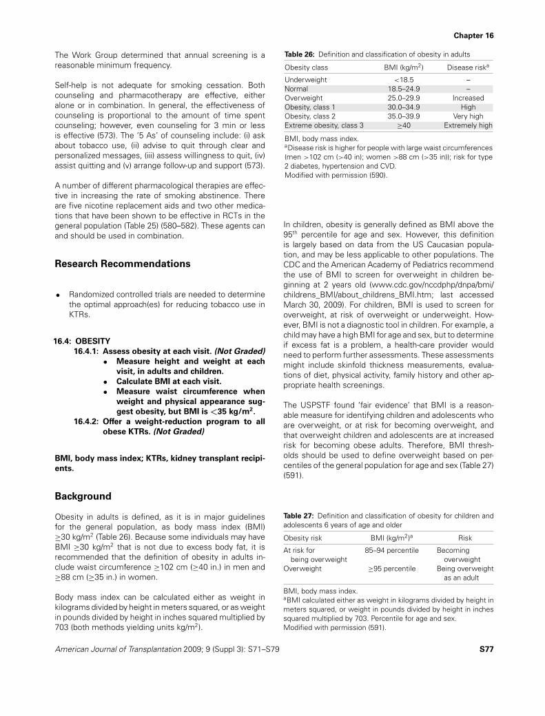
Chapter 16
The Work Group determined that annual screening is areasonable minimum frequency.
Self-help is not adequate for smoking cessation. Bothcounseling and pharmacotherapy are effective, eitheralone or in combination. In general, the effectiveness ofcounseling is proportional to the amount of time spentcounseling; however, even counseling for 3 min or lessis effective (573). The ‘5 As’ of counseling include: (i) askabout tobacco use, (ii) advise to quit through clear andpersonalized messages, (iii) assess willingness to quit, (iv)assist quitting and (v) arrange follow-up and support (573).
A number of different pharmacological therapies are effec-tive in increasing the rate of smoking abstinence. Thereare five nicotine replacement aids and two other medica-tions that have been shown to be effective in RCTs in thegeneral population (Table 25) (580–582). These agents canand should be used in combination.
Research Recommendations
• Randomized controlled trials are needed to determinethe optimal approach(es) for reducing tobacco use inKTRs.
16.4: OBESITY
16.4.1: Assess obesity at each visit. (Not Graded)
• Measure height and weight at each
visit, in adults and children.
• Calculate BMI at each visit.
• Measure waist circumference when
weight and physical appearance sug-
gest obesity, but BMI is <35 kg/m2.
16.4.2: Offer a weight-reduction program to all
obese KTRs. (Not Graded)
BMI, body mass index; KTRs, kidney transplant recipi-
ents.
Background
Obesity in adults is defined, as it is in major guidelinesfor the general population, as body mass index (BMI)≥30 kg/m2 (Table 26). Because some individuals may haveBMI ≥30 kg/m2 that is not due to excess body fat, it isrecommended that the definition of obesity in adults in-clude waist circumference ≥102 cm (≥40 in.) in men and≥88 cm (≥35 in.) in women.
Body mass index can be calculated either as weight inkilograms divided by height in meters squared, or as weightin pounds divided by height in inches squared multiplied by703 (both methods yielding units kg/m2).
Table 26: Definition and classification of obesity in adults
Obesity class BMI (kg/m2) Disease riska
Underweight <18.5 –Normal 18.5–24.9 –Overweight 25.0–29.9 IncreasedObesity, class 1 30.0–34.9 HighObesity, class 2 35.0–39.9 Very highExtreme obesity, class 3 ≥40 Extremely high
BMI, body mass index.aDisease risk is higher for people with large waist circumferences(men >102 cm (>40 in); women >88 cm (>35 in)); risk for type2 diabetes, hypertension and CVD.Modified with permission (590).
In children, obesity is generally defined as BMI above the95th percentile for age and sex. However, this definitionis largely based on data from the US Caucasian popula-tion, and may be less applicable to other populations. TheCDC and the American Academy of Pediatrics recommendthe use of BMI to screen for overweight in children be-ginning at 2 years old (www.cdc.gov/nccdphp/dnpa/bmi/childrens_BMI/about_childrens_BMI.htm; last accessedMarch 30, 2009). For children, BMI is used to screen foroverweight, at risk of overweight or underweight. How-ever, BMI is not a diagnostic tool in children. For example, achild may have a high BMI for age and sex, but to determineif excess fat is a problem, a health-care provider wouldneed to perform further assessments. These assessmentsmight include skinfold thickness measurements, evalua-tions of diet, physical activity, family history and other ap-propriate health screenings.
The USPSTF found ‘fair evidence’ that BMI is a reason-able measure for identifying children and adolescents whoare overweight, or at risk for becoming overweight, andthat overweight children and adolescents are at increasedrisk for becoming obese adults. Therefore, BMI thresh-olds should be used to define overweight based on per-centiles of the general population for age and sex (Table 27)(591).
Table 27: Definition and classification of obesity for children andadolescents 6 years of age and older
Obesity risk BMI (kg/m2)a Risk
At risk for 85–94 percentile Becomingbeing overweight overweight
Overweight ≥95 percentile Being overweightas an adult
BMI, body mass index.aBMI calculated either as weight in kilograms divided by height inmeters squared, or weight in pounds divided by height in inchessquared multiplied by 703. Percentile for age and sex.Modified with permission (591).
American Journal of Transplantation 2009; 9 (Suppl 3): S71–S79 S77

Chapter 16
Rationale
• In the general population, there is strong evidence thatobesity is a risk factor for CVD events and mortality inadults.
• In the general population, there are few studies exam-ining the effects of obesity treatment on CVD eventsor mortality, but there is evidence that the benefitsof treating obesity on intermediate outcomes for CVDoutweigh harm in adults.
• In KTRs, obesity is associated with CVD events andmortality.
• In KTRs, there is little reason to believe that weightreduction measures are not equally effective as in thegeneral population; however, there is some reason tobelieve that pharmacological and surgical managementof obesity may be more likely to cause harm than inthe general population.
Observational studies in the general population haveshown that obesity is an independent risk factor for CVD(592). Obesity is also associated with a number of riskfactors for CVD, including hypertension, dyslipidemias anddiabetes (590).
A number of RCTs in the general population have shownthat diet may cause modest weight reduction, at leastover a period of 12 months. Pharmacological interventionsare more effective in weight loss than diet alone, but areassociated with more adverse effects. Bariatric surgery iseffective, and may improve health outcomes. Guidelinesin the general population generally recommend screeningand treatment of obesity (www.cdc.gov/healthyweight/assessing/bmi/childrens_BMI/about_childrens_BMI.html;last accessed July 27, 2009) (591,593–597).
Observational studies in adult KTRs have reported an asso-ciation between obesity and mortality, CVD mortality andCHF (Table 18).
Counseling standard weight reduction diets, as recom-mended in guidelines in the general population, is unlikelyto cause harm in KTRs. The effects of pharmacologicalmanagement of obesity in KTRs are largely unexplored.Anecdotal evidence suggests that bariatric surgery canbe performed safely in KTRs and results in weight loss,at least over a relatively short duration of follow-up (598–600).
Small, uncontrolled trials in KTRs suggest that diet andother behavior modifications are safe and help reduceweight over the short term (601,602). There is no evidencethat any one diet is more effective than any other. A reason-able goal is to create a caloric deficit of 500–1000 kcal/day.Diets of 1000–1200 kcal/day for women and 1200–1500
kcal/day for men can be effective. Increased physical activ-ity may help to sustain weight reduction and reduce CVDrisk independent of weight reduction. Exercise may alsobe beneficial, although a small RCT in KTRs failed to showthat counseling to encourage exercise reduced weight orCVD risk factors at 1 year (603). Nevertheless, exercisecapacity increased in this study, and there was no harmassociated with exercise.
A large number of RCTs have examined pharmacologic in-terventions for weight loss in the general population. Thesetrials have shown modest weight reduction from medica-tions vs. placebo at 12 months (604). There are few long-term studies, and even fewer studies that have examinedhealth outcomes. In a 4-year RCT, 52% completed treat-ment with orlistat while 34% completed treatment withplacebo. Mean weight loss was greater with orlistat (–5.8kg) vs. placebo (–3.0 kg, p < 0.001). The cumulative inci-dence of diabetes was 6.2% with orlistat vs. 9.0% withplacebo (p = 0.0032). In a RCT, comparing the cannabinoidreceptor antagonist rimonabant with placebo in 839 pa-tients, rimonabant failed to reduce the primary end point,change in atheroma volume on coronary intravascular ultra-sound (605). Of concern are reports of psychiatric adverseeffects from rimonabant (606). Altogether, it remains un-clear whether the benefits outweigh harm of pharmaco-logical management of obesity in the general population.
Pharmacological treatment of obesity has not been ade-quately studied in KTRs. Adverse effects of available agentslimit their usefulness in the general population, and arelikely to have an even greater potential for adverse effectsin KTRs. Orlistat may interfere with the absorption of fat-soluble vitamins, and there have been case reports of aninteraction between orlistat and CsA, resulting in lowerCsA levels (607–609). Studies in the general populationhave shown that sibutamine can cause weight loss, butadverse effects are common and include increased bloodpressure and heart rate (604). There have been no studiesof sibutamine in KTRs.
There have been no RCTs examining the long-term effectsof bariatric surgery on health outcomes in the general pop-ulation. Nevertheless, bariatric surgery appears to be moreeffective than diet in causing weight reduction (610,611). Inthe largest case-control study to date, gastric bypass, verti-cal banded gastroplasty or gastric banding caused, respec-tively, −25%, −16% and −14% weight losses from base-line to 10 years (612). Importantly, there were 129 deathsin the control group and 101 deaths in the surgery group(p = 0.04). The most common cause of death in this studywas myocardial infarction (612). In another large observa-tional study, all-cause mortality (p < 0.0001), deaths fromdiabetes (p = 0.0005) and deaths from coronary arterydisease (CAD) (p = 0.006) were lower among 7925 pa-tients who had undergone bariatric surgery compared to7925 matched controls (613). Thus, it appears that bariatric
S78 American Journal of Transplantation 2009; 9 (Suppl 3): S71–S79

Chapter 16
Table 28: National Heart Lung Blood Institute weight-loss treatment guidelinesa
BMI (kg/m2)
Treatment 25–26.9 27–29.9 30–34.9 35–39.9 ≥40
Behavior modification Yes Yes Yes Yes YesPharmacotherapy If there are Yes Yes Yes
comorbiditiesb
Bariatric surgery If there are If there are If there arecomorbiditiesc comorbiditiesc comorbiditiesc
BMI, body mass index.a. Modified with permission (590).b. Comorbidities considered important enough to warrant pharmacotherapy are: established coronary heart disease, other atheroscleroticdiseases, type 2 diabetes, sleep apnea, hypertension, cigarette smoking, high LDL-C, low HDL-C, impaired fasting glucose, family historyof early CVD, and age (male ≥45 years, female ≥55 years).c. Comorbidities considered important enough to warrant surgery are: established coronary heart disease, other atheroscleroticdiseases, type 2 diabetes, and sleep apnea.
surgery can produce sustained weight reduction and im-prove health outcomes.
Guidelines in the general population recommend weightloss surgery in patients with severe obesity, that isBMI ≥40 kg/m2 or ≥35 kg/m2 with comorbid conditions.Bariatric surgery may include gastric banding or gastricbypass (Roux-en-Y). Uncontrolled studies suggest thatbariatric surgery may be performed safely in selected KTRs(598–600). However, the incidence of complications mayalso be greater in KTRs (614).
Guidelines in the general population recommend tailoringtreatment to the severity of obesity and its comorbidities(Table 28).
Childhood obesity in the general population is associatedwith a higher prevalence of CVD risk factors, such as
dyslipidemias, hypertension and diabetes. However, CVDevents may take decades to develop. Few studies haveexamined the safety and efficacy of weight reduction inchildren or adolescents. The USPSTF concluded that evi-dence was insufficient to recommend for or against rou-tine screening for obesity in children and adolescentsas a means to prevent adverse health outcomes. Thereare likewise few studies on the treatment of obesity inchildren and adolescent KTRs; therefore, there is no ba-sis for a different recommendation than for the generalpopulation.
Research Recommendations
• Additional research is needed to determine the effectof bariatric surgery on outcomes in KTRs.
American Journal of Transplantation 2009; 9 (Suppl 3): S71–S79 S79

Chapter 17
Chapter 17: Cardiovascular Disease Management
17.1: Consider managing CVD at least as intensively
in KTRs as in the general population, with ap-
propriate diagnostic tests and treatments. (Not
Graded)
17.2: We suggest using aspirin (65–100 mg/day) in all
patients with atherosclerotic CVD, unless there
are contraindications. (2B)
CVD, cardiovascular disease; KTRs, kidney transplant
recipients.
Background
The Work Group chose to deal with the prevention of CVDafter kidney transplantation, and considered the manage-ment of CVD complications to be beyond the scope of thisguideline. However, in patients with known CVD, prophy-laxis includes aspirin.
Rationale
• There is good evidence that atherosclerotic CVD isprevalent in KTRs.
• There is no reason to believe that the managementof complications of atherosclerotic CVD is different inKTRs than in the general population.
• In the general population, there is strong evidence thataspirin reduces atherosclerotic CVD events in patientswith known CVD.
• There is little reason to believe that the benefits ofaspirin would not exceed the harm in KTRs with CVD,as in patients with CVD in the general population.
Randomized controlled trials, and meta-analyses of thesetrials, have demonstrated that low-dose aspirin is safe
and effective in reducing CVD events in patients at highrisk for CVD. This has led to several guidelines suggest-ing that low-dose aspirin should be used in patients withknown CVD (secondary prevention) (615–617). The Amer-ican Heart Association, for example recommends usingaspirin for patients with established coronary and otheratherosclerotic vascular disease, including peripheral arte-rial disease, atherosclerotic aortic disease and carotid dis-ease (616).
In KTRs, there is little reason to believe that low-dose as-pirin would not be as effective as it is in the general pop-ulation. There is some evidence that platelet function isabnormal in KTRs, increasing the risk for thrombosis (618).Some observational data suggest that aspirin is safe inKTRs. In at least one retrospective observational study,the use of aspirin was associated with better graft survival(619). Given the high incidence of CVD in KTRs, the ben-efits of aspirin prophylaxis may be expected to outweighrisks, principally of bleeding.
Evidence from the general population suggests that as-pirin prophylaxis is effective in preventing CVD events inpatients at high risk for CVD events, such as patients withknown CVD. Most guidelines recommend that patients inthe general population with known CVD should receive as-pirin prophylaxis unless aspirin is contraindicated. Data forother antiplatelet agents are sparse; however, many guide-lines recommend that clopidogrel may be used in patientswho cannot take aspirin.
Research Recommendations
• A RCT is needed to determine the efficacy and safetyof aspirin in KTRs.
S80 American Journal of Transplantation 2009; 9 (Suppl 3): S80–S80

Section IV: Malignancy

Introduction
Introduction: Malignancy Risks After KidneyTransplantation
Kidney transplant recipients from around the world are atgreater risk of developing cancer compared to the generalpopulation (Table 29). This is especially true for cancersassociated with viral infections (e.g. EBV-associated lym-phomas). Some cancers are common in the general pop-ulation and also occur at a higher incidence in KTRs (e.g.colon cancer). Some are common in KTRs because theyare common in the general population and have a similarincidence in KTRs (e.g. breast cancer). Others are rare,but occur at a substantially higher rate in KTRs (e.g. Kaposisarcoma) (620,621). There are also cancers that may causestage 5 CKD, and are therefore seen more commonly inKTRs (e.g. myeloma and renal cell carcinoma).
Table 29: Cancers categorized by SIR for kidney transplant patients and cancer incidencea
Common cancers in transplantCommon cancersb population (estimated)c Rare cancersd
High SIRe Kaposi’s sarcoma Kaposi’s sarcomaf Eye(>5) (with HIV)e Vaginaf
Non-Hodgkin lymphomaKidneyNon-melanoma skinf
Lipf
ThyroidPenisf
Small intestinef
Moderate SIRe Lung Oro-nasopharynx Melanoma(>1–5, p < 0.05) Colon Esophagus Larynx
Cervix Bladder Multiple myelomaStomach Leukemia Anusf
Liver Hodgkin’s lymphomaNo increased risk showne Breast Ovary
Prostate UterusRectumf Pancreas
BrainTestis
HIV, human immunodeficiency virus; SIR, standardized incidence ratio.aIn approximate descending order of estimated frequency (SIR × rate in general population).bIncidence in both general and transplant population ≥10 per 100 000 people; based on world incidence (622). Age-standardized rate.Normalized to world population.cIncidence in general population <10 per 100 000, but estimated incidence in transplant population (SIR × general population incidence)≥10 per 100 000 people.dIncidence in both general and transplant population <10 per 100 000 people.eExcerpted from Table 4 of Grulich et al. (623).fBased on US incidence (624). Age standardized rate (normalized to US population).
Cohort studies have demonstrated the variability of riskfor cancer with both age and sex, with young KTRs hav-ing a risk 15–30 times greater than the general popula-tion of the same age, while the risk is only two timesgreater for 65-year-old KTRs (625). After the develop-ment of cancer, the survival of transplant recipients ispoor, and treatment options are limited by the trans-plant or comorbidities. It is thus important to consideroptions for preventative measures and screening KTRs,which can theoretically deliver benefits of lower mor-bidity and mortality through reduced incidence or earlyinterventions.
S82 American Journal of Transplantation 2009; 9 (Suppl 3): S82–S82

American Journal of Transplantation 2009; 9 (Suppl 3): S83–S83 S83
Rating Guideline Recommendations
Within each recommendation, the strength of recommendation is indicated as Level 1, Level 2, or Not Graded, and thequality of the supporting evidence is shown as A, B, C, or D.
Grade* Wording
Level 1 ‘We recommend’
Level 2 ‘We suggest’
Grade for quality of evidence Quality of evidence
A High
B Moderate
C Low
D Very low
*The additional category ‘Not Graded’ was used, typically, to provide guidance based on common sense orwhere the topic does not allow adequate application of evidence. The most common examples includerecommendations regarding monitoring intervals, counseling, and referral to other clinical specialists. Theungraded recommendations are generally written as simple declarative statements, but are not meant tobe interpreted as being stronger recommendations than Level 1 or 2 recommendations.

Chapter 18
Chapter 18: Cancer of the Skin and Lip
18.1: We recommend that KTRs, especially those who
have fair skin, live in high sun-exposure climates,
have occupations requiring sun exposure, have
had significant sun exposure as a child, or have
a history of skin cancer, be told that their risk of
skin and lip cancer is very high. (1C)
18.2: We recommend that KTRs minimize life-long sun
exposure and use appropriate ultraviolet light
blocking agents. (1D)
18.3: We suggest that adult KTRs perform skin and
lip self-examinations and report new lesions to
a health-care provider. (2D)
18.4: For adult KTRs, we suggest that a qualified health
professional, with experience in diagnosing skin
cancer, perform annual skin and lip examination
on KTRs, except possibly for KTRs with dark skin
pigmentation. (2D)
18.5: We suggest that patients with a history of skin
or lip cancer, or premalignant lesions, be referred
to and followed by a qualified health professional
with experience in diagnosing and treating skin
cancer. (2D)
18.6: We suggest that patients with a history of skin
cancer be offered treatment with oral acitretin, if
there are no contraindications. (2B)
KTRs, kidney transplant recipients.
Background
Skin cancers include basal cell carcinoma, squamous cellcarcinoma and malignant melanomas.
Fair-skin individuals are Caucasians and especially thosewith blond hair and light complexion (626).
High sun-exposure climates are in areas of the world thatare near the Equator, and those that have poor ozone-layerprotection.
Appropriate ultraviolet light/sun avoidance includes the useof shade and avoidance of sunlight during peak hours ofradiation, wearing protective clothing and the use of ultra-violet light blocking sunscreens.
Skin and lip self-examination is accomplished by close in-spection of all skin areas, using a mirror and/or the assis-tance of a family member, such as a spouse.
Qualified health professionals with experience in diagnos-ing skin cancer include physicians, physician’s assistantsor nurse practitioners with experience in diagnosing skincancer.
Qualified health professionals with experience in diagnos-ing and treating skin cancer include dermatologists, physi-cians or surgeons with experience in diagnosis (includingskin biopsies and their interpretation) and treatment of skincancer.
Acitretin has been used at doses between 0.2 and0.4 mg/kg/day in RCTs to prevent skin cancers.
Rationale
• Patients who are at high risk can be identified.• Patient behaviors can reduce the risk.• Educating patients who are at high risk will encourage
them to undertake behaviors that will reduce that risk.• Sun exposure is a risk factor for skin cancer.• Avoiding sun exposure may reduce the incidence of
skin cancer.• Self-examination will detect skin cancer at an earlier
stage than other measures.• Early detection and treatment will reduce the morbidity
and mortality of skin cancer.• Skin and lip examination by a qualified health profes-
sional can detect skin cancer early.• Advice to undertake regular skin self-examination is
poorly recalled and implemented.• Acitretin may reduce the risk for recurrent squamous
cell skin cancer in KTRs.• Although adverse effects associated with the use of
acitretin are common, and often necessitate discon-tinuing therapy, the benefits may outweigh harm inselected KTRs.
Skin cancers occur with a much higher incidence in KTRscompared to the general population. In addition, risk fac-tors for skin cancers in the general population are alsolikely to be risk factors for skin cancer in KTRs. These in-clude: fair skin, living in high sun-exposure climates, havingoccupations with sun exposure, having had significant sunexposure as a child, or having a history of skin cancer (627).
Most measures for reducing the risk of skin cancer (de-scribed in guideline statements above) require patientcooperation. Although there are only limited RCT data
S84 American Journal of Transplantation 2009; 9 (Suppl 3): S84–S85

Chapter 18
demonstrating that informing KTRs of their increased riskfor skin cancer helps to reduce that risk, the benefits ofpatient education are very likely to outweigh harm (628).
There is evidence that geographical locations associatedwith increased sun exposure are associated with increasedrisk of skin cancer in both KTRs and the general population(629). There is also evidence in the general population thatthe use of sunscreen reduces the incidence of squamouscell cancer (630). Although there is no evidence in KTRsthat avoidance of sun exposure or the use of sun block-ers reduces skin cancer, potential benefits likely outweighharm. Sun can be blocked by staying in shaded environ-ments, wearing protective clothing, a wide-brim hat andsunglasses that block ultraviolet light. There is a concernthat use of sunscreens may lead to behaviors which in-crease total sunlight exposure (631).
It is plausible that self-examination will lead to earlier de-tection of skin cancer than less frequent skin examinationsby health-care providers (632). It is also plausible that earlydetection will lead to early treatment, and thereby reducemorbidity and mortality. However, skin self-examinationhas not been shown to be effective in reducing overallcancer-specific mortality and morbidity in either the gen-eral population or in KTRs. Nevertheless, since the costsand adverse effects of self-screening are low, the use ofeducation programs to encourage self-examination, espe-cially in areas of high prevalence of skin cancer, is justified.
American (627) and European (633) transplantation pro-fessional guidelines recommend skin cancer screening inKTRs, monthly skin self-examination and at least annualtotal body skin examination by a dermatologist or expertphysician (634). The USPSTF concluded that there is insuf-ficient evidence to recommend for or against populationskin cancer screening using total skin examination (635),while The American College of Preventive Medicine rec-ommended screening for high-risk individuals (636).
Advice to undertake regular skin examination is poorlyrecalled and implementation is thus not reliable (637).Nonetheless, in a community-based RCT of regular skinscreening, the intervention group reported considerablyhigher rates of performance (638). Visual inspection byKTRs is also likely not to be as reliable for detecting skincancer as regular skin examinations by qualified health pro-fessionals. Studies in the general population have shownthat individuals with adequate training and experience, forexample dermatologists, detect skin cancer earlier thangeneral practitioners (639). General practitioners with ex-perience may perform as well as dermatologists in someareas (640). In the absence of experienced general practi-tioners, resources may be insufficient to allow KTRs to beseen annually by a dermatologist. Therefore, a strategy thatcombines primary screening with referral of suspicious le-sions to a dermatologist may be most cost-effective. Pa-
tients who have had a skin cancer are much more likely todevelop a second lesion than patients with no history ofskin cancer (641). Therefore, patients who have had a skincancer are more likely to benefit from regular screening bya dermatologist, or health-care professional with compara-ble training. Early diagnosis and removal of skin cancersis essential to reduce disfiguring surgery and to preventmortality from advanced or metastatic lesions.
There is a paucity of RCT data assessing whether the ben-efits of altering the immunosuppressive medication reg-imen to reduce the incidence of skin cancer outweighharm. For example, in a recent RCT, KTRs 10–15 yearsafter transplant were randomly allocated to convert CNI tosirolimus (N = 555) vs. remaining on CNI (N = 275) (119).At 2 years of follow-up, 12 (2.2%) in the conversion groupvs. 21 (7.7%) in the CNI group had investigator-reportedskin cancer (p < 0.001). However, the number of adverseeffects in the sirolimus conversion arm was higher thanthose in the CNI control arm. Indeed, the Drug Safety Mon-itoring Board halted enrollment for patients with eGFR 20–40 mL/min/1.73 m2 early, because in this stratum (N = 77)the composite safety end point (first occurrence of biopsy-proven acute rejection, graft failure or death) was signifi-cantly higher in the conversion vs. the control group (119).The Work Group concluded that it remains unclear whetherthere is a high-risk population of KTRs in which benefitsfrom converting one immunosuppressive regimen to an-other to reduce skin cancer outweigh harm.
In three RCTs, which together included a total of 93 KTRs(10–15 years after transplant), those treated with acitretinfor 6–12 months demonstrated a reduction in the rateof formation of new skin cancers compared to untreatedcontrols, with no differences between doses of 0.2 and0.4 mg/kg/day (642). In these trials, several individuals hadadverse effects attributed to therapy (642); however, theseadverse effects generally resolved upon discontinuationof treatment. Adverse effects that resulted in treatmentwithdrawal included: headache (N = 3), dyslipidemia(N = 2) musculoskeletal complaints (N = 2) and skinrash (N = 2). In addition, the duration of treatment andfollow-up were relatively short in these trials. Altogether,the Work Group concluded that there is moderate-qualityevidence that there are tradeoffs to prophylaxis withacitretin (see Evidence Profile in Supporting Table 52 athttp://www3.interscience.wiley.com/journal/118499698/toc); some KTRs may consider that the benefits oftreatment outweigh the harm.
Research Recommendations
• A RCT is needed to better define the optimal dose andthe benefits and harm of acitretin to prevent recurrentskin cancer in KTRs.
American Journal of Transplantation 2009; 9 (Suppl 3): S84–S85 S85

Chapter 19
Chapter 19: Non-Skin Malignancies
19.1: Develop an individualized screening plan for each
KTR that takes into account the patient’s past
medical and family history, tobacco use, compet-
ing risks for death, and the performance of the
screening methodology. (Not Graded)
19.2: Screen for the following cancers as per lo-
cal guidelines for the general population (Not
Graded):
• Women: cervical, breast and colon cancer;
• Men: prostate and colon cancer.
19.3: Obtain hepatic ultrasound and alpha feto-protein
every 12 months in patients with compensated
cirrhosis. (Not Graded) [See Recommendations
13.5.4 (HCV) and 13.6.5 (HBV).]
HBV, hepatitis B virus; HCV, hepatitis C virus; KTR, kid-
ney transplant recipient.
Background
Screening for cancer has both benefits and harm. In KTRswith multiple comorbidities, it is essential to consider theextent and magnitude of potential harm, so it can beweighed against the risks of disease and benefits of earlydetection. There is good reason to believe that screen-ing test performance, harm from interventions and the lifeyears to be gained by early intervention may be substan-tially different in KTRs compared to that in the generalpopulation. Hence, careful individual appraisal needs to beexercised when making recommendations for screeningof KTRs (643).
In general, the better the individual’s prognosis, the higherthe risk of disease, and the lower the risk of harm fromscreening, the greater is the chance of benefit (644). If, onthe other hand, the individual has a poor prognosis fromcardiac or other comorbidity, the risk of the disease to bescreened is not high and the harm from screening is sig-nificant, the less it can be justified. For example cervicalcancer screening of an unvaccinated 45-year-old patientwith a well-functioning kidney allograft and no comorbidi-ties is easier to recommend than fecal occult blood test-ing (FOBT) and subsequent colonoscopy in a 69-year-oldpatient with type II diabetes and severe CAD. The likely in-cidence of disease needs to be taken into account as wellas the standardized incidence ratio (SIR) as performed inTable 29, since the two factors taken together define thelikely risk of any given disease in an individual KTR. Un-fortunately, there are no RCTs on screening for cancer inKTRs.
Rationale
• Comorbidities and competing risks in KTRs may influ-ence the potential benefits and harm from screeningfor some cancers.
• The decision to screen for cancer should be individual-ized.
Screening for cervical cancer
• In the general population, there is good evidence thatthe benefits of screening outweigh harm.
• In KTRs, cervical cancer is more common than in thegeneral population, and screening may therefore bemore beneficial.
• In KTRs with quality of life and life expectancy notgreatly reduced from that of general population, thebenefits of screening may outweigh harm.
• In the general population, there is evidence that thebenefits of human papillomavirus (HPV) vaccinationoutweigh harm.
• In KTRs, although vaccination may be less effective,there is little reason to believe that benefits would notoutweigh harm.
Initiation of screening for cervical cancer is recommendedfor women within 3 years of onset of sexual activity or age21 (whichever comes first) in order to detect malignantlesions resulting from persistent human papillomavirus(HPV) infection (www.ahrq.gov/clinic/uspstf/uspscerv.htm;last accessed July 17, 2009) (645). Cervical cancer ismore common, may develop more rapidly and may bemore aggressive in immunosuppressed patients (646,647),suggesting that KTRs should be screened more fre-quently (648). American and European transplant guide-lines recommend annual screening for cervical cancer withpelvic examination and Pap smear (627,633). Use of HPVDNA testing has not achieved widespread acceptance(www.ahrq.gov/clinic/uspstf/uspscerv.htm; last accessedJuly 17, 2009). Screening for cervical cancer also providesan opportunity to inspect the anal, vaginal and vulvar re-gions for cancers that are also increased in female KTRs.The cost of cervical cancer screening in KTRs is modeledat US$ 12 000 per life-year saved comparable to the gen-eral population (US$ 25 000 to 50 000 per life-year saved)(649).
In the general population, there is strong evidence that thebenefits of vaccination outweigh harm, but the longest
S86 American Journal of Transplantation 2009; 9 (Suppl 3): S86–S88

Chapter 19
duration of follow-up is 52 months at present. HPVvaccination of girls prior to exposure to HPV infection (forthe oncogenic strains 16 and 18, which account for approx-imately 70% of cervical cancers, and for the wart-causingstrains 6 and 11) has been adopted in a number of coun-tries (650,651). The vaccine is inactivated and could thusbe used both prior to transplantation and in KTRs, but thereis no evidence for effectiveness or safety in immunosup-pressed patients.
Screening for breast cancer
• In the general population, there is weak evidence thatthe benefits of screening outweigh harm.
• In KTRs, the incidence of breast cancer is similar tothat in the general population.
• In KTRs with quality of life and life expectancy similarto that of general population, the benefits of screeningmay outweigh harm.
Mammography for women in the general population ages50–74 decreases breast cancer mortality by 23% (95% CI13–31%) (652,653). The incidence of breast cancer is verysimilar in both the general population and in KTRs. Thereare no RCTs or studies on which to base advice for oragainst breast cancer screening in KTRs. The two factorsthat might influence the decision to screen are screeningtest performance and potential life-years saved from inter-vention. American and European transplant guidelines rec-ommend screening in KTRs between 50 and 69 years withan option to screen above the age of 40 years (627,633).Test accuracy for mammography varies with the best re-sults in older women, and the worst results in youngerwomen. Consideration should also be given to the poten-tial physical and emotional harm from false-positive andfalse-negative screening tests. Models of screening forbreast cancer in KTRs suggest that it is cost-effective innondiabetic Caucasians (654).
Screening for prostate cancer
• In the general population, there is little evidence thatthe benefits of screening outweigh harm.
• In KTRs, the incidence of prostate cancer is similar tothat in the general population.
• In KTRs, it is unclear whether the benefits of screeningoutweigh harm.
Screening for prostate cancer, using prostate-specific anti-gen (PSA) and/or digital rectal examination, is controversialin the general population. The most recent recommenda-tion from the USPSTF is to avoid screening men 75 yearsor older (www.ahrq.gov/clinic/uspstf/uspsprca.htm; lastaccessed July 17, 2009). They also concluded that therewas insufficient evidence to assess the balance of bene-
fits and harm for screening men younger than 75 years old(www.ahrq.gov/clinic/uspstf/uspsprca.htm; last accessedJuly 17, 2009). The incidence of prostate cancer in KTRs issimilar to that in the general population, and being one ofthe commonest cancers in males, there is a high absoluterisk (Table 29). However, there are no data on screeningtest performance, or benefits in KTRs, and there is goodreason to believe that the performance of PSA testingmay be different in KTRs compared to the general pop-ulation. No advice is thus given for or against screeningfor prostate cancer in KTRs, beyond following local recom-mendations/standards for prostate cancer screening in thegeneral population.
Screening for colorectal cancer
• In the general population, there is good evidence thatthe benefits of screening outweigh harm for individualsage 50 years and older.
• In KTRs, the incidence of colon cancer is increasedcompared to the general population, especially amongKTRs less than 50 years of age.
• In KTRs, there are reasons to believe that FOBT maybe less specific for colon cancer than in the generalpopulation, but there is no evidence to believe thatcolonoscopy is less sensitive or specific.
Studies in the general population have demonstrated thatthe benefits of screening generally outweigh the harm(655–658). Guidelines for the general population in Aus-tralia/New Zealand, the US and in Europe, recommendscreening individuals 50 years and older, using annualFOBT and/or colonoscopy (655). The standardized inci-dence of colorectal cancer is increased in KTRs comparedto the general population, and there is good evidencethat colon cancer occurs at a younger age in KTRs com-pared to the general population (Table 29). American andEuropean transplantation guidelines recommend screen-ing either at age 50 years, or at the age at which it isrecommended in the general population in each country(627,633).
Screening with FOBT may be less specific in KTRs, giventhat the incidence of positive tests from CMV infectionand drug toxicities may be high. The harms of colonoscopymust be carefully considered in each individual based upontheir comorbidities, since the consequences of the poten-tial complications of colonoscopy are influenced negativelyby immunosuppression. In the absence of data on the ben-efits and harm of screening of KTRs for colon cancer, it issuggested that screening should be performed as currentlyrecommended for the general population with careful in-dividual risk–benefit analysis based upon overall prognosisand comorbidities. A recent analysis suggests that the ben-efits may outweigh the harm from screening of KTRs aged35–50 years (659).
American Journal of Transplantation 2009; 9 (Suppl 3): S86–S88 S87

Chapter 19
Screening for hepatocellular cancer
• In KTRs, the risk of hepatocellular carcinoma is higherthan in the general population.
• In the general population, there is no evidence that thebenefits of screening outweigh harm.
There are screening recommendations in high-risk groups(patients with cirrhosis and those who are hepatitis Bcarriers) that include abdominal ultrasound and alpha-fetoprotein testing every 6–12 months (660–662). Testing ev-ery 6 months is based on the estimated doubling timeof this tumor (660). The Work Group chose a 12-monthtesting interval, given uncertainties of the benefits andharm of testing. Both tests have limited specificity andsensitivity (663). Nonetheless screening by gastroenterol-ogists in high-risk patients is reported to be about 50%by questionnaire survey in the United States (664,665),the interventions have significant risks and no RCTs havedemonstrated survival benefits. There have been severalcost-effectiveness studies but the conclusions have var-ied widely from very cost-effective to values exceedingUS$ 250 000 per quality-adjusted life-year (666). The USNational Cancer Institute does not recommend screen-ing (www.cancer.gov/cancertopics/pdq/screening/hepatocellular/healthprofessional/page2; last accessed July 17,2009) largely because of a concern of uncommon but sig-nificant harm due to invasive testing after false-positivescreening. There have been two large population-basedRCTs in Asia in HBV-infected subjects. The larger studyshowed some benefit, but was of poor quality, and thesecond showed no benefit (667,668).
The highest-risk group of KTRs with otherwise good prog-nosis are those with compensated cirrhosis and chronicviral hepatitis, especially HBV (669). Given that the bene-fits are inconclusive in high-risk nontransplant patients, therecommendation of the US National Cancer Institute is notlikely to differ in KTRs.
Screening for renal cell cancer
• In KTRs, the incidence of renal cell carcinoma is muchhigher than in the general population; however, thereis no evidence that the benefits of screening outweighharm.
Screening is not generally recommended in the generalpopulation. Both relative and absolute risks of renal cellcancer are substantially increased in KTRs compared tothe general population. Although there is no good evi-dence that mortality is reduced, several United States,European and Asian centers are screening for renal cellcarcinoma after transplant (670–672). The rate of renal cellcarcinoma (number per years of follow-up) is difficult todetermine from these reports, but appears to vary con-siderably. Two important risk factors for renal cell carci-noma in these reports were prior renal cell carcinoma andthe presence of acquired cystic disease. A medical deci-sion analysis conducted several years ago, predominantlyin dialysis patients with low expected survival rates, de-termined that the benefits of routine screening would below (673). Screening will likely detect many unimportantlesions that will require further investigation, treatmentand thus possible harm. Nonetheless, significant benefitscould accrue to higher-risk transplant recipients with better-than-average life expectancy. Patients with prior renal cellcancer are at risk of both recurrence and new primaries, ir-respective of whether they have been transplanted. Somediseases, such as analgesic nephropathy, tuberous scle-rosis and acquired cystic disease are associated with anincreased risk of renal cell carcinoma. The American So-ciety of Transplantation guidelines found no evidence toadvise screening with either imaging or urine cytology(627).
Research Recommendations
• Observational studies are needed to better define age-specific SIR for most cancers, with preliminary analy-ses suggesting that younger KTRs have a greatly in-creased SIR compared to older KTRs.
• Studies on the performance of FOBT in KTRs wouldhelp determine its potential role for screening KTRs.
• A RCT should be performed to assess the benefits andharm of screening vs. no screening for renal cell carci-noma. Preliminary data are needed to define mortalityrates from renal cell carcinoma after transplantation,and determine age-specific SIR, since analyses sug-gest that younger KTRs have a greatly increased SIRin comparison to older KTRs.
S88 American Journal of Transplantation 2009; 9 (Suppl 3): S86–S88

Chapter 20
Chapter 20: Managing Cancer with Reductionof Immunosuppressive Medication
20.1: We suggest consideration be given to reducing
immunosuppressive medications for KTRs with
cancer. (2C)
20.1.1: Important factors for consideration in-
clude (Not Graded):
• the stage of cancer at diagnosis;
• whether the cancer is likely to be ex-
acerbated by immunosuppression;
• the therapies available for the cancer;
• whether immunosuppressive medi-
cations interfere with ability to ad-
minister the standard chemotherapy.
20.2: For patients with Kaposi sarcoma, we suggest
using mTORi along with a reduction in overall
immunosuppression. (2C)
KTRs, kidney transplant recipients; mTORi, mam-
malian target of rapamycin inhibitor(s).
Rationale
• In KTRs, cancers that have a high or moderately in-creased SIR (e.g. ≥3.0) are likely caused or exacer-bated by immunosuppressive medication.
• In KTRs that develop cancers likely to be caused or ex-acerbated by immunosuppressive medication, reduc-ing immunosuppressive medication may prolong sur-vival.
• In KTRs, cancers that have a low SIR (e.g. ≤1.5) areunlikely to have been caused or to be exacerbated byimmunosuppressive medication.
• In KTRs that develop cancers that are unlikely to becaused or exacerbated by immunosuppressive med-ication, reducing immunosuppressive medication isless likely to have a significant effect on survival, andmay increase the risk for acute rejection.
• Reduced quality of life from graft loss must be balancedagainst the potential for prolonging survival by reducingimmunosuppression.
• Reducing immunosuppressive medications may re-duce complications of cancer chemotherapy.
• In KTRs with Kaposi sarcoma, dramatic reductions inlesion size have been associated with a change in im-munosuppressive medication to mTORi.
In KTRs, non–renal cell cancers that have a high SIR (e.g.≥3.0) are likely caused or exacerbated by immunosuppres-sive medication. There is strong evidence that immuno-suppressive medication increases the risk of some spe-cific types of cancer, notably cancer that may be causedby viruses (Table 30). There is little evidence that specificimmunosuppressive agents are more likely than others toincrease the risk of cancer. It is more likely that the totalamount of immunosuppressive medication increases therisk for cancer, rather than the type of immunosuppressivemedication per se. Observational data have suggested thatthere is an association between PTLD and the use of bio-logical anti–T-cell agents (674). There is evidence from posthoc analysis of RCTs that there was a reduction in cancerincidence in sirolimus treatment arms (119,675). However,the numbers of patients developing cancer were small, andthe post hoc nature of the analysis increases the possibilitythat the results were due to chance.
To reduce immunosuppressive medications in KTRs diag-nosed with cancer is a difficult decision. There is evidencethat the risk of de novo cancer returns to pretransplant lev-els after graft failure (676–679), suggesting that reducingimmunosuppressive medication may be warranted. Exper-imental studies have demonstrated the specific capacity ofCNIs to increase metastasis (680). Clinical studies have im-plicated antiproliferative agents in increased, and mTORi inrelative reduction in cancer risk. However, there have beenno RCTs testing the effects of reducing or withdrawingimmunosuppressive medications in posttransplant cancer,and it is possible that established cancer and de novo can-cer behave differently under the influence of immunosup-pression. The standard established treatment for PTLD andKaposi’s sarcoma includes reducing immunosuppression,and this has proven to be sufficient to control or eliminatetumors in some KTRs (681).
The decision to reduce or withdraw immunosuppressivemedication must also balance quality of life with and with-out a functioning transplant, if cessation of medicationresults in graft rejection. Altogether, evidence suggeststhat consideration should be given to reducing immuno-suppressive medications in each individual, but since thisevidence is weak, the type of cancer, stage of disease, andpatient preferences should be taken into account.
In KTRs, cancers that have a low SIR (e.g. <3.0) are un-likely to be caused or exacerbated by immunosuppressivemedication. In distinction to those cancers in which theSIR is elevated in immunosuppressed KTRs, cancers in
American Journal of Transplantation 2009; 9 (Suppl 3): S89–S90 S89

Chapter 20
Table 30: Viral-associated cancers
Malignancy site/Type of cancer
Virus Sufficient evidence Limited evidence
HBV and HCV LiverHuman T-cell lymphotropic virus type 1 Non-Hodgkin lymphomaHuman Herpes virus 8 Kaposi sarcomaEBV Nasopharynx, Non-Hodgkin lymphoma,
Hodgkin lymphomaHPV Tongue, mouth, tonsil, anus, vagina, cervix, penis Nonmelanoma skin, larynx
EBV, Epstein-Barr virus; HBV, hepatitis B virus; HCV, hepatitis C virus; HPV, Human papillomavirus.Modified with permission (621).
which there is no evidence for an increased risk from im-munosuppression have no rationale for reducing or ceasingtherapy.
In KTRs who develop cancers that are unlikely caused orexacerbated by immunosuppressive medication, reducingimmunosuppressive medication will likely have little effecton survival, and may increase the risk for acute rejection.There are no data to support or refute altering immuno-suppression after development of cancer of the prostate,breast, ovary, uterus, pancreas, brain glioma or testis.However, many of the complications of cancer chemother-apy are also complications of immunosuppressive agentsused in KTRs, and reducing immunosuppressive medica-tions to prevent or treat complications of chemotherapy iswarranted.
Several case series in patients with established Kaposisarcoma have demonstrated benefits from conversionfrom standard immunosuppression to either sirolimus oreverolimus. Cases with disease limited to the skin havehad resolution of the skin lesions, while the responsesof disseminated solid-organ invasive disease have beenless convincing (682,683). The strong benefit seen inthese case series, together with experimental data anda clear scientific rationale for efficacy through inhibitionof vascular endothelial growth factor-F receptors, havelead to the conclusion that patients with Kaposi sar-coma should be immunosuppressed with these agentsin preference. On the other hand, there are also caseseries that have shown regression of Kaposi’s sarcomawith a reduction in immunosuppressive medication alone(684).
S90 American Journal of Transplantation 2009; 9 (Suppl 3): S89–S90

Section V: Other Complications

S92 American Journal of Transplantation 2009; 9 (Suppl 3): S92–S92
Rating Guideline Recommendations
Within each recommendation, the strength of recommendation is indicated as Level 1, Level 2, or Not Graded, and thequality of the supporting evidence is shown as A, B, C, or D.
Grade* Wording
Level 1 ‘We recommend’
Level 2 ‘We suggest’
Grade for quality of evidence Quality of evidence
A High
B Moderate
C Low
D Very low
*The additional category ‘Not Graded’ was used, typically, to provide guidance based on common sense orwhere the topic does not allow adequate application of evidence. The most common examples includerecommendations regarding monitoring intervals, counseling, and referral to other clinical specialists. Theungraded recommendations are generally written as simple declarative statements, but are not meant tobe interpreted as being stronger recommendations than Level 1 or 2 recommendations.

Chapter 21
Chapter 21: Transplant Bone Disease
(See KDIGO Clinical Practice Guideline for the Diagnosis,Evaluation, Prevention and Treatment of Chronic KidneyDisease–Mineral and Bone Disorder [CKD–MBD].)
21.1: In patients in the immediate post-kidney trans-
plant period, we recommend measuring serum
calcium and phosphorus at least weekly, until
stable. (1B)
21.2: In patients after the immediate post-kidney
transplant period, it is reasonable to base the
frequency of monitoring serum calcium, phos-
phorus and PTH on the presence and magnitude
of abnormalities, and the rate of progression of
CKD. (Not Graded)
21.2.1: Reasonable monitoring intervals would
be (Not Graded):
• In CKD stages 1–3T, for serum calcium
and phosphorus, every 6–12 months;
and for PTH, once, with subsequent
intervals depending on baseline level
and CKD progression.
• In CKD stage 4T, for serum calcium
and phosphorus, every 3–6 months;
and for PTH, every 6–12 months.
• In CKD stage 5T, for serum calcium
and phosphorus, every 1–3 months;
and for PTH, every 3–6 months.
• In CKD stages 3–5T, measurement
of alkaline phosphatases annually, or
more frequently in the presence of el-
evated PTH.
21.2.2: In CKD patients receiving treatments for
CKD–MBD, or in whom biochemical ab-
normalities are identified, it is reasonable
to increase the frequency of measure-
ments to monitor for efficacy and side
effects. (Not Graded)
21.2.3: It is reasonable to manage these abnor-
malities as for patients with CKD stages
3–5. (Not Graded)
21.3: In patients with CKD stages 1–5T, we suggest
that 25(OH)D (calcidiol) levels might be mea-
sured, and repeated testing determined by base-
line values and interventions. (2C)
21.4: In patients with CKD stages 1–5T, we sug-
gest that vitamin D deficiency and insufficiency
be corrected using treatment strategies recom-
mended for the general population. (2C)
21.5: In patients with an eGFR greater than approxi-
mately 30 mL/min/1.73 m2, we suggest measur-
ing BMD in the first 3 months after kidney trans-
plant if they receive corticosteroids or have risk
factors for osteoporosis as in the general popu-
lation. (2D)
21.6: In patients in the first 12 months after kid-
ney transplant with eGFR greater than approx-
imately 30 mL/min/1.73 m2 and low BMD, we
suggest that treatment with vitamin D, cal-
citriol/alfacalcidiol, or bisphosphonates be con-
sidered. (2D)
21.6.1: We suggest that treatment choices be in-
fluenced by the presence of CKD–MBD,
as indicated by abnormal levels of cal-
cium, phosphorus, PTH, alkaline phos-
phatases, and 25(OH)D. (2C)
21.6.2: It is reasonable to consider a bone biopsy
to guide treatment, specifically before
the use of bisphosphonates due to the
high incidence of adynamic bone dis-
ease. (Not Graded)
21.6.3: There are insufficient data to guide treat-
ment after the first 12 months. (Not
Graded)
21.7: In patients with CKD stages 4–5T, we suggest
that BMD testing not be performed routinely,
because BMD does not predict fracture risk as
it does in the general population and BMD does
not predict the type of kidney transplant bone
disease. (2B)
21.8: In patients with CKD stages 4–5T with a known
low BMD, we suggest management as for pa-
tients with CKD stages 4–5 not on dialysis. (2C)
25(OH)D, 25-hydroxyvitamin D; BMD, bone mineral
density; CKD, chronic kidney disease; CKD–MBD,
chronic kidney disease–mineral and bone disorder;
eGFR, estimated glomerular filtration rate; KDIGO,
Kidney Disease: Improving Global Outcomes; PTH,
parathyroid hormone.
Background
We largely deferred to the KDIGO CKD–MBD Guidelinethat is pertinent to KTRs (684a). We reviewed these rec-ommendations, but did not conduct independent evidencereviews.
American Journal of Transplantation 2009; 9 (Suppl 3): S93–S96 S93

Chapter 21
Rationale
• The risk of fractures following kidney transplantation ishigh.
• It is not clear how to identify KTRs who might benefitfrom treatment.
• Bone disease is multifactorial, and most KTRs havepreexisting CKD–MBD.
• In non-KTRs, low bone mineral density (BMD) or a lossof BMD predicts fractures, but data are scant for KTRs.
• No RCTs in KTRs have examined bone-specific thera-pies on patient-level outcomes, including mortality orfractures.
• Treatment with calcium, calcitriol or vitamin D analogs,and/or bisphosphonates has been suggested to im-prove BMD in KTRs.
• A small study of calcitriol demonstrated worsenedbone turnover, but improved mineralization.
• A small study of treatment with bisphosphonatesdemonstrated worsening bone turnover and mineral-ization.
• There are insufficient data to suggest any bone-specifictherapies after the first year of kidney transplantation.
CKD–MBD is common in KTRs. Most KTRs have somedegree of CKD, and thus CKD–MBD may be present.Transplant-specific therapies, especially corticosteroids,may further affect CKD–MBD management. Biochemicalabnormalities are common after transplantation. The scopeand magnitude of the biochemical abnormalities of CKD–MBD fluctuate early, compared to late after transplanta-tion. Posttransplant bone disease represents an importantcomplication observed in a substantial proportion of pa-tients, but the etiology and pathology vary. Early studieshave demonstrated a rapid decrease in BMD in the first6–12 months after successful kidney transplantation, andcontinued loss, albeit at a lower rate, for many years (685).Fractures are common and are associated with substantialmorbidity.
The etiology of transplant bone disease is multifactorial.Patients come to transplantation with preexisting CKD–MBD. In addition, there are potentially deleterious effectsof immunosuppressive agents (see Supporting Table 53 athttp://www3.interscience.wiley.com/journal/118499698/toc), impaired kidney function, and other factors, such aspostmenopausal status, presence of diabetes, smoking,physical inactivity and duration of CKD stage 5 (686). Pre-vious studies in KTRs have shown a correlation betweenthe cumulative dose of glucocorticoids and BMD. Basedon a few bone biopsy studies in KTRs, glucocorticoidsappear to be the primary determinant of subsequent bonevolume and turnover. Thus, the cumulative and meanprednisone dose correlated negatively with bone turnover,whereas there was no correlation with CsA cumulativedose or serum parathroid hormone (PTH) (687). The
possible role of CNIs remains incompletely studied, withcontradictory reports on their effects on bone turnover(687).
Arterial calcification is also common after a kidney trans-plant, but it may be due to the effects of the uremicstate and dialysis rather than the transplant itself. InKTRs (CKD stages 1–5T), only one prevalence study wasidentified, demonstrating a prevalence of calcification of24.4% (444). Although this cross-sectional study was large(n = 1117), calcification was assessed by posterio-anteriorplain abdominal X-ray examination of the aorto-iliac re-gion, which is likely to be less sensitive than computer-ized tomography based imaging. In addition, one of themajor difficulties in interpreting calcification in the trans-plant population is the carryover effect from CKD stage5 or stage 5D. Currently, only one preliminary study isavailable suggesting that the progression of cardiovascu-lar calcification may be halted after renal transplantation(688).
KTRs who develop persistently low levels of serum phos-phorus (<1.0 mmol/L) should be considered for treat-ment with phosphate supplementation. However, phos-phate administration is not without risk, and caution shouldbe exerted, as it may exacerbate an already existingsecondary hyperparathyroidism. Therefore, every attemptshould be made in order to prescribe the strict minimumdoses.
Although no clinical trials have specifically addressed thefrequency of monitoring in KTRs, KTRs usually have CKD,and therefore are likely to have CKD–MBD. Thus, the man-agement of the biochemical abnormalities of CKD–MBDafter transplant should be similar to that proposed for non-transplant CKD and based on the prevalence of abnormal-ities, and the risks associated with those abnormalities.
A recent study of 303 KTRs in the United States foundthat 11–25% had abnormal calcium or calcium X phospho-rus product in the first year following transplant, and 24%with eGFR 40–60 mL/min/1.73 m2 had intact PTH levels>130 pg/mL (130 ng/L) at 1 year after kidney transplanta-tion (689). Another series from the UK (690) evaluated 244KTRs; 104 in the first year, and the remainder more than1 year after transplant. Hypercalemia was present in40% of recently transplanted recipients and 25% of long-term patients. Vitamin D insufficiency (40–75 nmol/L) waspresent in 29% and 43%, deficiency (12–39 nmol/L) in56% and 46%, and severe deficiency (<12 nmol/L) in 12%and 5%, respectively. A larger cohort from Switzerland(691) evaluated 823 KTRs, on average 7 years after trans-plantation. They found only 27% had a PTH within normalrange (i.e., 15–65 pg/mL [15–65 ng/L]), whereas 70% hadhyperparathyroidism (PTH >65 pg/mL [65 ng/L]), and 2.8%were hypoparathyroid (PTH <15 pg/mL [15 ng/L]). Serumphosphorus was normal in 74% (0.85–1.45 mmol/L), andincreased in only 3.6%. Finally, serum calcium was normal
S94 American Journal of Transplantation 2009; 9 (Suppl 3): S93–S96

Chapter 21
in most patients (85.9%), with only 2.8% and 11.3% beinghypo- and hypercalcemic, respectively. Thus, disorders ofmineral metabolism may persist many years after trans-plantation.
There are few data describing the risk relationship ofbiochemical abnormalities of CKD–MBD and mortality inKTRs. A study of 773 KTRs found no relationship betweenserum calcium, phosphorus or PTH and mortality (692).However, patients with the highest quintile of phosphorushad increased risk of kidney allograft loss. Similarly, thosewith the highest quintile of calcium had an increased riskof kidney allograft loss.
Hypercalcemia following kidney transplantation is com-mon and is usually due to hyperparathyroidism that per-sists from the preceding period of CKD. In 30–50% ofKTRs, abnormal PTH secretion persists, causing hypercal-cemia that may require parathyroidectomy (693–696). Thesame principles for managing patients with CKD stages 3–5 with CKD–MBD will apply for patients with CKD stages3–5T.
Studies demonstrating that low BMD, or loss of BMD, pre-dict fractures are lacking in KTRs. In one study (697), re-ductions in BMD have been associated with an increasedfracture rate in studies of osteoporosis in postmenopausalwomen, in men, in patients treated with glucocorticoids,and in heart or liver transplant recipients. However, the eti-ology of posttransplant bone disease is likely influenced bypretransplant CKD–MBD, and ongoing CKD–MBD follow-ing transplantation, given that most patients have someimpairment of CKD. Thus, studies in the general popula-tion and other solid-organ transplant recipients may not beapplicable to KTRs.
Vitamin D
Trials evaluating vitamin D as preventive therapy assessedchanges in BMD as the primary outcome. In two stud-ies, an increase in BMD was observed with calcitrioland alfacalcidol, vs. ‘no treatment’ or placebo (698,699).Except for mild hypercalcemia in the study by Joseph-son et al. (700). there were few adverse effects. Un-fortunately, there are no RCTs examining beneficial orharmful effects of bone-protective agents on patient-level outcomes, for example fractures, hospitalizations ormortality.
Bisphosphonates
Two studies have evaluated bisphosphonates in KTRs.Coco et al. (701) studied KTRs who received intravenouspamidronate at baseline, 1, 2, 3 and 6 months after trans-plantation. A rapid decrease of lumbar spine BMD was pre-vented in the pamidronate group. No changes in hip BMDwere observed. There were no differences in the numberof fractures between the groups after 1 year. Bone biop-sies were done at the time of transplantation in 21 patients
and in 14 patients after 6 months, six in the pamidronategroup and eight in the control group (701). The mean acti-vation frequency after 6 months was significantly lower inthe pamidronate-treated patients than in the controls. Allof the pamidronate-treated patients had adynamic bonedisease on the 6-month biopsy; four patients with initialhyperparathyroidism and one with mixed uremic osteodys-trophy developed adynamic disease. In the control group,three of eight had adynamic bone disease. Bone turnoverimproved in five of eight (62%) of controls and in noneof the pamidronate biopsies. It worsened in one controlbiopsy (12%) and in five of six (83%) of pamidronate biop-sies. Overall, the histology shows development of ady-namic bone disease in the pamidronate-treated patients,but the results are limited by small numbers and shortfollow-up time. It is also not clear if the potential bene-fit from preserving bone volume outweighs the potentialharm of decreased bone formation and/or prolonged min-eralization.
Grotz et al. (702) evaluated intravenous ibandronateat baseline and 3, 6, and 9 months after trans-plantation. Loss of trabecular and cortical bone as-sessed by BMD was prevented by ibandronate.Fewer vertebral deformities by X-ray were observedin the ibandronate group compared to the controls.No significant side effects or decreased GFR werereported.
Overall, the quality of the preventive studies with bispho-sphonates was ranked as moderate. Some of the studiesshowed limited fracture data and/or bone biopsy informa-tion. The observation in the study by Coco et al. that pa-tients showed early evidence of and progression to ady-namic bone disease should raise caution about the use ofbisphosphonates in KTRs.
Only one RCT in KTRs late after transplantation evaluatedthe effect of calcitriol plus calcium carbonate vs. no treat-ment (703). This study enrolled 45 patients, with only 30of them completing the trial. Bone biopsies were an evalu-ated end point. Although significant improvement in BMDwas observed after 1 year in the treatment group, no dif-ferences were observed between the treatment and non-treatment groups. No fracture data were reported. Thus,the overall quality of the evidence is low. Bone biopsy re-sults showed that bone turnover was better in 43% ofthe control biopsies and 12% of the calcitriol biopsies, butworse in 28% of the control biopsies and 50% of the cal-citriol biopsies. No adverse effects were recorded.
Only one randomized comparison trial examined the effectof bisphosphonates in long-term KTRs with establishedosteopenia or osteoporosis. Jeffery et al. evaluated 117patients with reduced BMD (T score ≤ −1). Patients wererandomized to daily oral alendronate and calcium vs. cal-citriol and calcium (704). One year of therapy was com-pleted by 90 patients. Both treatments showed significant
American Journal of Transplantation 2009; 9 (Suppl 3): S93–S96 S95

Chapter 21
increases in lumbar spine and femur BMD. No differencesbetween groups were demonstrated.
Special considerations in children
In a four-arm study of 60 pediatric KTRs, alfacalcidol ± calci-tonin was compared to alendronate with respect to BMDand selected biochemical markers (705). No differenceswere found. No fracture data were reported. Another 30patients from the same investigators were given eitheralfacalcidol or placebo therapy, and BMD and selected bio-chemistries were assessed (706). There were no differ-ences in outcomes. Given the paucity of data about CKDstages 1–5T, and the inherent inaccuracy in the use ofdual energy X-ray absorptiometry to assess BMD in pe-diatric patients, there is currently insufficient evidence torecommend specific treatments for posttransplant renalbone disease in children.
Research Recommendations
• Observational studies are needed to determine thelevel of BMD that is predictive of fractures in KTRs.
• RCTs are needed in KTRs with low BMD at the timeof transplantation to evaluate the effects of bisphos-phonates or calcitriol and vitamin D analogs on patient-level outcomes, such as all-cause mortality, hospital-ization, fracture, cardiovascular morbidity and mortalityand quality of life.
• For KTRs with low serum calcidiol levels at the timeof transplantation, RCTs are needed to determine theeffect of vitamin D supplementation on change in BMDand patient-level outcomes, such as all-cause mortality,hospitalization, fracture, cardiovascular morbidity andmortality and quality of life.
S96 American Journal of Transplantation 2009; 9 (Suppl 3): S93–S96
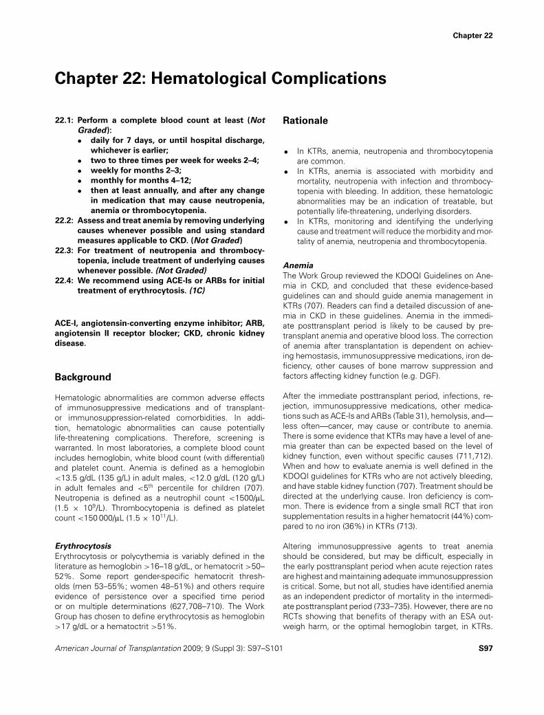
Chapter 22
Chapter 22: Hematological Complications
22.1: Perform a complete blood count at least (Not
Graded):
• daily for 7 days, or until hospital discharge,
whichever is earlier;
• two to three times per week for weeks 2–4;
• weekly for months 2–3;
• monthly for months 4–12;
• then at least annually, and after any change
in medication that may cause neutropenia,
anemia or thrombocytopenia.
22.2: Assess and treat anemia by removing underlying
causes whenever possible and using standard
measures applicable to CKD. (Not Graded)
22.3: For treatment of neutropenia and thrombocy-
topenia, include treatment of underlying causes
whenever possible. (Not Graded)
22.4: We recommend using ACE-Is or ARBs for initial
treatment of erythrocytosis. (1C)
ACE-I, angiotensin-converting enzyme inhibitor; ARB,
angiotensin II receptor blocker; CKD, chronic kidney
disease.
Background
Hematologic abnormalities are common adverse effectsof immunosuppressive medications and of transplant-or immunosuppression-related comorbidities. In addi-tion, hematologic abnormalities can cause potentiallylife-threatening complications. Therefore, screening iswarranted. In most laboratories, a complete blood countincludes hemoglobin, white blood count (with differential)and platelet count. Anemia is defined as a hemoglobin<13.5 g/dL (135 g/L) in adult males, <12.0 g/dL (120 g/L)in adult females and <5th percentile for children (707).Neutropenia is defined as a neutrophil count <1500/lL(1.5 × 109/L). Thrombocytopenia is defined as plateletcount <150 000/lL (1.5 × 10¹¹/L).
Erythrocytosis
Erythrocytosis or polycythemia is variably defined in theliterature as hemoglobin >16–18 g/dL, or hematocrit >50–52%. Some report gender-specific hematocrit thresh-olds (men 53–55%; women 48–51%) and others requireevidence of persistence over a specified time periodor on multiple determinations (627,708–710). The WorkGroup has chosen to define erythrocytosis as hemoglobin>17 g/dL or a hematoctrit >51%.
Rationale
• In KTRs, anemia, neutropenia and thrombocytopeniaare common.
• In KTRs, anemia is associated with morbidity andmortality, neutropenia with infection and thrombocy-topenia with bleeding. In addition, these hematologicabnormalities may be an indication of treatable, butpotentially life-threatening, underlying disorders.
• In KTRs, monitoring and identifying the underlyingcause and treatment will reduce the morbidity and mor-tality of anemia, neutropenia and thrombocytopenia.
Anemia
The Work Group reviewed the KDOQI Guidelines on Ane-mia in CKD, and concluded that these evidence-basedguidelines can and should guide anemia management inKTRs (707). Readers can find a detailed discussion of ane-mia in CKD in these guidelines. Anemia in the immedi-ate posttransplant period is likely to be caused by pre-transplant anemia and operative blood loss. The correctionof anemia after transplantation is dependent on achiev-ing hemostasis, immunosuppressive medications, iron de-ficiency, other causes of bone marrow suppression andfactors affecting kidney function (e.g. DGF).
After the immediate posttransplant period, infections, re-jection, immunosuppressive medications, other medica-tions such as ACE-Is and ARBs (Table 31), hemolysis, and—less often—cancer, may cause or contribute to anemia.There is some evidence that KTRs may have a level of ane-mia greater than can be expected based on the level ofkidney function, even without specific causes (711,712).When and how to evaluate anemia is well defined in theKDOQI guidelines for KTRs who are not actively bleeding,and have stable kidney function (707). Treatment should bedirected at the underlying cause. Iron deficiency is com-mon. There is evidence from a single small RCT that ironsupplementation results in a higher hematocrit (44%) com-pared to no iron (36%) in KTRs (713).
Altering immunosuppressive agents to treat anemiashould be considered, but may be difficult, especially inthe early posttransplant period when acute rejection ratesare highest and maintaining adequate immunosuppressionis critical. Some, but not all, studies have identified anemiaas an independent predictor of mortality in the intermedi-ate posttransplant period (733–735). However, there are noRCTs showing that benefits of therapy with an ESA out-weigh harm, or the optimal hemoglobin target, in KTRs.
American Journal of Transplantation 2009; 9 (Suppl 3): S97–S101 S97

Chapter 22
Table 31: Medications associated with hematologic abnormalities
Medications that cause hematologic abnormalities
Commonly Uncommonly
Anemia Azathioprine (714–717) CNIs (722,723)MPA (718,719) OKT3 (722,723)Sirolimus (50) Trimethoprim–sulfamethoxazoleLeflunomide (720)ACE-I (721)ARB (721)
Neutropenia Azathioprine (714,715) Rituximab (726)MPA (718) ACE-I (727)Sirolimus (50) Ticlopidine/clopidogrel (728)Leflunomide (720) Other antimicrobials (728)Lymphocyte-depleting antibodies (8)Valganciclovir (724)Trimethoprim–sulfamethoxazole (725)
Thrombocytopenia Sirolimus (42) OKT3 (730)MPA (729) Valganciclovir (722,723)Azathioprine (729) Ticlopidine/clopidogrel (731)Lymphocyte-depleting antibodies (8) Heparin (732)
ACE-I, angiotensin-converting enzyme inhibitor; ARB, angiotensin II receptor blocker; CNI, calcineurin inhibitor; MPA, mycophenolic acid;OKT3, muromonab (anti–T-cell antibody).
There are two small RCTs using ESAs in the early post-transplant period, but the overall effects on anemia weresmall (711,736). Another small trial showed that patientsreceiving ESAs before transplant, who attained normalhemoglobin levels, had outcomes that were no differentthan those with low hemoglobin levels (737). There is noevidence to support routine ESA administration in antici-pation of anemia (see Supporting Tables 54–55 at http://www3.interscience.wiley.com/journal/118499698/toc).
The European Best Practices Guidelines for kidney trans-plantation recommend regular screening and careful eval-uation of anemia (721). They also identify immunosuppres-sive agents, ACE-Is and ARBs as causative agents. Theyrecommend following the European Best Practices Guide-lines for anemia management, which recommend that anESA not normally be discontinued in patients undergoingsurgery or who develop an intercurrent illness (738). Norecommendation was made on whether to continue orstop ESAs in the immediate posttransplant period. Patientswith a failing kidney transplant should be followed as anyother patient with failing kidney function.
Neutropenia
Many of the same factors responsible for anemia alsocause neutropenia (Table 31). Although there are no RCTson screening for these abnormalities, the potential con-sequences of not screening are severe. Infection is thesecond most common cause of death, after CVD, in KTRs(739). In the nontransplant population with iatrogenic neu-tropenia (absolute neutrophil count <500/lL [5 × 10⁸/L]),patients are at increased risk for serious infection (740).A possible major contributor to neutropenia in KTRs isthat kidney dysfunction may delay clearance of medica-
tions that can suppress leukocyte production by the bonemarrow.
Medications are a common cause of leukocyte abnormali-ties. There are a number of RCTs that document leucope-nia in the first 1–3 years after transplantation. Unfortu-nately, the definition of leucopenia differs among studies;therefore, direct comparison across trials is problematic.Different classes of immunosuppressive agents have dif-fering effects on leucocytes. CNIs are not generally asso-ciated with leucopenia. In contrast, antiproliferative agentsare an important cause of leucopenia. In early trials, aza-thioprine was associated with leucopenia (714,715). In theEuropean Trial of MMF vs. placebo with CsA and pred-nisone, there was more leucopenia in the group treatedwith 2 g/day MMF (14%, n = 165) vs. placebo (4%,n = 166) (718). In the tricontinental MMF trial, there wasslightly less (significance not stated) leucopenia in the armtreated with 2 g/day MMF (19%, n = 171) vs. the armusing 100–150 mg/day azathioprine (30%, n = 162) (729).In two trials evaluating the safety of EC-MPS vs. MMF,there were no significant differences in leucopenia (42,43).These study protocols included rules to reduce the dose ordiscontinue these agents in the presence of leucopenia,which likely limited the severity and overall incidence ofvery low counts.
In a Cochrane systematic review, mTORi were asso-ciated with more leukopenia (RR 2.02, 95% CI 1.12–3.66, by meta-analysis) than CNIs (50). No mention wasmade of differences in leucopenia in patients treated withsirolimus vs. placebo with CsA and prednisone, or inthe meta-analysis comparing sirolimus to other antipro-liferative agents (50,741). The Symphony trial compared
S98 American Journal of Transplantation 2009; 9 (Suppl 3): S97–S101

Chapter 22
four interventions: standard-dose CsA and MMF (n = 384),low-dose CsA with MMF (n = 408), low-dose tacrolimusand MMF (n = 403) and sirolimus and MMF (n = 380) (30).At the end of 12 months, leucopenia occurred in 10.2%,10.1%, 13.4% and 10.3% of patients, respectively (p >
0.05).
There is no evidence that IL2-RAs cause significant hema-tologic abnormalities. In contrast, lymphocyte-depletingantibodies are associated with more (p < 0.001) leucope-nia (33%, n = 141) compared to the IL2-RA, basiliximab(14.6%, n = 137) (8). More leucopenia was demonstratedin a RCT comparing groups treated with lymphocyte-depleting antibodies with tacrolimus or CsA to one withtacrolimus and no lymphocyte depleting antibodies (7).Addition of steroids also has an impact on leucopenia. Inone trial, leucopenia was seen more often (significancenot stated) in the steroid-free (17.9%) and the steroid-withdrawal (16.5%) arms compared to the standard steroidarm (13.8%) (48).
Other medications commonly used in KTRs to treat co-morbidities are associated with leucopenia. Valganciclovirwas associated with more leucopenia compared to gan-ciclovir (8.2% vs. 3.2%) in a RCT of high-risk solid-organtransplant recipients (724). However, the alternative an-tiviral valacyclovir was not associated with more leucope-nia compared to placebo in a RCT of CMV prophylaxis inKTRs, but drug-induced leucopenia in the treatment armmay have offset the CMV-induced leucopenia in the controlarm (742). Combined therapy with antiviral and antiprolif-erative agents may increase the incidence of leucopenia(743).
The risk of neutropenia from trimethoprim–sulfametho-xazole in KTRs is unclear. There have been several smallRCTs, and they did not report differences in the incidenceof leucopenia (744,745). In a bone marrow transplantationstudy, prophylaxis with trimethoprim–sulfamethoxazole(vs. ciprofloxacin) was associated with a 6-day de-layed recovery of neutropenia (746). Case reports ofagranulocyctosis have been reported with trimethoprim–sulfamethoxazole (725).
Thrombocytopenia
Many of the factors that cause anemia and leucopeniaalso cause thrombocytopenia (Table 31). There are alsorelatively uncommon conditions, such as recurrent or denovo thrombotic microangiopathy, that can cause kid-ney dysfunction, hemolytic anemia and thrombocytope-nia (722,723). Thrombocytopenia is also associated withseveral medications used in KTRs. mTORi are associatedwith much higher RRs of thrombocytopenia comparedto CNIs (RR 7.0, 95% CI 3.0–16.4) (42). Sirolimus alsodemonstrated more thrombocytopenia in comparison toazathioprine and MMF (RR 1.95, 95% CI 1.29–2.97) (50).
Thrombocytopenia was also frequently observed in the tri-continental MMF trial (5% MMF 3 g/day; 9% MMF 2 g/day; 12% azathioprine, significance not stated) (729). Ina (potentially underpowered) study comparing thymoglob-ulin to basiliximab induction, thrombocytopenia (plateletcount <80 000/lL) was not significantly different (10.6%vs. 5.8%, p = 0.19) in the thymoglobulin group vs. the basil-iximab group (8). Thrombocytopenia is also observed inpatients with thrombotic microangiopathy associated withCNIs and, rarely, other medications such as clopidogrel andvalacyclovir (722,723,731).
Other causes of leucopenia and thrombocytopenia in-clude severe sepsis, viral infection (CMV, parvovirusB19) and other medications (716,717,719,720,726–728,730,732,747–753). Idiopathic thrombocytopenia hasrarely been described after transplantation, and can be re-lated to autoimmunity transferred from the donor (754).Transient thrombocytopenia has also been described in re-cipients of allografts whose donors had suffered dissemi-nated intravascular coagulation (755).
Patients with low platelet counts are at increased riskof bleeding. Treatment of thrombocytopenia includes re-moving the offending drugs or treating other underlyingcauses. For example, case series have shown that par-vovirus B19 associated hematologic abnormalities can betreated with intravenous immunoglobulin (751). Plasma-pheresis has also been used to treat HUS/thrombotic mi-croangiopathy that may be associated with thrombocy-topenia (723). There are several case reports documentingthe use of colony-stimulating factors (CSFs) to treat neu-tropenia in kidney transplant patients (756–758). However,there is potential for harm with treatment. One case re-port suggested that CSFs may have been associated withworsening graft function (758). There are clinical practiceguidelines in the cancer literature that can be referred tofor the use of CSFs (616). The review performed by theAmerican Society of Clinical Oncology (616) found there isample evidence that CSFs shorten the duration of neu-tropenia. There are, however, inadequate data to knowwhether or not there is benefit in afebrile neutropenic (ab-solute neutrophil count <1000/lL [1 × 10⁹/L]) patients.There is evidence, though, that patients with febrile neu-tropenia (absolute neutrophil count <500/lL [5 × 10⁸/L])benefit from CSFs along with antibiotics if there is pneu-monia, fungal infection, hypotension, sepsis syndrome ormultisystem organ failure.
The European Best Practice Guidelines on kidney trans-plantation recommend regular screening and careful eval-uation of neutropenia in KTRs (759). The combination ofallopurinol and azathioprine should be avoided to preventneutropenia (616). There are not likely to be any RCTs todetermine when to give CSFs in KTRs. Guidance for theiruse will be derived mostly from local clinical practice andoncology guidelines (708). There are similar guidelines for
American Journal of Transplantation 2009; 9 (Suppl 3): S97–S101 S99

Chapter 22
the treatment of thrombocytopenia with platelet transfu-sion (760).
Erythrocytosis
• Erythrocytosis is a well-known complication of kidneytransplantation.
• In the general population, erythrocytosis is associatedwith morbidity (fatigue, dyspnea, thrombotic events,etc.) and mortality.
• In the general population, there is some evidence thatcorrection is associated with a reduction in thromboticevents.
• In KTRs, adverse consequences of erythrocytosis maybe less common than in the general population.
• In KTRs, treatment of erythrocytosis is effective andsafe with angiotensin blockade.
The incidence of erythrocytosis varies from 8% to 22%among reports identified from earlier clinical practice guide-line publications (627,708–710). More recent studies doc-ument that erythrocytosis still occurs in KTRs (761–765).Many studies do not differentiate between increased redcell mass or reduced plasma volume. Erythrocyctosistends to occur within the first 2 years, but can occur muchlater. It may revert spontaneously in 20% or more of cases(709,710).
The mechanisms of erythrocytosis are unclear and arelikely multifactorial. Sustained increases in erythropoietinhave not been consistently found, but seem to be in-creased to a greater extent than expected for the levelof hematocrit (766). Other proposed mediators of erythro-cytosis include endogenous androgens, renin–angiotensinsystem activation and other growth factors (710). Iden-tified clinical risk factors that have been reported in-clude male gender, polycystic kidney disease, smoking,immunosuppression, reduced kidney function, absenceof rejection, renal artery stenosis, hydronephrosis, hyper-calcemia, longer duration of dialysis, higher pretransplanthemoglobin, angiotensin-converting enzyme genotype, hy-pertension and diabetes mellitus (709,710,761–765,767–778).
The consequences of erythrocytosis can be severe. Evi-dence for the adverse outcomes related to erythrocyto-sis arise mostly from observations in patients with poly-cythemia vera. Historical observations document 20%of polycythemia vera patients present with a thromboticevent, and subsequent thrombosis occurs in as many as50%; however, the associated risk of thrombosis has beendifficult to quantify (779,780). Patients with polycythemiavera have a reduced life expectancy, but this is, in part,related to malignant progression (781). In addition, a largestudy of elderly patients without polycythemia vera under-going noncardiac surgery showed that an elevated hemat-
ocrit was associated with short-term mortality and cardiacmorbidity (782).
In the general population, treatment of erythrocytosis iseffective. In a large observational study of patients in thegeneral population with polycythemia vera and a prior his-tory of thrombosis, pharmacological therapy to reduce redcell volume was associated with a 53% reduction in re-current thrombotic events (783). Many of the recurrencesoccurred in patients with inadequate treatment (hematocrit>45%).
In KTRs, erythrocytosis can be asymptomatic, or patientsmay complain of fatigue, headaches, plethora, dyspneaor blurred vision (709,767,776). The more serious con-sequences include increased risk of venous and arterialthrombosis (767,768,784). One small case control studyfound more thromboembolic events in patients with poly-cythemia (11 events in 53 patients) compared to thosewithout erythrocytosis (0 in 49 matched controls) (767).Most other studies in KTRs either did not report adverseevents, described no concurrent controls, or found no in-crease in adverse events (770,771,774). In a large registryanalysis of KTRs, erythrocytosis was not found to be a riskfactor for stroke (450). Since erythrocytosis is now readilytreatable, and the potential consequences of not treatingare severe (venous and arterial thrombosis), there are notlikely to be any long-term RCTs to compare the effect oftreatment vs. no treatment on outcomes.
There are a number of small RCTs of fair quality and caseseries demonstrating the use of ACE-Is or ARBs to re-duce hematocrit by an absolute value of between 4% and15% (785–797). Given the small sample sizes and the lackof data on critical clinical outcomes, there is only a lowlevel of evidence (see Evidence Profile and accompanyingevidence in Supporting Tables 56–58). In a RCT compar-ing enalapril (2.5 mg/day, n = 15) to placebo (n = 10),the hematocrit dropped by 6.6% in the treatment armcompared to only 1.3% in the control arm (p = 0.004)(788). In another small trial, 15 patients were random-ized to an ACE-I (enalapril) and 12 patients an ARB (losar-tan) (796). Hemoglobin levels decreased significantly inboth groups (174–149 g/L for enalapril and 171–159 g/Lfor losartan); however, the drop was greater (p = 0.05)with enalapril (32.6 g/L decrease) than losartan (17.0 g/Ldecrease). Theophylline has been found to be useful inthe transplant population with dramatic absolute reduc-tions in hematocrit of 8–12% (798,799). However, severaltrials have found that ACE-Is were superior when com-pared directly to theophylline (800–802). In the study byTrivedi et al., the hematocrit fell by 7.6% in the ACE-I arm (fosinipril, n = 9) and did not change significantly(rose by 2.3%) in the theophylline arm (n = 5) (802).Other strategies include phlebotomy and bilateral nephrec-tomy, but these are invasive and the latter can be asso-ciated with significant morbidity (803). Clinicians should
S100 American Journal of Transplantation 2009; 9 (Suppl 3): S97–S101

Chapter 22
also be aware that both ACE-Is and ARBs are associ-ated with small, reversible reductions in kidney function(557).
The European Best Practice Guidelines on kidney trans-plantation recommend that first-line treatment of erythro-cytosis (>52% hematocrit in men and >49% in women) beACE-Is or ARBs (708). The American Society of Transplan-tation states that erythrocytosis (>17–18 g/dL or hemat-ocrit >51–52%) causes potentially life-threatening compli-cations and is readily treatable.
Research Recommendations
• RCTs on the use of ESAs and the optimal hemoglobinin KTRs are needed.
• RCTs on the use of CSFs and target cell counts areneeded.
• Studies are needed to document the incidence andseverity of erythrocytosis with current drug regimens.
• Studies are needed to document the role of ACE-Is andARBs in reducing the incidence of erythrocytosis.
American Journal of Transplantation 2009; 9 (Suppl 3): S97–S101 S101

Chapter 23
Chapter 23: Hyperuricemia and Gout
23.1: We suggest treating hyperuricemia in KTRs when
there are complications, such as gout, tophi, or
uric acid stones. (2D)
23.1.1: We suggest colchicine for treating acute
gout, with appropriate dose reduction for
reduced kidney function and concomi-
tant CNI use. (2D)
23.1.2: We recommend avoiding allopurinol in
patients receiving azathioprine. (1B)
23.1.3: We suggest avoiding NSAIDs and COX-2
inhibitors whenever possible. (2D)
CNI, calcineurin inhibitor; COX-2, cyclo-oxygenase-
2; KTRs, kidney transplant recipients; NSAID, nons-
teroidal anti-inflammatory drug.
Background
Definitions of hyperuricemia differ widely. Local laborato-ries often report the upper normal range as a populationmean plus two standard deviations (gender-specific), andthis performs well in clinical practice (804). An interna-tional task force recommends that a level of >0.36 mmol/L(6.0 mg/dL) be defined as hyperuricemia in the general pop-ulation (804). For each 0.06 mmol/L (1.0 mg/dL) increaseabove 0.06 mmol/L, the adjusted RR of gout increases by2.33 (95% CI 2.00–2.71). The threshold of 0.36 mmol/Lis associated with 67% sensitivity and 78% specificity fordiagnosing gout. A threshold of 0.42 mmol/L (7.0 mg/dL)is associated with a 57% sensitivity and 92% specificity(805). However, because of gender differences, men areless likely to experience gout at level between 0.36 and0.42 mmol/L (6.0 and 7.0 mg/dL) and a higher level (>0.42mmol/L [7.0 mg/dL]) is generally used for men (804). De-tailed information is not available in KTRs, but the WorkGroup chose to define hyperuricemia as >0.36 mmol/L(6.0 mg/dL) in women and >0.42 mmol/L (7.0 mg/dL) inmen.
Rationale
• Hyperuricemia is very common in KTRs.• Hyperuricemia increases the incidence of gout and
other complications in KTRs, and it may be associatedwith loss of kidney function and CVD.
• Important drug interactions and precautions will altertreatment strategies in KTRs with gout.
The incidence of hyperuricemia approaches 80% in KTRs(806,807). A recent analysis of 29 597 US Medicare re-cipients found that the cumulative incidence of gout was7.6% at 3 years after transplantation (808). This relativelyhigh incidence is consistent with a number of smaller re-ports (809–812).
The mechanisms responsible for hyperuricemia and goutare complex. Several studies have shown rates to behigher with CNIs, and especially CsA, when comparedto azathioprine (806,808,809,811). The incidence of hype-ruricemia appears to be similar with CsA and tacrolimusregimens, both being higher compared to regimens with-out CNIs. For example, in a recent large RCT, uric acidlevels were similar between patients treated with low-dose CsA and tacrolimus at the end of 1 year, and signif-icantly higher in comparison to patients on sirolimus andMMF (813). Consistent with these results is a study inwhich 35 patients were converted from CsA to tacrolimushad no change in uric acid levels (814). However, in an-other report of patients converted from CNIs to sirolimus,there was a significant reduction in uric acid levels (815).Similarly, in a small (n = 28) RCT of liver transplant re-cipients, conversion from CNIs to MMF was associatedwith a 15–20% reduction in uric acid levels (816). Otherrisk factors associated with hyperuricemia and gout areprior history, higher BMI, diuretics, older age, more recentyear of transplantation and hypertension (806–809,812,817).
Of the clinical manifestations of hyperuricemia, gout isthe most common. It can be disabling and is associ-ated with lost time from work. Impressive tophaceousdeposits in the hands can occur (806,812). Evidence thathyperuricemia causes or contributes to progressive kid-ney disease or CVD is weak, even in the general popula-tion (804,818,819). Acute kidney injury from very high uricacid levels has been reported (820). A large registry co-hort study recently demonstrated an association of goutwith elevated mortality (adjusted hazard ratio 1.26, 95%CI 1.08–1.47) and graft loss (adjusted hazard ratio 1.22,95% CI 1.01–1.49) (808). This association with mortality,though, has not been observed in other studies. There areno RCTs to show that lowering uric acid levels is asso-ciated with better graft survival, kidney function or patientsurvival. There is one small (n = 54) recent RCT in nontrans-plant patients with kidney impairment, however, in whichimproved function with uric acid reduction failed to reachstatistical significance (821). Case series have not shown aconsistent benefit of uric acid reduction on kidney functionin CKD (822).
S102 American Journal of Transplantation 2009; 9 (Suppl 3): S102–S103

Chapter 23
Monitoring patients for hyperuricemia at the time of otherroutine blood monitoring might help prevent further in-creases in uric acid levels and greater risks for gout. Thereis evidence that dietary interventions (losing weight andreduced meat and alcohol consumption) and avoiding di-uretics in the general population can lower uric acid levels(804). There are no studies in KTRs. Several medicationsused in KTRs can lower uric acid levels. For example, ina randomized crossover trial of 26 KTRs, losartan was as-sociated with an 8% fall in uric acid levels (823). The uricacid lowering effect would not be the sole reason for us-ing these medications, but could be substituted if thesemedications were needed for other indications. Monitor-ing might also give clinicians an increased level of suspi-cion for dealing with atypical symptoms of gout. Measuringuric acid levels is indicated in patients with suspected gout;however, during an acute gouty attack, levels may be nor-mal (804). Treatment of asymptomatic hyperuricemia hasnot been generally recommended in the general populationor KTRs, but it is advocated in those with recurrent symp-tomatic episodes of gout, tophi or radiographic changes ofgout (627,804,824).
Treatment of gout is beyond the scope of these guide-lines. There are evidence-based reviews on the treatmentof hyperurcemia and gout (824). Briefly, oral colchicinesand/or nonsteroidal anti-inflammatory agents are recom-mended as first-line agents for gout (824). Nonsteroidalanti-inflammatory agents and cyclo-oxygenase-2 inhibitorscan be associated with significant reductions in kidneyfunction and acute kidney injury (825–827). Patients withnormal kidney function may use these agents in moder-ate doses for short periods of time, but nonsteroidal anti-inflammatory agents should be avoided in KTRs wheneverpossible (806).
Cochicine levels may be increased in patients with reducedkidney function and in patients treated with CsA (andpresumably tacrolimus). Life-threatening colchicine toxic-ity has been described in patients with reduced kidneyfunction receiving colchicine 1 mg/day for only 5–8 days(828). A disabling myoneuropathy has also been describedin patients with reduced kidney function receiving long-
term colchicine therapy (829,830). Therefore, prolongeduse of colchicine should be avoided in patients with eGFR<60 mL/min/1.73 m2. However, colchicine can be usedat reduced doses for <1 week in patients with eGFR>10 mL/min/1.73 m2 not requiring dialysis. In patientswith eGFR <60 mL/min/1.73 m2, avoid doses higher than0.6 mg/day. Intraarticular or short-term systemic steroidshave also been used if the above therapies are contraindi-cated or not tolerated (824).
Allopurinol is a common uric acid lowering agent (804).However, allopurinol and azathioprine used together canresult in profound, life-threatening pancytopenia (627,753),and thus this combination should be used with extremecaution, or not at all. If used together, azathioprine shouldbe reduced by at least 50% and frequent complete bloodcounts should be used to monitor the interaction (806).Further dose reductions may be needed. Mycophenolatedoes not interact with allopurinol and can be used in placeof azathioprine if an antiproliferative agent is necessary forimmunosuppression (831). Patients allergic to allopurinolmay be given benziodarone (832,833).
The American Society of Transplantation guidelines recom-mended measuring uric acid levels once 2–3 months aftertransplantation, with additional screening in patients withreduced function and on diuretics (627). The Caring for Aus-tralasians with Renal Impairment guidelines for patientswith CKD state that treating hyperuricemia does not re-tard progression and cannot be recommended; patients onprotein-restricted diets treated with allopurinol may requiredose reductions (822). The European Best Practice guide-line on kidney transplantation recommends that the com-bination of allopurinol and azathioprine be avoided (708).
Research Recommendations
• A RCT with adequate statistical power is neededto study the effect of treating asymptomatic hyper-uricemia on preventing loss of kidney function, goutand CVD.
American Journal of Transplantation 2009; 9 (Suppl 3): S102–S103 S103

Chapter 24
Chapter 24: Growth and Development
24.1: We recommend measuring growth and develop-
ment in children (1C):
• at least every 3 months if <3 years old (in-
cluding head circumference) (Not Graded);
• every 6 months in children ≥3 years until final
adult height. (Not Graded)
24.2: We recommend using rhGH 28 IU/m2/week
(or 0.05 mg/kg/day) in children with persistent
growth failure after kidney transplantation. (1B)
24.3: We suggest minimizing or avoiding corticos-
teroid use in children who still have growth po-
tential. (2C)
rhGH, recombinant human growth hormone.
Rationale
• CKD and CKD stage 5 can cause growth failure in chil-dren before kidney transplantation.
• Despite successful kidney transplantation, growth fail-ure can persist.
• Recombinant human growth hormone (rhGH) is safeand effective in children with growth failure after kidneytransplantation.
• Children with growth failure (height <3rd percentile,height target standard deviation score <−2, or heightvelocity <25% for chronological age) grow faster afterkidney transplantation with 28 IU/m2/week of rhGH for1 year compared to no treatment.
• Long-term steroid use has a negative effect on normalgrowth in children.
• Steroid minimization/avoidance protocols may be safeand effective in children.
The three major factors that can influence growth follow-ing successful kidney transplantation are age at transplan-tation (prepubertal vs. pubertal), allograft function and useof corticosteroid therapy. The height increment associatedwith the pubertal growth spurt is suboptimal in patientswith CKD (834) and the lack of normal pubertal growthspurt in KTRs contributes to inadequate final adult height(835). Persistent growth failure, despite successful kidneytransplantation, led to rhGH use being studied to addressconcerns regarding efficacy in the presence of corticos-teroid immunosuppression, increasing risk of acute rejec-tion and the potential for increasing the already raised inci-dence of malignancy in an immunosuppressed population.
Randomized controlled trials have shown that rhGH iseffective in improving the growth of children with CKDduring the first year of administration, with increases inall height indices (836), including children with growthretardation after kidney transplantation. The summaryof RCTs, eight of which included children with kidneytransplants (836), showed that treatment with rhGH (28IU/m2/week) resulted in a significant increase in heightstandard deviation score at 1 year and a significant in-crease in height velocity at 6 months and 1 year. However,there was no further increase in height indices during thesecond year of administration, compared to untreatedcontrols. On average, children treated with rhGH had animprovement in height standard deviation score by 0.8,height velocity by 3.8 cm/year and height velocity standarddeviation score by 6 above nontreated controls (836).Most of the children in the studies after kidney transplantwere on relatively low doses of glucocorticoids, with GFR>20 mL/min/1.73 m2 and all were greater than 1 year aftertransplant with height <3rd percentile, height standarddeviation score <−2 or height velocity <25% for chrono-logical age at the time of starting therapy. Overall, thereis a moderate level of evidence that rhGH is better thanplacebo for increasing growth and that 28 IU/m2/weekis superior to 14 IU/m2/week (see Evidence Profile andaccompanying evidence in Supporting Tables 59–61 athttp://www3.interscience.wiley.com/journal/118499698/toc). Alternatively, a multicenter placebo-controlled trialshowed that a rhGH dose of 0.05 mg/kg/day significantlyincreased height in children with CKD (837).
Cohort studies in children with CKD have demonstratedthat response to rhGH therapy is better in prepubertal thanpubertal children (838), and in CKD stages 3 and 4 com-pared to CKD stage 5 (838). However, in short-term stud-ies, there was no significant difference in the magnitude ofrhGH-related growth with either pubertal status (includingpediatric KTRs (839,840)) or between CKD stages 3 and 4compared to CKD stage 5 (836).
Although no RCTs have been published with final adultheight as an outcome, published data do provide someindirect support that rhGH improves final adult height inchildren with CKD, including KTRs. A longitudinal studyof children with CKD treated with rhGH and followed un-til they achieved final adult height indicated that treatedchildren had sustained catch-up growth where untreatedmatched children had progressive growth failure (838). Im-proved final height in rhGH-treated children has also beenreported from US Transplant Registry data (841). However,it still needs to be determined whether rhGH therapy will
S104 American Journal of Transplantation 2009; 9 (Suppl 3): S104–S105

Chapter 24
result in an increase in final adult height in children whohave received a kidney transplant.
Reported adverse events related to rhGH include asthma,acute rejection, deterioration in kidney function, pa-pilledema, raised fasting glucose and glucose intolerance.However, a meta-analysis found no significant differencebetween treatment and controls in the change in boneage, kidney function, cholesterol, triglycerides, apolipopro-teins and glucose tolerance (836). Additionally, there is noevidence that rhGH acts to advance the pubertal growthspurt.
Persistent growth failure, despite successful kidney trans-plantation, led to rhGH use being studied to address con-cerns regarding efficacy in the presence of corticosteroidimmunosuppression, increasing risk of acute rejection andthe potential for increasing the already raised incidenceof malignancy in an immunosuppressed population (842).None of the four RCTs in pediatric KTRs (839,843–845) re-ported an increase in acute rejection associated with rhGHtherapy or an adverse effect of this treatment on graft func-tion. However, two did determine that prior acute rejectionhistory is a risk factor for the development of acute re-jection following the initiation of rhGH therapy (844,845).The conclusion drawn from these RCTs was that rhGH is awell-tolerated and effective treatment in growth-retardedKTRs. However, no pharmaceutical company that manufac-tures rhGH has applied to the FDA or European agenciesto extend approval for this treatment to the pediatric KTRpopulation.
Concern about a relationship between rhGH use and thedevelopment of renal cell carcinoma in pediatric KTRs re-ceiving growth hormone therapy led researchers to probedatabases maintained by the pharmaceutical companiesthat produce rhGH for evidence of an association (846).Only the International Growth Database collected data onkidney malignancy in KTRs on rhGH. rhGH was not foundto be an independent risk factor for the development ofrenal cell carcinoma (846). Isolated incidents of PTLD havebeen reported in patients receiving rhGH, but a definitiverelationship has not been shown.
When considering rhGH therapy for growth-delayed pe-diatric KTRs, the health-care provider should inform thepatient and family that the benefits to growth need to bebalanced with possible adverse events and the difficulty ofadhering to a daily subcutaneous injection regimen.
Corticosteroids have been used in pediatric KTRs as main-tenance immunosuppressive therapy and as a treatmentfor acute rejection since the 1960s (847,848). A correlationbetween a daily corticosteroid dose in excess of 7 mg/m2
of body surface area and impaired growth in pediatric
KTRs has been reported (849). Over the years, practition-ers have made efforts to reduce steroid use in pediatricKTRs in an effort to avoid the potentially negative impacton growth. In a prospective clinical trial of steroid mini-mization, researchers studied 35 KTRs at 14–27 monthsfollowing transplantation, 17 of whom received alternate-day corticosteroid therapy and 18 of whom received dailycorticosteroid therapy (850). At 1 year, the mean heightstandard deviation score was +0.49 in the alternate-daygroup, compared with −0.12 in the daily-dose group. Ananalysis of the North American Pediatric Renal TransplantCooperative Study database also found that short-term im-provement in height standard deviation score was asso-ciated with alternate-day dosing of corticosteroids (851).No decline in graft function was observed in patients re-ceiving daily vs. alternate-day steroid therapy. However,it is important that, when considering alternate-day dos-ing as a strategy for steroid minimization, the health-careprovider address the potential for increased incidence ofnonadherence due to the potential difficulty of this dosingregimen.
In 2001, a pilot study reported the initial positive resultsof steroid avoidance using anti-IL2 receptor antibody forinduction and every 2 weeks for the first 5 months aftertransplant, in addition to tacrolimus and MMF as mainte-nance immunosuppression therapy (852). A follow-up re-port in 2003 indicated a significant improvement in themean height standard deviation score at 1 year in thecorticosteroid-avoidance group when compared with ahistorical control group who had received corticosteroidtreatment daily (853). This led to a prospective multicen-ter RCT of steroid avoidance where 130 unsensitized pri-mary KTRs 0–21 years of age were randomized to steroid-free vs. steroid-based immunosuppression (2004–2006)with a 3-year follow-up (854). Patients in both arms re-ceived tacrolimus and MMF immunosuppression. Prelimi-nary analysis does not reveal an overall significant growthadvantage at 1 year in children receiving steroid-free orsteroid-based immunosuppression. Longer-term follow-upof current and future RCTs will be important in determin-ing the effect of steroid-free or minimization protocols ongrowth and graft function in pediatric KTRs.
Research Recommendations
• A RCT is needed to determine whether higher dosesof rhGH during puberty improve pubertal growth in chil-dren with persistent growth failure after kidney trans-plantation.
• Further follow-up of ongoing and future studies isneeded to evaluate the effect of steroid minimizationor avoidance on growth.
American Journal of Transplantation 2009; 9 (Suppl 3): S104–S105 S105

Chapter 25
Chapter 25: Sexual Function and Fertility
25.1: SEXUAL FUNCTION
25.1.1: Evaluate adults for sexual dysfunction af-
ter kidney transplantation. (Not Graded)
25.1.2: Include discussion of sexual activity and
counseling about contraception and safe
sex practices in follow-up of adult KTRs.
(Not Graded)
KTRs, kidney transplant recipients.
Rationale
• Sexual dysfunction is common in men and womenKTRs.
• Many patients will not spontaneously report sexualdysfunction.
• Modification of medications may alleviate sexual dys-function.
• Therapies are available, although less are available forwomen than men.
• Sexual dysfunction negatively affects quality of life.• Contraception can help prevent unwanted pregnan-
cies.• Safe sex practices can help prevent the acquisition of
disease.
Sexual dysfunction is frequent in patients with all stagesof CKD, particularly among patients with CKD stage 5 orafter transplantation (855). Sexual dysfunction in KTRs mayhave both organic and psychological causes (856–858). Thescope of the problem includes erectile dysfunction, de-creased libido and lower frequency of intercourse.
Following kidney transplantation, the metabolic milieu im-proves, and for some patients sexual function improvesas well (859). For others, sexual function does not changeand may even worsen (860). One study compared sexualfunction in men and women on hemodialysis, peritonealdialysis, with patients with rheumatoid arthritis or aftertransplantation and found that men and women on dialysishad statistically increased incidence of ‘hypoactive sexualdesire’ compared with those after transplantation. Men onhemodialysis had a significantly higher incidence of ‘sex-ual aversion disorder’ as well as ‘inhibited male orgasm.’In this study, the ‘male erectile disorder’ did not differ be-tween the dialysis and transplant groups. Overall, the studyconcluded that sexual dysfunction in dialysis patients was
a consequence of lost sexual interest attributable to fatigue(858).
Problems with sexual function are common following kid-ney transplantation, but the reported prevalence varies.Problems with sexual function in general have been re-ported in the range of 45–50% (855,861). One surveyfound that more than 30% reported a problem to be mod-erate or severe in magnitude (861). There are few studiesfocused on this issue for women (861). For men, erec-tile dysfunction can affect quality of life and be associatedwith anxiety, depression and loss of self-esteem (862). Fol-lowing transplantation, erectile dysfunction may improve,especially for younger men (862,863). However, for oth-ers the problem may not change or may even get worse(862–864). An Egyptian study of 400 male KTRs reportederectile dysfunction in 36% (865). In a study of simultane-ous pancreas–kidney transplant recipients, 79% sufferedfrom some degree of erectile dysfunction (866). Transplantsurgery may contribute to erectile dysfunction. Diversionof blood from the penile arteries when the internal iliac ar-teries are used for transplant anastomosis may play a role(863).
Therapy with 5-phosphodiesterase inhibitors may be effec-tive. These agents are helpful for some, but not all patients(864,867). A double-blind crossover RCT of KTRs found thatsildenafil was more effective than placebo with respect toerectile function, orgasmic function, intercourse satisfac-tion and overall satisfaction (868). There was no significantdifference with respect to sexual desire in this study (868).Modification of medications may also be useful for patientswith erectile dysfunction and/or decreased libido.
Whether to evaluate men with sexual dysfunction, or initi-ate a trial with a 5-phosphodiesterase inhibitor, is often un-clear. When 5-phosphodiesterase inhibitors are prescribed,care must be taken that the patient is hemodynamicallystable and that he avoids alpha-adrenergic antagonists.How to counsel and approach therapy in women with sex-ual dysfunction is less clear.
Follow-up of KTRs should include discussion of sexual ac-tivity, and counseling about contraception and safe sexpractices, as is true for patients in the general popula-tion (and therefore beyond the scope of this guideline).Sexually active patients who are not in long-term monoga-mous relationships should use latex condoms during sex-ual contact to reduce their risk for exposure to CMV,HSV, HIV, HPV, HBV, HCV and other sexually transmit-ted infections. Sexually active KTRs should avoid sexual
S106 American Journal of Transplantation 2009; 9 (Suppl 3): S106–S109

Chapter 25
practices that could result in oral exposure to feces or gen-ital secretions.
Recommendations for contraception should be made onan individual basis with consideration given to what is mosteffective, as well as what can actually be used. Concernsregarding intrauterine devices have included the potentialfor infection, as well as that they may be less effectivein transplant recipients (869). Whether current intrauterinedevices may be more effective and less risky in this patientpopulation is unknown.
Research Recommendations
• Studies are needed to determine the etiology, diagno-sis and treatment of sexual dysfunction in KTRs.
25.2: FEMALE FERTILITY
25.2.1: We suggest waiting for at least 1 year
after transplantation before becoming
pregnant, and only attempting preg-
nancy when kidney function is stable
with <1 g/day proteinuria. (2C)
25.2.2: We recommend that MMF and EC-MPS
be discontinued or replaced with aza-
thioprine before pregnancy is attempted.
(1A)
25.2.3: We suggest that mTORi be discontin-
ued or replaced before pregnancy is at-
tempted. (2D)
25.2.4: Counsel female KTRs with child-bearing
potential and their partners about fertil-
ity and pregnancy as soon as possible
after transplantation. (Not Graded)
25.2.5: Counsel pregnant KTRs and their part-
ners about the risks and benefits of
breastfeeding. (Not Graded)
25.2.6: Refer pregnant patients to an obstetri-
cian with expertise in managing high-risk
pregnancies. (Not Graded)
EC-MPS, enteric-coated mycophenolate sodium; KTRs,
kidney transplant recipients; MMF, mycophenolate
mofetil; mTORi, mammalian target of rapamycin in-
hibitor(s).
Background
Female KTRs of child-bearing potential are those who arenot peri- or postmenopausal, and those who have a uterusand at least one ovary. There are no prospective studieson the risks of immunosuppressive medications in preg-nancies. Evidence that a drug is not safe in pregnancy may
come from case reports, or animal studies demonstrat-ing toxicity at doses comparable to those which might beused in humans (normalized to body surface area). In theabsence of data, a drug should be presumed to be unsafe,and patients should be treated accordingly.
Rationale
• Fertility is increased in KTRs compared to CKD stage5 before transplantation.
• Pregnancy and childbirth in KTRs have a high incidenceof complications to mother and child.
• Complications of pregnancy and childbirth can be min-imized by the use of lower-risk immunosuppressiveagents and multidisciplinary care that includes an ob-stetrician with expertise in managing high-risk preg-nancies.
Pregnancies in patients with CKD stage 5 are uncommon(870). However, fertility is improved and often restored af-ter successful kidney transplantation (871,872). The risksof pregnancy and childbirth to both mother and child arehigher for KTRs, compared to the general population, butin stable KTRs pregnancies most often have a good out-come. In KTRs with good kidney function, no proteinuria,and well-controlled blood pressure, there is little risk ofgraft loss (873–876). However, KTRs with reduced kidneyfunction are at higher risk for allograft dysfunction andgraft failure (877). There are few published data in KTRson which to base a safe recommended GFR. Data in thenontransplant population indicate that women with GFR<40 mL/min/1.73 m2 and proteinuria >1 g/day are at in-creased risk for a significantly accelerated GFR decrease,as well as low-birth-weight babies (878). These data wereused for the recommendations noted above. It is unclearwhether or not the same levels apply to KTRs. Cyclosporinelevels decline during pregnancy (877). Nevertheless, the in-cidence of acute rejection during pregnancy appears to berelatively low (877).
An American Society of Transplantation consensus confer-ence recommended that patients wait for 1 year withoutacute rejection before pregnancy with the proviso that in-dividual circumstances may modify the appropriate timeframe to a shorter or longer time period. Each situationneeds to be evaluated on a case-by-case basis (879). It wasrecently reported that pregnancy within the first 2 yearsfollowing transplantation may increase the risk of graftloss (880). On the other hand, there have been success-ful pregnancies before the end of the first posttransplantyear (872). Some reports suggest that there is a high inci-dence of hypertension (873) and preeclampsia in pregnantKTRs (881). Deliveries are more likely to be by caesareansection, for medical indications. The transplant kidney isneither affected by, nor does it affect, a vaginal delivery. In
American Journal of Transplantation 2009; 9 (Suppl 3): S106–S109 S107

Chapter 25
the absence of medical indications, vaginal deliveries arepossible (873).
There is also a higher risk to the fetus for pregnan-cies in KTRs. There is a higher risk of preterm delivery(<37 weeks) and low birth weight (<2500 g) (873,877,882).The fetus, of course, is exposed to potentially teratogenicimmunosuppressive agents (882). There are no RCTs indi-cating which, if any, immunosuppressive agents are safeto use in pregnancy.
Mycophenolate has been reported to cause severe struc-tural malformations. A characteristic phenotype associ-ated with in utero exposure to MMF is emerging thatincludes cleft lip and palate, microtia, and absence ofexternal auditory canals (883–885). Thus, MMF shouldgenerally be changed to azathioprine during pregnancy, apractice endorsed by the European Best Practice Guide-lines (886). These guidelines suggest a 6-week windowafter discontinuing MMF and starting azathioprine, be-fore pregnancy is attempted (886). These same concernsshould also apply to EC-MPS. Azathioprine is rated by theFDA as category ‘D’ (i.e. there is evidence of human fe-tal risk, but the benefits from use in pregnant womenmay be acceptable despite the risk). Despite the FDAcategory D, azathioprine has been used safely over theyears in pregnant transplant recipients. It is consideredan acceptable immunosuppressant to use in this clinicalsetting.
In a meta-analysis of the use of CsA during pregnancy, theincidence of major fetal malformations was 4.1% (2.6–7.0%) (877). This was numerically higher than, but notstatistically significantly different from, the rate with non-CNIs. Prednisone at doses low enough to prevent thymicaplasia (usually less than 15 mg/day) is safe in pregnantKTRs. High levels of azathioprine and prednisone can beassociated with problems that do not occur when they areused at standard doses.
There are few reports of the use of mTORi and preg-nancy. The FDA categorizes sirolimus as ‘C.’ CategoryC indicates that either studies in animals have revealedadverse effects on the fetus (teratogenic or embryoci-dal effects or other) and there are no controlled stud-ies in women, or studies in women and animals are notavailable (887). The FDA-approved package labeling forsirolimus notes that sirolimus was embryotoxic or feto-toxic in rats at doses 0.2–0.5 of the clinical doses ad-justed for body surface area (887). A voluntary registryreported only seven cases of pregnancy in organ trans-plant recipients receiving sirolimus (885). None were asso-ciated with adverse outcomes, although in most the drugwas discontinued when pregnancy was discovered. Thereare case reports of normal-term pregnancies in womenreceiving sirolimus (888,889). However, in the absenceof adequate safety data, it is prudent to avoid mTORi inpregnancy.
An American Society of Transplantation consensus con-ference concluded that breastfeeding for KTRs is notcontraindicated (879). For KTRs who opt to breastfeed,prednisone is likely to be safe (890). Prednisone and aza-thioprine are detectible in breast milk (891), but there areno data for MMF or sirolimus. CsA is excreted into breastmilk and is not recommended in breastfeeding mothers(877).
Research Recommendations
• Observational studies are needed to determine the in-cidence and complications of pregnancies in KTRs.
25.3: MALE FERTILITY
25.3.1: We suggest that male KTRs and their
partners be advised that:
• male fertility may improve after kid-
ney transplantation (2D);
• pregnancies fathered by KTRs appear
to have no more complications than
those in the general population. (2D)
25.3.2: We recommend that adult male KTRs be
informed of the possible risks of infertil-
ity from mTORi. (1C)
25.3.2.1: We suggest that adult male
KTRs who wish to maintain fer-
tility should consider avoiding
mTORi, or banking sperm prior
to mTORi use. (2C)
KTRs, kidney transplant recipients; mTORi, mam-
malian target of rapamycin inhibitor(s).
Rationale
• Male fertility improves in most KTRs, and may becomenormal.
• Outcomes of pregnancies fathered by KTRs are similarto those of the general population.
• Rapamycin is associated with low sperm counts. Theabnormality is reversible with discontinuation of ra-pamycin.
Chronic kidney disease is associated with impaired sper-matogenesis, decreased testosterone production, de-creased libido and increased gonadotropins (892). Uremichypogonadism is reversible in individuals with successfullong-term kidney transplantation (893). Studies from theazathioprine as well as CsA eras show that testosteronelevels rise after transplantation. Gonadotropins may de-crease, but may not normalize, and semen analysis in mostKTRs is normal (893–898). Longer time on dialysis prior to
S108 American Journal of Transplantation 2009; 9 (Suppl 3): S106–S109

Chapter 25
transplantation and kidney dysfunction may be risk factorsfor those with residual testicular dysfunction (896,899).Testicular biopsies performed after transplantation showsignificant improvement, with some residual reduction insertoli cells and spermatogonia (900).
Although it is unclear whether CsA plays a role in maleinfertility, it is clear that rapamycin can lead to male in-fertility (893,899). It causes low sperm counts (901–904)by interrupting the stem cell factor/c-kit system that reg-ulates germ cell proliferation, meiosis and apoptosis, con-sequently inhibiting spermatogenesis (905). The effects ofrapamycin appear to be reversible (901–903).
With regards to potential congenital abnormalities, out-comes of pregnancies fathered by male KTRs do not differ
from those of the general population (906). These con-clusions are based on data from the National TransplantPregnancy Registry. This registry is voluntary and thus po-tentially subject to reporting bias. Nevertheless, the datacaptured by this registry are crucial but limited. Fewer preg-nancy outcomes are reported to the registry for men thanwomen.
Research Recommendations
• Observational studies are needed to determine the in-cidence and complications of pregnancies fathered byKTRs.
American Journal of Transplantation 2009; 9 (Suppl 3): S106–S109 S109

Chapter 26
Chapter 26: Lifestyle
26: We recommend that patients are strongly encour-
aged to follow a healthy lifestyle, with exercise,
proper diet, and weight reduction as needed. (1C)
(See also Obesity, Recommendation 16.4.1.)
Rationale
• There are abundant data from the general populationthat a lifestyle that includes exercise, a proper diet andavoidance of obesity improves longevity and quality oflife.
• Although there is only one small RCT in KTRs, there isno reason to believe that exercise is not as beneficialin KTRs as in the general population.
• There is no reason not to believe that a proper diet canhelp prevent CVD and other complications in KTRs asin the general population.
• There is little harm associated with exercise, a properdiet and weight reduction; therefore, any benefit islikely to outweigh harm.
Data from RCTs in the general population suggest thatexercise, proper diet and weight reduction (in obese pa-tients) improve longevity, quality of life and other majorhealth outcomes. In a RCT involving 100 KTRs, the grouprandomly allocated to receive regular telephone counsel-ing on exercise had a greater exercise tolerance at 1year after transplantation compared to the control groupthat did not receive counseling (603). This study did nothave adequate statistical power to examine major CVDoutcomes.
S110 American Journal of Transplantation 2009; 9 (Suppl 3): S110–S110

Chapter 27
Chapter 27: Mental Health
27: Include direct questioning about depression and
anxiety as part of routine follow-up care after kid-
ney transplantation. (Not Graded)
Rationale
• Depression and anxiety are more common in KTRsthan in the general population.
• Depression and anxiety may be associated with med-ication nonadherence, sleep disorders and other ad-verse effects that make the diagnosis and treatmentof depression and anxiety important.
• Therapies are available for the treatment of depressionand anxiety.
Anxiety and depression are common in dialysis patients,much more so than in the general population (907–913). Formany patients with kidney failure, kidney transplantation isa better kidney replacement therapy than dialysis. How-ever, kidney transplantation does not change underlyingsystemic disorders or reverse previously sustained phys-ical damage. When the kidney graft is not working well,new medical problems may arise, and when the graft fails,the patient generally faces a return to an unwelcome formof therapy, thereby increasing stress. Whether because ofnew or preexisting medical conditions, medications suchas corticosteroids, or work status changes, KTRs are at riskfor anxiety and depression.
There have been few studies of mental-health disordersin KTRs. Many of the studies focus on quality of life,which encompasses psychological domains but does notnecessarily examine anxiety and depression, or othermental-health disorders (914). A meta-analysis comparingemotional distress and psychological well-being among dif-ferent forms of renal replacement therapies revealed lessemotional distress and greater well-being with success-ful kidney transplantation than with other CKD stage 5treatment modalities (915). However, case mix differencesamong the groups likely influenced the results, making itunclear whether it was the difference in patients or treat-ment modalities that accounted for the differences in out-come (915).
Studies of depression and anxiety in KTRs report conflict-ing results. Some have reported similarly high rates of de-pression in KTRs compared to dialysis patients (916,917),while others have reported less depression (918,919).Some have reported less anxiety in KTRs (920) and, in oth-
ers, no difference compared to dialysis patients (917,919).A study examining 5- to 22-year-old KTRs found that 36%had emotional trauma and/or depression (921). Return todialysis after graft loss has been associated with severedepression (918).
Anxiety and depression in KTRs have been associated witha poor quality of life, poor marital relations, sexual func-tion and sleep quality (922). In one study, a high level ofposttransplantation anxiety was associated with reducedfunctioning socially, having physical complaints, and moreeconomic problems. High levels of anxiety were also as-sociated with depression (923). Depression has also beenassociated with medication nonadherence (283,924).
Hospitalization for psychosis is not increased in KTRs com-pared with patients on chronic dialysis (925). Hospitaliza-tion for psychosis has been associated with increased riskfor death, as well as graft loss (925). Depression identifiedin Medicare claims has been associated with an increasedrisk of graft failure, return to dialysis and death with a func-tioning graft (926).
Psychotherapy may be helpful (927). One RCT comparingindividual and group psychotherapy in KTRs found that bothapproaches resulted in lower Beck Depression Inventoryscores. Individual therapy was associated with a better out-come in this study (928). As most transplant centers havesocial workers, these individuals may be a useful resourcefor counseling. Antidepressants are often used. Given thatsome of these drugs are metabolized by the CYP3A4 en-zyme pathway, levels of immunosuppressive medicationsalso metabolized through this pathway may need to beadjusted (929).
How best to assess the mental health of transplant recip-ient is unclear. The screening tools used by psychologistsare time-consuming and not familiar to general practition-ers. Simple strategies, such as direct questioning on re-view of systems, or brief screening tools (930), may be asimple and useful initial screening approach. Further stud-ies are needed to better understand how to monitor formental-health disorders in KTRs.
Research Recommendations
• Studies are needed to determine the optimal approachto screening and intervention for depression and othermental disorders in KTRs.
American Journal of Transplantation 2009; 9 (Suppl 3): S111–S111 S111

Appendix
Appendix: Methods for Guideline Development
Aim
The overall aim of the project was to create a clinicalpractice guideline with recommendations for the care ofthe KTRs using an evidence-based approach. After topicsand relevant clinical questions were identified, the perti-nent scientific literature on those topics was systematicallysearched and summarized.
Overview of process
The development of the guideline included sequential andconcurrent steps:
• Appoint the Work Group and Evidence Review Team(ERT), which were responsible for different aspects ofthe process.
• Confer to discuss process, methods and results.• Develop and refine topics.• Define specific populations, interventions or predictors
and outcomes of interest.• Create and standardize quality assessment methods.• Create data-extraction forms.• Develop literature search strategies and run searches.• Screen abstracts and retrieve full articles based on pre-
determined eligibility criteria.• Extract data and perform critical appraisal of the litera-
ture.• Grade quality of the outcomes of each study.• Tabulate data from articles into summary tables.• Grade the quality of evidence for each outcome and
assess the overall quality and findings of bodies ofevidence with the aid of evidence profiles.
• Write recommendations and supporting rationalestatements.
• Grade the strength of the recommendations based onthe quality and strength of the evidence and other con-siderations.
• Peer review by KDIGO Board of Directors in December2008 and the public (March 2009), with subsequentrevisions.
The Work Group, KDIGO Co-Chairs, ERT, liaisons andKDIGO support staff met for four 2-day meetings for train-ing in the guideline development process, topic discussionand consensus development.
Creation of groups
The KDIGO Co-Chairs appointed the Co-Chairs of the WorkGroup, who then assembled the Work Group to be re-sponsible for the development of the guideline. The WorkGroup consisted of domain experts, including individuals
with expertise in adult and pediatric nephrology, transplantsurgery and medicine, critical-care medicine, cardiology, in-fectious diseases, oncology and epidemiology, along witha patient advocate. Tufts Center for Kidney Disease Guide-line Development and Implementation at Tufts MedicalCenter in Boston, MA, USA, was contracted to provideexpertise in guideline development methodology and sys-tematic evidence review. The ERT consisted of physician–methodologists with expertise in nephrology and internalmedicine, and research associates and assistants. The ERTinstructed and advised Work Group members in all stepsof literature review, critical literature appraisal and guide-line development. The Work Group and the ERT collab-orated closely throughout the project. The ERT also in-cluded methodological input and assistance with literaturesearches from methodology experts at the Cochrane RenalGroup in Sydney, Australia.
Systematic Review: General Process
The first task of the Work Group was to define the overalltopics and goals for the guideline. The Work Group Co-Chairs drafted a preliminary list of topics. The Work Groupidentified the key clinical questions. The Work Group andERT further developed and refined each topic, specifiedscreening criteria, literature search strategies and data-extraction forms.
The ERT performed literature searches, and organizedscreening of abstracts and articles. The ERT also coordi-nated the methodological and analytic processes of thereport, and defined and standardized the methodology ofperforming literature searches, data extraction and sum-marizing the evidence. Throughout the project, the ERToffered suggestions for guideline development, led dis-cussions on systematic review, literature searches, dataextraction, assessment of quality and applicability of arti-cles, evidence synthesis, grading of evidence and recom-mendations and consensus development. With input fromthe Work Group, the ERT finalized eligible studies, per-formed all data extraction and summarized data into sum-mary tables. They also created preliminary evidence pro-files (described below), which were completed by the WorkGroup members. The Work Group members reviewed allincluded articles, data-extraction forms and summary ta-bles for accuracy and completeness. The Work Group tookthe primary role of writing the recommendations and ra-tionale statements, and retained final responsibility for thecontent of the recommendation statements and the ac-companying narrative.
S112 American Journal of Transplantation 2009; 9 (Suppl 3): S112–S124

Appendix
For questions of treatments in the KTRs, systematic re-views of the eligible RCTs were undertaken (Table 32).For these topics, the ERT created detailed data-extractionforms, and extracted information on baseline data for thepopulations, interventions, study design, results and pro-vided an assessment of quality of evidence. The ERT thentabulated studies in summary tables, and assigned gradesfor the quality of the evidence in consultation with theWork Group.
For nontreatment questions, that is questions related toprevalence, evaluation and risk relationships, the ERT con-ducted systematic searches, screened the yield for rele-vance and provided lists of citations to the Work Group.The ERT created summary tables of selected observa-tional incidence and predictor studies. The Work Grouptook primary responsibility for reviewing and summarizingthis literature in a narrative format. The ERT also searchedonline databases for estimates of incidence rates of differ-ent cancers among larger countries representative of differ-ent regions. The primary database used was Cancer Mon-dial (http://www-dep.iarc.fr). SIRs for cancer in solid-organtransplant recipients were taken from a meta-analysis byGrulich et al. (623).
For topics on which previous or ongoing KDIGO or KDOQIguidelines have provided recommendations for KTRs, newsystematic reviews were not performed. These includeanemia, hepatitis C, mineral and bone disorders and pe-diatric nutrition. For these topics, the relevant recommen-dations and rationale text were excerpted and refined asnecessary. The Work Group Chairs and selected mem-bers conferred with Co-Chairs of the concurrent KDIGOmineral and bone disorder guideline and KDOQI pediatricnutrition guideline on transplant bone disease (Chapter 21),and growth and development (Chapter 24).
Refinement of Topics
The Work Group Co-Chairs prepared the first draft of thescope of work document as a series of mock (prelimi-nary) recommendations to be considered by Work Groupmembers. At their first 2-day meeting, members addedfurther mock guideline topics until the initial working doc-ument included all topics of interest to the Work Group.The inclusive, combined set of questions formed the ba-sis for the deliberation and discussion that followed. TheWork Group strove to ensure that all topics deemed clini-cally relevant and worthy of review were identified and ad-dressed. The four major topic areas of interest for the careof KTRs included immunosuppression, infections, CVDand cancer. In addition, there were several miscellaneoustopics.
At the initiation of the guideline development process, itwas agreed that this guideline would focus on patients whohave had kidney transplantations. Thus, with few excep-
tions (e.g. the timing of vaccinations), all topics, systematicreviews and study eligibility criteria were restricted to pa-tients with existing kidney transplantations. The guidelinedoes not address management issues regarding choos-ing patients for kidney transplantation, pretransplant care,intraoperative care (except for the timing of initiating im-munosuppression) or management of patients who havelost their grafts. In addition, in regards to care of comor-bidities and complications after kidney transplantation (e.g.infections, cancer and CVD), this guideline focuses primar-ily on monitoring and prevention of the conditions, as op-posed to treatment of the conditions (with some excep-tions, e.g. for infectious diseases). However, where therecommended treatment of conditions differed from thegeneral population (e.g. due to drug interactions with im-munosuppression agents), standard treatment recommen-dations are offered.
Based on the list of topics, the Work Group and ERT devel-oped a list of specific research questions for which system-atic review would be performed (Table 32). For each sys-tematic review topic, the Work Group Co-Chairs and theERT formulated well-defined systematic review researchquestions using a well-established system (931). For eachquestion, explicit criteria were agreed on for the popula-tion, intervention or predictor, comparator, outcomes ofinterest and study design features. A list of outcomes ofinterest was generated. The Work Group ranked patient-centered clinical outcomes (such as death, graft loss orinfections) as more important than intermediate outcomes(such as cholesterol level or hypertension). The outcomeswere further categorized as being of critical, high or mod-erate importance to KTRs. Outcomes of low importancewere not considered for the purpose of systematic re-view and evidence synthesis. The specific criteria usedfor each topic are described below in the description ofthe review topics. In general, eligibility criteria were de-termined based on clinical value, relevance to the guide-line and clinical practice, determination whether a set ofstudies would affect recommendations or the strength ofevidence and practical issues such as available time andresources.
Literature Searches and Article Selection
The MEDLINE, Cochrane Central Registry for trials, andCochrane database of systematic reviews were searchedfrom 1985 through January 2007 by the ERT to captureall citations relevant to the topic of kidney transplanta-tion, including original articles, systematic reviews andprevious guidelines. The Cochrane Renal Group ran par-allel searches in their Renal Registry database and thesesupplemented the primary ERT searches. The search wasupdated through February 2008 and supplemented by arti-cles identified by Work Group members through November2008.
American Journal of Transplantation 2009; 9 (Suppl 3): S112–S124 S113

Appendix
Table 32: Systematic review topics and screening criteria
Chapter 1: Induction Therapy
Population KTRs in the first 24 h after transplantPredictor, reference standard IL2 (mab) vs. no induction, antithymoglobulin vs. no induction, antithymoglobulin vs. IL2Outcomes All-cause mortality, DGF, slow graft function, acute rejection, graft failure/survival, kidney function,
CAN, infection, cancer, NODAT, fracture, BMD, erythrocytosis, neutropenia, quality of life,adverse events
Study design RCTMinimum number of subjects N ≥ 50
Chapter 2: Initial Maintenance Immunosuppressive Medications
Population KTRsIntervention, reference standard Tac vs. CsA (CsA or CsA-ME) (with AZA, MMF, Sirolimus, Everolimus), CNI vs. non-CNI regimens,
MMF vs. AZA, MMF formulation vs. other MMF formulation, CNI-sparing (withdrawal), CNI-free,steroid withdrawal, steroid avoidance
Outcomes All-cause mortality, DGF, slow graft function, acute rejection, graft failure/survival, kidney function,CAN, infection, cancer, NODAT, erythrocytosis, neutropenia, fracture, BMD, hypertension,hyperuricemia, hyperlipidemia, quality of life, adverse events
Study design RCTMinimum number of subjects N ≥ 100
Chapter 3: Long-Term Maintenance Immunosuppressive Medications
Population KTRsIntervention Tac vs. CsA (CsA or CsA-ME) (with AZA, MMF, sirolimus, everolimus), CNI vs. non-CNI regimens,
MMF vs. AZA, MMF formulation vs. other MMF formulation, CNI-sparing (withdrawal), CNI-free,steroid withdrawal, steroid avoidance
Outcomes All-cause mortality, DGF, slow graft function, acute rejection, graft failure/survival, kidney function,CAN, infection, cancer, NODAT, erythrocytosis, neutropenia, fracture, BMD, hypertension,hyperuricemia, hyperlipidemia, quality of life, adverse events
Study design RCTMinimum number of subjects N ≥ 100
Chapter 4: Strategies to Reduce Drug Costs
Population KTRsIntervention CsA-ME generics, other generic medicationsOutcomes Pharmacokinetics, pharmacodynamicsStudy design RCTMinimum number of subjects N ≥ 20
Chapter 5: Monitoring Immunosuppressive Medications
Population KTRsIntervention MMF fixed dose vs. AUC-adjusted doses, C0 vs. C2 CsA to determine dosing, anti-HLA antibodiesOutcomes All-cause mortality, DGF, slow graft function, acute rejection, graft failure/survival, kidney function,
CAN, infection, cancer, NODAT, erythrocytosis, neutropenia, fracture, BMD, hypertension,hyperuricemia, hyperlipidemia, quality of life, adverse events
Study design RCTMinimum number of subjects N ≥ 10
Chapter 6: Treatment of Acute Rejection
Population KTRs with biopsy-proven acute rejectionPredictor Adding induction agents or other (intravenous immunoglobulin, plasma exchange), change of
maintenance regimenOutcomes All-cause mortality, DGF, slow graft function, acute rejection, graft failure/survival, kidney function,
CAN, infection, cancer, NODAT, fracture, BMD, erythrocytosis, neutropenia, quality of life,adverse events
Study design RCTMinimum number of subjects N ≥ 100
Chapter 7: Treatment of Chronic Allograft Injury
Population KTRs with CAN or biopsy-proven CNI toxicityIntervention, predictor Reduction in CNI, change in maintenance immunosuppression, adding ancillary treatments (ACE-I,
ARB, etc.), CNI dose reduction, CNI withdrawal, replacement of CNI with anotherimmunosuppression agent, comparisons with placebo or other treatments
Outcomes All-cause mortality, DGF, slow graft function, acute rejection, graft failure/survival, kidney function,CAN, biopsy changes, infection, cancer, NODAT, erythrocytosis, neutropenia, fracture, BMD,hypertension, hyperuricemia, hyperlipidemia, quality of life, adverse events
Continued.
S114 American Journal of Transplantation 2009; 9 (Suppl 3): S112–S124
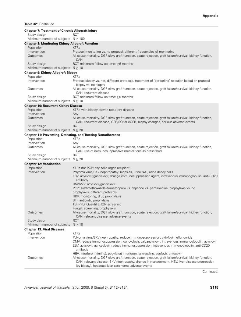
Appendix
Table 32: Continued
Chapter 7: Treatment of Chronic Allograft Injury
Study design RCTMinimum number of subjects N ≥ 100
Chapter 8: Monitoring Kidney Allograft Function
Population KTRsIntervention Protocol monitoring vs. no protocol, different frequencies of monitoringOutcomes All-cause mortality, DGF, slow graft function, acute rejection, graft failure/survival, kidney function,
CANStudy design RCT; minimum follow-up time: ≥6 monthsMinimum number of subjects N ≥ 10
Chapter 9: Kidney Allograft Biopsy
Population KTRsIntervention Protocol biopsy vs. not, different protocols, treatment of ‘borderline’ rejection based on protocol
biopsy vs. no biopsyOutcomes All-cause mortality, DGF, slow graft function, acute rejection, graft failure/survival, kidney function,
CAN, recurrent diseaseStudy design RCT; minimum follow-up time: ≥6 monthsMinimum number of subjects N ≥ 10
Chapter 10: Recurrent Kidney Disease
Population KTRs with biopsy-proven recurrent diseaseIntervention AnyOutcomes All-cause mortality, DGF, slow graft function, acute rejection, graft failure/survival, kidney function,
CAN, recurrent disease, GFR/SCr or eGFR, biopsy changes, serious adverse eventsStudy design RCTMinimum number of subjects N ≥ 20
Chapter 11: Preventing, Detecting, and Treating Nonadherence
Population KTRsIntervention AnyOutcomes All-cause mortality, DGF, slow graft function, acute rejection, graft failure/survival, kidney function,
CAN, use of immunosuppressive medications as prescribedStudy design RCTMinimum number of subjects N ≥ 20
Chapter 12: Vaccination
Population KTRs (for PCP: any solid-organ recipient)Intervention Polyoma virus/BKV nephropathy: biopsies, urine NAT, urine decoy cells
EBV: acyclovir/ganciclovir, change immunosuppression agent, intravenous immunoglobulin, anti-CD20antibody
HSV/VZV: acyclovir/ganciclovirPCP: sulfamethoxazole–trimethoprim vs. dapsone vs. pentamidine, prophylaxis vs. noprophylaxis, different protocolsHBV: monitoring, drug prophylaxisUTI: antibiotic prophylaxisTB: PPD, QuantiFERON screeningFungal: screening, prophylaxis
Outcomes All-cause mortality, DGF, slow graft function, acute rejection, graft failure/survival, kidney function,CAN, relevant disease, adverse events
Study design RCTMinimum number of subjects N ≥ 10
Chapter 13: Viral Diseases
Population KTRsIntervention Polyoma virus/BKV nephropathy: reduce immunosuppression, cidofovir, leflunomide
CMV: reduce immunosuppression, gancyclovir, valgancyclovir, intravenous immunoglobulin, acyclovirEBV: acyclovir, gancyclovir, reduce immunosuppression, intravenous immunoglobulin, anti-CD20
antibodyHBV: interferon (timing), pegylated interferon, lamivudine, adefovir, entecavir
Outcomes All-cause mortality, DGF, slow graft function, acute rejection, graft failure/survival, kidney function,CAN, relevant disease, BKV nephropathy, change in management, HBV, liver disease progression(by biopsy), hepatocellular carcinoma, adverse events
Continued.
American Journal of Transplantation 2009; 9 (Suppl 3): S112–S124 S115

Appendix
Table 32: Continued
Chapter 13: Viral Diseases
Study design RCT, cohortMinimum number of subjects N ≥ 20 for RCT; N ≥ 100 for cohort
Chapter 14: Other Infections
Population KTRsIntervention Antibiotic prophylaxis, PPD, Quantiferon screening, screening and prophylaxis for fungal infectionsOutcomes UTI, active TB, fungal disease, mortality, acute rejection, graft loss, kidney function, DGF, CAN,
adverse eventsStudy design RCT, cohortMinimum number of subjects N ≥ 20 for RCT; N ≥ 100 for cohort
Chapter 15: Diabetes Mellitus
Population KTRs with NODATIntervention Change in immunosuppressive medicationsOutcomes All-cause mortality, DGF, slow graft function, acute rejection, graft failure/survival, kidney function,
CAN, CVD events, fasting glucoseStudy design RCT, cohort; minimum follow-up time: ≥6 monthsMinimum number of subjects N ≥ 100 for RCT; N ≥ 500 for cohort
Chapter 16: Hypertension, Dyslipidemias, Tobacco Use, and Obesity
Population KTRs with CVD risk factorsIntervention Smoking cessation, obesity: weight lossOutcomes Reduction in risk factor, all-cause mortality, DGF, slow graft function, acute rejection, graft
failure/survival, kidney function, CAN, CVDStudy design RCT; minimum follow-up time: ≥6 monthsMinimum number of subjects N ≥ 20
Systematic reviews were not performed for hypertension or dyslipidemiaReferred to KDOQI Guidelines for hypertension and dyslipidemia
Chapter 17: Cardiovascular Disease Management
Population KTRs with CVDIntervention Aspirin, dipyridamole, ticlopidine, clopidogrel, cilostazol, pentoxyifyllineOutcomes BleedingStudy Design RCTMinimum number of subjects N ≥ 20
Chapter 18: Cancer of the Skin and Lip
Population KTRsIntervention Not applicableOutcomes IncidenceStudy Design Registry dataMinimum number of subjects N ≥ 1000
Chapter 19: Non-Skin Malignancies
Population KTRsIntervention Not applicableOutcomes IncidenceStudy design Registry data or systematic reviewMinimum number of subjects N ≥ 1000
Chapter 20: Managing Cancer with Reduction of Immunosuppressive Medication
Population KTRs with cancerIntervention Change in immunosuppressive regimensOutcomes Mortality, acute rejection, graft loss, kidney function, DGF, CAN, adverse eventsStudy design RCTMinimum number of subjects N ≥ 10
Chapter 21: Transplant Bone Disease
Systematic review not performedReferred to KDIGO CKD–MBD Guideline
Chapter 22: Hematological Complications
Population KTRs with anemia, erythrocytosis or neutropeniaIntervention Erythrocyte stimulation therapies, changes in immunosuppressive medications, granulocyte CSF,
other treatments
Continued.
S116 American Journal of Transplantation 2009; 9 (Suppl 3): S112–S124

Appendix
Table 32: Continued
Chapter 22: Hematological Complications
Outcomes All-cause mortality, DGF, slow graft function, acute rejection, graft failure/survival, kidney function,CAN, quality of life, CVD, infections, hemoglobin or hematocrit, neutropenia duration, adverseevents
Study design RCTMinimum number of subjects N ≥ 10
Chapter 23: Hyperuricemia and Gout
Population KTRs with hyperuricemiaIntervention Changes in immnosuppressive medications, allopurinol, serum uric acidOutcomes Gout, all-cause mortality, DGF, slow graft function, acute rejection, graft failure/survival, kidney
function, CAN, CVD eventsStudy Design RCT, cohortMinimum number of subjects N ≥ 20 for RCT; N ≥ 100 for cohort
Chapter 24: Growth and Development
Population Adult and pediatric KTRsIntervention Growth hormone, immunosuppressive regimensOutcomes Growth, growth retardation, developmentStudy design RCT, cohort, systematic reviewMinimum number of subjects N ≥ 20 for RCT; N ≥ 100 for cohort
Chapter 25: Sexual Function and Fertility
Population Kidney transplant patients with sexual dysfunction, mothers who are pregnant, have a transplantduring pregnancy, are lactating, or fathers who has a transplant at conception
Intervention Erectile dysfunction medicationsOutcomes All-cause mortality, DGF, slow graft function, acute rejection, graft failure/survival, kidney function,
CAN, erectile dysfunction, pregnancy outcomes, pregnancy complications, immunosuppressionmedication levels in milk
Study design RCT, cohortMinimum number of subjects N ≥ 10 for RCT; N ≥ 1 for mothers, fathers or N ≥ 50 for pregnancy in cohort
Chapter 26: Lifestyle
Systematic review not performedChapter 27: Mental Health
Population Kidney transplant patients with depression
ACE-I, angiotensin-converting enzyme inhibitor; Anti-HLA, anti-human leukocyte antigen; ARB, angiotensin II receptor blocker; AUC,area under the concentration-time curve; AZA, azathioprine; BKV, BK polyoma virus; BMD, bone mineral density; CAN, chronic allograftnephropathy; CKD–MBD, chronic kidney disease–mineral and bone disorder; CMV, cytomegalovirus; CNI, calcineurin inhibitor; CsA,cyclosporine A; CsA-ME, cyclosporine A microemulsion; CSF, colony-stimulating factor; CVD, cardiovascular disease; DGF, delayed graftfunction; EBV, Epstein-Barr virus; eGFR, estimated glomerular filtration rate; GFR, glomerular filtration rate; HBV, hepatitis B virus; HSV,herpes simplex virus; IL2 (mab), interleukin-2 (monoclonal antibody); KDIGO, Kidney Disease: Improving Global Outcomes; KDOQI,Kidney Disease Outcomes Quality Initiative; KTRs, kidney transplant recipients; MMF, mycophenolate mofetil; N, number of subjects;NAT, nucleic acid testing; NODAT, new-onset diabetes after transplantation; PCP, pneumocystis jirovecii pneumonia; PPD, purified proteinderivative; RCT, randomized controlled trial; SCr, serum creatinine; Tac, tacrolimus; TB, tuberculosis; UTI, urinary tract infection; VZV,varicella zoster virus.
During citation screening, journal articles reporting origi-nal data were reviewed. Editorials, letters, stand-alone ab-stracts, unpublished reports and articles published in non–peer-reviewed journals were excluded. The Work Groupalso decided to exclude publications from journal supple-ments and Transplantation Proceedings journal becauseof potential differences in the process of how they getsolicited, selected, reviewed and edited compared to peer-reviewed publications.
Potentially relevant existing systematic reviews were ex-amined. If these reviews were deemed to adequately ad-dress topics of interest (even if only selected outcomeswere reviewed), de novo searches on these topics were
limited to the time period since the end of the literaturesearch within the systematic reviews.
The MEDLINE and Cochrane search results were screenedby the ERT for relevance using predefined eligibility crite-ria (Table 32). Restrictions by sample size and duration offollow-up were based on methodological and clinical con-siderations. Generally, it was deemed that trials with fewerthan 100 people would be unlikely to have sufficient powerto find significant differences in patient-centered clinicaloutcomes in KTRs. However, for specific topics wheresparse data were available, lower sample-size thresholdswere used to provide some information for descriptivepurposes.
American Journal of Transplantation 2009; 9 (Suppl 3): S112–S124 S117

Appendix
Table 33: Literature search yield of RCTs
RCTs included SystematicAbstracts RCTs RCTs RCTs data- in summary reviews in
Topic identifieda retrieved accepted extracted tablesb evidence profiles
Immunosuppression 134 93 87 84 7Monitoring and infections 24 23 17 17 5CVD 15 094 11 2 2 0 0Malignancy 0 0 0 0 1Miscellaneous 18 18 13 13 2
CVD, cardiovascular disease; RCT, randomized controlled trial.aAll topics and all study designs combined.bAvailable at www.kdigo.org.
For most topics, the minimum mean duration of follow-up of 6 months was chosen based on clinical reasoning.For the treatments of interest, the proposed effects onpatient-centered clinical outcomes require long-term expo-sure and, typically, would not be expected to become evi-dent before several months of follow-up. For all treatmenttopics, all RCTs in children with five or more individuals perarm were included.
From the onset of the guideline development process, itwas known that for numerous topics of interest (e.g. careof comorbidities and complications after kidney transplan-tation) very few or no RCTs of KTRs exist. In addition,several topics required data on predictors of outcomesas opposed to treatment efficacy. Therefore, for selectedtopics, large observational studies were reviewed. As de-scribed below, in general, associations from only multivari-able regression analyses were considered. The observa-tional studies were not graded for quality. For these topics,the ERT completed its search in December 2007 and didnot update the search.
Figure 2: Literature search
diagram for systematically
reviewed RCTs. ERT, Evi-dence Review Team; RCT,randomized controlled trial.
Literature Yield for Systematic ReviewTopics
Table 33 and Figure 2 summarize the numbers of ab-stracts screened, articles retrieved and data extracted andincluded in summary tables.
Data Extraction
The ERT designed data-extraction forms to capture infor-mation on various aspects of the primary studies. Datafields for all topics included study setting, patient de-mographics, eligibility criteria, kidney transplantation de-tails, numbers of subjects randomized, study design, studyfunding source, descriptions of interventions (or predic-tors), description of outcomes, statistical methods used,results, quality of outcomes (as described below), limita-tions to generalizability and free-text fields for commentsand assessment of biases.
S118 American Journal of Transplantation 2009; 9 (Suppl 3): S112–S124

Appendix
Table 34: Hierarchy of outcomes
Hierarchya Outcomesb
Criticalimportance
Mortality, graft loss, cardiovascular events,malignancy (except skin)
High importance Acute rejection, CAN, skin cancer, NODAT,infection (disease), bone fracture, quality oflife
Moderateimportancec
DGF, kidney function, proteinuria, lipids, bloodpressure, BMD, bone marrow suppression,diarrhea, infection (marker)
BMD, bone mineral density; CAN, chronic allograft nephropathy;DGF, delayed graft function; NODAT, new-onset diabetes aftertransplantation.aOutcomes of lesser importance are excluded from review.bThis categorization was the consensus of the Work Group for thepurposes of this transplant guideline only. The lists are not meantto reflect outcome ranking for other areas of CKD management.The Work Group acknowledges that not all clinicians, patients orfamilies or societies would rank all outcomes the same.cAll surrogate (intermediate) outcomes that were evaluated wereclassified as moderate.
Summary Tables
Summary tables were developed to tabulate the data fromstudies pertinent to each question of intervention (seeSupporting Tables 2 and 4 as examples at http://www3.interscience.wiley.com/journal/118499698/toc). Each sum-mary table contains a brief description of the outcome,baseline characteristics of the population, intervention, re-sults and methodological quality. Baseline characteristicsinclude a description of the study size, country of resi-dence, age, percentage of deceased donors and dates oftransplant. Intervention and concomitant therapies and theresults were all captured. The final column was assignedfor a grade for methodological quality. The studies werelisted by outcome within the table based on the hierarchyof important outcomes (Table 34). Categorical and continu-ous outcomes were summarized in separate sets of tables.Work Group members were asked to proof all data in sum-mary tables on RCTs. Separate sets of summary tableswere created for nonrandomized studies of incidence andpredictors of outcomes.
Due to the large number of recommendations in-cluded here and the large volume of literature reviewed,summary tables are not published with this report.They are available at http://www3.interscience.wiley.com/journal/118499698/toc.
Evaluation of Individual Studies
Study size and duration
The study (sample) size is used as a measure of the weightof the evidence. In general, large studies provide moreprecise estimates. Similarly, longer-duration studies may
Table 35: Classification of study quality
Good quality: Low risk of bias and no obvious reporting errors,complete reporting of data. Must be prospective. If study ofintervention: Must be RCT.
Fair quality: Moderate risk of bias, but problems withstudy/paper are unlikely to cause major bias. If study ofstudy/intervention: Must be prospective.
Poor quality: High risk of bias or cannot exclude possiblesignificant biases. Poor methods, incomplete data, reportingerrors. Prospective or retrospective.
be of better quality and more applicable, depending onother factors.
Methodological quality
Methodological quality (internal validity) refers to the de-sign, conduct and reporting of the outcomes of a clinicalstudy. A three-level classification of study quality was used(Table 35). Given the potential differences in quality of astudy for its primary and other outcomes, the study qualitywas assessed for each outcome. Variations of this sys-tem have been used in most KDOQI and all KDIGO guide-lines, and have been recommended by the US Agencyfor Healthcare Research and Quality Evidence-Based Prac-tice Center program (http://effectivehealthcare.ahrq.gov/repFiles/2007_10DraftMethodsGuide.pdf; last accessedMarch 30, 2009).
Each study was given an overall quality grade. Each re-ported outcome was then evaluated and given an individualgrade depending on the quality of reporting and method-ological issues specific to that outcome. However, the qual-ity grade of an individual outcome could not exceed thequality grade for the overall study.
Results
The type of results used from a study was determined bythe study design, the purpose of the study and the WorkGroup’s question(s) of interest for which the results wereused. Decisions were based on the screening criteria andoutcomes of interest.
Grading the quality of evidence and the strength
of a recommendation
A structured approach, based on GRADE (932–934) andfacilitated by the use of Evidence Profiles (see Table 36 foran example), was employed in order to grade the qualityof the overall evidence and the strength of recommenda-tions. For each topic, the discussion on grading of the qual-ity of the evidence was led by the ERT, and the discussionregarding the strength of the recommendations was ledby the Work Group Chairs. The ‘strength of a recommen-dation’ indicates the extent to which one can be confidentthat adherence to the recommendation will do more goodthan harm. The ‘quality of a body of evidence’ refers to the
American Journal of Transplantation 2009; 9 (Suppl 3): S112–S124 S119

Appendix
Tab
le36:
Exa
mpl
eof
anev
iden
cepr
ofile
a
Sum
mar
yof
findi
ngs
No.
ofTo
talN
Met
hodo
logi
cal
Dire
ctne
ssof
the
Qua
litat
ive
stud
ies
(Non
qual
ityC
onsi
sten
cyev
iden
ceQ
ualit
yof
and
quan
titat
ive
Impo
rtan
cean
dst
udy
stud
yof
stud
ies
acro
ssge
nera
lizab
ility
/O
ther
evid
ence
for
desc
riptio
nof
Out
com
ede
sign
drug
)pe
rou
tcom
est
udie
sap
plic
abili
tyco
nsid
erat
ions
outc
ome
ofef
fect
outc
ome
Mor
talit
y4
RC
Ts(H
igh)
1584 (7
99)
Som
elim
itatio
ns(−
1)N
oim
port
ant
inco
nsis
tenc
ies
(0)
No
unce
rtai
nty
(0)
Not
adeq
uate
lypo
wer
edLo
wN
odi
ffere
nce
betw
een
CsA
low
dose
vs.
CsA
stan
dard
dose
Crit
ical
(−1)
Gra
ftlo
ss3
RC
Ts(H
igh)
1473 (7
46)
Som
elim
itatio
ns(−
1)N
oim
port
ant
inco
nsis
tenc
ies
(0)
No
unce
rtai
nty
(0)
Not
adeq
uate
lypo
wer
edLo
wN
odi
ffere
nce
betw
een
CsA
low
dose
vs.
CsA
stan
dard
dose
Crit
ical
(−1)
CV
Dev
ents
0R
CTs
Crit
ical
Can
cer
3R
CTs
(Hig
h)12
56 (635
)S
ome
limita
tions
(−)
No
impo
rtan
tin
cons
iste
ncie
s(0
)
No
unce
rtai
nty
(0)
Non
e(0
)M
oder
ate
No
diffe
renc
ebe
twee
nC
sAlo
wdo
sevs
.C
sAst
anda
rddo
se
Crit
ical
Acu
tere
ject
ion
4R
CTs
(Hig
h)15
84 (799
)S
ome
limita
tions
(−1)
No
impo
rtan
tin
cons
iste
ncie
s(0
)
No
unce
rtai
nty
(0)
Non
e(0
)M
oder
ate
No
diffe
renc
ebe
twee
nC
sAlo
wdo
sevs
.C
sAst
anda
rddo
se
Hig
h
CA
N1
RC
T(H
igh)
111 (5
3)S
ome
limita
tions
(−1)
No
impo
rtan
tin
cons
iste
ncie
s(0
)
No
unce
rtai
nty
(0)
Spa
rse
data
(−1)
Low
No
diffe
renc
ebe
twee
nC
sAlo
wdo
sevs
.C
sAst
anda
rddo
se
Hig
h
NO
DAT
1R
CT
(Hig
h)78
9 (399
)S
ome
limita
tions
(−1)
No
impo
rtan
tin
cons
iste
ncie
s(0
)
No
unce
rtai
nty
(0)
Spa
rse
data
(−1)
Low
No
diffe
renc
ebe
twee
nC
sAlo
wdo
sevs
.C
sAst
anda
rddo
se
Hig
h
Infe
ctio
n(d
isea
se)
4R
CTs
(Hig
h)15
84 (799
)S
ome
limita
tions
(−1)
No
impo
rtan
tin
cons
iste
ncie
s(0
)
No
unce
rtai
nty
(0)
Non
e(0
)M
oder
ate
No
diffe
renc
ebe
twee
nC
sAlo
wdo
sevs
.C
sAst
anda
rddo
se
Hig
h
Del
ayed
graf
tfu
nctio
n3
RC
Ts(H
igh)
1473 (7
46)
Som
elim
itatio
ns(−
1)N
oim
port
ant
inco
nsis
tenc
ies
(0)
No
unce
rtai
nty
(0)
Non
e(0
)M
oder
ate
No
diffe
renc
ebe
twee
nC
sAlo
wdo
sevs
.C
sAst
anda
rddo
se
Mod
erat
e
Con
tinue
d.
S120 American Journal of Transplantation 2009; 9 (Suppl 3): S112–S124

Appendix
Tab
le36:
Con
tinue
d
Sum
mar
yof
findi
ngs
No.
ofTo
talN
Met
hodo
logi
cal
Dire
ctne
ssof
the
Qua
litat
ive
stud
ies
(Non
qual
ityC
onsi
sten
cyev
iden
ceQ
ualit
yof
and
quan
titat
ive
Impo
rtan
cean
dst
udy
stud
yof
stud
ies
acro
ssge
nera
lizab
ility
/O
ther
evid
ence
for
desc
riptio
nof
Out
com
ede
sign
drug
)pe
rou
tcom
est
udie
sap
plic
abili
tyco
nsid
erat
ions
outc
ome
ofef
fect
outc
ome
Kid
ney
func
tion
4R
CTs
(Hig
h)15
84 (799
)S
ome
limita
tions
(−1)
No
impo
rtan
tin
cons
iste
ncie
s(0
)
No
unce
rtai
nty
(0)
Spa
rse
data
Low
Kid
ney
func
tion
isbe
tter
inth
eC
sAlo
wdo
seth
anC
sAst
anda
rddo
sein
two
ofth
ree
tria
lsan
dst
atis
tical
lysi
gnifi
cant
lybe
tter
intw
otr
ials
.
Mod
erat
e(−
1)
Prot
einu
ria1
RC
T(H
igh)
789 (3
99)
Som
elim
itatio
ns(−
1)N
oim
port
ant
inco
nsis
tenc
ies
(0)
No
unce
rtai
nty
(0)
Spa
rse
data
Low
No
diffe
renc
ebe
twee
nC
sAlo
wdo
sevs
.C
sAst
anda
rddo
se
Mod
erat
e(−
1)
Lipi
ds2
RC
Ts(H
igh)
1145 (5
82)
Som
elim
itatio
ns(−
1)N
oim
port
ant
inco
nsis
tenc
ies
(0)
No
unce
rtai
nty
(0)
Non
e(0
)M
oder
ate
No
diffe
renc
ebe
twee
nC
sAlo
wdo
sevs
.C
sAst
anda
rddo
se
Mod
erat
e
Blo
od pres
sure
2R
CTs
(Hig
h)11
45 (582
)S
ome
limita
tions
(−1)
No
impo
rtan
tin
cons
iste
ncie
s(0
)
No
unce
rtai
nty
(0)
Spa
rse
data
(−1)
Low
No
diffe
renc
ebe
twee
nC
sAlo
wdo
sevs
.C
sAst
anda
rddo
se
Mod
erat
e
Adv
erse
even
ts1
RC
T(H
igh)
328 (1
64)
Ther
eis
noev
iden
cefo
rdi
ffere
nces
inad
vers
eev
ent
profi
les
betw
een
CsA
low
dose
vs.
CsA
stan
dard
dose
Dep
ends
onou
tcom
e
Bala
nce
of
po
ten
tial
ben
efi
tsan
dh
arm
:Q
uality
of
overa
llevid
en
ce:
No
net
bene
fitor
harm
Low
CA
N,c
hron
ical
logr
aft
neph
ropa
thy;
CsA
,cyc
losp
orin
eA
;CV
D,c
ardi
ovas
cula
rdi
seas
e;N
,num
ber;
NO
DAT
,new
onse
tdi
abet
esaf
ter
tran
spla
nt;R
CT,
rand
omiz
edco
ntro
lled
tria
ls.
a Top
ic3.
1:C
sAlo
wdo
sevs
.CsA
stan
dard
dose
.
American Journal of Transplantation 2009; 9 (Suppl 3): S112–S124 S121

Appendix
Table 37: GRADE system for grading quality of evidence
Step 1: Starting grade for Final grade forquality of evidence based quality of evidenceon study design Step 2: Reduce grade Step 3: Raise grade for an outcome
Randomized trials = High Study quality Strength of association HighObservational study = Low −1 level if serious limitations +1 level is strong,a no plausible confounders ModerateAny other evidence = −2 levels if very serious +2 levels if very strong,b no major threats to Low
Very Low limitations validity Very LowConsistency Other−1 level if important
inconsistency+1 level if evidence of a dose response
gradientDirectness +1 level if all residual plausible confounders
would have reduced the observed effect−1 level if some uncertainty−2 levels if major uncertaintyOther−1 level if sparse or imprecise data−1 level if high probability of
reporting bias
Modified with permission (933); adapted from (932,935).aStrong evidence of association is defined as ‘significant RR of >2 (<0.5)’ based on consistent evidence from two or more observationalstudies, with no plausible confounders.bVery strong evidence of association is defined as ‘significant RR of >5 (<0.2)’ based on direct evidence with no major threats to validity.
extent to which our confidence in an estimate of effect issufficient to support a particular recommendation (934).
Grading the quality of evidence for each outcome
Following GRADE, the quality of a body of evidence per-taining to a particular outcome of interest was initially cat-egorized based on study design. For questions of interven-tions, the initial quality grade was ‘High’ when the bodyof evidence consisted of RCTs. In theory, the initial gradewould have been ‘Low’ if the evidence consisted of ob-servational studies or ‘Very Low’ if it consisted of studiesof other study designs; however, the quality of bodies ofevidence was formally determined only for topics wherewe performed systematic reviews of RCTs. The gradefor the quality of evidence for each intervention/outcomepair was decreased if there were serious limitations tothe methodological quality of the aggregate of studies, ifthere were important inconsistencies in the results acrossstudies, if there was uncertainty about the directness ofevidence including limited applicability of the findings tothe population of interest, if the data were imprecise orsparse, or if there was thought to be a high likelihood ofbias. The final grade for the quality of the evidence foran intervention/outcome pair could be one of the follow-ing four grades: ‘High,’ ‘Moderate,’ ‘Low’ or ‘Very Low’(Table 37).
Grading the overall quality of evidence
Each clinical outcome was ranked by the Work Group as toits level of clinical importance to the patient. The quality ofthe overall body of evidence was then determined basedon the quality grades for all outcomes of interest, takinginto account explicit judgments about the relative impor-tance of each outcome. The resulting four final categories
for the quality of overall evidence were: ‘A,’ ‘B,’ ‘C’ or ‘D’(Table 38) (932). This evidence grade is indicated withineach recommendation.
Assessment of the net health benefit across all
important clinical outcomes
The net health benefit was determined based on the an-ticipated balance of benefits and harm across all clinicallyimportant outcomes. The assessment of net medical ben-efit was affected by the judgment of the Work Group andthe ERT. The assessment of net health benefit is summa-rized in Table 39.
Grading the strength of the recommendations
The strength of a recommendation is graded as Level 1or Level 2. Table 40 shows the KDIGO nomenclature forgrading the strength of a recommendation and the implica-tions of each level for patients, clinicians and policy makers.Recommendations can be for or against doing something.Table 41 shows that the strength of a recommendation isdetermined not just by the quality of the evidence, but alsoby other, often complex, judgments regarding the size of
Table 38: Final grade for overall quality of evidence
A: High quality of evidence. We are confident that the true effectlies close to that of the estimate of the effect.
B: Moderate quality of evidence. The true effect is likely to beclose to the estimate of the effect, but there is a possibilitythat it is substantially different.
C: Low quality of evidence. The true effect may be substantiallydifferent from the estimate of the effect.
D: Very low quality of evidence. The estimate of effect is veryuncertain, and often will be far from the truth.
S122 American Journal of Transplantation 2009; 9 (Suppl 3): S112–S124
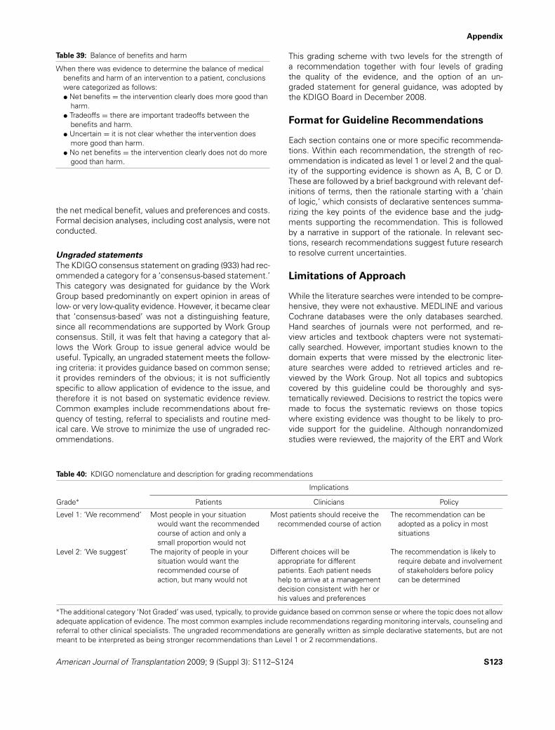
Appendix
Table 39: Balance of benefits and harm
When there was evidence to determine the balance of medicalbenefits and harm of an intervention to a patient, conclusionswere categorized as follows:• Net benefits = the intervention clearly does more good than
harm.• Tradeoffs = there are important tradeoffs between the
benefits and harm.• Uncertain = it is not clear whether the intervention does
more good than harm.• No net benefits = the intervention clearly does not do more
good than harm.
the net medical benefit, values and preferences and costs.Formal decision analyses, including cost analysis, were notconducted.
Ungraded statements
The KDIGO consensus statement on grading (933) had rec-ommended a category for a ‘consensus-based statement.’This category was designated for guidance by the WorkGroup based predominantly on expert opinion in areas oflow- or very low-quality evidence. However, it became clearthat ‘consensus-based’ was not a distinguishing feature,since all recommendations are supported by Work Groupconsensus. Still, it was felt that having a category that al-lows the Work Group to issue general advice would beuseful. Typically, an ungraded statement meets the follow-ing criteria: it provides guidance based on common sense;it provides reminders of the obvious; it is not sufficientlyspecific to allow application of evidence to the issue, andtherefore it is not based on systematic evidence review.Common examples include recommendations about fre-quency of testing, referral to specialists and routine med-ical care. We strove to minimize the use of ungraded rec-ommendations.
Table 40: KDIGO nomenclature and description for grading recommendations
Implications
Grade∗ Patients Clinicians Policy
Level 1: ‘We recommend’ Most people in your situationwould want the recommendedcourse of action and only asmall proportion would not
Most patients should receive therecommended course of action
The recommendation can beadopted as a policy in mostsituations
Level 2: ‘We suggest’ The majority of people in yoursituation would want therecommended course ofaction, but many would not
Different choices will beappropriate for differentpatients. Each patient needshelp to arrive at a managementdecision consistent with her orhis values and preferences
The recommendation is likely torequire debate and involvementof stakeholders before policycan be determined
∗The additional category ‘Not Graded’ was used, typically, to provide guidance based on common sense or where the topic does not allowadequate application of evidence. The most common examples include recommendations regarding monitoring intervals, counseling andreferral to other clinical specialists. The ungraded recommendations are generally written as simple declarative statements, but are notmeant to be interpreted as being stronger recommendations than Level 1 or 2 recommendations.
This grading scheme with two levels for the strength ofa recommendation together with four levels of gradingthe quality of the evidence, and the option of an un-graded statement for general guidance, was adopted bythe KDIGO Board in December 2008.
Format for Guideline Recommendations
Each section contains one or more specific recommenda-tions. Within each recommendation, the strength of rec-ommendation is indicated as level 1 or level 2 and the qual-ity of the supporting evidence is shown as A, B, C or D.These are followed by a brief background with relevant def-initions of terms, then the rationale starting with a ‘chainof logic,’ which consists of declarative sentences summa-rizing the key points of the evidence base and the judg-ments supporting the recommendation. This is followedby a narrative in support of the rationale. In relevant sec-tions, research recommendations suggest future researchto resolve current uncertainties.
Limitations of Approach
While the literature searches were intended to be compre-hensive, they were not exhaustive. MEDLINE and variousCochrane databases were the only databases searched.Hand searches of journals were not performed, and re-view articles and textbook chapters were not systemati-cally searched. However, important studies known to thedomain experts that were missed by the electronic liter-ature searches were added to retrieved articles and re-viewed by the Work Group. Not all topics and subtopicscovered by this guideline could be thoroughly and sys-tematically reviewed. Decisions to restrict the topics weremade to focus the systematic reviews on those topicswhere existing evidence was thought to be likely to pro-vide support for the guideline. Although nonrandomizedstudies were reviewed, the majority of the ERT and Work
American Journal of Transplantation 2009; 9 (Suppl 3): S112–S124 S123

Appendix
Table 41: Determinants of strength of recommendations
Factor Comment
Balance betweendesirable andundesirable effects
The larger the difference between the desirable and undesirable effects, the more likely a strongrecommendation is warranted. The narrower the gradient, the more likely a weak recommendation iswarranted.
Quality of the evidence The higher the quality of evidence, the more likely a strong recommendation is warranted.Values and preferences The more variability in values and preferences, or more uncertainty in values and preferences, the more likely
a weak recommendation is warranted.Costs (resource
allocation)The higher the costs of an intervention—that is, the more resources consumed—the less likely a strong
recommendation is warranted.
Reprinted with permission (934).
Group resources were devoted to review of randomizedtrials, since these were deemed to be most likely to pro-vide data to support level 1 recommendations with veryhigh- or high-quality (A or B) evidence. Where randomizedtrials are lacking, it was deemed to be sufficiently unlikelythat studies previously unknown to the Work Group wouldresult in a higher-quality level 1 recommendations. A smallnumber of supplemental sets of evidence were collectedwith a nonsystematic review approach. Any such evidencethat is summarized is noted. Decisions to take a nonsys-tematic review approach for these topics were made dueto time constraints and resource limitations.
Review of the Guideline DevelopmentProcess
Several tools and checklists have been developed toassess the quality of the methodological process forguideline development. These include the Appraisal ofGuidelines for Research and Evaluation (AGREE) crite-ria (936) and the Conference on Guideline Standardiza-tion (COGS) checklist (937). Supporting Table 62 showsthe COGS criteria that correspond to the AGREE check-list and how each one of them is addressed in thisguideline.
S124 American Journal of Transplantation 2009; 9 (Suppl 3): S112–S124

Biographic and Disclosure Information
Biographic and Disclosure Information
Bertram L. Kasiske, MD (Work Group Co-Chair), is Pro-fessor of Medicine and Medical Director of Kidney andPancreas Transplantation at University of Minnesota, USA.He received his medical degree from the University of Iowaand completed his Internal Medicine residency and fellow-ship training in Nephrology at Hennepin County MedicalCenter where he is also currently Director of Nephrologyand Medical Director of Kidney Transplantation. His pri-mary research interests encompass immunosuppression,dyslipidemia and CVD in transplant recipients. He is a Co-Investigator in a RCT of homocysteine (FAVORIT), Studyof Heart and Renal Protection (SHARP), and the US RenalData System (USRDS). Dr Kasiske has served as Medi-cal/Scientific Representative to the Board of Directors ofUnited Network for Organ Sharing (UNOS) and was for-merly Editor-in-Chief of American Journal of Kidney Dis-eases. He has published over 200 journal articles and hasrecently contributed book chapters in Brenner and Rec-tor’s The Kidney, Comprehensive Clinical Nephrology, TheKidney: Physiology and Pathophysiology and Kidney Trans-plantation: Principles and Practice. Dr Kasiske is also arecipient of the Outstanding Research AccomplishmentAward from University of Minnesota School of Medicinein 2002 and the National Kidney Foundation’s GarabedEknoyan Award in 2003.
Advisor/Consultant: Astellas; LithoLink; Novartis;Wyeth
Grant/Research Support: Bristol-Myers Squibb; Gen-zyme; Merck-Schering Plough
Martin G. Zeier, MD, FASN (Work Group Co-Chair), isHead of the Department of Nephrology and Hypertensionat the University of Heidelberg, where he also received histraining in Internal Medicine and Nephrology. He maintainsan active interest in areas of hereditary kidney disease(polycystic), and hypertension and viral infections in kid-ney transplant patients. Dr Zeier is a member of numerousprofessional organizations including, American Society ofNephrology, European Dialysis and Transplantation Associ-ation and International Society of Nephrology. He has pub-lished more than 200 publications and is currently SectionEditor of Nephrology Dialysis Transplantation and Co-Editorof Der Nephrologe.
Grant/Research Support: Astellas; Novartis; Parexel
Jonathan C. Craig, MBChB, MM (Clin Epi), DCH, FRACP,
PhD, is Professor of Clinical Epidemiology at the Univer-sity of Sydney’s School of Public Health; Senior Staff Spe-cialist in Pediatric Nephrology at the Children’s Hospital atWestmead; and Head of the Centre for Kidney Research
at the Children’s Hospital at Westmead, Australia. He be-gan his current appointment as the Coordinating Editor ofthe Cochrane Renal Group in 2000 and has cultivated hisresearch interests in RCTs, systematic reviews and diag-nostic test evaluation, and applied them to a variety ofkidney areas. Dr Craig is a contributor to and an editor ofthe text, Evidence-Based Nephrology, and has served onmany editorial boards, including American Journal of Kid-ney Diseases, Journal of the American Society of Nephrol-ogy and Nephrology. Dr Craig has authored over 200 arti-cles, including publications in the Lancet, The New Eng-land Journal of Medicine, Annals of Internal Medicine andthe British Medical Journal. He is also a member of morethan 12 professional societies and a recipient of the TJNeill Award for Outstanding Contribution to NephrologicalScience in 2009.
Dr Craig reported no relevant financial relationships.
Henrik Ekberg, MD, PhD, is Senior Transplant Surgeon atthe University Hospital in Malmo, Sweden, and Professorof Transplant Surgery at Lund University, Lund, Sweden.After obtaining his medical and doctoral degrees and sub-sequent board certification in General Surgery, he spent2 years (1986–1988) performing research in the field oftransplantation at the University of Sydney in Australia. Hismain research interests center on clinical and experimentalimmunosuppressive treatments and for the past 18 years,he has had an active role in a large number of pivotal mul-ticenter trials on immunosuppression in kidney transplan-tation. Professor Ekberg was also the sponsor and chair-man of the steering committee of the recently completed,multicenter, international Symphony study. He is an activemember of several professional societies and has servedas Councillor for The Transplantation Society (TTS) 2006–2008 and Editor-in-Chief of the TTS Newsletter. Until re-cently he was also Vice-President of the European Societyof Organ Transplantation (ESOT) and chairman of the ESOTeducation committee. Dr Ekberg is also Associate Editorof the American Journal of Transplantation and an Edito-rial Board member of Transplant International, Transplanta-tion, and Clinical Transplantation. He is a frequently invitedlecturer and has published more than 160 original articlesand reviews in the field of transplantation.
Advisor/Consultant: Astellas; Bristol-Myers Squibb;Hansa Medical; Hoffmann-LaRoche; Life CyclePharma; Novartis; Wyeth
Speaker: Astellas; Hoffmann-LaRoche
Catherine A. Garvey, RN, BA, CCTC, is a TransplantCoordinator at the University of Minnesota Medical
American Journal of Transplantation 2009; 9 (Suppl 3): S125–S128 S125

Biographic and Disclosure Information
Center-Fairway where she manages all living organ donorprograms, oversees the screening and evaluation of liv-ing organ donors, directs donor evaluations for kidney andpancreas transplant recipients and provides ongoing edu-cation to donors and their families. Ms. Garvey is currentlyPresident of NATCO (North American Transplant Coordi-nators Organization) Board and a member of the NationalLiving Donor Assistance Program (NLDAC) Advisory Com-mittee, AST (American Society of Transplantation) and ITNS(International Transplant Nurse Society). In recognition forher contributions to this field, she was honored with theNATCO Quality of Care Award in 2005 and most recently,the Health Care Hero for Nursing Award.
Mrs Garvey reported no relevant financial relation-ships.
Michael D. Green, MD, MPH, is Professor at Children’sHospital of Pittsburgh and Professor of Clinical and Trans-lational Science at University of Pittsburgh School ofMedicine. Dr Green obtained his medical degree from Uni-versity of Illinois and Masters of Public Health from Univer-sity of Pittsburgh and he is board certified in Pediatrics witha subspecialty in Pediatrics Infectious Diseases. His cur-rent research interests involve the studying of prophylacticantimicrobials in children with vesicoureteral reflux and im-pact of antimicrobial prophylaxis on the development ofantimicrobial resistance. Dr Green has written over 130publications and contributed more than 50 review articlesand book chapters in notable texts such as Infectious Dis-eases, Pediatric Solid Organ Transplantation, Principles &Practice of Pediatric Infectious Diseases and Textbook ofPediatric Infectious Diseases. In addition, he has servedas peer reviewer of more than 20 journals and as advisorycommittee member to various organizations including theFood & Drug Administration, International Pediatric Trans-plant Association and American Society for Transplanta-tion. Dr Green was named the St. Giles Foundation Fellow(2008–2009) at Columbia University Seminar on NationalHealth & Science Policy.
Dr Green reported no relevant financial relationships.
Vivekanand Jha, MD, FRCP, is Additional Profes-sor of Nephrology and Coordinator of Stem Cell Re-search Facility at the Postgraduate Institute of Medi-cal Education & Research, Chandigarh, India. Dr Jhahas held numerous committee positions in profes-sional bodies such as The Transplantation Society, In-ternational Society of Nephrology and most recentlya Steering Committee member of WHO initiativeon data harmonization in transplantation. His ongoingresearch projects include the development of optimalstrategies of immunosuppressive drug use after kidneytransplantation by pharmacogenomic approaches and thestudying of BMD and histomorphometry in chronic kidneyfailure and its evolution posttransplantation. He is Editor ofThe Cochrane Renal Group and a frequent peer reviewer for14 journals. Dr Jha has authored over 135 publications and
25 book chapters and serves as an editor of an upcomingtextbook, Management of Kidney Transplant Recipient.
Dr Jha reported no relevant financial relationships.
Michelle A. Josephson, MD, is Professor, Department ofMedicine, Section of Nephrology, and Medical Director ofTransplant Nephrology at University of Chicago. She ob-tained her medical degree from University of Pennsylvaniaand completed a Nephrology fellowship at University ofChicago. Having served as an investigator in areas relatedto bone disease and infections posttransplantation and effi-cacy of immunosuppressive medications, she maintains anactive research interest in medical complications of kidneytransplantation. Dr Josephson is an author of more than60 publications and a peer reviewer for journals includingAmerican Journal of Transplantation, American Journal ofKidney Disease and Kidney International. She is a frequentlecturer at national and international meetings and a re-cipient of the Excellence in Medical Education and ClinicalCare Award from University of Chicago in 2007.
Advisor/Consultant: Digitas Health; MKSAP; Wyeth
Speaker: Hoffmann-LaRoche
Grant/Research Support: Amgen; Astellas; Wyeth
Bryce A. Kiberd, MD, is Professor of Medicine at Dal-housie University, Halifax, NS, Canada. He received hismedical degree from the University of Toronto and contin-ued his nephrology fellowship at Toronto and Stanford Uni-versity. Dr Kiberd is a member of 11 professional medicalsocieties in addition to his ongoing participation on the Sci-entific Advisory Board for the Chronic Renal InsufficiencyCohort Study (NIH-NIDDK). He has long-standing interestsin areas of immunosuppressive medications and preven-tion of kidney disease progression and complications intransplant recipients. Dr Kiberd has authored over 100 pub-lications and he is a recipient of the Excellence Award forResearch, Dalhousie University from 1998 through 2006
Speaker: Hoffmann-LaRoche
Henri A. Kreis, MD, is Professor Emeritus of Nephrologyat Universite Paris Descartes and past Chairman of De-partment of Transplantation and Intensive Care at HopitalNecker, Paris, France. He was the Founding memberand Past President of the French Transplantation Societyand also served previously as General Secretary andVice-President of the France Transplant Association. DrKreis has been involved in many clinical and organizationalaspects of transplantation in Paris since 1960 and main-tains active interests in areas of immunosuppression andrejection. In addition to being a past member of variousHealth Ministry commissions, he currently serves asTreasurer of Comite de Protection des Personnes (CPP)Ile-de-France II. In 2004, he was awarded the Knight ofthe Legion of Honour.
Advisor/Consultant: Novimmune
S126 American Journal of Transplantation 2009; 9 (Suppl 3): S125–S128

Biographic and Disclosure Information
Ruth A. McDonald, MD, is Professor of Pediatrics at Uni-versity of Washington and Clinical Director of Nephrologyat Children’s Hospital and Regional Medical Center in Seat-tle, Washington. She completed her medical degree at Uni-versity of Minneosta School of Medicine where she is arecipient of the Top Medical Graduate: Hewlett-PackardAward. Dr McDonald is currently involved in numerousmulticenter clinical studies including a controlled trial ofAnti-CD20 monoclonal antibody therapy in historically un-sensitized renal transplant recipients with donor-specificantibodies; a Phase II study to determine safety and im-munomodulatory functions of induction therapy with Cam-path 1H, combined with MMF and sirolimus; a surveil-lance study of viral infections in renal transplant recipientsand many others. She is also a member of eight profes-sional organizations including American Society of Pedi-atric Nephrology, American Society of Transplantation andInternational Pediatric Transplant Association. Among herteaching responsibilities, she has trained over 25 fellowsand also served as Medical Student Research Mentor. DrMcDonald has written over 50 publications and has givenclose to 40 invited and extra-institutional lectures in thepast 10 years.
Dr McDonald reported no relevant financial relation-ships.
John M. Newmann, PhD, MPH, is President of Health Pol-icy Research & Analysis, Weston, Viriginia. As a prominenthealth-care consultant and a KTR himself, Dr Newmann hasbeen a tireless patient advocate on issues related to theMedicare end-stage renal disease program, organ trans-plantation policies, quality of life for dialysis patients andkidney disease education. In addition to speaking on theseissues before Congress, he has sought to improve patientand physician communication in presentations at nationalmeetings such as American Society of Nephrology, Na-tional Kidney Foundation and Renal Physicians Associa-tions. Dr Newmann is also extensively involved in manyorganizations including, the American Kidney Fund, Opti-mal Renal Care Medical Advisory Board and American As-sociation of Kidney Patients. He was also past Vice Presi-dent of Patient and Donor Affairs at UNOS. Dr Newmannis also a recipient of NKF’s Trustee Award and Quality ofLife Award from Nephrology News & Issues and he wasrecently recognized by American Nephrology Nurses’ As-sociation (ANNA) for his 30 years of service to the renalcommunity.
Advisor/Consultant: Arbor Research Collaborative;Renaissance Health Care
Gregorio T. Obrador, MD, MPH, is Professor of Medicineand Dean at the Universidad Panamericana School ofMedicine in Mexico City. He also serves as Adjunct Staff atthe Division of Nephrology of the Tufts-New England Medi-cal Center and Assistant Professor of Medicine at the TuftsUniversity School of Medicine in Boston. He earned hismedical degree from the University of Navarra (Pamplona,
Spain), completed his medicine residency at the West-ern Pennsylvania Hospital (Pittsburgh) and his Nephrologyfellowship at Boston University. While doing a clinical re-search fellowship at Tufts and a Master of Public Healthat Harvard University, he demonstrated that the manage-ment of patients with CKD prior to stage 5 is suboptimal,and that this is an important factor for the high morbidityand mortality observed in these patients. Dr Obrador hasbeen a member of the Advisory Board and of the AnemiaWorkgroup of the NKF KDOQI guidelines and has partic-ipated in several committees of the Kidney Disease: Im-proving Global Outcomes (KDIGO) initiative, the AmericanSociety of Nephrology, the International Society of Peri-toneal Dialysis, the Mexican Academy of Medicine andother Mexican nephrology societies. He is founding mem-ber of the Mexican Kidney Foundation and one of the lead-ers of the Mexico Kidney Early Evaluation Program (KEEP)program. He has given 97 lectures in national and interna-tional forums and has several publications in the area ofCKD.
Dr Obrador reported no relevant financial relation-ships.
Liaison Members
Jeremy R. Chapman, MD, FRACP, FRCP, is Network Direc-tor of Acute Interventional Medicine at Sydney West AreaHealth Service; Director of Renal Medicine at WestmeadHospital; and Clinical Professor at University of Sydney.He received his undergraduate and medical degrees fromCambridge University and had completed specialist train-ing in nephrology and transplantation at Oxford. Dr Chap-man is currently President of The Transplantation Societyand past President of World Marrow Donor Association.His research interests include clinical immunosuppression,post-kidney transplant malignancies, CAN and clinical bonemarrow, islet, kidney and pancreas transplantation. He hasauthored over 260 publications and book chapters andhas given more than 120 lectures since 1998. Dr Chap-man is also a member of numerous professional organi-zations and a recipient of the Order of Australia Medal in2003.
Advisor/Consultant: Astellas; Hoffmann-LaRoche;Novartis; Wyeth
Grant/Research Support: Bristol-Myers Squibb;Novartis; Wyeth
Flavio G. Vincenti, MD, is Professor of Clinical Medicineand Surgery at University of California, San Francisco andMedical Director of the Kidney Pancreas Program. He re-ceived his medical degree from American University Medi-cal Center at Beirut, Lebanon and completed his fellowshipin nephrology in Emory University School of Medicine,Atlanta, Georgia. Dr Vincenti is past President of Amer-ican Society of Transplantation and current member ofSteering Committee, Cooperative Trials in Pediatric (Renal)
American Journal of Transplantation 2009; 9 (Suppl 3): S125–S128 S127

Biographic and Disclosure Information
Transplantation (CCTPT) for NIH. As an attending physi-cian at an outpatient clinic and transplant ward, he de-votes much of his time to teaching and mentoring dutieswhile still actively engaging in 13 research projects primar-ily in the area of immunosuppressive medications. He iscurrently Associate Editor of CJASN and editorial boardmember of Transplantation. He has written over 190 publi-cations and authored chapters in several well-known textsincluding: Goodman & Gilman’s the Pharmacological Basisof Therapeutics, Pediatric Solid Organ Transplantation andSmith’s General Urology.
Grant/Research Support: Astellas; Bristol-MyersSquibb; Genentech; Hoffmann-LaRoche; Novartis;Wyeth
KDIGO Co-Chairs
Kai-Uwe Eckardt, MD, is Professor of Medicine andChief of Nephrology and Hypertension at the Universityof Erlangen—Nuremberg, Germany. He received his MDfrom the Westfalische Wilhelms-Universitat Munster, Ger-many. In 1993, following postgraduate training in internalmedicine, pathology and physiology, he was appointed As-sistant Professor of Physiology at the University of Regens-burg, Germany. Subsequently, he continued his training ininternal medicine and nephrology at the Charite, HumboldtUniversity in Berlin, where he was appointed AssociateProfessor of Nephrology in 2000. His major scientific in-terests are in the molecular mechanisms and physiologi-cal/pathophysiological relevance of oxygen sensing and themanagement of anemia. Professor Eckardt is Subject Edi-tor of Nephrology, Dialysis and Transplantation and serveson the editorial board of several other journals. He con-tributed to the development of the European Best Prac-tice Guidelines (EBPGs) for Anemia Management and is amember of the executive committee of KDIGO. Dr Eckardtis associated with CREATE and TREAT studies.
Advisor/Consultant: Affymax; Amgen; HEXAL;Hoffmann-LaRoche; Johnson & Johnson; OrthoBiotech; STADA
Speaker: Amgen; Hoffmann-LaRoche; Johnson &Johnson; Ortho Biotech
Grant/Research Support: Hoffmann-LaRoche; OrthoBiotech
Bertram L. Kasiske, MD. Please see earlier entry for WorkGroup Co-Chair.
Evidence Review Team
Ethan M. Balk, MD, MPH, is the Director, Evidence-basedMedicine at the Tufts Center for Kidney Disease GuidelineDevelopment and Implementation, in Boston, MA, and As-
sistant Professor of Medicine at Tufts University School ofMedicine. Dr Balk completed a fellowship in Clinical CareResearch. His primary research interests are evidence-based medicine, systematic review, clinical practice guide-line (CPG) development and critical literature appraisal.
Dr Balk reported no relevant financial relationships.
Martin Wagner, MD, MS, is currently a research fellowat the Tufts Center for Kidney Disease Guideline Develop-ment and Implementation in Boston, MA. He started histraining in internal medicine at the University of Wurzburg,Germany, where he also received his MD degree. He com-pleted his fellowship in nephrology at Tufts Medical Center,and received an MS in the Clinical Care Research programat the Sackler School at Tufts University. After his returnto the University of Wurzburg, he will remain as adjunctstaff at the Division of Nephrology of Tufts Medical Center.Dr Wagner’s research interests include CPG development,systematic review and predictive modeling.
Dr Wagner reported no relevant financial relation-ships.
Gowri Raman, MD, is Assistant Professor of Medicineat Tufts University School of Medicine and Assistant Di-rector, Tufts Evidence Practice Center at the Center forClinical Evidence Synthesis. Dr Raman is currently a Clini-cal Care Research Trainee fellow in the Institute for ClinicalResearch and Health Policy Studies at Tufts Medical Center.She has over 7 years experience in conducting systematicreviews and CPG development. Her primary research inter-ests are health technology assessment, systematic reviewand CPG development.
Dr Raman reported no relevant financial relation-ships.
Amy Earley, BS, is a project coordinator at the Tufts Centerfor Kidney Disease Guideline Development and Implemen-tation in Boston, MA. She assists in the development ofCPGs and conducts systematic reviews and critical litera-ture appraisals.
Ms. Earley reported no relevant financial relation-ships.
Samuel Abariga, MD, MS, was a Research Associatein the Division of Nephrology at Tufts-Medical Center.He specifically works with the Evidence Review Team atthe Tufts Center for Kidney Disease Guideline Develop-ment and Implementation. His research interest includesevidence-based medicine, systematic review, CPG devel-opment and critical literature appraisal.
Dr Abariga reported no relevant financial relation-ships.
S128 American Journal of Transplantation 2009; 9 (Suppl 3): S125–S128

Acknowledgments
Acknowledgments
A special debt of gratitude is owed to the current KDIGO Co-Chair Kai-Uwe Eckardt, founding Co-Chairs Garabed Eknoyan and Norbert Lameireand the KDIGO Board for their invaluable guidance throughout the de-velopment of this guideline. We also thank the efficient support pro-vided by the Kidney Disease: Improving Global Outcomes/National KidneyFoundation (KDIGO/NKF) staff that was instrumental to the success ofthis endeavor, particularly Michael Cheung, Donna Fingerhut and DekeyaSlaughter-Larkem. In particular, we thank the Evidence Review Team mem-bers: Ethan Balk, Martin Wagner, Amy Earley, Gowri Raman and SamAbariga, for their substantial contribution to the rigorous assessment ofthe available evidence. We are also especially grateful to the Work Groupmembers for their expertise throughout the entire process of literaturereview, data extraction, meeting participation, the critical writing and edit-ing of the statements and rationale which made the publication of thisguideline possible. The generous gift of their time and dedication is greatlyappreciated.
Finally, and on behalf of the Work Group, we gratefully acknowledge thecareful assessment of the draft guideline by external reviewers. The WorkGroup considered all of the valuable comments made and where appro-priate, suggested changes were incorporated into the final publication.The following individuals and organizations provided review of the draftguideline:
Kevin Abbott (Walter Reed Medical Center), Omar I Abboud, Mario Abbud-Filho (Instituto de Urologia & Nefrologia), Daniel Abramowicz (HopitalErasme; European Renal Best Practice), Nasrulla Abutaleb, Vidya N. Acharya(National Kidney Foundation/India), Horacio E. Adrogue (University of TexasMedical Branch), Mona Al-Rukhaimi (Dubai Hospital), Josefina Alberu (In-stituto Nacional De Ciencias Medicas Y. Nutricion S Z), Upton Allen (Hos-pital for Sick Children), Farhana Amanullah (Sindh Institute of Urology andTransplantation), American Society of Transplantation, American Societyof Transplant Surgeons, Mariano Arriola, Richard Baker (UK Renal Associ-ation), Ashraf Bakr (Mansoura Faculty of Medicine), Rashad S. Barsoum(Cairo University), Gregor Bartel, Scott Batty (Genzyme), Yolanda Tai Becker(University of Wisconsin), Luboslav Bena, Mohamed Benghanem Gharbi(Ibn Rochd Quartier des Hospitaux), Bassam Bernieh (Tawam Hospital),Emily A. Blumberg (University of Pennsylvania), Daniel C. Brennan (Wash-ington University), Brenda S. Brewer (NATCO), Bristol-Myers Squibb, GhilBusnach, Evelyn Butera, Canadian Society of Nephrology, Canadian Societyof Transplantation, Marcelo Cantarovich (McGill University Health Center;The Transplantation Society Council), Fernando Carrera (Hospital SAMS),Sue Cary (American Nephrology Nurses Association), Valerie Cass (McGillUniversity Health Center), Blanche M. Chavers (University of Minnesota),Pierre Cochat (Hospices Civils de Lyon et Universite de Lyon; EuropeanRenal Best Practice), Jean Colaneri (American Nephrology Nurses Associa-tion), William G. Couser (University of Washington), Ana Cusumano (LatinAmerican Society of Nephrology and Arterial Hypertension), Romina A.Danguilan (National Kidney and Transplant Institute), Gabriel M. Danovitch(David Geffen School of Medicine, UCLA), Jane Davis (University of Al-abama), Jean-Yves De Vos (AZ Werken Glorieux Dialysis Unit), Ian Dittmer(Auckland City Hospital), Grahame Elder (Westmead Hospital), Donna L.Ennis (NATCO), Tracy A. Evans-Walker (NATCO), Jeffrey Fadrowski (JohnsHopkins University School of Medicine), Randall J. Faull (Australian andNew Zealand Society Of Nephrology), Stuart M. Flechner (Cleveland Clinic),Joseph T. Flynn (Seattle Children’s Hospital; American Society of Pediatric
Nephrology), Patricia G. Folk (The International Transplant Nurses Society),Lorenzo G. Gallon (Northwestern University Feinberg School of Medicine),Alvaro A. Garcia, Valter D. Garcia (Brazilian Association of Organ Trans-plantation), Rozina Ghazalli (Penang Hospital), Osama Gheith, Elaine Go (StJoseph Hospital), Simin Goral (University of Pennsylvania), Darla K. Granger(American Society of Transplant Surgeons), Debbie Gregory (Renal Societyof Australasia), Sinead Gregory (Roche Diagnostics), Ann P. Guillot (Univer-sity of Vermont), William E. Haley (Mayo Clinic), Judith Hambleton (RogosinInstitute), Jeff Harder (University of Washington), Erica L. Hartmann (Ameri-can Society of Transplantation), Susan Hayes (S Hayes Consulting), RebeccaHays (University of Wisconsin Hospital), Domingo Hernandez-Marrero (Hos-pital Regional Universitario Carlos Haya), Hans H. Hirsch (University HospitalBasel), Hallvard Holdaas (Oslo University Hospital Rikshospitalet), HerwigHolzer (Medical University, Graz), Chakko K. Jacob (Christian Medical Col-lege and Hospital), Michel Jadoul (Cliniques Universitaires St Luc), MichelleJames (University of Minnesota), Alan Jardine (UK Renal Association),Pradeep V. Kadambi (University of Chicago), Nada Kanaan, Bruce Kaplan(University of Arizona School of Medicine), Frederick J. Kaskel (Children’sHospital at Montefiore), Tammy Keough-Ryan (Dalhousie University), RonH. Kerman (University of Texas Medical School), Markus Ketteler (KlinikumCoburg), Allan D. Kirk (Emory University Hospital), Greg A. Knoll (Cana-dian Society of Transplantation), Stephan Korte (Novartis), Andreas Kribben(Universitatsklinikum Essen), K. Sampath Kumar (Meenakshi Mission Hos-pital), Dirk Kuypers (University Hospitals Leuven), Sondra Kybartiene, JackLake (University of Minnesota), Craig B. Langman (Northwestern UniversityFeinberg School of Medicine), Anastasia Laskari, Daniel Lavanchy (WorldHealth Organization), Edgar V. Lerma (University of Illinois at Chicago Col-lege of Medicine), Zhi-Hong Liu (Nanjing University School of Medicine),Francesco Locatelli (‘A. Manzoni’ Hospital; European Renal Best Practice),Robert MacTier (UK Renal Association), Samir G. Mallat, Kevin C. Mange(Novartis), Martin Maraschio (Hospital Privado Centro Medico De CordobaS.A.), Pablo U. Massari (Catholic University of Cordoba), Arthur Matas (Uni-versity of Minnesota), Dianne McKay (Scripps Research Institute), EnisaMesic (University Medical Center Tuzla), Marian G. Michaels (Children’sHospital of Pittsburgh), Mark Mitsnefes (Cincinnati Children’s Medical Cen-ter), Rafique Moosa (University of Stellenbosch), Jose M. Morales (HospUniversitario Doce De Octubre), Alfredo Mota (Coimbra University Hos-pital), Barbara T. Murphy (Mount Sinai School of Medicine; American So-ciety of Transplantation), Judit Nagy (Hungarian Society of Nephrology),Nancy Nardelli (Medical City Dallas Hospital), Alicia M. Neu (Johns HopkinsUniversity School of Medicine), Joseph Nogueira (University of Maryland),Ole Oyen (Rikshospitalet University Hospital), Fatma N. Ozdemir (BaskentUniversity), Alejandro Lucas Penagos, Ronald Perrone (Tufts Medical Cen-ter), Todd E. Pesavento (Ohio State University; United Network for OrganSharing), Mark Pescovitz (Indiana University), Thomas G. Peters (Universityof Florida Health Science Center), Helen Pilmore (Auckland City Hospi-tal), Lee Po-Chang, G.V. Ramesh Prasad (University of Toronto), ErasmiaPsimenou, Ruth Rahamimov (Belinson Medical Center), Harun Ur. Rashid(Bangladesh Renal Association), Luis Re, Rafael Reyes (Hospital Miguel Hi-dalgo), Sally I. Rice (University of Louisville), John P. Roberts (American So-ciety of Transplant Surgeons), Bernardo Rodriguez-Iturbe (Hospital Univer-sitario and Universidad del Zulia, Maracaibo), Lionel Rostaing (Toulouse Uni-versity Hospital), Mahmoud Sadeghi, Kaija Salmela (Helsinki University Cen-tral Hospital), John D. Scandling (Stanford University Medical Center), HeidiSchaefer (Vanderbilt University Medical Center), Tammy Sebers (OregonHealth and Science University), Myra Sgorbini, Ayo Shonibare (St Nicholas
American Journal of Transplantation 2009; 9 (Suppl 3): S129–S130 S129

Acknowledgments
Hospital), Christopher O. Simon (Independent Dialysis Foundation), Am-mar Sirawan (Ajn W Sein Hospital), Dhavee Sirivongs (Khon Kaen MedicalSchool), William T. Smith (Wyeth), Edison Souza, Goce B. Spasovski (Univer-sity Clinical Center, Skopje; European Renal Best Practice), William P. Stam-ford, Sharon Swofford (American Nephrology Nurses Association), HiroshiToma (Tokyo Women’s Medical University), Yvette Tomacruz (National Kid-ney and Transplant Institute), Marcello Tonelli (University of Alberta; Cana-dian Society of Nephrology), Armando Torres (Universidad de La Laguna),Ray Trevitt (EDTNA/ERCA), Lara E. Tushla (Society for Transplant SocialWorkers), Ray Vanholder (University Hospital, Ghent; European Renal BestPractice), Joseph Vassalotti (National Kidney Foundation), Ruben L. Velez(Dallas Nephrology Associates), Alberto Vianello (Feltre General Hospital),Jelka Zaletel Vrtovec, Sean Wagner, Yi Wang, Bruno Watschinger (Medi-zinische Universitat Wien), Willem Weimar (University Medical Center Rot-terdam), Matthew R. Weir (University of Maryland), Barbara Weis Malone(University of Colorado), Jose R. Weisinger (Baptist Health South Florida),
Patricia Weiskittel (American Nephrology Nurses Association), Kerstin W.Westman (University Hospital of Malmo), David C. Wheeler (UniversityCollege London Medical School), Anne Wiland (Novartis), Alan Wilkinson(David Geffen School of Medicine, UCLA), Alexander Wiseman (Universityof Colorado; American Society of Transplantation), Germaine Wong (Chil-dren’s Hospital at Westmead), David Wu (Wyeth), Chul Woo Yang (CatholicUniversity of Korea), Jo-Anne Young (University of Minnesota), Carlton J.Young (University of Alabama), Zhu Zhou, Carmine Zoccali (Ospedali Riuniti;European Renal Best Practice).
Participation in the review does not necessarily constitute endorsementof the content of this report by above individuals, or the organization orinstitution they represent.
Bertram L. Kasiske, MD, Work Group Co-ChairMartin G. Zeier, MD, Work Group Co-Chair
S130 American Journal of Transplantation 2009; 9 (Suppl 3): S129–S130

References
References
1. Wolfe RA, Ashby VB, Milford EL et al. Comparison of mortality inall patients on dialysis, patients on dialysis awaiting transplanta-tion, and recipients of a first cadaveric transplant. N Engl J Med1999; 341: 1725–1730.
2. Webster AC, Playford EG, Higgins G et al. Interleukin 2 receptorantagonists for renal transplant recipients: A meta-analysis ofrandomized trials. Transplantation 2004; 77: 166–176.
3. Morton RL, Howard K, Webster AC et al. The cost-effectivenessof induction immunosuppression in kidney transplantation.Nephrol Dial Transplant 2009; 24: 2258–2269.
4. Szczech LA, Berlin JA, Aradhye S et al. Effect of anti-lymphocyteinduction therapy on renal allograft survival: A meta-analysis. JAm Soc Nephrol 1997; 8: 1771–1777.
5. Szczech LA, Berlin JA, Feldman HI. The effect of antilymphocyteinduction therapy on renal allograft survival. A meta-analysis ofindividual patient-level data. Anti-Lymphocyte Antibody InductionTherapy Study Group. Ann Intern Med 1998; 128: 817–826.
6. Thibaudin D, Alamartine E, de Filippis JP et al. Advantage ofantithymocyte globulin induction in sensitized kidney recipients:A randomized prospective study comparing induction with andwithout antithymocyte globulin. Nephrol Dial Transplant 1998;13: 711–715.
7. Charpentier B, Rostaing L, Berthoux F et al. A three-arm studycomparing immediate tacrolimus therapy with antithymocyteglobulin induction therapy followed by tacrolimus or cyclosporineA in adult renal transplant recipients. Transplantation 2003; 75:844–851.
8. Brennan DC, Daller JA, Lake KD et al. Rabbit antithymocyteglobulin versus basiliximab in renal transplantation. N Engl J Med2006; 355: 1967–1977.
9. Margreiter R, Klempnauer J, Neuhaus P et al. Alemtuzumab(Campath-1H) and tacrolimus monotherapy after renal transplan-tation: Results of a prospective randomized trial. Am J Transplant2008; 8: 1480–1485.
10. Thomas PG, Woodside KJ, Lappin JA et al. Alemtuzumab (Cam-path 1H) induction with tacrolimus monotherapy is safe for highimmunological risk renal transplantation. Transplantation 2007;83: 1509–1512.
11. Vathsala A, Ona ET, Tan SY et al. Randomized trial of Alem-tuzumab for prevention of graft rejection and preservation ofrenal function after kidney transplantation. Transplantation 2005;80: 765–774.
12. Ciancio G, Burke GW, Gaynor JJ et al. A randomized trial ofthree renal transplant induction antibodies: Early comparisonof tacrolimus, mycophenolate mofetil, and steroid dosing, andnewer immune-monitoring. Transplantation 2005; 80: 457–465.
13. Ciancio G, Burke GW, Gaynor JJ et al. A randomized trial ofthymoglobulin vs. alemtuzumab (with lower dose maintenanceimmunosuppression) vs. daclizumab in renal transplantation at24 months of follow-up. Clin Transplant 2008; 22: 200–210.
14. Gore JL, Pham PT, Danovitch GM et al. Obesity and outcomefollowing renal transplantation. Am J Transplant 2006; 6: 357–363.
15. Pallardo Mateu LM, Sancho Calabuig A, Capdevila Plaza L et al.Acute rejection and late renal transplant failure: Risk factors andprognosis. Nephrol Dial Transplant 2004; 19(Suppl. 3): iii38–42.
16. McDonald R, Donaldson L, Emmett L et al. A decade of liv-ing donor transplantation in North American children: The 1998annual report of the North American Pediatric Renal TransplantCooperative Study (NAPRTCS). Pediatr Transplant 2000; 4: 221–234.
17. Mota A, Figueiredo A, Cunha MF et al. Risk factors for acuterejection in 806 cyclosporine-treated renal transplants: A multi-variate analysis. Transplant Proc 2003; 35: 1061–1063.
18. Boom H, Mallat MJ, de Fijter JW et al. Delayed graft functioninfluences renal function, but not survival. Kidney Int 2000; 58:859–866.
19. Oien CM, Reisaeter AV, Leivestad T et al. Living donor kidneytransplantation: The effects of donor age and gender on short-and long-term outcomes. Transplantation 2007; 83: 600–606.
20. Quiroga I, McShane P, Koo DD et al. Major effects of delayedgraft function and cold ischaemia time on renal allograft survival.Nephrol Dial Transplant 2006; 21: 1689–1696.
21. Sagedal S, Nordal KP, Hartmann A et al. The impact ofcytomegalovirus infection and disease on rejection episodesin renal allograft recipients. Am J Transplant 2002; 2: 850–856.
22. Webster AC, Woodroffe RC, Taylor RS et al. Tacrolimus versusciclosporin as primary immunosuppression for kidney transplantrecipients: Meta-analysis and meta-regression of randomisedtrial data. BMJ 2005; 331: 810.
23. Kramer BK, Montagnino G, Del Castillo D et al. Efficacy andsafety of tacrolimus compared with cyclosporin A microemulsionin renal transplantation: 2 year follow-up results. Nephrol DialTransplant 2005; 20: 968–973.
24. Hardinger KL, Bohl DL, Schnitzler MA et al. A randomized,prospective, pharmacoeconomic trial of tacrolimus versus cy-closporine in combination with thymoglobulin in renal transplantrecipients. Transplantation 2005; 80: 41–46.
25. Vincenti F, Friman S, Scheuermann E et al. Results of an interna-tional, randomized trial comparing glucose metabolism disordersand outcome with cyclosporine versus tacrolimus. Am J Trans-plant 2007; 7: 1506–1514.
26. Rowshani AT, Scholten EM, Bemelman F et al. No differencein degree of interstitial Sirius red-stained area in serial biop-sies from area under concentration-over-time curves-guided cy-closporine versus tacrolimus-treated renal transplant recipientsat one year. J Am Soc Nephrol 2006; 17: 305–312.
27. Ekberg H, Tedesco-Silva H, Demirbas A et al. Reduced exposureto calcineurin inhibitors in renal transplantation. N Engl J Med2007; 357: 2562–2575.
28. Murphy GJ, Waller JR, Sandford RS et al. Randomized clinicaltrial of the effect of microemulsion cyclosporin and tacrolimuson renal allograft fibrosis. Br J Surg 2003; 90: 680–686.
29. Rostaing L, Cantarovich D, Mourad G et al. Corticosteroid-freeimmunosuppression with tacrolimus, mycophenolate mofetil,and daclizumab induction in renal transplantation. Transplanta-tion 2005; 79: 807–814.
30. Ekberg H, Grinyo J, Nashan B et al. Cyclosporine sparing withmycophenolate mofetil, daclizumab and corticosteroids in renalallograft recipients: The CAESAR Study. Am J Transplant 2007;7: 560–570.
American Journal of Transplantation 2009; 9 (Suppl 3): S131–S155 S131

References
31. Mycophenolate mofetil in renal transplantation: 3-year resultsfrom the placebo-controlled trial. European MycophenolateMofetil Cooperative Study Group. Transplantation 1999; 68: 391–396.
32. Shapiro R, Jordan ML, Scantlebury VP et al. A prospec-tive, randomized trial of tacrolimus/prednisone versustacrolimus/prednisone/mycophenolate mofetil in renal transplantrecipients. Transplantation 1999; 67: 411–415.
33. Knight SR, Russell NK, Barcena L et al. Mycophenolate mofetildecreases acute rejection and may improve graft survival in re-nal transplant recipients when compared with azathioprine: Asystematic review. Transplantation 2009; 87: 785–794.
34. A blinded, randomized clinical trial of mycophenolate mofetil forthe prevention of acute rejection in cadaveric renal transplanta-tion. The Tricontinental Mycophenolate Mofetil Renal Transplan-tation Study Group. Transplantation 1996; 61: 1029–1037.
35. Sollinger HW. Mycophenolate mofetil for the prevention of acuterejection in primary cadaveric renal allograft recipients. U.S. Re-nal Transplant Mycophenolate Mofetil Study Group. Transplanta-tion 1995; 60: 225–232.
36. Miller J, Mendez R, Pirsch JD et al. Safety and efficacy oftacrolimus in combination with mycophenolate mofetil (MMF) incadaveric renal transplant recipients. FK506/MMF Dose-RangingKidney Transplant Study Group. Transplantation 2000; 69: 875–880.
37. Remuzzi G, Lesti M, Gotti E et al. Mycophenolate mofetil versusazathioprine for prevention of acute rejection in renal transplan-tation (MYSS): A randomised trial. Lancet 2004; 364: 503–512.
38. Sadek S, Medina J, Arias M et al. Short-term combination ofmycophenolate mofetil with cyclosporine as a therapeutic op-tion for renal transplant recipients: A prospective, multicenter,randomized study. Transplantation 2002; 74: 511–517.
39. Ojo AO, Meier-Kriesche HU, Hanson JA et al. Mycophenolatemofetil reduces late renal allograft loss independent of acuterejection. Transplantation 2000; 69: 2405–2409.
40. Opelz G, Dohler B. Influence of immunosuppressive regimenson graft survival and secondary outcomes after kidney transplan-tation. Transplantation 2009; 87: 795–802.
41. Craig JC, Webster AC, McDonald SP. The case of azathioprineversus mycophenolate. Do different drugs really cause differenttransplant outcomes? Transplantation 2009; 87: 803–804.
42. Salvadori M, Holzer H, de Mattos A et al. Enteric-coated my-cophenolate sodium is therapeutically equivalent to mycopheno-late mofetil in de novo renal transplant patients. Am J Transplant2004; 4: 231–236.
43. Budde K, Curtis J, Knoll G et al. Enteric-coated mycophenolatesodium can be safely administered in maintenance renal trans-plant patients: Results of a 1-year study. Am J Transplant 2004;4: 237–243.
44. Kasiske BL, Chakkera HA, Louis TA et al. A meta-analysis ofimmunosuppression withdrawal trials in renal transplantation. JAm Soc Nephrol 2000; 11: 1910–1917.
45. Pascual J, Quereda C, Zamora J et al. Steroid withdrawal in renaltransplant patients on triple therapy with a calcineurin inhibitorand mycophenolate mofetil: A meta-analysis of randomized, con-trolled trials. Transplantation 2004; 78: 1548–1556.
46. ter Meulen CG, van Riemsdijk I, Hene RJ et al. Steroid-withdrawal at 3 days after renal transplantation with anti-IL-2receptor alpha therapy: A prospective, randomized, multicenterstudy. Am J Transplant 2004; 4: 803–810.
47. Woodle ES, First MR, Pirsch J et al. A prospective, random-ized, double-blind, placebo-controlled multicenter trial comparingearly (7 day) corticosteroid cessation versus long-term, low-dosecorticosteroid therapy. Ann Surg 2008; 248: 564–577.
48. Vincenti F, Schena FP, Paraskevas S et al. A randomized, mul-ticenter study of steroid avoidance, early steroid withdrawal orstandard steroid therapy in kidney transplant recipients. Am JTransplant 2008; 8: 307–316.
49. Kasiske BL, de Mattos A, Flechner SM et al. Mammalian target ofrapamycin inhibitor dyslipidemia in kidney transplant recipients.Am J Transplant 2008; 8: 1384–1392.
50. Webster AC, Lee VW, Chapman JR et al. Target of rapamycininhibitors (TOR-I; sirolimus and everolimus) for primary immuno-suppression in kidney transplant recipients. Cochrane DatabaseSyst Rev 2006: CD004290.
51. Buchler M, Caillard S, Barbier S et al. Sirolimus versus cy-closporine in kidney recipients receiving thymoglobulin, my-cophenolate mofetil and a 6-month course of steroids. Am JTransplant 2007; 7: 2522–2531.
52. Langer RM, Kahan BD. Incidence, therapy, and consequencesof lymphocele after sirolimus-cyclosporine-prednisone immuno-suppression in renal transplant recipients. Transplantation 2002;74: 804–808.
53. Troppmann C, Pierce JL, Gandhi MM et al. Higher surgical woundcomplication rates with sirolimus immunosuppression after kid-ney transplantation: A matched-pair pilot study. Transplantation2003; 76: 426–429.
54. Valente JF, Hricik D, Weigel K et al. Comparison of sirolimusvs. mycophenolate mofetil on surgical complications and woundhealing in adult kidney transplantation. Am J Transplant 2003; 3:1128–1134.
55. Dean PG, Lund WJ, Larson TS et al. Wound-healing compli-cations after kidney transplantation: A prospective, randomizedcomparison of sirolimus and tacrolimus. Transplantation 2004;77: 1555–1561.
56. Van Den Akker JM, Wetzels JF, Hoitsma AJ. Proteinuria follow-ing conversion from azathioprine to sirolimus in renal transplantrecipients. Kidney Int 2006; 70: 1355–1357.
57. Webster AC, Lee VW, Chapman JR et al. Target of rapamycininhibitors (sirolimus and everolimus) for primary immunosup-pression of kidney transplant recipients: A systematic reviewand meta-analysis of randomized trials. Transplantation 2006;81: 1234–1248.
58. Maes B, Hadaya K, de Moor B et al. Severe diarrhea in renaltransplant patients: Results of the DIDACT study. Am J Trans-plant 2006; 6: 1466–1472.
59. Jha V, Chugh K. Dialysis in developing countries: Priorities andobstacles. Nephrology 1996; 2: 65–71.
60. Chugh KS, Jha V, Chugh S. Economics of dialysis and renaltransplantation in the developing world. Transplant Proc 1999;31: 3275–3277.
61. Jha V, Chugh KS. The practice of dialysis in the developing coun-tries. Hemodialysis International 2003; 7: 239–249.
62. Jha V, Muthukumar T, Kohli HS et al. Impact of cyclosporinewithdrawal on living related renal transplants: A single-centerexperience. Am J Kidney Dis 2001; 37: 119–124.
63. Morgenstern GR, Powles R, Robinson B et al. Cyclosporin inter-action with ketoconazole and melphalan. Lancet 1982; 2: 1342.
64. Randall T. Cyclosporine-ketoconazole combination offerspromise in reducing antirejection therapy costs. JAMA 1990;264: 430–431.
65. Gueco IP, Tan-Torres T, Baniga U et al. Ketoconazole in post-transplant triple therapy: Comparison of costs and outcomes.Transplant Proc 1992; 24: 1709–1714.
66. First MR, Schroeder TJ, Michael A et al. Cyclosporine-ketoconazole interaction. Long-term follow-up and preliminaryresults of a randomized trial. Transplantation 1993; 55: 1000–1004.
S132 American Journal of Transplantation 2009; 9 (Suppl 3): S131–S155

References
66a. Butman SM, Wild JC, Nolan PE et al. Prospective study of thesafety and financial benefit of ketoconazole as adjuntive therapyto cyclosporine after heart transplantation. J Heart Lung Trans-plant 1991; 10: 351–358.
67. Patton PR, Brunson ME, Pfaff WW et al. A preliminary reportof diltiazem and ketoconazole. Their cyclosporine-sparing effectand impact on transplant outcome. Transplantation 1994; 57:889–892.
68. Keogh A, Spratt P, McCosker C et al. Ketoconazole to reducethe need for cyclosporine after cardiac transplantation. N Engl JMed 1995; 333: 628–633.
69. Sobh MA, Hamdy AF, El Agroudy AE et al. Coadministration ofketoconazole and cyclosporine for kidney transplant recipients:Long-term follow-up and study of metabolic consequences. AmJ Kidney Dis 2001; 37: 510–517.
70. Abraham MA, Thomas PP, John GT et al. Efficacy and safetyof low-dose ketoconazole (50 mg) to reduce the cost of cy-closporine in renal allograft recipients. Transplant Proc 2003; 35:215–216.
71. Carbajal H, Soltero L, Rodriguez-Montalvo C et al. Cyclosporineand low-dose ketoconazole in renal transplant recipients: Asingle-center experience. Transplantation 2004; 77: 1038–1040.
72. Thomas PP, Manivannan J, John GT et al. Sirolimus and keto-conazole co-prescription in renal transplant recipients. Transplan-tation 2004; 77: 474–475.
73. El-Dahshan KF, Bakr MA, Donia AF et al. Ketoconazole-tacrolimus coadministration in kidney transplant recipients: Two-year results of a prospective randomized study. Am J Nephrol2006; 26: 293–298.
73a. Soltero L, Carbajal H, Rodrıguez-Montalvo C et al. Coadministra-tion of tacrolimus and ketoconazole in renal transplant recipients:Cost analysis and review of metabolic effects. Transplant Proc2003; 35: 1319–1321.
74. El-Agroudy AE, Sobh MA, Hamdy AF et al. A prospective,randomized study of coadministration of ketoconazole and cy-closporine a in kidney transplant recipients: Ten-year follow-up.Transplantation 2004; 77: 1371–1376.
75. El-Dahshan KF, Bakr MA, Donia AF et al. Co-administration ofketoconazole to tacrolimus-treated kidney transplant recipients:A prospective randomized study. Nephrol Dial Transplant 2004;19: 1613–1617.
76. Gerntholtz T, Pascoe MD, Botha JF et al. The use of acyclosporin-ketoconazole combination: Making renal transplan-tation affordable in developing countries. Eur J Clin Pharmacol2004; 60: 143–148.
77. Foradori A, Mezzano S, Videla C et al. Modification of the pharma-cokinetics of cyclosporine A and metabolites by the concomitantuse of Neoral and diltiazem or ketoconazol in stable adult kidneytransplants. Transplant Proc 1998; 30: 1685–1687.
78. Videla C, Vega J, Borja H. Hepatotoxicity associated with cy-closporine monitoring using C2 recommendations in adults re-nal recipients receiving ketoconazole. Transplant Proc 2005; 37:1574–1576.
79. Guleria S, Kamboj M, Singh P et al. Tacrolimus (Pan Graf) as denovo therapy in renal transplant recipients in India. TransplantProc 2006; 38: 2029–2031.
80. Guleria S, Kamboj M, Sharma M et al. Tacrolimus (PanGraf) in live related renal transplantation: An initial experienceof 101 recipients in India. Transplant Proc 2007; 39: 747–749.
81. Guleria S, Kamboj M, Chatterjee A et al. Generic tacrolimus (PanGraf) in renal transplantation: An experience of 155 recipients inIndia. Transplant Proc 2008; 40: 2237–2239.
82. Dantal J, Hourmant M, Cantarovich D et al. Effect of long-termimmunosuppression in kidney-graft recipients on cancer inci-dence: Randomised comparison of two cyclosporin regimens.Lancet 1998; 351: 623–628.
83. Kyllonen LE, Salmela KT. Early cyclosporine C0 and C2 monitor-ing in de novo kidney transplant patients: A prospective random-ized single-center pilot study. Transplantation 2006; 81: 1010–1015.
84. Wallemacq P, Goffinet JS, O’Morchoe S et al. Multi-site analyticalevaluation of the Abbott ARCHITECT tacrolimus assay. Ther DrugMonit 2009; 31: 198–204.
85. Jorgensen K, Povlsen J, Madsen S et al. C2 (2-h) levels arenot superior to trough levels as estimates of the area underthe curve in tacrolimus-treated renal-transplant patients. NephrolDial Transplant 2002; 17: 1487–1490.
86. van Gelder T, Le Meur Y, Shaw LM et al. Therapeutic drug mon-itoring of mycophenolate mofetil in transplantation. Ther DrugMonit 2006; 28: 145–154.
87. Knight SR, Morris PJ. Does the evidence support the use ofmycophenolate mofetil therapeutic drug monitoring in clinicalpractice? A systematic review. Transplantation 2008; 85: 1675–1685.
88. Le Meur Y, Buchler M, Thierry A et al. Individualized mycophe-nolate mofetil dosing based on drug exposure significantly im-proves patient outcomes after renal transplantation. Am J Trans-plant 2007; 7: 2496–2503.
89. Hale MD, Nicholls AJ, Bullingham RE et al. The pharmacokinetic-pharmacodynamic relationship for mycophenolate mofetil in re-nal transplantation. Clin Pharmacol Ther 1998; 64: 672–683.
90. van Gelder T, Hilbrands LB, Vanrenterghem Y et al. A random-ized double-blind, multicenter plasma concentration controlledstudy of the safety and efficacy of oral mycophenolate mofetilfor the prevention of acute rejection after kidney transplantation.Transplantation 1999; 68: 261–266.
91. Weber LT, Shipkova M, Armstrong VW et al. Thepharmacokinetic-pharmacodynamic relationship for total andfree mycophenolic Acid in pediatric renal transplant recipients:A report of the german study group on mycophenolate mofetiltherapy. J Am Soc Nephrol 2002; 13: 759–768.
92. Oellerich M, Shipkova M, Schutz E et al. Pharmacokineticand metabolic investigations of mycophenolic acid in pediatricpatients after renal transplantation: Implications for therapeu-tic drug monitoring. German Study Group on MycophenolateMofetil Therapy in Pediatric Renal Transplant Recipients. TherDrug Monit 2000; 22: 20–26.
93. Kiberd BA, Lawen J, Fraser AD et al. Early adequate mycophe-nolic acid exposure is associated with less rejection in kidneytransplantation. Am J Transplant 2004; 4: 1079–1083.
94. Oellerich M, Armstrong VW. The role of therapeutic drug mon-itoring in individualizing immunosuppressive drug therapy: Re-cent developments. Ther Drug Monit 2006; 28: 720–725.
95. Kahan BD, Camardo JS. Rapamycin: Clinical results and futureopportunities. Transplantation 2001; 72: 1181–1193.
96. Neumayer HH, Paradis K, Korn A et al. Entry-into-human studywith the novel immunosuppressant SDZ RAD in stable re-nal transplant recipients. Br J Clin Pharmacol 1999; 48: 694–703.
97. Kovarik JM, Tedesco H, Pascual J et al. Everolimus therapeu-tic concentration range defined from a prospective trial withreduced-exposure cyclosporine in de novo kidney transplanta-tion. Ther Drug Monit 2004; 26: 499–505.
98. Nashan B. Review of the proliferation inhibitor everolimus. Ex-pert Opin Investig Drugs 2002; 11: 1845–1857.
American Journal of Transplantation 2009; 9 (Suppl 3): S131–S155 S133

References
99. Solez K, Colvin RB, Racusen LC et al. Banff 07 classification ofrenal allograft pathology: Updates and future directions. Am JTransplant 2008; 8: 753–760.
100. Rush D, Nickerson P, Gough J et al. Beneficial effects of treat-ment of early subclinical rejection: A randomized study. J AmSoc Nephrol 1998; 9: 2129–2134.
101. Rush DN, Karpinski ME, Nickerson P et al. Does subclinical rejec-tion contribute to chronic rejection in renal transplant patients?Clin Transplant 1999; 13: 441–446.
102. Rush D, Arlen D, Boucher A et al. Lack of benefit of early protocolbiopsies in renal transplant patients receiving TAC and MMF: Arandomized study. Am J Transplant 2007; 7: 2538–2545.
103. Kurtkoti J, Sakhuja V, Sud K et al. The utility of 1- and 3-monthprotocol biopsies on renal allograft function: A randomized con-trolled study. Am J Transplant 2008; 8: 317–323.
104. Gloor JM, Cohen AJ, Lager DJ et al. Subclinical rejection intacrolimus-treated renal transplant recipients. Transplantation2002; 73: 1965–1968.
105. Webster AC, Pankhurst T, Rinaldi F et al. Monoclonal and poly-clonal antibody therapy for treating acute rejection in kidneytransplant recipients: A systematic review of randomized trialdata. Transplantation 2006; 81: 953–965.
106. Mycophenolate mofetil for the treatment of a first acute re-nal allograft rejection: Three-year follow-up. The Mycopheno-late Mofetil Acute Renal Rejection Study Group. Transplantation2001; 71: 1091–1097.
107. Mariat C, Alamartine E, Diab N et al. A randomized prospec-tive study comparing low-dose OKT3 to low-dose ATG for thetreatment of acute steroid-resistant rejection episodes in kidneytransplant recipients. Transpl Int 1998; 11: 231–236.
108. Zarkhin V, Li L, Kambham N et al. A randomized, prospective trialof rituximab for acute rejection in pediatric renal transplantation.Am J Transplant 2008; 8: 2607–2617.
109. Solez K, Colvin RB, Racusen LC et al. Banff ‘05 Meeting Report:Differential diagnosis of chronic allograft injury and eliminationof chronic allograft nephropathy (‘CAN’). Am J Transplant 2007;7: 518–526.
110. Briganti EM, Russ GR, McNeil JJ et al. Risk of renal allograftloss from recurrent glomerulonephritis. N Engl J Med 2002; 347:103–109.
111. El-Zoghby ZM, Stegall MD, Lager DJ et al. Identifying specificcauses of kidney allograft loss. Am J Transplant 2009; 9: 527–535.
112. Meyers CM, Kirk AD. Workshop on late renal allograft dysfunc-tion. Am J Transplant 2005; 5: 1600–1605.
113. Nankivell BJ, Borrows RJ, Fung CL et al. The natural history ofchronic allograft nephropathy. N Engl J Med 2003; 349: 2326–2333.
114. Nankivell BJ, Chapman JR. Chronic allograft nephropathy: Cur-rent concepts and future directions. Transplantation 2006; 81:643–654.
115. Birnbaum LM, Lipman M, Paraskevas S et al. Management ofchronic allograft nephropathy: A systematic review. Clin J AmSoc Nephrol 2009; 4: 860–865.
116. Meier-Kriesche H, Heemann U, Merville P et al. TRANCEPT– A prospective observational global clinical study of patientsswitched to MMF at least 6 months after renal transplantation.Abstract 578 Transplantation 2006; 82 (1 Suppl 3): 261.
117. Dudley C, Pohanka E, Riad H et al.. Transplantation 2005; 79:466–475.
118. Shihab FS, Waid TH, Conti DJ et al. Conversion from cyclosporineto tacrolimus in patients at risk for chronic renal allograft failure:60-month results of the CRAF Study. Transplantation 2008; 85:1261–1269.
119. Schena FP, Pascoe MD, Alberu J et al. Conversion from cal-cineurin inhibitors to sirolimus maintenance therapy in renal al-lograft recipients: 24-month efficacy and safety results from theCONVERT trial. Transplantation 2009; 87: 233–242.
120. Carlier M, Squifflet JP, Pirson Y et al. Maximal hydration duringanesthesia increases pulmonary arterial pressures and improvesearly function of human renal transplants. Transplantation 1982;34: 201–204.
121. Morath C, Zeier M. When should post-transplantation protein-uria be attributed to the renal allograft rather than to the nativekidney? Nat Clin Pract Nephrol 2007; 3: 18–19.
122. Roodnat JI, Mulder PG, Rischen-Vos J et al. Proteinuria after renaltransplantation affects not only graft survival but also patientsurvival. Transplantation 2001; 72: 438–444.
123. Sis B, Campbell PM, Mueller T et al. Transplant glomerulopathy,late antibody-mediated rejection and the ABCD tetrad in kidneyallograft biopsies for cause. Am J Transplant 2007; 7: 1743–1752.
124. Sijpkens YW, Joosten SA, Wong MC et al. Immunologic risk fac-tors and glomerular C4d deposits in chronic transplant glomeru-lopathy. Kidney Int 2004; 65: 2409–2418.
125. Cosio FG, Grande JP, Wadei H et al. Predicting subsequent de-cline in kidney allograft function from early surveillance biopsies.Am J Transplant 2005; 5: 2464–2472.
126. Barama AA. Mechanisms and management of proteinuria in kid-ney transplant patients. Drugs 2008; 68(Suppl 1): 33–39.
127. Ivanyi B. A primer on recurrent and de novo glomerulonephri-tis in renal allografts. Nat Clin Pract Nephrol 2008; 4: 446–457.
128. David-Neto E, Prado E, Beutel A et al. C4d-positive chronic re-jection: A frequent entity with a poor outcome. Transplantation2007; 84: 1391–1398.
129. Fernandez-Fresnedo G, Escallada R, Rodrigo E et al. The riskof cardiovascular disease associated with proteinuria in renaltransplant patients. Transplantation 2002; 73: 1345–1348.
130. Reichel H, Zeier M, Ritz E. Proteinuria after renal transplantation:Pathogenesis and management. Nephrol Dial Transplant 2004;19: 301–305.
131. McLaren AJ, Fuggle SV, Welsh KI et al. Chronic allograft failure inhuman renal transplantation: A multivariate risk factor analysis.Ann Surg 2000; 232: 98–103.
132. Halimi JM, Matthias B, Al-Najjar A et al. Respective predictiverole of urinary albumin excretion and nonalbumin proteinuria ongraft loss and death in renal transplant recipients. Am J Trans-plant 2007; 7: 2775–2781.
133. Schwab SJ, Christensen RL, Dougherty K et al. Quantitation ofproteinuria by the use of protein-to-creatinine ratios in singleurine samples. Arch Intern Med 1987; 147: 943–944.
134. Ginsberg JM, Chang BS, Matarese RA et al. Use of single voidedurine samples to estimate quantitative proteinuria. N Engl J Med1983; 309: 1543–1546.
135. Rodby RA, Rohde RD, Sharon Z et al. The urine protein to cre-atinine ratio as a predictor of 24-hour urine protein excretionin type 1 diabetic patients with nephropathy. The CollaborativeStudy Group. Am J Kidney Dis 1995; 26: 904–909.
136. Steinhauslin F, Wauters JP. Quantitation of proteinuria in kidneytransplant patients: Accuracy of the urinary protein/creatinineratio. Clin Nephrol 1995; 43: 110–115.
137. Warram JH, Gearin G, Laffel L et al. Effect of duration of typeI diabetes on the prevalence of stages of diabetic nephropathydefined by urinary albumin/creatinine ratio. J Am Soc Nephrol1996; 7: 930–937.
138. American Diabetes Association clinical practice recommenda-tions 2001. Diabetes Care 2001; 24(Suppl 1): S1–133.
S134 American Journal of Transplantation 2009; 9 (Suppl 3): S131–S155

References
139. Hogg RJ, Portman RJ, Milliner D et al. Evaluation and manage-ment of proteinuria and nephrotic syndrome in children: Rec-ommendations from a pediatric nephrology panel established atthe National Kidney Foundation conference on proteinuria, albu-minuria, risk, assessment, detection, and elimination (PARADE).Pediatrics 2000; 105: 1242–1249.
140. Hogg RJ, Furth S, Lemley KV et al. National Kidney Foundation’sK/DOQI clinical practice guidelines for chronic kidney disease inchildren and adolescents: Evaluation, classification, and stratifi-cation. Pediatrics 2003; 111: 1416–1421.
141. K/DOQI clinical practice guidelines for chronic kidney disease:Evaluation, classification, and stratification. Am J Kidney Dis2002; 39 (2 Suppl 1): S1–266.
142. Perrone RD, Madias NE, Levey AS. Serum creatinine as an indexof renal function: New insights into old concepts. Clin Chem1992; 38: 1933–1953.
143. Levey AS, Perrone RD, Madias NE. Serum creatinine and renalfunction. Annu Rev Med 1988; 39: 465–490.
144. Kasiske BL, Andany MA, Danielson B. A thirty percent chronicdecline in inverse serum creatinine is an excellent predictor oflate renal allograft failure. Am J Kidney Dis 2002; 39: 762–768.
145. Kasiske BL, Andany MA, Hernandez D et al. Comparing methodsfor monitoring serum creatinine to predict late renal allograftfailure. Am J Kidney Dis 2001; 38: 1065–1073.
146. Ortiz F, Harmoinen A, Paavonen T et al. Is Cystatin C more sensi-tive than creatinine in detecting early chronic allograft nephropa-thy? Clin Nephrol 2008; 70: 18–25.
147. Sharma AP, Kathiravelu A, Nadarajah R et al. Body mass does nothave a clinically relevant effect on cystatin C eGFR in children.Nephrol Dial Transplant 2009; 24: 470–474.
148. Dajak M, Ignjatovic S, Jovicic S et al. The values of estimatedglomerular filtration rate calculated with creatinine and cystatinC based equations in healthy adults. Clin Lab 2008; 54: 153–159.
149. Nankivell BJ, Gruenewald SM, Allen RD et al. Predicting glomeru-lar filtration rate after kidney transplantation. Transplantation1995; 59: 1683–1689.
150. White CA, Huang D, Akbari A et al. Performance of creatinine-based estimates of GFR in kidney transplant recipients: A sys-tematic review. Am J Kidney Dis 2008; 51: 1005–1015.
151. Vassalotti JA, Stevens LA, Levey AS. Testing for chronic kidneydisease: A position statement from the National Kidney Founda-tion. Am J Kidney Dis 2007; 50: 169–180.
152. Herget-Rosenthal S, Bokenkamp A, Hofmann W. How to esti-mate GFR-serum creatinine, serum cystatin C or equations? ClinBiochem 2007; 40: 153–161.
153. Lamb EJ, Tomson CR, Roderick PJ. Estimating kidney func-tion in adults using formulae. Ann Clin Biochem 2005; 42: 321–345.
154. Walser M. Assessing renal function from creatinine measure-ments in adults with chronic renal failure. Am J Kidney Dis 1998;32: 23–31.
155. Goerdt PJ, Heim-Duthoy KL, Macres M et al. Predictive perfor-mance of renal function estimate equations in renal allografts.Br J Clin Pharmacol 1997; 44: 261–265.
156. Gaspari F, Perico N, Remuzzi G. Measurement of glomerularfiltration rate. Kidney Int 1997; (Suppl 63): S151–154.
157. Kasiske BL. Creatinine excretion after renal transplantation.Transplantation 1989; 48: 424–428.
158. Oterdoom LH, van Ree RM, de Vries AP et al. Urinary creatinineexcretion reflecting muscle mass is a predictor of mortality andgraft loss in renal transplant recipients. Transplantation 2008; 86:391–398.
159. Schwenger V, Korosoglou G, Hinkel UP et al. Real-time contrast-enhanced sonography of renal transplant recipients predictschronic allograft nephropathy. Am J Transplant 2006; 6: 609–615.
160. Hollenbeck M. New diagnostic techniques in clinical nephrol-ogy. Colour coded duplex sonography for evaluation of renaltransplants–tool or toy for the nephrologist? Nephrol Dial Trans-plant 1994; 9: 1822–1828.
161. Burgos FJ, Pascual J, Marcen R et al. The role of imaging tech-niques in renal transplantation. World J Urol 2004; 22: 399–404.
162. Browne RF, Tuite DJ. Imaging of the renal transplant: Compari-son of MRI with duplex sonography. Abdom Imaging 2006; 31:461–482.
163. Schwarz A, Hiss M, Gwinner W et al. Course and relevanceof arteriovenous fistulas after renal transplant biopsies. Am JTransplant 2008; 8: 826–831.
164. Mehta RL, Kellum JA, Shah SV et al. Acute Kidney Injury Net-work: Report of an initiative to improve outcomes in acute kidneyinjury. Crit Care 2007; 11: R31.
165. Patschan D, Kribben A, Pietruck F et al. OKT3 therapy in additionto tacrolimus is associated with improved long-term function inpatients with steroid refractory renal allograft rejection. NephronClin Pract 2006; 103: c94–99.
166. Giral-Classe M, Hourmant M, Cantarovich D et al. Delayed graftfunction of more than six days strongly decreases long-termsurvival of transplanted kidneys. Kidney Int 1998; 54: 972–978.
167. Qureshi F, Rabb H, Kasiske BL. Silent acute rejection during pro-longed delayed graft function reduces kidney allograft survival.Transplantation 2002; 74: 1400–1404.
168. Mikhalski D, Wissing KM, Ghisdal L et al. Cold ischemia is amajor determinant of acute rejection and renal graft survival inthe modern era of immunosuppression. Transplantation 2008;85: S3–9.
169. Rush DN, Jeffery JR, Gough J. Protocol biopsies in stable renaltransplant patients under triple immunosuppression: Results at6 months. Transplant Proc 1994; 26: 2576.
170. Nankivell BJ, Fenton-Lee CA, Kuypers DR et al. Effect of histo-logical damage on long-term kidney transplant outcome. Trans-plantation 2001; 71: 515–523.
171. Shapiro R, Randhawa P, Jordan ML et al. An analysis of early re-nal transplant protocol biopsies–the high incidence of subclinicaltubulitis. Am J Transplant 2001; 1: 47–50.
172. Gough J, Rush D, Jeffery J et al. Reproducibility of the Banffschema in reporting protocol biopsies of stable renal allografts.Nephrol Dial Transplant 2002; 17: 1081–1084.
173. Nankivell BJ, Borrows RJ, Fung CL et al. Natural history, risk fac-tors, and impact of subclinical rejection in kidney transplantation.Transplantation 2004; 78: 242–249.
174. Roberts IS, Reddy S, Russell C et al. Subclinical rejection andborderline changes in early protocol biopsy specimens after renaltransplantation. Transplantation 2004; 77: 1194–1198.
175. Nankivell BJ, Chapman JR. The significance of subclinical rejec-tion and the value of protocol biopsies. Am J Transplant 2006; 6:2006–2012.
176. Rush DN, Jeffery JR, Gough J. Sequential protocol biopsies inrenal transplant patients. Clinico-pathological correlations usingthe Banff schema. Transplantation 1995; 59: 511–514.
177. Shishido S, Asanuma H, Nakai H et al. The impact of repeatedsubclinical acute rejection on the progression of chronic allograftnephropathy. J Am Soc Nephrol 2003; 14: 1046–1052.
178. Kanetsuna Y, Yamaguchi Y, Toma H et al. Histological evaluationof renal allograft protocol biopsies in the early period and 1 yearafter transplantation. Clin Transplant 2003; 17(Suppl 10): 25–29.
American Journal of Transplantation 2009; 9 (Suppl 3): S131–S155 S135

References
179. Choi BS, Shin MJ, Shin SJ et al. Clinical significance of an earlyprotocol biopsy in living-donor renal transplantation: Ten-year ex-perience at a single center. Am J Transplant 2005; 5: 1354–1360.
180. Hergesell O, Felten H, Andrassy K et al. Safety of ultrasound-guided percutaneous renal biopsy-retrospective analysis of 1090consecutive cases. Nephrol Dial Transplant 1998; 13: 975–977.
181. Sakai K, Miyagi Y, Hasegawa T et al. The pathologic impact oftacrolimus on protocol biopsy in renal transplant patients withbasiliximab-based immunosuppression. Transplant Proc 2005;37: 1757–1759.
182. Moreso F, Seron D, Carrera M et al. Baseline immunosuppres-sion is associated with histological findings in early protocol biop-sies. Transplantation 2004; 78: 1064–1068.
183. Toz H, Sen S, Sezis M et al. Comparison of tacrolimus and cy-closporin in renal transplantation by the protocol biopsies. Trans-plant Proc 2004; 36: 134–136.
184. Fereira LC, Karras A, Martinez F et al. Complications of protocolrenal biopsy. Transplantation 2004; 77: 1475–1476.
185. Wilczek HE. Percutaneous needle biopsy of the renal allograft. Aclinical safety evaluation of 1129 biopsies. Transplantation 1990;50: 790–797.
186. Furness PN, Philpott CM, Chorbadjian MT et al. Protocol biopsyof the stable renal transplant: A multicenter study of methodsand complication rates. Transplantation 2003; 76: 969–973.
187. Schwarz A, Gwinner W, Hiss M et al. Safety and adequacy of re-nal transplant protocol biopsies. Am J Transplant 2005; 5: 1992–1996.
188. Mengel M, Chapman JR, Cosio FG et al. Protocol biopsies in re-nal transplantation: Insights into patient management and patho-genesis. Am J Transplant 2007; 7: 512–517.
189. Hariharan S, Peddi VR, Savin VJ et al. Recurrent and de novorenal diseases after renal transplantation: A report from the renalallograft disease registry. Am J Kidney Dis 1998; 31: 928–931.
190. Golgert WA, Appel GB, Hariharan S. Recurrent glomerulonephri-tis after renal transplantation: An unsolved problem. Clin J AmSoc Nephrol 2008; 3: 800–807.
191. Vincenti F, Ghiggeri GM. New insights into the pathogenesis andthe therapy of recurrent focal glomerulosclerosis. Am J Trans-plant 2005; 5: 1179–1185.
192. Seikaly MG. Recurrence of primary disease in children after renaltransplantation: An evidence-based update. Pediatr Transplant2004; 8: 113–119.
193. Newstead CG. Recurrent disease in renal transplants. NephrolDial Transplant 2003; 18(Suppl 6): vi68–74.
194. Myslak M, Amer H, Morales P et al. Interpreting post-transplantproteinuria in patients with proteinuria pre-transplant. Am JTransplant 2006; 6: 1660–1665.
195. Davenport RD. Apheresis treatment of recurrent focal segmentalglomerulosclerosis after kidney transplantation: Re-analysis ofpublished case-reports and case-series. J Clin Apher 2001; 16:175–178.
196. Matalon A, Markowitz GS, Joseph RE et al. Plasmapheresistreatment of recurrent FSGS in adult renal transplant recipients.Clin Nephrol 2001; 56: 271–278.
197. Ghiggeri GM, Carraro M, Vincenti F. Recurrent focal glomeru-losclerosis in the era of genetics of podocyte proteins: Theoryand therapy. Nephrol Dial Transplant 2004; 19: 1036–1040.
198. Gohh RY, Yango AF, Morrissey PE et al. Preemptive plasma-pheresis and recurrence of FSGS in high-risk renal transplantrecipients. Am J Transplant 2005; 5: 2907–2912.
199. Cochat P, Schell M, Ranchin B et al. Management of recurrentnephrotic syndrome after kidney transplantation in children. ClinNephrol 1996; 46: 17–20.
200. Mowry J, Marik J, Cohen A et al. Treatment of recurrent fo-cal segmental glomerulosclerosis with high-dose cyclosporine Aand plasmapheresis. Transplant Proc 1993; 25: 1345–1346.
201. Choy BY, Chan TM, Lai KN. Recurrent glomerulonephritis af-ter kidney transplantation. Am J Transplant 2006; 6: 2535–2542.
202. Moriyama T, Nitta K, Suzuki K et al. Latent IgA deposition fromdonor kidney is the major risk factor for recurrent IgA nephropa-thy in renal transplantation. Clin Transplant 2005; 19(Suppl 14):41–48.
203. Bantis C, Heering PJ, Aker S et al. Influence of interleukin-10gene G-1082A polymorphism on recurrent IgA nephropathy. JNephrol 2008; 21: 941–946.
204. Coppo R, Amore A, Chiesa M et al. Serological and geneticfactors in early recurrence of IgA nephropathy after renal trans-plantation. Clin Transplant 2007; 21: 728–737.
205. Ohmacht C, Kliem V, Burg M et al. Recurrent immunoglobulin Anephropathy after renal transplantation: A significant contributorto graft loss. Transplantation 1997; 64: 1493–1496.
206. Oka K, Imai E, Moriyama T et al. A clinicopathological study of IgAnephropathy in renal transplant recipients: Beneficial effect ofangiotensin-converting enzyme inhibitor. Nephrol Dial Transplant2000; 15: 689–695.
207. Berthoux F, El Deeb S, Mariat C et al. Antithymocyte globulin(ATG) induction therapy and disease recurrence in renal trans-plant recipients with primary IgA nephropathy. Transplantation2008; 85: 1505–1507.
208. Andresdottir MB, Assmann KJ, Hoitsma AJ et al. Recurrenceof type I membranoproliferative glomerulonephritis after re-nal transplantation: Analysis of the incidence, risk factors,and impact on graft survival. Transplantation 1997; 63: 1628–1633.
209. Little MA, Dupont P, Campbell E et al. Severity of primaryMPGN, rather than MPGN type, determines renal survival andpost-transplantation recurrence risk. Kidney Int 2006; 69: 504–511.
210. Lien YH, Scott K. Long-term cyclophosphamide treatment forrecurrent type I membranoproliferative glomerulonephritis aftertransplantation. Am J Kidney Dis 2000; 35: 539–543.
211. Saxena R, Frankel WL, Sedmak DD et al. Recurrent type I mem-branoproliferative glomerulonephritis in a renal allograft: Suc-cessful treatment with plasmapheresis. Am J Kidney Dis 2000;35: 749–752.
212. Kurtz KA, Schlueter AJ. Management of membranoproliferativeglomerulonephritis type II with plasmapheresis. J Clin Apher2002; 17: 135–137.
213. Oberkircher OR, Enama M, West JC et al. Regression of recur-rent membranoproliferative glomerulonephritis type II in a trans-planted kidney after plasmapheresis therapy. Transplant Proc1988; 20: 418–423.
214. Ahsan N, Manning EC, Dabbs DJ et al. Recurrent type I mem-branoproliferative glomerulonephritis after renal transplantationand protective role of cyclosporine in acute crescentic transfor-mation. Clin Transplant 1997; 11: 9–14.
215. Ponticelli C, Banfi G. Thrombotic microangiopathy after kidneytransplantation. Transpl Int 2006; 19: 789–794.
216. Quan A, Sullivan EK, Alexander SR. Recurrence of hemolyticuremic syndrome after renal transplantation in children: A reportof the North American Pediatric Renal Transplant CooperativeStudy. Transplantation 2001; 72: 742–745.
217. Loirat C, Niaudet P. The risk of recurrence of hemolytic uremicsyndrome after renal transplantation in children. Pediatr Nephrol2003; 18: 1095–1101.
S136 American Journal of Transplantation 2009; 9 (Suppl 3): S131–S155

References
218. Conlon PJ, Brennan DC, Pfaf WW et al. Renal transplantation inadults with thrombotic thrombocytopenic purpura/haemolytic-uraemic syndrome. Nephrol Dial Transplant 1996; 11: 1810–1814.
219. Lahlou A, Lang P, Charpentier B et al. Hemolytic uremic syn-drome. Recurrence after renal transplantation. Groupe Coop-eratif de l’Ile-de-France (GCIF). Medicine (Baltimore) 2000; 79:90–102.
220. Artz MA, Steenbergen EJ, Hoitsma AJ et al. Renal transplanta-tion in patients with hemolytic uremic syndrome: High rate ofrecurrence and increased incidence of acute rejections. Trans-plantation 2003; 76: 821–826.
221. Karthikeyan V, Parasuraman R, Shah V et al. Outcome of plasmaexchange therapy in thrombotic microangiopathy after renaltransplantation. Am J Transplant 2003; 3: 1289–1294.
222. Loirat C, Fremeaux-Bacchi V. Hemolytic uremic syndrome recur-rence after renal transplantation. Pediatr Transplant 2008; 12:619–629.
223. Landau D, Shalev H, Levy-Finer G et al. Familial hemolytic uremicsyndrome associated with complement factor H deficiency. JPediatr 2001; 138: 412–417.
224. Remuzzi G, Ruggenenti P, Codazzi D et al. Combined kidney andliver transplantation for familial haemolytic uraemic syndrome.Lancet 2002; 359: 1671–1672.
225. Jalanko H, Peltonen S, Koskinen A et al. Successful liver-kidneytransplantation in two children with aHUS caused by a mutationin complement factor H. Am J Transplant 2008; 8: 216–221.
226. Saland JM, Shneider BL, Bromberg JS et al. Successful splitliver-kidney transplant for factor H associated hemolytic uremicsyndrome. Clin J Am Soc Nephrol 2009; 4: 201–206.
227. Banerjee D, Kupin W, Roth D. Hemolytic uremic syndromeafter multivisceral transplantation treated with intravenous im-munoglobulin. J Nephrol 2003; 16: 733–735.
228. Yassa SK, Blessios G, Marinides G et al. Anti-CD20 monoclonalantibody (Rituximab) for life-threatening hemolytic-uremic syn-drome. Clin Transplant 2005; 19: 423–426.
229. Nachman PH, Segelmark M, Westman K et al. Recurrent ANCA-associated small vessel vasculitis after transplantation: A pooledanalysis. Kidney Int 1999; 56: 1544–1550.
230. Gera M, Griffin MD, Specks U et al. Recurrence of ANCA-associated vasculitis following renal transplantation in the mod-ern era of immunosupression. Kidney Int 2007; 71: 1296–1301.
231. Rosenstein ED, Ribot S, Ventresca E et al. Recurrence of We-gener’s granulomatosis following renal transplantation. Br JRheumatol 1994; 33: 869–871.
232. Nyberg G, Akesson P, Norden G et al. Systemic vasculitis ina kidney transplant population. Transplantation 1997; 63: 1273–1277.
233. Clarke AE, Bitton A, Eappen R et al. Treatment of Wegener’sgranulomatosis after renal transplantation: Is cyclosporine thepreferred treatment? Transplantation 1990; 50: 1047–1051.
234. Lobbedez T, Comoz F, Renaudineau E et al. Recurrence ofANCA-positive glomerulonephritis immediately after renal trans-plantation. Am J Kidney Dis 2003; 42: E2–6.
235. Geetha D, Seo P. Renal transplantation in the ANCA-associatedvasculitides. Am J Transplant 2007; 7: 2657–2662.
236. Geetha D, Seo P, Specks U et al. Successful induction of remis-sion with rituximab for relapse of ANCA-associated vasculitispost-kidney transplant: Report of two cases. Am J Transplant2007; 7: 2821–2825.
237. Adams PL, Iskandar SS, Rohr MS. Biopsy-proven resolution ofimmune complex-mediated crescentic glomerulonephritis withmycophenolate mofetil therapy in an allograft. Am J Kidney Dis1999; 33: 552–554.
238. Harzallah K, Badid C, Fouque D et al. Efficacy of mycophenolatemofetil on recurrent glomerulonephritis after renal transplanta-tion. Clin Nephrol 2003; 59: 212–216.
239. Hermle T, Goestemeyer AK, Sweny P et al. Successful thera-peutic use of rituximab in refractory Wegener’s granulomatosisafter renal transplantation. Clin Nephrol 2007; 68: 322–326.
240. Nowack R, Gobel U, Klooker P et al. Mycophenolate mofetilfor maintenance therapy of Wegener’s granulomatosis and mi-croscopic polyangiitis: A pilot study in 11 patients with renalinvolvement. J Am Soc Nephrol 1999; 10: 1965–1971.
241. Khandelwal M, McCormick BB, Lajoie G et al. Recurrence ofanti-GBM disease 8 years after renal transplantation. NephrolDial Transplant 2004; 19: 491–494.
242. Cibrik DM, Kaplan B, Arndorfer JA et al. Renal allograft survivalin patients with oxalosis. Transplantation 2002; 74: 707–710.
243. Onaca N, Sanchez EQ, Melton LB et al. Cadaveric orthotopic aux-iliary split liver transplantation and kidney transplantation: An al-ternative for type 1 primary hyperoxaluria. Transplantation 2005;80: 421–424.
244. Cochat P, Liutkus A, Fargue S et al. Primary hyperoxaluria type1: Still challenging! Pediatr Nephrol 2006; 21: 1075–1081.
245. Raju DL, Cantarovich M, Brisson ML et al. Primary hyperoxaluria:Clinical course, diagnosis, and treatment after kidney failure. AmJ Kidney Dis 2008; 51: e1–5.
246. Hoppe B, Beck BB, Milliner DS. The primary hyperoxalurias. Kid-ney Int 2009; 75: 1264–1271.
247. Sikora P, von Unruh GE, Beck B et al. [13C2]oxalate absorptionin children with idiopathic calcium oxalate urolithiasis or primaryhyperoxaluria. Kidney Int 2008; 73: 1181–1186.
248. Rumsby G, Williams E, Coulter-Mackie M. Evaluation of mutationscreening as a first line test for the diagnosis of the primaryhyperoxalurias. Kidney Int 2004; 66: 959–963.
249. Leumann E, Hoppe B, Neuhaus T. Management of primaryhyperoxaluria: Efficacy of oral citrate administration. PediatrNephrol 1993; 7: 207–211.
250. Milliner DS, Eickholt JT, Bergstralh EJ et al. Results of long-termtreatment with orthophosphate and pyridoxine in patients withprimary hyperoxaluria. N Engl J Med 1994; 331: 1553–1558.
251. Mignani R, Feriozzi S, Pisani A et al. Agalsidase therapy in pa-tients with Fabry disease on renal replacement therapy: A nation-wide study in Italy. Nephrol Dial Transplant 2008; 23: 1628–1635.
252. Shah T, Gill J, Malhotra N et al. Kidney transplant outcomes inpatients with Fabry disease. Transplantation 2009; 87: 280–285.
253. Mignani R, Panichi V, Giudicissi A et al. Enzyme replacementtherapy with agalsidase beta in kidney transplant patients withFabry disease: A pilot study. Kidney Int 2004; 65: 1381–1385.
254. Dziemianko I, Jezior D, Boratynska M et al. Kidney transplanta-tion and enzyme alpha-galactosidase A therapy in patient withFabry disease: A case report. Transplant Proc 2007; 39: 2925–2927.
255. Fine RN, Becker Y, De Geest S et al. Nonadherence consensusconference summary report. Am J Transplant 2009; 9: 35–41.
256. Halloran PF. Immunosuppressive drugs for kidney transplanta-tion. N Engl J Med 2004; 351: 2715–2729.
257. Gaston RS. Maintenance immunosuppression in the renal trans-plant recipient: An overview. Am J Kidney Dis 2001; 38: S25–35.
258. Osterberg L, Blaschke T. Adherence to medication. N Engl JMed 2005; 353: 487–497.
259. Krueger KP, Berger BA, Felkey B. Medication adherence andpersistence: A comprehensive review. Adv Ther 2005; 22: 313–356.
260. Morrissey PE, Flynn ML, Lin S. Medication noncompliance andits implications in transplant recipients. Drugs 2007; 67: 1463–1481.
American Journal of Transplantation 2009; 9 (Suppl 3): S131–S155 S137

References
261. Vlaminck H, Maes B, Evers G et al. Prospective study on lateconsequences of subclinical non-compliance with immunosup-pressive therapy in renal transplant patients. Am J Transplant2004; 4: 1509–1513.
262. Jarzembowski T, John E, Panaro F et al. Impact of non-compliance on outcome after pediatric kidney transplantation:An analysis in racial subgroups. Pediatr Transplant 2004; 8: 367–371.
263. Yen EF, Hardinger K, Brennan DC et al. Cost-effectivenessof extending Medicare coverage of immunosuppressivemedications to the life of a kidney transplant. Am J Transplant2004; 4: 1703–1708.
264. Butler JA, Roderick P, Mullee M et al. Frequency and impact ofnonadherence to immunosuppressants after renal transplanta-tion: A systematic review. Transplantation 2004; 77: 769–776.
265. Gaston RS, Hudson SL, Ward M et al. Late renal allograft loss:Noncompliance masquerading as chronic rejection. TransplantProc 1999; 31: 21S-23S.
266. Dobbels F, Vanhaecke J, Desmyttere A et al. Prevalence and cor-relates of self-reported pretransplant nonadherence with medi-cation in heart, liver, and lung transplant candidates. Transplan-tation 2005; 79: 1588–1595.
267. Feldman HI, Hackett M, Bilker W et al. Potential utility of elec-tronic drug compliance monitoring in measures of adverse out-comes associated with immunosuppressive agents. Pharma-coepidemiol Drug Saf 1999; 8: 1–14.
268. Butler JA, Peveler RC, Roderick P et al. Measuring compliancewith drug regimens after renal transplantation: Comparison ofself-report and clinician rating with electronic monitoring. Trans-plantation 2004; 77: 786–789.
269. Urquhart J. Patient non-compliance with drug regimens: Mea-surement, clinical correlates, economic impact. Eur Heart J1996; 17(Suppl A): 8–15.
270. Schafer-Keller P, Steiger J, Bock A et al. Diagnostic accuracy ofmeasurement methods to assess non-adherence to immuno-suppressive drugs in kidney transplant recipients. Am J Trans-plant 2008; 8: 616–626.
271. Peterson AM, Takiya L, Finley R. Meta-analysis of trials of inter-ventions to improve medication adherence. Am J Health SystPharm 2003; 60: 657–665.
272. Laederach-Hofmann K, Bunzel B. Noncompliance in organ trans-plant recipients: A literature review. Gen Hosp Psychiatry 2000;22: 412–424.
273. Schneider J, Kaplan SH, Greenfield S et al. Better physician-patient relationships are associated with higher reported adher-ence to antiretroviral therapy in patients with HIV infection. JGen Intern Med 2004; 19: 1096–1103.
274. Butler JA, Peveler RC, Roderick P et al. Modifiable risk factorsfor non-adherence to immunosuppressants in renal transplantrecipients: A cross-sectional study. Nephrol Dial Transplant 2004;19: 3144–3149.
275. Nevins TE. Non-compliance and its management in teenagers.Pediatr Transplant 2002; 6: 475–479.
276. Beck DE, Fennell RS, Yost RL et al. Evaluation of an educa-tional program on compliance with medication regimens in pe-diatric patients with renal transplants. J Pediatr 1980; 96: 1094–1097.
277. Dobbels F, Van Damme-Lombaert R, Vanhaecke J et al. Grow-ing pains: Non-adherence with the immunosuppressive regimenin adolescent transplant recipients. Pediatr Transplant 2005; 9:381–390.
278. Roter DL, Hall JA, Merisca R et al. Effectiveness of interventionsto improve patient compliance: A meta-analysis. Med Care 1998;36: 1138–1161.
279. Andrade AS, McGruder HF, Wu AW et al. A programmableprompting device improves adherence to highly active antiretro-viral therapy in HIV-infected subjects with memory impairment.Clin Infect Dis 2005; 41: 875–882.
280. Safren SA, Hendriksen ES, Desousa N et al. Use of an on-linepager system to increase adherence to antiretroviral medica-tions. AIDS Care 2003; 15: 787–793.
281. Loghman-Adham M. Medication noncompliance in patients withchronic disease: Issues in dialysis and renal transplantation. AmJ Manag Care 2003; 9: 155–171.
282. Raiz LR, Kilty KM, Henry ML et al. Medication compliance fol-lowing renal transplantation. Transplantation 1999; 68: 51–55.
283. Kiley DJ, Lam CS, Pollak R. A study of treatment compliancefollowing kidney transplantation. Transplantation 1993; 55: 51–56.
284. Claxton AJ, Cramer J, Pierce C. A systematic review of the as-sociations between dose regimens and medication compliance.Clin Ther 2001; 23: 1296–1310.
285. Chisholm MA, Mulloy LL, Jagadeesan M et al. Impact of clinicalpharmacy services on renal transplant patients’ compliance withimmunosuppressive medications. Clin Transplant 2001; 15: 330–336.
286. Kripalani S, Yao X, Haynes RB. Interventions to enhance med-ication adherence in chronic medical conditions: A systematicreview. Arch Intern Med 2007; 167: 540–550.
287. De Geest S, Schafer-Keller P, Denhaerynck K et al. Supportingmedication adherence in renal transplantation (SMART): A pilotRCT to improve adherence to immunosuppressive regimens.Clin Transplant 2006; 20: 359–368.
288. Shellmer DA, Zelikovsky N. The challenges of using medicationevent monitoring technology with pediatric transplant patients.Pediatr Transplant 2007; 11: 422–428.
289. Guidelines for vaccination of solid organ transplant candi-dates and recipients. Am J Transplant 2004; 4(Suppl 10): 160–163.
290. Burroughs M, Moscona A. Immunization of pediatric solid organtransplant candidates and recipients. Clin Infect Dis 2000; 30:857–869.
291. Molrine DC, Hibberd PL. Vaccines for transplant recipients. InfectDis Clin North Am 2001; 15: 273–305.
292. Luthy KE, Tiedeman ME, Beckstrand RL et al. Safety of live-virusvaccines for children with immune deficiency. J Am Acad NursePract 2006; 18: 494–503.
293. Prevention of influenza: Recommendations for influenza immu-nization of children, 2006–2007. Pediatrics 2007; 119: 846–851.
294. Keshtkar-Jahromi M, Argani H, Rahnavardi M et al. Antibody re-sponse to influenza immunization in kidney transplant recipientsreceiving either azathioprine or mycophenolate: A controlled trial.Am J Nephrol 2008; 28: 654–660.
295. Sanchez-Fructuoso AI, Prats D, Naranjo P et al. Influenza virusimmunization effectivity in kidney transplant patients subjectedto two different triple-drug therapy immunosuppression proto-cols: Mycophenolate versus azathioprine. Transplantation 2000;69: 436–439.
296. Recommendations for preventing transmission of infectionsamong chronic hemodialysis patients. MMWR Recomm Rep2001; 50: 1–43.
297. Are booster immunisations needed for lifelong hepatitis B im-munity? European Consensus Group on Hepatitis B Immunity.Lancet 2000; 355: 561–565.
298. Hirsch HH, Brennan DC, Drachenberg CB et al. Polyomavirus-associated nephropathy in renal transplantation: Interdisci-plinary analyses and recommendations. Transplantation 2005;79: 1277–1286.
S138 American Journal of Transplantation 2009; 9 (Suppl 3): S131–S155

References
299. Randhawa P, Brennan DC. BK virus infection in transplant re-cipients: An overview and update. Am J Transplant 2006; 6:2000–2005.
300. Brennan DC, Agha I, Bohl DL et al. Incidence of BK withtacrolimus versus cyclosporine and impact of preemptive im-munosuppression reduction. Am J Transplant 2005; 5: 582–594.
301. Almeras C, Foulongne V, Garrigue V et al. Does reduction in im-munosuppression in viremic patients prevent BK virus nephropa-thy in de novo renal transplant recipients? A prospective study.Transplantation 2008; 85: 1099–1104.
302. Williams JW, Javaid B, Kadambi PV et al. Leflunomide for poly-omavirus type BK nephropathy. N Engl J Med 2005; 352: 1157–1158.
303. Paya C, Razonable R. Cytomegalovirus infection after organ trans-plantation. In: Bowden R, Ljungman P, Paya C (eds). Transplantinfections, 2nd ed. Lippincott, Williams and Wilkins, 2003, pp298–325.
304. Hibberd PL, Tolkoff-Rubin NE, Cosimi AB et al. Symptomatic cy-tomegalovirus disease in the cytomegalovirus antibody seroposi-tive renal transplant recipient treated with OKT3. Transplantation1992; 53: 68–72.
305. Cytomegalovirus. Am J Transplant 2004; 4(Suppl 10): 51–58.306. Hodson EM, Barclay PG, Craig JC et al. Antiviral medications
for preventing cytomegalovirus disease in solid organ transplantrecipients. Cochrane Database Syst Rev 2005: CD003774.
307. Hodson EM, Jones CA, Strippoli GF et al. Immunoglobulins, vac-cines or interferon for preventing cytomegalovirus disease insolid organ transplant recipients. Cochrane Database Syst Rev2007: CD005129.
308. Strippoli GF, Hodson EM, Jones C et al. Preemptive treatmentfor cytomegalovirus viremia to prevent cytomegalovirus diseasein solid organ transplant recipients. Transplantation 2006; 81:139–145.
309. Kliem V, Fricke L, Wollbrink T et al. Improvement in long-termrenal graft survival due to CMV prophylaxis with oral ganciclovir:Results of a randomized clinical trial. Am J Transplant 2008; 8:975–983.
310. Hibberd PL, Tolkoff-Rubin NE, Conti D et al. Preemptive gan-ciclovir therapy to prevent cytomegalovirus disease in cy-tomegalovirus antibody-positive renal transplant recipients. Arandomized controlled trial. Ann Intern Med 1995; 123: 18–26.
311. Stratta RJ, Shaefer MS, Cushing KA et al. A randomized prospec-tive trial of acyclovir and immune globulin prophylaxis in livertransplant recipients receiving OKT3 therapy. Arch Surg 1992;127: 55–63.
312. Asberg A, Humar A, Rollag H et al. Oral valganciclovir isnoninferior to intravenous ganciclovir for the treatment of cy-tomegalovirus disease in solid organ transplant recipients. Am JTransplant 2007; 7: 2106–2113.
313. Humar A, Kumar D, Boivin G et al. Cytomegalovirus (CMV) virusload kinetics to predict recurrent disease in solid-organ trans-plant patients with CMV disease. J Infect Dis 2002; 186: 829–833.
314. Weinberg A, Hodges TN, Li S et al. Comparison of PCR, antigen-emia assay, and rapid blood culture for detection and preventionof cytomegalovirus disease after lung transplantation. J Clin Mi-crobiol 2000; 38: 768–772.
315. Epstein-Barr virus and lymphoproliferative disorders after trans-plantation. Am J Transplant 2004; 4: 59–65.
316. Cockfield SM, Preiksaitis JK, Jewell LD et al. Post-transplantlymphoproliferative disorder in renal allograft recipients. Clinicalexperience and risk factor analysis in a single center. Transplan-tation 1993; 56: 88–96.
317. McDonald RA, Smith JM, Ho M et al. Incidence of PTLD in pedi-atric renal transplant recipients receiving basiliximab, calcineurininhibitor, sirolimus and steroids. Am J Transplant 2008; 8: 984–989.
318. Rowe DT, Webber S, Schauer EM et al. Epstein-Barr virus loadmonitoring: Its role in the prevention and management of post-transplant lymphoproliferative disease. Transpl Infect Dis 2001;3: 79–87.
319. Paya CV, Fung JJ, Nalesnik MA et al. Epstein-Barr virus-inducedposttransplant lymphoproliferative disorders. ASTS/ASTP EBV-PTLD Task Force and The Mayo Clinic Organized InternationalConsensus Development Meeting. Transplantation 1999; 68:1517–1525.
320. Breinig MK, Zitelli B, Starzl TE et al. Epstein-Barr virus, cy-tomegalovirus, and other viral infections in children after livertransplantation. J Infect Dis 1987; 156: 273–279.
321. Lee TC, Savoldo B, Rooney CM et al. Quantitative EBV viralloads and immunosuppression alterations can decrease PTLDincidence in pediatric liver transplant recipients. Am J Transplant2005; 5: 2222–2228.
322. Green M, Michaels MG, Katz BZ et al. CMV-IVIG for prevention ofEpstein Barr virus disease and posttransplant lymphoproliferativedisease in pediatric liver transplant recipients. Am J Transplant2006; 6: 1906–1912.
323. Harris NL, Jaffe ES, Diebold J et al. The World Health Organiza-tion classification of neoplastic diseases of the haematopoieticand lymphoid tissues: Report of the Clinical Advisory CommitteeMeeting, Airlie House, Virginia, November 1997. Histopathology2000; 36: 69–86.
324. Dharnidharka VR, Sullivan EK, Stablein DM et al. Risk factors forposttransplant lymphoproliferative disorder (PTLD) in pediatrickidney transplantation: A report of the North American PediatricRenal Transplant Cooperative Study (NAPRTCS). Transplantation2001; 71: 1065–1068.
325. Caillard S, Lelong C, Pessione F et al. Post-transplant lymphopro-liferative disorders occurring after renal transplantation in adults:Report of 230 cases from the French Registry. Am J Transplant2006; 6: 2735–2742.
326. Opelz G, Dohler B. Lymphomas after solid organ transplantation:A collaborative transplant study report. Am J Transplant 2004; 4:222–230.
327. Dharnidharka VR, Harmon WE. Management of pediatric postre-nal transplantation infections. Semin Nephrol 2001; 21: 521–531.
328. Green M. Management of Epstein-Barr virus-induced post-transplant lymphoproliferative disease in recipients of solid organtransplantation. Am J Transplant 2001; 1: 103–108.
329. Goldstein SL, Somers MJ, Lande MB et al. Acyclovir prophy-laxis of varicella in children with renal disease receiving steroids.Pediatr Nephrol 2000; 14: 305–308.
330. Varicella-zoster infections. In: Pickering L, Baker C, Long S,McMillan J (eds). Red book: 2006 report of the committee oninfectious disease of the American Academy of Pediatrics, 27thedn. American Academy of Pediatrics: Elk Grove Village, IL, 2006,pp 711–725.
331. Koneru B, Tzakis AG, DePuydt LE et al. Transmission of fatalherpes simplex infection through renal transplantation. Trans-plantation 1988; 45: 653–656.
332. Wertheim P, Slaterus KW, Geelen JL et al. Cytomegalo andherpes simplex virus infections in renal transplant recipients.Scand J Urol Nephrol Suppl 1985; 92: 5–8.
333. Guidelines for the prevention and management of infectiouscomplications of solid organ transplantation: HHV-6, HHV-7,HHV-8, HSV-1 and -2, VZV. Am J Transplant 2004; 4: 66–71.
American Journal of Transplantation 2009; 9 (Suppl 3): S131–S155 S139

References
334. Rubin RH, Tolkoff-Rubin NE. Viral infection in the renal trans-plant patient. Proc Eur Dial Transplant Assoc 1983; 19: 513–526.
335. Rodriguez-Moreno A, Sanchez-Fructuoso AI, Calvo N et al. Vari-cella infection in adult renal allograft recipients: Experience atone center. Transplant Proc 2006; 38: 2416–2418.
336. Arora A, Mendoza N, Brantley J et al. Double-blind study com-paring 2 dosages of valacyclovir hydrochloride for the treatmentof uncomplicated herpes zoster in immunocompromised pa-tients 18 years of age and older. J Infect Dis 2008; 197: 1289–1295.
337. Kurokawa I, Murakawa K, Kumano K. The change in zoster-associated pain treated with oral valaciclovir in immunocompe-tent patients with acute herpes zoster. Int J Clin Pract 2007; 61:1223–1229.
338. Fehr T, Bossart W, Wahl C et al. Disseminated varicella infectionin adult renal allograft recipients: Four cases and a review of theliterature. Transplantation 2002; 73: 608–611.
339. Boeckh M. Prevention of VZV infection in immunosuppressedpatients using antiviral agents. Herpes 2006; 13: 60–65.
340. Kidney Disease: Improving Global Outcomes (KDIGO). KDIGOclinical practice guidelines for the prevention, diagnosis, evalu-ation, and treatment of hepatitis C in chronic kidney disease.Kidney Int 2008; (Suppl 109): S1–S99.
341. Burdick RA, Bragg-Gresham JL, Woods JD et al. Patterns ofhepatitis B prevalence and seroconversion in hemodialysis unitsfrom three continents: The DOPPS. Kidney Int 2003; 63: 2222–2229.
342. Tokars JI, Frank M, Alter MJ et al. National surveillance ofdialysis-associated diseases in the United States, 2000. SeminDial 2002; 15: 162–171.
343. Harnett JD, Zeldis JB, Parfrey PS et al. Hepatitis B disease in dial-ysis and transplant patients. Further epidemiologic and serologicstudies. Transplantation 1987; 44: 369–376.
344. Martin P, Friedman LS. Chronic viral hepatitis and the manage-ment of chronic renal failure. Kidney Int 1995; 47: 1231–1241.
345. Fabrizi F, Lunghi G, Finazzi S et al. Decreased serum amino-transferase activity in patients with chronic renal failure: Impacton the detection of viral hepatitis. Am J Kidney Dis 2001; 38:1009–1015.
346. Fabrizi F, Martin P, Dixit V et al. HBsAg seropositive status andsurvival after renal transplantation: Meta-analysis of observa-tional studies. Am J Transplant 2005; 5: 2913–2921.
347. Aroldi A, Lampertico P, Montagnino G et al. Natural history ofhepatitis B and C in renal allograft recipients. Transplantation2005; 79: 1132–1136.
348. Pfaff WW, Blanton JW. Hepatitis antigenemia and survival afterrenal transplantation. Clin Transplant 1997; 11: 476–479.
349. Mathurin P, Mouquet C, Poynard T et al. Impact of hepatitisB and C virus on kidney transplantation outcome. Hepatology1999; 29: 257–263.
350. Fornairon S, Pol S, Legendre C et al. The long-term virologic andpathologic impact of renal transplantation on chronic hepatitis Bvirus infection. Transplantation 1996; 62: 297–299.
351. Barclay S, Pol S, Mutimer D et al. Erratum to ‘The managementof chronic hepatitis B in the immunocompromised patient: Rec-ommendations from a single topic meeting’ [J. Clin. Virol. 41 (4)2008 243–254]. J Clin Virol 2008; 42: 104–115.
352. Jain P, Nijhawan S. Occult hepatitis C virus infection is morecommon than hepatitis B infection in maintenance hemodialysispatients. World J Gastroenterol 2008; 14: 2288–2289.
353. Altindis M, Uslan I, Cetinkaya Z et al. [Investigation of hemodial-ysis patients in terms of the presence of occult hepatitis B].Mikrobiyol Bul 2007; 41: 227–233.
354. Yakaryilmaz F, Gurbuz OA, Guliter S et al. Prevalence of occulthepatitis B and hepatitis C virus infections in Turkish hemodialy-sis patients. Ren Fail 2006; 28: 729–735.
355. Kanbay M, Gur G, Akcay A et al. Is hepatitis C virus pos-itivity a contributing factor to occult hepatitis B virus infec-tion in hemodialysis patients? Dig Dis Sci 2006; 51: 1962–1966.
356. Siagris D, Christofidou M, Triga K et al. Occult hepatitis B virusinfection in hemodialysis patients with chronic HCV infection. JNephrol 2006; 19: 327–333.
357. Peres AA, Dias EA, Chesky M et al. Occult hepatitis B in renaltransplant patients. Transpl Infect Dis 2005; 7: 51–56.
358. Besisik F, Karaca C, Akyuz F et al. Occult HBV infection andYMDD variants in hemodialysis patients with chronic HCV infec-tion. J Hepatol 2003; 38: 506–510.
359. Oesterreicher C, Hammer J, Koch U et al. HBV and HCVgenome in peripheral blood mononuclear cells in patients un-dergoing chronic hemodialysis. Kidney Int 1995; 48: 1967–1971.
360. Minuk GY, Sun DF, Greenberg R et al. Occult hepatitis B virusinfection in a North American adult hemodialysis patient popula-tion. Hepatology 2004; 40: 1072–1077.
361. Gwak GY, Huh W, Lee DH et al. Occult hepatitis B virus infectionin chronic hemodialysis patients in Korea. Hepatogastroenterol-ogy 2008; 55: 1721–1724.
362. Fabrizi F, Messa PG, Lunghi G et al. Occult hepatitis B virus infec-tion in dialysis patients: A multicentre survey. Aliment PharmacolTher 2005; 21: 1341–1347.
363. Goral V, Ozkul H, Tekes S et al. Prevalence of occult HBV infectionin haemodialysis patients with chronic HCV. World J Gastroen-terol 2006; 12: 3420–3424.
364. Knoll A, Pietrzyk M, Loss M et al. Solid-organ transplantation inHBsAg-negative patients with antibodies to HBV core antigen:Low risk of HBV reactivation. Transplantation 2005; 79: 1631–1633.
365. Berger A, Preiser W, Kachel HG et al. HBV reactivation afterkidney transplantation. J Clin Virol 2005; 32: 162–165.
366. Savas N, Colak T, Yilmaz U et al. Hepatitis B virus reactivationafter renal transplantation: Report of two cases. Transpl Int 2007;20: 301–304.
367. Durlik M, Gaciong Z, Rowinska D et al. Long-term results oftreatment of chronic hepatitis B, C and D with interferon-alphain renal allograft recipients. Transpl Int 1998; 11(Suppl 1): S135–139.
368. Fabrizi F, Dulai G, Dixit V et al. Lamivudine for the treatment ofhepatitis B virus-related liver disease after renal transplantation:Meta-analysis of clinical trials. Transplantation 2004; 77: 859–864.
369. Chan TM, Fang GX, Tang CS et al. Preemptive lamivudine ther-apy based on HBV DNA level in HBsAg-positive kidney allograftrecipients. Hepatology 2002; 36: 1246–1252.
370. Santos FR, Haiashi AR, Araujo MR et al. Lamivudine therapy forhepatitis B in renal transplantation. Braz J Med Biol Res 2002;35: 199–203.
371. Kamar N, Sandres-Saune K, Ribes D et al. Effects of long-termlamivudine therapy in renal-transplant patients. J Clin Virol 2004;31: 298–303.
372. Thabut D, Thibault V, Bernard-Chabert B et al. Long-term ther-apy with lamivudine in renal transplant recipients with chronichepatitis B. Eur J Gastroenterol Hepatol 2004; 16: 1367–1373.
373. de Silva HJ, Herath CA, Sheriff MH. Lamivudine therapy for hep-atitis B infection in post-renal transplant patients: Results after36 months follow-up. Liver Int 2005; 25: 1074–1075.
S140 American Journal of Transplantation 2009; 9 (Suppl 3): S131–S155

References
374. Lapinski TW, Flisiak R, Jaroszewicz J et al. Efficiency and safetyof lamivudine therapy in patients with chronic HBV infection,dialysis or after kidney transplantation. World J Gastroenterol2005; 11: 400–402.
375. Vigano M, Colombo M, Aroldi A et al. Long-term lamivu-dine monotherapy in renal-transplant recipients with hepatitis-B-related cirrhosis. Antivir Ther 2005; 10: 709–713.
376. Rostaing L, Henry S, Cisterne JM et al. Efficacy and safetyof lamivudine on replication of recurrent hepatitis B after ca-daveric renal transplantation. Transplantation 1997; 64: 1624–1627.
377. Goffin E, Horsmans Y, Cornu C et al. Lamivudine inhibits hepati-tis B virus replication in kidney graft recipients. Transplantation1998; 66: 407–409.
378. Jung YO, Lee YS, Yang WS et al. Treatment of chronic hepatitisB with lamivudine in renal transplant recipients. Transplantation1998; 66: 733–737.
379. Kletzmayr J, Watschinger B, Muller C et al. Twelve months oflamivudine treatment for chronic hepatitis B virus infection inrenal transplant recipients. Transplantation 2000; 70: 1404–1407.
380. Tsai MK, Lai MY, Hu RH et al. Managing hepatitis B reactivationin renal transplant recipients: A 12-year review with emphasison early detection and early use of lamivudine. Transplant Proc2000; 32: 1935–1936.
381. Lewandowska D, Durlik M, Kukula K et al. Treatment of chronichepatitis B with lamivudine in renal allograft recipients. Trans-plant Proc 2000; 32: 1369–1370.
382. Antoine C, Landau A, Menoyo V et al. Efficacy and safety oflamivudine in renal transplant patients with chronic hepatitis B.Transplant Proc 2000; 32: 384–385.
383. Mouquet C, Bernard B, Poynard T et al. Chronic hepatitis B treat-ment with lamivudine in kidney transplant patients. TransplantProc 2000; 32: 2762.
384. Fontaine H, Thiers V, Chretien Y et al. HBV genotypic resistanceto lamivudine in kidney recipients and hemodialyzed patients.Transplantation 2000; 69: 2090–2094.
385. Lee WC, Wu MJ, Cheng CH et al. Lamivudine is effective forthe treatment of reactivation of hepatitis B virus and fulminanthepatic failure in renal transplant recipients. Am J Kidney Dis2001; 38: 1074–1081.
386. Han DJ, Kim TH, Park SK et al. Results on preemptive or prophy-lactic treatment of lamivudine in HBsAg (+) renal allograft recipi-ents: Comparison with salvage treatment after hepatic dysfunc-tion with HBV recurrence. Transplantation 2001; 71: 387–394.
387. Park SK, Yang WS, Lee YS et al. Outcome of renal transplantationin hepatitis B surface antigen-positive patients after introductionof lamivudine. Nephrol Dial Transplant 2001; 16: 2222–2228.
388. Mosconi G, Scolari MP, Manna C et al. Lamivudine in recurrenthepatitis B after renal transplantation. Transplant Proc 2001; 33:1873–1874.
389. Filik L, Karakayali H, Moray G et al. Lamivudine therapy in kidneyallograft recipients who are seropositive for hepatitis B surfaceantigen. Transplant Proc 2006; 38: 496–498.
390. Wirth S. Antiviral treatment of hepatitis B following solid organtransplantation in children. Pediatr Transplant 2006; 10: 271–275.
391. Gane E, Pilmore H. Management of chronic viral hepatitis beforeand after renal transplantation. Transplantation 2002; 74: 427–437.
392. Chan TM, Tse KC, Tang CS et al. Prospective study onlamivudine-resistant hepatitis B in renal allograft recipients. AmJ Transplant 2004; 4: 1103–1109.
393. Fontaine H, Vallet-Pichard A, Chaix ML et al. Efficacy and safetyof adefovir dipivoxil in kidney recipients, hemodialysis patients,
and patients with renal insufficiency. Transplantation 2005; 80:1086–1092.
394. Lai CL, Rosmawati M, Lao J et al. Entecavir is superior to lamivu-dine in reducing hepatitis B virus DNA in patients with chronichepatitis B infection. Gastroenterology 2002; 123: 1831–1838.
395. Kamar N, Milioto O, Alric L et al. Entecavir therapy for adefovir-resistant hepatitis B virus infection in kidney and liver allograftrecipients. Transplantation 2008; 86: 611–614.
396. Ayoub WS, Keeffe EB. Review article: Current antiviral therapyof chronic hepatitis B. Aliment Pharmacol Ther 2008; 28: 167–177.
397. Marcellin P, Heathcote EJ, Buti M et al. Tenofovir disoproxil fu-marate versus adefovir dipivoxil for chronic hepatitis B. N Engl JMed 2008; 359: 2442–2455.
398. Solid organ transplantation in the HIV-infected patient. Am JTransplant 2004; 4: 83–88.
399. Gruber SA, Doshi MD, Cincotta E et al. Preliminary experiencewith renal transplantation in HIV+ recipients: Low acute rejectionand infection rates. Transplantation 2008; 86: 269–274.
400. Roland ME, Barin B, Carlson L et al. HIV-infected liver and kidneytransplant recipients: 1- and 3-year outcomes. Am J Transplant2008; 8: 355–365.
401. Frassetto LA, Browne M, Cheng A et al. Immunosuppressantpharmacokinetics and dosing modifications in HIV-1 infected liverand kidney transplant recipients. Am J Transplant 2007; 7: 2816–2820.
402. Schmaldienst S, Dittrich E, Horl WH. Urinary tract infections afterrenal transplantation. Curr Opin Urol 2002; 12: 125–130.
403. Fox BC, Sollinger HW, Belzer FO et al. A prospective, random-ized, double-blind study of trimethoprim-sulfamethoxazole forprophylaxis of infection in renal transplantation: Clinical efficacy,absorption of trimethoprim-sulfamethoxazole, effects on the mi-croflora, and the cost-benefit of prophylaxis. Am J Med 1990;89: 255–274.
404. Hibberd PL, Tolkoff-Rubin NE, Doran M et al. Trimethoprim-sulfamethoxazole compared with ciprofloxacin for the preventionof urinary tract infection in renal transplant recipients. A double-blind, randomized controlled trial. Online J Curr Clin Trials 1992;Doc No 15.
405. Munoz P. Management of urinary tract infections and lympho-cele in renal transplant recipients. Clin Infect Dis 2001; 33(Suppl1): S53–57.
406. Pneumocystis jiroveci (formerly Pneumocystis carinii). Am JTransplant 2004; 4 (Suppl 10): 135–141.
407. Hughes WT, Rivera GK, Schell MJ et al. Successful intermittentchemoprophylaxis for Pneumocystis carinii pneumonitis. N EnglJ Med 1987; 316: 1627–1632.
408. Guidelines for the prevention and treatment of opportunisticinfections among HIV-exposed and HIV-infected children. Rec-ommendations from CDC, the National Institutes of Health,the HIV Medicine Association of the Infections Diseases So-ciety of America, the Pediatric Infections Diseases Society, andthe American Academy of Pediatrics. Morbidity and MortalityWeekly Report 2009; 58(RR-11), pp. 1–176.
409. Hennequin C, Page B, Roux P et al. Outbreak of Pneumocystiscarinii pneumonia in a renal transplant unit. Eur J Clin MicrobiolInfect Dis 1995; 14: 122–126.
410. Sterling RP, Bradley BB, Khalil KG et al. Comparison of biopsy-proven Pneumocystis carinii pneumonia in acquired immune de-ficiency syndrome patients and renal allograft recipients. AnnThorac Surg 1984; 38: 494–499.
411. European best practice guidelines for renal transplantation. Sec-tion IV: Long-term management of the transplant recipient.
American Journal of Transplantation 2009; 9 (Suppl 3): S131–S155 S141

References
IV.7.2. Late infections. Tuberculosis. Nephrol Dial Transplant2002; 17(Suppl 4): 39–43.
412. Jha V, Chugh KS. Posttransplant infections in the tropical coun-tries. Artif Organs 2002; 26: 770–777.
413. Drobniewski FA, Ferguson J. Tuberculosis in renal transplantunits. Nephrol Dial Transplant 1996; 11: 768–770.
414. MMWR: Treatment of tuberculosis. Centers for Disease Controland Prevention. In (vol 52, RR11), Atlanta, GA, USA, AmericanThoracic Society, CDC, and Infectious Diseases Society of Amer-ica, 2003, pp 1–77
415. Agarwal SK, Gupta S, Dash SC et al. Prospective randomisedtrial of isoniazid prophylaxis in renal transplant recipient. Int UrolNephrol 2004; 36: 425–431.
416. Thomas PA, Jr., Manko MA. Chemoprophylaxis for the preven-tion of tuberculosis in the immunosuppressed renal allograft re-cipient. Transplantation 1975; 20: 76–77.
417. Naqvi R, Akhtar S, Noor H et al. Efficacy of isoniazid prophy-laxis in renal allograft recipients. Transplant Proc 2006; 38: 2057–2058.
418. Mycobacterium tuberculosis. Am J Transplant 2004; 4(Suppl 10):37–41.
419. Screening for tuberculosis and tuberculosis infection in high-riskpopulations. Recommendations of the Advisory Council for theElimination of Tuberculosis. MMWR Recomm Rep 1995; 44: 19–34.
420. Triverio PA, Bridevaux PO, Roux-Lombard P et al. Interferon-gamma release assays versus tuberculin skin testing for de-tection of latent tuberculosis in chronic haemodialysis patients.Nephrol Dial Transplant 2009; 24: 1952–1956.
421. Hursitoglu M, Cikrikcioglu MA, Tukek T et al. Acute effect oflow-flux hemodialysis process on the results of the interferon-gamma-based QuantiFERON-TB Gold In-Tube test in end-stagerenal disease patients. Transpl Infect Dis 2009; 11: 28–32.
422. Winthrop KL, Nyendak M, Calvet H et al. Interferon-gamma re-lease assays for diagnosing mycobacterium tuberculosis infec-tion in renal dialysis patients. Clin J Am Soc Nephrol 2008; 3:1357–1363.
423. Kobashi Y, Mouri K, Obase Y et al. Clinical evaluation of QuantiF-ERON TB-2G test for immunocompromised patients. Eur RespirJ 2007; 30: 945–950.
424. Sakhuja V, Jha V, Varma PP et al. The high incidence of tubercu-losis among renal transplant recipients in India. Transplantation1996; 61: 211–215.
425. Jha V, Sakhuja V, Gupta D et al. Successful management ofpulmonary tuberculosis in renal allograft recipients in a singlecenter. Kidney Int 1999; 56: 1944–1950.
426. Vachharajani TJ, Oza UG, Phadke AG et al. Tuberculosis in renaltransplant recipients: Rifampicin sparing treatment protocol. IntUrol Nephrol 2002; 34: 551–553.
427. Fungal infections. Am J Transplant 2004; 4(Suppl 10): 110–134.
428. Vasquez E, Pollak R, Benedetti E. Clotrimazole increasestacrolimus blood levels: A drug interaction in kidney transplantpatients. Clin Transplant 2001; 15: 95–99.
429. Aakhus S, Dahl K, Wideroe TE. Cardiovascular morbidity and riskfactors in renal transplant patients. Nephrol Dial Transplant 1999;14: 648–654.
430. Kasiske BL, Chakkera HA, Roel J. Explained and unexplainedischemic heart disease risk after renal transplantation. J Am SocNephrol 2000; 11: 1735–1743.
431. Woo YM, McLean D, Kavanagh D et al. The influence of pre-operative electrocardiographic abnormalities and cardiovascularrisk factors on patient and graft survival following renal trans-plantation. J Nephrol 2002; 15: 380–386.
432. Ducloux D, Kazory A, Chalopin JM. Predicting coronary heart dis-ease in renal transplant recipients: A prospective study. KidneyInt 2004; 66: 441–447.
433. Aakhus S, Dahl K, Wideroe TE. Cardiovascular disease in sta-ble renal transplant patients in Norway: Morbidity and mortalityduring a 5-yr follow-up. Clin Transplant 2004; 18: 596–604.
434. Kiberd B, Panek R. Cardiovascular outcomes in the outpatientkidney transplant clinic: The Framingham risk score revisited.Clin J Am Soc Nephrol 2008; 3: 822–828.
435. Ojo AO. Cardiovascular complications after renal transplantationand their prevention. Transplantation 2006; 82: 603–611.
436. USRDS 2007 annual data report: Atlas of chronic kidney diseaseand end-stage renal disease in the United States In, Bethesda,MD, US Renal Data System, National Institutes of Health, Na-tional Institute of Diabetes and Digestive and Kidney Diseases,2007.
437. Kasiske BL, Maclean JR, Snyder JJ. Acute myocardial infarctionand kidney transplantation. J Am Soc Nephrol 2006; 17: 900–907.
438. Aker S, Ivens K, Grabensee B et al. Cardiovascular risk factorsand diseases after renal transplantation. Int Urol Nephrol 1998;30: 777–788.
439. Arend SM, Mallat MJ, Westendorp RJ et al. Patient survival afterrenal transplantation; more than 25 years follow-up. Nephrol DialTransplant 1997; 12: 1672–1679.
440. Cosio FG, Alamir A, Yim S et al. Patient survival after renal trans-plantation: I. The impact of dialysis pre-transplant. Kidney Int1998; 53: 767–772.
441. Kasiske BL. Risk factors for accelerated atherosclerosis in renaltransplant recipients. Am J Med 1988; 84: 985–992.
442. Kasiske BL, Guijarro C, Massy ZA et al. Cardiovascular diseaseafter renal transplantation. J Am Soc Nephrol 1996; 7: 158–165.
443. Lentine KL, Schnitzler MA, Abbott KC et al. De novo congestiveheart failure after kidney transplantation: A common conditionwith poor prognostic implications. Am J Kidney Dis 2005; 46:720–733.
444. Hernandez D, Rufino M, Bartolomei S et al. Clinical impact ofpreexisting vascular calcifications on mortality after renal trans-plantation. Kidney Int 2005; 67: 2015–2020.
445. Humar A, Gillingham K, Payne WD et al. Increased incidenceof cardiac complications in kidney transplant recipients with cy-tomegalovirus disease. Transplantation 2000; 70: 310–313.
446. Kalil RS, Hudson SL, Gaston RS. Determinants of cardiovascularmortality after renal transplantation: A role for cytomegalovirus?Am J Transplant 2003; 3: 79–81.
447. Kim SJ, Schaubel DE, Fenton SS et al. Mortality after kidneytransplantation: A comparison between the United States andCanada. Am J Transplant 2006; 6: 109–114.
448. Lindholm A, Albrechtsen D, Frodin L et al. Ischemic heartdisease–major cause of death and graft loss after renal trans-plantation in Scandinavia. Transplantation 1995; 60: 451–457.
449. Matas AJ, Gillingham KJ, Sutherland DE. Half-life and risk fac-tors for kidney transplant outcome–importance of death withfunction. Transplantation 1993; 55: 757–761.
450. Oliveras A, Roquer J, Puig JM et al. Stroke in renal transplantrecipients: Epidemiology, predictive risk factors and outcome.Clin Transplant 2003; 17: 1–8.
451. Sanfilippo F, Vaughn WK, LeFor WM et al. Multivariate analysisof risk factors in cadaver donor kidney transplantation. Transplan-tation 1986; 42: 28–34.
452. Sung RS, Althoen M, Howell TA et al. Peripheral vascular oc-clusive disease in renal transplant recipients: Risk factors andimpact on kidney allograft survival. Transplantation 2000; 70:1049–1054.
S142 American Journal of Transplantation 2009; 9 (Suppl 3): S131–S155

References
453. Woo YM, Jardine AG, Clark AF et al. Early graft function andpatient survival following cadaveric renal transplantation. KidneyInt 1999; 55: 692–699.
454. Aalten J, Christiaans MH, de Fijter H et al. The influence ofobesity on short- and long-term graft and patient survival afterrenal transplantation. Transpl Int 2006; 19: 901–907.
455. Glanton CW, Kao TC, Cruess D et al. Impact of renal transplanta-tion on survival in end-stage renal disease patients with elevatedbody mass index. Kidney Int 2003; 63: 647–653.
456. Meier-Kriesche HU, Arndorfer JA, Kaplan B. The impact of bodymass index on renal transplant outcomes: A significant indepen-dent risk factor for graft failure and patient death. Transplantation2002; 73: 70–74.
457. Curtis JJ, Galla JH, Woodford SY et al. Effect of alternate-dayprednisone on plasma lipids in renal transplant recipients. KidneyInt 1982; 22: 42–47.
458. Groth CG, Backman L, Morales JM et al. Sirolimus (rapamycin)-based therapy in human renal transplantation: Similar efficacyand different toxicity compared with cyclosporine. Sirolimus Eu-ropean Renal Transplant Study Group. Transplantation 1999; 67:1036–1042.
459. Hilbrands LB, Demacker PN, Hoitsma AJ et al. The effects ofcyclosporine and prednisone on serum lipid and (apo)lipoproteinlevels in renal transplant recipients. J Am Soc Nephrol 1995; 5:2073–2081.
460. Hollander AA, Hene RJ, Hermans J et al. Late prednisone with-drawal in cyclosporine-treated kidney transplant patients: A ran-domized study. J Am Soc Nephrol 1997; 8: 294–301.
461. Hricik DE, Mayes JT, Schulak JA. Independent effects ofcyclosporine and prednisone on posttransplant hypercholes-terolemia. Am J Kidney Dis 1991; 18: 353–358.
462. Ingulli E, Tejani A, Markell M. The beneficial effects of steroidwithdrawal on blood pressure and lipid profile in children post-transplantation in the cyclosporine era. Transplantation 1993; 55:1029–1033.
463. John GT, Dakshinamurthy DS, Jeyaseelan L et al. The effect ofcyclosporin A on plasma lipids during the first year after renaltransplantation. Natl Med J India 1999; 12: 14–17.
464. Kupin W, Venkat KK, Oh HK et al. Complete replacement ofmethylprednisolone by azathioprine in cyclosporine-treated pri-mary cadaveric renal transplant recipients. Transplantation 1988;45: 53–55.
465. Vanrenterghem Y, Lebranchu Y, Hene R et al. Double-blindcomparison of two corticosteroid regimens plus mycophenolatemofetil and cyclosporine for prevention of acute renal allograftrejection. Transplantation 2000; 70: 1352–1359.
466. National Kidney Foundation. K/DOQI clinical practice guidelinesfor managing dyslipidemias in chronic kidney disease. Am J Kid-ney Dis 2003; 41 (Suppl 3): S1–91.
467. Diagnosis and classification of diabetes mellitus. Diabetes Care2009; 32(Suppl 1): S62–67.
468. Roth D, Milgrom M, Esquenazi V et al. Posttransplanthyperglycemia. Increased incidence in cyclosporine-treatedrenal allograft recipients. Transplantation 1989; 47: 278–281.
469. Fryer JP, Granger DK, Leventhal JR et al. Steroid-related com-plications in the cyclosporine era. Clin Transplant 1994; 8: 224–229.
470. Revanur VK, Jardine AG, Kingsmore DB et al. Influence of dia-betes mellitus on patient and graft survival in recipients of kidneytransplantation. Clin Transplant 2001; 15: 89–94.
471. Cosio FG, Pesavento TE, Osei K et al. Post-transplant diabetesmellitus: Increasing incidence in renal allograft recipients trans-planted in recent years. Kidney Int 2001; 59: 732–737.
472. Cosio FG, Pesavento TE, Kim S et al. Patient survival after renaltransplantation: IV. Impact of post-transplant diabetes. Kidney Int2002; 62: 1440–1446.
473. Johny KV, Nampoory MR, Costandi JN et al. High incidence ofpost-transplant diabetes mellitus in Kuwait. Diabetes Res ClinPract 2002; 55: 123–130.
474. Kasiske BL, Snyder JJ, Gilbertson D et al. Diabetes mellitus afterkidney transplantation in the United States. Am J Transplant2003; 3: 178–185.
475. Woodward RS, Schnitzler MA, Baty J et al. Incidence and cost ofnew onset diabetes mellitus among U.S. wait-listed and trans-planted renal allograft recipients. Am J Transplant 2003; 3: 590–598.
476. Abbott KC, Lentine KL, Bucci JR et al. Impact of diabetes andhepatitis after kidney transplantation on patients who are af-fected by hepatitis C virus. J Am Soc Nephrol 2004; 15: 3166–3174.
477. Gourishankar S, Jhangri GS, Tonelli M et al. Development ofdiabetes mellitus following kidney transplantation: A Canadianexperience. Am J Transplant 2004; 4: 1876–1882.
478. Andrade-Sierra J, Contreras AM, Monteon FJ et al. Risk factorsand incidence of posttransplant diabetes mellitus in Mexicankidney recipients. Arch Med Res 2006; 37: 961–966.
479. Araki M, Flechner SM, Ismail HR et al. Posttransplant diabetesmellitus in kidney transplant recipients receiving calcineurin ormTOR inhibitor drugs. Transplantation 2006; 81: 335–341.
480. Al-Uzri A, Stablein DM, R AC. Posttransplant diabetes mellitus inpediatric renal transplant recipients: A report of the North Amer-ican Pediatric Renal Transplant Cooperative Study (NAPRTCS).Transplantation 2001; 72: 1020–1024.
481. Sukthankar SA, Lewis MA, Webb NJ et al. Diabetes mellitusfollowing paediatric renal transplantation: A single centre expe-rience. Horm Res 2007; 67: 84–88.
482. Abbott KC, Bernet VJ, Agodoa LY et al. Differing manifestationsof hepatitis C and tacrolimus on hospitalized diabetes mellitusoccurring after kidney transplantation. Ann Epidemiol 2005; 15:558–563.
483. Gruber SA, Pescovitz MD, Simmons RL et al. Thromboem-bolic complications in renal allograft recipients. A report fromthe prospective randomized study of cyclosporine versusazathioprine-antilymphocyte globulin. Transplantation 1987; 44:775–778.
484. Romagnoli J, Citterio F, Violi P, et al. Post-transplant diabetesmellitus: Acase-control analysis of the risk factors. Transpl Int2005; 18: 309–312.
485. Shah T, Kasravi A, Huang E et al. Risk factors for development ofnew-onset diabetes mellitus after kidney transplantation. Trans-plantation 2006; 82: 1673–1676.
486. Matas AJ, Kandaswamy R, Gillingham KJ et al. Prednisone-free maintenance immunosuppression-a 5-year experience. AmJ Transplant 2005; 5: 2473–2478.
487. Johnston O, Rose CL, Webster AC et al. Sirolimus is associatedwith new-onset diabetes in kidney transplant recipients. J AmSoc Nephrol 2008; 19: 1411–1418.
488. Burroughs TE, Swindle J, Takemoto S et al. Diabetic complica-tions associated with new-onset diabetes mellitus in renal trans-plant recipients. Transplantation 2007; 83: 1027–1034.
489. Ajabnoor MA, El-Naggar MM, Elayat AA et al. Functional andmorphological study of cultured pancreatic islets treated withcyclosporine. Life Sci 2007; 80: 345–355.
490. Oetjen E, Baun D, Beimesche S et al. Inhibition of human insulingene transcription by the immunosuppressive drugs cyclosporinA and tacrolimus in primary, mature islets of transgenic mice.Mol Pharmacol 2003; 63: 1289–1295.
American Journal of Transplantation 2009; 9 (Suppl 3): S131–S155 S143
References
491. Ueki M, Yasunami Y, Ina K et al. Diabetogenic effects of FK506on renal subcapsular islet isografts in rat. Diabetes Res Clin Pract1993; 20: 11–19.
492. Hammond TG, Kind CN. Pancreatic and nephrotoxicity of im-munomodulator compounds. Toxicol Lett 1995; 82-83: 99–105.
493. Hernandez-Fisac I, Pizarro-Delgado J, Calle C et al. Tacrolimus-induced diabetes in rats courses with suppressed insulin geneexpression in pancreatic islets. Am J Transplant 2007; 7: 2455–2462.
494. Oberholzer J, Thielke J, Hatipoglu B et al. Immediate conver-sion from tacrolimus to cyclosporine in the treatment of post-transplantation diabetes mellitus. Transplant Proc 2005; 37: 999–1000.
495. Bouchta NB, Ghisdal L, Abramowicz D et al. Conversion fromtacrolimus to cyclosporin is associated with a significant im-provement of glucose metabolism in patients with new-onset di-abetes mellitus after renal transplantation. Transplant Proc 2005;37: 1857–1860.
496. Teutonico A, Schena PF, Di Paolo S. Glucose metabolism in renaltransplant recipients: Effect of calcineurin inhibitor withdrawaland conversion to sirolimus. J Am Soc Nephrol 2005; 16: 3128–3135.
497. Romagnoli J, Citterio F, Nanni G et al. Incidence of posttrans-plant diabetes mellitus in kidney transplant recipients immuno-suppressed with sirolimus in combination with cyclosporine.Transplant Proc 2006; 38: 1034–1036.
498. Kahan BD. Efficacy of sirolimus compared with azathioprine forreduction of acute renal allograft rejection: A randomised multi-centre study. The Rapamune US Study Group. Lancet 2000; 356:194–202.
499. Mendez R, Gonwa T, Yang HC et al. A prospective, randomizedtrial of tacrolimus in combination with sirolimus or mycopheno-late mofetil in kidney transplantation: Results at 1 year. Trans-plantation 2005; 80: 303–309.
500. Gonwa T, Mendez R, Yang HC et al. Randomized trial oftacrolimus in combination with sirolimus or mycophenolatemofetil in kidney transplantation: Results at 6 months. Trans-plantation 2003; 75: 1213–1220.
501. Ciancio G, Burke GW, Gaynor JJ et al. A randomized long-termtrial of tacrolimus/sirolimus versus tacrolimus/mycophenolatemofetil versus cyclosporine (NEORAL)/sirolimus in renal trans-plantation. II. Survival, function, and protocol compliance at 1year. Transplantation 2004; 77: 252–258.
502. Anil Kumar MS, Heifets M, Fyfe B et al. Comparison ofsteroid avoidance in tacrolimus/mycophenolate mofetil andtacrolimus/sirolimus combination in kidney transplantation moni-tored by surveillance biopsy. Transplantation 2005; 80: 807–814.
503. Stern MP, Williams K, Haffner SM. Identification of persons athigh risk for type 2 diabetes mellitus: Do we need the oral glu-cose tolerance test? Ann Intern Med 2002; 136: 575–581.
504. Kanaya AM, Wassel Fyr CL, de Rekeneire N et al. Predictingthe development of diabetes in older adults: The derivation andvalidation of a prediction rule. Diabetes Care 2005; 28: 404–408.
505. Lyssenko V, Almgren P, Anevski D et al. Predictors of and lon-gitudinal changes in insulin sensitivity and secretion precedingonset of type 2 diabetes. Diabetes 2005; 54: 166–174.
506. Sheu WH, Chuang SY, Lee WJ et al. Predictors of incident di-abetes, metabolic syndrome in middle-aged adults: A 10-yearfollow-up study from Kinmen, Taiwan. Diabetes Res Clin Pract2006; 74: 162–168.
507. Lee AJ, Hiscock RJ, Wein P et al. Gestational diabetes melli-tus: Clinical predictors and long-term risk of developing type 2diabetes: A retrospective cohort study using survival analysis.Diabetes Care 2007; 30: 878–883.
508. Qaseem A, Vijan S, Snow V et al. Glycemic control and type 2diabetes mellitus: The optimal hemoglobin A1c targets. A guid-ance statement from the American College of Physicians. AnnIntern Med 2007; 147: 417–422.
509. Intensive blood-glucose control with sulphonylureas or insulincompared with conventional treatment and risk of complicationsin patients with type 2 diabetes (UKPDS 33). UK ProspectiveDiabetes Study (UKPDS) Group. Lancet 1998; 352: 837–853.
510. Effect of intensive diabetes management on macrovascularevents and risk factors in the Diabetes Control and Complica-tions Trial. Am J Cardiol 1995; 75: 894–903.
511. Nathan DM, Cleary PA, Backlund JY et al. Intensive diabetestreatment and cardiovascular disease in patients with type 1diabetes. N Engl J Med 2005; 353: 2643–2653.
512. Holman RR, Paul SK, Bethel MA et al. 10-year follow-up of in-tensive glucose control in type 2 diabetes. N Engl J Med 2008;359: 1577–1589.
513. Gerstein HC, Miller ME, Byington RP et al. Effects of intensiveglucose lowering in type 2 diabetes. N Engl J Med 2008; 358:2545–2559.
514. Patel A, MacMahon S, Chalmers J et al. Intensive blood glucosecontrol and vascular outcomes in patients with type 2 diabetes.N Engl J Med 2008; 358: 2560–2572.
515. Abraira C, Duckworth W, McCarren M et al. Design of the coop-erative study on glycemic control and complications in diabetesmellitus type 2: Veterans Affairs Diabetes Trial. J Diabetes Com-plications 2003; 17: 314–322.
516. Barbosa J, Steffes MW, Sutherland DE et al. Effect of glycemiccontrol on early diabetic renal lesions. A 5-year randomized con-trolled clinical trial of insulin-dependent diabetic kidney transplantrecipients. JAMA 1994; 272: 600–606.
517. Harrower AD. Pharmacokinetics of oral antihyperglycaemicagents in patients with renal insufficiency. Clin Pharmacokinet1996; 31: 111–119.
518. Petitpierre B, Perrin L, Rudhardt M et al. Behaviour of chlor-propamide in renal insufficiency and under the effect of as-sociated drug therapy. Int J Clin Pharmacol 1972; 6: 120–124.
519. Ferner RE, Chaplin S. The relationship between the pharma-cokinetics and pharmacodynamic effects of oral hypoglycaemicdrugs. Clin Pharmacokinet 1987; 12: 379–401.
520. Sheldon J, Anderson J, Stoner L. Serum concentration and uri-nary excretion of oral sulfonylurea compounds: Relation to dia-betic control. Diabetes 1965; 14: 362–367.
521. Krepinsky J, Ingram AJ, Clase CM. Prolonged sulfonylurea-induced hypoglycemia in diabetic patients with end-stage renaldisease. Am J Kidney Dis 2000; 35: 500–505.
522. Wickersham RM, Novak KK, Schweain SL (eds). Drug factsand comparisons. Wolters Kluwer Health, Inc: St. Louis, MO,2006.
523. Charpentier G, Riveline JP, Varroud-Vial M. Management ofdrugs affecting blood glucose in diabetic patients with renal fail-ure. Diabetes Metab 2000; 26(Suppl 4): 73–85.
524. Snyder RW, Berns JS. Use of insulin and oral hypoglycemic med-ications in patients with diabetes mellitus and advanced kidneydisease. Semin Dial 2004; 17: 365–370.
525. Kajosaari LI, Niemi M, Neuvonen M et al. Cyclosporine markedlyraises the plasma concentrations of repaglinide. Clin PharmacolTher 2005; 78: 388–399.
526. O’Neil MA, Smith A, Heckelman PE et al. (eds). Merck index.John Wiley & Sons: Hoboken, NJ, 2001. 2564 pp.
527. Sureshkumar KK, Mubin T, Mikhael N et al. Assessment of qual-ity of life after simultaneous pancreas-kidney transplantation. AmJ Kidney Dis 2002; 39: 1300–1306.
S144 American Journal of Transplantation 2009; 9 (Suppl 3): S131–S155

References
528. Matas AJ, McHugh L, Payne WD et al. Long-term quality of lifeafter kidney and simultaneous pancreas-kidney transplantation.Clin Transplant 1998; 12: 233–242.
529. Adang EM, Engel GL, van Hooff JP et al. Comparisonbefore and after transplantation of pancreas-kidney andpancreas-kidney with loss of pancreas–a prospective con-trolled quality of life study. Transplantation 1996; 62: 754–758.
530. Robertson P, Davis C, Larsen J et al. Pancreas transplantation intype 1 diabetes. Diabetes Care 2004; 27(Suppl 1): S105.
531. Bromberg JS, Kaplan B, Halloran PF et al. The islet transplantexperiment: Time for a reassessment. Am J Transplant 2007; 7:2217–2218.
532. Campbell PM, Senior PA, Salam A et al. High risk of sensitizationafter failed islet transplantation. Am J Transplant 2007; 7: 2311–2317.
533. Standards of medical care in diabetes–2009. Diabetes Care2009; 32(Suppl 1): S13–S61.
534. Belch J, MacCuish A, Campbell I et al. The prevention of progres-sion of arterial disease and diabetes (POPADAD) trial: Factorialrandomised placebo controlled trial of aspirin and antioxidantsin patients with diabetes and asymptomatic peripheral arterialdisease. BMJ 2008; 337: a1840.
535. Ogawa H, Nakayama M, Morimoto T et al. Low-dose aspirinfor primary prevention of atherosclerotic events in patients withtype 2 diabetes: A randomized controlled trial. JAMA 2008; 300:2134–2141.
536. Chobanian AV, Bakris GL, Black HR et al. The seventh report ofthe Joint National Committee on Prevention, Detection, Evalua-tion, and Treatment of High Blood Pressure: The JNC 7 report.JAMA 2003; 289: 2560–2572.
537. Whitworth JA. 2003 World Health Organization (WHO)/International Society of Hypertension (ISH) statement on man-agement of hypertension. J Hypertens 2003; 21: 1983–1992.
538. K/DOQI clinical practice guidelines on hypertension and antihy-pertensive agents in chronic kidney disease. Am J Kidney Dis2004; 43: S1–290.
539. The fourth report on the diagnosis, evaluation, and treatment ofhigh blood pressure in children and adolescents. Pediatrics 2004;114: 555–576.
540. Mancia G, De Backer G, Dominiczak A et al. 2007 Guidelines forthe management of arterial hypertension: The Task Force for theManagement of Arterial Hypertension of the European Societyof Hypertension (ESH) and of the European Society of Cardiology(ESC). Eur Heart J 2007; 28: 1462–1536.
541. Screening for high blood pressure: US Preventive Services TaskForce reaffirmation recommendation statement. Ann Intern Med2007; 147: 783–786.
542. Kasiske BL, Anjum S, Shah R et al. Hypertension afterkidney transplantation. Am J Kidney Dis 2004; 43: 1071–1081.
543. Opelz G, Zeier M, Laux G et al. No improvement of patient orgraft survival in transplant recipients treated with angiotensin-converting enzyme inhibitors or angiotensin II type 1 recep-tor blockers: A collaborative transplant study report. J Am SocNephrol 2006; 17: 3257–3262.
544. Opelz G, Dohler B. Improved long-term outcomes after renaltransplantation associated with blood pressure control. Am JTransplant 2005; 5: 2725–2731.
545. Opelz G, Wujciak T, Ritz E. Association of chronic kidney graftfailure with recipient blood pressure. Collaborative TransplantStudy. Kidney Int 1998; 53: 217–222.
546. Mange KC, Cizman B, Joffe M et al. Arterial hypertension andrenal allograft survival. JAMA 2000; 283: 633–638.
547. Mange KC, Feldman HI, Joffe MM et al. Blood pressure and thesurvival of renal allografts from living donors. J Am Soc Nephrol2004; 15: 187–193.
548. Marques M, Prats D, Sanchez-Fuctuoso A et al. Incidence ofrenal artery stenosis in pediatric en bloc and adult single kidneytransplants. Transplantation 2001; 71: 164–166.
549. Patel NH, Jindal RM, Wilkin T et al. Renal arterial stenosis inrenal allografts: Retrospective study of predisposing factors andoutcome after percutaneous transluminal angioplasty. Radiology2001; 219: 663–667.
550. Polak WG, Jezior D, Garcarek J et al. Incidence and outcome oftransplant renal artery stenosis: Single center experience. Trans-plant Proc 2006; 38: 131–132.
551. Rengel M, Gomes-Da-Silva G, Inchaustegui L et al. Renal arterystenosis after kidney transplantation: Diagnostic and therapeuticapproach. Kidney Int 1998; (Suppl 68): S99–S106.
552. Voiculescu A, Schmitz M, Hollenbeck M et al. Managementof arterial stenosis affecting kidney graft perfusion: A single-centre study in 53 patients. Am J Transplant 2005; 5: 1731–1738.
553. Wong W, Fynn SP, Higgins RM et al. Transplant renal arterystenosis in 77 patients–does it have an immunological cause?Transplantation 1996; 61: 215–219.
554. Curtis JJ, Luke RG, Diethelm AG et al. Benefits of removal ofnative kidneys in hypertension after renal transplantation. Lancet1985; 2: 739–742.
555. Fricke L, Doehn C, Steinhoff J et al. Treatment of posttransplanthypertension by laparoscopic bilateral nephrectomy? Transplan-tation 1998; 65: 1182–1187.
556. Fornara P, Doehn C, Fricke L et al. Laparoscopic bilateral nephrec-tomy: Results in 11 renal transplant patients. J Urol 1997; 157:445–449.
557. Hiremath S, Fergusson D, Doucette S et al. Renin angiotensinsystem blockade in kidney transplantation: A systematic reviewof the evidence. Am J Transplant 2007; 7: 2350–2360.
558. Vlahakos DV, Canzanello VJ, Madaio MP et al. Enalapril-associated anemia in renal transplant recipients treated for hy-pertension. Am J Kidney Dis 1991; 17: 199–205.
559. Gossmann J, Kachel HG, Schoeppe W et al. Anemia in renaltransplant recipients caused by concomitant therapy with aza-thioprine and angiotensin-converting enzyme inhibitors. Trans-plantation 1993; 56: 585–589.
560. Stigant CE, Cohen J, Vivera M et al. ACE inhibitors and an-giotensin II antagonists in renal transplantation: An analysis ofsafety and efficacy. Am J Kidney Dis 2000; 35: 58–63.
561. Paoletti E, Cassottana P, Amidone M et al. ACE inhibitors andpersistent left ventricular hypertrophy after renal transplantation:A randomized clinical trial. Am J Kidney Dis 2007; 50: 133–142.
562. Curtis JJ, Luke RG, Jones P et al. Hypertension in cyclosporine-treated renal transplant recipients is sodium dependent. Am JMed 1988; 85: 134–138.
563. Knauf H, Cawello W, Schmidt G et al. The saluretic effect of thethiazide diuretic bemetizide in relation to the glomerular filtrationrate. Eur J Clin Pharmacol 1994; 46: 9–13.
564. Knauf H, Mutschler E. Diuretic effectiveness of hydrochloroth-iazide and furosemide alone and in combination in chronic renalfailure. J Cardiovasc Pharmacol 1995; 26: 394–400.
565. Dussol B, Moussi-Frances J, Morange S et al. A randomized trialof furosemide vs hydrochlorothiazide in patients with chronicrenal failure and hypertension. Nephrol Dial Transplant 2005; 20:349–353.
566. Kasiske B, Cosio FG, Beto J et al. Clinical practice guidelines formanaging dyslipidemias in kidney transplant patients: A report
American Journal of Transplantation 2009; 9 (Suppl 3): S131–S155 S145

References
from the Managing Dyslipidemias in Chronic Kidney DiseaseWork Group of the National Kidney Foundation Kidney DiseaseOutcomes Quality Initiative. Am J Transplant 2004; 4(Suppl 7):13–53.
567. Holdaas H, Fellstrom B, Jardine AG et al. Effect of fluvastatin oncardiac outcomes in renal transplant recipients: A multicentre,randomised, placebo-controlled trial. Lancet 2003; 361: 2024–2031.
568. Holdaas H, Fellstrom B, Cole E et al. Long-term cardiac outcomesin renal transplant recipients receiving fluvastatin: The ALERTextension study. Am J Transplant 2005; 5: 2929–2936.
569. Executive summary of the third report of the National Choles-terol Education Program (NCEP) Expert Panel on Detection,Evaluation, And Treatment of High Blood Cholesterol In Adults(Adult Treatment Panel III). JAMA 2001; 285: 2486–2497.
570. Screening for lipid disorders in children: US Preventive ServicesTask Force recommendation statement. Pediatrics 2007; 120:e215–219.
571. Cleeman JI, Grundy SM. National Cholesterol Education Pro-gram recommendations for cholesterol testing in young adults.A science-based approach. Circulation 1997; 95: 1646–1650.
572. Raw M, Anderson P, Batra A et al. WHO Europe evidence basedrecommendations on the treatment of tobacco dependence. TobControl 2002; 11: 44–46.
573. Counseling to prevent tobacco use and tobacco-caused disease.In, Rockville, MD, US Preventive Services Task Force. Agency forHealthcare Research and Quality 2003, pp 1–5
574. VA/DoD clinical practice guideline for the management of to-bacco use. Dept. of Veteran Affairs. In, Washington, DC, 2004,p 81
575. Ranney L, Melvin C, Lux L et al. Tobacco use: Prevention, cessa-tion, and control. Evidence Report/Technology Assessment No.140. (Prepared by the RTI International−University of North Car-olina Evidence-Based Practice Center under Contract No. 290–02-0016). AHRQ Publication No. 06-E015. In, Rockville, MD, USPreventive Services Task Force, Agency for Healthcare Researchand Quality (AHRQ), 2006.
576. Fiore MC, Jaen CR, Baker TB et al. Clinical practice guideline:Treating tobacco use and dependence: 2008 update. U.S. De-partment of Health and Human Services, Public Health Service,May 2008. In, Washington, DC, US Department of Health andHuman Services. Public Health Service, 2008.
577. Brief interventions and referral for smoking cessation in primarycare and other settings. Public Health Interventions AdvisoryCommittee (PHIAC). Public Health Intervention Guidance no. 1.March, 2006. NHS National Institute for Health and Clinical Ex-cellence (NICE). In, London, England, UK, 2006.
578. Counseling and interventions to prevent tobacco use andtobacco-caused disease in adults and pregnant women: U.S.Preventive Services Task Force reaffirmation recommendationstatement. Ann Intern Med 2009; 150: 551–555.
579. Teo KK, Ounpuu S, Hawken S et al. Tobacco use and risk ofmyocardial infarction in 52 countries in the INTERHEART study:A case-control study. Lancet 2006; 368: 647–658.
580. Ranney L, Melvin C, Lux L et al. Systematic review: Smokingcessation intervention strategies for adults and adults in specialpopulations. Ann Intern Med 2006; 145: 845–856.
581. Wu P, Wilson K, Dimoulas P et al. Effectiveness of smoking ces-sation therapies: A systematic review and meta-analysis. BMCPublic Health 2006; 6: 300.
582. Moore D, Aveyard P, Connock M et al. Effectiveness and safetyof nicotine replacement therapy assisted reduction to stop smok-ing: Systematic review and meta-analysis. BMJ 2009; 338:b1024.
583. Anthonisen NR, Skeans MA, Wise RA et al. The effects ofa smoking cessation intervention on 14.5-year mortality: Arandomized clinical trial. Ann Intern Med 2005; 142: 233–239.
584. Mohiuddin SM, Mooss AN, Hunter CB et al. Intensive smokingcessation intervention reduces mortality in high-risk smokerswith cardiovascular disease. Chest 2007; 131: 446–452.
585. Lewis BR, Aoun SL, Bernstein GA et al. Pharmacokinetic interac-tions between cyclosporine and bupropion or methylphenidate.J Child Adolesc Psychopharmacol 2001; 11: 193–198.
586. Cosio FG, Falkenhain ME, Pesavento TE et al. Patient survivalafter renal transplantation: II. The impact of smoking. Clin Trans-plant 1999; 13: 336–341.
587. Kasiske BL, Chakkera H. Successful renal transplantation inAmerican Indians. Transplantation 1998; 66: 209–214.
588. Kasiske BL, Klinger D. Cigarette smoking in renal transplant re-cipients. J Am Soc Nephrol 2000; 11: 753–759.
589. Nguyen PT, Galanti L, Pirson Y et al. Identification of currentsmokers among renal transplant recipients. Nephrol Dial Trans-plant 2007; 22: 1974–1978.
590. National Heart, Lung, and Blood Institute. The practical guide:Identification, evaluation, and treatment of overweight and obe-sity in adults. U.S. Department of Health and Human ServicesPublic Health Service, National Institutes of Health. In, Bethesda,MD, NIH, 2000.
591. US Preventive Services Task Force. Screening and interventionsfor overweight in children and adolescents: Recommendationstatement. Pediatrics 2005; 116: 205–209.
592. Eckel RH, Krauss RM. American Heart Association call to action:Obesity as a major risk factor for coronary heart disease. AHANutrition Committee. Circulation 1998; 97: 2099–2100.
593. Screening for obesity in adults: Recommendations and rationale.Ann Intern Med 2003; 139: 930–932.
594. Klein S, Burke LE, Bray GA et al. Clinical implications of obesitywith specific focus on cardiovascular disease: A statement forprofessionals from the American Heart Association Council onNutrition, Physical Activity, and Metabolism: Endorsed by theAmerican College of Cardiology Foundation. Circulation 2004;110: 2952–2967.
595. Daniels SR, Arnett DK, Eckel RH et al. Overweight in childrenand adolescents: Pathophysiology, consequences, prevention,and treatment. Circulation 2005; 111: 1999–2012.
596. VA/DoD clinical practice guideline for screening and manage-ment of overweight and obesity. Management of Overweightand Obesity Working Group. In, Washington, DC, Departmentof Veterans Affairs, Department of Defense, 2006, p 117.
597. Obesity: The prevention, identification, assessment and man-agement of overweight and obesity in adults and children. Na-tional Collaborating Centre for Primary Care and the Centre forPublic Health Excellence at NICE (NHS National Institute forHealth and Clinical Excellence, UK). In, London, England, UK,National Collaborating Centre for Primary Care and the Centrefor Public Health Excellence at NICE (NHS National Institute forHealth and Clinical Excellence, UK), 2006.
598. Marterre WF, Hariharan S, First MR et al. Gastric bypass in mor-bidly obese kidney transplant recipients. Clin Transplant 1996;10: 414–419.
599. Alexander JW, Goodman HR, Gersin K et al. Gastric bypass inmorbidly obese patients with chronic renal failure and kidneytransplant. Transplantation 2004; 78: 469–474.
600. Alexander JW, Goodman H. Gastric bypass in chronic renal fail-ure and renal transplant. Nutr Clin Pract 2007; 22: 16–21.
601. Patel MG. The effect of dietary intervention on weight gains afterrenal transplantation. J Ren Nutr 1998; 8: 137–141.
S146 American Journal of Transplantation 2009; 9 (Suppl 3): S131–S155
References
602. Lopes IM, Martin M, Errasti P et al. Benefits of a dietary inter-vention on weight loss, body composition, and lipid profile afterrenal transplantation. Nutrition 1999; 15: 7–10.
603. Painter PL, Hector L, Ray K et al. Effects of exercise training oncoronary heart disease risk factors in renal transplant recipients.Am J Kidney Dis 2003; 42: 362–369.
604. Li Z, Maglione M, Tu W et al. Meta-analysis: Pharmaco-logic treatment of obesity. Ann Intern Med 2005; 142: 532–546.
605. Nissen SE, Nicholls SJ, Wolski K et al. Effect of rimonabant onprogression of atherosclerosis in patients with abdominal obe-sity and coronary artery disease: The STRADIVARIUS random-ized controlled trial. JAMA 2008; 299: 1547–1560.
606. Christensen R, Kristensen PK, Bartels EM et al. Efficacy andsafety of the weight-loss drug rimonabant: A meta-analysis ofrandomised trials. Lancet 2007; 370: 1706–1713.
607. Barbaro D, Orsini P, Pallini S et al. Obesity in transplant patients:Case report showing interference of orlistat with absorption ofcyclosporine and review of literature. Endocr Pract 2002; 8: 124–126.
608. Errasti P, Garcia I, Lavilla J et al. Reduction in blood cyclosporineconcentration by orlistat in two renal transplant patients. Trans-plant Proc 2002; 34: 137–139.
609. Evans S, Michael R, Wells H et al. Drug interaction in a renaltransplant patient: Cyclosporin-neoral and orlistat. Am J KidneyDis 2003; 41: 493–496.
610. Buchwald H, Avidor Y, Braunwald E et al. Bariatric surgery: Asystematic review and meta-analysis. JAMA 2004; 292: 1724–1737.
611. Maggard MA, Shugarman LR, Suttorp M et al. Meta-analysis:Surgical treatment of obesity. Ann Intern Med 2005; 142: 547–559.
612. Sjostrom L, Narbro K, Sjostrom CD et al. Effects of bariatricsurgery on mortality in Swedish obese subjects. N Engl J Med2007; 357: 741–752.
613. Adams TD, Gress RE, Smith SC et al. Long-term mortality aftergastric bypass surgery. N Engl J Med 2007; 357: 753–761.
614. Buch KE, El-Sabrout R, Butt KM. Complications of laparoscopicgastric banding in renal transplant recipients: A case study. Trans-plant Proc 2006; 38: 3109–3111.
615. Patrono C, Bachmann F, Baigent C et al. Expert consensus docu-ment on the use of antiplatelet agents. The task force on the useof antiplatelet agents in patients with atherosclerotic cardiovas-cular disease of the European society of cardiology. Eur Heart J2004; 25: 166–181.
616. Smith SC, Jr., Allen J, Blair SN et al. AHA/ACC guidelinesfor secondary prevention for patients with coronary and otheratherosclerotic vascular disease: 2006 update: Endorsed by theNational Heart, Lung, and Blood Institute. Circulation 2006; 113:2363–2372.
617. Mosca L, Banka CL, Benjamin EJ et al. Evidence-based guide-lines for cardiovascular disease prevention in women: 2007 up-date. Circulation 2007; 115: 1481–1501.
618. Irish A. Hypercoagulability in renal transplant recipients. Identi-fying patients at risk of renal allograft thrombosis and evaluatingstrategies for prevention. Am J Cardiovasc Drugs 2004; 4: 139–149.
619. Grotz W, Siebig S, Olschewski M et al. Low-dose aspirin therapyis associated with improved allograft function and prolonged allo-graft survival after kidney transplantation. Transplantation 2004;77: 1848–1853.
620. Kasiske BL, Snyder JJ, Gilbertson DT et al. Cancer after kidneytransplantation in the United States. Am J Transplant 2004; 4:905–913.
621. Vajdic CM, McDonald SP, McCredie MR et al. Cancer incidencebefore and after kidney transplantation. JAMA 2006; 296: 2823–2831.
622. GLOBOCAN 2002. In, International Agency for Research on Can-cer, Cancer Mondial, 2002.
623. Grulich AE, van Leeuwen MT, Falster MO et al. Incidence ofcancers in people with HIV/AIDS compared with immunosup-pressed transplant recipients: A meta-analysis. Lancet 2007;370: 59–67.
624. United States Cancer Statistics: 1999–2005 incidence and mor-tality web-based report. U.S. Cancer Statistics Working Group,U.S. Department of Health and Human Services, Centers forDisease Control and Prevention and National Cancer Institute. In(vol 2009), Atlanta, GA, U.S. Cancer Statistics Working Group,U.S. Department of Health and Human Services, Centers forDisease Control and Prevention and National Cancer Institute,2009.
625. Webster AC, Craig JC, Simpson JM et al. Identifying high riskgroups and quantifying absolute risk of cancer after kidney trans-plantation: A cohort study of 15,183 recipients. Am J Transplant2007; 7: 2140–2151.
626. Carroll RP, Ramsay HM, Fryer AA et al. Incidence and predic-tion of nonmelanoma skin cancer post-renal transplantation: Aprospective study in Queensland, Australia. Am J Kidney Dis2003; 41: 676–683.
627. Kasiske BL, Vazquez MA, Harmon WE et al. Recommendationsfor the outpatient surveillance of renal transplant recipients.American Society of Transplantation. J Am Soc Nephrol 2000;11(Suppl 15): S1–86.
628. Helfand M, Krages KP: Counseling to prevent skin cancer: A sum-mary of the evidence. In, US Preventive Services Task Force,Agency for Healthcare Research and Quality (AHRQ), 2003,p 253
629. Saraiya M, Glanz K, Briss PA et al. Interventions to prevent skincancer by reducing exposure to ultraviolet radiation: A systematicreview. Am J Prev Med 2004; 27: 422–466.
630. Green A, Williams G, Neale R et al. Daily sunscreen applicationand betacarotene supplementation in prevention of basal-celland squamous-cell carcinomas of the skin: A randomised con-trolled trial. Lancet 1999; 354: 723–729.
631. Lamberg L. Dermatologists call for massive cover-up. JAMA1998; 279: 1426–1427.
632. Berwick M, Begg CB, Fine JA et al. Screening for cutaneousmelanoma by skin self-examination. J Natl Cancer Inst 1996; 88:17–23.
633. European best practice guidelines for renal transplantation.Section IV: Long-term management of the transplant recipi-ent. IV.6.2. Cancer risk after renal transplantation. Skin can-cers: Prevention and treatment. Nephrol Dial Transplant 2002;17(Suppl 4): 31–36.
634. Burton RC, Howe C, Adamson L et al. General practitionerscreening for melanoma: Sensitivity, specificity, and effect oftraining. J Med Screen 1998; 5: 156–161.
635. U.S. Preventive Services Task Force. Screening for skin cancer:Recommendations and rationale. Am J Nurs 2002; 102: 97, 99,101; discussion 103.
636. Hill L, Ferrini RL. Skin cancer prevention and screening: Sum-mary of the American College of Preventive Medicine’s practicepolicy statements. CA Cancer J Clin 1998; 48: 232–235.
637. Cowen EW, Billingsley EM. Awareness of skin cancer by kidneytransplant patients. J Am Acad Dermatol 1999; 40: 697–701.
638. Aitken JF, Youl PH, Janda M et al. Increase in skin cancer screen-ing during a community-based randomized intervention trial. IntJ Cancer 2006; 118: 1010–1016.
American Journal of Transplantation 2009; 9 (Suppl 3): S131–S155 S147

References
639. Federman DG, Concato J, Kirsner RS. Comparison of derma-tologic diagnoses by primary care practitioners and dermatolo-gists. A review of the literature. Arch Fam Med 1999; 8: 170–172.
640. Youl PH, Baade PD, Janda M et al. Diagnosing skin cancer inprimary care: How do mainstream general practitioners comparewith primary care skin cancer clinic doctors? Med J Aust 2007;187: 215–220.
641. Marcil I, Stern RS. Risk of developing a subsequent non-melanoma skin cancer in patients with a history of nonmelanomaskin cancer: A critical review of the literature and meta-analysis.Arch Dermatol 2000; 136: 1524–1530.
642. Chen K, Craig JC, Shumack S. Oral retinoids for the preventionof skin cancers in solid organ transplant recipients: A systematicreview of randomized controlled trials. Br J Dermatol 2005; 152:518–523.
643. Barratt A, Irwig L, Glasziou P et al. Users’ guides to the med-ical literature: XVII. How to use guidelines and recommen-dations about screening. Evidence-Based Medicine WorkingGroup. JAMA 1999; 281: 2029–2034.
644. Walter LC, Covinsky KE. Cancer screening in elderly patients: Aframework for individualized decision making. JAMA 2001; 285:2750–2756.
645. Mandelblatt JS, Lawrence WF, Gaffikin L et al. Costs and ben-efits of different strategies to screen for cervical cancer in less-developed countries. J Natl Cancer Inst 2002; 94: 1469–1483.
646. Ozsaran AA, Ates T, Dikmen Y et al. Evaluation of the risk ofcervical intraepithelial neoplasia and human papilloma virus in-fection in renal transplant patients receiving immunosuppressivetherapy. Eur J Gynaecol Oncol 1999; 20: 127–130.
647. Alloub MI, Barr BB, McLaren KM et al. Human papillomavirusinfection and cervical intraepithelial neoplasia in women withrenal allografts. BMJ 1989; 298: 153–156.
648. ACOG Practice Bulletin: Clinical management guidelines forobstetrician-gynecologists. Number 45, August 2003. Cervicalcytology screening (replaces committee opinion 152, March1995). Obstet Gynecol 2003; 102: 417–427.
649. Wong G, Howard K, Webster A et al. The health and economicimpact of cervical cancer screening and human papillomavirusvaccination in kidney transplant recipients. Transplantation 2009;87: 1078–1091.
650. Agosti JM, Goldie SJ. Introducing HPV vaccine in developingcountries–key challenges and issues. N Engl J Med 2007; 356:1908–1910.
651. Kim JJ, Goldie SJ. Health and economic implications of HPVvaccination in the United States. N Engl J Med 2008; 359: 821–832.
652. Roberts MM, Alexander FE, Anderson TJ et al. Edinburgh trialof screening for breast cancer: Mortality at seven years. Lancet1990; 335: 241–246.
653. Gotzsche PC, Nielsen M. Screening for breast cancer with mam-mography. Cochrane Database Syst Rev 2006: CD001877.
654. Kiberd BA, Keough-Ryan T, Clase CM. Screening for prostate,breast and colorectal cancer in renal transplant recipients. Am JTransplant 2003; 3: 619–625.
655. Kewenter J, Bjork S, Haglind E et al. Screening and rescreen-ing for colorectal cancer. A controlled trial of fecal occult bloodtesting in 27,700 subjects. Cancer 1988; 62: 645–651.
656. Kronborg O, Fenger C, Olsen J et al. Randomised study ofscreening for colorectal cancer with faecal-occult-blood test.Lancet 1996; 348: 1467–1471.
657. Australian Cancer Network Colorectal Cancer Guidelines Revi-sion Committee. Guidelines for the prevention, early detectionand management of colorectal cancer. The Cancer Council Aus-
tralia and Australian Cancer Network, Sydney. Chapter 3: Pop-ulation screening for colorectal cancer. In, Sydney, Australia,Australian Government National Health and Medical ResearchCouncil, 2005, pp. 32–45.
658. Hardcastle JD, Chamberlain JO, Robinson MH et al. Randomisedcontrolled trial of faecal-occult-blood screening for colorectal can-cer. Lancet 1996; 348: 1472–1477.
659. Wong G, Howard K, Craig JC et al. Cost-effectiveness of colorec-tal cancer screening in renal transplant recipients. Transplanta-tion 2008; 85: 532–541.
660. Ryder SD. Guidelines for the diagnosis and treatment of hepato-cellular carcinoma (HCC) in adults. Gut 2003; 52(Suppl 3): iii1–8.
661. McMahon BJ, London T. Workshop on screening for hepatocel-lular carcinoma. J Natl Cancer Inst 1991; 83: 916–919.
662. Nguyen MH, Keeffe EB. Screening for hepatocellular carcinoma.J Clin Gastroenterol 2002; 35: S86–91.
663. Colli A, Fraquelli M, Casazza G et al. Accuracy of ultrasonog-raphy, spiral CT, magnetic resonance, and alpha-fetoprotein indiagnosing hepatocellular carcinoma: A systematic review. AmJ Gastroenterol 2006; 101: 513–523.
664. Chalasani N, Said A, Ness R et al. Screening for hepatocellularcarcinoma in patients with cirrhosis in the United States: Resultsof a national survey. Am J Gastroenterol 1999; 94: 2224–2229.
665. Davila JA, Weston A, Smalley W et al. Utilization of screening forhepatocellular carcinoma in the United States. J Clin Gastroen-terol 2007; 41: 777–782.
666. Wolf DC. Screening for hepatocellular carcinoma: Is it cost-effective? Liver Transpl 2003; 9: 682–683.
667. Zhang BH, Yang BH, Tang ZY. Randomized controlled trial ofscreening for hepatocellular carcinoma. J Cancer Res Clin Oncol2004; 130: 417–422.
668. Chen JG, Parkin DM, Chen QG et al. Screening for liver cancer:Results of a randomised controlled trial in Qidong, China. J MedScreen 2003; 10: 204–209.
669. Lee WC, Shu KH, Cheng CH et al. Long-term impact of hepatitisB, C virus infection on renal transplantation. Am J Nephrol 2001;21: 300–306.
670. Goh ATH, Lu YM, Vathsala A. Immunosuppression is a risk factorfor urinary tract cancers in renal transplant recipients. Transplan-tation 2008; 86 (Suppl 2S): 100.
671. Schwarz A, Vatandaslar S, Merkel S et al. Renal cell carcinomain transplant recipients with acquired cystic kidney disease. ClinJ Am Soc Nephrol 2007; 2: 750–756.
672. Chiang YJ, Chu SH, Liu KL et al. Kidney ultrasound is useful tool inposttransplant follow-up. Transplant Proc 2006; 38: 2018–2019.
673. Sarasin FP, Wong JB, Levey AS et al. Screening for acquiredcystic kidney disease: A decision analytic perspective. KidneyInt 1995; 48: 207–219.
674. European best practice guidelines for renal transplantation. Sec-tion IV: Long-term management of the transplant recipient.IV.6.1. Cancer risk after renal transplantation. Post-transplantlymphoproliferative disease (PTLD): Prevention and treatment.Nephrol Dial Transplant 2002; 17(Suppl 4): 31–33, 35–36.
675. Oberbauer R, Segoloni G, Campistol JM et al. Early cyclosporinewithdrawal from a sirolimus-based regimen results in better renalallograft survival and renal function at 48 months after transplan-tation. Transpl Int 2005; 18: 22–28.
676. Van Leeuwen MT, Grulich A, Webster A et al. Currency of re-ceipt of immunosuppressive agents and other risk factors for lipcancer following renal transplantation. Transplantation 2008; 86(Suppl 2S): 101.
677. Van Leeuwen MT, Vajdic CM, Webster A et al. Risk for non-Hodgkin lymphoma following renal transplantation is associated
S148 American Journal of Transplantation 2009; 9 (Suppl 3): S131–S155

References
with currency of receipt of immunosuppression and reverts tonormal on cessation. Transplantation 2008; 86 (Suppl 2S): 294.
678. Grulich A, McCredie MR, Van Leeuwen MT et al. Rates of hu-man papillomavirus (HPV)-related cancers are increased in re-nal transplant recipients and return to low levels on cessationof immune suppression. Transplantation 2008; 86 (Suppl 2S):295.
679. Otley CC, Coldiron BM, Stasko T et al. Decreased skin cancerafter cessation of therapy with transplant-associated immuno-suppressants. Arch Dermatol 2001; 137: 459–463.
680. Guba M, von Breitenbuch P, Steinbauer M et al. Rapamycininhibits primary and metastatic tumor growth by antiangiogene-sis: Involvement of vascular endothelial growth factor. Nat Med2002; 8: 128–135.
681. Swinnen LJ, LeBlanc M, Grogan TM et al. Prospective studyof sequential reduction in immunosuppression, interferon alpha-2B, and chemotherapy for posttransplantation lymphoprolifera-tive disorder. Transplantation 2008; 86: 215–222.
682. Stallone G, Schena A, Infante B et al. Sirolimus for Kaposi’ssarcoma in renal-transplant recipients. N Engl J Med 2005; 352:1317–1323.
683. Campistol JM, Schena FP. Kaposi’s sarcoma in renal transplantrecipients–the impact of proliferation signal inhibitors. NephrolDial Transplant 2007; 22(Suppl 1): i17–22.
684. Duman S, Toz H, Asci G et al. Successful treatment of post-transplant Kaposi’s sarcoma by reduction of immunosuppres-sion. Nephrol Dial Transplant 2002; 17: 892–896.
684a. Kidney Disease: Improving Global Outcomes (KDIGO) CKD-MBDWork Group. KDIGO Clinical practice guideline for the diag-nosis, evaluation, prevention, and treatment of chronic kidneydisease-mineral and bone disorder (CKD-MBD). Kidney Int 2009;76(Suppl 113): S1–S130.
685. Julian BA, Laskow DA, Dubovsky J et al. Rapid loss of vertebralmineral density after renal transplantation. N Engl J Med 1991;325: 544–550.
686. Weisinger JR, Carlini RG, Rojas E et al. Bone disease after renaltransplantation. Clin J Am Soc Nephrol 2006; 1: 1300–1313.
687. Monier-Faugere MC, Mawad H, Qi Q et al. High prevalence oflow bone turnover and occurrence of osteomalacia after kidneytransplantation. J Am Soc Nephrol 2000; 11: 1093–1099.
688. Moe SM, O’Neill KD, Reslerova M et al. Natural history of vas-cular calcification in dialysis and transplant patients. Nephrol DialTransplant 2004; 19: 2387–2393.
689. Egbuna OI, Taylor JG, Bushinsky DA et al. Elevated calcium phos-phate product after renal transplantation is a risk factor for graftfailure. Clin Transplant 2007; 21: 558–566.
690. Stavroulopoulos A, Cassidy MJ, Porter CJ et al. Vitamin D statusin renal transplant recipients. Am J Transplant 2007; 7: 2546–2552.
691. Yakupoglu HY, Corsenca A, Wahl P et al. Posttransplant acidosisand associated disorders of mineral metabolism in patients witha renal graft. Transplantation 2007; 84: 1151–1157.
692. Schaeffner ES, Fodinger M, Kramar R et al. Prognostic asso-ciations of serum calcium, phosphate and calcium phosphateconcentration product with outcomes in kidney transplant recip-ients. Transpl Int 2007; 20: 247–255.
693. Torres A, Rodriguez AP, Concepcion MT et al. Parathyroid func-tion in long-term renal transplant patients: Importance of pre-transplant PTH concentrations. Nephrol Dial Transplant 1998;13(Suppl 3): 94–97.
694. Schmid T, Muller P, Spelsberg F. Parathyroidectomy after renaltransplantation: A retrospective analysis of long-term outcome.Nephrol Dial Transplant 1997; 12: 2393–2396.
695. Heaf J, Tvedegaard E, Kanstrup IL et al. Hyperparathyroidism andlong-term bone loss after renal transplantation. Clin Transplant2003; 17: 268–274.
696. Dumoulin G, Hory B, Nguyen NU et al. No trend toward a spon-taneous improvement of hyperparathyroidism and high boneturnover in normocalcemic long-term renal transplant recipients.Am J Kidney Dis 1997; 29: 746–753.
697. Akaberi S, Simonsen O, Lindergard B et al. Can DXA predictfractures in renal transplant patients? Am J Transplant 2008; 8:2647–2651.
698. De Sevaux RG, Hoitsma AJ, Corstens FH et al. Treatment withvitamin D and calcium reduces bone loss after renal transplan-tation: A randomized study. J Am Soc Nephrol 2002; 13: 1608–1614.
699. Torres A, Garcia S, Gomez A et al. Treatment with intermittentcalcitriol and calcium reduces bone loss after renal transplanta-tion. Kidney Int 2004; 65: 705–712.
700. Josephson MA, Schumm LP, Chiu MY et al. Calcium and cal-citriol prophylaxis attenuates posttransplant bone loss. Trans-plantation 2004; 78: 1233–1236.
701. Coco M, Glicklich D, Faugere MC et al. Prevention of bone lossin renal transplant recipients: A prospective, randomized trial ofintravenous pamidronate. J Am Soc Nephrol 2003; 14: 2669–2676.
702. Grotz W, Nagel C, Poeschel D et al. Effect of ibandronate onbone loss and renal function after kidney transplantation. J AmSoc Nephrol 2001; 12: 1530–1537.
703. Cueto-Manzano AM, Konel S, Freemont AJ et al. Effect of 1,25-dihydroxyvitamin D3 and calcium carbonate on bone loss associ-ated with long-term renal transplantation. Am J Kidney Dis 2000;35: 227–236.
704. Jeffery JR, Leslie WD, Karpinski ME et al. Prevalence and treat-ment of decreased bone density in renal transplant recipients:A randomized prospective trial of calcitriol versus alendronate.Transplantation 2003; 76: 1498–1502.
705. El-Husseini AA, El-Agroudy AE, El-Sayed MF et al. Treatmentof osteopenia and osteoporosis in renal transplant children andadolescents. Pediatr Transplant 2004; 8: 357–361.
706. El-Husseini AA, El-Agroudy AE, El-Sayed M et al. A prospectiverandomized study for the treatment of bone loss with vitamin dduring kidney transplantation in children and adolescents. Am JTransplant 2004; 4: 2052–2057.
707. National Kidney Foundation. KDOQI clinical practice guidelinesand clinical practice recommendations for anemia in chronic kid-ney disease. Am J Kidney Dis 2006; 47: S1–146.
708. European best practice guidelines for renal transplantation. Sec-tion IV: Long-term management of the transplant recipient.IV.9.2. Haematological complications. Leukopenia. Nephrol DialTransplant 2002; 17(Suppl 4): 49.
709. Gaston RS, Julian BA, Curtis JJ. Posttransplant erythrocytosis:An enigma revisited. Am J Kidney Dis 1994; 24: 1–11.
710. Vlahakos DV, Marathias KP, Agroyannis B et al. Posttransplanterythrocytosis. Kidney Int 2003; 63: 1187–1194.
711. Van Loo A, Vanholder R, Bernaert P et al. Recombinant humanerythropoietin corrects anaemia during the first weeks after renaltransplantation: A randomized prospective study. Nephrol DialTransplant 1996; 11: 1815–1821.
712. Chadban SJ, Baines L, Polkinghorne K et al. Anemia after kidneytransplantation is not completely explained by reduced kidneyfunction. Am J Kidney Dis 2007; 49: 301–309.
713. Moore LW, Smith SO, Winsett RP et al. Factors affecting erythro-poietin production and correction of anemia in kidney transplantrecipients. Clin Transplant 1994; 8: 358–364.
American Journal of Transplantation 2009; 9 (Suppl 3): S131–S155 S149
References
714. A randomized clinical trial of cyclosporine in cadaveric renal trans-plantation. N Engl J Med 1983; 309: 809–815.
715. Cyclosporin in cadaveric renal transplantation: One-year follow-up of a multicentre trial. Lancet 1983; 2: 986–989.
716. Vanrenterghem Y, Ponticelli C, Morales JM et al. Prevalence andmanagement of anemia in renal transplant recipients: A Euro-pean survey. Am J Transplant 2003; 3: 835–845.
717. Pruijt JF, Haanen JB, Hollander AA et al. Azathioprine-inducedpure red-cell aplasia. Nephrol Dial Transplant 1996; 11: 1371–1373.
718. Placebo-controlled study of mycophenolate mofetil combinedwith cyclosporin and corticosteroids for prevention of acuterejection. European Mycophenolate Mofetil Cooperative StudyGroup. Lancet 1995; 345: 1321–1325.
719. Engelen W, Verpooten GA, Van der Planken M et al. Four casesof red blood cell aplasia in association with the use of mycophe-nolate mofetil in renal transplant patients. Clin Nephrol 2003; 60:119–124.
720. Faguer S, Hirsch HH, Kamar N et al. Leflunomide treatment forpolyomavirus BK-associated nephropathy after kidney transplan-tation. Transpl Int 2007; 20: 962–969.
721. European best practice guidelines for renal transplantation. Sec-tion IV: Long-term management of the transplant recipient.IV.9.1. Haematological complications. Anaemia. Nephrol DialTransplant 2002; 17(Suppl 4): 48–49.
722. Chiurchiu C, Ruggenenti P, Remuzzi G. Thrombotic microan-giopathy in renal transplantation. Ann Transplant 2002; 7: 28–33.
723. Ponticelli C. De novo thrombotic microangiopathy. An under-rated complication of renal transplantation. Clin Nephrol 2007;67: 335–340.
724. Paya C, Humar A, Dominguez E et al. Efficacy and safety of val-ganciclovir vs. oral ganciclovir for prevention of cytomegalovirusdisease in solid organ transplant recipients. Am J Transplant2004; 4: 611–620.
725. Andres E, Noel E, Maloisel F. Trimethoprim-sulfamethoxazole-induced life-threatening agranulocytosis. Arch Intern Med 2003;163: 1975–1976.
726. Mitsuhata N, Fujita R, Ito S et al. Delayed-onset neutropenia ina patient receiving rituximab as treatment for refractory kidneytransplantation. Transplantation 2005; 80: 1355.
727. Donadio C, Lucchesi A. Neutropenia after treatment of posttrans-plantation erythrocytosis with enalapril. Transplantation 2001;72: 553–554.
728. Andersohn F, Konzen C, Garbe E. Systematic review: Agranu-locytosis induced by nonchemotherapy drugs. Ann Intern Med2007; 146: 657–665.
729. Mathew TH. A blinded, long-term, randomized multicenter studyof mycophenolate mofetil in cadaveric renal transplantation: Re-sults at three years. Tricontinental Mycophenolate Mofetil RenalTransplantation Study Group. Transplantation 1998; 65: 1450–1454.
730. Mackie F, Verran D, Horvath J et al. Severe thrombocytopeniawith OKT3 use for steroid-resistant rejection in a cadaveric renaltransplant recipient. Nephrol Dial Transplant 1996; 11: 2378.
731. Evens AM, Kwaan HC, Kaufman DB et al. TTP/HUS occurring in asimultaneous pancreas/kidney transplant recipient after clopido-grel treatment: Evidence of a nonimmunological etiology. Trans-plantation 2002; 74: 885–887.
732. Anderegg BA, Baillie GM, Lin A et al. Heparin-induced thrombo-cytopenia in a renal transplant recipient. Am J Transplant 2005;5: 1537–1540.
733. Imoagene-Oyedeji AE, Rosas SE, Doyle AM et al. Posttransplan-tation anemia at 12 months in kidney recipients treated with
mycophenolate mofetil: Risk factors and implications for mortal-ity. J Am Soc Nephrol 2006; 17: 3240–3247.
734. Molnar MZ, Czira M, Ambrus C et al. Anemia is associated withmortality in kidney-transplanted patients–a prospective cohortstudy. Am J Transplant 2007; 7: 818–824.
735. Winkelmayer WC, Chandraker A, Alan Brookhart M et al. Aprospective study of anaemia and long-term outcomes in kidneytransplant recipients. Nephrol Dial Transplant 2006; 21: 3559–3566.
736. Van Biesen W, Vanholder R, Veys N et al. Efficacy of erythropoi-etin administration in the treatment of anemia immediately afterrenal transplantation. Transplantation 2005; 79: 367–368.
737. Linde T, Ekberg H, Forslund T et al. The use of pretransplanterythropoietin to normalize hemoglobin levels has no deleteriouseffects on renal transplantation outcome. Transplantation 2001;71: 79–82.
738. European best practice guidelines for the management ofanaemia in patients with chronic renal failure. Working Partyfor European Best Practice Guidelines for the Management ofAnaemia in Patients with Chronic Renal Failure. Nephrol DialTransplant 1999; 14(Suppl 5): 1–50.
739. Ojo AO, Hanson JA, Wolfe RA et al. Long-term survival in renaltransplant recipients with graft function. Kidney Int 2000; 57:307–313.
740. Ozer H, Armitage JO, Bennett CL et al. 2000 update of rec-ommendations for the use of hematopoietic colony-stimulatingfactors: Evidence-based, clinical practice guidelines. AmericanSociety of Clinical Oncology Growth Factors Expert Panel. J ClinOncol 2000; 18: 3558–3585.
741. MacDonald AS. A worldwide, phase III, randomized, controlled,safety and efficacy study of a sirolimus/cyclosporine regimenfor prevention of acute rejection in recipients of primary mis-matched renal allografts. Transplantation 2001; 71: 271–280.
742. Lowance D, Neumayer HH, Legendre CM et al. Valacyclovirfor the prevention of cytomegalovirus disease after renal trans-plantation. International Valacyclovir Cytomegalovirus Prophy-laxis Transplantation Study Group. N Engl J Med 1999; 340:1462–1470.
743. Royer B, Zanetta G, Berard M et al. A neutropenia suggestingan interaction between valacyclovir and mycophenolate mofetil.Clin Transplant 2003; 17: 158–161.
744. Maki DG, Fox BC, Kuntz J et al. A prospective, randomized,double-blind study of trimethoprim-sulfamethoxazole for pro-phylaxis of infection in renal transplantation. Side effects oftrimethoprim-sulfamethoxazole, interaction with cyclosporine. JLab Clin Med 1992; 119: 11–24.
745. Tolkoff-Rubin NE, Cosimi AB, Russell PS, et al. A controlled studyof trimethoprim-sulfamethoxazole prophylaxis of urinary tract in-fection in renal transplant recipients. Rev Infect Dis 1982; 4:614–618.
746. Imrie KR, Prince HM, Couture F et al. Effect of antimicrobial pro-phylaxis on hematopoietic recovery following autologous bonemarrow transplantation: Ciprofloxacin versus co-trimoxazole.Bone Marrow Transplant 1995; 15: 267–270.
747. Razeghi E, Hadadi A, Mansor-Kiaei M et al. Clinical manifestation,laboratory findings, and the response of treatment in kidneytransplant recipients with CMV infection. Transplant Proc 2007;39: 993–996.
748. Eid AJ, Brown RA, Patel R et al. Parvovirus B19 infection aftertransplantation: A review of 98 cases. Clin Infect Dis 2006; 43:40–48.
749. Nuesch R, Cynke E, Jost MC et al. Thrombocytopenia after kid-ney transplantation. Am J Kidney Dis 2000; 35: 537–538.
S150 American Journal of Transplantation 2009; 9 (Suppl 3): S131–S155

References
750. Schaub S, Dickenmann M, Cynke E et al. Prednisone-inducedneutropenia after cadaveric kidney transplantation. Nephrol DialTransplant 2002; 17: 1119–1121.
751. Waldman M, Kopp JB. Parvovirus-B19-associated complicationsin renal transplant recipients. Nat Clin Pract Nephrol 2007; 3:540–550.
752. Murer L, Zacchello G, Bianchi D et al. Thrombotic microangiopa-thy associated with parvovirus B 19 infection after renal trans-plantation. J Am Soc Nephrol 2000; 11: 1132–1137.
753. Venkat Raman G, Sharman VL, Lee HA. Azathioprine and allop-urinol: A potentially dangerous combination. J Intern Med 1990;228: 69–71.
754. West KA, Anderson DR, McAlister VC et al. Alloimmune throm-bocytopenia after organ transplantation. N Engl J Med 1999;341: 1504–1507.
755. Bennett WM, Hansen KS, Houghton DC et al. Disseminatedintravascular coagulation (DIC) in a kidney donor associated withtransient recipient DIC. Am J Transplant 2005; 5: 412–414.
756. Moghal NE, Milford DV, Darbyshire P. Treatment of neutropeniain a renal transplant recipient with granulocyte colony-stimulatingfactor. Pediatr Nephrol 1998; 12: 14–15.
757. Peddi VR, Hariharan S, Schroeder TJ et al. Role of granulocytecolony stimulating factor (G-CSF) in reversing neutropenia in re-nal allograft recipients. Clin Transplant 1996; 10: 20–23.
758. Minguez C, Mazuecos A, Ceballos M et al. Worsening of re-nal function in a renal transplant patient treated with granulo-cyte colony-stimulating factor. Nephrol Dial Transplant 1995; 10:2166–2167.
759. European best practice guidelines for renal transplantation. Sec-tion IV: Long-term management of the transplant recipient.IV.9.3. Haematological complications. Nephrol Dial Transplant2002; 17 (Suppl 4): 48–50.
760. Schiffer CA, Anderson KC, Bennett CL et al. Platelet transfu-sion for patients with cancer: Clinical practice guidelines of theAmerican Society of Clinical Oncology. J Clin Oncol 2001; 19:1519–1538.
761. Akcay A, Kanbay M, Huddam B et al. Relationship of posttrans-plantation erythrocytosis to hypercalcemia in renal transplantrecipients. Transplant Proc 2005; 37: 3103–3105.
762. Einollahi B, Lessan-Pezeshki M, Nafar M et al. Erythrocytosisafter renal transplantation: Review of 101 cases. Transplant Proc2005; 37: 3101–3102.
763. Kurella M, Butterly DW, Smith SR. Post transplant erythrocytosisin hypercalcemic renal transplant recipients. Am J Transplant2003; 3: 873–877.
764. Singh V, Sud K, Mittal BR et al. Postrenal transplant erythro-cytosis: Risk factors and effectiveness of angiotensin receptorantagonists. Transplant Proc 2002; 34: 3191–3192.
765. Esposito R, Giammarino A, De Blasio A et al. Ramipril in post-renal transplant erythrocytosis. J Nephrol 2007; 20: 57–62.
766. Besarab A, Caro J, Jarrell BE et al. Dynamics of erythropoiesisfollowing renal transplantation. Kidney Int 1987; 32: 526–536.
767. Wickre CG, Norman DJ, Bennison A et al. Postrenal transplanterythrocytosis: A review of 53 patients. Kidney Int 1983; 23:731–737.
768. Sumrani NB, Daskalakis P, Miles AM et al. Erythrocytosis afterrenal transplantation. A prospective analysis. ASAIO J 1993; 39:51–55.
769. Kessler M, Hestin D, Mayeux D et al. Factors predisposing topost-renal transplant erythrocytosis. A prospective matched-paircontrol study. Clin Nephrol 1996; 45: 83–89.
770. Qunibi WY, Barri Y, Devol E et al. Factors predictive of post-transplant erythrocytosis. Kidney Int 1991; 40: 1153–1159.
771. Gruber SA, Simmons RL, Najarian JS et al. Erythrocytosis andthromboembolic complications after renal transplantation: Re-sults from a randomized trial of cyclosporine versus azathioprine-antilymphocyte globulin. Transplant Proc 1988; 20: 948–950.
772. Fang GX, Chan PC, Cheng IK et al. Haematological changes af-ter renal transplantation: Differences between cyclosporin-A andazathioprine therapy. Int Urol Nephrol 1990; 22: 181–187.
773. Klaassen RJ, van Gelder T, de Meester J et al. Incidence of post-transplant erythrocytosis (PTE) in kidney graft recipients wherethe recipient of the contralateral kidney developed PTE. Trans-plantation 1998; 65: 1138–1139.
774. Pollak R, Maddux MS, Cohan J et al. Erythrocythemia followingrenal transplantation: Influence of diuretic therapy. Clin Nephrol1988; 29: 119–123.
775. Glicklich D, Tellis VA, Matas AJ et al. No association betweenpost-transplant erythrocytosis, thromboembolic events, and cy-closporine therapy. Transplant Proc 1989; 21: 2141–2142.
776. Kay R, Bennett WM, Thorpe J et al. Polycythemia in renal trans-plant patients. Arch Intern Med 1980; 140: 281.
777. Yildiz A, Yazici H, Cine N et al. Angiotensin converting enzymegene polymorphism and development of post-transplant erythro-cytosis. J Nephrol 2003; 16: 399–403.
778. Webb DB, Price KA, Hutton RD et al. Polycythaemia followingrenal transplantation: An association with azathioprine dosage?Am J Nephrol 1987; 7: 221–225.
779. Finazzi G, Barbui T. Expertise-based management in essentialthrombocythemia and polycythemia vera. Cancer J 2007; 13:372–376.
780. Pearson TC. The risk of thrombosis in essential thrombo-cythemia and polycythemia vera. Semin Oncol 2002; 29: 16–21.
781. Passamonti F, Rumi E, Pungolino E et al. Life expectancy andprognostic factors for survival in patients with polycythemia veraand essential thrombocythemia. Am J Med 2004; 117: 755–761.
782. Wu WC, Schifftner TL, Henderson WG et al. Preoperative hema-tocrit levels and postoperative outcomes in older patients under-going noncardiac surgery. JAMA 2007; 297: 2481–2488.
783. De Stefano V, Za T, Rossi E et al. Recurrent thrombosis in pa-tients with polycythemia vera and essential thrombocythemia:Incidence, risk factors, and effect of treatments. Haematologica2008; 93: 372–380.
784. Koall W, Schabitz J, Kunsch R et al. [Thromboembolism risk fac-tors in kidney transplant patients with secondary erythrocytosisin relation to hemorheologic aspects]. Z Gesamte Inn Med 1988;43: 474–477.
785. Danovitch GM, Jamgotchian NJ, Eggena PH et al. Angiotensin-converting enzyme inhibition in the treatment of renal transplanterythrocytosis. Clinical experience and observation of mecha-nism. Transplantation 1995; 60: 132–137.
786. Ducloux D, Saint-Hillier Y, Chalopin JM. Effect of losartan onhaemoglobin concentration in renal transplant recipients–a retro-spective analysis. Nephrol Dial Transplant 1997; 12: 2683–2686.
787. Julian BA, Brantley RR, Jr., Barker CV et al. Losartan, an an-giotensin II type 1 receptor antagonist, lowers hematocrit inposttransplant erythrocytosis. J Am Soc Nephrol 1998; 9: 1104–1108.
788. Beckingham IJ, Woodrow G, Hinwood M, et al. A randomizedplacebo-controlled study of enalapril in the treatment of erythro-cytosis after renal transplantation. Nephrol Dial Transplant 1995;10: 2316–2320.
789. Lal SM, Trivedi HS, Ross G, Jr. Long term effects of ACE in-hibitors on the erythrocytosis in renal transplant recipients. Int JArtif Organs 1995; 18: 13–16.
American Journal of Transplantation 2009; 9 (Suppl 3): S131–S155 S151

References
790. MacGregor MS, Rowe PA, Watson MA et al. Treatment of postre-nal transplant erythrocytosis. Long-term efficacy and safety ofangiotensin-converting enzyme inhibitors. Nephron 1996; 74:517–521.
791. Montanaro D, Groupuzzo M, Boscutti G et al. Long-term therapyfor postrenal transplant erythrocytosis with ACE inhibitors: Effi-cacy, safety and action mechanisms. Clin Nephrol 2000; 53(Suppl4): 47–51.
792. Morale W, Puliatti C, Veroux P et al. [Treatment of post kidneytransplantation erythrocytosis (PTE) with ACE inhibitors]. Min-erva Urol Nefrol 2002; 54: 145–148.
793. Mulhern JG, Lipkowitz GS, Braden GL et al. Association of post-renal transplant erythrocytosis and microalbuminuria: Responseto angiotensin-converting enzyme inhibition. Am J Nephrol 1995;15: 318–322.
794. Perazella M, McPhedran P, Kliger A et al. Enalapril treatment ofposttransplant erythrocytosis: Efficacy independent of circulat-ing erythropoietin levels. Am J Kidney Dis 1995; 26: 495–500.
795. Rell K, Koziak K, Jarzyo I et al. Correction of posttransplant ery-throcytosis with enalapril. Transplantation 1994; 57: 1059–1063.
796. Yildiz A, Cine N, Akkaya V et al. Comparison of the effects ofenalapril and losartan on posttransplantation erythrocytosis inrenal transplant recipients: Prospective randomized study. Trans-plantation 2001; 72: 542–544.
797. Klaassen RJ, van Gelder T, Rischen-Vos J et al. Losartan, anangiotensin-II receptor antagonist, reduces hematocrits in kid-ney transplant recipients with posttransplant erythrocytosis.Transplantation 1997; 64: 780–782.
798. Bakris GL, Sauter ER, Hussey JL et al. Effects of theophyllineon erythropoietin production in normal subjects and in patientswith erythrocytosis after renal transplantation. N Engl J Med1990; 323: 86–90.
799. Grekas D, Dioudis C, Valkouma D et al. Theophylline modulateserythrocytosis after renal transplantation. Nephron 1995; 70: 25–27.
800. Mazzali M, Filho GA. Use of aminophylline and enalapril in post-transplant polycythemia. Transplantation 1998; 65: 1461–1464.
801. Ok E, Akcicek F, Toz H et al. Comparison of the effects ofenalapril and theophylline on polycythemia after renal transplan-tation. Transplantation 1995; 59: 1623–1626.
802. Trivedi H, Lal SM. A prospective, randomized, open labeledcrossover trial of fosinopril and theophylline in post renal trans-plant erythrocytosis. Ren Fail 2003; 25: 77–86.
803. Vanrenterghem Y, Waer M, Christiaens MR et al. Bilateralnephrectomy of the native kidneys reduces the incidence ofarterial hypertension and erythrocytosis in kidney graft recipi-ents treated with cyclosporin. Leuven Collaborative Group forTransplantation. Transpl Int 1992; 5(Suppl 1): S35–37.
804. Zhang W, Doherty M, Pascual E et al. EULAR evidence basedrecommendations for gout. Part I: Diagnosis. Report of a taskforce of the Standing Committee for International Clinical Stud-ies Including Therapeutics (ESCISIT). Ann Rheum Dis 2006; 65:1301–1311.
805. Campion EW, Glynn RJ, DeLabry LO. Asymptomatic hyper-uricemia. Risks and consequences in the Normative Aging Study.Am J Med 1987; 82: 421–426.
806. Clive DM. Renal transplant-associated hyperuricemia and gout.J Am Soc Nephrol 2000; 11: 974–979.
807. Mazzali M. Uric acid and transplantation. Semin Nephrol 2005;25: 50–55.
808. Abbott KC, Kimmel PL, Dharnidharka V et al. New-onset goutafter kidney transplantation: Incidence, risk factors and implica-tions. Transplantation 2005; 80: 1383–1391.
809. Lin HY, Rocher LL, McQuillan MA et al. Cyclosporine-inducedhyperuricemia and gout. N Engl J Med 1989; 321: 287–292.
810. West C, Carpenter BJ, Hakala TR. The incidence of gout in renaltransplant recipients. Am J Kidney Dis 1987; 10: 369–372.
811. Gores PF, Fryd DS, Sutherland DE et al. Hyperuricemia afterrenal transplantation. Am J Surg 1988; 156: 397–400.
812. Stamp L, Ha L, Searle M et al. Gout in renal transplant recipients.Nephrology (Carlton) 2006; 11: 367–371.
813. Vanrenterhem Y, Meier-Kriesche H-U, Schold J et al. Levels andprogression of parameters associated with metabolic syndromeby immunosuppressive regimen: Evidence from the SymphonyStudy. Abstract #155 (ORAL). Am J Transplant 2007; 7 (Suppl12): 186.
814. Kanbay M, Akcay A, Huddam B et al. Influence of cyclosporineand tacrolimus on serum uric acid levels in stable kidney trans-plant recipients. Transplant Proc 2005; 37: 3119–3120.
815. Bumbea V, Kamar N, Ribes D et al. Long-term results in re-nal transplant patients with allograft dysfunction after switchingfrom calcineurin inhibitors to sirolimus. Nephrol Dial Transplant2005; 20: 2517–2523.
816. Schlitt HJ, Barkmann A, Boker KH et al. Replacement of cal-cineurin inhibitors with mycophenolate mofetil in liver-transplantpatients with renal dysfunction: A randomised controlled study.Lancet 2001; 357: 587–591.
817. Edvardsson VO, Kaiser BA, Polinsky MS et al. Natural history andetiology of hyperuricemia following pediatric renal transplanta-tion. Pediatr Nephrol 1995; 9: 57–60.
818. Johnson RJ, Segal MS, Srinivas T et al. Essential hypertension,progressive renal disease, and uric acid: A pathogenetic link? JAm Soc Nephrol 2005; 16: 1909–1919.
819. Kanellis J, Feig DI, Johnson RJ. Does asymptomatic hyperuri-caemia contribute to the development of renal and cardiovascu-lar disease? An old controversy renewed. Nephrology (Carlton)2004; 9: 394–399.
820. Venkataseshan VS, Feingold R, Dikman S et al. Acute hype-ruricemic nephropathy and renal failure after transplantation.Nephron 1990; 56: 317–321.
821. Siu YP, Leung KT, Tong MK et al. Use of allopurinol in slowing theprogression of renal disease through its ability to lower serumuric acid level. Am J Kidney Dis 2006; 47: 51–59.
822. Johnson D. Uric acid. Nephrology 2006; 11 (Suppl S1): 25–26.823. Kamper AL, Nielsen AH. Uricosuric effect of losartan in patients
with renal transplants. Transplantation 2001; 72: 671–674.824. Zhang W, Doherty M, Bardin T et al. EULAR evidence based
recommendations for gout. Part II: Management. Report of atask force of the EULAR Standing Committee for InternationalClinical Studies Including Therapeutics (ESCISIT). Ann RheumDis 2006; 65: 1312–1324.
825. Harris KP, Jenkins D, Walls J. Nonsteroidal antiinflammatorydrugs and cyclosporine. A potentially serious adverse interaction.Transplantation 1988; 46: 598–599.
826. Clifford TM, Pajoumand M, Johnston TD. Celecoxib-inducednephrotoxicity in a renal transplant recipient. Pharmacotherapy2005; 25: 773–777.
827. Wolf G, Porth J, Stahl RA. Acute renal failure associated withrofecoxib. Ann Intern Med 2000; 133: 394.
828. Montseny JJ, Meyrier A, Gherardi RK. Colchicine toxicity in pa-tients with chronic renal failure. Nephrol Dial Transplant 1996;11: 2055–2058.
829. Dupont P, Hunt I, Goldberg L et al. Colchicine myoneuropathy ina renal transplant patient. Transpl Int 2002; 15: 374–376.
830. Kuncl RW, Duncan G, Watson D et al. Colchicine myopathy andneuropathy. N Engl J Med 1987; 316: 1562–1568.
S152 American Journal of Transplantation 2009; 9 (Suppl 3): S131–S155

References
831. Jacobs F, Mamzer-Bruneel MF, Skhiri H et al. Safety of themycophenolate mofetil-allopurinol combination in kidney trans-plant recipients with gout. Transplantation 1997; 64: 1087–1088.
832. Perez-Ruiz F, Gomez-Ullate P, Amenabar JJ et al. Long-termefficacy of hyperuricaemia treatment in renal transplant patients.Nephrol Dial Transplant 2003; 18: 603–606.
833. Bardin T. Current management of gout in patients unresponsiveor allergic to allopurinol. Joint Bone Spine 2004; 71: 481–485.
834. Schaefer F. Pubertal growth and final height in chronic renal fail-ure. In: Scharer K (ed). Growth and endocrine changes in childrenand adolescents with chronic renal failure S. Karger AG, Basel:Heidelburg, Germany, 1989, pp 359–371.
835. Hokken-Koelega AC, van Zaal MA, van Bergen W et al. Finalheight and its predictive factors after renal transplantation inchildhood. Pediatr Res 1994; 36: 323–328.
836. Vimalachandra D, Craig JC, Cowell CT et al. Growth hormonetreatment in children with chronic renal failure: A meta-analysisof randomized controlled trials. J Pediatr 2001; 139: 560–567.
837. Fine RN, Attie KM, Kuntze J et al. Recombinant human growthhormone in infants and young children with chronic renal insuf-ficiency. Genentech Collaborative Study Group. Pediatr Nephrol1995; 9: 451–457.
838. Haffner D, Schaefer F, Nissel R et al. Effect of growth hormonetreatment on the adult height of children with chronic renalfailure. German Study Group for Growth Hormone Treatmentin Chronic Renal Failure. N Engl J Med 2000; 343: 923–930.
839. Maxwell H, Rees L. Randomised controlled trial of recombi-nant human growth hormone in prepubertal and pubertal renaltransplant recipients. British Association for Pediatric Nephrol-ogy. Arch Dis Child 1998; 79: 481–487.
840. Maxwell H, Haffner D, Rees L. Catch-up growth occurs afterrenal transplantation in children of pubertal age. J Pediatr 1998;133: 435–440.
841. Fine RN, Stablein D. Long-term use of recombinant humangrowth hormone in pediatric allograft recipients: A report of theNAPRTCS Transplant Registry. Pediatr Nephrol 2005; 20: 404–408.
842. Dharnidharka VR, Douglas VK, Hunger SP et al. Hodgkin’s lym-phoma after post-transplant lymphoproliferative disease in a re-nal transplant recipient. Pediatr Transplant 2004; 8: 87–90.
843. Hokken-Koelega AC, Stijnen T, de Jong RC et al. A placebo-controlled, double-blind trial of growth hormone treatment in pre-pubertal children after renal transplant. Kidney Int 1996; (Suppl53): S128–134.
844. Guest G, Berard E, Crosnier H et al. Effects of growth hor-mone in short children after renal transplantation. French So-ciety of Pediatric Nephrology. Pediatr Nephrol 1998; 12: 437–446.
845. Fine RN, Stablein D, Cohen AH et al. Recombinant humangrowth hormone post-renal transplantation in children: A ran-domized controlled study of the NAPRTCS. Kidney Int 2002; 62:688–696.
846. Mehls O, Wilton P, Lilien M et al. Does growth hormone treat-ment affect the risk of post-transplant renal cancer? PediatrNephrol 2002; 17: 984–989.
847. Fine RN, De Palma JR, Lieberman E et al. Extended hemodialysisin children with chronic renal failure. J Pediatr 1968; 73: 706–713.
848. Fine RN, Korsch BM, Stiles Q et al. Renal homotransplantationin children. J Pediatr 1970; 76: 347–357.
849. Potter DE, Holliday MA, Wilson CJ et al. Alternate-day steroidsin children after renal transplantation. Transplant Proc 1975; 7:79–82.
850. Broyer M, Guest G, Gagnadoux MF. Growth rate in children re-ceiving alternate-day corticosteroid treatment after kidney trans-plantation. J Pediatr 1992; 120: 721–725.
851. Jabs K, Sullivan EK, Avner ED et al. Alternate-day steroid dosingimproves growth without adversely affecting graft survival orlong-term graft function. A report of the North American PediatricRenal Transplant Cooperative Study. Transplantation 1996; 61:31–36.
852. Sarwal MM, Yorgin PD, Alexander S et al. Promising early out-comes with a novel, complete steroid avoidance immunosup-pression protocol in pediatric renal transplantation. Transplanta-tion 2001; 72: 13–21.
853. Sarwal MM, Vidhun JR, Alexander SR et al. Continued supe-rior outcomes with modification and lengthened follow-up of asteroid-avoidance pilot with extended daclizumab induction inpediatric renal transplantation. Transplantation 2003; 76: 1331–1339.
854. Sarwal MM, Benfield M, Ettenger R et al. One year results of aprospective, randomized, multicenter trial of steroid avoidancein pediatric renal transplantation. Am J Transplant 2008; 8 (SupplS2): 192.
855. Diemont WL, Vruggink PA, Meuleman EJ et al. Sexual dysfunc-tion after renal replacement therapy. Am J Kidney Dis 2000; 35:845–851.
856. Palmer BF. Sexual dysfunction in uremia. J Am Soc Nephrol1999; 10: 1381–1388.
857. Palmer BF. Sexual dysfunction in men and women with chronickidney disease and end-stage kidney disease. Adv Ren ReplaceTher 2003; 10: 48–60.
858. Toorians AW, Janssen E, Laan E et al. Chronic renal failure andsexual functioning: Clinical status versus objectively assessedsexual response. Nephrol Dial Transplant 1997; 12: 2654–2663.
859. Ghahramani N, Behzadi A, Gholami S et al. Postrenal trans-plant improvement of sexual function. Transplant Proc 1999; 31:3144.
860. Tsujimura A, Matsumiya K, Tsuboniwa N et al. Effect of renaltransplantation on sexual function. Arch Androl 2002; 48: 467–474.
861. Raiz L, Davies EA, Ferguson RM. Sexual functioning followingrenal transplantation. Health Soc Work 2003; 28: 264–272.
862. Shamsa A, Motavalli SM, Aghdam B. Erectile function in end-stage renal disease before and after renal transplantation. Trans-plant Proc 2005; 37: 3087–3089.
863. Pourmand G, Emamzadeh A, Moosavi S et al. Does renal trans-plantation improve erectile dysfunction in hemodialysed pa-tients? What is the role of associated factors? Transplant Proc2007; 39: 1029–1032.
864. Zhang Y, Guan DL, Ou TW et al. Sildenafil citrate treatment forerectile dysfunction after kidney transplantation. Transplant Proc2005; 37: 2100–2103.
865. El-Bahnasawy MS, El-Assmy A, El-Sawy E et al. Critical evalua-tion of the factors influencing erectile function after renal trans-plantation. Int J Impot Res 2004; 16: 521–526.
866. Jurgensen JS, Ulrich C, Horstrup JH et al. Sexual dysfunctionafter simultaneous pancreas-kidney transplantation. TransplantProc 2008; 40: 927–930.
867. Demir E, Balal M, Paydas S et al. Efficacy and safety of vardenafilin renal transplant recipients with erectile dysfunction. TransplantProc 2006; 38: 1379–1381.
868. Sharma RK, Prasad N, Gupta A et al. Treatment of erectile dys-function with sildenafil citrate in renal allograft recipients: A ran-domized, double-blind, placebo-controlled, crossover trial. Am JKidney Dis 2006; 48: 128–133.
American Journal of Transplantation 2009; 9 (Suppl 3): S131–S155 S153

References
869. Zerner J, Doil KL, Drewry J et al. Intrauterine contraceptive de-vice failures in renal transplant patients. J Reprod Med 1981; 26:99–102.
870. Reddy SS, Holley JL. Management of the pregnant chronic dial-ysis patient. Adv Chronic Kidney Dis 2007; 14: 146–155.
871. Davison JM. Dialysis, transplantation, and pregnancy. Am J Kid-ney Dis 1991; 17: 127–132.
872. Kim HW, Seok HJ, Kim TH et al. The experience of pregnancy af-ter renal transplantation: Pregnancies even within postoperative1 year may be tolerable. Transplantation 2008; 85: 1412–1419.
873. Sibanda N, Briggs JD, Davison JM et al. Pregnancy after organtransplantation: A report from the UK Transplant pregnancy reg-istry. Transplantation 2007; 83: 1301–1307.
874. Rizzoni G, Ehrich JH, Broyer M et al. Successful pregnancies inwomen on renal replacement therapy: Report from the EDTARegistry. Nephrol Dial Transplant 1992; 7: 279–287.
875. Sturgiss SN, Davison JM. Effect of pregnancy on long-term func-tion of renal allografts. Am J Kidney Dis 1992; 19: 167–172.
876. Rahamimov R, Ben-Haroush A, Wittenberg C et al. Pregnancy inrenal transplant recipients: Long-term effect on patient and graftsurvival. A single-center experience. Transplantation 2006; 81:660–664.
877. Grimer M. The CARI guidelines. Calcineurin inhibitors in renaltransplantation: Pregnancy, lactation and calcineurin inhibitors.Nephrology (Carlton) 2007; 12(Suppl 1): S98-S105.
878. Imbasciati E, Gregorini G, Cabiddu G et al. Pregnancy in CKDstages 3 to 5: Fetal and maternal outcomes. Am J Kidney Dis2007; 49: 753–762.
879. McKay DB, Josephson MA, Armenti VT et al. Reproduction andtransplantation: Report on the AST Consensus Conference onReproductive Issues and Transplantation. Am J Transplant 2005;5: 1592–1599.
880. Zalunardo N, Johnston O, Rose C et al. Women who becomepregnant in the first two years after kidney transplantation havea higher risk of graft loss. Abstract 76. Am J Transplant 2008; 8:199.
881. Armenti VT, Radomski JS, Moritz MJ et al. Report from theNational Transplantation Pregnancy Registry (NTPR): Outcomesof pregnancy after transplantation. Clin Transpl 2004: 103–114.
882. McKay DB, Josephson MA. Pregnancy in recipients of solidorgans–effects on mother and child. N Engl J Med 2006; 354:1281–1293.
883. Pergola PE, Kancharla A, Riley DJ. Kidney transplantation dur-ing the first trimester of pregnancy: Immunosuppression withmycophenolate mofetil, tacrolimus, and prednisone. Transplan-tation 2001; 71: 994–997.
884. Le Ray C, Coulomb A, Elefant E et al. Mycophenolate mofetilin pregnancy after renal transplantation: A case of major fetalmalformations. Obstet Gynecol 2004; 103: 1091–1094.
885. Sifontis NM, Coscia LA, Constantinescu S et al. Pregnancy out-comes in solid organ transplant recipients with exposure tomycophenolate mofetil or sirolimus. Transplantation 2006; 82:1698–1702.
886. European best practice guidelines for renal transplantation. Sec-tion IV: Long-term management of the transplant recipient.Nephrol Dial Transplant 2002; 17(Suppl 4): 1–67.
887. Rapamune package insert. 2003.888. Jankowska I, Oldakowska-Jedynak U, Jabiry-Zieniewicz Z et al.
Absence of teratogenicity of sirolimus used during early preg-nancy in a liver transplant recipient. Transplant Proc 2004; 36:3232–3233.
889. Guardia O, Rial Mdel C, Casadei D. Pregnancy under sirolimus-based immunosuppression. Transplantation 2006; 81: 636.
890. American Academy of Pediatrics Committee on Drugs: The trans-fer of drugs and other chemicals into human milk. Pediatrics1994; 93: 137–150.
891. Coulam CB, Moyer TP, Jiang NS et al. Breast-feeding after renaltransplantation. Transplant Proc 1982; 14: 605–609.
892. Holdsworth S, Atkins RC, de Kretser DM. The pituitary-testicularaxis in men with chronic renal failure. N Engl J Med 1977; 296:1245–1249.
893. Haberman J, Karwa G, Greenstein SM et al. Male fertility incyclosporine-treated renal transplant patients. J Urol 1991; 145:294–296.
894. Aulakh BS, Singh SK, Khanna S et al. Impact of renal transplanta-tion on gonadal function in male uremic patients-our experience.Transplant Proc 2003; 35: 316.
895. De Celis R, Pedron-Nuevo N. Male fertility of kidney transplantpatients with one to ten years of evolution using a conventionalimmunosuppressive regimen. Arch Androl 1999; 42: 9–20.
896. Handelsman DJ, Ralec VL, Tiller DJ et al. Testicular function afterrenal transplantation. Clin Endocrinol (Oxf) 1981; 14: 527–538.
897. Handelsman DJ, McDowell IF, Caterson ID et al. Testicular func-tion after renal transplantation: Comparison of cyclosporin A withazathioprine and prednisone combination regimes. Clin Nephrol1984; 22: 144–148.
898. Holdsworth SR, de Kretser DM, Atkins RC. A comparison ofhemodialysis and transplantation in reversing the uremic dis-turbance of male reproductive function. Clin Nephrol 1978; 10:146–150.
899. Eid MM, Abdel-Hamid IA, Sobh MA et al. Assessment of spermmotion characteristics in infertile renal transplant recipients usingcomputerized analysis. Int J Androl 1996; 19: 338–344.
900. Rodrigues Netto N, Jr., Pecoraro G, Sabbaga E et al. Spermato-genesis before and after renal transplant. Int J Fertil 1980; 25:131–133.
901. Bererhi L, Flamant M, Martinez F et al. Rapamycin-inducedoligospermia. Transplantation 2003; 76: 885–886.
902. Huyghe E, Zairi A, Nohra J et al. Gonadal impact of target ofrapamycin inhibitors (sirolimus and everolimus) in male patients:An overview. Transpl Int 2007; 20: 305–311.
903. Skrzypek J, Krause W. Azoospermia in a renal transplant recipi-ent during sirolimus (rapamycin) treatment. Andrologia 2007; 39:198–199.
904. Zuber J, Anglicheau D, Elie C et al. Sirolimus may reduce fertilityin male renal transplant recipients. Am J Transplant 2008; 8:1471–1479.
905. Feng LX, Ravindranath N, Dym M. Stem cell factor/c-kit up-regulates cyclin D3 and promotes cell cycle progression via thephosphoinositide 3-kinase/p70 S6 kinase pathway in spermato-gonia. J Biol Chem 2000; 275: 25572–25576.
906. Armenti VT, Daller JA, Constantinescu S et al. Report from theNational Transplantation Pregnancy Registry: Outcomes of preg-nancy after transplantation. Clin Transpl 2006: 57–70.
907. Cukor D, Cohen SD, Peterson RA et al. Psychosocial aspectsof chronic disease: ESRD as a paradigmatic illness. J Am SocNephrol 2007; 18: 3042–3055.
908. Kimmel PL, Cukor D, Cohen SD et al. Depression in end-stagerenal disease patients: A critical review. Adv Chronic Kidney Dis2007; 14: 328–334.
909. Boulware LE, Liu Y, Fink NE et al. Temporal relation among de-pression symptoms, cardiovascular disease events, and mortal-ity in end-stage renal disease: Contribution of reverse causality.Clin J Am Soc Nephrol 2006; 1: 496–504.
910. Craven JL, Rodin GM, Littlefield C. The Beck Depression Inven-tory as a screening device for major depression in renal dialysispatients. Int J Psychiatry Med 1988; 18: 365–374.
S154 American Journal of Transplantation 2009; 9 (Suppl 3): S131–S155
References
911. Hedayati SS, Bosworth HB, Kuchibhatla M et al. The predictivevalue of self-report scales compared with physician diagnosis ofdepression in hemodialysis patients. Kidney Int 2006; 69: 1662–1668.
912. Lopes AA, Albert JM, Young EW et al. Screening for depressionin hemodialysis patients: Associations with diagnosis, treatment,and outcomes in the DOPPS. Kidney Int 2004; 66: 2047–2053.
913. Watnick S, Kirwin P, Mahnensmith R et al. The prevalence andtreatment of depression among patients starting dialysis. Am JKidney Dis 2003; 41: 105–110.
914. Joseph JT, Baines LS, Morris MC et al. Quality of life after kidneyand pancreas transplantation: A review. Am J Kidney Dis 2003;42: 431–445.
915. Cameron JI, Whiteside C, Katz J et al. Differences in quality of lifeacross renal replacement therapies: A meta-analytic comparison.Am J Kidney Dis 2000; 35: 629–637.
916. Karam VH, Gasquet I, Delvart V et al. Quality of life in adultsurvivors beyond 10 years after liver, kidney, and heart trans-plantation. Transplantation 2003; 76: 1699–1704.
917. Sayin A, Mutluay R, Sindel S. Quality of life in hemodialysis,peritoneal dialysis, and transplantation patients. Transplant Proc2007; 39: 3047–3053.
918. Akman B, Ozdemir FN, Sezer S et al. Depression levels beforeand after renal transplantation. Transplant Proc 2004; 36: 111–113.
919. Virzi A, Signorelli MS, Veroux M et al. Depression and quality oflife in living related renal transplantation. Transplant Proc 2007;39: 1791–1793.
920. Karaminia R, Tavallaii SA, Lorgard-Dezfuli-Nejad M et al. Anxietyand depression: A comparison between renal transplant recipi-ents and hemodialysis patients. Transplant Proc 2007; 39: 1082–1084.
921. Wallace J, Yorgin PD, Carolan R et al. The use of art therapy todetect depression and post-traumatic stress disorder in pediatricand young adult renal transplant recipients. Pediatr Transplant2004; 8: 52–59.
922. Noohi S, Khaghani-Zadeh M, Javadipour M et al. Anxiety and de-pression are correlated with higher morbidity after kidney trans-plantation. Transplant Proc 2007; 39: 1074–1078.
923. Perez-San-Gregorio MA, Martin-Rodriguez A, Diaz-Dominguez Ret al. The influence of posttransplant anxiety on the long-termhealth of patients. Transplant Proc 2006; 38: 2406–2408.
924. Bullington P, Pawola L, Walker R et al. Identification of medi-
cation non-adherence factors in adolescent transplant patients:The patient’s viewpoint. Pediatr Transplant 2007; 11: 914–921.
925. Abbott KC, Agodoa LY, O’Malley PG. Hospitalized psychoses af-ter renal transplantation in the United States: Incidence, riskfactors, and prognosis. J Am Soc Nephrol 2003; 14: 1628–1635.
926. Dobbels F, Skeans MA, Snyder JJ et al. Depressive disorderin renal transplantation: An analysis of Medicare claims. Am JKidney Dis 2008; 51: 819–828.
927. Baines LS, Joseph JT, Jindal RM. Emotional issues after kid-ney transplantation: A prospective psychotherapeutic study. ClinTransplant 2002; 16: 455–460.
928. Baines LS, Joseph JT, Jindal RM. Prospective randomized studyof individual and group psychotherapy versus controls in recipi-ents of renal transplants. Kidney Int 2004; 65: 1937–1942.
929. Vella JP, Sayegh MH. Interactions between cyclosporine andnewer antidepressant medications. Am J Kidney Dis 1998; 31:320–323.
930. Whooley MA, Avins AL, Miranda J et al. Case-finding instru-ments for depression. Two questions are as good as many. JGen Intern Med 1997; 12: 439–445.
931. Counsell C. Formulating questions and locating primary studiesfor inclusion in systematic reviews. Ann Intern Med 1997; 127:380–387.
932. Atkins D, Best D, Briss PA et al. Grading quality of evidence andstrength of recommendations. BMJ 2004; 328: 1490–1494.
933. Uhlig K, Macleod A, Craig J et al. Grading evidence and rec-ommendations for clinical practice guidelines in nephrology. Aposition statement from Kidney Disease: Improving Global Out-comes (KDIGO). Kidney Int 2006; 70: 2058–2065.
934. Guyatt GH, Oxman AD, Kunz R et al. Going from evidence torecommendations. BMJ 2008; 336: 1049–1051.
935. Farquhar C, Kunz R: Grading and the GRADE instrument. InSecond Guidelines International Network (G-I-N) Conference,Wellington, New Zealand, 2004.
936. Development and validation of an international appraisal instru-ment for assessing the quality of clinical practice guidelines: TheAGREE project. Qual Saf Health Care 2003; 12: 18–23.
937. Shiffman RN, Shekelle P, Overhage JM et al. Standardized re-porting of clinical practice guidelines: A proposal from the Confer-ence on Guideline Standardization. Ann Intern Med 2003; 139:493–498.
American Journal of Transplantation 2009; 9 (Suppl 3): S131–S155 S155