4312.full.pdf - Journal of Neuroscience
9
Postsynaptic Expression of a New Calcium Pathway in Hippocampal CA3 Neurons and Its Influence on Mossy Fiber Long-Term Potentiation Wataru Kakegawa, 1 Nobuaki Yamada, 1,2 Masae Iino, 1 Kimihiko Kameyama, 3 Tatsuya Umeda, 4 Keisuke Tsuzuki, 1 and Seiji Ozawa 1,2 1 Department of Physiology, Gunma University School of Medicine, Maebashi, Gunma 371-8511, Japan, 2 Core Research for Evolutional Science and Technology, Japan Science and Technology Corporation, Kawaguchi, Saitama 332-0012, Japan, 3 Molecular Neurophysiology Group, Neuroscience Research Institute, National Institute of Advanced Industrial Science and Technology, Tsukuba, Ibaraki 305-8566, Japan, and 4 Department of Anatomy and Cell Biology, School of Medicine, Tokyo Medical and Dental University, Bunkyo-ku, Tokyo 113-8519, Japan Long-term potentiation (LTP) in the CA1 region of the hip- pocampus is induced by postsynaptic Ca 2 influx via NMDA receptors (NMDARs). However, this synaptic plasticity occurs independently of NMDARs when Ca 2 -permeable AMPA re- ceptors (AMPARs) are expressed at postsynaptic sites using various genetic techniques, indicating that an increase in Ca 2 level at critical postsynaptic sites, regardless of its entry path- way, triggers the induction of LTP at CA1 synapses. In contrast, NMDARs are sparsely distributed on mossy fiber (MF) syn- apses in CA3 hippocampal neurons, and most evidence favors the presynaptic mechanism for LTP induction, although some reports suggested a postsynaptic mechanism. In this study, we examined whether Ca 2 influx through the newly produced postsynaptic receptors during high-frequency stimulation af- fects the induction of MF LTP. For this purpose, we expressed Ca 2 -permeable AMPARs in CA3 pyramidal neurons by Sind- bis viral-mediated gene transfer of the unedited form of the glutamate receptor 2 (GluR2Q) subunit, as a new pathway for postsynaptic Ca 2 entry, in rat hippocampal organotypic cul- tures. Virally expressed myc-tagged GluR2Q was detected at the complex spines known as the thorny excrescences, which serve as postsynaptic targets for MF synaptic input, on the proximal apical dendrites of CA3 pyramidal cells. Furthermore, endogenous Ca 2 -impermeable AMPARs at MF synapses were converted into Ca 2 -permeable receptors by GluR2Q expression. However, the postsynaptic expression of Ca 2 - permeable AMPARs had no significant influence on the two types of MF LTP induced by different stimulus protocols. These results supported the notion that MF LTP is independent of postsynaptic Ca 2 . Key words: mossy fiber LTP; Ca 2 -permeable AMPA recep- tors; GluR2; Ca 2 influx; Sindbis viral vector; high-frequency stimulation; CA3; hippocampus Long-term potentiation (LTP) at hippocampal synapses provides a cellular basis for learning and memory (Bliss and Collingridge, 1993). LTP at the CA1 synapses of the hippocampus is induced by Ca 2 entry through postsynaptic NMDA receptors (NMDARs) (Chittajallu et al., 1998; Nicoll and Malenka, 1999; Malinow et al., 2000). However, it is also induced without activation of NMDARs in mutant mice either lacking the GluR2 subunit or deficient in Q/R site editing in the GluR2 subunit that express Ca 2 -permeable AMPA receptors (AMPARs) in principal neu- rons in the C NS, including CA1 hippocampal pyramidal cells (Jia et al., 1996; Feldmeyer et al., 1999). We showed that LTP at CA1 synapses is induced in the presence of NMDAR antagonists when Ca 2 -permeable AMPARs are expressed at postsynaptic sites in CA1 pyramidal cells in rat hippocampal slice cultures by Sindbis viral-mediated gene transfer of the unedited form of glutamate receptor 2 (GluR2Q), in which an arginine (R) in the Q/R site of the edited GluR2 was replaced with glutamine (Q) (Okada et al., 2001). These results indicated that an increase in Ca 2 level at critical postsynaptic sites is sufficient for the induc- tion of LTP at CA1 synapses, regardless of the pathway of Ca 2 entry. In contrast to CA1 synapses, NMDARs are sparsely distrib- uted on mossy fiber (M F) synapses in CA3 hippocampal neurons. Several reports have suggested that postsynaptic increases in Ca 2 level originating from voltage-dependent Ca 2 channels (VDCCs) and/or internal Ca 2 stores are involved in the induc- tion of MF LTP (Jaffe and Johnston, 1990; Kapur et al., 1998; Yeckel et al., 1999). However, most evidence favors the notion that MF LTP is induced entirely by the presynaptic mechanism (Zalutsky and Nicoll, 1990; Ito and Sugiyama, 1991; Katsuki et al., 1991; Castillo et al., 1994; Mellor and Nicoll, 2001). The mechanism engaged by the rise in postsynaptic Ca 2 at CA1 synapses could be missing at MF synapses. This lack of involve- ment of the postsynaptic mechanism could be attributable to the lack of supply of C a 2 to the critical postsynaptic sites relevant to the initiation of LTP. Alternatively, the mechanism responsible for induction of the long-lasting increase in the postsynaptic sensitivity that responds to a Ca 2 rise could be missing. To address this issue, we expressed a new Ca 2 pathway, i.e., Ca 2 - permeable AMPARs, at the postsynaptic sites in CA3 pyramidal cells in hippocampal slice cultures using Sindbis viral vector- Received Feb. 20, 2002; revised March 19, 2002; accepted March 22, 2002. This work was supported by Japan Science and Technology Corporation. Correspondence should be addressed to Wataru Kakegawa, Department of Phys- iology, Gunma University School of Medicine, 3-39-22 Showa-machi, Maebashi, Gunma 371-8511, Japan. E-mail: [email protected]. Copyright © 2002 Society for Neuroscience 0270-6474/02/224312-09$15.00/0 The Journal of Neuroscience, June 1, 2002, 22(11):4312–4320
-
Upload
khangminh22 -
Category
Documents
-
view
3 -
download
0
Transcript of 4312.full.pdf - Journal of Neuroscience
Postsynaptic Expression of a New Calcium Pathway inHippocampal CA3 Neurons and Its Influence on Mossy FiberLong-Term Potentiation
Wataru Kakegawa,1 Nobuaki Yamada,1,2 Masae Iino,1 Kimihiko Kameyama,3 Tatsuya Umeda,4Keisuke Tsuzuki,1 and Seiji Ozawa1,2
1Department of Physiology, Gunma University School of Medicine, Maebashi, Gunma 371-8511, Japan, 2Core Researchfor Evolutional Science and Technology, Japan Science and Technology Corporation, Kawaguchi, Saitama332-0012, Japan, 3Molecular Neurophysiology Group, Neuroscience Research Institute, National Institute of AdvancedIndustrial Science and Technology, Tsukuba, Ibaraki 305-8566, Japan, and 4Department of Anatomy and Cell Biology,School of Medicine, Tokyo Medical and Dental University, Bunkyo-ku, Tokyo 113-8519, Japan
Long-term potentiation (LTP) in the CA1 region of the hip-pocampus is induced by postsynaptic Ca2� influx via NMDAreceptors (NMDARs). However, this synaptic plasticity occursindependently of NMDARs when Ca2�-permeable AMPA re-ceptors (AMPARs) are expressed at postsynaptic sites usingvarious genetic techniques, indicating that an increase in Ca2�
level at critical postsynaptic sites, regardless of its entry path-way, triggers the induction of LTP at CA1 synapses. In contrast,NMDARs are sparsely distributed on mossy fiber (MF) syn-apses in CA3 hippocampal neurons, and most evidence favorsthe presynaptic mechanism for LTP induction, although somereports suggested a postsynaptic mechanism. In this study, weexamined whether Ca2� influx through the newly producedpostsynaptic receptors during high-frequency stimulation af-fects the induction of MF LTP. For this purpose, we expressedCa2�-permeable AMPARs in CA3 pyramidal neurons by Sind-bis viral-mediated gene transfer of the unedited form of the
glutamate receptor 2 (GluR2Q) subunit, as a new pathway forpostsynaptic Ca2� entry, in rat hippocampal organotypic cul-tures. Virally expressed myc-tagged GluR2Q was detected atthe complex spines known as the thorny excrescences, whichserve as postsynaptic targets for MF synaptic input, on theproximal apical dendrites of CA3 pyramidal cells. Furthermore,endogenous Ca2�-impermeable AMPARs at MF synapseswere converted into Ca2�-permeable receptors by GluR2Qexpression. However, the postsynaptic expression of Ca2�-permeable AMPARs had no significant influence on the twotypes of MF LTP induced by different stimulus protocols. Theseresults supported the notion that MF LTP is independent ofpostsynaptic Ca2�.
Key words: mossy fiber LTP; Ca2�-permeable AMPA recep-tors; GluR2; Ca2� influx; Sindbis viral vector; high-frequencystimulation; CA3; hippocampus
Long-term potentiation (LTP) at hippocampal synapses providesa cellular basis for learning and memory (Bliss and Collingridge,1993). LTP at the CA1 synapses of the hippocampus is induced byCa2� entry through postsynaptic NMDA receptors (NMDARs)(Chittajallu et al., 1998; Nicoll and Malenka, 1999; Malinow et al.,2000). However, it is also induced without activation ofNMDARs in mutant mice either lacking the GluR2 subunit ordeficient in Q/R site editing in the GluR2 subunit that expressCa2�-permeable AMPA receptors (AMPARs) in principal neu-rons in the CNS, including CA1 hippocampal pyramidal cells (Jiaet al., 1996; Feldmeyer et al., 1999). We showed that LTP at CA1synapses is induced in the presence of NMDAR antagonistswhen Ca2�-permeable AMPARs are expressed at postsynapticsites in CA1 pyramidal cells in rat hippocampal slice cultures bySindbis viral-mediated gene transfer of the unedited form ofglutamate receptor 2 (GluR2Q), in which an arginine (R) in theQ/R site of the edited GluR2 was replaced with glutamine (Q)(Okada et al., 2001). These results indicated that an increase in
Ca2� level at critical postsynaptic sites is sufficient for the induc-tion of LTP at CA1 synapses, regardless of the pathway of Ca2�
entry.In contrast to CA1 synapses, NMDARs are sparsely distrib-
uted on mossy fiber (MF) synapses in CA3 hippocampal neurons.Several reports have suggested that postsynaptic increases inCa2� level originating from voltage-dependent Ca2� channels(VDCCs) and/or internal Ca2� stores are involved in the induc-tion of MF LTP (Jaffe and Johnston, 1990; Kapur et al., 1998;Yeckel et al., 1999). However, most evidence favors the notionthat MF LTP is induced entirely by the presynaptic mechanism(Zalutsky and Nicoll, 1990; Ito and Sugiyama, 1991; Katsuki etal., 1991; Castillo et al., 1994; Mellor and Nicoll, 2001). Themechanism engaged by the rise in postsynaptic Ca2� at CA1synapses could be missing at MF synapses. This lack of involve-ment of the postsynaptic mechanism could be attributable to thelack of supply of Ca2� to the critical postsynaptic sites relevant tothe initiation of LTP. Alternatively, the mechanism responsiblefor induction of the long-lasting increase in the postsynapticsensitivity that responds to a Ca2� rise could be missing. Toaddress this issue, we expressed a new Ca2� pathway, i.e., Ca2�-permeable AMPARs, at the postsynaptic sites in CA3 pyramidalcells in hippocampal slice cultures using Sindbis viral vector-
Received Feb. 20, 2002; revised March 19, 2002; accepted March 22, 2002.This work was supported by Japan Science and Technology Corporation.Correspondence should be addressed to Wataru Kakegawa, Department of Phys-
iology, Gunma University School of Medicine, 3-39-22 Showa-machi, Maebashi,Gunma 371-8511, Japan. E-mail: [email protected] © 2002 Society for Neuroscience 0270-6474/02/224312-09$15.00/0
The Journal of Neuroscience, June 1, 2002, 22(11):4312–4320
mediated gene transfer of GluR2Q and examined whether it hadan influence on MF LTP.
MATERIALS AND METHODSConstruction of Sindbis viral vector. The recombinant Sindbis virus con-taining RNAs of both enhanced green fluorescent protein (GFP) andGluR2Q (GluR2Q flip) (designated as SIN-EG-GluR2Q) for expressionof the GluR2Q subunit together with GFP was constructed as describedpreviously (Okada et al., 2001). As a control, the recombinant virusencoding both the GFP gene and LacZ, designated as SIN-EG-LacZ,was also constructed. The plasmid pSinEGdsp used to construct theseviruses was a generous gift from Drs. H. Nawa and M. Kawamura(Niigata University, Niigata, Japan). To trace both surface and intracel-lular expression of the desired protein visually, we produced the recom-binant virus for expression of c-myc- and GFP-tagged GluR2Q fusionprotein designated as SIN-myc-EG-GluR2Q, as described below: cDNAsof myc tag (EQKLISEEDL) and enhanced GFP were both insertedbetween the fifth and sixth amino acid codons after the signal peptide ofthe GluR2Q cDNA, and then this cDNA was ligated into the multiplecloning site of pSinRep5 (Invitrogen, Carlsbad, CA) immediately down-stream of the subgenomic promoter. This product was named pSinRep5/myc-EG-GluR2Q. Then, the recombinant RNA was transcribed from thelinearized pSinRep5/myc-EG-GluR2Q with an InvitroScript CAP SP6 invitro transcript kit (Invitrogen). The recombinant RNA was cotransfectedwith helper RNA into baby hamster kidney cells (BHK-21), and theproduced virion was harvested as described previously (Okada et al.,2001).
Hippocampal culture. Hippocampal neurons in dissociated cultures(125–200 cells/mm 2) were prepared from day 18 rat fetuses as describedpreviously (Brewer et al., 1993). The dissociated neurons derived fromthe area including the CA3 region and the dentate gyrus of the hip-pocampus were plated onto coverslips coated with poly-L-lysine andcultured in Neurobasal medium (Invitrogen) containing 2% B-27 sup-plement (Invitrogen), 0.5 mM L-glutamine and 25 �M glutamate in ahumidified atmosphere of 5% CO2 in air at 37°C. One-half of themedium without glutamate was exchanged weekly thereafter.
Organotypic slice cultures of the hippocampus were prepared as re-ported by Stoppini et al. (1991). Briefly, the brains were removed frompostnatal 9- or 10-d-old Wistar rats, and hippocampal slices at 250–350�m thickness were cut transversely using a McIlwain tissue chopper(Mickle Laboratory Engineering, Surrey, UK) and separated with Hi-bernate A solution (Invitrogen). The slices were then transferred ontoMillicell-CM membranes (Millipore, Bedford, MA) and cultured at 32°Cin a humidified atmosphere of 95% air and 5% CO2 in the medium. Theslices were cultured for 10–15 d before use. All experiments wereperformed according to the guidelines of the Animal Care and Exper-imentation Committee of Gunma University.
Infection with recombinant Sindbis viruses. For expression of GluR2Qin cultured slices, SIN-EG-GluR2Q (0.5–1.0 � 109 infectious particlesper ml) was injected into several points in the stratum pyramidale in theCA3 region. For dissociated neurons in primary cultures, 3 �l of the virussolution was added to the medium. To examine GluR2Q expression onthe surface membrane, SIN-myc-EG-GluR2Q (0.2–1.0 � 108 infectiousparticles per ml) was used instead of SIN-EG-GluR2Q. For controlexperiments to check cytotoxic effects of Sindbis viral vectors, SIN-EG-LacZ (0.5–1.0 � 108 infectious particles per ml) was used. Experimentswith dissociated cultured neurons and slices were performed at 24 and36–48 hr after infection, respectively.
Immunohistochemistry. To examine the expression of the desired pro-tein on the surface membrane, we added monoclonal anti-myc antibody(Santa Cruz Biotechnology, Santa Cruz, CA) directly to the culturemedium at a concentration of 10 �g/ml and incubated the cells for 20 minat 37°C. For slice cultures, Millicell-CM membranes were cut, and thenthe tissues were soaked in culture medium during immunoreaction (�30min). Dissociated cells or cultured slices were washed with prewarmedmedium three times and then reacted with Alexa594-conjugated anti-mouse secondary antibody (1:500; Molecular Probes, Eugene, OR) at37°C. They were rinsed in PBS and fixed with 4% paraformaldehyde inPBS at room temperature. Subsequently, to confirm the exogenousGluR2Q expression on the postsynaptic membranes, samples were re-acted with polyclonal anti-synaptophysin antibody (1:100; PharMingen,San Diego, CA) for 1 hr at room temperature after permeabilization with0.1% Triton X-100 and subsequent incubation in 10% normal goat serumin PBS to block nonspecific binding. Cy5-labeled secondary antibody(1:1000, goat anti-rabbit IgG; Amersham Biosciences, Piscataway, NJ)
was used to visualize the bound antibodies. The stained cells were viewedwith a confocal laser-scanning microscope (MRC1024; Bio-Rad, Cam-bridge, MA).
Electrophysiology. Whole-cell recordings from CA3 pyramidal cells wereperformed using an EPC-8 patch-clamp amplifier (Heka, Lambrecht,Germany), and the pClamp system (version 7; Axon Instruments, FosterCity, CA) was used for data acquisition and analysis. The current traceswere filtered at 1 kHz and digitized at 2 kHz for AMPA-induced currentsor 10 kHz for EPSCs). Patch pipettes had a resistance of 3–5 M� whenfilled with internal solution, and the series resistance during recordingwas typically 10–20 M�. Pipette solutions contained (in mM): 150 Csgluconate, 8 NaCl, 2 MgATP, 10 HEPES, 0.2 EGTA, 5 N-ethyl bromidequaternary salt (QX-314), and 0.1 spermine, adjusted to pH 7.2 withgluconic acid. QX-314 was added to the internal solution to blockvoltage-dependent Na � currents (Stuart and Sakmann, 1994), andspermine was added to maintain the inwardly rectifying properties of theCa 2�-permeable AMPARs (Isa et al., 1995). The actual membranepotential was corrected by the liquid junction potential of �10 mVbetween the pipette solution and the control external solution. The sliceswere transferred into a recording chamber on the stage of an infrareddifferential interference contrast upright microscope (Leica DM LFS;Leica, Wetzlar, Germany) and held down with a U-shaped platinum wirewith several fine nylon threads (fastened at 1.5 mm intervals) (Edwardset al., 1989). The slice was superfused continuously with the controlexternal solution composed of (in mM): 125 NaCl, 2.5 KCl, 2 CaCl2, 1MgCl2, 26 NaHCO3, 1.25 NaH2PO4 and 10 D-glucose (bubbled with 95%O2 and 5% CO2 at 32°C). To evoke AMPAR-mediated responses,AMPA was applied iontophoretically to the proximal portion of theapical dendrites of CA3 pyramidal cells using high-resistance (100–200M�) electrodes filled with 100 mM AMPA. AMPA was applied with5–10 msec current pulses of 500–1000 nA. For recording, 10 �M cy-clothiazide (CTZ) was added to the external solution to reduce desen-sitization (Yamada and Tang, 1993). For experiments to measure Ca 2�
permeability of AMPARs, the high Ca 2� external solution with thefollowing composition was used (in mM): 140 N-methyl-D-glucamine, 10CaCl2, 10 D-glucose, and 10 HEPES, adjusted to pH 7.4 with HCl.EPSCs were recorded by electrical stimulation (duration, 100–200 �sec)of MFs in the CA3 region using a tungsten concentric bipolar electrode(Unique Medical, Tokyo, Japan). We continuously applied two differentNMDAR antagonists, i.e., the competitive antagonist, D-2-amino-5-phosphonovalerate (D-APV) (25 �M) and the NMDAR open-channelblocker (�)-5-methyl-10,11-dihydro-5H-dibenzo [a,d] cyclohepten-5,10-imine maleate (MK-801) (20 �M), to abolish EPSCs mediated byNMDAR. The application of these antagonists abolished completely theNMDA component of EPSCs at CA1 synapses in organotypic hippocam-pal slices. Furthermore, EPSCs at MF synapses were unaffected by theapplication of these drugs, indicating that the NMDA component islacking in MF EPSCs. We also applied 100 �M picrotoxin to blockGABAA-mediated currents and 5 �M 2-chloroadenosine to removepolysynaptic postsynaptic currents (Hayashi et al., 2000).
In LTP experiments, the stimulating electrodes were placed in thestratum lucidum at a lateral distance of 200–400 �m from the patchpipette for recording MF EPSCs from CA3 neurons. Test stimuli wereapplied at a rate of 0.1 Hz, and two different patterns of high-frequencystimulation (HFS) were administered to induce LTP (see Results).Whole-cell currents were recorded at the holding potential of �60 mV.The stimulus intensity was adjusted to elicit a response of �30% of amaximal response. Series and input resistances were monitored every 10sec by measuring the peak and steady-state currents in response to 2 mV,50 msec hyperpolarizing steps. The magnitude of LTP was estimated bydividing the average amplitude of 30 responses evoked 25–30 min afterthe HFS by the average amplitude of responses evoked in the 5 minbefore delivery of the HFS. At the end of each LTP session, therectification index (RI) value (see Results) was estimated to quantify thedegree of inward rectification of current responses of AMPARs, andthen (2S,1�S,2�S)-2-(carboxycyclopropyl)-glycine (L-CCG-I) (20 �M), agroup II metabotropic glutamate receptor (mGluR) agonist, was appliedto check the selective stimulation of MFs.
All data are expressed as means � SEM. Statistical analysis wasperformed using the Mann–Whitney U test.
Materials. cDNA of the GluR2 flip subunit was kindly provided by Drs.Stephan F. Heinemann and Michael Hollmann (Salk Institute, La Jolla,CA). AMPA, D-APV, CTZ, and L-CCG-I were purchased from TocrisCookson (Bristol, UK). QX-314 was from Research Biochemicals(Natick, MA). Picrotoxin was from Wako (Osaka, Japan). Spermine and
Kakegawa et al. • Postsynaptic Expression of New Ca2� Pathway J. Neurosci., June 1, 2002, 22(11):4312–4320 4313
MK-801 were from Sigma (St. Louis, MO), and 2-chloroadenosine wasfrom ICN Biomedicals (Aurora, OH).
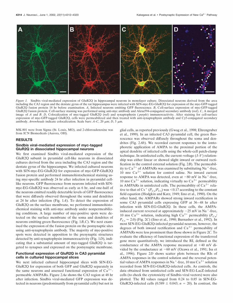
RESULTSSindbis viral-mediated expression of myc-taggedGluR2Q in dissociated hippocampal neuronsWe first examined Sindbis viral-mediated expression of theGluR2Q subunit in pyramidal cell-like neurons in dissociatedcultures derived from the area including the CA3 region and thedentate gyrus of the hippocampus. We infected cultured neuronswith SIN-myc-EG-GluR2Q for expression of myc-GFP-GluR2Qfusion protein and performed immunohistochemical staining us-ing myc-specific antibody 24 hr after infection in pyramidal cell-like neurons. GFP fluorescence from neurons infected with SIN-myc-EG-GluR2Q was observed as early as 6 hr, and one-half ofthe neurons emitted readily detectable levels of GFP fluorescencethat were diffusely observed throughout the soma and dendritesat 24 hr after infection (Fig. 1A). To detect the expression ofGluR2Q on the surface membrane, we performed immunohisto-chemical staining with anti-myc antibody under nonpermeabiliz-ing conditions. A large number of myc-positive spots were de-tected on the surface membrane of the soma and dendrites ofneurons emitting green fluorescence (Fig. 1B,C). We next exam-ined the expression of the fusion protein on the postsynaptic sitesusing anti-synaptophysin antibody. The majority of myc-positivespots were detected in apposition to the presynaptic structuresdetected by anti-synaptophysin immunoreactivity (Fig. 1D), indi-cating that a substantial amount of myc-tagged GluR2Q is tar-geted to synapses and expressed on the postsynaptic membrane.
Functional expression of GluR2Q in CA3 pyramidalcells in cultured hippocampal slicesWe next infected cultured hippocampal slices with SIN-EG-GluR2Q for expression of both GFP and GluR2Q separately inthe same neurons and assessed functional expression of Ca2�-permeable AMPARs. Figure 2Aa shows the CA3 region at 48 hrafter infection. Sindbis viral-mediated GFP expression was de-tected in neurons (predominantly from pyramidal cells) but not in
glial cells, as reported previously (Gwag et al., 1998; Ehrengruberet al., 1999). In an infected CA3 pyramidal cell, the green fluo-rescence was observed diffusely throughout the soma and den-drites (Fig. 2Ab). We recorded current responses to the ionto-phoretic application of AMPA to the proximal portion of theapical dendrite of infected cells using the whole-cell patch-clamptechnique. In uninfected cells, the current–voltage ( I–V) relation-ship was either linear or showed slight inward or outward recti-fication in the control external solution (Fig. 2B). The permeabil-ity to Ca 2� of AMPARs was examined by substituting Na�-free,10 mM Ca2� solution for control saline. No inward currentresponse to AMPA was detected, even at �80 mV in Na�-free,10 mM Ca2� solution, indicating virtually no Ca2� permeabilityin AMPARs in uninfected cells. The permeability of Ca2� rela-tive to that of Cs� (PCa/PCs) was �0.17 according to the constantfield equation (Hodgkin and Katz, 1949; Iino et al., 1990). On theother hand, the AMPARs showed strong inward rectification insome CA3 pyramidal cells expressing GFP at 36–48 hr afterinfection with SIN-EG-GluR2Q. In these cells, the AMPA-induced current reversed at approximately �25 mV in Na�-free,10 mM Ca2� solution, indicating high Ca2� permeability (PCa/PCs 2.0) (Fig. 2C) (Iino et al., 1990; Burnashev et al., 1992). Inmost SIN-EG-GluR2Q-infected pyramidal neurons, however, thedegrees of both inward rectification and Ca2� permeability ofAMPARs were less prominent than those shown in Figure 2C. Toevaluate the efficiency of functional expression of the transferredgene more quantitatively, we introduced the RI, defined as theconductance of the AMPA response measured at �40 mV di-vided by the conductance at �60 mV (Ozawa et al., 1991; Isa etal., 1996). Figure 2D shows scatter plots between the RIs ofAMPA responses in the control solution and the reversal poten-tial values of AMPA responses in Na�-free, 10 mM Ca2� solutionrecorded from SIN-EG-GluR2Q-infected cells. As controls, thedata obtained from uninfected cells and SIN-EG-LacZ-infectedcells (to check the cytotoxicity of Sindbis viral vectors) were alsoplotted. The RI values ranged from 0.24 to 0.85 in SIN-EG-GluR2Q-infected cells (0.589 � 0.043; n 20). In contrast, the
Figure 1. Sindbis viral-mediated expression of GluR2Q in hippocampal neurons in monolayer culture. Dissociated neurons derived from the areaincluding the CA3 region and the dentate gyrus of the rat hippocampus were infected with SIN-myc-EG-GluR2Q for expression of the myc-GFP-taggedGluR2Q fusion protein 18 hr before examination. A, Infected neurons emitting GFP fluorescence. B, Cell-surface expression of myc-GFP-taggedGluR2Q fusion protein. Cell-surface staining was performed using anti-myc antibody and Alexa594-conjugated secondary antibody (red). C, A mergedimage of A and B. D, Colocalization of myc-tagged GluR2Q (red) and synaptophysin ( purple) immunoreactivity. After staining for cell-surfaceexpression of myc-GFP-tagged GluR2Q, cells were permeabilized and then treated with anti-synaptophysin antibody and Cy5-conjugated secondaryantibody. Arrowheads indicate colocalization. Scale bars: A–C, 20 �m; D, 5 �m.
4314 J. Neurosci., June 1, 2002, 22(11):4312–4320 Kakegawa et al. • Postsynaptic Expression of New Ca2� Pathway
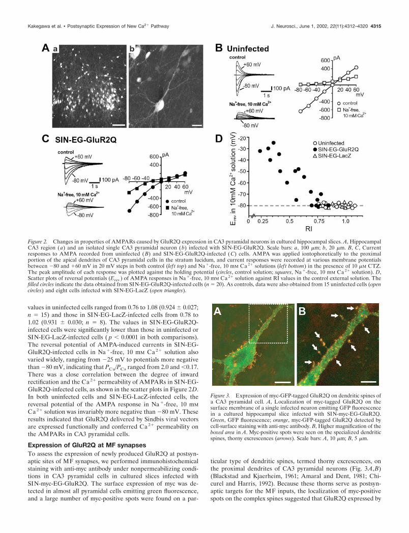
values in uninfected cells ranged from 0.76 to 1.08 (0.924 � 0.027;n 15) and those in SIN-EG-LacZ-infected cells from 0.78 to1.02 (0.931 � 0.030; n 8). The values in SIN-EG-GluR2Q-infected cells were significantly lower than those in uninfected orSIN-EG-LacZ-infected cells ( p � 0.0001 in both comparisons).The reversal potential of AMPA-induced currents in SIN-EG-GluR2Q-infected cells in Na�-free, 10 mM Ca2� solution alsovaried widely, ranging from �25 mV to potentials more negativethan �80 mV, indicating that PCa/PCs ranged from 2.0 and �0.17.There was a close correlation between the degree of inwardrectification and the Ca2� permeability of AMPARs in SIN-EG-GluR2Q-infected cells, as shown in the scatter plots in Figure 2D.In both uninfected cells and SIN-EG-LacZ-infected cells, thereversal potential of the AMPA response in Na�-free, 10 mM
Ca2� solution was invariably more negative than �80 mV. Theseresults indicated that GluR2Q delivered by Sindbis viral vectorsare expressed functionally and conferred Ca2� permeability onthe AMPARs in CA3 pyramidal cells.
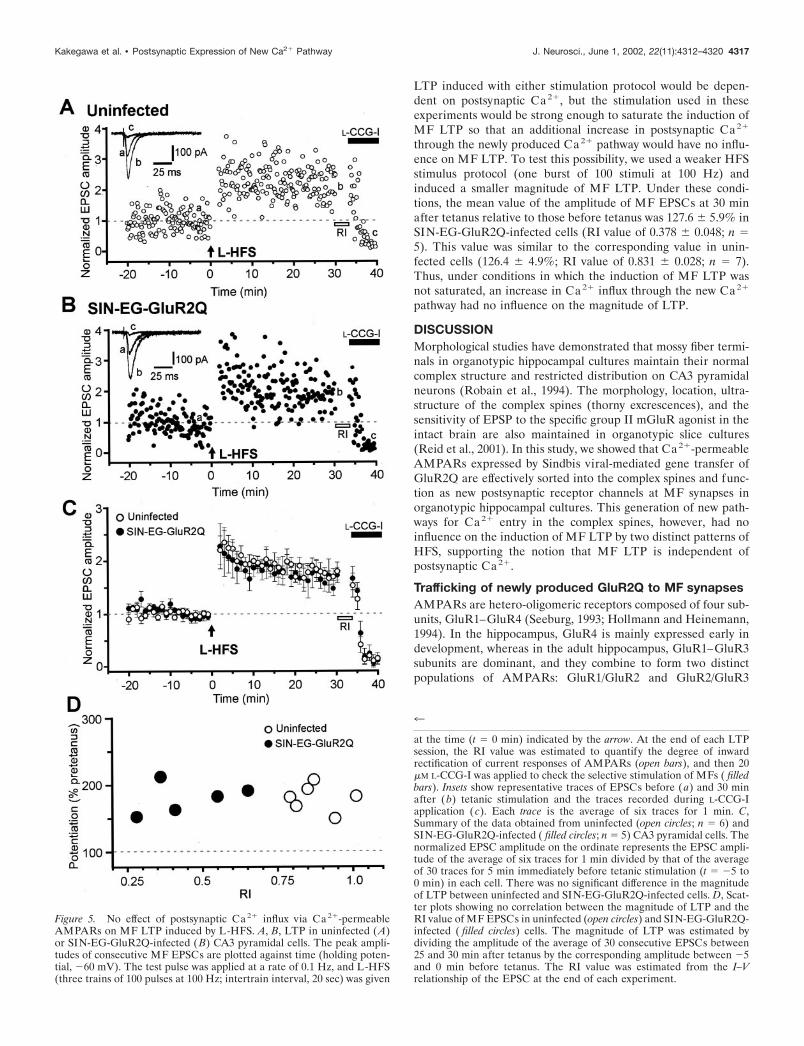
Expression of GluR2Q at MF synapsesTo assess the expression of newly produced GluR2Q at postsyn-aptic sites of MF synapses, we performed immunohistochemicalstaining with anti-myc antibody under nonpermeabilizing condi-tions in CA3 pyramidal cells in cultured slices infected withSIN-myc-EG-GluR2Q. The surface expression of myc was de-tected in almost all pyramidal cells emitting green fluorescence,and a large number of myc-positive spots were found on a par-
ticular type of dendritic spines, termed thorny excrescences, onthe proximal dendrites of CA3 pyramidal neurons (Fig. 3A,B)(Blackstad and Kjaerheim, 1961; Amaral and Dent, 1981; Chi-curel and Harris, 1992). Because these thorns serve as postsyn-aptic targets for the MF inputs, the localization of myc-positivespots on the complex spines suggested that GluR2Q expressed by
Figure 2. Changes in properties of AMPARs caused by GluR2Q expression in CA3 pyramidal neurons in cultured hippocampal slices. A, HippocampalCA3 region (a) and an isolated single CA3 pyramidal neuron (b) infected with SIN-EG-GluR2Q. Scale bars: a, 100 �m; b, 20 �m. B, C, Currentresponses to AMPA recorded from uninfected (B) and SIN-EG-GluR2Q-infected (C) cells. AMPA was applied iontophoretically to the proximalportion of the apical dendrites of CA3 pyramidal cells in the stratum lucidum, and current responses were recorded at various membrane potentialsbetween �80 and �60 mV in 20 mV steps in both control (lef t top) and Na �-free, 10 mM Ca 2� solutions (lef t bottom) in the presence of 10 �M CTZ.The peak amplitude of each response was plotted against the holding potential (circles, control solution; squares, Na �-free, 10 mM Ca 2� solution). D,Scatter plots of reversal potentials (Erev ) of AMPA responses in Na �-free, 10 mM Ca 2� solution against RI values in the control external solution. Thefilled circles indicate the data obtained from SIN-EG-GluR2Q-infected cells (n 20). As controls, data were also obtained from 15 uninfected cells (opencircles) and eight cells infected with SIN-EG-LacZ (open triangles).
Figure 3. Expression of myc-GFP-tagged GluR2Q on dendritic spines ofa CA3 pyramidal cell. A, Localization of myc-tagged GluR2Q on thesurface membrane of a single infected neuron emitting GFP fluorescencein a cultured hippocampal slice infected with SIN-myc-EG-GluR2Q.Green, GFP fluorescence; orange, myc-GFP-tagged GluR2Q detected bycell-surface staining with anti-myc antibody. B, Higher magnification of theboxed area in A. Myc-positive spots were seen on the specialized dendriticspines, thorny excrescences (arrows). Scale bars: A, 10 �m; B, 5 �m.
Kakegawa et al. • Postsynaptic Expression of New Ca2� Pathway J. Neurosci., June 1, 2002, 22(11):4312–4320 4315

the Sindbis virus was effectively targeted to the postsynaptic sitesat MF synapses.
We then examined whether GluR2Q targeted to the thornyexcrescences contributed to excitatory transmission at MF syn-apses by recording EPSCs evoked by stimulation of MFs fromCA3 pyramidal cells. In uninfected cells, the I–V relationship ofthe peak amplitude of the AMPA component of MF EPSC(AMPA EPSC) was either linear or exhibited a slight inward oroutward rectification in the control solution (Fig. 4A, C, opencircles). The RI value of the AMPA EPSC ranged from 0.78 to1.04 (0.898 � 0.013; n 31) (Fig. 4D). On the other hand, theAMPA EPSC showed inward rectification in SIN-EG-GluR2Q-infected cells (Fig. 4B, C, filled circles), and the RI value rangedfrom 0.23 to 0.81 (0.506 � 0.024; n 37) (Fig. 4D). This RI valuein SIN-EG-GluR2Q-infected cells was significantly lower thanthat in uninfected cells ( p � 0.0001). The significant reduction ofthe RI value in the AMPA EPSC by infection with SIN-EG-GluR2Q indicated that the EPSCs at MF synapses are generatedat least partially by activation of GluR2Q-including AMPARs.Thus, a new pathway for Ca2� influx into postsynaptic sites was
generated at MF synapses on CA3 pyramidal neurons by Sindbisviral-mediated gene transfer of GluR2Q.
Induction of MF LTP is independent of postsynapticCa2� influx via Ca2�-permeable AMPARsTo examine the influence of the newly expressed pathway forCa2� entry on MF LTP, we induced LTP by two differentpatterns of high-frequency stimulation, that is, long trains ofhigh-frequency stimulation (L-HFS) (three bursts of 100 stimuliat 100 Hz, given 20 sec apart) (Zalutsky and Nicoll, 1990) andbrief trains of high-frequency stimulation (B-HFS) (15 bursts ofseven stimuli at 100 Hz, repeated every 5 sec) (Jaffe andJohnston, 1990; Urban and Barrionuevo, 1996).
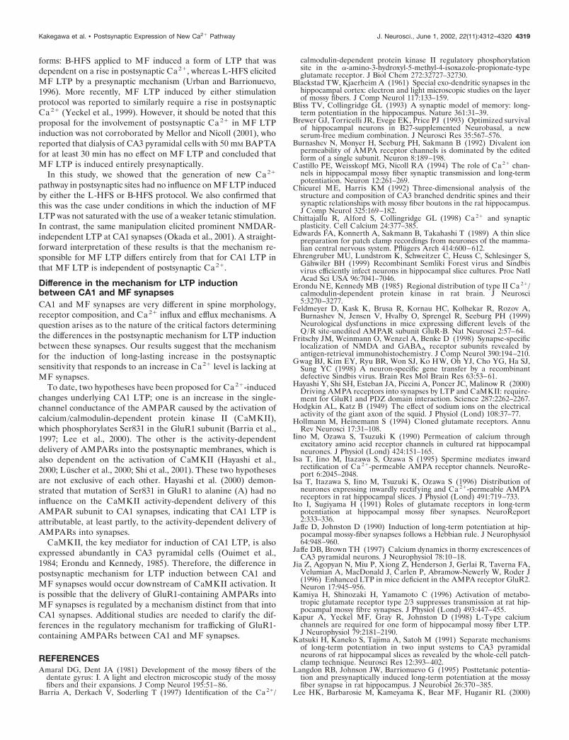
We first tested the influence of GluR2Q expression on MFLTP induced by L-HFS. MF EPSCs were recorded from CA3pyramidal cells infected with SIN-EG-GluR2Q. To determinewhether EPSCs were evoked monosynaptically by stimulation ofMFs, we applied L-CCG-I, a specific agonist of group II mGluRs,at the end of each experiment. The addition of 20 �M L-CCG-Ireduced the amplitude of the EPSC to �10% of control, indicat-ing that the signal was generated predominantly by MF inputs(Kamiya et al., 1996). As a control, LTP recording was performedfrom uninfected CA3 pyramidal cells under the same stimulusconditions. The L-HFS induced prominent LTP in both unin-fected and SIN-EG-GluR2Q-infected cells (Fig. 5A,B). Themean value of the amplitude of MF EPSCs at 30 min aftertetanus relative to that before tetanus was 172.5 � 11.2% (n 5)in SIN-EG-GluR2Q-infected cells, similar to that in uninfectedcells (173.4 � 9.6%; n 6). The time course of post-tetanicpotentiation (PTP) was also similar (Fig. 5C). The mean RI valueof the MF EPSC reflecting the degree of contribution of Ca2�-permeable AMPARs was 0.450 � 0.067 in the SIN-EG-GluR2Q-infeceted cells (n 5). This value was significantly lower than thecorresponding value in uninfected cells (0.878 � 0.034; n 6; p �0.05). There was no significant correlation between the magni-tude of LTP and the RI value (Fig. 5D), indicating that the newlyproduced Ca2� pathway at the postsynaptic membrane had noinfluence on MF LTP induced by L-HFS.
It has been reported that L-HFS and B-HFS induce twodistinct types of MF LTP, each having different time courses anddifferent sensitivities to intracellular dialysis during whole-cellrecordings (Urban et al., 1994; Langdon et al., 1995). In particu-lar, B-HFS has been suggested to induce a form of LTP thatdepends on an initial postsynaptic event (Urban and Barrionuevo,1996). Therefore, we next examined the influence of GluR2Qexpression on MF LTP induced by B-HFS. This pattern oftetanic stimulation elicited stable LTP in both uninfected andSIN-EG-GluR2Q-infected cells (Fig. 6A,B). In both groups ofcells, LTP induced by B-HFS showed a similar time course withno PTP, which was prominent in LTP induced by L-HFS (Fig.6C), as described previously (Urban and Barrionuevo, 1996). Themean value of the amplitude of MF EPSCs at 30 min aftertetanus relative to those before tetanus was 161.3 � 12.8% inSIN-EG-GluR2Q-infected cells (RI value of 0.416 � 0.070; n 5). This value was similar to the corresponding value in unin-fected cells (157.9 � 9.6%; RI value of 0.854 � 0.026; n 5).Furthermore, there was no significant correlation between themagnitude of LTP and the RI value (Fig. 6D). Thus, the newlyproduced Ca2� pathway had no influence on the B-HFS-inducedLTP, either.
The above results strongly suggested that MF LTP is indepen-dent of postsynaptic Ca2�. However, it could be argued that MF
Figure 4. Changes in properties of AMPA EPSCs caused by GluR2Qexpression at hippocampal MF synapses. A, B, Representative AMPAEPSCs evoked by stimulation of MFs in uninfected (A) and SIN-EG-GluR2Q-infected (B) CA3 pyramidal cells. The EPSCs were recorded atvarious membrane potentials between �80 and �60 mV in 20 mV stepsin the presence of 25 �M D-APV and 20 �M MK-801. To reduce polysyn-aptic excitation, 5 �M 2-chloroadenosine was also applied to the externalsolution. C, I–V relationship of AMPA EPSCs shown in A and B. Theamplitudes of EPSCs normalized to those at �60 mV were plotted againstthe holding potential. The open and filled circles indicate the data fromuninfected cells and those infected with SIN-EG-GluR2Q, respectively.D, Histogram showing the distribution of RI values of AMPA EPSCs inSIN-EG-GluR2Q-infected cells ( filled bars; n 37) and uninfected cells(open bars; n 31).
4316 J. Neurosci., June 1, 2002, 22(11):4312–4320 Kakegawa et al. • Postsynaptic Expression of New Ca2� Pathway
LTP induced with either stimulation protocol would be depen-dent on postsynaptic Ca2�, but the stimulation used in theseexperiments would be strong enough to saturate the induction ofMF LTP so that an additional increase in postsynaptic Ca2�
through the newly produced Ca2� pathway would have no influ-ence on MF LTP. To test this possibility, we used a weaker HFSstimulus protocol (one burst of 100 stimuli at 100 Hz) andinduced a smaller magnitude of MF LTP. Under these condi-tions, the mean value of the amplitude of MF EPSCs at 30 minafter tetanus relative to those before tetanus was 127.6 � 5.9% inSIN-EG-GluR2Q-infected cells (RI value of 0.378 � 0.048; n 5). This value was similar to the corresponding value in unin-fected cells (126.4 � 4.9%; RI value of 0.831 � 0.028; n 7).Thus, under conditions in which the induction of MF LTP wasnot saturated, an increase in Ca2� influx through the new Ca2�
pathway had no influence on the magnitude of LTP.
DISCUSSIONMorphological studies have demonstrated that mossy fiber termi-nals in organotypic hippocampal cultures maintain their normalcomplex structure and restricted distribution on CA3 pyramidalneurons (Robain et al., 1994). The morphology, location, ultra-structure of the complex spines (thorny excrescences), and thesensitivity of EPSP to the specific group II mGluR agonist in theintact brain are also maintained in organotypic slice cultures(Reid et al., 2001). In this study, we showed that Ca2�-permeableAMPARs expressed by Sindbis viral-mediated gene transfer ofGluR2Q are effectively sorted into the complex spines and func-tion as new postsynaptic receptor channels at MF synapses inorganotypic hippocampal cultures. This generation of new path-ways for Ca2� entry in the complex spines, however, had noinfluence on the induction of MF LTP by two distinct patterns ofHFS, supporting the notion that MF LTP is independent ofpostsynaptic Ca2�.
Trafficking of newly produced GluR2Q to MF synapsesAMPARs are hetero-oligomeric receptors composed of four sub-units, GluR1–GluR4 (Seeburg, 1993; Hollmann and Heinemann,1994). In the hippocampus, GluR4 is mainly expressed early indevelopment, whereas in the adult hippocampus, GluR1–GluR3subunits are dominant, and they combine to form two distinctpopulations of AMPARs: GluR1/GluR2 and GluR2/GluR3
Figure 5. No effect of postsynaptic Ca 2� influx via Ca 2�-permeableAMPARs on MF LTP induced by L-HFS. A, B, LTP in uninfected (A)or SIN-EG-GluR2Q-infected (B) CA3 pyramidal cells. The peak ampli-tudes of consecutive MF EPSCs are plotted against time (holding poten-tial, �60 mV). The test pulse was applied at a rate of 0.1 Hz, and L-HFS(three trains of 100 pulses at 100 Hz; intertrain interval, 20 sec) was given
4
at the time (t 0 min) indicated by the arrow. At the end of each LTPsession, the RI value was estimated to quantify the degree of inwardrectification of current responses of AMPARs (open bars), and then 20�M L-CCG-I was applied to check the selective stimulation of MFs ( filledbars). Insets show representative traces of EPSCs before (a) and 30 minafter (b) tetanic stimulation and the traces recorded during L-CCG-Iapplication (c). Each trace is the average of six traces for 1 min. C,Summary of the data obtained from uninfected (open circles; n 6) andSIN-EG-GluR2Q-infected ( filled circles; n 5) CA3 pyramidal cells. Thenormalized EPSC amplitude on the ordinate represents the EPSC ampli-tude of the average of six traces for 1 min divided by that of the averageof 30 traces for 5 min immediately before tetanic stimulation (t �5 to0 min) in each cell. There was no significant difference in the magnitudeof LTP between uninfected and SIN-EG-GluR2Q-infected cells. D, Scat-ter plots showing no correlation between the magnitude of LTP and theRI value of MF EPSCs in uninfected (open circles) and SIN-EG-GluR2Q-infected ( filled circles) cells. The magnitude of LTP was estimated bydividing the amplitude of the average of 30 consecutive EPSCs between25 and 30 min after tetanus by the corresponding amplitude between �5and 0 min before tetanus. The RI value was estimated from the I–Vrelationship of the EPSC at the end of each experiment.
Kakegawa et al. • Postsynaptic Expression of New Ca2� Pathway J. Neurosci., June 1, 2002, 22(11):4312–4320 4317

hetero-oligomers (Wenthold et al., 1996). Recently, Shi et al.(2001) showed that these AMPARs display different synapticdelivery mechanisms at hippocampal CA1 synapses in organo-typic slice cultures. GluR1/GluR2 receptors are added to syn-apses in an activity-dependent manner during plasticity. Thisrequires interaction between GluR1 and group I PDZ (postsyn-aptic density-95/Discs large/zona occludens-1) domain proteins.In contrast, GluR2/GluR3 receptors replace existing synapticreceptors continuously in an activity-independent manner, whichrequires interactions by GluR2 with N-ethylmaleimide-sensitivefactor and group II PDZ domain proteins (Shi et al., 2001). Ithas been shown that GluR2Q is readily incorporated into hip-pocampal CA1 synapses to form postsynaptic Ca2�-permeableAMPARs by the Sindbis virus expression system in an activity-independent manner in organotypic hippocampal cultures (Okadaet al., 2001; Shi et al., 2001). Because expression of GluR2 usingthis system led predominantly to the formation of homomericrecombinant receptors, it was suggested that newly producedGluR2Q homomeric AMPARs were incorporated into CA1 syn-apses and functioned as postsynaptic receptors (Shi et al., 2001).
In this study, we showed that GluR2Q AMPARs expressed bySindbis viral-mediated gene transfer are also effectively sortedinto thorny excrescences and function as new postsynaptic recep-tor channels at MF synapses. This synaptic delivery of GluR2Qoccurred in the presence of tetrodotoxin or CNQX (our unpub-lished data). It is thus likely that the mechanism for the activity-independent synaptic delivery of GluR2Q at MF synapses issimilar to that at CA1 synapses. However, it remains to bedetermined whether the synaptic delivery of AMPARs contain-ing GluR1 subunits also occurs in an activity-dependent mannerat MF synapses.
No influence of generation of new Ca2� pathways onMF LTPCA1 LTP is induced by postsynaptic Ca2� influx via NMDARs.In contrast, MF LTP has been proposed to be induced by apresynaptic mechanism (Staubli et al., 1990; Zalutsky and Nicoll,1990; Ito and Sugiyama, 1991; Katsuki et al., 1991; Castillo et al.,1994; Mellor and Nicoll, 2001). The difference in the mechanismfor LTP induction between the two synapses could be attributableto differences in the distribution of NMDARs in the postsynapticsites. NMDARs are distributed most abundantly at CA1 den-dritic spines postsynaptic to the Schaffer collaterals, whereas NR1and NR2A subunits are of low abundance and NR2B is almostundetectable in CA3 complex spines postsynaptic to MFs (Mon-aghan and Cotman, 1985; Fritschy et al., 1998; Watanabe et al.,1998). However, sufficient levels of Ca2� ions have been shown tobe supplied by Ca2� entry through VDCCs and/or Ca2� mobi-lization from internal stores during activation of MF synapses(Jaffe and Brown 1997; Yeckel et al.; 1999; Reid et al., 2001).Several reports have suggested that MF LTP is dependent on anincrease in postsynaptic Ca2� level (Williams and Johnston,1989; Jaffe and Johnston, 1990). Later, it was proposed thatNMDAR-independent MF LTP should be subdivided into two
Figure 6. No effect of postsynaptic Ca 2� influx via Ca 2�-permeableAMPARs on MF LTP induced by B-HFS. Experimental arrangementswere identical to those in Fig. 5, except that B-HFS (15 bursts of 7 stimuliat 100 Hz, repeated every 5 sec) was used, instead of L-HFS, for inductionof LTP. A, B, LTP in uninfected (A) or SIN-EG-GluR2Q-infected (B)
4
CA3 pyramidal cells. C, Summary of the data from uninfected (opencircles; n 5) and SIN-EG-GluR2Q-infected ( filled circles; n 5) CA3pyramidal cells. There was no significant difference in the magnitude ofLTP between uninfected and SIN-EG-GluR2Q-infected cells. D, Scatterplots showing no correlation between the magnitude of LTP and the RIvalue of MF EPSCs in uninfected (open circles) and SIN-EG-GluR2Q-infected ( filled circles) cells.
4318 J. Neurosci., June 1, 2002, 22(11):4312–4320 Kakegawa et al. • Postsynaptic Expression of New Ca2� Pathway
forms: B-HFS applied to MF induced a form of LTP that wasdependent on a rise in postsynaptic Ca2�, whereas L-HFS elicitedMF LTP by a presynaptic mechanism (Urban and Barrionuevo,1996). More recently, MF LTP induced by either stimulationprotocol was reported to similarly require a rise in postsynapticCa2� (Yeckel et al., 1999). However, it should be noted that thisproposal for the involvement of postsynaptic Ca2� in MF LTPinduction was not corroborated by Mellor and Nicoll (2001), whoreported that dialysis of CA3 pyramidal cells with 50 mM BAPTAfor at least 30 min has no effect on MF LTP and concluded thatMF LTP is induced entirely presynaptically.
In this study, we showed that the generation of new Ca2�
pathway in postsynaptic sites had no influence on MF LTP inducedby either the L-HFS or B-HFS protocol. We also confirmed thatthis was the case under conditions in which the induction of MFLTP was not saturated with the use of a weaker tetanic stimulation.In contrast, the same manipulation elicited prominent NMDAR-independent LTP at CA1 synapses (Okada et al., 2001). A straight-forward interpretation of these results is that the mechanism re-sponsible for MF LTP differs entirely from that for CA1 LTP inthat MF LTP is independent of postsynaptic Ca2�.
Difference in the mechanism for LTP inductionbetween CA1 and MF synapsesCA1 and MF synapses are very different in spine morphology,receptor composition, and Ca2� influx and efflux mechanisms. Aquestion arises as to the nature of the critical factors determiningthe differences in the postsynaptic mechanism for LTP inductionbetween these synapses. Our results suggest that the mechanismfor the induction of long-lasting increase in the postsynapticsensitivity that responds to an increase in Ca2� level is lacking atMF synapses.
To date, two hypotheses have been proposed for Ca2�-inducedchanges underlying CA1 LTP; one is an increase in the single-channel conductance of the AMPAR caused by the activation ofcalcium/calmodulin-dependent protein kinase II (CaMKII),which phosphorylates Ser831 in the GluR1 subunit (Barria et al.,1997; Lee et al., 2000). The other is the activity-dependentdelivery of AMPARs into the postsynaptic membranes, which isalso dependent on the activation of CaMKII (Hayashi et al.,2000; Luscher et al., 2000; Shi et al., 2001). These two hypothesesare not exclusive of each other. Hayashi et al. (2000) demon-strated that mutation of Ser831 in GluR1 to alanine (A) had noinfluence on the CaMKII activity-dependent delivery of thisAMPAR subunit to CA1 synapses, indicating that CA1 LTP isattributable, at least partly, to the activity-dependent delivery ofAMPARs into synapses.
CaMKII, the key mediator for induction of CA1 LTP, is alsoexpressed abundantly in CA3 pyramidal cells (Ouimet et al.,1984; Erondu and Kennedy, 1985). Therefore, the difference inpostsynaptic mechanism for LTP induction between CA1 andMF synapses would occur downstream of CaMKII activation. Itis possible that the delivery of GluR1-containing AMPARs intoMF synapses is regulated by a mechanism distinct from that intoCA1 synapses. Additional studies are needed to clarify the dif-ferences in the regulatory mechanism for trafficking of GluR1-containing AMPARs between CA1 and MF synapses.
REFERENCESAmaral DG, Dent JA (1981) Development of the mossy fibers of the
dentate gyrus: I. A light and electron microscopic study of the mossyfibers and their expansions. J Comp Neurol 195:51–86.
Barria A, Derkach V, Soderling T (1997) Identification of the Ca 2�/
calmodulin-dependent protein kinase II regulatory phosphorylationsite in the �-amino-3-hydroxyl-5-methyl-4-isoxazole-propionate-typeglutamate receptor. J Biol Chem 272:32727–32730.
Blackstad TW, Kjaerheim A (1961) Special exo-dendritic synapses in thehippocampal cortex: electron and light microscopic studies on the layerof mossy fibers. J Comp Neurol 117:133–159.
Bliss TV, Collingridge GL (1993) A synaptic model of memory: long-term potentiation in the hippocampus. Nature 361:31–39.
Brewer GJ, Torricelli JR, Evege EK, Price PJ (1993) Optimized survivalof hippocampal neurons in B27-supplemented Neurobasal, a newserum-free medium combination. J Neurosci Res 35:567–576.
Burnashev N, Monyer H, Seeburg PH, Sakmann B (1992) Divalent ionpermeability of AMPA receptor channels is dominated by the editedform of a single subunit. Neuron 8:189–198.
Castillo PE, Weisskopf MG, Nicoll RA (1994) The role of Ca 2� chan-nels in hippocampal mossy fiber synaptic transmission and long-termpotentiation. Neuron 12:261–269.
Chicurel ME, Harris KM (1992) Three-dimensional analysis of thestructure and composition of CA3 branched dendritic spines and theirsynaptic relationships with mossy fiber boutons in the rat hippocampus.J Comp Neurol 325:169–182.
Chittajallu R, Alford S, Collingridge GL (1998) Ca 2� and synapticplasticity. Cell Calcium 24:377–385.
Edwards FA, Konnerth A, Sakmann B, Takahashi T (1989) A thin slicepreparation for patch clamp recordings from neurones of the mamma-lian central nervous system. Pflugers Arch 414:600–612.
Ehrengruber MU, Lundstrom K, Schweitzer C, Heuss C, Schlesinger S,Gahwiler BH (1999) Recombinant Semliki Forest virus and Sindbisvirus efficiently infect neurons in hippocampal slice cultures. Proc NatlAcad Sci USA 96:7041–7046.
Erondu NE, Kennedy MB (1985) Regional distribution of type II Ca 2�/calmodulin-dependent protein kinase in rat brain. J Neurosci5:3270–3277.
Feldmeyer D, Kask K, Brusa R, Kornau HC, Kolhekar R, Rozov A,Burnashev N, Jensen V, Hvalby O, Sprengel R, Seeburg PH (1999)Neurological dysfunctions in mice expressing different levels of theQ/R site-unedited AMPAR subunit GluR-B. Nat Neurosci 2:57–64.
Fritschy JM, Weinmann O, Wenzel A, Benke D (1998) Synapse-specificlocalization of NMDA and GABAA receptor subunits revealed byantigen-retrieval immunohistochemistry. J Comp Neurol 390:194–210.
Gwag BJ, Kim EY, Ryu BR, Won SJ, Ko HW, Oh YJ, Cho YG, Ha SJ,Sung YC (1998) A neuron-specific gene transfer by a recombinantdefective Sindbis virus. Brain Res Mol Brain Res 63:53–61.
Hayashi Y, Shi SH, Esteban JA, Piccini A, Poncer JC, Malinow R (2000)Driving AMPA receptors into synapses by LTP and CaMKII: require-ment for GluR1 and PDZ domain interaction. Science 287:2262–2267.
Hodgkin AL, Katz B (1949) The effect of sodium ions on the electricalactivity of the giant axon of the squid. J Physiol (Lond) 108:37–77.
Hollmann M, Heinemann S (1994) Cloned glutamate receptors. AnnuRev Neurosci 17:31–108.
Iino M, Ozawa S, Tsuzuki K (1990) Permeation of calcium throughexcitatory amino acid receptor channels in cultured rat hippocampalneurones. J Physiol (Lond) 424:151–165.
Isa T, Iino M, Itazawa S, Ozawa S (1995) Spermine mediates inwardrectification of Ca 2�-permeable AMPA receptor channels. NeuroRe-port 6:2045–2048.
Isa T, Itazawa S, Iino M, Tsuzuki K, Ozawa S (1996) Distribution ofneurones expressing inwardly rectifying and Ca 2�-permeable AMPAreceptors in rat hippocampal slices. J Physiol (Lond) 491:719–733.
Ito I, Sugiyama H (1991) Roles of glutamate receptors in long-termpotentiation at hippocampal mossy fiber synapses. NeuroReport2:333–336.
Jaffe D, Johnston D (1990) Induction of long-term potentiation at hip-pocampal mossy-fiber synapses follows a Hebbian rule. J Neurophysiol64:948–960.
Jaffe DB, Brown TH (1997) Calcium dynamics in thorny excrescences ofCA3 pyramidal neurons. J Neurophysiol 78:10–18.
Jia Z, Agopyan N, Miu P, Xiong Z, Henderson J, Gerlai R, Taverna FA,Velumian A, MacDonald J, Carlen P, Abramow-Newerly W, Roder J(1996) Enhanced LTP in mice deficient in the AMPA receptor GluR2.Neuron 17:945–956.
Kamiya H, Shinozaki H, Yamamoto C (1996) Activation of metabo-tropic glutamate receptor type 2/3 suppresses transmission at rat hip-pocampal mossy fibre synapses. J Physiol (Lond) 493:447–455.
Kapur A, Yeckel MF, Gray R, Johnston D (1998) L-Type calciumchannels are required for one form of hippocampal mossy fiber LTP.J Neurophysiol 79:2181–2190.
Katsuki H, Kaneko S, Tajima A, Satoh M (1991) Separate mechanismsof long-term potentiation in two input systems to CA3 pyramidalneurons of rat hippocampal slices as revealed by the whole-cell patch-clamp technique. Neurosci Res 12:393–402.
Langdon RB, Johnson JW, Barrionuevo G (1995) Posttetanic potentia-tion and presynaptically induced long-term potentiation at the mossyfiber synapse in rat hippocampus. J Neurobiol 26:370–385.
Lee HK, Barbarosie M, Kameyama K, Bear MF, Huganir RL (2000)
Kakegawa et al. • Postsynaptic Expression of New Ca2� Pathway J. Neurosci., June 1, 2002, 22(11):4312–4320 4319

Regulation of distinct AMPA receptor phosphorylation sites duringbidirectional synaptic plasticity. Nature 405:955–959.
Luscher C, Nicoll RA, Malenka RC, Muller D (2000) Synaptic plasticityand dynamic modulation of the postsynaptic membrane. Nat Neurosci3:545–550.
Malinow R, Mainen ZF, Hayashi Y (2000) LTP mechanisms: from si-lence to four-lane traffic. Curr Opin Neurobiol 10:352–357.
Mellor J, Nicoll RA (2001) Hippocampal mossy fiber LTP is indepen-dent of postsynaptic calcium. Nat Neurosci 4:125–126.
Monaghan DT, Cotman CW (1985) Distribution of N-methyl-D-aspartate-sensitive L-[ 3H]glutamate-binding sites in rat brain. J Neuro-sci 5:2909–2919.
Nicoll RA, Malenka RC (1999) Expression mechanisms underlyingNMDA receptor-dependent long-term potentiation. Ann NY Acad Sci868:515–525.
Okada T, Yamada N, Kakegawa W, Tsuzuki K, Kawamura M, Nawa H,Iino M, Ozawa S (2001) Sindbis viral-mediated expression of Ca 2�-permeable AMPA receptors at hippocampal CA1 synapses and induc-tion of NMDA receptor-independent long-term potentiation. EurJ Neurosci 13:1635–1643.
Ouimet CC, McGuinness TL, Greengard P (1984) Immunocytochemicallocalization of calcium/calmodulin-dependent protein kinase II in ratbrain. Proc Natl Acad Sci USA 81:5604–5608.
Ozawa S, Iino M, Tsuzuki K (1991) Two types of kainate response incultured rat hippocampal neurons. J Neurophysiol 66:2–11.
Reid CA, Fabian-Fine R, Fine A (2001) Postsynaptic calcium transientsevoked by activation of individual hippocampal mossy fiber synapses.J Neurosci 21:2206–2214.
Robain O, Barbin G, Billette d, V, Jardin L, Jahchan T, Ben Ari Y (1994)Development of mossy fiber synapses in hippocampal slice culture.Brain Res Dev Brain Res 80:244–250.
Seeburg PH (1993) The TINS/TiPS Lecture. The molecular biology ofmammalian glutamate receptor channels. Trends Neurosci 16:359–365.
Shi SH, Hayashi Y, Esteban JA, Malinow R (2001) Subunit-specific rules
governing AMPA receptor trafficking to synapses in hippocampal py-ramidal neurons. Cell 105:331–343.
Staubli U, Larson J, Lynch G (1990) Mossy fiber potentiation and long-term potentiation involve different expression mechanisms. Synapse5:333–335.
Stoppini L, Buchs PA, Muller D (1991) A simple method for organotypiccultures of nervous tissue. J Neurosci Methods 37:173–182.
Stuart GJ, Sakmann B (1994) Active propagation of somatic action po-tentials into neocortical pyramidal cell dendrites. Nature 367:69–72.
Urban NN, Barrionuevo G (1996) Induction of hebbian and non-hebbian mossy fiber long-term potentiation by distinct patterns ofhigh-frequency stimulation. J Neurosci 16:4293–4299.
Urban NN, Langdon RB, Barrionuevo G (1994) The duration and pat-tern of high frequency stimulation influence the time course of poten-tiation and the mossy fiber-CA3 synapse. Soc Neurosci Abstr 20:715.
Watanabe M, Fukaya M, Sakimura K, Manabe T, Mishina M, Inoue Y(1998) Selective scarcity of NMDA receptor channel subunits in thestratum lucidum (mossy fibre-recipient layer) of the mouse hippocam-pal CA3 subfield. Eur J Neurosci 10:478–487.
Wenthold RJ, Petralia RS, Blahos II J, Niedzielski AS (1996) Evidencefor multiple AMPA receptor complexes in hippocampal CA1/CA2neurons. J Neurosci 16:1982–1989.
Williams S, Johnston D (1989) Long-term potentiation of hippocampalmossy fiber synapses is blocked by postsynaptic injection of calciumchelators. Neuron 3:583–588.
Yamada KA, Tang CM (1993) Benzothiadiazides inhibit rapid gluta-mate receptor desensitization and enhance glutamatergic synaptic cur-rents. J Neurosci 13:3904–3915.
Yeckel MF, Kapur A, Johnston D (1999) Multiple forms of LTP inhippocampal CA3 neurons use a common postsynaptic mechanism. NatNeurosci 2:625–633.
Zalutsky RA, Nicoll RA (1990) Comparison of two forms of long-termpotentiation in single hippocampal neurons. Science 248:1619–1624.
4320 J. Neurosci., June 1, 2002, 22(11):4312–4320 Kakegawa et al. • Postsynaptic Expression of New Ca2� Pathway