860.full.pdf - The Journal of Immunology
10
of March 22, 2022. This information is current as Protein SNFT Novel Basic Leucine Zipper p21 Repression of IL-2 Promoter Activity by the McGuire Milena Iacobelli, William Wachsman and Kathleen L. http://www.jimmunol.org/content/165/2/860 doi: 10.4049/jimmunol.165.2.860 2000; 165:860-868; ; J Immunol References http://www.jimmunol.org/content/165/2/860.full#ref-list-1 , 31 of which you can access for free at: cites 49 articles This article average * 4 weeks from acceptance to publication Fast Publication! • Every submission reviewed by practicing scientists No Triage! • from submission to initial decision Rapid Reviews! 30 days* • Submit online. ? The JI Why Subscription http://jimmunol.org/subscription is online at: The Journal of Immunology Information about subscribing to Permissions http://www.aai.org/About/Publications/JI/copyright.html Submit copyright permission requests at: Email Alerts http://jimmunol.org/alerts Receive free email-alerts when new articles cite this article. Sign up at: Print ISSN: 0022-1767 Online ISSN: 1550-6606. Immunologists All rights reserved. Copyright © 2000 by The American Association of 1451 Rockville Pike, Suite 650, Rockville, MD 20852 The American Association of Immunologists, Inc., is published twice each month by The Journal of Immunology by guest on March 22, 2022 http://www.jimmunol.org/ Downloaded from by guest on March 22, 2022 http://www.jimmunol.org/ Downloaded from
-
Upload
khangminh22 -
Category
Documents
-
view
1 -
download
0
Transcript of 860.full.pdf - The Journal of Immunology
of March 22, 2022.This information is current as
Protein SNFTNovel Basic Leucine Zipper p21
Repression of IL-2 Promoter Activity by the
McGuireMilena Iacobelli, William Wachsman and Kathleen L.
http://www.jimmunol.org/content/165/2/860doi: 10.4049/jimmunol.165.2.860
2000; 165:860-868; ;J Immunol
Referenceshttp://www.jimmunol.org/content/165/2/860.full#ref-list-1
, 31 of which you can access for free at: cites 49 articlesThis article
average*
4 weeks from acceptance to publicationFast Publication! •
Every submission reviewed by practicing scientistsNo Triage! •
from submission to initial decisionRapid Reviews! 30 days* •
Submit online. ?The JIWhy
Subscriptionhttp://jimmunol.org/subscription
is online at: The Journal of ImmunologyInformation about subscribing to
Permissionshttp://www.aai.org/About/Publications/JI/copyright.htmlSubmit copyright permission requests at:
Email Alertshttp://jimmunol.org/alertsReceive free email-alerts when new articles cite this article. Sign up at:
Print ISSN: 0022-1767 Online ISSN: 1550-6606. Immunologists All rights reserved.Copyright © 2000 by The American Association of1451 Rockville Pike, Suite 650, Rockville, MD 20852The American Association of Immunologists, Inc.,
is published twice each month byThe Journal of Immunology
by guest on March 22, 2022
http://ww
w.jim
munol.org/
Dow
nloaded from
by guest on March 22, 2022
http://ww
w.jim
munol.org/
Dow
nloaded from
Repression of IL-2 Promoter Activity by the Novel BasicLeucine Zipper p21SNFT Protein1,2
Milena Iacobelli,* William Wachsman,† and Kathleen L. McGuire3*
IL-2 is the major autocrine and paracrine growth factor produced by T cells upon T cell stimulation. The inducible expressionof IL-2 is highly regulated by multiple transcription factors, particularly AP-1, which coordinately activate the promoter. De-scribed here is the ability of the novel basic leucine zipper protein p21SNFT to repress AP-1 activity and IL-2 transcription. Adetailed analysis of the repression by p21SNFT repression on the IL-2 promoter distal NF-AT/AP-1 site demonstrates that it canbind DNA with NF-AT and Jun, strongly suggesting that it represses NF-AT/AP-1 activity by competing with Fos proteins for Jundimerization. The importance of this repression is that p21SNFT inhibits the trans-activation potential of protein complexes thatcontain Jun, thereby demonstrating an additional level of control for the highly regulated, ubiquitous AP-1 transcription factorand the IL-2 gene. The Journal of Immunology,2000, 165: 860–868.
A ctivated Th cells of the immune system produce highlevels of the IL-2 cytokine when properly stimulatedthrough both the TCR and the CD28 costimulatory mol-
ecule (1, 2). IL-2 production is indicative of T cell activation andis the major autocrine and paracrine growth factor for T cells.While not produced by normal, quiescent T cells, the IL-2 generapidly becomes transcriptionally active upon stimulation. IL-2mRNA production is detectable within 40 min, peaks at 4–6 h, andreturns to background levels by 24 h (2, 3). Although control ofIL-2 protein synthesis occurs at many levels, it is principally reg-ulated at the level of transcription.
Although there is evidence that sequences outside of the;300-bp human IL-2 promoter contribute to transcriptional con-trol in vivo, this region is primarily responsible for inducible ex-pression of the gene (4). The promoter contains a variety of bind-ing sites for known activating transcription factors, such as NF-AT, NF-kB, AP-1, and OCT,4 as well as the inhibitory proteinNIL-2 (2, 5). All four of the activating factors have been shown tohave multiple binding sites within the promoter. In addition, theproximal OCT (6, 7), the CD28 response element (8–10), and thedistal NF-AT (11, 12) elements have all been shown to work with
adjacent AP-1 or AP-1-like sites for binding and/or functional ac-tivity. The significance of AP-1 for the activity of this promoterhas been demonstrated for the positive regulation of IL-2 expres-sion, as well as in anergic T cells, where a lack of AP-1 activity isthought to be responsible for the inhibition of IL-2 production(13–17).
The AP-1 transcription factor consists of heterodimers betweenthe Fos and Jun families of proteins through their basic leucinezipper (bZIP) domains. The expression andtrans-activation po-tential of the AP-1 transcription factor are highly dependent on theactivation of stress and mitogenic signal transduction cascades(18). Activation of the JNK and MEK MAP kinase pathways notonly controls the overall levels of Fos and Jun proteins, but alsoleads to the proper phosphorylation states of these proteins, whichare required for transcriptional activity (18). Although c-Jun andc-Fos dimers are considered to be the classical AP-1 transcriptionfactor, a variety of dimer combinations can exist among Fos, Jun,and CREB/ATF family members, lending specificity of a particu-lar dimer pair to an AP-1 or AP-1-like enhancer (TRE). Dimers ofJun and Fos or CREB/ATF have been shown to regulate the ex-pression of a variety of immunologically important genes involvedin T cell activation and cytokine expression (19).
Additional proteins have been described that are capable ofdimerizing with Jun but that do not belong to either the Fos orCREB/ATF family. These rat proteins are called Jun dimerizationproteins 1 and 2 (JDP1 and JDP-2) and are capable of repressingthe expression of an AP-1-regulated reporter construct (20).JDP-1, like the closely related human B-ATF protein, can bindwith Jun to a TRE sequence (20, 21). JDP-1 and B-ATF are alsoclosely related to the Merek’s disease virus EcoQ protein (MEQ),another bZIP protein capable of Jun dimerization but whose inter-actions with Jun lead to viral gene transcription (22). It currentlyis not clear what the physiological relevance is of Jun interactionswith proteins such as B-ATF, JDP1, and JDP-2, because their abil-ity to dimerize with Jun has not been shown to influence the tran-scription of any cellular gene.
Recently, a novel human protein named p21SNFT (for 21-kDasmall nuclear factor isolated from T cells) was identified thatdimerizes with Jun proteins and represses AP-1 activity (W.Klump and W. Wachsman, manuscript in preparation). The bio-chemical characteristics of p21SNFT are similar to the known ac-tivities of the JDP proteins, and it shares high amino acid identity
*Department of Biology and Molecular Biology Institute, San Diego State University,San Diego, CA 92182; and†Medical and Research Services, San Diego VeteransAffairs Medical Center, Division of Hematology/Oncology, and Cancer Center, Uni-versity of California-San Diego, La Jolla, CA 92093
Received for publication January 10, 2000. Accepted for publication April 25, 2000.
The costs of publication of this article were defrayed in part by the payment of pagecharges. This article must therefore be hereby markedadvertisementin accordancewith 18 U.S.C. Section 1734 solely to indicate this fact.1 This work was supported by Grants CRP CA99-00542V-10051 (to K.L.M.),CA53382 (to K.L.M.), and CA59713 (to W.W.) from the National Cancer Institute(Bethesda, MD) and by a Veterens Affairs Career Development Award (to W.W.).2 The GenBank accession number for the p21SNFT coding sequence is AF255346.3 Address correspondence to Dr. Kathleen L. McGuire, Department of Biology, SanDiego State University, 5500 Campanile Drive, San Diego, CA 92182-4614. E-mailaddress: [email protected] Abbreviations used in this paper: OCT, octomer binding protein; NIL-2, negativeregulator of IL-2; bZIP, basic leucine zipper; JNK, Jun N-terminal kinase; MEK,mitogen-activated kinase-extracellular regulated kinase kinase; MEKK, MEK kinase;CREB, cAMP response element binding protein; ATF, activating transcription factor;p21SNFT, 21-kDa small nuclear factor isolated from T cells; TRE, 12-O-tetradecano-ate-13-acetate response element; JDP, Jun dimerization protein; MEQ, Merek’s dis-ease virus EcoQ protein; HTLV-I, human T cell leukemia virus type I; LTR, longterminal repeat; RSV, Rous sarcoma virus; FRK, Fos-regulating kinase; PKA, proteinkinase A; CAT, chloramphenicol acetyltransferase.
Copyright © 2000 by The American Association of Immunologists 0022-1767/00/$02.00
by guest on March 22, 2022
http://ww
w.jim
munol.org/
Dow
nloaded from
(69%) to rat JDP-1, suggesting that p21SNFT may be its humanhomologue. Due to the properties of p21SNFT and the function ofAP-1 in IL-2 gene expression, the involvement of this factor in thetranscriptional regulation of IL-2 was investigated.
The work shows that p21SNFT significantly and specificallydown-regulates IL-2 promoter activity and endogenous IL-2 pro-duction by Jurkat cells. The ability of p21SNFT to repress the IL-2promoter occurs though multiple IL-2 enhancers that functionallyrequire AP-1. p21SNFT is shown to bind TRE sequences with Jun,to the exclusion of Fos, and to bind to the IL-2 promoter distalNF-AT/AP-1 binding site with NF-AT and Jun in complexes thatalso do not contain Fos. Elevated concentrations of p21SNFT rel-ative to c-Fos result in a decrease in Fos/Jun dimer formation andsubsequent AP-1 activity. Therefore, the transcriptional inhibitionby p21SNFT appears to be a consequence of the ability of p21SNFT
to interact with Jun’s bZIP domain, thereby reducing Fos/Junassociations.
Materials and MethodsTransfections
Jurkat cells were transfected as previously described (8) with 5mg ofluciferase or chloramphenicol acetyltransferase (CAT) reporter construct,1–5mg of pCI/SNFT expression construct or empty vector (pCI), and 5mgof a human growth hormone-expressing construct for internal normaliza-tion of transfection efficiency and cell survival. Cells were stimulated with10 ng/ml PMA, 1.5–3mM ionomycin, and a 1/2500 dilution of mAb 9.3ascites where indicated 20 h posttransfection and were harvested 20 hpoststimulation. Luciferase, CAT, and growth hormone assays were per-formed as previously described (9). Enhancer studies were conducted usingthe following constructs: 1) three repeats of the human IL-2 CD28RE/AP-1sequence cloned upstream of a human T cell leukemia virus type I(HTLV-I) long terminal repeat (LTR) minimal promoter, 2 and 3) threerepeats of the distal NF-AT/AP-1 and the proximal AP-1/OCT sequencecloned upstream of an IL-2 minimal promoter, and 4) two repeats of theIL-1b promoter NF-kB (23) site upstream of a c-fosminimal promoter.pSV2Fos was used as a human c-fosexpression construct in the titrationstudies. Stable cell lines were generated by electroporating 13 107 Jurkatcells with 10mg of linearized pCI (for J-CI-1 and J-CI-2 lines) or pCI/SNFT (for J-SNFT-1 and J-SNFT-2 lines). Cells were electroporated in a0.5-ml volume of RPMI andL-glutamine in a 0.4-cm cuvette at 250 V and960 mF. After the electrical pulse was delivered, cells were incubated onice for 10 min, resuspended in 10 ml of medium, and cultured for 2 days.Transformants were then selected with 800mg/ml G418 (Life Technolo-gies, Gaithersburg, MD) for 2 wk and were subsequently maintained inmedium containing 500mg/ml G418.
Immunoprecipitations
J-CI-1, J-CI-2, J-SNFT-1, and J-SNFT-2 cells (33 107) were metaboli-cally labeled with 46mCi/ml [35S]methionine (New England Nuclear, Bos-ton, MA) in RPMI devoid of methionine and cysteine (Sigma, St. Louis,MO) containing 10% dialyzed FCS and 300 g/lL-glutamine. The cells wereallowed to incorporate the radiolabeled amino acid for 5 h before beingwashed with PBS and resuspended in normal culture medium containingPMA (10 ng/ml) and ionomycin (1.5mM). After 4 h, cells were harvestedand lysed in 1 ml of RIPA buffer (300 mM NaCl, 100 mM EDTA, 20 mMTris (pH 8.0), 2% Triton X, 0.02% deoxycholate, and 0.002% SDS). Chro-mosomal DNA was sheared by passing 6–10 times through an 18-gaugeneedle. Total lysate was precleared with the addition of 1mg of rabbit IgGand 50ml protein A-Sepharose (Pharmacia, Piscataway, NJ) and was in-cubated overnight with continuous agitation at 4°C. The cleared lysate wastransferred to a fresh tube containing 30ml of protein A-Sepharose and 10ml of p21SNFT antiserum and was incubated with continuous agitation for36 h at 4°C. The mixture was spun down, and the pellets were washed threetimes with 1 ml of wash buffer (TBS/13 RIPA, 3/1) before boiling andloading eluate onto a 12% gel for SDS-PAGE.14C-labeled protein markers(New England Nuclear) were used to confirm the migration of p21SNFT.The gel was dried and exposed to film at 80°C.
Northern analysis
The Jurkat stable cell lines were stimulated for 4 h with 10 ng/ml PMA and1.5mM ionomycin to determine relative IL-2 mRNA expression by North-ern analysis (24). Total RNA was prepared from 13 107 cells using RNA-
STAT-60 (Tel-Test, Friendswood, TX) and was probed with a radioac-tively labeled IL-2 probe. The filter was later probed with a TCRb probeto control for RNA loading.
Cytokine expression
The J-CI-1, J-CI-2, J-SNFT-1, and J-SNFT-2 stable cell lines were resus-pended at 2.53 105 cells/ml in culture medium and stimulated with 10ng/ml PMA and 1.5mM ionomycin with or without mAb 9.3 at a 1/2500dilution (CD28 stimulation). Cells were incubated for 4 days, and culturesupernatants were harvested and tested for IL-2 and GM-CSF levels byELISA (BioSource International, Camarillo, CA) according to the manu-facturer’s directions.
Electromobility shift assays
Nuclear extracts were isolated from Jurkat cells and used in the EMSA aspreviously described (10) except that 5mg of nuclear extract and 0.5–1mgof poly(dI-dC) were used. Molar equivalents of purified NF-AT, Jun, Fos,and GST were used (20–100 ng of each protein was used per sample).Molar equivalents of purified GST-SNFT was used at one of two concen-trations that equaled a 1 or 53 molar ratio in relation to the other purifiedproteins. Where indicated, 1ml of p21SNFT-specific antiserum, raised inrabbits against the GST-SNFT protein (W. Klump and W. Wachsman,manuscript in preparation), was added to the reaction before incubation.The Jun- and Fos-specific antisera were obtained from Santa Cruz Bio-technology (Santa Cruz, CA). The Jun-specific antiserum was a cocktail ofantisera for pan-Jun (D), JunB (N-17), c-Jun (N), and JunD (329). TheFos-specific antiserum was a cocktail of antisera for Fra-1 (R-20), FosB(102), and c-Fos (4). After incubation, 20,000 cpm of [32P]dATP end-labeled probe was added to the reaction and was allowed to incubate for anadditional 15 min at room temperature. The samples were loaded onto a6.6% native acrylamide gel and run at 175 V in 0.53TBE running bufferfor 5.5 h. Gels were dried and exposed to XAR5 film (Eastman Kodak,Rochester, NY) at280°C. The sequences of the probes used were:CD28RE/AP-1 (IL-2 promoter), 59-gatcCAGAAATTCCAAAGAGTCATCACAgatc-39; AP-1/OCT (IL-2 promoter), 59-gctagcTGTGTAATTATGTAAAACtgt-39;NF-AT/AP-1(IL-2promoter),59-gatcGGAGGAAAAACTGTTTCATACAG-39; NF-kB (IL-2 promoter), 59-ACAAAGAGGCTTTTCACCTACATC-39; and TRE (consensus), 59-GATCCGGCTGACTCATCA-39. The CD28RE/AP-1, NF-AT/AP-1, and AP-1/OCT probesequences are identical with those tested in the functional studies shown inFig. 3.
Isolation of bacterial fusion proteins
GST and GST-SNFT bacterial expression constructs were transformed intothe DH5a Escherichia colistrain. Cells were cultured in 1 liter of Luria-Bertoni broth in a 37°C shaker until cells reached an OD600 of 0.6. Cellswere induced to express the GST proteins with 0.5 mM isopropylb-D-thiogalactoside (IPTG) overnight at 30°C, then were lysed by incubation in20 ml of lysis buffer (50 mM Tris (pH 7.9), 12.5 mM MgCl2, 1 mM EDTA,100 mM KCl, 1% Triton X-100, 20mg/ml PMSF, 1mg/ml pepstatin A, andleupeptin), sonicated, and spun down to remove cellular debris. The lysatewas allowed to mix with glutathione-conjugated agarose beads for 2 h at4°C with gentle agitation. The beads were washed twice with 20 ml of coldbuffer I (50 mM Tris (pH 7.9), 1 M NaCl, 0.3% 2-ME, 20mg/ml PMSF,1 mg/ml pepstatin A, and leupeptin) and twice with 20 ml of cold buffer II(PBS, 1% Triton X-100, 0.3%b-ME, 20 mg/ml PMSF, 1mg/ml pepstatinA, and leupeptin). Proteins were eluted by resuspending the beads in 3column volumes of lysis buffer containing 15 mM free glutathione and20% glycerol. Proteins were dialyzed in dialysis buffer (20 mM HEPES(pH 7.4), 1 mM DTT, 100 mM NaCl, 2 mM EDTA, 20% glycerol, and0.01% azide) and stored at280°C. The pNF-ATpXS (1–297) construct,which was provided by Anjana Rao (25), encodes a histidine-tagged mu-rine NF-ATp DNA binding domain (aa 398–694). His-NF-AT was ex-pressed in DH5a cells and was purified using the Expressionist II Kit(Qiagen, Chatsworth, CA). The purified His-NF-AT was dialyzed in dial-ysis buffer and stored at280°C. Protein concentrations were determinationusing the Bio-Rad Protein Assay System (Hercules, CA). Purified histi-dine-tagged Jun (aa 187–334) and Fos (aa 139–380) proteins were pro-vided by Tom Kerppola and were described previously (26).
Western analysis
Whole cell extracts were made from Jurkat cells stimulated with PMA andionomycin for 0 and 30 min and 1, 2, 4, 8, and 24 h and from cellsstimulated for 4 h with PMA alone or PMA, ionomycin, and CD28 (mAb9.3). After stimulation, the cells were harvested, washed with PBS, thenresuspended and lysed in 13lysis buffer (Promega, Madison, WI). The
861The Journal of Immunology
by guest on March 22, 2022
http://ww
w.jim
munol.org/
Dow
nloaded from

protein concentration of the lysates was determined using the Bio-RadProtein Assay System. Biotinylated protein markers (New England Bio-labs, Beverly, MA) and 75mg of protein from each sample were separatedby SDS-PAGE on a 12% acrylamide gel using 13Tris-glycine buffer (25mM Tris, 250 mM glycine, and 0.1% SDS). Proteins were transferred tonitrocellulose using the Bio-Rad Mini Trans-Blot Electrophoretic TransferCell according to the manufacturer’s instructions. The filter was stainedwith Ponceau-S stain (0.2% Ponceau-S and 3% TCA) to confirm the equalloading of proteins. The stain was washed away using TBST buffer (50mM Tris (pH 7.5), 150 mM NaCl, and 0.1% Tween 20) before preblockingthe filter in blocking solution (13TBS (50 mM Tris (pH 7.5) and 150 mMNaCl), 5% serum, and 5% dry milk) for 1 h at room temperature. p21SNFT-specific antisera (no. 1662) was then added at a 1/3000 dilution and wasallowed to incubate at room temperature overnight. The filter was washedthree times, for 5 min each time, with TBST (to remove nonspecificallybound Abs) and was incubated for 1 h at room temperature in blockingbuffer containing HRP-conjugated secondary Abs (New England Biolabs).After three washes with TBST buffer, the filter was developed using Su-perSignal Substrate chemiluminescent reagents (Pierce, Rockford, IL) andexposed to film.
Resultsp21SNFTdimerizes with Jun to bind an AP-1 sequence and canfunctionally repress AP-1 activity
p21SNFT was purified and cloned from a HTLV-I-transformed Tcell line by its ability to bind to the HTLV-I LTRtax-responsiveelements (D. Kolk and W. Wachsman, manuscript in preparation).This novel 127-aa protein contains a bZIP domain and is mostclosely related to the bZIP transcription factor JDP-1, as deter-mined by amino acid homology. JDP proteins are able to dimerizewith Jun family members, but not with Fos proteins, and can bindas a heterodimer with Jun on AP-1 sequences (TREs) (20). Todemonstrate that p21SNFT has the same characteristics, a TREprobe was used in an EMSA to analyze DNA binding activity innuclear extracts from the Jurkat T cell line. As shown in Fig. 1A,lane 1, there is little binding on the TRE probe when nonstimulatednuclear extracts are used. Upon stimulation with the phorbol esterPMA and the calcium ionophore ionomycin, a lower and a highermigrating complex form (lane 2). Excess unlabeled TRE DNA(lane 3) competes for binding activity and therefore confirms thespecificity of both complexes for the AP-1 sequence. Jun proteinsare found in both complexes, because a pan-Jun antiserum cocktailsupershifts both species (lane 4), while a Fos antiserum cocktailsupershifts only the higher migrating complex (lane 5). Interest-ingly, p21SNFT is present only in the lower complex, because onlythis complex is completely disrupted by the addition of a p21SNFT-specific antisera (lane 6). The upper complex is not affected by thisantiserum. The effect of the p21SNFT antiserum cannot be ex-plained by its ability to cross-react with Fos or Jun proteins, be-cause the Fos/Jun heterodimer is unaffected by the addition of theantiserum. Therefore, this binding study indicates that p21SNFT
binds a consensus TRE with Jun proteins just as related proteins,B-ATF, MEQ, and JDP-1, have been demonstrated to do (20–22).In addition, the complex that contains Jun/Fos heterodimers ap-pears to exclude p21SNFT, and, conversely, the p21SNFT-containingcomplex excludes Fos.
The binding of p21SNFT in Jun-containing, but not Fos-contain-ing, complexes is also seen using recombinant proteins. Bacteriallypurified DNA binding and protein dimerization domains of c-Fosand c-Jun were used in combination with purified GST-SNFT andcontrol GST proteins in EMSAs. With the protein concentrationsused, neither Fos (Fig. 1B,lane 1), Jun (lane 2), nor p21SNFT(lane3) binds independently to the TRE probe. Fos/Jun heterodimersform when the two proteins are combined (lane 4). The addition of13 (lane 5) and 53(lane 6) molar excesses of p21SNFTto Fos andJun causes the formation of a larger complex in a dose-dependentfashion. This high m.w. complex migrates at the same rate as the
FIGURE 1. p21SNFT binds a TRE sequence with Jun proteins and caninhibit AP-1 activity. A, EMSA analysis was performed on a TRE probeusing 5mg of unstimulated (lane 1) or PMA/ionomycin-stimulated (lanes2–6) Jurkat nuclear extracts. A 100-fold excess of cold TRE (wild-type(WT)), pan-Jun, pan-Fos, or p21SNFT antiserum was used to determine thespecificity and identity of each protein complex. The data are representa-tive of at least three separate experiments.B, Binding of purified c-Fos,c-Jun, GST, and GST-SNFT (100 nM of each) to a TRE probe was alsoanalyzed. The proteins added to each sample are indicated above the figurewith a 1, and the proteins that make up each complex are indicated on theright margin. Note that the purified p21SNFT contains the GST tag, result-ing in a total size of 47 kDa, while the purified c-Fos was only 32 kDa.Consequently, the Jun/p21SNFT complex is larger than the Fos/Jun com-plex. This is the opposite of what is seen with the endogenous proteins innuclear extracts inA. C, A TRE-controlled reporter construct was tran-siently cotransfected into Jurkat cells in the presence of the p21SNFT ex-pression construct pCI/SNFT. Transfectants were stimulated with PMAand ionomycin. The data are the average of three controlled experiments.
862 REPRESSION OF THE IL-2 PROMOTER BY p21SNFT
by guest on March 22, 2022
http://ww
w.jim
munol.org/
Dow
nloaded from

Jun/p21SNFT complex (lanes 8and9), indicating the compositionof the complex to be Jun and p21SNFT only. It is important to notethat the increase in Jun/p21SNFT complexes causes a concomitantdecrease in Fos/Jun binding. There is no evidence for a trimolecu-lar complex containing Fos, Jun, and p21SNFT.
The functional consequence of the ability of p21SNFT to interactwith Jun on the TRE is the repression of AP-1 enhancer activity(Fig. 1C). A reporter gene controlled by multiple TRE enhancerswas transiently transfected into Jurkat cells in the presence or theabsence of a p21SNFT expression construct (pCI/SNFT). Transfec-tants were stimulated with PMA and ionomycin, which togethermimic TCR stimulation. Fig. 1C shows the negative effects ofp21SNFT overexpression, where it results in a 68% inhibition ofTRE activity compared with that in samples not overexpressingp21SNFT. This reproducible inhibition confirms the original studiesof the repressive effects of p21SNFT on AP-1 activity (W. Klumpand W. Wachsman, manuscript in preparation).
p21SNFTspecifically decreases IL-2 promoter activity intransient transfections
Transient transfections were used to determine whether p21SNFT
had an effect on IL-2 promoter activity, because IL-2 relies heavilyon the activity of AP-1 for proper activation. Jurkat cells werecotransfected with an IL-2 promoter reporter construct in the pres-ence of increasing amounts of pCI/SNFT. Transfectants were stim-ulated with PMA/ionomycin. The titration study shows thatp21SNFT inhibits IL-2 promoter activity in a dose-dependent fash-ion (Fig. 2A).
Under physiologic conditions, the IL-2 promoter requires simul-taneous stimulation of the TCR and CD28 receptors for full acti-vation (27, 28). Signals originating from the TCR activate NF-kB,NF-AT, and AP-1 transcription factors, while CD28 stimulationfurther increases AP-1 and NF-kB activity and ultimately leads toincreased binding on the CD28RE/AP-1 site of the IL-2 promoter(29–32). Therefore, studies were performed to address whetherCD28 stimulation could affect the repression seen by p21SNFT. TheIL-2 promoter reporter construct was transfected into Jurkat in thepresence and the absence of p21SNFT overexpression, as was areporter construct driven by the CD28RE/AP-1 element from theIL-2 promoter (8, 10). The effect of p21SNFT was analyzed in cellsstimulated with PMA/ionomycin in the presence or the absence ofCD28 engagement using the stimulating mAb 9.3 (Fig. 2B). Thesedata show that p21SNFT represses the CD28RE/AP-1 sequence aswell as the IL-2 promoter and that the repressive effects of thisprotein are not overcome by CD28 stimulation (Fig. 2B).
Stimulation does not alter p21SNFTexpression
PMA/ionomycin stimulation is known to activate IL-2 gene ex-pression, but it is not known how these stimuli affect p21SNFT
protein levels. To understand the expression of endogenousp21SNFT in response to these stimuli, Western analysis was per-formed on Jurkat whole cell extracts made from cells stimulatedfor various time periods with PMA/ionomycin as well as cellsstimulated with PMA alone or in conjunction with ionomycin andCD28 stimulation for 4 h (Fig. 3). The expression data show thatendogenous p21SNFT protein levels do not significantly change atany of the time points or conditions used compared with those inunstimulated cells. Although PMA or PMA/ionomycin stimulationappear to cause a slight decrease in p21SNFT levels at 4 h (com-pared with values at time zero), CD28 costimulation does not af-fect p21SNFT levels. Because PMA/ionomycin/CD28 stimulationis optimal for IL-2 production, p21SNFT levels per se do not cor-relate with IL-2 promoter activity. In addition, p21SNFT proteinexpression did not alter over the time course and conditions tested
when nuclear extracts were analyzed (data not shown), indicatingthat nuclear localization of p21SNFT is not affected by stimulationthat leads to IL-2 expression.
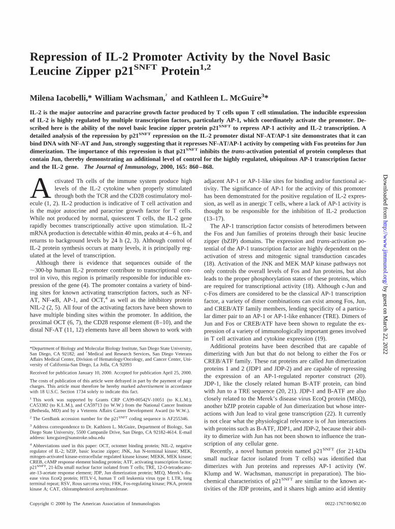
p21SNFTspecifically down-regulates IL-2 promoter activity
The specificity of p21SNFT for the IL-2 promoter was investigatedby testing the relative strengths of many viral and human cellularpromoters in the presence of overexpressed p21SNFT. Transienttransfections of Jurkat cells with a variety of promoter constructs
FIGURE 2. p21SNFT inhibits IL-2 promoter activity in Jurkat cells. Theactivity of the p21SNFT-containing samples is shown as a percentage of theactivity observed in the absence of p21SNFT, which was normalized to100%. The data represent the average of three controlled experiments.A,Cotransfections were conducted using the pIL-2-Luc construct in the pres-ence of increasing amounts of pCI/SNFT expression vector. Total DNAconcentration remained constant.B, pIL-2-Luc and pCD28RE/AP-1-Lucwere transfected in the absence (empty vector) or the presence of pCI/SNFT, and the cells were stimulated with PMA/ionomycin with or withoutanti-CD28. The samples without p21SNFT were set at 100% activity; there-fore, the data presented do not reflect the;3-fold increase in activity seenwith P/I/anti-CD28 compared with P/I simulation.
FIGURE 3. p21SNFT is constitutively expressed in Jurkat cells undervarious stimulation conditions and times. Western analysis of p21SNFT lev-els was performed using 75mg of Jurkat whole cell extracts from cellsstimulated with PMA/ionomycin (P/I) for various periods or stimulated for4 h with PMA or PMA/ionomycin/anti-CD28 as indicated. The data arerepresentative of three experiments.
863The Journal of Immunology
by guest on March 22, 2022
http://ww
w.jim
munol.org/
Dow
nloaded from
clearly show that of the promoters shown, only the IL-2 promoteris significantly repressed (70% inhibition) by p21SNFT (Fig. 4).This result indicates that p21SNFT acts not as a general transcrip-tion inhibitor, but, rather, as a selective inhibitor of IL-2 promoteractivity.
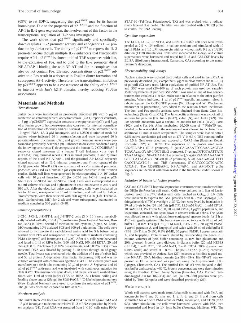
Overexpression of p21SNFT leads to repressed endogenous IL-2gene expression
To verify the biological significance of p21SNFT inhibition of IL-2promoter activity, the ability of p21SNFT to inhibit expression ofthe endogenous IL-2 gene was determined. To accomplish this,stable Jurkat T cell lines were generated to constitutively overex-press p21SNFT. Stable oligoclonal lines transfected with either anempty vector (J-CI-1 and J-CI-2) or the p21SNFT expression con-struct (J-SNFT-1 and J-SNFT-2) were compared for p21SNFT ex-pression. As shown in Fig. 5A, the J-CI-1 and -2 control linesexpress low levels of endogenous p21SNFT protein. In contrast,p21SNFTis markedly increased in the J-SNFT-1 and -2 lines. WhenIL-2 mRNA levels were analyzed 4 h after PMA and ionomycinstimulation, an average 67% decrease in message was seen in theJ-SNFT lines compared with the control J-CI cells (Fig. 5A). Thepercent decrease in IL-2 mRNA levels seen in the J-SNFT lines isequivalent to the repression of IL-2 promoter activity by overex-pressed p21SNFT in transient transfection, therefore supporting thehypothesis that p21SNFT is inhibiting the production of IL-2 at thelevel of transcription initiation.
The p21SNFT overexpressing and control Jurkat lines were alsoanalyzed for IL-2, IL-3, and GM-CSF cytokine expression byELISA on culture supernatants from stimulated cells. These threecytokines were chosen because all three genes respond to CD28costimulation through their CD28 response elements (27, 28, 33).Fig. 5B shows an average 77% decrease in IL-2 production inPMA/ionomycin-stimulated J-SNFT cells compared with J-CIcells and a 71% decrease with the addition of CD28 stimulation. Incontrast, GM-CSF (Fig. 5B) and IL-3 (data not shown) were ex-pressed at equivalent levels in the J-CI control and J-SNFT lines,indicating that p21SNFT does not globally inhibit endogenous cy-tokine production in T cells.
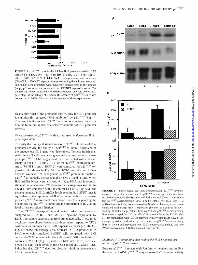
Multiple AP-1-containing sites within the IL-2 promoter aretargets of p21SNFTrepression
Because p21SNFT interacts with Jun family members and inhibitsthe activity of AP-1, p21SNFTmay decrease IL-2 promoter activity
FIGURE 4. p21SNFT specifically inhibits IL-2 promoter activity. CAT(HTLV-I 39 LTR, c-Fos,2404/141; HIV 59LTR, IL-2, 2575/155; IL-2R, 21240/2227; RSV 39LTR, SV40 early promoter) and luciferase(GM-CSF,2620/137) reporter vectors containing the indicated retroviraland human gene promoters were transiently cotransfected in the absence(empty pCI vector) or the presence of the pCI/SNFT expression vector. Thetransfectants were stimulated with PMA/ionomycin, and data shown are apercentage of the activity observed in the absence of p21SNFT, which wasnormalized to 100%. The data are the average of three experiments.
FIGURE 5. Stable Jurkat cell lines overexpressing p21SNFT have de-creased IL-2 protein expression.A, p21SNFT immunoprecipitations fromtwo PMA/ionomycin (P1I)-stimulated Jurkat control (lanes 1and2) andtwo p21SNFT-overexpressing (lanes 3and 4) stable cell lines (top). IL-2mRNA levels (middle) were assessed by Northern blot analysis and werecompared with TCRb mRNA expression (bottom) as a control for RNAloading.B, Culture supernatants from control and p21SNFT-overexpressinglines were assayed for IL-2 and GM-CSF cytokine levels by ELISA aftera 4-day stimulation with PMA/ionomycin with or without anti-CD28. Theaverage cytokine production by the control or p21SNFT-overexpressinglines is shown and represents two PMA/ionomycin-stimulated and onePMA/ionomycin/anti-CD28-stimulated experiments.
864 REPRESSION OF THE IL-2 PROMOTER BY p21SNFT
by guest on March 22, 2022
http://ww
w.jim
munol.org/
Dow
nloaded from
by targeting the sites in the IL-2 promoter that require AP-1 bind-ing. This includes the AP-1/OCT, CD28RE/AP-1, and NF-AT/AP-1 elements. A schematic of the IL-2 promoter and the relativepositions of these elements is shown in Fig. 6A. To determinewhether these sites are potential targets of p21SNFT repression,these AP-1-containing elements from the IL-2 promoter weretested for responsiveness to p21SNFT along with a reporter con-struct containing NF-kB elements. These data, shown in Fig. 6B,demonstrate that the AP-1-associated IL-2 promoter elementstested are inhibited by an average of 50% in the presence ofp21SNFT, as is the TRE element (Fig. 1B), while no inhibition ofNF-kB was observed. These results suggest that p21SNFT is exert-ing its effect on the IL-2 promoter through multiple sites, simul-taneously inhibiting the activities of a variety of enhancer sites thatrequire AP-1 to function.
p21SNFT is present in protein complexes that it can functionallyrepress
To determine whether the functional effect of p21SNFT on the IL-2enhancers is due to a direct or an indirect mechanism of action,protein binding to the IL-2 enhancer elements was analyzed byEMSA. To determine the presence or the absence of p21SNFT incomplexes that form on the AP-1/OCT, CD28RE/AP-1, NF-AT/AP-1, and NF-kB sequences, an antiserum specific for p21SNFT
was employed. When extracts from PMA/ionomycin-stimulatedJurkat cells are used, protein complexes form on each IL-2 element(Fig. 6C, lane 1). Competition for protein binding by the additionof a 100-fold excess of unlabeled wild-type probe shows the spec-ificity of these proteins for their respective sequences (lane 2).AP-1/OCT (7), CD28RE/AP-1 (9, 10), and NF-AT/AP-1 (12, 34–36) have all been reported to bind AP-1 proteins in cooperationwith other transcription factors for their full activity. Therefore, toconfirm AP-1 binding activity on each of these IL-2 enhancers, anexcess of unlabeled TRE probe was added to out-compete AP-1binding activity in the nuclear extracts. As shown, the AP-1/OCT,CD28RE/AP-1, and NF-AT/AP-1 probes each possess TRE-spe-cific complexes (lane 3). The addition of p21SNFTantiserum to thesamples blocked the formation of several, but not all, of the com-plexes that formed on these three sequences (lane 4). Conversely,NF-kB binding remained unaffected by both the TRE competitionand addition of p21SNFT antisera.
The ability of the p21SNFT antisera to disrupt complexes foundon the OCT/AP-1, CD28RE/AP-1, and NF-AT/AP-1 sequences,but not on the NF-kB sequence, correlates with the ability ofp21SNFTto repress the activity of these three enhancers. These datastrongly suggest that the mode of action for p21SNFT is direct and
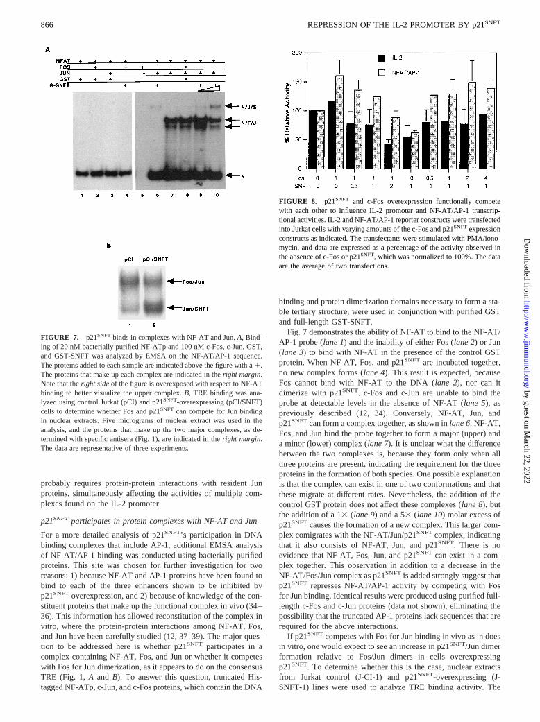
FIGURE 6. p21SNFT can inhibit the activity of AP-1-associated IL-2promoter enhancer elements and can be found in the protein complexes thatform on each sequence.A, A diagram of the various enhancer elementsfound throughout the human IL-2 promoter (2315 to155). Brackets in-dicate the sites used in the analysis inB. B, Reporter constructs containingmultimers of isolated IL-2 enhancers or an NF-kB element driving lucif-erase expression were transfected in the presence or the absence of thepCI/SNFT expression plasmid. The activity of the p21SNFT-containingsamples is shown as a percentage of the activity observed in the absence ofp21SNFT, which was normalized to 100%. The data are representative ofthree controlled experiments.C, Probes containing the IL-2 promoter en-hancer sequences AP-1/OCT, CD28RE/AP-1, NF-AT/AP-1, and NF-kBwere incubated with 5mg of nuclear extract from PMA/ionomycin-stim-ulated Jurkat cells. Samples were exposed to a 100-fold excess of unlabeledwild-type (lane 2) or TRE competitor (lane 3) DNA and p21SNFT (lane 4)antisera. Protein complexes that are TRE specific and contain p21SNFT areindicated with arrows. The data are representative of three experiments.
865The Journal of Immunology
by guest on March 22, 2022
http://ww
w.jim
munol.org/
Dow
nloaded from
probably requires protein-protein interactions with resident Junproteins, simultaneously affecting the activities of multiple com-plexes found on the IL-2 promoter.
p21SNFTparticipates in protein complexes with NF-AT and Jun
For a more detailed analysis of p21SNFT’s participation in DNAbinding complexes that include AP-1, additional EMSA analysisof NF-AT/AP-1 binding was conducted using bacterially purifiedproteins. This site was chosen for further investigation for tworeasons: 1) because NF-AT and AP-1 proteins have been found tobind to each of the three enhancers shown to be inhibited byp21SNFT overexpression, and 2) because of knowledge of the con-stituent proteins that make up the functional complex in vivo (34–36). This information has allowed reconstitution of the complex invitro, where the protein-protein interactions among NF-AT, Fos,and Jun have been carefully studied (12, 37–39). The major ques-tion to be addressed here is whether p21SNFT participates in acomplex containing NF-AT, Fos, and Jun or whether it competeswith Fos for Jun dimerization, as it appears to do on the consensusTRE (Fig. 1,A and B). To answer this question, truncated His-tagged NF-ATp, c-Jun, and c-Fos proteins, which contain the DNA
binding and protein dimerization domains necessary to form a sta-ble tertiary structure, were used in conjunction with purified GSTand full-length GST-SNFT.
Fig. 7 demonstrates the ability of NF-AT to bind to the NF-AT/AP-1 probe (lane 1) and the inability of either Fos (lane 2) or Jun(lane 3) to bind with NF-AT in the presence of the control GSTprotein. When NF-AT, Fos, and p21SNFT are incubated together,no new complex forms (lane 4). This result is expected, becauseFos cannot bind with NF-AT to the DNA (lane 2), nor can itdimerize with p21SNFT. c-Fos and c-Jun are unable to bind theprobe at detectable levels in the absence of NF-AT (lane 5), aspreviously described (12, 34). Conversely, NF-AT, Jun, andp21SNFTcan form a complex together, as shown inlane 6. NF-AT,Fos, and Jun bind the probe together to form a major (upper) anda minor (lower) complex (lane 7). It is unclear what the differencebetween the two complexes is, because they form only when allthree proteins are present, indicating the requirement for the threeproteins in the formation of both species. One possible explanationis that the complex can exist in one of two conformations and thatthese migrate at different rates. Nevertheless, the addition of thecontrol GST protein does not affect these complexes (lane 8), butthe addition of a 13(lane 9) and a 53 (lane 10) molar excess ofp21SNFT causes the formation of a new complex. This larger com-plex comigrates with the NF-AT/Jun/p21SNFT complex, indicatingthat it also consists of NF-AT, Jun, and p21SNFT. There is noevidence that NF-AT, Fos, Jun, and p21SNFT can exist in a com-plex together. This observation in addition to a decrease in theNF-AT/Fos/Jun complex as p21SNFTis added strongly suggest thatp21SNFT represses NF-AT/AP-1 activity by competing with Fosfor Jun binding. Identical results were produced using purified full-length c-Fos and c-Jun proteins (data not shown), eliminating thepossibility that the truncated AP-1 proteins lack sequences that arerequired for the above interactions.
If p21SNFTcompetes with Fos for Jun binding in vivo as in doesin vitro, one would expect to see an increase in p21SNFT/Jun dimerformation relative to Fos/Jun dimers in cells overexpressingp21SNFT. To determine whether this is the case, nuclear extractsfrom Jurkat control (J-CI-1) and p21SNFT-overexpressing (J-SNFT-1) lines were used to analyze TRE binding activity. The
FIGURE 7. p21SNFT binds in complexes with NF-AT and Jun.A, Bind-ing of 20 nM bacterially purified NF-ATp and 100 nM c-Fos, c-Jun, GST,and GST-SNFT was analyzed by EMSA on the NF-AT/AP-1 sequence.The proteins added to each sample are indicated above the figure with a1.The proteins that make up each complex are indicated in theright margin.Note that theright sideof the figure is overexposed with respect to NF-ATbinding to better visualize the upper complex.B, TRE binding was ana-lyzed using control Jurkat (pCI) and p21SNFT-overexpressing (pCI/SNFT)cells to determine whether Fos and p21SNFT can compete for Jun bindingin nuclear extracts. Five micrograms of nuclear extract was used in theanalysis, and the proteins that make up the two major complexes, as de-termined with specific antisera (Fig. 1), are indicated in theright margin.The data are representative of three experiments.
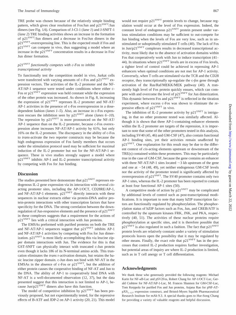
FIGURE 8. p21SNFT and c-Fos overexpression functionally competewith each other to influence IL-2 promoter and NF-AT/AP-1 transcrip-tional activities. IL-2 and NF-AT/AP-1 reporter constructs were transfectedinto Jurkat cells with varying amounts of the c-Fos and p21SNFTexpressionconstructs as indicated. The transfectants were stimulated with PMA/iono-mycin, and data are expressed as a percentage of the activity observed inthe absence of c-Fos or p21SNFT, which was normalized to 100%. The dataare the average of two transfections.
866 REPRESSION OF THE IL-2 PROMOTER BY p21SNFT
by guest on March 22, 2022
http://ww
w.jim
munol.org/
Dow
nloaded from
TRE probe was chosen because of the relatively simple bindingpattern, which gives clear resolution of Fos/Jun and p21SNFT/Jundimers (see Fig. 1A). Comparison of J-CI-1 (lane 1) and J-SNFT-1(lane 2) TRE binding activities shows an increase in the formationof p21SNFT/Jun dimers and a decrease in Fos/Jun dimers in thep21SNFT-overexpressing line. This is the expected result if Fos andp21SNFT can compete in vivo, thus suggesting a model where anincrease in the p21SNFT concentration results in a decrease in Fos/Jun dimer formation.
p21SNFT functionally competes with c-Fos to inhibittranscriptional activity
To functionally test the competition model in vivo, Jurkat cellswere transfected with varying amounts of c-Fos and p21SNFT ex-pression vectors. The activities of the IL-2 promoter and the NF-AT/AP-1 sequence were tested under conditions where either c-Fos or p21SNFT expression was held constant while the expressionof the other protein was increased. As shown in Fig. 8, increasingthe expression of p21SNFT represses IL-2 promoter and NF-AT/AP-1 activities in the presence of c-Fos overexpression in a dose-dependent fashion (lanes 1–5). Also, an increase in c-Fos expres-sion rescues the inhibition seen by p21SNFT alone (lanes 6–10).The repression by p21SNFT is more pronounced on the NF-AT/AP-1 sequence than on the IL-2 promoter, because c-Fos overex-pression alone increases NF-AT/AP-1 activity by 61%, but only16% on the IL-2 promoter. The discrepancy in the ability of c-Fosto trans-activate the two reporter constructs may be because thehigh endogenous expression of Fos family members that occursunder the stimulation protocol used may be sufficient for maximalinduction of the IL-2 promoter but not for the NF-AT/AP-1 se-quence. These in vivo studies strongly support a model wherep21SNFT inhibits AP-1 and IL-2 promoter transcriptional activityby competing with Fos for Jun binding.
DiscussionThe studies presented here demonstrate that p21SNFT represses en-dogenous IL-2 gene expression via its interaction with severalcis-acting promoter sites, including the AP-1/OCT, CD28RE/AP-1,and NF-AT/AP-1 elements. p21SNFT directly interacts with thesesequences in nuclear extracts either via protein-DNA and/or pro-tein-protein interactions with other transcription factors that havespecificity for the DNA. The strong correlation between the down-regulation of Jun-responsive elements and the presence of p21SNFT
in these complexes suggests that a requirement for the actions ofp21SNFT lies with a critical interaction with Jun proteins.
The EMSAs preformed with purified proteins on both the TREand NF-AT/AP-1 sequences suggest that p21SNFT inhibits AP-1and NF-AT/AP-1 activities by competing with Fos for Jun dimer-ization. p21SNFT is most likely accomplishing this via leucine zip-per domain interactions with Jun. The evidence for this is thatGST-SNFT can physically interact with truncated c-Jun proteineven though it lacks 186 of its N-terminal amino acids. This trun-cation eliminates thetrans r-activation domain, but retains the ba-sic leucine zipper domain. c-Jun does not bind with NF-AT in theEMSAs in the absence of c-Fos or p21SNFT, but the addition ofeither protein causes the cooperative binding of NF-AT and Jun tothe DNA. The ability of AP-1 to cooperatively bind DNA withNF-AT is a well-documented observation (12, 37), but the datapresented suggest that this interaction is not limited to AP-1, be-cause Jun/p21SNFT dimers also have this function.
The model of competitive inhibition by p21SNFT has been pre-viously proposed, but not experimentally tested, for the repressiveeffects of B-ATF and JDP-2 on AP-1 activity (20, 21). This model
would not require p21SNFT protein levels to change, because reg-ulation would occur at the level of Fos expression. Indeed, theconstant level of endogenous p21SNFT protein present under var-ious stimulation conditions may be sufficient to out-compete forJun binding when the levels of Fos are very low, such as in un-stimulated or suboptimally stimulated T cells (40). The lack of Fosin Jun/p21SNFT complexes results in decreased transcriptional ac-tivity, most likely due to the absence of activation domains withinFos that cooperatively work with Jun to induce transcription (41–44). In situations where p21SNFT levels are in excess of Fos levels,a tighter level of control could be achieved in repressing AP-1enhancers when optimal conditions are not met for AP-1 activity.Conversely, when T cells are stimulated via the TCR and the CD28receptor, they transcriptionally up-regulate the c-fos gene throughactivation of the Ras/Raf/MEKK/MEK pathway (40). A tran-siently high level of Fos protein quickly ensues, which can com-pete with and overcome the level of p21SNFT for Jun dimerization.This dynamic between Fos and p21SNFT is reflected in the titrationexperiment, where excess c-Fos was shown to eliminate the re-pressive effects of p21SNFT in vivo.
The inhibition of IL-2 promoter activity by p21SNFT is intrigu-ing, in that no other promoter tested was similarly affected. Al-though it is shown that three AP-1-containing enhancer elementswithin the IL-2 promoter are targets of the repression, it is impor-tant to note that some of the other promoters tested in this analysis,including SV40 (45, 46) and GM-CSF (47), also contain functionalAP-1 binding sites, yet their activities remain unaffected byp21SNFT. One explanation for this result may be due to the differ-ent context ofcis-acting elements upstream or downstream of theTRE(s) contained within these promoters. This may be particularlytrue in the case of GM-CSF, because the gene contains an enhancerwith three NF-AT/AP-1 sites located;3 kb upstream of the geneand one at254 (48, 49), yet neither endogenous GM-CSF levelsnor the activity of the promoter tested is significantly affected byoverexpression of p21SNFT. The SV40 promoter contains only twoAP-1 sites, whereas the IL-2 promoter has been reported to containat least four functional AP-1 sites (50).
A competitive mode of action by p21SNFT may be complicatedby additional levels of control such as post-transcriptional modi-fications. It is important to note that many bZIP transcription fac-tors are functionally regulated by phosphorylation. The phosphor-ylation status of bZIP factors such as Fos, Jun, and CREB/ATF iscontrolled by the upstream kinases FRK, JNK, and PKA, respec-tively (40, 51). The activities of these nuclear proteins requirephosphorylation at specific sites, and it is therefore possible thatp21SNFT is also regulated in such a fashion. The fact that p21SNFT
protein levels are relatively constant under a variety of stimulationprotocols leaves open the possibility that it may be regulated byother means. Finally, the exact role that p21SNFT has in the pro-cesses that control IL-2 production requires further investigation,but potential areas of inquiry are where IL-2 production is limited,such as in T cell anergy or T cell differentiation.
AcknowledgmentsWe thank those who generously provided the following reagents: MichaelKarin for NF-kB-Luc and pSV2Fos, Robert Chang for AP-1/OCT-Luc, Ger-ald Crabtree for NF-AT/AP-1-Luc, M. Frances Shannon for GM-CSF-Luc,Tom Kerppola for purified Fos and Jun proteins, Anjana Rao for pNF-AT-pXS1–127 expression construct, and Bristol-Myers Squibb PharmaceuticalResearch Institute for mAb 9.3. A special thanks goes to Hur-Song Changfor providing a variety of valuable reagents and helpful discussion.
867The Journal of Immunology
by guest on March 22, 2022
http://ww
w.jim
munol.org/
Dow
nloaded from
References1. Crabtree, G. R. 1989. Contingent genetic regulatory events in T lymphocyte
activation.Science 243:355.2. Jain, J., C. Loh, and A. Rao. 1995. Transcriptional regulation of the IL-2 gene.
Curr. Opin. Immunol. 7:333.3. Hughes, C. C. W., and J. Pober. 1996. Transcriptional regulation of the interleu-
kin-2 gene in normal human peripheral blood T cells.J. Biol. Chem. 271:5369.4. Brombacher, F., T. Schafer, U. Weissenstein, C. Tschopp, E. Anderson, K. Burki,
and G. Baumann. 1994. IL-2 promoter-drivenlacZ expression as a monitoringtool for IL-2 in primary T cells of transgenic mice.Int. Immunol. 6:189.
5. Williams, T. M., D. Moolten, J. Burlein, J. Romano, R. Bhaerman, A. Godillot,M. Mellon, F. J. I. Rauscher, and J. A. Kant. 1991. Identification of a zinc fingerprotein that inhibits IL-2 gene expression.Science 254:1791.
6. Ullman, K. S., W. M. Flanagan, C. A. Edwards, and G. R. Crabtree. 1991. Ac-tivation of early gene expression in T lymphocytes by Oct-1 and an inducibleprotein, OAP40. Science 254:558.
7. Ullman, K. S., J. P. Northrop, A. Admon, and G. R. Crabtree. 1993. Jun familymembers are controlled by a calcium-regulated, cyclosporin A-sensitive signalingpathway in activated T-lymphocytes.Genes Dev. 7:188.
8. Curtiss, V. E., R. Smilde, and K. L. McGuire. 1996. Requirements for interleukin2 promoter transactivation by the Tax protein of human T-cell leukemia virustype I. Mol. Cell. Biol. 16:3567.
9. Shapiro, V. S., K. E. Truitt, J. B. Imboden, and A. Weiss. 1997. CD28 mediatestranscriptional upregulation of the interleukin-2 (IL-2) promoter through a com-posite element containing the CD28RE and NF-IL-2B AP-1 sites.Mol. Cell. Biol.17:4051.
10. McGuire, K. L., and M. Iacobelli. 1997. Involvement of Rel, Fos and Jun proteinsin binding activity to the IL-2 promoter CD28 response element/AP-1 sequencein human T cells.J. Immunol. 159:1319.
11. Jain, J., G. McCaffery, V. E. Valge-Archer, and A. Rao. 1992. Nuclear factor ofactivated T cells contains Fos and Jun.Nature 356:801.
12. Jain, J., P. G. McCaffrey, Z. Miner, T. K. Kerppola, J. N. Lambert, G. L. Verdine,T. Curran, and A. Rao. 1993. The T-cell transcription factor NF-ATp is a sub-strate for calcineurin and interacts with Fos and Jun.Nature 365:352.
13. Kang, S.-M., B. Beverly, A.-C. Tran, K. Brorson, R. H. Schwartz, andM. J. Lenardo. 1992. Transactivation by AP-1 is a molecular target of T cellclonal anergy.Science 257:1134.
14. Li, W., C. D. Whaley, A. Mondino, and D. L. Mueller. 1996. Blocked signaltransduction to the ERK and JNK protein kinases in anergic CD41 T cells.Science 271:1272.
15. Fields, P. E., T. F. Gajewski, and F. W. Fitch. 1996. Blocked Ras activation inanergic CD41 T cells.Science 271:1276.
16. DeSilva, D. R., W. S. Freeser, T. E. J., and P. A. Scherle. 1996. Anergic T cellsare defective in both Jun NH2-terminal kinase and mitogen-activated proteinkinase signaling pathways.J. Exp. Med. 183:2017.
17. Kitagawa-Sakakida, S., and R. Schwartz. 1996. Multifactorcis-dominant nega-tive regulation of IL-2 gene expression in anergized T cells.J. Immunol. 157:2328.
18. Karin, M., Z.-g. Liu, and E. Zandi. 1997. AP-1 function and regulation.Curr.Opin. Cell Biol. 9:240.
19. Foletta, V. C., D. H. Segal, and D. R. Cohen. 1998. Transcriptional regulation inthe immune system: all roads lead to AP-1.J. Leukocyte Biol. 63:139.
20. Aronheim, A., E. Zandi, H. Hennemann, S. Elledge, and M. Karin. 1997. Isola-tion of an AP-1 repressor by a novel method for detecting protein-protein inter-actions.Mol. Cell. Biol. 17:3094.
21. Dorsey, M. J., H.-J. Tae, K. G. Sollenberger, N. T. Mascarenhas, L. M. Johansen,and E. J. Taparowsky. 1995. B-ATF: a novel human bZIP protein that associateswith members of the AP-1 transcription factor family.Oncogene 11:2255.
22. Qian, Z., P. Brunovskis, F. Rauscher III, L. Lee, and H.-J. Kung. 1995. Trans-activation activity of Meq, a Merek’s disease herpesvirus bZIP protein persis-tently expressed in latently infected transformed T cells.J. Virol. 69:4037.
23. Rosette, C., and M. Karin. 1995. Cytoskeletal control of gene expression: depo-lymerization of microtubules activates NF-kB. J. Cell Biol. 128:1111.
24. Sambrook, J., E. F. Fritsch, and T. Maniatis. 1989.Molecular Cloning: A Lab-oratory Manual. Cold Spring Harbor Laboratory Press, Cold Spring Harbor,p. 7.12.
25. Jain, J., E. Burgeon, T. M. Badalian, P. G. Hogan, and A. Rao. 1995. A similarDNA-binding motif in NF-AT family proteins and the Rel homology region.J. Biol. Chem. 270:4138.
26. Kerppola, T. K., and T. Curran. 1997. The transcription activation domains of Fosand Jun induce DNA bending through electrostatic interactions.EMBO J. 16:2907.
27. Fraser, J. D., B. A. Irving, G. R. Crabtree, and A. Weiss. 1991. Regulation ofinterleukin-2 gene enhancer activity by the T cell accessory molecule CD28.Science 251:313.
28. Verweij, C. L., M. Geerts, and L. A. Aarden. 1991. Activation of interleukin-2gene transcription via the T-cell surface molecule CD28 is mediated through anNF-kB-like response element.J. Biol. Chem. 266:14179.
29. Fraser, J. D., D. Straus, and A. Weiss. 1993. Signal transduction events leadingto T-cell lymphokine gene expression.Immunol. Today 14:357.
30. Su, B., E. Jacinto, M. Hibi, T. Kallunki, M. Karin, and Y. Ben-Neriah. 1994. JNKis involved in signal integration during costimulation of T-lymphocytes.Cell77:727.
31. Lai, J.-H., and T.-H. Tan. 1994. CD28 signaling causes a sustained down-regu-lation of IkBa which can be prevented by the immunosuppressant rapamycin.J. Biol. Chem. 269:30077.
32. Ramano, M. F., R. Bisogni, A. Lamberti, M. C. Turco, A. Petrella, P. Tassone,R. van Lier, S. Formisano, and S. Venuta. 1994. Regulation of NF-kB nuclearactivity in peripheral blood mononuclear cells: role of CD28 antigen.Cell. Im-munol. 156:371.
33. Fraser, J. D., and A. Weiss. 1992. Regulation of T-cell lymphokine gene tran-scription by the accessory molecule CD28.Mol. Cell. Biol. 12:4357.
34. Boise, L. H., B. Petryniak, X. Mao, C. June, C.-Y. Wang, T. Lindsten, R. Bravo,K. Kovary, J. M. Leiden, and C. Thompson. 1993. The NF-AT-1 DNA bindingcomplex in activated T cells contains Fra-1 and JunB.Mol. Cell. Biol. 13:1911.
35. Cockerill, P. N., A. G. Bert, F. Jenkins, G. R. Ryan, M. F. Shannon, andM. A. Vadas. 1995. Human granulocyte-macrophage colony-stimulating factorenhancer function is associated with cooperative interactions between AP-1 andNF-ATp/c. Mol. Cell. Biol. 15:2071.
36. Northrop, J. P., K. S. Ullman, and G. R. Crabtree. 1993. Characterization of thenuclear and cytoplasmic components of the lymphoid-specific nuclear factor ofactivated T-cells (NF-AT) complex.J. Biol. Chem. 268:2917.
37. McCaffrey, P. G., C. Luo, T. K. Kerppola, J. Jain, T. M. Badalian, A. M. Ho,E. Burgeon, W. S. Lane, J. N. Lambert, T. Curran, et al. 1993. Isolation of thecyclosporin-sensitive T cell transcription factor NF-ATp.Science 262:750.
38. Diebold, R. J., N. Rajaram, D. A. Leonard, and T. K. Kerppola. 1998. Molecularbasis of cooperative DNA bending and oriented heterodimer binding in the NF-AT1-Fos-Jun-ARRE2 complex.Proc. Natl. Acad. Sci. USA 95:7915.
39. Chen, L., M. J. N. Glover, P. G. Hogan, A. Rao, and S. C. Harrison. 1998.Structure of the DNA-binding domains from NF-AT, Fos and Jun bound specif-ically to DNA. Nature 392:42.
40. Foletta, V. C., D. H. Segal, and D. R. Cohen. 1998. Transcriptional regulation theimmune system: all roads lead to AP-1.J. Leukocyte Biol. 63:139.
41. Sutherland, J. A., A. Cook, A. J. Bannister, and T. Kouzarides. 1992. Conservedmotifs in Fos and Jun define a new class of activation domain.Genes Dev.6:1810.
42. Wisdom, R., and I. Verma. 1993. Transformation by Fos proteins requires aC-terminal transactivation domain.Mol. Cell. Biol. 13:7429.
43. Deng, T., and M. Karin. 1994. c-Fos transcriptional activity stimulated by H-Ras-activated protein kinase distinct from JNK and ERK.Nature 371:171.
44. Jooss, K. U., M. Funk, and R. Muller. 1994. An autonomous N-terminal trans-activation domain in Fos protein plays a crucial role in transformation.EMBO J.13:1467.
45. Angel, P., M. Imagawa, R. Chiu, B. Stein, R. J. Imbra, H. J. Rahmsdorf, C. Jonat,P. Herrlich, and M. Karin. 1987. Phorbol ester-inducible genes contain a commoncis element recognized by a TPA-modulatedtrans-acting factor.Cell 49:729.
46. Lee, W., P. Mitchell, and R. Tijan. 1987. Purified transcription factor AP-1 in-teracts with TPA-induced enhancer elements.Cell 49:741.
47. Thomas, R. S., M. J. Tymms, L. H. McKinlay, M. F. Shannon, A. Seth, andI. Kola. 1997. ETS1, NF-kB and AP1 synergistically transactivate the humanGM-CSF promoter.Oncogene 14:2845.
48. Masuda, E. S., H. Tokumitsu, A. Tsuboi, J. Shlomai, P. Hung, K. Arai, andN. Arai. 1993. The granulocyte-macrophage-stimulating factor promotercis-act-ing element CLEO mediates induction signals in T cells and is recognized byfactors related to AP-1 and NF-AT.Mol. Cell Biol. 13:7399.
49. Cockerill, P. N., M. F. Shannon, A. G. Bert, G. R. Ryan, and M. A. Vadas. 1993.The granulocyte-macrophage colony-stimulating factor/interleukin 3 locus is reg-ulated by and an inducible cyclosporin A sensitive enhancer.Proc. Natl. Acad.Sci. USA 90:2466.
50. Rooney, J. W., Y.-L. Sun, L. H. Glimcher, and T. Hoey. 1995. Novel NF-AT sitesthat mediate activation of the interleukin-2 promoter in response to T-cell recep-tor stimulation.Mol. Cell. Biol. 15:6299.
51. Hunter, T., and M. Karin. 1992. The regulation of transcription by phosphory-lation. Cell 70:375.
868 REPRESSION OF THE IL-2 PROMOTER BY p21SNFT
by guest on March 22, 2022
http://ww
w.jim
munol.org/
Dow
nloaded from