Ocean acidification risk assessment for Alaska's fishery sector
Upload
khangminh22Category
view
3download
0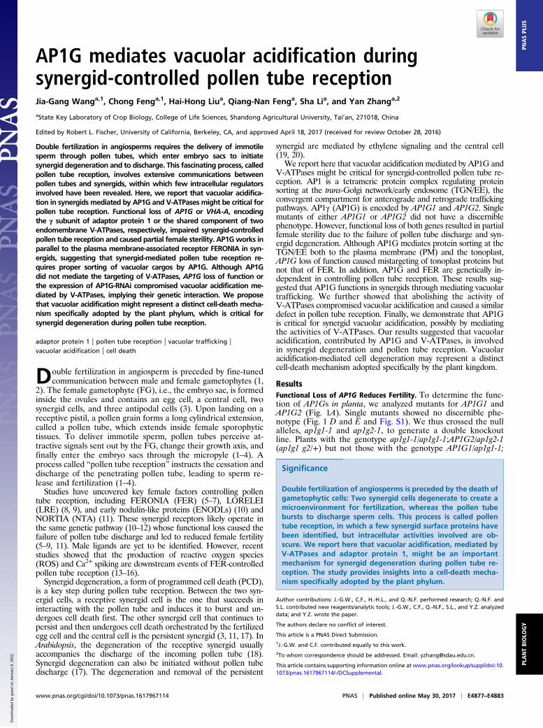
AP1G mediates vacuolar acidification duringsynergid-controlled pollen tube receptionJia-Gang Wanga,1, Chong Fenga,1, Hai-Hong Liua, Qiang-Nan Fenga, Sha Lia, and Yan Zhanga,2
aState Key Laboratory of Crop Biology, College of Life Sciences, Shandong Agricultural University, Tai’an, 271018, China
Edited by Robert L. Fischer, University of California, Berkeley, CA, and approved April 18, 2017 (received for review October 28, 2016)
Double fertilization in angiosperms requires the delivery of immotilesperm through pollen tubes, which enter embryo sacs to initiatesynergid degeneration and to discharge. This fascinating process, calledpollen tube reception, involves extensive communications betweenpollen tubes and synergids, within which few intracellular regulatorsinvolved have been revealed. Here, we report that vacuolar acidifica-tion in synergids mediated by AP1G and V-ATPases might be critical forpollen tube reception. Functional loss of AP1G or VHA-A, encodingthe γ subunit of adaptor protein 1 or the shared component of twoendomembrane V-ATPases, respectively, impaired synergid-controlledpollen tube reception and caused partial female sterility. AP1Gworks inparallel to the plasma membrane-associated receptor FERONIA in syn-ergids, suggesting that synergid-mediated pollen tube reception re-quires proper sorting of vacuolar cargos by AP1G. Although AP1Gdid not mediate the targeting of V-ATPases, AP1G loss of function orthe expression of AP1G-RNAi compromised vacuolar acidification me-diated by V-ATPases, implying their genetic interaction. We proposethat vacuolar acidification might represent a distinct cell-death mecha-nism specifically adopted by the plant phylum, which is critical forsynergid degeneration during pollen tube reception.
adaptor protein 1 | pollen tube reception | vacuolar trafficking |vacuolar acidification | cell death
Double fertilization in angiosperm is preceded by fine-tunedcommunication between male and female gametophytes (1,
2). The female gametophyte (FG), i.e., the embryo sac, is formedinside the ovules and contains an egg cell, a central cell, twosynergid cells, and three antipodal cells (3). Upon landing on areceptive pistil, a pollen grain forms a long cylindrical extension,called a pollen tube, which extends inside female sporophytictissues. To deliver immotile sperm, pollen tubes perceive at-tractive signals sent out by the FG, change their growth axis, andfinally enter the embryo sacs through the micropyle (1–4). Aprocess called “pollen tube reception” instructs the cessation anddischarge of the penetrating pollen tube, leading to sperm re-lease and fertilization (1–4).Studies have uncovered key female factors controlling pollen
tube reception, including FERONIA (FER) (5–7), LORELEI(LRE) (8, 9), and early nodulin-like proteins (ENODLs) (10) andNORTIA (NTA) (11). These synergid receptors likely operate inthe same genetic pathway (10–12) whose functional loss caused thefailure of pollen tube discharge and led to reduced female fertility(5–9, 11). Male ligands are yet to be identified. However, recentstudies showed that the production of reactive oxygen species(ROS) and Ca2+ spiking are downstream events of FER-controlledpollen tube reception (13–16).Synergid degeneration, a form of programmed cell death (PCD),
is a key step during pollen tube reception. Between the two syn-ergid cells, a receptive synergid cell is the one that succeeds ininteracting with the pollen tube and induces it to burst and un-dergoes cell death first. The other synergid cell that continues topersist and then undergoes cell death orchestrated by the fertilizedegg cell and the central cell is the persistent synergid (3, 11, 17). InArabidopsis, the degeneration of the receptive synergid usuallyaccompanies the discharge of the incoming pollen tube (18).Synergid degeneration can also be initiated without pollen tubedischarge (17). The degeneration and removal of the persistent
synergid are mediated by ethylene signaling and the central cell(19, 20).We report here that vacuolar acidification mediated by AP1G and
V-ATPases might be critical for synergid-controlled pollen tube re-ception. AP1 is a tetrameric protein complex regulating proteinsorting at the trans-Golgi network/early endosome (TGN/EE), theconvergent compartment for anterograde and retrograde traffickingpathways. AP1γ (AP1G) is encoded by AP1G1 and AP1G2. Singlemutants of either AP1G1 or AP1G2 did not have a discerniblephenotype. However, functional loss of both genes resulted in partialfemale sterility due to the failure of pollen tube discharge and syn-ergid degeneration. Although AP1G mediates protein sorting at theTGN/EE both to the plasma membrane (PM) and the tonoplast,AP1G loss of function caused mistargeting of tonoplast proteins butnot that of FER. In addition, AP1G and FER are genetically in-dependent in controlling pollen tube reception. These results sug-gested that AP1G functions in synergids through mediating vacuolartrafficking. We further showed that abolishing the activity ofV-ATPases compromised vacuolar acidification and caused a similardefect in pollen tube reception. Finally, we demonstrate that AP1Gis critical for synergid vacuolar acidification, possibly by mediatingthe activities of V-ATPases. Our results suggested that vacuolaracidification, contributed by AP1G and V-ATPases, is involvedin synergid degeneration and pollen tube reception. Vacuolaracidification-mediated cell degeneration may represent a distinctcell-death mechanism adopted specifically by the plant kingdom.
ResultsFunctional Loss of AP1G Reduces Fertility. To determine the func-tion of AP1Gs in planta, we analyzed mutants for AP1G1 andAP1G2 (Fig. 1A). Single mutants showed no discernible phe-notype (Fig. 1 D and E and Fig. S1). We thus crossed the nullalleles, ap1g1-1 and ap1g2-1, to generate a double knockoutline. Plants with the genotype ap1g1-1/ap1g1-1;AP1G2/ap1g2-1(ap1g1 g2/+) but not those with the genotype AP1G1/ap1g1-1;
Significance
Double fertilization of angiosperms is preceded by the death ofgametophytic cells: Two synergid cells degenerate to create amicroenvironment for fertilization, whereas the pollen tubebursts to discharge sperm cells. This process is called pollentube reception, in which a few synergid surface proteins havebeen identified, but intracellular activities involved are ob-scure. We report here that vacuolar acidification, mediated byV-ATPases and adaptor protein 1, might be an importantmechanism for synergid degeneration during pollen tube re-ception. The study provides insights into a cell-death mecha-nism specifically adopted by the plant phylum.
Author contributions: J.-G.W., C.F., H.-H.L., and Q.-N.F. performed research; Q.-N.F. andS.L. contributed new reagents/analytic tools; J.-G.W., C.F., Q.-N.F., S.L., and Y.Z. analyzeddata; and Y.Z. wrote the paper.
The authors declare no conflict of interest.
This article is a PNAS Direct Submission.1J.-G.W. and C.F. contributed equally to this work.2To whom correspondence should be addressed. Email: [email protected].
This article contains supporting information online at www.pnas.org/lookup/suppl/doi:10.1073/pnas.1617967114/-/DCSupplemental.
www.pnas.org/cgi/doi/10.1073/pnas.1617967114 PNAS | Published online May 30, 2017 | E4877–E4883
PLANTBIOLO
GY
PNASPL
US
Dow
nloa
ded
by g
uest
on
Janu
ary
8, 2
022
ap1g2-1/ap1g2-1 (ap1g1/+ g2) were dwarf and bushy (Fig. S1),possibly due to haplo-insufficiency, because the expression ofAP1G1 is higher than that of AP1G2 in most tissues (21). A sig-nificant portion of ap1g1/+ g2 or ap1g1 g2/+ ovules did not grow,compared with that in wild type (WT) or in the single mutants (Fig.1 C–G). To determine whether these ovules were unfertilized ordefective in embryogenesis, we analyzed these ovules 24 h afterpollination (HAP) by confocal laser scanning microscopy (CLSM)and after whole-mount ovule clearing. Almost all wild-typeovules and approximately 75% of ap1g1/+ g2 ovules contained anembryo and peripheral endosperm (Fig. 1 H and J and Table S1),indicative of fertilization. However, approximately 25% ap1g1/+ g2ovules contained a central cell and egg cell at 24 HAP (Fig. 1I andTable S1), indicative of failed fertilization. The unfertilized ap1g1/+g2 ovules contained entangled pollen tubes (Fig. 1K), whereas theyceased growth and discharge contents in WT.
No double homozygous plants were obtained from self-pollinatedap1g1/+ g2 or ap1g1 g2/+ (Table S2). Segregation ratios fromreciprocal crosses showed that ap1g1 g2 was male gametophyticlethal and partially defective in female gametophytic transmis-sion (Table S2). Because of the haplo-insufficient phenotype ofap1g1 g2/+ in vegetative tissue development (Fig. S1), most ofour following studies used ap1g1/+ g2 (hereafter as ap1g/+) toavoid the influence of poor vegetative growth. However, eithergene driven by the ProUBQ10 promoter fully complemented themutant phenotype (Fig. S1), indicating that the two genes arefunctionally interchangeable.
Impaired Pollen Tube Reception in the FG of ap1g/+. To determinethe cause of reduced female transmission of ap1g, we first in-troduced ProES1:NLS-YFP into WT or ap1g/+ and examined thedevelopment of the FG. Because ProES1 is active in all cells ofthe FGs (22), the nucleus-targeting YFP (NLS-YFP) driven byProES1 should show FGs with seven nuclei. CLSM on unfertilizedmature ovules at flower stage 12 indicated that the position ofsynergid cells and the organization of FG were comparable be-tween WT and ap1g/+ (Fig. S2), indicating that AP1Gs were notessential for FG development.Next, we hand-pollinated emasculated wild-type or ap1g/+ pistils
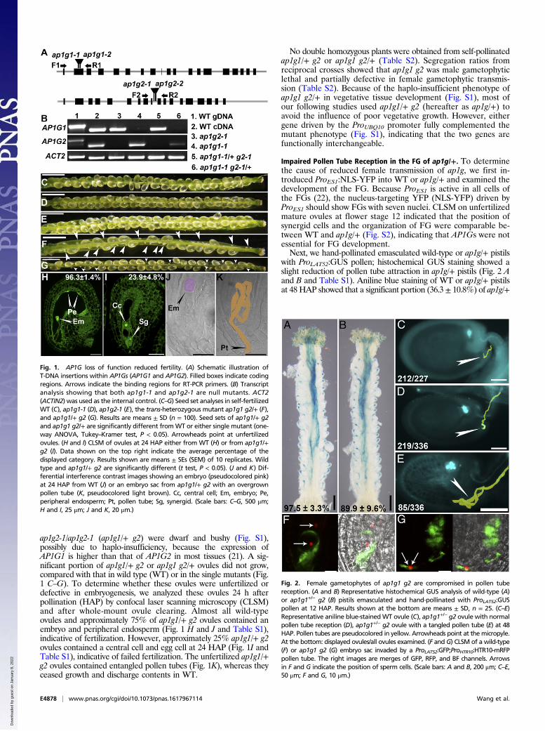
with ProLAT52:GUS pollen; histochemical GUS staining showed aslight reduction of pollen tube attraction in ap1g/+ pistils (Fig. 2 Aand B and Table S1). Aniline blue staining of WT or ap1g/+ pistilsat 48 HAP showed that a significant portion (36.3 ± 10.8%) of ap1g/+
Fig. 1. AP1G loss of function reduced fertility. (A) Schematic illustration ofT-DNA insertions within AP1Gs (AP1G1 and AP1G2). Filled boxes indicate codingregions. Arrows indicate the binding regions for RT-PCR primers. (B) Transcriptanalysis showing that both ap1g1-1 and ap1g2-1 are null mutants. ACT2(ACTIN2) was used as the internal control. (C–G) Seed set analyses in self-fertilizedWT (C), ap1g1-1 (D), ap1g2-1 (E), the trans-heterozygous mutant ap1g1 g2/+ (F),and ap1g1/+ g2 (G). Results are means ± SD (n = 100). Seed sets of ap1g1/+ g2and ap1g1 g2/+ are significantly different fromWT or either single mutant (one-way ANOVA, Tukey–Kramer test, P < 0.05). Arrowheads point at unfertilizedovules. (H and I) CLSM of ovules at 24 HAP either from WT (H) or from ap1g1/+g2 (I). Data shown on the top right indicate the average percentage of thedisplayed category. Results shown are means ± SEs (SEM) of 10 replicates. Wildtype and ap1g1/+ g2 are significantly different (t test, P < 0.05). (J and K) Dif-ferential interference contrast images showing an embryo (pseudocolored pink)at 24 HAP from WT (J) or an embryo sac from ap1g1/+ g2 with an overgrownpollen tube (K, pseudocolored light brown). Cc, central cell; Em, embryo; Pe,peripheral endosperm; Pt, pollen tube; Sg, synergid. (Scale bars: C–G, 500 μm;H and I, 25 μm; J and K, 20 μm.)
Fig. 2. Female gametophytes of ap1g1 g2 are compromised in pollen tubereception. (A and B) Representative histochemical GUS analysis of wild-type (A)or ap1g1+/− g2 (B) pistils emasculated and hand-pollinated with ProLAT52:GUSpollen at 12 HAP. Results shown at the bottom are means ± SD, n = 25. (C–E)Representative aniline blue-stainedWT ovule (C), ap1g1+/− g2 ovule with normalpollen tube reception (D), ap1g1+/− g2 ovule with a tangled pollen tube (E) at 48HAP. Pollen tubes are pseudocolored in yellow. Arrowheads point at themicropyle.At the bottom: displayed ovules/all ovules examined. (F and G) CLSM of a wild-type(F) or ap1g1 g2 (G) embryo sac invaded by a ProLAT52:GFP;ProHTR10:HTR10-mRFPpollen tube. The right images are merges of GFP, RFP, and BF channels. Arrowsin F and G indicate the position of sperm cells. (Scale bars: A and B, 200 μm; C–E,50 μm; F and G, 10 μm.)
E4878 | www.pnas.org/cgi/doi/10.1073/pnas.1617967114 Wang et al.
Dow
nloa
ded
by g
uest
on
Janu
ary
8, 2
022
ovules were unfertilized (Table S1), in contrast to that in WT(Table S1). Overgrown pollen tubes were present in unfertilizedovules (85 of 117; Fig. 2E), indicating impaired pollen tube re-ception (5–7, 9, 11, 23, 24).To verify the impaired pollen tube reception, we hand-pollinated
emasculated wild-type or ap1g/+ pistils with ProLAT52:GFP;ProHTR10:HTR10-mRFP pollen, in which sperm nuclei are labeledwith mRFP and pollen tubes express cytoplasmic GFP (25). Pollentubes discharged their content, that is, cytoplasm and sperm cells(67 of 79; Fig. 2F) upon arrival in WT. However, 18 of 75 ap1g/+FGs contained a nondischarging pollen tube (Fig. 2G). As a result,sperm cells were trapped way back in pollen tubes rather than beingreleased into the FG (Fig. 2G). These results demonstrated a crit-ical role of AP1G in FG-controlled pollen tube reception.
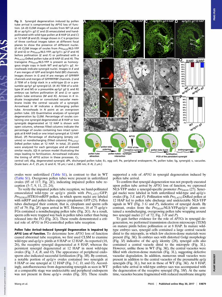
Pollen Tube Arrival–Induced Synergid Degeneration Is Impaired byAP1G Loss of Function. To determine how AP1G loss of functioncaused abnormal tube reception, we first used CLSM to examinewild-type and ap1g/+ pistils at 8 HAP or 12 HAP. As reported (18,26), the receptive synergid degenerated at 8 HAP, whereas thepersistent synergid degenerated at 12 HAP in most wild-typeovules (Fig. 3 A, B, and O). The appearance of peripheral endo-sperm also indicated successful fertilization (Fig. 3B). By contrast,a notable portion of ap1g/+ ovules contained two synergids at8 HAP or one synergid at 12 HAP (Fig. 3 C, D, and O). Accord-ingly, autofluorescence from degenerating synergids as seen in WTat a comparable stage was undetectable and peripheral endospermwas not present in those ap1g/+ ovules (Fig. 3D). These results
supported a role of AP1G in synergid degeneration induced bypollen tube arrival.To confirm that synergid degeneration was not properly executed
upon pollen tube arrival by AP1G loss of function, we expressedNLS-YFP under a synergid-specific promoter ProDD39 (27). Syner-gid nuclei were labeled in both unfertilized wild-type and ap1g/+ovules (Fig. 3 E and F). Pollination with ProLAT52:DsRed pollen at12 HAP led to pollen tube discharge and undetectable NLS-YFPsignals in WT (Fig. 3 G and P), indicative of synergid death. Bycontrast, ovules from the ProDD39:NLS-YFP;ap1g/+ plants con-tained a nondischarging, overgrowing pollen tube wrapping aroundtwo synergid nuclei (17 of 72; Fig. 3 H and P).To gain further evidence for the role of AP1Gs in synergid de-
generation, we performed transmission electron microscopy (TEM)on mature pistils before pollination or at 8 HAP. In mature wild-type embryo sacs, synergid cells contained a large central vacuoledistal to the micropyle, in which few electron-dense materials werepresent (Fig. 3K). In embryo sacs with distorted Golgi morphology(Fig. 3J) indicative of the ap1g identity (28), synergid cells alsocontained a central vacuole distal to the micropyle (Fig. 3L).However, unlike those in WT (Fig. 3K), the central vacuoles werefilled with electron-dense materials (Fig. 3L), suggesting defectivevacuolar degradation. In addition, numerous small vacuoles werepresent in addition to the central vacuoles of the presumable ap1gsynergids (Fig. 3L), but not in those of WT (Fig. 3K). At 8 HAP,pollen tube arrival in wild-type embryo sacs was accompanied withthe degeneration of the receptive synergid (Fig. 3M). At the sametime, vacuoles became fragmented with reduced membrane integrity
Fig. 3. Synergid degeneration induced by pollentube arrival is compromised by AP1G loss of func-tion. (A–D) CLSM images of ovules from WT (A andB) or ap1g1/+ g2 (C and D) emasculated and hand-pollinated with wild-type pollen at 8 HAP (A and C )or 12 HAP (B and D). Image shown in C is projectionof three confocal images taken at different focalplanes to show the presence of different nuclei.(E–H) CLSM image of ovules from ProDD39:NLS-YFP(E and G) or ProDD39:NLS-YFP; ap1g1/+ g2 (F and H)before pollination (E and F ) or pollinated with aProLAT52:DsRed pollen tube at 8 HAP (G and H). Thetransgene ProDD39:NLS-YFP is present as homozy-gous single copy in both WT and ap1g1/+ g2. Ar-rowheads indicate synergid nuclei. Images in E andF are merges of GFP and bright-field (BF) channels.Images shown in G and H are merges of GFP/RFPchannels and merges of GFP/RFP/BF channels. (I andJ) TEM of a Golgi stack in a wild-type (I) or a pre-sumble ap1g1 g2 synergid (J). (K–N ) TEM of a wild-type (K and M ) or a presumble ap1g1 g2 (L and N )embryo sac before pollination (K and L) or uponpollen tube entrance (M and N ). Arrows in K in-dicate invaginated or convoluted vacuolar mem-brane inside the central vacuole of a synergid.Arrowhead in M indicates a discharging pollentube. Arrowheads in N point at an overgrownpollen tube. (O) Quantitative analyses of synergiddegeneration by CLSM. Percentage of ovules con-taining one synergid degenerated at 8 HAP or twosynergids degenerated at 12 HAP is shown withopen columns, whereas filled columns indicate thepercentage of ovules containing two intact syner-gids at 8 HAP (red) or one intact synergid at 12 HAP(blue). (P) Percentage of discharging (empty col-umns) or nondischarging (filled columns) ProLAT52:DsRed pollen tubes at 12 HAP. In total, 25 pistilswere analyzed for each genotype and all showedsimilar results. (Q) A cartoon model illustrating thesteps leading to fertilization. Arrowhead indicatesthe timing of AP1G action in these processes. Cc,central cell; dSg, degenerated synergid; dPt, discharged pollen tube; Ec, egg cell; Pe, peripheral endosperm; Pt, pollen tube; Sg, synergid; v, vacuoles.(Scale bars: A–F, 25 μm; G and H, 10 μm; I and J, 200 nm; K–N, 2 μm.)
Wang et al. PNAS | Published online May 30, 2017 | E4879
PLANTBIOLO
GY
PNASPL
US
Dow
nloa
ded
by g
uest
on
Janu
ary
8, 2
022
of the persistent synergid (Fig. 3M), indicating an immediate celldeath. Consistent with the results obtained by using fluorescentpollen tubes (Fig. 3P), nondischarging pollen tubes were observed inthe presumable ap1g embryo sacs after pollination (Fig. 3N) basedon the Golgi morphology (Fig. 3J). No synergid degeneration oc-curred in these embryo sacs (Fig. 3N). Fragmented vacuoles filledwith electron-dense materials were observed in synergid cells, forwhich membrane integrity was well preserved (Fig. 3N), unlike thatof WT (Fig. 3M). These results indicated that functional loss ofAP1Gs compromised synergid degeneration upon pollen tube arrival,which is likely crucial for pollen tube discharge (18).To determine whether the role of AP1G in synergid de-
generation was cell-autonomous, we confirmed their expressionin ovules and synergid cells by RNA in situ hybridization and byhistochemical GUS staining of their promoter-reporter lines.Both AP1G1 and AP1G2 were expressed in ovules, especially atthe micropylar region where synergid cells are located (Fig. S3).
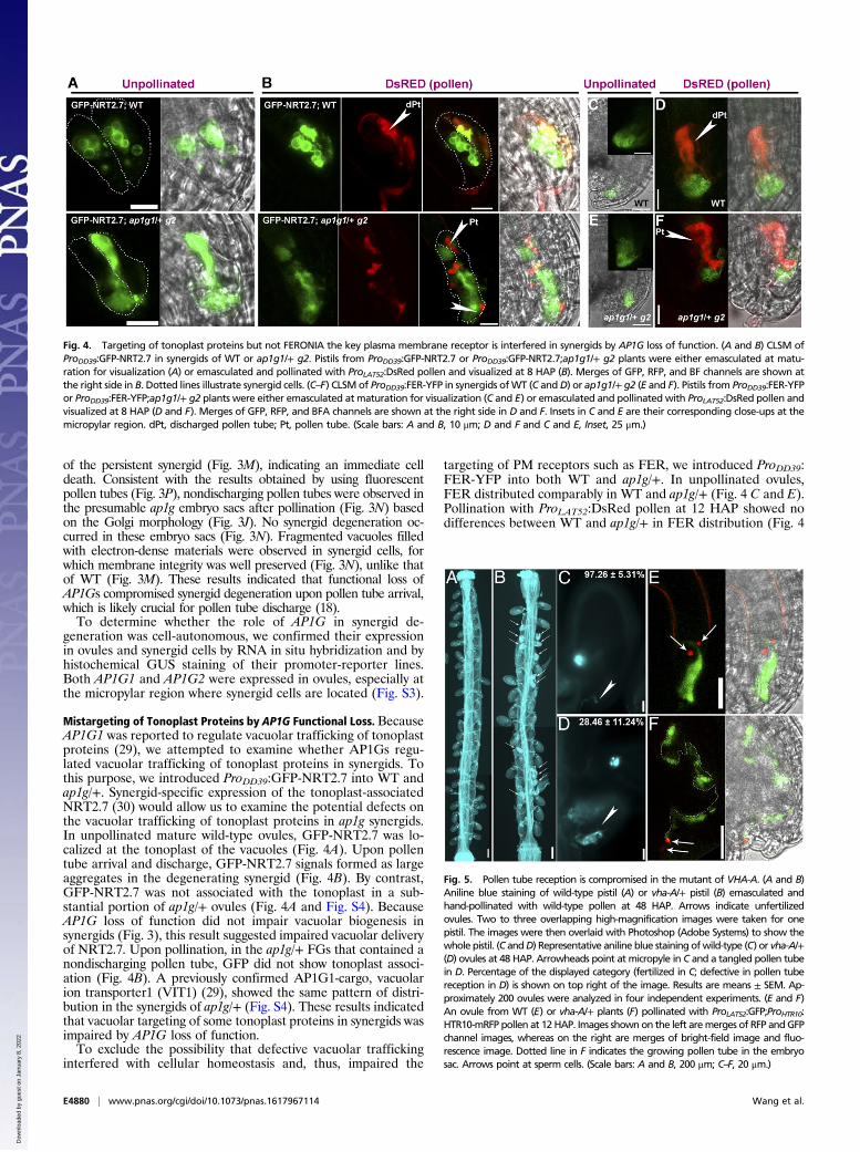
Mistargeting of Tonoplast Proteins by AP1G Functional Loss. BecauseAP1G1 was reported to regulate vacuolar trafficking of tonoplastproteins (29), we attempted to examine whether AP1Gs regu-lated vacuolar trafficking of tonoplast proteins in synergids. Tothis purpose, we introduced ProDD39:GFP-NRT2.7 into WT andap1g/+. Synergid-specific expression of the tonoplast-associatedNRT2.7 (30) would allow us to examine the potential defects onthe vacuolar trafficking of tonoplast proteins in ap1g synergids.In unpollinated mature wild-type ovules, GFP-NRT2.7 was lo-calized at the tonoplast of the vacuoles (Fig. 4A). Upon pollentube arrival and discharge, GFP-NRT2.7 signals formed as largeaggregates in the degenerating synergid (Fig. 4B). By contrast,GFP-NRT2.7 was not associated with the tonoplast in a sub-stantial portion of ap1g/+ ovules (Fig. 4A and Fig. S4). BecauseAP1G loss of function did not impair vacuolar biogenesis insynergids (Fig. 3), this result suggested impaired vacuolar deliveryof NRT2.7. Upon pollination, in the ap1g/+ FGs that contained anondischarging pollen tube, GFP did not show tonoplast associ-ation (Fig. 4B). A previously confirmed AP1G1-cargo, vacuolarion transporter1 (VIT1) (29), showed the same pattern of distri-bution in the synergids of ap1g/+ (Fig. S4). These results indicatedthat vacuolar targeting of some tonoplast proteins in synergids wasimpaired by AP1G loss of function.To exclude the possibility that defective vacuolar trafficking
interfered with cellular homeostasis and, thus, impaired the
targeting of PM receptors such as FER, we introduced ProDD39:FER-YFP into both WT and ap1g/+. In unpollinated ovules,FER distributed comparably in WT and ap1g/+ (Fig. 4 C and E).Pollination with ProLAT52:DsRed pollen at 12 HAP showed nodifferences between WT and ap1g/+ in FER distribution (Fig. 4
Fig. 4. Targeting of tonoplast proteins but not FERONIA the key plasma membrane receptor is interfered in synergids by AP1G loss of function. (A and B) CLSM ofProDD39:GFP-NRT2.7 in synergids of WT or ap1g1/+ g2. Pistils from ProDD39:GFP-NRT2.7 or ProDD39:GFP-NRT2.7;ap1g1/+ g2 plants were either emasculated at matu-ration for visualization (A) or emasculated and pollinated with ProLAT52:DsRed pollen and visualized at 8 HAP (B). Merges of GFP, RFP, and BF channels are shown atthe right side in B. Dotted lines illustrate synergid cells. (C–F) CLSM of ProDD39:FER-YFP in synergids ofWT (C andD) or ap1g1/+ g2 (E and F). Pistils from ProDD39:FER-YFPor ProDD39:FER-YFP;ap1g1/+ g2 plants were either emasculated at maturation for visualization (C and E) or emasculated and pollinated with ProLAT52:DsRed pollen andvisualized at 8 HAP (D and F). Merges of GFP, RFP, and BFA channels are shown at the right side in D and F. Insets in C and E are their corresponding close-ups at themicropylar region. dPt, discharged pollen tube; Pt, pollen tube. (Scale bars: A and B, 10 μm; D and F and C and E, Inset, 25 μm.)
Fig. 5. Pollen tube reception is compromised in the mutant of VHA-A. (A and B)Aniline blue staining of wild-type pistil (A) or vha-A/+ pistil (B) emasculated andhand-pollinated with wild-type pollen at 48 HAP. Arrows indicate unfertilizedovules. Two to three overlapping high-magnification images were taken for onepistil. The images were then overlaid with Photoshop (Adobe Systems) to show thewhole pistil. (C andD) Representative aniline blue staining ofwild-type (C) or vha-A/+(D) ovules at 48 HAP. Arrowheads point at micropyle in C and a tangled pollen tubein D. Percentage of the displayed category (fertilized in C; defective in pollen tubereception in D) is shown on top right of the image. Results are means ± SEM. Ap-proximately 200 ovules were analyzed in four independent experiments. (E and F)An ovule from WT (E) or vha-A/+ plants (F) pollinated with ProLAT52:GFP;ProHTR10:HTR10-mRFP pollen at 12 HAP. Images shownon the left aremerges of RFP andGFPchannel images, whereas on the right are merges of bright-field image and fluo-rescence image. Dotted line in F indicates the growing pollen tube in the embryosac. Arrows point at sperm cells. (Scale bars: A and B, 200 μm; C–F, 20 μm.)
E4880 | www.pnas.org/cgi/doi/10.1073/pnas.1617967114 Wang et al.
Dow
nloa
ded
by g
uest
on
Janu
ary
8, 2
022
D and F), suggesting that AP1G loss of function did not affectthe distribution of key PM proteins such as FER.To provide further evidence that FER pathway was not af-
fected in ap1g/+, we introduced a null mutant of FER, fer-4 (31),into ap1g/+. We were able to obtain fer-4/+ ap1g/+ plants inwhich both fer-4 and ap1g1 are heterozygous, whereas ap1g2 washomozygous (Fig. S5). Self-fertilized fer-4/+ ap1g/+ plants pro-duced a significantly reduced seed set compared with either fer-4/+or ap1g/+ (Table S1). In addition, aniline blue staining of the fer-4/+ap1g/+ pistils at 48 HAP showed that more than 75% of ovuleswere undeveloped, significantly different from either single mutant(Fig. S5 and Table S1). Tangled pollen tubes were detected inundeveloped ovules (Fig. S5), indicative of defective pollen tubereception. The reduced seed set and pollen tube reception of fer-4/+ap1g/+ was a combination of fer-4 (31) and AP1G loss of function(Fig. S5 and Table S1), strongly suggesting that AP1G and FER actindependently in pollen tube reception.
Functional Loss of V-ATPases Phenocopies That of AP1G in DefectivePollen Tube Reception. Above results suggested that vacuolartrafficking from the TGN/EE was impaired by AP1G loss offunction in synergids.Arabidopsis VHA-A is the single gene encodingthe catalytic subunit of V-ATPases located at the TGN/EE and thetonoplast (32). Its mutation caused complete male gametophyticlethality and partial female transmission defect (32), resemblingthat of AP1G. In addition, functional loss of V-ATPases andAP1 both resulted in distorted Golgi morphology (28, 32, 33). Wethus hypothesized that V-ATPases might be involved in synergiddegeneration during pollen tube reception. To test this hypothesis,we analyzed the FG defects of the heterozygous mutant of VHA-A,vha-A/+ (Fig. S6). Histochemical GUS staining of vha-A/+ pistilspollinated with the ProLAT52:GUS pollen showed a slight re-duction of pollen tube attraction in vha-A/+ (Fig. S7 and TableS1). A significant portion of vha-A/+ ovules were unfertilized andcontained entangled pollen tubes at 48 HAP (Fig. 5B). Finally, a
notable portion of vha-A/+ embryo sacs (15 of 83) were pene-trated by a nondischarging ProLAT52:GFP;ProHTR10:HTR10-mRFPpollen tube containing unreleased sperm cells (Fig. 5 D and F),suggesting that V-ATPases are critical for synergid-controlledpollen tube reception.Because several plant-specific PCD processes are executed by
vacuolar processing enzymes (VPE) that are delivered by vacu-olar trafficking and mediate vacuolar rupture (34), we thustested whether it also participated in pollen tube arrival–inducedsynergid degeneration. To this purpose, we obtained a quadruplemutant of VPEs (atvpe), in which all four Arabidopsis VPE-codinggenes were mutated (35). However, pollination assays and seed setanalysis showed that atvpe did not show any abnormalities in pollentube reception or fertility (Fig. S8), suggesting that synergid de-generation does not involve VPE-mediated vacuolar rupture.
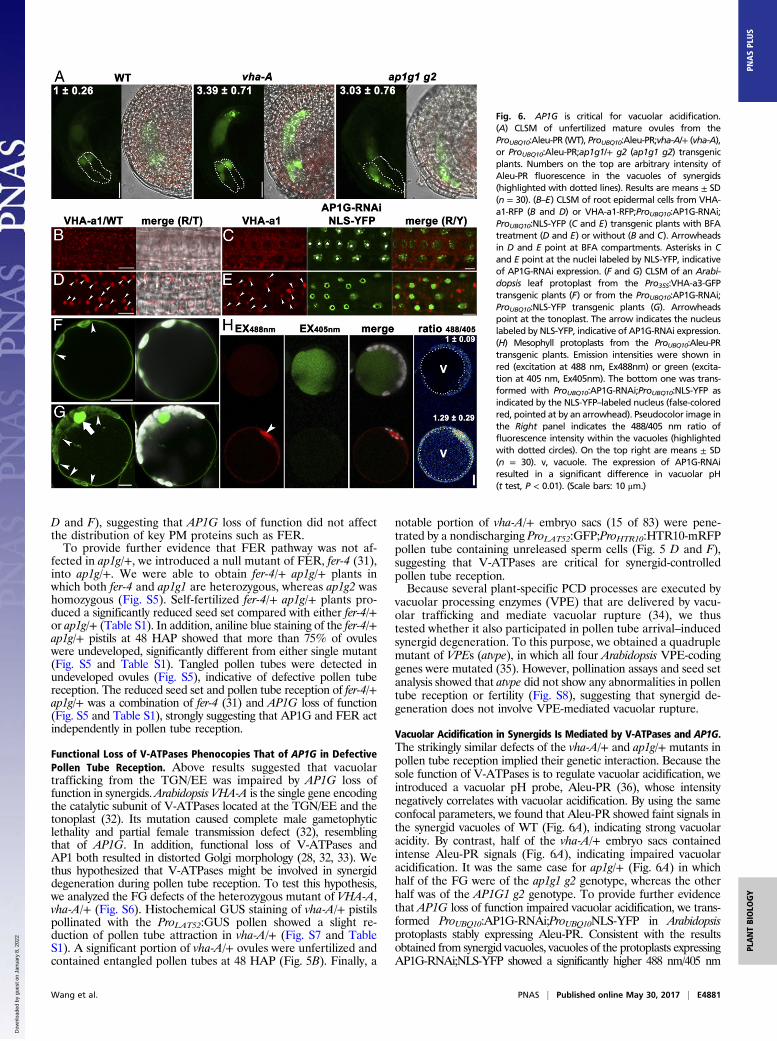
Vacuolar Acidification in Synergids Is Mediated by V-ATPases and AP1G.The strikingly similar defects of the vha-A/+ and ap1g/+ mutants inpollen tube reception implied their genetic interaction. Because thesole function of V-ATPases is to regulate vacuolar acidification, weintroduced a vacuolar pH probe, Aleu-PR (36), whose intensitynegatively correlates with vacuolar acidification. By using the sameconfocal parameters, we found that Aleu-PR showed faint signals inthe synergid vacuoles of WT (Fig. 6A), indicating strong vacuolaracidity. By contrast, half of the vha-A/+ embryo sacs containedintense Aleu-PR signals (Fig. 6A), indicating impaired vacuolaracidification. It was the same case for ap1g/+ (Fig. 6A) in whichhalf of the FG were of the ap1g1 g2 genotype, whereas the otherhalf was of the AP1G1 g2 genotype. To provide further evidencethat AP1G loss of function impaired vacuolar acidification, we trans-formed ProUBQ10:AP1G-RNAi;ProUBQ10NLS-YFP in Arabidopsisprotoplasts stably expressing Aleu-PR. Consistent with the resultsobtained from synergid vacuoles, vacuoles of the protoplasts expressingAP1G-RNAi;NLS-YFP showed a significantly higher 488 nm/405 nm
Fig. 6. AP1G is critical for vacuolar acidification.(A) CLSM of unfertilized mature ovules from theProUBQ10:Aleu-PR (WT), ProUBQ10:Aleu-PR;vha-A/+ (vha-A),or ProUBQ10:Aleu-PR;ap1g1/+ g2 (ap1g1 g2) transgenicplants. Numbers on the top are arbitrary intensity ofAleu-PR fluorescence in the vacuoles of synergids(highlighted with dotted lines). Results are means ± SD(n = 30). (B–E) CLSM of root epidermal cells from VHA-a1-RFP (B and D) or VHA-a1-RFP;ProUBQ10:AP1G-RNAi;ProUBQ10:NLS-YFP (C and E) transgenic plants with BFAtreatment (D and E) or without (B and C). Arrowheadsin D and E point at BFA compartments. Asterisks in Cand E point at the nuclei labeled by NLS-YFP, indicativeof AP1G-RNAi expression. (F and G) CLSM of an Arabi-dopsis leaf protoplast from the Pro35S:VHA-a3-GFPtransgenic plants (F) or from the ProUBQ10:AP1G-RNAi;ProUBQ10:NLS-YFP transgenic plants (G). Arrowheadspoint at the tonoplast. The arrow indicates the nucleuslabeled by NLS-YFP, indicative of AP1G-RNAi expression.(H) Mesophyll protoplasts from the ProUBQ10:Aleu-PRtransgenic plants. Emission intensities were shown inred (excitation at 488 nm, Ex488nm) or green (excita-tion at 405 nm, Ex405nm). The bottom one was trans-formed with ProUBQ10:AP1G-RNAi;ProUBQ10:NLS-YFP asindicated by the NLS-YFP–labeled nucleus (false-coloredred, pointed at by an arrowhead). Pseudocolor image inthe Right panel indicates the 488/405 nm ratio offluorescence intensity within the vacuoles (highlightedwith dotted circles). On the top right are means ± SD(n = 30). v, vacuole. The expression of AP1G-RNAiresulted in a significant difference in vacuolar pH(t test, P < 0.01). (Scale bars: 10 μm.)
Wang et al. PNAS | Published online May 30, 2017 | E4881
PLANTBIOLO
GY
PNASPL
US
Dow
nloa
ded
by g
uest
on
Janu
ary
8, 2
022
value (36) than that of WT (Fig. 6H), indicating compromisedvacuolar acidification by the expression of AP1G-RNAi.VHA-A is a shared component for V-ATPases at the TGN/EE
and at the tonoplast, which are specified by the presence ofTGN/EE-associated VHA-a1 and tonoplast-associated VHA-a2 or VHA-a3, respectively (32). To determine whether AP1Gaffected the targeting of V-ATPases, we examined the effect ofAP1G-RNAi on the subcellular targeting of VHA-a1 and VHA-a3 by using root epidermal cells or protoplasts because synergidsignals were difficult to image. As reported (37), VHA-a1 wasdistributed at cytosolic vesicles (Fig. 6B) sensitive to BFA (Fig.5D), indicative of its TGN/EE identity. Expression of AP1G-RNAi;NLS-YFP did not detectably alter the TGN/EE distribu-tion of VHA-a1 (Fig. 6 C and D). Similarly, the tonoplast-specific targeting of VHA-a3 was not affected by AP1G-RNAi;NLS-YFP (Fig. 6 F and G). These results suggested that AP1Gwas not involved in the subcellular targeting of V-ATPases.
DiscussionWe show here that AP1G is crucial for synergid-controlled pollentube reception. The ap1g/+ FGs were penetrated by nondischargingpollen tubes, a typical feature of defective pollen tube reception.CLSM of ovules at different HAP showed that synergid de-generation was impaired by AP1G loss of function, which is furthersupported by that fact that nondischarging pollen tubes alwayscorrelated with the failure of synergid degeneration. However,synergid degeneration could be initiated without pollen tube dis-charge (17), which explains the comparable ratio of ovules with twointact synergids at 8 HAP and one intact synergid at 12 HAP withinap1g/+ pistils. The impaired degeneration of the remaining synergidin ap1g/+ might be a secondary effect due to the failure of pollentube discharge (23, 24). Although as a fertilization recovery strategyafter defective sperm cell release, nondegenerating synergids keepsending out attractive signals for pollen tubes (26, 38), no super-numerary pollen tubes were observed in ap1g/+ FGs likely due toreduced pollen tube guidance.We propose that AP1G mediates synergid degeneration during
pollen tube reception through V-ATPases. First, functional loss ofV-ATPases resulted in defective pollen tube reception, similar tothat of AP1G. Second, ap1g synergids have a vacuolar pH compa-rable to that of vha-A. Third, genetic interference of AP1G byRNAi impaired vacuolar acidification in mesophyll protoplasts.Because V-ATPases are directly responsible for vacuolar acidifica-tion (39), whereas AP1G functions through mediating protein
sorting at the TGN/EE to the tonoplast, it is more feasible thatV-ATPases are genetically downstream of AP1G.The next obvious question was how AP1G regulates V-ATPases
to mediate vacuolar acidification. A recent study indicated thatvacuolar acidification is collectively contributed by the TGN/EE-associated and the tonoplast-associated V-ATPases (40). Thus,the effect of VHA-A loss of function could be from the TGN/EE andthe vacuoles (39). Tonoplast targeting of NRT2.7 and VIT1 wasdisrupted by AP1G functional loss, implying the key role of vacuolartrafficking in pollen tube reception. However, our results showedthat the subcellular localization of both VHA-a1 and VHA-a3 wasnot detectably affected by genetic interference of AP1G. An alter-native, which requires further investigation, is that AP1G may affectthe activity of V-ATPases by mediating the targeting of some reg-ulatory proteins along the vacuolar trafficking route.Because of its specific feature compared with metazoan lyso-
somes, vacuole participates in plant cell death in two ways. One isthrough vacuolar membrane collapse to release hydrolytic enzymesin the cytosol, whereas the other is through fusion of the tonoplastand the PM (34). Because the quadruple mutant of VPE, whichencode key proteins involved in vacuolar rupture-mediated plantPCD (34, 35), was comparably with WT during pollen tube re-ception, vacuolar rupture during pollen tube arrival as detected byTEM and CLSM is more likely an effect rather than the cause ofsynergid degeneration. Then, how vacuolar acidification may initiatesynergid cell death is an intriguing question. Proton homeostasisaffects endomembrane trafficking (39). Thus, defective vacuolaracidification may have compromised vesicular trafficking, leading toimpaired cell death of synergids. However, the distribution of FERas the key receptor was not affected, suggesting that post-Golgisecretion was not compromised by defective vacuolar acidification.A more likely scenario is that proton homeostasis affects ROS andCa2+ spiking, which have recently been shown to mediate pollentube reception (13–16).
Materials and MethodsDetails onmaterials andmethods, including the plantmaterial and chemicals,plasmid construction, confocal imaging, and other analyses and tools used areprovided in SI Materials and Methods.
ACKNOWLEDGMENTS. We thank Profs. Gregory Copenhaver for ProLAT52:DsRed, Frédéric Berger for ProHTR10:HTR10-mRFP, Liwen Jiang for Aleu-PR,Alice Cheung for fer-4, Lei Ge for the VHA-a1:RFP lines, and Prof. SheilaMcCormick for the English editing of the manuscript.
1. Johnson MA, Preuss D (2002) Plotting a course: Multiple signals guide pollen tubes to
their targets. Dev Cell 2:273–281.2. Kessler SA, Grossniklaus U (2011) She’s the boss: Signaling in pollen tube reception.
Curr Opin Plant Biol 14:622–627.3. Drews GN, Yadegari R (2002) Development and function of the angiosperm female
gametophyte. Annu Rev Genet 36:99–124.4. Berger F, Hamamura Y, Ingouff M, Higashiyama T (2008) Double fertilization - caught
in the act. Trends Plant Sci 13:437–443.5. Escobar-Restrepo JM, et al. (2007) The FERONIA receptor-like kinase mediates male-
female interactions during pollen tube reception. Science 317:656–660.6. Huck N, Moore JM, Federer M, Grossniklaus U (2003) TheArabidopsismutant feronia disrupts
the female gametophytic control of pollen tube reception. Development 130:2149–2159.7. Rotman N, et al. (2003) Female control of male gamete delivery during fertilization in
Arabidopsis thaliana. Curr Biol 13:432–436.8. Tsukamoto T, Qin Y, Huang Y, Dunatunga D, Palanivelu R (2010) A role for LORELEI, a
putative glycosylphosphatidylinositol-anchored protein, in Arabidopsis thaliana
double fertilization and early seed development. Plant J 62:571–588.9. Capron A, et al. (2008) Maternal control of male-gamete delivery in Arabidopsis involves a
putative GPI-anchored protein encoded by the LORELEI gene. Plant Cell 20:3038–3049.10. Hou Y, et al. (2016) Maternal ENODLs are required for pollen tube reception in
Arabidopsis. Curr Biol 26:2343–2350.11. Kessler SA, et al. (2010) Conserved molecular components for pollen tube reception
and fungal invasion. Science 330:968–971.12. Li C, et al. (2015) Glycosylphosphatidylinositol-anchored proteins as chaperones and
co-receptors for FERONIA receptor kinase signaling in Arabidopsis. eLife 4:4.13. Duan Q, et al. (2014) Reactive oxygen species mediate pollen tube rupture to release
sperm for fertilization in Arabidopsis. Nat Commun 5:3129.14. Denninger P, et al. (2014) Male-female communication triggers calcium signatures
during fertilization in Arabidopsis. Nat Commun 5:4645.
15. Ngo QA, Vogler H, Lituiev DS, Nestorova A, Grossniklaus U (2014) A calcium dialogmediated by the FERONIA signal transduction pathway controls plant sperm delivery.Dev Cell 29:491–500.
16. Hamamura Y, et al. (2014) Live imaging of calcium spikes during double fertilizationin Arabidopsis. Nat Commun 5:4722.
17. Leydon AR, et al. (2015) Pollen tube discharge completes the process of synergiddegeneration that is initiated by pollen tube-synergid interaction in Arabidopsis.Plant Physiol 169:485–496.
18. Sandaklie-Nikolova L, Palanivelu R, King EJ, Copenhaver GP, Drews GN (2007) Syn-ergid cell death in Arabidopsis is triggered following direct interaction with thepollen tube. Plant Physiol 144:1753–1762.
19. Maruyama D, et al. (2015) Rapid elimination of the persistent synergid through a cellfusion mechanism. Cell 161:907–918.
20. Völz R, Heydlauff J, Ripper D, von Lyncker L, Groß-Hardt R (2013) Ethylene signaling is requiredfor synergid degeneration and the establishment of a pollen tube block. Dev Cell 25:310–316.
21. Zimmermann P, Hirsch-Hoffmann M, Hennig L, GruissemW (2004) GENEVESTIGATOR.Arabidopsis microarray database and analysis toolbox. Plant Physiol 136:2621–2632.
22. Pagnussat GC, Alandete-Saez M, Bowman JL, Sundaresan V (2009) Auxin-dependentpatterning and gamete specification in the Arabidopsis female gametophyte. Science324:1684–1689.
23. Leydon AR, et al. (2013) Three MYB transcription factors control pollen tube differ-entiation required for sperm release. Curr Biol 23:1209–1214.
24. Liang Y, et al. (2013) MYB97, MYB101 and MYB120 function as male factors thatcontrol pollen tube-synergid interaction in Arabidopsis thaliana fertilization. PLoSGenet 9:e1003933.
25. Ingouff M, Hamamura Y, Gourgues M, Higashiyama T, Berger F (2007) Distinct dy-namics of HISTONE3 variants between the two fertilization products in plants. CurrBiol 17:1032–1037.
26. Beale KM, Leydon AR, Johnson MA (2012) Gamete fusion is required to block multiplepollen tubes from entering an Arabidopsis ovule. Curr Biol 22:1090–1094.
E4882 | www.pnas.org/cgi/doi/10.1073/pnas.1617967114 Wang et al.
Dow
nloa
ded
by g
uest
on
Janu
ary
8, 2
022
27. Steffen JG, Kang IH, Macfarlane J, Drews GN (2007) Identification of genes expressedin the Arabidopsis female gametophyte. Plant J 51:281–292.
28. Park M, et al. (2013) Arabidopsis μ-adaptin subunit AP1M of adaptor protein complex1 mediates late secretory and vacuolar traffic and is required for growth. Proc NatlAcad Sci USA 110:10318–10323.
29. Wang X, et al. (2014) Trans-Golgi network-located AP1 gamma adaptins mediatedileucine motif-directed vacuolar targeting in Arabidopsis. Plant Cell 26:4102–4118.
30. Chopin F, et al. (2007) The Arabidopsis ATNRT2.7 nitrate transporter controls nitratecontent in seeds. Plant Cell 19:1590–1602.
31. Duan Q, Kita D, Li C, Cheung AY, Wu H-M (2010) FERONIA receptor-like kinase regulatesRHO GTPase signaling of root hair development. Proc Natl Acad Sci USA 107:17821–17826.
32. Dettmer J, et al. (2005) Essential role of the V-ATPase in male gametophyte devel-opment. Plant J 41:117–124.
33. Strompen G, et al. (2005) Arabidopsis vacuolar H-ATPase subunit E isoform 1 is required forGolgi organization and vacuole function in embryogenesis. Plant J 41:125–132.
34. Hara-Nishimura I, Hatsugai N (2011) The role of vacuole in plant cell death. Cell DeathDiffer 18:1298–1304.
35. Kuroyanagi M, et al. (2005) Vacuolar processing enzyme is essential for mycotoxin-induced cell death in Arabidopsis thaliana. J Biol Chem 280:32914–32920.
36. Shen J, et al. (2013) Organelle pH in the Arabidopsis endomembrane system. MolPlant 6:1419–1437.
37. Viotti C, et al. (2010) Endocytic and secretory traffic in Arabidopsismerge in the trans-Golgi network/early endosome, an independent and highly dynamic organelle. PlantCell 22:1344–1357.
38. Kasahara RD, et al. (2012) Fertilization recovery after defective sperm cell release inArabidopsis. Curr Biol 22:1084–1089.
39. Schumacher K, Krebs M (2010) The V-ATPase: Small cargo, large effects. Curr OpinPlant Biol 13:724–730.
40. Kriegel A, et al. (2015) Job sharing in the endomembrane system: Vacuolar acidifi-cation requires the combined activity of V-ATPase and V-PPase. Plant Cell 27:3383–3396.
41. Zhou LZ, et al. (2013) Protein S-ACYL Transferase10 is critical for development and salttolerance in Arabidopsis. Plant Cell 25:1093–1107.
42. Wang JG, et al. (2013) HAPLESS13, the Arabidopsis μ1 adaptin, is essential for proteinsorting at the trans-Golgi network/early endosome. Plant Physiol 162:1897–1910.
43. Li S, et al. (2013) Arabidopsis COBRA-LIKE 10, a GPI-anchored protein, mediates di-rectional growth of pollen tubes. Plant J 74:486–497.
44. Karimi M, Inzé D, Depicker A (2002) GATEWAY vectors for Agrobacterium-mediatedplant transformation. Trends Plant Sci 7:193–195.
45. Guo J, Wang F, Song J, Sun W, Zhang XS (2010) The expression of Orysa;CycB1;1 isessential for endosperm formation and causes embryo enlargement in rice. Planta231:293–303.
Wang et al. PNAS | Published online May 30, 2017 | E4883
PLANTBIOLO
GY
PNASPL
US
Dow
nloa
ded
by g
uest
on
Janu
ary
8, 2
022
Copyright © 2022 FDOKUMEN