
Polar vacuolar distribution is essential for accurate asymmetric ...
6
Polar vacuolar distribution is essential for accurate asymmetric division of Arabidopsis zygotes Yusuke Kimata a,1 , Takehide Kato b,1 , Takumi Higaki c,d , Daisuke Kurihara a , Tomomi Yamada a,e , Shoji Segami f , Miyo Terao Morita f,g , Masayoshi Maeshima f , Seiichiro Hasezawa c , Tetsuya Higashiyama a,e , Masao Tasaka b , and Minako Ueda a,e,2 a Division of Biological Science, Graduate School of Science, Nagoya University, Furo-cho, Chikusa-ku, Nagoya, 464-8602 Aichi, Japan; b Graduate School of Biological Sciences, Nara Institute of Science and Technology, Ikoma, 630-0192 Nara, Japan; c Department of Integrated Biosciences, Graduate School of Frontier Sciences, The University of Tokyo, Kashiwanoha, Kashiwa, 277-8562 Chiba, Japan; d International Research Organization for Advanced Science and Technology, Kumamoto University, Chuo-ku, 860-8555 Kumamoto, Japan; e Institute of Transformative Bio-Molecules, Nagoya University, Furo-cho, Chikusa- ku, Nagoya, 464-8601 Aichi, Japan; f Graduate School of Bioagricultural Sciences, Nagoya University, Nagoya, 464-8601 Aichi, Japan; and g National Institute for Basic Biology, Myodaiji, Okazaki, 444-8585 Aichi, Japan Edited by Dominique C. Bergmann, Stanford University, Stanford, CA, and approved December 14, 2018 (received for review August 27, 2018) In most flowering plants, the asymmetric cell division of the zygote is the initial step in establishing the apical– basal axis of the mature plant. The zygote is polarized, possessing the nucleus at the apical tip and large vacuoles at the basal end. Despite their known polar localization, whether the positioning of the vacuoles and the nucleus is coordinated and what the role of the vacuole is in the asymmetric zygotic division remain elusive. In the present study, we utilized a live-cell imaging system to visualize the dynamics of vacuoles during the entire process of zygote polarization in Arabidopsis. Image analysis revealed that the vacuoles formed tubular strands around the apically migrating nucleus. They gradually accumulated at the basal region and filled the space, resulting in asymmetric distribution in the mature zygote. To assess the role of vacuoles in the zygote, we screened various vacuole mutants and identified that shoot gravitropism2 (sgr2), in which the vacuolar structural change was impaired, failed to form tubular vacuoles and to polarly distribute the vacuole. In sgr2, large vacuoles occupied the apical tip and thus nuclear migration was blocked, resulting in a more symmetric zygotic division. We further observed that tubular vacuole formation and asymmetric vacuolar distribution both depended on the longitudinal array of actin filaments. Overall, our results show that vacuolar dynamics is crucial not only for the polar distribution along actin filaments but also for adequate nuclear positioning, and consequently zygote-division asymmetry. Arabidopsis thaliana | zygote | vacuole | apical–basal axis | live-cell imaging I nitiation of the main body axis is an incipient event required for a unicellular zygote to develop into a multicellular organism. In most flowering plants (both monocots and dicots), the apical– basal axis is formed along the longitudinal axis of the zygote marked by the nucleus in the apical region and large vacuoles in the basal region (1, 2). Large vacuoles are also observed in unfertilized egg cells but, after fertilization, the vacuoles are shrunk and/or dispersed into the entire cell (3–5). In the mature zygote, vacuoles are accu- mulated at the basal region and most vacuoles are inherited in the basal cell after asymmetric division of the zygote (1, 4, 5). Despite such obvious changes, the dynamics of vacuolar distribution and the role of vacuoles in the asymmetric zygotic division are unknown. We recently developed a high-resolution live-cell imaging system utilizing an in vitro ovule cultivation method and two-photon exci- tation microscopy, and visualized the intracellular dynamics in Arabidopsis zygotes (6, 7). Using this system, we observed that actin filaments (F-actin) align longitudinally in the elongating zygote and are essential for apically directed nuclear migration (6). However, it remained unknown whether other organelles also distribute along F-actin and, if so, whether their positioning is coordinated or independent. In this study, we performed live-cell imaging of vacuolar dy- namics during zygote polarization. Various morphological fea- tures of the vacuoles were extensively compared at different zygotic stages in the wild type and the shoot gravitropism2 (sgr2) mutant, which affects the vacuolar membrane protein, and after exposure to specific inhibitors for F-actin. We found that dy- namic vacuolar distribution along F-actin is necessary for nuclear migration to the future division site. Our findings provide in- sights into the role of vacuoles in setting the accurate asymmetric division of the zygote in flowering plants. Results Vacuoles Form Thin Tubular Strands Around the Nucleus and Gradually Accumulate at the Basal Region During Zygote Elongation. To perform live-cell imaging of the vacuole in the polarizing zygote, we de- veloped a dual-color marker that simultaneously labels the vacuolar membrane and nucleus (vacuole/nucleus) and could be imaged using two-photon excitation microscopy (2PEM) (Fig. 1 A and B). We detected the same pattern of polar localization for the vacuole and the nucleus as the fixed ovules of nontransgenic plants (Fig. 1 and SI Appendix, Fig. S1) (5), thus indicating that our system visu- alized native intracellular patterns. We then captured the behavior using time-lapse imaging until the first zygotic cell division (Fig. 1 B and C and Movie S1). Before fertilization, the egg cell showed a clear polar organiza- tion, with the nucleus at the apical pole and huge vacuoles oc- cupying the basal region. After fertilization, the cell shrunk as we previously reported (6), and vacuolar size was also markedly Significance The vacuole is one of the largest plant organelles. It occupies the basal region of the zygote and is mostly inherited in the basal daughter cell after zygotic division. In spite of the obvi- ous asymmetry, how dynamically the vacuole is distributed and whether it contributes to apical–basal axis formation are unknown. In the present study, we report that the vacuole actively changes its shape and size and positions along actin filaments. We further show that vacuolar distribution supports nuclear localization at the opposite cell end, and thus ensures asymmetric division of the zygote. These results provide in- sights into cooperative organelle positioning during zygote polarization and the crucial roles of vacuoles in the initial steps of plant ontogeny. Author contributions: Y.K., T.K., D.K., M.T.M., S.H., T. Higashiyama, M.T., and M.U. de- signed research; Y.K., T.K., T.Y., S.S., and M.U. performed research; Y.K., T. Higaki, and M.U. analyzed data; and Y.K., T. Higaki, D.K., M.T.M., M.M., and M.U. wrote the paper. The authors declare no conflict of interest. This article is a PNAS Direct Submission. Published under the PNAS license. 1 Y.K. and T.K. contributed equally to this work. 2 To whom correspondence should be addressed. Email: [email protected]. This article contains supporting information online at www.pnas.org/lookup/suppl/doi:10. 1073/pnas.1814160116/-/DCSupplemental. Published online January 16, 2019. 2338–2343 | PNAS | February 5, 2019 | vol. 116 | no. 6 www.pnas.org/cgi/doi/10.1073/pnas.1814160116 Downloaded by guest on January 8, 2022
-
Upload
khangminh22 -
Category
Documents
-
view
1 -
download
0
Transcript of Polar vacuolar distribution is essential for accurate asymmetric ...
Polar vacuolar distribution is essential for accurateasymmetric division of Arabidopsis zygotesYusuke Kimataa,1, Takehide Katob,1, Takumi Higakic,d, Daisuke Kuriharaa, Tomomi Yamadaa,e, Shoji Segamif,Miyo Terao Moritaf,g, Masayoshi Maeshimaf, Seiichiro Hasezawac, Tetsuya Higashiyamaa,e, Masao Tasakab,and Minako Uedaa,e,2
aDivision of Biological Science, Graduate School of Science, Nagoya University, Furo-cho, Chikusa-ku, Nagoya, 464-8602 Aichi, Japan; bGraduate School ofBiological Sciences, Nara Institute of Science and Technology, Ikoma, 630-0192 Nara, Japan; cDepartment of Integrated Biosciences, Graduate School ofFrontier Sciences, The University of Tokyo, Kashiwanoha, Kashiwa, 277-8562 Chiba, Japan; dInternational Research Organization for Advanced Science andTechnology, Kumamoto University, Chuo-ku, 860-8555 Kumamoto, Japan; eInstitute of Transformative Bio-Molecules, Nagoya University, Furo-cho, Chikusa-ku, Nagoya, 464-8601 Aichi, Japan; fGraduate School of Bioagricultural Sciences, Nagoya University, Nagoya, 464-8601 Aichi, Japan; and gNational Institutefor Basic Biology, Myodaiji, Okazaki, 444-8585 Aichi, Japan
Edited by Dominique C. Bergmann, Stanford University, Stanford, CA, and approved December 14, 2018 (received for review August 27, 2018)
In most flowering plants, the asymmetric cell division of the zygote isthe initial step in establishing the apical–basal axis of the mature plant.The zygote is polarized, possessing the nucleus at the apical tip andlarge vacuoles at the basal end. Despite their known polar localization,whether the positioning of the vacuoles and the nucleus is coordinatedand what the role of the vacuole is in the asymmetric zygotic divisionremain elusive. In the present study, we utilized a live-cell imagingsystem to visualize the dynamics of vacuoles during the entire processof zygote polarization in Arabidopsis. Image analysis revealed that thevacuoles formed tubular strands around the apically migrating nucleus.They gradually accumulated at the basal region and filled the space,resulting in asymmetric distribution in themature zygote. To assess therole of vacuoles in the zygote, we screened various vacuole mutantsand identified that shoot gravitropism2 (sgr2), in which the vacuolarstructural change was impaired, failed to form tubular vacuoles and topolarly distribute the vacuole. In sgr2, large vacuoles occupied theapical tip and thus nuclear migration was blocked, resulting in a moresymmetric zygotic division. We further observed that tubular vacuoleformation and asymmetric vacuolar distribution both depended on thelongitudinal array of actin filaments. Overall, our results show thatvacuolar dynamics is crucial not only for the polar distribution alongactin filaments but also for adequate nuclear positioning, andconsequently zygote-division asymmetry.
Arabidopsis thaliana | zygote | vacuole | apical–basal axis | live-cell imaging
Initiation of the main body axis is an incipient event requiredfor a unicellular zygote to develop into a multicellular organism.
In most flowering plants (both monocots and dicots), the apical–basal axis is formed along the longitudinal axis of the zygote markedby the nucleus in the apical region and large vacuoles in the basalregion (1, 2). Large vacuoles are also observed in unfertilized eggcells but, after fertilization, the vacuoles are shrunk and/or dispersedinto the entire cell (3–5). In the mature zygote, vacuoles are accu-mulated at the basal region and most vacuoles are inherited in thebasal cell after asymmetric division of the zygote (1, 4, 5). Despitesuch obvious changes, the dynamics of vacuolar distribution and therole of vacuoles in the asymmetric zygotic division are unknown.We recently developed a high-resolution live-cell imaging system
utilizing an in vitro ovule cultivation method and two-photon exci-tation microscopy, and visualized the intracellular dynamics inArabidopsis zygotes (6, 7). Using this system, we observed that actinfilaments (F-actin) align longitudinally in the elongating zygote andare essential for apically directed nuclear migration (6). However, itremained unknown whether other organelles also distribute alongF-actin and, if so, whether their positioning is coordinated orindependent.In this study, we performed live-cell imaging of vacuolar dy-
namics during zygote polarization. Various morphological fea-tures of the vacuoles were extensively compared at differentzygotic stages in the wild type and the shoot gravitropism2 (sgr2)
mutant, which affects the vacuolar membrane protein, and afterexposure to specific inhibitors for F-actin. We found that dy-namic vacuolar distribution along F-actin is necessary for nuclearmigration to the future division site. Our findings provide in-sights into the role of vacuoles in setting the accurate asymmetricdivision of the zygote in flowering plants.
ResultsVacuoles Form Thin Tubular Strands Around the Nucleus and GraduallyAccumulate at the Basal Region During Zygote Elongation. To performlive-cell imaging of the vacuole in the polarizing zygote, we de-veloped a dual-color marker that simultaneously labels the vacuolarmembrane and nucleus (vacuole/nucleus) and could be imagedusing two-photon excitation microscopy (2PEM) (Fig. 1 A and B).We detected the same pattern of polar localization for the vacuoleand the nucleus as the fixed ovules of nontransgenic plants (Fig. 1and SI Appendix, Fig. S1) (5), thus indicating that our system visu-alized native intracellular patterns.We then captured the behavior using time-lapse imaging until
the first zygotic cell division (Fig. 1 B and C and Movie S1).Before fertilization, the egg cell showed a clear polar organiza-tion, with the nucleus at the apical pole and huge vacuoles oc-cupying the basal region. After fertilization, the cell shrunk as wepreviously reported (6), and vacuolar size was also markedly
Significance
The vacuole is one of the largest plant organelles. It occupiesthe basal region of the zygote and is mostly inherited in thebasal daughter cell after zygotic division. In spite of the obvi-ous asymmetry, how dynamically the vacuole is distributedand whether it contributes to apical–basal axis formation areunknown. In the present study, we report that the vacuoleactively changes its shape and size and positions along actinfilaments. We further show that vacuolar distribution supportsnuclear localization at the opposite cell end, and thus ensuresasymmetric division of the zygote. These results provide in-sights into cooperative organelle positioning during zygotepolarization and the crucial roles of vacuoles in the initial stepsof plant ontogeny.
Author contributions: Y.K., T.K., D.K., M.T.M., S.H., T. Higashiyama, M.T., and M.U. de-signed research; Y.K., T.K., T.Y., S.S., and M.U. performed research; Y.K., T. Higaki, andM.U. analyzed data; and Y.K., T. Higaki, D.K., M.T.M., M.M., and M.U. wrote the paper.
The authors declare no conflict of interest.
This article is a PNAS Direct Submission.
Published under the PNAS license.1Y.K. and T.K. contributed equally to this work.2To whom correspondence should be addressed. Email: [email protected].
This article contains supporting information online at www.pnas.org/lookup/suppl/doi:10.1073/pnas.1814160116/-/DCSupplemental.
Published online January 16, 2019.
2338–2343 | PNAS | February 5, 2019 | vol. 116 | no. 6 www.pnas.org/cgi/doi/10.1073/pnas.1814160116
Dow
nloa
ded
by g
uest
on
Janu
ary
8, 2
022
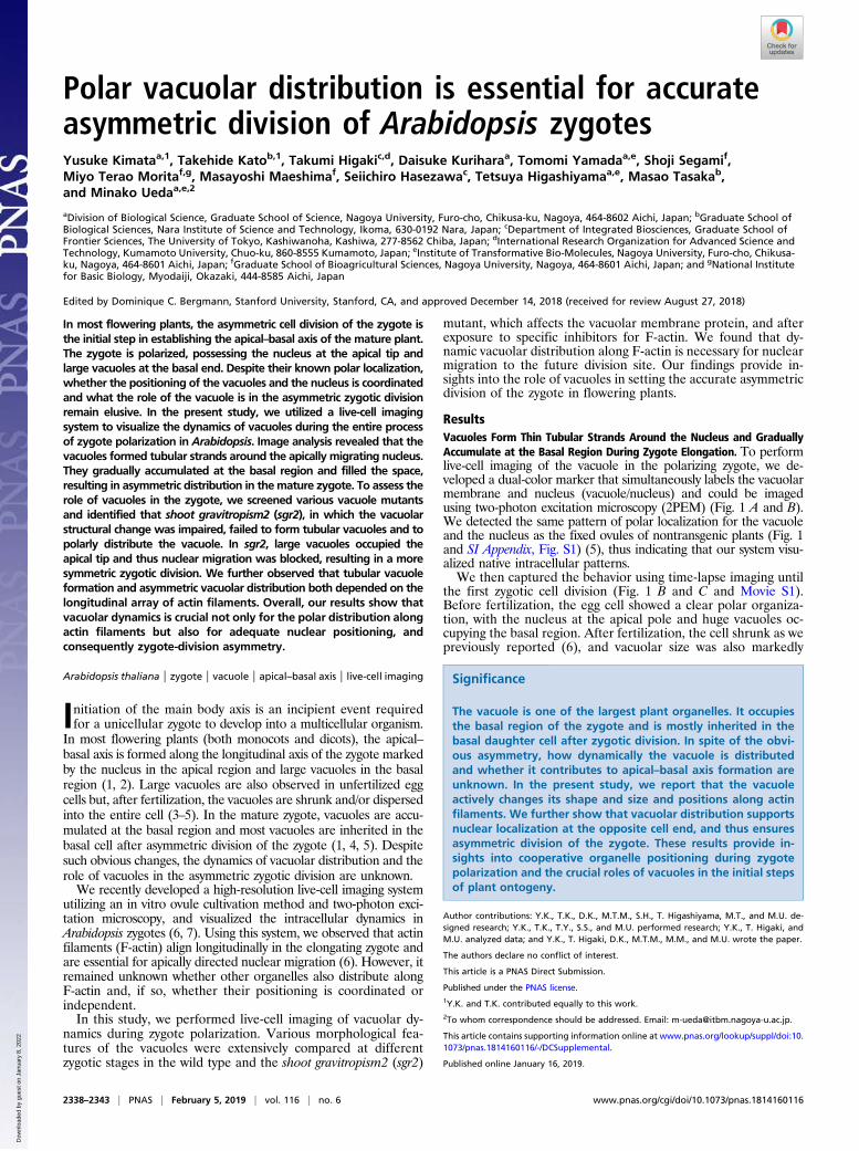
reduced. The nucleus shifted to the cell center, and the vacuoleswere detected both in the apical and basal regions (Fig. 1 D and E).Then, the zygote elongated, and the vacuoles formed thin tubularstructures around the nucleus, which migrates toward the apical tip(Fig. 1 B–D). Simultaneously, vacuolar size was also gradually in-creased. The vacuoles in the apical region remained small whilevacuolar size in the basal area was massively increased, resulting in ahighly asymmetric distribution of the vacuoles on completion ofnuclear migration and zygotic elongation (Fig. 1 B, C, and E).Therefore, after chromosome segregation and cytokinesis, most ofthe vacuoles were inherited into the basal cell.In summary, fertilization triggers vacuolar shrinkage, and the
vacuoles swell again during zygote elongation. The nucleus becomessurrounded by thin vacuolar tubules and migrates to the apical apex,while the vacuoles gradually accumulate at the basal region.
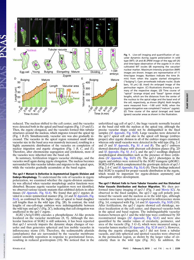
The sgr2-1 Mutant Is Defective in Asymmetrical Zygotic Division andEmbryo Morphology. To understand the role of vacuoles in zygotepolarization, we examined whether the zygote-division asymme-try was affected when vacuolar morphology and/or functions weredisturbed. Because zygotic vacuolar regulators were not identified,we observed various vacuole mutants that exhibited defects in othertissues (SI Appendix, Fig. S1A). We found that sgr2-1 zygotes di-vided in a more symmetrical manner (Fig. 2A and SI Appendix, Fig.S1A), as confirmed by the higher ratio of apical to basal daughtercell lengths than in the wild type (Fig. 2B). In contrast, the totallengths of one-cell-stage embryos were indistinguishable betweenthe wild type and sgr2-1 (Fig. 2C), indicating that cell elongation wasunaffected in the sgr2-1 zygote.SGR2 (At1g31480) encodes a phospholipase A1-like protein
localized on the vacuolar membrane (8, 9). Although the mo-lecular function of SGR2 is still unknown, it has been reportedthat sgr2-1 severely impairs the dynamic shape changes of vac-uoles and thus generates spherical and less mobile vacuoles ininflorescence stems (10). Therefore, the sedimentable plastids(amyloplasts) that are surrounded by the vacuolar membranecannot smoothly reposition in response to gravitational forces,resulting in reduced gravitropism (10). We noticed that in the
unfertilized egg cell of sgr2-1, the huge vacuole normally locatedat the basal end with the nucleus at the apical top, although theprecise vacuolar shape could not be distinguished in the fixedsamples (SI Appendix, Fig. S1B). Large vacuoles were detected inthe sgr2-1 apical cell and also in the apical cell lineage (embryoproper) after zygotic division in all of the embryo stages examined,whereas only small vacuoles were detected in the wild type (Fig. 2 Aand D and SI Appendix, Fig. S1 A and B). The sgr2-1 embryosshowed distorted shapes with aberrant cell-division planes (Fig. 2Dand SI Appendix, Fig. S1 A–C), and the sgr2-1 seedlings displayedmorphological abnormalities such as the presence of three cotyle-dons (SI Appendix, Fig. S1D) (9). The sgr2-1 phenotypes in thezygote and embryo were restored by the SGR2 transgene (pSGR2::SGR2i-GFP), which complemented the gravitropic defects of sgr2-1(Fig. 2 A–C and SI Appendix, Fig. S1A) (8). These findings indicatedthat SGR2 is required for proper vacuole distribution in the zygote,which would be important for zygote-division asymmetry andsubsequent embryo development.
The sgr2-1 Mutant Fails to Form Tubular Vacuoles and Has DisturbedPolar Vacuole Distribution and Nuclear Migration. We then per-formed time-lapse imaging of sgr2-1 (Fig. 3 and Movie S2). Asobserved in the fixed samples, the sgr2-1 egg cell polarly posi-tioned the vacuole and the nucleus as in the wild type, althoughvacuoles were more spherical, as reported in inflorescence stems(Fig. 3A, compared with Fig. 1A and SI Appendix, Fig. S1B) (10).After fertilization, the sgr2-1 zygote showed cell shrinkage, nu-clear localization at the cell center, and dispersed vacuoles bothin the apical and basal regions. The indistinguishable vacuolarfeatures between sgr2-1 and the wild type were confirmed by 3Dreconstructed images (SI Appendix, Fig. S2A) and were alsoquantified by the similar values of circularity, perimeter, andarea of the vacuole (Fig. 3 G–I), which were measured using avacuolar lumen marker (SI Appendix, Fig. S2 B and C). However,during the zygotic elongation, sgr2-1 did not form a tubularvacuole, and only spherical and spotted vacuoles were observedin the perinuclear region, as shown by the larger value of cir-cularity than in the wild type (Fig. 3G). In addition, the
Fig. 1. Live-cell imaging and quantification of vac-uolar dynamics during zygote polarization in wildtype (WT). (A and B) 2PEM image of the egg cell (A)and time-lapse observation of the zygote in in vitrocultivated WT ovules (B) expressing the vacuolar/nuclear marker. Maximum-intensity projection (MIP)images are shown. Images are representative of 11time-lapse images. Numbers indicate the time (h:min) from when the zygote started elongation(“bulging”). Cyan arrowheads indicate nuclei. (Scalebars, 10 μm.) (B, Inset) An enlarged image of theperinuclear region. (C) Illustrations showing a sum-mary of the respective stages. (D) Time course of“apical” (orange stripe) and “basal” (green stripe)lengths, which are the distances from the center ofthe nucleus to the apical edge and the basal end ofthe cell, respectively, as shown (Right). Both lengthswere measured from −1:00 until 14:00, when thezygote elongation was completed (“mature” zygote).(E) Time course of the apical (orange) and basal(green) vacuolar areas as shown in the illustration.
Kimata et al. PNAS | February 5, 2019 | vol. 116 | no. 6 | 2339
PLANTBIOLO
GY
Dow
nloa
ded
by g
uest
on
Janu
ary
8, 2
022
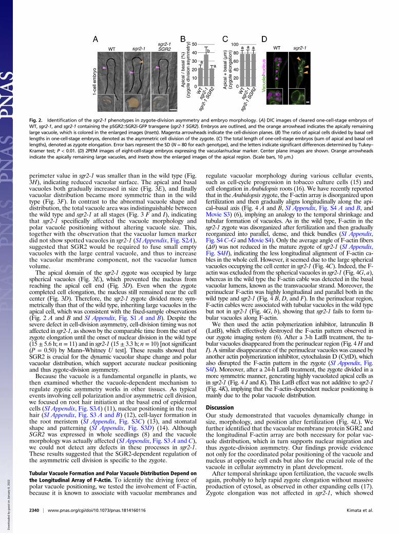
perimeter value in sgr2-1 was smaller than in the wild type (Fig.3H), indicating reduced vacuolar surface. The apical and basalvacuoles both gradually increased in size (Fig. 3E), and finallyvacuolar distribution became more symmetric than in the wildtype (Fig. 3F). In contrast to the abnormal vacuole shape anddistribution, the total vacuole area was indistinguishable betweenthe wild type and sgr2-1 at all stages (Fig. 3 F and I), indicatingthat sgr2-1 specifically affected the vacuole morphology andpolar vacuole positioning without altering vacuole size. This,together with the observation that the vacuolar lumen markerdid not show spotted vacuoles in sgr2-1 (SI Appendix, Fig. S2A),suggested that SGR2 would be required to fuse small emptyvacuoles with the large central vacuole, and thus to increasethe vacuolar membrane component, not the vacuolar lumenvolume.The apical domain of the sgr2-1 zygote was occupied by large
spherical vacuoles (Fig. 3E), which prevented the nucleus fromreaching the apical cell end (Fig. 3D). Even when the zygotecompleted cell elongation, the nucleus still remained near the cellcenter (Fig. 3D). Therefore, the sgr2-1 zygote divided more sym-metrically than that of the wild type, inheriting large vacuoles in theapical cell, which was consistent with the fixed-sample observations(Fig. 2 A and B and SI Appendix, Fig. S1 A and B). Despite thesevere defect in cell-division asymmetry, cell-division timing was notaffected in sgr2-1, as shown by the comparable time from the start ofzygote elongation until the onset of nuclear division in the wild type(15 ± 5.6 h; n = 11) and in sgr2-1 (15 ± 3.3 h; n = 10) [not significant(P = 0.50) by Mann–Whitney U test]. These results showed thatSGR2 is crucial for the dynamic vacuolar shape change and polarvacuolar distribution, which support accurate nuclear positioningand thus zygote-division asymmetry.Because the vacuole is a fundamental organelle in plants, we
then examined whether the vacuole-dependent mechanism toregulate zygotic asymmetry works in other tissues. As typicalevents involving cell polarization and/or asymmetric cell division,we focused on root hair initiation at the basal end of epidermalcells (SI Appendix, Fig. S3A) (11), nuclear positioning in the roothair (SI Appendix, Fig. S3 A and B) (12), cell-layer formation inthe root meristem (SI Appendix, Fig. S3C) (13), and stomatalshape and patterning (SI Appendix, Fig. S3D) (14). AlthoughSGR2 was expressed in whole seedlings (8) and the vacuolemorphology was actually affected (SI Appendix, Fig. S3 A and C),we could not detect any defects in these processes in sgr2-1.These results suggested that the SGR2-dependent regulation ofthe asymmetric cell division is specific to the zygote.
Tubular Vacuole Formation and Polar Vacuole Distribution Depend onthe Longitudinal Array of F-Actin. To identify the driving force ofpolar vacuole positioning, we tested the involvement of F-actin,because it is known to associate with vacuolar membranes and
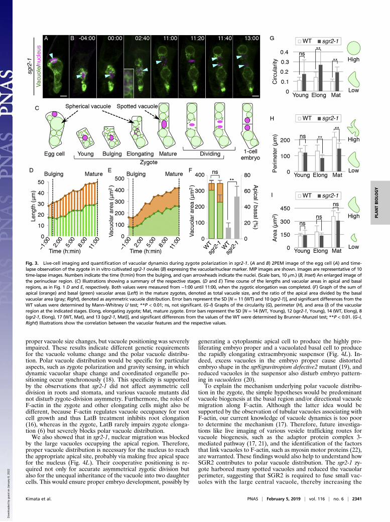
regulate vacuolar morphology during various cellular events,such as cell-cycle progression in tobacco culture cells (15) andcell elongation in Arabidopsis roots (16). We have recently reportedthat in the Arabidopsis zygote, the F-actin array is disorganized uponfertilization and then gradually aligns longitudinally along the api-cal–basal axis (Fig. 4 A and B, SI Appendix, Fig. S4 A and B, andMovie S3) (6), implying an analogy to the temporal shrinkage andtubular formation of vacuoles. As in the wild type, F-actin in thesgr2-1 zygote was disorganized after fertilization and then graduallyreorganized into parallel, dense, and thick bundles (SI Appendix,Fig. S4 C–G andMovie S4). Only the average angle of F-actin fibers(Δθ) was not reduced in the mature zygote of sgr2-1 (SI Appendix,Fig. S4H), indicating the less longitudinal alignment of F-actin ca-bles in the whole cell. However, it seemed due to the large sphericalvacuoles occupying the cell center in sgr2-1 (Fig. 4C). Indeed, the F-actin was excluded from the spherical vacuoles in sgr2-1 (Fig. 4G, a),whereas in the wild type the F-actin cable was detected in the basalvacuolar lumens, known as the transvacuolar strand. Moreover, theperinuclear F-actin was highly longitudinal and parallel both in thewild type and sgr2-1 (Fig. 4 B, D, and F). In the perinuclear region,F-actin cables were associated with tubular vacuoles in the wild typebut not in sgr2-1 (Fig. 4G, b), showing that sgr2-1 fails to form tu-bular vacuoles along F-actin.We then used the actin polymerization inhibitor, latrunculin B
(LatB), which effectively destroyed the F-actin pattern observed inour zygote imaging system (6). After a 3-h LatB treatment, the tu-bular vacuoles disappeared from the perinuclear region (Fig. 4H andI). A similar disappearance of the perinuclear vacuoles was caused byanother actin polymerization inhibitor, cytochalasin D (CytD), whichalso disrupted the F-actin pattern in the zygote (SI Appendix, Fig.S4I). Moreover, after a 24-h LatB treatment, the zygote divided in amore symmetric manner, generating highly vacuolated apical cells asin sgr2-1 (Fig. 4 J and K). This LatB effect was not additive to sgr2-1(Fig. 4K), implying that the F-actin–dependent nuclear positioning ismainly due to the polar vacuole distribution.
DiscussionOur study demonstrated that vacuoles dynamically change insize, morphology, and position after fertilization (Fig. 4L). Wefurther identified that the vacuolar membrane protein SGR2 andthe longitudinal F-actin array are both necessary for polar vac-uole distribution, which in turn supports nuclear migration andthus zygote-division asymmetry. Our findings provide evidencenot only for the coordinated polar positioning of the vacuole andnucleus at opposite cell ends but also for the crucial role of thevacuole in cellular asymmetry in plant development.After temporal shrinkage upon fertilization, the vacuole swells
again, probably to help rapid zygote elongation without massiveproduction of cytosol, as observed in other expanding cells (17).Zygote elongation was not affected in sgr2-1, which showed
Fig. 2. Identification of the sgr2-1 phenotypes in zygote-division asymmetry and embryo morphology. (A) DIC images of cleared one-cell-stage embryos ofWT, sgr2-1, and sgr2-1 containing the pSGR2::SGR2i-GFP transgene (sgr2-1 SGR2). Embryos are outlined, and the orange arrowhead indicates the apically remaininglarge vacuole, which is colored in the enlarged images (Insets). Magenta arrowheads indicate the cell-division planes. (B) The ratio of apical cells divided by basal celllengths in one-cell-stage embryos, denoted as the asymmetric cell division of the zygote. (C) The total length of one-cell-stage embryos (sum of apical and basal celllengths), denoted as zygote elongation. Error bars represent the SD (N = 80 for each genotype), and the letters indicate significant differences determined by Tukey–Kramer test; P < 0.01. (D) 2PEM images of eight-cell-stage embryos expressing the vacuolar/nuclear marker. Center plane images are shown. Orange arrowheadsindicate the apically remaining large vacuoles, and Insets show the enlarged images of the apical region. (Scale bars, 10 μm.)
2340 | www.pnas.org/cgi/doi/10.1073/pnas.1814160116 Kimata et al.
Dow
nloa
ded
by g
uest
on
Janu
ary
8, 2
022
proper vacuole size changes, but vacuole positioning was severelyimpaired. These results indicate different genetic requirementsfor the vacuole volume change and the polar vacuole distribu-tion. Polar vacuole distribution would be specific for particularaspects, such as zygote polarization and gravity sensing, in whichdynamic vacuolar shape change and coordinated organelle po-sitioning occur synchronously (18). This specificity is supportedby the observations that sgr2-1 did not affect asymmetric celldivision in roots and stomata, and various vacuole mutants didnot disturb zygote-division asymmetry. Furthermore, the roles ofF-actin in the zygote and other elongating cells might also bedifferent, because F-actin regulates vacuole occupancy for rootcell growth and thus LatB treatment inhibits root elongation(16), whereas in the zygote, LatB rarely impairs zygote elonga-tion (6) but severely blocks polar vacuole distribution.We also showed that in sgr2-1, nuclear migration was blocked
by the large vacuoles occupying the apical region. Therefore,proper vacuole distribution is necessary for the nucleus to reachthe appropriate apical site, probably via making free apical spacefor the nucleus (Fig. 4L). Their cooperative positioning is re-quired not only for accurate asymmetrical zygotic division butalso for the unequal inheritance of the vacuole into two daughtercells. This would ensure proper embryo development, possibly by
generating a cytoplasmic apical cell to produce the highly pro-liferating embryo proper and a vacuolated basal cell to producethe rapidly elongating extraembryonic suspensor (Fig. 4L). In-deed, excess vacuoles in the embryo proper cause distortedembryo shape in the sgr8/gravitropism defective2 mutant (19), andreduced vacuoles in the suspensor also disturb embryo pattern-ing in vacuoleless (20).To explain the mechanism underlying polar vacuole distribu-
tion in the zygote, the simple hypotheses would be predominantvacuole biogenesis at the basal region and/or directional vacuolemigration along F-actin. Although the latter idea would besupported by the observation of tubular vacuoles associating withF-actin, our current knowledge of vacuole dynamics is too poorto determine the mechanism (17). Therefore, future investiga-tions like live imaging of various vesicle trafficking routes forvacuole biogenesis, such as the adaptor protein complex 3-mediated pathway (17, 21), and the identification of the factorsthat link vacuoles to F-actin, such as myosin motor proteins (22),are warranted. These findings would also help to understand howSGR2 contributes to polar vacuole distribution. The sgr2-1 zy-gote harbored many spotted vacuoles and reduced the vacuolarperimeter, suggesting that SGR2 is required to fuse small vac-uoles with the large central vacuole, thereby increasing the
Fig. 3. Live-cell imaging and quantification of vacuolar dynamics during zygote polarization in sgr2-1. (A and B) 2PEM image of the egg cell (A) and time-lapse observation of the zygote in in vitro cultivated sgr2-1 ovules (B) expressing the vacuolar/nuclear marker. MIP images are shown. Images are representative of 10time-lapse images. Numbers indicate the time (h:min) from the bulging, and cyan arrowheads indicate the nuclei. (Scale bars, 10 μm.) (B, Inset) An enlarged image ofthe perinuclear region. (C) Illustrations showing a summary of the respective stages. (D and E) Time course of the lengths and vacuolar areas in apical and basalregions, as in Fig. 1 D and E, respectively. Both values were measured from −1:00 until 11:00, when the zygotic elongation was completed. (F) Graph of the sum ofapical (orange) and basal (green) vacuolar areas (Left) in the mature zygotes, denoted as total vacuole size, and the ratio of the apical area divided by the basalvacuolar area (gray; Right), denoted as asymmetric vacuole distribution. Error bars represent the SD [N = 11 (WT) and 10 (sgr2-1)], and significant differences from theWT values were determined by Mann–Whitney U test; **P < 0.01; ns, not significant. (G–I) Graphs of the circularity (G), perimeter (H), and area (I) of the vacuolarregion at the indicated stages. Elong, elongating zygote; Mat, mature zygote. Error bars represent the SD [N = 14 (WT, Young), 12 (sgr2-1, Young), 14 (WT, Elong), 8(sgr2-1, Elong), 17 (WT, Mat), and 13 (sgr2-1, Mat)], and significant differences from the values of the WT were determined by Brunner–Munzel test; **P < 0.01. (G–I,Right) Illustrations show the correlation between the vacuolar features and the respective values.
Kimata et al. PNAS | February 5, 2019 | vol. 116 | no. 6 | 2341
PLANTBIOLO
GY
Dow
nloa
ded
by g
uest
on
Janu
ary
8, 2
022
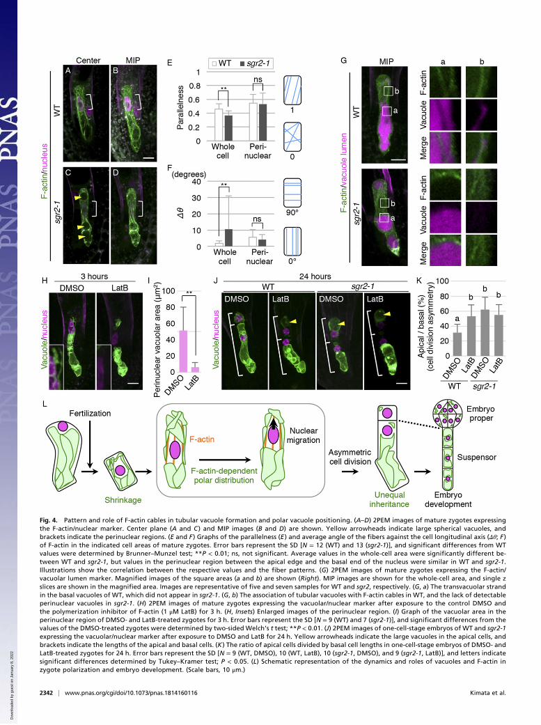
Fig. 4. Pattern and role of F-actin cables in tubular vacuole formation and polar vacuole positioning. (A–D) 2PEM images of mature zygotes expressingthe F-actin/nuclear marker. Center plane (A and C ) and MIP images (B and D) are shown. Yellow arrowheads indicate large spherical vacuoles, andbrackets indicate the perinuclear regions. (E and F ) Graphs of the parallelness (E ) and average angle of the fibers against the cell longitudinal axis (Δθ; F )of F-actin in the indicated cell areas of mature zygotes. Error bars represent the SD [N = 12 (WT) and 13 (sgr2-1)], and significant differences from WTvalues were determined by Brunner–Munzel test; **P < 0.01; ns, not significant. Average values in the whole-cell area were significantly different be-tween WT and sgr2-1, but values in the perinuclear region between the apical edge and the basal end of the nucleus were similar in WT and sgr2-1.Illustrations show the correlation between the respective values and the fiber patterns. (G) 2PEM images of mature zygotes expressing the F-actin/vacuolar lumen marker. Magnified images of the square areas (a and b) are shown (Right). MIP images are shown for the whole-cell area, and single zslices are shown in the magnified area. Images are representative of five and seven samples for WT and sgr2, respectively. (G, a) The transvacuolar strandin the basal vacuoles of WT, which did not appear in sgr2-1. (G, b) The association of tubular vacuoles with F-actin cables in WT, and the lack of detectableperinuclear vacuoles in sgr2-1. (H) 2PEM images of mature zygotes expressing the vacuolar/nuclear marker after exposure to the control DMSO andthe polymerization inhibitor of F-actin (1 μM LatB) for 3 h. (H, Insets) Enlarged images of the perinuclear region. (I) Graph of the vacuolar area in theperinuclear region of DMSO- and LatB-treated zygotes for 3 h. Error bars represent the SD [N = 9 (WT) and 7 (sgr2-1)], and significant differences from thevalues of the DMSO-treated zygotes were determined by two-sided Welch’s t test; **P < 0.01. (J) 2PEM images of one-cell-stage embryos of WT and sgr2-1expressing the vacuolar/nuclear marker after exposure to DMSO and LatB for 24 h. Yellow arrowheads indicate the large vacuoles in the apical cells, andbrackets indicate the lengths of the apical and basal cells. (K ) The ratio of apical cells divided by basal cell lengths in one-cell-stage embryos of DMSO- andLatB-treated zygotes for 24 h. Error bars represent the SD [N = 9 (WT, DMSO), 10 (WT, LatB), 10 (sgr2-1, DMSO), and 9 (sgr2-1, LatB)], and letters indicatesignificant differences determined by Tukey–Kramer test; P < 0.05. (L) Schematic representation of the dynamics and roles of vacuoles and F-actin inzygote polarization and embryo development. (Scale bars, 10 μm.)
2342 | www.pnas.org/cgi/doi/10.1073/pnas.1814160116 Kimata et al.
Dow
nloa
ded
by g
uest
on
Janu
ary
8, 2
022

amount of vacuolar membrane. It is easy to imagine that theabundant vacuolar surface is necessary for flexible shape change,and thus insufficient vacuolar membrane would cause rigid andspherical vacuoles as found in sgr2-1. This, taken together withthe fact that SGR2 encodes a phospholipase A1-like proteinlocalized on the vacuolar membrane (8), shows SGR2 might beimportant to degrade and/or produce some specific phospho-lipids, which regulate vacuolar membrane fusion. Previous studieshave shown that vacuolar organization is controlled by variousphospholipids, such as phosphatidylinositol 3,5-bisphosphate andphosphatidylinositol 3-phosphate (PI3P) (23–25). In particular,pharmacological depletion of PI3P rescues the vacuolar defectsof the vesicle transport through interaction with t-snares11 (vti11)mutant of vacuolar/PVC Qb-SNARE, which regulates vacuolarmembrane fusion (26–28), showing the negative effect of PI3Pon vacuolar fusion. Mutants in vti11 produce spherical vacuolesand show impaired gravitropic response (9, 29), implying ananalogy to sgr2-1. Therefore, SGR2 might similarly contribute toincreasing vacuolar surface and thus indirectly assist flexiblevacuolar shape change along F-actin. However, we cannotexclude the possibility that SGR2 supports the interaction of thevacuole and F-actin in a more direct manner, because severalphospholipids are known to bind to F-actin–associating factors,such as myosin and actin-binding proteins in animal cells (30,31). Future studies, such as the identification of enzymatic tar-gets of SGR2 and live imaging of various phospholipids duringzygote polarization (32), will demonstrate how SGR2 regulatesvacuolar dynamics in the zygote.
Materials and MethodsDetailed materials and methods are described in SI Appendix.
Strains and Growth Conditions. All Arabidopsis lines were in the Columbia(Col-0) background, and sgr2-1 has been described previously (9). The com-plemented sgr2-1 by pSGR2::SGR2i-GFP was described previously (8). Plantswere grown at 18 to 22 °C under continuous light or long-day conditions(16-h light/8-h dark).
Plasmid Construction. The vacuolar marker for the egg cell and the zygote wasEC1p::VHP1-mGFP, consisting of the EGG CELL1 (EC1) promoter (33), mo-nomeric GFP (mGFP), and VACUOLAR H+-PPASE (VHP1) (34). The vacuolarlumen marker was EC1p::SP-mTur2-CTPP, containing the signal peptide (SP),mTurquoise2 (mTur2), and the vacuolar sorting signal COOH-terminal pro-peptide (CTPP) (35). Details of cloning procedures are shown in SI Appendix.
Zygote Imaging and Inhibitor Treatment. In vitro ovule culturing and 2PEMlive-cell imaging were performed as previously described (6, 7). For the in-hibitor treatment, 0.1% DMSO and 1 μM LatB (Sigma) and 100 μM CytD(Sigma) dissolved in 0.1% DMSO were added to the ovule cultivation mediaas previously described (6). An AxioImager A2 (Zeiss) was used for differ-ential interference contrast (DIC) microscopy.
Quantitative Analysis of Vacuoles and F-Actin Structures. Image processing andmeasurements of lengths and vacuolar areas were performed using thebinarized maximum-intensity projection images of vacuolar/nuclear markerswith ImageJ software (https://imagej.nih.gov/ij/index.html). ImageJ was alsoused to quantify the circularity, perimeter, and area of the vacuolar lumenmarker. The quantification of F-actin pattern was performed as previouslydescribed (6).
ACKNOWLEDGMENTS. We thank Hanae Tsuchiya, Yumi Kuwabara, andYuriko Toda for technical support; Daisuke Maruyama for providing thevacuolar lumen reporter; and Kazuo Ebine and Takashi Ueda for providingthe sand-2 mutant. This work was supported by the Japan Society for thePromotion of Science: Grant-in-Aid for JSPS Research Fellow (JP18J10512 toY.K.), Grants-in-Aid for Scientific Research on Innovative Areas [JP15H05962,JP15H05955, and JP17H05838 to M.U.; JP16H06465, JP16H06464, and JP16K21727to T. Higashiyama; JP24114007 to S.H.; JP16H01235 to M.M.; and JP16H06280(Advanced Bioimaging Support)], Grants-in-Aid for Young Scientists (A,JP25711017 to T. Higaki; and B, JP16K18687 to S.S.), Grants-in-Aid for Scien-tific Research (B, JP16H04802 to S.H.; JP17H03697 to D.K.; and A, JP26252011to M.M.), Grants-in-Aid for Challenging Exploratory Research (JP16K14753 toM.U.; and JP17K19380 to T. Higaki), Grant-in-Aid for Scientific Research onPriority Areas (JP16085205 to M.T.M.), and PRESTO Project, Japan Science andTechnology Agency (M.T.M.).
1. Mansfield SG, Briarty LG (1991) Early embryogenesis in Arabidopsis thaliana. II. Thedeveloping embryo. Can J Bot 69:461–476.
2. Suzuki K, Taniguchi T, Maeda E (1992) Ultrastructure and cleavage pattern of riceproembryos. Jpn J Sci 61:292–303.
3. Faure JE, Rotman N, Fortuné P, Dumas C (2002) Fertilization in Arabidopsis thalianawild type: Developmental stages and time course. Plant J 30:481–488.
4. Jensen WA (1968) Cotton embryogenesis: The zygote. Planta 79:346–366.5. Ueda M, Zhang Z, Laux T (2011) Transcriptional activation of Arabidopsis axis pat-
terning genes WOX8/9 links zygote polarity to embryo development. Dev Cell 20:264–270.
6. Kimata Y, et al. (2016) Cytoskeleton dynamics control the first asymmetric cell divisionin Arabidopsis zygote. Proc Natl Acad Sci USA 113:14157–14162.
7. Kurihara D, Kimata Y, Higashiyama T, Ueda M (2017) In vitro ovule cultivation for live-cell imaging of zygote polarization and embryo patterning in Arabidopsis thaliana.J Vis Exp, 10.3791/55975.
8. Morita MT, et al. (2002) Involvement of the vacuoles of the endodermis in the earlyprocess of shoot gravitropism in Arabidopsis. Plant Cell 14:47–56.
9. Kato T, et al. (2002) SGR2, a phospholipase-like protein, and ZIG/SGR4, a SNARE, areinvolved in the shoot gravitropism of Arabidopsis. Plant Cell 14:33–46.
10. Toyota M, et al. (2013) Amyloplast displacement is necessary for gravisensing inArabidopsis shoots as revealed by a centrifuge microscope. Plant J 76:648–660.
11. Fischer U, Ikeda Y, Grebe M (2007) Planar polarity of root hair positioning in Arabi-dopsis. Biochem Soc Trans 35:149–151.
12. Chytilova E, et al. (2000) Nuclear dynamics in Arabidopsis thaliana. Mol Biol Cell 11:2733–2741.
13. Van Norman JM (2016) Asymmetry and cell polarity in root development. Dev Biol419:165–174.
14. Facette MR, Smith LG (2012) Division polarity in developing stomata. Curr Opin PlantBiol 15:585–592.
15. Higaki T, Kutsuna N, Okubo E, Sano T, Hasezawa S (2006) Actin microfilaments reg-ulate vacuolar structures and dynamics: Dual observation of actin microfilaments andvacuolar membrane in living tobacco BY-2 cells. Plant Cell Physiol 47:839–852.
16. Scheuring D, et al. (2016) Actin-dependent vacuolar occupancy of the cell determinesauxin-induced growth repression. Proc Natl Acad Sci USA 113:452–457.
17. Krüger F, Schumacher K (2018) Pumping up the volume—Vacuole biogenesis inArabidopsis thaliana. Semin Cell Dev Biol 80:106–112.
18. Singh M, Gupta A, Laxmi A (2017) Striking the right chord: Signaling enigma duringroot gravitropism. Front Plant Sci 8:1304.
19. Silady RA, et al. (2008) The GRV2/RME-8 protein of Arabidopsis functions in the lateendocytic pathway and is required for vacuolar membrane flow. Plant J 53:29–41.
20. Rojo E, Gillmor CS, Kovaleva V, Somerville CR, Raikhel NV (2001) VACUOLELESS1 is anessential gene required for vacuole formation and morphogenesis in Arabidopsis.Dev Cell 1:303–310.
21. Feraru E, et al. (2010) The AP-3 β adaptin mediates the biogenesis and function of lyticvacuoles in Arabidopsis. Plant Cell 22:2812–2824.
22. Avisar D, et al. (2009) A comparative study of the involvement of 17 Arabidopsis myosinfamily members on the motility of Golgi and other organelles. Plant Physiol 150:700–709.
23. Whitley P, Hinz S, Doughty J (2009) Arabidopsis FAB1/PIKfyve proteins are essentialfor development of viable pollen. Plant Physiol 151:1812–1822.
24. Nováková P, et al. (2014) SAC phosphoinositide phosphatases at the tonoplast me-diate vacuolar function in Arabidopsis. Proc Natl Acad Sci USA 111:2818–2823.
25. Lee Y, et al. (2008) The Arabidopsis phosphatidylinositol 3-kinase is important forpollen development. Plant Physiol 147:1886–1897.
26. Ebine K, et al. (2014) Plant vacuolar trafficking occurs through distinctly regulatedpathways. Curr Biol 24:1375–1382.
27. Sanderfoot AA, Kovaleva V, Bassham DC, Raikhel NV (2001) Interactions betweensyntaxins identify at least five SNARE complexes within the Golgi/prevacuolar systemof the Arabidopsis cell. Mol Biol Cell 12:3733–3743.
28. Zheng J, Han SW, Rodriguez-Welsh MF, Rojas-Pierce M (2014) Homotypic vacuolefusion requires VTI11 and is regulated by phosphoinositides. Mol Plant 7:1026–1040.
29. Saito C, et al. (2011) The occurrence of ‘bulbs’, a complex configuration of the vac-uolar membrane, is affected by mutations of vacuolar SNARE and phospholipase inArabidopsis. Plant J 68:64–73.
30. Senju Y, et al. (2017) Mechanistic principles underlying regulation of the actin cyto-skeleton by phosphoinositides. Proc Natl Acad Sci USA 114:E8977–E8986.
31. Liu X, et al. (2016) Mammalian nonmuscle myosin II binds to anionic phospholipidswith concomitant dissociation of the regulatory light chain. J Biol Chem 291:24828–24837.
32. Simon ML, et al. (2014) A multi-colour/multi-affinity marker set to visualize phos-phoinositide dynamics in Arabidopsis. Plant J 77:322–337.
33. Sprunck S, et al. (2012) Egg cell-secreted EC1 triggers sperm cell activation duringdouble fertilization. Science 338:1093–1097.
34. Segami S, Makino S, Miyake A, Asaoka M, Maeshima M (2014) Dynamics of vacuoles andH+-pyrophosphatase visualized by monomeric green fluorescent protein in Arabidopsis:Artifactual bulbs and native intravacuolar spherical structures. Plant Cell 26:3416–3434.
35. Vitale A, Raikhel NV (1999) What do proteins need to reach different vacuoles?Trends Plant Sci 4:149–155.
Kimata et al. PNAS | February 5, 2019 | vol. 116 | no. 6 | 2343
PLANTBIOLO
GY
Dow
nloa
ded
by g
uest
on
Janu
ary
8, 2
022