Vacuolar Pathology in the Median Eminence of the Hypothalamus After Hyponatremia
6
ORIGINAL ARTICLE Vacuolar Pathology in the Median Eminence of the Hypothalamus After Hyponatremia Seymour Levine, MD, Arthur Saltzman, MS, and Stephen D. Ginsberg, PhD Abstract The median eminence of the hypothalamus is an important conduit by which neurosecretory hormones from hypothalamic nuclei are delivered to the pars nervosa (neural lobe) of the pituitary en route to the bloodstream. Dilutional hyponatremia was produced in adult rats to determine the effect on the morphology of the median emi- nence of the hypothalamus. Hyponatremia was caused by reducing electrolyte and organic osmolyte reserves to block the excretion of water through delivery of the nephrotoxin mercuric chloride (HgCl 2 ). Histological examination of the brain 1 day after a hyponatremic insult revealed vacuolation within the median eminence of the hy- pothalamus. No other lesions were found in other parts of the brain after hyponatremia. The hyponatremic lesion consisted of a band of closely packed vacuoles that crossed the floor of the third ventricle. Vacuoles associated with hyponatremia were predominantly in the subependymal, fiber, reticular, and palisade layers of the median eminence. Vacuolation was not observed in the tanycyte layer of the median eminence. This study indicates that the median eminence is a potentially vulnerable site in human hyponatremic conditions that should be evaluated further in relevant animal models. Key Words: Adenohypophysis, Cisterns, Hyponatremia, Pituitary, Tanycyte, Vacuoles. INTRODUCTION Hyponatremia, also referred to as water intoxication, is the result of the progressive hypotonicity of body fluids (1, 2). Severe hyponatremic insults can result in brain edema and death. Hyponatremia is a common complication of dis- orders in which there is rapid sodium loss (e.g. profuse di- arrhea or vomiting) or when excess water accumulates at a higher rate than can be excreted (3). Hyponatremia is also observed after the aberrant secretion of the antidiuretic hor- mone vasopressin; antagonists of vasopressin are effective for the treatment of hyponatremia under certain conditions (4Y6). Vasopressin is a secretory product of the magnocellular su- praoptic and paraventricular nuclei in the hypothalamus and passes through the median eminence en route to the pars nervosa (neural lobe) of the pituitary gland, where it is dis- charged into the bloodstream (7, 8). Hyponatremia is also observed when serum sodium is rapidly diluted (e.g. hyper- triglyceridemic and hyperglycemic conditions and in patients with various illnesses when excessive quantities of hypotonic fluids are administered for therapy [9]), and it is associated with a fatal syndrome of central diabetes mellitus and diabetes insipidus (10). Importantly, hyponatremia is the most com- mon electrolyte balance disorder found in hospitalized patients (11, 12). Hyponatremia can be associated with neuropsychi- atric disorders, including schizophrenia, whereby affected individuals voluntarily consume excessive quantities of water (polydipsia) (5, 13). In rats, acute hyponatremia has also been demonstrated to be a secondary insult after traumatic brain injury, in which it may involve the swelling of astrocytic end feet near perivas- cular spaces and the upregulation of the glial water channel aquaporin-4 (14, 15). Renal tubular necrosis with subsequent pituitary damage can be produced in relevant animal models by delivery of a nephrotoxin such as intravenous injection of HgCl 2 or intraperitoneal injection of D-serine (16). Our group has reported hydropic degeneration of the pituitary pars distalis (adenohypophysis) in both of these experimental rat models of uremia, which are associated with hyponatremia (16, 17). Specifically, we demonstrated that delivery of HgCl 2 (as well as injection of nephrotoxic levels of the amino acid D-serine) caused hyponatremic insults within the adenohypophysis that were indistinguishable morphologically from one another (17). Despite the clinical importance of understanding brain and pituitary changes during hyponatremic insults, little morpho- logical or mechanistic information is currently available in relevant animal models. The present study aimed to determine whether an acute hyponatremic insult in rats could produce brain as well as pituitary pathology. MATERIALS AND METHODS All procedures were approved by the Institutional Animal Care and Use Committee of the Nathan Kline Institute. Lewis rats of either sex (n = 19) were kept in hanging shoebox-type plastic cages with corncob bedding. Laboratory Rodent Diet 5001 chow (Purina; Scott’s Distribution, Hudson, NH) was 151 J Neuropathol Exp Neurol Volume 70, Number 2, February 2011 J Neuropathol Exp Neurol Copyright Ó 2011 by the American Association of Neuropathologists, Inc. Vol. 70, No. 2 February 2011 pp. 151Y156 From the Center for Dementia Research, Nathan Kline Institute, Orangeburg, New York (SL, AS, SDG); and Departments of Psychiatry and Physiol- ogy and Neuroscience, New York University Langone Medical Center (SDG), New York, New York. Send correspondence and reprint requests to: Stephen D. Ginsberg, PhD, Center for Dementia Research, Nathan Kline Institute, NYU Langone Medical Center, 140 Old Orangeburg Rd, Orangeburg, NY 10962; E-mail: [email protected] Seymour Levine died in the spring of 2010. Arthur Saltzman and Stephen D. Ginsberg dedicate this manuscript to the memory of Seymour Levine and acknowledge his prolific scientific career in neuropathology. This work was supported by National Institutes of Health grants MH083571 (Stephen Ginsberg), MH086385 (Stephen Ginsberg), and an anonymous private donation (Seymour Levine). Copyright © 2011 by the American Association of Neuropathologists, Inc. Unauthorized reproduction of this article is prohibited.
Transcript of Vacuolar Pathology in the Median Eminence of the Hypothalamus After Hyponatremia

ORIGINAL ARTICLE
Vacuolar Pathology in the Median Eminence of theHypothalamus After Hyponatremia
Seymour Levine, MD, Arthur Saltzman, MS, and Stephen D. Ginsberg, PhD
AbstractThe median eminence of the hypothalamus is an important conduit
by which neurosecretory hormones from hypothalamic nuclei aredelivered to the pars nervosa (neural lobe) of the pituitary en routeto the bloodstream. Dilutional hyponatremia was produced in adultrats to determine the effect on the morphology of the median emi-nence of the hypothalamus. Hyponatremia was caused by reducingelectrolyte and organic osmolyte reserves to block the excretion ofwater through delivery of the nephrotoxin mercuric chloride (HgCl2).Histological examination of the brain 1 day after a hyponatremicinsult revealed vacuolation within the median eminence of the hy-pothalamus. No other lesions were found in other parts of the brainafter hyponatremia. The hyponatremic lesion consisted of a band ofclosely packed vacuoles that crossed the floor of the third ventricle.Vacuoles associated with hyponatremia were predominantly in thesubependymal, fiber, reticular, and palisade layers of the medianeminence. Vacuolation was not observed in the tanycyte layer of themedian eminence. This study indicates that the median eminence is apotentially vulnerable site in human hyponatremic conditions thatshould be evaluated further in relevant animal models.
Key Words: Adenohypophysis, Cisterns, Hyponatremia, Pituitary,Tanycyte, Vacuoles.
INTRODUCTIONHyponatremia, also referred to as water intoxication,
is the result of the progressive hypotonicity of body fluids(1, 2). Severe hyponatremic insults can result in brain edemaand death. Hyponatremia is a common complication of dis-orders in which there is rapid sodium loss (e.g. profuse di-arrhea or vomiting) or when excess water accumulates at ahigher rate than can be excreted (3). Hyponatremia is also
observed after the aberrant secretion of the antidiuretic hor-mone vasopressin; antagonists of vasopressin are effective forthe treatment of hyponatremia under certain conditions (4Y6).Vasopressin is a secretory product of the magnocellular su-praoptic and paraventricular nuclei in the hypothalamus andpasses through the median eminence en route to the parsnervosa (neural lobe) of the pituitary gland, where it is dis-charged into the bloodstream (7, 8). Hyponatremia is alsoobserved when serum sodium is rapidly diluted (e.g. hyper-triglyceridemic and hyperglycemic conditions and in patientswith various illnesses when excessive quantities of hypotonicfluids are administered for therapy [9]), and it is associatedwith a fatal syndrome of central diabetes mellitus and diabetesinsipidus (10). Importantly, hyponatremia is the most com-mon electrolyte balance disorder found in hospitalized patients(11, 12). Hyponatremia can be associated with neuropsychi-atric disorders, including schizophrenia, whereby affectedindividuals voluntarily consume excessive quantities of water(polydipsia) (5, 13).
In rats, acute hyponatremia has also been demonstrated tobe a secondary insult after traumatic brain injury, in which itmay involve the swelling of astrocytic end feet near perivas-cular spaces and the upregulation of the glial water channelaquaporin-4 (14, 15). Renal tubular necrosis with subsequentpituitary damage can be produced in relevant animal models bydelivery of a nephrotoxin such as intravenous injection ofHgCl2 or intraperitoneal injection of D-serine (16). Our grouphas reported hydropic degeneration of the pituitary pars distalis(adenohypophysis) in both of these experimental rat modelsof uremia, which are associated with hyponatremia (16, 17).Specifically, we demonstrated that delivery of HgCl2 (as wellas injection of nephrotoxic levels of the amino acid D-serine)caused hyponatremic insults within the adenohypophysis thatwere indistinguishable morphologically from one another (17).Despite the clinical importance of understanding brain andpituitary changes during hyponatremic insults, little morpho-logical or mechanistic information is currently available inrelevant animal models. The present study aimed to determinewhether an acute hyponatremic insult in rats could producebrain as well as pituitary pathology.
MATERIALS AND METHODSAll procedures were approved by the Institutional Animal
Care and Use Committee of the Nathan Kline Institute. Lewisrats of either sex (n = 19) were kept in hanging shoebox-typeplastic cages with corncob bedding. Laboratory Rodent Diet5001 chow (Purina; Scott’s Distribution, Hudson, NH) was
151J Neuropathol Exp Neurol � Volume 70, Number 2, February 2011
J Neuropathol Exp NeurolCopyright � 2011 by the American Association of Neuropathologists, Inc.
Vol. 70, No. 2February 2011pp. 151Y156
From the Center for Dementia Research, Nathan Kline Institute, Orangeburg,New York (SL, AS, SDG); and Departments of Psychiatry and Physiol-ogy and Neuroscience, New York University Langone Medical Center(SDG), New York, New York.
Send correspondence and reprint requests to: Stephen D. Ginsberg, PhD,Center for Dementia Research, Nathan Kline Institute, NYU LangoneMedical Center, 140 Old Orangeburg Rd, Orangeburg, NY 10962;E-mail: [email protected]
Seymour Levine died in the spring of 2010. Arthur Saltzman and Stephen D.Ginsberg dedicate this manuscript to the memory of Seymour Levine andacknowledge his prolific scientific career in neuropathology.
This work was supported by National Institutes of Health grants MH083571(Stephen Ginsberg), MH086385 (Stephen Ginsberg), and an anonymousprivate donation (Seymour Levine).
Copyright © 2011 by the American Association of Neuropathologists, Inc. Unauthorized reproduction of this article is prohibited.
available ad libitum. Rats were used when 7 to 10 weeks old(180Y260 g). At the time of experimentation, rat chow wasreplaced by pure sucrose cubes (Domino Dots, West PalmBeach, FL). Metal grids were placed at the bottom of each cageto reduce coprophagy. Rats were given an intravenous injectionof HgCl2 (Sigma, St Louis, MO) in 1 mg/mL distilled waterwhile anesthetized with isoflurane, as previously described(17). There were 2 dosing regimens, that is, 0.25 mL/100 gbody weight (n = 9) and 0.5 mL/100 g body weight (n = 5).Distilled water (10 mg/100 g body weight) was injected sub-cutaneously 10 minutes after the HgCl2 injection on the leftside of the body; this was repeated 5 hours later on the rightside of the body. To prevent leakage, the 20-gauge needle wasadvanced alternately through the subcutis and then the dermis3 or 4 times before the water was injected into the subcutis(valve technique). Control rats (n = 5) were treated identically,except that they received distilled water instead of HgCl2.
Rats were killed by exsanguination while under carbondioxide anesthesia. Serum sodium levels were assessed bypreparing blood taken from the inferior vena cava at the timeof death. Sodium was assayed by atomic absorption spec-trophotometry, as described (18, 19). The brains were re-moved quickly, placed into Bouin’s fluid, and the base of theskull was immersed in Bouin’s fluid to fix the pituitary in situfor 24 hours at 22-C. The brain and pituitary were embeddedin paraffin, serially sectioned at 5 Km, and selected sectionswere stained with hematoxylin and eosin (Sigma) and withperiodic acid-SchiffYhematoxylin (18, 19). Particular empha-sis was placed on evaluating the median eminence based uponprevious results (16, 17). The median eminence measuresapproximately 1.6 to 1.8 mm in the anteroposterior plane infixed rat brain (20). Tissue sections were evaluated at 4 levelsthroughout the extent of the median eminence to assess theependymal (tanycyte) layer, subependymal layer, fiber layer,reticular layer, and palisade layer, as well as the pars tuberalisof the infundibulum (19). Tissue sections were examined at400� magnification, and vacuoles throughout the medianeminence were quantified as previously described (16, 17).Vacuolar pathology was assessed on a semiquantitative scaleas follows: 0, no vacuolar pathology; 1+, scattered vacuoles inany one of the layers; 2+, numerous vacuoles in 1 to 2 layers,sometimes grouped together; 3+, profuse vacuolation, organ-ized in short bands across part of the width of the medianeminence and spanning at least 2 laminae; 4+, severe vacuolarpathology organized in long bands, spanning the medianeminence. This approach obviated concerns regarding tissueselection bias (17Y19). Vacuoles in the median eminence hadno detectable content and were interpreted as a hydropicchange (16, 17).
RESULTSSodium levels were significantly decreased in hypona-
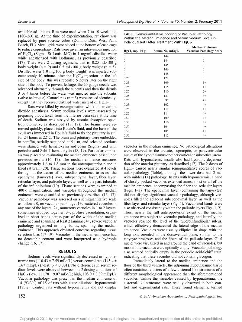
tremic rats (110.43 T 7.59 mEq/L) versus control rats (145.4 T1.67 mEq/L) (t-test; p G 0.001). No differences in serum so-dium levels were observed between the 2 dosing conditions ofHgCl2 (low, 111.78 T 9.07 mEq/L; high, 108.0 T 3.39 mEq/L).Vacuolar pathology was present in the median eminence of14 (93.3%) of 15 of rats with acute dilutional hyponatremia(Table). Control rats without hyponatremia did not display
vacuoles in the median eminence. No pathological alterationswere observed in the arcuate, supraoptic, or paraventricularnuclei of the hypothalamus or other cortical or subcortical areas.Rats with hyponatremic insults also had hydropic degenera-tion of the anterior pituitary, as described (17). The 2 doses ofHgCl2 caused nearly similar semiquantitative scores of vac-uolar pathology (Table), although the lower dose had 2 ratswith milder (1+) pathology. In rats with hyponatremia, a bandof closely packed vacuoles extended across most or all of themedian eminence, encompassing the fiber and reticular layers(Figs. 1Y3). The ependymal layer (containing the tanycytes)did not display significant vacuolar pathology, although vac-uoles filled the adjacent subependymal layer, as well as thefiber layer and reticular layer (Fig. 1). Vacuolated bands werealso observed consistently within the palisade layer (Figs. 1, 2).Thus, nearly the full anteroposterior extent of the medianeminence was subject to vacuolar pathology, and laterally, thevacuoles reached the level of the tuberoinfundibular sulcus,which effectively demarcated the lateral edge of the medianeminence. Vacuoles were usually elliptical in shape with thelong axis oriented in the dorsoventral plane, similar to thetanycyte processes and the fibers of the palisade layer. Glialnuclei were visualized in and around the band of vacuoles, butmost of the vacuoles were optically empty. Vacuolar pathologyalso seemed optically empty in the periodic acid-Schiff stain,indicating that these vacuoles did not contain glycogen.
Immediately lateral to the median eminence and thefloor of the third ventricle, the adjoining hypothalamic tissueoften contained clusters of a few cisternal-like structures of adifferent morphological appearance than the aforementionedvacuoles. Unlike the vacuoles caused by hyponatremia, thecisternal-like structures were readily observed in both con-trol and experimental rats. These round elements, termed
TABLE. Semiquantitative Scoring of Vacuolar PathologyWithin the Median Eminence and Serum Sodium Levels inIndividual Rats After Treatment With HgCl2
HgCl2 mg/100 g Serum Na, mEq/LMedian Eminence
Vacuolar Pathology Score
0 144 0
0 144 0
0 145 0
0 146 0
0 148 0
0.25 121 0
0.25 109 1+
0.25 115 1+
0.25 110 2+
0.25 127 3+
0.25 97 4+
0.25 102 4+
0.25 111 4+
0.25 114 4+
0.50 109 3+
0.50 110 3+
0.50 104 4+
0.50 105 4+
0.50 112 4+
Levine et al J Neuropathol Exp Neurol � Volume 70, Number 2, February 2011
� 2011 American Association of Neuropathologists, Inc.152
Copyright © 2011 by the American Association of Neuropathologists, Inc. Unauthorized reproduction of this article is prohibited.

FIGURE 1. Low- and high-magnification images of the mediobasal hypothalamus in coronal sections from control (A) andhyponatremic (BYD) rats. The median eminence extends below the third ventricle (III v.), across the bottom of the photomicro-graphs. Cisterns (arrows) located just lateral to the median eminence often seemed to be attached to the pars tuberalis at the levelwhere it separates the median eminence from the rest of the hypothalamus. (A) Control rat. (B) Vacuolation is symmetricallydistributed in the subependymal layer of the median eminence of a hyponatremic rat (scored at 1+). Tanycytes, which form thefloor of the third ventricle, are not vacuolated. (C) Vacuolation is distributed symmetrically in the subependymal, fiber, and reticularlayers (scored at 3+). (D) Vacuoles are prominent in the fiber, reticular, and palisade layers of the median eminence (scored at 4+).Vacuoles on the left side of the palisade layer adjacent to the pars tuberalis are confluent with an asymmetric group of largercisterns. Hematoxylin and eosin. Scale bars = (AYD) 20 Km.
J Neuropathol Exp Neurol � Volume 70, Number 2, February 2011 Median Eminence Vacuolation From Hyponatremia
� 2011 American Association of Neuropathologists, Inc. 153
Copyright © 2011 by the American Association of Neuropathologists, Inc. Unauthorized reproduction of this article is prohibited.

hypothalamic cisternae (19, 21Y23), were present in thelateral margins of the median eminence and the tuber-ohypophysial sulci and were markedly different in locationand shape from the vacuoles of the median eminence asso-ciated with the hyponatremic insult (Fig. 1Y3). Importantly,they were seen in rats with or without vacuolar degenerationof the median eminence (Fig. 1).
DISCUSSIONVacuolar pathology within the median eminence
seemed to be specific to the hyponatremic insult; no similarvacuoles have been reported in the median eminence (or otherbrain regions) of rodents using a variety of experimentalparadigms including excitotoxicity, hyperlipemia, inflamma-tion, lithium treatment, gonadal steroid delivery, and vitaminD treatment, among others (18, 19, 24Y29). Dilutional hypo-natremia was generated in the present study through injectionof the nephrotoxic agent HgCl2, similar to our previousstudies (16, 17). The nephrotoxicity of HgCl2 delivery is wellestablished (30, 31), notably within the dosing range used inthis report (16, 17). Mercury, particularly in the vapor form, is
also neurotoxic (32). Mercuric salts, including HgCl2,although neurotoxic to varying degrees, seem to be less neu-rotoxic than the vapor form and are less blood-brain barrierpenetrant (32). Glial cells, including astrocytes, may be po-tential mediators of HgCl2 toxicity within the brain, althoughtheir contribution to hypothalamic and/or pituitary changesremain unknown (33, 34). To our knowledge, the potential ofdirect neurotoxicity of intravenous HgCl2 delivery on themediobasal hypothalamus and pituitary has not been deter-mined. Accordingly, we cannot formally exclude the possi-bility that HgCl2 has some direct effects upon the medianeminence. However, this is unlikely within the current para-digm because the 2 doses of HgCl2 produced equivalent levelsof serum sodium depletion, as well as vacuolar pathologywithin the median eminence, consistent with our previousanalysis (17).
The present objective was to determine whether acutedilutional hyponatremia could produce lesions in brain andpituitary as observed in humans with a fatal syndrome ofdiabetes mellitus and diabetes insipidus (10). The vacuolarfluid-filled character of the lesions could have been expectedinasmuch as hyponatremia and water intoxication are almost
FIGURE 2. Low (left panels) and higher (right panels) photomicrographs showing prominent vacuolation in the fiber, reticular, andpalisade layers after a hyponatremic insult. (A) Clusters of vacuoles are concentrated in the palisade layer adjacent to the parstuberalis of the pituitary (scored at 3+). Numerous cisterns are also seen at higher magnification. (B) Widespread symmetricalvacuolar pathology throughout the median eminence (scored at 4+). In this rat, hypothalamic cisterns were scarce and separatedlaterally from vacuoles within the median eminence. Scale bars = (A, B) 20 Km.
Levine et al J Neuropathol Exp Neurol � Volume 70, Number 2, February 2011
� 2011 American Association of Neuropathologists, Inc.154
Copyright © 2011 by the American Association of Neuropathologists, Inc. Unauthorized reproduction of this article is prohibited.

synonymous. However, the location of these vacuoles withinthe median eminence of the hypothalamus was not antici-pated. There is experimental evidence that cerebrospinal fluidcan pass directly from the third ventricle through the medianeminence (35Y37). Moreover, the orientation of the ellipticalvacuoles in hyponatremic rats is in register with the ori-entation of the tanycytes and their processes. Therefore, thecontent of the vacuoles observed in the median eminence afterhyponatremia may be cerebrospinal fluid. To our knowledge,vacuolar pathology of this type after a hyponatremic insulthas not been reported in the brain. It has been reported thathyponatremic rats exhibit degeneration of the adenohypoph-ysis (17), as well as vacuolar pathology within the medianeminence. Moreover, the present findings within the medianeminence are similar to peripheral vacuolar changes in renalglomeruli after nephrotoxic insults (38, 39).
The layers of the median eminence from dorsal (upper)to ventral (lower) are the ependymal layer (tanycyte layer),subependymal layer, fiber layer, reticular layer, and palisadelayer (19, 20). The palisade layer is readily identified by the
closely compacted parallel fibers perpendicular to the parstuberalis of the pituitary and by the highly vascularized (portalcirculation) lowest portion of the layer (19, 20). The delicatearachnoid membrane is attached to the pars tuberalis. Theobservation that vacuolar pathology after an acute hypona-tremic insult was selective for the laminae of the medianeminence is intriguing from several perspectives. The goal isto determine the intrinsic morphological, molecular, and orcellular underpinnings that cause this selective vulnerabilitywithin the median eminence. The relative sparing of vacuolarpathology within the tanycyte layer is also of interest becausethis layer displays more mitotic figures (likely originatingfrom specialized astrocytes called pituicytes) after osmoticstress than the other median eminence laminae (19). Fur-thermore, we suspect that the cisterns that are normally pres-ent within the median eminence predispose this region tovacuolation. Tangential evidence for this hypothesis is sup-ported by a study whereby adrenalectomy in adult rats in-creased the abundance of hypothalamic cisterns (40). Becauseadrenalectomy causes low serum sodium, this observation is
FIGURE 3. Prominent vacuolation within the median eminence after hyponatremia. Low (left panel) and higher (right panel)photomicrographs illustrating profuse vacuoles throughout the median eminence but not the adjacent mediobasal hypothalamus.(A) Severe vacuolar lesions within the median eminence of a hyponatremic rat (scored at 4+). The shallow tuberoinfundibularsulcus (arrow) is seen near cisterns within the lateral margins of the median eminence. (B) Vacuolation throughout the sub-ependymal, fiber, reticular, and palisade layers of the median eminence. Hypothalamic cisterns are large and numerous in thelateral margins of the median eminence. Scale bars = (A, B) 20 Km.
J Neuropathol Exp Neurol � Volume 70, Number 2, February 2011 Median Eminence Vacuolation From Hyponatremia
� 2011 American Association of Neuropathologists, Inc. 155
Copyright © 2011 by the American Association of Neuropathologists, Inc. Unauthorized reproduction of this article is prohibited.
consistent with our findings in the present hyponatremicmodel. Additional ultrastructural assessment within the lami-nae of the median eminence is warranted after hyponatremiato determine the precise morphological characteristics of thesevacuolar lesions. Furthermore, assessment of vasopressinlevels within the hypothalamus and the median eminence inhyponatremic models may prove useful to determine the po-tential mechanism of action in human hyponatremic syn-dromes. Although numerous models and systems exist tostudy the effects of hyponatremia peripherally, relatively littleis available for the brain and pituitary. Our morphologicalresults in a rat model of acute hyponatremia suggest that themedian eminence is a crucial structure to monitor and assessfor understanding mechanisms underlying the hyponatremicprocess, as well as providing a substrate (vacuolar pathology)to test interventions aimed at reducing the negative sequelaassociated with hyponatremia.
ACKNOWLEDGMENTThe authors thank Corrinne M. Peterhoff for assistance
with the photomicrography.
REFERENCES1. Sterns RH, Hix JK, Silver S. Treatment of hyponatremia. Curr Opin
Nephrol Hypertens 2010;19:493Y982. Oh MS. Pathogenesis and diagnosis of hyponatremia. Nephron 2002;
92(Suppl 1):2Y83. Mount DB. The brain in hyponatremia: Both culprit and victim. Semin
Nephrol 2009;29:196Y2154. Arai Y, Fujimori A, Sasamata M, et al. New topics in vasopressin
receptors and approach to novel drugs: Research and development ofconivaptan hydrochloride (YM087), a drug for the treatment of hypona-tremia. J Pharmacol Sci 2009;109:53Y59
5. Josiassen RC, Goldman M, Jessani M, et al. Double-blind, placebo-controlled, multicenter trial of a vasopressin V2-receptor antagonist inpatients with schizophrenia and hyponatremia. Biol Psychiatry 2008;64:1097Y2100
6. Rozen-Zvi B, Yahav D, Gheorghiade M, et al. Vasopressin receptorantagonists for the treatment of hyponatremia: Systematic review andmeta-analysis. Am J Kidney Dis 2010;56:325Y37
7. Armstrong WE, Warach S, Hatton GI, et al. Subnuclei in the rat hypo-thalamic paraventricular nucleus: A cytoarchitectural, horseradish perox-idase and immunocytochemical analysis. Neuroscience 1980;5:1931Y58
8. Swanson LW, Sawchenko PE. Hypothalamic integration: Organizationof the paraventricular and supraoptic nuclei. Annu Rev Neurosci 1983;6:269Y324
9. Baylis PH. The syndrome of inappropriate antidiuretic hormone secre-tion. Int J Biochem Cell Biol 2003;35:1495Y99
10. Fraser CL, Arieff AI. Fatal central diabetes mellitus and insipidusresulting from untreated hyponatremia: A new syndrome. Ann InternMed 1990;112:113Y19
11. Bagshaw SM, Townsend DR, McDermid RC. Disorders of sodium andwater balance in hospitalized patients. Can J Anaesth 2009;56:151Y67
12. Upadhyay A, Jaber BL, Madias NE. Epidemiology of hyponatremia.Semin Nephrol 2009;29:227Y38
13. Goldman MB. The assessment and treatment of water imbalance inpatients with psychosis. Clin Schizophr Relat Psychoses 2010;4:115Y23
14. Ke C, Poon WS, Ng HK, et al. Impact of experimental acute hypona-tremia on severe traumatic brain injury in rats: Influences on injuries, per-meability of blood-brain barrier, ultrastructural features, and aquaporin-4expression. Exp Neurol 2002;178:194Y206
15. Vajda Z, Promeneur D, Doczi T, et al. Increased aquaporin-4 immuno-reactivity in rat brain in response to systemic hyponatremia. BiochemBiophys Res Commun 2000;270:495Y503
16. Levine S, Saltzman A. Acute uremia produced in rats by nephrotoxicchemicals is alleviated by protein deficient diet. Ren Fail 2003;25:517Y23
17. Levine S, Saltzman A. Hydropic degeneration of the anterior pituitarygland (adenohypophysis) in uremic rats. Toxicol Lett 2004;147:121Y26
18. Levine S, Saltzman A, Katof B, et al. Proliferation of glial cells inducedby lithium in the neural lobe of the rat pituitary is enhanced by dehy-dration. Cell Prolif 2002;35:167Y72
19. Levine S, Saltzman A, Ginsberg SD. Mitotic figures in the median emi-nence of the hypothalamus. Neurochem Res 2010;35:1743Y46
20. Monroe BG, Paull WK. Ultrastructural changes in the hypothalamusduring development and hypothalamic activity: The median eminence.Prog Brain Res 1974;41:185Y208
21. Bodoky M, Koritsanszky S, Rethelyi M. A system of intraependymalcisternae along the margins of the median eminence in the rat: Structure,three-dimensional arrangement and ontogeny. Cell Tissue Res 1979;196:163Y73
22. Scott V, Brown CH. State-dependent plasticity in vasopressin neurones:Dehydration-induced changes inactivity patterning. J Neuroendocrinol2010;22:343Y54
23. Srebro Z. Electron microscopic observations on giant vacuoles present inthe median eminence of rats. Folia Biol (Krakow) 1986;34:421Y24
24. Hatton GI, Perlmutter LS, Salm AK, et al. Dynamic neuronal-glialinteractions in hypothalamus and pituitary: Implications for control ofhormone synthesis and release. Peptides 1984;5(Suppl 1):121Y38
25. Holzwarth-McBride MA, Hurst EM, Knigge KM. Monosodium gluta-mate induced lesions of the arcuate nucleus. I. Endocrine deficiency andultrastructure of the median eminence. Anat Rec 1976;186:185Y205
26. Lawson LJ, Perry VH, Gordon S. Microglial responses to physiologicalchange: Osmotic stress elevates DNA synthesis of neurohypophysealmicroglia. Neuroscience 1993;56:929Y38
27. Levine S, Saltzman A. A procedure for inducing sustained hyperlipemiain rats by administration of a surfactant. J Pharmacol Toxicol Methods2007;55:224Y26
28. Levine S, Saltzman A, Ginsberg SD. Different inflammatory reactions tovitamin D(3) among the lateral, third and fourth ventricular choroidplexuses of the rat. Exp Mol Pathol 2008;85:117Y21
29. Olney JW. Brain lesions, obesity, and other disturbances in mice treatedwith monosodium glutamate. Science 1969;164:719Y21
30. Kim KB, Um SY, Chung MW, et al. Toxicometabolomics approach tourinary biomarkers for mercuric chloride (HgCl)-induced nephrotoxicityusing proton nuclear magnetic resonance ([1]H NMR) in rats. ToxicolAppl Pharmacol 2010;249:114Y26
31. Durante P, Romero F, Perez M, et al. Effect of uric acid on nephro-toxicity induced by mercuric chloride in rats. Toxicol Ind Health 2010;26:163Y74
32. Aschner M, Aschner JL. Mercury neurotoxicity: Mechanisms of blood-brain barrier transport. Neurosci Biobehav Rev 1990;14:169Y76
33. Aschner M, Mullaney KJ, Fehm MN, et al. Astrocytes as potentialmodulators of mercuric chloride neurotoxicity. Cell Mol Neurobiol 1994;14:637Y52
34. Cookson MR, Pentreath VW. Alterations in the glial fibrillary acidicprotein content of primary astrocyte cultures for evaluation of glial celltoxicity. Toxicol In Vitro 1994;8:351Y59
35. Amat P, Pastor FE, Blazquez JL, et al. Lateral evaginations from the thirdventricle into the rat mediobasal hypothalamus: An amplification of theventricular route. Neuroscience 1999;88:673Y77
36. Guerra M, Blazquez JL, Peruzzo B, et al. Cell organization of the rat parstuberalis. Evidence for open communication between pars tuberalis cells,cerebrospinal fluid and tanycytes. Cell Tissue Res 2010;339:359Y81
37. Knigge KM, Joseph SA, Sladek JR, et al. Uptake and transport activityof the median eminence of the hypothalamus. Int Rev Cytol 1976;45:283Y408
38. Perez-Perez AJ, Pazos B, Sobrado J, et al. Acute renal failure followingmassive mannitol infusion. Am J Nephrol 2002;22:573Y75
39. Riemenschneider T, Bohle A. Morphologic aspects of low-potassium andlow-sodium nephropathy. Clin Nephrol 1983;19:271Y79
40. Brion JP, Depierreux M, Couck AM, et al. Transmission and scanningelectron-microscopic observations on tanycytes in the mediobasal hypo-thalamus and the median eminence of adrenalectomized rats. Cell TissueRes 1982;221:643Y55
Levine et al J Neuropathol Exp Neurol � Volume 70, Number 2, February 2011
� 2011 American Association of Neuropathologists, Inc.156
Copyright © 2011 by the American Association of Neuropathologists, Inc. Unauthorized reproduction of this article is prohibited.