Arbuscular mycorrhizal fungi induce differential activation of the plasma membrane and vacuolar H +...
12
ORIGINAL PAPER Arbuscular mycorrhizal fungi induce differential activation of the plasma membrane and vacuolar H + pumps in maize roots Alessandro C. Ramos & Marco A. Martins & Anna L. Okorokova-Façanha & Fábio Lopes Olivares & Lev A. Okorokov & Nuno Sepúlveda & José A. Feijó & Arnoldo R. Façanha Received: 19 May 2008 / Accepted: 16 September 2008 / Published online: 8 October 2008 # Springer-Verlag 2008 Abstract Roots undergo multiple changes as a conse- quence of arbuscular mycorrhizal (AM) interactions. One of the major alterations expected is the induction of membrane transport systems, including proton pumps. In this work, we investigated the changes in the activities of vacuolar and plasma membrane (PM) H + pumps from maize roots (Zea mays L.) in response to colonization by two species of AM fungi, Gigaspora margarita and Glomus clarum. Both the vacuolar and PM H + -ATPase activities were inhibited, while a concomitant strong stimulation of the vacuolar H + -PPase was found in the early stages of root colonization by G. clarum (30 days after inoculation), localized in the younger root regions. In contrast, roots colonized by G. margarita exhibited only stimulation of these enzymatic activities, suggesting a species-specific phenomenon. How- ever, when the root surface H + effluxes were recorded using a noninvasive vibrating probe technique, a striking activa- tion of the PM H + -ATPases was revealed specifically in the elongation zone of roots colonized with G. clarum. The data provide evidences for a coordinated regulation of the H + pumps, which depicts a mechanism underlying an activation of the root H + -PPase activity as an adaptative response to the energetic changes faced by the host root during the early stages of the AM interaction. Keywords Arbuscular mycorrhiza . H + -ATPase . H + -PPase . H + fluxes . Ion-selective vibrating probe Introduction The bidirectional exchange of nutrients is the basis of the arbuscular mycorrhizal symbiosis; in this way, the fungus interacts with host plant roots to increase their absorption of water, phosphate, and other nutrients from the soil. In turn, the plant provides photosynthesized sugars to the fungus, a phenomenon that provokes many cellular, physiological, and energetic changes in the host roots (Harrison 2005; Ramos et al. 2008a). Variations have been observed in the Mycorrhiza (2009) 19:69–80 DOI 10.1007/s00572-008-0204-3 Electronic supplementary material The online version of this article (doi:10.1007/s00572-008-0204-3) contains supplementary material, which is available to authorized users. A. C. Ramos : J. A. Feijó Developmental Biology Center, Instituto Gulbenkian de Ciência, Pt-2780-156 Oeiras, Portugal M. A. Martins Centro de Ciências e Tecnologia Agropecuária, Universidade Estadual do Norte Fluminense Darcy Ribeiro, Campos dos Goytacazes, RJ 28013-600, Brazil A. L. Okorokova-Façanha : F. L. Olivares : L. A. Okorokov : A. R. Façanha (*) Centro de Biociências e Biotecnologia, Universidade Estadual do Norte Fluminense Darcy Ribeiro, Campos dos Goytacazes, RJ 28015-620, Brazil e-mail: [email protected] N. Sepúlveda Centro de Estatística e Aplicações da Universidade de Lisboa, Campo Grande Ed C6, 1749-016 Lisbon, Portugal N. Sepúlveda Theoretical Immunology Group, Instituto Gulbenkian de Ciência, Pt-2780-156 Oeiras, Portugal J. A. Feijó Faculdade de Ciências, Universidade de Lisboa, DBV, Campo Grande Ed. C2, 1749-016 Lisbon, Portugal
-
Upload
independent -
Category
Documents
-
view
1 -
download
0
Transcript of Arbuscular mycorrhizal fungi induce differential activation of the plasma membrane and vacuolar H +...

ORIGINAL PAPER
Arbuscular mycorrhizal fungi induce differential activationof the plasma membrane and vacuolar H+ pumpsin maize roots
Alessandro C. Ramos & Marco A. Martins &
Anna L. Okorokova-Façanha & Fábio Lopes Olivares &
Lev A. Okorokov & Nuno Sepúlveda & José A. Feijó &
Arnoldo R. Façanha
Received: 19 May 2008 /Accepted: 16 September 2008 / Published online: 8 October 2008# Springer-Verlag 2008
Abstract Roots undergo multiple changes as a conse-quence of arbuscular mycorrhizal (AM) interactions. One ofthe major alterations expected is the induction of membranetransport systems, including proton pumps. In this work, weinvestigated the changes in the activities of vacuolar andplasma membrane (PM) H+ pumps from maize roots (Zea
mays L.) in response to colonization by two species of AMfungi, Gigaspora margarita and Glomus clarum. Both thevacuolar and PM H+-ATPase activities were inhibited,while a concomitant strong stimulation of the vacuolarH+-PPase was found in the early stages of root colonizationby G. clarum (30 days after inoculation), localized in theyounger root regions. In contrast, roots colonized by G.margarita exhibited only stimulation of these enzymaticactivities, suggesting a species-specific phenomenon. How-ever, when the root surface H+ effluxes were recorded usinga noninvasive vibrating probe technique, a striking activa-tion of the PM H+-ATPases was revealed specifically inthe elongation zone of roots colonized with G. clarum. Thedata provide evidences for a coordinated regulation of theH+ pumps, which depicts a mechanism underlying anactivation of the root H+-PPase activity as an adaptativeresponse to the energetic changes faced by the host rootduring the early stages of the AM interaction.
Keywords Arbuscular mycorrhiza . H+-ATPase .
H+-PPase . H+ fluxes . Ion-selective vibrating probe
Introduction
The bidirectional exchange of nutrients is the basis of thearbuscular mycorrhizal symbiosis; in this way, the fungusinteracts with host plant roots to increase their absorption ofwater, phosphate, and other nutrients from the soil. In turn,the plant provides photosynthesized sugars to the fungus, aphenomenon that provokes many cellular, physiological,and energetic changes in the host roots (Harrison 2005;Ramos et al. 2008a). Variations have been observed in the
Mycorrhiza (2009) 19:69–80DOI 10.1007/s00572-008-0204-3
Electronic supplementary material The online version of this article(doi:10.1007/s00572-008-0204-3) contains supplementary material,which is available to authorized users.
A. C. Ramos : J. A. FeijóDevelopmental Biology Center, Instituto Gulbenkian de Ciência,Pt-2780-156 Oeiras, Portugal
M. A. MartinsCentro de Ciências e Tecnologia Agropecuária,Universidade Estadual do Norte Fluminense Darcy Ribeiro,Campos dos Goytacazes, RJ 28013-600, Brazil
A. L. Okorokova-Façanha : F. L. Olivares : L. A. Okorokov :A. R. Façanha (*)Centro de Biociências e Biotecnologia,Universidade Estadual do Norte Fluminense Darcy Ribeiro,Campos dos Goytacazes, RJ 28015-620, Brazile-mail: [email protected]
N. SepúlvedaCentro de Estatística e Aplicações da Universidade de Lisboa,Campo Grande Ed C6,1749-016 Lisbon, Portugal
N. SepúlvedaTheoretical Immunology Group, Instituto Gulbenkian de Ciência,Pt-2780-156 Oeiras, Portugal
J. A. FeijóFaculdade de Ciências, Universidade de Lisboa, DBV,Campo Grande Ed. C2,1749-016 Lisbon, Portugal

host growth responses depending on the particular plantgenotype and fungal species involved. In fact, it has beenobserved in many instances that during the early stages ofmycorrhizal colonization, the host plant exhibits no growthincrease or even growth depression. These depressions willoccur when fungal demands for photoassimilates outweighthe benefits obtained to the host. Nevertheless, it is nowbecoming clear that the cost/benefit relationship is morecomplex than was previously thought (Li et al. 2008).
The development of AM interaction starts when hyphalgrowth and branching are followed by the formation ofappressorium leading to the hyphal penetration in the rootsystem (Giovannetti et al. 1993, 1996). Inside the rootcortex, fungal hyphae form highly branched arbusculesinvaginated in the plasma membrane of several corticalcells. A specific genetic program is triggered to change theexpression profile of the plant cell membrane proteins,resulting in the periarbuscular membrane (Gianinazzi-Pearson1996; Rausch et al. 2001; Bestel-Corre et al. 2002;Gianinazzi-Pearson and Brechenmacher 2004; Hohnjec etal. 2005; Valot et al. 2005; Lambais 2006). Several lines ofevidence suggest that a H+-ATPase transport protonsacross the periarbuscular membrane (Marx et al. 1982;Gianinazzi-Pearson et al. 1991) and is responsible forgenerating an acidic intercellular compartment (Guttenberger2000). This proton gradient seems to be related to theexchange of phosphate (Poulsen et al. 2005), sugars(Harrison 1996; Schußler et al. 2006) and amino acids (Cruzet al. 2007; Cappellazzo et al. 2008) at the symbioticinterface.
Both plant and fungi cells use P-type H+-ATPases toenergize their secondary systems of nutrient transport(Portillo 2000; Palmgren 2001; Buch-Pedersen et al.2006). Two other H+ pumps are located in the vacuolarmembrane, namely the H+-ATPase (V-ATPase) and H+-pyrophosphatase (H+-PPases). It has been postulated that acoordinated control of the plasmalemma and vacuolar H+
pumps is a key process for root development andphysiology (Gaxiola et al. 2007; Zandonadi et al. 2007).It was previously reported that the host P-type H+-ATPasesare up-regulated during the AM fungal colonization(Murphy et al. 1996; Gianinazzi-Pearson et al. 2000; Ferrolet al. 2002; Krajinski et al. 2002). However, several studieshave failed to demonstrate a respective enzymatic activa-tion of these pumps. In these reports, specific ATPaseactivities from microsomal membrane vesicles were foundto be not affected (Bago et al. 1997; Benabdellah et al.1999) or at most marginally influenced (McArthur andKnowles 1993). On the other hand, Bago et al. (1997) haveshown that plasmalemmal, mitochondrial, and vacuolarATPase activities could be increased in mycorrhizal roots atlater stages of colonization. Furthermore, Fieschi et al.(1992) reported on cell membrane potential hyperpolariza-
tion in arbuscular mycorrhizae of Allium porrum, but onlyindirect correlations with a H+-ATPase activation wereprovided.
The H+-PPases have been found in higher plants,protozoa, eubacteria, and archeabacteria (Drozdowicz andRea 2001). However, there is limited information on H+-PPases in fungi cells (Lichko and Okorokov 1991;Okorokov et al. 2001). In previous work, we have reportedthe possible involvement of a microsomal pyrophosphataseactivity in AM interactions (Ramos et al. 2005). In thispaper, we report a coordinate activation of the host root H+
pumps during the AM symbiosis by isolating the plasmamembrane and tonoplast-enriched fractions from tworegions of maize roots colonized with Gigaspora margaritaor Glomus clarum. This pattern varies with the fungalspecies, the stage of colonization, and region of thecolonized root.
Materials and methods
Plant material, inoculation, and growth conditions
Experiments were conducted in the greenhouses of theUniversidade Estadual do Norte Fluminense Darcy Ribeiro(Campos dos Goytacazes) located in the Northern region ofRio de Janeiro State, Brazil (latitude=21°45′ S, longitude=41°18′ W, altitude=11 m). The three experiments wereperformed in a completely randomized design consisting ofthree treatments (control, inoculated with G. margarita orG. clarum), four periods (20, 30, 40, and 60 days), and tenreplicates. In the 60-day experiment, 20 replicates weredone to overcome the difficulty of extracting proteins ofplant roots in advanced growth stages. Seeds of maize (Zeamays L., var. UENF 506-6) were surface-sterilized with10% NaClO solution for 3 min, rinsed four to five changesof sterile distilled water, and then sown in pots (3 L) withsterilized substrate (Cambisoil/sand, 1:1) containing thearbuscular mycorrhizal inoculum. The inoculum consistedof a mixture of hyphae, spores, and mycorrhizal rootfragments of G. margarita Becker & Hall and G. clarum.Control pots received the same inoculum, but after priorsterilization. Plants were maintained in a greenhouse duringthe experiment and watered daily and fertilized twice aweek with 200 ml of a diluted (1\4 F) Clark’s solution(Clark 1975).
Plant harvest for root membrane preparations
The plants were harvested and divided into shoots androots, their fresh weight and shoot height were determined,and a small amount of roots for mycorrhizal colonizationmeasurements were collected. The roots were immediately
70 Mycorrhiza (2009) 19:69–80

wrapped in aluminum foil and kept under ice-cold wateruntil the beginning of the extraction of root membraneproteins. After that, we determined the hydrolytic activitiesof H+-ATPase (PM H+-ATPase) in enriched vesicles ofplasma membrane and H+-ATPase (V-ATPase) and pyro-phosphatase (H+-PPase) in enriched vesicles of tonoplastextracted from whole maize root systems. At 30 days afterinoculation (d.a.i.), the same analysis was performed, buton the partial root system (20 plants per treatment), in orderto evaluate the effect of mycorrhizal colonization underdistinct regions of the maize root system (Fig. 1). Wedecided to use partial roots because it was observed aninhibition of the PM H+-ATPase activity at 30 d.a.i. in thewhole roots. This analysis allowed us to emphasize anddetect which root region of the mycorrhizal colonization isaffecting the ATP hydrolysis in the initial stages. The firstroot region, denominated mature region (MR), was charac-terized by the predominance of intense emergence of lateralroots and root hairs. On the other hand, the second one wasdenominated young region (YR), encompassing the rootcap, meristematic, and elongation root zones. This modelwas considered as the standard for plant membrane vesiclespreparation and also the determination of mycorrhizalcolonization.
Determination of mycorrhizal colonization
Maize roots were collected and stained following themethod of Phillip and Hayman (1970), and the AMcolonization was quantified using the gridline insertionmethod (Giovannetti and Mosse 1980). Fine roots werewashed thoroughly in running tap water and cut into 1-cmpieces, which were subsequently treated with 10% KOHsolution for 1 h. Afterward, the root pieces were washedfive times with sterilized distilled water, treated with 1%HCl for 3 min, and finally stained with 0.05% Trypan blue.The infected root pieces were examined under a dissectingmicroscope at 10–40× magnification.
Preparation of plasma membrane and tonoplast-enrichedvesicles
Plasma membrane and vacuolar vesicles were isolated fromroots using differential centrifugation essentially as de-scribed by De Michelis and Spanswick (1986), withmodifications described in Façanha and De Meis (1995,1998). About 100 g (fresh weight) of corn roots werehomogenized using a mortar and pestle in 2 mL/g of ice-cold buffer containing 250 mM sucrose, 10% (w/v)glycerol, 0.5% (v/v) PVP (PVP-40, 40 kDa), 2 mM ethyl-enediaminetetraacetic acid (EDTA), 0.5% (w/v) bovineserum albumin, and 0.1 M Tris–HCl buffer, pH 8.0. Justprior to use, 150 mM KI, 2 mM dithiothreitol (DTT), and1 mM phenylmethylsulphonyl fluoride (PMSF) were addedto the buffer. The homogenate was strained through fourlayers of cheesecloth and centrifuged at 8,000×g for10 min. The supernatant was recovered and centrifuged at100,000×g for 40 min. The pellet was resuspended in asmall volume of ice-cold buffer containing 10 mM Tris–HCl, pH 7.6, 15% (v/v) glycerol, 1 mM DTT, 1 mM PMSF,and 1 mM EDTA. The suspension containing the rootvesicles was layered over a 20/30/42% (w/w) discontinuoussucrose gradient that contained, in addition to sucrose,10 mM Tris–HCl buffer, pH 7.6, 1 mM DTT, and 1 mMEDTA. After centrifugation at 100,000×g for 3 h in aswinging bucket, the vesicles that sedimented at theinterface between 20/30% (tonoplast) and 30/42% (plasmamembrane) sucrose were collected, diluted with 5.0 mL ofice-cold buffer containing 10 mM Tris–HCl, pH 7.6, 10%(v/v) glycerol, 1 mM DTT, and 1 mM EDTA, andimmediately frozen under liquid N2 and stored at −70°Cuntil use. Protein concentrations were determined by themethod of Lowry et al. (1951).
Vesicles sidedness controls were performed by comparingthe inhibitor-specific ATPase latency in detergent-permeabi-lized and non-permeabilized vesicles. The sidedness oftonoplast vesicles was quite stable, consisting of 80–85%of right-side-out vesicles independently of treatments.
Fig. 1 Mycorrhizal colonization rate of maize plants inoculated withG. margarita (Gm) or G. clarum (Gc) measured in whole maize rootsystem at 20, 30, 40, and 60 days after inoculation (d.a.i.) or in thepartial root system at 30 d.a.i.. Bars represent the means±SD. In allperiods, G. margarita had a lower colonization rate than G. clarum atthe significance level of p≤0.01. Data represent means from threeindependent experiments with five replicates per treatment
Mycorrhiza (2009) 19:69–80 71

Immediately after isolation, about 60–70% of the plasma-lemma vesicles were right-side-out according to vanadate-sensitive ATPase latency. After two freezing and thawingcycles, however, plasmalemma vesicles reoriented (inside-out), while orientation of tonoplast vesicles remainedunchanged as indicated by nitrate-sensitive ATPaselatency.
ATPase and pyrophosphatase hydrolytic activity
The ATPase and pyrophosphatase activities were deter-mined by measuring the release of Pi, either colorimetri-cally (Fiske and Subbarow 1925). The reaction was startedby addition of 0.03 mg L−1 vesicle protein to the reactionmedium and stopped with ice-cold 5% trichloracetic acidafter 30-min incubation at 35°C. Before the hydrolysisassay, vesicles were always frozen twice. The reactionmedium for the PM H+-ATPase contained 50 mM HEPES-KOH (pH 6.5), 5 mM MgSO4, 100 mM KCl, and 1 mMATP. For tonoplast H+-ATPase and H+-PPase, the sameprotocol was used, but modifying the reaction medium pHto 7.0 and the substrate to pyrophosphate (0.1 mM PPi) forH+-PPase. In all experiments, the ATPase activities weremeasured in the absence and presence of their inhibitors(0.2 mM vanadate for PM H+-ATPase; 100 mM KNO3 forV-H+-ATPase) obtaining the vanadate-dependent and ni-trate-dependent activities. For potassium-dependent H+-PPase, the activity was measured in the reaction mediumwith or without 100 mM KCl.
Light and electron microscopy
Root samples from the different treatments were harvestedand immediately freehand cut in segments of 0.3 to 0.7 cmof length onto a paraffin plate, followed by immersion inthe fixative solution containing 2.5% glutaraldehyde and4.0% formaldehyde in 0.05 mM phosphate buffer (pH 7.0;James et al. 1994; Olivares et al. 1997). Fixed samples (12–24 days) were subsequently washed three times in the samebuffer and post-fixed with a 2.0% osmium tetroxidesolution in water at room temperature for 2 h. The post-fixed samples were washed three times as described aboveand dehydrated in a graded series of ethanol solutions (30,50, 70, 90, and 2×100%; 20 min each). The material wasinfiltrated and embedded in LR white acrylic resin (AgarScientific, Stansted, UK) for 7 days and polymerized ingelatin capsules filled with the same resin for 16 h at 60°C(James et al. 1994). Semi-thin sections (0.8 to 1.0 μm) forlight microscopy (LM) were obtained using a glass knife ona Reichert Ultracut Ultramicrotome and stained with 0.1%toluidine blue in an aqueous solution of 1.0% sodiumtetraborate. The slides were examined and the digitalimages were captured with an Axioplan ZEISS optical
microscope coupled with ZVS-47EC camera using theprogram Analysis®. Ultrathin sections (50 to 70 nm) fortransmission electron microscopy were obtained using adiamond knife as described above for LM. The sectionswere collected on formvar-coated copper grids (300 mesh)stained in 5% uranyl acetate aqueous solution for 30 minfollowed and 0.2% lead citrate solution in NaOH 0.01 Nfor 4 min, left to dry for 1 h, and viewed under atransmission electron microscopy Zeiss EM 900 under80 KV.
Measurements of the root H+ efflux rates
A detailed description of the experimental setup of the H+-selective vibrating probe technique used for H+ effluxmeasurements was published recently by Ramos et al.(2008b). Further details regarding the vibrating probesystem are available in Feijó et al. (1999) and Kunkel etal. (2006).
Maize seeds were superficially sterilized with 5%sodium hypochlorite (v/v) for 15 min, rinsed with fivechanges of sterile water, and plated in Petri dishes filled up(23.5×23.5 cm) with 80 mL of modified Clark solution atone fourth strength (Clark 1975) in 0.5% (w/v) Phytagel(Sigma-Aldrich, Gillingham, UK). After 4 days, 20aseptically germinated spores of G. clarum were placedaround the roots. These were left for 25 days in acontrolled-environment growth chamber, with 16 h of light(26°C, 350 μmol m−2 s−1) and 8 h of dark periods, forfungal colonization purposes. Subsequently, 100 mL of thesame liquid medium was added to the dishes, and theproton flux measurements were performed in roots of non-mycorrhizal or mycorrhizal intact maize plants. In addition,pieces of root system were washed and samples subse-quently collected for microscopic evaluation of mycorrhizalcolonization. Lateral roots were used as a model for H+ fluxmeasurements, as they showed to be more accessible thanprimary roots in microscopic assays. Readings were takenin five defined root zones of mycorrhizal and non-mycorrhizal plants, i.e., apex (0–150 μm); elongation(600–1,200 μm); root hairs (zone with major presence ofthese structures); and finally mature zone (posterior to roothair zone). The vanadate-sensitive H+ flux rate wasobtained by subtracting the fluxes from roots withoutincubation with 50 μM vanadate from those left untreated.The analysis of fungal exudates and Fusicoccin (FC) effectson H+ flux rate were performed in similar conditions.Therefore, in these experiments, 0.3 mL of fungal exudates(extracted from G. clarum germinated spores) was added tothe liquid medium, roots were incubated for 60 min, and themeasurements were restarted. For Fusicoccin experiments,10 nM Fusicoccin was added and incubated for 30 minprior to analysis.
72 Mycorrhiza (2009) 19:69–80

Fungal exudates extraction
About 500 spores of G. clarum were surface-sterilized asdescribed by Bécard and Fortin (1988). Afterwards, theywere placed in glass tubes filled up with 5 mL of sterilewater, pH 5.7, and then incubated in darkness at 26°C for8 to 10 days. Then, the incubation media were collectedand centrifuged in centricons (Vivascience, 5 kDa).
Statistical analysis
We used two-way analyses of variance (ANOVAs) tocompare the temporal mean profiles of the groups in termsof plant growth parameters, mycorrhizal colonization, andhydrolytic activity of proton pumps. The two factors inthese analyses were “fungal treatment” and “day afterinoculation”. We also performed one-way ANOVAs tocompare “fungal treatment” means in each time point. Tocompare mycorrhizal plants in mature and young regions,we used again one- or two-way ANOVAs when appropri-ate, considering “fungal treatment” and “root regions” asfactors. All analyses were validated by convenient residualanalyses that did not show departure from the normaldistribution according to the Kolmogorov–Smirnov test andshowed homocedasticity among factors (data not shown).The t test was used to compare the fungal treatments fortwo independent samples and calculated confidence inter-vals for the mean difference with Bonferroni correction formultiple comparisons in order to guarantee a global 95%confidence level. Pearson’s coefficient was used to test thecorrelation between different variables. Enzymatic activitydata represent means from three independent experimentswith five replicates per treatment. All statistical analyzeswere conducted in R program, and the level of significancewas set up at 5% (Ihaka and Gentleman 1996).
Results
Plant growth and mycorrhizal colonization
Changes in plant growth were influenced by the species ofAM fungus and the stage of colonization. At 20 d.a.i.,plants inoculated with G. margarita showed the same shootfresh weight (SFW; Table 1) and shoot height (SH; Table 1;p=0.09 and 0.99, respectively) when compared to control,but displayed a decrease in root fresh weight (RFW;Table 1; p<0.001). On the other hand, in the same period,inoculation with G. clarum promoted a decrease in the threeplant growth parameters when compared to control plants(p values for principal effects≤0.001; Table 1). At 30 d.a.i.,all plant growth parameters remained under inhibitionmainly in those plants inoculated with G. clarum, with the
exception of G. margarita for SH (p=0.07; Table 1). At40 d.a.i., mycorrhizal plants exhibited higher growthparameters when compared to non-mycorrhizal counter-parts (p<0.001). The same occurred at 60 d.a.i., except forplants with G. margarita that have RFW in the borderlineof statistical significance (p=0.09; Table 1).
Both AM colonization rates enhanced with the time, butthe increase was more pronounced in plants inoculated withG. clarum (Fig. 1; p<0.001). We performed quantificationof fungal colonization at 30 d.a.i. using partial root systems,which showed a clear-cut differential distribution of the fungalcolonization along two sections of root system (Fig. 1). TheMR contained many root hairs and exhibited a higher fungalcolonization rates than YR (Fig. 1). The results showed nostatistical interaction (p=0.76) between the two fungaltreatments and root regions but indicated that G. margaritahas a lower colonization rate than G. clarum (p<0.001).
Structural analyzes using light and transmission electronmicroscopy revealed a relationship between timescale plantgrowth parameters and colonization rates over the time ofinteraction. Micrographs shown in Electronic supplementarymaterial are from roots colonized with G. clarum at 30 d.a.i(earlier phase, micrographs a and b) and at 60 d.a.i (laterphase, micrographs c and d). Based on mycorrhizal crosssections obtained at 30 d.a.i., it was possible to observe theintraradical hyphae colonizing intercellular parenchymaticcells and penetrating through cell wall mainly at the outercortical hypertrophy cells (S1A in Electronic supplementarymaterial). At advanced harvest time, a greater proportion of
Table 1 Growth parameters of shoot and root fresh weight, andheight of non-inoculated (C) or inoculated maize plants with G.margarita (Gm) or G. clarum (Gc) at 20, 30, 40, and 60 d.a.i.
d.a.i. Treatments Shootheight (SH)
Fresh weights
Root (RFW) Shoot (SFW)cm g plant−1
20 C 34.42 a 0.43 a 0.38 aGm 36.62 a 0.25 b 0.32 aGc 26.52 b 0.21 b 0.23 b
30 C 42.65 a 0.38 a 0.42 aGm 46.20 a 0.29 b 0.33 bGc 33.68 b 0.24 b 0.32 b
40 C 50.54 b 0.33 b 0.53 bGm 61.35 a 0.67 a 0.91 aGc 60.98 a 0.73 a 0.95 a
60 C 84.50 c 1.04 b 1.38 cGm 93.16 b 1.12 b 1.84 bGc 110.3 a 1.45 a 2.28 a
Data were analyzed by ANOVA combined with Tukey’s test.Averages followed by the same lowercase letter, in a same d.a.i., arenot significantly different by Tukey’s test at p<0.05. Data representmeans from three independent experiments with ten plants analyzedper treatment. Exclusively at 60 d.a.i., 20 plants were analyzed
Mycorrhiza (2009) 19:69–80 73

cortical cells were infected by hyphae in agreement with thecolonization rates (Fig. 1 and Electronic supplementarymaterial). In addition, there was a clear relative predominanceof structures involved in bidirectional exchange of nutrients(arbuscules) compared to penetration structures (infectivehyphae), which was compatible with the biomass pattern.
Mycorrhizal colonization effects on the PM and vacuolarH+-ATPases and H+-PPase activities on the whole rootsystem of the maize host
Hydrolytic activities of proton pumps from whole maizeroots were analyzed in four harvesting times after inocula-tion with AM fungi (G. margarita or G. clarum). ATPhydrolysis of PM fractions was highly sensitive to 0.2 mMvanadate and insensitive to 100 mM KNO3, indicating lowcontamination by tonoplast membranes (data no shown).After 30 d.a.i., PM-enriched vesicles from roots inoculatedwith G. margarita presented a vanadate-sensitive H+-ATPase activity (PM H+-ATPase) higher than non-mycor-rhizal plants (Fig. 2, p<0.001). This was also observed inplants inoculated with G. clarum, but only at 40 and 60
d.a.i (p<0.001), while a significant inhibition in enzymaticactivity was detected at 30 d.a.i. (p<0.01, Fig. 2). At 20 d.a.i.,there was no statistical difference between control andinoculated plants (Fig. 2, p>0.50).
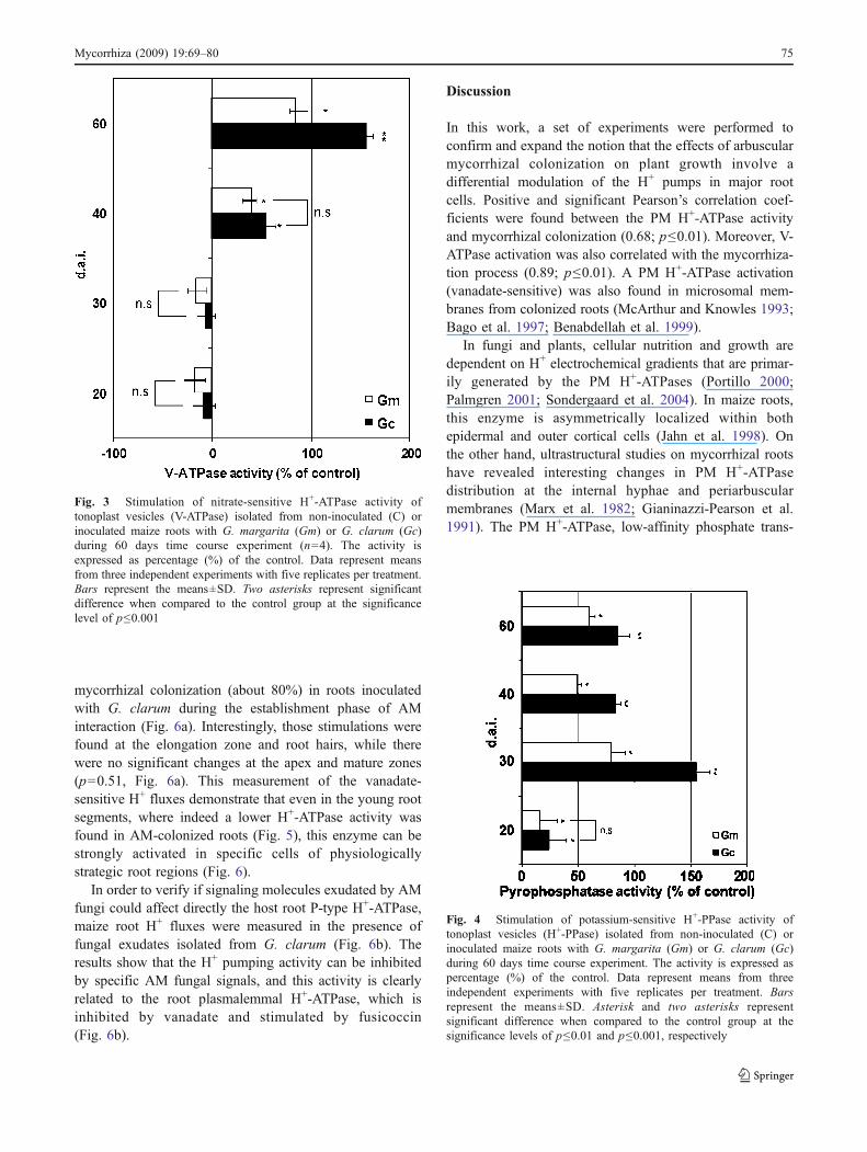
Tonoplast vesicles exhibited a V-ATPase activity that wasstimulated by fungal inoculation only at 40 and 60 d.a.i.(p<0.001 for Gc and 0.01 for Gm), since at 20 and 30 d.a.i.there were no statistical changes induced by the mycorrhizalcolonization (Fig. 3, p>0.70). There was no statisticalsignificance among the stimulations promoted by Gm orGc at 40 d.a.i (p<0.65). On the other hand, H+-PPaseactivity exhibited a strong induction at the early stages ofcolonization (30 d.a.i., p<0.001), mainly in G. clarumtreatment (Fig. 4), in parallel with a decrease of PM H+-ATPase activity (p≤0.01).
Effects of mycorrhizal colonization on H+-ATPaseand H+-PPase activities on the partial roots
It is well known that young fast growing meristematicregions of a root system displayed highest H+ pumpactivities and abundance than older mature regions (Jahnet al. 1998). Measurements of colonization rates alsorevealed that root YR are much less colonized than MRregions that exhibited a clear hyphal abundance. Thus, inorder to explore the effects of this spatial segregation ofmycorrhizal colonization on H+ pump activities, membranevesicles were isolated from these two regions of colonizedmaize roots (Fig. 5).
Membrane vesicles isolated from MR of maize rootsinoculated with G. clarum (Fig. 5a) showed a significantinhibition of V-ATPase at early stages of colonization (30 d.a.i., p<0.01), but without any significant effect on both thePM H+-ATPase and H+-PPase (p=0.68 and 0.36, respec-tively). On the other hand, PM H+-ATPase activity wasfound to be inhibited in YR colonized with G. clarum(Fig. 5a, p < 0.001), whereas it was stimulated by G.margarita (p<0.01). In contrast, inoculation with G.margarita stimulated the V-ATPase activity, while the sameactivity was inhibited with G. clarum (Fig. 5b, p<0.01).
Divided root systems revealed an apparent downregulation of the H+-ATPase activity at early stages of themycorrhizal colonization in YR, localized straight belowthe major colonization sites (Figs. 1, 5b). An oppositebehavior was observed for the H+-PPase activity, since bothfungal species promoted a strong activation of the PPihydrolysis in the same region (Fig. 5).
H+ efflux rate in mycorrhizal roots and the effect of fungalexudates
Analysis in vitro of the root H+ flux rate using noninvasivetechnique revealed high vanadate-sensitive H+ efflux and
Fig. 2 Stimulation of vanadate-sensitive H+-ATPase activity ofplasma membrane vesicles (PM H+-ATPase) isolated from non-inoculated (C) or inoculated maize roots with G. margarita (Gm) orG. clarum (Gc) during 60 days time course experiment (n=4). Theactivity is expressed as percentage (%) of the control. Data representmeans from three independent experiments with five replicates pertreatment. Bars represent the means±SD. Asterisk and two asterisksrepresent significant difference when compared to the control group atthe significance levels of p≤0.01 and p≤0.001, respectively
74 Mycorrhiza (2009) 19:69–80
mycorrhizal colonization (about 80%) in roots inoculatedwith G. clarum during the establishment phase of AMinteraction (Fig. 6a). Interestingly, those stimulations werefound at the elongation zone and root hairs, while therewere no significant changes at the apex and mature zones(p=0.51, Fig. 6a). This measurement of the vanadate-sensitive H+ fluxes demonstrate that even in the young rootsegments, where indeed a lower H+-ATPase activity wasfound in AM-colonized roots (Fig. 5), this enzyme can bestrongly activated in specific cells of physiologicallystrategic root regions (Fig. 6).
In order to verify if signaling molecules exudated by AMfungi could affect directly the host root P-type H+-ATPase,maize root H+ fluxes were measured in the presence offungal exudates isolated from G. clarum (Fig. 6b). Theresults show that the H+ pumping activity can be inhibitedby specific AM fungal signals, and this activity is clearlyrelated to the root plasmalemmal H+-ATPase, which isinhibited by vanadate and stimulated by fusicoccin(Fig. 6b).
Discussion
In this work, a set of experiments were performed toconfirm and expand the notion that the effects of arbuscularmycorrhizal colonization on plant growth involve adifferential modulation of the H+ pumps in major rootcells. Positive and significant Pearson’s correlation coef-ficients were found between the PM H+-ATPase activityand mycorrhizal colonization (0.68; p≤0.01). Moreover, V-ATPase activation was also correlated with the mycorrhiza-tion process (0.89; p≤0.01). A PM H+-ATPase activation(vanadate-sensitive) was also found in microsomal mem-branes from colonized roots (McArthur and Knowles 1993;Bago et al. 1997; Benabdellah et al. 1999).
In fungi and plants, cellular nutrition and growth aredependent on H+ electrochemical gradients that are primar-ily generated by the PM H+-ATPases (Portillo 2000;Palmgren 2001; Sondergaard et al. 2004). In maize roots,this enzyme is asymmetrically localized within bothepidermal and outer cortical cells (Jahn et al. 1998). Onthe other hand, ultrastructural studies on mycorrhizal rootshave revealed interesting changes in PM H+-ATPasedistribution at the internal hyphae and periarbuscularmembranes (Marx et al. 1982; Gianinazzi-Pearson et al.1991). The PM H+-ATPase, low-affinity phosphate trans-
Fig. 4 Stimulation of potassium-sensitive H+-PPase activity oftonoplast vesicles (H+-PPase) isolated from non-inoculated (C) orinoculated maize roots with G. margarita (Gm) or G. clarum (Gc)during 60 days time course experiment. The activity is expressed aspercentage (%) of the control. Data represent means from threeindependent experiments with five replicates per treatment. Barsrepresent the means±SD. Asterisk and two asterisks representsignificant difference when compared to the control group at thesignificance levels of p≤0.01 and p≤0.001, respectively
Fig. 3 Stimulation of nitrate-sensitive H+-ATPase activity oftonoplast vesicles (V-ATPase) isolated from non-inoculated (C) orinoculated maize roots with G. margarita (Gm) or G. clarum (Gc)during 60 days time course experiment (n=4). The activity isexpressed as percentage (%) of the control. Data represent meansfrom three independent experiments with five replicates per treatment.Bars represent the means±SD. Two asterisks represent significantdifference when compared to the control group at the significancelevel of p≤0.001
Mycorrhiza (2009) 19:69–80 75

Fig. 6 a Vanadate-sensitive H+
flux activity in four regions ofthe maize root system non-inoc-ulated (open bars) or inoculated(closed bars) with the AM fun-gus, G. clarum (Gc), under invitro conditions (n=8). Theanalysis was done 10 days afterspore germination. b Effect ofwater (control), fungal exudatesfrom 500 germinated spores ofG. clarum, 10 nM fusicoccin,and 50 μM vanadate on H+
effluxes of elongation zone. Anegative modulation of the H+
flux by addition of fungal fac-tors suggests a possible partici-pation of those “myc factors” inthe regulation of the PM H+-ATPase, since the addition ofFC stimulated the fluxes andvanadate inhibited. c–e Imagesof mycorrhizal maize roots,extraradical hyphae, and sporesof G. clarum during the mea-surements of H+ fluxes under invitro conditions. Bars representthe means±SD. Asterisk and twoasterisks represent significantdifference when compared to thecontrol group at the significancelevels of p≤0.01 and p≤0.001,respectively
Fig. 5 Vanadate-sensitive PM H+-ATPase, nitrate-sensitive vacuolarH+-ATPase and potassium-sensitive vacuolar H+-PPase activities ofpurified membranes isolated from mycorrhizal and non-mycorrhizalmaize partial roots. a Enzymatic activities in mature regions (MR) andb in young regions (YR) of the maize root system expressed as
percentage of the control. Data represent means from three indepen-dent experiments with four replicates per treatment. Bars represent themeans±SD. Asterisk and two asterisks represent significant differencewhen compared to the control group at the significance levels of p≤0.01 and p≤0.001, respectively
76 Mycorrhiza (2009) 19:69–80

porter, and hexose transporters have increased expression incortical arbuscule-containing cells (Gianinazzi- Pearson etal. 2000; Krajinski et al. 2002; Harrison 1996). Thissupports the notion that the H+ electrochemical gradientgenerated by the H+-ATPase provides a driving force forthe flux of nutrients and solutes across symbiotic mem-branes (Marx et al. 1982; Smith and Smith 1990;Gianinazzi 1991; Guttenberger 2000).
Since in this work the PM H+-ATPase activation was notrestricted to the root regions containing higher abundanceof arbuscular structures (MR), it is tempting to speculatethat such activation may also represent an acidic mecha-nism by which the cell wall is plasticized in order tofacilitate the penetration and differentiation of the fungalhyphae in the root apoplast during the establishment ofmycorrhizal colonization. The acid growth theory proposedby Rayle and Cleland (1992) involves the activation of H+
efflux via PM H+-ATPase, resulting in acidification of theapoplast. This will consequently trigger expansin activityresponsible for cell wall plasticity, which leads to cellexpansion (Morsomme and Boutry 2000). Siciliano et al.(2007) characterized the transcriptome of the pre-penetra-tion apparatus during AM interaction. They found thatexpansin-like genes were up-regulated in mycorrhizalplants. In agreement with this notion, we observed a highepidermal H+ efflux in mycorrhizal roots leading to anincreased capacity of these roots to acidify the localmedium. These effluxes proved to be dependent on thePM H+-ATPase given that they were sensitive to vanadateand activated by fusicoccin. This possibility is also in linewith the results obtained by Bago et al. (1998) who showedthat the pH of the medium decreased with the establishmentof AM symbiosis, and high acidic pH values were foundaround extraradical hyphae and spores. It is worth notingthat the H+ flux activation reported here should also resultin modulations of different ion channels and signalreceptors sensitive to changes of the pH gradient and/ormembrane potential.
Previously, it was reported that the mycorrhizationprocess involves a long-distance signaling derived fromthe AM fungi, which results in a mycorrhizal membranepotential hyperpolarization (Fieschi et al. 1992). Here, weprovided original evidences that signaling moleculesexudated by AM fungi could act directly on the host rootPM H+-ATPase and its related H+ fluxes. However, aninhibition of the rate of H+ efflux was observed specificallyat the elongation zone of non-colonized roots incubatedwith G. clarum fungal exudates. In addition, at the earlystages of fungal colonization, the PM H+-ATPase activitywas inhibited in maize roots colonized by G. clarum. Theseinhibitory phenomena could be related to the fungalenergetic cost that is proportionally higher in the firststages of colonization due to an increased fungal demand.
Therefore, it is tempting to speculate that AM fungi caninduce a down-regulation of the root main systems of ATPconsumption as a preparatory stage for the root infection.Moreover, it seems likely that after the establishment ofthe AM colonization, the fungi can induce a reactivationof the pumps not only to facilitate the dynamic progressionof the hyphal invasion through the cell wall but also for thereestablishment of a health root growth and metabolism. Infact, the H+ flux was strongly stimulated at the elongationzone of maize mycorrhizae colonized with G. clarum.
Although it is very difficult to correlate data of geneexpression with total enzymatic activities, our data are in agood correlation with molecular analyses performed byRosewarne et al. (2007) showing a down-regulation of themain H+-ATPase genes (LHA1 and LHA4) in tomato plantsduring the initial stages of their interaction with Glomusintraradices. This reveals the necessity to perform detailedstudies integrating enzymatic and molecular analyses inorder to elucidate the functional participation of each H+
pump isoform during plant colonization by mycorrhizae orpathogenic fungi. Actually, it has been postulated thatregulation of H+-ATPases plays an important role in thesignaling processes involving pathogenic interactions(Schaller and Oecking 1999) or at the very least participatesas an intermediate event in this signal transduction pathway(Xing et al. 1996).
During the same period (i.e., 30 d.a.i.), the potassium-sensitive H+-PPase activity was found to be higher whenthe lowest levels of ATP hydrolysis and plant growth weredetected. It is well known that at least for some plantspecies, it is common to observe inhibition of the plantgrowth during the initial stages of the mycorrhizalcolonization (Graham and Eissenstat 1994; Wright et al.1998a, b). This initial stress response seems to be promotedby some factors such as the intense AM fungal coloniza-tion, defense responses, or by the high consumption ofplant photosynthetic carbon by fungal cells. Willians et al.(1987) reported that plant growth depression in mycorrhizalcitrus plants might be due to the high cost of carbonprovided by the plant for fungal growth and lipid storage inthe vesicles of Glomus spp. On the other hand, G.margarita does not form intraradical vesicles but exten-sively forms extraradical storage auxiliary cells, whichshould also imply differences of energetic metabolism and/or carbon demand that, in turn, could account for thedifferences found in the balance of the pumps between thetwo fungi species.
Furthermore, along with these global observations in theroots, we also observed distinct local regions of differentialregulation of proton pump expression and activity as wellas distinct regions of fungal colonization. The root systemwas subdivided in two different areas to localize and revealthe negative modulation of ATP hydrolysis. We observed a
Mycorrhiza (2009) 19:69–80 77

very low fungal colonization rate at the distal root regions(YR). These regions encompass the younger tissues of theroots and are located straight below the major fungalcolonization zone. An intense drain of photoassimilates bythe fungal cells affects strongly this zone (Bonfante-Fasoloand Perotto 1992). In contrast, high fungal colonization wasexhibited in the mature root regions without any significanteffect on both the PM H+-ATPase and H+-PPase; however,a significant inhibition of V-ATPase was observed at earlystages of the colonization by G. clarum (30 d.a.i.). Thissuggests that the spatial distribution of the AM fungi mayaccount for the decrease of the H+-ATPase activities and theactivation of the H+-PPase observed in the young regions.This effect was also species-specific, and since the activityof the root meristems influences the root development andarchitecture (Berta et al. 1993), the energetic stress imposedon the root distal region by sugar availability couldmodulate the root morphology and thereby regulate thedistribution of H+ pumps along the root system. This is inagreement with previous reports that observed a down-regulation of the PM H+-ATPase activity in plant cellsundergoing sugar deprivation (e.g., Mito et al. 1996).
Our previous data suggested that the stimulation of H+-PPase is an important part of the host response to theenergy consumption by AM fungi (Ramos et al. 2005).There are already several reports indicating that the H+-PPase may replace the role of H+-ATPases under conditionsof stress (Carystinos et al. 1995; Davies et al. 1997). Thisnotion is supported by recent studies that also highlight therequirement of this pump during other stress responsesmediated by plant cell (e.g., Nakanishi and Maeshima1998; Park et al. 2005; Gaxiola et al. 2007). Takingtogether the present data, it seems clear that the energeticbackup system represented by the H+-PPase could bespecially regulated in the young meristematic region dueto its higher energy demand. The young root region ofmycorrhizal roots should be more depleted of sugar drainedby the AM fungi colonizing the above regions. Therefore,the activation of the PPi metabolism in this region wouldrepresent a critical energetic advantage, for instance, bysaving the ATP needed to the cells with higher metabolicactivity of physiologically strategic root regions such as theelongation zone and root hairs.
Conclusions
The activity of H+ pumps in host root cells duringcolonization are differentially modulated by their interac-tions with AM fungi. These biochemical changes seem toinfluence the rate of host growth and that of fungalcolonization. The lowest rates of ATP hydrolysis in theearly stages of AM symbiosis might be compensated by
activation of the tonoplast H+-PPase in those root regionsthat have the lowest available photoassimilates. Furtherstudies are necessary to understand the dynamics of H+-ATPase and H+-PPase expression and activity in order toprovide new insights into the role of H+ pumps in such animportant symbiotic interaction.
Acknowledgments This work was supported by CAPES (Brazil)and by Post-Doctoral fellowship (SFRH/BPD/21061/2004) concededto ACR by Fundação para a Ciência e Tecnologia and InstitutoGulbenkian de Ciência (Portugal). ARF is supported by grants fromInternational Foundation for Science (IFS) (C/3483-1) and ConselhoNacional de Desenvolvimento Científico e Tecnológico (CNPq)(475522/01-0 and 479286/03-5). The authors would like to acknowl-edge Dr. Carlos Tadokoro and Dr Rui Gardner for the manuscriptrevision, and Dr. Mark Seldon for the critical review and helpfulsuggestions of the manuscript. We also thank the Microscopy Center“Raul Dodsworth Machado” (UENF-BRAZIL) and Quíssila Batistafor the help in preparing the samples for microscopy analysis.
References
Bago B, Donaire JP, Azcón-Aguilar C (1997) ATPases activities ofroot from mycorrhizal sunflower (Helianthus annuus) and onion(Allium cepa) plants. New Phytol 136:305–311. doi:10.1046/j.1469-8137.1997.00741.x
Bago B, Azcon-Aguilar C, Piché Y (1998) Architecture anddevelopmental dynamics of the external mycelium of thearbuscular mycorrhizal fungus Glomus intraradices grown undermonoxenic conditions. Mycologia 90:52–62. doi:10.2307/3761011
Bécard G, Fortin J (1988) Early events of vesicular-arbuscularmycorrhiza formation on Ri T-DNA transformed roots. NewPhytol 108:211–218. doi:10.1111/j.1469-8137.1988.tb03698.x
Benabdellah K, Azcón-Aguilar C, Ferrol N (1999) Plasma membraneATPase and H+ transport activities in microsomal membranesfrom mycorrhizal tomato roots. J Exp Bot 50:1343–1349.doi:10.1093/jexbot/50.337.1343
Berta G, Fusconi A, Trotta A (1993) VA mycorrhizal infection and themorphology and function of root systems. Environ Exp Bot33:159–173. doi:10.1016/0098-8472(93)90063-L
Bestel-Corre G, Dumas-Gaudot E, Poinsot V, Dieu M, Dierick JF, vanTuinen D, Remacle J, Gianinazzi-Pearson V, Gianinazzi S (2002)Proteome analysis and identification of symbiosis-related proteinsfrom Medicago truncatula Gaertn. by two-dimensional electropho-resis and mass spectrometry. Electrophoresis 23:122–137
Bonfante-Fasolo P, Perotto S (1992) Plant and endomycorrhizal fungi:the cellular and molecular basis of their interaction. In: VermaDPS (ed) Molecular signals in plant-microbe communications.CRC, Boca Raton, FL, pp 445–470
Buch-Pedersen MJ, Rudashevskaya EL, Berner TS, Venema K,Palmgren MG (2006) Potassium as an intrinsic uncoupler of theplasma membrane H+-ATPase. J Biol Chem 281:38285–38292.doi:10.1074/jbc.M604781200
Cappellazzo G, Lanfranco L, Fitz M, Wipf D, Bonfante P (2008)Characterization of an amino acid permease from the Endomy-corrhizal fungus Glomus mosseae. Plant Physiol 147:429–437.doi:10.1104/pp.108.117820
Carystinos GD, MacDonald HR, Monroy AF, Dhindsa RS, Poole RJ(1995) Vacuolar H+-translocating pyrophosphatase is induced byanoxia or chilling in seedlings of rice. Plant Physiol 108:641–649. doi:10.1104/pp.108.2.641
78 Mycorrhiza (2009) 19:69–80

Clark RB (1975) Characterization of phosphatase of intact maizeroots. J Agric Food Chem 23:458–460. doi:10.1021/jf60199a002
Cruz C, Egsgaard H, Trujillo C, Ambus P, Requena N, Martins-LouçãoMA, Jakobsen I (2007) Enzymatic evidence for the key roleof arginine in nitrogen translocation by arbuscular mycorrhi-zal fungi. Plant Physiol 144:782–792. doi:10.1104/pp.106.090522
Davies JM, Darley CP, Sanders D (1997) Energetics of the plasmamembrane pyrophosphatase. Trends Plant Sci 2:9–10.doi:10.1016/S1360-1385(97)82732-X
De Michelis MI, Spanswick RM (1986) H+-pumping driven byvanadate sensitive ATPase in membrane vesicles from cornsroots. Plant Physiol 81:542–547
Drozdowicz YM, Rea PA (2001) Vacuolar H+-pyrophosphatases: fromevolutionary backwaters into mainstream. Trends Plant Sci6:206–211. doi:10.1016/S1360-1385(01)01923-9
Façanha AR, De Meis L (1995) Inhibition of maize root H+-ATPaseby fluoride and fluoroaluminate complexes. Plant Physiol108:241–246
Façanha AR, De Meis L (1998) Reversibility of H+-ATPase and H+-Pyrophosphatase in tonoplast vesicles from maize coleoptilesand seeds. Plant Physiol 116:1487–1495. doi:10.1104/pp.116.4.1487
Feijó JA, Sainhas J, Hackett GR, Kunkel JG, Hepler PK (1999)Growing pollen tubes posses a constitutive alkaline band in theclear zone and a growth-dependent acidic tip. J Cell Biol144:483–496. doi:10.1083/jcb.144.3.483
Ferrol N, Pozo MJ, Antelo M, Azcón-Aguilar C (2002) Arbuscularmycorrhizal symbiosis regulates plasma membrane H+-ATPasegene expression in tomato plants. J Exp Bot 53:1683–1687.doi:10.1093/jxb/erf014
Fieschi M, Alloatti G, Sacco S, Berta G (1992) Membrane-potentialhyperpolarization in vesicular arbuscular mycorrhizae of Alliumporrum L.: A non-nutritional long-distance effect of the fungus.Protoplasma 168:136–140. doi:10.1007/BF01666259
Fiske CF, Subbarow Y (1925) The colorometric determination ofphosphorus. J Biol Chem 66:375–400
Gaxiola RA, Palmgren MG, Schumacher K (2007) Plant proton pumps.FEBS Lett 581:2204–2214. doi:10.1016/j.febslet.2007.03.050
Gianinazzi S (1991) Vesicular-arbuscular (endo-) mycorrhizas: cellu-lar, biochemical and genetic aspects. Agric Ecosyst Environ35:105–119
Gianinazzi-Pearson V (1996) Plant cell responses to arbuscularmycorrhizal fungi: getting to the roots of the symbiosis. PlantCell 8:1871–1883
Gianinazzi-Pearson V, Brechenmacher L (2004) Functional genomicsof arbuscular mycorrhiza: decoding the symbiotic cellprogramme. Can J Bot 82:1228–1234. doi:10.1139/b04-096
Gianinazzi-Pearson V, Smith SE, Gianinazzi S, Smith FA (1991)Enzymatic studies on the metabolism of vesicular–arbuscularmycorrhiza. V. Is H+-ATPase a component of ATP-Hydrolysingenzyme activities in plant-fungi interfaces? New Phytol 117:61–74. doi:10.1111/j.1469-8137.1991.tb00945.x
Gianinazzi-Pearson V, Arnould C, Oufattole M, Arango M, GianinazziS (2000) Differential activation of H+-ATPase genes by anarbuscular mycorrhizal fungus in root cells of transgenic tobacco.Planta 211:609–613. doi:10.1007/s004250000323
Giovannetti M, Mosse B (1980) An evaluation of techniques to mea-sure vesicular–arbuscular mycorrhizal infection in roots. NewPhytol 84:484–500. doi:10.1111/j.1469-8137.1980.tb04556.x
Giovannetti M, Sbrana C, Avio L, Citernesi AS, Logi C (1993)Differential hyphal morphogenesis in arbuscular mycorrhizalfungi during preinfection stages. New Phytol 125:587–593.doi:10.1111/j.1469-8137.1993.tb03907.x
Giovannetti M, Sbrana C, Citernesi AS, Avio L (1996) Analysis offactors involved in fungal recognition responses to host derived
signals by arbuscular mycorrhizal fungi. New Phytol 133:65–71.doi:10.1111/j.1469-8137.1996.tb04342.x
Graham JH, Eissenstat DM (1994) Host genotype and the formationand function of VA mycorrhizae. Plant Soil 159:179–185
Guttenberger M (2000) Arbuscules of vesicular–arbuscular mycorrhi-zal fungi inhabit an acidic compartment within plant roots. Planta211:112–118. doi:10.1007/s004250000324
Harrison MJ (1996) A sugar transporter from Medicago truncatula:altered expression pattern in roots during vesicular–arbuscular(VA) mycorrhizal associations. Plant J 9:491–503. doi:10.1046/j.1365-313X.1996.09040491.x
Harrison MJ (2005) Signaling in the arbuscular mycorrhizal symbiosis.Annu Rev Microbiol 59:19–42. doi:10.1146/annurev.micro.58.030603.123749
Hohnjec N, Vieweg MF, Pühler A, Becker A, Küster H (2005)Overlaps in the transcriptional profiles of Medicago truncatularoots inoculated with two different Glomus fungi provide insightsinto the genetic program activated during arbuscular mycorrhiza.Plant Physiol 137:1283–1301. doi:10.1104/pp.104.056572
Ihaka R, Gentleman R (1996) R: a language for data analysis andgraphics. J Comput Graph Statist 5:299–314. doi:10.2307/1390807
Jahn T, Baluska F, Michalke W, Harper JF, Volkmann D (1998)Plasma membrane H+-ATPase in the root apex: evidence forstrong expression in xylem parenchyma and asymmetric locali-zation within cortical and epidermal cells. Physiol Plant 104:311–316. doi:10.1034/j.1399-3054.1998.1040304.x
James EK, Reis VM, Olivares FL, Baldani J, Döbereiner J (1994)Infection of sugar cane by the nitrogen-fixing bacteriumAcetobacter diazotrophicus. J Exp Bot 45:757–766. doi:10.1093/jxb/45.6.757
Krajinski F, Hause B, Gianinazzi-Pearson V, Franken P (2002) Mtha1,a plasma membrane H+-ATPase gene from Medicago truncatula,shows arbuscule-specific induced expression in mycorrhizaltissue. Plant Biol 4:754–761. doi:10.1055/s-2002-37407
Kunkel JG, Cordeiro S, Xu J, Shipley AM, Feijó JA (2006) The use ofnon-invasive ion-selective microelectrode techniques for thestudy of plant development. In: Volkov V (ed) Plant electro-physiology—theory and methods. Springer, Berlin, pp 109–137
Lambais MR (2006) Unraveling the signaling and signal transductionmechanisms controlling arbuscular mycorrhiza development. Scien-tia Agricola 63:405–413. doi:10.1590/S0103-90162006000400013
Li H, Smith FA, Dickson S, Holloway RE, Smith SE (2008) Plantgrowth depressions in arbuscular mycorrhizal symbioses: not justcaused by carbon drain? New Phytol 178:852–862. doi:10.1111/j.1469-8137.2008.02410.x
Lichko L, Okorokov L (1991) Purification and some properties ofmembrane bound and soluble pyrophosphatase of yeast vacuoles.Yeast 7:805–812. doi:10.1002/yea.320070805
Lowry OH, Rosebrough NJ, Farr AL, Randall RJ (1951) Proteinmeasurement with the Folin phenol reagent. J Biol Chem193:265–275
Marx C, Dexheimer J, Gianinazzi-Pearson V, Gianinazzi S (1982)Enzymatic studies on the metabolism of vesicular–arbuscularmycorrhiza. IV. Ultracitoenzymological evidence (ATPase) foractive transfer processes in the host-arbuscular interface. NewPhytol 90:37–43. doi:10.1111/j.1469-8137.1982.tb03238.x
Mcarthur DAJ, Knowles NR (1993) Influence of vesicular–arbuscularmycorrhizal fungi on the response of potato to phosphorusdeficiency. Plant Physiol 101:147–160
Mito N, Wimmers LE, Bennett AB (1996) Sugar regulates mRNAabundance of H+-ATPase gene family members in tomato. PlantPhysiol 112:1229–1236. doi:10.1104/pp.112.3.1229
Morsomme P, Boutry M (2000) The plant plasma membrane H+-ATPase: structure, function and regulation. Biochim BiophysActa 1465:1–16. doi:10.1016/S0005-2736(00)00128-0
Mycorrhiza (2009) 19:69–80 79

Murphy PJ, Langridge P, Smith SE (1996) Cloning plant genesdifferentially expressed during colonization of roots of Hordeumvulgare by the vesicular–arbuscular mycorrhizal fungus Glomusintraradices. New Phytol 135:291–301. doi:10.1046/j.1469-8137.1997.00652.x
Nakanishi Y, Maeshima M (1998) Molecular cloning of vacuolar H+-pyrophosphatase and its developmental expression in growinghypocotyl of mung bean. Plant Physiol 116:589–597.doi:10.1104/pp.116.2.589
Olivares FL, James EK, Baldani JI, Döbereiner J (1997) Infection ofmottled stripe disease susceptible and resistant varieties of sugarcane by the endophytic diazotroph Herbaspirillum. New Phytol135:723–737. doi:10.1046/j.1469-8137.1997.00684.x
Okorokov LA, Silva FE, Okorokova-Façanha AL (2001) Ca2+ and H+
homeostasis in fission yeast: a role of Ca2+/H+ exchange anddistinct V-H+-ATPases of the secretory pathway organelles.FEBS Lett 505:321–324. doi:10.1016/S0014-5793(01)02852-6
Palmgren MG (2001) Plant plasma membrane H+-ATPases: power-houses for nutrient uptake. Annu Rev Plant Physiol Plant MolBiol 52:817–845. doi:10.1146/annurev.arplant.52.1.817
Park S, Li J, Pittman JK, Berkowitz GA, Yang H, Undurraga S,Morris J, Hirschi KD, Gaxiola RA (2005) Up-regulation of a H+-pyrophosphatase (H+-PPase) as strategy to engineer drought-resistant crop plants. Proc Natl Acad Sci U S A 102:18830–18835.doi:10.1073/pnas.0509512102
Philips JM, Hayman DS (1970) Improved procedure for clearingroots and staining parasitic and vesicular-arbuscular mycorrhi-zal fungi for rapid assessment of infection. Trans Br Mycol Soc55:158–161
Portillo F (2000) Regulation of plasma membrane H+-ATPase in fungiand plants. Biochim Et Biophys Acta-Rev Biomembr 1469:31–42
Poulsen KH, Nagy R, Gao LL, Smith SE, BucherM, Smith FA, Jakobsen I(2005) Physiological and molecular evidence for pi uptake via thesymbiotic pathway in a reduced mycorrhizal colonization mutant intomato associated with a compatible fungus. New Phytol 168:445–454. doi:10.1111/j.1469-8137.2005.01523.x
Ramos AC, Martins MA, Façanha AR (2005) ATPase and pyrophos-phatase activities in corn root microsomes colonized witharbuscular mycorrhizal fungi. Braz J Soil Sci 29:207–213
Ramos AC, Façanha AR, Feijó JA (2008a) Ion dynamics during thepolarized growth of arbuscular mycorrhizal fungi: from presym-biosis to symbiosis. In: Varma A, Hock B (eds) Mycorrhiza.Springer, Germany, pp 241–261
Ramos AC, Façanha AR, Feijó JA (2008b) A proton (H+) fluxsignature of the presymbiotic development of the arbuscularmycorrhizal fungi. New Phytol 178:177–188. doi:10.1111/j.1469-8137.2007.02344.x
Rayle DL, Cleland RE (1992) The acid growth theory of auxin-inducedcell elongation is alive and well. Plant Physiol 99:1271–1274
Rausch C, Daram P, Brunner S, Jansa J, Laloi M, Leggewie G,Amrhein N, Bucher M (2001) A phosphate transporter expressedin arbuscule-containing cells in potato. Nature 414:462–466.doi:10.1038/35106601
Rosewarne GM, Smith FA, Schachtman DP, Smith SE (2007)Localization of proton-ATPase genes expressed in arbuscularmycorrhizal tomato plants. Mycorrhiza 17:249–258. doi:10.1007/s00572-006-0101-6
Schaller A, Oecking C (1999) Modulation of plasma membrane H+-ATPase activity differentially activates wound and pathogendefense responses in tomato plants. Plant Cell 11:263–272
Schüβler A,Martin H, Cohen D, Fitz M,Wipf D (2006) Characterizationof a carbohydrate transporter from symbiotic glomeromycotanfungi. Nature 444:933–936
Siciliano V, Genre A, Balestrini R, Cappellazzo G, deWit PJGM,Bonfante P (2007) Transcriptome analysis of arbuscular mycor-rhizal roots during development of the prepenetration apparatus.Plant Physiol 144:1455–1466. doi:10.1104/pp.107.097980
Smith SE, Smith FA (1990) Structure and function of the interfaces inbiotrophic symbioses as they relate to nutrient transport. NewPhytol 144:1–38
Sondergaard TE, Schulz A, Palmgren MG (2004) Energization oftransport processes in plants. roles of the plasma membrane H+-ATPase. Plant Physiol 136:2475–2482. doi:10.1104/pp.104. 048231
Valot B, Dieu M, Recorbet G, Raes M, Gianinazzi S, Dumas-Gaudot E(2005) Identification of membrane-associated proteins regulated bythe arbuscular mycorrhizal symbiosis. Plant Mol Biol 59:565–580
Willians K, Percival F, Merino J, Mooney HA (1987) Estimation oftissue construction cost from heat of combustion and organicnitrogen content. Plant Cell Environ 10:725–734
Wright DP, Read DJ, Scholes JD (1998a) Mycorrhizal sink strengthinfluences whole plant carbon balance of Trifolium repens L. PlantCell Environ 21:881–891. doi:10.1046/j.1365-3040.1998.00351.x
Wright DP, Scholes JD, Read DJ (1998b) Effects of VA mycorrhizalcolonization on photosynthesis and biomass production ofTrifolium repens L. Plant Cell Environ 21:209–216. doi:10.1046/j.1365-3040.1998.00280.x
Xing T, Higgins VJ, Blumwald E (1996) Regulation of plant defenseresponse to fungal pathogens: two types of protein kinases in thereversible phosphorylation of the host plasma membrane H+-ATPase. Plant Cell 8:555–564
Zandonadi DB, Canellas LP, Facanha AR (2007) Indolacetic andhumic acids induce lateral root development through a concen-trated plasmalemma and tonoplast H+-pumps activation. Planta225:1583–1595. doi:10.1007/s00425-006-0454-2
80 Mycorrhiza (2009) 19:69–80