Fruit Development, Ripening and Quality Related Genes in the Papaya Genome
Upload
independentCategory
view
2download
0
lable at ScienceDirect
Plant Physiology and Biochemistry 48 (2010) 893e902
Contents lists avai
Plant Physiology and Biochemistry
journal homepage: www.elsevier .com/locate/plaphy
Research article
Symbiotic interactions between arbuscular mycorrhizal (AM) fungiand male papaya plants: Its status, role and implications
Sharda W. Khade*, Bernard F. Rodrigues, Prabhat K. SharmaDepartment of Botany, Goa University, Taleigao Plateau, Goa 403206, India
a r t i c l e i n f o
Article history:Received 7 January 2010Accepted 18 August 2010Available online 25 August 2010
Keywords:AM fungiMale C. papayaRoot colonizationRoot phosphatases
* Corresponding author. Current address: IInd FlooColony, Carenzalem Post, Miramar, Panaji, Goa 4032462978.
E-mail addresses: [email protected] (S.Wcom (B.F. Rodrigues), [email protected]
0981-9428/$ e see front matter � 2010 Elsevier Masdoi:10.1016/j.plaphy.2010.08.010
a b s t r a c t
Experiments were conducted to study the arbuscular mycorrhizal (AM) status and its role in P-uptakethrough assay of root phosphatases activities in four varieties ofmale Carica papaya L. viz. CO-1, CO-2, HoneyDewandWashington duringflowering stages. In the present study,mean total root colonization of AM fungirecorded peak increase in flowering stage-II while mean root phosphatase (acid and alkaline) activitiesrecorded peak increase in flowering stage-I. Unlike root colonization and root phosphatase activities, sporedensity did not exhibit any definite patterns and recorded a narrow range of fluctuation during differentflowering stages ofmale C. papaya. The study brought out the fact that root colonization and spore densityofAM fungi along with root phosphatase activities varied significantly within the four varieties of maleC. papaya plants during each flowering stage. The study also recorded consistently higher acid root phos-phatase activity than alkaline root phosphatase activity under P-deficient, acidic soil conditions during allflowering stages ofmale C. papaya plants. Studies revealed that the root colonization of AM fungi influencedroot phosphatase activities (acid andalkaline) positively and significantly during allflowering stages ofmaleC. papaya plants. A total of twelve species of AM fungi belonging tofive generaviz. Acaulospora,Dentiscutata,Gigaspora, Glomus, and Racocetra were recovered from the rhizosphere of male C. papaya plants.
� 2010 Elsevier Masson SAS. All rights reserved.
1. Introduction
Carica papaya L. belongs to family Caricaceae. The genus Caricahas about 48 species of which only C. papaya is grown for its ediblefruits. C. papaya is a polygamous plant and has many sex forms. Thethree basic sex types are: staminate or male, hermaphrodite andpistillate or female. In India, large numbers of varieties are found forcultivation. This variability is seen due open pollination and indis-criminate multiplication using open pollinated seeds. In C. papayathere are two basic types of varieties. Those varieties, that produceonly female and male plants are said to be dioecious and those thatproduce female and hermaphrodite plants are called gynodioecious.Some of the important gynodioeciuos varieties are Coorg HoneyDew, Sunrise Solo, CO-7 and Surya. The varieties like CO-1, Wash-ington and Pusa Dwarf come under dioecious types [26].
Interestingly, India is the one of the largest producer of C. papayain theworld after Brazil with production (in thousandmetric tones)up to 1500 [26]. It is cultivated on commercial scale in states of
r, Darshan Apts, Vidhyanagar002, India. Tel.: þ91 0832
. Khade), [email protected] (P.K. Sharma).
son SAS. All rights reserved.
Karnataka, Bihar, Gujarat, Kerala, Tamil Nadu, Andhra Pradesh,Maharashtra and Madhya Pradesh. In fact in southern region, thearea under cultivation has increased by 24.37% whereas theproduction has increased from 1.65% to 32.04%. However, in Goa, C.papaya cultivation is mostly confined to kitchen garden and alongthe bunds due to constraints in productivity resulting from fungaland viral diseases. Therefore at present, most of the demand for C.papaya is met by importing the fruit from neighbouring states [26].
The wide spread symbiotic arbuscular mycorrhizal (AM) fungalassociation with host plants is gaining importance due to the factthat they help in plant productivity. The AM fungi were found to beassociated with different varieties of fruit bearing C. papaya in agro-based ecosystem of Goa, India. Here, the root colonization by AMfungi ranged from 26 to 77%, while the spore density ranged from100 to 236 spores.100 g of rhizosphere soil�1 [30]. The AM fungi alsoreported seasonal variation in the rhizosphere of fruit bearingC. papaya from Goa, India [28]. Further, AM fungi were spatially [29]and spatio-temporally [33] distributed in rhizosphere of fruitbearing C. papaya plants in Goa, India. So, far AM fungi belonging tothe genera Acaulospora, Glomus, Gigaspora, Dentiscutata [30],Cetraspora and Racocetra [33] were found to be associated with fruitbearing C. papaya in Goa, India. C. papaya is known to exhibit a stronggrowth response to colonization by AM fungi. Inoculation of vege-tative C. papaya with AM fungi, under greenhouse conditions
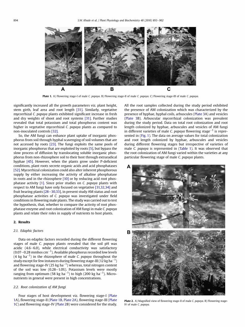
Plate 1. A) Flowering stage-I of male C. papaya. B) Flowering stage-II of male C. papaya. C) Flowering stage-III of male C. papaya.
Plate 2. A) Magnified view of flowering stage-II of male C. papaya. B) Flowering stage-IV of male C. papaya.
S.W. Khade et al. / Plant Physiology and Biochemistry 48 (2010) 893e902894
significantly increased all the growth parameters viz. plant height,stem girth, leaf area and root length [31]. Similarly, vegetativemycorrhizal C. papaya plants exhibited significant increase in freshand dry weights of shoot and root systems [31]. Further studiesrevealed that total potassium and total phosphorus content washigher in vegetative mycorrhizal C. papaya plants as compared tonon-inoculated controls [32].
So, the AM fungi can enhance plant uptake of inorganic phos-phorus from soil through hyphal scavenging of soil volumes that arenot accessed by roots [23]. The fungi exploits the same pools ofinorganic phosphorus that are exploited by roots [5], but bypass theslow process of diffusion by translocating soluble inorganic phos-phorus from non-rhizosphere soil to their host through extraradicalhyphae [45]. However, when the plants grow under P-deficientconditions, plant roots secrete organic acids and acid phosphatases[52].Mycorrhizal colonization could also alter inherent phosphoroussupply by either increasing the activity of alkaline phosphatasein roots and in the rhizosphere [10] or by reducing acid root phos-phatase activity [1]. Since prior studies on C. papaya plants withrespect to AM fungi have only focused on vegetative [31,32,34] andfruit bearing plants [28e30,33], in present study AM status and rootphosphatase activities of C. papaya was investigated under fieldconditions infloweringmale plants. The studywas carried out to testthe hypothesis, that, whether to compare the activity of root phos-phatase enzyme and root colonization of AM fungi inmale C. papayaplants and relate their roles in supply of nutrients to host plants.
2. Results
2.1. Edaphic factors
Data on edaphic factors recorded during the different floweringstages of male C. papaya plants revealed that the soil pH wasacidic (4.6e6.0), while electrical conductivity was satisfactory(0.07e0.28mmhos cm�1). Available phosphorus recorded low levels(4 kg ha�1) in the rhizosphere of male C. papaya throughout thestudyexcept for few instances duringflowering stage-III (12 kg ha�1)and flowering stage-IV (25 kg ha�1) whereas, total nitrogen contentof the soil was low (0.28e1.0%). Potassium levels were mostlyranging from optimum (58 kg ha�1) to high (200 kg ha�1). Micro-nutrients in general were present in high concentrations.
2.2. Root colonization of AM fungi
Four stages of host development viz. flowering stage-I (Plate1A), flowering stage-II (Plate 1B, Plate 2A), flowering stage-III (Plate1C) and flowering stage-IV (Plate 2B) were considered for the study.
All the root samples collected during the study period exhibitedthe presence of AM colonization which was characterized by thepresence of hyphae, hyphal coils, arbuscules (Plate 3A) and vesicles(Plate 3B). Arbuscular mycorrhizal colonization was prevalentduring the study period. Data on total root colonization and rootlength colonized by hyphae, arbuscules and vesicles of AM fungiin different varieties of male C. papaya flowering stage�1 is repre-sented in (Fig. 1). The data on average values for total colonizationand root length colonized by hyphae, arbuscules and vesiclesduring different flowering stages but irrespective of varieties ofmale C. papaya is represented in (Table 1). It was observed thatthe root colonization of AM fungi varied within the varieties at anyparticular flowering stage of male C. papaya plants.
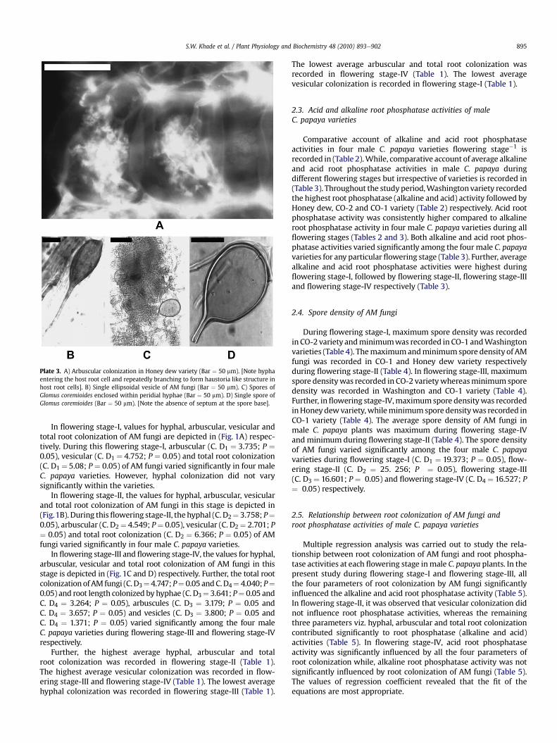
Plate 3. A) Arbuscular colonization in Honey dew variety (Bar ¼ 50 mm). [Note hyphaentering the host root cell and repeatedly branching to form haustoria like structure inhost root cells]. B) Single ellipsoidal vesicle of AM fungi (Bar ¼ 50 mm). C) Spores ofGlomus coremioides enclosed within peridial hyphae (Bar ¼ 50 mm). D) Single spore ofGlomus coremioides (Bar ¼ 50 mm). [Note the absence of septum at the spore base].
S.W. Khade et al. / Plant Physiology and Biochemistry 48 (2010) 893e902 895
In flowering stage-I, values for hyphal, arbuscular, vesicular andtotal root colonization of AM fungi are depicted in (Fig. 1A) respec-tively. During this flowering stage-I, arbuscular (C. D1 ¼ 3.735; P ¼0.05), vesicular (C. D1 ¼ 4.752; P ¼ 0.05) and total root colonization(C. D1 ¼ 5.08; P ¼ 0.05) of AM fungi varied significantly in four maleC. papaya varieties. However, hyphal colonization did not varysignificantly within the varieties.
In flowering stage-II, the values for hyphal, arbuscular, vesicularand total root colonization of AM fungi in this stage is depicted in(Fig.1B). During thisflowering stage-II, thehyphal (C. D2¼ 3.758; P¼0.05), arbuscular (C. D2¼ 4.549; P¼ 0.05), vesicular (C. D2¼ 2.701; P¼ 0.05) and total root colonization (C. D2 ¼ 6.366; P ¼ 0.05) of AMfungi varied significantly in four male C. papaya varieties.
In flowering stage-III and flowering stage-IV, the values for hyphal,arbuscular, vesicular and total root colonization of AM fungi in thisstage is depicted in (Fig. 1C and D) respectively. Further, the total rootcolonizationof AM fungi (C. D3¼ 4.747; P¼ 0.05 andC.D4¼ 4.040; P¼0.05) and root length colonized by hyphae (C. D3¼ 3.641; P¼ 0.05 andC. D4 ¼ 3.264; P ¼ 0.05), arbuscules (C. D3 ¼ 3.179; P ¼ 0.05 andC. D4 ¼ 3.657; P ¼ 0.05) and vesicles (C. D3 ¼ 3.800; P ¼ 0.05 andC. D4 ¼ 1.371; P ¼ 0.05) varied significantly among the four maleC. papaya varieties during flowering stage-III and flowering stage-IVrespectively.
Further, the highest average hyphal, arbuscular and totalroot colonization was recorded in flowering stage-II (Table 1).The highest average vesicular colonization was recorded in flow-ering stage-III and flowering stage-IV (Table 1). The lowest averagehyphal colonization was recorded in flowering stage-III (Table 1).
The lowest average arbuscular and total root colonization wasrecorded in flowering stage-IV (Table 1). The lowest averagevesicular colonization is recorded in flowering stage-I (Table 1).
2.3. Acid and alkaline root phosphatase activities of maleC. papaya varieties
Comparative account of alkaline and acid root phosphataseactivities in four male C. papaya varieties flowering stage�1 isrecorded in (Table 2).While, comparative account of average alkalineand acid root phosphatase activities in male C. papaya duringdifferent flowering stages but irrespective of varieties is recorded in(Table 3). Throughout the studyperiod,Washingtonvariety recordedthe highest root phosphatase (alkaline and acid) activity followed byHoney dew, CO-2 and CO-1 variety (Table 2) respectively. Acid rootphosphatase activity was consistently higher compared to alkalineroot phosphatase activity in four male C. papaya varieties during allflowering stages (Tables 2 and 3). Both alkaline and acid root phos-phatase activities varied significantly among the fourmale C. papayavarieties for any particular flowering stage (Table 3). Further, averagealkaline and acid root phosphatase activities were highest duringflowering stage-I, followed by flowering stage-II, flowering stage-IIIand flowering stage-IV respectively (Table 3).
2.4. Spore density of AM fungi
During flowering stage-I, maximum spore density was recordedin CO-2 variety andminimumwas recorded in CO-1 andWashingtonvarieties (Table 4). Themaximumandminimumspore density of AMfungi was recorded in CO-1 and Honey dew variety respectivelyduring flowering stage-II (Table 4). In flowering stage-III, maximumspore densitywas recorded in CO-2 varietywhereasminimum sporedensity was recorded in Washington and CO-1 variety (Table 4).Further, inflowering stage-IV,maximumspore densitywas recordedinHoneydewvariety,whileminimumspore densitywas recorded inCO-1 variety (Table 4). The average spore density of AM fungi inmale C. papaya plants was maximum during flowering stage-IVandminimum during flowering stage-II (Table 4). The spore densityof AM fungi varied significantly among the four male C. papayavarieties during flowering stage-I (C. D1 ¼ 19.373; P ¼ 0.05), flow-ering stage-II (C. D2 ¼ 25. 256; P ¼ 0.05), flowering stage-III(C. D3 ¼ 16.601; P ¼ 0.05) and flowering stage-IV (C. D4 ¼ 16.527; P¼ 0.05) respectively.
2.5. Relationship between root colonization of AM fungi androot phosphatase activities of male C. papaya varieties
Multiple regression analysis was carried out to study the rela-tionship between root colonization of AM fungi and root phospha-tase activities at each flowering stage inmale C. papaya plants. In thepresent study during flowering stage-I and flowering stage-III, allthe four parameters of root colonization by AM fungi significantlyinfluenced the alkaline and acid root phosphatase activity (Table 5).In flowering stage-II, it was observed that vesicular colonization didnot influence root phosphatase activities, whereas the remainingthree parameters viz. hyphal, arbuscular and total root colonizationcontributed significantly to root phosphatase (alkaline and acid)activities (Table 5). In flowering stage-IV, acid root phosphataseactivity was significantly influenced by all the four parameters ofroot colonization while, alkaline root phosphatase activity was notsignificantly influenced by root colonization of AM fungi (Table 5).The values of regression coefficient revealed that the fit of theequations are most appropriate.
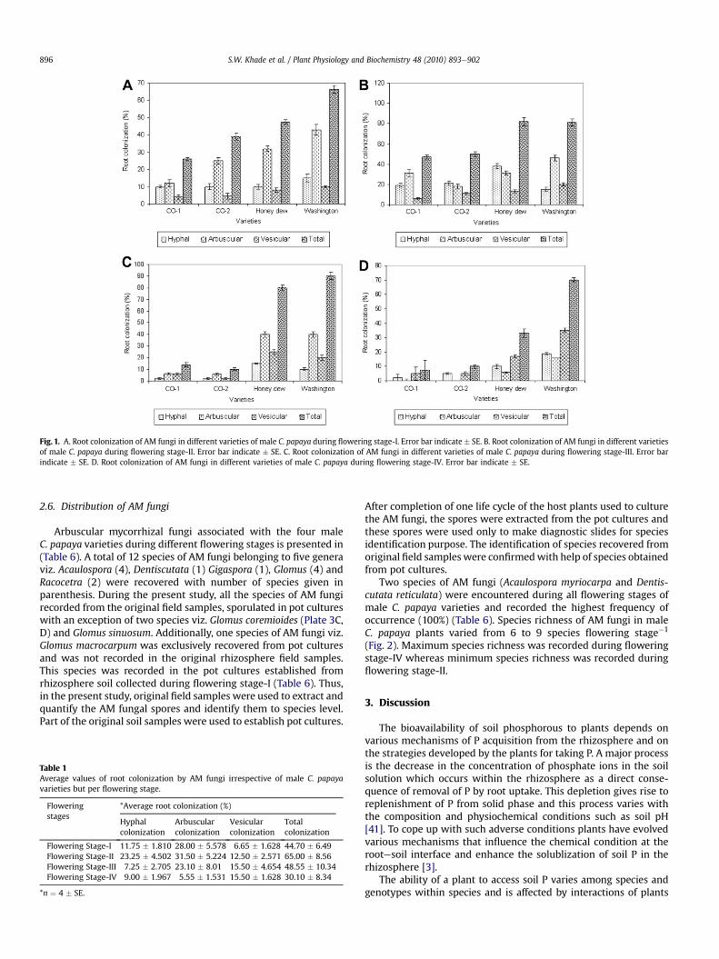
Fig. 1. A. Root colonization of AM fungi in different varieties of male C. papaya during flowering stage-I. Error bar indicate � SE. B. Root colonization of AM fungi in different varietiesof male C. papaya during flowering stage-II. Error bar indicate � SE. C. Root colonization of AM fungi in different varieties of male C. papaya during flowering stage-III. Error barindicate � SE. D. Root colonization of AM fungi in different varieties of male C. papaya during flowering stage-IV. Error bar indicate � SE.
S.W. Khade et al. / Plant Physiology and Biochemistry 48 (2010) 893e902896
2.6. Distribution of AM fungi
Arbuscular mycorrhizal fungi associated with the four maleC. papaya varieties during different flowering stages is presented in(Table 6). A total of 12 species of AM fungi belonging to five generaviz. Acaulospora (4), Dentiscutata (1) Gigaspora (1), Glomus (4) andRacocetra (2) were recovered with number of species given inparenthesis. During the present study, all the species of AM fungirecorded from the original field samples, sporulated in pot cultureswith an exception of two species viz. Glomus coremioides (Plate 3C,D) and Glomus sinuosum. Additionally, one species of AM fungi viz.Glomus macrocarpum was exclusively recovered from pot culturesand was not recorded in the original rhizosphere field samples.This species was recorded in the pot cultures established fromrhizosphere soil collected during flowering stage-I (Table 6). Thus,in the present study, original field samples were used to extract andquantify the AM fungal spores and identify them to species level.Part of the original soil samples were used to establish pot cultures.
Table 1Average values of root colonization by AM fungi irrespective of male C. papayavarieties but per flowering stage.
Floweringstages
*Average root colonization (%)
Hyphalcolonization
Arbuscularcolonization
Vesicularcolonization
Totalcolonization
Flowering Stage-I 11.75 � 1.810 28.00 � 5.578 6.65 � 1.628 44.70 � 6.49Flowering Stage-II 23.25 � 4.502 31.50 � 5.224 12.50 � 2.571 65.00 � 8.56Flowering Stage-III 7.25 � 2.705 23.10 � 8.01 15.50 � 4.654 48.55 � 10.34Flowering Stage-IV 9.00 � 1.967 5.55 � 1.531 15.50 � 1.628 30.10 � 8.34
*n ¼ 4 � SE.
After completion of one life cycle of the host plants used to culturethe AM fungi, the spores were extracted from the pot cultures andthese spores were used only to make diagnostic slides for speciesidentification purpose. The identification of species recovered fromoriginal field samples were confirmedwith help of species obtainedfrom pot cultures.
Two species of AM fungi (Acaulospora myriocarpa and Dentis-cutata reticulata) were encountered during all flowering stages ofmale C. papaya varieties and recorded the highest frequency ofoccurrence (100%) (Table 6). Species richness of AM fungi in maleC. papaya plants varied from 6 to 9 species flowering stage�1
(Fig. 2). Maximum species richness was recorded during floweringstage-IV whereas minimum species richness was recorded duringflowering stage-II.
3. Discussion
The bioavailability of soil phosphorous to plants depends onvarious mechanisms of P acquisition from the rhizosphere and onthe strategies developed by the plants for taking P. A major processis the decrease in the concentration of phosphate ions in the soilsolution which occurs within the rhizosphere as a direct conse-quence of removal of P by root uptake. This depletion gives rise toreplenishment of P from solid phase and this process varies withthe composition and physiochemical conditions such as soil pH[41]. To cope up with such adverse conditions plants have evolvedvarious mechanisms that influence the chemical condition at therootesoil interface and enhance the solublization of soil P in therhizosphere [3].
The ability of a plant to access soil P varies among species andgenotypes within species and is affected by interactions of plants
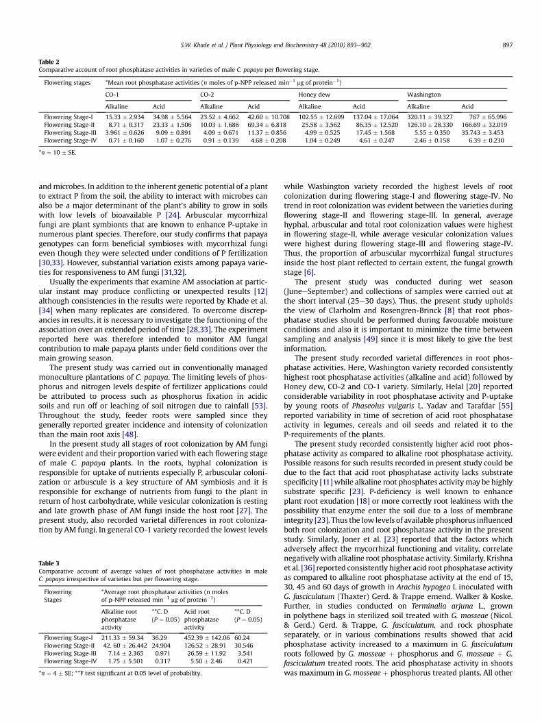
Table 2Comparative account of root phosphatase activities in varieties of male C. papaya per flowering stage.
Flowering stages *Mean root phosphatase activities (n moles of p-NPP released min�1 mg of protein�1)
CO-1 CO-2 Honey dew Washington
Alkaline Acid Alkaline Acid Alkaline Acid Alkaline Acid
Flowering Stage-I 15.33 � 2.934 34.98 � 5.564 23.52 � 4.662 42.60 � 10.708 102.55 � 12.699 137.04 � 17.064 320.11 � 39.327 767 � 65.996Flowering Stage-II 8.71 � 0.317 23.33 � 1.506 10.03 � 1.686 69.34 � 6.818 25.58 � 3.562 86.35 � 12.520 126.10 � 28.330 166.69 � 32.019Flowering Stage-III 3.961 � 0.626 9.09 � 0.891 4.09 � 0.671 11.37 � 0.856 4.99 � 0.525 17.45 � 1.568 5.55 � 0.350 35.743 � 3.453Flowering Stage-IV 0.71 � 0.160 1.07 � 0.276 0.91 � 0.139 4.68 � 0.208 1.04 � 0.249 4.61 � 0.247 2.46 � 0.158 6.39 � 0.230
*n ¼ 10 � SE.
S.W. Khade et al. / Plant Physiology and Biochemistry 48 (2010) 893e902 897
andmicrobes. In addition to the inherent genetic potential of a plantto extract P from the soil, the ability to interact with microbes canalso be a major determinant of the plant’s ability to grow in soilswith low levels of bioavailable P [24]. Arbuscular mycorrhizalfungi are plant symbionts that are known to enhance P-uptake innumerous plant species. Therefore, our study confirms that papayagenotypes can form beneficial symbioses with mycorrhizal fungieven though they were selected under conditions of P fertilization[30,33]. However, substantial variation exists among papaya varie-ties for responsiveness to AM fungi [31,32].
Usually the experiments that examine AM association at partic-ular instant may produce conflicting or unexpected results [12]although consistencies in the results were reported by Khade et al.[34] when many replicates are considered. To overcome discrep-ancies in results, it is necessary to investigate the functioning of theassociation over an extended period of time [28,33]. The experimentreported here was therefore intended to monitor AM fungalcontribution to male papaya plants under field conditions over themain growing season.
The present study was carried out in conventionally managedmonoculture plantations of C. papaya. The limiting levels of phos-phorus and nitrogen levels despite of fertilizer applications couldbe attributed to process such as phosphorus fixation in acidicsoils and run off or leaching of soil nitrogen due to rainfall [53].Throughout the study, feeder roots were sampled since theygenerally reported greater incidence and intensity of colonizationthan the main root axis [48].
In the present study all stages of root colonization by AM fungiwere evident and their proportion varied with each flowering stageof male C. papaya plants. In the roots, hyphal colonization isresponsible for uptake of nutrients especially P, arbuscular coloni-zation or arbuscule is a key structure of AM symbiosis and it isresponsible for exchange of nutrients from fungi to the plant inreturn of host carbohydrate, while vesicular colonization is restingand late growth phase of AM fungi inside the host root [27]. Thepresent study, also recorded varietal differences in root coloniza-tion by AM fungi. In general CO-1 variety recorded the lowest levels
Table 3Comparative account of average values of root phosphatase activities in maleC. papaya irrespective of varieties but per flowering stage.
FloweringStages
*Average root phosphatase activities (n molesof p-NPP released min�1 mg of protein�1)
Alkaline rootphosphataseactivity
**C. D(P ¼ 0.05)
Acid rootphosphataseactivity
**C. D(P ¼ 0.05)
Flowering Stage-I 211.33 � 59.34 36.29 452.39 � 142.06 60.24Flowering Stage-II 42. 60 � 26.442 24.904 126.52 � 28.91 30.546Flowering Stage-III 7.14 � 2.365 0.971 26.59 � 11.92 3.541Flowering Stage-IV 1.75 � 5.501 0.317 5.50 � 2.46 0.421
*n ¼ 4 � SE; **F test significant at 0.05 level of probability.
while Washington variety recorded the highest levels of rootcolonization during flowering stage-I and flowering stage-IV. Notrend in root colonizationwas evident between the varieties duringflowering stage-II and flowering stage-III. In general, averagehyphal, arbuscular and total root colonization values were highestin flowering stage-II, while average vesicular colonization valueswere highest during flowering stage-III and flowering stage-IV.Thus, the proportion of arbuscular mycorrhizal fungal structuresinside the host plant reflected to certain extent, the fungal growthstage [6].
The present study was conducted during wet season(JuneeSeptember) and collections of samples were carried out atthe short interval (25e30 days). Thus, the present study upholdsthe view of Clarholm and Rosengren-Brinck [8] that root phos-phatase studies should be performed during favourable moistureconditions and also it is important to minimize the time betweensampling and analysis [49] since it is most likely to give the bestinformation.
The present study recorded varietal differences in root phos-phatase activities. Here, Washington variety recorded consistentlyhighest root phosphatase activities (alkaline and acid) followed byHoney dew, CO-2 and CO-1 variety. Similarly, Helal [20] reportedconsiderable variability in root phosphatase activity and P-uptakeby young roots of Phaseolus vulgaris L. Yadav and Tarafdar [55]reported variability in time of secretion of acid root phosphataseactivity in legumes, cereals and oil seeds and related it to theP-requirements of the plants.
The present study recorded consistently higher acid root phos-phatase activity as compared to alkaline root phosphatase activity.Possible reasons for such results recorded in present study could bedue to the fact that acid root phosphatase activity lacks substratespecificity [11]while alkaline root phosphates activitymay be highlysubstrate specific [23]. P-deficiency is well known to enhanceplant root exudation [18] or more correctly root leakiness with thepossibility that enzyme enter the soil due to a loss of membraneintegrity [23]. Thus the low levels of available phosphorus influencedboth root colonization and root phosphatase activity in the presentstudy. Similarly, Joner et al. [23] reported that the factors whichadversely affect the mycorrhizal functioning and vitality, correlatenegativelywith alkaline root phosphatase activity. Similarly, Krishnaet al. [36] reported consistently higher acid root phosphatase activityas compared to alkaline root phosphatase activity at the end of 15,30, 45 and 60 days of growth in Arachis hypogea L inoculated withG. fasciculatum (Thaxter) Gerd. & Trappe emend. Walker & Koske.Further, in studies conducted on Terminalia arjuna L., grownin polythene bags in sterilized soil treated with G. mosseae (Nicol.& Gerd.) Gerd. & Trappe, G. fasciculatum, and rock phosphateseparately, or in various combinations results showed that acidphosphatase activity increased to a maximum in G. fasciculatumroots followed by G. mosseae þ phosphorus and G. mosseae þ G.fasciculatum treated roots. The acid phosphatase activity in shootswas maximum in G. mosseae þ phosphorus treated plants. All other
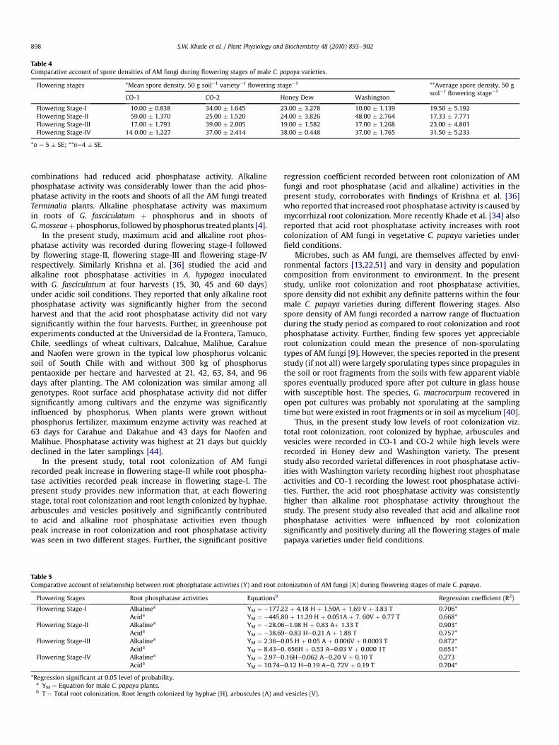
Table 4Comparative account of spore densities of AM fungi during flowering stages of male C. papaya varieties.
Flowering stages *Mean spore density. 50 g soil�1 variety�1flowering stage�1 **Average spore density. 50 g
soil�1flowering stage�1
CO-1 CO-2 Honey Dew Washington
Flowering Stage-I 10.00 � 0.838 34.00 � 1.645 23.00 � 3.278 10.00 � 1.139 19.50 � 5.192Flowering Stage-II 59.00 � 1.370 25.00 � 1.520 24.00 � 3.826 48.00 � 2.764 17.33 � 7.771Flowering Stage-III 17.00 � 1.793 39.00 � 2.005 19.00 � 1.582 17.00 � 1.268 23.00 � 4.801Flowering Stage-IV 14 0.00 � 1.227 37.00 � 2.414 38.00 � 0.448 37.00 � 1.765 31.50 � 5.233
*n ¼ 5 � SE; **n¼4 � SE.
S.W. Khade et al. / Plant Physiology and Biochemistry 48 (2010) 893e902898
combinations had reduced acid phosphatase activity. Alkalinephosphatase activity was considerably lower than the acid phos-phatase activity in the roots and shoots of all the AM fungi treatedTerminalia plants. Alkaline phosphatase activity was maximumin roots of G. fasciculatum þ phosphorus and in shoots ofG.mosseaeþ phosphorus, followed by phosphorus treated plants [4].
In the present study, maximum acid and alkaline root phos-phatase activity was recorded during flowering stage-I followedby flowering stage-II, flowering stage-III and flowering stage-IVrespectively. Similarly Krishna et al. [36] studied the acid andalkaline root phosphatase activities in A. hypogea inoculatedwith G. fasciculatum at four harvests (15, 30, 45 and 60 days)under acidic soil conditions. They reported that only alkaline rootphosphatase activity was significantly higher from the secondharvest and that the acid root phosphatase activity did not varysignificantly within the four harvests. Further, in greenhouse potexperiments conducted at the Universidad de la Frontera, Tamuco,Chile, seedlings of wheat cultivars, Dalcahue, Malihue, Carahueand Naofen were grown in the typical low phosphorus volcanicsoil of South Chile with and without 300 kg of phosphoruspentaoxide per hectare and harvested at 21, 42, 63, 84, and 96days after planting. The AM colonization was similar among allgenotypes. Root surface acid phosphatase activity did not differsignificantly among cultivars and the enzyme was significantlyinfluenced by phosphorus. When plants were grown withoutphosphorus fertilizer, maximum enzyme activity was reached at63 days for Carahue and Dakahue and 43 days for Naofen andMalihue. Phosphatase activity was highest at 21 days but quicklydeclined in the later samplings [44].
In the present study, total root colonization of AM fungirecorded peak increase in flowering stage-II while root phospha-tase activities recorded peak increase in flowering stage-I. Thepresent study provides new information that, at each floweringstage, total root colonization and root length colonized by hyphae,arbuscules and vesicles positively and significantly contributedto acid and alkaline root phosphatase activities even thoughpeak increase in root colonization and root phosphatase activitywas seen in two different stages. Further, the significant positive
Table 5Comparative account of relationship between root phosphatase activities (Y) and root co
Flowering Stages Root phosphatase activities Equationsb
Flowering Stage-I Alkalinea YM ¼ �177.2Acida YM ¼ �445.8
Flowering Stage-II Alkalinea YM ¼ �28.06Acida YM ¼ �38.69
Flowering Stage-III Alkalinea YM ¼ 2.36e0Acida YM ¼ 8.43e0
Flowering Stage-IV Alkalinea YM ¼ 2.97e0Acida YM ¼ 10.74e
*Regression significant at 0.05 level of probability.a YM ¼ Equation for male C. papaya plants.b T ¼ Total root colonization. Root length colonized by hyphae (H), arbuscules (A) and
regression coefficient recorded between root colonization of AMfungi and root phosphatase (acid and alkaline) activities in thepresent study, corroborates with findings of Krishna et al. [36]who reported that increased root phosphatase activity is caused bymycorrhizal root colonization. More recently Khade et al. [34] alsoreported that acid root phosphatase activity increases with rootcolonization of AM fungi in vegetative C. papaya varieties underfield conditions.
Microbes, such as AM fungi, are themselves affected by envi-ronmental factors [13,22,51] and vary in density and populationcomposition from environment to environment. In the presentstudy, unlike root colonization and root phosphatase activities,spore density did not exhibit any definite patterns within the fourmale C. papaya varieties during different flowering stages. Alsospore density of AM fungi recorded a narrow range of fluctuationduring the study period as compared to root colonization and rootphosphatase activity. Further, finding few spores yet appreciableroot colonization could mean the presence of non-sporulatingtypes of AM fungi [9]. However, the species reported in the presentstudy (if not all) were largely sporulating types since propagules inthe soil or root fragments from the soils with few apparent viablespores eventually produced spore after pot culture in glass housewith susceptible host. The species, G. macrocarpum recovered inopen pot cultures was probably not sporulating at the samplingtime but were existed in root fragments or in soil as mycelium [40].
Thus, in the present study low levels of root colonization viz.total root colonization, root colonized by hyphae, arbuscules andvesicles were recorded in CO-1 and CO-2 while high levels wererecorded in Honey dew and Washington variety. The presentstudy also recorded varietal differences in root phosphatase activ-ities with Washington variety recording highest root phosphataseactivities and CO-1 recording the lowest root phosphatase activi-ties. Further, the acid root phosphatase activity was consistentlyhigher than alkaline root phosphatase activity throughout thestudy. The present study also revealed that acid and alkaline rootphosphatase activities were influenced by root colonizationsignificantly and positively during all the flowering stages of malepapaya varieties under field conditions.
lonization of AM fungi (X) during flowering stages of male C. papaya.
Regression coefficient (R2)
2 þ 4.18 H þ 1.50A þ 1.69 V þ 3.83 T 0.706*0 þ 11.29 H þ 0.051A þ 7. 60V þ 0.77 T 0.668*e1.98 H þ 0.83 Aþ 1.33 T 0.903*e0.83 He0.21 A þ 1.88 T 0.757*.05 H þ 0.05 A þ 0.006V þ 0.0003 T 0.872*. 656H þ 0.53 Ae0.03 V þ 0.000 1T 0.651*.16He0.062 A�0.20 V þ 0.10 T 0.2730.12 He0.19 Ae0. 72V þ 0.19 T 0.704*
vesicles (V).
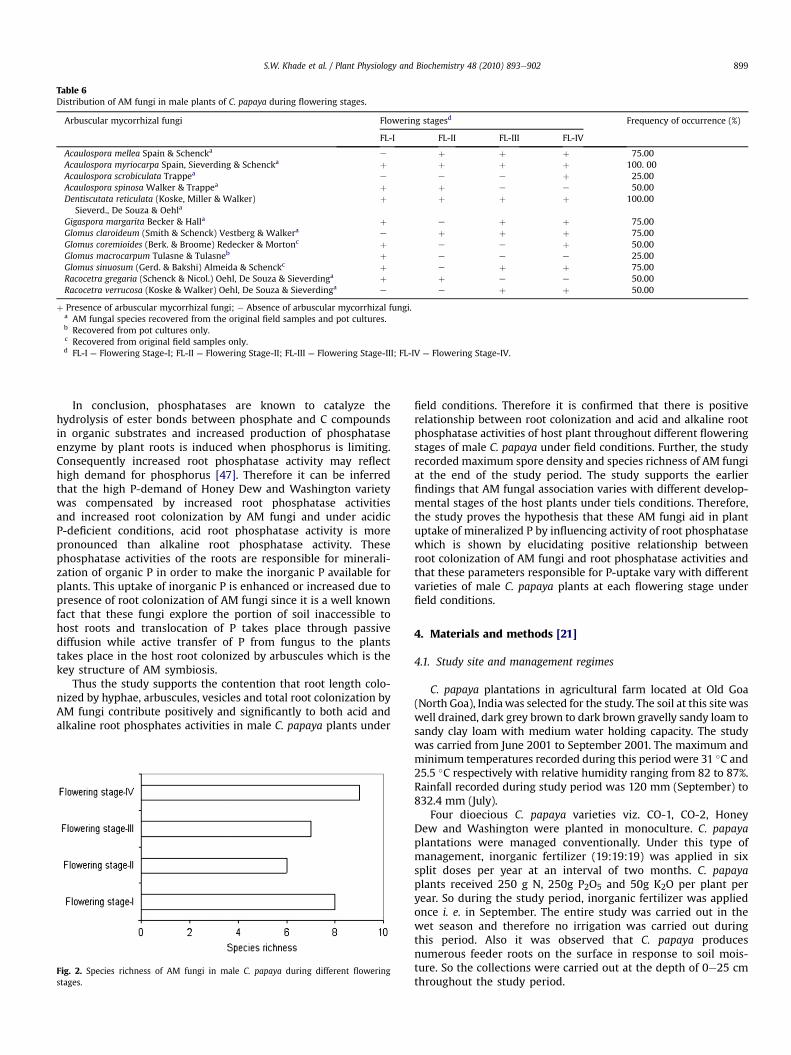
Table 6Distribution of AM fungi in male plants of C. papaya during flowering stages.
Arbuscular mycorrhizal fungi Flowering stagesd Frequency of occurrence (%)
FL-I FL-II FL-III FL-IV
Acaulospora mellea Spain & Schencka e þ þ þ 75.00Acaulospora myriocarpa Spain, Sieverding & Schencka þ þ þ þ 100. 00Acaulospora scrobiculata Trappea e e e þ 25.00Acaulospora spinosa Walker & Trappea þ þ e e 50.00Dentiscutata reticulata (Koske, Miller & Walker)
Sieverd., De Souza & Oehlaþ þ þ þ 100.00
Gigaspora margarita Becker & Halla þ e þ þ 75.00Glomus claroideum (Smith & Schenck) Vestberg & Walkera e þ þ þ 75.00Glomus coremioides (Berk. & Broome) Redecker & Mortonc þ e e þ 50.00Glomus macrocarpum Tulasne & Tulasneb þ e e e 25.00Glomus sinuosum (Gerd. & Bakshi) Almeida & Schenckc þ e þ þ 75.00Racocetra gregaria (Schenck & Nicol.) Oehl, De Souza & Sieverdinga þ þ e e 50.00Racocetra verrucosa (Koske & Walker) Oehl, De Souza & Sieverdinga e e þ þ 50.00
þ Presence of arbuscular mycorrhizal fungi; � Absence of arbuscular mycorrhizal fungi.a AM fungal species recovered from the original field samples and pot cultures.b Recovered from pot cultures only.c Recovered from original field samples only.d FL-I ¼ Flowering Stage-I; FL-II ¼ Flowering Stage-II; FL-III ¼ Flowering Stage-III; FL-IV ¼ Flowering Stage-IV.
S.W. Khade et al. / Plant Physiology and Biochemistry 48 (2010) 893e902 899
In conclusion, phosphatases are known to catalyze thehydrolysis of ester bonds between phosphate and C compoundsin organic substrates and increased production of phosphataseenzyme by plant roots is induced when phosphorus is limiting.Consequently increased root phosphatase activity may reflecthigh demand for phosphorus [47]. Therefore it can be inferredthat the high P-demand of Honey Dew and Washington varietywas compensated by increased root phosphatase activitiesand increased root colonization by AM fungi and under acidicP-deficient conditions, acid root phosphatase activity is morepronounced than alkaline root phosphatase activity. Thesephosphatase activities of the roots are responsible for minerali-zation of organic P in order to make the inorganic P available forplants. This uptake of inorganic P is enhanced or increased due topresence of root colonization of AM fungi since it is a well knownfact that these fungi explore the portion of soil inaccessible tohost roots and translocation of P takes place through passivediffusion while active transfer of P from fungus to the plantstakes place in the host root colonized by arbuscules which is thekey structure of AM symbiosis.
Thus the study supports the contention that root length colo-nized by hyphae, arbuscules, vesicles and total root colonization byAM fungi contribute positively and significantly to both acid andalkaline root phosphates activities in male C. papaya plants under
Fig. 2. Species richness of AM fungi in male C. papaya during different floweringstages.
field conditions. Therefore it is confirmed that there is positiverelationship between root colonization and acid and alkaline rootphosphatase activities of host plant throughout different floweringstages of male C. papaya under field conditions. Further, the studyrecorded maximum spore density and species richness of AM fungiat the end of the study period. The study supports the earlierfindings that AM fungal association varies with different develop-mental stages of the host plants under tiels conditions. Therefore,the study proves the hypothesis that these AM fungi aid in plantuptake of mineralized P by influencing activity of root phosphatasewhich is shown by elucidating positive relationship betweenroot colonization of AM fungi and root phosphatase activities andthat these parameters responsible for P-uptake vary with differentvarieties of male C. papaya plants at each flowering stage underfield conditions.
4. Materials and methods [21]
4.1. Study site and management regimes
C. papaya plantations in agricultural farm located at Old Goa(North Goa), Indiawas selected for the study. The soil at this site waswell drained, dark grey brown to dark brown gravelly sandy loam tosandy clay loam with medium water holding capacity. The studywas carried from June 2001 to September 2001. The maximum andminimum temperatures recorded during this period were 31 �C and25.5 �C respectively with relative humidity ranging from 82 to 87%.Rainfall recorded during study period was 120 mm (September) to832.4 mm (July).
Four dioecious C. papaya varieties viz. CO-1, CO-2, HoneyDew and Washington were planted in monoculture. C. papayaplantations were managed conventionally. Under this type ofmanagement, inorganic fertilizer (19:19:19) was applied in sixsplit doses per year at an interval of two months. C. papayaplants received 250 g N, 250g P2O5 and 50g K2O per plant peryear. So during the study period, inorganic fertilizer was appliedonce i. e. in September. The entire study was carried out in thewet season and therefore no irrigation was carried out duringthis period. Also it was observed that C. papaya producesnumerous feeder roots on the surface in response to soil mois-ture. So the collections were carried out at the depth of 0e25 cmthroughout the study period.
S.W. Khade et al. / Plant Physiology and Biochemistry 48 (2010) 893e902900
4.2. Collection of samples
In the present study, C. papaya seedlings were raised from seedsof four dioecious viz. CO-1, CO-2, Honey dew and Washington inthe nursery for two months and than they were transplanted in thefield. The early vegetative stage (two-four months old plants)mainly comprises of tap root system with feeble primary branchesmaking it difficult to collect root (feeder roots) samples to carry outmycorrhizal studies. The differences in male plants and femaleplants can be observed only during flowering stages when theplants were seven months old plants and exhibited extensive rootsystem with ample of feeder roots available for collection. Gener-ally, the male plants are maintained in the field for pollinationpurpose and at the end of growing season they are destroyed. Sothe present studywas carried out during flowering stages when themale C. papaya plants were seven (June) to ten (September) monthsold.
In the present study, sampling was carried out four times atshort intervals when the C. papaya male plants were in the flow-ering stages viz., last week of June (Fl-I), last week of July (Fl-II), midAugust (Fl-III) and mid September (Fl-IV). Sampling procedureswere carried out according to Tews and Koske [54] except that thecore size was bigger (15 cm in diameter).
Two healthy plants variety�1flowering stage�1 were
randomly selected for the collection of rhizosphere soil and rootsamples. For each plant, three random soil cores were collectedfrom within 60 cm of each plant, each at the depth of 0e25 cm.Immediately, samples collected from both plants were thencombined to give approximately 400g moist soil after thoroughmixing in laboratory. From this, 250 g air-dried soil wasemployed for extraction of AM fungal spores, 100 g was utilizedfor nutrient analysis and 50 g soil was utilized for setting openpot cultures. From each composite sample, five sub-sampleswere made for quantification of spore density, two sub-samplesfor nutrient analysis and two sub-samples for establishment ofpot cultures.
Roots were packed separately for assay of phosphatases and forestimation of root colonization plant�1 variety�1
flowering stage�1
in the field. Root samples collected for assay of phosphatases, weretransported in ice to the laboratory. These root samples (feederroots)werewashed in ice coldwater and rinsedwith double distilledwater, packed in polyethylene bags, labeled and stored at �80 C.For estimation of root colonization and establishment of potcultures, the root samples of both the plants were combined. Fromthis composite sample,five sub-sampleswere utilized for estimationof degree of root colonization and two sub-samples were utilized forpot cultures.
4.3. Establishment of pot cultures
Baiting of native AM fungi were carried out by using openpot cultures [16]. Two pot cultures were maintainedvariety�1
flowering stage�1. For establishing single pot culture,25 g of rhizosphere soil along with root bits of C. papaya wasmixed with equal quantity of sterilized sand and placed in12.5 cm diameter pots. Seeds of Eleusine coracana (L.) Gartnerwere sterilized with 0.1% HgCl2 washed thoroughly with distilledwater and placed over the soil sand mixture and covered with2 cm of soil. Cuttings of Coleus sp. were also used as host plantsfor baiting the native AM fungi. The plants were maintainedunder glass house conditions and watered adequately. The rootsof host species were checked for AM colonization after 45 dayssince, this stage represents the vesicular colonization by AMfungi which is easily detectable after root staining procedure.Pots showing successful mycorrhization were maintained for
period of six months and application of water was reduced atfinal three weeks to maximize spore production. While otherpots which lacked AM colonization were rejected. At the endof six months the plants were cut near the base, the growingsubstrate (cultures) were air-dried and checked for the presenceof spores. Spores isolated from pot cultures were used to makediagnostic slides only. Further, these slides were used for veri-fying the identification of AM fungi recovered from original fieldsamples.
4.4. Soil sample analysis
Soil samples were analyzed for 11 edaphic factors. Soil pHwas measured in 1:2 soil water suspension using pH meter(LI 120 Elico, India). Electrical conductivity was measured atroom temperature in 1:5 soil suspension using Conductivitymeter (CM-180 Elico, India). Standard soil analysis techniques viz.micro-Kjeldahl method [21] and Bray and Kurtz [7] methodwere employed for determination of total nitrogen and availablephosphorus respectively. Available potassium was estimatedby ammonium acetate method [19] using Flame Photometer(Systronic 3292). Available zinc, copper, manganese and ironwere quantified by DTPAeCaCl2eTEA method [37] using AtomicAbsorption Spectrophotometer (AAS 4139, Electronic Corporationof India Ltd, India).
4.5. Estimation of root colonization by AM fungi
Roots were cleared in 10% KOH, acidified in 1N HCL andstained in 0.05% trypan blue in lactoglycerol [43]. Total rootcolonization and length of the root colonized by hyphae, arbus-cules and vesicles by AM fungi were estimated by of modificationof grid intersection method [39] and slide method [17]. Tertiaryroots of 1 cm length were randomly selected for estimationof root colonization. Stained root bits were placed on the micro-scopic slides and the field of the view was moved across the slide.The hairline graticule inserted in the eyepiece acted as a line ofintersection. At each intersection, presence of hyphae, arbusculeand/or vesicle was noted. Hundred and fifty intersections wereexamined per samples under compound microscope at 200�magnifications and root colonization of AM fungi was expressedin percentage [34].
4.6. Assay of root phosphatase enzyme activity
This experiment comprised of two sets of observations foreach type of assay variety�1
flowering stage�1. Thus, the studycomprised of two repetitions�1 variety�1
flowering stage andeach repetition had five replications. One gram of root tissue wasweighed for each repetition. Activities were assayed separatelyfor acid phosphatase and alkaline phosphatase. In all 40 readingseach were recorded for acid and alkaline phosphatase separatelyflowering stage�1. In total 160 readings were recorded for eachacid and alkaline root phosphatase activity separately for fourflowering stages of male C. papaya plants. Methods for quanti-fication of root phosphatase activities were standardized forC. papaya based on procedures provided by Kapoor et al. [25]and Sukhada [50].
4.7. Extraction of enzyme
Enzymewas extracted by macerating 1 g of detached root tissueat 4 �C using 20 ml of phosphate (0.1 M KH2 PO4, pH 6.6) buffer. Thehomogenate was filtered through muslin cloth and the filtrate wascentrifuged at 5000 revmin�1 for 15 min using a cooling centrifuge
S.W. Khade et al. / Plant Physiology and Biochemistry 48 (2010) 893e902 901
(Remy C-24, India). The supernatant was stored at 4 �C and furtheremployed for protein estimation of enzyme and assay of rootphosphatases [34].
Frequency ð%Þ ¼ Number of soil sample that possess spores of particular speciesTotal number of soil samples analyzed:
� 100
4.8. Protein estimation
Protein content of the enzyme extract was estimated by Lowryet al. [38] method using bovine serum albumin as standard.
4.9. Assay of root phosphatases
For alkaline root phosphatase activity, 5 mg protein equiva-lent enzyme extract was incubated with 2 ml of 15 mMp-nitrophenyl phosphate (p-NPP) and 0.8 ml 0.25 M Tris HCl(pH 9.8). For acid root phosphatase activity, 5 mg proteinequivalent enzyme extract was incubated with 2 ml of 15 mMp-nitrophenyl phosphate (p-NPP) and 0.8 ml 0.25 M Sodiumacetate (pH 6.0) [34].
The reactions in both the above mentioned cases wereterminated by adding 2 ml of 1N NaOH. For each repetition, T00(zero time of incubation) and T030 (30 min after incubation)were taken separately for alkaline and acid root phosphataseactivity. Optical density (O.D) was read at 410 hm using uv-1201Shimadzu spectrophotometer (Japan) against a blank solvent(distilled water). Root phosphatase activity was expressed interms of n moles of p-nitrophenol released�1 min�1 mg protein[34].
4.10. Quantification of spore density of AM fungi
Only, spores and sporocarps of AM fungi from original rhizo-spheric field samples were isolated by wet sieving and decantingmethod [15] and quantification of spore density of AM fungi wascarried out using method described by using Gaur and Adholeya[14]. Here, only healthy spores, which generally comprised of 75% ofextracted spores, were used for quantification of spore density ofAM fungi.
4.11. Identification of AM fungi
Diagnostic slides containing intact and crushed spores andsporocarps of AM fungi were prepared in polyvinyl alcohol lac-toglycerol (PVLG) [35]. Spore morphology and wall characteris-tics were considered for the identification of AM fungi and thesecharacteristics were ascertained using compound microscope,Leica WILD MP3 and Nikon E 800. The AM fungi were identifiedto species level by using Manual of Identification ArbuscularMycorrhizal Fungi by Schenck and Perez [46]. Taxonomic iden-tification of spores was also carried out by matching thedescriptions provided by International Collection of VesicularArbuscular Mycorrhizal fungi (http://invam.caf.wvu.edu) andOehl et al. [42].
4.12. Diversity indices
Species richness is the mean number of AM fungal speciesrecovered at any particular sampling period [2].
4.13. Frequency of occurrence
Frequency of occurrence of AM fungi was calculated using thefollowing formula [2].
4.14. Statistical analysis
Data on root colonization and spore density of AM fungi androot phosphatase activity was subjected to analysis of variance inorder to investigate the variations in these parameters with respectto four varieties of male C. papaya at each flowering stage. Priorto analysis of variance, root colonization values were subjectedto arcsine transformations. Additionally, multiple linear regressionanalysis was carried out to determine the relationship betweenroot colonization and root phosphatase activity at each floweringstage. Data was statistically analyzed using mstac package andWASP 1.0. Standard error was calculated for each parameterundertaken for the study.
Acknowledgements
Shri. Waman M. Khade, Ex- director of Agriculture Departmentand Directorate of Agriculture, State Government of Goa arethanked for their assistance to carry out research work.
References
[1] R. Azcón, F. Borie, J.M. Barea, Exocellular acid phosphatase activity of lavenderand wheat roots as affected by phytate and mycorrhizal inoculation. LesMycorrhizae: Biologie et Utilisation. INRA Publication (Les Colloques de 1’INRA,no13), Dijon, 1982, pp. 5e6.
[2] K.R. Beena, N.S. Raviraja, A.D. Arun, K.R. Sridhar, Diversity of arbuscularmycorrhizal fungi on coastal sand dunes of the West Coast of India, Curr. Sci.79 (10) (2000) 1459e1465.
[3] I. Bertrand, P. Hinsinger, B. Jaillard, J.C. Arrieu, Dynamics of phosphorus in therhizosphere of maize and rape grown on synthetic, phosphated calcite andgoethite, Plant Soil 211 (1999) 111e119.
[4] B. Bhadraiah, V.V. Kankadurga, P. Ramarao, C. Manoharachary, Effect of VAMfungi and rock phosphate on phosphatase activities in Terminalia arjuna,Physiol. Biochem. (1999) 5e7 National Conference on Mycorrhiza. Section 3(Poster).
[5] N.S. Bolan, A critical review on the role of mycorrhizal fungi in the uptake ofphosphorus by plants, Plant Soil 134 (1991) 189e207.
[6] P. Bonfante-Fasalo, Anatomy and morphology. in: C.L. Powell, D.J. Bagyaraj(Eds.), VA Mycorrhiza. CRC Press Inc, Boca Raton: Florida, 1984, pp. 5e33.
[7] R.H. Bray, L.T. Kurtz, Determination of total organic carbon and available formsof phosphorus in soils, Soil Sci. 59 (1945) 39e45.
[8] M. Clarholm, U. Rosengren-Brinck, Phosphorous and nitrogen fertilization ofa Norway spruce forest- effect on needle concentrations and acid phosphataseactivity in the humus layer, Plant Soil 175 (1995) 239e249.
[9] E.A. Davis, J.L. Young, S.L. Rose, Detection of high-phosphorous tolerantVAM-fungi colonizing hops and peppermint, Plant Soil 81 (1984) 29e36.
[10] D.D. Douds Jr., L. Galvez, M. Frankesnyder, C. Reider, L.E. Drinkwater, Effect ofcompost addition and crop rotation point upon vesicular-arbuscular mycor-rhizal fungi, Agric. Ecosyst. Environ. 65 (1997) 257e266.
[11] S.M.G. Duff, G. Sarath, W.C. Plaxton, The role of acid phosphatase in plantphosphorus metabolism, Physiol. Plant 90 (1994) 791e800.
[12] M.J. Dunne, A.H. Fitter, The phosphorous budget of a field-grown strawberry(Fragaria � ananassa cv. Hapil) crop: evidence for a mycorrhizal contribution,Ass. Appl. Biol. 114 (1989) 185e193.
[13] J.R. Ellis, Post flood syndrome and vesicular-arbuscular mycorrhizal fungi,J. Prod. Agric. 11 (1998) 200e204.
[14] A. Gaur, A. Adholeya, Estimation of VAM spores in the soil e a modifiedmethod, Mycorrhiza News 6 (1994) 10e11.
[15] J.W. Gerdemann, T.H. Nicolson, Spore density of Endogone species extractedfrom soil wet sieving and decanting, Trans. Brit. Mycol. Soc. 46 (1963) 235e244.
S.W. Khade et al. / Plant Physiology and Biochemistry 48 (2010) 893e902902
[16] A.E. Gilmore, Phycomycetous mycorrhizal organisms collected by open potcultures, Hilgardia 39 (1968) 87e105.
[17] M. Giovannetti, B. Mosse, An evaluation of techniques for measuring vesic-ular arbuscular mycorrhizal infection in roots, New Phytol. 84 (1980)489e500.
[18] H. Graham, R.T. Leonard, J.A. Menge, Membrane mediated decrease in rootexudation responsible for phosphorous inhibition of vesicularearbuscularmycorrhiza formation, Plant Physiol. 68 (1981) 549e552.
[19] J.J. Hanway, H. Heidal, Soil analysis method as used in Iowa State College soiltesting laboratory, Iowa Agri. 57 (1952) 1e31.
[20] H.M. Helal, Varietals difference in the root phosphatase activity as related tothe utilization of organic phosphates, Plant Soil 123 (1990) 161e163.
[21] M.L. Jackson, Soil Chemical Analysis. Prentice Hall, New Delhi, 1971.[22] N.C. Johnson, F.L. Pfleger, Vesicular-arbuscular mycorrhizae and cultural
stresses. in: G.J. Bethlenfalvay, R.G. Linderman (Eds.), Mycorrhizae inSustainable Agriculture. ASA, CSSA, and SSSA, Madison, WI, 1992, pp. 71e99ASA Spec. Publ. 54.
[23] E.J. Joner, I.M. Van Aarle, M. Vosatka, Phosphatase activity of extra- radicalarbuscular mycorrhizal hyphae: a review, Plant Soil 226 (2000) 199e210.
[24] S.M. Kaeppler, J.L. Parke, S.M. Mueller, L. Senior, C. Stuber, W.F. Tracy, Varia-tion among maize inbred lines and detection of quantitative trait loci forgrowth at low phosphorus and responsiveness to arbuscular mycorrhizalfungi, Crop Sci. 40 (2000) 358e364.
[25] A. Kapoor, V.P. Singh, K.G. Mukerji, Studies on the phosphatase of mycor-rhizal and non mycorrhizal Trigonella roots. in: A. Mahadeva, A. Raman,K. Natarajan (Eds.), Mycorrhizae for Green Asia. CAS, Madras: India, 1989,pp. pp.125e127.
[26] S.W. Khade, Studies on arbuscularmycorrhizal status of Carica papaya L. in agro-based ecosystem of Goa, Ph.D thesis, Goa University, India, 2003, pp. 1e235.
[27] S.W. Khade, A. Adholeya, Arbuscular mycorrhizae in natural ecosystem:incentives in diversity and ecology. in: T. Satyanarayana, B.N. Johri (Eds.),Current Perspectives and Potential Applications. I. K International Pvt. Ltd.,New Delhi: India, 2005, pp. 225e258.
[28] S.W. Khade, B.F. Rodrigues, Ecology of arbuscular mycorrhizal fungi associatedwith Carica papaya L. in agro-based ecosystem of Goa, India, Trop. Subtrop.Agroecosys. 8 (2008) 265e278.
[29] S.W. Khade, B.F. Rodrigues, Spatial variations in arbuscular mycorrhizal (AM)fungi associated with Carica papaya L. in a tropical agro-based ecosystem, Biol.Agri. Horti. 26 (2008) 149e174.
[30] S.W. Khade, B.F. Rodrigues, Arbuscular mycorrhizal fungi associated withvarieties of Carica papaya L. in tropical agro-based ecosystem of Goa, India,Trop. Subtrop. Agroecosys. 10 (2009) 369e381.
[31] S.W. Khade, B.F. Rodrigues, Studies on arbuscular mycorrhisation of papaya,Afr. Crop Sci. J. 17 (2009) 155e165.
[32] S.W. Khade, B.F. Rodrigues, Studies on effects of arbuscular mycorrhizal (AM)fungi on mineral nutrition of Carica papaya L, Not. Bot. Hort. Agrobot. Cluj 37(2009) 183e186.
[33] S.W. Khade, B.F. Rodrigues, Spatio-temporal variations of arbuscular mycor-rhizal (AM) fungi associated with Carica papaya L. in agro-based ecosystem ofGoa, India, Arch. Agron. Soil Sci. 56 (2010) 237e263.
[34] S.W. Khade, B.F. Rodrigues, P.K. Sharma, Arbuscular mycorrhizal status androot phosphatase activities in vegetative Carica papaya L. varieties, ActaPhysiol. Plant 32 (2010) 565e574.
[35] R.E. Koske, B. Tessier, A convenient permanent slide mounting medium,Mycol. Soc. Am. News Let 34 (1983) 59.
[36] K.R. Krishna, D.J. Bagyaraj, K.G. Papavinashasundaram, Acid and alkalinephosphatase activities in mycorrhizal and uninfected roots of Arachis hypo-gaea L, Ann. Bot. 51 (1983) 551e553.
[37] W.L. Lindsay, W.A. Norvel, Development of DPTA soil test for zinc, iron,manganese and copper, Soil Sci. Soc. Am. J. 42 (1978) 421e488.
[38] O.H. Lowry, N.J. Rosebrough, A.L. Fan, R.J. Randall, Protein measurement withthe FolinePhenol reagent, J. Biol. Chem. 193 (1951) 265e275.
[39] T.P. Mcgonigle, M.H. Miller, D.G. Evans, G.L. Fairchild, J.A. Swan, A newmethodwhich gives an objective measure of colonization of roots by vesicular-arbuscular mycorrhizal fungi, New Phytol. 115 (1990) 495e501.
[40] D.D. Miller, P.A. Domoto, C. Walkar, Colonization and efficacy of differentendomycorrhyzal fungi with apple seedlings at two phosphorus levels, NewPhytol. 100 (1985) 393e402.
[41] C. Morel, P. Hinsinger, Root inducedmodifications of the exchange of phosphateion between soil solution and soil solid phase, Plant Soil 211 (1999) 103e110.
[42] F. Oehl, F.A. de Souza, E. Sieverding, Revision of Scutellospora and descriptionof five new genera and three new families in the arbuscular mycorrhiza-forming Glomeromycetes, Mycotaxon 106 (2008) 311e360.
[43] J.M. Phillips, D.S. Hayman, Improved procedure for clearing roots and stainingof mycorrhizal fungi for rapid assessment of infection, Trans. Brit. Mycol. Soc.55 (1970) 158e161.
[44] R. Rubio, E. Moraga, F. Borie, Acid phosphatase activity and vesicular-arbus-cular mycorrhizal infection associated with roots of 4 wheat cultivars, J. PlantNut. 13 (1990) 585e598.
[45] F.E. Sanders, P.B. Tinker, Mechanism of absorption of phosphate from modelsubstrates and P compounds in soil by Endogone mycorrhizas, Nature 133(1971) 278e279.
[46] N.C. Schenck, Y. Perez, Manual for Identification of VA Mycorrhizal Fungi.INVAM, University of Florida, Gainesville: USA, 1990, pp. 1e283.
[47] K. Schneider, M.B. Turrion, P.F. Grierson, J.F. Gallardo, Phosphatase activity,microbial phosphorous and fine root growth in forest soils in the Sierra deGata, West Central Spain, Biol. Fertil. Soils 34 (2001) 151e155.
[48] S.E. Smith, N.A. Walker, A qualitative study of mycorrhizal plants in Trifolium:separate determination of the rates of infection and of mycelial growth, NewPhytol. 89 (1981) 225e240.
[49] T.W. Speir, D.J. Ross, Effect of storage on the activity of protease, urease,phosphatase and sulphates in the three soils under pasture. New Zeal, J. Sci.18 (1975) 231e237.
[50] M. Sukhada, Effect of VAM inoculation on plant growth, nutrient level androot phosphatase activity in papaya (Carica papaya cv. Coorg Honey Dew),Fert. Res. 31 (1992) 263e267.
[51] D.M. Sylvia, S.E. Williams, Vesicular-arbuscular mycorrhizae and environ-mental stresses. in: G.J. Bethlenfalvay, R.G. Linderman (Eds.), Mycorrhizae inSustainable Agriculture. ASA, CSSA, and SSSA, Madison, WI, 1992, pp.101e124 ASA Spec. Publ. 54.
[52] T. Tadano, K. Ozawa, H. Sakai, M. Oaski, H. Matsui, Secretion of acid phosphataseby the roots of crop plants under phosphorous deficient conditions and someproperties of the enzyme secreted by lupin roots, Plant Soil 156 (1993) 95e98.
[53] H.S.L. Tandon, Fertilizer Guide, second ed. Fertilizer Development andConsultation Organization, New Delhi, 1994, pp. 156.
[54] L.L. Tews, R.E. Koske, Towards a sampling strategy for vesicular arbuscularmycorrhizas, Trans. Brit. Mycol. Soc. 87 (1986) 353e358.
[55] R.S. Yadav, J.C. Tarafdar, Influence of organic and inorganic phosphoroussupply on the maximum secretion of acid phosphatase by plants, Biol. Fertil.Soils 34 (2001) 140e143.
Copyright © 2022 FDOKUMEN