Septoglomus fuscum and S. furcatum, two new species of arbuscular mycorrhizal fungi (Glomeromycota)
11
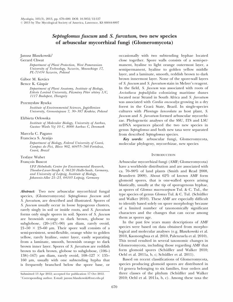
Septoglomus fuscum and S. furcatum, two new species of arbuscular mycorrhizal fungi (Glomeromycota) Janusz Blaszkowski 1 Gerard Chwat Department of Plant Protection, West Pomeranian University of Technology, Szczecin, Slowackiego 17, PL-71434 Szczecin, Poland Ga ´bor M. Kova ´cs Bence K. Ga ´spa ´r Department of Plant Anatomy, Institute of Biology, Eo ¨tvo ¨s Lora ´nd University, Pa ´zma ´ny Pe ´ter se ´ta ´ny 1/C, 1117 Budapest, Hungary Przemyslaw Ryszka Institute of Environmental Sciences, Jagiellonian University, Gronostajowa 7, 30–387 Krako ´w, Poland Elz _bieta Orlowska Institute of Molecular Biology, University of Aarhus, Gustav Wieds Vej 10 C, 8000 Aarhus C, Denmark Marcela C. Pagano Francisca S. Arau ´jo Department of Biology, Federal University of Ceara ´, Campus do Pici, Bloco 902, 60455–760 Fortaleza, Ceara ´, Brazil Tesfaye Wubet Franc ¸ois Buscot UFZ Helmholtz Centre for Environmental Research, Theodor-Lieser-Straße 4, 06120 Halle-Saale, Germany, and University of Leipzig, Institute of Biology, Johannis-Allee 21–23, 04103 Leipzig, Germany Abstract: Two new arbuscular mycorrhizal fungal species, (Glomeromycota) Septoglomus fuscum and S. furcatum, are described and illustrated. Spores of S. fuscum usually occur in loose hypogeous clusters, rarely singly in soil or inside roots, and S. furcatum forms only single spores in soil. Spores of S. fuscum are brownish orange to dark brown, globose to subglobose, (20–)47(–90) mm diam, rarely ovoid, 21–50 3 23–60 mm. Their spore wall consists of a semi-persistent, semi-flexible, orange white to golden yellow, rarely hyaline, outer layer, easily separating from a laminate, smooth, brownish orange to dark brown inner layer. Spores of S. furcatum are reddish brown to dark brown, globose to subglobose, (106–) 138(–167) mm diam, rarely ovoid, 108–127 3 135– 160 mm, usually with one subtending hypha that is frequently branched below the spore base, or occasionally with two subtending hyphae located close together. Spore walls consists of a semi-per- manent, hyaline to light orange outermost layer, a semipermanent, hyaline to golden yellow middle layer, and a laminate, smooth, reddish brown to dark brown innermost layer. None of the spore-wall layers of S. fuscum and S. furcatum stain in Melzer’s reagent. In the field, S. fuscum was associated with roots of Arctotheca populifolia colonizing maritime dunes located near Strand in South Africa and S. furcatum was associated with Cordia oncocalyx growing in a dry forest in the Ceara ´ State, Brazil. In single-species cultures with Plantago lanceolata as host plant, S. fuscum and S. furcatum formed arbuscular mycorrhi- zae. Phylogenetic analyses of the SSU, ITS and LSU nrDNA sequences placed the two new species in genus Septoglomus and both new taxa were separated from described Septoglomus species. Key words: arbuscular fungi, Glomeromycota, molecular phylogeny, mycorrhizae, new species INTRODUCTION Arbuscular mycorrhizal fungi (AMF; Glomeromycota) have a worldwide distribution and are associated with ca. 70–90% of land plants (Smith and Read 2008, Brundrett 2009). About 62% of known AMF form glomoid spores, that is one-walled spores arising blastically, usually at the tip of sporogenous hyphae, as spores of Glomus macrocarpum Tul. & C. Tul., the type species of genus Glomus Tul. & C. Tul. (Schu ¨ ßler and Walker 2010). These AMF are especially difficult to identify based solely on spore morphology because of a limited number of taxonomically significant characters and the changes that can occur among them as spores age. In the past few years many descriptions of AMF species were based on data obtained from morpho- logical and molecular analyses (e.g. Blaszkowski et al. 2010, Kaonongbua et al. 2010, Palenzuela et al. 2010). This trend resulted in several taxonomic changes in Glomeromycota, including those regarding AMF that form glomoid spores (Schu ¨ ßler and Walker 2010; Oehl et al. 2011a, b, c; Schu ¨ßler et al. 2011). Based on recent classifications of Glomeromycota, species producing glomoid spores are distributed in 14 genera belonging to six families, four orders and three classes of the phylum (Schu ¨ ßler and Walker 2010; Oehl et al. 2011a, b, c). Among these taxa the Submitted 13 Apr 2012; accepted for publication 17 Oct 2012. 1 Corresponding author. E-mail: [email protected] Mycologia, 105(3), 2013, pp. 670–680. DOI: 10.3852/12-127 # 2013 by The Mycological Society of America, Lawrence, KS 66044-8897 670
Transcript of Septoglomus fuscum and S. furcatum, two new species of arbuscular mycorrhizal fungi (Glomeromycota)
Septoglomus fuscum and S. furcatum, two new speciesof arbuscular mycorrhizal fungi (Glomeromycota)
Janusz Błaszkowski1
Gerard ChwatDepartment of Plant Protection, West PomeranianUniversity of Technology, Szczecin, Słowackiego 17,PL-71434 Szczecin, Poland
Gabor M. KovacsBence K. Gaspar
Department of Plant Anatomy, Institute of Biology,Eotvos Lorand University, Pazmany Peter setany 1/C,1117 Budapest, Hungary
Przemysław RyszkaInstitute of Environmental Sciences, JagiellonianUniversity, Gronostajowa 7, 30–387 Krakow, Poland
Elz_bieta OrłowskaInstitute of Molecular Biology, University of Aarhus,Gustav Wieds Vej 10 C, 8000 Aarhus C, Denmark
Marcela C. PaganoFrancisca S. Araujo
Department of Biology, Federal University of Ceara,Campus do Pici, Bloco 902, 60455–760 Fortaleza,Ceara, Brazil
Tesfaye WubetFrancois Buscot
UFZ Helmholtz Centre for Environmental Research,Theodor-Lieser-Straße 4, 06120 Halle-Saale, Germany,and University of Leipzig, Institute of Biology,Johannis-Allee 21–23, 04103 Leipzig, Germany
Abstract: Two new arbuscular mycorrhizal fungalspecies, (Glomeromycota) Septoglomus fuscum andS. furcatum, are described and illustrated. Spores ofS. fuscum usually occur in loose hypogeous clusters,rarely singly in soil or inside roots, and S. furcatumforms only single spores in soil. Spores of S. fuscumare brownish orange to dark brown, globose tosubglobose, (20–)47(–90) mm diam, rarely ovoid,21–50 3 23–60 mm. Their spore wall consists of asemi-persistent, semi-flexible, orange white to goldenyellow, rarely hyaline, outer layer, easily separatingfrom a laminate, smooth, brownish orange to darkbrown inner layer. Spores of S. furcatum are reddishbrown to dark brown, globose to subglobose, (106–)138(–167) mm diam, rarely ovoid, 108–127 3 135–160 mm, usually with one subtending hypha thatis frequently branched below the spore base, or
occasionally with two subtending hyphae locatedclose together. Spore walls consists of a semi-per-manent, hyaline to light orange outermost layer, asemipermanent, hyaline to golden yellow middlelayer, and a laminate, smooth, reddish brown to darkbrown innermost layer. None of the spore-wall layersof S. fuscum and S. furcatum stain in Melzer’s reagent.In the field, S. fuscum was associated with roots ofArctotheca populifolia colonizing maritime duneslocated near Strand in South Africa and S. furcatumwas associated with Cordia oncocalyx growing in a dryforest in the Ceara State, Brazil. In single-speciescultures with Plantago lanceolata as host plant, S.fuscum and S. furcatum formed arbuscular mycorrhi-zae. Phylogenetic analyses of the SSU, ITS and LSUnrDNA sequences placed the two new species ingenus Septoglomus and both new taxa were separatedfrom described Septoglomus species.
Key words: arbuscular fungi, Glomeromycota,molecular phylogeny, mycorrhizae, new species
INTRODUCTION
Arbuscular mycorrhizal fungi (AMF; Glomeromycota)have a worldwide distribution and are associated withca. 70–90% of land plants (Smith and Read 2008,Brundrett 2009). About 62% of known AMF formglomoid spores, that is one-walled spores arisingblastically, usually at the tip of sporogenous hyphae,as spores of Glomus macrocarpum Tul. & C. Tul., thetype species of genus Glomus Tul. & C. Tul. (Schußlerand Walker 2010). These AMF are especially difficultto identify based solely on spore morphology becauseof a limited number of taxonomically significantcharacters and the changes that can occur amongthem as spores age.
In the past few years many descriptions of AMFspecies were based on data obtained from morpho-logical and molecular analyses (e.g. Błaszkowski et al.2010, Kaonongbua et al. 2010, Palenzuela et al. 2010).This trend resulted in several taxonomic changes inGlomeromycota, including those regarding AMF thatform glomoid spores (Schußler and Walker 2010;Oehl et al. 2011a, b, c; Schußler et al. 2011).
Based on recent classifications of Glomeromycota,species producing glomoid spores are distributed in14 genera belonging to six families, four orders andthree classes of the phylum (Schußler and Walker2010; Oehl et al. 2011a, b, c). Among these taxa the
Submitted 13 Apr 2012; accepted for publication 17 Oct 2012.1 Corresponding author. E-mail: [email protected]
Mycologia, 105(3), 2013, pp. 670–680. DOI: 10.3852/12-127# 2013 by The Mycological Society of America, Lawrence, KS 66044-8897
670

genus Septoglomus Sieverd. et al. was erected primarilybased on phylogenetic analyses of rDNA sequences offour species formerly classified in Glomus, S. africa-num (Błaszk. & Kovacs) Sieverd. et al., S. constrictum(Trappe) Sieverd. et al., S. deserticola (Trappe et al.)G.A. Silva et al. and S. xanthium (Błaszk. et al.) G.A.Silva et al. (Oehl et al. 2011b). All these species formdark spores with 2–3-layered spore walls, the inner-most layer being laminate. Their spore subtendinghypha usually is cylindrical or constricted, and itspore is closed by a septum.
We isolated glomoid spores of two AMF, whichseemed to be undescribed species, from pot trapcultures. Subsequent morphological studies of thesefungi and molecular phylogenetic analyses of se-quences of their small subunit (SSU), internaltranscribed spacer (ITS) region, the DNA barcodeof the Fungi (Schoch et al. 2012) and large subunit(LSU) of nrDNA genes confirmed our suppositionsand indicated their generic position within Glomer-omycota. Consequently the AMF are described hereas Septoglomus fuscum and S. furcatum spp. nov.
MATERIALS AND METHODS
Establishment and growth of trap and single-species cultures,extraction of spores and staining of mycorrhizae.—Sporesexamined in this study were derived from both pot trap andsingle-species cultures. Trap cultures were established andmaintained as described by Błaszkowski et al. (2012) toobtain living spores and to initiate sporulation of speciesthat may not have sporulated in the field collections (Stutzand Morton 1996). The growing substrate for trap cultureswas the field-collected rhizosphere soil and roots of theplant species sampled, mixed with autoclaved coarsegrained sand.
Single-species cultures also were established and grown asdescribed in Błaszkowski et al. (2012). Briefly, the culturesof S. fuscum were established from small clusters of spores(3–5) attached by a common mycelium and those of S.furcatum from ca. 10 spores. Attempts to establish single-spore isolates failed. The growing substrate of the cultureswas autoclaved, commercially available coarse-grained sand(80.5% grains, 1.0–10.0 mm diam; 17.3% grains, 0.1–1.0 mmdiam; 2.2% grains, , 0.1 mm diam) mixed (5:1, v/v) withclinopthilolite (Zeocem, Bystre, Slovakia) grains, 2.5–5 mm.Clinopthilolite is a crystalline hydrated alumosilicate ofalkali metals and alkaline earth metals having a high ionexchange capability as well as a reversible hydration anddehydration. pH of the sand-clinopthilolite mixture was 7.3.To prevent contamination with other AMF, cultures werekept in transparent plastic bags, 15 cm wide and 22 cm highas suggested by Walker and Vestberg (1994). The cultureswere watered with tap water once or twice a week, andharvested after 5 mo when spores were extracted for study.To reveal mycorrhizal root structures, root fragmentslocated ca. 1–5 cm below the upper level of the growingmedium were excised with a scalpel. Plantago lanceolata L.
was used as a host plant in both trap and single-speciescultures.
Microscopy.—Morphological properties of spores and theirwall structures were determined after examination of atleast 100 spores mounted in water, lactic acid, polyvinylalcohol/lactic acid/glycerol (PVLG; Omar et al. 1979) anda mixture of PVLG and Melzer’s reagent (1:1, v/v). Sporesat all developmental stages were crushed to varying degreesby applying pressure to the cover slip, and slides were storedat 65 C for 24 h to clear spore contents from oil droplets.Spores were examined under an Olympus BX 50 compoundmicroscope equipped with Nomarski differential interfer-ence contrast optics. Microphotographs were recorded on aSony 3CDD color video camera attached to the microscope.
Terminology of spore characters is that suggested bySturmer and Morton (1997) and Walker (1983). Sporecolor was examined under a dissecting microscope on freshspecimens immersed in water. Color names are fromKornerup and Wanscher (1983). Nomenclature of plantsis after Mirek et al. (http://info.botany.pl/czek/check.htm,http://florabase.dec.wa.gov.au/browse/profile.php/7839)and that of fungi and the authors of fungal names are thosepresented at the Index Fungorum website (http://www.indexfungorum.org/AuthorsOfFungalNames.htm). Vouch-er specimens were mounted in PVLG and a mixture ofPVLG and Melzer’s reagent (1:1, v/v) on slides anddeposited in the Department of Plant Protection (DPP),West Pomeranian University of Technology, Szczecin,Poland, and in the herbarium at Oregon State University(OSC) in Corvallis, Oregon, USA.
DNA extraction, polymerase chain reaction and DNAsequencing.—Crude DNA was isolated from small sporeclusters (S. fuscum) or 3–8 single spores (S. furcatum)crushed with a pipette tip in PCR tubes or a needle inultraclean water on sterile microscope slides under adissecting microscope. Amplification, cloning and sequenc-ing were carried out as described in Błaszkowski et al.(2012). Partial nrSSU segment was amplified with theprimers AML1 and AML2 (Lee et al. 2008), and partial LSUsegment was amplified using the primers LR1 (van Tuinenet al. 1998) and FLR2 (Trouvelot et al. 1999) followed bynested amplification with primers 28G1 and 28G2 (da Silvaet al. 2006) as described in Błaszkowski et al. (2012). TheITS region was amplified with GLOM1310 (Redecker 2000)and ITS4 primers following the protocol described inKovacs et al. (2007). In addition, using the methods andprimers described above, we obtained partial nrSSUsequences of S. constrictum (three sequences) and S.xanthium (seven sequences) and partial LSU sequencesof S. africanum (four sequences) and S. xanthium (sixsequences) from our single-species culture collection. TheS. constrictum culture was established from spores obtainedfrom Dr Chris Walker, Royal Botanic Garden, Edinburgh,UK (culture 262-5[6]).
The sequences were deposited in GenBank underaccessions numbers JX683146–JX683149 for S. africanum,JX683150–JX683151, JX683154–JX683157 and JX683158–JX683163 for S. furcatum, JX683152–JX683153, JX683168–JX683170 and JX683165–JX683167 for S. fuscum and
BŁASZKOWSKI ET AL.: TWO NEW SEPTOGLOMUS SPP. 671

JX683171, JX683178–JX683182, JX683172–JX683177 andJX683183 for S. xanthium.
Sequence alignment and phylogenetic analyses.—Phylogeneticanalyses were performed separately with SSU, ITS and LSUsequences. After pilot analyses of sequences obtained by uswith 54 SSU and 53 LSU known sequences representing allrecognized genera of Glomeromycota, the final datasetscomprised all our sequences of each new species, all oursequences of S. africanum, S. constrictum and S. xanthium,several published sequences of Septoglomus spp., except thesequence AF145741 (da Silva et al. 2006) and sequences ofother species with glomoid spores mainly deriving fromthe former Glomus group A (Schwarzott et al. 2001). Thesequence AF145741 probably represents Funneliformiscoronatus (Giovann.) C. Walker & A. Schußler (G.A. daSilva pers comm) whose small-spored isolates closelyresemble S. constrictum spores (Błaszkowski, unpubl).
The SSU and LSU sequences were aligned with Clustal W(Thompson et al. 1994) with default parameters. The ITSsequence dataset was aligned with MAFFT 6 Q-INS-i (Katohand Toh 2008). The alignment with LSU sequences wasdeposited in TreeBASE (S13527).
Maximum likelihood (ML) and Bayesian (BI) analyseswere performed with PHYML (Guindon and Gascuel 2003)and MrBayes 3.1 (Huelsenbeck and Ronquist 2001, Ronquistand Huelsenbeck 2003) respectively. Before the analyses thebest-fit substitution models for the alignments were estimat-ed by the Akaike information criterion (AIC) using eitherTopali 2.5 (Milne et al. 2004) or jModeltest (Posada 2008).Claroideoglomus claroideum (N.C. Schenck & G.S. Sm.) C.Walker & A. Schußler was outgroup in analyses of SSU andLSU sequences, and Rhizophagus intraradices (N.C. Schenck& G.S. Sm.) C. Walker & A. Schußler in those of ITSsequences. In the ML and BI analyses of both SSU and LSUsequences the models employed were TrN + G and GTR + Grespectively. In the ML analysis the transition/transversionratio for DNA models and the gamma distribution parameterwere estimated. Six substitution rate categories were set.Topology and branch lengths and rate parameters wereoptimized. Support of branches in the ML analysis wasestimated in a bootstrap analysis with 1000 replicates.
Because indels can improve the phylogenetic potential offungal ITS sequences (Nagy et al. 2012), we coded indels inITS alignment by means of the simple indel-codingalgorithm (Simmons and Ochoterena 2001) as implementedin GapCode (Ree 2008), which converts all indels withdifferent starting and/or end positions to a matrix of binarypresence/absence characters. Indels showing a completeoverlap with a longer indel were coded as unknowncharacters. Leading and trailing gaps of the alignments werescored as missing data. This binary character set was added tothe nucleotide alignment of the ITS dataset. In Bayesianinference nucleotide-only analyses (SSU and LSU) were notpartitioned, combined nucleotide plus indel analysis (ITS)was divided into two partitions. Within the nucleotidealignments, gaps were treated as missing data (i.e. not as afifth character state). We applied the GTR + G model and atwo-parameter Markov model (Mk2 Lewis) for nucleotidedata and indel matrices respectively. In the BI analyses the
Markov chain was run 5 000 000 generations, sampling every500 steps, with a burn-in at 3000. The details of the analysesare available on request. Phylogenetic trees were visualizedand edited in MEGA5 (Tamura et al. 2011). The alignmentwith LSU sequences was deposited in TreeBASE (S13527).
RESULTS
Molecular analyses.—The analyses of all regions wesequenced (partial SSU, data not shown, ITS andpartial LSU) unambiguously placed our two newspecies in genus Septoglomus and separated them fromthe known species within the genus (FIGS. 1, 2). Theanalyses of LSU (FIG. 1) and SSU sequences resolved S.fuscum as a monophyletic group in a clade togetherwith S. xanthium sequences. In the ITS analyses S.fuscum sequences formed a monophyletic group withtwo environmental sequences also in a clade with S.xanthium (FIG. 2). In all analyses S. furcatum formed adistinct clade within the genus (FIGS. 1, 2). Both thephylogenetic analyses of the SSU and LSU (FIG. 1)sequences indicated that S. africanum and S. constric-tum represent a clade.
TAXONOMY
Septoglomus fuscum Błaszk., Chwat & Kovacs, Ryszka,sp. nov. FIGS. 1–10
MycoBank MB801338Sporocarps unknown. Spores formed in soil in
loose clusters, rarely singly, occasionally inside roots(FIGS. 3–8); develop blastically at the tip of, rarelyalong (intercalary spores) sporogenous hyphae eitherbranched from a parent hypha continuous with amycorrhizal extraradical hypha (spores in clusters) ordirectly developed from mycorrhizal extraradicalhyphae (single spores). Clusters 89–194 3 141–248 mm with 2–7 spores and usually abundant sterile(without spores) hyphae (FIG. 3). Spores yellowishwhite (4A2) when juvenile, brownish orange (6C7) todark brown (7F8) at maturity; globose to subglobose;(20–)47(–90) mm diam; rarely ovoid; 21–50 3 23–60 mm; with one subtending hypha (FIGS. 3–8). Sporewall consists of two layers (FIGS. 5–8). Layer 1,forming the spore surface, semi-persistent, semi-flexible, orange white (5A2) to golden yellow (5B7),rarely hyaline, (1.0–)1.6(–2.5) mm thick, rarely partlydeteriorated in mature and older spores, usually easilyseparating from layer 2 in crushed spores (FIGS. 5–8).Layer 2 laminate, smooth, yellowish white (4A2) injuvenile spores, brownish orange (6C7) to dark brown(7F8) in mature spores, (2.0–)3.8(–7.0) mm thick(FIGS. 5–8). Spore wall layers 1 and 2 do not stain inMelzer’s reagent (FIG. 6). Subtending hypha brown-ish orange (6C7) to dark brown (7F8) in mature
672 MYCOLOGIA

spores; straight or recurved, cylindrical to funnel-shaped, sometimes slightly constricted at the sporebase; (5.5–)7.4(–13.6) mm wide at the spore base(FIGS. 3, 5–7). Wall of subtending hypha brownishorange (6C7) to dark brown (7F8); (1.0–)2.9(–6.2) mmthick at the spore base; continuous with spore walllayers 1 and 2, always extending far below the sporebase (FIGS. 7, 8). Pore (1.2–)1.7(–2.4) mm diam, open,with age gradually narrowing due to thickening of walllayer 2 of the subtending hypha toward the center of its
lumen (FIGS. 7, 8). Sterile hyphae of spore clustershyaline to grayish orange (5B3), (2.4–)4.8(–7.3) mmwide, with walls (1.0–)1.6(–2.0) mm thick (FIG. 3).Germination unknown.
Mycorrhizal associations: In the field S. fuscum wasassociated with roots of Arctotheca populifolia (P.J.Bergius) Norl. (Asteraceae) in South Africa (seebelow). In single-species cultures with P. lanceolataas host plant, S. fuscum formed mycorrhizae witharbuscules, intra- and extraradical hyphae (FIGS. 9,
FIG. 1. Maximum likelihood (ML) tree inferred from LSU nrDNA sequences with Claroideoglomus claroideum as outgroup.GenBank accession numbers of the sequences are in parentheses. ML bootstrap values $ 50% and the Bayesian posteriorprobabilities $ 0.50 are shown near the branches respectively. * 5 sensu Oehl et al. 2011a, ** 5 sensu Schußler and Walker2010. Bar indicates 0.05 expected change per site per branch.
BŁASZKOWSKI ET AL.: TWO NEW SEPTOGLOMUS SPP. 673

10). No vesicles were found even in roots from 3 y oldcultures. Arbuscules were usually widely dispersedalong the root fragments examined (FIG. 9). Intrar-adical hyphae were abundant, (2.0–)4.9(–8.0) mm wide,and frequently formed coils, 10.3–18.0 3 36.5–64.8 mm(FIG. 10). Extraradical hyphae occurred rarely. All thestructures stained intensively (violet white, 17A2, todeep violet, 17E8) in 0.1% trypan blue (FIGS. 9, 10).
Specimens examined: POLAND, Szczecin, under pot-cultured P. lanceolata, 10 Mar 2012, Błaszkowski, J., 3281(HOLOTYPE, DPP); Błaszkowski, J., 3283–3284 and 3286–3308 (ISTOTYPES, DPP) and two slides at OSC.
Etymology: Latin, fuscum, referring to the darkbrown spores formed by this species.
Distribution and habitat: Spores of S. fuscum werefound in one trap culture established from a mixture ofthe rhizosphere soil and root fragments of A. populifolia
growing in maritime dunes located near Strand(34u069S, 18u499E), ca. 50 km southeast of Cape Town,South Africa. This mixture was sampled on 2 Oct 2005.In the field sample only one other undescribed AMFwith glomoid spores was found. Glomus microcarpumTul. & C. Tul. and Rhizophagus irregularis (Błaszk.,Wubet, Renker & Buscot) C. Walker & A. Schußler alsooccurred in the original trap culture with S. fuscum.
Sequences of the partial SSU gene of S. fuscumwere 99% similar to in planta sequences obtained
FIG. 2. Fifty percent majority rule consensus phylogram inferred from the Bayesian analysis of ITS dataset (nucleotide +gap coded) showing the phylogenetic position of Septoglomus fuscum and S. furcatum. The three-genus dataset was analyzedwith Rhizophagus intraradices as outgroup. GenBank accession numbers of the sequences are in parentheses. ML bootstrapvalues $ 70% and the Bayesian posterior probabilities $ 0.90 are near the branches respectively. Bar indicates 0.1 expectedchange per site per branch.
674 MYCOLOGIA

FIGS. 3–10. Spores of Septoglomus fuscum. 3. Spores in loose cluster with sterile hyphae (sh). 4. Intraradical spores. 5, 6.Spore wall layer (swl) 1 separated from swl2. 7, 8. Subtending hyphal wall layers (shwl) 1 and 2 continuous with spore walllayers (swl) 1 and 2; note the open lumen of the subtending hypha. 9, 10. Mycorrhizae of S. fuscum in roots of Plantagolanceolata stained in 0.1% trypan blue. 9. Arbuscule (a) with trunk (t). 10. Intraradical straight (sh) and Y-branched (Yb)hyphae and coils (c). 3, 5, 7, 8. spores in PVLG. 4, 6. Spores in PVLG + Melzer’s reagent. 9, 10. Mycorrhizae in PVLG. 3–10,differential interference microscopy. Bars: 3 5 20 mm, 4–10 5 10 mm.
BŁASZKOWSKI ET AL.: TWO NEW SEPTOGLOMUS SPP. 675

from BLAST queries. Among them there were forexample sequences from Juniperus procera Hochst. exEndlicher and Olea europea L. subsp. sylvestris growingin the Afromontane region of Ethiopia, Africa, and insoutheastern Spain, Europe respectively (Wubet et al.2006, Alguacil et al. 2011). No significant conspecificsimilarity of LSU and ITS sequences of S. fuscum wasfound when they were compared to environmentalsequences.
Notes: The most distinctive morphological characters ofS. fuscum are its loose clusters with small dark spores andabundant sterile hyphae (FIG. 3). The simple, two-layeredspore wall with the long-lived, usually colored layer 1easily separating from the structural laminate layer 2 incrushed spores also distinguishes this species (FIGS. 5–8).
The closest relative of S. fuscum is S. xanthium, bothmolecular phylogenetic analyses (FIGS. 1, 2) and studyof morphology show. The two species can be separatedeven under a dissecting microscope. Spores of S. fuscumusually occur in loose clusters in soil and rarely insideroots (FIGS. 3, 4). Septoglomus xanthium forms onlysingle spores, which usually tightly adhere to roots andfrequently develop inside roots (Błaszkowski et al. 2004).
When observed under a compound microscope S.fuscum and S. xanthium differ clearly. Although sporewall layers 1 and 2 of S. fuscum (FIGS. 5–8) are similarphenotypically to spore wall layers 1 and 3 respectivelyof S. xanthium the former species lacks the perma-nent spore wall layer 2 of the latter fungus (Błasz-kowski et al. 2004). In addition the laminate sporewall layer (layer 2) of S. fuscum is 2.3–3.2-fold thickerthan that (layer 3) of S. xanthium.
Glomus antarcticum Cabello, G. badium Oehl et al., G.liquidambaris (C.G. Wu & Z.C. Chen) Y.J. Yao and G.fuegianum (Speg.) Trappe & Gerd. also produce dark,small spores (Wu and Chen 1987, Almeida and Schenck1990, Cabello et al. 1994, Błaszkowski pers obs,Błaszkowski et al. 1998, Oehl et al. 2005,). However,except for G. antarcticum, spores of the other threespecies arise in compact sporocarps (vs. usually in looseclusters in S. fuscum; FIG. 3). Spores of G. antarcticumoccur in loose clusters, but the spores are much lighter,spore wall layer 1 always is hyaline (vs. usually colored inS. fuscum) and their subtending hypha is much widerand has a thicker wall and a wider pore. In addition thespore wall of G. badium comprises three layers, and that
of G. liquidambaris is one-layered (two-layered in S.fuscum). Of the species listed above, the molecularphylogenetic position within Glomeromycota is knownonly for G. badium (Oehl et al. 2005, 2011b).
Septoglomus furcatum Błaszk., Chwat & Kovacs,Ryszka, sp. nov. FIGS. 1, 2, 11–18
MycoBank MB801339Sporocarps unknown. Spores formed singly in soil
(FIG. 11); develop blastically at the tip of sporogenoushyphae continuous with mycorrhizal extraradicalhyphae. Spores reddish brown (9E8) to dark brown(9F8); globose to subglobose; (106–)138(–167) mmdiam; rarely ovoid; 108–127 3 135–160 mm; usuallywith one subtending hypha, sometimes with twosubtending hypha, located close together (within amaximum of 28 mm), never on opposite sides of aspore (FIG. 11). Spore wall consists of three layers(FIGS. 12–14). Layer 1, forming the spore surface,semipermanent, hyaline to light orange (5A6), (1.3–)1.6(–2.5) mm thick, smooth when intact in youth,gradually deteriorating with age, rarely completelysloughed, sometimes incorporating sand grains orsoil debris (FIGS. 12–14). Layer 2 semipermanent,hyaline to golden yellow (5B7), (1.3–)1.9(–3.3) mmthick, usually intact in mature spores (FIGS. 12–14),occasionally slightly deteriorated in older spores.Layer 3 laminate, smooth, reddish brown (9E8) todark brown (9F8), (5.3–)7.6(–11.3) mm thick(FIGS. 12–14). None of spore wall layers 1–3 stains inMelzer’s reagent (FIGS. 13, 16). Subtending hyphareddish brown (9E8) to dark brown (9F8); straight orrecurved, cylindrical to slightly funnel-shaped, some-times slightly constricted at the spore base; (10.5–)17.9(–30.3) mm wide at the spore base; frequently witha forked branch formed 16–70 mm below the sporebase (FIGS. 11, 15, 16). Wall of subtending hyphareddish brown (9E8) to dark brown (9F8); (6.0–)8.2(–12.0) mm thick at the spore base; continuouswith spore wall layers 1–3, which extend far below thespore base (FIGS. 15, 16). Pore (3.5–)4.3(–6.5) mmdiam, narrowing with spore age due to thickening ofwall layer 3 of the subtending hypha toward thecenter of its lumen, usually closed by a straight orcurved septum continuous with some innermostlaminae of spore wall layer 2 (FIG. 16); septum
F
R
IGS. 11–18. Spores of Septoglomus furcatum. 11. Spores with unbranched subtending hyphae, spore with a forked branch(fb) of the subtending hypha and spore with two subtending hyphae (2sh). 12. Intact spore wall layers (swl) 1–3. 13. Highlydeteriorated hyaline spore wall layer (swl) 1 with incorporated sand grain and soil debris and intact hyaline swl2 tightlyadherent to the colored laminate swl3. 14. Slightly deteriorated spore wall layer (swl) 1 and intact swl2 and 3; note the coloredswl1 and 2. 15. Subtending hyphal wall layers (shwl) 1–3; note the septa in the lumen and the forked branch (fb) of thesubtending hypha. 16. Subtending hyphal wall layers (shwl) 2 and 3; shwl1 is completely sloughed; note the septa present at
676 MYCOLOGIA

the spore base and in the lumen of the subtending hypha. 17, 18. Mycorrhizae of S. furcatum in roots of Plantago lanceolatastained in 0.1% trypan blue. 17. Arbuscule (a) with trunk (t) and straight (sh) and Y-branched (Yb) hyphae. 18. Vesicles (v),straight hypha (sh) and coil (c). 11. Spores in lactic acid. 12, 14, 15. Spores in PVLG. 13, 16. Spores in PVLG + Melzer’sreagent. 17, 18. Mycorrhizae in PVLG. 11–18, differential interference microscopy. Bars: 11 5 100 mm, 18 5 20 mm, 12–17 5
10 mm.
BŁASZKOWSKI ET AL.: TWO NEW SEPTOGLOMUS SPP. 677

2.0–3.8 mm thick, always positioned at the level ofspore wall layer 3 (not invaginated into the lumen ofthe subtending hypha); pore rarely open; thesubtending hyphal lumen frequently partitioned withnumerous transverse or sloping, straight or archedsepta, 7.0–11.5 mm wide, 1.0–2.3 mm thick, spaced12.3–44.5 mm apart (FIGS. 15, 16). Germinationunknown.
Mycorrhizal associations: In the field S. furcatum wasassociated with roots of Cordia oncocalyx Allemao(Boraginaceae) in Brazil (see below).
In single-species culture with P. lanceolata as hostplant S. furcatum formed mycorrhizae with arbuscules,vesicles, and intra- and extraradical hyphae (FIGS. 17,18). Arbuscules were numerous and evenly distributedalong roots (FIG. 17). Vesicles occurred rarely. Theywere ellipsoidal to oblong, 42.5–51.0 3 80.0–216.5 mm,when observed in a plan view (FIG. 18). Intraradicalhyphae were abundant, evenly distributed along theroot fragments examined and measured (2.0–)4.4(–7.8) mm wide (FIGS. 17, 18). They frequently formedY-shaped branches and coils, 22.5–25.5 3 36.5–65.0 mm(FIGS. 17, 18). Extraradical hyphae were (2.0–)4.6(–8.0) mm wide and occurred not abundantly. All thestructures stained intensively, pale violet (17A3) todeep violet (17D8), in 0.1% trypan blue (FIGS. 17, 18).
Specimens examined: POLAND, Szczecin, under pot-cultured P. lanceolata, 10 Mar 2012, Błaszkowski, J., 3309(HOLOTYPE, DPP); Błaszkowski, J., 3311, 3313–3327(ISTOTYPES, DPP) and two slides at OSC.
Etymology: Latin, furcatum, referring to thebranched subtending hypha frequently formed bythe species.
Distribution and habitat: Spores of S. furcatum wereisolated from one pot trap culture with the rhizo-sphere soil and root fragments of Cordia oncocalyxAllemao (Boraginaceae) growing in a dry forest(5u009S 40u489W) in Ceara state, Brazil. The soil androots were collected by M. Pagano, 4 Apr 2010.
The S. furcatum partial SSU gene sequencesshowed 99% similarity with in planta sequences fromenvironmental samples, suggesting the species occursin other sites. Closely related sequences were foundfrom O. europea subsp. cuspidata, Podocarpus falcatus(Thunb.) R.Br. ex Mirb. and Prunus africana Hook.f.roots growing in the Afromontane region of Ethiopia(Wubet et al. 2009) and from O. europea subsp.sylvestris growing in southeastern Spain (Alguacil et al.2011).
Notes: Septoglomus furcatum is distinguished mainlyby its dark and relatively large spores with a three-layered spore wall and by a frequently branchedsubtending hypha located a short distance below thespore base (FIGS. 11–16). Another feature distinguish-ing S. furcatum spores is that they commonly have two
subtending hyphae located one near another. Thisfeature is less common or absent in other Septoglomusspp.
When observed under a dissecting microscope,older S. furcatum spores may be indistinguishablefrom those of S. constrictum (Trappe) Sieverd., G.A.Silva & Oehl. However maturing and freshly maturespores of S. furcatum usually are darker due topresence of a higher amount of brown pigments inthe structural laminate spore wall layer 3; maturingand freshly mature S. constrictum spores usually arebrownish orange (6C8; http://www.zor.zut.edu.pl/Glomeromycota/).
The most evident morphological differences read-ily separating S. furcatum from S. constrictum regardthe number and phenotypic features of their sporewall layers. The structural laminate layer of the sporewall of S. furcatum is its third component and it iscovered with two long-lived layers, which are hyalinein youth but color with age (FIGS. 12–14). The sporewall of S. constrictum consists of only two layers (asemipermanent layer 1 and a laminate layer 2), andlayer 1, when not completely sloughed, remainshyaline, even in older spores (http://www.zor.zut.edu.pl/Glomeromycota/).
Spores of other known Septoglomus spp. (FIGS. 1, 2)are much lighter and smaller, usually occur in looseclusters (vs. only singly in S. furcatum) and do nothave branched subtending hypha (Trappe et al. 1984;Błaszkowski et al. 2004, 2010). In addition the sporewall of S. fuscum and S. deserticola (Trappe, Bloss &J.A. Menge) G.A. Silva, Oehl & Sieverd. comprisesonly two layers (FIGS. 5–8).
A branched subtending hypha also occurs in G.brohultii Sieverd. & R.A. Herrera spores (Błaszkowskiunpubl, Herrera-Peraza et al. 2003). However sporesof this species are much lighter, have a spore wall withtwo layers only, and their subtending hypha is clearlylighter than the spore wall (vs. the subtending hyphais the same color as the spore wall in S. furcatum;FIGS. 11, 15, 16).
Seven other species also produce glomoid spores ofcolor and size similar to those of S. furcatum spores.Of them G. ambisporum G.S. Sm. & N.C. Schenck, G.atrouva McGee & Pattison, G. heterosporum G.S. Sm. &N.C. Schenck, G. melanosporum Gerd. & Trappe andG. warcupii McGee form spores in compact sporo-carps (Gerdemann and Trappe 1974, McGee 1986,Smith and Schenck 1985, McGee and Trappe 2002),and G. botryoides F.M. Rothwell & Victor usually inloose clusters (Rothwell and Victor 1984; vs. usuallysingly in soil in S. furcatum; FIG. 11). Moreover, onlyG. ambisporum has a three-layered spore wall, but itslayer 3 is uniform, flexible and , 1 mm thick (vs.laminate, semirigid and 5.3–11.3 mm thick in S.
678 MYCOLOGIA

furcatum). Although F. coronatus forms single spores,they usually are slightly larger than those of S.furcatum, have a two-layered spore wall (vs. three-layered; FIGS. 12–14) and their subtending hypha isfunnel-shaped (vs. cylindrical to funnel-shaped) andmuch wider (Giovannetti et al. 1991; http://www.zor.zut.edu.pl/Glomeromycota/). Of the species com-pared here only F. coronatus and S. furcatum haveknown molecular phylogenetic positions within Glo-meromycota (Oehl et al. 2011b, Kruger et al. 2012).
ACKNOWLEDGMENTS
This study was supported in part by the Polish Committee ofScientific Researches, grant No. N N304 061739 and theHungarian Research Fund, OTKA K72776, NI81157. A partof the study was conducted with the Experienced Research-er Fellowship of the Alexander von Humboldt Foundationawarded to G.M. Kovacs. We thank Dr P. Schreiner,Mycologia associate editor, and two anonymous reviewersfor valuable comments.
LITERATURE CITED
Alguacil MM, Torrecillas E, Kohler J, Roldan A. 2011. Amolecular approach to ascertain the success of in situAM fungi inoculation in the revegetation of a semi-arid,degraded land. Sci Total Environ 409:2874–2880,doi:10.1016/j.scitotenv.2011.04.029
Almeida RT, Schenck NC. 1990. A revision of the genusSclerocystis (Glomaceae, Glomales). Mycologia 82:703–714, doi:10.2307/3760157
Błaszkowski J, Blanke V, Renker C, Buscot F. 2004. Glomusaurantium and G. xanthium, new species in Glomer-omycota. Mycotaxon 90:447–467.
———, Kovacs GM, Balazs T, Orłowska E, Sadravi M, WubetT, Buscot F. 2010. Glomus africanum and G. iranicum,two new species of arbuscular mycorrhizal fungi(Glomeromycota). Mycologia 102:1450–1462, doi:10.3852/09-302
———, ———, Gaspar BK, Balazs TK, Buscot F, Ryszka P.2012. The arbuscular mycorrhizal Paraglomus majewskiisp. nov. represents a new, distinct basal lineage inParaglomeraceae (Glomeromycota). Mycologia 104:148–156, doi:10.3852/10-430
———, Madej T, Tadych M. 1998. Entrophospora balticasp. nov. and Glomus fuegianum, two species in theGlomales from Poland. Mycotaxon 68:165–184.
Brundrett MC. 2009. Mycorrhizal associations and othermeans of nutrition of vascular plants: understanding theglobal diversity of host plants by resolving conflictinginformation and developing reliable means of diagnosis.Plant Soil 320:37–77, doi:10.1007/s11104-008-9877-9
Cabello M, Gaspar L, Pollero R. 1994. Glomus antarcticumsp. nov., a vesicular-arbuscular mycorrhizal fungus fromArgentina. Mycotaxon 60:123–128.
da Silva GA, Lumini E, Maia LC, Bonfante P, Bianciotto V.2006. Phylogenetic analysis of Glomeromycota by
partial LSU rDNA sequences. Mycorrhiza 16:183–189,doi:10.1007/s00572-005-0030-9
Gerdemann JW, Trappe JM. 1974. The Endogonaceae inthe Pacific Northwest. Mycol Memoir 5:1–76.
Giovannetti M, Avio L, Salutini L. 1991. Morphological,cytochemical and ontogenetic characteristics of a newspecies of vesicular-arbuscular mycorrhizal fungus. CanJ Bot 69:161–167, doi:10.1139/b91-023
Guindon S, Gascuel O. 2003. A simple, fast and accuratealgorithm to estimate large phylogenies by maxi-mum likelihood. Syst Biol 52:696–704, doi:10.1080/10635150390235520
Herrera-Peraza RA, Ferrer RL, Sieverding E. 2003. Glomusbrohultii: a new species in the arbuscular mycorrhizaforming Glomerales. J Appl Bot 77:37–40.
Huelsenbeck JP, Ronquist F. 2001. MrBayes: Bayesianinference of phylogeny. Bioinformatics 17:754–755,doi:10.1093/bioinformatics/17.8.754
Kaonongbua W, Morton JB, Bever JD. 2010. Taxonomicrevision transferring species in Kuklospora to Acaulos-pora (Glomeromycota) and a description of Acaulos-pora colliculosa sp. nov. from field-collected spores.Mycologia 102:1497–1509, doi:10.3852/10-011
Katoh K, Toh H. 2008. Improved accuracy of multiplencRNA alignment by incorporating structural informa-tion into a MAFFT-based framework. BMC Bioinfor-matics 9:212, doi:10.1186/1471-2105-9-212
Kornerup A, Wanscher JH. 1983. Methuen handbook ofcolor. 3rd ed. London: Eyre Methuen. 252 p.
Kovacs GM, Balazs T, Penzes Z. 2007. Molecular study of thearbuscular mycorrhizal fungi colonizing the sporo-phore sporophyte of the eusporangiate rattlesnakefern (Botrychium virginianum, Ophioglossaceae). My-corrhiza 17:597–605, doi:10.1007/s00572-007-0137-2
Kruger M, Kruger C, Walker C, Stockinger H, Schußler A.2011. Phylogenetic reference data for systematics andphylotaxonomy of arbuscular mycorrhizal fungi fromphylum to species level. New Phytol 193:970–984,doi:10.1111/j.1469-8137.2011.03962.x
Lee J, Lee S, Young PW. 2008. Improved PCR primers forthe detection and identification of arbuscular mycor-rhizal fungi. FEMS Microbiol Ecol 65:339–349,doi:10.1111/j.1574-6941.2008.00531.x
McGee PA. 1986. Further sporocarpic species of Glomus(Endogonaceae) from south Australia. Trans Br MycolSoc 87:123–129, doi:10.1016/S0007-1536(86)80011-0
———, Trappe JM. 2002. The Australian zygomycetousmycorrhizal fungi II. Further Australian sporocarpicGlomaceae. Austral Sys Bot 15:115–124, doi:10.1071/SB00038
Milne I, Wright F, Rowe G, Marshal DF, Husmeier McGuireG. 2004. TOPALi: software for automatic identificationof recombinant sequences within DNA multiple align-ments. Bioinformatics 20:1806–1807, doi:10.1093/bioinformatics/bth155
Nagy GL, Kocsube S, Csanadi Z, Kovacs GM, Petkovics T,Vagvolgyi Cs, Papp T. 2012. Re-Mind the gap! Insertion-deletion data reveal neglected phylogenetic potentialof the nuclear ribosomal internal transcribed spacer
BŁASZKOWSKI ET AL.: TWO NEW SEPTOGLOMUS SPP. 679

(ITS) of fungi. PLoS ONE 7:e49794, doi:10.1371/journal.pone.0049794
Oehl F, da Silva GA, Goto BT, Maia LC, Sieverding E. 2011a.Glomeromycota: two new classes and a new order.Mycotaxon 116:365–379, doi:10.5248/116.365
———, ———, ———, Sieverding E. 2011b. Glomeromy-cota: three new genera and glomoid species reorga-nized. Mycotaxon 116:75–120, doi:10.5248/116.75
———, ———, Sanchez-Castro I, Goto BT, Maia LC, VieiraHEE, Barea J-M, Sieverding E, Palenzuela J. 2011c.Revision of Glomeromycetes with entrophosporioidand glomoid spore formation with three new genera.Mycotaxon 117:297–316, doi:10.5248/117.297
———, Redecker D, Sieverding E. 2005. Glomus badium, anew sporocarpic mycorrhizal fungal species fromEuropean grasslands with higher soil pH. J Appl BotFood Qual 79:38–43.
Omar MB, Bollan L, Heather WA. 1979. A permanentmounting medium for fungi. Bull Br Mycol Soc 13:31–32, doi:10.1016/S0007-1528(79)80038-3
Palenzuela J, Barea JM, Ferror N, Azcon-Aguilar C, Oehl F.2010. Entrophospora nevadensis, a new arbuscularmycorrhizal fungus from Sierra Nevada National Park(southeastern Spain). Mycologia 102:624–632, doi:10.3852/09-145
Posada. 2008. jModelTest: phylogenetic model averaging. MolBiol Evol 25:1253–1256, doi:10.1093/molbev/msn083
Redecker D. 2000. Specific PCR primers to identifyarbuscular mycorrhizal fungi within colonized roots.Mycorrhiza 10:73–80, doi:10.1007/s005720000061
Ree R. 2008. Gapcode.py 2.1. Distributed by the author at:http://www.reelab.net/home/node/44
Ronquist F, Huelsenbeck JP. 2003. MrBayes 3: Bayesianphylogenetic inference under mixed models. Bioin-formatics 19:1572–1574, doi:10.1093/bioinformatics/btg180
Rothwell FM, Victor BJ. 1984. A new species of Endogona-ceae: Glomus botryoides. Mycotaxon 20:163–167.
Schoch CL, Seifert KA, Huhndorf S, Robert V, Spouge JL,Levesque CA, Chen W, Fungal Barcoding Consortium.2012. Nuclear ribosomal internal transcribed spacer(ITS) region as a universal DNA barcode marker forFungi. Proc Natl Acad Sci USA (in press). doi: 10.1073/pnas.1117018109.
Schußler A, Kruger M, Walker C. 2011. Revealing naturalrelationships among arbuscular mycorrhizal fungi:culture line BEG47 represents Diversispora epigaea,not Glomus versiforme. PLoS ONE 6:e23333, doi:10.1371/journal.pone.0023333
———, Walker C. 2010. The Glomeromycota. A species listwith new families and new genera. Gloucester: UK.Published in libraries at Royal Botanic Garden Edin-burgh, Kew, Botanische Staatssammlung Munich,Oregon State University. 56 p.
Schwarzott D, Walker C, Schußler A. 2001. Glomus, thelargest genus of the arbuscular mycorrhizal fungi(Glomales) is nonmonophyletic. Mol Phylogenet Evol21:190–197, doi:10.1006/mpev.2001.1007
Simmons MP, Ochoterena H, Carr TG. 2001. Incorpora-tion, relative homoplasy and effect of gap characters insequence-based phylogenetic analyses. Syst Biol 50:454–462, doi:10.1080/106351501300318049
Smith GS, Schenck NC. 1985. Two new dimorphic species inthe Endogonaceae: Glomus ambisporum and Glomusheterosporum. Mycologia 77:566–574, doi:10.2307/3793355
Smith SE, Read DJ. 2008. Mycorrhizal symbiosis. 3rd ed. SanDiego: Academic Press. 787 p.
Sturmer SL, Morton JB. 1997. Developmental patterns definingmorphological characters in spores of four species inGlomus. Mycologia 89:72–81, doi:10.2307/3761174
Stutz JC, Morton JB. 1996. Successive pot cultures reveal highspecies richness of arbuscular mycorrhizal fungi in aridecosystems. Can J Bot 74:1883–1889, doi:10.1139/b96-225
Tamura K, Peterson D, Peterson N, Stecher G, Nei M,Kumar S. 2011. MEGA5: molecular evolutionary genet-ics analysis using maximum likelihood, evolutionarydistance and maximum parsimony methods. Mol BiolEvol 28:2731–2739, doi:10.1093/molbev/msr121
Thompson JD, Higgins DG, Gibson TJ. 1994. Clustal W:improving the sensitivity of progressive multiple sequencealignment through sequence weighting, position-specificgap penalties and weight matrix choice. Nucleic Acids Res22:4673–4680, doi:10.1093/nar/22.22.4673
Trappe JW, Bloss E, Menge J. 1984. Glomus deserticola sp.nov. Mycotaxon 20:123–127.
Trouvelot S, Tuinen D van, Hijri M, Gianinazzi-Pearson V.1999. Visualization of ribosomal DNA loci in sporeinterphasic nuclei of glomalean fungi by fluorescencein situ hybridization. Mycorrhiza 8:203–206, doi:10.1007/s005720050235
van Tuinen D, Zhao B, Gianinazzi-Pearson V. 1998. PCR instudies of AM fungi: from primers to application. In:Varma AK, ed. Mycorrhizal manual. Berlin, Heidelberg,New York: Springer. p 387–399.
Walker C. 1983. Taxonomic concepts in the Endogonaceae:spore wall characteristics in species descriptions.Mycotaxon 18:443–455.
———, Vestberg M. 1994. A simple and inexpensivemethod for producing and maintaining closed potcultures of arbuscular mycorrhizal fungi. Agric SciFinland 3:233–240.
Wu C-G, Chen Z-C. 1987. The Endogonaceae of Taiwan II:two new species of Sclerocystis from Taiwan. TransMycol Soc ROC 2:73–83.
Wubet T, Kottke I, Teketay D, Oberwinkler F. 2009.Arbuscular mycorrhizal fungal community structuresdiffer between co-occurring tree species of dry Afro-montane tropical forest and their seedlings exhibitpotential to trap isolates suited for reforestation. MycolProg 8:317–328, doi:10.1007/s11557-009-0602-8
———, Weiss M, Kottke I, Teketay D, Oberwinkler F. 2006.Phylogenetic analysis of nuclear small subunit rDNAsequences suggests that the endangered African pencilcedar, Juniperus procera, is associated with distinctmembers of Glomeraceae. Mycol Res 110:1059–1069,doi:10.1016/j.mycres.2006.04.005
680 MYCOLOGIA