Alleviation of salt stress in Lotus glaber by Glomus intraradices

Research
© The Authors (2008) www.newphytologist.org 1Journal compilation © New Phytologist (2008)
Blackwell Publishing Ltd
Nonself vegetative fusion and genetic exchange in the arbuscular mycorrhizal fungus Glomus intraradices
Daniel Croll1*, Manuela Giovannetti2*, Alexander M. Koch1*, Cristiana Sbrana3, Martine Ehinger1, Peter J. Lammers4 and Ian R. Sanders1
1Department of Ecology and Evolution, Biophore, University of Lausanne, 1015 Lausanne, Switzerland; 2Department of Crop Plant Biology, University of Pisa,
Via del Borghetto 80, 56124 Pisa, Italy; 3Institute of Biology and Plant Biotechnology, UO Pisa, CNR, Via del Borghetto 80, 5 6124 Pisa, Italy; 4Department
of Chemistry and Biochemistry, New Mexico State University, PO Box 30001, MSC 3C, Las Cruces, NM 88003 USA
Summary
• Arbuscular mycorrhizal fungi (AMF) form symbioses with the majority of plantsand form extensive underground hyphal networks simultaneously connecting theroots of different plant species. No empirical evidence exists for either anastomosisbetween genetically different AMF or genetic exchange.• Five isolates of one population of Glomus intraradices were used to study anasto-mosis between hyphae of germinating spores. We show that genetically distinctAMF, from the same field, anastomose, resulting in viable cytoplasmic connectionsthrough which genetic exchange could potentially occur.• Pairs of genetically different isolates were then co-cultured in an in vitro system.Freshly produced spores were individually germinated to establish new cultures.Using several molecular tools, we show that genetic exchange occurred betweengenetically different AMF. Specific genetic markers from each parent were transmittedto the progeny. The progeny were viable, forming symbioses with plant roots. Thephenotypes of some of the progeny were significantly different from either parent.• Our results indicate that considerable promiscuity could occur in these fungibecause nine out of 10 combinations of different isolates anastomosed. The abilityto perform genetic crosses between AMF experimentally lays a foundation forunderstanding the genetics and evolutionary biology of these important plantsymbionts.
Author for correspondence:Ian R. SandersTel: +41 21 692 4261Fax: +41 21 692 4265Email: [email protected]
Received: 1 October 2008Accepted: 11 November 2008
New Phytologist (2008)doi: 10.1111/j.1469-8137.2008.02726.x
Key words: anastomoses, arbuscular mycorrhizal fungi (AMF), crossing experiment, genetic exchange, genetic markers, Glomus intraradices, nonself vegetative fusion, phenotypic traits.
Introduction
The arbuscular mycorrhizal fungi (AMF), belonging to theGlomeromycota, are a monophyletic group representing oneof the main fungal phyla ( James et al., 2006). This phylummust have diverged from other fungi at least 400 million yrago, although some studies indicate it could be much earlier(Heckman et al., 2001; Corradi et al., 2004). Despite its basalposition in fungal evolution, diversification is extremely low.The AMF form a symbiosis with the majority of plants,having important consequences for plant nutrition and plantdiversity (Smith & Read, 1997; Van der Heijden et al., 1998).AMF live inside plant roots but also produce extraradical
hyphae that extend from the roots into the surrounding soil.The fungi form extensive underground hyphal networks,simultaneously connecting the roots of different plant species(Giovannetti et al., 2004). The hyphae are coenocytic andwere shown to be heterokaryotic, possessing genetically differentnuclei coexisting in a common cytoplasm (Kuhn et al., 2001).However, the evidence for heterokaryosis is controversial inAMF (Pawlowska & Taylor, 2004; Hijri & Sanders, 2005;Rosendahl, 2008). Hyphae form below-ground networks,connecting many plants, even of different species, genera andfamilies (Giovannetti et al., 2004). Knowledge of the geneticsand mating systems, if any, is completely lacking. One reasonfor this is that AMF have been presumed to be asexual for atleast 400 million yr (Remy et al., 1994; Judson & Normark,1996). AMF populations were shown to be highly diverse and*These authors contributed equally to this work.

www.newphytologist.org © The Authors (2008)Journal compilation © New Phytologist (2008)
Research2
the individuals were assumed to form clonal networks in thefield that are separate from each other (Koch et al., 2004;Stukenbrock & Rosendahl, 2005). However, anastomosishas been observed between hyphae of the same isolate(Giovannetti et al., 1999), and hyphae of the same AMFisolate growing from different plants were shown to anastomoseto create larger networks (Giovannetti et al., 2004). Hyphalfusions among genetically identical hyphae are a ubiquitousphenomenon in filamentous fungi and are thought to becrucial in improving within-network homeostasis and intra-hyphal communication (Glass et al., 2004). However, nonselfrecognition mechanisms have been shown to prevent mostvegetative fusions among genetically different fungi (Glasset al., 2000). If anastomosis occurs between genetically differentAMF, this could allow the formation of extensive geneticallydiverse networks and also possibly allow exchange of nuclei.Genetically different nuclei could fuse and recombine in acommon cytoplasm (Pontecorvo, 1956). Even low degrees ofparasexuality (i.e. fusion of nuclei and recombination in theabsence of meiosis) could dramatically alter the geneticstructure of populations and, therefore, their evolutionarytrajectory (Hoekstra, 1994). In order to understand the geneticsof these important plant symbionts it is necessary to identifywhether exchange of nuclei occurs between genetically differentindividuals of AMF, leading to viable offspring.
The aims of this study were to test whether hyphae ofgenetically different isolates in a population form anastomoses;whether genetic material is exchanged through potential hyphalfusions resulting in genetically heterogeneous mycelia; andwhether any observed genetic exchange could influence thephenotype of the fungus.
Materials and Methods
Isolation and culturing of co-occurring arbuscular mycorrhizal fungi (AMF)
Single spores of Glomus intraradices from one agricultural fieldsite in Tänikon, Switzerland, were used to establish axeniccultures (St-Arnaud et al., 1996; Koch et al., 2004). Samplingwas performed on four different plots each separated by30–85 m. Isolates A4, B3 and D1 originate from differentplots, while isolates C2 and C3 originate from the same plot.All isolates used in this work were previously shown to begenetically distinct and differ in their phenotypic traits (Kochet al., 2004; Croll et al., 2008b). The chosen isolates cover themost divergent genotypes found in the field population. Foreach isolate, the internal transcribed spacer region was sequencedand compared with deposited sequences of G. intraradices toconfirm species identity (Croll et al., 2008b). Before allexperiments, the fungi were cultivated over 3 yr in identicalenvironmental conditions with Ri T-DNA-transformed carrotroot on standard M growth medium (Koch et al., 2004,2006).
Expt 1: observation of hyphal anastomosis and measurement of anastomosis frequency
The experiment on anastomosis between AMF isolates ofG. intraradices was carried out twice using two differentculture conditions; water and M medium (Bécard & Fortin,1988). In both systems, spores were placed on to 30 × 40 mmsterile cellophane membranes (Hoefer, San Francisco, CA, USA)that were then put into 9-cm-diameter sterile Petri dishes. Inthe water culture system, the membranes were placed in thebottom of Petri dishes and moistened with sterile distilledwater (SDW) by means of a sterile wet cotton roll. In the Mmedium culture system, the membranes were placed on to Mmedium lacking sucrose and vitamins.
Anastomosis frequency In order to estimate anastomosisfrequency among and within the isolates of G. intraradices, acluster of six spores of one isolate was placed on a cellophanemembrane next to a cluster of six spores of the same or adifferent isolate (Fig. 1a). The spore clusters were placed 3mm apart on parallel lines. This arrangement of spores wasrepeated five times per membrane. There were three replicatemembranes per pairing of isolates. All possible combinationsof pairings were made with the isolates A4, B3, C2, C3 andD1, giving a total of 15 combinations (10 between differentisolates and five within-isolate pairings). This whole procedurewas repeated at least five times for each pairing and with bothwater and M medium (Table 1 and Supporting Information,Table S1).
Petri dishes were then sealed with parafilm and incubatedat 25°C in the dark. After 35 d incubation, the germinatedhyphae were observed for contact. To do this, hyphae thattouched each other were observed under a dissecting micro-scope. Hyphae were traced back to the original spore to ensurethat the contact was between two hyphae originating fromtwo different spores. Viability of the hyphae at the contactpoint was then assessed by staining for the presence of succinatedehydrogenase (SDH) activity (Giovannetti et al., 2004).Deposition of formazan salts in hyphae allowed the visualizationof viable mycelia and of protoplasmic continuity between fusinghyphae. Membranes bearing contacts that stained positivelywith SDH were mounted on microscope slides and stainedwith 0.05% Trypan blue in lactic acid and observed under aPolyvar microscope (Reichert-Jung, Vienna, Austria). Hyphalcontacts were scored at magnifications of ×125–500 and veri-fied at ×1250 for the status of the contact. A contact betweentwo hyphae was scored as noninteracting when hyphae touched,but no morphological changes were observed and no fusionoccurred. Fusion was scored as perfect when anastomosisoccurred, streaming of the protoplasm could subsequentlybe observed between the two hyphae, and staining with SDHshowed metabolic activity at the hyphal bridge. Two otherintermediate states were observed and scored. Pre-fusionincompatibility was scored where morphological changes

© The Authors (2008) www.newphytologist.orgJournal compilation © New Phytologist (2008)
Research 3
occurred between the two hyphae, indicating the first stagesof anastomosis but then the protoplasm of one hypha retracted.Post-fusion incompatibility was scored when, followinganastomosis, the protoplasm of one hypha withdrew and aseptum formed between the two hyphae.
To observe the process of incompatibility in more detail inhyphal contacts, the presence of wall thickenings and retractionsepta was assessed on hyphae on membranes stained with DAPI,which were mounted in a 0.01% (w/v) solution of CalcofluorWhite (Sigma-Aldrich s.r.l., Milano, Italy). These were observedunder epifluorescence with the filter combination U1. Imageswere captured through a three-CCD colour video cameraconnected to a computer, by using the software Pinnacle 8.
Time-lapse microscopy of hyphal anastomosis formationContinuous observations of in vivo hyphal growth and
anastomosis were performed in the water system cultureconditions. Plates were incubated in the dark at 25°C andobserved daily under an inverted microscope equipped with avideo camera and connected to the computer (DMIRB,Leica, Milano, Italy). Movies showing anastomosis formation,protoplasmic streaming through hyphal connections andincompatibility responses were recorded using the Pinnaclesoftware. Anastomosing hyphae were monitored, and pre- andpost-fusion events and elapsed times recorded.
Correlation of perfect fusion frequency and genetic distancebetween isolates Amplified fragment length polymorphism(AFLP) data from Koch et al. (2004) was used to calculategenetic distance among the five isolates from the population.Euclidean distances were calculated using PAUP 4.b10(Swofford, 2002), representing percentages of shared AFLP
Fig. 1 Conceptual drawings of the experimental design. (a) Observation of hyphal contacts between different isolates showing the ease of tracing the two fused hyphae back to the original parental spores. (b) Culturing procedure to obtain single-spore progeny from plates containing pairs of parental isolates. For further details see the Materials and Methods section.

www.newphytologist.org © The Authors (2008)Journal compilation © New Phytologist (2008)
Research4
bands. The correlation between genetic distances and per-centages of perfect fusions among all pairs of isolates wascalculated using the Mantel test implemented in FSTAT 2.9.3(Goudet, 2001).
Expt 2: detection of genetic exchange among isolates
Two pairs of isolates (C3 and C2, originating from the samefield plot; C3 and D1, originating from different field plots)were co-cultured together on Petri dishes containing Mmedium in order to obtain progeny that could potentially bethe result of genetic exchange between isolates. Five replicatecultures of each pairing were established by transferring blocksof c. 4 cm2 of media containing hyphae and spores of eachisolate (Fig. 1b). Ten days after the initial transfer, new hyphaegrowing out of each of the transferred blocks were observed.After 30 wk of co-culturing (two growth periods of 15 wkeach), 60 single spores were isolated from each culture andindividually transferred to new plates containing M medium(one spore per plate), with a nonmycorrhizal Ri T-DNA-transformed carrot root. Spores isolated from cultures ofisolate pairings germinated at rates of 68–82% and 63–72%for the pairings C2–C3 and C3–D1, respectively. Ten to 18single spores of each replicate culture of the pairings of isolatesC2 and C3 successfully colonized the transformed carrotroots. Eight to 14 spores from each of the pairings of isolatesC3 and D1 successfully colonized the transformed carrots.Single-spore cultures were further cultivated for 60 wkthrough clonal subculturing (four growth periods of 15 wk)
by transferring c. 4 cm2 of media containing mycelium androots. One single-spore culture from each replicate culture ofthe two pairings was chosen randomly (if hyphal growth andspore production were sufficient for subsequent culturing).For each single-spore culture, six to 10 two-compartmentplates were inoculated (St.-Arnaud et al., 1996). The two-compartment plates allow the proliferation of the fungusin one compartment that is root-free, while remainingconnected to the roots in the other compartment (St.-Arnaudet al., 1996). After 15 wk, the root-free fungal compartmentsof all plates were removed and pooled per single-spore line forextraction of hyphae and spores (Koch et al., 2004). Fivesingle-spore lines each of different replicate co-cultures ofC2 and C3 were successfully cultured for sufficient DNAextraction, while four single-spore lines of different replicateco-cultures of C3 and D1 were successfully cultured.
Establishment of single-spore lines from parental isolate C3Single spores were isolated from an in vitro culture of isolateC3 in the same way that single spores were isolated fromcultures in Expt 1. Genetic analysis of single spore linesoriginating from a single isolate allows one to control forconsistency in genotypes among spores. Furthermore,this allowed us to test whether variation in the geneticfingerprint among spores originating from a single sporeculture could be as strong as variation seen among sporesoriginating from pairings of isolates. A total of four single-spore lines from isolate C3 were successfully cultivated forDNA extraction.
Table 1 Frequency of anastomosis (fusion) between pairs of arbuscular mycorrhizal fungiCa PFb PFIc PrFId NIe
Within isolate A4–A4 156 75 (48.1%) 0 (0%) 0 (0%) 81 (51.9%) B3–B3 158 78 (49.4%) 0 (0%) 0 (0%) 80 (50.6%) C2–C2 202 103 (51.0%) 0 (0%) 0 (0%) 99 (49.0%) C3–C3 101 48 (47.5%) 0 (0%) 0 (0%) 53 (52.5%) D1–D1 144 66 (45.8%) 0 (0%) 0 (0%) 78 (54.2%)Between isolates A4–B3 106 2 (1.9%) 9 (8.5%) 9 (8.5%) 86 (81.1%) A4–C2 108 5 (4.6%) 15 (13.9%) 2 (1.9%) 86 (79.6%) A4–C3 97 10 (10.3%) 11 (11.3%) 4 (4.1%) 72 (74.2%) A4–D1 82 0 (0%) 11 (13.4%) 6 (7.3%) 65 (79.3%) C2–B3 106 2 (1.9%) 13 (12.3%) 7 (6.6%) 84 (79.2%) C2–C3 95 5 (5.3%) 18 (18.9%) 0 (0%) 72 (75.8%) C2–D1 108 2 (1.9%) 22 (20.4%) 9 (8.3%) 75 (69.4%) C3–B3 103 1 (1.0%) 11 (10.7%) 6 (5.8%) 85 (82.5%) C3–D1 98 1 (1.0%) 15 (15.3%) 4 (4.1%) 78 (79.6%) D1–B3 91 4 (4.4%) 8 (8.8%) 3 (3.3%) 76 (83.5%)
aNumber of hyphal contacts (C) observed either between hyphae of the same isolate or between hyphae of genetically different isolates growing in water.bContacts resulting in perfect fusion (PF).cContacts resulting in prefusion incompatibility (PrFI).dContacts resulting in post-fusion incompatibility (PFI).eContacts showing no interaction (NI).

© The Authors (2008) www.newphytologist.orgJournal compilation © New Phytologist (2008)
Research 5
With these cultures, AFLP could be performed on single-spore lines that could potentially be the result of geneticexchange. In addition, we already knew that these lines wereviable as they had colonized the plant roots and produced newextraradical hyphae and spores. In parallel, the isolates C2, C3and D1 used to establish the pairings were also cultivated ontwo-compartment plates to grow material for DNA extraction.
Genotyping of the single spore lines and parental isolates
DNA extraction Freshly isolated hyphae and spores wereused for extraction of DNA from each single-spore line(originating from pairings of isolates and from isolate C3) andthe parental isolates. All fungal material was separately driedovernight at 48°C and ground into a fine powder using theRetsch MM300 machine from Qiagen Inc. (Hombrechtikon,Switzerland). The DNA was extracted using a modified versionof the Cenis method for fungal DNA extraction (Cenis,1992) with an additional step of 1 : 1 dilution with asolution of 24 : 1 of chloroform isoamyl alcohol before thefinal precipitation, to remove remaining impurities.
Amplified fragment length polymorphism AFLP was usedto score total genetic variation using 200 ng of DNA for eachof two replicate reactions. All DNA quantifications wereperformed using Picogreen reagents (Invitrogen, Inc., Carlsbad,CA, USA). The AFLP protocol by Koch et al. (2004) wasused, except for the following. Digestion was performed in25 µl reaction volumes containing 15 U EcoRI and 15 UMseI restriction enzymes, 0.025 µg BSA and EcoRI reactionbuffer (all from New England Biolabs Inc., Ipswich, UK).Incubation was at 37°C for 3 h. Ligation was performedovernight at 10°C with 1 U of T4 ligase and T4 ligase reactionbuffer (both from New England Biolabs) in a total volume of30 µl. Ligated reaction products were diluted 1 : 1 with TEbuffer. The preamplification reaction contained 2.5 µl of thediluted ligation product, 2.5 µl 10× PCR buffer plus Mg(Qbiogene, Inc., Morgan Irvine, CA, USA), 0.08 mm dNTPs,0.324 µm E-0 primer, 0.36 µm M-0 primer and 2.5 UQbiogene Taq DNA polymerase in a total volume of 25 µl.The reaction product was diluted 1 : 10 with ddH2O. Selectiveamplification was in a 10 µl mix containing 2.5 µl of thediluted preamplification product, 1 µl 10× PCR buffer plusMg (Qbiogene), 0.1 mm dNTPs, 0.3 µm EcoRI fluorescence-labelled selective primer, 0.1125 µm MseI selective primer,0.5 U Qbiogene Taq DNA polymerase. Five combinations ofselective primers were used for the amplification of fragments:EcoRI-TC (FAM)/MseI-TT, EcoRI-GTG (FAM)/MseI-TTG,EcoRI-AAG (FAM)/MseI-CCT, EcoRI-TT (HEX)/MseI-ATand EcoRI-TT (HEX)/MseI-TT.
Selective PCR products labelled with different fluorescentdyes (HEX or FAM) were multiplexed in pairs before beingrun on am ABI-3100 Genetic Analyzer (Applied Biosystems,
Inc., Foster City, CA, USA). From each of the two PCRproducts, 1 µl was used and added to 12.8 µl of Hi-Di formamideand 0.2 µl of ROX-500 size standard. AFLP fragments between75 and 500 bp were manually scored using GeneMapper3.7™ software (AppliedBiosystems).
Scoring procedure for AFLP loci For all loci, band presencewas scored if bands of both replicate runs exceeded 50 relativefluorescence units (RFU) out of approx. 10 000 RFU detectedin capillary electrophoresis. The setting of this threshold wasnecessary because band scoring can be unreliable at very lowabundance. If bands of either of the two replicates of a samplewere below 50 RFU, the locus was scored as an unknown statefor that sample. Therefore, only unequivocal loci were consideredto score genetic variation among single-spore lines and theirrespective parental isolate. A complete overview of fingerprintsof parental isolates and single-spore cultures of pairings ofisolates can be found in Tables S2–S4.
Control for scoring accuracy and potential variation withinparental lines The four single-spore lines originating fromisolate C3 were scored in the same way as for the other linesoriginating from pairings of isolates, by applying the samethreshold of 50 RFU to all loci. For each locus of parentalisolate C3, we checked whether all four single-spore linesshowed band presence. In this way, it was possible to ensurethat our method of scoring genetic variation by AFLP wasreliable and did not produce artefacts by applying the scoringthreshold of 50 RFU (potentially excluding bands showingmarginally smaller peak heights than 50 RFU) and that singlespores from parental lines did not give significantly differentfingerprints from the parental genotype. Comparing all foursingle-spore lines from C3, a total of 489 loci were scored, ofwhich 443 (91%) showed clear presence of a peak (bothreplicates of each of the four single-spore lines showed a peakabove 50 RFU; see earlier).
In Table S3, a complete overview of AFLP fingerprints ofparental isolates and single-spore progeny cultures of pairingsof isolates is shown as in Table S2, but all loci showing incon-sistent scorings among single spore cultures of C3 were excluded(see earlier). This most conservative assessment of fingerprintsshows that evidence for genetic exchange as presented in Table 2and Table S2 is not significantly affected by the scoring method.
Sequence-based markers We used six previously describednuclear loci to genotype parental isolates and single-sporeprogeny (Croll et al., 2008b). These loci were sequenced andshown to exhibit length polymorphism among the parentalisolates by Croll et al. (2008b). Loci Bg26, Bg62, Bg196,Bg273, Bg348 and Bg355 were amplified, and fluroescence-labelled fragments were visualized on a ABI-3100 GeneticAnalyzer according to Croll et al. (2008b). Alleles of parentalisolates and progeny were scored manually using GeneMapper3.7 software.
www.newphytologist.org © The Authors (2008)Journal compilation © New Phytologist (2008)
Research6
Additionally, PCR reactions were set up for each of the sixloci containing a 50 : 50 mixture of DNA from the parentalisolates C2 and C3. Electropherograms revealed the presenceof both alleles, each being specific to either of the two parentalisolates (data not shown). This confirmed that the amplifica-tions of alleles are sensitive to amplify two parental specificalleles in one reaction.
Alleles in progeny were cloned with the TOPO TA CloningKit (Invitrogen) following the manufacturer’s instructions.Sequencing was performed with the BigDye 3.1 Terminatorcycle sequencing kit (Applied Biosystems, Inc.) using M13primers and run on an ABI Prism 3100 Genetic Analyzer.
Copy number polymorphisms in ribosomal RNA genesIsolates of the G. intraradices population were recently shownto harbour different copy numbers for three ribosomal RNAgenes (Corradi et al., 2007). In that study, isolate C2 wasshown to harbour significantly more copies of the 5.8S, 18Sand 25S ribosomal RNA genes than isolate C3. Meanwhile,the single-copy gene Rad15 was used to control for homo-geneity in copy number estimates among different DNAsamples. Following the established protocol (Corradi et al.,
2007), the relative copy numbers for the ribosomal gene 5.8Swere measured in parental isolates C2 and C3, as well as in thesingle-spore line S2. The single-copy gene Rad15 was used asa control in both parental isolates and S2 (Corradi et al.,2007). All primer sites were shown to be conserved amongisolates of G. intraradices (Corradi et al., 2007). Two replicateamplifications were performed for each sample and averagedfor plotting.
Measurement of phenotypic traits on parental and single-spore cultures Parental isolates C2, C3 and D1, as well as thefour single-spore cultures obtained from each of the twopairings of parental isolates, were measured for hyphal andspore density as described in Koch et al. (2004). Progeny S2was not included in the measurements of phenotypic traits, asthere were insufficient numbers of spores and hyphae onculture plates at the moment of the setup of replicate cultures.Nevertheless, at the subsequent round of clonal subculturing,hyphal and spore densities of progeny S2 did not appearto be lower than in other progeny. Consistent differences inphenotypic traits among parental isolates were observed overseveral years of cultivation in this axenic system (Koch et al.,
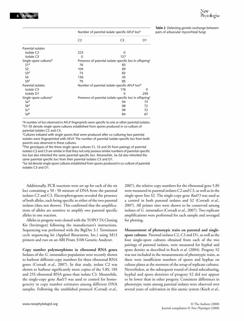
Table 2 Detecting genetic exchange between pairs of arbuscular mycorrhizal fungiNumber of parental isolate specific AFLP locia
C2 C3 D1
Parental isolates Isolate C2 223 0 Isolate C3 0 127Single-spore culturesb Presence of parental isolate-specific loci in offspringc
S1d 76 83 S2 104 69 S3d 73 83 S4 136 45 S5d 75 85Parental isolates Number of parental isolate-specific AFLP locid
Isolate C3 176 0 Isolate D1 0 259Single-spore culturese Presence of parental isolate-specific loci in offspringc
Sad 94 73 Sbd 98 72 Scd 99 72 Sdd 84 67
aA number of loci observed in AFLP fingerprints were specific to one or other parental isolates.bS1–S5 denote single-spore cultures established from spores produced in co-culture of parental isolates C2 and C3.cCultures initiated with single spores that were produced after co-culturing two parental isolates were fingerprinted with AFLP. The number of parental isolate-specific loci from both parents was observed in these cultures.dThe genotypes of the three single-spore cultures S1, S3 and S5 from pairings of parental isolates C2 and C3 are similar in that they not only possess similar numbers of parental-specific loci but also inherited the same parental-specific loci. Meanwhile, Sa–Sd also inherited the same parental specific loci from their parental isolates C3 and D1.eSa–Sd denote single spore cultures established from spores produced in co-culture of parental isolates C3 and D1.

© The Authors (2008) www.newphytologist.orgJournal compilation © New Phytologist (2008)
Research 7
2004, 2006). To reduce potential maternal effects resultingfrom the establishment of single-spore cultures from pairingsof parental isolates, single-spore cultures were grown for60 wk on culture medium. During this period, c. 4 cm2 piecesof media containing hyphae and spores were transferred tofresh plates every 15 wk. Sixteen replicate plates were establishedfor each parental isolate and each of the single-spore culturesfrom pairings of isolates for phenotypic measurements. Aftera further growth period of 15 wk, all plates were measured forhyphal and spore density as described in Koch et al. (2004).A total of 176 plates were included in the statistical analysis.The data were analysed separately for parental isolates C2, C3and progeny S1, S3–S5 and parental isolates C3, D1 andprogeny Sa–Sd by a one-way analysis of variance (ANOVA).Pairwise comparisons among parental isolates and progeny wereperformed by a Tukey–Kramer test (α = 0.05). A Dunnett’stest was also performed using the parental isolates as controlsamples (α = 0.025). The results were qualitatively identical.
Results
Anastomoses among isolates of a population (Expt 1)
We tested the occurrence of anastomosis among all possiblecombinations of pairs of five G. intraradices isolates, analysingbetween 82 and 202 hyphal contacts per pair of isolates(Table 1). When hyphae from spores of the same isolate grewtogether, between 46 and 51% of the contacts resulted inperfect fusion (Table 1). All other contacts, where hyphaetouched but no morphological changes were observed and no
fusion occurred, were scored as noninteracting. Pairings betweengenetically different isolates resulted in much lower numbersof anastomoses, but in all pairings, except for the pairing ofisolate A4 with D1, some perfect fusions were observed(Table 1, Fig. 2a). At the hyphal bridge of a perfect fusion,rapid cytoplasmic streaming occurred, either in one direction(Video S1) or in both directions (Video S2). Although themajority of contacts resulted in no interaction (Fig. 2b), a largenumber of hyphal contacts produced intermediate interactionsthat were not observed in pairings between hyphae of the sameisolate (Table 1). These were scored as pre-fusion incompatibility,where morphological changes occur within two hyphae,indicating the first stages of hyphal recognition but then theprotoplasm of one hypha retracts (Table 1, Fig. 2c). Post-fusionincompatibility also occurred, where, following anastomosis,the protoplasm of one hypha withdraws and a septum formsbetween the two hyphae (Table 1, Fig. 2d). The experimentsof hyphal fusion were repeated in water and gellan gum-basedculture medium and yielded very similar results (Table S1).
The percentage of perfect fusions between geneticallydifferent isolates was negatively correlated with the geneticdistance between the isolates in the water system (R2 = 0.41;P < 0.05), but not in the M medium system (R2 = 0.03;P = 0.7).
Molecular evidence for genetic exchange (Expt 2)
AFLP markers Perfect fusions (and possibly fusions thatresult in post-fusion incompatibility) offer the possibility forexchange of nuclei between genetically different AMF. In
Fig. 2 Contacts between hyphae of genetically different isolates. (a) Perfect fusion (arrow), protoplasmic streaming occurs within the fusion bridge (also see Supporting Information, Videos S1 and S2). (b) No interaction; no morphological change is observed in hyphae even though there is contact between hyphae. (c) Pre-fusion incompatibility; protoplasm withdrawal occurs in the approaching hypha. Arrows show two contact points. (d) Incompatible fusion; protoplasm withdrawal and septum formation occur (arrow) in one of the fused hyphae after anastomosis. Staining for succinate dehydrogenase (a) and with Trypan blue (a–d).

www.newphytologist.org © The Authors (2008)Journal compilation © New Phytologist (2008)
Research8
order to test for genetic exchange between genetically differentAMF, we analysed the AFLP banding patterns of DNA fromthe progeny of the co-cultured parental AMF. We looked forthe presence and absence of markers that were shown to bespecific for each of the parental isolates (Fig. 3a–c, Table 2).All five progeny from the pairing of parental isolates C2 withC3 possessed many markers that were specific to each of thetwo parental isolates (Table 2). Three of the five progeny (S1,S3 and S5) had almost identical genotypes (Table 2), eventhough they originated from crossings performed on in-dependent plates. All four progeny from the pairing of parentalisolates C3 with D1 showed biparental inheritance, as eachprogeny possessed several markers that were specific to each ofthe two parental isolates (Table 2). Additionally, all four progenyappeared to harbour almost identical genotypes even thoughthey also originated from crossings performed on independentplates. AFLP analysis showed that a fraction of the loci detectedin progeny was not found in either of the two respective
parental isolates (Table S2). Similarly, a fraction of AFLP locicommon to the two respective parental isolates was not foundin the progeny (Table S2).
In the control experiment, four single spores from parentalisolate C3 were cultivated and genotyped in the same way asprogeny from the crossing experiment. In total, 9% of all lociwere found to be variable among single-spore progeny fromthe parental isolate C3. This corresponds to approximatelyonly 10 AFLP fragments. This number is therefore much lowerthan the differences seen between progeny of the co-culturesand the parental isolates (> 100 fragments). This shows thatthe genetic differences among progeny from the co-culturesmust be the result of genetic exchange, as the variation couldnot be explained by genetic drift occurring during sporeformation, in the absence of genetic exchange. Table S3 showsdata where these variable loci were excluded to obtain a con-servative estimate of polymorphism among parental isolatesand single-spore progeny from the crossing experiment. Both
Fig. 3 Detecting genetic exchange using genetic markers. (a) Part of an electropherogram of an AFLP fingerprint showing five loci, two of which are specific to parental isolate C2 (202 bp, 209 bp; shown in green), one of which is specific to parental isolate C3 (201 bp; shown in black) and two that are not parental-specific (205 bp, 206 bp). The fingerprint of the single-spore culture S2 (progeny of co-culture of parental isolates C2 and C3; shown in blue) shows the presence of two parental-specific loci, one from each parental isolate. Lines of the same colour represent replicate fingerprints of the same sample. (b, c) Two additional examples of electropherograms of AFLP fingerprints showing biparental inheritance of parental-specific loci. For numbers of all loci observed, see Table 2 and Supporting Information, Tables S2 and S3. (d–f) Electropherograms of amplified alleles from the polymorphic loci Bg348, Bg196 and Bg62, respectively. Parental isolates C2 and C3 exhibit alleles of different sizes (shown in green and black, respectively). For all three loci, the fingerprint of DNA from an arbuscular mycorrhizal fungal (AMF) culture initiated from single spore S2 (shown in blue) shows the presence of both alleles. Alleles were scored as present in all genetic analyses when the relative fluorescence units for a peak were > 50.

© The Authors (2008) www.newphytologist.orgJournal compilation © New Phytologist (2008)
Research 9
scoring methods show that each progeny possesses severalmarkers that were specific to either of the two parental isolatesand that the scoring method does not bias the results.
Sequence-based markers We also used six different sequence-based markers that distinguish nuclear genomes of parentalisolates C2, C3 and D1 by length differences among thespecific alleles (Croll et al., 2008b) to test whether progenyinherit DNA from both parents. One progeny (S2) from thepairing of parental isolates C2 and C3 showed clear evidencefor biparental inheritance (Fig. 3d–f ). For each of these sixloci, parental-specific alleles from both parents co-occurred inthe progeny S2. The progeny S1, S3, S5 and Sa–Sd showedthe presence of only alleles from parental isolate C3. Theprogeny S4 showed only alleles from parental isolate C2. Allelesin the progeny were sequenced and this confirmed their identitywith parental isolates.
Copy number polymorphism The parental isolates wereshown to differ in ribosomal gene copy number (Corradiet al., 2007). We therefore measured copy number of the 5.8Sgene of progeny S2 that showed evidence for genetic exchangebased on the six sequence-based markers. The copy numberestimate was compared with that of the parental isolates C2and C3. A single-copy gene (Rad15) was used as a controlamplification for all samples (Corradi et al., 2007). Copynumber of progeny S2 was intermediate between that of theparental isolates C2 and C3 (Fig. 4). Relative copy numbersof three single-spore progeny from pairings of parentalisolates C2 and C3 (S1, S3, S5) indicated similar copynumbers to parental isolate C3, and one single-spore progeny(S4) indicated a similar copy number to parental isolate C2(data not shown).
Phenotypic traits of parental isolates and single-spore progeny
The parental isolates showed large and heritable differences inhyphal and spore densities similar to that previously seen byKoch et al. (2004, 2006) (Fig. 5). Hyphal and spore densitiesamong progeny varied significantly among offspring, whichwould not be expected if they were genetically similar to theparental isolates (Fig. 5). The factor isolate/progeny was highlysignificant (P < 0.0001) for both ANOVA. Progeny S1–S4showed a range of hyphal and spore densities exceeding thedifference between the parental isolates C2 and C3. Hyphaldensities of two progeny (S3 and S5) from the pairing ofisolates C2 and C3 were significantly higher than in bothparental isolates (Fig. 5). All other progeny of both pairingsexhibited either intermediate hyphal and spore densitiescompared with the parental isolates or did not differ signi-ficantly from one of the parental isolates.
Discussion
The results of the three different molecular analyses on theprogeny of the pairings between parental isolates show thatgenetic exchange had indeed occurred. It is notable that, usingAFLP markers, we found evidence of genetic exchange in allnine progeny from two pairings, even though anastomosisfrequency in these pairings was low. Moreover, one progenyout of nine gave strong evidence of exchange using sixindependent genetic markers. Two lines of evidence suggestthat, following anastomosis and exchange of nuclei, replicationof DNA originating from the two different individuals issustained, rather than the genetic material of one isolate out-competing all of that from the other isolate. First, the genetic
Fig. 4 Real-time quantitative PCR results showing linear regressions of cycle threshold values and log DNA quantity. Each line represents the average of two replicate amplifications. (a) Amplification of the 5.8S ribosomal gene shows copy number polymorphism between parental isolates C2 and C3, while single-spore culture S2 shows an intermediate copy number. (b) Amplification of Rad15, a single-copy gene in parental isolates C2 and C3 and single-spore culture S2.

www.newphytologist.org © The Authors (2008)Journal compilation © New Phytologist (2008)
Research10
analyses were performed on a large amount of spores andhyphae that have regrown clonally from an individual sporeafter having successfully formed a symbiosis with a plant root.Second, the progeny were also maintained in symbiosis overa period of 18 months before genetic analyses.
Anastomoses among isolates in a population
Anastomoses play a critical role in many filamentous fungi,including improving network interconnectedness within agrowing mycelium and during the encounter betweengenetically different individuals during sexual reproduction(Glass et al., 2004). Hyphal fusions within mycelia of AMFwere observed in isolates of G. mosseae, G. caledonium andG. intraradices (Giovannetti et al., 1999, 2001, 2004), butvegetative incompatibility mechanisms, known from Ascomy-cota and Basidiomycota, were hypothesized to prevent hyphalfusions among genetically different isolates (Pawlowska &Taylor, 2005). Indeed, vegetative incompatibility was foundto occur between isolates of G. mosseae from different geographiclocations (Giovannetti et al., 2004). Our results show thatgenetically well-characterized isolates of one population formfunctional anastomoses. Such fusions have the potential toallow exchange of nuclear material among growing mycelia.The negative correlation of genetic distance and perfect fusionfrequency identified in the water system suggests that vegetativeincompatibility may be a gradual process in AMF, that is, thatgenetically similar isolates are more likely to fuse than genetically
distant isolates. The relative ease of tracing hyphae back to theoriginal spore, not requiring flurorescence-labelling, make theexperimental system ideal to study larger numbers of differentisolates, originating from different locations. Furthermore,the ease of tracing the fused hyphae back to the parents avoidsthe need to develop molecular labelling of nuclei to show suchfusions in fungi that form a dense mat of mycelium. However,potential anastomoses among mature mycelia should be studiedif reliable tracking of different isolates of AMF becomes feasible.Anastomosis frequencies among secondary mycelia could bedifferent from those for germinating mycelia and be under theinfluence of various environmental factors.
Inheritance of genetic markers from parental isolates
AFLP analysis of progeny The AFLP fingerprints of single-spore cultures established from spores isolated from pairingsof parental isolates showed evidence of biparental inheritanceand exchange of genetic material. AFLP gives a genome-widescan of polymorphic loci among individuals, thus providing alarge number of reproducible loci for genotyping. All ninetested cultures showed evidence of genetic exchange using AFLPfingerprinting. None of the single-spore progeny showedexactly one of the parental genotypes. This suggests that hyphalfusions and eventual exchange of genetic material occur relativelyfrequently in in vitro cultures.
A fundamental concern with the application of arbitrarygenetic markers, such as AFLP, to genotyping single-spore
Fig. 5 Phenotypic trait characterization of progeny cultures. (a, b) Mean hyphal and spore densities of parental isolates C2, C3 and single-spore progeny cultures S1, S3, S4 and S5 from pairings of the parental isolates. (c, d) Mean hyphal and spore densities of parental isolates C3 and D1 and single-spore progeny cultures Sa–Sd. Error bars represent +1SE. Different letters indicate significant differences based on a Tukey–Kramer test (α = 0.05).
© The Authors (2008) www.newphytologist.orgJournal compilation © New Phytologist (2008)
Research 11
progeny is that processes other than genetic exchange mayinfluence the genotype of the progeny. In particular, in thecase of a heterokaryotic mycelium where genetically differentnuclei coexist (Kuhn et al., 2001; Hijri & Sanders, 2005),spores inheriting nuclei through a random process (i.e. geneticdrift) could be genetically different, even though they originatefrom the same mycelium (Sanders, 2002). However, theheterokaryosis hypothesis has been disputed in AMF (Pawlowska& Taylor, 2004) and no data on genetic differences amongnuclei are available for G. intraradices. In our control experi-ment, we were able to distinguish between potential segregationof nuclei within isolates and genetic exchange. Relative to thegenetic difference among the different parental isolates, only asmall amount of genetic variation was found among progenyspores from one isolate. Our results corroborate the occurrenceof genetic exchange among parental isolates because the AFLPdata cannot be explained solely by segregation of geneticallydifferent nuclei that coexist within an isolate. Furthermore,this control experiment also shows that potential artefacts fromAFLP genotyping, such as random shifts in frequency ofmarkers or detection thresholds, are negligible.
Genetic entities in AMF comprise nuclei in a continuouscytoplasm, but also mitochondria and potentially endosym-biotic bacteria. Even though endosymbiotic bacteria were notobserved in in vitro cultures of G. intraradices (M. Hijri & I.R. Sanders, unpublished), AFLP does not provide informationabout the location in the genome where particular loci occur.Thus evidence for genetic exchange based on AFLP may bepartly the result of exchange of mitochondria and/or endo-symbiotic bacteria. However, the nuclear DNA content isapprox. 15 Mb (Hijri & Sanders, 2004). The G. intraradicesgenome must therefore be significantly larger than the mito-chondrial genome. Thus, the large proportion of parentalspecific loci that were inherited by offspring spores suggeststhat the genetic exchange is likely to be mostly based onnuclear AFLP markers.
Not all specific AFLP loci of each of the two parents werefound in the progeny, suggesting either shifts in frequency orcomplete disappearance of these particular genetic markers(see Tables S2–S4 for full AFLP data). If a given locus in aDNA sample becomes rare, AFLP fingerprints may indicatethat locus as missing, although it may be present but belowthe detectable limit of AFLP. Similarly, AFLP fingerprintsmay indicate an appearance of a putatively new locus in thecase where a locus at low frequency simply becomes morefrequent. These two scenarios might account for disappearanceor appearance of loci in fingerprints of progeny. In this way,the AFLP data could reflect changes in frequencies in locirather than novel loci, or complete disappearance (see columns‘Loci common to parental isolates missing in progeny’ and‘Loci found in progeny not observed in parental isolates’ inTables S2 and S3).
Several single-spore progeny showing evidence of biparentalinheritance were very similar based on their AFLP genotype
(S1, S3, S5 and Sa–Sd), as they share a large proportion of loci(Table S4). It is notable that S1, S3 and S5, as well as Sa–Sdwere isolated from independent replicate plates of pairings ofparental isolates. Single-spore progeny showing evidence ofgenetic exchange and having very similar AFLP genotypessuggest that nonrandom processes could play a role duringgenetic exchange between two parental isolates. These processescould include compatibility mechanisms selecting for specificcombinations of parental genomes. In other fungi, genomicconflict was suggested to occur after fusion of geneticallydifferent individuals (Roca et al., 2003, 2004). Whether mixingof parental genomes in progeny provides an opportunity forconflict in AMF remains to be investigated.
In order to overcome the limiting amounts of DNA fromsingle spores and to ensure high reproducibility, cultures orig-inating from germinated spores were clonally subcultured.Even though we were able to control for potential segregationamong single-spore lines during the subculturing, the idealexperimental design would be based on genotyping hyphaeand spores directly produced by a fused mycelium of differentisolates.
Detecting genetic exchange through sequence-based markersThe sequence-based markers used in this study werepreviously developed to show genetic differences in a muchlarger sample of the same G. intraradices population (Crollet al., 2008b). The large number of SNPs and indels makesthese sequence-based markers suitable for a reliable identi-fication of isolates (Croll et al., 2008a). The lengths of shortrepeat motifs or indels at each locus were identified by capillaryelectrophoresis (see Supporting Information in Croll et al.,2008b) and the electropherograms exhibit typical stutterpeaks of repeat locus amplifications (Griffiths, 1996; Fig. 2).The sequencing of the alleles in the progeny furtherconfirmed the results of our analysis. The results obtainedwith the sequence-based markers showed biparental inheritancein one single-spore line (S2) from the pairing of isolates C2and C3 but not in others. This shows that, in at least onesingle-spore line, nuclei of genetically different parents mixedand formed viable offspring. This is not contradictory to theresults from AFLP showing biparental inheritance in all ninelines, as it could mean either that not all nuclei from bothparental isolates were inherited simultaneously or that thealleles from one parental isolate were present at very lowfrequency, below detection by amplification. Relative heightsof inherited alleles in offspring S2 suggest that parental nucleiwere not inherited in equal proportions. Alleles specific toparental isolate C3 appear to be more abundant than allelesspecific to parental isolate C2. This suggests that parental nucleipotentially mix in different proportions to form single-sporeprogeny.
There was a general congruence between AFLP finger-prints and sequence-based marker genotypes across all single-spore progeny, to the extent that all progeny of the parental

www.newphytologist.org © The Authors (2008)Journal compilation © New Phytologist (2008)
Research12
isolates C3 and D1 showed very similar AFLP fingerprintsand, in all cases, the specific sequences of parental isolate C3.Among single-spore progeny of C2 and C3, S4 showed anAFLP fingerprint closest to parental isolate C2 and inheritedthe specific allele from the same parent. Furthermore, single-spore progeny S1, S3 and S5 all showed AFLP fingerprintsmost similar to parental isolate C3, in accordance with theirsequence-based marker genotype. The general correlationbetween AFLP fingerprints and sequence-based markergenotypes was also shown to occur within a population ofG. intraradices isolates (Croll et al., 2008b).
Using ribosomal gene copy number, single-spore progenyS2 showed an intermediate copy number, suggesting that amixing of genomes of the parental isolates can produceintermediate copy numbers for ribosomal genes. Theintermediate copy number shown by progeny S2 supportsthe genetic analyses based on the other markers in ourstudy.
Phenotypic traits of parental isolates and single-spore progeny
Hyphal densities and spore production were shown to bestrongly heritable traits over many generations of clonalsubculturing (Koch et al., 2006). Indeed, hyphal densities andspore production of parental isolates C2, C3 and D1 showedvery similar phenotypes compared with measurements of thesame isolates in two previous studies in the same in vitroculture system (Koch et al., 2004, 2006). Single-spore progenyof pairings of parental isolates show a range of distinct hyphaland spore densities compared with their respective parentalisolates. Progeny S4 shows very similar phenotypic traits toparental isolate C2, corroborating the genetic analysis showingthat S4 inherited most of the specific AFLP loci of parentalisolate C2. Meanwhile, progeny S3 and S5 showed significantlyhigher hyphal densities than both parental isolates. The findingof phenotypic traits in progeny that exceed both parentalisolates suggests that the inheritance of these traits is notstrictly additive, that is, progeny did not inherit an intermediatephenotype of parental isolates. Also, S1, S3 and S5 showedsignificantly different hyphal and spore densities, despite theirgenotypes being very similar. This suggests that either minorgenetic differences (as detected by AFLP) can result in clearlydistinct phenotypic traits or epigenetic mechanisms play arole in determining these traits in the progeny. Similarly,progeny Sa–Sd show significant differences in hyphal andspore densities among each other. Analysis of genetic markersassociated with particular phenotypes may help to elucidatethe genetic basis of these phenotypic traits. Progeny showingdistinct phenotypes from the respective parental isolates couldhave important consequences for the symbiosis with hostplants, as genetic differences among isolates were already shownto influence plant growth and phosphate uptake (Munkvoldet al., 2004; Koch et al., 2006).
Fate of genetically different nuclei in a common mycelium
Genetically different nuclei might undergo recombination ina common mycelium following potential nuclear fusion. Thegenotyping methods used in this study would not allow thedetection of whether the mixing of genetic material in singlespores resulted in recombination among genetically differentnuclei in the mycelium. Novel loci found by AFLP could beexplained either by frequency shifts or by recombination events.The presence of both parent-specific alleles at several loci insingle-spore culture S2 could reflect the coexistence ofgenetically different nuclei without nuclear fusion. However,recombination may have occurred among nuclei, but alldaughter nuclei resulting from such events could have remainedin the common cytoplasm. As a result, alleles specific to bothparental may be found in single-spore progeny. Parent-specificlabelling of nuclei would be required to track eventualrecombination events. Furthermore, segregation among singlespores produced by a progeny could shed light on the fate ofnuclear genotypes following exchange through hyphal fusions.
Genetic exchange and evolution of genetic diversity
Our results suggest that AMF are promiscuous in the sensethat an individual will form hyphal connections with severalother members of the population and this creates the possibilityfor genetic exchange. In the absence of a sexual reproductivecycle, such promiscuity could allow many genetically differentindividuals of a population to connect, thereby forming alarge, genetically diverse hyphal network. Exchange of nucleiamong isolates provides the opportunity for fusion ofgenetically different nuclei and recombination. Geneticexchange could also act as an important mechanism inmaintaining the multigenomic state (Bever & Wang, 2005;Hijri & Sanders, 2005).
Knowledge of the compatibility of isolates and their poten-tial to recombine is critical in the understanding of theireffects on plants, as genetic differences among isolates wereshown to influence costs and benefits to the host (Koch et al.,2006). Genetic diversity within AMF networks may maintainthe unusually low host specificity seen in these symbionts(Sanders, 2003). Because we were able to create viable geneticcrosses between genetically and phenotypically different AMF,our study lays a foundation for experimentally investigatingthe genetics, control of compatibility/incompatibility and thegenetic architecture of phenotypic traits in this important fungalphylum. Finally, the evolution and diversification of species inthe phylum Glomeromycota should no longer be assumed tobe the result of the long-term absence of genetic exchange.
Acknowledgements
The Swiss National Science Foundation is acknowledged forfinancially supporting this work with a grant to IRS (grant
© The Authors (2008) www.newphytologist.orgJournal compilation © New Phytologist (2008)
Research 13
number 3100AO-105790/1). Rui Rodrigues MartinsCandeias helped with in vitro cultivation and DNA extraction.
References
Bécard G, Fortin JA. 1988. Early events of vesicular arbuscular mycorrhiza formation on Ri T-DNA transformed roots. New Phytologist 108: 211–218.
Bever JD, Wang M. 2005. Arbuscular mycorrhizal fungi: hyphal fusion and multigenomic structure. Nature 433: E3–4; discussion E4.
Cenis JL. 1992. Rapid extraction of fungal DNA for PCR amplification. Nucleic Acids Research 20: 2380.
Corradi N, Croll D, Colard A, Kuhn G, Ehinger M, Sanders IR. 2007. Gene copy number polymorphisms in an arbuscular mycorrhizal fungal population. Applied and Environmental Microbiology 73: 366–369.
Corradi N, Hijri M, Fumagalli L, Sanders IR. 2004. Arbuscular mycorrhizal fungi (Glomeromycota) harbour ancient fungal tubulin genes that resemble those of the chytrids (Chytridiomycota). Fungal Genetics and Biology 41: 1037–1045.
Croll D, Corradi N, Gamper HA, Sanders IR. 2008a. Multilocus genotyping of arbuscular mycorrhizal fungi and marker suitability for population genetics. New Phytologist 173: 564–568.
Croll D, Wille L, Gamper HA, Mathimaran N, Lammers PJ, Corradi N, Sanders IR. 2008b. Genetic diversity and host plant preferences revealed by simple sequence repeat and mitochondrial markers in a population of the arbuscular mycorrhizal fungus Glomus intraradices. New Phytologist 178: 672–687.
Giovannetti M, Azzolini D, Citernesi AS. 1999. Anastomosis formation and nuclear and protoplasmic exchange in arbuscular mycorrhizal fungi. Applied and Environmental Microbiology 65: 5571–5575.
Giovannetti M, Fortuna P, Citernesi AS, Morini S, Nuti MP. 2001. The occurrence of anastomosis formation and nuclear exchange in intact arbuscular mycorrhizal networks. New Phytologist 151: 717–724.
Giovannetti M, Sbrana C, Avio L, Strani P. 2004. Patterns of below-ground plant interconnections established by means of arbuscular mycorrhizal networks. New Phytologist 164: 175–181.
Glass NL, Jacobson DJ, Shiu PKT. 2000. The genetics of hyphal fusion and vegetative incompatibility in filamentous ascomycete fungi. Annual Review of Genetics 34: 165–186.
Glass NL, Rasmussen C, Roca MG, Read ND. 2004. Hyphal homing, fusion and mycelial interconnectedness. Trends in Microbiology 12: 135–141.
Goudet J. 2001. FSTAT, a program to estimate and test gene diversities and fixation indices version 2.9.3. http://www.unil.ch/izea/softwares/fstat.html.In.
Griffiths AJF, Miller JF, Suzuki DT, Lewontin RC, Gelbart WM. 1996. Introduction to genetic analysis. New York, NY, USA: WH Freeman.
Heckman DS, Geiser DM, Eidell BR, Stauffer RL, Kardos NL, Hedges SB. 2001. Molecular evidence for the early colonization of land by fungi and plants. Science 293: 1129–1133.
Hijri M, Sanders IR. 2004. The arbuscular mycorrhizal fungus Glomus intraradices is haploid and has a small genome size in the lower limit of eukaryotes. Fungal Genetics and Biology 41: 253–261.
Hijri M, Sanders IR. 2005. Low gene copy number shows that arbuscular mycorrhizal fungi inherit genetically different nuclei. Nature 433: 160–163.
Hoekstra RF. 1994. Population-genetics of filamentous fungi. Antonie Van Leeuwenhoek International Journal of General and Molecular Microbiology 65: 199–204.
James TY, Kauff F, Schoch CL, Matheny PB, Hofstetter V, Cox CJ, Celio G, Gueidan C, Fraker E, Miadlikowska J et al. 2006. Reconstructing the early evolution of fungi using a six-gene phylogeny. Nature 443: 818–822.
Judson OP, Normark BB. 1996. Ancient asexual scandals. Trends in Ecology and Evolution 11: 41–46.
Koch AM, Croll D, Sanders IR. 2006. Genetic variability in a population of arbuscular mycorrhizal fungi causes variation in plant growth. Ecology Letters 9: 103–110.
Koch AM, Kuhn G, Fontanillas P, Fumagalli L, Goudet I, Sanders IR. 2004. High genetic variability and low local diversity in a population of arbuscular mycorrhizal fungi. Proceedings of the National Academy of Sciences, USA 101: 2369–2374.
Kuhn G, Hijri M, Sanders IR. 2001. Evidence for the evolution of multiple genomes in arbuscular mycorrhizal fungi. Nature 414: 745–748.
Munkvold L, Kjoller R, Vestberg M, Rosendahl S, Jakobsen I. 2004. High functional diversity within species of arbuscular mycorrhizal fungi. New Phytologist 164: 357–364.
Pawlowska TE, Taylor JW. 2004. Organization of genetic variation in individuals of arbuscular mycorrhizal fungi. Nature 427: 733–737.
Pawlowska TE, Taylor JW. 2005. Arbuscular mycorrhizal fungi: hyphal fusion and multigenomic structure (reply). Nature 433: E4–E4.
Pontecorvo G. 1956. Parasexual cycle in fungi. Annual Review of Microbiology 10: 393–400.
Remy W, Taylor TN, Hass H, Kerp H. 1994. Four hundred-million-year-old vesicular arbuscular mycorrhizae. Proceedings of the National Academy of Sciences, USA 91: 11841–11843.
Roca MG, Davide LC, Davide LM, Mendes-Costa MC, Schwan RF, Wheals AE. 2004. Conidial anastomosis fusion between Colletotrichum species. Mycological Research 108: 1320–1326.
Roca MG, Davide LC, Mendes-Costa MC, Wheals A. 2003. Conidial anastomosis tubes in Colletotrichum. Fungal Genetics and Biology 40: 138–145.
Rosendahl S. 2008. Communities, populations and individuals of arbuscular mycorrhizal fungi. New Phytologist 178: 253–266.
Sanders IR. 2002. Ecology and evolution of multigenomic arbuscular mycorrhizal fungi. American Naturalist 160: S128–S141.
Sanders IR. 2003. Preference, specificity and cheating in the arbuscular mycorrhizal symbiosis. Trends in Plant Science 8: 143–145.
Smith SE, Read DJ. 1997. Mycorrhizal symbiosis. San Diego, CA, USA: Academic Press.
St-Arnaud M, Hamel C, Vimard B, Caron M, Fortin JA. 1996. Enhanced hyphal growth and spore production of the arbuscular mycorrhizal fungus Glomus intraradices in an in vitro system in the absence of host roots. Mycological Research 100: 328–332.
Stukenbrock EH, Rosendahl S. 2005. Clonal diversity and population genetic structure of arbuscular mycorrhizal fungi (Glomus spp.) studied by multilocus genotyping of single spores. Molecular Ecology 14: 743–752.
Swofford DL 2002. PAUP*: phylogenetic analysis using parsimony, version 4b10. Sunderland, MA, USA: Sinauer Associates.
Van der Heijden MGA, Klironomos JN, Ursic M, Moutoglis P, Streitwolf-Engel R, Boller T, Wiemken A, Sanders IR. 1998. Mycorrhizal fungal diversity determines plant biodiversity, ecosystem variability and productivity. Nature 396: 69–72.
Supporting Information
Additional supporting information may be found in theonline version of this article.
Table S1 Frequency of anastomosis (fusion) between pairs ofarbuscular mycorrhizal fungi
Table S2 Summary of AFLP loci detecting genetic exchangebetween pairs of arbuscular mycorrhizal fungi

www.newphytologist.org © The Authors (2008)Journal compilation © New Phytologist (2008)
Research14
Table S3 Summary of AFLP loci detecting genetic exchangebetween pairs of arbuscular mycorrhizal fungi using a moreconservative analysis in which all loci showing inconsistentscoring among single spore cultures of isolate C3 wereremoved
Table S4 Summary of AFLP loci detecting exchangebetween pairs of parental isolates using AFLP (S1–S5 origi-nating from pairings of isolates C2 and C3; Sa–Sd originatingfrom pairings of isolates C3 and D1)
Video S1 The video shows two hyphae of genetically differentGlomus intraradices isolates that have fused.
Video S2 The video shows two living hyphae (stained withfast green) of two genetically different Glomus intraradicesisolates that have fused.
Please note: Wiley-Blackwell are not responsible for the contentor functionality of any supporting information supplied bythe authors. Any queries (other than missing material) shouldbe directed to the New Phytologist Central Office.
Copyright © 2022 FDOKUMEN