Threshold-Based Mechanisms to Discriminate Transient from Intermittent Faults
Upload
independentCategory
view
0download
0
(This is a sample cover image for this issue. The actual cover is not yet available at this time.)
This article appeared in a journal published by Elsevier. The attachedcopy is furnished to the author for internal non-commercial researchand education use, including for instruction at the authors institution
and sharing with colleagues.
Other uses, including reproduction and distribution, or selling orlicensing copies, or posting to personal, institutional or third party
websites are prohibited.
In most cases authors are permitted to post their version of thearticle (e.g. in Word or Tex form) to their personal website orinstitutional repository. Authors requiring further information
regarding Elsevier’s archiving and manuscript policies areencouraged to visit:
http://www.elsevier.com/copyright
Author's personal copy
Respiratory Physiology & Neurobiology 185 (2013) 600– 607
Contents lists available at SciVerse ScienceDirect
Respiratory Physiology & Neurobiology
j o ur nal homep age : www.elsev ier .com/ locate / resphys io l
Inhibition of rat carotid body glomus cells TASK-like channels by acute hypoxia isenhanced by chronic intermittent hypoxia
Fernando C. Ortiza, Rodrigo Del Rioa, German Ebenspergerb, Victor R. Reyesb, Julio Alcayagac,Rodrigo Varasa, Rodrigo Iturriagaa,∗
a Laboratorio de Neurobiología, Facultad de Ciencias Biológicas, P. Universidad Católica de Chile, Santiago, Chileb Programa de Fisiopatología, ICBM, Facultad de Medicina, Universidad de Chile, Santiago, Chilec Laboratorio de Fisiología Celular, Facultad de Ciencias, Universidad de Chile, Santiago, Chile
a r t i c l e i n f o
Article history:Accepted 27 November 2012
Keywords:Background K+ channelsGlomus cellsCarotid bodyOxygen sensingObstructive sleep apnea
a b s t r a c t
Chronic intermittent hypoxia (CIH), the main feature of obstructive sleep apnea, enhances carotid body(CB) chemosensory responses to acute hypoxia. In spite of that, the primary molecular target of CIHin the CB remains unknown. A key step of the hypoxic response in the CB is the chemoreceptor celldepolarization elicited by the inhibition of K+ channels. Thus, we tested the hypothesis that CIH potenti-ates the hypoxic-induced depolarization of rat CB chemoreceptor cells by enhancing the inhibition of abackground K+ TASK-like channel. Membrane potential, single channel and macroscopic currents wererecorded in the presence of TEA and 4-aminopyridine in CB chemoreceptor cells isolated from adult ratsexposed to CIH. The CIH treatment did not modify the resting membrane properties but the hypoxic-evoked depolarization increased by 2-fold. In addition, the hypoxic inhibition of the TASK-like channelcurrent was larger and faster in glomus cells from CIH-treated animals. This novel effect of CIH maycontribute to explain the enhancing effect of CIH on CB oxygen chemoreception.
© 2012 Elsevier B.V. All rights reserved.
1. Introduction
Chronic intermittent hypoxia (CIH), which is the main featureof the obstructive sleep apnea (OSA) syndrome, is recognized as anindependent risk factor for hypertension and other cardiovascu-lar diseases (Somers et al., 2008). The current evidence shows thatthe CIH-induced potentiation of the carotid body (CB) chemosen-sory responses to acute hypoxia contributes to the OSA-inducedhypertension (Iturriaga et al., 2009; Prabhakar and Kumar, 2010;Smith and Pacchia, 2007; Weiss et al., 2007). Indeed, patientswith recently diagnosed OSA show enhanced sympathetic and car-diorespiratory responses to hypoxia, attributed to an increased CBchemoreflex response to hypoxia (Narkiewicz et al., 1998, 1999).Moreover, studies performed in animals models of OSA have shownthat CIH enhances CB chemosensory and ventilatory responses toacute hypoxia (Del Rio et al., 2010; Pawar et al., 2009; Peng et al.,2003; Reeves et al., 2003; Rey et al., 2004), produces autonomicdysfunction (Prabhakar and Kumar, 2010; Rey et al., 2004, 2008;Zoccal et al., 2008) and induces a long-term potentiation of motorventilatory activity (McGuire et al., 2004; Mitchell et al., 2001).
∗ Corresponding author at: Laboratorio de Neurobiología, Facultad de CienciasBiológicas, P. Universidad Católica de Chile, Casilla 193, Santiago,Chile. Tel.: +56 2 686 2852; fax: +56 2 354 1850.
E-mail address: [email protected] (R. Iturriaga).
The mechanisms underlying the CIH enhancement of CBchemosensory reactivity to hypoxia are not entirely understood(Del Rio et al., 2011a,b; Iturriaga et al., 2009). Oxidative stress (DelRio et al., 2010; Marcus et al., 2010; Peng et al., 2003), endothelin-1 (Pawar et al., 2009; Rey et al., 2006) and pro-inflammatorycytokines (Del Rio et al., 2011a; Iturriaga et al., 2009) have beenassociated with the carotid chemosensory potentiation. How-ever, the primary molecular target responsible for the increasedchemoreceptor discharge remains unknown (Iturriaga et al., 2009).The current model of CB chemoreception states that acute hypoxiadepolarizes the chemoreceptor (glomus or type-I) cells, increasingintracellular [Ca2+] and subsequently releasing one or more trans-mitters (Iturriaga and Alcayaga, 2004; Iturriaga et al., 2007). Inneonatal rat glomus cells, the depolarization induced by hypoxiais initiated by the inhibition of a background K+ current, whichis likely due to the activity of the heterodimeric TASK-1/TASK-3channel (Buckler, 1999, 2010; Kim et al., 2009). Accordingly, wehypothesized that CIH enhances CB chemosensory responses toacute hypoxia by increasing the inhibition of TASK-like channelactivity, resulting in an increased membrane depolarization. There-fore, we studied whether CIH enhanced the inhibition of TASK-likechannels and increased the amplitude of the hypoxic-induceddepolarization in CB glomus cells isolated from rats exposed toCIH for 7 days, which show enhanced chemosensory and venti-latory responses to acute hypoxia (Del Rio et al., 2011a; Iturriagaet al., 2009). Finally, since functional TASK channels have not been
1569-9048/$ – see front matter © 2012 Elsevier B.V. All rights reserved.http://dx.doi.org/10.1016/j.resp.2012.11.015

Author's personal copy
F.C. Ortiz et al. / Respiratory Physiology & Neurobiology 185 (2013) 600– 607 601
reported in the Sprague Dawley rat adult CB glomus cells, wecharacterized the mRNA and protein expression as well as the elec-trophysiological distinctiveness of TASK channels in CBs of adultanimals.
2. Materials and methods
2.1. Animals and ethical standards
Experiments were performed on adult male Sprague-Dawleyrats (200–250 g, postnatal day ∼90), fed with standard chow dietand kept on a 12-h light/dark schedule (8:00 am – 8:00 pm). Exper-imental procedures were approved by the Bioethical and BiosafetyCommittee of the Faculty of Biological Sciences of the PontificiaUniversidad Católica de Chile and the Ethical Committee of theFacultad de Ciencias of the Universidad de Chile, and were con-ducted in accordance to the guidelines of the National Fund forScientific and Technological Research (FONDECYT, Chile).
2.2. Experimental groups and exposure to chronic intermittenthypoxia
Rats were exposed to intermittent hypoxia as previouslydescribed (Iturriaga et al., 2009; Del Rio et al., 2010, 2011a,b). Unre-strained, freely moving rats were housed in individual chambers(12 cm × 35 cm, 3.96 L) and exposed to a CIH protocol consisting ofhypoxic cycles of 5–6% inspired O2 for 20 s, followed by room airfor 280 s, applied 12 times/h, 8 h/day for 7 days. Rats have accessto food and water ad libitum and the hypoxic pattern was appliedduring the daytime (8:00 am to 4:00 pm). The animals were keptin the chambers during 7 days, and were able to move freely andturn around in the chambers. Chambers were equipped with a rearinlet to admit N2 and an extractor fan in the front. During nor-moxic periods the fans were continuously running, assuring lowCO2 levels in the chambers. During hypoxic exposure, the fans werestopped and solenoid valves allow 100% N2 flow into the cham-bers. An automatized system controlled the solenoid valves andthe alternating cycles of the fans. The O2 level in the chambers wascontinuously monitored with an oxygen analyzer (Ohmeda 5120,USA). Rats of the control group were kept in the normoxic conditionin the same room that rats were exposed to CIH. Room temperaturewas kept at 23–25 ◦C.
2.3. CB acute dissociation
CBs were surgically removed from the rats anesthetized withketamine and xylazine (75/7.5 mg/kg) and placed in ice cold mod-ified Hanks’ balanced salt solution (pH 7.4 at 4 ◦C). The CBs wereenzymatically (trypsin/collagenase) and mechanically dissociatedas previously described (Buckler, 1997). The cellular suspensionwas plated onto poly-l-lysine (0.1 mg/mL) coated glass coverslips.Cells were kept at 37 ◦C in a culture chamber with air – 5% CO2in a water saturated atmosphere, until its use (2–8 h). The dissoci-ated cells were used in electrophysiological and immunostainingstudies.
2.4. Electrophysiological recordings
Electrophysiological studies were conducted using eithercell-attached or whole-cell configuration in current-clamp orvoltage-clamp mode with a patch clamp amplifier (Axopatch 200B,Molecular Devices, USA). Membrane current and voltage werefiltered at 2 kHz and recorded at 20 kHz with a Digidata 1200AD (Molecular Devices, USA). Microelectrodes were made fromborosilicate glass capillaries and heat-polished before use (resis-tance 5–8 M� and seal resistance ≥ 5 G�). Cell-attached recordings
were performed with several values of imposed pipette poten-tial (Vp) to determine the I–V relationship. In the whole-cellvoltage-clamp mode the macroscopic membrane current wasrecorded by imposing a voltage ramp from −120 mV to +20 mV(0.7 mV/ms, from a holding potential of −55 mV; see Fig. 1C inset).Current–clamp recordings were performed with no imposed cur-rent (I = 0, “voltage follower”). The coverslip with the platted cellswas placed on a recording chamber (volume ∼400 �L) on the stageof an inverted microscope, and continuously superfused with bathsolution at 1.2–1.5 mL/min controlled with a two channel peri-staltic pump (Masterflex CL, Cole Parmer, USA). For cell-attachedand whole cell recordings, cells were superfused with a saline solu-tion containing (in mM) 117 NaCl, 4.5 KCl, 23 NaHCO3, 1 MgCl2, 2.5CaCl2 and glucose 11, pH = 7.4 at 37 ◦C. The extracellular solutionwas equilibrated with 5% CO2 in air (normoxia: PO2 ∼ 140 mmHg)or 5% CO2 and 95% N2 (hypoxia: PO2 ∼ 5 mmHg in the recordingchamber). PO2 of the solutions was monitored in the recordingchamber with a 24 gauge oxygen needle electrode, connectedto a polarographic chemical microsensor (Diamond Electro-Tech,USA). The cells were stimulated with the hypoxic solutionfor 30 or 90 s.
To isolate TASK currents, voltage-dependent K+ currents presentin glomus cells (Buckler, 1997; Buckler and Honoré, 2004) wereblocked with 10 mM tetraethylammonium (TEA) and 5 mM 4-aminopyridine (4-AP) added either to the medium (whole cell)or to the internal pipette solution (cell-attached). No change onresting membrane potential (Vm) was observed when we appliedTEA + 4-AP on bath solution (not shown). The pipette solution forcell-attached recordings contained KCl 140 or 70 mM (in ionic sub-stitution experiments), 4 MgCl2, 1 EGTA, 10 HEPES, 10 TEA and 54-AP, and equilibrated to pH = 7.4 at 37 ◦C. For whole cell recor-dings pipette solution was (in mM): KCl 140, MgCl2 1.5, MgATP 2,EGTA10, CaCl2 2, HEPES 10, pH 7.2.
Conventional intracellular recordings were performed to con-firm the values of Vm recorded from CB glomus cells using thewhole-cell configuration. Microelectrodes were made from borosil-icate glass capillaries and backfilled with KCl 3 M (resistance:30–60 M�). The microelectrodes were connected to an electrome-ter (IE-210, Warner Instruments, USA) and the signal was acquire at20 kHz with the AxoScope 8.0 software. The entire CB was attachedto the bottom of the recording chamber, and blind intracellularrecordings were made from the whole CB. Impaled cells wereclassified as glomus cell when they respond with membrane depo-larization to the hypoxic or acidic stimuli (puff of saline equilibratedat pH 6.5 with PIPES buffer). Only stable recordings lasting for morethan 5 min were considered for the analysis.
2.5. RNA isolation and RT-PCR assays
Total RNA was extracted from two independent pools of CBsfrom 8 control and 8 CIH-treated rats, respectively, using Trizol(Invitrogen Life Technologies, CA, USA). cDNA was synthesized byreverse transcription using random hexamers and the Revert aidH Minus reverse transcriptase (Fermentas Life Sciences, Ontario,Canada). Procedures were carried out according to the manu-facturer’s instructions. Primers for amplification of partial DNAsequences from TASK-1, (Forward 5′-CACCGTCATCACCACAATCGA-3′ and Reverse 5′-TGCTCTGCATCACGCTTCTC-3′, accession numberAF031384) and TASK-3 (Forward 5′-CAGTGGAAGTTCGCCGGGT-3′ and Reverse 5′-GCTTCCTCTGCAGGGCA-3′, accession numberNM 053405) were derived from the corresponding rat genes. PCRamplification was carried out from cDNA synthesized from 0.1 �g oftotal RNA, with 1 U of Taq polymerase (Promega), 10 mM Tris–HClpH 9.0, 50 mM KCl, 0.1% Triton X-100, 1.5 mM MgCl2, 0.2 mM dNTPmix and 0.3 �M of each one of the primers. The PCR products and

Author's personal copy
602 F.C. Ortiz et al. / Respiratory Physiology & Neurobiology 185 (2013) 600– 607
Fig. 1. Electrophysiological and pharmacological properties of TASK-like current in CB chemoreceptor cells from adult rats. (A) Cell-attached recording of channel current(Vp = 0 mV) in a CB glomus cell from an adult rat and its corresponding event-amplitude histogram (obtained from an all-point analysis). (C) Close level; OI, open level 1; OII,open level 2. Scale: 1 pA, 10 ms. (B) Effects of reducing pippete [K+] from 140 mM (K+140) to 70 mM (K+70) on the unitary current (Iu) – pipette potential (Vp) relationship.The reduction of external [K+] from 140 to 70 mM did not modify the single channel conductance (15.2 ± 0.6 pS K+140 vs. 13.1 ± 0.2 pS in K+70, P > 0.05, Mann Whitney test,n = 4 cells), displacing the inversion potential by 18.3 ± 0.2 mV, close to the expected value from the Nernst equilibrium (18.1 mV). (C) Extracellular acidification (pH 6.5;dotted line) reduced the macroscopic control (pH 7.4; solid line) conductance by ∼47% (P < 0.05, Mann–Whitney test, n = 5 cells), bath solution contained 10 mM TEA and5 mM 4-AP (see Section 2) (D) Bupivacaine (200 �M; dotted line) reduced the macroscopic control (solid line) conductance by ∼60% (P < 0.05, Mann Whitney test, n = 4 cells),bath solution contained 10 mM TEA and 5 mM 4-AP (see Section 2).
molecular weight standard were separated by electrophoresis onethydium bromide agarose gels and visualized under UV light.
2.6. Immunohistochemical and cytochemical detection of TASK-1and TASK-3 potassium channel subunits
The TASK-1 and TASK-3 subunits in the rat CB were identifiedusing immunohistochemistry. Briefly, anesthetized rats were per-fused intracardially with phosphate buffer saline (PBS) at pH 7.4for 10 min followed by buffered 4% paraformaldehyde (PFA, Sigma,USA). The carotid bifurcations containing the CB were dissected andpost-fixed in PFA 4% for 12 h at 4 ◦C. Samples were then dehydratedin ethanol, included in paraffin, cut in 5 �m width sections, deparaf-finized, rehydrated and mounted on silanized slides. Slides weresubmitted to microwave based antigen retrieval protocol (700 Wfor 6 min in citrate buffer 1 M, pH 6.0) for unmasking antigens whichhave become modified by the tissue fixation process. Samples wereincubated with 0.3% H2O2 to inhibit endogenous peroxidase andthen in a universal ready to use blocking solution (Vectastain EliteABC Kit, Vector Lab, USA). The slides were incubated overnight at4 ◦C in humidity chambers with specific antibodies for detectionof TASK-1 (rabbit polyclonal antibody 1:100 anti-TASK-1, #APC-024, Allomone Labs, Israel) and TASK-3 (rabbit polyclonal 1:100anti- TASK-3, #APC-044, Allomone Labs, Israel) subunits. Negativecontrols were performed by preadsorbtion of the primary anti-body with the proper control peptide. After rinsing slides in coldPBS, samples were incubated with proper secondary antibodiesconjugated with biotin followed by a ready-to-use stabilized ABCreagent (Vectastain Elite ABC Kit, Vector Lab, USA), and revealedat 37 ◦C in a dark chamber with 3,3-diaminobenzidine tetrahy-drochloride (DAB; Sigma, USA). To avoid false positives during DABchromogen quantification, special attention was kept to prevent
DAB signal saturation. Finally, samples were counterstained withHarris Hematoxylin and permanently mounted with Entellan®
(Merck, USA). Photomicrographs of the CB tissue were taken at100× with a CCD-camera coupled to an Olympus CX 31 microscope(Olympus Corp., Japan), digitized and analyzed with the ImageJsoftware (NIH, USA). A color deconvolution algorithm was usedto separate the Harris Hematoxylin and DAB staining, and quan-tify the immunoreactive (-ir) mark from the RGB (red–green–blue)images.
In another set of experiments, immunocytochemical detectionof TASK subunits in CB glomus cells was performed. Isolated glo-mus cells were plated onto poly-l-lysine (0.1 mg/mL) coated 10 mmglass coverslips and fixed in 4% PFA for 10 min. Coverslips werethen incubated for 12 h at 4 ◦C with a mixture of anti-TASK-1antibodies (rabbit polyclonal antibody 1:100, number APC-024,Allomone Labs, Israel) or anti-TASK-3 antibodies (rabbit polyclonal1:100, number APC-044, Allomone Labs, Israel) and anti-tyrosinehydroxylase (mouse monoclonal antibody 1:200 anti-TH, numberMAB318, Millipore, USA), the latter used as a positive control forCB glomus cells recognition. After washing with cold (4 ◦C) PBS,cells were sequentially incubated with a mixture of fluorescentconjugated secondary antibodies anti-rabbit IgG (AlexaFluor488,Molecular Probes, USA) and anti-mouse IgG (AlexaFluor594, Molec-ular Probes, USA). Finally, the preparations were mounted in afluorescent mounting media (Vectashield, Vector Labs, USA) andslides were observed in a Nikon Eclipse E epifluorescence micro-scope connected to a CCD camera.
2.7. Data and statistical analyses
Single channel recordings were analyzed with the pClamp 7.0software (Molecular Devices, USA). Channel activity was reported

Author's personal copy
F.C. Ortiz et al. / Respiratory Physiology & Neurobiology 185 (2013) 600– 607 603
as the open probability times the number of channels in a givenpatch (NPo). Open events were defined using a threshold crossingmethod set at 50% of the main conductance level (defined from all-point histograms). Since the number of channels in a given patchmay differ, the effect of hypoxia was standardized as the percent-age of channel activity during normoxia. Analyses of inter-eventand dwell time histograms for the first open level were performedfor cell-attached recordings. The curves were fitted to a simpleexponential equation to obtain the characteristic time constant(�). Whole-cell I–V curves were fit to the Goldman–Hodgkin–Katzequation for simple electrodiffusion of K+ (Koizumi et al., 2010),which predicts that in asymmetric K+ conditions the I–V curve forthe K+ current must shows a small outward rectification at morepositive potentials (Hille, 2001).
Data was expressed as mean ± SEM. The number of cells (n)reported here corresponds to the number of cells from differentCB preparations tested for a given treatment. Each data groupwas tested for normal distribution using D’Agostino and Pear-son test. Two group comparisons were performed using Studentt test (paired/unpaired) or Mann–Whitney test, to parametricand non-parametric data, respectively. Kruskal–Wallis test fol-lowed by Dunn’s multiple comparisons post hoc was used tocompare two or more non-parametric groups. Curves with twovariation factors were compared with two-way ANOVA followedby a Bonferroni post hoc test. All statistical tests were performedwith GraphPad Prism 4.03 software for Windows (GraphPad Soft-ware, San Diego CA, USA) and the level of significance was setat P < 0.05.
3. Results
3.1. Characterization of the TASK-like current in glomus cellsfrom adult rats
In cell-attached recordings under conditions that allows isola-tion of TASK-like currents (Vp = 0 mV; pipette solution containing140 mM KCl, 4 mM Mg2+ 10 mM TEA and 5 mM 4-AP, and 4.5 mMKCl in the bath solution), we routinely observed single channelopenings with mean current amplitude of ∼1 pA and rapid flicker-ing openings (∼1 ms) and burst discharges (Figs. 1A and 3A), similarto what has been described for a TASK channel in glomus cells fromthe neonatal rat (Buckler, 1999; Kim et al., 2011, 2009; Williamsand Buckler, 2004). All-point histograms constructed from theserecordings suggest that events with larger current amplitude weredue to simultaneous openings of two or more of the same chan-nels (Fig. 1A). The I–V curve for single channel conductance in140 mM K+ and 4 mM Mg2+ in the pipette showed an unitarychannel conductance of 15.4 ± 0.8 pS (Fig. 1B; n = 16 cells). Confir-mation that the recorded current was carried primarily by K+ ionswas obtained with pipettes containing 70 mM K+ (Fig. 1B), whichresulted in a shift of the current reversal potential to a membranepotential value close to the calculated by the Nernst equilibriumequation. Indeed, changing the pipette [K+] from 140 to 70 mM pro-duced a displacement of the reversal potential of +18.3 ± 0.2 mV(n = 4 cells), value in close agreement with the theoretical expectedvalue of +18.1 mV. In addition, we tested the effects of acidifi-cation and the local anesthetic bupivacaine, which are knowninhibitors of TASK channels (Buckler et al., 2000). The whole-cellK+ current was reduced by acidification of the external solution.Indeed, as is shown in Fig. 1C the macroscopic conductance wasreduced from 1.59 ± 0.04 nS at pH 7.4 to 0.85 ± 0.03 nS at pH 6.5(P < 0.05, Mann–Whitney, n = 5 cells). Similarly, administration ofbupivacaine (200 �M) reduced the macroscopic conductance from1.94 ± 0.06 to 0.78 ± 0.04 nS (P < 0.05, Mann–Whitney, n = 4 cells;Fig. 1D).
3.2. mRNA and immunoreactive presence of TASK-1 and TASK-3in glomus cells from adult rat CB
Since our functional evidences show the presence of TASK-like currents in glomus cells isolated from adult rat CB, we firstdetected specific mRNA for principal candidates TASK channels,named TASK-1 and TASK-3, and then used immunodetection tech-niques to verify the expression of the corresponding proteins. Asis shown in Fig. 2A, the RT-PCR assay allowed the amplification ofpartial sequences of TASK-1 (∼450 bp) and TASK-3 (∼540 bp) cDNAin the adult rat CB. Additionally, the CB tissue was immunoreactivefor TASK-1 and TASK-3 proteins. The immunoreactivity for TASK-1 and TASK-3 subunits was mainly confined to clusters of cells,which have been defined as glomus cells in classical light micro-scope studies. Indeed, most of the positive immunoreactivity wasfound in round to ovoid cells with prominent nuclei organized inclusters around blood vessels (Fig. 2B). It is worth noting that TASK-1 immunostaining was also observed in the walls of blood vessels.In isolated CB cells, we determined that glomus cells expressedTASK-1 and TASK-3 subunits. As is shown in Fig. 2C, TASK-1 andTASK-3 positive immunoreactivity co-localized with the glomuscell marker tyrosine hydroxylase.
3.3. Effect of CIH on the TASK-like current inhibition induced byacute hypoxia
Under normoxic conditions, CIH-treated glomus cells showan I–V relationship for TASK-like currents that is not differentto the control one (P > 0.05, Two way ANOVA) showing a singlechannel conductance of 14.3 ± 0.8 pS and a reversal potential of−58.9 ± 2.6 mV (n = 12). Moreover, when comparing the histogramof inter-event intervals for the first open state of the channel (OIin Fig. 1A) from control and CIH-treated glomus cells showed thatunder normoxic condition there are no differences with >90%of the open events grouped in the 0–5 ms interval. Similarly,the dwell-time of opening for the OI level during normoxia didnot differ among both groups (� = 1.1 ± 0.5 ms for control group(n = 12) versus � = 0.9 ± 0.1 ms for CIH group (n = 9); P > 0.05;Mann–Whitney).
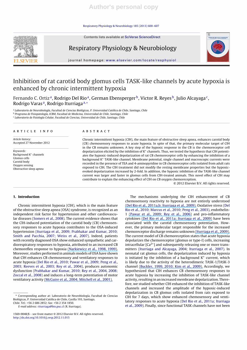
Isolated glomus cells were exposed to hypoxic superfusion(PO2 ∼5 mmHg) for 30 s while recording TASK-like currents. Thehypoxic stimulus produced a marked reduction of the channelactivity (NPo) in control and CIH-treated glomus cells (Fig. 3),without significant changes in the current amplitude (Fig. 4A). Thehypoxic inhibition of NPo in both groups was voltage-independent(not shown) and had no effects on the unitary current amplitude(Fig. 4A). We found that the hypoxic stimuli in control cells evokeda reversible inhibition of the standardized NPo of 62.3 ± 2.4% com-pared to the previous normoxic condition (P < 0.05, Kruskal–Wallisfollowed by Dunn’s post hoc test, n = 16; Fig. 3B). In the cells fromCIH-treated rats, the inhibition was 96.4 ± 1.5% compared with theprevious normoxic condition (P < 0.05, Kruskal–Wallis followedby Dunn’s post hoc test, n = 12 cells; Fig. 3B). Thus, acute hypoxiainhibition of TASK-like currents was significantly larger (P < 0.05,Student t test) in cells from CIH-treated than from control animals.Moreover, the inhibitory effect of acute hypoxia on NPo was fasterin CIH-treated cells related to the control group (�50 of 9.0 ± 0.4 sin 16 control cells vs. 2.3 ± 0.4 s in 12 CIH-cells; P < 0.05, Student ttest; Fig. 3B).
In control cells the histogram of inter-event intervals for thefirst open state of the channel showed that >90% of the openevents grouped in the 0–5 ms interval. During acute hypoxia, thedistribution of the inter-event intervals changed to longer durationintervals, reaching up to 40 ms (Fig. 4B). Similarly, the dwell-timeof opening for the OI level during normoxia (� = 1.1 ± 0.5 ms;n = 12) was significantly reduced (P < 0.05; Student t test) by the

Author's personal copy
604 F.C. Ortiz et al. / Respiratory Physiology & Neurobiology 185 (2013) 600– 607
Fig. 2. Expression of TASK-1 and TASK-3 channel subunits in the CB chemoreceptor cells from adult rats. (A) cDNA bands corresponding to the amplification of TASK-1and TASK-3 products of 450 base pairs (bp) and ∼540 bp, respectively. Left row bp standards. Middle and right row duplicates of the CB samples. (B) Immunohistochemicalstaining for TASK-1 and TASK-3 subunits in the CB tissue from an adult rat. Inset, negative control. (C) Immunocytochemical detection of TASK-1 and TASK-3 subunits inisolated CB cells from an adult rat. Double-labeling of immunofluorescence for TASK-1 or TASK-3 (green) with the glomus cell marker tyrosine hydroxylase (TH, red), showingco-localization of the markers.
Fig. 3. Effect of CIH on the hypoxic-induced inhibition of TASK-like channels. (A) Cell-attached recording (Vp = 0 mV) of single channels from a control cell (left panel) and aCIH-treated cell (right panel) during normoxia, acute hypoxia (PO2 ∼ 5 mmHg) and recovery. Scale bar: 1 pA, 50 ms. (B) Time-course of the NPo inhibition induced by acutehypoxia (bar) in control cells (left panel) and CIH-treated cells (right panel). Acute hypoxia produced a larger and faster inhibition of the TASK-like channel open probabilityin cells from CIH rats. In 16 control cells the hypoxic stimulus evoked a reversible inhibition of the standardized NPo of 62.3 ± 2.4%, while in 12 cells from CIH group theinhibition was 96.4 ± 1.5% (P < 0.01, Mann–Whitney). The half-time to the maximum effect (�50) was significantly (P < 0.05, Mann–Whitney test) faster in the CIH-treatedcells (2.3 ± 0.4 ms; n = 12) than in control cells (9.0 ± 0.4 ms; n = 16). Note that only 30 s of stimulation was enough to trigger the NPo inhibition.

Author's personal copy
F.C. Ortiz et al. / Respiratory Physiology & Neurobiology 185 (2013) 600– 607 605
Fig. 4. Effect of CIH on TASK-like channel inhibition induced by acute hypoxia. (A) Amplitude histograms for channel recordings showed in Fig. 3A. In cells from control andCIH-treated rats the hypoxic stimuli decreased the number of open events without changing the current amplitude. (B) Inter-event interval and dwell-time histograms forthe main open level in a control cell. Note that hypoxic stimuli modified the distributions of both the inter-event interval and dwell time.
Table 1Resting membrane properties of adult CB glomus cells from control and CIH rats.
Control CIH
Vm (mV) −54.5 ± 1.9 (58) −50.1 ± 1.5 (22)Rin (M�) 226 ± 68 (12) 291 ± 34 (20)Cm (pF) 0.98 ± 0.3 (11) 1.1 ± 0.2 (18)
Vm , resting membrane potential, Rin, input resistance, Cm membrane capacity. Nodifference between control and CIH groups were observed (P > 0.05, Unpaired Ttest. Values correspond to average ± SEM. Number of recorded cells is indicated inbrackets.
acute hypoxic challenge (� = 0.4 ± 0.1 ms, n = 12). It should be notedthat in spite of this value is near the limit of resolution given ourloss-pass filter of 2 kHz we did not observed any effects on singleevents kinetics or burst hallmarks. Because TASK-like channelactivity inhibition by acute hypoxia was larger than 95% in theCIH-treated cells, it was not possible to perform the equivalentanalyses for these cells, since the minimum number of intervalsor open sates (dwell time of opening) required to perform theanalysis was not reached.
3.4. Enhanced TASK-like current inhibition induced by CIHcontributed to increase the amplitude of the membranedepolarization evoked by acute hypoxia
We did not find any statistical difference in the restingmembrane potential of glomus cells measured with conven-tional intracellular electrodes or with whole-cell configurationtechniques (−54.3 ± 2.4 mV (n = 30) and −55.1 ± 1.4 mV (n = 28);respectively); therefore we pooled the data. CIH did not modifythe resting membrane properties of CB glomus cells. No changeswere observed neither in the resting membrane potential nor inthe input resistance or in the glomus cell capacitance (Table 1).
Although the resting membrane potential was similar in bothgroups, the depolarization induced by acute hypoxia was enhancedby CIH exposure (Fig. 5A and C). Indeed, the amplitude of the depo-larization evoked by hypoxia (�VHx) was 2-fold larger in cells fromthe CIH-animals compared to control cells (�VHx = 27.5 ± 2.8 mVin 16 CIH-cells vs. �VHx 15.7 ± 1.5 mV in 20 control cells; P < 0.05,Kruskal–Wallis, followed by Dunn’s post hoc; Fig. 5A and C). Simi-larly, the amplitude of the depolarization evoked by acute hypoxia
when K+ voltage-dependent conductances were blocked by TEAand 4-AP in the bath solution was 3-fold higher in the CIH-treatedcells as compared to the control group (32.9 ± 1.1 mV in 12 CIH-cells vs. 11.3 ± 0.8 mV in 17 control cells; P < 0.05, Kruskal–Wallisfollowed by Dunn’s post hoc, Fig. 5B and C).
4. Discussion
4.1. General
We studied the effects evoked by CIH on the TASK-like channelactivity and the depolarization induced by acute hypoxia in CB glo-mus cells from adult rats. The main findings of this study are thatglomus cells of adult rats express TASK transcripts and proteins, aswell as functional TASK-like currents, and the exposure to CIH for 7days enhanced the hypoxic-induced inhibition of TASK-like chan-nels in glomus cells. Indeed, the inhibition of the TASK-like channelopening probability (NPo) induced by acute hypoxia was more pro-found and faster in glomus cells of CIH-treated than in controlrats. Similarly, although CIH did not modify the passive mem-brane properties of glomus cells in normoxia (PO2 ∼ 140 mmHg),the amplitude of the hypoxia-induced depolarization increased by2-fold, and by 3-fold in the absence or presence of TEA and 4-AP.Thus, present results strongly support the idea that CIH increasesthe hypoxic-induced depolarization by enhancing the inhibition ofTASK channels in glomus cell. Accordingly, this increased inhibi-tion may contribute to the potentiation of the rat CB chemosensoryresponses to acute hypoxia found after 7 days of CIH exposure (DelRio et al., 2011a; Iturriaga et al., 2009).
4.2. TASK channels in adult CB glomus cells
The participation of TASK channels in the CB hypoxic trans-duction process has been well established in the neonatal rat.Indeed, TASK-1, TASK-3 and TASK-1/3 heterodimers are function-ally expressed in neonatal CB glomus cells, being the TASK-1/3heterodimer the main hypoxic-sensitive conductance. Recently,Kim et al. (2011) reported that TASK channel activity in normoxicrat glomus cells did not differ between 0 and 16 days of birth, buthypoxic stimuli produced a progressive age-dependent decrease inTASK channel activity and a larger cell depolarization. The effect of

Author's personal copy
606 F.C. Ortiz et al. / Respiratory Physiology & Neurobiology 185 (2013) 600– 607
Fig. 5. Effect of CIH on CB cell depolarization induced by acute hypoxia. Recording of membrane potential during acute hypoxic stimuli (bar) in a control and in a CIH-treatedcell in the absence (A) and presence (B) of 4-AP and TEA in the bath solution. (C) Summary of the effects of CIH on the CB cell depolarization evoked by acute hypoxia instandard solution (20 control and 16 CIH-treated cells) and in the presence of 4-AP and TEA (17 controlTASK and 12 CIHTASK cells). *P < 0.05, Dunn test after Kruskal–Wallistest.
hypoxia on TASK channel activity was significantly larger in glomuscells from P16 to P18 when compared to cells obtained from P0 toP1 day old rats (Kim et al., 2011). In the present study, we demon-strated that TASK-like channels are functionally expressed in therat glomus cells from ∼90 days old rats, which present enhancedCB chemosensory and ventilatory responses to acute hypoxia after7 days of CIH exposure (Del Rio et al., 2010; Iturriaga et al., 2009).Our conclusion that TASK-like channels are functionally expressedin CB cells from adult rats is based on several lines of evidence.The membrane current recorded in the presence of TEA and 4-APwas reversible inhibited by hypoxia in glomus cells from both con-trol and CIH-treated rats, and therefore, could be involved in theadult CB hypoxic response to acute hypoxia. We found that thehypoxic challenges (PO2 ∼ 5 mmHg) evoked a reversible reductionof NPo of 62.3 ± 2.4% in glomus cells from adult control rats. Thisvalue is similar to the reported for the inhibition of TASK channelsin glomus cells from neonatal rats (Buckler et al., 2000). In addi-tion, the current recorded in CB cells from adult rats was inhibitedby acidification and bupivacaine, and the reduction of pipette [K+]from 140 to 70 mM resulted in an expected displacement of thereversal potential according to the expected Nernst equilibriumpotential for K+ ions. The unitary channel conductance measuredin our experiments, 15.4 ± 0.8 pS with 140 mM K+ and 4 mM Mg2+
in the patch pipette, was also similar to the one reported for glo-mus cells from neonatal rats (Buckler et al., 2000; Williams andBuckler, 2004; Kim et al., 2009). Since we used 4 mM Mg2+ we can-not exclude the possibility that other TASK channels are present,such as the occasional 34 pS and 78 pS single channel conductancesdescribed by Kim et al. (2009) for the neonatal rat CB.
Similarly to what was found in Sprague-Dawley neonatal rats(Kim et al., 2006) and adult Wistar rats (Yamamoto et al., 2002), ourimmunohistochemical studies support that glomus cells from adultrats express the TASK-1 and TASK-3 channel subunits. Therefore,there is strong evidence showing that TASK-1 and TASK-3 are bothexpressed at the mRNA and protein levels in the rat CB.
4.3. Contribution of TASK-like channels to the CB chemosensorypotentiation induced by CIH
Hypoxia causes depolarization of rat CB glomus cells, at leastin part, by inhibiting the background TASK K+ current (Buckler,1997; Buckler et al., 2000). In order to establish a relationshipbetween the increased hypoxic-inhibition of TASK-like channelopen activity and the augmented depolarization observed in glo-mus cells from rats exposed to CIH, we studied the hypoxia-induceddepolarization evoked by the inhibition of K+ background con-ductance when the voltage-gated K+ were blocked by TEA and4-AP. In this condition, the depolarization evoked in the control
cells was significantly smaller than the one observed in absenceof the blockers. This observation is not surprising since severalother voltage-dependent K+ channels (i.e. BK or KO2 channels) havebeen shown to be involved in the hypoxic response in glomus cells(López-Barneo et al., 1988; López-López et al., 1989; Peers, 1990).Thus, the depolarization evoked by hypoxia could be explainedmainly by TASK channel inhibition (11.3 ± 0.8 mV of 15.7 ± 1.5 mV;Fig. 5) while the remaining response can be attributed to voltage-dependent K+ channels blocked by TEA and/or 4-AP. Interestingly,when TEA and 4-AP were present in the bath solution, the depo-larization induced by acute hypoxia was about 3-fold in glomuscells from CIH-treated animals, while only a 2-fold depolariza-tion was observed in glomus cells from control rats. We ruledout the possibility that the CIH treatment increases TASK chan-nel expression (and therefore enhanced depolarization) because anincreased functional expression of a background K+ conductanceshould necessarily reduce the input resistance and displace theresting membrane potential toward more negative values, scenariothat we did not observed. Accordingly, CB type-I cells obtainedfrom a mice deficient in TASK channels, show higher membraneresistance and less hyperpolarized resting membrane potential(Ortega-Sáenz et al., 2010). The finding that in the presence ofTEA and 4-AP in the external solution, the depolarization inducedby acute hypoxia was about 3-fold in glomus cells from CIH-treated animals strongly suggests that the enhanced depolarizationobserved in glomus cells from rats exposed to CIH was explainedin part by a potentiation in the inhibition of TASK channel. Theresting membrane properties of glomus cells were not modifiedby CIH, although a small but non-significant reduction of Vm wasobserved in cell from CIH-treated animals (−52.5 ± 1.5 mV; n = 28)with respect to control cells (−54.5 ± 1.9; n = 58). However, TASK-like channel conductance appears not to be modified by CIH, sincethe current amplitude distribution histogram in CIH and controlglomus cells remain unchanged, suggesting that CIH has no directeffect on channel conductance. On the other hand, acute hypoxiain control glomus cells increased the inter-event interval to longerdurations and reduced the dwell-time for the main open state ofthe channel, suggesting that acute hypoxia decreases channel activ-ity by destabilizing the open state. Thus, CIH exposure may affectsome regulatory factors involved in the hypoxic control of TASK-like channel activity, rather than having a direct effect on channelnumber or expression.
In summary, present results showed that (i) mRNA and proteinof TASK channel subunits are present in rat adult glomus cells and(ii) CIH increased the acute hypoxia-induced inhibition of TASK-like K+ channels, enhancing glomus cell depolarization. This noveleffect of CIH may contribute to explain the enhancing effect of CIHon CB oxygen chemoreception.

Author's personal copy
F.C. Ortiz et al. / Respiratory Physiology & Neurobiology 185 (2013) 600– 607 607
Conflict of interest
No conflicts of interest, financial or otherwise are declared bythe author(s).
Acknowledgements
This work was supported by grant 1100405 from the NationalFund for Scientific and Technological Development of Chile(FONDECYT). Fernando Ortiz was supported by a CONICYT AT-fellowship.
References
Buckler, K.J., 1997. A novel oxygen-sensitive potassium–current in rat carotid bodytype I cells. Journal of Physiology 498, 649–662.
Buckler, K.J., 1999. Background leak K+-currents and oxygen sensing in carotid bodytype 1 cells. Respiration Physiology 115, 179–187.
Buckler, K.J., 2010. Two-pore domain K+ channels and their role in chemoreception.Advances in Experimental Medicine and Biology 66, 15–30.
Buckler, K.J., Honoré, E., 2004. Molecular strategies for studying oxygen-sensitive K+
channels. Methods in Enzymology 381, 233–256.Buckler, K.J., Williams, B., Honoré, E., 2000. An oxygen, acid- and anaesthetic sen-
sitive TASK-like background potassium channel in rat arterial chemoreceptorcells. Journal of Physiology 525, 135–142.
Del Rio, R., Moya, E.A., Iturriaga, R., 2010. Carotid body and cardiorespiratory alter-ations in intermittent hypoxia: the oxidative link. European Respiratory Journal36, 143–150.
Del Rio, R., Moya, E.A., Iturriaga, R., 2011a. Differential expression of pro-inflammatory cytokines, endothelin-1 and nitric oxide synthases in the ratcarotid body exposed to intermittent hypoxia. Brain Research 1395, 74–85.
Del Rio, R., Moya, E.A., Munoz, C., Arias, P., Court, F.A., Iturriaga, R., 2011b. Chronicintermittent hypoxia-induced vascular enlargement and VEGF upregulation inthe rat carotid body is not prevented by antioxidant treatment. American Journalof Physiology Lung Cellular and Molecular Physiology 301, L702–L711.
Hille, B., 2001. Ion Channels of Excitable Membranes, 3rd ed. Sinauer Associates,Sutherland, MA, USA.
Iturriaga, R., Alcayaga, J., 2004. Neurotransmission in the carotid body: transmittersand modulators between glomus cells and petrosal ganglion nerve terminals.Brain Research Reviews 47, 46–53.
Iturriaga, R., Moya, E.A., Del Rio, R., 2009. Carotid body potentiation induced byintermittent hypoxia: implications for cardiorespiratory changes induced bysleep apnoea. Clinical and Experimental Pharmacology and Physiology 36,1197–1204.
Iturriaga, R., Varas, R., Alcayaga, J., 2007. Electrical and pharmacological proper-ties of petrosal ganglion neurons that innervate the carotid body. RespiratoryPhysiology and Neurobiology 157, 130–139.
Kim, D., Cavanaugh, E.J., Kim, I., Carroll, J.L., 2009. Heteromeric TASK-1/TASK-3 is themajor oxygen-sensitive background K+ channel in rat carotid body glomus cells.Journal of Physiology 587, 2963–2975.
Kim, I., Kim, J.H., Carroll, J.L., 2006. Postnatal changes in gene expression of subfami-lies of TASK K+ channels in rat carotid body. Advances in Experimental Medicineand Biology 580, 43–47.
Kim, D., Papreck, J.R., Kim, I., Donnelly, D.F., Carroll, J.L., 2011. Changes in oxygen sen-sitivity of TASK in carotid body glomus cells during early postnatal development.Respiratory Physiology and Neurobiology 177, 228–235.
Koizumi, H., Smerin, S.E., Yamanishi, T., Moorjani, B.R., Zhang, R., Smith, J.C., 2010.TASK channels contribute to the K+-dominated leak current regulating respira-tory rhythm generation in vitro. Journal of Neuroscience 30, 4273–4284.
López-Barneo, J., López-López, J.R., Urena, J., González, C., 1988. Chemotransductionin the carotid body: K+ current modulated by pO2 in type I chemoreceptor cells.Science 241, 580–582.
López-López, J., González, C., Urena, J., López-Barneo, J., 1989. Low pO2 selectivelyinhibits K channel activity in chemoreceptor cells of the mammalian carotidbody. Journal of General Physiology 93, 1001–1015.
Marcus, N.J., Li, Y.L., Bird, C.E., Schultz, H.D., Morgan, B.J., 2010. Chronic intermit-tent hypoxia augments chemoreflex control of sympathetic activity: role of theangiotensin II type 1 receptor. Respiration Physiology and Neurobiology 171,36–45.
McGuire, M., Zhang, Y., White, D.P., Ling, L., 2004. Serotonin receptor subtypesrequired for ventilatory long-term facilitation and its enhancement after chronicintermittent hypoxia in awake rats. American Journal of Physiology Regulatory,Integrative and Comparative Physiology 286, R334–R341.
Mitchell, G.S., Powell, F.L., Hopkins, S.R., Milsom, W.K., 2001. Time domains of thehypoxic ventilatory response in awake ducks: episodic and continuous hypoxia.Respiration Physiology 124, 117–128.
Narkiewicz, K., Pesek, C.A., Kato, M., Phillips, B.G., Davison, D.E., Somers, V.K., 1998.Baroreflex control of sympathetic nerve activity and heart rate in obstructivesleep apnea. Hypertension 32, 1039–1043.
Narkiewicz, K., Van de Borne, P.J., Pesek, C.A., Dyken, M.E., Montano, N., Somers, V.K.,1999. Selective potentiation of peripheral chemoreflex sensitivity in obstructivesleep apnea. Circulation 99, 1183-1119.
Ortega-Sáenz, P., Levitsky, K.L., Marcos-Almaraz, M.T., Bonilla-Henao, V., Pascual, A.,López-Barneo, J., 2010. Carotid body chemosensory responses in mice deficientof TASK channels. Journal of General Physiology 135, 379–392.
Pawar, A., Nanduri, J., Yuan, G., Khan, S.A., Wang, N., Kumar, G.K., Prabhakar, N.R.,2009. Reactive oxygen species-dependent endothelin signaling is required foraugmented hypoxic sensory response of the neonatal carotid body by inter-mittent hypoxia. American Journal of Physiology Regulatory, Integrative andComparative Physiology 296, R735–R742.
Peers, C., 1990. Hypoxic suppression of K+ currents in type-I carotid-body cells-selective effect on the Ca2+-activated K+ current. Neuroscience Letters 119,253–256.
Peng, Y.J., Overholt, J.L., Kline, D., Kumar, G.K., Prabhakar, N.R., 2003. Inductionof sensory long-term facilitation in the carotid body by intermittent hypoxia:implications for recurrent apneas. Proceedings of the National Academy of Sci-ences of the United States of America 100, 10073–10078.
Prabhakar, N.R., Kumar, G.K., 2010. Mechanisms of sympathetic activation and bloodpressure elevation by intermittent hypoxia. Respiratory Physiology and Neuro-biology 174, 156–161.
Reeves, S.R., Gozal, E., Guo, S.Z., Sachleben Jr., L.R., Brittian, K.R., Lipton, A.J., Gozal, D.,2003. Effect of long-term intermittent and sustained hypoxia on hypoxic ven-tilatory and metabolic responses in the adult rat. Journal of Applied Physiology95, 1767–1774.
Rey, S., Del Rio, R., Alcayaga, J., Iturriaga, R., 2004. Chronic intermittent hypoxiaenhances cat chemosensory and ventilatory responses to hypoxia. Journal ofPhysiology 560, 577–586.
Rey, S., Del Rio, R., Iturriaga, R., 2006. Contribution of endothelin-1 to the enhancedcarotid body chemosensory responses induced by chronic intermittent hypoxia.Brain Research 1086, 152–159.
Rey, S., Tarvainen, M.P., Karjalainen, P.A., Iturriaga, R., 2008. Dynamic time-varyinganalysis of heart rate and blood pressure variability in cats exposed to short-term chronic intermittent hypoxia. American Journal of Physiology Regulatory,Integrative and Comparative Physiology 295, R28–R37.
Smith, M.L., Pacchia, C.F., 2007. Sleep apnoea and hypertension: role of chemore-flexes in humans. Experimental Physiology 92, 45–50.
Somers, V.K., White, D.P., Amin, R., Abraham, W.T., Costa, F., Culebras, A., Daniels, S.,Floras, J.S., Hunt, C.E., Olson, L.J., Pickering, T.G., Russell, R., Woo, M., Young, T.,2008. Sleep apnea and cardiovascular disease. Journal of the American Collegeof Cardiology 52, 686–717.
Weiss, J.W., Liu, M.D., Huang, J., 2007. Physiological basis for a causal relation-ship of obstructive sleep apnoea to hypertension. Experimental Physiology 92,21–26.
Williams, B.A., Buckler, K.J., 2004. Biophysical properties and metabolic regu-lation of a TASK-like potassium channel in rat carotid body type 1 cells.American Journal of Physiology Lung Cellular and Molecular Physiology 286,L221–L230.
Yamamoto, Y., Kummer, W., Atoji, Y., Suzuki, Y., 2002. TASK-1, TASK-2, TASK-3and TRAAK immunoreactivities in the rat carotid body. Brain Research 950,304–307.
Zoccal, D.B., Simms, A.E., Bonagamba, L.G., Braga, V.A., Pickering, A.E., Paton, J.F.,Machado, B.H., 2008. Increased sympathetic outflow in juvenile rats submittedto chronic intermittent hypoxia correlates with enhanced expiratory activity.Journal of Physiology 586, 3253–3265.
Copyright © 2022 FDOKUMEN