Influence of Intermittent Hypobaric Exposure on SOD and TBARS Levels in Trained Rats

Oxidative Stress in Rat Testis and Epididymis Under IntermittentHypobaric Hypoxia: Protective Role of Ascorbate Supplementation
JORGE GONZALO FARIAS,* MARIELA PUEBLA,* ALEJANDRO ACEVEDO,* PABLO JOSE TAPIA,*
EDUARDO GUTIERREZ,* ANDREA ZEPEDA,* GLORIA CALAF,{ CAMILA JUANTOK,* AND
JUAN G. REYES{
From the *Instituto de Biotecnologıa de Tarapaca, Universidad Arturo Prat, Iquique, Chile; the �Instituto de Alta
Investigacion Universidad de Tarapaca, Arica, Chile; and the `Instituto de Quımica, Pontificia Universidad Catolica de
Valparaıso, Chile.
ABSTRACT: Hypobaric hypoxia (HH), an environmental condition
of high altitude encountered by mountaineers, miners, and observa-
tory, rural health, border patrol, and rural education workers,
jeopardizes normal physiologic functions in humans. The present
study was conducted to evaluate the effects of intermittent HH (IHH;
equivalent to 4600 m above mean sea level) on oxidative stress and
the protective role of dietary ascorbic acid on rat testis and
epididymis. Ten-week-old male Wistar rats were assigned to 1 of 6
groups: 1) normobaric (Nx), 2) Nx + physiologic solution (Nx + PS),
3) Nx + ascorbic acid (Nx + AA), 4) IHH, 5) IHH + PS, or 6) IHH + AA.
Animals subjected to IHH were exposed for 96 hours followed by
normobaric conditions for 96 hours for a total of 32 days. The control
groups (2 and 5) were injected with doses of PS, and the treated
groups (3 and 6) were injected with doses of AA (10 mg 6kg21 body
weight) at an interval of 96 hours. Rats were sacrificed on day 32
after initiation of the protocol. The testis and epididymis were
collected to determine the activity and expression of glutathione
reductase and the levels of lipid peroxide formation. An epididymal
sperm count was also performed in each animal. The results of this
study revealed that IHH induced lipid peroxidation, a reduction in
glutathione reductase activity in testis and epididymis, and a
significant decrease in epididymal sperm count. Treatment with AA
prevented these changes. In conclusion, AA was capable of
decreasing oxidative stress in testis and epididymis under IHH. This
protection by AA of the IHH-induced lipid peroxidation can be
explained in part by the preservation of glutathione reductase activity
in these organs.
Key words: Lipid peroxidation, glutathione reductase, ascorbic
acid.
J Androl 2010;31:314–321
I t has been suggested that hypobaric hypoxia (HH)
reduces fertility in humans. However, epidemiologic
studies of high- and low-altitude populations have not
been able to support this hypothesis (Vitzthum and
Wiley, 2003; Bartsch et al, 2004). Previous publications
from our laboratories showed that the exposure of male
rats to continuous chronic HH and intermittent chronic
HH induced evident changes in testicular morphology,loss of germinal cells, arrest of spermatogenesis, and
metabolic stress in the mitochondria of round sperma-
tids, consistent with oxygen-consumption processes
related to lipid peroxidation (Farias et al, 2005b).
Furthermore, high-altitude exposure has been shown
to induce oxidative stress (Vats et al, 2008) that is
accompanied by decreased levels of reduced glutathione
(GSH) and ascorbic acid (AA) and by an increase in
antioxidant enzymes like glutathione reductase (GR).
Thus, a likely mechanism of HH-induced inhibition of
spermatogenesis and sperm production (eg, Bustos-
Obregon and Olivares, 1982; Farias et al, 2005b) can
be related to oxidative stress in male reproductive
organs.
Lipid peroxides have been implicated in decreased
organ weight, tissue damage, cell loss, and cellular aging
processes (Koksal et al, 2003; Voss and Siems, 2006).
Antioxidants arrest these processes (Frei, 1999; Gilgun-
Sherki et al, 2002) and prevent oxidation by inactivating
free radicals or reactive oxygen species (ROS). AA or
vitamin C is a water-soluble, nonenzymatic antioxidant
that is able to react with aqueous free radicals and ROS
and has the potential to protect both cytosolic and
membrane components of cells from oxidative damage
(Devi et al, 2007).
The glutathione system (and enzymatic antioxidant
mechanism) plays an essential role in preventing
oxidative damage in cells and tissues (Meister and
Anderson, 1983). The levels of GSH are maintained by
the action of GR (EC 1.8.1.7), which recycles oxidized
This work was supported by Instituto de Biotecnologıa de Tarapaca
(Innova-CORFO project grant 06FCO11BC-118) and DI 2007 Arturo
Prat University.
Correspondence to: Jorge Gonzalo Farias, Instituto de Biotecnolo-
gıa de Tarapaca, Universidad Arturo Prat, Av Arturo Prat 2120,
Iquique, Chile (e-mail: [email protected]).
Received for publication November 3, 2008; accepted for publica-
tion October 13, 2009.
DOI: 10.2164/jandrol.108.007054
Journal of Andrology, Vol. 31, No. 3, May/June 2010Copyright E American Society of Andrology
314

glutathione (GSSG) to GSH using NADPH as the
electron donor (Meister and Anderson, 1983). It has
been reported that GR activity is affected by ROS and
that AA is capable of preventing this effect (El-Missiry,
1999).
The present study, which was designed to understand
the mechanisms associated with spermatogenic and
sperm production impairment by HH, examined the
effects of IHH on body weight, testis and epididymis
weight, epididymal sperm count, lipid peroxidation and
activity and expression of GR in rat testis and
epididymis in the presence and absence of AA dietary
supplementation. Whereas IHH induced a decrease in
body weight and testis and epididymis weight, as well as
a decrease in GR expression and activity, it induced an
increase in lipid peroxidation. AA treatment played a
protective role in oxidative stress processes in testis and
epididymis under the IHH condition and maintained
GR activity at levels similar to normobaric conditions.
Materials and Methods
Experimental Design
Ten-week-old male Wistar rats (225 6 14 g; n 5 36) from the
University of Valparaiso Biotery were assigned to 1 of 6
groups (6 rats in each group): 1) normobaric conditions (Nx),
2) Nx + physiologic NaCl solution (Nx + PS), 3) Nx + AA, 4)
intermittent HH (IHH), 5) IHH + PS, and 6) IHH + AA. A
362-factorial experimental design was used considering 3
injection treatments (no injection, PS, and AA) and 2
environmental treatments (Nx and IHH). The animals were
injected (intraperitoneally) with doses of AA (10 mg 6 kg21
body weight) or vehicle (0.1 mL of PS) at intervals of 96 hours.
The dose of AA was that described by Acharya et al. (2008)
that had significantly inhibited lipid peroxidation induced by
CdCl2 in rat testicles. The IHH group of animals were exposed
to HH for 96 hours (428 torr; PO2 89.6 mm Hg) followed by
the Nx condition for 96 hours (96 hours of hypoxia/96 hours of
normoxia) for a total period of 32 days. Pressure changes in
the hypobaric chamber were achieved by steps of 150 mm Hg
per minute, which simulated altitude changes. The Nx animals
were housed in the same room next to the IHH animals (22 6
2uC, 15 g of food pellet per day, and 1 L of water per cage). All
procedures were performed in agreement with the Principles of
Laboratory Animal Care, advocated by the National Society
of Medical Research, and the Guide for the Care and Use of
Laboratory Animals (Institute of Animal Laboratory Resourc-
es, 1996)
Hematocrit
Blood samples were obtained by cardiac puncture of the left
ventricle. Hematocrit was determined by centrifugation of a
capillary tube with heparinized blood in a microhematocrit
centrifuge (IEC model MB; GSR Technical Sales, Edmondton,
Alberta, Canada).
Determination of ThiobarbituricAcid–Reactive Substances
Body weight was determined 32 days after initiation of the
protocol. The animals were then killed by cervical dislocation.
The testis and epididymis were collected in phosphate-buffered
saline (PBS; pH 7.2). Determination of thiobarbituric acid–
reactive substances (TBARS) was undertaken as an index of
lipid peroxidation (Magalhaes et al, 2005). TBARS were
estimated at 532 nm, and their concentrations were calculated
using a molar extinction coefficient of 1.56 6 105 M21 cm21
obtained utilizing malondialdehyde (MDA; Sigma-Aldrich, St
Louis, Missouri) as a standard. The results were expressed as
nmol of MDA equivalents/mg tissue.
Isolation and Sperm Count
Epididymal spermatozoa were separated by cutting the caudal
epididymis into segments of approximately 1 mm3 with a sharp
razor blade in 1 mL of PBS (pH 7.2). Spermatozoa from
caudal regions were completely removed by vortexing gently in
PBS, and the tissue debris was allowed to settle for 5 minutes.
Spermatozoa released in the buffer were aspirated, centri-
fuged at 800 6 g for 15 minutes, and used for biochemical
determinations. All of these procedures were completed at 4uC.
The number of sperm in the suspension was counted using a
Neubauer chamber.
Preparation of Tissue Homogenates and Protein Assay
Testis and epididymis tissues were homogenized in 0.5 mL of
extraction buffer (Tris 50 mM, NaCl 100 mM, EDTA 1 mM,
EGTA 2.5 mM, Tween-20 0.1% [pH 7.4], phenylmethylsulfo-
nyl fluoride [PMSF] 100 mg/mL; Sigma-Aldrich) with a Potter
homogenizer (Glass-Col K4424; Glas-Col, Terre Haute,
Indiana) at .050301 6 g. The samples were then centrifuged
at 7820 6g for 30 minutes at 4uC. Protein concentrations were
determined using the Coomassie blue method (Bradford,
1976).
SDS-PAGE and Western Blot Analysis
Protein samples were subjected to 12% sodium dodecyl sulfate
polyacrylamide gel electrophoresis (SDS-PAGE) and then
transferred to Hybond-C (Amersham Pharmacia, Piscataway,
New Jersey) under semidry conditions by means of a Trans-
Blot SD Semi-Dry Transfer Cell (Bio-Rad, Tokyo, Japan).
The membranes were then blocked by incubation with 5%
skimmed milk in PBS (pH 7.2) for 1 hour at room
temperature. Subsequently, the membranes were incubated
with rabbit anti–rat GR Ig (1:500 dilution) (Santa Cruz
Biotechnology, Santa Cruz, California) for 12 hours at 4uC.
After washing with PBS (pH 7.2) containing 0.05% Tween-20,
the membranes were incubated with 1:1000 diluted peroxidase-
conjugated goat anti–rabbit IgG Ig (Jackson ImmunoResearch
Laboratories, West Grove, Pennsylvania) for 2 hours. After
washing, peroxidase activity was detected by a chemilumines-
cence method using an ECL Plus kit (Amersham Pharmacia).
An anti–b-tubulin antibody was used as a protein-loading
control in Western blots. The immunoblots were analyzed with
ImageJ software (http://rsb.info.nih.gov/ij).
Farias et al N Hypoxia and Oxidative Stress in Testis and Epididymis 315

Enzyme Assay
GR activity was determined spectrophotometrically by mea-
suring the rate of NADPH oxidation at 340 nm. The reaction
mixture consisted of 0.2 M potassium phosphate (pH 7.3;
Merck KGaA, Darmstadt, Germany), 3.9 mM NADPH
(Sigma-Aldrich), 20 mM GSSG (Merck KGaA), and tissue
homogenates. The decrease in absorbance at 340 nm at 21uCwas recorded as a function of time. The control reaction
mixture contained buffer instead of NADPH. One unit of GR
activity was defined as the amount of enzyme that catalyzed
the oxidation of 1 mmol of NADPH per minute (Carlberg and
Mannervik, 1985). All assays were performed in triplicate, and
means 6 SE are reported.
Statistical Analysis
The results were analyzed using a 2-way analysis of variance
(ANOVA) test followed by Bonferroni analysis. Two-way
ANOVA was performed to determine the presence of a
significant interaction between injection (AA) and environ-
ment because this would indicate a different effect of AA
under IHH in comparison to Nx. The Kruskal-Wallis test
(nonparametric) was used to analyze hematocrit results. The
level of statistical significance was set at P , .05 for all tests.
The data were analyzed using the GraphPad Prism software
version 4.0 (GraphPad Software, San Diego, California). The
results are presented as means 6 SE.
Results
Body, Testicular, and Caudal Epididymis Weight in IHH
According to the 2-way ANOVA on body-weight values
(Figure 1), the interaction between environment and
injection (AA) was significant (P , .001) (ie, AA had a
different effect on body weight in the IHH condition
compared with the Nx condition). Also, IHH signifi-
cantly affected the body-weight results (P ,
.0001).There was a significant difference (P , .01)
between IHH + AA with respect to IHH + PS and IHH,
and there were no significant differences in the rest of
the comparisons within hypobaric and Nx groups.
Complementary analysis (1-way ANOVA; data not
shown) showed that there was a significant difference
(P , .05) in body weight between the Nx vs IHH groups
(no injection) and no significant difference (P . .05)
between the IHH + AA group vs all Nx groups.
Treatment with AA abolished the effects of decreased
rat body weight induced by hypoxia. The loss in body
weight of hypoxic rats observed in this study (approx-
imately 1.5 g/d in hypoxic animals without AA
treatment) was not expected to give rise to significant
reproductive organ weight loss in adult animals (eg,
Lanning et al, 2002; Fleeman et al, 2005). However,
because of a possible correlation between both param-
eters, the data for testis and epididymis weight were
normalized to body weight for each animal (Table 1).
Testis and epididymis relative weight analysis (Ta-
ble 1) showed that the interaction between environment
and injection was not significant (P . .05; ie, the way
that AA affects Nx and IHH conditions is similar);
interestingly, the environment significantly affected only
the testis results (P , .05; ie, there was a testicular mass
loss induced by IHH). There was no significant
difference (P . .05) between hypobaric and Nx groups.
Rat Hematocrit in IHH and Nx Groups
The hematocrit was significantly greater in all of the HH
groups in comparison with the Nx groups (P , .05)
after 32 days of IHH. There were no significant
differences between groups treated with AA and their
respective IHH and control groups (P . .05) (Table 2),
indicating that the IHH condition effectively triggered
the characteristic hypoxia-induced increase in red blood
cell production and that AA did not affect this
physiologic response to hypoxia.
Oxidative Stress in Testis and Epididymis
The analysis of TBARS formation in testis and
epididymis (Figure 2) showed that the interaction
between environment and injection was significant (P
, .05) in both testis and epididymis; this indicates that
AA has a different effect on lipoperoxidation under
IHH conditions compared with Nx conditions. Also,IHH significantly affected the results (P , .0001). In
testis and epididymis, there was a significant difference
between IHH + AA with respect to IHH + PS and IHH
(P , .01 and P , .01, respectively), and there were no
significant differences in the rest of the comparisons
within the hypobaric and Nx groups. Complementary
Figure 1. Effect of IHH and AA on body weight. Statistical analysis: 2-way analysis of variance followed by Bonferroni analysis. Nxindicates normobaric conditions; PS, physiologic NaCl solution; AA,ascorbic acid; IHH, intermittent hypobaric hypoxia; a, P , .05, IHH vsNx; b, P , .05, IHH vs IHH + AA; c, P , .05, IHH + PS vs Nx; d,P , .05, IHH + PS vs IHH + AA.
316 Journal of Andrology N May �June 2010

analysis (1-way ANOVA; data not shown) showed that
there was a significant difference (P , .05) in lipid
peroxide formation between the Nx vs IHH (no
injection) groups and no significant difference (P .
.05) between IHH + AA vs all Nx groups. These results
strongly suggest a protective role of AA in animal
epididymis lipid peroxidation that results from exposure
to IHH (Figure 2B). Thus, in a pattern that resembles
the results observed in body weight and testicular and
epididymal weight, AA treatment decreased TBARS
content in both testis and epididymis. Figure 2 indicates
that under both Nx and IHH, the levels of lipid
peroxidation were higher in testis than in epididymis.
Only under IHH did the lipid peroxidation reach values
that were not significantly different in both organs.
However, the significance level was against a possible
interpretation that saline injection affected the levels of
lipid peroxidation in any of these 2 organs.
Caudal Epididymis Sperm Count
Sperm count values analysis (Figure 3) showed that the
interaction between environment and injection was not
significant (P . .05), indicating that under Nx and IHH
conditions, AA affects the sperm count similarly. Also,
IHH significantly affected the sperm count values (P ,
.0001). There was a significant difference between IHH +AA with respect to IHH + PS (P , .01) and IHH (P ,
.001), and there were no significant differences in the rest
of the comparisons within hypobaric and Nx groups.
Complementary analysis (1-way ANOVA; data not
shown) showed that there was a significant difference
(P , .05) between the Nx vs IHH (no injection) groups
and no significant difference (P . .05) between IHH +AA groups vs all Nx groups. Hence, IHH did not
produce any significant decline in the sperm count in
IHH animals treated with AA compared with Nx groups.
Expression of GR in Testis and Epididymis
The interaction between environment and injection in
testis was not significant (P . .05), and environmental
treatment did not significantly affect the relative
expression values (P . .05). No significant differences
were found in testis among the different groups (P .
.05; Figure 4A). In epididymis, the interaction between
environment and injection was significant (P , .05), and
environmental treatment significantly affected the val-
ues (P , .05), indicating an activating effect of IHH on
epididymis GR expression. Also, there was a significant
difference (P , .01) between IHH + AA with respect to
IHH, strongly suggesting that AA was able to inhibit the
IHH-induced GR expression in epididymis (P , .01;
Figure 4B). No significant differences (P . .05) were
observed in the rest of the comparisons within hypo-
baric and Nx groups. Complementary analysis (1-way
ANOVA; data not shown) showed that there was a
significant difference (P , .05) between the Nx vs IHH
(no injection) and no significant difference (P . .05)
between IHH + AA vs all Nx groups.
Activity of GR in Testis and Epididymis
The interaction between environment and injection in
testis and epididymis was not significant (P . .05), but
environmental treatment significantly affected the rela-
tive expression values (P , .05). AA groups were
significantly different with respect to PS and no injection
groups (Figure 5A and B) in both the Nx and IHH
treatment groups. Complementary analysis (1-way
ANOVA; data not shown) showed that there was a
Table 1. Effect of IHH and AA on testis and epididymisa
Group Right testis, % Left testis, % Right epididymis, % Left epididymis, %
Nx .55 6 .00 .54 6 .00 .10 6 .00 .11 6 .01
Nx + PS .51 6 .02 .52 6 .01 .09 6 .01 .09 6 .00
Nx + AA .54 6 .02 .54 6 .03 .09 6 .01 .09 6 .01
IHH .50 6 .02 .50 6 .02 .08 6 .01 .09 6 .01
IHH + PS .47 6 .01 .47 6 .02 .08 6 .01 .09 6 .01
IHH + AA .50 6 .01 .50 6 .02 .09 6 .01 .09 6 .00
Abbreviations: AA, ascorbic acid; IHH, intermittent hypobaric hypoxia; Nx, normobaric conditions; PS, physiologic NaCl solution.a Values indicate the mean 6 SE of N 5 6. Testis and epididymis mass values are expressed as percentages. Statistical analysis: 2-way
analysis of variance followed by Bonferroni analysis. No significant difference was found in the normobaric and hypobaric group comparisons.
Table 2. Effect of IHH and AA on hematocrit a
Groups Hematocrit, %
Nx 36 6 2
Nx + PS 38 6 1
Nx + AA 37 6 2
IHH 50 6 2b
IHH + PS 49 6 2b
IHH + AA 51 6 3b
Abbreviations: AA, ascorbic acid; IHH, intermittent hypobaric
hypoxia; Nx, normobaric conditions; PS, physiologic NaCl solution.a Values indicate the mean 6 SD of N 5 6. Statistical analysis:
Kruskal-Wallis test (nonparametric) was used to analyze hemato-
crit values.b P , .05 vs Nx.
Farias et al N Hypoxia and Oxidative Stress in Testis and Epididymis 317

significant difference (P , .05) between the Nx vs IHH
(no injection) groups. The GR activity under IHH + AA
conditions in testis and epididymis suggests a protective
role of AA in animals exposed to IHH through the
maintenance of GR activity and lipid peroxidation
formation levels similar to those in the Nx condition.
Discussion
Previous results showed that high-altitude exposure
induced a decrease in testicular function that appeared
to be related to both direct early effects on spermato-
genesis and later effects on the hypophysis-gonad
hormonal axis (Farias et al, 2005a, 2008). The molecular
mechanisms associated with these changes are not
known. Hypoxia in general, and specifically HH, is
known to induce oxidative stress in animal models and
humans (Radak et al, 1994; Joanny et al, 2001, Askew,
2002; Jefferson et al, 2004; Magalhaes et al, 2004; Behn
et al, 2007; Vats et al, 2008), suggesting that ROS can be
involved in the changes in testicular function and
spermatogenesis observed in rats under HH conditions.
In this study, we showed that AA did not affect the
polycythemic response to HH, indicating that the
systemic hypoxic condition was present in all of the
IHH groups. Our results also showed that the body
weight was significantly lower in the intermittent IHH
groups than in Nx groups after 32 days of IHH, whereas
the body weight of IHH groups treated with AA were
similar to control rats after 32 days. Hence, considering
that body and organ weights are the result of both
anabolic and catabolic processes, it can be argued that
AA was able to counteract some of the effects of
hypoxia on metabolic balance in rats.
As previously described for sperm count from human
ejaculates (Bustos-Obregon E and Olivares. 1982), there
was a decrease in the number of epididymal sperm in the
animals subjected to IHH in comparison with Nx
groups. This effect of IHH on epididymal sperm count
was counteracted by AA, which normalized the sperm
count levels to those of Nx animals.
Our results indicate that IHH induced testicle and
epididymis lipid peroxidation (TBARS production).
Furthermore, such organs had higher lipid peroxidation
values under IHH than those of rats treated with AA in
the same hypoxic conditions, indicating that AA was
effectively acting by increasing antioxidant mechanisms
in these organs and IHH animals.
In the present study, we found different levels of GR
expression among the IHH groups, and the level of GR
expression was higher in the epididymis than in the
testis. There were no significant differences in GR
protein levels among the different groups (Nx, Nx + PS,
Figure 3. Effect of IHH and AA on caudal epididymis sperm count.The bars indicate the mean 6 SD of N 5 6. Statistical analysis: 2-way analysis of variance followed by Bonferroni analysis. Nxindicates normobaric conditions; PS, Nx physiologic NaCl solution;AA, ascorbic acid; IHH, intermittent hypobaric hypoxia; a, P , .05,IHH vs Nx; b, P , .05, IHH vs IHH + AA; c, P , .05, IHH + PS vs Nx;d, P , .05, IHH + PS vs IHH + AA.
Figure 2. Effect of IHH and AA on thiobarbituric acid–reactive substances formation (nmol of malondialdehyde/mg of tissues). (A) Testis. (B)Epididymis. The bars indicate the mean 6 SD of N 5 6. Statistical analysis: 2-way analysis of variance followed by Bonferroni test. Nx indicatesnormobaric conditions; PS, physiologic NaCl solution; AA, ascorbic acid; IHH, intermittent hypobaric hypoxia; a, P , .05, IHH vs Nx; b, P , .05,IHH vs IHH + AA; c, P , .05, IHH + PS vs Nx; d, P , .05, IHH + PS vs IHH + AA.
318 Journal of Andrology N May �June 2010

Nx + AA, IHH, IHH + PS, and IHH + AA) in the testis.
In contrast, the levels of GR in the epididymis in the IHH
group were significantly higher in comparison with those
of the Nx group. These data suggest that in epididymis,
the oxidative stress induced by IHH is capable of
triggering an up-regulation of GR expression levels as a
likely protective mechanism in response to increases in
ROS production (Vats et al, 2008). In other organs such
as brain (Maiti et al, 2006), HH appears to decrease
antioxidant mechanisms. In testis, the expression level
and activity of the protein were not modified by IHH.
Although this response mechanism was not observed
in testis, these data are in agreement with previous
reports, supporting the notion that the GR up-
regulation induced by ROS to prevent oxidative damage
is less important in testis than in epididymis (Ikeda et al,
1999). This difference is attributed to the fact that in the
testicle, arrest of the spermatogenic cycle is triggered as
Figure 4. Effect of IHH and AA on expression of glutathione reductase (GR). (A) Testis. (B) Epididymis. The bars indicate the mean 6 SD of N5 3. Statistical analysis: 2-way analysis of variance followed by Bonferroni analysis. Nx indicates normobaric conditions; PS, physiologic NaClsolution; AA, ascorbic acid; IHH, intermittent hypobaric hypoxia; a, P , .05, IHH vs Nx; b, P , .05, IHH vs IHH + AA; c, P , .05, IHH + PS vsNx; d, P , .05, IHH + PS vs IHH + AA.
Figure 5. Effect of IHH and AA on glutathione reductase (GR) activity. (A) Testis. (B) Epididymis. Values indicate mean 6 SD of N 5 6.Statistical analysis: 2-way analysis of variance followed by Bonferroni analysis. Nx indicates normobaric conditions; PS, physiologic NaClsolution; AA, ascorbic acid; IHH, intermittent hypobaric hypoxia; a, P , .05, IHH vs Nx; b, P , .05, IHH vs IHH + AA; c, P , .05, IHH + PS vsNx; d, P , .05, IHH + PS vs IHH + AA; e, P , .05, Nx + AA vs Nx; f, P , .05, Nx + AA vs Nx + PS.
Farias et al N Hypoxia and Oxidative Stress in Testis and Epididymis 319
an environmental protection measure, preventing the
generation of defective sperm and thus, a loss of
testicular mass (Table 1); in contrast, in epididymis,
the antioxidant response is much more important in
protecting the sperm.
Treatment with AA caused GR expression in IHH to
reach similar values as Nx in epididymis, suggesting thatby decreasing ROS production, AA supplementation was
capable of preventing the GR up-regulation mechanism.
Consistent with the results of GR expression, we observed
that GR activity was significantly higher in epididymis
than in testis, both under IHH and Nx conditions
(Kaneko et al, 2002). Additionally, AA induced an
increase in activity of GR under both Nx and IHH
conditions. These results are in agreement with the notionthat in testis, other antioxidant mechanisms besides those
that are glutathione dependent can also be active (Ikeda et
al, 1999). Our results demonstrating greater GR activity in
epididymis than in testis confirmed previously described
data published by Kaneko et al. (2002). Because AA does
not significantly affect lipoperoxidation in the testis and
epididymis under Nx conditions, the GR activity data that
show activation by AA under Nx and IHH conditionsstrongly suggest that AA could be preventing lipoperox-
idation in IHH by activating GR and that the oxidative
stress by IHH is well balanced by the glutathione
antioxidant system in both organs but might differ in its
initiating mechanisms from those oxidative processes that
give rise to Nx lipoperoxidation. No information in the
literature was found on the possible mechanisms by which
AA could activate GR activity in Nx testis and epididymishomogenates (El-Missiry, 1999).
Thus, our results indicated that AA alone can counter-
act the effects of hypoxia on body, testicular, andepididymal mass; protect against testicular and epididymal
lipid peroxidation; and preserve epididymal sperm count.
These results may have important practical conse-quences for those people subjected to IHH, such as sea-
level natives exposed to high altitude because of tourism,
border patrol, mining, or rural health and education
activities. Based on our results, it is expected that AA
treatment could counteract the deleterious effects of HH
on spermatogenesis and sperm count observed in animal
models and humans (as described in this article; Gasco
et al, 2003; Okumura et al, 2003; Farias et al, 2005b).
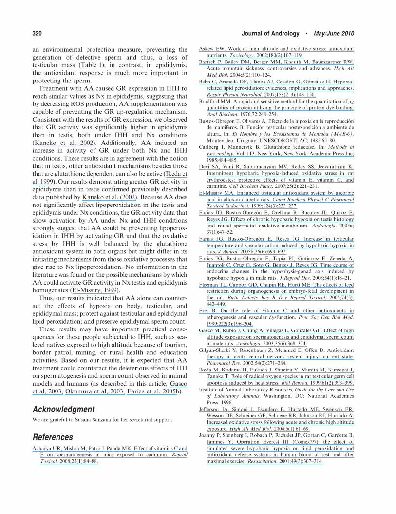
AcknowledgmentWe are grateful to Susana Sanzana for her secretarial support.
ReferencesAcharya UR, Mishra M, Patro J, Panda MK. Effect of vitamins C and
E on spermatogenesis in mice exposed to cadmium. Reprod
Toxicol. 2008;25(1):84–88.
Askew EW. Work at high altitude and oxidative stress: antioxidant
nutrients. Toxicology. 2002;180(2):107–119.
Bartsch P, Bailey DM, Berger MM, Knauth M, Baumgartner RW.
Acute mountain sickness: controversies and advances. High Alt
Med Biol. 2004;5(2):110–124.
Behn C, Araneda OF, Llanos AJ, Celedon G, Gonzalez G. Hypoxia-
related lipid peroxidation: evidences, implications and approaches.
Respir Physiol Neurobiol. 2007;158(2–3):143–150.
Bradford MM. A rapid and sensitive method for the quantitation of mg
quantities of protein utilizing the principle of protein dye binding.
Anal Biochem. 1976;72:248–254.
Bustos-Obregon E, Olivares A. Efecto de la hipoxia en la reproduccion
de mamiferos. B. Funcion testicular postexposicion a ambiente de
altura. In: El Hombre y los Ecosistemas de Montaoa (MAB-6).
Montevideo, Uruguay: UNESCOROSTLAC; 1982;65–80.
Carlberg I, Mannervik B. Glutathione reductase. In: Methods in
Enzymology. Vol. 113. New York, New York: Academic Press Inc;
1985;484–485.
Devi SA, Vani R, Subramanyam MV, Reddy SS, Jeevaratnam K.
Intermittent hypobaric hypoxia-induced oxidative stress in rat
erythrocytes: protective effects of vitamin E, vitamin C, and
carnitine. Cell Biochem Funct. 2007;25(2):221–231.
El-Missiry MA. Enhanced testicular antioxidant system by ascorbic
acid in alloxan diabetic rats. Comp Biochem Physiol C Pharmacol
Toxicol Endocrinol. 1999;124(3):233–237.
Farias JG, Bustos-Obregon E, Orellana R, Bucarey JL, Quiroz E,
Reyes JG. Effects of chronic hypobaric hypoxia on testis histology
and round spermatid oxidative metabolism. Andrologia. 2005a;
37(1):47–52.
Farias JG, Bustos-Obregon E, Reyes JG. Increase in testicular
temperature and vascularization induced by hypobaric hypoxia in
rats. J Androl. 2005b;26(6):693–697.
Farias JG, Bustos-Obregon E, Tapia PJ, Gutierrez E, Zepeda A,
Juantok C, Cruz G, Soto G, Benites J, Reyes JG. Time course of
endocrine changes in the hypophysis-gonad axis induced by
hypobaric hypoxia in male rats. J Reprod Dev. 2008;54(1):18–21.
Fleeman TL, Cappon GD, Chapin RE, Hurtt ME. The effects of feed
restriction during organogenesis on embryo-fetal development in
the rat. Birth Defects Res B Dev Reprod Toxicol. 2005;74(5):
442–449.
Frei B. On the role of vitamin C and other antioxidants in
atherogenesis and vascular dysfunction. Proc Soc Exp Biol Med.
1999;222(3):196–204.
Gasco M, Rubio J, Chung A, Villegas L, Gonzales GF. Effect of high
altitude exposure on spermatogenesis and epididymal sperm count
in male rats. Andrologia. 2003;35(6):368–374.
Gilgun-Sherki Y, Rosenbaum Z, Melamed E, Offen D. Antioxidant
therapy in acute central nervous system injury: current state.
Pharmacol Rev. 2002;54(2):271–284.
Ikeda M, Kodama H, Fukuda J, Shimizu Y, Murata M, Kumagai J,
Tanaka T. Role of radical oxygen species in rat testicular germ cell
apoptosis induced by heat stress. Biol Reprod. 1999;61(2):393–399.
Institute of Animal Laboratory Resources, Guide for the Care and Use
of Laboratory Animals. Washington, DC: National Academies
Press; 1996.
Jefferson JA, Simoni J, Escudero E, Hurtado ME, Swenson ER,
Wesson DE, Schreiner GF, Schoene RB, Johnson RJ, Hurtado A.
Increased oxidative stress following acute and chronic high altitude
exposure. High Alt Med Biol. 2004;5(1):61–69.
Joanny P, Steinberg J, Robach P, Richalet JP, Gortan C, Gardette B,
Jammes Y. Operation Everest III (Comex’97): the effect of
simulated severe hypobaric hypoxia on lipid peroxidation and
antioxidant defense systems in human blood at rest and after
maximal exercise. Resuscitation. 2001;49(3):307–314.
320 Journal of Andrology N May �June 2010

Kaneko T, Iuchi Y, Kobayashi T, Fujii T, Saito H, Kurashi H,
Fujii J. The expression of glutathione reductase in the male re-
productive system of rats supports the enzymatic basis of
glutathione function in spermatogenesis. Eur J Biochem. 2002;
269(5):1570–1578.
Koksal IT, Usta M, Orhan I, Abbasoglu S, Kadioglu A. Potential role
of reactive oxygen species on testicular pathology associated with
infertility. Asian J Androl. 2003;5(2):95–99.
Lanning LL, Creasy DM, Chapin RE, Mann PC, Barlow NJ, Regan KS,
Goodman DG. Recommended approaches for the evaluation of
testicular and epididymal toxicity. Toxicol Pathol. 2002;30(4):
507–520.
Magalhaes J, Ascensao A, Soares JM, Ferreira R, Neuparth MJ,
Oliveira J, Amado F, Marques F, Duarte JA. Acute and chronic
exposition of mice to severe hypoxia: the role of acclimatization
against skeletal muscle oxidative stress. Int J Sports Med.
2005;26(2):102–109.
Magalhaes J, Ascensao A, Viscor G, Soares J, Oliveira J, Marques F,
Duarte J. Oxidative stress in humans during and after 4 hours of
hypoxia at a simulated altitude of 5500 m. Aviat Space Environ
Med. 2004;75(1):16–22.
Maiti K, Mukherjee K, Gantait A, Saha BP, Mukherjee PK. Enhanced
therapeutic potential of naringenin-phospholipid complex in rats.
J Pharm Pharmacol. 2006;58(9):1227–1233.
Meister A, Anderson M. Glutathione. Annu Rev Biochem. 1983;52:
711–760.
Okumura A, Fuse H, Kawauchi Y, Mizuno I, Akashi T. Changes in
male reproductive function after high altitude mountaineering.
High Alt Med Biol. 2003;4(3):349–353.
Radak Z, Lee K, Choi W, Sunoo S, Kizaki T, Ohishi S, Suzuki K,
Taniguchi N, Ohno H, Asano K. Oxidative stress induced by
intermittent exposure at a simulated altitude of 4000 m decreases
mitochondrial superoxide dismutase content in soleus muscle of
rats. Eur J Appl Physiol Occup Physiol. 1994;69(5):392–405.
Vats P, Singh VK, Singh SN, Singh SB. Glutathione metabolism under
high-altitude stress and effect of antioxidant supplementation.
Aviat Space Environ Med. 2008;79(12):1106–1111.
Vitzthum VJ, Wiley AS. The proximate determinants of fertility in
population exposed to chronic hypoxia. High Alt Med Biol.
2003;4(2):125–139.
Voss P, Siems W. Clinical oxidation parameters of aging. Free Radic
Res. 2006;40(12):1339–1349.
Farias et al N Hypoxia and Oxidative Stress in Testis and Epididymis 321
Copyright © 2022 FDOKUMEN