Pharmacokinetic and electroencephalographic study of intravenous diazepam, midazolam, and placebo
Upload
independentCategory
view
3download
0
11/4/10 8:10 PMIntravenous Ascorbate as a Tumor Cytotoxic
Page 1 of 12file:///Users/admin/Desktop/Local%20Sites/Intravenous%20Ascorbate%20as%20a%20Tumor%20Cytotoxic.html
www.brightspot.org
The Center For The Improvement Of Human Functioning InternationalA Non-profit Medical, Research and Educational Organization
3100 North Hillside Avenue, Wichita, KS 67219 USAPhone: 316-682-3100; Fax: 316-682-5054
Intravenous Ascorbate as a TumorCytotoxic Chemotherapeutic Agent
From Medical Hypotheses (1995), 44, 207-213@ Pearson Professional Ltd 1995Date received 11 May 1994. Date accepted 24 August 1994
N. H. RIORDAN, H. D. RIORDAN, X. MENG, Y. Li and J. A. JACKSON*
Project RECNAC, Bio-Communications Research Institute, 3100 N. Hillside, Wichita,Kansas 67219, *Graduate School, Wichita State University, 1845 N. Fairmount,Wichita, Kansas 67260-0004, USA (Correspondence to NHR)
AbstractAscorbic acid and its salts (AA) are preferentially toxic to tumor cells in vitro and invivo. Given in high enough doses to maintain plasma concentrations above levelsthat have been shown to be toxic to tumor cells in vitro, AA has the potential toselectively kill tumor cells in a manner similar to other tumor cytotoxicchemotherapeutic agents. Most studies of AA and cancer to date have not utilizedhigh enough doses of AA to maintain tumor cytotoxic plasma concentrations of AA.Data are presented which demonstrate the ability to sustain plasma levels of AA inhumans above levels which are toxic to tumor cells in vitro and suggests thefeasibility of using AA as a cytotoxic chemotherapeutic agent.
Introduction
11/4/10 8:10 PMIntravenous Ascorbate as a Tumor Cytotoxic
Page 2 of 12file:///Users/admin/Desktop/Local%20Sites/Intravenous%20Ascorbate%20as%20a%20Tumor%20Cytotoxic.html
Cytotoxic drugs began to be considered consistently successful for therapy of somecancers around 1950 (1). A large jump in the cure rate for several cancer types -especially childhood, acute lymphoblastic leukemia, Hodgkin's disease, and testiculartumors - was seen between 1950 and 1990 (from 0% for all in 1950 to 75%, 80%and 90% respectively) (2). Other, relatively common, types of cancer (3), includinghead and neck, large bowel, stomach, pancreatic, liver, cervical, and melanoma, forthe most part remain refractory to cytotoxic chemotherapy, with and withoutadjuvant chemotherapy, with no demonstrable prolongation of life (2).
Even though the term 'chemotherapy' generally includes hormonal and cytotoxicagents, this discussion is limited to cytotoxic agents. Whether they are alkylatingagents, antimetabolites, or antibiotics, the rational for using chemotherapeutic agentsin the treatment of malignancy is to preferentially induce cytotoxicity of malignantcells. Because of the similarities between normal and malignant cells - both beingborn of the same host - a chemotherapeutic dose which is cytotoxic to cancer cellscan also be toxic to normal cells. Oncologists must often push the limits of acceptabletoxic side effects in order to effect a remission. Ideally, there should be a large gapbetween the lower dose required for efficacy and the higher dose of toxicity to thepatient. Adverse effects of chemotherapy include hair loss, nausea and vomiting,cardiac toxicity, and secondary cancers (4). One of the most common toxicmanifestations of many cytotoxic agents is bone marrow suppression (2) which canlead to immune suppression and hematopoietic dysfunctions. Because infectiouscomplications are one of the major causes of death in cancer patients (5), morehost-non-toxic compounds - particularly compounds without immune suppressivequalities - need to be investigated for their chemotherapeutic value.
There is a 10-100-fold greater content of catalase in normal cells than in tumor cells(6). This potentially creates a large gap between the toxic dose for normal cells andfor tumor cells of agents which induce hydrogen peroxide generation. Ascorbic acidand its salts (AA) are preferentially toxic to tumor cells in vitro (6-13) and in vivo(14-19). This preferential cytotoxicity has been demonstrated to be related tointracellular hydrogen peroxide generation (6,8,14). AA thus belongs in a class ofsubstances which, given at the correct dosage, can preferentially induce cytotoxicityof tumor cells with negligible toxic effects to the host.
This communication will demonstrate that plasma AA concentrations exceeding thoserequired to kill 100% of tumor cells in vitro can be sustained in humans and thatthose levels can generally only be obtained by intravenous administration of AA. Wepropose that intravenous AA, administered in sufficient doses to achieve plasmaconcentrations that have demonstrable cytotoxic effects on tumor cells, beinvestigated as a chemotherapeutic agent on the basis of the above facts and thefollowing positive qualities of AA:
1. Preferentially kills neoplastic cells.
2. Is virtually non-toxic at any dosage (20).
3. Does not suppress the immune system, unlike most chemotherapy agents.
4. Increases animal and human resistance to infectious agents by enhancinglymphocyte blastogenesis, enhancing cellular immunity, strengthening the
11/4/10 8:10 PMIntravenous Ascorbate as a Tumor Cytotoxic
Page 3 of 12file:///Users/admin/Desktop/Local%20Sites/Intravenous%20Ascorbate%20as%20a%20Tumor%20Cytotoxic.html
lymphocyte blastogenesis, enhancing cellular immunity, strengthening theextracellular matrix, and enhancing bactericidal activity of neutrophils andmodulation of complement protein (21-31).
5. Strengthens the structural integrity of the extracellular matrix which isresponsible for stromal resistance to malignant invasiveness (20).
Cellular In Vitro StudiesIn 1969, researchers at the NCI reported AA was highly toxic to Ehrlich ascites cellsin vitro. The goal of the study was to exploit the 10-100-fold lower catalase activityin tumor cells compared to normal cells. The proposed cytotoxic mechanism wasgeneration of toxic hydrogen peroxide. The toxicity was greatly enhanced byconcomitant administration of 3amino-1,2,4-triazole (ATA), a catalase inhibitor.Catalase and glucose added to the culture medium and a low oxygen tensionreduced the toxic effects of AA and ATA. The addition of vitamin K3 (menadionesodium bisulfite) to the medium overcame the protective effects of low oxygentension and glucose (6).
In 1977, Bram et al reported preferential AA toxicity for several malignant melanomacell lines, including four human-derived lines. They found that catalyticconcentrations of CU2+ greatly increased the preferential toxicity for melanoma cells(7). Another French group also found that AA and CU2+ were toxic to mousemelanoma cells in vitro. Noto et al reported that AA plus vitamin K3 had growthinhibiting action against three human tumor cell lines at non-toxic levels (8).Helgestad et al recently reported that new malignant T-cell line, isolated from a boywith malignant lymphoma, was very sensitive to AA in culture at concentrationsachievable in human plasma (9). In 1980, Park reported that several leukemic cellcultures were sensitive to AA at concentrations attainable in vivo, while normalhemopoietic cells were not suppressed (10). Metabolites of AA have also shownantitumor activity in vitro (11,12).
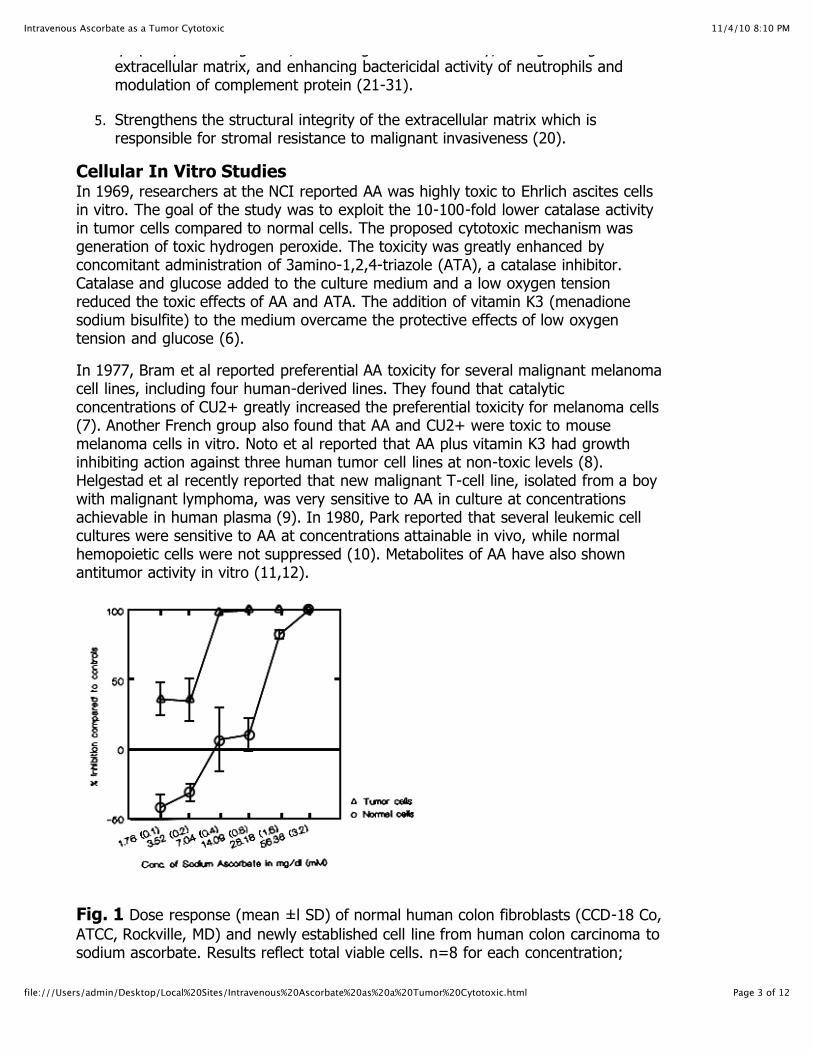
Fig. 1 Dose response (mean ±l SD) of normal human colon fibroblasts (CCD-18 Co,ATCC, Rockville, MD) and newly established cell line from human colon carcinoma tosodium ascorbate. Results reflect total viable cells. n=8 for each concentration;
11/4/10 8:10 PMIntravenous Ascorbate as a Tumor Cytotoxic
Page 4 of 12file:///Users/admin/Desktop/Local%20Sites/Intravenous%20Ascorbate%20as%20a%20Tumor%20Cytotoxic.html
cultured 4 days after supplementation of sodium ascorbate. Medium changed every24 h. DMEM Hi-glucose culture medium (Irvine Sci.) w/10% heat inactivated fetalcalf serum + antibiotics + Fungizone. 24-well (Nunc) tissue culture plates, 5% C02humidified incubator at 370C. 5000 cells/well seeded.
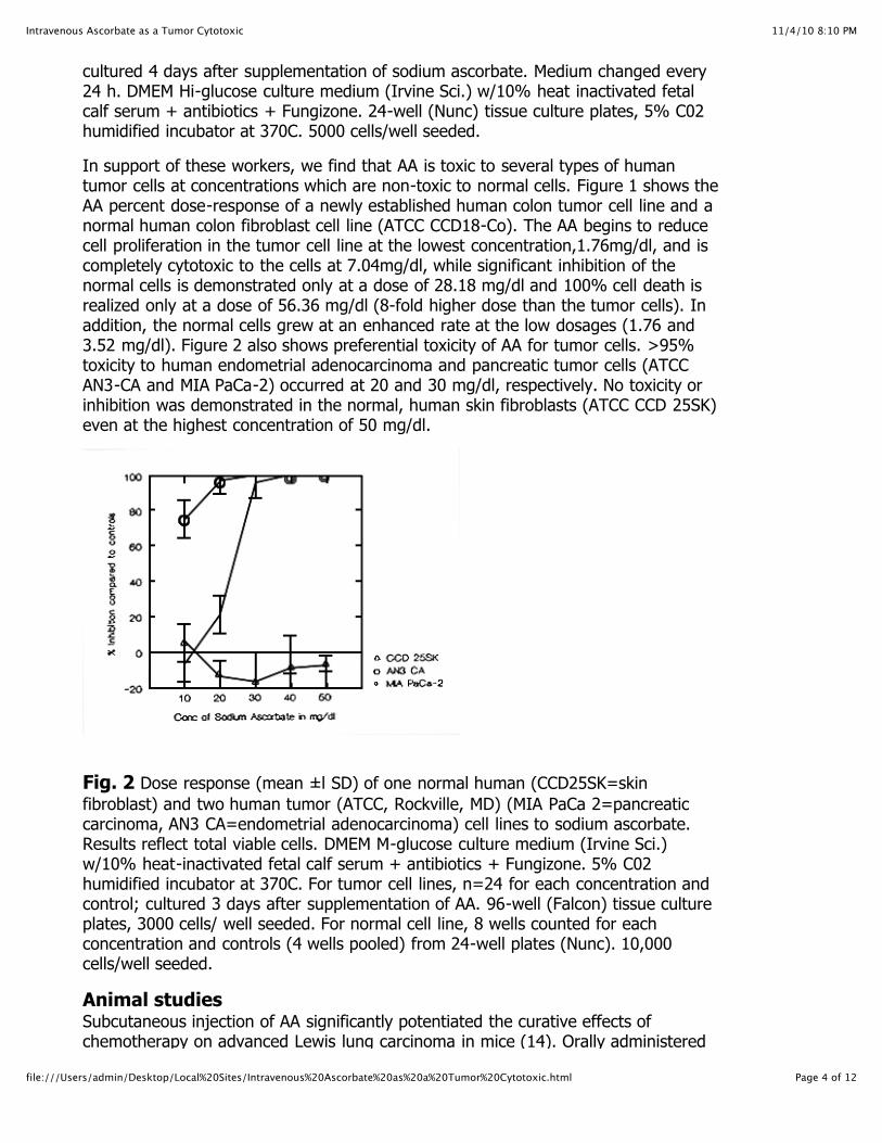
In support of these workers, we find that AA is toxic to several types of humantumor cells at concentrations which are non-toxic to normal cells. Figure 1 shows theAA percent dose-response of a newly established human colon tumor cell line and anormal human colon fibroblast cell line (ATCC CCD18-Co). The AA begins to reducecell proliferation in the tumor cell line at the lowest concentration,1.76mg/dl, and iscompletely cytotoxic to the cells at 7.04mg/dl, while significant inhibition of thenormal cells is demonstrated only at a dose of 28.18 mg/dl and 100% cell death isrealized only at a dose of 56.36 mg/dl (8-fold higher dose than the tumor cells). Inaddition, the normal cells grew at an enhanced rate at the low dosages (1.76 and3.52 mg/dl). Figure 2 also shows preferential toxicity of AA for tumor cells. >95%toxicity to human endometrial adenocarcinoma and pancreatic tumor cells (ATCCAN3-CA and MIA PaCa-2) occurred at 20 and 30 mg/dl, respectively. No toxicity orinhibition was demonstrated in the normal, human skin fibroblasts (ATCC CCD 25SK)even at the highest concentration of 50 mg/dl.
Fig. 2 Dose response (mean ±l SD) of one normal human (CCD25SK=skinfibroblast) and two human tumor (ATCC, Rockville, MD) (MIA PaCa 2=pancreaticcarcinoma, AN3 CA=endometrial adenocarcinoma) cell lines to sodium ascorbate.Results reflect total viable cells. DMEM M-glucose culture medium (Irvine Sci.)w/10% heat-inactivated fetal calf serum + antibiotics + Fungizone. 5% C02humidified incubator at 370C. For tumor cell lines, n=24 for each concentration andcontrol; cultured 3 days after supplementation of AA. 96-well (Falcon) tissue cultureplates, 3000 cells/ well seeded. For normal cell line, 8 wells counted for eachconcentration and controls (4 wells pooled) from 24-well plates (Nunc). 10,000cells/well seeded.
Animal studies Subcutaneous injection of AA significantly potentiated the curative effects ofchemotherapy on advanced Lewis lung carcinoma in mice (14). Orally administered
11/4/10 8:10 PMIntravenous Ascorbate as a Tumor Cytotoxic
Page 5 of 12file:///Users/admin/Desktop/Local%20Sites/Intravenous%20Ascorbate%20as%20a%20Tumor%20Cytotoxic.html
chemotherapy on advanced Lewis lung carcinoma in mice (14). Orally administeredAA inhibited DNA, RNA, and protein synthesis in epithelial neoplastic cells in mice andin rats (15), inhibited transplantable melanoma tumor development in mice (16), andenhanced carbidopalevodopa methyl ester antitumor activity against pigmented B16melanoma in mice (17). Administered to the drinking water of Swiss mice, AA (0.1%)inhibited the growth of solid sarcoma 180 and increased the survival time incomparison to the controls (18). Tsao et al reported that AA in the drinking water ofmice significantly inhibited the growth of human mammary tumor fragmentxenografts implanted in immunocompetent mice. AA was also effective as a tumorinhibitor in this model when given in the diet along with cupric sulfate (19). As adietary additive alone, AA was not effective. This finding supports the theory that theinhibitory action of AA is due to hydrogen peroxide (and hydroxyl radical) productionbecause of CU2+ls known ability to catalyze the production of these substances inthe presence of AA (32).
Human studiesAlthough the use of very high-dose intravenous AA for the treatment of cancer wasproposed as early as 1971 (33), and Cameron published a protocol in this journal forthe use of AA in the treatment for cancer (34) which included initial intravenous AAadministration, to our knowledge there has been no large study of intravenous AA atlevels high enough to maintain plasma levels above a level known to inhibit or killtumor cells. Two of us (HDR and JAJ) reported apparent positive effects ofintravenous AA on metastatic kidney adenocarcinoma (35). Cameron and Paulinghave published extensive suggestive evidence for prolonged life in terminal cancerpatients orally supplemented (with and without initial intravenous AA therapy) with10 g/day of AA (36-44). Although both of these reports administered intravenous AA,plasma levels during infusion were not monitored, therefore it was not possible todetermine if cytotoxic plasma levels of AA were achieved.
Morishige and Murata also reported evidence for increased survival and prolongationof life in terminal cancer patients with oral AA supplementation (45,46). In contrast,Creagan et al reported that oral AA had no effect on the survival of patients withadvanced cancer and Moertal et al reported that oral AA did 'not increase thesurvival of another group of advanced cancer patients who had no priorchemotherapy (47,48). These negative results from controlled trials have been thesubject of much debate centered mostly on protocol. These arguments will not berehashed here because the long-term, oral dosage used in those experiments (10g/day), while substantial and capable of producing immunostimulatory andextracellular matrix modulation effects, was not high enough to achieve plasmaconcentrations that are generally cytotoxic to tumor cells in culture.
An average of high and low normal plasma AA limits from five studies of adults yieldsa normal range (95% range) of 0.39-1.13 mg/dl (49).The highest plasma level of AAthat we have seen achievable in humans via oral supplementation is 4.5 mg/dl. Thelowest cytotoxic level of AA in vitro for any of the cellular studies mentioned abovewas 0.88 mg/dl for a malignant lymphoma (9). This low cytotoxic level of AA isexceedingly rare. In our experience, 5-40 mg/dl of AA is required in vitro to kill 100%of tumor cells within 3 days. The 100% kill levels of 30 mg/dl for the endometrialcarcinoma cells and 40 mg/dl for the pancreatic carcinoma cells in Figure 2 aretypical.
11/4/10 8:10 PMIntravenous Ascorbate as a Tumor Cytotoxic
Page 6 of 12file:///Users/admin/Desktop/Local%20Sites/Intravenous%20Ascorbate%20as%20a%20Tumor%20Cytotoxic.html
For a chemotherapeutic agent to be effective, its plasma levels must reach the tumorcell toxicity range. The longer the plasma level can be kept in that range, the moreeffective the agent will be. For this reason, plasma pharmacokinetic studies aresometimes performed to assess the plasma level of chemotherapeutic agents overtime.
Because AA is so readily cleared from the body, we decided to measure plasma levelsof AA during extended intravenous infusions of AA in a few cancer patients. The AAdetermination method was that of Henry (50). A representative example is apancreatic cancer patient, a male aged 69, weighing 70 kg. After initial screening fora toxic reaction to intravenous AA with small doses given intravenously during a I hinfusion, he was given large doses of AA in 1000 cc Ringer's Lactate infused over an8-h period.
1 h after beginning his first 8-h infusion of 115 g AA (Merit Pharmaceuticals, LosAngeles, CA), the plasma AA was 3.7 mg/dl and at 5 h was 19 mg/dl. During hisfourth 8-h infusion, 8 days later, the I h plasma level was 158 mg/dl and 5 h was185 mg/dl. Both values in the fourth 8-h infusion are well above the concentrationrequired to kill 100% of human pancreatic tumor cells in our laboratory (Fig. 2).
Plasma levels during the first infusion were much lower than during the fourthinfusion; this indicates an enormous capacity for destruction of ascorbate by thisindividual and highlights the need for measurements to ensure that adequate plasmalevels of AA are achieved during therapy.
So far, plasma levels of over 100 mg/dl have been maintained in 3 patients for morethan 5 h using continuous intravenous infusion. The patient cited above has, to date,received 39 of the 8-h infusions of AA, ranging in dose from 57.5 to 115 g, over a13-week period. A recent CT scan revealed that there had been no progression oftumor growth during the treatment period.
Altogether, six patients have been infused intravenously with similar doses of AA over8-h periods with no reported side-effects. In all cases, the patients had either beengiven no further therapeutic options by their oncologists, had refused furtherconventional treatment, or in one case, requested the use of AA in conjunction withstandard chemotherapy. Intravenous AA administration in these cases was approvedby our Institutional Review Board.
DiscussionCounter arguments Many oncologists believe that the issue of AA and cancer is closed mainly due to theMayo Clinic studies of Creagan and Moertel (47,48). Even those directly involved incancer research with AA are skeptical of its therapeutic value due to the high levelsrequired for cytotoxic effects - even if those effects are preferential. One unansweredquestion is whether plasma AA levels can be maintained above the levels that have adirect cytotoxic action on tumor cells in the human body. This has partially beenanswered by the example given above. Evidence that tumor cytotoxic effects havebeen achieved in cancer patients has been reported by Cameron and Pauling(20,37,51). In rare instances of patients with widely disseminated and rapidiyproliferating tumors, intravenous AA administration (10-45 g/day) precipitatedwidespread tumor hemorrhage and necrosis, resulting in death. Although the
11/4/10 8:10 PMIntravenous Ascorbate as a Tumor Cytotoxic
Page 7 of 12file:///Users/admin/Desktop/Local%20Sites/Intravenous%20Ascorbate%20as%20a%20Tumor%20Cytotoxic.html
outcomes were disastrous in these cases, they are similar to the description oftumor-necrosis-factor-induced hemorrhage and necrosis in mice (52) and seem todemonstrate the ability of AA to kill tumor cells in vivo.<
Another unresolved question is to what extent the in vitro AA cytotoxicity may beextrapolated to in vivo conditions. In vitro cultures contain 'free' iron or copper ions.These ions, which are capable of catalyzing the oxidation of AA, could be responsible,at least in part, for the cytotoxicity of AA. The in vitro levels of these ions,particularly for copper, are probably unachievable in plasma. However, the work ofTsao suggests that some ionic copper is available in vivo (at least in mice) as acatalyst for ascorbate oxidation. Tsao reported that dietarily supplemented ioniccopper potentiated the inhibitory effect of ascorbate on human mammary xenograftsin mice (19). In the case of the cell culture data presented in Figure I and 2, it isunlikely that ionic copper was responsible for catalytic oxidation of ascorbate, as nocopper was added to, or in the original formulation of, the medium. The above-mentioned necrosis and hemorrhage case reports, and the work of Tsao, suggestthat the limitation of ionic catalysts is not always critically limiting, but this issuerequires further clarification and study. It may be possible, for example, to find waysto adjust in vivo conditions to ensure adequate vulnerability of tumor cells to highlevels of AA,
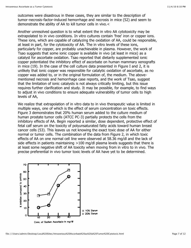
We realize that extrapolation of in vitro data to in vivo therapeutic value is limited inmultiple ways, one of which is the effect of serum concentration on toxic effects.Figure 3 demonstrates that 20% human serum added to the culture medium ofhuman prostate tumor cells (ATCC PC-3) partially protects the cells from theinhibitory effects of AA. Begin reported a similar, dose dependent, protective effect offetal calf serum on the toxicity of polyunsaturated fatty acids toward human breastcancer cells (53). This leaves us not knowing the exact toxic dose of AA for eithernormal or tumor cells. The combination of the data from Figure 2, in which toxiceffects of AA on one normal cell line were observed at 58.36 mg/dl and the lack ofside effects in patients maintaining >100 mg/dl plasma levels suggests that there isat least some negative shift of AA toxicity when moving from in vitro to in vivo. Theprecise preferential in vivo tumor toxic levels of AA have yet to be determined.
11/4/10 8:10 PMIntravenous Ascorbate as a Tumor Cytotoxic
Page 8 of 12file:///Users/admin/Desktop/Local%20Sites/Intravenous%20Ascorbate%20as%20a%20Tumor%20Cytotoxic.html
Fig. 3 Dose response (mean of pool of 8 samples) of human prostate tumor cell line(PC-3, ATCC, Rockville, MD) to sodium ascorbate. Results reflect total viable cells. 10and 20% human serum from patient with prostate adenocarcinoma was added toDMEM Hi-glucose culture medium (Irvine Sci.) w/10% heatinactivated fetal calfserum + antibiotics + Fungizone. 5% C02 humidified incubator at 370C. Cultured for7 days after supplementation of ascorbate. Seeded with 5000 cells/well in 24-wellculture plates (Nunc).
One other factor which could influence how in vitro effects are extrapolated to in vivoeffects is active transport of AA into certain tissues. Several tissues have beenidentified as containing greater than plasma concentrations of AA (wt of AA/unit ofwet tissue), thus indicating an active transport of AA. In descending order, tissuelevels of AA in humans rank as follows: adrenals, leukocytes, pituitary, brain, eyelens,pancreas, kidney, liver, spleen, heart-muscle, and plasma (54). Plasmaconcentrations of AA required for toxicity of both normal and tumor cells in thesetissues could potentially be lower.
SafetyAlthough it is very rare, tumor necrosis, hemorrhage, and subsequent death shouldbe the highest priority concern for the safety of intravenous AA for cancer patients.While there is little data on the safety of the high doses of AA that we are proposinghere other than the reports of Klenner, who reported no ill effects of dosages as highas 150 g intravenously over a 24-h period (33), many of the publications cited earlierdescribe the relative lack of toxicity of high dose oral AA, including Cathcart (55) whodescribes no ill effects with doses of up to 200 g/d in patients with variouspathological conditions. According to Rivers, (56) high dose AA is generally safe withcontraindications in the following circumstances: renal insufficiency, chronichemodialysis patients, unusual forms of iron overload, and oxalate stone formers.Care should be taken to screen potential patients for the above-mentionedconditions prior to beginning any high-dose AA therapy. Screening for red cellglucose-6-phosphate dehydrogenase deficiency, which can give rise to hemolysis ofred blood cells under oxidative stress (57), should also be performed. Afterscreening, any cancer therapy should be started at a low dosage to ensure thattumor hemorrhage does not occur.
Cameron described a rebound effect that can occur in response to high circulatinglevels of AA. He proposed that abnormally low levels of AA can occur betweenintravenous infusions of AA and that his effect is caused by increased levels ofhepatic enzymes responsible for degradation and metabolism of AA (34). We havenot found that to be the case, at least when the patient is orally supplementingbetween infusions. All of our patients have supplemented between infusions,therefore we have no data that allows us to directly compare our results toCameron's. Plasma levels of two patients who were receiving 15 and 22.5g infusionsof AA 3 times weekly and orally supplementing to bowel tolerance with <10 g/d ofAA were 2.4 and 2.8 mg/dl immediately prior to infusions. Another patient who wasalso supplementing with oral AA and receiving infusions 2 times weekly, recordedplasma AA levels of between 2.6 and 4.5 mg/dl on the 6 occasions prior to infusion.These examples indicate that a scorbutic rebound effect can be avoided with oralsupplementation.
11/4/10 8:10 PMIntravenous Ascorbate as a Tumor Cytotoxic
Page 9 of 12file:///Users/admin/Desktop/Local%20Sites/Intravenous%20Ascorbate%20as%20a%20Tumor%20Cytotoxic.html
Because of the possibility of a rebound effect, measurement of plasma levels duringthe periods between infusions should be performed to ensure that no such effecttakes place. Every effort should be made to monitor plasma AA levels when a patientdiscontinues intravenous AA therapy.
ConclusionWe believe that sufficient evidence exists to support the testing of intravenous AA forextended periods as a cytotoxic chemotherapy agent, in doses high enough tomaintain plasma levels above those which have been found to be preferentiallycytotoxic to tumor cells in vitro. AA is relatively non-toxic and is preferentiallycytotoxic to tumor cells at levels attainable in human plasma, at least in vitro. Furtherstudies on the in vivo effects and mechanism of AA’s preferential cytotoxicity need tobe performed.
AcknowledgmentsWe would like to thank Don Davis, Ph.D. (University of Texas at Austin) for hisconsultation on this manuscript and Brian McClune and Kirk Pappan for theirexperimental contributions and maintenance of cell cultures. This work wassupported in part by a grant from the Flossie West Charitable Trust and the OliveWhite Garvey Trust.
Reference 1. Kennedy B J. Guest editorial: evolution of chemotherapy. CA Cancer J Clin 1991; 41: 261-2.
2. Krakoff I H. Cancer chemotherapeutic and biologic agents. CA Cancer J Clin 1991; 41: 265-278.
3. Boring C C, Squires T S, Tong T, Montgomery S. Cancer statistics 1994. CA Cancer J Clin 1994;44: 7-26.
4. DeVita V T, Hellman S, Rosenberg S A, eds. Cancer: Principle and Practice of Oncology.Philadelphia: J B Lippincott, 1982: 1703-1720.
5. DeVita V T, Hellman S, Rosenberg S A, eds. Cancer: Principle and Practice of Oncology.Philadelphia: J B Lippincott, 1982: 1677.
6. Benade L, Howard T, Burk D. Synergistic killing of Ehrlich ascites carcinoma cells by ascorbateand 3-aniino-1, 2, 4triazole. Oncology 1969; 23: 33-43.
7. Bram S, Froussard P et al. Vitamin C preferential toxicity for malignant melanoma cells. Nature1980; 284: 629-631.
8. Noto V, Taper H S et al. Effects of sodium ascorbate (vitamin
C) and 2-methyl-1, 4-naphthoquinone (vitamin K3) treatment on human tumor cell growth invitro. Cancer 1989; 63: 901906.
9. Helgestad J, Pettersen R, Storm-Mathisen I et al. Characterization of a new malignant humanT-cell line (PFI-285) sensitive to ascorbic acid. Eur J Haematol 1990; 44: 9-17.
10. Park C H, Aniare M, Savin M A, Hoogstraten B. Growth suppression of human leukemic cells invitro by L-ascorbic acid. Cancer Res 1980; 40: 1062-1065.
11. Yamafuji K, Nakamurar Y, Omura H, Soeda T, Gyotok K. Anti-tumor potency of ascorbic,dehydroascorbid, or 2,3-diiketogulonic acid and their action on deoxyribonucleic acid. A
11/4/10 8:10 PMIntravenous Ascorbate as a Tumor Cytotoxic
Page 10 of 12file:///Users/admin/Desktop/Local%20Sites/Intravenous%20Ascorbate%20as%20a%20Tumor%20Cytotoxic.html
Krebsforsch 1971; 76:1-7.
12. Yagashita K, Takahashi N, Yamamoto H, Jinnouchi H, Hiyoshi S, Miyakawa T. Effects oftetraacetyl-bis-dehydroAA, a derivative of ascorbic acid, on Ehrlich cells and HeLa cells (humancarcinoma cells). J Nutr Sci Vitaminol 1976; 22: 419427.
13.. Koch C J, Biaglow J E. Toxicity, radiation sensitivity modification, and metabolic effects ofdehydroascorbate and ascorbate in marmnalian cells. J Cell Physiol 1978; 94: 299-306.
14. Cohen M H, Krasnow S H. Cure of advanced Lewis lung carcinoma (LL): a new treatmentstrategy. Proceedings of AACR 1987; 28: 416.
15. Lupulesco A. Vitamin C inhibits DNA, RNA and protein synthesis in epithelial neoplastic cells.Intl J Vit Nutr Res 1991; 61: 125-129.
16. Varga J M, Airoldi L. Inhibition of transplantable melanoma tumor development in n-tice byprophylactic administration of Ca-ascorbate. Life Sciences 1983; 32: 1559-1564.
17. Pierson H F, Meadows G G. Sodium ascorbate enhancement of carbidopa-levodopa methylester antitumor activity against pigmented B-16 melanoma. Cancer Res 1983; 43: 2047-2051.
18. Chakrabarti R N, Dasgupta P S. Effects of ascorbic acid on survival and cell-mediatedimmunity in tumor bearing mice. IRCS Med Sci 1984; 12: 1147-1148.
19. Tsao C S, Dunham W B, Ping Y L. In vivo antineoplastic activity of ascorbic acid for humanmainmary tumor. In vivo 1988; 2: 147-150.
20. Cameron E, Pauling L, Leibovitz B. Ascorbic acid and cancer: a review. Cancer Res 1979; 39:663-681.
21. Anderson R. Vitamin C and immune functions: mechanisms of inununostimulation. In:Counsell J N, Homig D H, eds. Vitamin C. London: Applied Science Publishers, 1981: 249272.
22. Henson D E, Block G, Levine M. Ascorbic acid: biologic functions and relation to cancer:conunentary. JNCI 1991; 83: 547-550.
23. Kennes B, Dumont 1, Brohee D, Hubert C, Neve P. Effect of vitamin C supplements on cell-mediated immunity in old people. Gerontology 1983; 29: 305-310.
24. YJenner F R. The treatment of poliomyelitis and other virus diseases with vitamin C. SouthernMed Surg 1949; 209-214.
25. Klenner F R. Massive doses of vitamin C and the virus diseases. J Southern Med Surg 1951;103: 101-107.
26. Kienner F R. Observations on the dose and administration of ascorbic acid when employedbeyond the range of a vitamin in human pathology. J Appl Nutr 1971; 23: 61-88.
27. McCormick W J. Ascorbic acid as a chemotherapeutic agent. Arch Pediat NY 1952; 69: 151-155.
28. Panush R S, Delafluente J C, Katz P, Johnson J. Modulation of certain immunologic responsesby vitamin C. 111. Potentiation in vitro and in vivo lymphocyte responses. Int J Vit Nutr Res 1982;23: 35-37.
29. Siegel B V, Leibovitz B. The multifactorial role of vitamin C in health and disease. Int J Vit NutrRes 1982; 23: 9-22.
11/4/10 8:10 PMIntravenous Ascorbate as a Tumor Cytotoxic
Page 11 of 12file:///Users/admin/Desktop/Local%20Sites/Intravenous%20Ascorbate%20as%20a%20Tumor%20Cytotoxic.html
30. Siegel B V, Morton J 1. Vitamin C and the immune responses. Experientia 1977; 33: 393-395.
31. Yonemoto R H, Chretien P B, Fehniger T F. Enhanced lymphocyte blastogenesis by oralascorbic acid. Proc Am Assoc Cancer Res 1976; 17: 288.
32. Halliwell B, Gutteridge J M C, eds. Free radicals in biology and medicine, 2nd edn. Oxford:Clarendon Press, 1989: 124.
33. Klenner F R. Observations on the dose and administration of ascorbic acid when employedbeyond the range of a vitamin in human pathology. J Appl Nutr 1971; 23: 61-88.
34. Cameron E. Protocol for the use of vitamin C in the treatment of cancer. Med Hypotheses1991; 36: 190-194.
35. Riordan H D, Jackson J A, Schultz M. Case study: high-dose intravenous vitan-dn C in thetreatment of a patient with adenocarcinoma of the kidney. J Ortho Med 1990; 5: 5-7.
36. Cameron E, Pauling L. The orthomotecular treatment of cancer. 1. The role of ascorbate inhost resistance. Chem Biol Interact 1974; 9: 273-283.
37. Cameron E, Campbell A. The orthomolecular treatment of cancer. II. Clinical trial of high-doseascorbic acid supplements in advanced human cancer. Chem Biol Interact 1974; 9: 285-315.
38. Cameron E, Campbell A, Jack T. The orthomolecular treatment of cancer. 111. Reticulum cellsarcoma: double complete regression induced by high-dose ascorbic acid therapy. Chem BiolInteract 1975; 11: 387-393.
39. Cameron E, Pauling L. Supplemental ascorbate in the supportive treatment of cancer:prolongation of survival times in terminal human cancer. Proc Natl Acad Sci USA 1976; 73: 3685-89.
40. Cameron E, Campbell A. Innovation vs quality control: an lunpublishable' clinical trial ofsupplemental ascorbate in incurable cancer. Med Hypotheses 1991; 36: 185-189.
41. Cameron E, Pauling L. Supplemental ascorbate in the supportive treatment of cancer:reevaluation of prolongation of survival times in terminal human cancer. Proc Natl Acad Sci. USA1978; 75: 4538-4542.
42. Cameron E. Vitamin C for cancer (letter). N Engl J Med 1980; 302: 299.
43. Cameron E, Pauling L, Leibovitz B. Ascorbic acid and cancer: a review. Cancer Res 1979; 39:663-681.
44. Pauling L. Vitamin C therapy of advanced cancer (letter). N Engl J Med 1980; 302: 694.
45. Morishige F, Murata A. Prolongation of survival times in terniinal human cancer byadministration of supplemental ascorbate. J Int Acad Prev Med 1979; 5: 47-52.
46. Murata A, Morishige F, Yamaguchi H. Prolongation of survival times of terminal cancerpatients by administration of large doses of ascorbate. In: Hanck A. Vitamin C: New ClinicalApplications. Bem: Huber, 1982: 103-113.
47. Creagan E T, Moertal C G, O'Fallon J R et al. Failure of high-dose vitamin C (ascorbic acid) tobenefit patients with advanced cancer: a controlled trial. N Engl J Med 1979; 301: 687-690.
48. Moertel C G, Fleming T R, Creagan E T et al. High-dose vitaniin C versus placebo in thetreatment of patients with advanced cancer who had had no prior chemotherapy. N Engl J Med1985; 312: 137-141.
11/4/10 8:10 PMIntravenous Ascorbate as a Tumor Cytotoxic
Page 12 of 12file:///Users/admin/Desktop/Local%20Sites/Intravenous%20Ascorbate%20as%20a%20Tumor%20Cytotoxic.html
49. Lentner C. Geigy Scientific Tables. Vol 3. 8th ed. West Caldwell: Ciba-Geigy, 1984: 132.
50. Henry R J. Determination of AA. In: Henry R J, ed. Clinical Chemistry, Principles andTechniques. New York: Hoeber Medical Division, Harper & Row, 1964: 715-719.
51. Campbell A, Jack T. Acute reactions to mega ascorbic acid therapy in malignant disease. ScotMed J 1979; 24: 151.
52. Carswell E A, Old L J, Kassel R L, Green S, Fiore N, Williamson B. An endotoxin-induced serumfactor that causes necrosis of tumors. Proc Natl Acad Sci USA 1975; 72: 36663670.
53. Begin M E. Selective antineoplastic effects of polyunsaturated fatty acids. In: Kabara J J, ed.Pharmacological Effects of Lipids III. Champaign, Illinois: American Oil Chemists Society, 1989:181-193.
54. Lewin S. Vitamin C: its molecular biology and medical potential. London: Academic Press,1976: 76-79.
55. Cathcart R F. Vitamin C: the nontoxic, nonrate-liniited, antioxidant free radical scavenger. MedHypotheses 1985; 18: 61-77.
56. Rivers J M. Safety of high-level vitamin C ingestion. In: Third Conference on AA. Ann NY AcadSci 1987; 498: 95-102.
57. Karunanithy R, Saba N, Ng S E. Serum and red blood cell magnesium, copper, and zinccontent in G6PD deficiency. Am J Hem 1990; 35: 136-138.
The Center for the Improvement of Human Functioning International3100 North Hillside Avenue
Wichita, KS 67219 USAPhone: 316-682-3100; Fax: 316-682-5054
Medical Records Fax: 316-618-8537 * Laboratory Fax: 316-682-2062
Copyright © 2022 FDOKUMEN