Cyclophosphamide Facilitates Antitumor Efficacy against Subcutaneous Tumors following Intravenous...
18
Cyclophosphamide Facilitates Antitumor Efficacy against Subcutaneous Tumors following Intravenous Delivery of Reovirus Jian Qiao 1 , Hongxun Wang 1 , Timothy Kottke 1 , Christine White 3 , Katie Twigger 3 , Rosa Maria Diaz 1,2 , Jill Thompson 1 , Peter Selby 4 , Johann de Bono 3 , Alan Melcher 4 , Hardev Pandha 5 , Matt Coffey 6 , Richard Vile 1,2,4 , and Kevin Harrington 3 1 Molecular Medicine Program and Mayo Clinic College of Medicine, Rochester, Minnesota 2 Department of Immunology, Mayo Clinic College of Medicine, Rochester, Minnesota 3 The Institute of Cancer Research, London, United Kingdom 4 Cancer Research UK Clinical Centre, Leeds Teaching Hospitals NHS Trust and Leeds Institute of Molecular Medicine, University of Leeds, Leeds, United Kingdom 5 Royal Surrey Hospital, Guildford, United Kingdom 6 Oncolytics Biotech, Inc., Calgary, Alberta, Canada Abstract Purpose—The purpose of the present study was to investigate whether it is possible to achieve truly systemic delivery of oncolytic reovirus, in immunocompetent hosts, using cyclophosphamide to overcome some of the barriers to effective intratumoral delivery and replication of i.v. injected virus. Experimental Design—I.v. delivery of reovirus was combined with different regimens of i.p. administered cyclophosphamide in C57Bl/6 mice bearing established s.c. B16 tumors. Intratumoral viral replication, tumor size, and survival were measured along with levels of neutralizing antibody (NAb) in the blood. Finally, differential toxicities of the virus/ cyclophosphamide regimens were monitored through viral replication in systemic organs, survival, and cardiac damage. Results—Repeated i.v. injection of reovirus was poorly effective at seeding intratumoral viral replication/oncolysis. However, by combining i.v. virus with cyclophosphamide, viral titers of between 10 7 and 10 8 plaque-forming units per milligram were recovered from regressing tumors. Doses of cyclophosphamide that ablated NAb were associated with severe toxicities, characterized by viral replication in systemic organs—toxicities that are mirrored by repeated reovirus injections into B-cell knockout mice. Next, we restructured the dosing of cyclophosphamide and i.v. virus such that a dose of 3 mg cyclophosphamide was administered 24 h before reovirus injection, and this schedule was repeated every 6 days. Using this protocol, high levels of intratumoral viral access and replication (~10 7 plaque-forming units per milligram tumor) were maintained along with systemically protective levels of NAb and only very mild, non – life-threatening toxicity. © 2008 American Association for Cancer Research. Requests for reprints: Richard G.Vile, Molecular Medicine Program, Mayo Clinic, Guggenheim 1836, 200 1st Street Southwest, Rochester, MN 55902. Phone: 507-284-9941; Fax: 507-266-2122; E-mail: [email protected]. NIH Public Access Author Manuscript Clin Cancer Res. Author manuscript; available in PMC 2011 March 1. Published in final edited form as: Clin Cancer Res. 2008 January 1; 14(1): 259–269. doi:10.1158/1078-0432.CCR-07-1510. NIH-PA Author Manuscript NIH-PA Author Manuscript NIH-PA Author Manuscript
-
Upload
independent -
Category
Documents
-
view
1 -
download
0
Transcript of Cyclophosphamide Facilitates Antitumor Efficacy against Subcutaneous Tumors following Intravenous...
Cyclophosphamide Facilitates Antitumor Efficacy againstSubcutaneous Tumors following Intravenous Delivery ofReovirus
Jian Qiao1, Hongxun Wang1, Timothy Kottke1, Christine White3, Katie Twigger3, RosaMaria Diaz1,2, Jill Thompson1, Peter Selby4, Johann de Bono3, Alan Melcher4, HardevPandha5, Matt Coffey6, Richard Vile1,2,4, and Kevin Harrington31Molecular Medicine Program and Mayo Clinic College of Medicine, Rochester, Minnesota2Department of Immunology, Mayo Clinic College of Medicine, Rochester, Minnesota3The Institute of Cancer Research, London, United Kingdom4Cancer Research UK Clinical Centre, Leeds Teaching Hospitals NHS Trust and Leeds Instituteof Molecular Medicine, University of Leeds, Leeds, United Kingdom5Royal Surrey Hospital, Guildford, United Kingdom6Oncolytics Biotech, Inc., Calgary, Alberta, Canada
AbstractPurpose—The purpose of the present study was to investigate whether it is possible to achievetruly systemic delivery of oncolytic reovirus, in immunocompetent hosts, using cyclophosphamideto overcome some of the barriers to effective intratumoral delivery and replication of i.v. injectedvirus.
Experimental Design—I.v. delivery of reovirus was combined with different regimens of i.p.administered cyclophosphamide in C57Bl/6 mice bearing established s.c. B16 tumors.Intratumoral viral replication, tumor size, and survival were measured along with levels ofneutralizing antibody (NAb) in the blood. Finally, differential toxicities of the virus/cyclophosphamide regimens were monitored through viral replication in systemic organs,survival, and cardiac damage.
Results—Repeated i.v. injection of reovirus was poorly effective at seeding intratumoral viralreplication/oncolysis. However, by combining i.v. virus with cyclophosphamide, viral titers ofbetween 107 and 108 plaque-forming units per milligram were recovered from regressing tumors.Doses of cyclophosphamide that ablated NAb were associated with severe toxicities, characterizedby viral replication in systemic organs—toxicities that are mirrored by repeated reovirus injectionsinto B-cell knockout mice. Next, we restructured the dosing of cyclophosphamide and i.v. virussuch that a dose of 3 mg cyclophosphamide was administered 24 h before reovirus injection, andthis schedule was repeated every 6 days. Using this protocol, high levels of intratumoral viralaccess and replication (~107 plaque-forming units per milligram tumor) were maintained alongwith systemically protective levels of NAb and only very mild, non – life-threatening toxicity.
© 2008 American Association for Cancer Research.Requests for reprints: Richard G.Vile, Molecular Medicine Program, Mayo Clinic, Guggenheim 1836, 200 1st Street Southwest,Rochester, MN 55902. Phone: 507-284-9941; Fax: 507-266-2122; E-mail: [email protected].
NIH Public AccessAuthor ManuscriptClin Cancer Res. Author manuscript; available in PMC 2011 March 1.
Published in final edited form as:Clin Cancer Res. 2008 January 1; 14(1): 259–269. doi:10.1158/1078-0432.CCR-07-1510.
NIH
-PA Author Manuscript
NIH
-PA Author Manuscript
NIH
-PA Author Manuscript
Conclusion—NAb to oncolytic viruses play a dual role in the context of systemic viral delivery;on one hand, they hinder repeated administration of virus but on the other, they provide animportant safety mechanism by which virus released from vigorous intratumoral replication isneutralized before it can disseminate and cause toxicity. These data support the use ofcyclophosphamide to modulate, but not ablate, patient NAb, in development of carefullycontrolled clinical trials of the systemic administration of oncolytic viruses.
A major goal of gene therapy/virotherapy is to generate vectors that can be deliveredsystemically to metastatic disease deposits that are inaccessible to direct intratumoralinjection (1–4). In a virus-naive host, antiviral activities in the circulation (4,5) includecomplement (6), preimmune IgM (7) and other components of the innate immune systemthat inhibit replication within tumors (8–10). Circulating vector must also avoid bothnonspecific adhesion to multiple cells (11) as well as specific sequestration in organs such asthe liver (12). Vectors must also extravasate specifically at the tumor (13). In a virus-immune host, NAb is a major additional inhibitor of oncolytic viruses (4), dependent, atleast partly, upon the route of administration (14). In addition, in contrast to preclinicalmodels, even vectors directly injected into human tumors do not migrate far from the end ofthe needle (15). These findings inspired the development of replication-competent vectors,which would, in theory, require only low levels of seeding in a tumor to initiate spreadinginfections to cover the tumor comprehensively (16,17). However, significant problems stillpersist even in the context of direct intratumoral injections, including stromal and immunebarriers to effective intratumoral spread (8,18,19), which may be partially overcome bycombination with preexisting modalities (20,21). In light of these considerations, an intactimmune system should also be viewed as a critical component that contributes to the“tightness” of the oncolytic specificity of a virus.
Reoviruses (respiratory enteric orphan) are double-stranded RNA viruses isolated from therespiratory and gastrointestinal tracts of humans but not linked to disease (22–24). They do,however, cause fatal infections in neonatal and severe combined immunodeficient nonobesediabetic (SCID/NOD) mice (24,25), reiterating the importance of an intact immune systemas a component determining oncolytic specificity. About 90% of patients are preimmune toreovirus. The use of reovirus as an oncolytic agent was proposed on the basis of findingsthat an activated Ras pathway in tumor cells prevents RNA-activated protein kinase fromaborting infection, leading to lytic viral replication in tumors but not in normal cells (22,26–28).
Based on these considerations, we have completed a phase I clinical trial of systemicallydelivered reovirus given to 32 patients as a 1 h i.v. infusion every 4 weeks (one cycle). 7Dose escalation reached 3 × 10¹⁰ TCID50 for 5 days every 4 weeks without dose-limitingtoxicity and a maximum tolerated dose was not defined. No viral shedding at any dose wasseen by reverse transcription-PCR in blood, urine, stool, and sputum. Patients fell into twodistinct groups in terms of NAb. Those with preexisiting antireovirus antibody had titerincreases of up to 250-fold (median 80-fold) by day 5. Titers in patients without preexistingantireovirus antibody increased by 700- to 6,500-fold. However, in all cases, final titersended up close to the same level during the subsequent cycles.8 In addition to showing thati.v. reovirus is safe and well tolerated, replicating reovirus could also be detected in somecases in metastases after systemic viral delivery.7
The preliminary data from this, and other, clinical trials have suggested that transientimmunosuppression may facilitate viral delivery to metastatic tumors by removing, or
7Vidal et al., submitted for publication.8White et al., in preparation.
Qiao et al. Page 2
Clin Cancer Res. Author manuscript; available in PMC 2011 March 1.
NIH
-PA Author Manuscript
NIH
-PA Author Manuscript
NIH
-PA Author Manuscript
reducing, the protective blanket of NAb and other immune effectors. In this respect,cyclophosphamide has been used to facilitate delivery/efficacy of oncolytic viruses (6–10,29,30) and is already used as a chemotherapy at doses that are heavilyimmunosuppressive (>120 or >400 mg/kg in mice; refs. 31–33). Short-term exposure tocyclophosphamide suppressed innate immune responses (natural killer cells, macrophages,and IFN-γ), which inhibit intravascular delivery of herpes simplex virus to rat gliomas (7),producing significant improvements in therapy (8). At lower doses (<100 mg/kg),cyclophosphamide enhances immune responses against tumors (34–36) through selective,transient depletion of regulatory T cells that suppress antitumor CD8+ T-cell responses (34–41).
In this report, we have used our clinical experience using systemic delivery of oncolyticreovirus to return to our preclinical model to test new protocols by which replicating vectorscan be delivered to metastatic disease, in immune-competent tumor-bearing hosts, in theabsence of direct access by a needle. Reovirus is an attractive experimental oncolytic virusbecause it replicates in murine tumor cells that are tumorigenic in immunocompetent mice(42–45). We have used the B16/C57Bl/6 immunocompetent model to test different regimensof administration of reovirus with cyclo-phosphamide, as either a single low dose (150 mg/kg = 3 mg per mouse), as a high dose (3 mg for 3 days), or as iterative injections(metronomic dosing) of 3 mg once every 6 days (31–33,35). Our data show thatcyclophosphamide can facilitate high levels of i.v. administered virus to reach, and replicatein, s.c. tumors, associated with dramatic reductions in levels of circulating neutralizingantibodies (NAb). However, cyclophosphamide-mediated loss of protective NAb can also beassociated with severe toxicity probably as a result of subsequent dissemination of the virusfrom the tumor to systemic organs, showing that NAb can also provide a safety barrier to thewidespread dissemination/toxicity of oncolytic viruses. Finally, we show that iterativeinjections (metronomic dosing) of 3 mg cyclophosphamide and reovirus once every 6 daysallows both high levels of viral access to tumors and minimal toxicity. These data will drivethe development of further clinical trials for delivery of reovirus to patients with metastaticdisease.
Materials and MethodsCells and virus
Murine B16 melanoma cells (H2-Kb; ref. 46) were grown in DMEM (Life Technologies)supplemented with 10% (v/v) FCS (Life Technologies) and L-glutamine (Life Technologies).All cell lines were monitored routinely and found to be free of Mycoplasma infection.
Reovirus used in these studies is a wild-type reovirus type 3 (Dearing strain). Virus stocktiters were measured by standard plaque assays of serially diluted samples on L929 cells.
Antibody titration from mouse serumPreheated mouse antiserum was mixed with an equal volume of reovirus (predetermined askilling 80% of target L929 cells) and incubated at 37°C for 2 h to allow antibody to bind tovirus. The virus/antibody mix was transferred to L929 monolayers and cell survival wasassayed at 48 h by 3-(4,5-dimethylthiazol-2-yl)-2,5-diphenyltetrazolium bromide assay. Ananti-reovirus polyclonal antiserum was used as a positive control. The neutralizing titer isthe highest dilution of serum that blocks the killing of L929 cells.
Virus titration from tumor and organsTumor and organs recovered from mice were harvested and weighed and, within 2 h ofremoval from the mouse, were lysed by three cycles of freeze/thawing. Virus was recovered
Qiao et al. Page 3
Clin Cancer Res. Author manuscript; available in PMC 2011 March 1.
NIH
-PA Author Manuscript
NIH
-PA Author Manuscript
NIH
-PA Author Manuscript
from the lysates and titers were determined on L929 cells and expressed as pfu of reovirusper milligram of tissue.
In vivo studiesAll procedures were approved by the Mayo Foundation Institutional Animal Care and UseCommittee. C57BL/6 or B6.129S2-Igh-6tm1Cgn/j mice (Jackson 002288), which lackmature B cells and cannot, therefore, make antiviral antibodies, were age and sex matchedfor individual experiments and were purchased from The Jackson Laboratory at 6 to 8 weeksof age. To establish s.c. tumors, 2 × 10⁵ B16 cells were injected s.c. (100 µL) into the flankregion. Animals were examined daily until the tumor became palpable, after which thediameter, in two dimensions, was measured thrice weekly using calipers. Animals werekilled when tumor size was ~ 1.0 × 1.0 cm in two perpendicular directions. Mice wereeuthanized based on the double criteria of both active progression in measurable size overseveral days and of reaching a diameter of 1.0 cm. This is based on our histologicexperience that tumors that have progressively increased in size to a final size of 1.0 cmdiameter represent actively growing tumor rather than predominantly necrotic tumordestruction. To establish systemic metastatic disease, C57BL/6 mice were injected i.v. with2 × 105 B16ova cells to form lung metastases. Mice were killed at the first sign of anydistress.
Histopathology of tumor sectionsTumors and major organs were harvested and fixed in 10% formalin in PBS, then paraffin-embedded and sectioned. H&E-stained sections were prepared for analysis of tissuedestruction and gross infiltrate. A pathologist examining H&E sections, blinded to theexperimental design, scored the degree of necrosis.
StatisticsSurvival data from the animal studies were analyzed using the log-rank test (47), and thetwo-sample unequal variance Student’s t test analysis was applied for in vitro assays.Statistical significance was determined at the level of P < 0.05.
ResultsReovirus is oncolytic in the B16 murine model
Direct intratumoral injection of reovirus induced significant regressions of established B16tumors in fully immunocompetent C57Bl/6 mice, demonstrating that it is an effectiveoncolytic agent if sufficient levels of virus can be delivered to tumors (P = 0.0027 comparedwith heat inactivated virus; Fig. 1A). However, our ultimate goal is the treatment ofmetastatic disease not accessible to direct intratumoral injection. Therefore, reovirus wasadministered i.v. according to the protocol in Fig. 1B to treat either established lungmetastases (Fig. 1C) or s.c. tumor (Fig. 1D). Over multiple, repeated experiments, i.v.delivery of reovirus to either lung metastatic, or s.c., B16 tumors never generated long-termcures of tumor-bearing animals. However, in the lung metastatic model, we were typicallyable to obtain statistically significant, but very small, improvements in survival times withreovirus, compared with control treatments, in some experiments (n = 5),of which Fig. 1C isrepresentative (P = 0.0014). In contrast, although some experiments generated significantlyimproved survival after i.v. delivery of reovirus to s.c. tumors, the more representative case(n = 6) was that there was no significant difference between virus and control treatments, ofwhich Fig. 1D is representative (P = 0.054).
Qiao et al. Page 4
Clin Cancer Res. Author manuscript; available in PMC 2011 March 1.
NIH
-PA Author Manuscript
NIH
-PA Author Manuscript
NIH
-PA Author Manuscript
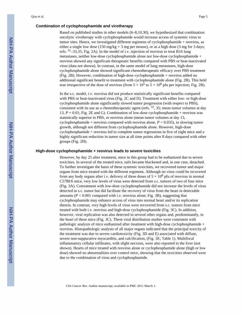
Combination of cyclophosphamide and virotherapyBased on published studies in other models (6–8,10,30), we hypothesized that combinationoncolytic virotherapy with cyclophosphamide would increase access of systemic virus totumor sites. Hence, we investigated different regimens of cyclophosphamide + reovirus, aseither a single low dose (150 mg/kg = 3 mg per mouse), or as a high dose (3 mg for 3 days;refs. 31–33,35; Fig. 2A). In the model of i.v. injection of reovirus to treat B16 lungmetastases, neither low-dose cyclophosphamide alone nor low-dose cyclophosphamide +reovirus showed any significant therapeutic benefits compared with PBS or heat-inactivatedvirus (data not shown). In contrast, in the same model of lung metastases, high-dosecyclophosphamide alone showed significant chemotherapeutic efficacy over PBS treatment(Fig. 2B). However, combination of high-dose cyclophosphamide + reovirus added noadditional significant benefit to treatment with cyclophosphamide alone (Fig. 2B). This heldtrue irrespective of the dose of reovirus (from 5 × 10⁵ to 5 × 10⁸ pfu per injection; Fig. 2B).
In the s.c. model, i.v. reovirus did not produce statistically significant benefits comparedwith PBS or heat-inactivated virus (Fig. 2C and D). Treatment with either low- or high-dosecyclophosphamide alone significantly slowed tumor progression (with respect to PBS),consistent with its use as a chemotherapeutic agent (refs. 32, 35; mean tumor volumes at day13, P = 0.01; Fig. 2E and G). Combination of low-dose cyclophosphamide + reovirus wasstatistically superior to PBS, or reovirus alone (mean tumor volumes at day 13,cyclophosphamide + reovirus compared with reovirus alone, P = 0.035), in slowing tumorgrowth, although not different from cyclophosphamide alone. However, high-dosecyclophosphamide + reovirus led to complete tumor regressions in five of eight mice and ahighly significant reduction in tumor size at all time points after 8 days compared with othergroups (Fig. 2H).
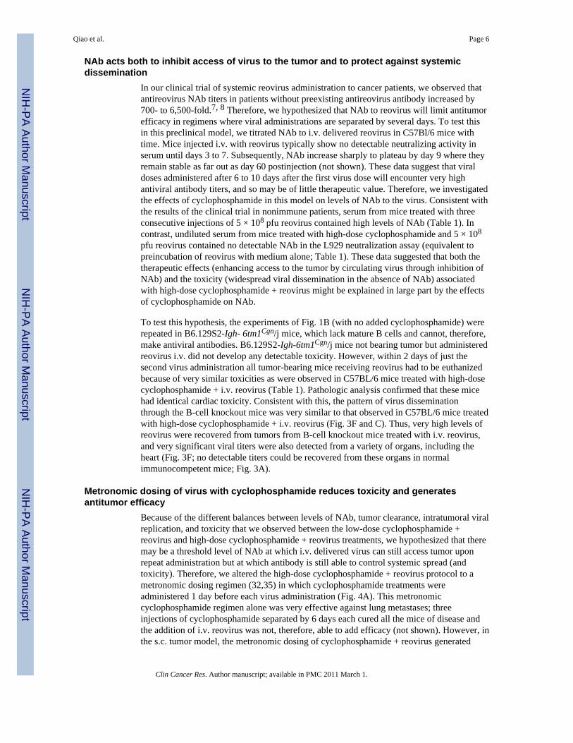
High-dose cyclophosphamide + reovirus leads to severe toxicitiesHowever, by day 25 after treatment, mice in this group had to be euthanized due to severetoxicities. In several of the treated mice, tails became blackened and, in one case, detached.To further investigate the basis of these systemic toxicities, we recovered tumor and majororgans from mice treated with the different regimens. Although no virus could be recoveredfrom any body organs after i.v. delivery of three doses of 5 × 10⁸ pfu of reovirus in normalC57Bl/6 mice, very low levels of virus were detected from s.c. tumors of two of four mice(Fig. 3A). Cotreatment with low-dose cyclophosphamide did not increase the levels of virusdetected in s.c. tumor but did facilitate the recovery of virus from the heart in detectableamounts (P < 0.001 compared with i.v. reovirus alone; Fig. 3B), suggesting thatcyclophosphamide may enhance access of virus into normal heart and/or its replicationtherein. In contrast, very high levels of virus were recovered from s.c. tumors from micetreated with both i.v. reovirus and high-dose cyclophosphamide (Fig. 3C). In addition,however, viral replication was also detected in several other organs and, predominantly, inthe heart of these mice (Fig. 3C). These viral distribution studies were consistent withpathologic analysis of mice euthanized after treatment with high-dose cyclophosphamide +reovirus. Histopathologic analysis of all major organs indicated that the principal toxicity ofthe treatment was due to severe cardiotoxicity (Fig. 3D and E) associated with diffuse,severe non-suppurative myocarditis, and calcification, (Fig. 3E; Table 1). Multifocalinflammatory cellular infiltrates, with slight necrosis, were also reported in the liver (notshown). Hearts of mice treated with reovirus alone or cyclophosphamide alone (high or lowdose) showed no abnormalities over control mice, showing that the toxicities observed weredue to the combination of virus and cyclophosphamide.
Qiao et al. Page 5
Clin Cancer Res. Author manuscript; available in PMC 2011 March 1.
NIH
-PA Author Manuscript
NIH
-PA Author Manuscript
NIH
-PA Author Manuscript
NAb acts both to inhibit access of virus to the tumor and to protect against systemicdissemination
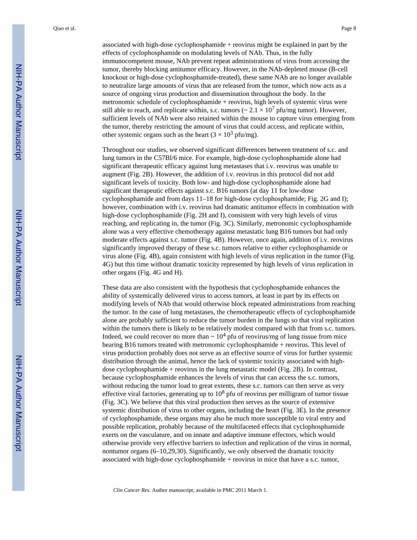
In our clinical trial of systemic reovirus administration to cancer patients, we observed thatantireovirus NAb titers in patients without preexisting antireovirus antibody increased by700- to 6,500-fold.7, 8 Therefore, we hypothesized that NAb to reovirus will limit antitumorefficacy in regimens where viral administrations are separated by several days. To test thisin this preclinical model, we titrated NAb to i.v. delivered reovirus in C57Bl/6 mice withtime. Mice injected i.v. with reovirus typically show no detectable neutralizing activity inserum until days 3 to 7. Subsequently, NAb increase sharply to plateau by day 9 where theyremain stable as far out as day 60 postinjection (not shown). These data suggest that viraldoses administered after 6 to 10 days after the first virus dose will encounter very highantiviral antibody titers, and so may be of little therapeutic value. Therefore, we investigatedthe effects of cyclophosphamide in this model on levels of NAb to the virus. Consistent withthe results of the clinical trial in nonimmune patients, serum from mice treated with threeconsecutive injections of 5 × 10⁸ pfu reovirus contained high levels of NAb (Table 1). Incontrast, undiluted serum from mice treated with high-dose cyclophosphamide and 5 × 10⁸
pfu reovirus contained no detectable NAb in the L929 neutralization assay (equivalent topreincubation of reovirus with medium alone; Table 1). These data suggested that both thetherapeutic effects (enhancing access to the tumor by circulating virus through inhibition ofNAb) and the toxicity (widespread viral dissemination in the absence of NAb) associatedwith high-dose cyclophosphamide + reovirus might be explained in large part by the effectsof cyclophosphamide on NAb.
To test this hypothesis, the experiments of Fig. 1B (with no added cyclophosphamide) wererepeated in B6.129S2-Igh- 6tm1Cgn/j mice, which lack mature B cells and cannot, therefore,make antiviral antibodies. B6.129S2-Igh-6tm1Cgn/j mice not bearing tumor but administeredreovirus i.v. did not develop any detectable toxicity. However, within 2 days of just thesecond virus administration all tumor-bearing mice receiving reovirus had to be euthanizedbecause of very similar toxicities as were observed in C57BL/6 mice treated with high-dosecyclophosphamide + i.v. reovirus (Table 1). Pathologic analysis confirmed that these micehad identical cardiac toxicity. Consistent with this, the pattern of virus disseminationthrough the B-cell knockout mice was very similar to that observed in C57BL/6 mice treatedwith high-dose cyclophosphamide + i.v. reovirus (Fig. 3F and C). Thus, very high levels ofreovirus were recovered from tumors from B-cell knockout mice treated with i.v. reovirus,and very significant viral titers were also detected from a variety of organs, including theheart (Fig. 3F; no detectable titers could be recovered from these organs in normalimmunocompetent mice; Fig. 3A).
Metronomic dosing of virus with cyclophosphamide reduces toxicity and generatesantitumor efficacy
Because of the different balances between levels of NAb, tumor clearance, intratumoral viralreplication, and toxicity that we observed between the low-dose cyclophosphamide +reovirus and high-dose cyclophosphamide + reovirus treatments, we hypothesized that theremay be a threshold level of NAb at which i.v. delivered virus can still access tumor uponrepeat administration but at which antibody is still able to control systemic spread (andtoxicity). Therefore, we altered the high-dose cyclophosphamide + reovirus protocol to ametronomic dosing regimen (32,35) in which cyclophosphamide treatments wereadministered 1 day before each virus administration (Fig. 4A). This metronomiccyclophosphamide regimen alone was very effective against lung metastases; threeinjections of cyclophosphamide separated by 6 days each cured all the mice of disease andthe addition of i.v. reovirus was not, therefore, able to add efficacy (not shown). However, inthe s.c. tumor model, the metronomic dosing of cyclophosphamide + reovirus generated
Qiao et al. Page 6
Clin Cancer Res. Author manuscript; available in PMC 2011 March 1.
NIH
-PA Author Manuscript
NIH
-PA Author Manuscript
NIH
-PA Author Manuscript
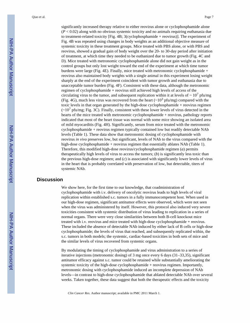
significantly increased therapy relative to either reovirus alone or cyclophosphamide alone(P < 0.02) along with no obvious systemic toxicity and no animals requiring euthanasia dueto treatment-related toxicity [Fig. 4B; 3(cyclophosphamide + reovirus)]. The experiment ofFig. 4B was repeated using changes in body weights as an additional objective measure ofsystemic toxicity in these treatment groups. Mice treated with PBS alone, or with PBS andreovirus, showed a gradual gain of body weight over the 20- to 30-day period after initiationof treatment, at which time they needed to be euthanized due to tumor growth (Fig. 4C andD). Mice treated with metronomic cyclophosphamide alone did not gain weight as in thecontrol groups but only lost weight toward the end of the experiment at which time tumorburdens were large (Fig. 4E). Finally, mice treated with metronomic cyclophosphamide +reovirus also maintained body weights with a single animal in this experiment losing weightsharply at the end of the experiment coincident with tumor growth and euthanasia due tounacceptable tumor burden (Fig. 4F). Consistent with these data, although the metronomicregimen of cyclophosphamide + reovirus still achieved high levels of access of thecirculating virus to the tumor, and subsequent replication within it at levels of ~ 107 pfu/mg(Fig. 4G), much less virus was recovered from the heart (~103 pfu/mg) compared with thetoxic levels in that organ generated by the high-dose cyclophosphamide + reovirus regimen(~107 pfu/mg; Fig. 3C). Finally, consistent with these lower levels of virus detected in thehearts of the mice treated with metronomic cyclophosphamide + reovirus, pathology reportsindicated that most of the heart tissue was normal with some mice showing an isolated areaof mild myocarditis (Fig. 4H). Significantly, serum from mice treated with the metronomiccyclophosphamide + reovirus regimen typically contained low but readily detectable NAblevels (Table 1). These data show that metronomic dosing of cyclophosphamide withreovirus in vivo preserves low, but significant, levels of NAb to the virus compared with thehigh-dose cyclophosphamide + reovirus regimen that essentially ablates NAb (Table 1).Therefore, this modified high-dose reovirus/cyclophosphamide regimen (a) permitstherapeutically high levels of virus to access the tumors; (b) is significantly less toxic thanthe previous high-dose regimen; and (c) is associated with significantly lower levels of virusin the heart that is probably correlated with preservation of low, but detectable, titers ofsystemic NAb.
DiscussionWe show here, for the first time to our knowledge, that coadministration ofcyclophosphamide with i.v. delivery of oncolytic reovirus leads to high levels of viralreplication within established s.c. tumors in a fully immunocompetent host. When used inour high-dose regimen, significant antitumor effects were observed, which were not seenwhen the virus was administered by itself. However, this protocol also induced very severetoxicities consistent with systemic distribution of virus leading to replication in a series ofnormal organs. There were very close similarities between both B-cell knockout micetreated with i.v. reovirus and mice treated with high-dose cyclophosphamide + reovirus.These included the absence of detectable NAb induced by either lack of B cells or high-dosecyclophosphamide; the levels of virus that reached, and subsequently replicated within, thes.c. tumors in both models; the systemic, cardiac-based toxicities in both sets of mice andthe similar levels of virus recovered from systemic organs.
By modulating the timing of cyclophosphamide and virus administration to a series ofiterative injections (metronomic dosing) of 3 mg once every 6 days (31–33,35), significantantitumor efficacy against s.c. tumor could be retained while substantially ameliorating thesystemic toxicity of the high-dose cyclophosphamide + reovirus regimen. Importantly,metronomic dosing with cyclophosphamide induced an incomplete depression of NAblevels—in contrast to high-dose cyclophosphamide that ablated detectable NAb over severalweeks. Taken together, these data suggest that both the therapeutic effects and the toxicity
Qiao et al. Page 7
Clin Cancer Res. Author manuscript; available in PMC 2011 March 1.
NIH
-PA Author Manuscript
NIH
-PA Author Manuscript
NIH
-PA Author Manuscript
associated with high-dose cyclophosphamide + reovirus might be explained in part by theeffects of cyclophosphamide on modulating levels of NAb. Thus, in the fullyimmunocompetent mouse, NAb prevent repeat administrations of virus from accessing thetumor, thereby blocking antitumor efficacy. However, in the NAb-depleted mouse (B-cellknockout or high-dose cyclophosphamide-treated), these same NAb are no longer availableto neutralize large amounts of virus that are released from the tumor, which now acts as asource of ongoing virus production and dissemination throughout the body. In themetronomic schedule of cyclophosphamide + reovirus, high levels of systemic virus werestill able to reach, and replicate within, s.c. tumors (~ 2.1 × 10⁷ pfu/mg tumor). However,sufficient levels of NAb were also retained within the mouse to capture virus emerging fromthe tumor, thereby restricting the amount of virus that could access, and replicate within,other systemic organs such as the heart (3 × 10³ pfu/mg).
Throughout our studies, we observed significant differences between treatment of s.c. andlung tumors in the C57Bl/6 mice. For example, high-dose cyclophosphamide alone hadsignificant therapeutic efficacy against lung metastases that i.v. reovirus was unable toaugment (Fig. 2B). However, the addition of i.v. reovirus in this protocol did not addsignificant levels of toxicity. Both low- and high-dose cyclophosphamide alone hadsignificant therapeutic effects against s.c. B16 tumors (at day 11 for low-dosecyclophosphamide and from days 11–18 for high-dose cyclophosphamide; Fig. 2G and I);however, combination with i.v. reovirus had dramatic antitumor effects in combination withhigh-dose cyclophosphamide (Fig. 2H and I), consistent with very high levels of virusreaching, and replicating in, the tumor (Fig. 3C). Similarly, metronomic cyclophosphamidealone was a very effective chemotherapy against metastatic lung B16 tumors but had onlymoderate effects against s.c. tumor (Fig. 4B). However, once again, addition of i.v. reovirussignificantly improved therapy of these s.c. tumors relative to either cyclophosphamide orvirus alone (Fig. 4B), again consistent with high levels of virus replication in the tumor (Fig.4G) but this time without dramatic toxicity represented by high levels of virus replication inother organs (Fig. 4G and H).
These data are also consistent with the hypothesis that cyclophosphamide enhances theability of systemically delivered virus to access tumors, at least in part by its effects onmodifying levels of NAb that would otherwise block repeated administrations from reachingthe tumor. In the case of lung metastases, the chemotherapeutic effects of cyclophosphamidealone are probably sufficient to reduce the tumor burden in the lungs so that viral replicationwithin the tumors there is likely to be relatively modest compared with that from s.c. tumors.Indeed, we could recover no more than ~ 104 pfu of reovirus/mg of lung tissue from micebearing B16 tumors treated with metronomic cyclophosphamide + reovirus. This level ofvirus production probably does not serve as an effective source of virus for further systemicdistribution through the animal, hence the lack of systemic toxicity associated with high-dose cyclophosphamide + reovirus in the lung metastatic model (Fig. 2B). In contrast,because cyclophosphamide enhances the levels of virus that can access the s.c. tumors,without reducing the tumor load to great extents, these s.c. tumors can then serve as veryeffective viral factories, generating up to 108 pfu of reovirus per milligram of tumor tissue(Fig. 3C). We believe that this viral production then serves as the source of extensivesystemic distribution of virus to other organs, including the heart (Fig. 3E). In the presenceof cyclophosphamide, these organs may also be much more susceptible to viral entry andpossible replication, probably because of the multifaceted effects that cyclophosphamideexerts on the vasculature, and on innate and adaptive immune effectors, which wouldotherwise provide very effective barriers to infection and replication of the virus in normal,nontumor organs (6–10,29,30). Significantly, we only observed the dramatic toxicityassociated with high-dose cyclophosphamide + reovirus in mice that have a s.c. tumor,
Qiao et al. Page 8
Clin Cancer Res. Author manuscript; available in PMC 2011 March 1.
NIH
-PA Author Manuscript
NIH
-PA Author Manuscript
NIH
-PA Author Manuscript
supporting the hypothesis that it is tumor-derived virus that serves as the source of thedisseminating virus that causes systemic toxicity as opposed to the input dose of virus.
Indeed, the toxicities we observed in immunocompetent, tumor-bearing mice treated withhigh-dose cyclophosphamide, and in tumor-bearing B-cell knockout mice, were associatedwith dramatic manifestations, including heart failure and the “black foot syndrome,” both ofwhich have been reported previously in SCID or SCID/NOD mice treated with reovirusintratumorally (48–50). Black foot syndrome was described as the discoloration and necrosisof feet, tails, distal legs, and ears in SCID/NOD mice several weeks after injection ofreovirus intratumorally (48), again suggesting that virus replicating within the tumor servesas a source for systemic dissemination (49). The pathogenesis of black foot syndrome wascharacterized as due to venous vasculitis secondary to reovirus infection along withreovirus-induced myocarditis and heart failure (48,50) and typically, these symptomsdeveloped weeks or months after the reovirus therapy into the tumor (48,49). We observed avariation of this syndrome in which tails of cyclophosphamide-treated mice became swollen,very sensitive, and, in a minority of cases, detached from the animal. This is the first time, toour knowledge, that black foot syndrome has been observed in wild-type mice; moreover,onset of both reovirus-induced myocarditis and black foot syndrome was more rapid underthe influence of cyclophosphamide treatment than has been reported in SCID or SCID/NODmice (48,49), indicating that cyclophosphamide is having multiple effects in vivo, which canresemble the induction of a severely immunocompromised state.
In this respect, it seems highly likely that activities of cyclophosphamide, in addition to thesuppression of NAb to the virus, are important to facilitate the high levels of intratumoralviral replication, systemic survival, and access to both tumor and normal tissues that weobserved. In particular, we are currently investigating what other effects cyclophosphamideexerts in our studies through suppression of both innate immune responses (natural killercell, macrophages, and IFN-γ), which may inhibit intravascular delivery, and intratumoralreplication, of virus (6–10,29,30), as well as through effects on adaptive T-cell responses(34–41).
Significantly, the metronomic dosing regimen induced only a few isolated areas of mildmyocarditis, which was not seen in control animals receiving either reovirus orcyclophosphamide alone, but which did not seem to manifest in any detectable clinical wayin these mice. The potential risk of development of mild myocarditis, or calcifications,during combination therapy with reovirus plus cyclophosphamide will remain unclear untilclinical experience of this approach accrues. However, existing clinical data suggest thatcardiac effects of i.v. reovirus do not represent a significant problem. In our recentlycompleted phase I clinical trial, reovirus was administered by i.v. infusion at doses between1 × 108 on day 1, to 3 × 10¹⁰ on days 1 to 5 of a 4-weekly cycle to 33 patients withadvanced cancers.7 Careful assessment of the cardiac effects of i.v. reovirus administrationformed part of the design of this phase I trial. Patients underwent electrocardiogram,radioisotopic multiple uptake gated acquisition scan, and creatine kinase MB isoenzymescreening and troponin I estimation at baseline. Electrocardiogram and creatine kinase MBisoenzyme and troponin I were repeated on the days of reovirus administration and weeklybetween cycles. Multiple uptake gated acquisition was repeated every two cycles. In regardto cardiac toxic events, a single patient treated at the 1 × 10¹⁰ dose level experienced grade 3elevation in creatine kinase MB isoenzyme and troponin I on day 3 of the first treatmentcycle. At this time, the patient was found to have a normal electrocardiogram andechocardiogram and all biochemical disturbance resolved to normal at day 15. The patientwas subsequently rechallenged at the 3 × 10⁹ dose level with no further sequelae. No othercardiac event was seen in the study. The preclinical data derived from the current murinestudies and the existing clinical data provide a clear framework for cardiac monitoring in
Qiao et al. Page 9
Clin Cancer Res. Author manuscript; available in PMC 2011 March 1.
NIH
-PA Author Manuscript
NIH
-PA Author Manuscript
NIH
-PA Author Manuscript
future trials of reovirus in combination with cyclophosphamide. Such safety assessmentswill be a key determinant in decisions regarding dose escalations between patient cohorts.
In summary, we have shown that cyclophosphamide can be used to facilitate theintratumoral access, and replication, of an oncolytic virus to s.c. tumors when delivered i.v.in a fully immunocompetent host. The balance between intratumoral virus replicationfacilitated by cyclophosphamide and systemic toxicity generated by viral spread to normalorgans can be manipulated by instigating a metronomic dosing of virus andcyclophosphamide, whereas this allowed sufficient depression of the levels of NAb to thevirus to maintain high levels of intratumoral virus access/replication, it also maintainedsufficient levels of NAb to neutralize virus released from the tumors to prevent access ofhigh levels of virus to normal organs. These data support the development of clinicalprotocols in which cyclophosphamide is combined with systemically administered oncolyticviruses. Such studies will necessitate careful analysis of toxicity (especially cardiac) endpoints and retreatment and dose-escalation decisions will need to be based on real-timeanalysis of the levels of NAb in the patients’ circulation. Under these circumstances, itshould be possible to test the clinical hypothesis that judicious combination ofcyclophosphamide and reovirus will enhance systemic delivery of the virus to the tumor butretain the ability of low-level NAb responses to protect the patient against toxicity inducedby systemic dissemination of the virus.
AcknowledgmentsWe thank Toni L. Higgins for expert secretarial assstance.
Grant support: Mayo Foundation, NIH grant CA RO1107082-02, and Oncolytics Biotech, Inc., Calgary, Canada.
References1. Vile RG. Vironcology—not yet, but soon? Nat Biotechnol 2001;19:1020–1022. [PubMed:
11689843]2. Vile RG, Ando D, Kirn DH. The oncolytic virotherapy treatment platform for cancer: unique
biological and biosafety points to consider. Cancer Gene Ther 2002;9:1062–1067. [PubMed:12522445]
3. Vile RG, Russell SJ. Gene transfer technologies for the gene therapy of cancer. Gene Ther 1994Jan;:88–98. [PubMed: 7584073]
4. Fisher K. Striking out at disseminated metastases: the systemic delivery of oncolytic viruses. CurrOpin Mol Ther 2006;8:301–313. [PubMed: 16955693]
5. Harrington KJ, Alvarez-Vallina L, Crittenden M, et al. Cells as vehicles for cancer gene therapy: themissing link between targeted vectors and systemic delivery. Hum Gene Ther 2002;13:1263–1280.[PubMed: 12162810]
6. Ikeda K, Wakimoto H, Ichikawa T, et al. Complement depletion facilitates the infection of multiplebrain tumors by an intravascular, replication-conditional herpes simplex virus mutant. J Virol2000;74:4765–4775. [PubMed: 10775615]
7. Ikeda K, Ichikawa T, Wakimoto H, et al. Oncolytic virus therapy of multiple tumors in the brainrequires suppression of innate and elicited antiviral responses. Nat Med 1999;5:881–887. [PubMed:10426310]
8. Fulci G, Breymann L, Gianni D, et al. Cyclophosphamide enhances glioma virotherapy byinhibiting innate immune responses. Proc Natl Acad Sci U S A 2006;103:12873–12878. [PubMed:16908838]
9. Friedman A, Tian JP, Fulci G, Chiocca EA, Wang J. Glioma virotherapy: effects of innate immunesuppression and increased viral replication capacity. Cancer Res 2006;66:2314–2319. [PubMed:16489036]
Qiao et al. Page 10
Clin Cancer Res. Author manuscript; available in PMC 2011 March 1.
NIH
-PA Author Manuscript
NIH
-PA Author Manuscript
NIH
-PA Author Manuscript
10. Wakimoto H, Johnson PR, Knipe DM, Chiocca EA. Effects of innate immunity on herpes simplexvirus and its ability to kill tumor cells. Gene Ther 2003;10:983–990. [PubMed: 12756419]
11. Pizzato M, Blair ED, Fling M, et al. Evidence for nonspecific adsorption of targeted retrovirusvector particles to cells. Gene Ther 2001;8:1088–1096. [PubMed: 11526456]
12. Alemany R, Suzuki K, Curiel DT. Blood clearance rates of adenovirus type 5 in mice. J Gen Virol2000;81(Pt 11):2605–2609. [PubMed: 11038370]
13. Liu Y, Deisseroth A. Tumor vascular targeting therapy with viral vectors. Blood 2006;107:3027–3033. [PubMed: 16373660]
14. Nemunaitis J, Ganly I, Khuri F, et al. Selective replication and oncolysis in p53 mutant tumorswith ONYX-015,an E1B-55kD gene-deleted adenovirus, in patients with advanced head and neckcancer: a phase II trial. Cancer Res 2000;60:6359–6366. [PubMed: 11103798]
15. Ram Z. Summary of results and conclusions of the gene therapy of malignant brain tumors: clinicalstudy. J Neurosurg 1995;82:343A.
16. Russell SJ. Replicating vectors for cancer therapy: a question of strategy. Semin Cancer Biol1994;5:437–443. [PubMed: 7703443]
17. Kirn D, Martuza RL, Zwiebel J. Replication-selective virotherapy for cancer: biological principles,risk management and future directions. Nat Med 2001;7:781–787. [PubMed: 11433341]
18. Kirn D. Oncolytic virotherapy for cancer with the adenovirus dl1520 (Onyx-015): results of phaseI and II trials. Expert Opin Biol Ther 2001;1:525–538. [PubMed: 11727523]
19. Nemunaitis J, Khuri F, Ganly I, et al. Phase II trial of intratumoral administration of ONYX-015, areplication-selective adenovirus, in patients with refractory head and neck cancer. J Clin Oncol2001;19:289–298. [PubMed: 11208818]
20. Geoerger B, Grill J, Opolon P, et al. Potentiation of radiation therapy by the oncolytic adenovirusdl1520 (ONYX-015) in human malignant glioma xenografts. Br J Cancer 2003;89:577–584.[PubMed: 12888833]
21. Hecht JR, Bedford R, Abbruzzese JL, et al. A phase I/II trial of intratumoral endoscopic ultrasoundinjection of ONYX-015 with intravenous gemcitabine in unresectable pancreatic carcinoma. ClinCancer Res 2003;9:555–561. [PubMed: 12576418]
22. Norman KL, Lee PWK. Not all viruses are bad guys: the case for reovirus in cancer therapy. DrugDiscov Today 2005;10:847–856. [PubMed: 15970267]
23. Rosen L, Evans HE, Spickard A. Reovirus infections in human volunteers. Am J Hyg 1963;77:29–37. [PubMed: 13974840]
24. Tyler, KL.; Fields, BN.; Reoviruses. Fields Virology. 3rd ed. Fields, BN.; Knipe, DM.; Howley,PM., editors. Philadelphia: Lippincott-Raven; 1996. p. 1597-1623.
25. Sherry B, Li XY, Tyler KL, Cullen JM, Virgin HW 4th. Lymphocytes protect against and are notrequired for reovirus-induced myocarditis. J Virol 1993;67:6119–6124. [PubMed: 8396673]
26. Strong JE, Coffey MC, Tang D, Sabinin P, Lee PW. The molecular basis of viral oncolysis:usurpation of the Ras signaling pathway by reovirus. EMBO J 1998;17:3351–3362. [PubMed:9628872]
27. Strong JE, Lee PW. The v-erbB oncogene confers enhanced cellular susceptibility to reovirusinfection. J Virol 1996;70:612–616. [PubMed: 8523580]
28. Coffey MC, Strong JE, Forsyth PA, Lee PW. Reovirus therapy of tumors with activated Raspathway. Science 1998;282:1332–1334. [PubMed: 9812900]
29. Smith TA, White BD, Gardner JM, Kaleko M, McClelland A. Transient immunosuppressionpermits successful repetitive intravenous administration of an adenovirus vector. Gene Ther1996;3:496–502. [PubMed: 8789799]
30. Wakimoto H, Fulci G, Tyminski E, Chiocca EA. Altered expression of antiviral cytokine mRNAsassociated with cyclophosphamide’s enhancement of viral oncolysis. Gene Ther 2004;11:214–223.[PubMed: 14712306]
31. Gasparini G. Metronomic scheduling: the future of chemotherapy? Lancet Oncol 2001;2:733–740.[PubMed: 11902515]
Qiao et al. Page 11
Clin Cancer Res. Author manuscript; available in PMC 2011 March 1.
NIH
-PA Author Manuscript
NIH
-PA Author Manuscript
NIH
-PA Author Manuscript
32. Shaked Y, Emmenegger U, Francia G, et al. Low-dose metronomic combined with intermittentbolus-dose cyclophosphamide is an effective long-term chemotherapy treatment strategy. CancerRes 2005;65:7045–7051. [PubMed: 16103050]
33. Glode LM, Barqawi A, Crighton F, Crawford ED, Kerbel R. Metronomic therapy withcyclophosphamide and dexamethasone for prostate carcinoma. Cancer 2003;98:1643–1648.[PubMed: 14534880]
34. Ghiringhelli F, Larmonier N, Schmitt E, et al. CD4+CD25+ regulatory T cells suppress tumorimmunity but are sensitive to cyclophosphamide which allows immunotherapy of establishedtumors to be curative. Eur J Immunol 2004;34:336–344. [PubMed: 14768038]
35. Ghiringhelli F, Menard C, Puig PE, et al. Metronomic cyclophosphamide regimen selectivelydepletes CD4(+)CD25 (+) regulatory T cells and restores T and NK effector functions in end stagecancer patients. Cancer Immunol Immunother 2006;PMID:16960692.
36. Di Paolo NC, Tuve S, Ni S, Hellstrom KE, Hellstrom IE, Lieber A. Effects of adenovirus-mediatedheat shock protein expression and oncolysis in combination with low-dose cyclophosphamidetreatment on anti tumor immune responses. Cancer Res 2006;66:960–969. [PubMed: 16424031]
37. Glaser M. Augmentation of specific immune response against a syngeneic SV40-induced sarcomain mice by depletion of suppressor T cells with cyclo-phosphamide. Cell Immunol 1979;48:339–345. [PubMed: 228870]
38. Rollinghoff M, Starzinski-Powitz A, Pfizenmaier K, Wagner H. Cyclophosphamide-sensitive Tlymphocytes suppress the in vivo generation of antigen-specific cytotoxic T lymphocytes. J ExpMed 1977;145:455–459. [PubMed: 299883]
39. Beyer M, Kochanek M, Darabi K, et al. Reduced frequencies and suppressive function ofCD4+CD25hi regulatory T cells in patients with chronic lymphocytic leukemia after therapy withfludarabine. Blood 2005;106:2018–2025. [PubMed: 15914560]
40. Ikezawa Y, Nakazawa M, Tamura C, Takahashi K, Minami M, Ikezawa Z. Cyclophosphamidedecreases the number, percentage and the function of CD25+ CD4+ regulatory T cells, whichsuppress induction of contact hypersensitivity. J Dermatol Sci 2005;39:105–112. [PubMed:15899580]
41. Lutsiak ME, Semnani RT, De Pascalis R, Kashmiri SV, Schlom J, Sabzevari H. Inhibition of CD4(+) 25+ T regulatory cell function implicated in enhanced immune response by low-dosecyclophosphamide. Blood 2005;105:2862–2868. [PubMed: 15591121]
42. Wilcox ME, Yang W, Senger D, et al. Reovirus as an oncolytic agent against experimental humanmalignant gliomas. J Natl Cancer Inst 2001;93:903–912. [PubMed: 11416111]
43. Hirasawa K, Nishikawa SG, Norman KL, Alain T, Kossakowska A, Lee PW. Oncolytic reovirusagainst ovarian and colon cancer. Cancer Res 2002;62:1696–1701. [PubMed: 11912142]
44. Norman KL, Coffey MC, Hirasawa K, et al. Reovirus oncolysis of human breast cancer. HumGene Ther 2002;13:641–652. [PubMed: 11916487]
45. Hirasawa K, Nishikawa S, Norman KL, et al. Systemic reovirus therapy of metastatic cancer inimmune-competent mice. Cancer Res 2003;63:348–353. [PubMed: 12543787]
46. Linardakis E, Bateman A, Phan V, et al. Enhancing the efficacy of a weak allogeneic melanomavaccine by viral fusogenic membrane glycoprotein-mediated tumor cell-tumor cell fusion. CancerRes 2002;62:5495–5504. [PubMed: 12359759]
47. Altman, DG. Practical statistics for medical research. London: Chapman Hall; 1991. Analysis ofsurvival times; p. 365-395.
48. Loken SD, Norman K, Hirasawa K, Nodwell M, Lester WM, Demetrick DJ. Morbidity in immuno-suppressed (SCID/NOD) mice treated with reovirus (Dearing 3) as an anti-cancer biotherapeutic.Cancer Biol Ther 2004;3:734–738. [PubMed: 15197356]
49. Kim M, Egan C, Alain T, et al. Acquired resistance to reoviral oncolysis in Ras-transformedfibrosarcome cells. Oncogene 2007;26:4124–4134. [PubMed: 17213803]
50. DeBiasi RL, Robinson BA, Sherry B, et al. Caspase inhibition protects against reovirus-inducedmyocardial injury in vitro and in vivo. J Virol 2004;78:11040–11050. [PubMed: 15452224]
Qiao et al. Page 12
Clin Cancer Res. Author manuscript; available in PMC 2011 March 1.
NIH
-PA Author Manuscript
NIH
-PA Author Manuscript
NIH
-PA Author Manuscript
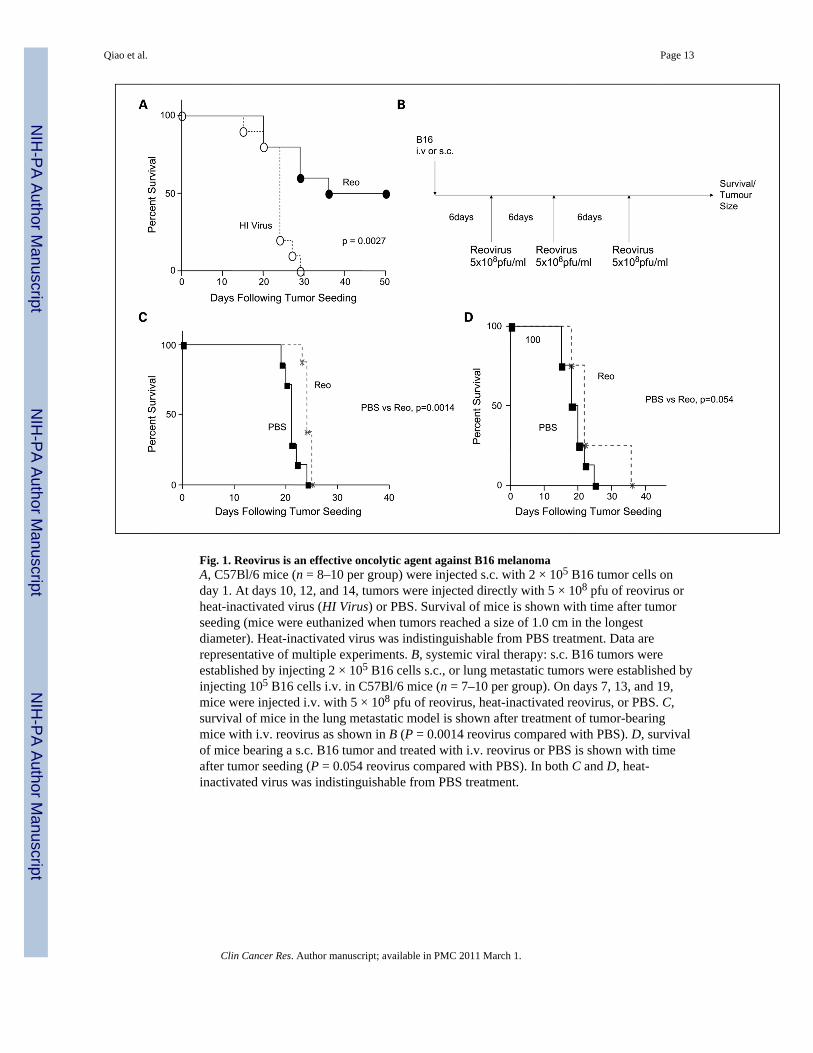
Fig. 1. Reovirus is an effective oncolytic agent against B16 melanomaA, C57Bl/6 mice (n = 8–10 per group) were injected s.c. with 2 × 10⁵ B16 tumor cells onday 1. At days 10, 12, and 14, tumors were injected directly with 5 × 10⁸ pfu of reovirus orheat-inactivated virus (HI Virus) or PBS. Survival of mice is shown with time after tumorseeding (mice were euthanized when tumors reached a size of 1.0 cm in the longestdiameter). Heat-inactivated virus was indistinguishable from PBS treatment. Data arerepresentative of multiple experiments. B, systemic viral therapy: s.c. B16 tumors wereestablished by injecting 2 × 10⁵ B16 cells s.c., or lung metastatic tumors were established byinjecting 105 B16 cells i.v. in C57Bl/6 mice (n = 7–10 per group). On days 7, 13, and 19,mice were injected i.v. with 5 × 10⁸ pfu of reovirus, heat-inactivated reovirus, or PBS. C,survival of mice in the lung metastatic model is shown after treatment of tumor-bearingmice with i.v. reovirus as shown in B (P = 0.0014 reovirus compared with PBS). D, survivalof mice bearing a s.c. B16 tumor and treated with i.v. reovirus or PBS is shown with timeafter tumor seeding (P = 0.054 reovirus compared with PBS). In both C and D, heat-inactivated virus was indistinguishable from PBS treatment.
Qiao et al. Page 13
Clin Cancer Res. Author manuscript; available in PMC 2011 March 1.
NIH
-PA Author Manuscript
NIH
-PA Author Manuscript
NIH
-PA Author Manuscript
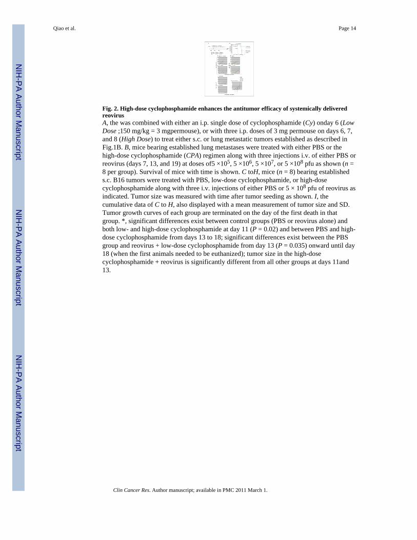
Fig. 2. High-dose cyclophosphamide enhances the antitumor efficacy of systemically deliveredreovirusA, the was combined with either an i.p. single dose of cyclophosphamide (Cy) onday 6 (LowDose ;150 mg/kg = 3 mgpermouse), or with three i.p. doses of 3 mg permouse on days 6, 7,and 8 (High Dose) to treat either s.c. or lung metastatic tumors established as described inFig.1B. B, mice bearing established lung metastases were treated with either PBS or thehigh-dose cyclophosphamide (CPA) regimen along with three injections i.v. of either PBS orreovirus (days 7, 13, and 19) at doses of5 ×105, 5 ×10⁶, 5 ×10⁷, or 5 ×10⁸ pfu as shown (n =8 per group). Survival of mice with time is shown. C toH, mice (n = 8) bearing establisheds.c. B16 tumors were treated with PBS, low-dose cyclophosphamide, or high-dosecyclophosphamide along with three i.v. injections of either PBS or 5 × 10⁸ pfu of reovirus asindicated. Tumor size was measured with time after tumor seeding as shown. I, thecumulative data of C to H, also displayed with a mean measurement of tumor size and SD.Tumor growth curves of each group are terminated on the day of the first death in thatgroup. *, significant differences exist between control groups (PBS or reovirus alone) andboth low- and high-dose cyclophosphamide at day 11 (P = 0.02) and between PBS and high-dose cyclophosphamide from days 13 to 18; significant differences exist between the PBSgroup and reovirus + low-dose cyclophosphamide from day 13 (P = 0.035) onward until day18 (when the first animals needed to be euthanized); tumor size in the high-dosecyclophosphamide + reovirus is significantly different from all other groups at days 11and13.
Qiao et al. Page 14
Clin Cancer Res. Author manuscript; available in PMC 2011 March 1.
NIH
-PA Author Manuscript
NIH
-PA Author Manuscript
NIH
-PA Author Manuscript
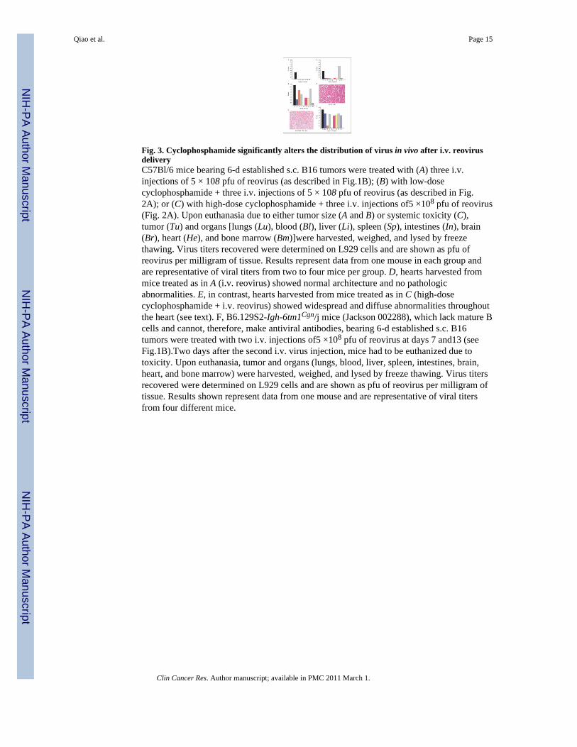
Fig. 3. Cyclophosphamide significantly alters the distribution of virus in vivo after i.v. reovirusdeliveryC57Bl/6 mice bearing 6-d established s.c. B16 tumors were treated with (A) three i.v.injections of 5 × 10⁸ pfu of reovirus (as described in Fig.1B); (B) with low-dosecyclophosphamide + three i.v. injections of 5 × 10⁸ pfu of reovirus (as described in Fig.2A); or (C) with high-dose cyclophosphamide + three i.v. injections of5 ×108 pfu of reovirus(Fig. 2A). Upon euthanasia due to either tumor size (A and B) or systemic toxicity (C),tumor (Tu) and organs [lungs (Lu), blood (Bl), liver (Li), spleen (Sp), intestines (In), brain(Br), heart (He), and bone marrow (Bm)]were harvested, weighed, and lysed by freezethawing. Virus titers recovered were determined on L929 cells and are shown as pfu ofreovirus per milligram of tissue. Results represent data from one mouse in each group andare representative of viral titers from two to four mice per group. D, hearts harvested frommice treated as in A (i.v. reovirus) showed normal architecture and no pathologicabnormalities. E, in contrast, hearts harvested from mice treated as in C (high-dosecyclophosphamide + i.v. reovirus) showed widespread and diffuse abnormalities throughoutthe heart (see text). F, B6.129S2-Igh-6tm1Cgn/j mice (Jackson 002288), which lack mature Bcells and cannot, therefore, make antiviral antibodies, bearing 6-d established s.c. B16tumors were treated with two i.v. injections of5 ×108 pfu of reovirus at days 7 and13 (seeFig.1B).Two days after the second i.v. virus injection, mice had to be euthanized due totoxicity. Upon euthanasia, tumor and organs (lungs, blood, liver, spleen, intestines, brain,heart, and bone marrow) were harvested, weighed, and lysed by freeze thawing. Virus titersrecovered were determined on L929 cells and are shown as pfu of reovirus per milligram oftissue. Results shown represent data from one mouse and are representative of viral titersfrom four different mice.
Qiao et al. Page 15
Clin Cancer Res. Author manuscript; available in PMC 2011 March 1.
NIH
-PA Author Manuscript
NIH
-PA Author Manuscript
NIH
-PA Author Manuscript
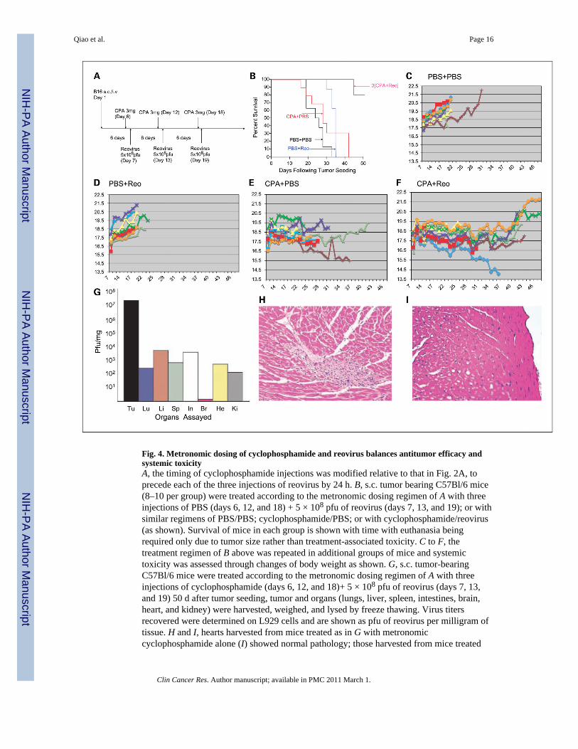
Fig. 4. Metronomic dosing of cyclophosphamide and reovirus balances antitumor efficacy andsystemic toxicityA, the timing of cyclophosphamide injections was modified relative to that in Fig. 2A, toprecede each of the three injections of reovirus by 24 h. B, s.c. tumor bearing C57Bl/6 mice(8–10 per group) were treated according to the metronomic dosing regimen of A with threeinjections of PBS (days 6, 12, and 18) + 5 × 10⁸ pfu of reovirus (days 7, 13, and 19); or withsimilar regimens of PBS/PBS; cyclophosphamide/PBS; or with cyclophosphamide/reovirus(as shown). Survival of mice in each group is shown with time with euthanasia beingrequired only due to tumor size rather than treatment-associated toxicity. C to F, thetreatment regimen of B above was repeated in additional groups of mice and systemictoxicity was assessed through changes of body weight as shown. G, s.c. tumor-bearingC57Bl/6 mice were treated according to the metronomic dosing regimen of A with threeinjections of cyclophosphamide (days 6, 12, and 18)+ 5 × 10⁸ pfu of reovirus (days 7, 13,and 19) 50 d after tumor seeding, tumor and organs (lungs, liver, spleen, intestines, brain,heart, and kidney) were harvested, weighed, and lysed by freeze thawing. Virus titersrecovered were determined on L929 cells and are shown as pfu of reovirus per milligram oftissue. H and I, hearts harvested from mice treated as in G with metronomiccyclophosphamide alone (I) showed normal pathology; those harvested from mice treated
Qiao et al. Page 16
Clin Cancer Res. Author manuscript; available in PMC 2011 March 1.
NIH
-PA Author Manuscript
NIH
-PA Author Manuscript
NIH
-PA Author Manuscript
with metronomic cyclophosphamide + i.v. reovirus (H) showed predominantly normalpathology but with sporadic and isolated areas of myocarditis.
Qiao et al. Page 17
Clin Cancer Res. Author manuscript; available in PMC 2011 March 1.
NIH
-PA Author Manuscript
NIH
-PA Author Manuscript
NIH
-PA Author Manuscript
NIH
-PA Author Manuscript
NIH
-PA Author Manuscript
NIH
-PA Author Manuscript
Qiao et al. Page 18
Table 1
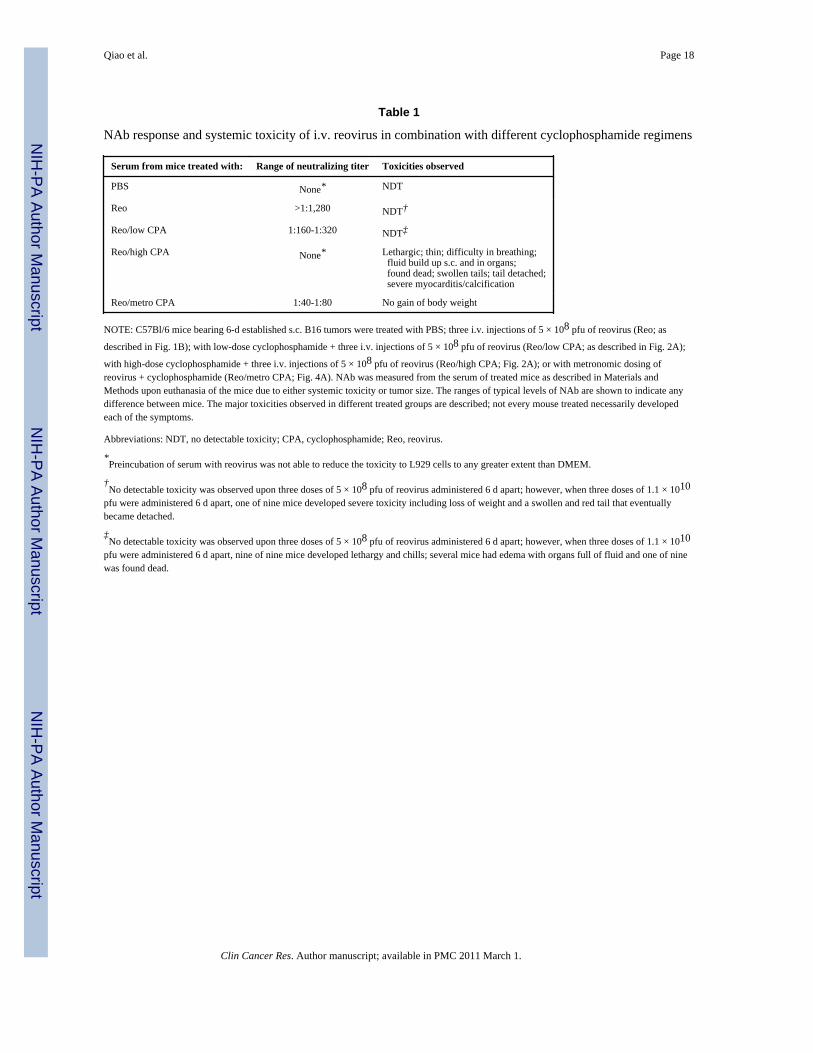
NAb response and systemic toxicity of i.v. reovirus in combination with different cyclophosphamide regimens
Serum from mice treated with: Range of neutralizing titer Toxicities observed
PBS None* NDT
Reo >1:1,280 NDT†
Reo/low CPA 1:160-1:320 NDT‡
Reo/high CPA None* Lethargic; thin; difficulty in breathing; fluid build up s.c. and in organs; found dead; swollen tails; tail detached; severe myocarditis/calcification
Reo/metro CPA 1:40-1:80 No gain of body weight
NOTE: C57Bl/6 mice bearing 6-d established s.c. B16 tumors were treated with PBS; three i.v. injections of 5 × 10⁸ pfu of reovirus (Reo; as
described in Fig. 1B); with low-dose cyclophosphamide + three i.v. injections of 5 × 10⁸ pfu of reovirus (Reo/low CPA; as described in Fig. 2A);
with high-dose cyclophosphamide + three i.v. injections of 5 × 10⁸ pfu of reovirus (Reo/high CPA; Fig. 2A); or with metronomic dosing ofreovirus + cyclophosphamide (Reo/metro CPA; Fig. 4A). NAb was measured from the serum of treated mice as described in Materials andMethods upon euthanasia of the mice due to either systemic toxicity or tumor size. The ranges of typical levels of NAb are shown to indicate anydifference between mice. The major toxicities observed in different treated groups are described; not every mouse treated necessarily developedeach of the symptoms.
Abbreviations: NDT, no detectable toxicity; CPA, cyclophosphamide; Reo, reovirus.
*Preincubation of serum with reovirus was not able to reduce the toxicity to L929 cells to any greater extent than DMEM.
†No detectable toxicity was observed upon three doses of 5 × 10⁸ pfu of reovirus administered 6 d apart; however, when three doses of 1.1 × 10¹⁰
pfu were administered 6 d apart, one of nine mice developed severe toxicity including loss of weight and a swollen and red tail that eventuallybecame detached.
‡No detectable toxicity was observed upon three doses of 5 × 10⁸ pfu of reovirus administered 6 d apart; however, when three doses of 1.1 × 10¹⁰
pfu were administered 6 d apart, nine of nine mice developed lethargy and chills; several mice had edema with organs full of fluid and one of ninewas found dead.
Clin Cancer Res. Author manuscript; available in PMC 2011 March 1.