An Auxin-Regulated Gene of Arabidopsis thaliana Encodes a DNA-Binding Protein
Upload
independentCategory
view
1download
0
Voue1Iubr2 94NcecAisRsac
Moeculr doning and sequencing of the reovirus (serotype 3) Sl gene which encodes the virl cellattachment protein al
Les Nagata, Saad A.Masri, David C.W.Mah and Patrick W.K.Lee*
Department of Microbiology and Infectious Diseases, University of Calgary Health Sciences Centre,Calgary, Alberta T2N 4N1, Canada
Received 31 July 1984; Revised and Accepted 18 October 1984
ABSTRACTTFhe complete sequence of the reovirus (serotype 3) S1 gene was obtained
by using cloned cDNA derived from the RNA segment. This gene is 1416nucleotides in length and contains two open reading frames. The first readingframe has a coding capacity of 455 amino acids, sufficient to account for theknown S1 product, protein ol (42,000 MW). It possesses a signal peptide aswell as three possible glycosylation sites. No homology could be detectedwhen this gene sequence and the deduced amino acid sequence were compared topublished sequences of the corresponding gene of a human rotavirus. Thesecond reading frame (not in phase with the first) starts at the second ATGrecently shown to be a functional initiation site. It has a coding capacityof 120 amino acids. Its outstanding feature is the highly basic amino-terminal region, a characteristic apparently shared by a number of DNA bindingproteins. It is speculated that this protein, hitherto undetected, may playa role in mediating viral and/or host nucleic acid transcription.
INTRODUCTIONThe genome of mammalian reoviruses consists of 10 double-stranded (ds)
RNA segments, each of which appears to encode one single protein (1,2). Eightof these proteins are structural proteins (3) which are assembled in themature virion as a double capsid (4). The outer capsid shell is composed ofthree proteins, namely, U1C, a3 and al. Protein plC and a3 are presumablyclosely associated with each other (5), and they also make up over 60% of thevirion mass (3). Protein al, on the other hand, is a minor component (about24 molecules) (3) which is located at the 12 vertices of the viral icosahedron(6). The location and distribution of this protein on the virion surface areprobably of strategic significance since this protein is used by the virus toanchor itself onto receptors of the host cell plasma membrane (6). Proteinal is also the reovirus hemagglutinin and is responsible for eliciting theformation of neutralizing antibody as well as for triggering host immuneresponses (7,8,9,10). In addition, this protein defines tissue tropism andvirulence (11), and mediates the binding of virions to cellular microtubules(12), and the inhibition of host DNA synthesis (13). Protein al therefore
© I RL Press Limited, Oxford, England.
Nucleic Acids ResearchVolume 12 Number 22 1984
8699

Nucleic Acids Research
specifies how reovirus particles interact with host cells and with the host.Another interesting feature of this protein is that it is the only reovirusprotein that confers serotype specificity (5,14,15); three immunologicallydistinct types (serotypes 1, 2, and 3) have been identified thus far (16).That al is the most type-specific of all reovirus proteins is consistent withthe recent observation that of the 10 reovirus genes, the gene encoding the alprotein, gene S1, is the gene that has undergone the most marked evolutionarydivergence (maximum of 10% homology between serotypes) (17).
Little is known concerning the molecular anatomy of protein al. At thepresent time, the major domains of interest on this protein are the onesinvolved in interaction with cell surface structures. Using a number of mono-clonal antibodies directed against the al protein, Fields and co-workers (18,19) have identified at least three distinct functional domains on this proteinand concluded that the hemagglutination and the neutralization domainscorrespond to discrete antigenic regions on the protein. Since anti-alantibodies neutralize reovirus by preventing viral adsorption (6), theseobservations strongly suggest that different recognition mechanisms areinvolved in reovirus hemagglutination and host cell attachment.
In view of the various functions displayed by protein ol and the signif-icant role it plays in viral pathogenesis (20), we have cloned and sequencedthe S1 gene of reovirus serotype 3. Two open reading frames were found: onespans 455 codons, the other 120 codons. The predicted features of these twoproteins are discussed in light of the deduced amino acid sequences.
MATERIALS AND METHODSVirus
The virus used was reovirus serotype 3 (strain Dearing). It waspropagated in suspension cultures of L-929 strain of mouse fibroblasts andpurified by the method described by Smith et al. (3).Cloning of Reovirus Genes
The procedure used for the isolation of genomic dsRNA, the preparationof ds cDNA, and the cloning of cDNA into the Pstl site of the plasmid pBR322was basically the same as described previously (21), with the exception thatin place of DMSO, methylmercuric hydroxide (10 mM) was used to denature thedsRNA (10 min at room temperature) prior to 3' termini polyadenylation andreverse transcription.Identification of Cloned S1 Gene
Of all transformed bacterial colonies (selected by screening for resis-
8700
Nucleic Acids Research
tance to tetracycline and sensitivity to ampicillin), 20 were randomly chosen
and their plasmids isolated. Plasmid DNA was then 32P-labelled by nicktranslation (22) and hybridized to reovirus dsRNA which had been resolved ina 7.5% polyacrylamide gel (in which the Si RNA segment is well separated fromthe other segments), and subsequently transferred to nitrocellulose paper(NEN Genescreen) as described previously (21). The marker RNA pattern on suchpaper was identified by hybridizing a strip to 32P-labelled DNA transcripts oftotal reovirus dsRNA using the random primer method of Taylor et al. (23).DNA Sequencing and Computer Analysis
Recombinant plasmids containing inserts from the S1 gene were isolatedusing the method described by Birnboim and Doly (24). Restriction digests ofwhole plasmids or of inserts (obtained by Pstl digestion of plasmids followedby their isolation from agarose gels) were then subcloned in various vectorsof the M13 mp series (25,26). Sequencing was done by the dideoxy chaintermination method of Sanger (27).
Analyses of nucleotide and amino acid sequences were done using publishedalgorithm (28) programmed in Fortran on the Honeywell DPS-870 computer.Graphic comparison matrices were obtained using a Calcomp 1051 plotter.
RESULTS AND DISCUSSIONIdentification of Clones Derived from Reovirus Si Gene
Of the twenty cloned plasmids randomly chosen, 3 were found to containSi-derived inserts using the Northern blotting technique. Two such plasmids(pL662 and pL676) are shown in Fig. 1. To size the inserts, the two plasmidswere digested with Pstl and analyzed by polyacrylamide gel electrophoresis(Fig. 2). Clone pL662 was found to contain an insert approximately 740 basepairs in length. The insert of clone pL676 has an internal Pstl site, thusyielding two S1-specific segments, one of about 870, the other of about 430base pairs in length. Upon sequencing, clones pL662 and pL676 were found tocontain, respectively, sequences from the 3' and 5' end of the S1 gene asdeduced previously from RNA sequencing of the termini of reovirus genes (29,30). Since the S1 RNA segment was estimated by polyacrylamide gel electro-phoresis to be between 1400 and 1500 base pairs long, the two plasmidspresumably span the entire sequence of the S1 gene.Gene S1 Sequence
Inserts from the two plasmids (pL662 and pL676) were sequenced accordingto the strategy shown in Fig. 3. The majority of the sequence (90%) was
determined in both directions and unambiguous data were obtained for the
8701

Nucleic Acids Research
2) 3 Fig. 1. Identification of clonesderived from the Si gene. Total reovirusgenomic RNA was resolved in a 7.5% poly-acrylamide gel and transferred to nitro-cellulose paper. Transblotted RNA wasthen hybridized to nick translatedplasmids isolated from transformedbacterial colonies. Lane 1, RNA hybrid-ized to 32P-labelled DNA transcripts oftotal genomic RNA. Lanes 2 and 3, RNAhybridized to nick translated recombinantplasmids pL676 and pL662, respectively.
-S
1 2 3 4 5 6 7 Fig... 2. Sizing of Si-derived insertsin a 7.5X polyacrylamide gel. The gelwas stained with ethidium bromide afterelectrophoresis. Lane 1, HindIII frag-ments of XDNA (size markers). Lane 2,HaeIII fragments of 0X174 DNA (sizemarkers). Lane 3, plasmid pL676restricted with Pstl. Lane 4, plasmidpL662 restricted with Pstl. Lane 5,
-861 pBR322 restricted with Pstl. Lane 6,123 base pair ladder marker (from BRL).
738 Lane 7, reovirus genomic dsRNA. Thenumbers on the right designate size(base pairs) of the 123 base pair ladder-492 marker.
-369
S1-S2-
S3&S4=
8702

Nucleic Acids Research
B M A PA A,S AA AAM A H5'T TITI11 ,I III, ,3'1 200 400 600 800 1000 1200 1400
pL676___ pL662
Fig. 3. Strategy of sequencing SI-derived inserts in pL676 and pL662containing overlapping Si sequences. Restriction sites are indicated: A,Alul; B, BamH1; H, HaeIII; M, Mspl; P, Pstl; S, Sstl; T, Taql. The arrowsindicate the direction and extent of sequence analysis performed on each ofthe restriction fragments.
remaining regions. All restriction sites used for the generation of fragmentsfor subcloning into M13 were covered by overlapping sequences. The completenucleotide sequence of the positive strand of the S1 gene is presented in Fig.4. The 5' and 3' terminal sequences were in excellent agreement with thosepreviously determined by RNA sequencing techniques (30).
The entire Si sequence is 1416 nucleotides long. The quantitative dis-tribution of the four bases was found to be relatively even: 27% A, 21% C,26% G, and 26% T. There is an open reading frame of 1365 bases (455 codons)which starts at nucleotide 13 with the first possible initiation codon andterminates at nucleotide 1377. The 5'- and 3'-noncoding sequences are 12 and39 nucleotides long, respectively. The absence of adenine-rich strings ofnucleotides at the 3'-noncoding region is consistent with the lack of poly-adenylation of reovirus mRNAs. There is a second open reading frame whichstarts at nucleotide 71 and extends for 120 codons until a termination codonat nucleotide 431 is reached (Fig. 4). The detection of two open readingframes of significant length (over 100 amino acids) is in accord with theprevious observation that the two initiation sites in the reovirus Si mRNAare both recognized by ribosomes (31), and with the more recent finding thatboth sites are functional (32). In this regard, it is interesting to notethat a mRNA of Influenza B virus was recently found to be bicistronic (33).No additional reading frames longer than 100 amino acids beginning with a
methionine were detected on either the plus or minus sense strand.A computer analysis comparing nucleotide sequences of the S1, S2 and S3
genes revealed no homologous region of statistical significance (data not
shown).
8703

Nucleic Acids Research
5'-GCTATTGGTCGG ATG GAT CCT CGC CTA CGT GAA GM GTA GTA CGG CTG ATA ATC GCA TTA ACG AGT GAT MT GGA 75met asp pro arg leu arg glu glu val val arg leu lie ile ala leu thr ser asp asn gly 21
met glu 2
GCA TCA CTG TCA AAA GGG CTT GAA TCA AGG GTC TCG GCG CTC GAG MG ACG TCT CAA ATA CAC TCT GAT ACT ATC 4150ala ser leu ser lys gly leu glu ser arg val ser ala leu glu lys thr ser gin ile his ser asp thr ile 46his his cys gin lys gly leu asn gin gly ser arg arg ser arg arg arg leu lys tyr thr leu ile leu ser 27
CTC CGG ATC ACC CAG GGA CTC GAT GAT GCA MC AM CGA ATC ATC GCT CTT GAG CAA AGT CGG GAT GAC TTG GTT 225leu arg ile thr gin gly leu asp asp ala asn lys arg ile ile ala leu glu gin ser arg asp asp leu val 71ser gly ser pro arg asp ser met met gin thr asn glu ser ser leu leu ser lys val gly met thr trp leu 52
GCA TCA GTC AGT GAT GCT CM CTT GCA ATC TCC AGA TTG GAA AGC TCT ATC GGA GCC CTC CAA ACA GTT GTC MT 300ala ser val ser asp ala gin leu ala ile ser arg leu glu ser ser ile gly ala leu gin thr val val asn 96his gin ser val met leu asn leu gin ser pro asp trp lys ala leu ser glu pro ser lys gin leu ser met 77
GGA CTT GAT TCG AGT GTT ACC CAG TTG GGT GCT CGA GTG GGA CAA CTT GAG ACA GGA CTT GCA GAC GTA CGC GTT 375gly leu asp ser ser val thr gin leu gly ala arg val gly gin leu glu thr gly leu ala asp val arg val 121asp leu lie arg val leu pro ser trp val leu glu trp asp asn leu arg gin asp leu gin thr tyr ala leu 102
GAT CAC GAC AAT CTC GTT GCG AG GTG GAT ACT GCA GAA CGT AAC ATT GGA TCA TTG ACC ACT GAG CTA TCA ACT 450asp his asp asn leu val ala arg val asp thr aia glu arg asn ile gly ser leu thr thr glu leu ser thr 146ile thr thr ile ser leu arg glu trp ile leu gin asn val thr leu asp his 120
CTG ACG TTA CGA GTA ACA TCC ATA CAA GCG GAT TTC GAA TCT AGG ATA TCC ACG TTA GAG CGC ACG GCG GTC ACT 525leu thr leu arg val thr ser ile gin ala asp phe glu ser arg ile ser thr leu glu arg thr ala val thr 171
AGC GCG GGA GCT CCC CTC TCA ATC CGT MT MC CGT ATG ACC ATG GGA TTA AAT GAT GGA CTC ACG TTG TCA GGG 600ser ala gly ala pro leu ser ile arg asn asn arg met thr met gly leu asn asp gly leu thr leu ser gly 196
MT MT CTC GCC ATC CGA TTG CCA GGA MT ACG GGT CTG MT ATT CM MT GGT GGA CTT CAG TTT CGA TTT MT 675asn asn leu ala ile arg leu pro gly asn thr gly leu asn ile gin asn gly gly leu gin phe arg phe asn 221
ACT GAT CM TTC CAG ATA GTT MT MT AAC TTG ACT CTC AMG ACG ACT GTG TTT GAT TCT ATC MC TCA AGG ATA 750thr asp- gin phe gin ile val asn asn asn leu thr leu lys thr thr val phe asp ser ile asn ser arg ile 246
GGC GCA ACT GAG CM AGT TAC GTG GCG TCG GCA GTG ACT CCC TTG AGA TTA MC AGT AGC ACG MG GTG CTG GAT 825gly ala thr glu gin ser tyr val ala ser ala val thr pro leu arg leu asn ser ser thr lys val leu asp 271
ATG CTA ATA GAC AGT TCA ACA CTT GM ATT MT TCT AGT GGA CAG CTA ACT GTT AGA TCG ACA TCC CCG MT TTG 900met leu ile asp ser ser thr leu glu ile asn ser ser gly gin leu thr val arg ser thr ser pro asn leu 296
AGG TAT CCG ATA GCT GAT GTT AGC GGC GGT ATC GGA ATG AGT CCA MT TAT AGG TTT AGG CAG AGC ATG TGG ATA 975arg tyr pro ile ala asp val ser gly gly ile gly met ser pro asn tyr arg phe arg gin ser met trp ile 321
GGA ATT GTC TCC TAT TCT GGT AGT GGG CTG MT TGG AGG GTA CAG GTG MC TCC GAC ATT TTT ATT GTA GAT GAT 1050gly ile val ser tyr ser gly ser gly leu asn trp arg val gin val asn ser asp ile phe ile val asp asp 346
TAC ATA CAT ATA TGT CTT CCA GCT TTT GAC GGT TTC TCT ATA GCT GAC GGT GGA GAT CTA TCG TTG MC TTT GTT 1125tyr ile his ile cys leu pro ala phe asp gly phe ser ile ala asp gly gly asp leu ser leu asn phe val 371
ACC GGA TTG TTA CCA CCG TTA CTT ACA GGA GAC ACT GAG CCC GCT TTT CAT MT GAC GTG GTC ACA TAT GGA GCA 1200thr gly leu leu pro pro leu leu thr gly asp thr glu pro ala phe his asn asp val val thr tyr gly ala 396
CAG ACT GTA GCT ATA GGG TTG TCG TCG GGT GGT GCG CCT CAG TAT ATG AGT AAG MT CTG TGG GTG GAG CAG TGG 1275gin thr val ala ile gly leu ser ser gly gly ala pro gin tyr met ser lys asn leu trp val glu gin trp 421
CAG GAT GGA GTA CTT CGG TTA CGT GTT GAG GGG GGT GGC TCA ATT ACG CAC TCA MC AGT AAG TGG CCT GCC ATG 1350gin asp gly val leu arg leu arg val glu gly gly gly ser ile thr his ser asn ser lys trp pro ala met 446
ACC GTT TCG rAC CCG CGT AGT TTC ACG TGA GGA TCA GAC CAC CCC GCG GCA CTG GGG CAT TTC ATC-3' 1416thr val ser ty,r pro arg ser phe thr 455
Fig. 4. Sequence of the Si gene written in the mRNA sense. The two predictedamino acid sequences are shown below the gene sequence: the first readingframe starts at nucleotide 13 and terminates at nucleotide 1377; the secondreading frame starts at nucleotide 71 and terminates at nucleotide 431.Possible glycosylation sites are underlined.
8704

Nucleic Acids Research
Deduced Amino Acid Sequences and Their FeaturesFirst Open Reading Frame. The deduced amino acid sequence of the polypeptideencoded by the first reading frame is shown in Fig. 4. Its length (455 aminoacid residues) is sufficient to account for the size of the known S1 geneproduct, al, which has an estimated molecular weight of 42,000 (3). Thecalculated net charge of this protein at pH 7.0, assuming glutamic andaspartic acid are each -1, and arginine and lysine each +1, histidine 0.5 andthe remaining amino acids are neutral (charge 0) at this pH (34), is -6.5.This is in agreement with the relatively low pI value of 5.5 to 5.9 previouslyreported for protein al (35). There are three potential glycosylation sites(Asn-X-Thr/Ser) (Fig. 4), consistent with previous glycosylation studies whichsuggest that al may be a glycoprotein (36). There are also indications thatone or more of these sites may be involved in the attachment of al (and hencevirus particles) to cellular receptors (Paul and Lee, unpublished data).
An examination of the amino-terminal region of the deduced sequencerevealed the presence of a hydrophobic core (residues 9 through 16). Usingthe rules proposed by Perlman and Halvorsen (37), a signal peptide comprisedof the first 18 amino acids (2,109 MW) can be identified. Cleavage after theserine residue would then result in an amino-terminal aspartic acid residue ofthe protein. While it is likely that the presumptive signal peptide iscleaved off before the incorporation of al in the mature virion, the pos-sibility remains that this short hydrophobic region may be used for interact-ing with host cell plasma membrane during viral attachment. It would thereforebe of interest to identify the amino-terminal residue of al in the maturevirion. However, this may prove difficult since it has been shown that mostof the proteins in reovirions contain a blocked amino terminus (38).
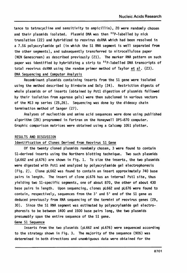
The hydrophilicity profile of the entire sequence was then deduced usingthe method described by Kyte and Doolittle (39), and is shown in Fig. 5A. Ahighly hydrophobic amino-terminal region corresponding to the presumptivesignal peptide is clearly discernible. Clusters of hydrophobic regions arealso found in the carboxyl-terminal half of the molecule. The carboxyl terminus,however, is relatively hydrophilic. The three glycosylation sites, clusteredaround the middle section of the molecule, do not appear to be located inregions of marked hydrophilicity. A second hydrophilicity plot [Hopp andWoods method (40)], based on a slightly different set of hydrophilicity valuesassigned to the amino acids, is shown in Fig. 5B. Such a plot has been foundto be useful for the prediction of antigenic determinants since they correspondto highly hydrophilic sites. Two such sites were found, one around residue 56,
8705
Nucleic Acids Research
..I
B
Amino acid number
Fig. 5. Relative hydrophobicity plots of the reovirus (serotype 3) olprotein. (A) Kyte and Doolittle (39) plot. The arrows indicate potentialglycosylation sites. (B) Hopps and Woods (40) plot of relating hydrophilicsites to antigenic determinants.
and the other around residue 66. Since a library of monoclonal antibodiesdirected against protein al is now available (5,18) and al which retains cell-binding capacity has recently been isolated from intact virions (Yeung and Lee,unpublished data), it should now be feasible to pinpoint such sites (includingneutralization sites) on the protein.
The deduced amino acid sequence yields limited information on the second-ary structure of the protein. Using the rules proposed by Chow and Fasman (41),most a-helical regions were found to be confined to the amino-terminal thirdof the molecule (data not shown). In this regard it is interesting to note
that 13 of the 14 proline residues which do not favor a-helix formation residein the latter two-thirds of the sequence. The presence of a single cysteineresidue also precludes the formation of intramolecular disulfide bonds.Comparison of Deduced al Sequence with Sequence of Rotavirus Type-SpecificProtein. As mentioned above, protein cl is the typerspecific protein ofreovirus and the genes (Si) encoding this protein of the three reovirus sero-types are related to the extent of not more than 1-10% (17). The correspond-ing gene of mammalian rotaviruses, however, appears to be highly conserved:both the nucleotide and amino acid sequences of human, simian and bovinerotaviruses were found to be closely related (approximately 80%) (42). Thesefindings are therefore consistent with earlier observations that rotaviruses
8706
A
cI-1 L.IirI4FML

Nucleic Acids Research
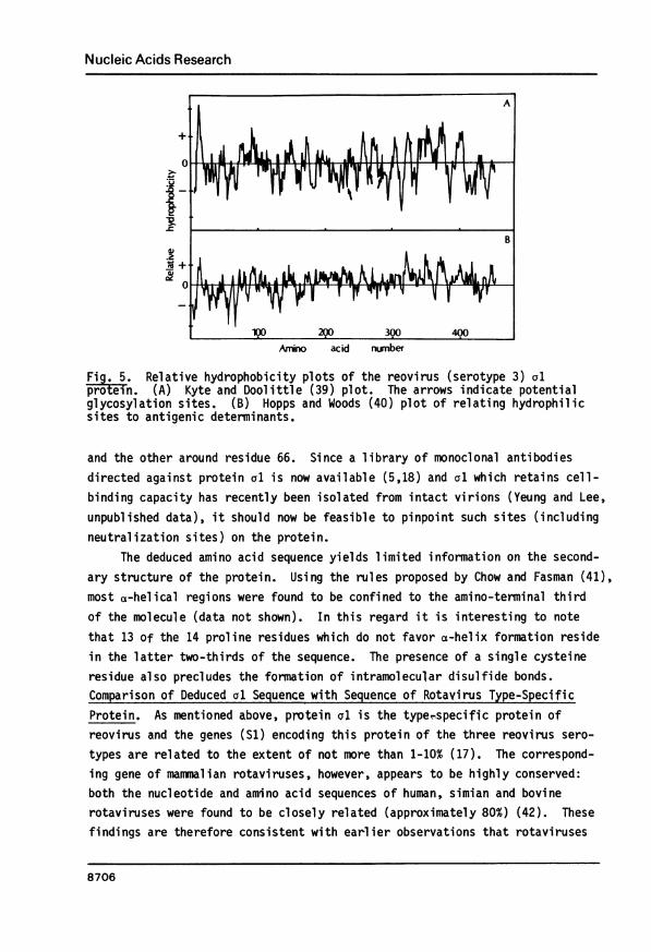
A Fig. 6. Relative hydrophobicity plots of the seconddeduced amino acid sequence of gene Si. (A) Kyteand Doolittle (39) plot. (B) Hopps and Woods (40)
+ plot.
0
B
0 .
Ano adid number
and reoviruses are not antigenically related (43,44), although both virusesbelong to the same family (Reoviridae) and have similar morphology andstructural composition (45).
The extent of such unrelatedness was then measured by comparing thededuced amino acid sequence of reovirus protein al to the reported sequence ofthe serotype-specific protein of human rotavirus Hu/5 (42) using computer-generated comparison matrices. Although the lowest stringency condition wasused (single amino acid comparison), no sequence homology was detected betweenthese two proteins (data not shown). This was further reflected by the lackof homology between their nucleotide sequences (data not shown). These datastrongly suggest that the corresponding genes of the two viruses might haveevolved independently during the more recent period. It is interesting tonote, however, that both proteins possess an amino-terminal hydrophobic regionand a carboxyl-terminal hydrophilic region (46,47). The significance and theuniversality of such an arrangement among serotype-specific proteins ofReoviridae is not known at this time.Second Open Reading Frame. Recent evidence that the second AUG of the S1
8707
Nucleic Acids Research
mRNA is a functional initiation codon (31,32) suggests the possible presence
of another Si-specified protein, hitherto undetected, in reovirus-infectedcells. Starting at nucleotide 71 and spanning 120 codons (Fig. 4), thisprotein has a calculated molecular weight of 13,990 and is somewhat basic,
with an estimated net charge of +5 at pH 7.0.A sequence analogous to the amino-terminal hydrophobic core found in the
first reading frame is absent in this protein. In its place is an arginine-
rich region (residues 14 through 19), rendering this end of the moleculehighly basic. That this region is also the most hydrophilic region of the
protein is clearly indicated from hydrophilicity plots (Fig. 6). It is worthnoting that the concentration of basic residues at the amino end appears to be
a common feature of a number of DNA binding proteins such as the Cro proteinof bacteriophage 434 (48), the cl repressor and the N protein of bacteriophagex (49,50), and the Salmonella phage P22 c2 repressor (51). It is thereforetempting to speculate that this smaller S1-specified protein may have an
analogous function of binding to viral genomic RNA and/or host DNA, therebymediating positive and/or negative control of viral and/or host gene trans-
scription.Two possible glycosylation sites are also detected (Fig. 4). However,
the lack of a signal peptide makes it unlikely that this protein is glyco-sylated. Like the al protein, only one cysteine residue was found.
CONCLUDING REMARKSIt would now be of interest to clone and sequence the S1 genes of the
other two reovirus serotypes (serotypes 1 and 2). Since protein al is used by
all three serotypes for cell attachment (6), and yet this protein is the most
type-specific of all reovirus proteins, sequence analysis of the S1 genes (andal) of the three serotypes may distinguish functional conserved sites (e.g.,sites for attachment and hemagglutination) from sites that confer diverseantigenicity. Such data may also prove helpful for the ultimate elucidiation
of the nature of tissue tropism and virulence.The detection of a second reading frame with a functional initiation site
is of interest and deserves further investigation. A logical point of
departure would be to identify such a protein in reovirus-infected cells or inin vitro translation products of the S1 gene or mRNA. Its isolation and
characterization would then follow, and its possible function (such as the
mediating of viral and/or host nucleic acid transcription mentioned above)would be probed. Undoubtedly, the implication on the functional significance
8708

Nucleic Acids Research
of this putative protein would be dramatically amplified should the Si genesof the other two reovirus serotypes, upon sequencing, both be found to possessa second reading frame of comparable characteristics to the one reported herefor reovirus serotype 3.
Experiments following the forementioned courses are now in progress.
ACKNOWLEDGEMENTSWe thank Ralph Paul and Tomonori Morinaga for stimulating discussions;
David Pot and Michael Yeung for their assistance with computer analysis ofsequences; and Shirley Eikerman for typing the manuscript.
This work was supported by the Medical Research Council of Canada and bythe Alberta Heritage Foundation for Medical Research (AHFMR). L.N. and
D.C.W.M. are recipients of AHFMR Studentships, and P.W.K.L. is an AHFMR Scholar.
* To whom correspondence should be addressed
REFERENCES1. McCrae, M.A. and Joklik, W.K. (1978) Virology 89, 578-593.2. Mustoe, T.A., Ramig, R.F., Sharpe, A.H. and Fields, B.N. (1978)
Virology 85, 545-556.3. Smith, R.E., Zweerink, H.J. and Joklik, W.K. (1969) Virology 39, 791-
810.4. Lufti g, R.B., Kilham, S.S., Hay, A.J., Zweerink, H.J. and Joklik, W.K.
(1972) Virology 48, 170-181.5. Lee, P.W.K., Hayes, E.C. and Joklik, W.K. (1981) Virology 108, 134-
146.6. Lee, P.W.K., Hayes, E.C. and Joklik, W.K. (1981) Virology 108, 156-
163.7. Weiner, H.L. and Fields, B.N. (1977) J. Exp. Med. 146, 1305-1310.8. Weiner, H.L., Greene, M.I. and Fields, B.N. (1980) J. Imnunol. 125,
278-282.9. Fontana, A. and Weiner, H.L. (1980) J. Immunol. 125, 2660-2664.
10. Finberg, R., Weiner, H.L., Fields, B.N., Benecerraf, B. and Burakoff,S.J. (1979) Proc. Natl. Acad. Sci. USA 76, 442-446.
11. Weiner, H.L., Drayna, D., Averill, D.R., Jr. and Fields, B.N. (1977)Proc. Natl. Acad. Sci. USA 74, 5744-5748.
12. Babiss, L.E., Luftig, R.F., Weatherbee, J.A., Weihing, R., Ray, U.R. andFields, B.N. (1979) J. Virol. 30, 863-874.
13. Sharpe, A.H. and Fields, B.N. (1981) J. Virol. 38, 389-392.14. Hayes, E.C., Lee, P.W.K., Miller, S.E. and Joklik, W.K. (1981) Virology
108, 147-155.15. Gaillard, R.K. and Joklik, W.K. (1980) Virology 107, 533-536.16. Joklik, W.K. (1974) in Comprehensive Virology, Fraenkel-Conrat, H. and
Wagner, R.R., (eds.), pp. 231-334, Plenum Press, New York.17. Gaillard, R.K. and Joklik, W.K. (1982) Virology 123, 152-164.18. Birstin, S., Spriggs, D.R. and Fields, B.N. (1982) Virology 117, 146-
155.19. Spriggs, D.R., Kaye, K. and Fields, B.N. (1983) Virology 127, 220-224.
8709

Nucleic Acids Research
20. Fields, B.N. and Greene, M.I. (1982) Nature 300, 19-23.21. Cashdollar, L.W., Esparza, J., Hudson, G.R., Chmelo, R., Lee, P.W.K. and
Joklik, W.K. (1982) Proc. Natl. Acad. Sci. USA 79, 7644-7648.22. Rigby, P.W., Dieckmann, M., Rhodes, C. and Berg, P. (1977) J. Mol. Biol.
113, 237-251.23. Taylor, J.M., Illmensee, R. and Summers, J. (1976) Biochem. Biophys.
Acta 442, 324-330.24. Birnboim, H.C. and Doly, J. (1979) Nucleic Acids Res. 7, 1513-1523.25. Messing, J., Crea, R. and Seeberg, P.H. (1981) Nucleic Acids Res. 9,
309-321.26. Norrander, J., Kempe, T. and Messing, J. (1983) Gene 26, 101-106.27. Sanger, F., Nicklen, S. and Coulson, A.R. (1977) Biochem. Biophys. Acta
442, 324-330.28. Novotny, J. (1981) Nucleic Acids Res. 10, 127-131.29. McCrae, M.A. (1981) J. Gen. Virol. 55, 393-403.30. Antczak, J.B., Chmelo, R., Pickup, D.J. and Joklik, W.K. (1982) Virology
121,307-319.31. Kozak, M. (1982) J. Mol. Biol. 156, 807-820.32. Cenatiempo, Y., Twardowski, T., Shoeman, R., Ernst, H., Brot, N.,
Weissbach, H. and Shatkin, A.J. (1984) Proc. Natl. Acad. Sci. USA 81,1084-1088.
33. Shaw, M.W., Choppin, P.W. and Lamb, R.A. (1983) Proc. Natl. Acad. Sci.USA 80, 4879-4883.
34. Huddleston, J.A. and Brownlee, G.G. (1982) Nucleic Acids Res. 10,1029-1038.
35. Samuel, C.E. (1983) in Double-Stranded.RNA Viruses, Compans, R.W. andBishop, D.H.L. (eds,), pp. 219-230, Elsevier, New York.
36. Lee, P.W.K. (1983) in Double-Stranded RNA Viruses, Compans, R.W. andBishop, D.H.L. (eds.), pp. 193-206, Elsevier, New York.
37. Perlman, D. and Halvorson, H.0. (1983) J. Mol. Biol. 167, 391-409.38. Pett, D.M., Vanaman, T.C. and Joklik, W.K. (1973) Virology 52, 174-186.39. Kyte, J. and Doolittle, R.F. (1982) J. Mol. Biol. 157, 105-132.40. Hopp, T.P. and Woods, K.R. (1981) Proc. Natl. Acad. Sci. USA 78, 3824-
3828.41. Chow, P.Y. and Fasman, G.D. (1978) Ann. Rev. Biochem. 47, 151-276.42. Dyall-Smith, M.L. and Holmes, I.H. (1984) Nucleic Acids Res. 12, 3973-
3982.43. Bridger, J.C. and Woode, G.N. (1975) Brit. Vet. J. 131, 528-535.44. Flewett, T.H. and Woode, G.N. (1978) Arch. Virol. 57, 1-23.45. Joklik, W.K. (1983) in The Reoviridae, Joklik, W.K. (ed.), pp. 1-7,
Plenum, New York.46. Arias, C.F., Lopez, S., Bell, J.R. and Strauss, J.H. (1984) J. Virol.
50, 657-661.47. Elleman, T.C., Hoyme, P.A., Dyall-Smith, M.L., Holmes, I.H. and Azad, A.A.
(1983) Nucleic Acids Res. 11, 4689-4701.48. Grosschedl, R. and Schwarz, E. (1979) Nucleic Acids Res. 6, 867-881.49. Sauer, R.T. (1978) Nature (London) 276, 301-302.50. Franklin, N.C. and Bennett, G.N. (1979) Gene 8, 107-119.51. Sauer, R.T., Pan, J., Hopper, P., Hehir, K., Brown, J. and Poteete, A.R.
(1981) Biochemistry 20, 3591-3598.
8710
Copyright © 2022 FDOKUMEN