A Versatile Molecular Tagging Method for Targeting Proteins to Avian Reovirus muNS Inclusions. Use...
14
A Versatile Molecular Tagging Method for Targeting Proteins to Avian Reovirus muNS Inclusions. Use in Protein Immobilization and Purification Alberto Brandariz-Nun ˜ ez, Rebeca Menaya-Vargas, Javier Benavente, Jose Martinez-Costas* Department of Biochemistry and Molecular Biology, Faculty of Pharmacy and Center for Research in Biological Chemistry and Molecular Materials, University of Santiago de Compostela, Santiago de Compostela, Spain Abstract Background: Avian reoviruses replicate in viral factories, which are dense cytoplasmic compartments estabilished by protein-protein interactions. The non-structural protein muNS forms the factory scaffold that attracts other viral components in a controlled fashion. To create such a three-dimensional network, muNS uses several different self- interacting domains. Methodology/Principal Findings: In this study we have devised a strategy to identify muNS regions containing self- interacting domains, based on the capacity of muNS-derived inclusions to recruit muNS fragments. The results revealed that the muNS region consisting of residues 477–542 was recruited with the best efficiency, and this raised the idea of using this fragment as a molecular tag for delivering foreign proteins to muNS inclusions. By combining such tagging system with our previously established method for purifying muNS inclusions from baculovirus-infected insect cells, we have developed a novel protein purification protocol. Conclusions/Significance: We show that our tagging and inclusion-targeting system can be a simple, versatile and efficient method for immobilizing and purifying active proteins expressed in baculovirus-infected cells. We also demonstrate that muNS inclusions can simultaneously recruit several tagged proteins, a finding which may be used to generate protein complexes and create multiepitope particulate material for immunization purposes. Citation: Brandariz-Nun ˜ ez A, Menaya-Vargas R, Benavente J, Martinez-Costas J (2010) A Versatile Molecular Tagging Method for Targeting Proteins to Avian Reovirus muNS Inclusions. Use in Protein Immobilization and Purification. PLoS ONE 5(11): e13961. doi:10.1371/journal.pone.0013961 Editor: Anna Mitraki, University of Crete, Greece Received June 22, 2010; Accepted October 21, 2010; Published November 12, 2010 Copyright: ß 2010 Brandariz-Nun ˜ ez et al. This is an open-access article distributed under the terms of the Creative Commons Attribution License, which permits unrestricted use, distribution, and reproduction in any medium, provided the original author and source are credited. Funding: This work was supported by grants from the European Commission under contracts ERAS-CT-2003-980409 (as part of the European Science Foundation EUROCORES Programme EuroSCOPE, web: http://www.esf.org/euroscope); the Spanish Ministerio de Ciencia y Tecnologı ´a (BFU2007-61330, BFU 205- 24982-E, web: http://www.mityc.es/) and Xunta de Galicia (08CSA009203PR, web: http://www.conselleriaiei.org/ga/dxidi/index.php). ABN was the recipient of a predoctoral FPI fellowship from the Spanish Ministerio de Ciencia y Tecnologı ´a. The funders had no role in study design, data collection and analysis, decision to publish, or preparation of the manuscript. Competing Interests: The results of this study are patent pending. Application number: P201030204. Country: Spain. Inventors: Alberto Brandariz-Nun ˜ ez; Rebeca Menaya-Vargas; Javier Benavente and Jose Martinez-Costas. Application date: 12th February 2010. All materials described in the manuscript will be available for research purposes. The authors confirm that this does not alter their adherence to all the PLoS ONE policies on sharing data and materials. * E-mail: [email protected] Introduction Avian reoviruses are fusogenic viruses that belong to the Orthoreovirus, one of the twelve genera of the Reoviridae family [1,2]. They are pathogenic viruses involved in several syndromes that affect poultry [3,4]. Avian reovirus replicates in the cytoplasm and is one of the few non-enveloped viruses that are able to induce fusion of infected cells [5]. The viral genome is composed of 10 segments of double-stranded RNA, which are enclosed within a double-layered protein capsid with an external diameter of 85 nm and icosahedral symmetry. Details of avian reovirus structure, protein composition and replicative cycle have been described elsewhere [6,7,8]. Avian reoviruses replicate within cytoplasmic globular inclu- sions termed viral factories. These structures contain viral structural and non-structural proteins, together with viral RNA, but they lack cell organelles and membranes [9,10]. The expression of individual proteins by cell transfection revealed that the non-structural protein muNS is the only viral protein that forms cytoplasmic inclusions in the absence of any other viral factor [10]. These muNS-derived inclusions are very similar to the native viral factories, suggesting that this protein forms the basic scaffold of the factories in avian-reovirus infected cells. Analysis of transfected cells co-expressing muNS and other viral proteins revealed that muNS plays an important role in the early steps of viral morphogenesis by temporally and selectively controlling the recruitment of specific viral proteins to viral factories [9]. We have recently carried out an extensive characterization of inclusion formation by avian reovirus muNS [11]. We found, in clear contrast with the situation reported for mammalian reoviruses and many other animal viruses [12,13,14], that neither ARV-derived factories nor muNS-derived inclusions are associat- ed to the cytoskeleton, their formation and evolution are not dependent on the microtubule network, and are not related to aggresome or autophagosome generation. By two-hybrid analysis, we demonstrated that muNS monomers have the ability to self- PLoS ONE | www.plosone.org 1 November 2010 | Volume 5 | Issue 11 | e13961
-
Upload
independent -
Category
Documents
-
view
1 -
download
0
Transcript of A Versatile Molecular Tagging Method for Targeting Proteins to Avian Reovirus muNS Inclusions. Use...

A Versatile Molecular Tagging Method for TargetingProteins to Avian Reovirus muNS Inclusions. Use inProtein Immobilization and PurificationAlberto Brandariz-Nunez, Rebeca Menaya-Vargas, Javier Benavente, Jose Martinez-Costas*
Department of Biochemistry and Molecular Biology, Faculty of Pharmacy and Center for Research in Biological Chemistry and Molecular Materials, University of Santiago
de Compostela, Santiago de Compostela, Spain
Abstract
Background: Avian reoviruses replicate in viral factories, which are dense cytoplasmic compartments estabilished byprotein-protein interactions. The non-structural protein muNS forms the factory scaffold that attracts other viralcomponents in a controlled fashion. To create such a three-dimensional network, muNS uses several different self-interacting domains.
Methodology/Principal Findings: In this study we have devised a strategy to identify muNS regions containing self-interacting domains, based on the capacity of muNS-derived inclusions to recruit muNS fragments. The results revealed thatthe muNS region consisting of residues 477–542 was recruited with the best efficiency, and this raised the idea of using thisfragment as a molecular tag for delivering foreign proteins to muNS inclusions. By combining such tagging system with ourpreviously established method for purifying muNS inclusions from baculovirus-infected insect cells, we have developed anovel protein purification protocol.
Conclusions/Significance: We show that our tagging and inclusion-targeting system can be a simple, versatile and efficientmethod for immobilizing and purifying active proteins expressed in baculovirus-infected cells. We also demonstrate thatmuNS inclusions can simultaneously recruit several tagged proteins, a finding which may be used to generate proteincomplexes and create multiepitope particulate material for immunization purposes.
Citation: Brandariz-Nunez A, Menaya-Vargas R, Benavente J, Martinez-Costas J (2010) A Versatile Molecular Tagging Method for Targeting Proteins to AvianReovirus muNS Inclusions. Use in Protein Immobilization and Purification. PLoS ONE 5(11): e13961. doi:10.1371/journal.pone.0013961
Editor: Anna Mitraki, University of Crete, Greece
Received June 22, 2010; Accepted October 21, 2010; Published November 12, 2010
Copyright: � 2010 Brandariz-Nunez et al. This is an open-access article distributed under the terms of the Creative Commons Attribution License, which permitsunrestricted use, distribution, and reproduction in any medium, provided the original author and source are credited.
Funding: This work was supported by grants from the European Commission under contracts ERAS-CT-2003-980409 (as part of the European ScienceFoundation EUROCORES Programme EuroSCOPE, web: http://www.esf.org/euroscope); the Spanish Ministerio de Ciencia y Tecnologıa (BFU2007-61330, BFU 205-24982-E, web: http://www.mityc.es/) and Xunta de Galicia (08CSA009203PR, web: http://www.conselleriaiei.org/ga/dxidi/index.php). ABN was the recipient of apredoctoral FPI fellowship from the Spanish Ministerio de Ciencia y Tecnologıa. The funders had no role in study design, data collection and analysis, decision topublish, or preparation of the manuscript.
Competing Interests: The results of this study are patent pending. Application number: P201030204. Country: Spain. Inventors: Alberto Brandariz-Nunez;Rebeca Menaya-Vargas; Javier Benavente and Jose Martinez-Costas. Application date: 12th February 2010. All materials described in the manuscript will beavailable for research purposes. The authors confirm that this does not alter their adherence to all the PLoS ONE policies on sharing data and materials.
* E-mail: [email protected]
Introduction
Avian reoviruses are fusogenic viruses that belong to the
Orthoreovirus, one of the twelve genera of the Reoviridae family [1,2].
They are pathogenic viruses involved in several syndromes that
affect poultry [3,4]. Avian reovirus replicates in the cytoplasm and
is one of the few non-enveloped viruses that are able to induce
fusion of infected cells [5]. The viral genome is composed of 10
segments of double-stranded RNA, which are enclosed within a
double-layered protein capsid with an external diameter of 85 nm
and icosahedral symmetry. Details of avian reovirus structure,
protein composition and replicative cycle have been described
elsewhere [6,7,8].
Avian reoviruses replicate within cytoplasmic globular inclu-
sions termed viral factories. These structures contain viral
structural and non-structural proteins, together with viral RNA,
but they lack cell organelles and membranes [9,10]. The
expression of individual proteins by cell transfection revealed that
the non-structural protein muNS is the only viral protein that
forms cytoplasmic inclusions in the absence of any other viral
factor [10]. These muNS-derived inclusions are very similar to the
native viral factories, suggesting that this protein forms the basic
scaffold of the factories in avian-reovirus infected cells. Analysis of
transfected cells co-expressing muNS and other viral proteins
revealed that muNS plays an important role in the early steps of
viral morphogenesis by temporally and selectively controlling the
recruitment of specific viral proteins to viral factories [9].
We have recently carried out an extensive characterization of
inclusion formation by avian reovirus muNS [11]. We found, in
clear contrast with the situation reported for mammalian
reoviruses and many other animal viruses [12,13,14], that neither
ARV-derived factories nor muNS-derived inclusions are associat-
ed to the cytoskeleton, their formation and evolution are not
dependent on the microtubule network, and are not related to
aggresome or autophagosome generation. By two-hybrid analysis,
we demonstrated that muNS monomers have the ability to self-
PLoS ONE | www.plosone.org 1 November 2010 | Volume 5 | Issue 11 | e13961

associate. We also developed a simple method for purifying the
inclusions made by muNS in baculovirus-infected cells, and the
analysis of their protein composition indicated that muNS is the
main building block of these cytoplasmic globular structures.
Analysis of the domain composition of the 635-residue muNS
protein produced the following results: i) the region comprising
residues 448 to 635 constitutes the minimal muNS portion able to
form inclusions; we designated it muNS-Mi. ii) muNS-Mi is
composed of four differentiated domains: two predicted coiled-coil
elements that we termed Coil1 (C1; residues 448 to 477) and Coil2
(C2; residues 539 to 605), a stretch of amino acids linking both
coiled-coils that we termed Intercoil (IC; residues 477 to 542), and
a C-terminal part of the protein that we termed C-Tail (CT;
residues 605 to 635).
We also investigated the contribution of the four muNS-Mi
domains to inclusion-forming activity and determined that all of
them are essential for inclusion formation. Domain C1 can be
replaced by exogenous dimeric domains, and CT plays an
important role in orienting the muNS inter-monomer contacts
to form basal oligomers as well as influencing inclusion shape and
inclusion formation efficiency. We also identified an additional
domain located at the N-terminus of muNS, which is not essential
for inclusion formation, but plays a role in inclusion maturation.
The original aim of this study was to develop an alternative
method for detecting interactions between the different muNS
domains. Towards this end, we analyzed the ability of individual
muNS domains to get incorporated into cytoplasmic inclusions
formed by muNS in CEF. The domains that were most efficiently
incorporated into inclusions were the N-terminal part of the
protein and IC. This information was then used to develop a
method that used IC as a molecular tag. We demonstrate the
validity of our system by purifying proteins that remained active
while integrated inside muNS-derived protein inclusions. We show
that our method can be used to purify soluble and inclusion-
integrated active proteins. We also show that muNS-inclusions
have the capacity to simultaneously integrate several different
proteins, which may be useful for improving the efficiency of
supra-molecular complex generation as well as producing multi-
epitope particulate material suitable for vaccination.
Results
Detection of muNS domains implicated in inter-monomer interactions
Avian reovirus muNS possesses several domains that are directly
involved in self-association [11]. As a first approach to determine
the role that the different muNS domains play in forming inter-
monomer contacts, we decided to express individual muNS
fragments and check their incorporation into muNS- and muNS-
Mi-derived inclusions. We divided the protein muNS in 5 regions
or domains (Figure 1A): the N terminal two-thirds of the protein
that were shown to be dispensable to form inclusions but were
involved in inclusion maturation [11] (domain 1, residues 1 to
447); and the four domains of muNS-Mi that were also previously
described (C1 or domain 2, IC or domain 3, C2 or domain 4 and
CT or domain 5, [11]). We constructed plasmids expressing the
domains independently with a C-terminal hemagglutinin epitope
tag. This tag allows us to differentiate the inclusions formed by full-
length muNS and muNS-Mi from the HA-containing fragments
by immunostaining. All constructs were sequenced and their
expression checked by Western blot (not shown). For unknown
reasons, we could not detect the expression of domains 2 and 5,
either untagged, or HA-tagged at their N or C-terminus. To test
their activity, we decided to add 2 and 5 to domains 1 and 4
respectively, to check the influence that their addition has on the
incorporation of domains 1 and 4 into muNS inclusions
(Figure 1A).
When individually expressed in CEF, none of the fragments
shown in Figure 1 was able to form inclusions, but were evenly
distributed throughout the cell (Figure 1A, left panels, -muNS).
When co-expressed with full-length muNS the following results
were obtained: i) domain 3 was exclusively detected in association
with muNS inclusions, and is therefore the one that is recruited to
inclusions with the best efficiency (Figure 1A, right panels, 3); ii)
domain 1 also gets incorporated into inclusions quite efficiently,
although a fraction of this protein was also detected in the nucleus
and cytoplasm (Figure 1A, right panels, 1); iii) domain 4 showed
some incorporation, although less than domains 1 or 3 (Figure 1A,
right panels, 4); and iv) fusing 2 and 5 to domains 1 and 4
respectively, did not improve the incorporation efficiency of the
latter domains. Furthermore, moving the HA tag to the N-
terminus of domains 1 and 4 to avoid any interference with
domains 2 and 5 had no effect on the incorporation of the fused
constructs (not shown). Taken together, these results suggest either
that 2 and 5 do not play an important role in muNS inter-
monomer contacts, or that the approach used here is not suitable
to uncover their roles. This situation contrasts with our previous
observations that: i) domain 2 is directly implicated in establishing
muNS inter-monomer contacts and ii) that domain 5 plays also a
crucial role in inclusion construction [11]. Probably these two
domains require additional muNS sequences for proper folding,
proper spatial disposition, or both.
On the other hand, in our previous characterization of protein
muNS [11], we have shown that domains 3 and 4 also play main
roles in the inclusion formation, and that sequences of domain 1
are involved in inclusion maturation, but this is the first time where
direct inter-monomer interaction is demonstrated for these three
domains.
To map with more detail the domain 1-interacting sequences
we expressed different fragments of this domain (1a, 381–448; 1b,
1–154; and 1c, 1–380) and analyzed their capacity to incorporate
into muNS inclusions. Fragment 1a showed a good incorporating
activity, similar to that of domain 3, whereas that of fragments 1b
and 1c was very low (Figure 1A, right panels, 1a, 1b/1c). These
results indicate that domain 1a is directly and strongly implicated
in inter-monomer interactions. Remarkably, fragment 1a showed
better incorporation efficiency than domain 1.
Domains 3, 4 and 5 showed similar incorporating activity when
coexpresed with muNS-Mi instead of muNS (not shown), whereas
domain 1 did not associate to muNS-Mi inclusions, suggesting that
it interacts with sequences within the muNS region 1–448,
upstream of muNS-Mi (Figure 1B, right panel 1). Addition of
domain 2 to domain 1 did slightly improve the incorporation
ability of domain 1, suggesting that sequences within domain 2 are
involved in inter-monomer interactions (Figure 1B, right panel
1+2). With the full-length protein such improvement could not be
detected, probably masked by the better incorporation of domain
1 itself.
Using muNS domains as molecular tags for cytoplasmicinclusion targeting
The results of the experiments shown in Figure 1 prompted us
to use muNS domains as molecular tags for targeting proteins of
interest to muNS-derived inclusions, which would constitute a
novel tagging system with many potential applications. To explore
this possibility, we tagged the green fluorescent protein (GFP) with
different muNS domains, and analyzed its incorporation into
muNS and muNS-Mi inclusions. GFP was chosen for this assay
Protein Relocation-Novel Tag
PLoS ONE | www.plosone.org 2 November 2010 | Volume 5 | Issue 11 | e13961

Figure 1. Incorporation of HA-tagged muNS regions into muNS or muNS-Mi-derived inclusions in transfected cells. A. muNSinclusions. Full-length muNS is schematically indicated by a horizontal black bar comprising residues 1–635 and regions 1 to 5 are also indicated.Horizontal black bars represent each single muNS fragment generated, with the HA epitope indicated as a small red box. The positions of twopreviously described coiled-coil elements predicted in the muNS sequence are indicated by two grey boxes and by vertical grey bars. Each constructwas expressed alone (2muNS) or co-expressed with muNS (+muNS), and representative immunofluorescence images of transfected CEF cells areshown at the right side of the Figure. The HA epitope was detected by immunofluorescence (red) and nuclei were stained blue with DAPI. B. muNS-Mi inclusions. As in A, but the indicated constructs were expressed alone (2muNS-Mi) or co-expressed with muNS-Mi (+muNS-Mi). In the 1+2 image,the inset is an enlargement of the boxed area.doi:10.1371/journal.pone.0013961.g001
Protein Relocation-Novel Tag
PLoS ONE | www.plosone.org 3 November 2010 | Volume 5 | Issue 11 | e13961

because it can be detected by fluorescence without antibodies, and
also because its auto-fluorescence capability relies on its correct
folding, thus allowing us to easily monitor the proper folding of the
inclusion-associated tagged GFP. Although we were unable to
detect the expression of untagged and HA-tagged domains 2 and
5, we could successfully use them to tag GFP (Figure 2A). Thus, all
five domains described in Figure 1, as well as subdomain 1a, were
used for the tagging experiment described in Figure 2. All
recombinant plasmids expressing the GFP chimeras shown in
Figure 2 were sequenced and their protein expression checked by
Western blot (not shown). Like GFP, all the chimeras were evenly
distributed throughout the cell when expressed alone (Figure 2A,
left panels, -muNS). However, when co-expressed with muNS the
following results were obtained (Figure 2A): i) untagged GFP
distributed uniformly throughout the whole cell including the
nucleus and, although it was not excluded from muNS inclusions,
it associated with them very poorly (Figure 2A, right panels, GFP);
ii) domain 1, but not subdomain 1a, successfully directed the GFP
Figure 2. Incorporation of GFP tagged with muNS regions into muNS or muNS-Mi-derived inclusions in transfected cells. A. muNSinclusions. Full-length muNS and GFP-fused muNS regions are represented as in Figure 1, with the green fluorescent protein represented as a greenbarrel. Each construct was expressed alone (2muNS) or co-expressed with muNS (+muNS), and representative fluorescence images of transfectedCEF cells are represented at the right side of the Figure. Nuclei were stained blue with DAPI. B. muNS-Mi inclusions. As in A, but the indicatedconstructs were expressed alone (2muNS-Mi) or co-expressed with muNS-Mi (+muNS-Mi).doi:10.1371/journal.pone.0013961.g002
Protein Relocation-Novel Tag
PLoS ONE | www.plosone.org 4 November 2010 | Volume 5 | Issue 11 | e13961

moiety to inclusions, suggesting that the attached GFP somehow
hinders 1a interaction with muNS monomers (Figure 2A, right
panels 1 and 1a); iii) GFP tagged with domains 2 and 5 behaves
exactly the same as untagged GFP (Figure 2A, right panels, 1a,
GFP); iv) domain 4 efficiently targeted tagged GFP to inclusions,
although a minor fraction of this protein was diffusely detected in
the cytoplasm and nucleus (Figure 2A, right panels, 4); and v)
domain 3 is the best tagging domain as 3-tagged GFP was
exclusively detected within inclusions (Figure 2A, right panels, 3).
When muNS-Mi was used as inclusion-forming unit (Figure 2B),
the same results were obtained except that: i) GFP fused to domain
1 did not incorporate into muNS-Mi inclusions (not shown); and ii)
GFP-domain 2 (GFP-2), which did not incorporate into muNS
inclusions, showed poor but significant incorporation into muNS-
Mi inclusions.
IC-tagging for protein purificationSome of the tested muNS domains were shown to be useful tags
for targeting GFP to muNS inclusions. Domain 3, that we have
previously named Intercoil (IC), seems the most adequate for such
purpose, since it is very small and very efficient in directing a
tagged protein to the inclusions formed by muNS and muNS-Mi.
Domain 1 also works quite efficiently with full-length muNS, but
not with muNS-Mi, and besides it is too large for a suitable tag.
Although domain 4 has a small size and works with both inclusion-
forming units, it is not as efficient as IC.
We have recently devised a simple protocol for purifying the
inclusions generated by baculovirus expression of muNS and
muNS-Mi in insect cells [11]. Thus, we decided to use this
protocol for purifying inclusion-associated IC-tagged proteins
expressed in baculovirus-infected cells. For this, recombinant
baculoviruses expressing untagged and IC-tagged GFP were
constructed, and the latter was engineered to contain a protease
factor Xa target sequence between IC and GFP, in order to
facilitate GFP release and subsequent purification. Analysis by
SDS-PAGE and Coomassie-blue staining of extracts from cells
infected with these baculoviruses revealed the presence of two
prominent bands with the electrophoretic mobility expected for
both GFP (Figure 3A, lane 5) and GFP-IC (lane 6, GFP-IC), which
were not present in uninfected or wild-type baculovirus-infected
cells (lanes 1 and 2). The identity of the two proteins was
confirmed by Western blot using monoclonal antibodies against
GFP (Figure 3A, right panel). The expression of muNS (lane 3)
and muNS-Mi (lane 4) is also included in the stained gel. Analysis
Figure 3. Incorporation of GFP-IC into muNS-derived inclusions in baculovirus-infected cells. A. SDS-PAGE and immunoblot analysis.Extracts from Sf9 cells were subjected to SDS-PAGE analysis and the gel was stained with Coomassie blue (left panel). Extracts from mock-infectedand wild-type baculovirus-infected Sf9 cells are shown in lanes 1 and 2 respectively. Lanes 3 to 6 show extracts from Sf9 cells infected withrecombinant baculovirus expressing muNS (lane 3), muNS-Mi (lane 4), GFP (lane 5) and GFP-IC (lane 6). The extracts were also subjected to Westernblot analysis with anti-muNS antibodies (middle panel) or with an anti-GFP monoclonal antibody (right panel). The positions of the recombinantproteins are indicated on the right and the molecular weight markers on the left. B–C. Immunofluorecence analysis. Sf9 cells were infected withrecombinant baculoviruses expressing muNS, muNS-Mi, GFP-IC and GFP (panel B), or co-infected with recombinant baculoviruses expressing muNS(left column) or muNS-Mi (right column) and GFP-IC (upper row) or GFP (lower row) (panel C). After 72 h, the cells were fixed and immunostainedwith rabbit antibodies against muNS (green), except those containing GFP that were directly detected. Nuclei were stained blue with DAPI.doi:10.1371/journal.pone.0013961.g003
Protein Relocation-Novel Tag
PLoS ONE | www.plosone.org 5 November 2010 | Volume 5 | Issue 11 | e13961

of the expressed proteins by fluorescence microscopy revealed that
while GFP and GFP-IC distributed diffusely throughout the whole
cell when individually expressed, muNS and muNS-Mi accumu-
lated into large cytoplasmic inclusions (Figure 3B). However, GFP-
IC, but not GFP, relocated to inclusions in cells coexpressing
either muNS or muNS-Mi (Figure 3C). These results showed both
that the incorporation of GFP into inclusions does not dismantle
the inclusions and that GFP is properly folded while inclusion-
associated, because it still emits its characteristic fluorescence.
These results further indicate that the tagging and relocalization
system described here works independently of the cell type and
expression system.
The inclusions made by muNS in insect cells coexpressing GFP
or GFP-IC were purified as described in the legend for Figure 4.
After cell lysis in hypotonic buffer and subsequent centrifugation,
most GFP-IC (Figure 4B), but not GFP (Figure 4A), remained
associated to pelleted muNS inclusions, indicating that the
association of GFP-IC with muNS inclusions is promoted by the
Intercoil tag. Furthermore, the association of GFP-IC to muNS
inclusions was not disrupted during the purification process, as
revealed by its presence in the final purified inclusions (Figure 4B,
lane 5). It should be pointed out that the low protein amount
observed in lane 3 of Figure 4B is caused by the inability of
pelleted inclusions to be resuspended in the absence of salt, since
higher protein amounts were detected when salt was used for
resuspending the final pelleted inclusions (lane 5). Salt was not
used in this step because it would dismantle the inclusions and
abort GFP purification. After dismantling the final purified
inclusions with salt, the sample was centrifuged and the super-
natant shown in lane 6 was desalted and centrifuged again. The
resulting supernatant contained negligible amounts of muNS (lane
7), which were eliminated upon storage in low-salt buffer (not
shown). The purified GFP could be used at this stage, without the
need of an affinity column to purify the soluble tagged protein.
Although lane 7 shows some bands between the positions of tagged
and untagged GFP, the Western blot in the lower panel
demonstrates that all are cleavage fragments of GFP-IC. We have
observed that the IC tag becomes quite labile after inclusion
solubilization. To further purify the protein from the IC tag we
followed the standard methods used in any other affinity
purification method. Thus, incubation of the final supernatant
with factor Xa released free GFP (lane 8), which was subsequently
separated from IC and factor Xa by ion-exchange chromatogra-
phy. The GFP-containing chromatographic fractions were pooled
and concentrated, and the analysis of the final sample by SDS-
PAGE (Figure 4B, lane 9) and by Western blot (Figure 4B, lane 10)
showed that it contains pure GFP. The same results were obtained
when using muNS-Mi as the inclusion-forming unit (not shown),
instead of muNS.
In order to increase the versatility of our method, two new
recombinant baculoviruses expressing GFP-muNS and GFP-muNS-
Mi were generated. The chimeric proteins not only generated
Figure 4. Purification of GFP targeted to muNS inclusions. A. Untagged GFP. Insect Sf9 cells co-infected with recombinant baculovirusesexpressing muNS and GFP were lysed in hypotonic buffer at 72 h.p.i., and the resulting cell extract (lane 1) was fractionated by centrifugation intopellet and supernatant fractions (the supernatant fraction is shown in lane 2). The pellet was then washed twice with hypotonic buffer, resuspendedin the same volume of hypotonic buffer and sonicated. The sonicated extract (lane 3) was centrifuged (the supernatant fraction is shown in lane 4),and the pelleted inclusions were washed five times with hypotonic buffer (lane 5). All samples were analyzed by 12% SDS-PAGE and the proteinbands were visualized by Coomassie blue staining (upper panel). The samples were also subjected to Western blot analysis with anti-muNSantibodies (middle panel) or with anti-GFP monoclonal antibodies (bottom panel). The positions of muNS and GFP are indicated on the right and thatof the molecular weight markers on the left. B. IC-tagged GFP. Protein expression and inclusion purification were performed as above. The finalpurified pellet was resuspended in 500 mM of NaCl (lane 5) and centrifuged. The supernatant (lane 6) was loaded on a desalting column. The elutedmaterial was centrifugated again and the supernatant (lane 7) was incubated with factor Xa (lane 8). Free GFP was purified by ion exchangechromatography and the GFP-containing fractions were pooled and concentrated (lane 9) and subjected to Western blot analysis with an anti-GFPmonoclonal antibody) (lane 10). Samples 1–8 were subjected to Western blot analysis with antibodies against muNS (middle panel) or GFP (bottompanel). The positions of muNS and GFP-IC proteins are indicated on the right and that of the molecular weight markers on the left.doi:10.1371/journal.pone.0013961.g004
Protein Relocation-Novel Tag
PLoS ONE | www.plosone.org 6 November 2010 | Volume 5 | Issue 11 | e13961

inclusions that could be easily purified (not shown), but formed
fluorescent green inclusions, which greatly facilitates their detection
and monitoring during the purification process. This method
represents a novel, inexpensive and simple approach for the
purification of proteins expressed in baculovirus-infected cells.
Inclusion-immobilization of active enzymesThe fact that avian reovirus replication and morphogenesis takes
place exclusively within viral factories indicates that the viral
enzymes involved in these processes are able to display their catalytic
activity while inserted into these structures. In the same way, IC-
tagged enzymes might retain their activity when incorporated into
muNS-derived inclusions and, if this is true, the enzymatic activity
could be easily removed from the solution, after completion of the
reaction time by a simple centrifugation step. This would be useful
for eliminating enzymes from processes where serial reactions are
needed and/or for reusing the enzyme in another reaction.
Photinus pyralis firefly Luciferase (Luc) was used to test the utility
of our method for purifying active enzymes. For this, recombinant
baculoviruses expressing Luc and IC-tagged Luc (Luc-IC) were
generated and used for infecting insect cells. Immunofluorescence
analysis of the infected insect cells revealed that both luciferase
proteins were diffusely distributed in the cytoplasm (Figure 5A,
panels 1 and 2). Untagged and tagged Luc displayed similar
specific activity (not shown), indicating that IC tagging has no
negative effect on Luc activity. As with GFP, IC tagging caused
relocation of Luc to muNS-related inclusions in insect cells co-
expressing GFP-muNS-Mi (Figure 5A, panel 4), while untagged
Luc showed no association with inclusions (Figure 5A, panel 3).
Similar results were obtained when using muNS, muNS-Mi and
GFP-muNS as inclusion-forming units, instead of GFP-muNS-Mi.
Next, Luc-containing inclusions were purified using the same
protocol shown in Figure 4, and the final purified inclusions were
shown to contain GFP-muNS-Mi and Luc-IC (Figure 5C, lane 5),
but not untagged Luc (Figure 5B, lane 5). Furthermore, a similar
value of relative activity was obtained when we performed
densitometric analysis of inclusion-free and inclusion-associated
Luc, demonstrating that its association with muNS structures does
not negatively affect its activity (not shown). Unlike the inclusions
containing GFP-IC and muNS (Figure 4B), the pelleted inclusions
containing GFP-muNS-Mi Luc-IC could be easily resuspended
without the use of salt, suggesting that the solubility of the
inclusions is influenced by the nature of the inclusion-forming unit
and/or the tagged protein. Thus, having four different inclusion-
forming units makes our system more adaptable for protein
purification.
The Luc activity of the extracts shown in the stained gels of
Figures 5B and 5C were measured. The results shown in
Figures 5D and 5E, respectively, confirmed that untagged Luc is
lost in the initial supernatants (Figure 5D, lane 2), since no activity
is detected in the final purified pellet (Figure 5D, lane 5). However,
Luc-IC not only remains strongly associated to inclusions
(Figure 5C, lane 5), but also retains its enzymatic activity
(Figure 5E, lanes 3 and 5). The inclusions could be easily removed
from solution in one simple step, either by centrifugation
(Figure 5C, lane 6) or filtration through a 0.22 mm membrane
(Figure 5C, lane 7). In both cases, the Luc activity is completely
removed (Figure 5E, lanes 6 and 7).
Since Luc activity of purified inclusions had been analyzed in
vitro, we tried to determine whether the activity of an inclusion-
associated IC-tagged protein could also be monitored in vivo. For
this, we used the HaloTag protein (Promega Corp.), which is a
genetically modified hydrolase that is able to catalyze its covalent
binding to a series of membrane-spanning ligands and can be used
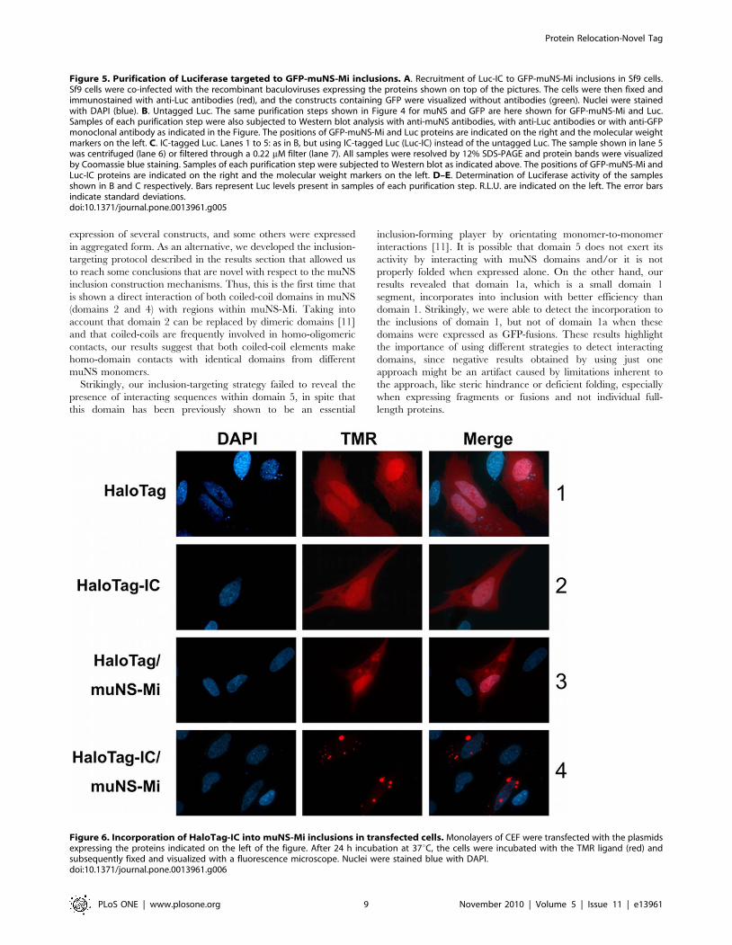
for in vivo labeling [15]. Thus, HaloTag and IC-tagged HaloTag
(HaloTag-IC) were transiently expressed in transfected CEF cells.
The transfected cells were then labeled in vivo with tetramethyl
rodamine (TMR) ligand and the HaloTag intracellular distribu-
tion was analyzed by fluorescence microscopy. Both proteins were
diffusely distributed throughout the whole cell and bound TMR,
showing that IC tagging did not affect HaloTag activity (Figure 6,
rows 1 and 2). When the HaloTag proteins were co-expressed with
muNS-Mi, we did not observe any changes in the untagged
HaloTag distribution (Figure 6, row 3) but, as expected, HaloTag-
IC was completely relocated to inclusions (Figure 6, row 4). In
addition, both proteins showed the same TMR labeling efficiency,
demonstrating that inclusion association does not diminish
HaloTag activity. Similar results were obtained with all four
described inclusion-forming proteins (not shown).
Simultaneous targeting of several proteins to muNS-related inclusions
The versatility of our inclusion-targeting system would be
greatly improved if several proteins could be recruited simulta-
neously to muNS-derived inclusions. To test this possibility, we
used two different IC-tagged proteins, GFP-IC [11] and p53-IC.
Instead of baculovirus-infected insect cells we used transfected
CEF cells where individual inclusions are dispersed throughout the
cytoplasm making easier to monitor by immunofluorescence the
integration of individual proteins in the same inclusion. We first
demonstrated that both untagged p53 and GFP do not associate
with muNS-inclusions (not shown). We also checked that p53 does
not incorporate into GFP-IC-containing muNS inclusions
(Figure 7, upper row), and that GFP does not integrate into
p53-IC-containing inclusions (Figure 7, middle row). Strikingly, we
observed that p53-IC was completely relocated to cytoplasmic
inclusions when coexpressed with muNS (Figure 7, middle row),
indicating that our inclusion-targeting system can also be used for
recruiting a nuclear protein like p53. Finally, when monolayers of
CEF cells were cotransfected with plasmids expressing GFP-IC,
p53-IC and muNS, the inclusions formed by muNS were found to
contain both IC-tagged proteins, indicating that our inclusion-
targeting system enables the simultaneous integration of more than
one protein into muNS inclusions.
Discussion
We have previously investigated the inclusion-forming contri-
bution of the different domains of the avian reovirus non-structural
protein muNS, by expressing N- and C-terminal muNS
truncations and by replacing different muNS domains by dimeric
proteins [11]. We determined that the N-terminal two thirds of the
protein are dispensable for inclusion formation but in some way
regulates or influences the shape of the inclusions formed by
muNS. On the other hand, we showed that the muNS C-terminal
one third contains 4 different domains that are absolutely essential
for inclusion formation. We were able to show that is possible to
replace domain 2 by different dimeric domains, and that domain 5
determinates the inclusion formation efficiency and inclusion
shape. However, in that study we could not determine how these
domains interact with each other to create the highly structured
muNS inclusions.
In order to gather more information about muNS sequences
involved in intermonomer contacts, we decided to try alternative
approaches. First of all, we tried the mammalian two-hybrid
system for analyzing the interaction of different muNS regions
with muNS and with muNS truncations. However, the results
were not satisfactory, since we were not able to detect the
Protein Relocation-Novel Tag
PLoS ONE | www.plosone.org 7 November 2010 | Volume 5 | Issue 11 | e13961

Protein Relocation-Novel Tag
PLoS ONE | www.plosone.org 8 November 2010 | Volume 5 | Issue 11 | e13961
expression of several constructs, and some others were expressed
in aggregated form. As an alternative, we developed the inclusion-
targeting protocol described in the results section that allowed us
to reach some conclusions that are novel with respect to the muNS
inclusion construction mechanisms. Thus, this is the first time that
is shown a direct interaction of both coiled-coil domains in muNS
(domains 2 and 4) with regions within muNS-Mi. Taking into
account that domain 2 can be replaced by dimeric domains [11]
and that coiled-coils are frequently involved in homo-oligomeric
contacts, our results suggest that both coiled-coil elements make
homo-domain contacts with identical domains from different
muNS monomers.
Strikingly, our inclusion-targeting strategy failed to reveal the
presence of interacting sequences within domain 5, in spite that
this domain has been previously shown to be an essential
inclusion-forming player by orientating monomer-to-monomer
interactions [11]. It is possible that domain 5 does not exert its
activity by interacting with muNS domains and/or it is not
properly folded when expressed alone. On the other hand, our
results revealed that domain 1a, which is a small domain 1
segment, incorporates into inclusion with better efficiency than
domain 1. Strikingly, we were able to detect the incorporation to
the inclusions of domain 1, but not of domain 1a when these
domains were expressed as GFP-fusions. These results highlight
the importance of using different strategies to detect interacting
domains, since negative results obtained by using just one
approach might be an artifact caused by limitations inherent to
the approach, like steric hindrance or deficient folding, especially
when expressing fragments or fusions and not individual full-
length proteins.
Figure 5. Purification of Luciferase targeted to GFP-muNS-Mi inclusions. A. Recruitment of Luc-IC to GFP-muNS-Mi inclusions in Sf9 cells.Sf9 cells were co-infected with the recombinant baculoviruses expressing the proteins shown on top of the pictures. The cells were then fixed andimmunostained with anti-Luc antibodies (red), and the constructs containing GFP were visualized without antibodies (green). Nuclei were stainedwith DAPI (blue). B. Untagged Luc. The same purification steps shown in Figure 4 for muNS and GFP are here shown for GFP-muNS-Mi and Luc.Samples of each purification step were also subjected to Western blot analysis with anti-muNS antibodies, with anti-Luc antibodies or with anti-GFPmonoclonal antibody as indicated in the Figure. The positions of GFP-muNS-Mi and Luc proteins are indicated on the right and the molecular weightmarkers on the left. C. IC-tagged Luc. Lanes 1 to 5: as in B, but using IC-tagged Luc (Luc-IC) instead of the untagged Luc. The sample shown in lane 5was centrifuged (lane 6) or filtered through a 0.22 mM filter (lane 7). All samples were resolved by 12% SDS-PAGE and protein bands were visualizedby Coomassie blue staining. Samples of each purification step were subjected to Western blot as indicated above. The positions of GFP-muNS-Mi andLuc-IC proteins are indicated on the right and the molecular weight markers on the left. D–E. Determination of Luciferase activity of the samplesshown in B and C respectively. Bars represent Luc levels present in samples of each purification step. R.L.U. are indicated on the left. The error barsindicate standard deviations.doi:10.1371/journal.pone.0013961.g005
Figure 6. Incorporation of HaloTag-IC into muNS-Mi inclusions in transfected cells. Monolayers of CEF were transfected with the plasmidsexpressing the proteins indicated on the left of the figure. After 24 h incubation at 37uC, the cells were incubated with the TMR ligand (red) andsubsequently fixed and visualized with a fluorescence microscope. Nuclei were stained blue with DAPI.doi:10.1371/journal.pone.0013961.g006
Protein Relocation-Novel Tag
PLoS ONE | www.plosone.org 9 November 2010 | Volume 5 | Issue 11 | e13961

From the positive results obtained in this study with domain 1,
we can deduce that there are sequences within the region 381-448
that are directly involved in inter-monomer interactions. Such
sequences have not been previously described and probably form
part of the muNS region that we have previously shown to
influence inclusion size and morphology [11]. Our results further
suggest that interacting sequences within domain 1 make contacts
with sequences within the same domain of another muNS
monomer, since domain 1 incorporates into the inclusions formed
by muNS, but not by domain 1-lacking muNS-Mi.
Domain 3 or IC produced the best inclusion-targeting results,
incorporating into muNS and muNS-Mi inclusions in a very
efficient way. Previous studies performed with both avian and the
related mammalian reoviruses [11,16] had shown that the
Intercoil domain is very important in the muNS inclusion
construction, because point mutations of two critical His and
Cys residues abolished all the muNS ability to form inclusions.
Some authors had reasoned that they could represent half of a
metal-chelating domain and that the full chelating domain would
be formed by muNS-muNS dimerization or by dimerizing with a
different protein [16]. However, this is the first time where direct
and strong interaction ability is demonstrated for the IC domain,
something that adds some support to the mentioned theory. The
reproducibility of the results obtained with the IC suggests that it
represents a domain with a very independent folding. The strong
interaction with the inclusions, the reproducibility of its results and
its small size prompted us to use it as a protein tag. In our hands it
worked perfectly, promoting inclusion incorporation without
modifying the activity and/or intracellular distribution (in absence
of the inclusion) of the tagged protein.
We have recently developed a simple protocol for purifying the
inclusions formed by baculovirus-expressed muNS in insect cells
[11]. Here, we have adapted that protocol to the purification of
IC-tagged proteins, and have successfully used that protocol for
purifying IC-tagged GFP and luciferase in a simple an inexpensive
way. In both cases the purified proteins remained active
throughout the purification process. The final purified product
can be soluble or inclusion-integrated. Inclusion-integrated
proteins remain active and can be utilized to catalyze enzymatic
reactions. Immobilized enzymes can be easily removed from
solution for purification of the product and/or allow enzyme re-
utilization. Thus, our system integrates purification and enzyme-
immobilization, which is an efficient process widely used in
industry. Additionally, the use of HaloTag also allowed us to
establish that the inclusion-immobilized proteins are also active in
vivo, something that could be useful for different applications. Our
tagging and inclusion-targeting system described in the present
study was very reliable, since it worked well for all the proteins
tested by us. The system allowed inclusion targeting of nuclear and
cytoplasmic proteins, expressed in baculovirus-infected, or avian
transfected cells.
We have detected that the nature of the tagged protein can
sometimes influence the texture of the containing inclusions,
producing sticky pellets that are not easily re-suspended. That
circumstance can be circumvented in our system by making use of
the four different inclusion-forming proteins, which present slightly
different characteristics between them: muNS, muNS-Mi, GFP-
muNS and GFP-muNS-Mi. As an example, the Luciferase
purification results shown in Figure 5 were obtained with
inclusions formed by GFP-muNS-Mi, while Luc-IC integrated
into GFP-muNS inclusions produced pellets that were more
difficult to handle. Furthermore, the use of GFP-fused inclusion-
forming proteins facilitates the purification process, since they
allow following the destiny of the inclusions along the purification
process by its green color and/or fluorescence, which adds more a
versatility and adaptability to our purification system.
A nuclear protein like p53 is completely relocalized to the
cytoplasmic muNS-derived inclusions when IC-tagged. This
represents a novel way to purify nuclear proteins in eukaryotes
without having to use a different protocol for their solubilization
and extraction from the nucleus, which represents an additional
advantage over pre-existing purification methods.
Avian reoviruses use viral factories to sequentially concentrate
the viral components required for viral replication and morpho-
Figure 7. Simultaneous targeting of GFP-IC and P53-IC to muNS inclusions. Semiconfluent monolayers of CEF were transfected with theplasmids expressing the proteins indicated on the left of the Figure. The cells were immunostained with an anti-p53 monoclonal antibody (red) andGFP was directly visualized (green). Nuclei were stained with DAPI (blue).doi:10.1371/journal.pone.0013961.g007
Protein Relocation-Novel Tag
PLoS ONE | www.plosone.org 10 November 2010 | Volume 5 | Issue 11 | e13961

genesis [7]. For mammalian reoviruses, it was established that
different regions of their muNS protein attract different structural
capsid proteins without interfering with the stoichiometry of capsid
assembly [17]. In a similar way, our inclusion-targeting system,
which has been proved to be suitable for the simultaneous
recruitment of several proteins, should be expected to increase the
building efficiency of supra-molecular complexes, a step that is
currently the bottleneck for many structural and functional studies.
Although further studies are required to demonstrate the
capability of our method for the generation of supramoleuclar
complexes, our preliminary results indicate that the DsRed
protein, whose tetramerization is essential for its autofluorescence
[18], is still fluorescent when IC-tagged and integrated into muNS-
derived inclusions (not shown), which in turn indicates that IC-
tagging and inclusion-targeting does not avoid the proper
interaction between the tagged proteins. To increase the versatility
of this approach, we are now testing the capacity of other muNS
domains (2, 4 and 1a) to function as molecular tags for directing
proteins to inclusions, which would allow directing proteins
containing different tags to the same inclusion.
Our system might be also exploited to produce immunogens
that would have potential advantages as vaccines: i) inclusions are
particulate matter, and particulate immunogens are the best for
stimulating both humoral and cellular immune responses [19]; ii)
inclusion-derived immunogens can be easily produced and
purified; iii) they should be biologically safe, because organisms
would be immunized with proteins and not with genetic material
or viruses; and iv) different epitopes can be simultaneously exposed
on the same particle. In that way, for example we could arrange
several epitopes of a single virus, or epitopes from different
serotypes on the same vaccine to increase its overall efficiency.
The results presented here shed light on the general rules that
govern the construction of the highly structured protein aggregates
formed by protein muNS and additionally allowed to develop a
novel protein tagging and inclusion-targeting system with many
potential applications.
Materials and Methods
Cells, viruses and antibodiesPrimary cultures of chicken embryo fibroblasts (CEF) were
prepared from 9- to 10-day-old chicken embryos [20] and grown
in monolayers in medium 199 supplemented with 10% (w/v)
tryptose-phosphate broth and 5% (v/v) calf serum. The Sf9 insect
cell line was grown in suspension culture at 27uC in serum-free
Sf-900 II medium (Invitrogen, Barcelona, Spain). Propagation of
baculoviruses in Sf9 cells has been described previously [21].
Rabbit polyclonal serum against the avian reovirus S1133 muNS
protein was raised in our laboratory [10]. Goat polyclonal
antibody specific for recombinant firefly Luciferase (Photinus pyralis)
was from Promega (Madrid, Spain). Mouse monoclonal antibody
against Aequorea victoria green fluorescent protein (GFP) was from
Roche (Barcelona, Spain). Rabbit polyclonal antibodies against
the influenza virus hemagglutinin (HA) epitope, monoclonal
antibody PAB40 specific for p53, Cy3 conjugated antibodies
against both goat and rabbit IgG, and Alexa 594 conjugated
antibody against mouse IgG, all were from Sigma-Aldrich
(Madrid, Spain).
Transfections, IF microscopy and labeling with TMRLigand
Transfections of preconfluent cell monolayers were performed
with the Lipofectamine Plus reagent (Invitrogen, Barcelona, Spain)
according to the manufacturer’s instructions. Transfected cells
were incubated at 37uC for 24 h, unless otherwise stated.
For tetramethyl rodamine (TMR) labeling, transfected cells
were incubated for 15 min with TMR, then the cells were rinsed
twice with PBS and incubated in fresh medium for 30 min. The
medium was removed, and the cells were washed 3 times with PBS
before being fixed with 4% paraformaldehyde and used for
imaging. Images were obtained with an Olympus DP-71 digital
camera mounted on an Olympus BX51 fluorescence microscope,
and processed with Adobe Photoshop (Adobe Systems, USA). For
proper image acquisition and better resolution of the inclusions,
images of muNS-derived inclusions or inclusion-containing
proteins are underexposed in comparison to those of cells lacking
inclusions. For indirect immunofluorescence, cell monolayers
grown on coverslips were infected or transfected as indicated in
the Figure legends and, at the indicated times, the monolayers
were washed twice with PBS and fixed with 4% paraformaldehyde
in PBS at room temperature for 10 min. Paraformaldehyde-fixed
cells were washed twice with PBS, incubated for 4 min in
permeabilizing buffer (0.5% Triton X-100 in PBS), and then
incubated for 1 h at room temperature with primary antibodies
diluted in blocking buffer (2% bovine serum albumin in PBS). The
cells were then washed three more times with PBS and incubated
with secondary antibodies and with 1 mg/ml of DAPI (49, 69-dia-
midino-2-phenylindole)/ml. The coverslips were then washed six
times with PBS and mounted on glass slides. Images were obtained
and processed as indicated above.
ImmunoblottingFor Western blot analysis, cell extracts were resolved by SDS-
PAGE and proteins in unfixed gels were transferred to PVDF
membranes (Immobilon-P Millipore, Madrid, Spain) for 1 h at
100 mA in a semidry blotting apparatus (Bio-Rad, California,
USA). Protein bands were detected with specific antibodies using
the Immobilon Western Chemiluminiscent HRP Substrate (Milli-
pore, Madrid, Spain).
Plasmid constructionsPlasmid pCMV-p53, expressing the human p53 protein was a
generous gift of Dr. Anxo Vidal (Universidad de Santiago de
Compostela) and has been previously described [22]. The
construction of pCINeo-muNS, which expresses full-length muNS,
and of pCINeo-muNS(448–635), which expresses muNS-Mi have
been described [9,11].
i) muNS-GFP fusions. The construction of most of the
recombinant plasmids, which express GFP fused to the N termini
of different muNS regions has been described previously [11].
To express GFP fused to the N terminus of the muNS(381–448)
(this construction was named GFP-1a in results), the recombinant
plasmid pGEMT-M3 [9] was subjected to PCR amplification with
the following primers. The forward primer was 59-GCGGAATTC-
TATGCCATCCTTCTTACTCGGTG-39(EcoRI site is single
underlined) and the reverse primer was 59-GCGGGATCCTTA-
TGGACCAACGGACGAATCG-39 (BamHI site is single under-
lined and the stop codon is double underlined). The resulting
amplified product was digested with EcoRI and BamHI and then
ligated to the pEGFP-C1 vector (Clontech, Saint Germain en Laye,
Francia), that had been cut with the same enzymes, to generate the
recombinant plasmid pEGFP-C1-M3(381-448).
To express GFP-IC (where the factor Xa cleavage site (Xacs) is
fused to the C terminus of GFP and to the N terminus of
muNS(477–542), the recombinant plasmid pGEMT-M3 [9] was
subjected to PCR amplification with the following primers: the
forward primer was 59-GCGGAATTCTATCGAGGGAAGG-
Protein Relocation-Novel Tag
PLoS ONE | www.plosone.org 11 November 2010 | Volume 5 | Issue 11 | e13961

GAAGATCACTTGTTGGCTTATC-39 (EcoRI site is single
underlined and factor Xa cleavage site is double underlined) and
the reverse primer 59-GCGGGATCCTTACGCTTCCACACG-
GGGTTCCCAC-39 (BamHI site is single underlined and the stop
codon is double underlined). The PCR product was cut with
EcoRI and BamHI and ligated to pEGFP-C1 that had been cut
with the same enzymes, to generate the recombinant plasmid
pEGFP-C1-Xacs-muNS(477–542).
ii) HaloTag-IC. To express HaloTag-IC, the recombinant
plasmid pGEMT-M3 [9] was subjected to PCR amplification with
the following primers. The forward primer was 59-GCGTCTAGA-
ATCATGGCGGAAGATCACTTGTTGGCTTATC-39 (XbaI
site is single underlined) and the reverse primer was 59-GCGGGG-
CCCTTACGCTTCCACACGGGGTTCCCAC-39 (ApaI site is
single underlined and the stop codon is double underlined). The
resulting amplified product was digested with XbaI and ApaI and
then ligated to the pCDNA3.1/Zeo vector (Promega, Madrid,
Spain) that had been cut with the same enzymes, to generate the
recombinant plasmid pCDNA3.1/Zeo-muNS(477–542). The
HaloTag sequence was obtained by PCR-amplification using the
plasmid pHT2 as template (Promega, Madrid, Spain), and the
following primers: the forward primer was 59-GCGGGATCC AC-
CATGGGCTCCGAAATCGGTACAGGC-39 (BamHI site is
single underlined and the start codon is double underlined), and
the reverse primer was 59- GCATAAGAATGCGGCCGCCAG-
CCGGCCAGCCCGGGGAG-39 (NotI site is single underlined).
The PCR product was digested and cloned into the BamHI and
NotI sites of pCDNA3.1/Zeo-muNS(477–542) to generate
pCDNA3.1/Zeo-HaloTag-muNS(477–542).
iii) p53-IC. To express p53-IC, the recombinant plasmid
pCMV-wtp53 [22] was subjected to PCR amplification with the
following primers: the forward primer was 59-GCGGGATCC-
ATCATGGAGGAGCCGCAGTCAGATCC-39 (BamHI site is
single underlined and the start codon is double underlined), and
the reverse primer was 59-GCGGAATTCGTCTGAGTCAGG-
CCCTTCTGTCTTG-39 (EcoRI site is single underlined) to
amplify the complete human p53 coding sequence. The PCR
product was cut with BamHI and EcoRI and then ligated to
pCDNA3.1/Zeo-muNS(477–542) that had been cut with the same
enzymes.
iv) muNS-HA fusions. To express the influenza virus
hemagglutinin epitope (HA) fused to the C-terminus of different
muNS regions, the HA-encoding sequence, the start and stop
codons and the restriction sites were introduced at different
positions of the M3 gene by PCR amplification of the desired M3
region. Each PCR was performed using pGEMT-M3 as a template
[9], and the primers used are listed in Table 1. Each PCR product
was cut with EcoRI and XbaI and then ligated to the pCDNA3.1/
Zeo vector that had been cut with these same enzymes. Each
construct was checked by sequencing and Western blot analysis and
named for the muNS residues that the expressed protein contains
(Table 1). The correctness of the constructs was confirmed by
sequencing and Western blot analysis of the expressed proteins.
Construction of recombinant baculovirusesAll the recombinant baculoviruses were generated using the
Bac-to-Bac system (Invitrogen, Barcelona, Spain) following the
supplier protocols. The construction of the recombinant baculo-
viruses Bac-muNS, which expresses full-length muNS, and Bac-
muNS-Mi, which expresses muNS residues 448 to 635, have been
described previously [11].
i) Bac-GFP-IC. To express GFP-IC in insect cells, the GFP-
Xacs-muNS(477–542)-coding sequence of the pEGFP-C1-Xacs-
muNS(477–542) plasmid was amplified by PCR using the forward
primer 59-GCGGGATCCACCATGGTGAGCAAGGGCGAG-39
(BamHI site is single underlined and the start codon is double
underlined) and the reverse primer 59-GCGTCTAGATTACG-
CTTCCACACGGGGTTCCCAC-39 (XbaI site is single under-
Table 1. Construction of plasmids to express HA-tagged muNS fusions.
Constructa Primersb (59-39) Expressed proteinc
pCINeo-M3(1–154)-HA F-GCGGAATTCATCATGGCGTCAACCAAGTGG muNS(1–154)-HA
pCINeo-M3(1–154)-HA R-GCGTCTAGATTACGCATAATCCGGCACATCATACGGATAATCGGGGGAATCAGCGGTGG (region 1b)
pCINeo-M3(1–380)-HA F-GCGGAATTCATCATGGCGTCAACCAAGTGG muNS(1–380)-HA
pCINeo-M3(1–380)-HA R-GCGTCTAGATTACGCATAATCCGGCACATCATACGGATATGGAGACCGTCTAGCGAGAAG (region 1c)
pCINeo-M3(1–448)-HA F-GCG GAATTCATCATGGCGTCAACCAAGTGG muNS(1–448)-HA
pCINeo-M3(1–448)-HA R-GCGTCTAGATTACGCATAATCCGGCACATCATACGGATATGGACCAACGGACGAATCG (region 1)
pCINeo-M3(1–477)-HA F-GCGGAATTCATCATGGCGTCAACCAAG TGG muNS(1–477)-HA
pCINeo-M3(1–477)-HA R-GCGTCTAGATTACGCATAATCCGGCACATCATACGGATATTCCCGAGCAGGTTGAACATC (region 1+2)
pCDNA3.1/Zeo-M3(539–635)-HA F-GCGGAATTCATCATGGCGCGTGTGGAAGCGTTAAACCAAG muNS(539–635)-HA
pCDNA3.1/Zeo-M3(539–635)-HA R-GCGTCTAGATTACGCATAATCCGGCACATCATACGGATACAGATCATCCACCAATTCTTC (region 4+5)
pCDNA3.1/Zeo-M3(539–605)-HA F-GCGGAATTCATCATGGCGCGTGTGGAAGCGTTAAACCAAG muNS(539–605)-HA
pCDNA3.1/Zeo-M3(539–605)-HA R-GCGTCTAGATTACGCATAATCCGGCACATCATACGGATAGACACGTGTCGCACGACTCATC (region 4)
pCDNA3.1/Zeo-M3(477–542)-HA F-GCGGAATTCATCATGGAAGATCACTTGTTGGCTTATC muNS(477–542)-HA
pCDNA3.1/Zeo-M3(477–542)-HA R-GCGTCTAGATTACGCATAATCCGGCACATCATACGGATACGCTTCCACACGGGGTTCCCAC (region 3)
pCDNA3.1/Zeo-M3(381–448)-HA F-GCGGAATTCATCATGCCATCCTTCTTACTCGGTG muNS(381–448)-HA
pCDNA3.1/Zeo-M3(381–448)-HA R-GCGTCTAGATTACGCATAATCCGGCACATCATACGGATATGGACCAACGGACGAATCG (region 1a)
aEach construct was designed to contain the portion of the M3 gene encoding the indicated amino acid residues of muNS.bFor each truncation construct, reverse primer (R) is in the reverse orientation relative to the coding strand, and the added stop codon and the added HA sequence are
double underlined, and forward primer (F) is in the forward orientation relative to the coding strand, and the added start codon is double underlined. The EcoRI andXbaI restriction sites added near the 59 end of each primer are single underlined.
cEach construct was designed to express a truncated muNS protein comprising the indicated amino acid residues. In brackets, muNS regions as termed in results.doi:10.1371/journal.pone.0013961.t001
Protein Relocation-Novel Tag
PLoS ONE | www.plosone.org 12 November 2010 | Volume 5 | Issue 11 | e13961

lined and the stop codon is double underlined). The PCR product
was digested and cloned into the BamHI and XbaI sites of pFastBac1.ii) Bac-GFP. To generate a recombinant baculovirus expressing
the GFP protein, the GFP coding sequence of the pEGFP-C1
plasmid was amplified by PCR using the forward primer 59-GC-
GGAATTCACCATGGTGAGCAAGGGCGAG-39 (EcoRI site is
single underlined and the start codon is double underlined), and the
reverse primer 59- GCGTCTAGATTACTTGTACAGCTCG-
TCCATGCC-39 (XbaI site is single underlined and the stop codon
is double underlined). The PCR product was digested and cloned into
the EcoRI and XbaI sites of pFastBac1.iii) Bac-Luc. To generate the recombinant baculovirus
expressing the firefly Luciferase (Photinus pyralis), the Luciferase
coding sequence was amplified by RT-PCR using the Luciferase
RNA as template (Promega, Madrid, Spain), and the following
primers: the forward primer was 59-GCGGGATCCATCATGG-
AAGACGCCAAAAAC-39 (BamHI site is single underlined and
the start codon is double underlined), and the reverse primer was
59-GCATAAGAATGCGGCCGCTTACAATTTGGACTTTC-
CGCCC-39 (NotI site is single underlined and the stop codon is
double underlined). The PCR product was digested and cloned
into the BamHI and NotI sites of pFastBac1.iv) Bac-Luc-IC. To express Luc-IC, the recombinant plasmid
pGEMT-M3 [9] was subjected to PCR amplification with the fol-
lowing primers. The forward primer was 59-GCATAAGAATC-
TCGAGATCATGGCGGAAGATCACTTGTTGGCTTATC-
39 (XhoI site is single underlined) and the reverse primer was 59-
GCATAAGAATAAGCTTTTACGCTTCCACACGGGGTT-
CCCAC-39 (HindIII site is single underlined and the stop codon is
double underlined). The resulting amplified product was digested
with XhoI and HindIII and then ligated to the pFastBac1 vector
that had been cut with the same enzymes, to generate the re-
combinant plasmid pFastBac1-muNS(477–542). The recombinant
plasmid pFastBac1-Luc-IC was generated by PCR-amplification
using pFastBac1-Luc as template, and the following primers: the
forward primer was 59-GCGGGATCCATCATGGAAGAC-
GCCAAAAAC-39 (BamHI site is single underlined and the start
codon is double underlined), and the reverse primer was 59-
GCATAAGAATGCGGCCGCCAATTTGGACTTTCCGCC-
C-39 (NotI site is single underlined). The PCR product was
digested and cloned into the BamHI and NotI sites of pFastBac1-
IC to generate pFastBac1-Luc-IC.v) Bac-GFP-muNS and Bac-GFP-muNS-Mi. To generate
recombinant baculoviruses expressing GFP-muNS and GFP-
muNS-Mi, the GFP-muNS(448–635) coding sequence of the
pEGFP-C1-M3(448–635) plasmid [11] or the GFP-muNS coding
sequence of pEGFP-C1-M3 plasmid [9] were amplified by PCR
using the forward primer 59-GCGGGATCCACCATGGTGA-
GCAAGGGCGAG-39 (BamHI site is single underlined and the
start codon is double underlined) and the reverse primer 59-G-
CGTCTAGATCACAGATCATCCACCAATTCTTC-39 (XbaI
site is single underlined and the stop codon is double underlined).
The PCR products were digested and cloned into the BamHI and
XbaI sites of pFastBac1.
The pFastBac1 recombinant constructs were then used to
generate the corresponding recombinant baculovirus, according to
the supplier’s protocol. The correctness of the constructs was
confirmed by DNA sequencing.
Baculovirus expression and inclusion purificationSf9 insect cells growing in suspension were infected with 5 PFU/cell
of the different recombinant baculoviruses, as indicated in the figure
legends, and incubated at 27uC for 72 h. Cells were then pelleted by
centrifugation for 10 min at 10006g, resuspended in hypotonic buffer
(10 mM Hepes pH: 7.9, 10 mM KCl, 1.5 mM MgCl2) and placed on
ice for 15 min. The extracts were then centrifuged at 4uC for 10 min
at 2,0006g, and the pellets were washed twice with 10 ml of hypotonic
buffer. The resulting pellets were resuspended in 10 ml of hypotonic
buffer and sonicated (6 pulses at 45 cycles) to break the nuclei and to
shear genomic DNA. The sonicated extracts were centrifuged at 4uCfor 5 min at 2506g, and the inclusion-containing pellet was washed
and centrifuged five times using the same conditions. The final pellet
was resuspended in 1 ml of hypotonic buffer. Samples of each
purification step were analized by SDS-PAGE and Western blot. For
GFP-IC purification, the final pellet was resuspended in 1 ml of
hypotonic buffer containing 500 mM NaCl, centrifuged 10 min at
4uC and 16,0006g, and the supernatant was loaded onto a HiTrapTM
desalting column pre-equilibrated with hypotonic buffer (GE
Healthcare, Madrid, Spain). The eluted material was centrifuged
5 min at 4uC and 16,0006g and the supernatant was incubated with
Xa factor (New England Biolabs, Ipswich, England) for 48 h at 4uC.
The extract was then centrifuged 5 min at 4uC and 16,0006g and the
supernatant loaded on a HiTrap Q-Sepharose column pre-equili-
brated with hypotonic buffer in order to purify the GFP protein (GE
Healthcare, Madrid, Espana). The column was eluted with increasing
concentrations of NaCl. The GFP-containing fractions eluted from the
column after a 400 mM NaCl wash. Luc-IC-containing inclusions
were similarly purified, with the differences indicated on the text.
Determination of Luciferase activitySf9 insect cells were infected with 5 PFU/cell of the different
recombinant baculoviruses and incubated at 27uC for 72 h. Then,
the cells were lysed, and Luciferase activity of the cells extracts was
determined with a Luminoskan-ascent luminometer (Thermo,
Waltham, Massachusetts, USA). Results of six replicates are
expressed as the mean relative light units (R.L.U.) per well 6 the
standard deviation. During the purification of Luc-IC, samples of
each purification step were diluted 1/200 before Luciferase
activity was determined.
Acknowledgments
We thank Anxo Vidal for donating plasmid pCMV-wtp53, Mark van Raaij
for critical reading of the manuscript and Leticia Barcia Castro for
excellent technical assistance. We also thank Laboratorios Intervet
(Salamanca, Spain) for providing pathogen-free embryonated chicken eggs.
Author Contributions
Conceived and designed the experiments: ABN JMC. Performed the
experiments: ABN RMV. Analyzed the data: ABN RMV JB JMC.
Contributed reagents/materials/analysis tools: ABN RMV. Wrote the
paper: ABN JB JMC.
References
1. Attoui H, Billoir F, Biagini P, de Micco P, de Lamballerie X (2000) Complete
sequence determination and genetic analysis of Banna virus and Kadipiro virus:
proposal for assignment to a new genus (Seadornavirus) within the familyReoviridae. J Gen Virol 81: 1507–1515.
2. Mertens P (2004) The dsRNA viruses. Virus Res 101: 3–13.
3. Jones RC (2000) Avian reovirus infections. Rev Sci Tech 19: 614–625.
4. van der Heide L (2000) The history of avian reovirus. Avian Dis 44: 638–641.
5. Duncan R, Chen Z, Walsh S, Wu S (1996) Avian reovirus-induced syncytium
formation is independent of infectious progeny virus production and enhances
the rate, but is not essential, for virus-induced cytopathology and virus egress.
Virology 224: 453–464.
6. Zhang X, Tang J, Walker SB, O’Hara D, Nibert ML, et al. (2005) Structure of
avian Orthoreovirus virion by electron cryo-microscopy and image reconstruc-
tion. Virology 343: 25–35.
Protein Relocation-Novel Tag
PLoS ONE | www.plosone.org 13 November 2010 | Volume 5 | Issue 11 | e13961

7. Benavente J, Martinez-Costas J (2006) Early steps in avian reovirus
morphogenesis. Curr Top Microbiol Immunol 309: 67–85.8. Benavente J, Martinez-Costas J (2007) Avian reovirus: structure and biology.
Virus Res 2: 105–119.
9. Touris-Otero F, Cortez-San Martin M, Martinez-Costas J, Benavente J (2004)Avian reovirus morphogenesis occurs within viral factories and begins with the
selective recruitment of sigmaNS and lambdaA to microNS inclusions. J MolBiol 341: 361–374.
10. Touris-Otero F, Martinez-Costas J, Vakharia VN, Benavente J (2004) Avian
reovirus non-structural protein microNS forms viroplasm-like inclusions andrecruits protein sigmaNS to these structures. Virology 319: 94–106.
11. Brandariz-Nunez A, Menaya-Vargas R, Benavente J, Martinez-Costas J (2010)Avian reovirus muNS protein forms homo-oligomeric inclusions in a
microtubule-independent fashion, which involves specific regions of itsC-terminal domain. J Virol 84: 4289–4301.
12. Broering TJ, Parker JS, Joyce PL, Kim J, Nibert ML (2002) Mammalian
reovirus non-structural protein microNS forms large inclusions and colocalizeswith reovirus microtubule-associated protein micro2 in transfected cells. J Virol
76: 8285–8297.13. Wileman T (2006) Aggresomes and autophagy generate sites for virus
replication. Science 312: 875–878.
14. Wileman T (2007) Aggresomes and pericentriolar sites of virus assembly: cellulardefense or viral design. Annu Rev Microbiol 61: 149–167.
15. Los GV, Wood K (2007) The HaloTag: a novel technology for cell imaging and
protein analysis. Methods Mol Biol 356: 195–208.
16. Broering TJ, Arnold MM, Miller CL, Hurt JA, Joyce PL, et al. (2005) Carboxyl-
proximal regions of reovirus non-structural protein muNS necessary and
sufficient for forming factory-like inclusions. J Virol 79: 6194–6206.
17. Miller CL, Arnold MM, Broering TJ, Hastings CE, Nibert ML (2010)
Localization of mammalian orthoreovirus proteins to cytoplasmic factory-like
structures via nonoverlapping regions of microNS. J Virol 84: 867–882.
18. Baird GS, Zacharias DA, Tsien RY (2000) Biochemistry, mutagenesis, and
oligomerization of DsRed, a red fluorescent protein from coral. Proc Natl Acad
Sci U S A 97: 11984–89.
19. Roy P (1996) Genetically engineered particulate virus-like structures and their
use as vaccine delivery systems. Intervirology 39: 62–71.
20. Martinez-Costas J, Varela R, Benavente J (1995) Endogenous enzymatic
activities of the avian reovirus S1133: identification of the viral capping enzyme.
Virology 206: 1017–26.
21. Hsiao J, Martinez-Costas J, Benavente J, Vakharia VN (2002) Cloning,
expression, and characterization of avian reovirus guanylyltransferase. Virology
296: 288–299.
22. Friedlander P, Haupt Y, Prives C, Oren M (1996) A mutant p53 that
discriminates between p53-responsive genes cannot induce apoptosis. Mol Cell
Biol 9: 4961–71.
Protein Relocation-Novel Tag
PLoS ONE | www.plosone.org 14 November 2010 | Volume 5 | Issue 11 | e13961