Biphasic Myopathic Phenotype of Mouse DUX, an ORF within Conserved FSHD-Related Repeats
Upload
kalasalingamCategory
view
0download
0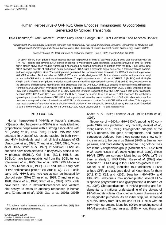
Human Herpesvirus-8 ORF K8.1 Gene Encodes Immunogenic GlycoproteinsGenerated by Spliced Transcripts
Bala Chandran,*,1 Clark Bloomer,* Szeman Ruby Chan,* Liangjin Zhu,* Elliot Goldstein,† and Rebecca Horvat‡
*Department of Microbiology, Molecular Genetics and Immunology, †Division of Infectious Diseases, Department of Medicine, and‡Department of Pathology and Clinical Laboratories, The University of Kansas Medical Center, Kansas City, Kansas 66160
Received March 25, 1998; returned to author for revision June 8, 1998; accepted June 26, 1998
A cDNA library from phorbol ester-induced human herpesvirus-8 (HHV-8) carrying BCBL-1 cells was screened with anHIV1KS1 serum, and several cDNA clones encoding HHV-8 proteins were identified. Sequence analysis of two full-lengthcDNA clones show open reading frames (ORFs) encoded by spliced messages originating from the HHV-8 K8.1 gene. OnecDNA encodes an ORF of 228 amino acids, designated K8.1.A, with a cleavable signal sequence, a transmembrane domain,and four N-glycosylation sites. The splicing event generated the transmembrane domain in the ORF not seen in the genomicK8.1 ORF. Another cDNA encodes an ORF of 167 amino acids, designated K8.1.B, that shares similar amino and carboxyltermini with ORF K8.1.A but with an in-frame deletion. The primary translation products of ORF K8.1A (34 kDa) and K8.1B (20kDa) in the in vitro–transcription–translation experiments shifted into glycosylated species of 43 and 32 kDa, respectively, inthe presence of microsomal membranes. This suggested that the ORF K8.1A and K8.1B encode for glycoproteins. Riboprobesfrom the K8.1A cDNA insert hybridized with an HHV-8-specific 0.9-kb abundant transcript from BCBL-1 cells. Synthesis of thisRNA was eliminated in the presence of a DNA synthesis inhibitor, suggesting that this RNA was a late gene transcript.Because ORFs K8.1A and K8.1B are unique for HHV-8, human sera were tested in Western blot reactions for antibodiesagainst glutathione-S-transferase–ORF K8.1A fusion protein. All sera that were positive for HHV-8 antibodies in immunoflu-orescence assays with phorbol ester-induced BCBL-1 cells were also positive for anti-ORF K8.1A antibodies. This suggeststhat measurement of anti-ORF K8.1A antibodies would provide an HHV-8-specific serological assay. Further work is neededto define the biological role of the HHV-8 ORF K8.1A and K8.1B glycoproteins. © 1998 Academic Press
INTRODUCTION
Human herpesvirus-8 (HHV-8), or Kaposi’s sarcoma(KS)-associated herpesvirus (KSHV), is a newly identifiedlymphotropic herpesvirus with a strong association withKS (Chang et al., 1994, 1995). HHV-8 DNA has beendetected in .95% of KS lesions studied, in both HIV2and HIV1 individuals and in all clinical subtypes of KS(Ambroziak et al., 1995; Chang et al., 1994, 1996; Mooreet al., 1995; Smith et al., 1997). In addition, HHV-8 se-quences have been detected in body cavity-based B-celllymphomas (BCBLs). Cell lines (BC-1, HBL-6, andBCBL-1) have been established from the BCBL tumors(Cesarman et al., 1995; Gao et al., 1996, 1996; Moore etal., 1996; Renne et al., 1996). BC-1 and HBL-6 carry bothHHV-8 and human Epstein–Barr virus (EBV). BCBL-1 cellscarry only HHV-8, and lytic cycles can be induced byphorbol ester (TPA) (Chan et al., 1998; Chandran et al.,1998; Renne et al., 1996; Smith et al., 1997). BCBL cellshave been used in immunofluorescence and Westernblot assays to measure antibody responses in humansera (Chandran et al., 1998; Gao et al., 1996a, 1996b;
Kedes et al., 1996; Lennette et al., 1996; Smith et al.,1997).
Sequence of ;140-kb HHV-8 DNA encoding 80 com-plete ORFs was published very recently (Neipel et al.,1997; Russo et al., 1996). Phylogenetic analysis of theHHV-8 genome, the gene arrangements, and proteinsequences deduced from these sequences show strik-ing similarity to herpesvirus Saimiri (HVS), a Simian her-pesvirus, and more distantly related to EBV; both virusesare in the g-herpesvirus group (Albrecht et al., 1992; Kieffet al., 1996; Russo et al., 1996; Neipel et al., 1997). TheseHHV-8 ORFs are currently identified as ORFs 1–75 bytheir similarity to HVS ORFs. Russo et al. (1996) alsoidentified 15 ORFs unique for HHV-8 designated K1–K15.Neipel et al. (1997) identified four additional HHV-8unique ORFs and assigned decimal K numbers for them(K4.1, K4.2, K8.1, and K10.1). Sera from HIV1KS1 andHIV1KS2 individuals immunoprecipitated several HHV-8-specific polypeptides and glycoproteins (Chandran etal., 1998). Characterizations of HHV-8 proteins are fun-damental to a rational understanding of the biology ofHHV-8 and its role in KS and other human diseases andfor diagnostic purposes. As an initial step, we screeneda cDNA library from TPA-induced BCBL-1 cells with anHIV1KS1 serum and identified cDNAs encoding severalHHV-8 proteins (Chandran et al., 1998). Among these, we
1 To whom reprint requests should be addressed. Fax: (913) 588-7295. E-mail: [email protected].
VIROLOGY 249, 140–149 (1998)ARTICLE NO. VY989316
0042-6822/98 $25.00Copyright © 1998 by Academic PressAll rights of reproduction in any form reserved.
140

have identified two cDNAs encoded by the genomicHHV-8 ORF K8.1. These two cDNAs are generated byspliced transcripts and encode immunogenic glycopro-teins.
RESULTS
Identification of cDNA encoding glycoproteins HHV-8ORF K8.1A and K8.1B
To identify the genes encoding immunogenic HHV-8proteins, serum from an HIV1KS1 individual with animmunofluorescence assay (IFA) titer of 1:10,240 withinduced BCBL-1 cells was used to screen the cDNAlibrary from TPA-induced BCBL-1 cells. After four screen-ings, 56 cDNA clones were isolated and released in thephagemid forms by cocultivation with the helper phage.These cDNAs were sequenced from both orientations.Sequences were analyzed for ORF and compared withthe published genomic HHV-8 sequence (Russo et al.,1996; Neipel et al., 1997). Several ORFs were identified(Chandran et al., 1998). Complete HHV-8 ORF 65 wasencoded in 11 cDNA clones. In the other cDNA clones,the length of ORFs ranged from 280 to 800 amino acids
(AA). Because these clones were identified by theHIV1KS1 serum, this suggests that they encode immu-nogenic protein regions.
Initial comparison of the nucleotide sequences of fourcDNA clones from the above screening showed 100%identity to a 1200-bp region in the HHV-8 genomic se-quence characterized by Russo et al. (1996) and Neipelet al. (1997) (Fig. 1A). In this region, there is an ORFdesignated K8.1, which starts at the genomic nucleotideposition 76,214 bp, ends at 76,808 bp, and thus encodesa 197-AA-long ORF with a predicted molecular mass of;22 kDa (Fig. 1A) (Neipel et al., 1997). The genomic K8.1ORF has an amino-terminal signal sequence and fiveputative N-glycosylation sites but no transmembrane se-quence. Among the four cDNA clones identified, twocDNA clones encoded two full-length ORFs of 228 and167 AA (Figs. 1B, 1C, and 2). A comparison of these twoORFs sequences with the genomic sequence showedthat they were encoded by spliced transcripts. The largercDNA is 752 bp long, starts at the genomic nucleotideposition 76,214 bp, ends at position 76,941 bp, and usesthe polyadenylation signal sequence (AATAAA) at posi-tion 77,013 bp. This cDNA encodes a 228-AA-long ORF
FIG. 1. Schematic representation of the cDNAs encoding the HHV-8 ORF K8.1A and K8.1B. (Top line) Schematic representation of HHV-8 long uniqueregion (LUR). (A) schematic representation of HHV-8 ORFs. (B and C) Location of the cDNAs in the HHV-8 genome. (Numbers) Genomic nucleotidepositions of ORFs (Russo et al., 1996). (Arrows) Direction of transcription. The cDNAs encoding ORFs K8.1A and K8.1B of 228 and 167 AA, respectively,are shown. The splice sites are shown by the inverted V, and AAAA represents the poly(A)1 tail. (D) Schematic hydropathic analysis of the deducedORF K8.1A protein sequence from the cDNA. (Peaks above the horizontal axis) Hydrophilicity. (Peaks below the horizontal axis) Hydrophobicity. (S)Putative signal sequence. (N) Putative N-glycosylation sites (N-X-T/S). (TM) Predicted transmembrane AA sequences. (K8.1B.DEL) AA sequencesdeleted in the ORF K8.1B by splicing.
141HHV-8 GLYCOPROTEIN ORF K8.1

with a predicted molecular mass of ;25 kDa and isdesignated K8.1.A. The first 142 AA encoded by the K8.1AcDNA are identical to the genomic K8.1 ORF sequence.Analysis suggested that the protein encoded by theK8.1A cDNA is a typical class I glycoprotein with a cleav-able amino-terminal signal sequence or sequences, atransmembrane domain (197–213 AA), seven cysteineresidues, and four putative N-glycosylation sites (N)(Figs. 1B, 1D, and 2). This cDNA is derived from a tran-script with a 95-bp sequence spliced out [CAG/(GT)GTATdonor site and TCTAC(AG)/G acceptor site] and ends atthe genomic nucleotide position 76,941 bp, which is 187bp beyond the end of genomic ORF K8.1 (Fig. 1B). Thishas resulted in the generation of a transmembrane do-main not seen in the genomic ORF K8.1.
The smaller cDNA is 569 bp long with a 183-bp se-quence spliced out. The splice acceptor site for the ORFK8.1B transcript is the same as that for the ORF K8.1Atranscript (Figs. 1C and 2). However, the splice donor site
[CGA/(GT)GAGT] for the K8.1B cDNA is upstream of thesplice donor site of the K8.1.A cDNA, resulting in anin-frame deletion of 61 AA in the smaller ORF (Figs. 1Dand 2). The resulting 167-AA-long ORF is predicted tocode for a protein of ;18.5 kDa and is designated K8.1.B.ORF K8.1B is also a typical class I glycoprotein with acleavable signal sequence, a transmembrane domain,and three putative N-glycosylation sites (N). Except oneAA change near the splice site (S to R), the ORF K8.1Bshares identical AA sequences with ORF K8.1.A (Figs. 1Cand 2). To confirm the sequence and the splicing pat-terns, 59-rapid amplification of cDNA ends (RACE) wasdone. RNA extracted from TPA-induced BCBL-1 cells (48h p.i.) was used for this. Single-stranded cDNAs fromTPA-induced cells were synthesized using oligo(dT)primer, and the second strand was subsequently synthe-sized. Double-stranded adapters were ligated to bothends of the cDNA molecule, and polymerase chain re-action (PCR) was performed using an adapter-specific
FIG. 2. Sequence analysis of the HHV-8 ORF K8.1A and K8.1B cDNA. Nucleotide and the predicted AA sequence of the full-length cDNA encodingORF K8.1A and K8.1B are shown. The AA position is numbered on the right. The ORF termination codon (TAA), the putative polyadenylation signalsequence (ATTAAA), and putative N-glycosylation sites (N-X-T/S) are underlined and in bold. (1) Predicted transmembrane (TM) AA sequence. (Dottedlines) AA sequences of ORF K8.1B shared with ORF K8.1A. (p) Splice site in ORF K8.1B.
142 CHANDRAN ET AL.

primer and the ORF K8.1A- and K8.1B-specific primercorresponding to the 39-end stop site. Several cloneswere cloned and sequenced. The sequences and splic-ing patterns were identical to those of the ORF K8.1A andK8.1B cDNAs, confirming the sequences and the splicingpattens seen in the K8.1A and K8.1B cDNAs.
The proteins encoded by cDNAs HHV-8 ORFs K8.1Aand K8.1B are glycoproteins
By AA sequence analysis, the ORFs K8.1A and K8.1Bidentified here are predicted to encode for glycoproteins.To verify this, in vitro transcription-translation experi-ments were done. No specific protein was detected incontrol reactions without the addition of any RNA (Fig. 3,lane 1). Translation of the RNA transcribed from K8.1BcDNA produced a polypeptide of ;20 kDa (Fig. 3, lane 2).With the addition of canine microsomal membranes tothe translation reaction mixture, this polypeptide shiftedin mobility to a polypeptide of ;32 kDa (Fig. 3, lane 3).The major primary translation product of K8.1.A cDNA isa 34-kDa polypeptide (Fig. 3, lane 4), and in the presenceof microsomal membranes, it shifted into a glycosylatedspecies of 43 kDa (Fig. 3, lane 5). Treatment of these invitro synthesized products in the presence of mem-branes with N-glyconase decreased the 43- and 32-kDapolypeptides into smaller polypeptides of ;34 and ;20
kDa (data not shown), indicating the removal of N-linkedoligosaccharides. These results showed that the HHV-8ORF K8.1A and K8.1B cDNAs encode glycoproteins.
HHV-8 ORF K8.1A and K8.1B cDNAs are generatedfrom a late class transcript
To define further the viral specificity of the HHV-8 ORFK8.1A and K8.1B cDNA inserts, RNA was isolated fromuninduced and TPA-induced BCBL-1 and BJAB cells. To-tal RNAs were analyzed by Northern blot hybridizationwith a 0.7-kb antisense RNA probe transcribed from theHHV-8 ORF K8.1A cDNA insert. No hybridization wasdetected with total RNAs from uninduced and TPA-in-duced BJAB (HHV-82) cells (Fig. 4A, lanes 1 and 2).Under the conditions of high stringency, the K8.1A RNAprobe hybridized specifically to a prominent band of;0.9 kb and a less prominent band of ;0.7 kb fromuninduced and TPA-induced cells (48 h p.i.) (Fig. 4A,lanes 3 and 4). Longer exposure of the autoradiographwas required to detect the hybridization with uninducedBCBL cell RNA. Longer exposure also revealed severaladditional RNAs from uninduced and TPA-inducedBCBL-1 cells (Figs. 4A, lanes 3 and 4, and 4B, lanes 3–7),
FIG. 4. (A, Top) Identification of transcripts hybridized with the ribo-probe from the ORF K8.1A cDNA insert. Total unfractionated RNA wasisolated from uninduced and TPA-induced BJAB (lanes 1 and 2) andfrom uninduced and TPA-induced BCBL-1 cells (lanes 3 and 4). Sam-ples were analyzed by Northern blot hybridization with a radiolabeled,in vitro transcribed RNA probe synthesized from the 0.7-kb ORF K8.1AcDNA insert. (A, Bottom) Membrane from A was stripped and probedwith a radiolabeled GAPDH RNA probe. The sizes (in kb) of the hybrid-ized RNAs are indicated. Standard RNA marker molecules of knownsizes were included in parallel lanes. (B) Kinetics of RNAs identified byHHV-8 ORF K8.1A insert. (Top) Total unfractionated RNA was isolatedfrom cells. (Lane 1) Uninduced BJAB. (Lane 2) TPA-induced BJAB cells.(Lane 3) Uninduced BCBL-1 cells. (Lane 4) BCBL-1 cells collected after24 h of TPA induction. (Lane 5) BCBL-1 cells collected after 24 h of PAAand TPA treatment. (Lane 6) BCBL-1 cells collected after 48 h of TPAinduction. (Lane 7) BCBL-1 cells collected after 72 h of TPA induction.This autoradiograph was exposed longer than the one shown in Figure4A. (Middle) Membrane from B was stripped and probed with a radio-labeled GAPDH RNA probe. (Bottom) Membrane from the middle wasstripped and probed with a riboprobe synthesized from an HHV-8 cDNAencoding the early-late HHV-8 ORF 59 protein (Chan et al., 1998).
FIG. 3. In vitro translation of RNAs transcribed from the cDNA insertsencoding HHV-8 ORF K8.1A and K8.1B. Antisense transcripts preparedin vitro from the cDNA insert were translated in vitro using rabbitreticulocyte lysates and [35S]methionine. (Lane 1) Translation withoutthe addition of RNA. (Lane 2) Translation of K8.1B RNA transcribed fromSP6 promoter in the absence of microsomal membranes. (Lane 3)Translation of K8.1B RNA in the presence of microsomal membranes.(Lane 4) Translation of K8.1A RNA transcribed from SP6 promoter in theabsence of microsomal membranes. (Lane 5) Translation of K8.1A RNAin the presence of microsomal membranes. (Numbers) Approximatemolecular mass (in kDa) of the major in vitro translated polypeptidesand the glycosylated species. Samples were analyzed on 12% acryl-amide cross-linked with N,N9-diallyltartardiamide.
143HHV-8 GLYCOPROTEIN ORF K8.1

with the 0.9-kb species being the most abundant (Figs.4A and 4B). The calculated sizes of the other faint RNAbands detected were 1.4 and 0.5 kb (Figs. 4A and 4B).Because the 0.9-kb transcript was similar in size to thecDNA clones of ORF K8.1A and K8.1B, we concluded thatthis transcript must arise from the K8.1 gene. To confirmthat an equal amount of RNA was loaded in each sampleand to verify the integrity of the RNA samples, themembrane was stripped and then rehybridized with theglyceraldehyde-3-phosphate dehydrogenase (GAPDH)probe, a cellular constitutive enzyme expressed in allcells. No appreciable difference in the intensity ofGAPDH RNA was observed in samples from uninducedand TPA-induced BCBL-1 and BJAB cells (Fig. 4A).
To determine the kinetics of the transcript identified byHHV-8 ORF K8.1A insert, RNA extracted from TPA-in-duced cells collected at 24, 48, and 72 h p.i. was hybrid-ized with the antisense RNA probe transcribed from theK8.1A cDNA insert (Fig. 4B). This autoradiograph wasexposed for a longer time than the autoradiographshown in Figure 4A. No hybridization was detected withtotal RNA from uninduced and TPA-induced BJAB (HHV-82) cells (Fig. 4B, lanes 1 and 2). All the different RNAspecies hybridized with the K8.1A probe could be de-tected in both uninduced and TPA-induced cells (24 hp.i.) (Fig. 4B, lanes 3 and 4). The highest level of the0.9-kb message was detected at 48 h p.i. (Fig. 4B, lane 6)and reduced by 72 h p.i. (Fig. 4B, lane 7). In contrast, thesynthesis of the 1.4-kb band decreased appreciably by48 h and was barely detectable by 72 h. This indicateddifferential regulation of the transcripts from the HHV-8ORF K8.1A gene locus.
To determine whether the HHV-8 ORF K8.1A RNA tran-script belongs to the early or late class of transcriptsexpressed in lytic infection, TPA-induced BCBL-1 cellswere incubated with PAA for 24 h. At 24 h p.i., the 0.9-kbtranscript was not detectable in the TPA-induced, PAA-treated cells (Fig. 4B, lane 5). In contrast, the 1.4-kbtranscript was detected in the presence of PAA but in alower amount than the untreated control (Fig. 4B, lanes 4and 5). When this membrane was stripped and rehybrid-ized with the GAPDH riboprobe, no inhibition of theGAPDH RNA by PAA was observed. In contrast, the RNAtranscripts detected by a riboprobe synthesized from acDNA encoding the early-late class HHV-8 protein P50-ORF59 (Chan et al., 1998) was not completely inhibitedby the PAA treatment (Fig. 4B, bottom). The completeinhibition of the major 0.9-kb transcript by PAA sug-gested that this transcript encoded by the HHV-8 genelocus K8.1 should be classified as truly late transcript(g-2). The 1.4-kb RNA resembles an early class of RNA.The origin of this transcript is not known at present andmay arise from the adjacent ORF K8 or ORF 50 genesencoding larger proteins. Similarly, the origin of the 0.7-and 0.5-kb RNAs is not known at present. The polyade-nylation signal sequence used by the K8.1A and K8.1B
transcripts also can be used by other preceding genes,such as ORF K8 and ORF 50, with different splice pat-terns with the 59 part of the K8.1A and K8.1B ORF withoutincluding the 39 end of the K8.1A and K8.1B sequences.Hence, K8.1A riboprobe still could recognize, albeitweekly, other transcripts such as the 1.4-, 0.7-, and 0.5-kbtranscripts. The RACE techniques used here rely on theamplification using the primer corresponding to the 39stop site of K8.1A and K8.1B, and hence we specificallyamplified only the clones with the intact 39 end and notthe clones with sequences corresponding to the K8.1Aand K8.1B 39 part spliced out.
Expression of ORF K8.1A and K8.1B as a glutathione-S-transferase (GST) fusion protein
HHV-8 ORFs K8.1A and K8.1B were subcloned intoGST-expression vectors and induced to express the fu-sion proteins. The expected sizes of ORF K8.1A and K8.1Bfusion proteins were 52–54 and 42–44 kDa, respectively.To verify their expression in bacteria, whole bacterial cellextracts were tested in Western blot reaction with rabbitanti-ORF K8.1A peptide antibodies and human sera. Therabbit antibodies, designated Ra8.1A-NP and Ra8.1A-CP,were raised against synthetic peptides corresponding toORF K8.1A AA residues 93–111 and 140–156, respectively(Fig. 2). Because residues 93–111 are not present in theORF K8.1B protein due to splicing, antibodies against thispeptide were not expected to react with ORF K8.1B.However, residues 140–156 are common to both ORFs,and hence the Ra8.1A-CP antibodies were expected torecognize both ORFs. No specific reactivity was seenwith the preimmune serum (Fig. 5A, lanes 1–3). As ex-pected, the purified Ra8.1A-NP rabbit IgG antibodies re-acted strongly with the 52–54-kDa GST-K8.1A fusion pro-tein (Fig. 5B, lane 3) but not with the K8.1B fusion protein(Fig. 5B, lane 2) or GST alone (Fig. 5B, lane 1). In contrast,purified Ra 8.1A-CP rabbit IgG antibodies reactedstrongly with both K8.1A and K8.1B fusion proteins (Fig.5C, lanes 2 and 3) but not with the GST protein (Fig. 5C,lane 1). The reactivities of rabbit sera further verified theAA sequences of the cDNA clones encoding K8.1A andK8.1B and their expression in the GST system.
Reactivities of human sera with ORF8.1A expressed inGST-expression system
Reactivities of human sera were next tested with theunpurified GST K8.1A and K8.1B fusion proteins (Figs.5D–5F). No specific reactivity was seen when the blotswere tested only with the alkaline phosphatase-labeledanti-human antibodies (Fig. 5D, lanes 1–3). In contrast,sera from HIV1KS1 and HIV1KS2 individuals recog-nized the K8.1A and K8.1B fusion proteins but not the GSTprotein (Figs. 5E and 5F). Although these reactivitieswere specific, this method was not used further to test
144 CHANDRAN ET AL.

human sera due to the presence of other bacterial pro-teins in these whole-cell extracts.
The GST-ORF K8.1A fusion protein and the GST alonewere further purified by passing through the GlutothioneSepharose 4B, and the purity was verified by CoomassieBlue stain of the gel (Fig. 6A) and by the reactivities ofpurified Ra 8.1A-NP rabbit antibodies (Fig. 6B). Approxi-mately equal quantities of these proteins (100 ng/10 ml)were loaded per lane. The purified GST protein wasalways localized below the 29-kDa marker (Fig. 6A, lane1). The GST-ORF K8.1A protein was ;52–54 kDa, and theadditional lower-molecular-weight proteins detectedprobably represent proteolytic products (Fig. 6A, lane 2).A high-molecular-weight protein of ;72 kDa also wasdetected and could represent a dimeric aggregate ofGST-ORF K8.1A protein (Fig. 6A, lane 2). The rabbit anti-peptide antibodies (K8.1A-NP) recognized the 52–54-kDaGST-ORF K8.1A protein and the high-molecular-weightprotein in Western blot reactions but not the GST protein(Fig. 6B, lanes 1 and 2). To test the reactivities of serawith the GST and GST-ORF K8.1A proteins, we used apanel of sera that were previously tested for HHV-8antibodies by IFA with TPA-induced BCBL-1 (HHV-81)cells, with TPA-induced BJAB cells, and with TPA-inducedEBV producer cells P3HR-1 (Chandran et al., 1998). Spe-cific bright cytoplasmic fluorescence with ;30% of TPA-induced BCBL-1 cells were considered as recognizinglytic HHV-8 antigens. Although sera were tested with a
starting dilution of 1:10, several sera showed nonspecificfluorescence at 1:10 and 1: 20 dilutions with BCBL-1 andBJAB cell. Hence, sera showing a reactivity at a dilutionof $1:40 were considered positive. These sera weretested at a dilution of 1:40 with the GST and GST-ORFK8.1A proteins by Western blotting, and positive serawere retested by titration.
The sera were coded and tested in the Western blotreactions in a blind manner. Several sera showed veryspecific reactivities with the ORF-K8.1A protein but notwith the GST proteins (representative examples areshown in Figs. 6C and 6D). Sera from 62 HIV1KS1patients were tested, and 60 (97%) of them showedspecific reactivities with ORF K8.1A fusion protein (Table1). The titer of these sera in these Western blot reactionsranged from 1:160 to 1:10,240 (an example is shown inFig. 6C, lanes 1–6). All the sera that tested positive byWestern blotting were also positive for HHV-8 antibodiesby IFA with TPA-induced BCBL-1 cells (Table 1). Amongthe 38 sera from HIV1KS2 patients tested, 23 (61%)showed reactivities with the GST-ORF K8.1A fusion pro-tein, and the titer of these sera ranged from 1:40 to1:2560. Initial testing of HIV1KS2 sera at a dilution of1:40 dilution of sera showed varying degrees of GST-ORFK8.1A band intensity (Fig. 6D, lanes 1–6). This probablyreflected the titer of these sera. All the HIV1KS2 sera
FIG. 6. Reactivities of human sera with column purified GST-ORF8.1A. (A) GST and GST-ORF K8.1A fusion protein were purified bypassing through the Glutathione Sepharose 4B, and the purity wasverified by Coomassie Blue stain of the gel. Samples were analyzed on12% acrylamide cross-linked with DATD. (Numbers) Approximate mo-lecular mass (in kDa). (Lane 1) Column-purified GST. (Lane 2) Column-purified GST-ORF K8.1.A. (Lane 3) Molecular weight markers. (Arrows)Location of GST and the GST-ORF K8.1A fusion protein. (B–E) Reactiv-ities of sera in Western blot reactions. (Lanes 1, 3, and 5) Column-purified GST. (Lanes 2, 4, and 6) Column-purified GST-ORF K8.1.A. (B)Reactivities of rabbit anti-K8.1A-CP peptide antibodies (1:500 dilution).(C) Reactivities of an HIV1KS1 serum at a dilution of 1:640 (lanes 1and 2), 1:1280 (lanes 3 and 4), and 1:2560 (lanes 5 and 6). (D and E)Reactivity of HIV1KS2 sera at a dilution of 1:40. (Arrow) Location ofGST-ORF K8.1A fusion protein.
FIG. 5. Western blot reactions with total bacterial cell extracts con-taining GST and GST-ORFs 8.1A and 8.1B. (Lanes 1–3) Total bacterialcell extracts expressing GST only, GST-ORF K8.1B, and GST-ORF K8.1A,respectively. All rabbit sera were tested at 1:500 dilution. (A) Reactivi-ties of the preimmune rabbit serum. (B) Reactivities of rabbit anti-K8.1A-NP antibodies raised against synthetic peptide (AA 93–111) cor-responding to the amino terminus of ORF K8.1A not present in ORFK8.1B. (C) Reactivities of rabbit anti-K8.1A-CP antibodies raised againstsynthetic peptide (AA 140–156) corresponding to the carboxyl termini ofORFs K8.1A and K8.1B. (D) Reactivities of alkaline phosphatase-labeledanti-human antibodies. (E) Reactivities of a serum (1:640 dilution) fromHIV1KS2 individual. (F) Reactivities of a serum (1:640 dilution) fromHIV1KS1 individual. The locations of GST, ORF 8.1A, and ORF 8.1B areindicated.
145HHV-8 GLYCOPROTEIN ORF K8.1

that were positive by Western blotting were also positivefor HHV-8 lytic antibodies by IFA (Table 1).
Sera from HIV1KS2 patients that were negative forHHV-8 antibodies by IFA were also negative when testedwith the ORF K8.1A protein (Fig. 6E, lanes 1–6). Similarly,sera from 48 children (ages 1–5) were negative both inIFA and in Western blots (Table 1). We also tested 120sera from age-matched (18–50 years) HIV2 healthy adultmen, and among these, 45 were from blood bank donorsand 75 were from adult men from the general population.The sexual orientation of these individuals is not known.Only 9 (8%) of the 120 HIV2 men were positive for HHV-8lytic antibodies by IFA. All 9 sera were also positive inWestern blot reactions, and their antibody titers rangedfrom 1:40 to 1:160. (Table 1). Three (3 of 45, or 6.6%) ofthese positive sera were from blood bank donors, and six(6 of 75, or 8%) were from the other adult male groups.These results suggested that testing the antibodiesagainst ORF K8.1A protein provide a specific HHV-8 se-rological assay.
DISCUSSION
Our earlier studies showed that human humoral re-sponses were directed against a number of HHV-8 pro-teins (Chandran et al., 1998). Sera from HIV2KS2 indi-viduals and from a majority of HIV1KS2 individuals hadlow titers of HHV-8 antibodies, recognizing only a subsetof viral proteins (Chandran et al., 1998). Hence, it iscritical to use immunogenic antigens detecting the lowlevel of antibodies, presumably the persisting antibodyresponses, to study the HHV-8 seroprevalence. HHV-8ORF 73 is identified as being responsible for the majorityof the LANA-type reactivities of sera (Rainbow et al.,1997). Simpson et al., (1996) and Lin et al., (1996) identi-fied the HHV-8 ORF65 protein as an immunogenic lyticcycle associated protein. However, not all HIV1KS1sera showed positive reactions with the ORF 65 protein,suggesting that a more immunodominant antigen needs
to be used. The initial identification of cDNA encodingthe HHV-8 ORF K8.1A and K8.1B by screening the cDNAlibrary with an HIV1KS1 serum suggested that theseproteins encoded immunogenic domains. We selectedthe ORF K8.1A for further testing with sera because itpossesses AA not present in ORF K8.1B. Our resultsshowed a high degree of concordance between IFAdetecting HHV-8 lytic antibody responses and Westernblot reactions detecting anti-ORF K8.1A antibodies. Allsera that were seropositive by IFA with TPA-inducedBCBL-1 cells were also positive for anti-ORF K8.1A anti-bodies. Although the IFA using TPA-induced BCBL-1cells is specific for HHV-8 and detects a spectrum ofresponses against the various HHV-8 antigens, it is te-dious, subjective, and hampered by nonspecific reac-tions at lower dilutions of sera. The ORF K8.1A is specificfor HHV-8, suggesting that this protein can be used todevelop a sensitive and specific assay to measureHHV-8 seroprevalence in the population. We have clonedand expressed the ORF K8.1A and K8.1B, ORF 73, andORF 65 in baculovirus system, and attempts are on theway to develop a rapid comparative ELISA measuringlytic and latent antibodies.
Our results showed that the HHV-8 ORF K8.1 geneencodes glycoproteins generated from spliced tran-scripts. The Northern blot hybridization kinetic experi-ments suggested that these transcripts belong to thetruly late class transcript. Analysis of HHV-8 genomicsequences show that like other herpes viruses, HHV-8encodes for several glycoproteins. We have shown thatHHV-8-specific glycoproteins are good targets for humanhumoral immune responses (Chandran et al., 1998).From BCBL-1 cells, HHV-8 antibody-positive human serarecognized several glycoproteins with an approximatemolecular weights of 128,000, 116,000, 105,000, 80,000,75,000, 65,000, 60,000–55,000, 50,000, 45,000, 41,000–38,000, and 34,000–32,000. These glycoproteins areprobably encoded by the HHV-8 ORF K1, 8, 22, 47, andK8.1 genes (Neipel et al., 1997; Russo et al., 1996). Theidentity of the individual glycoproteins encoded by thesegenes, including the ORF K8.1A and K8.1B, must bedetermined. Preliminary studies with anti-ORF K8.1A andK8.1B antibodies show that ORF 8.1A and 8.1B glycopro-teins are present in the HHV-8 virion envelopes, and thebroader glycoproteins in the 45,000–38,000 regions rec-ognized by human sera (Chandran et al., 1998) are en-coded by the HHV-8 ORF K8.1A and K8.1B (data notshown). Further characterization of these glycoproteinswill be presented elsewhere.
Among the different glycoproteins encoded by HHV-8,ORFs 8, 22, and 47 are homologous to the glycoproteinsgB, gH, and gL, respectively, which are conservedamong the other herpes viruses. In contrast, compari-sons with the human or animal herpes viruses se-quences to date show that the ORFs K8.1A and K8.1B areunique for HHV-8. This suggests that ORF K8.1A and
TABLE 1
Reactivities of Human Sera with HHV-8 ORF K8.1A
Sera fromNumbertested
Number 1 (%)
ORF K8.1A WBa BCBL-IFAb
HIV1KS1 patients (USA) 62 60/62 (97%) 60/62 (97%)HIV1KS2 gay men (USA) 38 23/38 (61%) 23/38 (61%)Healthy adult men (HIV2)
(USA)120 9/120 (8%) 9/120 (8%)
Children (1–5 yr) (USA) 48 0/48 0/48
a Sera were tested at a starting dilution of 1:40 with Western blottedGST and GST-ORF K8.1A protein.
b Sera were tested at a starting dilution of 1:10 with TPA-inducedHHV-81 BCBL-1 cells and TPA-induced HHV-82 BJAB cells (Chandranet al., 1998).
146 CHANDRAN ET AL.

K8.1B glycoproteins may be mediating important biolog-ical function or functions specific for HHV-8. The locationof the HHV-8 ORF K8.1A gene in the genome strengthenssuch a notion. The HHV-8 K8.1A gene is positionallycolinear to the EBV gene encoding the major EBV glyco-proteins gp350/gp220 (Kieff et al., 1996; Neipel et al.,1997). Similar to HHV-8 ORF K8.1B, the EBV gp220 arisesfrom an in-frame deletion of gp350 gene (Kieff et al.,1996). The gp350 and gp220 are highly glycosylated withseveral N- and O-linked sugar attachments (Kieff et al.,1996). In addition to the potential N-glycosylation sites,the HHV-8 ORFs K8.1A and K8.1B possess a high numberof serine and threonine residues (28 and 20, respective-ly), which are the potential sites for O-glycosylation.Studies are in progress to determine the nature of sugarsin the ORF K8.1A and K8.1B. The EBV gp350 and gp220are responsible for the specific B-cell tropism of the virusin that they are the first viral contact protein mediatingthe binding to the receptor for complement fragment C3d(Kieff et al., 1996). HHV-8 also infects B-cells (Boshoff etal., 1995; Rennne et al., 1996), and it is not clear whetherit uses the same receptor as EBV. Because HHV-8 DNAand transcripts were shown to be present in macro-phages (Orenstein et al., 1997) and endothelial cells(Staskus et al., 1997), HHV-8 may have broader tropismthan EBV. Further studies are in progress to define theHHV-8 ORF 8.1A and 8.1B glycoproteins in the virions; therole of these two different proteins in HHV-8 biology,such as binding and entry into cells; and the ability ofanti-K8.1A and -K8.1B antibodies to neutralize the infec-tivity.
MATERIALS AND METHODS
Cell lines
HHV-81 and EBV2 body cavity B-cell lymphoma cellline BCBL-1 (a gift from Dr. McGrath, University of Cali-fornia at Los Angeles) (Renne et al., 1996) was grown inRPMI 1640 medium with glutaMAX I (GIBCO BRL, GrandIsland, New York) supplemented with 10% heat-inacti-vated fetal bovine serum and antibiotics.
Serum samples and IFA
Sera from HIV1KS1 and HIV1KS2 homosexual adultmale patients (ages 18–50), age-matched HIV2KS2adult men, and children (ages 1–5) were used in thesestudies (Chandran et al., 1998; Smith et al., 1997). All serawere stored at 220°C and heat inactivated at 56°C for30 min before use. IFA with TPA-induced BCBL-1 cellswas performed as described previously (Chandran et al.,1998; Smith et al., 1997).
Construction of cDNA library and screening of ZAPIIexpression system
BCBL-1 cells were induced with 20 ng/ml TPA (SigmaChemical Co., St. Louis, Missouri), and aliquots were
collected at 24, 48, 72, and 96 h postinduction. For re-verse transcription (RT)–PCR, total RNA was extractedfrom these cells, treated with RQ1-DNase (Promega,Madison, Wisconsin), and verified for the absence ofHHV-8 DNA by DNA-PCR using ORF 25 and 26 primers(Smith et al., 1997). The mRNA was isolated by OligotexmRNA isolation kit (Qiagen, Chatsworth, California) andused in cDNA synthesis using oligo(dT) linker-primercontaining XhoI site (Stratagene, La Jolla, California) ac-cording to the manufacturer’s recommendations. Synthe-sized cDNAs were tested for HHV-8 messages by DNA-PCR using HHV-8 ORFs 22, 25, and 26 primers asdescribed previously (Smith et al., 1997), and appropri-ate-size fragments were detected. The cDNA library inthe ZAPII expression system was made according to themanufacturer’s recommendations. After amplification,this library had a titer of ;4 3 109 pfu/ml with 99%recombinant phages.
More than 100,000 plaques from this library werescreened with an HIV1KS1 serum (IFA titer, 1:10,240)using procedures described previously (Chang and Bala-chandran, 1991). To remove nonspecific reactivities, theserum was absorbed with HHV-82 control B-cells (BJAB)and with bacterial lysates containing nonrecombinantphages. The absorbed serum was diluted 1:6000 in theblocking buffer (Chang and Balachandran, 1991) andused to screen the library. Immunoreactive phages werepicked and purified by four subsequent steps of screen-ing. After four screenings, 56 clones were isolated. Theidentified cDNAs were released into the phagemid formsby in vivo excision using the ExAssist helper phage.
59 RACE
The sequences of the isolated K8.1A and K8.1B cDNAclones and the splicing events were verified by 59 RACE.This was done by using the Marathon cDNA amplifica-tion kit (Clontech, Palo Alto, California) and the RNAextracted from TPA-induced BCBL-1 cells (48 h postin-duction). Single-stranded cDNAs were synthesized usingMoloney murine leukemia virus–RT and an oligo(dT)primer provided with the kit. The second strand wassynthesized, and double-stranded adapters were ligatedto both ends of the cDNA molecules. These cDNAs wereamplified by PCR using an adapter-specific primer pro-vided in the kit and with an HHV-8-specific primer cor-responding to the ORF K8.1A and K8.1B 39-end stop site(59-ATGGTTTTGTGTTACACTATGTAGGG-39). These dou-ble-stranded cDNA PCR products were ligated into thePGEM-T vector.
DNA sequence analysis
The cDNA clones (both orientations) were sequencedin the Biotechnology Center at the University of KansasMedical Center. Sequences were analyzed and com-pared with the data bank by BLAST program. The two
147HHV-8 GLYCOPROTEIN ORF K8.1

ORFs encoded by the cDNAs were designated as HHV-8ORF K8.1A and K8.1B.
Raising rabbit antibodies against synthetic peptidesrecognizing HHV-8 ORF K8.1A and K8.1B
To raise anti-peptide antibodies, two peptide regionsin the HHV-8 ORF K8.1A AA sequences were selectedbased on the hydropathic (Kyte–Doolittle method), anti-genic (Jameson–Wolf method), and surface probability(Emini method) profiles. The peptide designatedK8.1A-NP (TSASGSGEDAIDESGSGEE- AA 93–111) corre-sponds to the amino terminus of the HHV-8 ORF K8.1A.This stretch of AA is not present in the ORF K8.1B due toa splicing event resulting in an in-frame deletion. Thepeptide designated K8.1A-CP (FSGSYSSGEPSRTTRIR-AA 140–156) corresponds to the carboxyl terminus ofHHV-8 ORFs K8.1A and K8.1B and is absent in thegenomic HHV-8 ORF K8.1. These two peptides were syn-thesized over a core of poly-L-lysine in the BiotechnologyCenter. New Zealand White male rabbits were first im-munized subcutaneously and intramuscularly with 100mg of peptides in Freund’s complete adjuvant. Twentyand 40 days later, rabbits were immunized with the sameamount of antigens in Freund’s incomplete adjuvant. Therabbits were boosted 2 months after the last injectionand were bled 10 days later. IgG antibodies (Ra 8.1A-NPand Ra8.1A-CP) were purified from the sera by affinitychromatography on a Protein A–agarose column.
Construction and expression of HHV-8 ORF K8.1A andK8.1B in the GST gene fusion system
HHV-8 ORFs K8.1A and K8.1B from the cDNAs wereamplified using 59 primer (cggggatccatgagttccacacagat-tcgc with a BamHI site) and 39 primer (atggtctcgagtta-cactatgtagggtttc with an XhoI site). Amplified PCR frag-ments were purified and ligated into pGEM-T vector(Promega). Orientation was verified by restriction en-zyme digestion and sequence analysis. These plasmidswere digested with BamHI and XhoI and inserted into theGST expression vector pGEX-4T-1 (Pharmacia, Piscat-away, New Jersey). Orientation was verified by restrictionenzyme digestion. Induction of fusion protein was doneby adding IPTG (1 mM) to bacterial cultures grown to anOD600 of 0.2–0.5. The induced GST and GST-ORF K8.1Afusion proteins were purified by Glutathione Sepharose4B (Pharmacia) according to the manufacturer’s recom-mendations.
In vitro transcription and in vitro translation of cDNA
In vitro transcription of the HHV-8 ORF K8.1A and K8.1BcDNA inserts in pGEM-T was done by linearizing theplasmid with appropriate restriction enzymes and syn-thesizing sense RNA transcripts using Sp6 polymerase.Capping of RNA at 59 was carried out according toprocedures described in the Riboprobe transcription sys-
tem instruction manual (Promega). RNA samples from invitro transcription experiments were translated in vitrousing [35S]methionine and rabbit reticulocyte lysatepreparations (Promega) with and without microsomalmembranes according to the manufacturer’s recommen-dations. In vitro translated products were boiled in sam-ple buffer, and equal trichloroacetic precipitable countswere analyzed by SDS–PAGE. The in vitro synthesizedproducts in the presence of membranes were treatedwith N-glyconase according to the procedures of Bala-chandran and Hutt-Fletcher (1995).
Western blot reactions
Approximately 100 ng of each protein in a 10-ml vol-ume was loaded per lane in the SDS–PAGE minigelapparatus (BioRad, Hercules, California). Protein sam-ples were separated in 12% acrylamide cross-linked withDATD and electrophoretically transferred to nitrocellu-lose sheets (Chang and Balachandran, 1991). Standardmolecular weight markers (Sigma) were included in par-allel lanes. After the transfer, the membranes werestained with Ponceau-S for 5 min, and the positions ofthe transferred proteins and molecular weight markerswere marked. The stain was washed by distilled water.The nitrocellulose membranes were subsequently incu-bated overnight with blocking buffer (10 mM Tris–HCl, pH7.2, 0.15 M NaCl, and 5% skim milk) and then reacted with2–4 ml of blocking buffer containing appropriate dilutionsof rabbit antisera or human sera for 3 h at room temper-ature. The membranes were washed with washing buffer(10 mM Tris–HCl, pH 7.2, 0.15 M NaCl, and 0.3% Tween20), and finally incubated for 1 h with alkaline phos-phatase-conjugated goat anti-rabbit IgG or anti-humanIgG antibodies (HyClone Laboratories). After washing,the bound antibodies were detected using 5-bromo-4-chloro-3-indolylphosphate and nitroblue tetrazolium sub-strate (Sigma).
RNA extraction, riboprobe synthesis, and Northernblot hybridization
Total RNA was isolated from uninduced and TPA-in-duced BCBL-1 cells and BJAB cells with and without PAAtreatment as described previously (Chan et al., 1998). TheRNA samples were analyzed by Northern blot hybridizationwith antisense RNA probes transcribed from the HHV-8ORF K8.1A and 59 cDNA (Chan et al., 1998) using theRiboprobe kit (Promega). Northern blots were performed onnylon membranes using standard methodology with 10 mgof total RNA for each lane. Blotted membranes were hy-bridized overnight at 65°C in 5 ml of RNA hybridizationbuffer (53 Denhardt’s reagent, 53 SSPE, 0.5% SDS, 50%formamide, and 100 mg/ml salmon sperm DNA) containing;1 3 107 cpm of radiolabeled in vitro transcribed RNA.After hybridization, membranes were washed as per stan-dard procedures and exposed to Kodak XAR5 film at
148 CHANDRAN ET AL.

270°C. The membranes were stripped and then rehybrid-ized with a GAPDH riboprobe and a riboprobe from a cDNA(HHV-8 ORF 59) encoding HHV-8 early-late protein P50(Chan et al., 1998).
Nucleotide sequence accession numbers
The nucleotide sequences reported here have beendeposited in the GenBank database and assigned ac-cession numbers AF068829 and AF068830.
ACKNOWLEDGMENTS
This study was supported in part by U.S. Public Health ServiceGrants AI-33502, AI-30356, CA-75911, and P20-CA57129 and by GrantKUMCRI-8906. Technical assistance from Dongmei Wang is greatlyacknowledged.
REFERENCES
Albrecht, J.-C., Nicholas, J., Biller, D., Cameron, K. R., Biesinger, B.,Newman, C., Wittman, S., Craxton, M. A., Coleman, H., Fleckenstein,B., and Honess, R. W. (1992). Primary structure of the herpesvirusSaimiri genome. J. Virol. 66, 5047–5058.
Ambroziak, J. A, Blackbourn, D. J., Herndier, B. G., Glogau, R. G., Gullett,J. H., McDonald, A. R., Lennett, E. T., and Levy, J. A. (1995). Herpes-likesequences in HIV-infected and uninfected Kaposi’s sarcoma pa-tients. Science 268, 582–583.
Boshoff, C., Schulz, T. F., Kennedy, M. M., Graham, A. K., Fisher, C.,Thomas, A., McGee, J. O., Weiss, R. A., and O’Leary, J. J. (1995).Kaposi’s sarcoma-associated herpesvirus infects endothelial andspindle cells. Nat. Med. 1, 1274–1278.
Cesarman, E., Moore, P. S., Rao, P. H., Inghirami, G., Knowles, D. M.,and Chang, Y. (1995). In vitro establishment and characterization oftwo acquired immunodeficiency syndrome-related lymphoma celllines (BC-1 and BC-2) containing Kaposi’s sarcoma-associated her-pesvirus-like (KSHV) DNA sequences. Blood 86, 2708–2714.
Chan, S. R., Bloomer, C., and Chandran, B. (1998). Identification andcharacterization of human herpesvirus-8 lytic cycle associated ORF59 protein and the encoding cDNA by monoclonal antibody. Virology240, 118–126.
Chandran, B., Smith, M. S., Koelle, D. M., Corey, L., Horvat, R., and Gold-stein, E. (1998). Reactivities of human sera with human herpesvirus-8infected BCBL-1 cells and identification of HHV-8 specific proteins andglycoproteins and the encoding cDNAs. Virology 243, 208–217.
Chang, Y., Cesarman, E., Pessin, M. S., Lee, F., Culpepper, J., Knowles,D. M., and Moore, P. S. (1994). Identification of herpesvirus-like DNAsequences in AIDS-associated Kaposi’s sarcoma. Science 266,1865–1869.
Chang, Y., Ziegler, J., Wabinga, H., Katangole-Mbidde, E., Boshoff, C.,Schulz, T., Whitby, D., Maddalena, D., Jaffe, H. W., Weiss, R. A., Moore,P. S., and The Uganda Kaposi’s Sarcoma Study Group. (1996).Kaposi’s sarcoma-associated herpesvirus and Kaposi’s sarcoma inAfrica. Arch. Intern. Med. 156, 202–204.
Chang, C. K., and Balachandran, N. (1991). Identification, characteriza-tion and sequence analysis of a cDNA encoding a phosphoprotein ofHHV-6. J. Virol. 65, 2884–2894.
Gao, S. J., Kingsley, L., Li, M., Zheng, W., Parravicini, C., Ziegler, J.,Newton, R., Rinaldo, C. R., Saah, A., Phair, J., Detels, R., Chang, Y., andMoore, P. S. (1996). KSHV antibodies among Americans, Italians andUgandans with and without Kaposi’s sarcoma. Nat. Med. 2, 925–928.
Gao, S. J., Kingsley, L., Hoover, D. R., Spira, T. J., Rinaldo, C. R., Saah, A.,Phair, J., Detels, R., Perry, P., Chang, Y., and Moore, P. S. (1996).Seroconversion to antibodies against Kaposi’s sarcoma-associatedherpesvirus-related latent nuclear antigens before the developmentof Kaposi’s sarcoma. N. Engl. J. Med. 335, 233–241.
Kedes, D. H., Operskalski, E., Busch, M., Kohn, R., Flood, J., and Ganem,D. (1996). The seroepidemiology of human herpesvirus 8 (Kaposi’ssarcoma-associated herpesvirus): Distribution of infection in KS riskgroups and evidence for sexual transmission. Nat. Med. 2, 918–925.
Kieff, E. (1996). Epstein-Barr virus and its replication. In “Field’s Virol-ogy” (Fields, R. N., Knipe, D. M., Howley, P. M., et al.,, Eds.), pp.2343–2396. Lippincott-Raven, Phildelphia.
Lennette, E. T., Blackbourn, D. J., and Levy, J. A. (1996). Antibodies toHHV-8 in the general population and in Kaposi’s sarcoma patients.Lancet 348, 858–861.
Lin, S.-F., Sen, R., Heston, L., Gradoville, L., Shedd, D., Haglund, K.,Rigsby, M., and Miller, G. (1997). Identification, expression, and im-munogenicity of Kaposi’s sarcoma-associated herpesvirus-encodedsmall viral capsid antigen. J. Virol. 71, 3069–3076.
Moore, P. S., and Chang, Y. (1995). Detection of herpesvirus like DNAsequences in Kaposi’s sarcoma in patients with and those withoutHIV infection. N. Engl. J. Med. 332, 1181–1185.
Moore, P. S., Gao, S. J., Dominguez, G., Casarman, E., Lungu, O.,Knowles, D. M., Garber, R., Pellett, P. E., McGeoch, D. J., and Chang,Y. (1996). Primary characterization of a herpesvirus agent associatedwith Kaposi’s sarcoma. J. Virol. 70, 549–558.
Neipel, F., Albrecht, J. C., and Fleckenstein, B. (1997). Cell-homologousgenes in the Kaposi’s sarcoma-associated rhadinovirus human her-pesvirus 8: Determinants of its pathogenicity? J. Virol. 71, 4187–4192.
Orenstein, J. M., Alkan, S., Blauvlt, A., Jeang, K. T., Weinstein, M. D.,Ganem, D., and Herndier, B. (1997). Visualization of HHV-8 in Kaposi’ssarcoma by light and transmission electron microscopy. AIDS 11,F35–F45.
Rainbow, L., Platt, G. M., Simpson, G. R., Sarid, R., Gao, S. J., Stoiber, H.,Herrington, C. S., Moore, P. S., and Schulz, T. F. (1997). The 222- to234-kilodalton latent nuclear protein (LNA) of Kaposi’s sarcoma-associated herpesvirus (HHV-8) is encoded by ORF73 and is acomponent of the latency-associated nuclear antigen. J. Virol. 1,5915–5921.
Renne, R., Zhong, W., Herndier, B., McGrath, M., Abbey, N., Kedes, D.,and Ganem, D. (1996). Lytic growth of Kaposi’s sarcoma-associatedherpesvirus (human herpesvirus 8) in culture. Nat. Med. 2, 342–346.
Russo, J. J., Bohenzky, R. A., Chien, M. C., Chen, J., Yan, M., Maddalena,D., Parry, J. P., Peruzzi, D., Edelmen, I. S., Chang, Y., and Moore, P. S.(1996). Nucleotide sequence of the Kaposi’s sarcoma-associatedherpesvirus (HHV-8). Proc. Natl. Acad. Sci. USA 93, 14862–14867.
Simpson, G. R., Schulz, T. F., Whitby, D., Cook, P. M., Boshoff, C.,Rainbow, L., Howard, M. R., Gao, S. J., Bohenzky, R. A., Simmonds, P.,Lee, C., de Ruiter, A., Hatzakis, A., Tedder, R. S., Weller, I. V. D., Weiss,R. A., and Moore, P. S. (1996). Prevalence of Kaposi’s sarcomaassociated herpesvirus infection measured by antibodies to recom-binant capsid protein and latent immunofluorescence antigen. Lan-cet 348, 1133–1138.
Staskus, K. A., Zhong, W., Gebhard, K., Herndier, B., Wang, H., Renne, R.,Beneke, J., Pudney, J., Anderson, D. J., Ganem, D., and Haase, A. T.(1997). Kaposi’s sarcoma-associated herpesvirus gene expression inendothelial (spindle) tumor cells. J. Virol. 71, 715–719.
Smith, M. S., Bloomer, C., Horvat, R., Goldstein, E., Casparian, M., andChandran, B. (1997). Detection of human herpesvirus 8 DNA inKaposi’s sarcoma lesions and peripheral blood of HIV1 patients andcorrelation with serological measurements. J. Infect. Dis. 176, 84–93.
149HHV-8 GLYCOPROTEIN ORF K8.1
Copyright © 2022 FDOKUMEN