Characterization of the Arginine Decarboxylase Gene (ORF HP0422, speA) Involved in Acid Tolerance in...
12
Characterization of the Arginine Decarboxylase Gene (ORF HP0422, speA) Involved in Acid Tolerance in Helicobacter pylori Manuel Valenzuela,* ,†,1 An ıbal C aceres,* ,1 Oscar Almarza,* Denisse Bravo, ‡ Sarita Soto,* Oscar Cerda § and H ector Toledo* *Faculty of Medicine, Department of Molecular and Cellular Biology, Laboratory of Molecular Microbiology, ICBM, University of Chile, Santiago, Chile, † Toxicology and Cancer Biology Research Group, Louvain Drug Research Institute, Universit e Catholique de Louvain, Bruxelles, Belgium, ‡ Faculty of Dentistry, Department of Pathology and Oral Medicine, Laboratory Oral Microbiology, University of Chile, Santiago, Chile, § Faculty of Medicine, Department of Molecular and Cellular Biology, Laboratory of Cellular and Molecular Physiology, ICBM, University of Chile, Santiago, Chile Keywords speA, adiA, arginine decarboxylase, acidic pH, Helicobacter pylori, agmatine. Reprint requests to: H ector Toledo, Programa de Biolog ıa Celular y Molecular, ICBM, Facultad de Medicina, Universidad de Chile, Avenida Independencia 1027, Casilla 70086, Santiago-7, Chile. E-mail: [email protected] 1 These authors contributed equally to this work. Abstract Background: Helicobacter pylori is a motile microaerophilic bacterium that colo- nizes the human stomach. H. pylori infection triggers gastric diseases, such as gastritis, peptic ulcer and gastric cancer. Stomach represents a barrier for micro- organism colonization, particularly because of its high hydrochloric acid concentration. The main mechanism developed by H. pylori to maintain intra- cellular pH homeostasis in this environment is the urease activity. However, urease negative strains can be also isolated from clinical samples, suggesting that H. pylori presents other components involved in acid resistance. Objective: Here, we present some evidence that the arginine decarboxylase gene (speA) in H. pylori could be involved in an acid adaptation mechanism similar to the one in Enterobacteriaceae, which is dependent on the presence of arginine. Methods: Indeed, speA mRNA and protein expression are acutely induced by acid stress. Results: Moreover, we showed that H. pylori uses arginine in an acid response mechanism required for its growth in acid conditions. Conclusion: Altogether, these results provide novel information regarding the H. pylori physiology and acid response mechanism. Bacterial survival in adverse environments depends on the presence of adaptive systems that sense and coordi- nate molecular and cellular responses upon the harmful conditions. Helicobacter pylori is a motile Gram-negative human pathogen that causes gastritis, duodenal and gas- tric ulcers and increases the risk of gastric cancer. This pathogen survives intragastric acidity long enough to colonize the stomach and to live within the gastric mucous layer [1,2]. The pH in the gastric mucous layer varies between 4.0 and 6.5, with occasional decreases to less than pH 2.0 [3]. Thus, H. pylori is subjected to changes in the internal milieu that include transient pH fluctuations [4]. Specialized to live in a single environ- ment, H. pylori has a small genome (1.67 megabases) that contains a minimal set of metabolic genes, including specialized factors required for survival and colonization [5,6]. The bacterium must then overcome acidic condi- tions using acid-protective mechanisms [7]. One striking feature of H. pylori is the constitutive high-level expres- sion of a urease with an optimum activity at neutral pH [8–10]. Urease increases the pH of the bacterial microen- vironment by converting urea into ammonia and carbon dioxide [8]. Both products protect the bacterium by buf- fering the gastric acid [4, 11–14]. Moreover, H. pylori also displays an acid resistance response to maintain the internal pH after an acid challenge [15]. Such response is characterized by a robust change in transcription and protein translation, whereby many H. pylori proteins are either downregulated or upregulated [6,14,15]. Interest- ingly, studies with a H. pylori ureI mutant, which is unable to incorporate urea, have shown that H. pylori develops an effective urease-independent acid stress response mechanism [14]. For instance, specific activi- ties of several enzymes in H. pylori are altered depending on the pH of the growth media [4,8]. Conversely, acidic pH causes changes in the lipopolysaccharide composition [16], increases the expression of chaperone-like proteins [17], and induces ammonia-producing pathways [18]. Furthermore, some recent experiments in H. pylori have explored the relationship between this genome-wide © 2014 John Wiley & Sons Ltd, Helicobacter 19: 182–193 182 Helicobacter ISSN 1523-5378 doi: 10.1111/hel.12115
Transcript of Characterization of the Arginine Decarboxylase Gene (ORF HP0422, speA) Involved in Acid Tolerance in...

Characterization of the Arginine Decarboxylase Gene (ORFHP0422, speA) Involved in Acid Tolerance in Helicobacter pyloriManuel Valenzuela,*,†,1 An�ıbal C�aceres,*,1 Oscar Almarza,* Denisse Bravo,‡ Sarita Soto,* Oscar Cerda§
and H�ector Toledo*
*Faculty of Medicine, Department of Molecular and Cellular Biology, Laboratory of Molecular Microbiology, ICBM, University of Chile, Santiago,
Chile, †Toxicology and Cancer Biology Research Group, Louvain Drug Research Institute, Universit�e Catholique de Louvain, Bruxelles, Belgium,‡Faculty of Dentistry, Department of Pathology and Oral Medicine, Laboratory Oral Microbiology, University of Chile, Santiago, Chile, §Faculty of
Medicine, Department of Molecular and Cellular Biology, Laboratory of Cellular and Molecular Physiology, ICBM, University of Chile, Santiago, Chile
Keywords
speA, adiA, arginine decarboxylase, acidic
pH, Helicobacter pylori, agmatine.
Reprint requests to: H�ector Toledo, Programa de
Biolog�ıa Celular y Molecular, ICBM, Facultad de
Medicina, Universidad de Chile, Avenida
Independencia 1027, Casilla 70086, Santiago-7,
Chile. E-mail: [email protected]
1These authors contributed equally to this work.
Abstract
Background: Helicobacter pylori is a motile microaerophilic bacterium that colo-
nizes the human stomach. H. pylori infection triggers gastric diseases, such as
gastritis, peptic ulcer and gastric cancer. Stomach represents a barrier for micro-
organism colonization, particularly because of its high hydrochloric acid
concentration. The main mechanism developed by H. pylori to maintain intra-
cellular pH homeostasis in this environment is the urease activity. However,
urease negative strains can be also isolated from clinical samples, suggesting
that H. pylori presents other components involved in acid resistance.
Objective: Here, we present some evidence that the arginine decarboxylase gene
(speA) in H. pylori could be involved in an acid adaptation mechanism similar to
the one in Enterobacteriaceae, which is dependent on the presence of arginine.
Methods: Indeed, speA mRNA and protein expression are acutely induced
by acid stress.
Results: Moreover, we showed that H. pylori uses arginine in an acid
response mechanism required for its growth in acid conditions.
Conclusion: Altogether, these results provide novel information regarding
the H. pylori physiology and acid response mechanism.
Bacterial survival in adverse environments depends on
the presence of adaptive systems that sense and coordi-
nate molecular and cellular responses upon the harmful
conditions. Helicobacter pylori is a motile Gram-negative
human pathogen that causes gastritis, duodenal and gas-
tric ulcers and increases the risk of gastric cancer. This
pathogen survives intragastric acidity long enough to
colonize the stomach and to live within the gastric
mucous layer [1,2]. The pH in the gastric mucous layer
varies between 4.0 and 6.5, with occasional decreases to
less than pH 2.0 [3]. Thus, H. pylori is subjected to
changes in the internal milieu that include transient pH
fluctuations [4]. Specialized to live in a single environ-
ment, H. pylori has a small genome (1.67 megabases)
that contains a minimal set of metabolic genes, including
specialized factors required for survival and colonization
[5,6]. The bacterium must then overcome acidic condi-
tions using acid-protective mechanisms [7]. One striking
feature of H. pylori is the constitutive high-level expres-
sion of a urease with an optimum activity at neutral pH
[8–10]. Urease increases the pH of the bacterial microen-
vironment by converting urea into ammonia and carbon
dioxide [8]. Both products protect the bacterium by buf-
fering the gastric acid [4, 11–14]. Moreover, H. pylori
also displays an acid resistance response to maintain the
internal pH after an acid challenge [15]. Such response
is characterized by a robust change in transcription and
protein translation, whereby many H. pylori proteins are
either downregulated or upregulated [6,14,15]. Interest-
ingly, studies with a H. pylori ureI mutant, which is
unable to incorporate urea, have shown that H. pylori
develops an effective urease-independent acid stress
response mechanism [14]. For instance, specific activi-
ties of several enzymes in H. pylori are altered depending
on the pH of the growth media [4,8]. Conversely, acidic
pH causes changes in the lipopolysaccharide composition
[16], increases the expression of chaperone-like proteins
[17], and induces ammonia-producing pathways [18].
Furthermore, some recent experiments in H. pylori have
explored the relationship between this genome-wide
© 2014 John Wiley & Sons Ltd, Helicobacter 19: 182–193182
Helicobacter ISSN 1523-5378
doi: 10.1111/hel.12115

response mechanism and some regulatory systems, such
as NikR, Fur, and the ArsRS two-component system
[19]. Thus, H. pylori has evolved multiple mechanisms
to resist acid conditions. In spite of the relevance of
these adaptive mechanisms, the effects of pH on gene
expression and the analysis of protein expression in
response to pH stress still remain largely uncharacter-
ized. Clarification of these response mechanisms may be
critical to the understanding of H. pylori physiology and
pathogenesis.
Interestingly, Escherichia coli survives extremely
acidic conditions by displaying three adaptive acid resis-
tance strategies defined as acid resistance (AR) systems
1, 2, and 3 (AR1, AR2, and AR3), which are thought to
maintain the internal pH in the bacterium [20]. AR1 is
displayed by Luria–Bertani-grown stationary-phase cells
and protects E. coli at pH 2.5 in defined minimal med-
ium [20,21]. AR2 requires glutamic acid to protect cells
at pH 2.5. Two isoforms of a glutamate decarboxylase
convert glutamic acid to c-amino butyric acid in a pro-
cess involving the consumption of an intracellular pro-
ton [21]. The third acid resistance system, AR3,
requires extracellular arginine to protect cells at pH 2.5
and appears to function much like AR2. In this system,
it is critical the adiA gene that encodes an inducible
arginine decarboxylase induced by arginine, anaerobio-
sis, and low pH [20]. This enzyme decarboxylates argi-
nine to agmatine in a mechanism that is similar to that
of glutamate decarboxylase [20]. In addition, a putative
antiporter is required to exchange the extracellular
arginine for the intracellular agmatine [22]. Bioinfor-
matics analyses of the H. pylori genome addressed to
the identification of AR components of E. coli have
shown that all the H. pylori sequenced genomes contain
an arginine decarboxylase gene, speA (hp0422 locus in
the H. pylori 26695 chromosome). Therefore, we postu-
late that H. pylori presents a type AR3 system, similar to
that observed in E. coli. In this study, we investigate
how pH influences speA gene transcription and its regu-
lation. We found that the speA induction in acid condi-
tions is a Fur-regulated process. Moreover, our results
suggest that SpeA plays an important role in the acid
adaptation mechanism of H. pylori in presence of argi-
nine. The study of these response mechanisms may be
critical to the understanding of H. pylori physiology,
colonization, and pathogenesis processes.
Material and Methods
Bacterial Strains and Culture Conditions
H. pylori strains (listed in Table 1) were cultured at
37 °C in an Shel Lab incubator with 5.5% CO2 and
80% humidity on trypticase soy agar plates (TSA plates)
containing 5% v/v horse serum, Vitox, and antibiotic
Dent supplement (Oxoid; Basingstoke, Hampshire, UK).
E. coli strains (listed in Table 1) were cultured in Luri-
a–Bertani (LB) media containing 100 lg/mL ampicillin,
20 lg/mL chloramphenicol, or 30 lg/mL kanamycin.
RNA Extraction, RT-PCR, and qRT-PCR
Total mRNA was isolated from H. pylori strains grown
on TSA plates adjusted to pH 5.5 or pH 7.2 for 18, 24,
36, and 48 hours or incubated in RPMI 1640 media
adjusted to pH 5.5 or pH 7.2 for 0–160 minutes to a
final concentration of 3 9 108 CFU/mL using the
RNeasy Mini Kit (Qiagen, Valencia, CA, USA). Total
cDNA was synthesized using the cDNA CoreKit (BIO-
line, Randolph, MA, USA) following manufacturer’s
instructions. PCRs were performed in a PTC-100 MJ
Research Thermal Cycler using forward (F) and reverse
(R) primers to amplify speA, 16S rrna, and intergenic
transcripts (listed in Table 2 and indicated in the
operon diagram in Fig. 1). PCR conditions were opti-
mized for each primer pair. All reaction products were
analyzed after 30 amplification cycles, each of which
involved consecutive 1-minute steps at 94, 55, and
72 °C. Amplification products were subsequently ana-
lyzed by electrophoresis on 1% w/v agarose gels stained
with ethidium bromide. UV-revealed amplicons images
were acquired and quantified with the ImageQuant 5.0
program (Amersham Biosciences, Little Chalfont, UK).
For qRT-PCR, complementary DNA synthesis was per-
formed by the cDNA Synthesis AffinityScriptTM kit
(Stratagene, La Jolla, CA, USA). The reactions were
performed in an ABI7300 equipment (Applied Biosys-
tems, Foster City, CA, USA) using the SYBR Sensimix
Kit reagents (Bioline, Tauton, MA, USA). Complemen-
tary DNAs were amplified by PCR using the primer
pairs SpeA-Fq and SpeA-Rq or 16S-F and 16S-R for speA
Table 1 Bacterial strains used in this study
Strains Source
Hp 26695 American Type Culture Collection (ATCC700392)
Hp 43504 American Type Culture Collection (ATCC43504)
Hp FUR504 fur::cat, camr. This study
Hp 60190 American Type Culture Collection (ATCC49503)
Hp U2.1 Kindly gifted by Dr. Guillermo P�erez-P�erez.
Urease-negative strain derived from
H. pylori 60190
E. coli BL21 hsdS, gal, lcIts857, ind1,Sam7, Nin5,
lacUV5-T7gene1
E. coli BL21 ΔadiA adiA::aph, Kmr, This study
© 2014 John Wiley & Sons Ltd, Helicobacter 19: 182–193 183
Valenzuela et al. Helicobacter pylori speA Induction

and 16S rrna, respectively (Table 2). PCR conditions
were optimized for each primer pair, under the same
amplification conditions, namely initial step one cycle
for 10 minutes at 95 °C; second step of 40 cycles of
15 seconds at 95 °C, 15 seconds at 58 °C, and 30 sec-
onds at 72 °C; third step performing a dissociation curve
as follows: 15 seconds at 95 °C, 1 minute at 60 °C,15 seconds at 95 °C, and finally, 15 seconds at 60 °C.The qRT-PCR assays data were analyzed in the 7300 Sys-
tem SDS software, version 1.4.0.27 software (Foster
City, CA, USA) provided by the real-time thermal cycler.
GST Fusion Protein Production and Anti-SpeA
Antibody Generation
The speA gene was PCR-amplified from H. pylori 26695
genomic DNA using the HP0422BamHI and HP0422XhoI
primers (listed in Table 2). The amplicon was digested
and cloned into BamHI/XhoI sites of pGEX-KG plasmid to
generate a pGEX-SpeA plasmid. The GST-SpeA fusion
protein was purified from the E. coli BL21/DE3 strain
transformed with pGEX-SpeA. To this end, these cells
were grown overnight at 37 °C in LB broth with
mechanical shaking. Next, GST-SpeA protein expression
was induced by the addition of IPTG 0.2 mmol/L at
16 °C for 2 hours. Bacterial cells were harvested by cen-
trifugation, resuspended in wash buffer (150 mmol/L
Tris-HCl, 150 mmol/L NaCl, 1 mmol/L EDTA, pH 7.4),
and lysed by sonication in lysis buffer (150 mmol/L Tris-
HCl, 150 mmol/L NaCl, 1 mmol/L EDTA, 1% v/v Triton
X-100, pH 7.4) containing protease inhibitors (1 mmol/L
PMSF, 10 lg/mL leupeptin, 10 lg/mL pepstatin). The
lysate was centrifuged for 45 minutes at 20,200 9 g at
4 °C, and the supernatant was subjected to affinity puri-
fication on Glutathione Sepharose 4B (GE Amersham
Pharmacia, Champaign, IL, USA) at 4 °C for 2 hours.
The matrix was washed five times with wash buffer, and
the GST-SpeA protein was eluted with 10 mmol/L gluta-
thione in 150 mmol/L Tris-HCl (pH 8.0). The purified
protein (2.0 mg) was used for anti-SpeA rabbit poly-
clonal antibody generation (Bios-Chile, Santiago, Chile).
Western Blot Analysis
Bacteria were disrupted by sonication in lysis buffer
(20 mmol/L HEPES pH 7.4, 12.5 lg/mL leupeptin,
10 lg/mL antipain, 100 lg/mL benzamidine, 1 mmol/L
PMSF, 1 mmol/L sodium orthovanadate, 0.1% w/v
SDS, 0.05% v/v NP-40) (IGEPAL CA-630). Protein con-
centration was determined using the BCA protein assay
reagent following the manufacturer’s instructions
(Pierce, Rockford, IL, USA). Total protein extracts
(30 lg per lane) were size fractionated by 10% SDS-
PAGE. Following SDS-PAGE, proteins were transferred
to nitrocellulose membranes (Bio-Rad, Hercules, CA,
USA), which were then blocked for 1 hour with a
blocking solution (5% w/v nonfat milk, 0.1% v/v
Tween-20 in PBS) followed by overnight incubation
with the rabbit polyclonal anti-H. pylori SpeA antibody
(1 : 15,000 dilution). After three washes with 0.1% v/v
Tween-20/PBS, membranes were incubated with a
horseradish peroxidase-conjugated anti-rabbit antibody
(1 : 2,500 dilution) for 1 hour. After three washes with
0.1% v/v Tween-20/PBS, the immunoblots were visual-
ized by EZ-ECL Chemiluminescence reagent (Biological
Industries, Kibbutz Beit Haemek, Israel) and exposure
to BioMax MR film (Kodak, Rochester, NY, USA) [23].
Mutagenesis of Escherichia coli adiA and
Helicobacter pylori fur Genes by Homologous
Recombination
Mutagenesis in E. coli was performed by the Red/Swap
method of Datsenko and Wanner [24] to produce a
Table 2 Primers used in this study
Primer Sequence
SpeA-Fq 50AGAGGAGTGCCTTTGTTGCGA30
SpeA-Rq 50ACTTGCCCTCTACAGTCCATC30
SpeA-F 50ATGAAATGCTAGACTTGCTC30
SpeA-R 50TCGCTATCGCAACTAATATC30
16S-F 50GCTAAGAGATCAGCCTAT30
16S-R 50CCTACCTCTCCCACACTCTA30
Hp 0416F (1) 50AAGGCAAGACTAACGCATGG30
Hp 0417R (2) 50TTGATGTTCGTCTGTGG30
Hp 0417F (3) 50AGTAGAGATCAAAGTGGGGC30
Hp 0418R (4) 50AGCTTGATCCATGCCACATC30
Hp 0418F (5) 50ACAACGGCAAGGGTGATTGT30
Hp 0419F (6) 50TTAGCGCGTTAAAAGGGTGG30
Hp 0420R (7) 50ACGCAATAAGCACAAACCCG30
Hp 0421R (8) 50AAGGCACTTGCATTTCTCGC30
Hp 0421F (9) 50AATAGCCCTTTAAGCGCGAC30
Hp 0422R (10) 50GGGAAAACCGCCTTAAAAGC30
Hp 0422F (11) 50TTTATTCACGCACCCTACGG30
Hp 0423R (12) 50AAGCCCTATCCTTGACACTC30
HP0422BamHI 50GCGGGATCCATGCAAGAAGTCCATGATTATG30
HP0422XhoI 50GCGCTCGAGTTAAGAAATCGTGCGCAAATAC30
WAdiAF 50ATGAAAGTATTAATTGTTGAAAGCGAGTTTCATATGAATATCCTCCTTAG30
WAdiAR 50TTACGCTTTCACGCACATAACGTGGTAAATGTGTAGGCTGGAGCTGCTTCG30
AdiAF 50AAGACGATATCAGTATCAGC30
AdiAR 50CGGCGTAATGTTATTTAAAC30
SpeA-NdeIF 50ATACATATGCAAGAAGTCCATGATTA30
SpeA-XhoIR 50TCCTCGAGTTAAGAAATCGTGCGCAAAT30
adiA-NdeIF 50CCGCATATGATGAAAGTATTAATTGT30
adiA-XhoIR 50CATCTCGAGTTACGCTTTCACGCACATAA30
© 2014 John Wiley & Sons Ltd, Helicobacter 19: 182–193184
Helicobacter pylori speA Induction Valenzuela et al.

homologous recombination using PCR products. Briefly,
E. coli BL21 cells containing the pKD46 plasmid, which
contains the k-Red recombinase system genes, were
transformed with PCR products that were generated
using the pKD46 plasmid as template, which contains
the FRT-flanked kanamycin resistance gene (aph). Each
primer pair also carried 25 base segments that were
homologous to the edges of the gene targeted for dis-
ruption, WAdiAF and WAdiAR (Table 2). Kanamycin-
resistant colonies were replica plated in the absence of
antibiotic selection at 37 °C and assayed for ampicillin
sensitivity to confirm the loss of pKD46. Correct inser-
tional gene replacement was confirmed by PCR using
the AdiAF and AdiAR primers. The mutant was named
as E. coli BL21 ΔadiA. The fur mutant was obtained by
allelic exchange in the H. pylori 43504 strain using the
pUC19 fur::cat plasmid described in [7].
Expression Vectors and Bacterial Transformation
H. pylori speA and E. coli adiA genes were PCR-amplified
using the SpeA-NdeIF plus SpeA-XhoIR or the adiA-
NdeIF plus adiA-XhoIR primer pairs, respectively (listed
in Table 2), and subsequently cloned into the NdeI and
XhoI restriction sites of the pET21a+ plasmid (Novagen,
Darmstadt, Germany), generating the pET-speA and
pET-adiA plasmids, which were used to transform the
chemocompetent E. coli BL21 Δadia strain, as described
in [25]. Protein expression was induced by adding
1 mmol/L IPTG to the culture media.
Extraction and Detection of Agmatine and
Putrescine
Differential extraction and detection of agmatine and
putrescine was performed according to the protocol
described by Goldschmidt and Lockhart [26]. Briefly,
2 mL of synthetic media at pH 7.2 (3 mmol/L L-argi-
nine, 1.5% w/v glucose and 1 mmol/L IPTG) contained
in a screw-cap test tube was inoculated with a E. coli
suspension from a 16-hour liquid culture to reach a
final OD560 = 0.4. For H. pylori cultures, extraction was
performed from tubes with 2 mL of TSB broth supple-
mented with 0.35% w/v L-arginine and 5% horse
serum with a suspension of bacteria equal to
OD560 = 0.8. After 12–48 hours incubation at 37 °Cwithout agitation, cell suspensions were mixed with
2 mL of NaCl-saturated KOH solution (10% w/v) and
2 mL of n-butanol, then the tubes were agitated for
2 minutes and allowed to settle. The upper butanol
layer (extract) was removed, and a 20 lL aliquot was
separated by ascending one-dimensional thin-layer
A
B
1
2
3
4
5 6
7 8
9
10
11
12
1000
750
500
250
3000
1500
2000
1–2 3–4 6–7 5–7 6–8 9–10 6–10 11–12
Figure 1 Mapping the speA operon in H. pylori 26695. (A) Schematic representation of the chromosome region surrounding the hp0422 gene
and the oligonucleotide annealing sites. (B) H. pylori cDNA was generated by reverse transcription of total RNA (Materials and Methods) and ampli-
fied by PCR using combinations of forward and reverse oligonucleotides (listed in Table 1). The amplicons were resolved by 1% agarose gel elec-
trophoresis and visualized by ethidium bromide staining and further exposure to UV light. A representative picture is shown. Molecular sizes of
DNA standard fragments (1 kb DNA ladder) are indicated on the right.
© 2014 John Wiley & Sons Ltd, Helicobacter 19: 182–193 185
Valenzuela et al. Helicobacter pylori speA Induction

chromatography on silica gel plates (TLC Silica gel
60 F254; Merck, Darmstadt, Germany). The solvent
system consisted of a phenol/acetic acid/water
(6 : 1 : 6, v/v/v) mixture. The chromatographic plates
were dried and developed with a ninhydrin spray
reagent (0.1 g of ninhydrin in 100 mL of chloroform).
Alternatively, 20 lL of the extract was directly devel-
oped on Whatman paper, when indicated. Also, putres-
cine (3 lg) and agmatine (5 lg) standards (both from
Sigma-Aldrich, St Luis, MO, USA) were included.
Escherichia coli Acid Resistance Assay
E. coli acid resistance assay was performed as described
in [20]. Briefly, bacteria were grown in LBG broth (LB
supplemented with 0.4% w/v glucose) at pH 5.0 until
stationary phase (18–20 hours at 37 °C). Acid shock
resistance was assayed by diluting a 10-lL aliquot of
the cell suspension with 1 mL of prewarmed acid shock
medium (40 mmol/L KCl, 80 mmol/L KH2PO4,
33 mmol/L H3PO4, 1.7 mmol/L sodium citrate, and
20 mmol/L glucose, pH 2.5) supplemented with or
without 3 mmol/L L-arginine at 37 °C. Survival was
determined after 2–3 hours of acid exposure by plating
serial dilutions of bacterial suspensions on LB agar
plates. The cell survival rate (percentage) was obtained
by comparing the number of colony-forming units
(CFU) observed after acid exposure with those observed
at the start of the acid challenge.
Bacterial Growth under Acidic Conditions
For experiments in liquid medium, a suspension of
H. pylori (OD560 = 0.05) obtained from fresh cultures
on TSA plates were used to inoculate a 100-mL flask
containing 30 mL of trypticase soy broth (Becton-Dick-
inson, Sparks, MD, USA), supplemented with 5% v/v
fetal horse serum, Dent, and Vitox. The broth was
adjusted to the desired pH with 1 N HCl and subse-
quently sterilized by filtration. When indicated,
5 mmol/L L-arginine was added to the culture media.
Bacteria were grown under the conditions described
before with agitation. Bacterial viability was determined
by counting CFU after seeding serial dilutions on TSA
plates.
E. coli strains were cultured in the synthetic mini-
mum medium described by Goldschmidt and Lockhart
[26], containing 1.5% w/v glucose and 5 mmol/L L-
arginine (when indicated) adjusted to pH 4.0 with 1 N
HCl [26]. A flask with 15 mL of this medium was inoc-
ulated with a bacterial suspension (OD560 = 0.025) and
cultured for 0–5 hours at 37 °C with agitation. Bacterial
viability was determined by CFU counting.
Results and Discussion
Mapping of a speA Operon
Three acid resistance systems have been identified in
E. coli. AR1 relies directly on rS, the stationary-phase
sigma factor, and protects cells at pH 2.5 in minimal
media [27,28]. The other two systems involve specific
amino acid decarboxylases and rely on extracellular
supplied glutamate (AR2) or arginine (AR3) for protec-
tion during acidic shock. These two systems are thought
to provide acid resistance by consuming intracellular
protons via the amino acid decarboxylation reaction
[8,29]. These amino acid-dependent acid resistance sys-
tems involve dedicated pairs of amino acid decarboxy-
lases and antiporters [20,29]. Bioinformatics analyses
revealed that a unique arginine decarboxylase gene,
equivalent to the E. coli adiA gene, is present in the
H. pylori 26695 genome (hp0422). These data suggest
that H. pylori have an arginine decarboxylase, which
could play the role of the E. coli AR3.
Genome sequencing of six different H. pylori strains
showed that all of them contain the speA gene. Interest-
ingly, the speA gene (hp0422) appears to be part of a
short array of genes sharing the same DNA strand,
which is highly suggestive of an operon structure.
According to the H. pylori 26695 DNA sequence [5],
such putative operon would contain six open reading
frames (ORF): hp0417, methionyl-tRNA synthetase;
hp0418, hypothetical protein; hp0419, hypothetical
methyl-transferase protein; hp0420, hypothetical thio-
esterase protein; hp0421, type 1 capsular polysaccharide
biosynthesis protein J; and hp0422, arginine decarboxyl-
ase. To confirm whether these genes are actually
transcribed as a poly-cistronic mRNA, we examined the
presence of these cotranscripts by RT-PCR. As shown in
Fig. 1, the amplicons produced (Fig. 1B) using different
combinations of oligonucleotides (Fig. 1A) corroborated
that the speA gene in H. pylori is transcribed into a
polycistronic mRNA that includes from the hp0417 to
the hp0422 ORFs (Fig. 1B). These results confirmed the
postulate of Sharma et al. [30], who predicted the
operon structure including those ORFs by complement-
ing their transcriptional start site maps with DOOR
[31].
Effect of Arginine on the Growth Curve of
Helicobacter pylori
Ammonia production by the enzymatic activity of ure-
ase is one of the most important mechanisms involved
in H. pylori protection against acid challenge. However,
urea concentration in gastric juice is around 1 mmol/L,
© 2014 John Wiley & Sons Ltd, Helicobacter 19: 182–193186
Helicobacter pylori speA Induction Valenzuela et al.

which may be insufficient to ensure H. pylori survival
[6]. On the other hand, the occurrence of urease-nega-
tive strains [32] with the ability to colonize and induce
gastric ulcers in Mongolian gerbils [33] also suggests
the existence of alternative mechanisms for acid resis-
tance. One of those mechanisms could involve the
decarboxylation reaction of amino acids and the conse-
quent production of basic molecules that buffer the
enhanced concentration of protons in the intracellular
environment during exposure to acid pH in a similar
manner to E. coli [20–22]. Therefore, we studied
whether decarboxylation of arginine by the enzymatic
activity of SpeA could be also an important mechanism
in H. pylori acid resistance.
First, we compared the rates of growth of H. pylori
strains in presence or absence of arginine at different
pH. In these experiments, we observed that supplemen-
tation of the medium with arginine 5 mmol/L
improved the growth of the H. pylori 43504 (Fig. 2A)
and the urease-negative U2.1 (Fig. 2B) strains at pH
5.5, suggesting that arginine supports the bacterial
growth in acid conditions by serving as substrate for
agmatine but not urea production. Conversely, the
addition of arginine did not result in a faster growth of
the 43504 and U2.1 strains at pH 7.0 (Fig. 2A,B, respec-
tively), suggesting that the arginine overload was used
mainly to support bacterial growth in acid conditions as
a resistance mechanism but not as an essential factor
for bacterial growth.
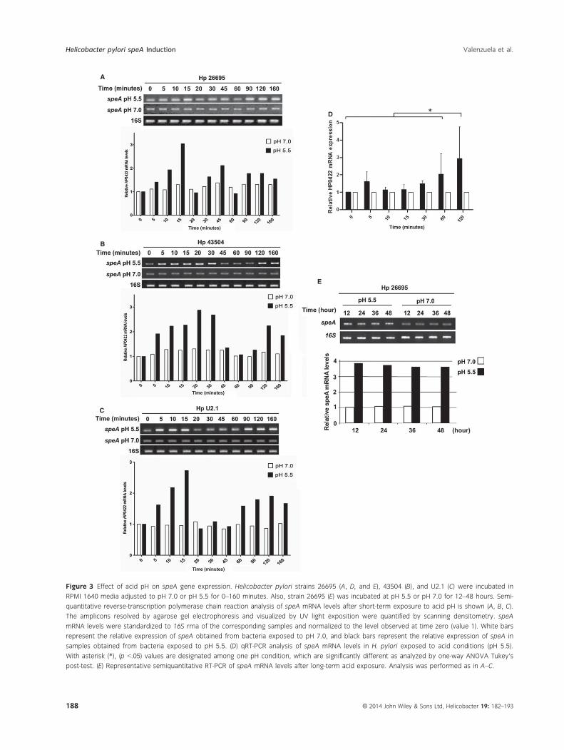
Effect of Acid pH on the speA Gene Transcription
Having identified that arginine enhances bacterial
growth in acid conditions, we next analyzed the effect
of acid exposure on the H. pylori speA gene expression.
H. pylori 26695, 43504, and U2.1 strains were incubated
in acid conditions over various time periods between 0
and 160 minutes, and the speA mRNA levels were ana-
lyzed using RT-PCR. In these experiments, we deter-
mined that when H. pylori 26695 bacteria were
incubated at pH 5.5, the speA mRNA levels experienced
a biphasic increase with a first peak at 15–20 minutes
and a second peak between 90 and 160 minutes
(Fig. 3A). Similar changes were observed with the
43504 and U2.1 strains (Fig. 3B,C, respectively).
We confirmed these results by measuring the speA
mRNA levels in the 26695 strain using qRT-PCR
(Fig. 3D). In these experiments, we observed a similar
progression of the mRNA expression, with an increase
at 10 minutes. Then, we concluded that speA is acutely
induced after acid exposure.
To determine whether the speA gene is overexpres-
sed during a long-term acid exposure, we cultured
H. pylori on TSA plates at pH 5.5 for 12, 24, 36, and
48 hours. Here, we observed an approximately 4.0-fold
increase in speA mRNA expression at 12 hours of acid
exposure (pH 5.5) (Fig. 3E). This increase was persis-
tent over the various times of incubation (Fig. 3E). No
differences in speA mRNA expression were observed at
neutral conditions (Fig. 3E). Altogether these results
suggest that speA expression is induced by acid external
pH. Loss of arginine-dependent acid resistance in E. coli
was not completely complemented by the H. pylori speA
gene.
E. coli can survive in extreme pH conditions by at
least three different mechanisms, one of which, AR3,
involves arginine decarboxylation. To determine
whether SpeA participates in a similar mechanism in
H. pylori, we designed a complementation test using an
adiA-deficient E. coli strain, because we have failed to
produce an H. pylori speA knockout mutant probably
106
107
108
109
Hp 43504
A
0 10 20 30 40 50103
104
105
Time (hour)
0 10 20 30 40 50
Time (hour)
Log
CFU
/mL
104
105
106
108
107
B
100
101
102
103
Log
CFU
/mL
pH7.0/+ArgpH7.0/–ArgpH5.5/+ArgpH5.5/–Arg
Hp U2.1
Figure 2 Effect of arginine supplementation on the growth of
H. pylori in acidic conditions. H. pylori strains 43504 (A) and U2.1 (B)
grown on TSA plates were used to inoculate nutritive broths at pH
7.0 and pH 5.5. Cultures were supplemented with or without 5 mmol/
L L-arginine (+/�Arg). Bacteria were grown at 37 °C for 48 hours. At
the indicated times, aliquots from each culture were taken, serially
diluted, and seeded on TSA plates. After 3 days of growth, CFU were
counted. Values from three independent experiments are shown
(mean � SD, p ≤.05).
© 2014 John Wiley & Sons Ltd, Helicobacter 19: 182–193 187
Valenzuela et al. Helicobacter pylori speA Induction
A
B
C
E
D
Figure 3 Effect of acid pH on speA gene expression. Helicobacter pylori strains 26695 (A, D, and E), 43504 (B), and U2.1 (C) were incubated in
RPMI 1640 media adjusted to pH 7.0 or pH 5.5 for 0–160 minutes. Also, strain 26695 (E) was incubated at pH 5.5 or pH 7.0 for 12–48 hours. Semi-
quantitative reverse-transcription polymerase chain reaction analysis of speA mRNA levels after short-term exposure to acid pH is shown (A, B, C).
The amplicons resolved by agarose gel electrophoresis and visualized by UV light exposition were quantified by scanning densitometry. speA
mRNA levels were standardized to 16S rrna of the corresponding samples and normalized to the level observed at time zero (value 1). White bars
represent the relative expression of speA obtained from bacteria exposed to pH 7.0, and black bars represent the relative expression of speA in
samples obtained from bacteria exposed to pH 5.5. (D) qRT-PCR analysis of speA mRNA levels in H. pylori exposed to acid conditions (pH 5.5).
With asterisk (*), (p <.05) values are designated among one pH condition, which are significantly different as analyzed by one-way ANOVA Tukey’s
post-test. (E) Representative semiquantitative RT-PCR of speA mRNA levels after long-term acid exposure. Analysis was performed as in A–C.
© 2014 John Wiley & Sons Ltd, Helicobacter 19: 182–193188
Helicobacter pylori speA Induction Valenzuela et al.

due to an essential character. As shown above, arginine
supplementation improves H. pylori growth under acid
conditions (Fig. 2). Thus, we tested whether or not
H. pylori SpeA could improve growth of E. coli BL21
ΔadiA under acid conditions in the presence of argi-
nine. As shown in Fig. 4, that was the case when
growth of the parental, ΔadiA mutant and comple-
mented (pET21a+ or pET-speA) E. coli strains were
compared in the presence or absence of 3 mmol/L argi-
nine at pH 4.0. Interestingly, speA expression both
restored and improved the deficient growth of the
BL21 ΔadiA in presence of arginine at pH 4.0 over a 5-
hour culture period. Given this result, we wondered
whether the speA expression could also protect against
a more lethal exposure to acid in a similar manner to
that observed in the E. coli AR3 system. As expected,
deletion of the adiA gene by allelic exchange resulted
in the complete loss of the arginine-dependent acid
resistance in the E. coli BL21 strain at pH 2.5 (Fig. 5A).
Phenotypic reversion in this strain was obtained by
recombinant expression of the autologous AdiA protein
after transforming the mutant bacteria with the pET-
adiA plasmid (Fig. 5A). However, recombinant expres-
sion of H. pylori speA gene in the E. coli BL21 strain did
not completely rescue the resistant phenotype when
compared with the positive control (pET-adiA). This
observation was particularly clear after a 3-hour acid
challenge.
Then, we conducted an assay to extract and identify
agmatine and putrescine from the E. coli and H. pylori
cultures. As shown in Fig. 5B, TLC analysis of butanol
extracts from E. coli cultures in synthetic medium
revealed the increasing presence of agmatine from
12 hours of culture onwards and, as expected, it was
accompanied by putrescine production. Note that
agmatine and putrescine were the only polyamine
compounds detected on TCL plates by the ninhydrin–
chloroform reagent. As anticipated by the acid resis-
0
5
10
15
20
25
30
35
Time (hour)
BL21BL21∆adiA/pET21aBL21∆adiA/pET-speA
0 1 2 3 4 5
CF
U(+
Arg
)/C
FU
(–A
rg)
Figure 4 Effect of the expression of H. pylori speA on the growth of
E. coli BL21ΔadiA mutant in acid conditions. BL21 and BL21ΔadiA/pET21a and BL21ΔadiA/pET-speA strains were grown overnight in LBG
medium at pH 5.0. Volumes of the three cell suspensions correspond-
ing to OD560 = 0.025 (approximately 1.8 9 07 CFU/mL) were used to
inoculate 15 mL of synthetic medium adjusted to pH 4.0 and supple-
mented with or without 3 mmol/L arginine. Cells were grown at 37 °C
for 0–5 hours, and samples were taken at 1-hour intervals for CFU
counting. Bars represent the CFU (+Arg)/CFU(�Arg) ratios from three
independent experiments � SD (*p ≤.05).
BL21 BL21 ∆adiA pET-speA pET-adiA pET21aBL21 ∆adiA
+Arg–Arg
Surv
ival
(%)
A
B
C
Std M 12 24 48
Agmatine
Putrescine
Time (hour)
Agmatine
Putrescine
E. coli ctl 26695 U2.1 26695 U2.1
pH 7.0 pH 5.0
Time (hour) 2 | 3 2 | 3 2 | 3 2 | 3 2 | 3
Figure 5 Heterologous expression of H. pylori SpeA protein in the
E. coli BL21 ΔadiA mutant. (A) Determination of arginine-dependent
acid resistance in E. coli. Bacteria were grown overnight in LBG med-
ium adjusted to pH 5.0. A 10-lL aliquot was taken from the culture,
added to 1 mL of acid shock medium pH 2.5, and incubated for 2 and
3 hours in the presence or absence of 3 mmol/L arginine. Survival
under each experimental condition is represented by the percentage
of cell viability (CFU) compared with bacteria maintained in synthetic
medium (pH 7.0). Data represent mean values and standard deviations
from three independent experiments (p <.05). Arginine-dependent
acid resistance was assayed in E. coli BL21, BL21 ΔadiA, and in the
BL21 ΔadiA strain complemented with either the pET-speA or pET-
adiA plasmids, in presence of 1 mmol/L IPTG. (B, C) Detection of agm-
atine and putrescine production in E. coli and H. pylori strains. (B)
E. coli was incubated at pH 7.0 for 12, 24, and 48 hours in synthetic
medium supplemented with 3 mmol/L arginine; (C) H. pylori was incu-
bated at pH 7.0 or 5.0 for 24 hours in TSA broth supplemented with
3 mmol/L arginine. For comparison, a butanol extract from E. coli cul-
ture (24 hours) is shown. Aliquots of butanol extracts from the cul-
tures were chromatographed on TLC plates and revealed with
ninhydrin. Agmatine and putrescine standards (3 and 5 lg, respec-
tively) and a noninoculated sample were routinely included as
controls.
© 2014 John Wiley & Sons Ltd, Helicobacter 19: 182–193 189
Valenzuela et al. Helicobacter pylori speA Induction

tance assay, polyamine production by the ΔadiA mutant
strain was undetectable, and speA complementation
was not suffice to fully restore polyamine production,
and agmatine and putrescine were poorly detected
(data not shown). Based on these results, we tested
agmatine and putrescine production directly in H. pylori
26695 and U2.1 cultures. Interestingly, both polyam-
ines were present in butanol extracts from H. pylori
grown at pH 5.0 (Fig. 5C), thus suggesting a role for an
arginine decarboxylase in an acid adaptation mecha-
nism, similar to the E. coli AR3. This result suggests that
under acidic conditions, H. pylori expels agmatine to
control pH homeostasis and is concordant with a previ-
ous report showing that agmatine levels are higher in
the gastric juice of H. pylori-infected patients than in
uninfected controls [34]. However, the amounts of
agmatine and putrescine produced by H. pylori U2.1
were significantly higher than those produced by the
H. pylori 26695 strain and notably less pronounced
when compared with E. coli. It is still not clearly under-
stood why the plasmid-borne speA gene (pET-speA)
failed to fully complement the acid resistance defective
phenotype of the E. coli adiA mutant. It is possible that
the pET-speA is not as active as the H. pylori chro-
mosomal gene or that the cloned fragment lacks a
downstream DNA element of the speA gen that partici-
pates in the acid resistance mechanism. According to
our results, the speA gene is the last ORF of that
operon, and our construction (pET-speA) does not con-
tain the DNA promoter elements that could be regulat-
ing its expression.
Transcriptional Factor Fur Regulates the
Expression of the speA Gene in the Acid
Response
Three H. pylori regulators have been implicated in acid
adaptation: NikR, Fur, and the ArsRS two-component
system [19]. The transcriptional factor Fur has been
characterized as a regulator of gene expression during
acid exposure in H. pylori [7]. Furthermore, fur knock-
out H. pylori strains present a sensitized phenotype to
acid challenge in association with downregulation of
several genes [7]. In agreement with this observation,
Clustal analysis (http://www.ebi.ac.uk/Tools/es/egi-bin/
jobresults.cgi/clustalw2/clustalw2-20090723-19004517
30.aln) revealed that putative binding sites for Fur
described previously by Ernst et al. [35], namely Fur,
Pfr I, and Pfr II boxes, actually are present in the speA
operon promoter (data not shown). Therefore, we won-
dered whether Fur is involved in the observed increase
in speA mRNA levels following acid exposure. To that
purpose, we isolated mRNA from H. pylori fur mutants
after long-term cultures in acid condition (pH 5.5, 12–
36 hours), and the speA mRNA levels were analyzed by
RT-PCR. Interestingly, we observed that the levels of
the speA mRNA remained unaltered in H. pylori
FUR504 strain (Fig. 6A,B). This lack of effect was in
open contrast with the responsiveness of the corre-
sponding parental strain (Fig. 6A,B). Interestingly, speA
gene transcription changes were related to changes in
protein levels. As shown in Fig. 6D, an in-house rabbit
polyclonal antibody raised against H. pylori SpeA pro-
tein (Fig. 6C) revealed a strong increase in the levels of
the SpeA protein after 24 hours of growth in acid con-
ditions. Also, the SpeA protein levels remained basically
unaltered when the Fur-negative strain was grown in
acid conditions in contrast to the control condition at
pH 7.0 (Fig. 6D, FUR504).
Adaptation of H. pylori to the acidic pH environment
involves a plethora of biochemical processes. H. pylori
responds to acid stress through multimechanisms includ-
ing many proteins and depending on the acidity level
[36]. Urease has been shown to play an important role in
this process [4]. A urease-independent acid resistance
mechanism in H. pylori has been also reported [14,37].
This mechanism is regulated by the ferric uptake regula-
tor Fur [7]. Our results are in agreement with those
reported by Bijlsma et al. [38] as to the growth defect of
a fur mutant at low pH is consistent with findings of acid-
sensitive fur mutants. This might be explained by failure
in the increase in transcription of genes that are involved
in the control of pH homeostasis under Fur regulation. In
this study, the speA gene downregulation observed in the
H. pylori fur knockout mutant supports that postulate.
Such finding strongly supports our hypothesis that Fur is
involved in the urease-dependent and urease-indepen-
dent acid resistance-associated arginine decarboxylase
activity in H. pylori [7].
Recently, we demonstrated that the arginine-66
residue of the transcriptional factor Fur is necessary for
its regulatory function in the acid adaptation mecha-
nisms of H. pylori [39]. However, the mechanism by
which Fur influences arginine decarboxylase and acid
resistance in H. pylori remains unknown and requires
further investigation on Fur in association with gastric
acidity.
Concluding Remarks
Our results showed that the speA gene (adiA in E. coli) is
induced by acid conditions and that growth of H. pylori is
enhanced in the presence of arginine, the amino acid
substrate of the arginine decarboxylase, SpeA. Moreover,
upregulation of the speA mRNA and protein levels was
observed early after H. pylori exposure to pH 5.5 and also
© 2014 John Wiley & Sons Ltd, Helicobacter 19: 182–193190
Helicobacter pylori speA Induction Valenzuela et al.

when H. pylori was cultured under acidic conditions,
thus suggesting that the arginine decarboxylase activity
could play a role in protecting the bacterium from the
acid pH and in contributing to maintain pH homeo-
stasis through the exchange of intracellular protons
while expelling agmatine to the medium. Interestingly,
enhanced speA mRNA levels following acid stress were
totally dependent on Fur presence in either short- or
long-term acid exposures. Agmatine production by
H. pylori was detectable when bacteria were grown at pH
5.0, although the amounts detected were notably lower
than those of E. coli cultures. This observation would
suggest that speA generates agmatine and contributes to
the habituation of H. pylori to the acid environment
rather than being part of an acute response to the acidic
pH stress, as anticipated in the complementation acid
stress assay at pH 2.5 (Fig. 5). Conversely, our results
suggest that the SpeA activity constitutes a urease-
independent acid resistance system in H. pylori.
Acknowledgements and Disclosures
This work was funded by Grants No. 1085193 (HT), 1120126
(HT) and 11121239 (OC) from FONDECYT-Chile. This study
was also part of a Thesis (AC) submitted in partial fulfillment
of requirements for a Degree in Biology at the Pontificia Uni-
versidad Cat�olica de Valparaiso. We thank Dr. Remigio L�opez-
Sol�ıs for his critical analysis of the manuscript. We also thank
Mr. Nicanor Villaroel for his technical support.
Competing interests: the authors do not have any disclosure
relevant to the manuscript.
0
0.5
1
1.5
2
2.5
3
3.5
4
pH 7 5.5 7 5.5 7 5.5
speA
16S
Time (hour)
pH
speA
16S
Time (hour)
12 24 36
7 5.5 7 5.5 7 5.5
12 24 36
pH
Time (hour)
7 5.5 7 5.5 7 5.5
pH 7 5.5 7 5.5 7 5.5
12 24 36
Time (hour) 12 24 36
pH
Time (hour)
7 5.5 7 5.5 7 5.5
12 24 36
43504
FUR504
A B
C
SpeA
43504
FUR504
SpeA
D
SpeA
GST-SpeA
H. pylori43504
H. pyloriFUR504
E. colipGEX-speA
FUR504
WT
Rel
ativ
e sp
eA m
RN
A le
vels
Figure 6 Effect of Fur deletion on the acid pH response of speA gene. H. pylori 43504 and the isogenic fur::cat mutant strains were incubated at
pH 5.5 or pH 7.0 for 0–36 hours. (A) Representative RT-PCR analyses of speA mRNA levels at various times of incubation are shown. (B) Experi-
mental data were analyzed and represented as described in the legend to Fig. 3C. (C) Anti-SpeA antibody validation. Immunoblot analysis of the
SpeA protein levels in H. pylori 43504, the recombinant SpeA-GST protein expressed in E. coli (pGEX-speA) induced with IPTG and in H. pylori
FUR504. (D) Immunoblot analyses of SpeA in the H. pylori 43504 and FUR504 strains incubated at pH 5.5 or pH 7.0 for 0–36 hours. A representa-
tive blot is shown.
© 2014 John Wiley & Sons Ltd, Helicobacter 19: 182–193 191
Valenzuela et al. Helicobacter pylori speA Induction

References
1 Suerbaum S, Michetti P. Helicobacter pylori infection. N Engl J
Med 2002;347:1175–86.
2 Schreiber S, Konradt M, Groll C, Scheid P, Hanauer G, Werling
HO, Josenhans C, Suerbaum S. The spatial orientation of
Helicobacter pylori in the gastric mucus. Proc Natl Acad Sci USA
2004;101:5024–9.
3 Chu S, Tanaka S, Kaunitz JD, Montrose MH. Dynamic regula-
tion of gastric surface pH by luminal pH. J Clin Invest
1999;103:605–12.
4 Sachs G, Weeks DL, Melchers K, Scott DR. The gastric biology
of Helicobacter pylori. Annu Rev Physiol 2003;65:349–69.
5 Tomb JF, White O, Kerlavage AR, et al. The complete genome
sequence of the gastric pathogen Helicobacter pylori. Nature
1997;388:539–47.
6 Wen Y, Marcus EA, Matrubutham U, Gleeson MA, Scott DR,
Sachs G. Acid-adaptive genes of Helicobacter pylori. Infect Immun
2003;71:5921–39.
7 Valenzuela M, Albar JP, Paradela A, Toledo H. Helicobacter pylori
exhibits a fur-dependent acid tolerance response. Helicobacter
2011;16:189–99.
8 Sachs G, Weeks DL, Wen Y, Marcus EA, Scott DR, Melchers K.
Acid acclimation by Helicobacter pylori. Physiology (Bethesda)
2005;20:429–38.
9 Mobley HL, Island MD, Hausinger RP. Molecular biology of
microbial ureases. Microbiol Rev 1995;59:451–80.
10 Bauerfeind P, Garner R, Dunn BE, Mobley HL. Synthesis and
activity of Helicobacter pylori urease and catalase at low pH. Gut
1997;40:25–30.
11 Hawtin PR, Stacey AR, Newell DG. Investigation of the struc-
ture and localization of the urease of Helicobacter pylori using
monoclonal antibodies. J Gen Microbiol 1990;136:1995–2000.
12 Phadnis SH, Parlow MH, Levy M, Ilver D, Caulkins CM, Con-
nors JB, Dunn BE. Surface localization of Helicobacter pylori ure-
ase and a heat shock protein homolog requires bacterial
autolysis. Infect Immun 1996;64:905–12.
13 Scott DR, Marcus EA, Weeks DL, Sachs G. Mechanisms of acid
resistance due to the urease system of Helicobacter pylori. Gastro-
enterology 2002;123:187–95.
14 Toledo H, Valenzuela M, Rivas A, Jerez CA. Acid stress
response in Helicobacter pylori. FEMS Microbiol Lett 2002;213:67–
72.
15 Allan E, Clayton CL, McLaren A, Wallace DM, Wren BW.
Characterization of the low-pH responses of Helicobacter pylori
using genomic DNA arrays. Microbiology 2001;147(Pt 8):2285–
92.
16 Moran AP, Knirel YA, Senchenkova SN, Widmalm G, Hynes
SO, Jansson PE. Phenotypic variation in molecular mimicry
between Helicobacter pylori lipopolysaccharides and human
gastric epithelial cell surface glycoforms. Acid-induced
phase variation in Lewis(x) and Lewis(y) expression by
H. Pylori lipopolysaccharides. J Biol Chem 2002;277:5785–
95.
17 Huesca M, Goodwin A, Bhagwansingh A, Hoffman P, Ling-
wood CA. Characterization of an acidic-pH-inducible stress
protein (hsp70), a putative sulfatide binding adhesin, from Heli-
cobacter pylori. Infect Immun 1998;66:4061–7.
18 van Vliet AH, Kuipers EJ, Waidner B, Davies BJ, de Vries N,
Penn CW, Vandenbroucke-Grauls CM, Kist M, Bereswill S,
Kusters JG. Nickel-responsive induction of urease expression in
Helicobacter pylori is mediated at the transcriptional level. Infect
Immun 2001;69:4891–7.
19 Bury-Mone S, Thiberge JM, Contreras M, Maitournam A, Lab-
igne A, De Reuse H. Responsiveness to acidity via metal ion
regulators mediates virulence in the gastric pathogen Helicobact-
er pylori. Mol Microbiol 2004;53:623–38.
20 Castanie-Cornet MP, Penfound TA, Smith D, Elliott JF, Foster
JW. Control of acid resistance in Escherichia coli. J Bacteriol
1999;181:3525–35.
21 Castanie-Cornet MP, Foster JW. Escherichia coli acid resistance:
cAMP receptor protein and a 20 bp cis-acting sequence control
pH and stationary phase expression of the gadA and gadBC
glutamate decarboxylase genes. Microbiology 2001;147(Pt
3):709–15.
22 Gong S, Richard H, Foster JW. YjdE (AdiC) is the arginine:agm-
atine antiporter essential for arginine-dependent acid resistance
in Escherichia coli. J Bacteriol 2003;185:4402–9.
23 Valenzuela M, Perez-Perez G, Corvalan AH, Carrasco G, Urra
H, Bravo D, Toledo H, Quest AF. Helicobacter pylori-induced loss
of the inhibitor-of-apoptosis protein survivin is linked to gastri-
tis and death of human gastric cells. J Infect Dis 2010;202:1021–
30.
24 Datsenko KA, Wanner BL. One-step inactivation of chromo-
somal genes in Escherichia coli K-12 using PCR products. Proc
Natl Acad Sci USA 2000;97:6640–5.
25 Chung CT, Niemela SL, Miller RH. One-step preparation of
competent Escherichia coli: transformation and storage of bacte-
rial cells in the same solution. Proc Natl Acad Sci USA
1989;86:2172–5.
26 Goldschmidt MC, Lockhart BM. Rapid methods for determining
decarboxylase activity: arginine decarboxylase. Appl Microbiol
1971;22:350–7.
27 Lange R, Hengge-Aronis R. Identification of a central regulator
of stationary-phase gene expression in Escherichia coli. Mol
Microbiol 1991;5:49–59.
28 Loewen PC, Hengge-Aronis R. The role of the sigma factor
sigma S (KatF) in bacterial global regulation. Annu Rev Microbiol
1994;48:53–80.
29 Richard H, Foster JW. Escherichia coli glutamate- and arginine-
dependent acid resistance systems increase internal pH and
reverse transmembrane potential. J Bacteriol 2004;186:
6032–41.
30 Sharma CM, Hoffmann S, Darfeuille F, et al. The primary tran-
scriptome of the major human pathogen Helicobacter pylori. Nat-
ure 2010;464:250–5.
31 Mao F, Dam P, Chou J, Olman V, Xu Y. DOOR: a database for
prokaryotic operons. Nucleic Acids Res 2009;37(Database issue):
D459–63.
32 Muraoka H, Kobayashi I, Hasegawa M, Saika T, Toda H,
Nishida M, Suzuki J, Mine T, Fujita T. [Urease-negative
Helicobacter pylori isolates from gastrointestinal mucosa of
patients with peptic ulcer]. Kansenshogaku Zasshi 1997;71:
1216–20.
33 Mine T, Muraoka H, Saika T, Kobayashi J. Characteristics of a
clinical isolate of urease-negative Helicobacter pylori and its abil-
ity to induce gastric ulcers in Mongolian gerbils. Helicobacter
2005;10:125–31.
34 Molderings GJ, Burian M, Homann J, Nilius M, G€othert M.
Potential relevance of agmatine activity as a virulence factor of
Helicobacter pylori. Dig Dis Sci 1999;44:2397–404.
35 Ernst FD, Bereswill S, Waidner B, Stoof J, Mader U, Kusters
JG, Kuipers EJ, Kist M, van Vliet AH, Homuth G.
Transcriptional profiling of Helicobacter pylori Fur- and
iron-regulated gene expression. Microbiology 2005;151(Pt
2):533–46.
© 2014 John Wiley & Sons Ltd, Helicobacter 19: 182–193192
Helicobacter pylori speA Induction Valenzuela et al.

36 Shao C, Zhang Q, Tang W, Qu W, Zhou Y, Sun Y, Yu H, Jia J.
The changes of proteomes components of Helicobacter pylori in
response to acid stress without urea. J Microbiol 2008;46:331–
7.
37 Bijlsma JJ, Gerrits MM, Imamdi R, Vandenbroucke-Grauls CM,
Kusters JG. Urease-positive, acid-sensitive mutants of Helicobact-
er pylori: urease-independent acid resistance involved in growth
at low pH. FEMS Microbiol Lett 1998;167:309–13.
38 Bijlsma JJ, Waidner B, Vliet AH, Hughes NJ, Hag S, Bereswill S,
Kelly DJ, Vandenbroucke-Grauls CM, Kist M, Kusters JG. The
Helicobacter pylori homologue of the ferric uptake regulator is
involved in acid resistance. Infect Immun 2002;70:606–11.
39 Toledo H, Villafaena C, Valenzuela M, Lopez-Solis R. Arginine
66 residue of Fur is required for the regulatory function of this
protein in the acid adaptation mechanism of Helicobacter pylori.
Helicobacter 2011;17:16–22.
© 2014 John Wiley & Sons Ltd, Helicobacter 19: 182–193 193
Valenzuela et al. Helicobacter pylori speA Induction